Cystic lymph node metastases of squamous cell carcinoma of Waldeyer's ring origin. (1/35)
We analysed in a retrospective study the frequency of cystic lymph node (LN) metastases in neck dissection specimens of 123 patients with primary squamous cell carcinoma (SCC) arising in the palatine tonsils (62 M/14 F), the base of the tongue (38 M/5 F) and the nasopharynx (2 M/2 F). Eighty-two per cent of patients had metastases (64 tonsillar SCC, 33 base of tongue SCC and all four nasopharynx SCC) in 368 LN of a total 2298 sampled LN. Thirty-nine per cent of patients had exclusively solid metastases and 37% of patients had exclusively cystic metastases. A total of 62 patients had some signs of cyst formation in one or more metastatically affected LN (27 with only histological evidence of cyst formation with luminal diameters < 5 mm, 35 with clinically detectable cyst with luminal diameter > 5 mm). Cystic metastases were more common in patients with SCC of the base of the tongue (P = 0.005), while solitary clinically evident cystic metastasis with lumina > 5 mm were found exclusively in tonsillar carcinoma (P = 0.024). In comparison with solid metastases, cyst formation was associated with N-categories (N2b and N3, P = 0.005) in SCC of the base of the tongue origin. No such association was observed for tonsillar SCC (P = 0.65). The primary mechanism of cyst formation was cystic degeneration. (+info)Solitary nodal metastases presenting as branchial cysts: a diagnostic pitfall. (2/35)
Two patients with metastatic squamous cell carcinoma are presented. Both were initially clinically diagnosed as branchial cysts. The importance of a full examination of the upper aerodigestive tract, and fine needle aspiration cytology is emphasised to avoid the possibility of excision as a branchial cyst, which could lead to tumour dissemination. (+info)Second branchial cleft cysts: variability of sonographic appearances in adult cases. (3/35)
BACKGROUND AND PURPOSE: Previous reports have suggested that second branchial cleft cysts (BCCs) appear on sonograms as well-defined, cystic masses with thin walls and posterior enhancement. Previous CT and MR imaging findings, however, have indicated heterogeneity of these masses, and, in our experience, sonography also shows a similar variable appearance. In this communication, we report the cases of 17 patients with second BCCs and document the variability of sonographic patterns. METHODS: The sonograms of 17 adults with second BCCs were reviewed. Only patients with surgical or cytologic evidence of BCCs were included in this study. The features evaluated were the location, internal echogenicity, posterior enhancement, and presence of septa and fistulous tract. RESULTS: Four patterns of second BCCs were identified: anechoic (41%), homogeneously hypoechoic with internal debris (23.5%), pseudosolid (12%), and heterogeneous (23.5%). The majority (70%) showed posterior enhancement. All were situated in their classical location, posterior to the submandibular gland, superficial to the carotid artery and internal jugular vein, and closely related to the medial and anterior margin of the sternomastoid muscle. Fourteen (82%) of the 17 BCCs had imperceptible walls, and all were well defined. For none of the patients was a fistulous tract revealed by sonography; the presence of internal septations was revealed for three patients. CONCLUSION: As previously suggested by CT and MR imaging findings, sonography reinforces that second BCCs in adults are not simple cysts but have a complex sonographic pattern ranging from a typical anechoic to a pseudosolid appearance. (+info)Intrathyroidal lymphoepithelial (branchial) cyst: sonographic features of a rare lesion. (4/35)
Intrathyroidal lymphoepithelial cysts are rare, and only 15 such cases have been reported. Although sonography has been performed in some cases, the findings have not been discussed previously. Despite its rarity, the sonographic appearances of this lesion are similar to those of other commonly encountered congenital cystic lesions in the head and neck, such as thyroglossal duct cysts and second branchial cleft cysts, and this may provide a clue to its diagnosis. We describe the sonographic appearances of intrathyroidal lymphoepithelial cysts. (+info)Parapharyngeal second branchial cyst manifesting as cranial nerve palsies: MR findings. (5/35)
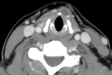
SUMMARY: We report the MR findings of parapharyngeal branchial cleft cyst manifesting as multiple, lower cranial nerve palsies in a 35-year-old woman. On MR images, a well-marginated cystic mass was detected in the right parapharyngeal space, with displacement of both the right internal carotid artery and the right internal jugular vein on the posterolateral side. The cyst contained a whitish fluid that was slightly hyperintense on T1-weighted images and slightly hypointense to CSF on T2-weighted images. No enhancement on contrast-enhanced T1-weighted images was present. The right side of the tongue showed high signal intensity on T2-weighted images, suggesting denervation. (+info)Infected branchial cleft cyst due to Bordetella bronchiseptica in an immunocompetent patient. (6/35)
A healthy 23-year-old man with fever and a tender mass in his right anterior neck was found to have a branchial cleft cyst infected with Bordetella bronchiseptica. Initial testing suggested a Brucella species, but further laboratory testing identified the organism definitively. B. bronchiseptica infection in healthy adults is an unusual event. (+info)A case of second branchial cleft cyst with oropharyngeal presentation. (7/35)
Second branchial cleft cysts are the most common type of branchial abnormalities and usually found high in the neck. Oropharyngeal presence of branchial cleft cyst is very rare. We report a case of oropharyngeal branchial cleft cyst in 2-yr-old girl with about 1 x 1 cm sized cystic mass, which had not any specific symptom. It was removed completely under impression of mucocele and did not have tract-like structure. However, cyst had a squamous epithelium-lined wall with lymphoid aggregation in histopathologic study, which was characteristic finding of branchial cleft cyst. Patient discharged without any complication and there was no evidence of recurrence for 18 months follow-up. We review reported oropharyngeal or nasopharyngeal presentation of these cases in English literature and embryological explanation. (+info)Pathological analysis of congenital cervical cysts in children: 20 years of experience at Chang Gung Memorial Hospital. (8/35)
BACKGROUND: Congenital cervical cysts are frequently encountered in pediatric populations, and constitute one of the most intriguing areas of pediatric pathology. This report analyzes cervical cysts in Taiwanese children diagnosed at Chang Gung Memorial Hospital (CGMH) over the past 20 years. The pathologic and clinical findings are reviewed. METHODS: Files on 331 patients under the age of 18 years, with a diagnosis of congenital cervical cyst at CGMH from January 1, 1983 to June 30, 2002, were retrieved from the Department of Pathology. There were 204 boys and 127 girls. We reviewed the histology of all cases and correlated it with clinical information in the medical records. RESULTS: Thyroglossal duct cysts, the most common congenital neck cyst, accounted for 54.68% of all cases, followed by cystic hygromas (25.08%), branchial cleft cysts (16.31%), bronchogenic cysts (0.91%), and thymic cysts (0.30%). Nine cases (2.72%) remained unclassified. CONCLUSIONS: This is the largest series regarding pediatric cervical cysts in the literature to date. Thyroglossal duct cysts were the most common congenital cervical cyst encountered. Our experience indicates that each type of cyst has its unique location in the neck and is highly associated with its embryonic origin. Complete and precise clinical information is a prerequisite in order for pathologists to make accurate diagnoses of congenital cervical cysts. (+info)A branchioma is a benign tumor formed from the remnants of the branchial apparatus, typically appearing along the lateral neck region in children and adults, characterized by slow growth and potential complications due to airway or digestive tract obstruction if left untreated.
A branchioma is a benign (non-cancerous) tumor that develops from remnants of the branchial apparatus, which are structures that form in the embryo during fetal development and give rise to parts of the head and neck. Branchiomas are also known as branchial cleft cysts or branchial apparatus remnants.
Branchioma typically occurs in the neck region and can cause a variety of symptoms, such as a painless lump, difficulty swallowing, or breathing problems if the tumor is large enough to compress the airway. Treatment usually involves surgical removal of the tumor.
 Branchioma (Branchial Cysts): Symptoms, Diagnosis and Treatment - Symptoma
Branchioma (Branchial Cysts): Read more about Symptoms, Diagnosis, Treatment, Complications, Causes and Prognosis. ... You may have a branchioma if you have an olive or egg-sized growth under the skin on the side of your neck, a moveable mass ... Causes What Causes a Branchioma to Grow? There are small grooves in the neck that appear during early fetal development. These ... branchiogenous branchiogenous branchioma branchiomeric Brandt-Andrews maneuver Brandt-Daroff maneuvers Branham sign sign ...
 Neck Cysts: Practice Essentials, Problem, Epidemiology
 No questions in Mediastinal Cyst - lookformedical.com
Chia-Yu Wu - 指紋 - 臺北醫學大學
 Branchial anomalies in the pediatric population<...
 Búsqueda | BVS Bolivia
Branchioma is a rare benign neoplasm occurring in the lower neck. Occurrence of malignant neoplasms arising in branchioma is ... while the low-grade component and branchioma component were negative for p53. Targeted sequencing analysis for the branchioma ... Adenocarcinoma arising in branchioma with a KRAS and TP53 mutation. Taniguchi, Natsuki; Satou, Akira; Ito, Takanori; Nakaguro, ... Here, we report a case of adenocarcinoma arising in branchioma. A 62-year-old man had a right supraclavicular mass measuring ...
Neck Cysts: Background, Problem, Epidemiology
淋巴上皮囊肿恶变<strong>1<...
 Pancreatic Cyst (medical concept explorer)
Neck Mass: Causes & Reasons - Symptoma
 GYEMSZI - MOB
 Bronchogenic Cyst | Profiles RNS
Colloid Cysts | Profiles RNS
Mesenteric Cyst | Profiles RNS
 Bronchogenic Cyst | Profiles RNS
Tarlov Cysts | Profiles RNS
 Words starting with b
 MeSH Browser
 Acanthoma etymology in English | Etymologeek.com
 Biphenotypic Branchioma: A Better Name Than Ectopic Hamartomatous Thymoma for a Neoplasm with HRAS Mutation - PubMed
Biphenotypic Branchioma: A Better Name Than Ectopic Hamartomatous Thymoma for a Neoplasm with HRAS Mutation Lester D R Thompson ... Biphenotypic Branchioma: A Better Name Than Ectopic Hamartomatous Thymoma for a Neoplasm with HRAS Mutation Lester D R Thompson ... The name biphenotyic branchioma more correctly reflects the true nature of this dual mesoderm and endoderm derived tumor ... Ectopic hamartomatous thymoma (biphenotypic branchioma): A case report and review of the literature. Cui W, Li D, Liu Y, Pang Y ...
 IndexCat
Biomarkers Search
Neck Cysts Treatment & Management: Medical Therapy, Surgical Therapy, Preoperative Details
MeSH Browser
Branchioma Preferred Term Term UI T005515. Date01/01/1999. LexicalTag NON. ThesaurusID NLM (1966). ... Branchioma Preferred Concept UI. M0002890. Scope Note. A tumor derived from branchial epithelium or branchial rests. (Dorland, ... Branchioma. Tree Number(s). C04.182.117. Unique ID. D001935. RDF Unique Identifier. http://id.nlm.nih.gov/mesh/D001935 ...
Human Genome Epidemiology Literature Finder|Home|PHGKB
 DeCS
DeCS
IndexCat
 Lymphocele | Harvard Catalyst Profiles | Harvard Catalyst
 Purchase Vytorin Purchase Online Canada Perth, How to order vytorin generic does it work
Pesquisa | Portal Regional da BVS
Branchioma (previously called ectopic hamartomatous thymoma, branchial anlage mixed tumor, or thymic anlage tumor) is a rare ... Branchioma with a nested/organoid morphology: molecular profiling of a distinctive potentially misleading variant and ... Histology revealed classical branchioma areas merging with nested/organoid cellular component lacking conventional features of ... We herein report the histological, immunohistochemical, and molecular genetic analysis of a branchioma with a nested/organoid ( ...
t
Urachal Cyst | Profiles RNS
Thyroglossal Cyst | Profiles RNS
Farquhar DR, Rawal RB, Masood MM, McClain WG, Kilpatrick LA, Rose AS, Zdanski CJ. Outpatient management and surgeon specialty for thyroglossal duct cyst excision: A retrospective analysis of 377 patients and 30-day outcomes in the American College of Surgeons NSQIP-P Database. Clin Otolaryngol. 2018 10; 43(5):1402-1406 ...
Mediastinal Cyst | Profiles RNS
Cysts of one of the parts of the mediastinum: the superior part, containing the trachea, esophagus, thoracic duct and thymus organs; the inferior middle part, containing the pericardium; the inferior anterior part containing some lymph nodes; and the inferior posterior part, containing the thoracic duct and esophagus ...