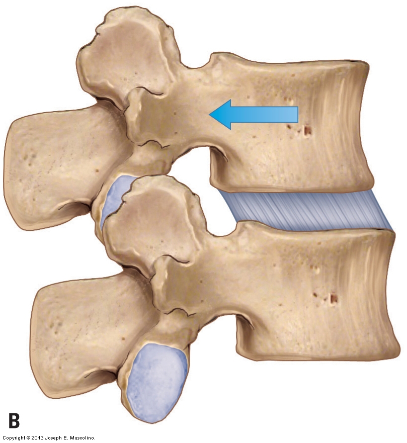
Zygapophyseal Joint
Anatomy of deer spine and its comparison to the human spine. (1/250)
The anatomical parameters of the thoracic and lumbar regions of the deer spine were evaluated and compared with the existing data of the human spine. The objective was to create a database for the anatomical parameters of the deer spine, with a view to establish deer spine as a valid model for human spine biomechanical experiments in vitro. To date, the literature has supported the use of both calf and sheep spines as a suitable model for human spine experiments as the difficulty in procuring the human cadaveric spines is well appreciated. With the advent of Bovine Spongiform Encephalopathy (BSE) and its likely transmission to human in form of new variant Creutzfeld Jakob disease (CJD), there is a slight risk of transmission to humans through food chain if proper precautions for disposal of specimen are not adhered to. There is also a significant risk of transmission through direct inoculation to the researchers (Wells et al. Vet. Rec., 1998:142:103-106), working with infected bovine and sheep spine. The deer spines are readily available and there are no reported cases of deer being carriers of prion diseases (Ministry of Agriculture, Fisheries and Food, 1998). Six complete deer spines were measured to determine 22 dimensions from the vertebral bodies, endplates, disc, pedicles, spinal canal, transverse and spinous processes, articular facets. This was compared with the existing data of the human spine in the literature. The deer and human vertebrae show many similarities in the lower thoracic and upper lumbar spine, although they show substantial differences in certain dimensions. The cervical spine was markedly different in comparison. The deer spine may represent a suitable model for human experiments related to gross anatomy of the thoracic and lumbar spine. A thorough database has been provided for deciding the validity of deer spine as a model for the human spine biomechanical in vitro experiments. (+info)Hangman's fracture: the relationship between asymmetry and instability. (2/250)
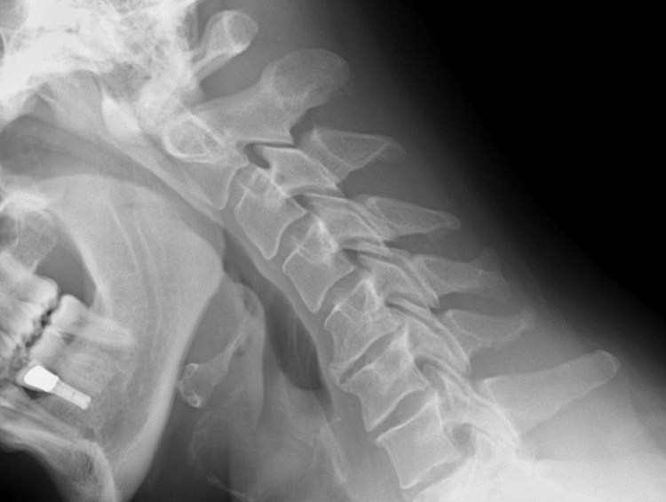
There is ambiguity concerning the nomenclature and classification of fractures of the ring of the second cervical vertebra (C2). Disruption of the pars interarticularis which defines true traumatic spondylolisthesis of C2, is often wrongly called a pedicle fracture. Our aim in this study was to assess the influence of asymmetry on the anatomical and functional outcome and to evaluate the criteria of instability established by Roy-Camille et al. We studied the plain radiographs and CT scans of 24 patients: 13 were judged to be asymmetrical, ten were considered unstable and 14 stable. Treatment was with a Minerva jacket in 15 fractures and by operation in nine. Surgery was undertaken in patients with severe C2 to C3 sprains. One patient with an unstable lesion refused operation and was treated conservatively with a poor radiological result. Our study showed that asymmetry of the fracture did not affect the outcomes of treatment and should not therefore influence decisions in treatment. The criteria of Roy-Camille seem to be reliable and useful. We prefer the posterior approach to the cervical spine, which allows both stabilisation of the fracture and correction of a local kyphosis. (+info)The orientation of the articular facets of the zygapophyseal joints at the cervical and upper thoracic region. (3/250)
Knowledge of the orientation of facet joints in the cervical and upper thoracic region is important for understanding the biomechanical properties and clinical conditions relevant to the neck. The study was undertaken on dry macerated bones from 30 adult male human vertebral columns. The orientation of the superior articular facets in relation to their inclination with the sagittal and transverse planes was examined between C3 and T3 vertebrae in each column. The linear dimensions of the superior articular facets and the width/height ratio were also calculated. The results show that all vertebrae at C3 level and 73% at C4 level displayed posteromedially facing superior articular facets. Similarly at T1 level (C7/T1 joint) and below, all columns showed posterolaterally facing superior articular facets. The level of change in orientation, from posteromedial to posterolateral facing superior facets, was not constant and occurred anywhere between C4 (C3/C4 joint) and T1 (C7/T1 joint). The change in orientation followed 2 different patterns, i.e. sudden or gradual. The C6 vertebra (C5/C6 joint) was the most frequent site to show the transition. The shape of the superior articular facets was circular to oval at C3, C4 and C5 levels and gradually changed to a transversely elongated surface at C7 and T1. These findings correlate well with various cervical movements and associated clinical conditions. (+info)An anatomical investigation of the human cervical facet capsule, quantifying muscle insertion area. (4/250)
Facet capsule injury has been hypothesised as a mechanism for neck pain. While qualitative studies have demonstrated the proximity of neck muscles to the cervical facet capsule, the magnitude of their forces remains unknown owing to a lack of quantitative muscle geometry. In this study, histological techniques were employed to quantify muscle insertions on the human cervical facet capsule. Computerised image analysis of slides stained with Masson's trichrome was performed to characterise the geometry of the cervical facet capsule and determine the total insertion area of muscle fibres into the facet capsule for the C4-C5 and C5-C6 joints. Muscle insertions were found to cover 22.4+/-9.6% of the capsule area for these cervical levels, corresponding to a mean muscle insertion area of 47.6+/-21.8 mm2. The magnitude of loading to the cervical facet capsule due to eccentric muscle contraction is estimated to be as high as 51 N. When taken in conjunction with the forces acting on the capsular ligament due to vertebral motions, these forces can be as high as 66 N. In that regard, these anatomical data provide quantitative evidence of substantial muscle insertions into the cervical facet capsular ligament and provide a possible mechanism for injury to this ligament and the facet joint as a whole. (+info)Orientation and tropism of lumbar facet joints in degenerative spondylolisthesis. (5/250)
The orientation and tropism of the lumbar facet joints at L4-5 level was assessed by magnetic resonance imaging (MRI) in 53 patients with degenerative L4-5 spondylolisthesis and 53 age- and sex-matched normal control subjects. The degree of disc degeneration at the L4-5 level and of vertebral slip on lateral radiographs was also evaluated. Patients with degenerative spondylolisthesis had more sagittally orientated facet joints (P < 0.01) and more significant facet joint tropism (P < 0.05) than normal control subjects. For patients with degenerative spondylolisthesis, the facet joint tropism was significantly correlated with the degree of disc degeneration (P < 0.05). The results suggest that morphological abnormalities of the lumbar facet joints are a predisposing factor in the development of degenerative spondylolisthesis. (+info)Comparative and functional anatomy of the mammalian lumbar spine. (6/250)
As an essential organ of both weight bearing and locomotion, the spine is subject to the conflict of providing maximal stability while maintaining crucial mobility, in addition to maintaining the integrity of the neural structures. Comparative morphological adaptation of the lumbar spine of mammals, especially in respect to locomotion, has however received only limited scientific attention. Specialised features of the human lumbar spine, have therefore not been adequately highlighted through comparative anatomy. Mathematical averages were determined of 14 measurements taken on each lumbar vertebrae of ten mammalian species (human, chimpanzee, orang-utan, kangaroo, dolphin, seal, Przewalski's horse, cheetah, lama, ibex). The revealed traits are analysed with respect to the differing spinal loading patterns. All examined mammalian lumbar spines suggest an exact accommodation to specific biomechanical demands. The lumbar spine has reacted to flexion in a predominant plane with narrowing of the vertebral bodies in quadrupeds. Torsion of the lumbar spine is withstood by an increase in the transverse distance between the inferior articular processes in the upper lumbar spine in primates, but lower lumbar spine in humans, quadrupeds and the seal. Sagittal zygapophyseal joint areas resist torsion in the seal and humans. Ventral shear is resisted by frontal zygapophyseal joint areas in humans and primates, and dorsal shear by encompassing joints in the ibex. The human fifth lumbar vertebra is remarkable in possessing the largest endplate surface area and the widest distance between the inferior articular processes, as an indicator of the high degree of axial load and torsion in bipedalism. (+info)Brucellar spondylitis: MRI findings. (7/250)
This study was carried out to identify the distinguishing features of brucellosis on magnetic resonance imaging (MRI). MRI examinations were performed in 14 patients with spinal brucellosis. A 1-T Magnetom (Erlangen, Siemens) was used to obtain T1-weighted (TR/TE 500/30) and T2-weighted (TR/TE 2000/80/20) spin echo sequences, in both sagittal and axial planes. Thirty-three percent of the vertebrae and 18 levels of disc were involved in the 14 brucellar spondylitis cases. Eleven patients (79.8%) with discitis revealed anterior superior vertebral body involvement. Fourteen (77.7%) of the levels with discitis displayed soft tissue swelling without presence of abscess formation. Seven facet joints of five patients with discitis displayed signal increase after contrast enhancement. Vertebral body signal changes without morphologic changes marked signal increase in the intervertebral disc on T2-weighted and contrast-enhanced sequences, and soft tissue involvement without abscess formation can be accepted as specific MRI features of brucellar spondylitis. The facet joint signal changes following contrast enhancement is another MRI sign of spinal brucellosis, which has not been mentioned so far. (+info)Morphometrical study of the joint surface and capsule of the lumbar zygapophysial joint with special reference to their laterality. (8/250)
Using 26 osteoligamentous lumbar vertebral columns (260 facets), we morphometrically investigated the cartilagenous joint surface, inner capsular surface and capsular thickness. We also examined whether the subcapsular pocket was present and, if present, how far it extended along the joint margin. The proportion of the inner capsular area in the total joint surface area in a facet (the capsular-joint surface ratio) was hypothesized to correspond to the potential looseness (or tightness) of the facet. The absolute data themselves seemed to be useful for better understanding of the joint morphology. However, further evaluations of the differences between segments, left/right differences, individual segmental fluctuation patterns and correlations between parameters provided a novel classification of specimens according to the hypothetical progress of joint degeneration. Criteria for the classification existed in 1) the laterality in parameters defined as more than 100% larger or smaller than the contralateral facet and 2) the drastic segmental difference in parameters over 50% larger or smaller than the adjacent segment. Consequently, three types were identified: 1) outside of the criteria in both area and thickness (-/- type, 9 of 26); 2) the criteria did not fit the area parameters but did fit the thickness parameters (-/+ type, 8); the criteria were filled in both categories of parameters (+/+ type, 9). Notably, in the +/+ types, the capsular thickness and capsular-joint surface ratio correlated significantly (p < 0.01), i.e., the hypothetical loose joint had a thick capsule. We speculated that early joint degeneration starts from the -/- type and advances via the -/+ type to the +/+ type. Considerating these results, we recommended using MR imaging for detailed identification of laterality in the capsular thickness for low-back pain patients to discriminate candidates for future severe degenerative changes of the articular cartilage in the lumbar spine. (+info)A zygapophyseal joint, also known as a facet joint, is a type of synovial joint that connects the articulating processes of adjacent vertebrae in the spine. These joints are formed by the superior and inferior articular processes of the vertebral bodies and are covered with hyaline cartilage. They allow for smooth movement between the vertebrae, providing stability and limiting excessive motion while allowing flexibility in the spine. The zygapophyseal joints are supported by a capsule and ligaments that help to maintain their alignment and restrict abnormal movements. These joints can become sources of pain and discomfort when they become inflamed or damaged due to conditions such as arthritis, degenerative disc disease, or injury.
Spinal manipulation is a manual therapy technique often used in the practice of chiropractic, osteopathic medicine, and physical therapy. It involves applying controlled force to the spinal joints, usually through quick and precise thrusting movements. The goal of this technique is to improve mobility and range of motion in the spine, reduce pain and muscle tension, and promote overall function of the nervous system. Spinal manipulation may also be used to treat various conditions such as low back pain, neck pain, headaches, and other musculoskeletal disorders. It is important to note that spinal manipulation should only be performed by licensed healthcare professionals with proper training and expertise in this technique.
A joint is the location at which two or more bones make contact. They are constructed to allow movement and provide support and stability to the body during motion. Joints can be classified in several ways, including structure, function, and the type of tissue that forms them. The three main types of joints based on structure are fibrous (or fixed), cartilaginous, and synovial (or diarthrosis). Fibrous joints do not have a cavity and have limited movement, while cartilaginous joints allow for some movement and are connected by cartilage. Synovial joints, the most common and most movable type, have a space between the articular surfaces containing synovial fluid, which reduces friction and wear. Examples of synovial joints include hinge, pivot, ball-and-socket, saddle, and condyloid joints.
 Digital motion X-ray
 Cervical Zygapophyseal Joint Injection - Pain Management
 Facet (Zygapophyseal) Intraarticular Joint Injections | Clinical Gate
 Cervical zygapophyseal joint pain patterns. I: A study in normal volunteers. | Read by QxMD
 A comparison of intraarticular lumbar facet joint steroid injections and lumbar facet joint radiofrequency denervation in the...
 Osteophytes on the zygapophyseal (facet) joints of the cervical spine (C3-C7): A skeletal study - Fingerprint
- Tel Aviv...
Osteophytes on the zygapophyseal (facet) joints of the cervical spine (C3-C7): A skeletal study - Fingerprint
- Tel Aviv... Lumbosacral Facet Syndrome: Practice Essentials, Background, Epidemiology
 PDF) A Clinical Prediction Rule for Classifying Patients with Low Back Pain Who Demonstrate Short-Term Improvement With Spinal...
 Diagnosing axial spondyloarthropathy. The new Assessment in SpondyloArthritis international Society criteria: MRI entering...
 Positive Health Online | Article - VDUs and the Computer Posture
 LCD - Epidural Steroid Injections for Pain Management (L38994)
 back pain Archives - Erik Dalton Blog
 Connail Mc Crory'School of Medicine - Trinity College Dublin
 Antinociceptive Effects of Spinal Manipulative Therapy on Nociceptive Behavior of Adult Rats during the Formalin Test
 Omer F. Munshi, M.D. | Physical Medicine & Rehabilitation Interventional Spine Houston
 Joint Illnesses and Diseases: Causes and Treatments
 Biomedical
 Keith Bridwell - Research output
- Research Profiles at Washington University School of Medicine
Keith Bridwell - Research output
- Research Profiles at Washington University School of Medicine Changes in pressure pain thresholds and Basal electromyographic activity after instrument-assisted spinal manipulative therapy...
 US Patent for Method for reducing pain of dermatological treatments Patent (Patent # 9,364,287 issued June 14, 2016) - Justia...
 Treatment of Pain - Neurologic Disorders - Merck Manuals Professional Edition