Yersinia enterocolitica
Yersinia ruckeri
Plague Vaccine
Virulence
Bacterial Outer Membrane Proteins
Adhesins, Bacterial
Pore Forming Cytotoxic Proteins
Virulence Factors
Gene Expression Regulation, Bacterial
Plasmids
Molecular Sequence Data
Arthritis, Infectious
Arthritis, Reactive
Serotyping
Bacterial Adhesion
Food Microbiology
Amino Acid Sequence
Siderophores
Base Sequence
Moles
O Antigens
Temperature
Mice, Inbred BALB C
Escherichia coli
Species Specificity
HLA-B27 Antigen
Lethal Dose 50
Bodily Secretions
Feces
Bacterial Toxins
Sequence Analysis, DNA
Mutation
Protein Tyrosine Phosphatases
Periplasmic Binding Proteins
Enterobacteriaceae
Gene Deletion
Culture Media
Operon
Pasteurella
Lipid A
Macrophages
Plasminogen Activators
Polymerase Chain Reaction
Genetic Complementation Test
HeLa Cells
Bacterial Typing Techniques
Fish Diseases
Chromosomes, Bacterial
Bacterial Vaccines
Salmonella
DNA Transposable Elements
Colony Count, Microbial
Mutagenesis, Insertional
Oncorhynchus mykiss
Water Microbiology
Agglutination Tests
Sequence Homology, Amino Acid
Cloning, Molecular
RNA, Bacterial
Disease Reservoirs
Bacteriocins
Lipopolysaccharides
Bacterial Secretion Systems
Molecular Chaperones
Polymyxin B
Conjunctivitis
Microbial Viability
Blood Bactericidal Activity
Salmonella typhimurium
Rhamnose
Site-Specific DNA-Methyltransferase (Adenine-Specific)
Host-Pathogen Interactions
Shigella
Mice, Inbred C57BL
Multigene Family
Genomic Islands
Restriction Mapping
Protein Transport
Phenotype
Hexoses
Probing the function of the conserved tryptophan in the flexible loop of the Yersinia protein-tyrosine phosphatase. (1/443)
The involvement of the strictly conserved Trp354 residue in the catalysis of the Yersinia protein tyrosine phosphatase (PTPase) has been investigated by site-directed mutagenesis and kinetic studies. Crystallographic structural data have revealed that Trp354 interacts with the active site Arg409 and is located at one of the hinge positions of the flexible surface loop (WpD loop) which also harbors the general acid/base (Asp356) essential for catalysis [Schubert, H. L., Fauman, E. B., Stuckey, J. A., Dixon, J. E. & Saper, M. A. (1995) Protein Sci. 4, 1904-1913]. Two mutants were constructed and expressed that contained the Trp354-->Phe and Trp354-->Ala substitutions. The K(m) of the W354F and W354A mutants were not significantly different from that of the wild-type. However, a major decrease in the affinity for oxyanions was observed for the mutants, which is consistent with Trp354 playing a role in aligning Arg409 for oxyanion binding. In addition replacement of Trp354 with Phe or Ala caused a decrease in kcat of 200-fold and 480-fold, respectively, and impaired the ability of the mutant enzymes to stabilize the negative charge in the leaving group at the transition state. In fact, the W354F and W354A mutants exhibited catalytic efficiency and leaving group dependency similar to those observed for the general acid-deficient PTPase D356N. These results indicate that Trp354 is an important residue that keeps the WpD loop in a catalytically competent conformation and positions the general acid/base Asp356 in the correct orientation for proton transfer. (+info)Host cell death due to enteropathogenic Escherichia coli has features of apoptosis. (2/443)
Enteropathogenic Escherichia coli (EPEC) is a cause of prolonged watery diarrhea in children in developing countries. The ability of EPEC to kill host cells was investigated in vitro in assays using two human cultured cell lines, HeLa (cervical) and T84 (colonic). EPEC killed epithelial cells as assessed by permeability to the vital dyes trypan blue and propidium iodide. In addition, EPEC triggered changes in the host cell, suggesting apoptosis as the mode of death; such changes included early expression of phosphatidylserine on the host cell surface and internucleosomal cleavage of host cell DNA. Genistein, an inhibitor of tyrosine kinases, and wortmannin, an inhibitor of host phosphatidylinositol 3-kinase, markedly increased EPEC-induced cell death and enhanced the features of apoptosis. EPEC-induced cell death was contact dependent and required adherence of live bacteria to the host cell. A quantitative assay for EPEC-induced cell death was developed by using the propidium iodide uptake method adapted to a fluorescence plate reader. With EPEC, the rate and extent of host cell death were less that what has been reported for Salmonella, Shigella, and Yersinia, three other genera of enteric bacteria known to cause apoptosis. However, rapid apoptosis of the host cell may not favor the pathogenic strategy of EPEC, a mucosa-adhering, noninvasive pathogen. (+info)The detection of DNA from a range of bacterial species in the joints of patients with a variety of arthritides using a nested, broad-range polymerase chain reaction. (3/443)
OBJECTIVE: Bacteria have been implicated in the pathogenesis of many types of inflammatory arthritides. The aim of this study was to identify any bacterial DNA in synovial fluid (SF) from patients with a range of inflammatory arthritides. METHODS: A highly sensitive, broad-range, nested polymerase chain reaction (PCR) protocol targeting the bacterial 16S rRNA gene was designed and applied to SF from 65 patients with a range of rheumatic diseases. RESULTS: Bacterial DNA was detected in 26 SF samples, including eight from patients with rheumatoid arthritis and five with juvenile arthritides. PCR products were identified by sequencing and searching of bacterial genomic databases; 'best fits' included Haemophilus influenzae, Bordetella and Yersinia. CONCLUSIONS: These finding suggest an association between bacterial infection and inflammatory arthritides in some patients. Further research is required to determine the role of these organisms in the pathogenesis and whether such patients might respond to prolonged antibiotic therapy. (+info)Sulfated polysaccharide-directed recruitment of mammalian host proteins: a novel strategy in microbial pathogenesis. (4/443)
Fundamental to the virulence of microbial pathogens is their capacity for adaptation and survival within variable, and often hostile, environments encountered in the host. We describe a novel, extragenomic mechanism of surface modulation which may amplify the adaptive and pathogenic potential of numerous bacterial species, including Staphylococcus, Yersinia, and pathogenic Neisseria species, as well as Helicobacter pylori and Streptococcus pyogenes. The mechanism involves specific bacterial recruitment of heparin, glycosaminoglycans, or related sulfated polysaccharides, which in turn serve as universal binding sites for a diverse array of mammalian heparin binding proteins, including adhesive glycoproteins (vitronectin and fibronectin), inflammatory (MCP-3, PF-4, and MIP-1alpha) and immunomodulatory (gamma interferon) intermediates, and fibroblast growth factor. This strategy impacts key aspects of microbial pathogenicity as exemplified by increased bacterial invasion of epithelial cells and inhibition of chemokine-induced chemotaxis. Our findings illustrate a previously unrecognized form of parasitism that complements classical virulence strategies encoded within the microbial genome. (+info)Generalized transduction of small Yersinia enterocolitica plasmids. (5/443)
To study phage-mediated gene transfer in Yersinia, the ability of Yersinia phages to transduce naturally occurring plasmids was investigated. The transduction experiments were performed with a temperate phage isolated from a pathogenic Yersinia enterocolitica strain and phage mixtures isolated from sewage. Small plasmids (4.3 and 5.8 kb) were transduced at a frequency of 10(-5) to 10(-7)/PFU. However, we could not detect the transduction of any indigenous virulence plasmid (ca. 72 kb) in pathogenic Yersinia strains. Transductants obtained by infection with the temperate phage were lysogenic and harbored the phage genome in their chromosomes. (+info)Purification and characterization of an extracellular protease from the fish pathogen Yersinia ruckeri and effect of culture conditions on production. (6/443)
A novel protease, hydrolyzing azocasein, was identified, purified, and characterized from the culture supernatant of the fish pathogen Yersinia ruckeri. Exoprotease production was detected at the end of the exponential growth phase and was temperature dependent. Activity was detected in peptone but not in Casamino Acid medium. Its synthesis appeared to be under catabolite repression and ammonium control. The protease was purified in a simple two-step procedure involving ammonium sulfate precipitation and ion-exchange chromatography. Sodium dodecyl sulfate-polyacrylamide gel electrophoresis (SDS-PAGE) analysis of the purified protein indicated an estimated molecular mass of 47 kDa. The protease had characteristics of a cold-adapted protein, i.e., it was more active in the range of 25 to 42 degrees C and had an optimum activity at 37 degrees C. The activation energy for the hydrolysis of azocasein was determined to be 15.53 kcal/mol, and the enzyme showed a rapid decrease in activity at 42 degrees C. The enzyme had an optimum pH of around 8. Characterization of the protease showed that it required certain cations such as Mg(2+) or Ca(2+) for maximal activity and was inhibited by EDTA, 1,10-phenanthroline, and EGTA but not by phenylmethylsulfonyl fluoride. Two N-methyl-N-nitro-N-nitrosoguanidine mutants were isolated and analyzed; one did not show caseinolytic activity and lacked the 47-kDa protein, while the other was hyperproteolytic and produced increased amounts of the 47-kDa protein. Azocasein activity, SDS-PAGE, immunoblotting by using polyclonal anti-47-kDa-protease serum, and zymogram analyses showed that protease activity was present in 8 of 14 strains tested and that two Y. ruckeri groups could be established based on the presence or absence of the 47-kDa protease. (+info)The highly similar TMP kinases of Yersinia pestis and Escherichia coli differ markedly in their AZTMP phosphorylating activity. (7/443)
Thymidine monophosphate (TMP) kinases are key enzymes in nucleotide synthesis for all living organisms. Although eukaryotic and viral TMP kinases have been studied extensively, little is known about their bacterial counterparts. To characterize the TMP kinase of Yersinia pestis, a chromosomal region encompassing its gene (tmk) was cloned and sequenced; a high degree of conservation with the corresponding region of Escherichia coli was found. The Y. pestis tmk gene was overexpressed in E. coli, where the enzyme represented over 20% of total soluble proteins. The CD spectrum of the purified TMP kinase from Y. pestis was characteristic for proteins rich in alpha-helical structures. Its thermodynamic stability was significantly lower than that of E. coli TMP kinase. However, the most striking difference between the two enzymes was related to their ability to phosphorylate 3'-deoxy-3'-azidothymidine monophosphate (AZTMP). Although the enzymes of both species had comparable Km values for this analogue, they differed significantly in their Vmax for AZTMP. Whereas E. coli used AZTMP as a relatively good substrate, the Y. pestis enzyme had a Vmax 100 times lower with AZTMP than with TMP. This fact explains why AZT, a potent bactericidal agent against E. coli, is only moderately active on Y. enterocolitica. Sequence comparisons between E. coli and Y. pestis TMP kinases along with the three-dimensional structure of the E. coli enzyme suggest that segments lying outside the main regions involved in nucleotide binding and catalysis are responsible for the different rates of AZTMP phosphorylation. (+info)Clinical and experimental evidence for persistent Yersinia infection in reactive arthritis. (8/443)
The findings of bacterial antigens in the joint and persistent triggering infection elsewhere in the body are thought to be important in the pathogenesis of reactive arthritis (ReA). We describe a patient with clinical and laboratory features consistent with this. The initial presentation with erythema nodosum and periarthritis due to infection with Yersinia pseudotuberculosis IV was followed 13 months later by recurrent erythema nodosum with joint effusion. At that time, synovial fluid was shown to contain Yersinia antigens, and, surprisingly, Yersinia-specific 16S ribosomal RNA (rRNA) sequences were also identified by reverse transcriptase-polymerase chain reaction and sequencing. Since there was no serologic evidence of reinfection, we postulate that a silent persistent Yersinia infection was reactivated, leading to dissemination of organisms to the joint, with consequent induction of ReA. Although the finding of synovial Yersinia antigens years after the original infection in ReA has previously been reported, the presence of Yersinia 16S rRNA indicates that viable organisms were also able to reach the joint. (+info)"Yersinia enterocolitica" is a gram-negative, facultatively anaerobic, rod-shaped bacterium that is capable of causing gastrointestinal infections in humans. It is commonly found in the environment, particularly in water and soil, as well as in animals such as pigs, cattle, and birds.
Infection with Yersinia enterocolitica can cause a range of symptoms, including diarrhea, abdominal pain, fever, and vomiting. The infection is typically transmitted through the consumption of contaminated food or water, although it can also be spread through person-to-person contact.
Yersinia enterocolitica infections are more common in young children and older adults, and they tend to occur more frequently during colder months of the year. The bacterium is able to survive at low temperatures, which may contribute to its prevalence in cooler climates.
Diagnosis of Yersinia enterocolitica infection typically involves the detection of the bacterium in stool samples or other clinical specimens. Treatment usually involves antibiotics and supportive care to manage symptoms. Prevention measures include good hygiene practices, such as washing hands thoroughly after using the bathroom and before handling food, as well as cooking meats thoroughly and avoiding consumption of raw or undercooked foods.
Yersinia infections are caused by bacteria of the genus Yersinia, with Y. pestis (causing plague), Y. enterocolitica, and Y. pseudotuberculosis being the most common species associated with human illness. These bacteria can cause a range of symptoms depending on the site of infection.
Y. enterocolitica and Y. pseudotuberculosis primarily infect the gastrointestinal tract, causing yersiniosis. Symptoms may include diarrhea (often containing blood), abdominal pain, fever, vomiting, and inflammation of the lymph nodes in the abdomen. In severe cases, these bacteria can spread to other parts of the body, leading to more serious complications such as sepsis or meningitis.
Y. pestis is infamous for causing plague, which can manifest as bubonic, septicemic, or pneumonic forms. Bubonic plague results from the bite of an infected flea and causes swollen, painful lymph nodes (buboes) in the groin, armpits, or neck. Septicemic plague occurs when Y. pestis spreads through the bloodstream, causing fever, chills, extreme weakness, and potential organ failure. Pneumonic plague is a severe respiratory infection caused by inhaling infectious droplets from an infected person or animal; it can lead to rapidly progressing pneumonia, sepsis, and respiratory failure if left untreated.
Proper diagnosis of Yersinia infections typically involves laboratory testing of bodily fluids (e.g., blood, stool) or tissue samples to identify the bacteria through culture, PCR, or serological methods. Treatment usually consists of antibiotics such as doxycycline, fluoroquinolones, or aminoglycosides, depending on the severity and type of infection. Preventive measures include good hygiene practices, prompt treatment of infected individuals, and vector control to reduce the risk of transmission.
"Yersinia ruckeri" is a species of gram-negative bacterium that belongs to the family Enterobacteriaceae. It is the causative agent of enteric redmouth disease (ERM), a serious and often fatal infection in salmonid fish such as rainbow trout and Atlantic salmon. The bacteria can cause septicemia, hemorrhagic septicemia, and skin lesions in infected fish. It is not known to cause disease in humans or other animals.
A plague vaccine is a type of immunization used to protect against the bacterial infection caused by Yersinia pestis, the causative agent of plague. The vaccine contains killed or weakened forms of the bacteria, which stimulate the immune system to produce antibodies and activate immune cells that can recognize and fight off the infection if the person is exposed to the bacteria in the future.
There are several types of plague vaccines available, including whole-cell killed vaccines, live attenuated vaccines, and subunit vaccines. The choice of vaccine depends on various factors, such as the target population, the route of exposure (e.g., respiratory or cutaneous), and the desired duration of immunity.
Plague vaccines have been used for many years to protect military personnel and individuals at high risk of exposure to plague, such as laboratory workers and people living in areas where plague is endemic. However, their use is not widespread, and they are not currently recommended for general use in the United States or other developed countries.
It's important to note that while plague vaccines can provide some protection against the disease, they are not 100% effective, and other measures such as antibiotics and insect control are also important for preventing and treating plague infections.
Siphonaptera is the scientific order that includes fleas. Fleas are small, wingless insects with laterally compressed bodies and strong legs adapted for jumping. They are external parasites, living by hematophagy off the blood of mammals and birds. Fleas can be a nuisance to their hosts, and some people and animals have allergic reactions to flea saliva. Fleas can also transmit diseases, such as bubonic plague and murine typhus, and parasites like tapeworms.
Virulence, in the context of medicine and microbiology, refers to the degree or severity of damage or harm that a pathogen (like a bacterium, virus, fungus, or parasite) can cause to its host. It is often associated with the ability of the pathogen to invade and damage host tissues, evade or suppress the host's immune response, replicate within the host, and spread between hosts.
Virulence factors are the specific components or mechanisms that contribute to a pathogen's virulence, such as toxins, enzymes, adhesins, and capsules. These factors enable the pathogen to establish an infection, cause tissue damage, and facilitate its transmission between hosts. The overall virulence of a pathogen can be influenced by various factors, including host susceptibility, environmental conditions, and the specific strain or species of the pathogen.
Bacterial outer membrane proteins (OMPs) are a type of protein found in the outer membrane of gram-negative bacteria. The outer membrane is a unique characteristic of gram-negative bacteria, and it serves as a barrier that helps protect the bacterium from hostile environments. OMPs play a crucial role in maintaining the structural integrity and selective permeability of the outer membrane. They are involved in various functions such as nutrient uptake, transport, adhesion, and virulence factor secretion.
OMPs are typically composed of beta-barrel structures that span the bacterial outer membrane. These proteins can be classified into several groups based on their size, function, and structure. Some of the well-known OMP families include porins, autotransporters, and two-partner secretion systems.
Porins are the most abundant type of OMPs and form water-filled channels that allow the passive diffusion of small molecules, ions, and nutrients across the outer membrane. Autotransporters are a diverse group of OMPs that play a role in bacterial pathogenesis by secreting virulence factors or acting as adhesins. Two-partner secretion systems involve the cooperation between two proteins to transport effector molecules across the outer membrane.
Understanding the structure and function of bacterial OMPs is essential for developing new antibiotics and therapies that target gram-negative bacteria, which are often resistant to conventional treatments.
Bacterial proteins are a type of protein that are produced by bacteria as part of their structural or functional components. These proteins can be involved in various cellular processes, such as metabolism, DNA replication, transcription, and translation. They can also play a role in bacterial pathogenesis, helping the bacteria to evade the host's immune system, acquire nutrients, and multiply within the host.
Bacterial proteins can be classified into different categories based on their function, such as:
1. Enzymes: Proteins that catalyze chemical reactions in the bacterial cell.
2. Structural proteins: Proteins that provide structural support and maintain the shape of the bacterial cell.
3. Signaling proteins: Proteins that help bacteria to communicate with each other and coordinate their behavior.
4. Transport proteins: Proteins that facilitate the movement of molecules across the bacterial cell membrane.
5. Toxins: Proteins that are produced by pathogenic bacteria to damage host cells and promote infection.
6. Surface proteins: Proteins that are located on the surface of the bacterial cell and interact with the environment or host cells.
Understanding the structure and function of bacterial proteins is important for developing new antibiotics, vaccines, and other therapeutic strategies to combat bacterial infections.
Bacterial adhesins are proteins or structures on the surface of bacterial cells that allow them to attach to other cells or surfaces. This ability to adhere to host tissues is an important first step in the process of bacterial infection and colonization. Adhesins can recognize and bind to specific receptors on host cells, such as proteins or sugars, enabling the bacteria to establish a close relationship with the host and evade immune responses.
There are several types of bacterial adhesins, including fimbriae, pili, and non-fimbrial adhesins. Fimbriae and pili are thin, hair-like structures that extend from the bacterial surface and can bind to a variety of host cell receptors. Non-fimbrial adhesins are proteins that are directly embedded in the bacterial cell wall and can also mediate attachment to host cells.
Bacterial adhesins play a crucial role in the pathogenesis of many bacterial infections, including urinary tract infections, respiratory tract infections, and gastrointestinal infections. Understanding the mechanisms of bacterial adhesion is important for developing new strategies to prevent and treat bacterial infections.
Pore-forming cytotoxic proteins are a group of toxins that can create pores or holes in the membranes of cells, leading to cell damage or death. These toxins are produced by various organisms, including bacteria, fungi, and plants, as a defense mechanism or to help establish an infection.
The pore-forming cytotoxic proteins can be divided into two main categories:
1. Membrane attack complex/perforin (MACPF) domain-containing proteins: These are found in many organisms, including humans. They form pores by oligomerizing, or clustering together, in the target cell membrane. An example of this type of toxin is the perforin protein, which is released by cytotoxic T cells and natural killer cells to destroy virus-infected or cancerous cells.
2. Cholesterol-dependent cytolysins (CDCs): These are mainly produced by gram-positive bacteria. They bind to cholesterol in the target cell membrane, forming a prepore structure that then undergoes conformational changes to create a pore. An example of a CDC is alpha-hemolysin from Staphylococcus aureus, which can lyse red blood cells and damage various other cell types.
These pore-forming cytotoxic proteins play a significant role in host-pathogen interactions and have implications for the development of novel therapeutic strategies.
Virulence factors are characteristics or components of a microorganism, such as bacteria, viruses, fungi, or parasites, that contribute to its ability to cause damage or disease in a host organism. These factors can include various structures, enzymes, or toxins that allow the pathogen to evade the host's immune system, attach to and invade host tissues, obtain nutrients from the host, or damage host cells directly.
Examples of virulence factors in bacteria include:
1. Endotoxins: lipopolysaccharides found in the outer membrane of Gram-negative bacteria that can trigger a strong immune response and inflammation.
2. Exotoxins: proteins secreted by some bacteria that have toxic effects on host cells, such as botulinum toxin produced by Clostridium botulinum or diphtheria toxin produced by Corynebacterium diphtheriae.
3. Adhesins: structures that help the bacterium attach to host tissues, such as fimbriae or pili in Escherichia coli.
4. Capsules: thick layers of polysaccharides or proteins that surround some bacteria and protect them from the host's immune system, like those found in Streptococcus pneumoniae or Klebsiella pneumoniae.
5. Invasins: proteins that enable bacteria to invade and enter host cells, such as internalins in Listeria monocytogenes.
6. Enzymes: proteins that help bacteria obtain nutrients from the host by breaking down various molecules, like hemolysins that lyse red blood cells to release iron or hyaluronidases that degrade connective tissue.
Understanding virulence factors is crucial for developing effective strategies to prevent and treat infectious diseases caused by these microorganisms.
Gene expression regulation in bacteria refers to the complex cellular processes that control the production of proteins from specific genes. This regulation allows bacteria to adapt to changing environmental conditions and ensure the appropriate amount of protein is produced at the right time.
Bacteria have a variety of mechanisms for regulating gene expression, including:
1. Operon structure: Many bacterial genes are organized into operons, which are clusters of genes that are transcribed together as a single mRNA molecule. The expression of these genes can be coordinately regulated by controlling the transcription of the entire operon.
2. Promoter regulation: Transcription is initiated at promoter regions upstream of the gene or operon. Bacteria have regulatory proteins called sigma factors that bind to the promoter and recruit RNA polymerase, the enzyme responsible for transcribing DNA into RNA. The binding of sigma factors can be influenced by environmental signals, allowing for regulation of transcription.
3. Attenuation: Some operons have regulatory regions called attenuators that control transcription termination. These regions contain hairpin structures that can form in the mRNA and cause transcription to stop prematurely. The formation of these hairpins is influenced by the concentration of specific metabolites, allowing for regulation of gene expression based on the availability of those metabolites.
4. Riboswitches: Some bacterial mRNAs contain regulatory elements called riboswitches that bind small molecules directly. When a small molecule binds to the riboswitch, it changes conformation and affects transcription or translation of the associated gene.
5. CRISPR-Cas systems: Bacteria use CRISPR-Cas systems for adaptive immunity against viruses and plasmids. These systems incorporate short sequences from foreign DNA into their own genome, which can then be used to recognize and cleave similar sequences in invading genetic elements.
Overall, gene expression regulation in bacteria is a complex process that allows them to respond quickly and efficiently to changing environmental conditions. Understanding these regulatory mechanisms can provide insights into bacterial physiology and help inform strategies for controlling bacterial growth and behavior.
Bacterial antigens are substances found on the surface or produced by bacteria that can stimulate an immune response in a host organism. These antigens can be proteins, polysaccharides, teichoic acids, lipopolysaccharides, or other molecules that are recognized as foreign by the host's immune system.
When a bacterial antigen is encountered by the host's immune system, it triggers a series of responses aimed at eliminating the bacteria and preventing infection. The host's immune system recognizes the antigen as foreign through the use of specialized receptors called pattern recognition receptors (PRRs), which are found on various immune cells such as macrophages, dendritic cells, and neutrophils.
Once a bacterial antigen is recognized by the host's immune system, it can stimulate both the innate and adaptive immune responses. The innate immune response involves the activation of inflammatory pathways, the recruitment of immune cells to the site of infection, and the production of antimicrobial peptides.
The adaptive immune response, on the other hand, involves the activation of T cells and B cells, which are specific to the bacterial antigen. These cells can recognize and remember the antigen, allowing for a more rapid and effective response upon subsequent exposures.
Bacterial antigens are important in the development of vaccines, as they can be used to stimulate an immune response without causing disease. By identifying specific bacterial antigens that are associated with virulence or pathogenicity, researchers can develop vaccines that target these antigens and provide protection against infection.
A plasmid is a small, circular, double-stranded DNA molecule that is separate from the chromosomal DNA of a bacterium or other organism. Plasmids are typically not essential for the survival of the organism, but they can confer beneficial traits such as antibiotic resistance or the ability to degrade certain types of pollutants.
Plasmids are capable of replicating independently of the chromosomal DNA and can be transferred between bacteria through a process called conjugation. They often contain genes that provide resistance to antibiotics, heavy metals, and other environmental stressors. Plasmids have also been engineered for use in molecular biology as cloning vectors, allowing scientists to replicate and manipulate specific DNA sequences.
Plasmids are important tools in genetic engineering and biotechnology because they can be easily manipulated and transferred between organisms. They have been used to produce vaccines, diagnostic tests, and genetically modified organisms (GMOs) for various applications, including agriculture, medicine, and industry.
Molecular sequence data refers to the specific arrangement of molecules, most commonly nucleotides in DNA or RNA, or amino acids in proteins, that make up a biological macromolecule. This data is generated through laboratory techniques such as sequencing, and provides information about the exact order of the constituent molecules. This data is crucial in various fields of biology, including genetics, evolution, and molecular biology, allowing for comparisons between different organisms, identification of genetic variations, and studies of gene function and regulation.
Infectious arthritis, also known as septic arthritis, is a type of joint inflammation that is caused by a bacterial or fungal infection. The infection can enter the joint through the bloodstream or directly into the synovial fluid of the joint, often as a result of a traumatic injury, surgery, or an underlying condition such as diabetes or a weakened immune system.
The most common symptoms of infectious arthritis include sudden onset of severe pain and swelling in the affected joint, fever, chills, and difficulty moving the joint. If left untreated, infectious arthritis can lead to serious complications such as joint damage or destruction, sepsis, and even death. Treatment typically involves antibiotics or antifungal medications to eliminate the infection, along with rest, immobilization, and sometimes surgery to drain the infected synovial fluid.
It is important to seek medical attention promptly if you experience symptoms of infectious arthritis, as early diagnosis and treatment can help prevent long-term complications and improve outcomes.
A bacterial gene is a segment of DNA (or RNA in some viruses) that contains the genetic information necessary for the synthesis of a functional bacterial protein or RNA molecule. These genes are responsible for encoding various characteristics and functions of bacteria such as metabolism, reproduction, and resistance to antibiotics. They can be transmitted between bacteria through horizontal gene transfer mechanisms like conjugation, transformation, and transduction. Bacterial genes are often organized into operons, which are clusters of genes that are transcribed together as a single mRNA molecule.
It's important to note that the term "bacterial gene" is used to describe genetic elements found in bacteria, but not all genetic elements in bacteria are considered genes. For example, some DNA sequences may not encode functional products and are therefore not considered genes. Additionally, some bacterial genes may be plasmid-borne or phage-borne, rather than being located on the bacterial chromosome.
Reactive arthritis is a form of inflammatory arthritis that occurs in response to an infection in another part of the body, such as the genitals, urinary tract, or gastrointestinal tract. It is also known as Reiter's syndrome. The symptoms of reactive arthritis include joint pain and swelling, typically affecting the knees, ankles, and feet; inflammation of the eyes, skin, and mucous membranes; and urethritis or cervicitis. It is more common in men than women and usually develops within 1-4 weeks after a bacterial infection. The diagnosis is made based on the symptoms, medical history, physical examination, and laboratory tests. Treatment typically includes antibiotics to eliminate the underlying infection and medications to manage the symptoms of arthritis.
Bacterial DNA refers to the genetic material found in bacteria. It is composed of a double-stranded helix containing four nucleotide bases - adenine (A), thymine (T), guanine (G), and cytosine (C) - that are linked together by phosphodiester bonds. The sequence of these bases in the DNA molecule carries the genetic information necessary for the growth, development, and reproduction of bacteria.
Bacterial DNA is circular in most bacterial species, although some have linear chromosomes. In addition to the main chromosome, many bacteria also contain small circular pieces of DNA called plasmids that can carry additional genes and provide resistance to antibiotics or other environmental stressors.
Unlike eukaryotic cells, which have their DNA enclosed within a nucleus, bacterial DNA is present in the cytoplasm of the cell, where it is in direct contact with the cell's metabolic machinery. This allows for rapid gene expression and regulation in response to changing environmental conditions.
Serotyping is a laboratory technique used to classify microorganisms, such as bacteria and viruses, based on the specific antigens or proteins present on their surface. It involves treating the microorganism with different types of antibodies and observing which ones bind to its surface. Each distinct set of antigens corresponds to a specific serotype, allowing for precise identification and characterization of the microorganism. This technique is particularly useful in epidemiology, vaccine development, and infection control.
Bacterial antibodies are a type of antibodies produced by the immune system in response to an infection caused by bacteria. These antibodies are proteins that recognize and bind to specific antigens on the surface of the bacterial cells, marking them for destruction by other immune cells. Bacterial antibodies can be classified into several types based on their structure and function, including IgG, IgM, IgA, and IgE. They play a crucial role in the body's defense against bacterial infections and provide immunity to future infections with the same bacteria.
Bacterial adhesion is the initial and crucial step in the process of bacterial colonization, where bacteria attach themselves to a surface or tissue. This process involves specific interactions between bacterial adhesins (proteins, fimbriae, or pili) and host receptors (glycoproteins, glycolipids, or extracellular matrix components). The attachment can be either reversible or irreversible, depending on the strength of interaction. Bacterial adhesion is a significant factor in initiating biofilm formation, which can lead to various infectious diseases and medical device-associated infections.
Food microbiology is the study of the microorganisms that are present in food, including bacteria, viruses, fungi, and parasites. This field examines how these microbes interact with food, how they affect its safety and quality, and how they can be controlled during food production, processing, storage, and preparation. Food microbiology also involves the development of methods for detecting and identifying pathogenic microorganisms in food, as well as studying the mechanisms of foodborne illnesses and developing strategies to prevent them. Additionally, it includes research on the beneficial microbes found in certain fermented foods and their potential applications in improving food quality and safety.
An amino acid sequence is the specific order of amino acids in a protein or peptide molecule, formed by the linking of the amino group (-NH2) of one amino acid to the carboxyl group (-COOH) of another amino acid through a peptide bond. The sequence is determined by the genetic code and is unique to each type of protein or peptide. It plays a crucial role in determining the three-dimensional structure and function of proteins.
Mesenteric lymphadenitis is a condition characterized by inflammation of the lymph nodes in the mesentery, which is the membrane that attaches the intestine to the abdominal wall. These lymph nodes are part of the immune system and help fight infection.
Mesenteric lymphadenitis can be caused by a variety of factors, including bacterial or viral infections, inflammatory bowel disease, or autoimmune disorders. In many cases, however, a specific cause cannot be identified. Symptoms may include abdominal pain, fever, nausea, vomiting, and diarrhea.
In most cases, mesenteric lymphadenitis is a self-limiting condition, which means that it will resolve on its own without treatment. However, in some cases, antibiotics may be necessary to treat an underlying infection. In rare cases, surgery may be required to remove severely inflamed or infected lymph nodes.
Siderophores are low-molecular-weight organic compounds that are secreted by microorganisms, such as bacteria and fungi, to chelate and solubilize iron from their environment. They are able to bind ferric iron (Fe3+) with very high affinity and form a siderophore-iron complex, which can then be taken up by the microorganism through specific transport systems. This allows them to acquire iron even in environments where it is present at very low concentrations or in forms that are not readily available for uptake. Siderophores play an important role in the survival and virulence of many pathogenic microorganisms, as they help them to obtain the iron they need to grow and multiply.
A base sequence in the context of molecular biology refers to the specific order of nucleotides in a DNA or RNA molecule. In DNA, these nucleotides are adenine (A), guanine (G), cytosine (C), and thymine (T). In RNA, uracil (U) takes the place of thymine. The base sequence contains genetic information that is transcribed into RNA and ultimately translated into proteins. It is the exact order of these bases that determines the genetic code and thus the function of the DNA or RNA molecule.
A mole (nevus) is a benign growth on the skin that is usually brown or black. Moles can appear anywhere on the body, alone or in groups. Most adults have between 10 and 40 moles. They typically appear during childhood and adolescence. Some moles may change over time, possibly becoming raised and/or changing color. It's important to keep an eye on moles and see a healthcare provider if any changes are noticed, as melanoma, a type of skin cancer, can develop from moles.
It is also worth noting that there are different types of moles including congenital nevi (moles present at birth), dysplastic nevi (atypical moles) and acquired nevi (moles that appear after birth). Dysplastic nevi are larger than average and irregular in shape, with color variations. They are more likely to develop into melanoma than regular moles.
"O antigens" are a type of antigen found on the lipopolysaccharide (LPS) component of the outer membrane of Gram-negative bacteria. The "O" in O antigens stands for "outer" membrane. These antigens are composed of complex carbohydrates and can vary between different strains of the same species of bacteria, which is why they are also referred to as the bacterial "O" somatic antigens.
The O antigens play a crucial role in the virulence and pathogenesis of many Gram-negative bacteria, as they help the bacteria evade the host's immune system by changing the structure of the O antigen, making it difficult for the host to mount an effective immune response against the bacterial infection.
The identification and classification of O antigens are important in epidemiology, clinical microbiology, and vaccine development, as they can be used to differentiate between different strains of bacteria and to develop vaccines that provide protection against specific bacterial infections.
Temperature, in a medical context, is a measure of the degree of hotness or coldness of a body or environment. It is usually measured using a thermometer and reported in degrees Celsius (°C), degrees Fahrenheit (°F), or kelvin (K). In the human body, normal core temperature ranges from about 36.5-37.5°C (97.7-99.5°F) when measured rectally, and can vary slightly depending on factors such as time of day, physical activity, and menstrual cycle. Elevated body temperature is a common sign of infection or inflammation, while abnormally low body temperature can indicate hypothermia or other medical conditions.
Peyer's patches are specialized lymphoid nodules found in the mucosa of the ileum, a part of the small intestine. They are a component of the immune system and play a crucial role in monitoring and defending against harmful pathogens that are ingested with food and drink. Peyer's patches contain large numbers of B-lymphocytes, T-lymphocytes, and macrophages, which work together to identify and eliminate potential threats. They also have a unique structure that allows them to sample and analyze the contents of the intestinal lumen, providing an early warning system for the immune system.
BALB/c is an inbred strain of laboratory mouse that is widely used in biomedical research. The strain was developed at the Institute of Cancer Research in London by Henry Baldwin and his colleagues in the 1920s, and it has since become one of the most commonly used inbred strains in the world.
BALB/c mice are characterized by their black coat color, which is determined by a recessive allele at the tyrosinase locus. They are also known for their docile and friendly temperament, making them easy to handle and work with in the laboratory.
One of the key features of BALB/c mice that makes them useful for research is their susceptibility to certain types of tumors and immune responses. For example, they are highly susceptible to developing mammary tumors, which can be induced by chemical carcinogens or viral infection. They also have a strong Th2-biased immune response, which makes them useful models for studying allergic diseases and asthma.
BALB/c mice are also commonly used in studies of genetics, neuroscience, behavior, and infectious diseases. Because they are an inbred strain, they have a uniform genetic background, which makes it easier to control for genetic factors in experiments. Additionally, because they have been bred in the laboratory for many generations, they are highly standardized and reproducible, making them ideal subjects for scientific research.
'Escherichia coli' (E. coli) is a type of gram-negative, facultatively anaerobic, rod-shaped bacterium that commonly inhabits the intestinal tract of humans and warm-blooded animals. It is a member of the family Enterobacteriaceae and one of the most well-studied prokaryotic model organisms in molecular biology.
While most E. coli strains are harmless and even beneficial to their hosts, some serotypes can cause various forms of gastrointestinal and extraintestinal illnesses in humans and animals. These pathogenic strains possess virulence factors that enable them to colonize and damage host tissues, leading to diseases such as diarrhea, urinary tract infections, pneumonia, and sepsis.
E. coli is a versatile organism with remarkable genetic diversity, which allows it to adapt to various environmental niches. It can be found in water, soil, food, and various man-made environments, making it an essential indicator of fecal contamination and a common cause of foodborne illnesses. The study of E. coli has contributed significantly to our understanding of fundamental biological processes, including DNA replication, gene regulation, and protein synthesis.
Agglutination is a medical term that refers to the clumping together of particles, such as cells, bacteria, or precipitates, in a liquid medium. It most commonly occurs due to the presence of antibodies in the fluid that bind to specific antigens on the surface of the particles, causing them to adhere to one another and form visible clumps.
In clinical laboratory testing, agglutination is often used as a diagnostic tool to identify the presence of certain antibodies or antigens in a patient's sample. For example, a common application of agglutination is in blood typing, where the presence of specific antigens on the surface of red blood cells causes them to clump together when mixed with corresponding antibodies.
Agglutination can also occur in response to certain infectious agents, such as bacteria or viruses, that display antigens on their surface. In these cases, the agglutination reaction can help diagnose an infection and guide appropriate treatment.
A "Medical History, Medieval" typically refers to the study and documentation of medical practices, knowledge, and beliefs during the Middle Ages, which spanned approximately from the 5th to the 15th century. This era saw significant developments in medicine, including the translation and dissemination of ancient Greek and Roman medical texts, the establishment of hospitals and medical schools, and the growth of surgical techniques.
During this time, medical theories were heavily influenced by the works of Hippocrates and Galen, who believed that diseases were caused by an imbalance in the four bodily fluids or "humors" (blood, phlegm, black bile, and yellow bile). Treatments often involved attempts to restore this balance through diet, lifestyle changes, and various medical interventions such as bloodletting, purgatives, and herbal remedies.
The Medieval period also saw the rise of monastic medicine, in which monasteries and convents played a crucial role in providing medical care to the sick and poor. Monks and nuns often served as healers and were known for their knowledge of herbs and other natural remedies. Additionally, during this time, Islamic medicine flourished, with physicians such as Avicenna and Rhazes making significant contributions to the field, including the development of new surgical techniques and the creation of comprehensive medical texts that were widely translated and studied in Europe.
Overall, the Medieval period was a critical time in the development of medical knowledge and practice, laying the groundwork for many modern medical concepts and practices.
Species specificity is a term used in the field of biology, including medicine, to refer to the characteristic of a biological entity (such as a virus, bacterium, or other microorganism) that allows it to interact exclusively or preferentially with a particular species. This means that the biological entity has a strong affinity for, or is only able to infect, a specific host species.
For example, HIV is specifically adapted to infect human cells and does not typically infect other animal species. Similarly, some bacterial toxins are species-specific and can only affect certain types of animals or humans. This concept is important in understanding the transmission dynamics and host range of various pathogens, as well as in developing targeted therapies and vaccines.
HLA-B27 antigen is a type of human leukocyte antigen (HLA) found on the surface of white blood cells. HLAs are proteins that help the body's immune system distinguish its own cells from foreign substances such as viruses and bacteria.
HLA-B27 is a specific type of HLA-B antigen, which is part of the major histocompatibility complex (MHC) class I molecules. The presence of HLA-B27 antigen can be inherited from parents to their offspring.
While most people with the HLA-B27 antigen do not develop any health problems, this antigen is associated with an increased risk of developing certain inflammatory diseases, particularly spondyloarthritis, a group of disorders that affect the joints and spine. Examples of these conditions include ankylosing spondylitis, reactive arthritis, psoriatic arthritis, and enteropathic arthritis associated with inflammatory bowel disease. However, not everyone with HLA-B27 will develop these diseases, and many people without the antigen can still develop spondyloarthritis.
Medical Definition:
Lethal Dose 50 (LD50) is a standard measurement in toxicology that refers to the estimated amount or dose of a substance, which if ingested, injected, inhaled, or absorbed through the skin by either human or animal, would cause death in 50% of the test population. It is expressed as the mass of a substance per unit of body weight (mg/kg, μg/kg, etc.). LD50 values are often used to compare the toxicity of different substances and help determine safe dosage levels.
Bodily secretions are substances that are produced and released by various glands and organs in the body. These secretions help maintain the body's homeostasis, protect it from external threats, and aid in digestion and other physiological processes. Examples of bodily secretions include:
1. Sweat: A watery substance produced by sweat glands to regulate body temperature through evaporation.
2. Sebaceous secretions: Oily substances produced by sebaceous glands to lubricate and protect the skin and hair.
3. Saliva: A mixture of water, enzymes, electrolytes, and mucus produced by salivary glands to aid in digestion and speech.
4. Tears: A mixture of water, electrolytes, and proteins produced by the lacrimal glands to lubricate and protect the eyes.
5. Mucus: A slippery substance produced by mucous membranes lining various body cavities, such as the respiratory and gastrointestinal tracts, to trap and remove foreign particles and pathogens.
6. Gastric juices: Digestive enzymes and hydrochloric acid produced by the stomach to break down food.
7. Pancreatic juices: Digestive enzymes produced by the pancreas to further break down food in the small intestine.
8. Bile: A greenish-brown alkaline fluid produced by the liver and stored in the gallbladder, which helps digest fats and eliminate waste products.
9. Menstrual blood: The shedding of the uterine lining that occurs during menstruation, containing blood, mucus, and endometrial tissue.
10. Vaginal secretions: Fluid produced by the vagina to maintain its moisture, pH balance, and provide a protective barrier against infections.
11. Semen: A mixture of sperm cells, fluids from the seminal vesicles, prostate gland, and bulbourethral glands that aids in the transportation and survival of sperm during sexual reproduction.
Feces are the solid or semisolid remains of food that could not be digested or absorbed in the small intestine, along with bacteria and other waste products. After being stored in the colon, feces are eliminated from the body through the rectum and anus during defecation. Feces can vary in color, consistency, and odor depending on a person's diet, health status, and other factors.
Bacterial toxins are poisonous substances produced and released by bacteria. They can cause damage to the host organism's cells and tissues, leading to illness or disease. Bacterial toxins can be classified into two main types: exotoxins and endotoxins.
Exotoxins are proteins secreted by bacterial cells that can cause harm to the host. They often target specific cellular components or pathways, leading to tissue damage and inflammation. Some examples of exotoxins include botulinum toxin produced by Clostridium botulinum, which causes botulism; diphtheria toxin produced by Corynebacterium diphtheriae, which causes diphtheria; and tetanus toxin produced by Clostridium tetani, which causes tetanus.
Endotoxins, on the other hand, are components of the bacterial cell wall that are released when the bacteria die or divide. They consist of lipopolysaccharides (LPS) and can cause a generalized inflammatory response in the host. Endotoxins can be found in gram-negative bacteria such as Escherichia coli and Pseudomonas aeruginosa.
Bacterial toxins can cause a wide range of symptoms depending on the type of toxin, the dose, and the site of infection. They can lead to serious illnesses or even death if left untreated. Vaccines and antibiotics are often used to prevent or treat bacterial infections and reduce the risk of severe complications from bacterial toxins.
DNA Sequence Analysis is the systematic determination of the order of nucleotides in a DNA molecule. It is a critical component of modern molecular biology, genetics, and genetic engineering. The process involves determining the exact order of the four nucleotide bases - adenine (A), guanine (G), cytosine (C), and thymine (T) - in a DNA molecule or fragment. This information is used in various applications such as identifying gene mutations, studying evolutionary relationships, developing molecular markers for breeding, and diagnosing genetic diseases.
The process of DNA Sequence Analysis typically involves several steps, including DNA extraction, PCR amplification (if necessary), purification, sequencing reaction, and electrophoresis. The resulting data is then analyzed using specialized software to determine the exact sequence of nucleotides.
In recent years, high-throughput DNA sequencing technologies have revolutionized the field of genomics, enabling the rapid and cost-effective sequencing of entire genomes. This has led to an explosion of genomic data and new insights into the genetic basis of many diseases and traits.
A mutation is a permanent change in the DNA sequence of an organism's genome. Mutations can occur spontaneously or be caused by environmental factors such as exposure to radiation, chemicals, or viruses. They may have various effects on the organism, ranging from benign to harmful, depending on where they occur and whether they alter the function of essential proteins. In some cases, mutations can increase an individual's susceptibility to certain diseases or disorders, while in others, they may confer a survival advantage. Mutations are the driving force behind evolution, as they introduce new genetic variability into populations, which can then be acted upon by natural selection.
Protein Tyrosine Phosphatases (PTPs) are a group of enzymes that play a crucial role in the regulation of various cellular processes, including cell growth, differentiation, and signal transduction. PTPs function by removing phosphate groups from tyrosine residues on proteins, thereby counteracting the effects of tyrosine kinases, which add phosphate groups to tyrosine residues to activate proteins.
PTPs are classified into several subfamilies based on their structure and function, including classical PTPs, dual-specificity PTPs (DSPs), and low molecular weight PTPs (LMW-PTPs). Each subfamily has distinct substrate specificities and regulatory mechanisms.
Classical PTPs are further divided into receptor-like PTPs (RPTPs) and non-receptor PTPs (NRPTPs). RPTPs contain a transmembrane domain and extracellular regions that mediate cell-cell interactions, while NRPTPs are soluble enzymes located in the cytoplasm.
DSPs can dephosphorylate both tyrosine and serine/threonine residues on proteins and play a critical role in regulating various signaling pathways, including the mitogen-activated protein kinase (MAPK) pathway.
LMW-PTPs are a group of small molecular weight PTPs that localize to different cellular compartments, such as the endoplasmic reticulum and mitochondria, and regulate various cellular processes, including protein folding and apoptosis.
Overall, PTPs play a critical role in maintaining the balance of phosphorylation and dephosphorylation events in cells, and dysregulation of PTP activity has been implicated in various diseases, including cancer, diabetes, and neurological disorders.
Periplasmic binding proteins (PBPs) are a type of water-soluble protein found in the periplasmic space of gram-negative bacteria. They play a crucial role in the bacterial uptake of specific nutrients, such as amino acids, sugars, and ions, through a process known as active transport.
PBPs function by specifically binding to their target substrates in the extracellular environment and then shuttling them across the inner membrane into the cytoplasm. This is achieved through a complex series of interactions with other proteins, including transmembrane permeases and ATP-binding cassette (ABC) transporters.
The binding of PBPs to their substrates typically results in a conformational change that allows for the transport of the substrate across the inner membrane. Once inside the cytoplasm, the substrate can be used for various metabolic processes, such as energy production or biosynthesis.
PBPs are often used as targets for the development of new antibiotics, as they play a critical role in bacterial survival and virulence. Inhibiting their function can disrupt essential physiological processes and lead to bacterial death.
A bacterial genome is the complete set of genetic material, including both DNA and RNA, found within a single bacterium. It contains all the hereditary information necessary for the bacterium to grow, reproduce, and survive in its environment. The bacterial genome typically includes circular chromosomes, as well as plasmids, which are smaller, circular DNA molecules that can carry additional genes. These genes encode various functional elements such as enzymes, structural proteins, and regulatory sequences that determine the bacterium's characteristics and behavior.
Bacterial genomes vary widely in size, ranging from around 130 kilobases (kb) in Mycoplasma genitalium to over 14 megabases (Mb) in Sorangium cellulosum. The complete sequencing and analysis of bacterial genomes have provided valuable insights into the biology, evolution, and pathogenicity of bacteria, enabling researchers to better understand their roles in various diseases and potential applications in biotechnology.
Enterobacteriaceae is a family of gram-negative, rod-shaped bacteria that are commonly found in the intestines of humans and animals. Many species within this family are capable of causing various types of infections, particularly in individuals with weakened immune systems. Some common examples of Enterobacteriaceae include Escherichia coli (E. coli), Klebsiella pneumoniae, Proteus mirabilis, and Salmonella enterica.
These bacteria are typically characterized by their ability to ferment various sugars and produce acid and gas as byproducts. They can also be distinguished by their biochemical reactions, such as their ability to produce certain enzymes or resist specific antibiotics. Infections caused by Enterobacteriaceae can range from mild to severe, depending on the species involved and the overall health of the infected individual.
Some infections caused by Enterobacteriaceae include urinary tract infections, pneumonia, bloodstream infections, and foodborne illnesses. Proper hygiene, such as handwashing and safe food handling practices, can help prevent the spread of these bacteria and reduce the risk of infection.
Gene deletion is a type of mutation where a segment of DNA, containing one or more genes, is permanently lost or removed from a chromosome. This can occur due to various genetic mechanisms such as homologous recombination, non-homologous end joining, or other types of genomic rearrangements.
The deletion of a gene can have varying effects on the organism, depending on the function of the deleted gene and its importance for normal physiological processes. If the deleted gene is essential for survival, the deletion may result in embryonic lethality or developmental abnormalities. However, if the gene is non-essential or has redundant functions, the deletion may not have any noticeable effects on the organism's phenotype.
Gene deletions can also be used as a tool in genetic research to study the function of specific genes and their role in various biological processes. For example, researchers may use gene deletion techniques to create genetically modified animal models to investigate the impact of gene deletion on disease progression or development.
Culture media is a substance that is used to support the growth of microorganisms or cells in an artificial environment, such as a petri dish or test tube. It typically contains nutrients and other factors that are necessary for the growth and survival of the organisms being cultured. There are many different types of culture media, each with its own specific formulation and intended use. Some common examples include blood agar, which is used to culture bacteria; Sabouraud dextrose agar, which is used to culture fungi; and Eagle's minimum essential medium, which is used to culture animal cells.
An operon is a genetic unit in prokaryotic organisms (like bacteria) consisting of a cluster of genes that are transcribed together as a single mRNA molecule, which then undergoes translation to produce multiple proteins. This genetic organization allows for the coordinated regulation of genes that are involved in the same metabolic pathway or functional process. The unit typically includes promoter and operator regions that control the transcription of the operon, as well as structural genes encoding the proteins. Operons were first discovered in bacteria, but similar genetic organizations have been found in some eukaryotic organisms, such as yeast.
"Pasteurella" is a genus of Gram-negative, facultatively anaerobic coccobacilli that are part of the family Pasteurellaceae. These bacteria are commonly found as normal flora in the upper respiratory tracts of animals, including cats, dogs, and livestock. They can cause a variety of infections in humans, such as wound infections, pneumonia, and septicemia, often following animal bites or scratches. Two notable species are Pasteurella multocida and Pasteurella canis. Proper identification and antibiotic susceptibility testing are essential for appropriate treatment.
Bacteriological techniques refer to the various methods and procedures used in the laboratory for the cultivation, identification, and study of bacteria. These techniques are essential in fields such as medicine, biotechnology, and research. Here are some common bacteriological techniques:
1. **Sterilization**: This is a process that eliminates or kills all forms of life, including bacteria, viruses, fungi, and spores. Common sterilization methods include autoclaving (using steam under pressure), dry heat (in an oven), chemical sterilants, and radiation.
2. **Aseptic Technique**: This refers to practices used to prevent contamination of sterile materials or environments with microorganisms. It includes the use of sterile equipment, gloves, and lab coats, as well as techniques such as flaming, alcohol swabbing, and using aseptic transfer devices.
3. **Media Preparation**: This involves the preparation of nutrient-rich substances that support bacterial growth. There are various types of media, including solid (agar), liquid (broth), and semi-solid (e.g., stab agar). The choice of medium depends on the type of bacteria being cultured and the purpose of the investigation.
4. **Inoculation**: This is the process of introducing a bacterial culture into a medium. It can be done using a loop, swab, or needle. The inoculum should be taken from a pure culture to avoid contamination.
5. **Incubation**: After inoculation, the bacteria are allowed to grow under controlled conditions of temperature, humidity, and atmospheric composition. This process is called incubation.
6. **Staining and Microscopy**: Bacteria are too small to be seen with the naked eye. Therefore, they need to be stained and observed under a microscope. Gram staining is a common method used to differentiate between two major groups of bacteria based on their cell wall composition.
7. **Biochemical Tests**: These are tests used to identify specific bacterial species based on their biochemical characteristics, such as their ability to ferment certain sugars, produce particular enzymes, or resist certain antibiotics.
8. **Molecular Techniques**: Advanced techniques like PCR and DNA sequencing can provide more precise identification of bacteria. They can also be used for genetic analysis and epidemiological studies.
Remember, handling microorganisms requires careful attention to biosafety procedures to prevent accidental infection or environmental contamination.
Lipid A is the biologically active component of lipopolysaccharides (LPS), which are found in the outer membrane of Gram-negative bacteria. It is responsible for the endotoxic activity of LPS and plays a crucial role in the pathogenesis of gram-negative bacterial infections. Lipid A is a glycophosphatidylinositol (GPI) anchor, consisting of a glucosamine disaccharide backbone with multiple fatty acid chains and phosphate groups attached to it. It can induce the release of proinflammatory cytokines, fever, and other symptoms associated with sepsis when introduced into the bloodstream.
Macrophages are a type of white blood cell that are an essential part of the immune system. They are large, specialized cells that engulf and destroy foreign substances, such as bacteria, viruses, parasites, and fungi, as well as damaged or dead cells. Macrophages are found throughout the body, including in the bloodstream, lymph nodes, spleen, liver, lungs, and connective tissues. They play a critical role in inflammation, immune response, and tissue repair and remodeling.
Macrophages originate from monocytes, which are a type of white blood cell produced in the bone marrow. When monocytes enter the tissues, they differentiate into macrophages, which have a larger size and more specialized functions than monocytes. Macrophages can change their shape and move through tissues to reach sites of infection or injury. They also produce cytokines, chemokines, and other signaling molecules that help coordinate the immune response and recruit other immune cells to the site of infection or injury.
Macrophages have a variety of surface receptors that allow them to recognize and respond to different types of foreign substances and signals from other cells. They can engulf and digest foreign particles, bacteria, and viruses through a process called phagocytosis. Macrophages also play a role in presenting antigens to T cells, which are another type of immune cell that helps coordinate the immune response.
Overall, macrophages are crucial for maintaining tissue homeostasis, defending against infection, and promoting wound healing and tissue repair. Dysregulation of macrophage function has been implicated in a variety of diseases, including cancer, autoimmune disorders, and chronic inflammatory conditions.
Plasminogen activators are a group of enzymes that play a crucial role in the body's fibrinolytic system, which is responsible for breaking down and removing blood clots. These enzymes activate plasminogen, a zymogen (inactive precursor) found in circulation, converting it into plasmin - a protease that degrades fibrin, the insoluble protein mesh that forms the structural basis of a blood clot.
There are two main types of plasminogen activators:
1. Tissue Plasminogen Activator (tPA): This is a serine protease primarily produced by endothelial cells lining blood vessels. tPA has a higher affinity for fibrin-bound plasminogen and is therefore more specific in activating plasmin at the site of a clot, helping to localize fibrinolysis and minimize bleeding risks.
2. Urokinase Plasminogen Activator (uPA): This is another serine protease found in various tissues and body fluids, including urine. uPA can be produced by different cell types, such as macrophages and fibroblasts. Unlike tPA, uPA does not have a strong preference for fibrin-bound plasminogen and can activate plasminogen in a more general manner, which might contribute to its role in processes like tissue remodeling and cancer progression.
Plasminogen activators are essential for maintaining vascular homeostasis by ensuring the proper removal of blood clots and preventing excessive fibrin accumulation. They have also been implicated in various pathological conditions, including thrombosis, hemorrhage, and tumor metastasis.
Polymerase Chain Reaction (PCR) is a laboratory technique used to amplify specific regions of DNA. It enables the production of thousands to millions of copies of a particular DNA sequence in a rapid and efficient manner, making it an essential tool in various fields such as molecular biology, medical diagnostics, forensic science, and research.
The PCR process involves repeated cycles of heating and cooling to separate the DNA strands, allow primers (short sequences of single-stranded DNA) to attach to the target regions, and extend these primers using an enzyme called Taq polymerase, resulting in the exponential amplification of the desired DNA segment.
In a medical context, PCR is often used for detecting and quantifying specific pathogens (viruses, bacteria, fungi, or parasites) in clinical samples, identifying genetic mutations or polymorphisms associated with diseases, monitoring disease progression, and evaluating treatment effectiveness.
A genetic complementation test is a laboratory procedure used in molecular genetics to determine whether two mutated genes can complement each other's function, indicating that they are located at different loci and represent separate alleles. This test involves introducing a normal or wild-type copy of one gene into a cell containing a mutant version of the same gene, and then observing whether the presence of the normal gene restores the normal function of the mutated gene. If the introduction of the normal gene results in the restoration of the normal phenotype, it suggests that the two genes are located at different loci and can complement each other's function. However, if the introduction of the normal gene does not restore the normal phenotype, it suggests that the two genes are located at the same locus and represent different alleles of the same gene. This test is commonly used to map genes and identify genetic interactions in a variety of organisms, including bacteria, yeast, and animals.
"Rodentia" is not a medical term, but a taxonomic category in biology. It refers to the largest order of mammals, comprising over 40% of all mammal species. Commonly known as rodents, this group includes mice, rats, hamsters, gerbils, guinea pigs, squirrels, prairie dogs, capybaras, beavers, and many others.
While "Rodentia" itself is not a medical term, certain conditions or issues related to rodents can have medical implications. For instance, rodents are known to carry and transmit various diseases that can affect humans, such as hantavirus, leptospirosis, salmonellosis, and lymphocytic choriomeningitis (LCMV). Therefore, understanding the biology and behavior of rodents is important in the context of public health and preventive medicine.
Pasteurella infections are diseases caused by bacteria belonging to the genus Pasteurella, with P. multocida being the most common species responsible for infections in humans. These bacteria are commonly found in the upper respiratory tract and gastrointestinal tracts of animals, particularly domestic pets such as cats and dogs.
Humans can acquire Pasteurella infections through animal bites, scratches, or contact with contaminated animal secretions like saliva. The infection can manifest in various forms, including:
1. Skin and soft tissue infections: These are the most common types of Pasteurella infections, often presenting as cellulitis, abscesses, or wound infections after an animal bite or scratch.
2. Respiratory tract infections: Pasteurella bacteria can cause pneumonia, bronchitis, and other respiratory tract infections, especially in individuals with underlying lung diseases or weakened immune systems.
3. Ocular infections: Pasteurella bacteria can infect the eye, causing conditions like conjunctivitis, keratitis, or endophthalmitis, particularly after an animal scratch to the eye or face.
4. Septicemia: In rare cases, Pasteurella bacteria can enter the bloodstream and cause septicemia, a severe and potentially life-threatening condition.
5. Other infections: Pasteurella bacteria have also been known to cause joint infections (septic arthritis), bone infections (osteomyelitis), and central nervous system infections (meningitis or brain abscesses) in some cases.
Prompt diagnosis and appropriate antibiotic treatment are crucial for managing Pasteurella infections, as they can progress rapidly and lead to severe complications, particularly in individuals with compromised immune systems.
HeLa cells are a type of immortalized cell line used in scientific research. They are derived from a cancer that developed in the cervical tissue of Henrietta Lacks, an African-American woman, in 1951. After her death, cells taken from her tumor were found to be capable of continuous division and growth in a laboratory setting, making them an invaluable resource for medical research.
HeLa cells have been used in a wide range of scientific studies, including research on cancer, viruses, genetics, and drug development. They were the first human cell line to be successfully cloned and are able to grow rapidly in culture, doubling their population every 20-24 hours. This has made them an essential tool for many areas of biomedical research.
It is important to note that while HeLa cells have been instrumental in numerous scientific breakthroughs, the story of their origin raises ethical questions about informed consent and the use of human tissue in research.
Bacterial typing techniques are methods used to identify and differentiate bacterial strains or isolates based on their unique characteristics. These techniques are essential in epidemiological studies, infection control, and research to understand the transmission dynamics, virulence, and antibiotic resistance patterns of bacterial pathogens.
There are various bacterial typing techniques available, including:
1. **Bacteriophage Typing:** This method involves using bacteriophages (viruses that infect bacteria) to identify specific bacterial strains based on their susceptibility or resistance to particular phages.
2. **Serotyping:** It is a technique that differentiates bacterial strains based on the antigenic properties of their cell surface components, such as capsules, flagella, and somatic (O) and flagellar (H) antigens.
3. **Biochemical Testing:** This method uses biochemical reactions to identify specific metabolic pathways or enzymes present in bacterial strains, which can be used for differentiation. Commonly used tests include the catalase test, oxidase test, and various sugar fermentation tests.
4. **Molecular Typing Techniques:** These methods use genetic markers to identify and differentiate bacterial strains at the DNA level. Examples of molecular typing techniques include:
* **Pulsed-Field Gel Electrophoresis (PFGE):** This method uses restriction enzymes to digest bacterial DNA, followed by electrophoresis in an agarose gel under pulsed electrical fields. The resulting banding patterns are analyzed and compared to identify related strains.
* **Multilocus Sequence Typing (MLST):** It involves sequencing specific housekeeping genes to generate unique sequence types that can be used for strain identification and phylogenetic analysis.
* **Whole Genome Sequencing (WGS):** This method sequences the entire genome of a bacterial strain, providing the most detailed information on genetic variation and relatedness between strains. WGS data can be analyzed using various bioinformatics tools to identify single nucleotide polymorphisms (SNPs), gene deletions or insertions, and other genetic changes that can be used for strain differentiation.
These molecular typing techniques provide higher resolution than traditional methods, allowing for more accurate identification and comparison of bacterial strains. They are particularly useful in epidemiological investigations to track the spread of pathogens and identify outbreaks.
"Fish diseases" is a broad term that refers to various health conditions and infections affecting fish populations in aquaculture, ornamental fish tanks, or wild aquatic environments. These diseases can be caused by bacteria, viruses, fungi, parasites, or environmental factors such as water quality, temperature, and stress.
Some common examples of fish diseases include:
1. Bacterial diseases: Examples include furunculosis (caused by Aeromonas salmonicida), columnaris disease (caused by Flavobacterium columnare), and enteric septicemia of catfish (caused by Edwardsiella ictaluri).
2. Viral diseases: Examples include infectious pancreatic necrosis virus (IPNV) in salmonids, viral hemorrhagic septicemia virus (VHSV), and koi herpesvirus (KHV).
3. Fungal diseases: Examples include saprolegniasis (caused by Saprolegnia spp.) and cotton wool disease (caused by Aphanomyces spp.).
4. Parasitic diseases: Examples include ichthyophthirius multifiliis (Ich), costia, trichodina, and various worm infestations such as anchor worms (Lernaea spp.) and tapeworms (Diphyllobothrium spp.).
5. Environmental diseases: These are caused by poor water quality, temperature stress, or other environmental factors that weaken the fish's immune system and make them more susceptible to infections. Examples include osmoregulatory disorders, ammonia toxicity, and low dissolved oxygen levels.
It is essential to diagnose and treat fish diseases promptly to prevent their spread among fish populations and maintain healthy aquatic ecosystems. Preventative measures such as proper sanitation, water quality management, biosecurity practices, and vaccination can help reduce the risk of fish diseases in both farmed and ornamental fish settings.
Bacterial chromosomes are typically circular, double-stranded DNA molecules that contain the genetic material of bacteria. Unlike eukaryotic cells, which have their DNA housed within a nucleus, bacterial chromosomes are located in the cytoplasm of the cell, often associated with the bacterial nucleoid.
Bacterial chromosomes can vary in size and structure among different species, but they typically contain all of the genetic information necessary for the survival and reproduction of the organism. They may also contain plasmids, which are smaller circular DNA molecules that can carry additional genes and can be transferred between bacteria through a process called conjugation.
One important feature of bacterial chromosomes is their ability to replicate rapidly, allowing bacteria to divide quickly and reproduce in large numbers. The replication of the bacterial chromosome begins at a specific origin point and proceeds in opposite directions until the entire chromosome has been copied. This process is tightly regulated and coordinated with cell division to ensure that each daughter cell receives a complete copy of the genetic material.
Overall, the study of bacterial chromosomes is an important area of research in microbiology, as understanding their structure and function can provide insights into bacterial genetics, evolution, and pathogenesis.
Bacterial vaccines are types of vaccines that are created using bacteria or parts of bacteria as the immunogen, which is the substance that triggers an immune response in the body. The purpose of a bacterial vaccine is to stimulate the immune system to develop protection against specific bacterial infections.
There are several types of bacterial vaccines, including:
1. Inactivated or killed whole-cell vaccines: These vaccines contain entire bacteria that have been killed or inactivated through various methods, such as heat or chemicals. The bacteria can no longer cause disease, but they still retain the ability to stimulate an immune response.
2. Subunit, protein, or polysaccharide vaccines: These vaccines use specific components of the bacterium, such as proteins or polysaccharides, that are known to trigger an immune response. By using only these components, the vaccine can avoid using the entire bacterium, which may reduce the risk of adverse reactions.
3. Live attenuated vaccines: These vaccines contain live bacteria that have been weakened or attenuated so that they cannot cause disease but still retain the ability to stimulate an immune response. This type of vaccine can provide long-lasting immunity, but it may not be suitable for people with weakened immune systems.
Bacterial vaccines are essential tools in preventing and controlling bacterial infections, reducing the burden of diseases such as tuberculosis, pneumococcal disease, meningococcal disease, and Haemophilus influenzae type b (Hib) disease. They work by exposing the immune system to a harmless form of the bacteria or its components, which triggers the production of antibodies and memory cells that can recognize and fight off future infections with that same bacterium.
It's important to note that while vaccines are generally safe and effective, they may cause mild side effects such as pain, redness, or swelling at the injection site, fever, or fatigue. Serious side effects are rare but can occur, so it's essential to consult with a healthcare provider before receiving any vaccine.
Salmonella is a genus of rod-shaped, Gram-negative bacteria that are facultative anaerobes and are motile due to peritrichous flagella. They are non-spore forming and often have a single polar flagellum when grown in certain conditions. Salmonella species are important pathogens in humans and other animals, causing foodborne illnesses known as salmonellosis.
Salmonella can be found in the intestinal tracts of humans, birds, reptiles, and mammals. They can contaminate various foods, including meat, poultry, eggs, dairy products, and fresh produce. The bacteria can survive and multiply in a wide range of temperatures and environments, making them challenging to control completely.
Salmonella infection typically leads to gastroenteritis, characterized by symptoms such as diarrhea, abdominal cramps, fever, and vomiting. In some cases, the infection may spread beyond the intestines, leading to more severe complications like bacteremia (bacterial infection of the blood) or focal infections in various organs.
There are two main species of Salmonella: S. enterica and S. bongori. S. enterica is further divided into six subspecies and numerous serovars, with over 2,500 distinct serotypes identified to date. Some well-known Salmonella serovars include S. Typhi (causes typhoid fever), S. Paratyphi A, B, and C (cause paratyphoid fever), and S. Enteritidis and S. Typhimurium (common causes of foodborne salmonellosis).
DNA transposable elements, also known as transposons or jumping genes, are mobile genetic elements that can change their position within a genome. They are composed of DNA sequences that include genes encoding the enzymes required for their own movement (transposase) and regulatory elements. When activated, the transposase recognizes specific sequences at the ends of the element and catalyzes the excision and reintegration of the transposable element into a new location in the genome. This process can lead to genetic variation, as the insertion of a transposable element can disrupt the function of nearby genes or create new combinations of gene regulatory elements. Transposable elements are widespread in both prokaryotic and eukaryotic genomes and are thought to play a significant role in genome evolution.
A "colony count" is a method used to estimate the number of viable microorganisms, such as bacteria or fungi, in a sample. In this technique, a known volume of the sample is spread onto the surface of a solid nutrient medium in a petri dish and then incubated under conditions that allow the microorganisms to grow and form visible colonies. Each colony that grows on the plate represents an individual cell (or small cluster of cells) from the original sample that was able to divide and grow under the given conditions. By counting the number of colonies that form, researchers can make a rough estimate of the concentration of microorganisms in the original sample.
The term "microbial" simply refers to microscopic organisms, such as bacteria, fungi, or viruses. Therefore, a "colony count, microbial" is a general term that encompasses the use of colony counting techniques to estimate the number of any type of microorganism in a sample.
Colony counts are used in various fields, including medical research, food safety testing, and environmental monitoring, to assess the levels of contamination or the effectiveness of disinfection procedures. However, it is important to note that colony counts may not always provide an accurate measure of the total number of microorganisms present in a sample, as some cells may be injured or unable to grow under the conditions used for counting. Additionally, some microorganisms may form clusters or chains that can appear as single colonies, leading to an overestimation of the true cell count.
Insertional mutagenesis is a process of introducing new genetic material into an organism's genome at a specific location, which can result in a change or disruption of the function of the gene at that site. This technique is often used in molecular biology research to study gene function and regulation. The introduction of the foreign DNA is typically accomplished through the use of mobile genetic elements, such as transposons or viruses, which are capable of inserting themselves into the genome.
The insertion of the new genetic material can lead to a loss or gain of function in the affected gene, resulting in a mutation. This type of mutagenesis is called "insertional" because the mutation is caused by the insertion of foreign DNA into the genome. The effects of insertional mutagenesis can range from subtle changes in gene expression to the complete inactivation of a gene.
This technique has been widely used in genetic research, including the study of developmental biology, cancer, and genetic diseases. It is also used in the development of genetically modified organisms (GMOs) for agricultural and industrial applications.
Oncorhynchus mykiss is the scientific name for a species of fish that is commonly known as the Rainbow Trout. According to the medical or clinical definition provided by the US National Library of Medicine, Oncorhynchus mykiss is "a freshwater fish that is widely cultured and an important food source in many parts of the world." It is also a popular game fish and is often stocked in lakes and rivers for recreational fishing. Rainbow trout are native to cold-water tributaries that flow into the Pacific Ocean in Asia and North America. They have been introduced widely throughout the world and can now be found in freshwater systems on every continent except Antarctica. Rainbow trout are a valuable species for both commercial and recreational fisheries, and they also play an important role in the food web as both predators and prey.
Water microbiology is not a formal medical term, but rather a branch of microbiology that deals with the study of microorganisms found in water. It involves the identification, enumeration, and characterization of bacteria, viruses, parasites, and other microscopic organisms present in water sources such as lakes, rivers, oceans, groundwater, drinking water, and wastewater.
In a medical context, water microbiology is relevant to public health because it helps to assess the safety of water supplies for human consumption and recreational activities. It also plays a critical role in understanding and preventing waterborne diseases caused by pathogenic microorganisms that can lead to illnesses such as diarrhea, skin infections, and respiratory problems.
Water microbiologists use various techniques to study water microorganisms, including culturing, microscopy, genetic analysis, and biochemical tests. They also investigate the ecology of these organisms, their interactions with other species, and their response to environmental factors such as temperature, pH, and nutrient availability.
Overall, water microbiology is a vital field that helps ensure the safety of our water resources and protects public health.
Agglutination tests are laboratory diagnostic procedures used to detect the presence of antibodies or antigens in a sample, such as blood or serum. These tests work by observing the clumping (agglutination) of particles, like red blood cells or bacteriophages, coated with specific antigens or antibodies when mixed with a patient's sample.
In an agglutination test, the sample is typically combined with a reagent containing known antigens or antibodies on the surface of particles, such as latex beads, red blood cells, or bacteriophages. If the sample contains the corresponding antibodies or antigens, they will bind to the particles, forming visible clumps or agglutinates. The presence and strength of agglutination are then assessed visually or with automated equipment to determine the presence and quantity of the target antigen or antibody in the sample.
Agglutination tests are widely used in medical diagnostics for various applications, including:
1. Bacterial and viral infections: To identify specific bacterial or viral antigens in a patient's sample, such as group A Streptococcus, Legionella pneumophila, or HIV.
2. Blood typing: To determine the ABO blood group and Rh type of a donor or recipient before a blood transfusion or organ transplantation.
3. Autoimmune diseases: To detect autoantibodies in patients with suspected autoimmune disorders, such as rheumatoid arthritis, systemic lupus erythematosus, or Hashimoto's thyroiditis.
4. Allergies: To identify specific IgE antibodies in a patient's sample to determine allergic reactions to various substances, such as pollen, food, or venom.
5. Drug monitoring: To detect and quantify the presence of drug-induced antibodies, such as those developed in response to penicillin or hydralazine therapy.
Agglutination tests are simple, rapid, and cost-effective diagnostic tools that provide valuable information for clinical decision-making and patient management. However, they may have limitations, including potential cross-reactivity with other antigens, false-positive results due to rheumatoid factors or heterophile antibodies, and false-negative results due to the prozone effect or insufficient sensitivity. Therefore, it is essential to interpret agglutination test results in conjunction with clinical findings and other laboratory data.
Sequence homology, amino acid, refers to the similarity in the order of amino acids in a protein or a portion of a protein between two or more species. This similarity can be used to infer evolutionary relationships and functional similarities between proteins. The higher the degree of sequence homology, the more likely it is that the proteins are related and have similar functions. Sequence homology can be determined through various methods such as pairwise alignment or multiple sequence alignment, which compare the sequences and calculate a score based on the number and type of matching amino acids.
Molecular cloning is a laboratory technique used to create multiple copies of a specific DNA sequence. This process involves several steps:
1. Isolation: The first step in molecular cloning is to isolate the DNA sequence of interest from the rest of the genomic DNA. This can be done using various methods such as PCR (polymerase chain reaction), restriction enzymes, or hybridization.
2. Vector construction: Once the DNA sequence of interest has been isolated, it must be inserted into a vector, which is a small circular DNA molecule that can replicate independently in a host cell. Common vectors used in molecular cloning include plasmids and phages.
3. Transformation: The constructed vector is then introduced into a host cell, usually a bacterial or yeast cell, through a process called transformation. This can be done using various methods such as electroporation or chemical transformation.
4. Selection: After transformation, the host cells are grown in selective media that allow only those cells containing the vector to grow. This ensures that the DNA sequence of interest has been successfully cloned into the vector.
5. Amplification: Once the host cells have been selected, they can be grown in large quantities to amplify the number of copies of the cloned DNA sequence.
Molecular cloning is a powerful tool in molecular biology and has numerous applications, including the production of recombinant proteins, gene therapy, functional analysis of genes, and genetic engineering.
Bacterial RNA refers to the genetic material present in bacteria that is composed of ribonucleic acid (RNA). Unlike higher organisms, bacteria contain a single circular chromosome made up of DNA, along with smaller circular pieces of DNA called plasmids. These bacterial genetic materials contain the information necessary for the growth and reproduction of the organism.
Bacterial RNA can be divided into three main categories: messenger RNA (mRNA), ribosomal RNA (rRNA), and transfer RNA (tRNA). mRNA carries genetic information copied from DNA, which is then translated into proteins by the rRNA and tRNA molecules. rRNA is a structural component of the ribosome, where protein synthesis occurs, while tRNA acts as an adapter that brings amino acids to the ribosome during protein synthesis.
Bacterial RNA plays a crucial role in various cellular processes, including gene expression, protein synthesis, and regulation of metabolic pathways. Understanding the structure and function of bacterial RNA is essential for developing new antibiotics and other therapeutic strategies to combat bacterial infections.
Iron-binding proteins, also known as transferrins, are a type of protein responsible for the transport and storage of iron in the body. They play a crucial role in maintaining iron homeostasis by binding free iron ions and preventing them from participating in harmful chemical reactions that can produce reactive oxygen species (ROS) and cause cellular damage.
Transferrin is the primary iron-binding protein found in blood plasma, while lactoferrin is found in various exocrine secretions such as milk, tears, and saliva. Both transferrin and lactoferrin have a similar structure, consisting of two lobes that can bind one ferric ion (Fe3+) each. When iron is bound to these proteins, they are called holo-transferrin or holo-lactoferrin; when they are unbound, they are referred to as apo-transferrin or apo-lactoferrin.
Iron-binding proteins have a high affinity for iron and can regulate the amount of free iron available in the body. They help prevent iron overload, which can lead to oxidative stress and cellular damage, as well as iron deficiency, which can result in anemia and other health problems.
In summary, iron-binding proteins are essential for maintaining iron homeostasis by transporting and storing iron ions, preventing them from causing harm to the body's cells.
A disease reservoir refers to a population or group of living organisms, including humans, animals, and even plants, that can naturally carry and transmit a particular pathogen (disease-causing agent) without necessarily showing symptoms of the disease themselves. These hosts serve as a source of infection for other susceptible individuals, allowing the pathogen to persist and circulate within a community or environment.
Disease reservoirs can be further classified into:
1. **Primary (or Main) Reservoir**: This refers to the species that primarily harbors and transmits the pathogen, contributing significantly to its natural ecology and maintaining its transmission cycle. For example, mosquitoes are the primary reservoirs for many arboviruses like dengue, Zika, and chikungunya viruses.
2. **Amplifying Hosts**: These hosts can become infected with the pathogen and experience a high rate of replication, leading to an increased concentration of the pathogen in their bodies. This allows for efficient transmission to other susceptible hosts or vectors. For instance, birds are amplifying hosts for West Nile virus, as they can become viremic (have high levels of virus in their blood) and infect feeding mosquitoes that then transmit the virus to other animals and humans.
3. **Dead-end Hosts**: These hosts may become infected with the pathogen but do not contribute significantly to its transmission cycle, as they either do not develop sufficient quantities of the pathogen to transmit it or do not come into contact with potential vectors or susceptible hosts. For example, humans are dead-end hosts for many zoonotic diseases like rabies, as they cannot transmit the virus to other humans.
Understanding disease reservoirs is crucial in developing effective strategies for controlling and preventing infectious diseases, as it helps identify key species and environments that contribute to their persistence and transmission.
Bacteriocins are ribosomally synthesized antimicrobial peptides produced by bacteria as a defense mechanism against other competing bacterial strains. They primarily target and inhibit the growth of closely related bacterial species, although some have a broader spectrum of activity. Bacteriocins can be classified into different types based on their structural features, molecular masses, and mechanisms of action.
These antimicrobial peptides often interact with the cell membrane of target bacteria, causing pore formation, depolarization, or disrupting cell wall biosynthesis, ultimately leading to bacterial cell death. Bacteriocins have gained interest in recent years as potential alternatives to conventional antibiotics due to their narrow spectrum of activity and reduced likelihood of inducing resistance. They are being explored for use in food preservation, agricultural applications, and as therapeutic agents in the medical field.
Lipopolysaccharides (LPS) are large molecules found in the outer membrane of Gram-negative bacteria. They consist of a hydrophilic polysaccharide called the O-antigen, a core oligosaccharide, and a lipid portion known as Lipid A. The Lipid A component is responsible for the endotoxic activity of LPS, which can trigger a powerful immune response in animals, including humans. This response can lead to symptoms such as fever, inflammation, and septic shock, especially when large amounts of LPS are introduced into the bloodstream.
Bacterial secretion systems are specialized molecular machines that allow bacteria to transport proteins and other molecules across their cell membranes. These systems play a crucial role in bacterial survival, pathogenesis, and communication with their environment. They are composed of several protein components organized into complex structures that span the bacterial cell envelope.
There are several types of bacterial secretion systems, including type I to type IX secretion systems (T1SS to T9SS). Each type has a unique structure and mechanism for transporting specific substrates across the membrane. Here are some examples:
* Type II secretion system (T2SS): This system transports folded proteins across the outer membrane of gram-negative bacteria. It is composed of 12 to 15 protein components that form a complex structure called the secretion apparatus or "secretion nanomachine." The T2SS secretes various virulence factors, such as exotoxins and hydrolases, which contribute to bacterial pathogenesis.
* Type III secretion system (T3SS): This system transports effector proteins directly into the cytosol of host cells during bacterial infection. It is composed of a hollow needle-like structure that extends from the bacterial cell surface and injects effectors into the host cell. The T3SS plays a critical role in the pathogenesis of many gram-negative bacteria, including Yersinia, Salmonella, and Shigella.
* Type IV secretion system (T4SS): This system transports DNA or proteins across the bacterial cell envelope and into target cells. It is composed of a complex structure that spans both the inner and outer membranes of gram-negative bacteria and the cytoplasmic membrane of gram-positive bacteria. The T4SS plays a role in bacterial conjugation, DNA uptake and release, and delivery of effector proteins to host cells.
* Type VI secretion system (T6SS): This system transports effector proteins into neighboring cells or the extracellular environment. It is composed of a contractile sheath-tube structure that propels effectors through a hollow inner tube and out of the bacterial cell. The T6SS plays a role in interbacterial competition, biofilm formation, and virulence.
Overall, these secretion systems play crucial roles in bacterial survival, pathogenesis, and communication with their environment. Understanding how they function and how they contribute to bacterial infection and disease is essential for developing new strategies to combat bacterial infections and improve human health.
Molecular chaperones are a group of proteins that assist in the proper folding and assembly of other protein molecules, helping them achieve their native conformation. They play a crucial role in preventing protein misfolding and aggregation, which can lead to the formation of toxic species associated with various neurodegenerative diseases. Molecular chaperones are also involved in protein transport across membranes, degradation of misfolded proteins, and protection of cells under stress conditions. Their function is generally non-catalytic and ATP-dependent, and they often interact with their client proteins in a transient manner.
The spleen is an organ in the upper left side of the abdomen, next to the stomach and behind the ribs. It plays multiple supporting roles in the body:
1. It fights infection by acting as a filter for the blood. Old red blood cells are recycled in the spleen, and platelets and white blood cells are stored there.
2. The spleen also helps to control the amount of blood in the body by removing excess red blood cells and storing platelets.
3. It has an important role in immune function, producing antibodies and removing microorganisms and damaged red blood cells from the bloodstream.
The spleen can be removed without causing any significant problems, as other organs take over its functions. This is known as a splenectomy and may be necessary if the spleen is damaged or diseased.
Polymyxin B is an antibiotic derived from the bacterium Paenibacillus polymyxa. It belongs to the class of polypeptide antibiotics and has a cyclic structure with a hydrophobic and a hydrophilic region, which allows it to interact with and disrupt the bacterial cell membrane. Polymyxin B is primarily active against gram-negative bacteria, including many multidrug-resistant strains. It is used clinically to treat serious infections caused by these organisms, such as sepsis, pneumonia, and urinary tract infections. However, its use is limited due to potential nephrotoxicity and neurotoxicity.
Conjunctivitis is an inflammation or infection of the conjunctiva, a thin, clear membrane that covers the inner surface of the eyelids and the outer surface of the eye. The condition can cause redness, itching, burning, tearing, discomfort, and a gritty feeling in the eyes. It can also result in a discharge that can be clear, yellow, or greenish.
Conjunctivitis can have various causes, including bacterial or viral infections, allergies, irritants (such as smoke, chlorine, or contact lens solutions), and underlying medical conditions (like dry eye or autoimmune disorders). Treatment depends on the cause of the condition but may include antibiotics, antihistamines, anti-inflammatory medications, or warm compresses.
It is essential to maintain good hygiene practices, like washing hands frequently and avoiding touching or rubbing the eyes, to prevent spreading conjunctivitis to others. If you suspect you have conjunctivitis, it's recommended that you consult an eye care professional for a proper diagnosis and treatment plan.
Microbial viability is the ability of a microorganism to grow, reproduce and maintain its essential life functions. It can be determined through various methods such as cell growth in culture media, staining techniques that detect metabolic activity, or direct observation of active movement. In contrast, non-viable microorganisms are those that have been killed or inactivated and cannot replicate or cause further harm. The measurement of microbial viability is important in various fields such as medicine, food safety, water quality, and environmental monitoring to assess the effectiveness of disinfection and sterilization procedures, and to determine the presence and concentration of harmful bacteria in different environments.
A cell line is a culture of cells that are grown in a laboratory for use in research. These cells are usually taken from a single cell or group of cells, and they are able to divide and grow continuously in the lab. Cell lines can come from many different sources, including animals, plants, and humans. They are often used in scientific research to study cellular processes, disease mechanisms, and to test new drugs or treatments. Some common types of human cell lines include HeLa cells (which come from a cancer patient named Henrietta Lacks), HEK293 cells (which come from embryonic kidney cells), and HUVEC cells (which come from umbilical vein endothelial cells). It is important to note that cell lines are not the same as primary cells, which are cells that are taken directly from a living organism and have not been grown in the lab.
Blood bactericidal activity refers to the ability of an individual's blood to kill or inhibit the growth of bacteria. This is an important aspect of the body's immune system, as it helps to prevent infection and maintain overall health. The bactericidal activity of blood can be influenced by various factors, including the presence of antibodies, white blood cells (such as neutrophils), and complement proteins.
In medical terms, the term "bactericidal" specifically refers to an agent or substance that is capable of killing bacteria. Therefore, when we talk about blood bactericidal activity, we are referring to the collective ability of various components in the blood to kill or inhibit the growth of bacteria. This is often measured in laboratory tests as a way to assess a person's immune function and their susceptibility to infection.
It's worth noting that not all substances in the blood are bactericidal; some may simply inhibit the growth of bacteria without killing them. These substances are referred to as bacteriostatic. Both bactericidal and bacteriostatic agents play important roles in maintaining the body's defense against infection.
Anti-bacterial agents, also known as antibiotics, are a type of medication used to treat infections caused by bacteria. These agents work by either killing the bacteria or inhibiting their growth and reproduction. There are several different classes of anti-bacterial agents, including penicillins, cephalosporins, fluoroquinolones, macrolides, and tetracyclines, among others. Each class of antibiotic has a specific mechanism of action and is used to treat certain types of bacterial infections. It's important to note that anti-bacterial agents are not effective against viral infections, such as the common cold or flu. Misuse and overuse of antibiotics can lead to antibiotic resistance, which is a significant global health concern.
"Salmonella enterica" serovar "Typhimurium" is a subspecies of the bacterial species Salmonella enterica, which is a gram-negative, facultatively anaerobic, rod-shaped bacterium. It is a common cause of foodborne illness in humans and animals worldwide. The bacteria can be found in a variety of sources, including contaminated food and water, raw meat, poultry, eggs, and dairy products.
The infection caused by Salmonella Typhimurium is typically self-limiting and results in gastroenteritis, which is characterized by symptoms such as diarrhea, abdominal cramps, fever, and vomiting. However, in some cases, the infection can spread to other parts of the body and cause more severe illness, particularly in young children, older adults, and people with weakened immune systems.
Salmonella Typhimurium is a major public health concern due to its ability to cause outbreaks of foodborne illness, as well as its potential to develop antibiotic resistance. Proper food handling, preparation, and storage practices can help prevent the spread of Salmonella Typhimurium and other foodborne pathogens.
An abattoir is a facility where animals are slaughtered and processed for human consumption. It is also known as a slaughterhouse. The term "abattoir" comes from the French word "abattre," which means "to take down" or "slaughter." In an abattoir, animals such as cattle, pigs, sheep, and chickens are killed and then butchered into smaller pieces of meat that can be sold to consumers.
Abattoirs must follow strict regulations to ensure the humane treatment of animals and the safety of the meat products they produce. These regulations cover various aspects of the slaughtering and processing process, including animal handling, stunning, bleeding, evisceration, and inspection. The goal of these regulations is to minimize the risk of contamination and ensure that the meat is safe for human consumption.
It's important to note that while abattoirs play an essential role in providing a reliable source of protein for humans, they can also be controversial due to concerns about animal welfare and the environmental impact of large-scale animal agriculture.
Rhamnose is a naturally occurring sugar or monosaccharide, that is commonly found in various plants and some fruits. It is a type of deoxy sugar, which means it lacks one hydroxyl group (-OH) compared to a regular hexose sugar. Specifically, rhamnose has a hydrogen atom instead of a hydroxyl group at the 6-position of its structure.
Rhamnose is an essential component of various complex carbohydrates and glycoconjugates found in plant cell walls, such as pectins and glycoproteins. It also plays a role in bacterial cell wall biosynthesis and is used in the production of some antibiotics.
In medical contexts, rhamnose may be relevant to research on bacterial infections, plant-derived medicines, or the metabolism of certain sugars. However, it is not a commonly used term in clinical medicine.
Host-pathogen interactions refer to the complex and dynamic relationship between a living organism (the host) and a disease-causing agent (the pathogen). This interaction can involve various molecular, cellular, and physiological processes that occur between the two entities. The outcome of this interaction can determine whether the host will develop an infection or not, as well as the severity and duration of the illness.
During host-pathogen interactions, the pathogen may release virulence factors that allow it to evade the host's immune system, colonize tissues, and obtain nutrients for its survival and replication. The host, in turn, may mount an immune response to recognize and eliminate the pathogen, which can involve various mechanisms such as inflammation, phagocytosis, and the production of antimicrobial agents.
Understanding the intricacies of host-pathogen interactions is crucial for developing effective strategies to prevent and treat infectious diseases. This knowledge can help identify new targets for therapeutic interventions, inform vaccine design, and guide public health policies to control the spread of infectious agents.
Shigella is a genus of Gram-negative, facultatively anaerobic, rod-shaped bacteria that are primarily responsible for causing shigellosis, also known as bacillary dysentery. These pathogens are highly infectious and can cause severe gastrointestinal illness in humans through the consumption of contaminated food or water, or direct contact with an infected person's feces.
There are four main species of Shigella: S. dysenteriae, S. flexneri, S. boydii, and S. sonnei. Each species has distinct serotypes that differ in their epidemiology, clinical presentation, and antibiotic susceptibility patterns. The severity of shigellosis can range from mild diarrhea to severe dysentery with abdominal cramps, fever, and tenesmus (the strong, frequent urge to defecate). In some cases, Shigella infections may lead to complications such as bacteremia, seizures, or hemolytic uremic syndrome.
Preventive measures include maintaining good personal hygiene, proper food handling and preparation, access to clean water, and adequate sanitation facilities. Antibiotic treatment is generally recommended for severe cases of shigellosis, but the emergence of antibiotic-resistant strains has become a growing concern in recent years.
C57BL/6 (C57 Black 6) is an inbred strain of laboratory mouse that is widely used in biomedical research. The term "inbred" refers to a strain of animals where matings have been carried out between siblings or other closely related individuals for many generations, resulting in a population that is highly homozygous at most genetic loci.
The C57BL/6 strain was established in 1920 by crossing a female mouse from the dilute brown (DBA) strain with a male mouse from the black strain. The resulting offspring were then interbred for many generations to create the inbred C57BL/6 strain.
C57BL/6 mice are known for their robust health, longevity, and ease of handling, making them a popular choice for researchers. They have been used in a wide range of biomedical research areas, including studies of cancer, immunology, neuroscience, cardiovascular disease, and metabolism.
One of the most notable features of the C57BL/6 strain is its sensitivity to certain genetic modifications, such as the introduction of mutations that lead to obesity or impaired glucose tolerance. This has made it a valuable tool for studying the genetic basis of complex diseases and traits.
Overall, the C57BL/6 inbred mouse strain is an important model organism in biomedical research, providing a valuable resource for understanding the genetic and molecular mechanisms underlying human health and disease.
A multigene family is a group of genetically related genes that share a common ancestry and have similar sequences or structures. These genes are arranged in clusters on a chromosome and often encode proteins with similar functions. They can arise through various mechanisms, including gene duplication, recombination, and transposition. Multigene families play crucial roles in many biological processes, such as development, immunity, and metabolism. Examples of multigene families include the globin genes involved in oxygen transport, the immune system's major histocompatibility complex (MHC) genes, and the cytochrome P450 genes associated with drug metabolism.
"Genomic Islands" are horizontally acquired DNA segments in bacterial and archaeal genomes that exhibit distinct features, such as different nucleotide composition (e.g., GC content) and codon usage compared to the rest of the genome. They often contain genes associated with mobile genetic elements, such as transposons, integrases, and phages, and are enriched for functions related to adaptive traits like antibiotic resistance, heavy metal tolerance, and virulence factors. These islands can be transferred between different strains or species through various mechanisms of horizontal gene transfer (HGT), including conjugation, transformation, and transduction, contributing significantly to bacterial evolution and diversity.
Bacteriophages, often simply called phages, are viruses that infect and replicate within bacteria. They consist of a protein coat, called the capsid, that encases the genetic material, which can be either DNA or RNA. Bacteriophages are highly specific, meaning they only infect certain types of bacteria, and they reproduce by hijacking the bacterial cell's machinery to produce more viruses.
Once a phage infects a bacterium, it can either replicate its genetic material and create new phages (lytic cycle), or integrate its genetic material into the bacterial chromosome and replicate along with the bacterium (lysogenic cycle). In the lytic cycle, the newly formed phages are released by lysing, or breaking open, the bacterial cell.
Bacteriophages play a crucial role in shaping microbial communities and have been studied as potential alternatives to antibiotics for treating bacterial infections.
Immune evasion is a term used in immunology to describe the various strategies employed by pathogens (such as viruses, bacteria, parasites) to avoid or subvert the host's immune system. This can include mechanisms that allow the pathogen to directly inhibit or escape the actions of immune cells, like T cells and neutrophils, or to prevent the detection of their presence by masking themselves from the immune system.
For example, some viruses may change their surface proteins to avoid recognition by antibodies, while others may block the presentation of their antigens to T cells. Similarly, some bacteria can produce enzymes that degrade or modify components of the immune system, allowing them to evade detection and destruction.
Immune evasion is a major challenge in the development of effective vaccines and therapies for infectious diseases, as it allows pathogens to persist and cause chronic infections. Understanding the mechanisms of immune evasion can help researchers develop strategies to overcome these challenges and improve outcomes for patients.
Restriction mapping is a technique used in molecular biology to identify the location and arrangement of specific restriction endonuclease recognition sites within a DNA molecule. Restriction endonucleases are enzymes that cut double-stranded DNA at specific sequences, producing fragments of various lengths. By digesting the DNA with different combinations of these enzymes and analyzing the resulting fragment sizes through techniques such as agarose gel electrophoresis, researchers can generate a restriction map - a visual representation of the locations and distances between recognition sites on the DNA molecule. This information is crucial for various applications, including cloning, genome analysis, and genetic engineering.
Protein transport, in the context of cellular biology, refers to the process by which proteins are actively moved from one location to another within or between cells. This is a crucial mechanism for maintaining proper cell function and regulation.
Intracellular protein transport involves the movement of proteins within a single cell. Proteins can be transported across membranes (such as the nuclear envelope, endoplasmic reticulum, Golgi apparatus, or plasma membrane) via specialized transport systems like vesicles and transport channels.
Intercellular protein transport refers to the movement of proteins from one cell to another, often facilitated by exocytosis (release of proteins in vesicles) and endocytosis (uptake of extracellular substances via membrane-bound vesicles). This is essential for communication between cells, immune response, and other physiological processes.
It's important to note that any disruption in protein transport can lead to various diseases, including neurological disorders, cancer, and metabolic conditions.
A phenotype is the physical or biochemical expression of an organism's genes, or the observable traits and characteristics resulting from the interaction of its genetic constitution (genotype) with environmental factors. These characteristics can include appearance, development, behavior, and resistance to disease, among others. Phenotypes can vary widely, even among individuals with identical genotypes, due to differences in environmental influences, gene expression, and genetic interactions.
Potassium compounds refer to substances that contain the element potassium (chemical symbol: K) combined with one or more other elements. Potassium is an alkali metal that has the atomic number 19 and is highly reactive, so it is never found in its free form in nature. Instead, it is always found combined with other elements in the form of potassium compounds.
Potassium compounds can be ionic or covalent, depending on the properties of the other element(s) with which it is combined. In general, potassium forms ionic compounds with nonmetals and covalent compounds with other metals. Ionic potassium compounds are formed when potassium donates one electron to a nonmetal, forming a positively charged potassium ion (K+) and a negatively charged nonmetal ion.
Potassium compounds have many important uses in medicine, industry, and agriculture. For example, potassium chloride is used as a salt substitute and to treat or prevent low potassium levels in the blood. Potassium citrate is used to treat kidney stones and to alkalinize urine. Potassium iodide is used to treat thyroid disorders and to protect the thyroid gland from radioactive iodine during medical imaging procedures.
It's important to note that some potassium compounds can be toxic or even fatal if ingested in large quantities, so they should only be used under the supervision of a healthcare professional.
Hexoses are simple sugars (monosaccharides) that contain six carbon atoms. The most common hexoses include glucose, fructose, and galactose. These sugars play important roles in various biological processes, such as serving as energy sources or forming complex carbohydrates like starch and cellulose. Hexoses are essential for the structure and function of living organisms, including humans.
Enterotoxins are types of toxic substances that are produced by certain microorganisms, such as bacteria. These toxins are specifically designed to target and affect the cells in the intestines, leading to symptoms such as diarrhea, vomiting, and abdominal cramps. One well-known example of an enterotoxin is the toxin produced by Staphylococcus aureus bacteria, which can cause food poisoning. Another example is the cholera toxin produced by Vibrio cholerae, which can cause severe diarrhea and dehydration. Enterotoxins work by interfering with the normal functioning of intestinal cells, leading to fluid accumulation in the intestines and subsequent symptoms.
 Yersinia
Yersinia intermedia
Yersinia frederiksenii
Yersinia bercovieri
Yersinia hibernica
Yersinia nurmii
Yersinia aleksiciae
Yersinia pestis
Yersinia kristensenii
Yersinia similis
Yersinia rohdei
Yersinia ruckeri
Yersinia pseudotuberculosis
Yersinia aldovae
Yersinia proxima
Yersinia wautersii
Yersinia rochesterensis
Yersinia enterocolitica
Yersinia thracica
Yersinia massiliensis
Yersinia vastinensis
Yersinia entomophaga
Yersinia pekkanenii
Yersinia mollaretii
Yersinia canariae
Yersinia (computing)
Yersinia artesiana
Yersinia alsatica
Yersinia virus L413C
TyeA protein domain
Pseudotuberculosis17
- A infecção por Yersinia pseudotuberculosis é incomum e causa sintomas semelhantes aos da yersiniose. (bmj.com)
- Infection with Yersinia pseudotuberculosis is uncommon and causes similar symptoms to yersiniosis. (bmj.com)
- The phages exhibited relatively wide host ranges among Yersinia pseudotuberculosis and related species. (mdpi.com)
- Pasteurella pestis roimhe seo) a bhaineann le Yersinia pseudotuberculosis agus Yersinia enterocolitica araon. (wikipedia.org)
- Yersinia pseudotuberculosis infection as a cause of reactive arthritis as seen in a genitourinary clinic: case report. (bmj.com)
- We report a case of reactive polyarthritis in which serological evidence of Yersinia pseudotuberculosis infection was found in the absence of the other common precipitating organisms, such as Chlamydia spp, Klebsiella spp, Shigella spp, Campylobacter spp, or Yersinia enterocolitica. (bmj.com)
- We report that larvae of the wax moth ( Galleria mellonella ) are susceptible to infection with the human enteropathogen Yersinia pseudotuberculosis at 37 °C. Confocal microscopy demonstrated that in the initial stages of infection the bacteria were taken up into haemocytes. (microbiologyresearch.org)
- We compare all five currently sequenced Y. pestis genomes and the corresponding features in Yersinia pseudotuberculosis . (asm.org)
- It was shown that Y. pestis recently diverged from Yersinia pseudotuberculosis , an enteropathogen, and likely comprises a clonal lineage ( 1 , 3 , 37 , 40 ). (asm.org)
- Yersinia pseudotuberculosis Infections" is a descriptor in the National Library of Medicine's controlled vocabulary thesaurus, MeSH (Medical Subject Headings) . (ucdenver.edu)
- Infections with bacteria of the species YERSINIA PSEUDOTUBERCULOSIS. (ucdenver.edu)
- This graph shows the total number of publications written about "Yersinia pseudotuberculosis Infections" by people in this website by year, and whether "Yersinia pseudotuberculosis Infections" was a major or minor topic of these publications. (ucdenver.edu)
- Below are the most recent publications written about "Yersinia pseudotuberculosis Infections" by people in Profiles. (ucdenver.edu)
- Yersinia (Y.) pseudotuberculosis is the causative agent of pseudotuberculosis/rodentiosis , an infectious disease all mammalian and bird species can contract. (laboklin.com)
- The genus Yersinia contains three pathogenic species, Y. pestis, the causative agent of plague, and the enteric food- and water-borne pathogens Y. pseudotuberculosis and Y. enterocolitica. (kitpcr.com)
- An outbreak of Yersinia pseudotuberculosis in New Zealand caught the interest of healthcare providers this month. (medscape.com)
- For more in-depth clinical information, see Pseudotuberculosis (Yersinia) . (medscape.com)
Plague14
- To learn about Yersinia pestis and the disease it causes, visit CDC's plague website . (cdc.gov)
- The most well-known disease caused by Yersinia pestis is the Bubonic plague. (aboutdarwin.com)
- Did Yersinia pestis really cause Black Plague? (scienceblogs.com)
- Despite its reputation as a scourge of antiquity, Yersinia pestis --the bacterium that causes bubonic plague-- still causes thousands of human illnesses every year . (scienceblogs.com)
- Until recently, it was not certain whether the bacterium Yersinia pestis -- known to cause the plague today -- was responsible for that most deadly outbreak of disease ever. (sciencedaily.com)
- Now, the University of Tübingen's Institute of Scientific Archaeology and McMaster University in Canada have been able to confirm that Yersinia pestis was behind the great plague. (sciencedaily.com)
- While studying Yersinia pestis , the bacteria responsible for epidemics of plague such as the Black Death, Wyndham Lathem, Ph.D., assistant professor in microbiology-immunology at Northwestern University Feinberg School of Medicine, found a single small genetic change that fundamentally influenced the evolution of the deadly pathogen, and thus the course of human history. (phys.org)
- Yersinia infection can cause plague or yersiniosis, both of which are notifiable conditions. (bmj.com)
- In humans, Yersinia pestis causes plague and Yersinia enterocolitica causes yersiniosis. (bmj.com)
- From 1966 to 1982, 861 strains of Yersinia pestis were isolated from plague foci in Northeastern Brazil. (scielo.br)
- Relative immunogenicity and protection potential of candidate Yersinia Pestis antigens against lethal mucosal plague challenge in Balb/C mice. (umassmed.edu)
- Wang S, Joshi S, Mboudjeka I, Liu F, Ling T, Goguen JD, Lu S. Relative immunogenicity and protection potential of candidate Yersinia Pestis antigens against lethal mucosal plague challenge in Balb/C mice. (umassmed.edu)
- It belongs to the family Yersiniaceae, which also includes Yersinia pestis, the causative agent of plague. (stemcelldaily.com)
- Plague , caused by Yersinia pestis , is characterized by quiescent periods punctuated by rapidly spreading epizootics. (bvsalud.org)
Bacteria13
- Yersinia is a genus of bacteria in the family Yersiniaceae. (wikipedia.org)
- Yersinia species are Gram-negative, coccobacilli bacteria, a few micrometers long and fractions of a micrometer in diameter, and are facultative anaerobes. (wikipedia.org)
- An interesting feature peculiar to some of the Yersinia bacteria is the ability to not only survive, but also to actively proliferate at temperatures as low as 1-4 °C (e.g., on cut salads and other food products in a refrigerator). (wikipedia.org)
- Yersinia bacteria are relatively quickly inactivated by oxidizing agents such as hydrogen peroxide and potassium permanganate solutions. (wikipedia.org)
- Yersiniosis is an infection caused most often by eating raw or undercooked pork contaminated with Yersinia enterocolitica bacteria. (cdc.gov)
- When I think of infamous bacteria, the one that always comes to mind is Yersinia pestis. (aboutdarwin.com)
- Within these host cells, Yersinia pestis multiplies until they rupture-releasing more bacteria that continue this destructive cycle. (aboutdarwin.com)
- Yersinia pestis is a rod shaped gram-negative bacteria that can also have a spherical shape. (kenyon.edu)
- A scanning electron microscope micrograph depicting a mass of Yersinia pestis bacteria in the foregut of an infected flea. (phys.org)
- Yersinia is able to multiply at temperatures in normal refrigerators, so sometimes if meat is kept without freezing large numbers of the bacteria may be present. (sa.gov.au)
- Yersinia enterocolitica is a type of bacteria that causes yersiniosis, a foodborne disease that affects the intestines and can lead to diarrhea, fever, and abdominal pain. (stemcelldaily.com)
- Yersinia bacteria are found in soil and water and can be transferred to food through cross-contamination during food handling, processing, or storage. (stemcelldaily.com)
- A STUDY published 2015 at the AgriFood Economics Centre has calculated the number of cases and costs of five different bacteria, transmitted through food: Campylobacter, salmonella, EHEC, yersinia and shigellosis. (lu.se)
Infection5
- If Yersinia infection is suspected, the clinical laboratory should be notified and instructed to culture on cefsulodin-irgasan-novobiocin (CIN) agar or other agar specific for growing it. (medscape.com)
- This regurgitation spreads Yersinia pestis into the new host - and thus begins infection. (aboutdarwin.com)
- Yersinia infection (yersiniosis) is a bacterial infection of the bowel (intestine) usually caused by Yersinia enterocolitica . (sa.gov.au)
- Exclude people with Yersinia infection from childcare, preschool, school and work until there has been no diarrhoea for at least 24 hours. (sa.gov.au)
- Infants, children and adults with Yersinia infection should not swim until there has been no diarrhoea for 24 hours. (sa.gov.au)
Yersiniosis3
- Information on this website focuses on Yersinia enterocolitica , which causes yersiniosis. (cdc.gov)
- Unique cell adhesion and invasion properties of Yersinia enterocolitica O:3, the most frequent cause of human Yersiniosis. (uni-muenchen.de)
- Yersinia enterocolitica food poisoning, also known as yersiniosis, can cause a variety of symptoms depending on the age and health status of the infected person. (stemcelldaily.com)
Genus2
- In 1944, van Loghem reclassified the species P. pestis and P. rondentium into a new genus, Yersinia. (wikipedia.org)
- A species of the genus YERSINIA, isolated from both man and animal. (uams.edu)
Salmonella1
- Similarly, Salmonella and Yersinia species also invade cells but do not cause cell death. (medscape.com)
Bacterium7
- Yersinia pestis is a gram-negative, rod-shaped bacterium belonging to the family Enterobacteriaceae. (aboutdarwin.com)
- Let's delve into the signs, symptoms, and conditions associated with Yersinia Pestis- a bacterium that is known to trigger serious illnesses. (aboutdarwin.com)
- June 15, 2022 The Black Death, the biggest pandemic of our history, was caused by the bacterium Yersinia pestis and lasted in Europe between the years 1346 and 1353. (sciencedaily.com)
- Yersinia enterocolitica is mainly transmitted by eating raw or undercooked pork contaminated with the bacterium, but other animals such as cattle, sheep, goats, rodents, and poultry can also carry it. (stemcelldaily.com)
- Yersinia enterocolitica is a bacterium that can cause food poisoning when people eat contaminated food. (stemcelldaily.com)
- One of the most important reservoirs of Yersinia enterocolitica is pigs , which can carry the bacterium in their oral cavity, intestines, feces, and lymph nodes. (stemcelldaily.com)
- Yersinia enterocolitica is a psychrotrophic bacterium, which means it can grow at refrigeration temperatures. (stemcelldaily.com)
Strains3
- The β -lactamase isoelectric focusing patterns of 37 strains of Yersinia enterocolitica from various serological and biochemical groups were examined. (microbiologyresearch.org)
- A numerical taxonomic study of Yersinia enterocolitica strains. (microbiologyresearch.org)
- Yersinia enterocolitica has different strains that vary in their pathogenicity and serotype. (stemcelldaily.com)
Genome3
- The creation of YersiniaBase, a data and tools collection for the reporting and comparison of Yersinia species genome sequence data, was reported in January 2015. (wikipedia.org)
- Genome sequence of Yersinia pestis KIM. (genome.jp)
- Having the genome sequenced also means that they are able to determine other species that are related to yersinia pestis which can prevent future outbreaks. (kenyon.edu)
Ruckeri1
- Using SDS-PAGE, spectroscopic methods and differential scanning calorimetry, a detailed study of thermally induced changes in the spatial structure of OmpF porin from the fish pathogen Yersinia ruckeri (Yr-OmpF) was carried out. (usf.edu)
Causative3
- Yersinia pestis , the causative agent of bubonic and pneumonic plagues, has undergone detailed study at the molecular level. (asm.org)
- The causative agent, Yersinia pestis , primarily infects a wide range of rodents and is transmitted via flea vectors. (asm.org)
- For microbiologic aspects of the causative organism(s), see Yersinia pestis . (wikidoc.org)
Serological1
- Escherichia coli , Yersinia chemical and serological tests. (who.int)
Infections1
- Through an exploration of its life cycle to typical diseases it causes, biochemical tests for its identification, and crucially, how we can prevent and treat infections caused by it - you'll gain a comprehensive understanding of Yersinia pestis. (aboutdarwin.com)
Enterobacterales1
- Yersinia enterocolitica (see the image below) is a bacterial species in the family Enterobacterales that causes enterocolitis, acute diarrhea, terminal ileitis, mesenteric lymphadenitis , and pseudoappendicitis but, if it spreads systemically, can also result in fatal sepsis. (medscape.com)
Outbreaks1
- Some of the foods that have been associated with Yersinia enterocolitica outbreaks include chitterlings (pig intestines), pork , tofu , lettuce , carrots , milk , and cheese . (stemcelldaily.com)
Taxonomy1
- Taxonomy browser (Yersinia pestis biovar Microtus str. (nih.gov)
Organism1
- As a biologist, I am fascinated by the complexity and diversity of biological organisms, and one such organism that piques my curiosity is Yersinia pestis. (aboutdarwin.com)
Polymerase chain re1
- The diagnosis is usually made by a faecal specimen or by detecting Yersinia using a PCR (polymerase chain reaction) test in a pathology laboratory. (sa.gov.au)
Isolation2
Pathogens2
- Yersinia is included as a target on 3 commercial, FDA-cleared, multiplex assays for the detection of gastrointestinal pathogens ie, Verigene EP, FilmArray GI, and xTAG GPP. (medscape.com)
- Targeted enrichment of ancient pathogens yielding the pPCP1 plasmid of Yersinia pestis from victims of the Black Death. (scienceblogs.com)
Charakterisierung1
- Die biochemische Charakterisierung von Yersinia enterocolitica (syn. (microbiologyresearch.org)
NCBI1
- Yersinia - Taxon details on National Center for Biotechnology Information (NCBI). (wikimedia.org)
Reactive arthritis2
- Yersinia is implicated as one of the causes of reactive arthritis worldwide. (wikipedia.org)
- Yersinia-triggered reactive arthritis. (wikipedia.org)
Rodents1
- Rodents are a potential source of these Yersinia species. (kitpcr.com)
Infectious2
- In case of Yersinia- associated post infectious reactive arthropathy, joint aspirate would be non-purulent. (medscape.com)
- Instead, Yersinia pestis has more of an infectious pathway. (aboutdarwin.com)
Flea2
- The consumed Yersinia pestis multiplies inside the flea and forms a plugged mass, which blocks its digestion. (aboutdarwin.com)
- Within the blocked flea model, Yersinia murine toxin (Ymt) has been shown to be important for facilitating colonization of the midgut within the flea . (bvsalud.org)
Intestines1
- Many domesticated and wild animals carry Yersinia in their intestines. (sa.gov.au)
Bacterial1
- Studies on Yersinia enterocolitica, with special reference to bacterial diagnosis and occurrence in human acute enteric disease. (microbiologyresearch.org)
Chez1
- RÉSUMÉ La présente étude a été réalisée sur 1600 écouvillonnages rectaux effectués chez des enfants de moins de 5 ans admis au centre de santé d'Islamshahr, province de Téhéran (République islamique d'Iran) en 1998-1999. (who.int)
Disease1
- Yersinia may be associated with Crohn's disease, an inflammatory autoimmune condition of the gut. (wikipedia.org)
Vaccine2
- 8:45 am] BILLING CODE 3410-05-P DEPARTMENT OF AGRICULTURE Animal and Plant Health Inspection Service [Docket No. APHIS-2013-0020] Availability of an Environmental Assessment for Field Testing of a Yersinia Pestis Vaccine, Live Raccoon Poxvirus Vector Animal and Plant Health Inspection Service, USDA. (justia.com)
- AGENCY: We are advising the public that the Animal and Plant Health Inspection Service has prepared an environmental assessment concerning authorization to ship for the purpose of field testing, and then to field test, an unlicensed Yersinia Pestis Vaccine, Live Raccoon Poxvirus Vector. (justia.com)
OmpF1
- Peculiarities of Thermal Denaturation of OmpF Porin from Yersinia Ruck" by Olga D. Novikova, Dmitry K. Chistyulin et al. (usf.edu)
Étude1
- Les résultats de cette étude montrent l'importance de Yersinia spp. (who.int)
Microorganism1
- Yersinia enterocolitica is a biosafety level 2 microorganism and must be used within Biological Safety Level 2 facility or cabinet. (gentaur.fr)
Gram3
- Gram stain of Yersinia enterocolitica. (medscape.com)
- Yersinia enterocolitica is a pleomorphic, gram-negative cocobacillus that belongs to the family Enterobacteriales. (medscape.com)
- Yersinia enterocolitica is a gram-negative, non-spore forming bacillus or coccobacillus that can grow on common culture media. (stemcelldaily.com)
Search1
- These images are a random sampling from a Bing search on the term "Yersinia enterocolitica. (fpnotebook.com)
Temperatures1
- Therefore, storing food at low temperatures may not prevent the growth of Yersinia enterocolitica. (stemcelldaily.com)
Significance1
- The results of this study demonstrate the significance of Yersinia spp. (who.int)
Detection1
- Detection of Yersinia spp. (bccdc.ca)

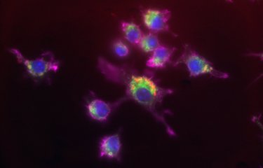


![Niche distribution and influence of environmental parameters in marine microbial communities: a systematic review [PeerJ]](https://dfzljdn9uc3pi.cloudfront.net/2015/1008/1/fig-1-1x.jpg)


![Know-how:blacklists [Pannonia IT Knowledgebase]](http://doku.pannoniait.at/lib/exe/fetch.php?w=90&h=90&tok=61577a&media=know-how:0_controller_add_rollen_hinzufuegen_domain_dienste.png)