Visual Acuity
Vision Disorders
Evoked Potentials, Visual
Visual Fields
Visual Cortex
Visual Perception
Amblyopia
Visual Pathways
Eyeglasses
Refractive Errors
Vision, Low
Fluorescein Angiography
Cataract
Vitrectomy
Macular Edema
Contrast Sensitivity
Vision, Ocular
Vision Screening
Blindness
Tomography, Optical Coherence
Macula Lutea
Visually Impaired Persons
Fovea Centralis
Visual Field Tests
Fundus Oculi
Macular Degeneration
Strabismus
Pattern Recognition, Visual
Astigmatism
Lens Implantation, Intraocular
Eye Injuries
Retinal Detachment
Photic Stimulation
Intravitreal Injections
Laser Coagulation
Myopia
Phacoemulsification
Sensory Deprivation
Retina
Diabetic Retinopathy
Retinal Diseases
Glare
Keratoconus
Epiretinal Membrane
Retinal Perforations
Corneal Topography
Vitreous Body
Retinal Vein Occlusion
Choroidal Neovascularization
Nystagmus, Congenital
Nystagmus, Pathologic
Scotoma
Triamcinolone Acetonide
Follow-Up Studies
Treatment Outcome
Color Vision Defects
Keratoplasty, Penetrating
Keratomileusis, Laser In Situ
Retinitis Pigmentosa
Retrospective Studies
Glaucoma
Fixation, Ocular
Ophthalmology
Lasers, Excimer
Hyperopia
Presbyopia
Photorefractive Keratectomy
Psychophysics
Prospective Studies
Nystagmus, Optokinetic
Scleral Buckling
Aberrometry
Corneal Opacity
Endophthalmitis
Retinoscopy
Optic Atrophy
Cornea
Corneal Wavefront Aberration
Descemet Stripping Endothelial Keratoplasty
Wet Macular Degeneration
Distance Perception
Uveitis
Choroid Diseases
Retinal Photoreceptor Cell Inner Segment
Optic Neuritis
Accommodation, Ocular
Retinal Artery Occlusion
Retinal Photoreceptor Cell Outer Segment
Light Coagulation
Central Serous Chorioretinopathy
Ocular Physiological Phenomena
Reading
Eye Infections, Fungal
Angiogenesis Inhibitors
Visual Prosthesis
Attention
Choroid
Optic Nerve Diseases
Laser Therapy
Uveitis, Posterior
Corneal Ulcer
Diagnostic Techniques, Ophthalmological
Dark Adaptation
Optic Nerve Neoplasms
Corneal Transplantation
Color Vision
Lens Subluxation
Retinal Cone Photoreceptor Cells
Papilledema
Silicone Oils
Albinism, Ocular
Dominance, Ocular
Esotropia
Triamcinolone
Antibodies, Monoclonal, Humanized
Electrooculography
Eye Infections, Bacterial
Eye
Postoperative Complications
Fluocinolone Acetonide
Optometry
Night Vision
Psychomotor Performance
Keratotomy, Radial
Ophthalmic Solutions
Aphakia
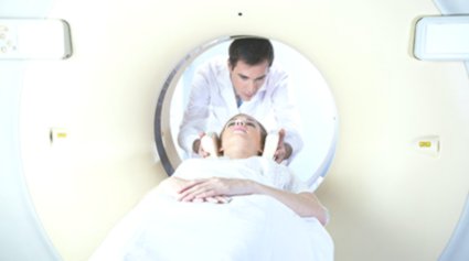
Magnetic Resonance Imaging
Hemianopsia
Exudates and Transudates
Lens Capsule, Crystalline
Indocyanine Green
Lighting
Lenses
Eye Enucleation
Optic Nerve
Panuveitis
Optic Chiasm
Reproducibility of Results
Glucocorticoids
Flicker Fusion
Brain Mapping
Adaptation, Ocular
Photography
Optic Nerve Glioma
Optic Neuropathy, Ischemic
Microscopy, Acoustic
Choroid Neoplasms
Photophobia
Interferometry
Retinal Pigments
Subretinal Fluid
Corneal Edema
Eye Diseases, Hereditary
Glaucoma, Open-Angle
Saccades
Pupil Disorders
Cues
Retinal Ganglion Cells
Photochemotherapy
Perceptual Disorders
Zernike representation of corneal topography height data after nonmechanical penetrating keratoplasty. (1/5631)
PURPOSE: To demonstrate a mathematical method for decomposition of discrete corneal topography height data into a set of Zernike polynomials and to demonstrate the clinical applicability of these computations in the postkeratoplasty cornea. METHODS: Fifty consecutive patients with either Fuchs' dystrophy (n = 20) or keratoconus (n = 30) were seen at 3 months, 6 months, and 1 year (before suture removal) and again after suture removal following nonmechanical trephination with the excimer laser. Patients were assessed using regular keratometry, corneal topography (TMS-1, simulated keratometry [SimK]), subjective refraction, and best-corrected visual acuity (VA) at each interval. A set of Zernike coefficients with radial degree 8 was calculated to fit two model surfaces: a complete representation (TOTAL) and a representation with parabolic terms only to define an approximate spherocylindrical surface (PARABOLIC). The root mean square error (RMS) was calculated comparing the corneal raw height data with TOTAL (TOTALRMS) and PARABOLIC (PARABOLICRMS). The cylinder of subjective refraction was correlated with the keratometric readings, the SimK, and the respective Zernike parameter. Visual acuity was correlated with the tilt components of the Zernike expansion. RESULTS: The measured corneal surface could be approximated by the composed surface 1 with TOTALRMS < or = 1.93 microm and by surface 2 with PARABOLICRMS < or = 3.66 microm. Mean keratometric reading after suture removal was 2.8+/-0.6 D. At all follow-up examinations, the SimK yielded higher values, whereas the keratometric reading and the refractive cylinder yielded lower values than the respective Zernike parameter. The correlation of the Zernike representation and the refractive cylinder (P = 0.02 at 3 months, P = 0.05 at 6 months and at 1 year, and P = 0.01 after suture removal) was much better than the correlation of the SimK and refractive cylinder (P = 0.3 at 3 months, P = 0.4 at 6 months, P = 0.2 at 1 year, and P = 0.1 after suture removal). Visual acuity increased from 0.23+/-0.10 at the 3-month evaluation to 0.54+/-0.19 after suture removal. After suture removal, there was a statistically significant inverse correlation between VA and tilt (P = 0.02 in patients with keratoconus and P = 0.05 in those with Fuchs' dystrophy). CONCLUSIONS: Zernike representation of corneal topography height data renders a reconstruction of clinically relevant corneal topography parameters with a marked reduction of redundance and a small error. Correlation of amount/axis of refractive cylinder with respective Zernike parameters is more accurate than with keratometry or respective SimK values of corneal topography analysis. (+info)Management of phacolytic glaucoma: experience of 135 cases. (2/5631)
We retrospectively analyzed 135 eyes with phacolytic glaucoma. A trabeculectomy was added to standard cataract surgery if symptoms endured for more than seven days, or if preoperative control of intraocular pressure (IOP) with maximal medical treatment was inadequate. In the early postoperative period, IOP was significantly lower in the combined surgery group (89 eyes) compared to the cataract surgery group (46 eyes) (p < 0.001). At 6 months there was no difference in IOP or visual acuity between the two groups. There were no serious complications related to trabeculectomy. It is reasonable to conclude that in eyes with a long duration of phacolytic glaucoma, addition of a trabeculectomy to cataract surgery is safe, prevents postoperative rise in intraocular pressure and decreases the need for systemic hypotensive medications. A randomized trial is on to further address this question. (+info)Effect of pilocarpine on visual acuity and on the dimensions of the cornea and anterior chamber. (3/5631)
The effect of pilocarpine on visual acuity and on the dimensions of the cornea, anterior chamber, and lens were studied in two groups of subjects. Significant changes in ocular tension, corneal curvature, anterior chamber depth, and lens anterior radius were found in a group of 55 glaucomatous eyes as a result of pilocarpine treatment, but there was no change in corneal thickness. Out of 102 glaucomatous eyes 78 became relatively myopic, and this appears to be due to changes in the dimensions of the lens of the eye similar to those occurring in accommodation, as a result of the effect of the drug on the ciliary muscle. The effect of pilocarpine on anterior chamber depth, area, and volume was studied in 125 eyes using a photographic method, and a significant reduction in the dimensions of the anterior chamber was observed as a result of the administration of pilocarpine. A significant correlation between depth and volume was also noted and the implications of this are discussed. (+info)De novo lesions in presumed ocular histoplasmosis-like syndrome. (4/5631)
Two patients with multifocal choroiditis similar or identical to POHS are presented. Colour photographs and fluorescein angiography document the occurrence of de novo lesions in the originally involved eye. The cases also demonstrate the development of new choroidal lesions within the originally involved eye, the early evolution of the "basic choroidal lesion", and the need for fluorescein angiography for visualizing the underlying choroidal lesion. (+info)Prognosis of perforating eye injury. (5/5631)
The assessment of visual function in a series of 130 consecutive patients of perforating eye injuries, revealed that visual acuity of 6/12 or better was regained in 63 per cent, between 6/60 and 6/18 in 9-2 per cent, less than 6/60 in 15-3 per cent, and enucleation was necessary in 9-2 per cent. In 3 per cent, the eyes were retained as blind, symptomfree, and cosmetically satisfactory organs. Two eyes were found to develop complete traumatic aniridia. None in the series was found to have sympathetic ophthalmitis. (+info)Vitrectomy in 125 eyes with diabetic vitreous haemorrhage. (6/5631)
A total of 125 consecutive eyes, all registered blind with diabetic vitreous haemorrhage, underwent pars plana vitrectomy with the vitrophage. Sixty-six per cent experienced some improvement in their visual acuity; 24 per cent were unchanged and 10 per cent were worse postoperatively. The major surgical complication was controllable haemorrhage (23 per cent). No retinal dialysis occurred. Significant postoperative complications were transient (71 per cent) and persistent (11 per cent) corneal oedema, early (8 per cent) and late (13 per cent) vitreous haemorrhage, transient (30 per cent) and persistent (6 per cent) rise in intraocular pressure, and rubeosis iridis (5 per cent). (+info)A prospective study of xenon arc photocoagulation for central retinal vein occlusion. (7/5631)
Twenty patients with central retinal vein occlusion were randomly divided into two groups in a prospective study to evaluate the effects of xenon are photocoagulation in central retinal vein occlusion. The patients in one group were treated with 360 degrees scatter xenon photocoagulation and the others received no treatment. The average follow-up was 18 months. There were no cases of rubeosis or neovascular glaucoma in the treated group. Two patients in the untreated group developed rubeosis with subsequent neovascular glaucoma. There was no significant difference in the visual prognosis or in fundus neovascularization between the groups. (+info)Characteristics of discrepancies between self-reported visual function and measured reading speed. Salisbury Eye Evaluation Project Team. (8/5631)
PURPOSE: Visual impairment is a risk factor for morbidity in the elderly and is often screened for by self-report. This study evaluates whether there are subsets for whom there is a discrepancy between self-reported and measured function. METHODS: The prevalence of a discrepancy between self-reported difficulty reading a newspaper and measured reading speed was determined in 2520 community-based men and women, aged 65 to 84 years, and the discrepant group characterized by polychotomous regression. RESULTS: Of subjects who reported minimal difficulty reading a newspaper, 10.8% (227/2107) read newsprint-sized text (0.21 degrees) more slowly than 80 words/min, a level previously shown to be necessary for sustained reading. Poor visual acuity, presence of psychiatric symptoms, and less satisfaction with vision were associated with being in the group that read slowly and reported difficulty with reading. Better cognition, better visual acuity, more years of education, white race, and fewer psychiatric symptoms were associated with being in the group that read more quickly and reported minimal difficulty. When reading the text size at which subjects read their fastest, only 2.6% of those with minimal difficulty remained discrepant. These individuals were more likely to have less education, be male, be African American, and have poorer cognitive status than those who did not remain discrepant. CONCLUSIONS: A subset of the elderly population have a substantial discrepancy between self-reported reading difficulty and measured reading speed. In some, this discrepancy may be based on underlying expectations and experiences, and in others it may represent a transition from no visual impairment to visual impairment. (+info)Visual acuity is a measure of the sharpness or clarity of vision. It is usually tested by reading an eye chart from a specific distance, such as 20 feet (6 meters). The standard eye chart used for this purpose is called the Snellen chart, which contains rows of letters that decrease in size as you read down the chart.
Visual acuity is typically expressed as a fraction, with the numerator representing the testing distance and the denominator indicating the smallest line of type that can be read clearly. For example, if a person can read the line on the eye chart that corresponds to a visual acuity of 20/20, it means they have normal vision at 20 feet. If their visual acuity is 20/40, it means they must be as close as 20 feet to see what someone with normal vision can see at 40 feet.
It's important to note that visual acuity is just one aspect of overall vision and does not necessarily reflect other important factors such as peripheral vision, depth perception, color vision, or contrast sensitivity.
Vision tests are a series of procedures used to assess various aspects of the visual system, including visual acuity, accommodation, convergence, divergence, stereopsis, color vision, and peripheral vision. These tests help healthcare professionals diagnose and manage vision disorders, such as nearsightedness, farsightedness, astigmatism, amblyopia, strabismus, and eye diseases like glaucoma, cataracts, and macular degeneration. Common vision tests include:
1. Visual acuity test (Snellen chart or letter chart): Measures the sharpness of a person's vision at different distances.
2. Refraction test: Determines the correct lens prescription for glasses or contact lenses by assessing how light is bent as it passes through the eye.
3. Color vision test: Evaluates the ability to distinguish between different colors and color combinations, often using pseudoisochromatic plates or Ishihara tests.
4. Stereopsis test: Assesses depth perception and binocular vision by presenting separate images to each eye that, when combined, create a three-dimensional effect.
5. Cover test: Examines eye alignment and the presence of strabismus (crossed eyes or turned eyes) by covering and uncovering each eye while observing eye movements.
6. Ocular motility test: Assesses the ability to move the eyes in various directions and coordinate both eyes during tracking and convergence/divergence movements.
7. Accommodation test: Evaluates the ability to focus on objects at different distances by using lenses, prisms, or dynamic retinoscopy.
8. Pupillary response test: Examines the size and reaction of the pupils to light and near objects.
9. Visual field test: Measures the peripheral (side) vision using automated perimetry or manual confrontation techniques.
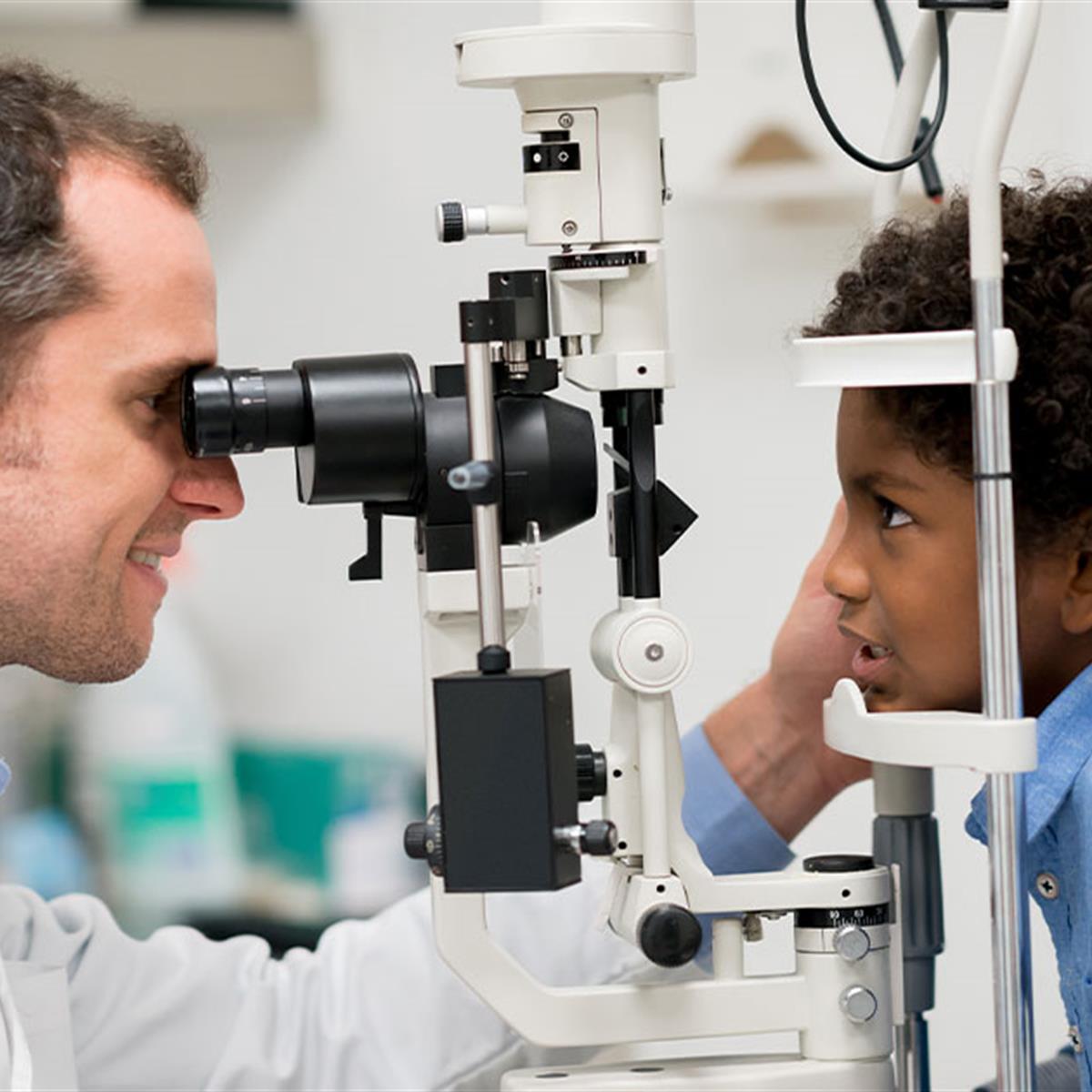
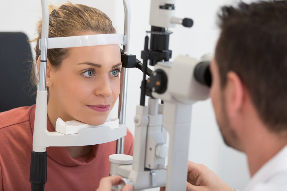
10. Slit-lamp examination: Inspects the structures of the front part of the eye, such as the cornea, iris, lens, and anterior chamber, using a specialized microscope.
These tests are typically performed by optometrists, ophthalmologists, or other vision care professionals during routine eye examinations or when visual symptoms are present.
Vision disorders refer to a wide range of conditions that affect the visual system and result in various symptoms, such as blurry vision, double vision, distorted vision, impaired depth perception, and difficulty with visual tracking or focusing. These disorders can be categorized into several types, including:
1. Refractive errors: These occur when the shape of the eye prevents light from focusing directly on the retina, resulting in blurry vision. Examples include myopia (nearsightedness), hyperopia (farsightedness), astigmatism, and presbyopia (age-related loss of near vision).
2. Strabismus: Also known as crossed eyes or walleye, strabismus is a misalignment of the eyes where they point in different directions, which can lead to double vision or loss of depth perception.
3. Amblyopia: Often called lazy eye, amblyopia is a condition where one eye has reduced vision due to lack of proper visual development during childhood. It may be caused by strabismus, refractive errors, or other factors that interfere with normal visual development.
4. Accommodative disorders: These involve problems with the focusing ability of the eyes, such as convergence insufficiency (difficulty focusing on close objects) and accommodative dysfunction (inability to maintain clear vision at different distances).
5. Binocular vision disorders: These affect how the eyes work together as a team, leading to issues like poor depth perception, eye strain, and headaches. Examples include convergence insufficiency, divergence excess, and suppression.
6. Ocular motility disorders: These involve problems with eye movement, such as nystagmus (involuntary eye movements), strabismus, or restricted extraocular muscle function.
7. Visual processing disorders: These affect the brain's ability to interpret and make sense of visual information, even when the eyes themselves are healthy. Symptoms may include difficulty with reading, recognizing shapes and objects, and understanding spatial relationships.
8. Low vision: This term refers to significant visual impairment that cannot be fully corrected with glasses, contact lenses, medication, or surgery. It includes conditions like macular degeneration, diabetic retinopathy, glaucoma, and cataracts.
9. Blindness: Complete loss of sight in both eyes, which can be caused by various factors such as injury, disease, or genetic conditions.
Evoked potentials, visual, also known as visually evoked potentials (VEPs), are electrical responses recorded from the brain following the presentation of a visual stimulus. These responses are typically measured using electroencephalography (EEG) and can provide information about the functioning of the visual pathways in the brain.
There are several types of VEPs, including pattern-reversal VEPs and flash VEPs. Pattern-reversal VEPs are elicited by presenting alternating checkerboard patterns, while flash VEPs are elicited by flashing a light. The responses are typically analyzed in terms of their latency (the time it takes for the response to occur) and amplitude (the size of the response).
VEPs are often used in clinical settings to help diagnose and monitor conditions that affect the visual system, such as multiple sclerosis, optic neuritis, and brainstem tumors. They can also be used in research to study the neural mechanisms underlying visual perception.
Visual fields refer to the total area in which objects can be seen while keeping the eyes focused on a central point. It is the entire area that can be observed using peripheral (side) vision while the eye gazes at a fixed point. A visual field test is used to detect blind spots or gaps (scotomas) in a person's vision, which could indicate various medical conditions such as glaucoma, retinal damage, optic nerve disease, brain tumors, or strokes. The test measures both the central and peripheral vision and maps the entire area that can be seen when focusing on a single point.
The visual cortex is the part of the brain that processes visual information. It is located in the occipital lobe, which is at the back of the brain. The visual cortex is responsible for receiving and interpreting signals from the retina, which are then transmitted through the optic nerve and optic tract.
The visual cortex contains several areas that are involved in different aspects of visual processing, such as identifying shapes, colors, and movements. These areas work together to help us recognize and understand what we see. Damage to the visual cortex can result in various visual impairments, such as blindness or difficulty with visual perception.
Visual perception refers to the ability to interpret and organize information that comes from our eyes to recognize and understand what we are seeing. It involves several cognitive processes such as pattern recognition, size estimation, movement detection, and depth perception. Visual perception allows us to identify objects, navigate through space, and interact with our environment. Deficits in visual perception can lead to learning difficulties and disabilities.
Amblyopia is a medical condition that affects the visual system, specifically the way the brain and eyes work together. It is often referred to as "lazy eye" and is characterized by reduced vision in one or both eyes that is not correctable with glasses or contact lenses alone. This occurs because the brain favors one eye over the other, causing the weaker eye to become neglected and underdeveloped.
Amblyopia can result from various conditions such as strabismus (eye misalignment), anisometropia (significant difference in prescription between the two eyes), or deprivation (such as a cataract that blocks light from entering the eye). Treatment for amblyopia typically involves correcting any underlying refractive errors, patching or blurring the stronger eye to force the weaker eye to work, and/or vision therapy. Early intervention is crucial to achieve optimal visual outcomes.
Visual pathways, also known as the visual system or the optic pathway, refer to the series of specialized neurons in the nervous system that transmit visual information from the eyes to the brain. This complex network includes the retina, optic nerve, optic chiasma, optic tract, lateral geniculate nucleus, pulvinar, and the primary and secondary visual cortices located in the occipital lobe of the brain.
The process begins when light enters the eye and strikes the photoreceptor cells (rods and cones) in the retina, converting the light energy into electrical signals. These signals are then transmitted to bipolar cells and subsequently to ganglion cells, whose axons form the optic nerve. The fibers from each eye's nasal hemiretina cross at the optic chiasma, while those from the temporal hemiretina continue without crossing. This results in the formation of the optic tract, which carries visual information from both eyes to the opposite side of the brain.
The majority of fibers in the optic tract synapse with neurons in the lateral geniculate nucleus (LGN), a part of the thalamus. The LGN sends this information to the primary visual cortex, also known as V1 or Brodmann area 17, located in the occipital lobe. Here, simple features like lines and edges are initially processed. Further processing occurs in secondary (V2) and tertiary (V3-V5) visual cortices, where more complex features such as shape, motion, and depth are analyzed. Ultimately, this information is integrated to form our perception of the visual world.
Eyeglasses are a medical device used to correct vision problems. Also known as spectacles, they consist of frames that hold one or more lenses through which a person looks to see clearly. The lenses may be made of glass or plastic and are designed to compensate for various visual impairments such as nearsightedness, farsightedness, astigmatism, or presbyopia. Eyeglasses can be custom-made to fit an individual's face and prescription, and they come in a variety of styles, colors, and materials. Some people wear eyeglasses all the time, while others may only need to wear them for certain activities such as reading or driving.
Cataract extraction is a surgical procedure that involves removing the cloudy lens (cataract) from the eye. This procedure is typically performed to restore vision impairment caused by cataracts and improve overall quality of life. There are two primary methods for cataract extraction:
1. Phacoemulsification: This is the most common method used today. It involves making a small incision in the front part of the eye (cornea), inserting an ultrasonic probe to break up the cloudy lens into tiny pieces, and then removing those pieces with suction. After removing the cataract, an artificial intraocular lens (IOL) is inserted to replace the natural lens and help focus light onto the retina.
2. Extracapsular Cataract Extraction: In this method, a larger incision is made on the side of the cornea, allowing the surgeon to remove the cloudy lens in one piece without breaking it up. The back part of the lens capsule is left intact to support the IOL. This technique is less common and typically reserved for more advanced cataracts or when phacoemulsification cannot be performed.
Recovery from cataract extraction usually involves using eye drops to prevent infection and inflammation, as well as protecting the eye with a shield or glasses during sleep for a few weeks after surgery. Most people experience improved vision within a few days to a week following the procedure.
Refractive errors are a group of vision conditions that include nearsightedness (myopia), farsightedness (hyperopia), astigmatism, and presbyopia. These conditions occur when the shape of the eye prevents light from focusing directly on the retina, causing blurred or distorted vision.
Myopia is a condition where distant objects appear blurry while close-up objects are clear. This occurs when the eye is too long or the cornea is too curved, causing light to focus in front of the retina instead of directly on it.
Hyperopia, on the other hand, is a condition where close-up objects appear blurry while distant objects are clear. This happens when the eye is too short or the cornea is not curved enough, causing light to focus behind the retina.
Astigmatism is a condition that causes blurred vision at all distances due to an irregularly shaped cornea or lens.
Presbyopia is a natural aging process that affects everyone as they get older, usually around the age of 40. It causes difficulty focusing on close-up objects and can be corrected with reading glasses, bifocals, or progressive lenses.
Refractive errors can be diagnosed through a comprehensive eye exam and are typically corrected with eyeglasses, contact lenses, or refractive surgery such as LASIK.
Low vision is a term used to describe significant visual impairment that cannot be corrected with standard glasses, contact lenses, medication or surgery. It is typically defined as visual acuity of less than 20/70 in the better-seeing eye after best correction, or a visual field of less than 20 degrees in the better-seeing eye.
People with low vision may have difficulty performing everyday tasks such as reading, recognizing faces, watching television, driving, or simply navigating their environment. They may also experience symptoms such as sensitivity to light, glare, or contrast, and may benefit from the use of visual aids, assistive devices, and rehabilitation services to help them maximize their remaining vision and maintain their independence.
Low vision can result from a variety of causes, including eye diseases such as macular degeneration, diabetic retinopathy, glaucoma, or cataracts, as well as congenital or inherited conditions, brain injuries, or aging. It is important for individuals with low vision to receive regular eye examinations and consult with a low vision specialist to determine the best course of treatment and management.
Fluorescein angiography is a medical diagnostic procedure used in ophthalmology to examine the blood flow in the retina and choroid, which are the inner layers of the eye. This test involves injecting a fluorescent dye, Fluorescein, into a patient's arm vein. As the dye reaches the blood vessels in the eye, a specialized camera takes rapid sequences of photographs to capture the dye's circulation through the retina and choroid.
The images produced by fluorescein angiography can help doctors identify any damage to the blood vessels, leakage, or abnormal growth of new blood vessels. This information is crucial in diagnosing and managing various eye conditions such as age-related macular degeneration, diabetic retinopathy, retinal vein occlusions, and inflammatory eye diseases.
It's important to note that while fluorescein angiography is a valuable diagnostic tool, it does carry some risks, including temporary side effects like nausea, vomiting, or allergic reactions to the dye. In rare cases, severe adverse reactions can occur, so patients should discuss these potential risks with their healthcare provider before undergoing the procedure.
A cataract is a clouding of the natural lens in the eye that affects vision. This clouding can cause vision to become blurry, faded, or dim, making it difficult to see clearly. Cataracts are a common age-related condition, but they can also be caused by injury, disease, or medication use. In most cases, cataracts develop gradually over time and can be treated with surgery to remove the cloudy lens and replace it with an artificial one.
A vitrectomy is a surgical procedure that involves the removal of some or all of the vitreous humor, which is the clear gel-like substance filling the center of the eye. This surgery is often performed to treat various retinal disorders such as diabetic retinopathy, retinal detachment, macular hole, and vitreous hemorrhage.
During a vitrectomy, the ophthalmologist makes small incisions in the sclera (the white part of the eye) to access the vitreous cavity. The surgeon then uses specialized instruments to remove the cloudy or damaged vitreous and may also repair any damage to the retina or surrounding tissues. Afterward, a clear saline solution is injected into the eye to maintain its shape and help facilitate healing.
In some cases, a gas bubble or silicone oil may be placed in the eye after the vitrectomy to help hold the retina in place while it heals. These substances will gradually be absorbed or removed during follow-up appointments. The body naturally produces a new, clear vitreous to replace the removed material over time.
Vitrectomy is typically performed under local anesthesia and may require hospitalization or outpatient care depending on the individual case. Potential risks and complications include infection, bleeding, cataract formation, retinal detachment, and increased eye pressure. However, with proper care and follow-up, most patients experience improved vision after a successful vitrectomy procedure.
Macular edema is a medical condition characterized by the accumulation of fluid in the macula, a small area in the center of the retina responsible for sharp, detailed vision. This buildup of fluid causes the macula to thicken and swell, which can distort central vision and lead to vision loss if not treated promptly. Macular edema is often a complication of other eye conditions such as diabetic retinopathy, age-related macular degeneration, retinal vein occlusion, or uveitis. It's important to note that while macular edema can affect anyone, it is more common in people with certain medical conditions like diabetes.
Contrast sensitivity is a measure of the ability to distinguish between an object and its background based on differences in contrast, rather than differences in luminance. Contrast refers to the difference in light intensity between an object and its immediate surroundings. Contrast sensitivity is typically measured using specially designed charts that have patterns of parallel lines with varying widths and contrast levels.
In clinical settings, contrast sensitivity is often assessed as part of a comprehensive visual examination. Poor contrast sensitivity can affect a person's ability to perform tasks such as reading, driving, or distinguishing objects from their background, especially in low-light conditions. Reduced contrast sensitivity is a common symptom of various eye conditions, including cataracts, glaucoma, and age-related macular degeneration.
Ocular vision refers to the ability to process and interpret visual information that is received by the eyes. This includes the ability to see clearly and make sense of the shapes, colors, and movements of objects in the environment. The ocular system, which includes the eye and related structures such as the optic nerve and visual cortex of the brain, works together to enable vision.
There are several components of ocular vision, including:
* Visual acuity: the clarity or sharpness of vision
* Field of vision: the extent of the visual world that is visible at any given moment
* Color vision: the ability to distinguish different colors
* Depth perception: the ability to judge the distance of objects in three-dimensional space
* Contrast sensitivity: the ability to distinguish an object from its background based on differences in contrast
Disorders of ocular vision can include refractive errors such as nearsightedness or farsightedness, as well as more serious conditions such as cataracts, glaucoma, and macular degeneration. These conditions can affect one or more aspects of ocular vision and may require medical treatment to prevent further vision loss.
Vision screening is a quick and cost-effective method used to identify individuals who are at risk of vision problems or eye diseases. It is not a comprehensive eye examination, but rather an initial evaluation that helps to determine if a further, more in-depth examination by an eye care professional is needed. Vision screenings typically involve tests for visual acuity, distance and near vision, color perception, depth perception, and alignment of the eyes. The goal of vision screening is to detect potential vision issues early on, so that they can be treated promptly and effectively, thereby preventing or minimizing any negative impact on a person's overall vision and quality of life.
Blindness is a condition of complete or near-complete vision loss. It can be caused by various factors such as eye diseases, injuries, or birth defects. Total blindness means that a person cannot see anything at all, while near-complete blindness refers to having only light perception or the ability to perceive the direction of light, but not able to discern shapes or forms. Legal blindness is a term used to define a certain level of visual impairment that qualifies an individual for government assistance and benefits; it usually means best corrected visual acuity of 20/200 or worse in the better eye, or a visual field no greater than 20 degrees in diameter.
Optical coherence tomography (OCT) is a non-invasive imaging technique that uses low-coherence light to capture high-resolution cross-sectional images of biological tissues, particularly the retina and other ocular structures. OCT works by measuring the echo time delay of light scattered back from different depths within the tissue, creating a detailed map of the tissue's structure. This technique is widely used in ophthalmology to diagnose and monitor various eye conditions such as macular degeneration, diabetic retinopathy, and glaucoma.
Ocular refraction is a medical term that refers to the bending of light as it passes through the optical media of the eye, including the cornea and lens. This process allows the eye to focus light onto the retina, creating a clear image. The refractive power of the eye is determined by the curvature and transparency of these structures.
In a normal eye, light rays are bent or refracted in such a way that they converge at a single point on the retina, producing a sharp and focused image. However, if the curvature of the cornea or lens is too steep or too flat, the light rays may not converge properly, resulting in a refractive error such as myopia (nearsightedness), hyperopia (farsightedness), or astigmatism.
Ocular refraction can be measured using a variety of techniques, including retinoscopy, automated refraction, and subjective refraction. These measurements are used to determine the appropriate prescription for corrective lenses such as eyeglasses or contact lenses. In some cases, ocular refractive errors may be corrected surgically through procedures such as LASIK or PRK.
The macula lutea, often simply referred to as the macula or fovea centralis, is a part of the eye that is responsible for central vision and color perception. It's located in the center of the retina, the light-sensitive tissue at the back of the eye. The macula contains a high concentration of pigments called xanthophylls, which give it a yellowish color and protect the photoreceptor cells in this area from damage by blue light.
The central part of the macula is called the fovea, which is a small depression that contains only cones, the photoreceptor cells responsible for color vision and high visual acuity. The fovea is surrounded by the parafovea and the perifovea, which contain both cones and rods, the photoreceptor cells responsible for low-light vision and peripheral vision.
Damage to the macula can result in a loss of central vision and color perception, a condition known as age-related macular degeneration (AMD), which is a leading cause of blindness in older adults. Other conditions that can affect the macula include macular edema, macular holes, and macular pucker.
Medical definitions for visual impairment often vary, but according to the World Health Organization (WHO), visually impaired persons are those who have a best-corrected visual acuity of less than 0.3 (6/12) in their better eye or a visual field of less than 20 degrees in their better eye. This includes people who are blind, as well as those with partial sight.
Visual impairment can range from mild to severe and may result from a variety of causes, including genetic disorders, diseases, trauma, or aging. It is important to note that visual impairment does not necessarily mean total blindness; many visually impaired individuals have some remaining vision and can benefit from low vision services and assistive devices.
The fovea centralis, also known as the macula lutea, is a small pit or depression located in the center of the retina, an light-sensitive tissue at the back of the eye. It is responsible for sharp, detailed vision (central vision) and color perception. The fovea contains only cones, the photoreceptor cells that are responsible for color vision and high visual acuity. It has a higher concentration of cones than any other area in the retina, allowing it to provide the greatest detail and color discrimination. The center of the fovea is called the foveola, which contains the highest density of cones and is avascular, meaning it lacks blood vessels to avoid interfering with the light passing through to the photoreceptor cells.
A visual field test is a method used to measure an individual's entire scope of vision, which includes what can be seen straight ahead and in peripheral (or side) vision. During the test, the person being tested is asked to focus on a central point while gradually identifying the appearance of objects moving into their peripheral vision. The visual field test helps detect blind spots (scotomas) or gaps in the visual field, which can be caused by various conditions such as glaucoma, brain injury, optic nerve damage, or retinal disorders. It's an essential tool for diagnosing and monitoring eye-related diseases and conditions.
"Fundus Oculi" is a medical term that refers to the back part of the interior of the eye, including the optic disc, macula, fovea, retinal vasculature, and peripheral retina. It is the area where light is focused and then transmitted to the brain via the optic nerve, forming visual images. Examinations of the fundus oculi are crucial for detecting various eye conditions such as diabetic retinopathy, macular degeneration, glaucoma, and other retinal diseases. The examination is typically performed using an ophthalmoscope or a specialized camera called a retinal camera.
Macular degeneration, also known as age-related macular degeneration (AMD), is a medical condition that affects the central part of the retina, called the macula. The macula is responsible for sharp, detailed vision, which is necessary for activities such as reading, driving, and recognizing faces.
In AMD, there is a breakdown or deterioration of the macula, leading to gradual loss of central vision. There are two main types of AMD: dry (atrophic) and wet (exudative). Dry AMD is more common and progresses more slowly, while wet AMD is less common but can cause rapid and severe vision loss if left untreated.
The exact causes of AMD are not fully understood, but risk factors include age, smoking, family history, high blood pressure, obesity, and exposure to sunlight. While there is no cure for AMD, treatments such as vitamin supplements, laser therapy, and medication injections can help slow its progression and reduce the risk of vision loss.
Strabismus is a condition of the ocular muscles where the eyes are not aligned properly and point in different directions. One eye may turn inward, outward, upward, or downward while the other one remains fixed and aligns normally. This misalignment can occur occasionally or constantly. Strabismus is also commonly referred to as crossed eyes or walleye. The condition can lead to visual impairments such as amblyopia (lazy eye) and depth perception problems if not treated promptly and effectively, usually through surgery, glasses, or vision therapy.
Visual pattern recognition is the ability to identify and interpret patterns in visual information. In a medical context, it often refers to the process by which healthcare professionals recognize and diagnose medical conditions based on visible signs or symptoms. This can involve recognizing the characteristic appearance of a rash, wound, or other physical feature associated with a particular disease or condition. It may also involve recognizing patterns in medical images such as X-rays, CT scans, or MRIs.
In the field of radiology, for example, visual pattern recognition is a critical skill. Radiologists are trained to recognize the typical appearances of various diseases and conditions in medical images. This allows them to make accurate diagnoses based on the patterns they see. Similarly, dermatologists use visual pattern recognition to identify skin abnormalities and diseases based on the appearance of rashes, lesions, or other skin changes.
Overall, visual pattern recognition is an essential skill in many areas of medicine, allowing healthcare professionals to quickly and accurately diagnose medical conditions based on visible signs and symptoms.
Astigmatism is a common eye condition that occurs when the cornea or lens has an irregular shape, causing blurred or distorted vision. The cornea and lens are typically smooth and curved uniformly in all directions, allowing light to focus clearly on the retina. However, if the cornea or lens is not smoothly curved and has a steeper curve in one direction than the other, it causes light to focus unevenly on the retina, leading to astigmatism.
Astigmatism can cause blurred vision at all distances, as well as eye strain, headaches, and fatigue. It is often present from birth and can be hereditary, but it can also develop later in life due to eye injuries or surgery. Astigmatism can be corrected with glasses, contact lenses, or refractive surgery such as LASIK.
Intraocular lens (IOL) implantation is a surgical procedure that involves placing a small artificial lens inside the eye to replace the natural lens that has been removed. This procedure is typically performed during cataract surgery, where the cloudy natural lens is removed and replaced with an IOL to restore clear vision.
During the procedure, a small incision is made in the eye, and the cloudy lens is broken up and removed using ultrasound waves or laser energy. Then, the folded IOL is inserted through the same incision and positioned in the correct place inside the eye. Once in place, the IOL unfolds and is secured into position.
There are several types of IOLs available, including monofocal, multifocal, toric, and accommodating lenses. Monofocal lenses provide clear vision at one distance, while multifocal lenses offer clear vision at multiple distances. Toric lenses correct astigmatism, and accommodating lenses can change shape to focus on objects at different distances.
Overall, intraocular lens implantation is a safe and effective procedure that can help restore clear vision in patients with cataracts or other eye conditions that require the removal of the natural lens.
Eye injuries refer to any damage or trauma caused to the eye or its surrounding structures. These injuries can vary in severity and may include:
1. Corneal abrasions: A scratch or scrape on the clear surface of the eye (cornea).
2. Chemical burns: Occurs when chemicals come into contact with the eye, causing damage to the cornea and other structures.
3. Eyelid lacerations: Cuts or tears to the eyelid.
4. Subconjunctival hemorrhage: Bleeding under the conjunctiva, the clear membrane that covers the white part of the eye.
5. Hyphema: Accumulation of blood in the anterior chamber of the eye, which is the space between the cornea and iris.
6. Orbital fractures: Breaks in the bones surrounding the eye.
7. Retinal detachment: Separation of the retina from its underlying tissue, which can lead to vision loss if not treated promptly.
8. Traumatic uveitis: Inflammation of the uvea, the middle layer of the eye, caused by trauma.
9. Optic nerve damage: Damage to the optic nerve, which transmits visual information from the eye to the brain.
Eye injuries can result from a variety of causes, including accidents, sports-related injuries, violence, and chemical exposure. It is important to seek medical attention promptly for any suspected eye injury to prevent further damage and potential vision loss.
Retinal detachment is a serious eye condition that occurs when the retina, a thin layer of tissue at the back of the eye responsible for processing light and sending visual signals to the brain, pulls away from its normal position. This can lead to significant vision loss or even blindness if not promptly treated. Retinal detachment can be caused by various factors such as aging, trauma, eye disease, or an inflammatory condition. Symptoms of retinal detachment may include sudden flashes of light, floaters, a shadow in the peripheral vision, or a curtain-like covering over part of the visual field. Immediate medical attention is necessary to prevent further damage and preserve vision.
Photic stimulation is a medical term that refers to the exposure of the eyes to light, specifically repetitive pulses of light, which is used as a method in various research and clinical settings. In neuroscience, it's often used in studies related to vision, circadian rhythms, and brain function.
In a clinical context, photic stimulation is sometimes used in the diagnosis of certain medical conditions such as seizure disorders (like epilepsy). By observing the response of the brain to this light stimulus, doctors can gain valuable insights into the functioning of the brain and the presence of any neurological disorders.
However, it's important to note that photic stimulation should be conducted under the supervision of a trained healthcare professional, as improper use can potentially trigger seizures in individuals who are susceptible to them.
Eye diseases are a range of conditions that affect the eye or visual system, causing damage to vision and, in some cases, leading to blindness. These diseases can be categorized into various types, including:
1. Refractive errors: These include myopia (nearsightedness), hyperopia (farsightedness), astigmatism, and presbyopia, which affect the way light is focused on the retina and can usually be corrected with glasses or contact lenses.
2. Cataracts: A clouding of the lens inside the eye that leads to blurry vision, glare, and decreased contrast sensitivity. Cataract surgery is the most common treatment for this condition.
3. Glaucoma: A group of diseases characterized by increased pressure in the eye, leading to damage to the optic nerve and potential blindness if left untreated. Treatment includes medications, laser therapy, or surgery.
4. Age-related macular degeneration (AMD): A progressive condition that affects the central part of the retina called the macula, causing blurry vision and, in advanced stages, loss of central vision. Treatment may include anti-VEGF injections, laser therapy, or nutritional supplements.
5. Diabetic retinopathy: A complication of diabetes that affects the blood vessels in the retina, leading to bleeding, leakage, and potential blindness if left untreated. Treatment includes laser therapy, anti-VEGF injections, or surgery.
6. Retinal detachment: A separation of the retina from its underlying tissue, which can lead to vision loss if not treated promptly with surgery.
7. Amblyopia (lazy eye): A condition where one eye does not develop normal vision, often due to a misalignment or refractive error in childhood. Treatment includes correcting the underlying problem and encouraging the use of the weaker eye through patching or other methods.
8. Strabismus (crossed eyes): A misalignment of the eyes that can lead to amblyopia if not treated promptly with surgery, glasses, or other methods.
9. Corneal diseases: Conditions that affect the transparent outer layer of the eye, such as keratoconus, Fuchs' dystrophy, and infectious keratitis, which can lead to vision loss if not treated promptly.
10. Uveitis: Inflammation of the middle layer of the eye, which can cause vision loss if not treated promptly with anti-inflammatory medications or surgery.
An intravitreal injection is a medical procedure in which medication is delivered directly into the vitreous cavity of the eye, which is the clear, gel-like substance that fills the space between the lens and the retina. This type of injection is typically used to treat various eye conditions such as age-related macular degeneration, diabetic retinopathy, retinal vein occlusion, and uveitis. The medication administered in intravitreal injections can help to reduce inflammation, inhibit the growth of new blood vessels, or prevent the formation of abnormal blood vessels in the eye.
Intravitreal injections are usually performed in an outpatient setting, and the procedure typically takes only a few minutes. Before the injection, the eye is numbed with anesthetic drops to minimize discomfort. The medication is then injected into the vitreous cavity using a small needle. After the injection, patients may experience some mild discomfort or a scratchy sensation in the eye, but this usually resolves within a few hours.
While intravitreal injections are generally safe, there are some potential risks and complications associated with the procedure, including infection, bleeding, retinal detachment, and increased intraocular pressure. Patients who undergo intravitreal injections should be closely monitored by their eye care provider to ensure that any complications are promptly identified and treated.
Laser coagulation, also known as laser photocoagulation, is a medical procedure that uses a laser to seal or destroy abnormal blood vessels or tissue. The laser produces a concentrated beam of light that can be precisely focused on the target area. When the laser energy is absorbed by the tissue, it causes the temperature to rise, which leads to coagulation (the formation of a clot) or destruction of the tissue.
In ophthalmology, laser coagulation is commonly used to treat conditions such as diabetic retinopathy, age-related macular degeneration, and retinal tears or holes. The procedure can help to seal leaking blood vessels, reduce fluid leakage, and prevent further vision loss. It is usually performed as an outpatient procedure and may be repeated if necessary.
In other medical specialties, laser coagulation may be used to control bleeding, destroy tumors, or remove unwanted tissue. The specific technique and parameters of the laser treatment will depend on the individual patient's needs and the condition being treated.
Binocular vision refers to the ability to use both eyes together to create a single, three-dimensional image of our surroundings. This is achieved through a process called binocular fusion, where the images from each eye are aligned and combined in the brain to form a unified perception.
The term "binocular vision" specifically refers to the way that our visual system integrates information from both eyes to create depth perception and enhance visual clarity. When we view an object with both eyes, they focus on the same point in space and send slightly different images to the brain due to their slightly different positions. The brain then combines these images to create a single, three-dimensional image that allows us to perceive depth and distance.
Binocular vision is important for many everyday activities, such as driving, reading, and playing sports. Disorders of binocular vision can lead to symptoms such as double vision, eye strain, and difficulty with depth perception.
Anisometropia is a medical term that refers to a condition where there is a significant difference in the refractive power between the two eyes. In other words, one eye has a significantly different optical prescription compared to the other eye. This condition can cause issues with binocular vision and depth perception, and can sometimes lead to amblyopia (lazy eye) if not corrected early in life. It is typically diagnosed through a comprehensive eye examination and can be corrected with glasses or contact lenses.
Myopia, also known as nearsightedness, is a common refractive error of the eye. It occurs when the eye is either too long or the cornea (the clear front part of the eye) is too curved. As a result, light rays focus in front of the retina instead of directly on it, causing distant objects to appear blurry while close objects remain clear.
Myopia typically develops during childhood and can progress gradually or rapidly until early adulthood. It can be corrected with glasses, contact lenses, or refractive surgery such as LASIK. Regular eye examinations are essential for people with myopia to monitor any changes in their prescription and ensure proper correction.
While myopia is generally not a serious condition, high levels of nearsightedness can increase the risk of certain eye diseases, including cataracts, glaucoma, retinal detachment, and myopic degeneration. Therefore, it's crucial to manage myopia effectively and maintain regular follow-ups with an eye care professional.
Phacoemulsification is a surgical procedure used in cataract removal. It involves using an ultrasonic device to emulsify (break up) the cloudy lens (cataract) into small pieces, which are then aspirated or sucked out through a small incision. This procedure allows for smaller incisions and faster recovery times compared to traditional cataract surgery methods. After the cataract is removed, an artificial intraocular lens (IOL) is typically implanted to replace the natural lens and restore vision.
Sensory deprivation, also known as perceptual isolation or sensory restriction, refers to the deliberate reduction or removal of stimuli from one or more of the senses. This can include limiting input from sight, sound, touch, taste, and smell. The goal is to limit a person's sensory experiences in order to study the effects on cognition, perception, and behavior.
In a clinical context, sensory deprivation can occur as a result of certain medical conditions or treatments, such as blindness, deafness, or pharmacological interventions that affect sensory processing. Prolonged sensory deprivation can lead to significant psychological and physiological effects, including hallucinations, delusions, and decreased cognitive function.
It's important to note that sensory deprivation should not be confused with meditation or relaxation techniques that involve reducing external stimuli in a controlled manner to promote relaxation and focus.
Intraocular lenses (IOLs) are artificial lens implants that are placed inside the eye during ophthalmic surgery, such as cataract removal. These lenses are designed to replace the natural lens of the eye that has become clouded or damaged, thereby restoring vision impairment caused by cataracts or other conditions.
There are several types of intraocular lenses available, including monofocal, multifocal, toric, and accommodative lenses. Monofocal IOLs provide clear vision at a single fixed distance, while multifocal IOLs offer clear vision at multiple distances. Toric IOLs are designed to correct astigmatism, and accommodative IOLs can change shape and position within the eye to allow for a range of vision.
The selection of the appropriate type of intraocular lens depends on various factors, including the patient's individual visual needs, lifestyle, and ocular health. The implantation procedure is typically performed on an outpatient basis and involves minimal discomfort or recovery time. Overall, intraocular lenses have become a safe and effective treatment option for patients with vision impairment due to cataracts or other eye conditions.
The retina is the innermost, light-sensitive layer of tissue in the eye of many vertebrates and some cephalopods. It receives light that has been focused by the cornea and lens, converts it into neural signals, and sends these to the brain via the optic nerve. The retina contains several types of photoreceptor cells including rods (which handle vision in low light) and cones (which are active in bright light and are capable of color vision).
In medical terms, any pathological changes or diseases affecting the retinal structure and function can lead to visual impairment or blindness. Examples include age-related macular degeneration, diabetic retinopathy, retinal detachment, and retinitis pigmentosa among others.
Diabetic retinopathy is a diabetes complication that affects the eyes. It's caused by damage to the blood vessels of the light-sensitive tissue at the back of the eye (retina).
At first, diabetic retinopathy may cause no symptoms or only mild vision problems. Eventually, it can cause blindness. The condition usually affects both eyes.
There are two main stages of diabetic retinopathy:
1. Early diabetic retinopathy. This is when the blood vessels in the eye start to leak fluid or bleed. You might not notice any changes in your vision at this stage, but it's still important to get treatment because it can prevent the condition from getting worse.
2. Advanced diabetic retinopathy. This is when new, abnormal blood vessels grow on the surface of the retina. These vessels can leak fluid and cause severe vision problems, including blindness.
Diabetic retinopathy can be treated with laser surgery, injections of medication into the eye, or a vitrectomy (a surgical procedure to remove the gel-like substance that fills the center of the eye). It's important to get regular eye exams to detect diabetic retinopathy early and get treatment before it causes serious vision problems.
Retinal diseases refer to a group of conditions that affect the retina, which is the light-sensitive tissue located at the back of the eye. The retina is responsible for converting light into electrical signals that are sent to the brain and interpreted as visual images. Retinal diseases can cause vision loss or even blindness, depending on their severity and location in the retina.
Some common retinal diseases include:
1. Age-related macular degeneration (AMD): A progressive disease that affects the central part of the retina called the macula, causing blurred or distorted vision.
2. Diabetic retinopathy: A complication of diabetes that can damage the blood vessels in the retina, leading to vision loss.
3. Retinal detachment: A serious condition where the retina becomes separated from its underlying tissue, requiring immediate medical attention.
4. Macular edema: Swelling or thickening of the macula due to fluid accumulation, which can cause blurred vision.
5. Retinitis pigmentosa: A group of inherited eye disorders that affect the retina's ability to respond to light, causing progressive vision loss.
6. Macular hole: A small break in the macula that can cause distorted or blurry vision.
7. Retinal vein occlusion: Blockage of the retinal veins that can lead to bleeding, swelling, and potential vision loss.
Treatment for retinal diseases varies depending on the specific condition and its severity. Some treatments include medication, laser therapy, surgery, or a combination of these options. Regular eye exams are essential for early detection and treatment of retinal diseases.
Electroretinography (ERG) is a medical test used to evaluate the functioning of the retina, which is the light-sensitive tissue located at the back of the eye. The test measures the electrical responses of the retina to light stimulation.
During the procedure, a special contact lens or electrode is placed on the surface of the eye to record the electrical activity generated by the retina's light-sensitive cells (rods and cones) and other cells in the retina. The test typically involves presenting different levels of flashes of light to the eye while the electrical responses are recorded.
The resulting ERG waveform provides information about the overall health and function of the retina, including the condition of the photoreceptors, the integrity of the inner retinal layers, and the health of the retinal ganglion cells. This test is often used to diagnose and monitor various retinal disorders, such as retinitis pigmentosa, macular degeneration, and diabetic retinopathy.
In the context of ophthalmology and optometry, glare refers to a visual sensation caused by excessive brightness or contrast that interferes with the ability to see comfortably or clearly. It can be caused by direct or reflected light sources that enter the eye and scatter within the eye or on the surface of the eye, reducing contrast and visibility. Glare can lead to discomfort, disability, or both, and it can significantly impact visual performance in various activities such as driving, reading, and using digital devices. There are different types of glare, including direct glare, reflected glare, and veiling glare, each with its own characteristics and effects on vision.
Keratoconus is a degenerative non-inflammatory disorder of the eye, primarily affecting the cornea. It is characterized by a progressive thinning and steepening of the central or paracentral cornea, causing it to assume a conical shape. This results in irregular astigmatism, myopia, and scattering of light leading to blurred vision, visual distortions, and sensitivity to glare. The exact cause of keratoconus is unknown, but it may be associated with genetics, eye rubbing, and certain medical conditions. It typically starts in the teenage years and progresses into the third or fourth decade of life. Treatment options include glasses, contact lenses, cross-linking, and corneal transplantation in advanced cases.
An epiretinal membrane, also known as a macular pucker or cellophane maculopathy, is a thin and transparent layer of tissue that forms over the macula (the central part of the retina responsible for sharp, detailed vision) in the eye. This membrane can contract and wrinkle the macula, distorting central vision.
Epiretinal membranes are typically caused by the migration and proliferation of glial cells or other cell types onto the surface of the retina following retinal injury, inflammation, or aging. In some cases, they may be associated with other eye conditions such as diabetic retinopathy, retinal vein occlusion, or age-related macular degeneration.
Mild epiretinal membranes may not require treatment, but if the distortion of vision is significant, a vitrectomy surgery may be recommended to remove the membrane and improve visual acuity.
A retinal perforation is a full-thickness break or hole in the retina, which is the light-sensitive tissue that lines the inner surface of the eye. This condition can lead to a serious complication called retinal detachment, where the retina separates from the underlying tissue, potentially resulting in vision loss if not promptly treated. Retinal perforations may be caused by trauma, certain eye conditions, or invasive eye procedures. Immediate medical attention is required for retinal perforations to prevent further damage and preserve vision.
Penetrating eye injuries are a type of ocular trauma where a foreign object or substance pierces the outer layers of the eye and damages the internal structures. This can result in serious harm to various parts of the eye, such as the cornea, iris, lens, or retina, and may potentially cause vision loss or blindness if not promptly treated.
The severity of a penetrating eye injury depends on several factors, including the type and size of the object that caused the injury, the location of the wound, and the extent of damage to the internal structures. Common causes of penetrating eye injuries include sharp objects, such as metal shards or glass fragments, projectiles, such as pellets or bullets, and explosive materials.
Symptoms of a penetrating eye injury may include pain, redness, sensitivity to light, blurred vision, floaters, or the presence of a foreign body in the eye. If you suspect that you have sustained a penetrating eye injury, it is essential to seek immediate medical attention from an ophthalmologist or other healthcare professional with experience in treating eye trauma.
Treatment for penetrating eye injuries may include removing any foreign objects or substances from the eye, repairing damaged tissues, and administering medications to prevent infection and reduce inflammation. In some cases, surgery may be necessary to repair the injury and restore vision. Preventing eye injuries is crucial, and appropriate protective eyewear should be worn when engaging in activities that pose a risk of eye trauma.
Corneal topography is a non-invasive medical imaging technique used to create a detailed map of the surface curvature of the cornea, which is the clear, dome-shaped surface at the front of the eye. This procedure provides valuable information about the shape and condition of the cornea, helping eye care professionals assess various eye conditions such as astigmatism, keratoconus, and other corneal abnormalities. It can also be used in contact lens fitting, refractive surgery planning, and post-surgical evaluation.
The vitreous body, also known simply as the vitreous, is the clear, gel-like substance that fills the space between the lens and the retina in the eye. It is composed mainly of water, but also contains collagen fibers, hyaluronic acid, and other proteins. The vitreous helps to maintain the shape of the eye and provides a transparent medium for light to pass through to reach the retina. With age, the vitreous can become more liquefied and may eventually separate from the retina, leading to symptoms such as floaters or flashes of light.
Monocular vision refers to the ability to see and process visual information using only one eye. It is the type of vision that an individual has when they are using only one eye to look at something, while the other eye may be covered or not functioning. This can be contrasted with binocular vision, which involves the use of both eyes working together to provide depth perception and a single, combined visual field.
Monocular vision is important for tasks that only require the use of one eye, such as when looking through a microscope or using a telescope. However, it does not provide the same level of depth perception and spatial awareness as binocular vision. In some cases, individuals may have reduced visual acuity or other visual impairments in one eye, leading to limited monocular vision in that eye. It is important for individuals with monocular vision to have regular eye exams to monitor their eye health and ensure that any visual impairments are detected and treated promptly.
Retinal vein occlusion (RVO) is a medical condition that occurs when one of the retinal veins, which drains blood from the retina, becomes blocked by a blood clot or atherosclerotic plaque. This blockage can cause hemorrhages, fluid accumulation, and damage to the retinal tissue, leading to vision loss.
There are two types of RVO: branch retinal vein occlusion (BRVO) and central retinal vein occlusion (CRVO). BRVO affects a smaller branch retinal vein, while CRVO affects the main retinal vein. CRVO is generally associated with more severe vision loss than BRVO.
Risk factors for RVO include hypertension, diabetes, high cholesterol levels, smoking, and glaucoma. Age is also a significant risk factor, as RVO becomes more common with increasing age. Treatment options for RVO may include controlling underlying medical conditions, laser therapy, intravitreal injections of anti-VEGF agents or steroids, and surgery in some cases.
Sensory thresholds are the minimum levels of stimulation that are required to produce a sensation in an individual, as determined through psychophysical testing. These tests measure the point at which a person can just barely detect the presence of a stimulus, such as a sound, light, touch, or smell.
There are two types of sensory thresholds: absolute and difference. Absolute threshold is the minimum level of intensity required to detect a stimulus 50% of the time. Difference threshold, also known as just noticeable difference (JND), is the smallest change in intensity that can be detected between two stimuli.
Sensory thresholds can vary between individuals and are influenced by factors such as age, attention, motivation, and expectations. They are often used in clinical settings to assess sensory function and diagnose conditions such as hearing or vision loss.
Choroidal neovascularization (CNV) is a medical term that refers to the growth of new, abnormal blood vessels in the choroid layer of the eye, which is located between the retina and the sclera. This condition typically occurs as a complication of age-related macular degeneration (AMD), although it can also be caused by other eye diseases or injuries.
In CNV, the new blood vessels that grow into the choroid layer are fragile and can leak fluid or blood, which can cause distortion or damage to the retina, leading to vision loss. Symptoms of CNV may include blurred or distorted vision, a blind spot in the center of the visual field, or changes in color perception.
Treatment for CNV typically involves medications that are designed to stop the growth of new blood vessels, such as anti-VEGF drugs, which target a protein called vascular endothelial growth factor (VEGF) that is involved in the development of new blood vessels. Laser surgery or photodynamic therapy may also be used in some cases to destroy the abnormal blood vessels and prevent further vision loss.
Congenital nystagmus is a type of involuntary eye movement that is present at birth or develops within the first few months of life. It is characterized by rhythmic oscillations or repetitive, rapid movements of the eyes in either horizontal, vertical, or rotatory directions. These movements can impair vision and may be associated with other ocular conditions such as albinism, congenital cataracts, or optic nerve hypoplasia. The exact cause of congenital nystagmus is not fully understood, but it is believed to result from abnormal development or dysfunction in the areas of the brain that control eye movements. In some cases, congenital nystagmus may be inherited as a genetic trait. Treatment options for congenital nystagmus include corrective lenses, prism glasses, surgery, and vision therapy, depending on the underlying cause and severity of the condition.
Pathological nystagmus is an abnormal, involuntary movement of the eyes that can occur in various directions (horizontal, vertical, or rotatory) and can be rhythmical or arrhythmic. It is typically a result of a disturbance in the vestibular system, central nervous system, or ocular motor pathways. Pathological nystagmus can cause visual symptoms such as blurred vision, difficulty with fixation, and oscillopsia (the sensation that one's surroundings are moving). The type, direction, and intensity of the nystagmus may vary depending on the underlying cause, which can include conditions such as brainstem or cerebellar lesions, multiple sclerosis, drug toxicity, inner ear disorders, and congenital abnormalities.
Corneal diseases are a group of disorders that affect the cornea, which is the clear, dome-shaped surface at the front of the eye. The cornea plays an important role in focusing vision, and any damage or disease can cause significant visual impairment or loss. Some common types of corneal diseases include:
1. Keratoconus: A progressive disorder in which the cornea thins and bulges outward into a cone shape, causing distorted vision.
2. Fuchs' dystrophy: A genetic disorder that affects the inner layer of the cornea called the endothelium, leading to swelling, cloudiness, and decreased vision.
3. Dry eye syndrome: A condition in which the eyes do not produce enough tears or the tears evaporate too quickly, causing discomfort, redness, and blurred vision.
4. Corneal ulcers: Open sores on the cornea that can be caused by infection, trauma, or other factors.
5. Herpes simplex keratitis: A viral infection of the cornea that can cause recurrent episodes of inflammation, scarring, and vision loss.
6. Corneal dystrophies: Inherited disorders that affect the structure and clarity of the cornea, leading to visual impairment or blindness.
7. Bullous keratopathy: A condition in which the endothelium fails to pump fluid out of the cornea, causing it to swell and form blisters.
8. Corneal trauma: Injury to the cornea caused by foreign objects, chemicals, or other factors that can lead to scarring, infection, and vision loss.
Treatment for corneal diseases varies depending on the specific condition and severity of the disease. Options may include eyedrops, medications, laser surgery, corneal transplantation, or other treatments.
A scotoma is a blind spot or area of reduced vision within the visual field. It's often surrounded by an area of less distinct vision and can be caused by various conditions such as eye diseases, neurological disorders, or brain injuries. A scotoma may be temporary or permanent, depending on its underlying cause.
There are different types of scotomas, including:
1. Central scotoma - a blind spot in the center of the visual field, often associated with conditions like age-related macular degeneration and diabetic retinopathy.
2. Paracentral scotoma - a blind spot located slightly away from the center of the visual field, which can be caused by optic neuritis or other optic nerve disorders.
3. Peripheral scotoma - a blind spot in the peripheral vision, often associated with retinal diseases like retinitis pigmentosa.
4. Absolute scotoma - a complete loss of vision in a specific area of the visual field.
5. Relative scotoma - a partial loss of vision in which some details can still be perceived, but not as clearly or vividly as in normal vision.
It is essential to consult an eye care professional if you experience any changes in your vision or notice a scotoma, as early detection and treatment can help prevent further vision loss.
Triamcinolone Acetonide is a synthetic glucocorticoid, which is a class of corticosteroids. It is used in the form of topical creams, ointments, and sprays to reduce skin inflammation, itching, and allergies. It can also be administered through injection for the treatment of various conditions such as arthritis, bursitis, and tendonitis. Triamcinolone Acetonide works by suppressing the immune system's response, reducing inflammation, and blocking the production of substances that cause allergies.
It is important to note that prolonged use or overuse of triamcinolone acetonide can lead to side effects such as thinning of the skin, easy bruising, and increased susceptibility to infections. Therefore, it should be used under the guidance of a healthcare professional.
Follow-up studies are a type of longitudinal research that involve repeated observations or measurements of the same variables over a period of time, in order to understand their long-term effects or outcomes. In medical context, follow-up studies are often used to evaluate the safety and efficacy of medical treatments, interventions, or procedures.
In a typical follow-up study, a group of individuals (called a cohort) who have received a particular treatment or intervention are identified and then followed over time through periodic assessments or data collection. The data collected may include information on clinical outcomes, adverse events, changes in symptoms or functional status, and other relevant measures.
The results of follow-up studies can provide important insights into the long-term benefits and risks of medical interventions, as well as help to identify factors that may influence treatment effectiveness or patient outcomes. However, it is important to note that follow-up studies can be subject to various biases and limitations, such as loss to follow-up, recall bias, and changes in clinical practice over time, which must be carefully considered when interpreting the results.
Treatment outcome is a term used to describe the result or effect of medical treatment on a patient's health status. It can be measured in various ways, such as through symptoms improvement, disease remission, reduced disability, improved quality of life, or survival rates. The treatment outcome helps healthcare providers evaluate the effectiveness of a particular treatment plan and make informed decisions about future care. It is also used in clinical research to compare the efficacy of different treatments and improve patient care.
Contact lenses are thin, curved plastic or silicone hydrogel devices that are placed on the eye to correct vision, replace a missing or damaged cornea, or for cosmetic purposes. They rest on the surface of the eye, called the cornea, and conform to its shape. Contact lenses are designed to float on a thin layer of tears and move with each blink.
There are two main types of contact lenses: soft and rigid gas permeable (RGP). Soft contact lenses are made of flexible hydrophilic (water-absorbing) materials that allow oxygen to pass through the lens to the cornea. RGP lenses are made of harder, more oxygen-permeable materials.
Contact lenses can be used to correct various vision problems, including nearsightedness, farsightedness, astigmatism, and presbyopia. They come in different shapes, sizes, and powers to suit individual needs and preferences. Proper care, handling, and regular check-ups with an eye care professional are essential for maintaining good eye health and preventing complications associated with contact lens wear.
Color vision defects, also known as color blindness, are conditions in which a person has difficulty distinguishing between certain colors. The most common types of color vision defects involve the inability to distinguish between red and green or blue and yellow. These deficiencies result from an alteration or absence of one or more of the three types of cone cells in the retina that are responsible for normal color vision.
In red-green color vision defects, there is a problem with either the red or green cones, or both. This results in difficulty distinguishing between these two colors and their shades. Protanopia is a type of red-green color vision defect where there is an absence of red cone cells, making it difficult to distinguish between red and green as well as between red and black or green and black. Deuteranopia is another type of red-green color vision defect where there is an absence of green cone cells, resulting in similar difficulties distinguishing between red and green, as well as between blue and yellow.
Blue-yellow color vision defects are less common than red-green color vision defects. Tritanopia is a type of blue-yellow color vision defect where there is an absence of blue cone cells, making it difficult to distinguish between blue and yellow, as well as between blue and purple or yellow and pink.
Color vision defects are usually inherited and present from birth, but they can also result from eye diseases, chemical exposure, aging, or medication side effects. They affect both men and women, although red-green color vision defects are more common in men than in women. People with color vision defects may have difficulty with tasks that require color discrimination, such as matching clothes, selecting ripe fruit, reading colored maps, or identifying warning signals. However, most people with mild to moderate color vision defects can adapt and function well in daily life.
Penetrating keratoplasty (PK) is a type of corneal transplant surgery where the entire thickness of the host's damaged or diseased cornea is removed and replaced with a similar full-thickness portion of a healthy donor's cornea. The procedure aims to restore visual function, alleviate pain, and improve the structural integrity of the eye. It is typically performed for conditions such as severe keratoconus, corneal scarring, or corneal ulcers that cannot be treated with other, less invasive methods. Following the surgery, patients may require extended recovery time and rigorous postoperative care to minimize the risk of complications and ensure optimal visual outcomes.
Laser In Situ Keratomileusis (LASIK) is a type of refractive surgery used to correct vision issues such as myopia (nearsightedness), hyperopia (farsightedness), and astigmatism. The procedure involves reshaping the cornea, which is the clear, dome-shaped surface at the front of the eye, using an excimer laser.
In LASIK, a thin flap is created on the surface of the cornea using a femtosecond or microkeratome laser. The flap is then lifted, and the excimer laser is used to reshape the underlying tissue. After the reshaping is complete, the flap is replaced, allowing for quicker healing and visual recovery compared to other refractive surgery procedures.
LASIK is an outpatient procedure that typically takes about 30 minutes or less per eye. Most people can expect to see improved vision within a few days of the procedure, although it may take several weeks for vision to fully stabilize. LASIK has a high success rate and is generally considered safe when performed by a qualified surgeon. However, as with any surgical procedure, there are risks involved, including dry eye, infection, and visual complications such as glare or halos around lights.
Retinitis pigmentosa (RP) is a group of rare, genetic disorders that involve a breakdown and loss of cells in the retina - a light-sensitive tissue located at the back of the eye. The retina converts light into electrical signals which are then sent to the brain and interpreted as visual images.
In RP, the cells that detect light (rods and cones) degenerate more slowly than other cells in the retina, leading to a progressive loss of vision. Symptoms typically begin in childhood with night blindness (difficulty seeing in low light), followed by a gradual narrowing of the visual field (tunnel vision). Over time, this can lead to significant vision loss and even blindness.
The condition is usually inherited and there are several different genes that have been associated with RP. The diagnosis is typically made based on a combination of genetic testing, family history, and clinical examination. Currently, there is no cure for RP, but researchers are actively working to develop new treatments that may help slow or stop the progression of the disease.
Ophthalmologic surgical procedures refer to various types of surgeries performed on the eye and its surrounding structures by trained medical professionals called ophthalmologists. These procedures aim to correct or improve vision, diagnose and treat eye diseases or injuries, and enhance the overall health and functionality of the eye. Some common examples of ophthalmologic surgical procedures include:
1. Cataract Surgery: This procedure involves removing a cloudy lens (cataract) from the eye and replacing it with an artificial intraocular lens (IOL).
2. LASIK (Laser-Assisted In Situ Keratomileusis): A type of refractive surgery that uses a laser to reshape the cornea, correcting nearsightedness, farsightedness, and astigmatism.
3. Glaucoma Surgery: Several surgical options are available for treating glaucoma, including laser trabeculoplasty, traditional trabeculectomy, and various drainage device implantations. These procedures aim to reduce intraocular pressure (IOP) and prevent further optic nerve damage.
4. Corneal Transplant: This procedure involves replacing a damaged or diseased cornea with a healthy donor cornea to restore vision and improve the eye's appearance.
5. Vitreoretinal Surgery: These procedures focus on treating issues within the vitreous humor (gel-like substance filling the eye) and the retina, such as retinal detachment, macular holes, or diabetic retinopathy.
6. Strabismus Surgery: This procedure aims to correct misalignment of the eyes (strabismus) by adjusting the muscles responsible for eye movement.
7. Oculoplastic Surgery: These procedures involve reconstructive, cosmetic, and functional surgeries around the eye, such as eyelid repair, removal of tumors, or orbital fracture repairs.
8. Pediatric Ophthalmologic Procedures: Various surgical interventions are performed on children to treat conditions like congenital cataracts, amblyopia (lazy eye), or blocked tear ducts.
These are just a few examples of ophthalmic surgical procedures. The specific treatment plan will depend on the individual's condition and overall health.
Retrospective studies, also known as retrospective research or looking back studies, are a type of observational study that examines data from the past to draw conclusions about possible causal relationships between risk factors and outcomes. In these studies, researchers analyze existing records, medical charts, or previously collected data to test a hypothesis or answer a specific research question.
Retrospective studies can be useful for generating hypotheses and identifying trends, but they have limitations compared to prospective studies, which follow participants forward in time from exposure to outcome. Retrospective studies are subject to biases such as recall bias, selection bias, and information bias, which can affect the validity of the results. Therefore, retrospective studies should be interpreted with caution and used primarily to generate hypotheses for further testing in prospective studies.
A pupil, in medical terms, refers to the circular opening in the center of the iris (the colored part of the eye) that allows light to enter and reach the retina. The size of the pupil can change involuntarily in response to light intensity and emotional state, as well as voluntarily through certain eye exercises or with the use of eye drops. Pupillary reactions are important in clinical examinations as they can provide valuable information about the nervous system's functioning, particularly the brainstem and cranial nerves II and III.
Glaucoma is a group of eye conditions that damage the optic nerve, often caused by an abnormally high pressure in the eye (intraocular pressure). This damage can lead to permanent vision loss or even blindness if left untreated. The most common type is open-angle glaucoma, which has no warning signs and progresses slowly. Angle-closure glaucoma, on the other hand, can cause sudden eye pain, redness, nausea, and vomiting, as well as rapid vision loss. Other less common types of glaucoma also exist. While there is no cure for glaucoma, early detection and treatment can help slow or prevent further vision loss.
Mydriatics are medications that cause mydriasis, which is the dilation of the pupil. These drugs work by blocking the action of the muscarinic receptors in the iris, leading to relaxation of the circular muscle and constriction of the radial muscle, resulting in pupil dilation. Mydriatics are often used in eye examinations to facilitate examination of the interior structures of the eye. Commonly used mydriatic agents include tropicamide, phenylephrine, and cyclopentolate. It is important to note that mydriatics can have side effects such as blurred vision, photophobia, and accommodation difficulties, so patients should be advised accordingly.
Ocular fixation is a term used in ophthalmology and optometry to refer to the ability of the eyes to maintain steady gaze or visual focus on an object. It involves the coordinated movement of the extraocular muscles that control eye movements, allowing for clear and stable vision.
In medical terminology, fixation specifically refers to the state in which the eyes are aligned and focused on a single point in space. This is important for maintaining visual perception and preventing blurring or double vision. Ocular fixation can be affected by various factors such as muscle weakness, nerve damage, or visual processing disorders.
Assessment of ocular fixation is often used in eye examinations to evaluate visual acuity, eye alignment, and muscle function. Abnormalities in fixation may indicate the presence of underlying eye conditions or developmental delays that require further investigation and treatment.
Ophthalmology is a branch of medicine that deals with the diagnosis, treatment, and prevention of diseases and disorders of the eye and visual system. It is a surgical specialty, and ophthalmologists are medical doctors who complete additional years of training to become experts in eye care. They are qualified to perform eye exams, diagnose and treat eye diseases, prescribe glasses and contact lenses, and perform eye surgery. Some subspecialties within ophthalmology include cornea and external disease, glaucoma, neuro-ophthalmology, pediatric ophthalmology, retina and vitreous, and oculoplastics.
An excimer laser is a type of laser that is used in various medical procedures, particularly in ophthalmology and dermatology. The term "excimer" is derived from "excited dimer," which refers to a short-lived molecule formed when two atoms combine in an excited state.
Excimer lasers emit light at a specific wavelength that is determined by the type of gas used in the laser. In medical applications, excimer lasers typically use noble gases such as argon, krypton, or xenon, combined with halogens such as fluorine or chlorine. The most commonly used excimer laser in medical procedures is the excimer laser that uses a mixture of argon and fluoride gas to produce light at a wavelength of 193 nanometers (nm).
In ophthalmology, excimer lasers are primarily used for refractive surgery, such as LASIK and PRK, to correct vision problems like myopia, hyperopia, and astigmatism. The laser works by vaporizing tiny amounts of tissue from the cornea, reshaping its curvature to improve the way light is focused onto the retina.
In dermatology, excimer lasers are used for various skin conditions, including psoriasis, vitiligo, and atopic dermatitis. The laser works by emitting high-energy ultraviolet (UV) light that selectively targets and destroys the abnormal cells responsible for these conditions while leaving surrounding healthy tissue intact.
Excimer lasers are known for their precision, accuracy, and minimal side effects, making them a popular choice in medical procedures where fine detail and tissue preservation are critical.
Ophthalmoscopy is a medical examination technique used by healthcare professionals to observe the interior structures of the eye, including the retina, optic disc, and vitreous humor. This procedure typically involves using an ophthalmoscope, a handheld device that consists of a light and magnifying lenses. The healthcare provider looks through the ophthalmoscope and directly observes the internal structures of the eye by illuminating them.
There are several types of ophthalmoscopy, including direct ophthalmoscopy, indirect ophthalmoscopy, and slit-lamp biomicroscopy. Each type has its own advantages and disadvantages, and they may be used in different situations depending on the specific clinical situation and the information needed.
Ophthalmoscopy is an important diagnostic tool for detecting and monitoring a wide range of eye conditions, including diabetic retinopathy, glaucoma, age-related macular degeneration, and other retinal disorders. It can also provide valuable information about the overall health of the individual, as changes in the appearance of the retina or optic nerve may indicate the presence of systemic diseases such as hypertension or diabetes.
A Vitreous Hemorrhage is a medical condition where there is bleeding into the vitreous cavity of the eye. The vitreous cavity is the space in the eye that is filled with a clear, gel-like substance called the vitreous humor. This substance helps to maintain the shape of the eye and transmit light to the retina.
When a vitreous hemorrhage occurs, blood cells from the bleeding mix with the vitreous humor, causing it to become cloudy or hazy. As a result, vision can become significantly impaired, ranging from mildly blurry to complete loss of vision depending on the severity of the bleed.
Vitreous hemorrhages can occur due to various reasons such as trauma, retinal tears or detachments, diabetic retinopathy, age-related macular degeneration, and other eye conditions that affect the blood vessels in the eye. Treatment for vitreous hemorrhage depends on the underlying cause and may include observation, laser surgery, or vitrectomy (a surgical procedure to remove the vitreous humor and stop the bleeding).
Eye movements, also known as ocular motility, refer to the voluntary or involuntary motion of the eyes that allows for visual exploration of our environment. There are several types of eye movements, including:
1. Saccades: rapid, ballistic movements that quickly shift the gaze from one point to another.
2. Pursuits: smooth, slow movements that allow the eyes to follow a moving object.
3. Vergences: coordinated movements of both eyes in opposite directions, usually in response to a three-dimensional stimulus.
4. Vestibulo-ocular reflex (VOR): automatic eye movements that help stabilize the gaze during head movement.
5. Optokinetic nystagmus (OKN): rhythmic eye movements that occur in response to large moving visual patterns, such as when looking out of a moving vehicle.
Abnormalities in eye movements can indicate neurological or ophthalmological disorders and are often assessed during clinical examinations.
Hyperopia, also known as farsightedness, is a refractive error in which the eye does not focus light directly on the retina when looking at a distant object. Instead, light is focused behind the retina, causing close-up objects to appear blurry. This condition usually results from the eyeball being too short or the cornea having too little curvature. It can be corrected with eyeglasses, contact lenses, or refractive surgery.
Presbyopia is a age-related eye condition, typically occurring after the age of 40, where the lens of the eye loses its flexibility and makes it difficult to focus on near objects. This results in blurred vision when reading, sewing or focusing on other close-up tasks. It's a natural part of the aging process and is not a disease. Corrective measures such as reading glasses, bifocals, multifocal lenses or contact lenses, or refractive surgery can help manage this condition.
Space perception, in the context of neuroscience and psychology, refers to the ability to perceive and understand the spatial arrangement of objects and their relationship to oneself. It involves integrating various sensory inputs such as visual, auditory, tactile, and proprioceptive information to create a coherent three-dimensional representation of our environment.
This cognitive process enables us to judge distances, sizes, shapes, and movements of objects around us. It also helps us navigate through space, reach for objects, avoid obstacles, and maintain balance. Disorders in space perception can lead to difficulties in performing everyday activities and may be associated with neurological conditions such as stroke, brain injury, or neurodevelopmental disorders like autism.
Depth perception is the ability to accurately judge the distance or separation of an object in three-dimensional space. It is a complex visual process that allows us to perceive the world in three dimensions and to understand the spatial relationships between objects.
Depth perception is achieved through a combination of monocular cues, which are visual cues that can be perceived with one eye, and binocular cues, which require input from both eyes. Monocular cues include perspective (the relative size of objects), texture gradients (finer details become smaller as distance increases), and atmospheric perspective (colors become less saturated and lighter in value as distance increases). Binocular cues include convergence (the degree to which the eyes must turn inward to focus on an object) and retinal disparity (the slight difference in the images projected onto the two retinas due to the slightly different positions of the eyes).
Deficits in depth perception can occur due to a variety of factors, including eye disorders, brain injuries, or developmental delays. These deficits can result in difficulties with tasks such as driving, sports, or navigating complex environments. Treatment for depth perception deficits may include vision therapy, corrective lenses, or surgery.
Photorefractive Keratectomy (PRK) is a type of refractive surgery used to correct vision issues such as nearsightedness, farsightedness, and astigmatism. It works by reshaping the cornea using a laser, which alters how light enters the eye and focuses on the retina.
In PRK, the surgeon removes the thin outer layer of the cornea (epithelium) with an alcohol solution or a blunt surgical instrument before using the laser to reshape the underlying stromal layer. The epithelium then grows back during the healing process, which can take several days.
Compared to LASIK (another type of refractive surgery), PRK has a longer recovery time and may cause more discomfort in the first few days after surgery. However, it is an option for people who are not good candidates for LASIK due to thin corneas or other eye conditions.
It's important to note that while refractive surgeries like PRK can significantly improve vision and reduce dependence on glasses or contact lenses, they may not completely eliminate the need for corrective eyewear in all cases. Additionally, as with any surgical procedure, there are potential risks and complications associated with PRK, including infection, dry eye, and visual disturbances such as glare or halos around lights.
Psychophysics is not a medical term per se, but rather a subfield of psychology and neuroscience that studies the relationship between physical stimuli and the sensations and perceptions they produce. It involves the quantitative investigation of psychological functions, such as how brightness or loudness is perceived relative to the physical intensity of light or sound.
In medical contexts, psychophysical methods may be used in research or clinical settings to understand how patients with neurological conditions or sensory impairments perceive and respond to different stimuli. This information can inform diagnostic assessments, treatment planning, and rehabilitation strategies.
Intraocular injections are a type of medical procedure where medication is administered directly into the eye. This technique is often used to deliver drugs that treat various eye conditions, such as age-related macular degeneration, diabetic retinopathy, and endophthalmitis. The most common type of intraocular injection is an intravitreal injection, which involves injecting medication into the vitreous cavity, the space inside the eye filled with a clear gel-like substance called the vitreous humor. This procedure is typically performed by an ophthalmologist in a clinical setting and may be repeated at regular intervals depending on the condition being treated.
Prospective studies, also known as longitudinal studies, are a type of cohort study in which data is collected forward in time, following a group of individuals who share a common characteristic or exposure over a period of time. The researchers clearly define the study population and exposure of interest at the beginning of the study and follow up with the participants to determine the outcomes that develop over time. This type of study design allows for the investigation of causal relationships between exposures and outcomes, as well as the identification of risk factors and the estimation of disease incidence rates. Prospective studies are particularly useful in epidemiology and medical research when studying diseases with long latency periods or rare outcomes.
Optokinetic nystagmus (OKN) is a type of involuntary eye movement that occurs in response to large moving visual patterns. It serves as a mechanism for stabilizing the image on the retina during head movement and helps in maintaining visual fixation.
In OKN, there are two phases of eye movement: a slow phase where the eyes follow or track the moving pattern, and a fast phase where the eyes quickly reset to the starting position. This results in a back-and-forth or "to-and-fro" motion of the eyes.
Optokinetic nystagmus can be elicited by observing a large moving object or a series of alternating visual stimuli, such as stripes on a rotating drum. It is often used in clinical settings to assess various aspects of the visual system, including oculomotor function and visual acuity.
Abnormalities in OKN can indicate problems with the vestibular system, brainstem, or cerebellum, and may be associated with conditions such as brain injury, multiple sclerosis, or cerebral palsy.
Scleral buckling is a surgical procedure used to treat retinal detachment, a serious eye condition that can cause vision loss. In this procedure, the sclera (the white outer coat of the eye) is "buckled" or indented with a piece of silicone rubber or sponge material. This brings the detached retina into contact with the wall of the eye, allowing the retina to reattach and heal. The buckle is usually left in place permanently. Scleral buckling has been a standard treatment for retinal detachment for many years and is often combined with vitrectomy or cryotherapy to improve outcomes.
Aberrometry is a medical diagnostic technique used to measure the amount and type of aberration or distortion in the optical system of the eye. It is often used to evaluate the quality of vision, particularly in cases where traditional methods of measuring visual acuity are not sufficient.
During an aberrometry test, the patient looks into a specialized instrument called a wavefront sensor while a series of light patterns are projected onto the retina. The sensor then measures how the light is distorted as it passes through the eye's optical system, including the cornea and lens. This information is used to create a detailed map of the eye's aberrations, which can help doctors identify any irregularities that may be contributing to visual symptoms such as blurred vision, glare, or halos around lights.
Aberrometry is often used in conjunction with other diagnostic tests to evaluate patients who are considering refractive surgery, such as LASIK or PRK. By identifying any abnormalities in the eye's optical system, doctors can determine whether a patient is a good candidate for surgery and make more informed decisions about how to proceed with treatment.
Corneal opacity refers to a condition in which the cornea, the clear front part of the eye, becomes cloudy or opaque. This can occur due to various reasons such as injury, infection, degenerative changes, or inherited disorders. As a result, light is not properly refracted and vision becomes blurred or distorted. In some cases, corneal opacity can lead to complete loss of vision in the affected eye. Treatment options depend on the underlying cause and may include medication, corneal transplantation, or other surgical procedures.
Endophthalmitis is a serious inflammatory eye condition that occurs when an infection develops inside the eyeball, specifically within the vitreous humor (the clear, gel-like substance that fills the space between the lens and the retina). This condition can be caused by bacteria, fungi, or other microorganisms that enter the eye through various means, such as trauma, surgery, or spread from another infected part of the body.
Endophthalmitis is often characterized by symptoms like sudden onset of pain, redness, decreased vision, and increased sensitivity to light (photophobia). If left untreated, it can lead to severe complications, including blindness. Treatment typically involves administering antibiotics or antifungal medications, either systemically or directly into the eye, and sometimes even requiring surgical intervention to remove infected tissues and relieve intraocular pressure.
Retinoscopy is a diagnostic technique used in optometry and ophthalmology to estimate the refractive error of the eye, or in other words, to determine the prescription for eyeglasses or contact lenses. This procedure involves shining a light into the patient's pupil and observing the reflection off the retina while introducing different lenses in front of the patient's eye. The examiner then uses specific movements and observations to determine the amount and type of refractive error, such as myopia (nearsightedness), hyperopia (farsightedness), astigmatism, or presbyopia. Retinoscopy is a fundamental skill for eye care professionals and helps ensure that patients receive accurate prescriptions for corrective lenses.
Pseudophakia is a medical term that refers to the condition where a person's natural lens in the eye has been replaced with an artificial one. This procedure is typically performed during cataract surgery, where the cloudy, natural lens is removed and replaced with a clear, artificial lens to improve vision. The prefix "pseudo" means false or fake, and "phakia" refers to the natural lens of the eye, hence the term "Pseudophakia" implies a false or artificial lens.
Optic atrophy is a medical term that refers to the degeneration and shrinkage (atrophy) of the optic nerve, which transmits visual information from the eye to the brain. This condition can result in various vision abnormalities, including loss of visual acuity, color vision deficiencies, and peripheral vision loss.
Optic atrophy can occur due to a variety of causes, such as:
* Traumatic injuries to the eye or optic nerve
* Glaucoma
* Optic neuritis (inflammation of the optic nerve)
* Ischemic optic neuropathy (reduced blood flow to the optic nerve)
* Compression or swelling of the optic nerve
* Hereditary or congenital conditions affecting the optic nerve
* Toxins and certain medications that can damage the optic nerve.
The diagnosis of optic atrophy typically involves a comprehensive eye examination, including visual acuity testing, refraction assessment, slit-lamp examination, and dilated funduscopic examination to evaluate the health of the optic nerve. In some cases, additional diagnostic tests such as visual field testing, optical coherence tomography (OCT), or magnetic resonance imaging (MRI) may be necessary to confirm the diagnosis and determine the underlying cause.
There is no specific treatment for optic atrophy, but addressing the underlying cause can help prevent further damage to the optic nerve. In some cases, vision rehabilitation may be recommended to help patients adapt to their visual impairment.
A retinal hemorrhage is a type of bleeding that occurs in the blood vessels of the retina, which is the light-sensitive tissue located at the back of the eye. This condition can result from various underlying causes, including diabetes, high blood pressure, age-related macular degeneration, or trauma to the eye. Retinal hemorrhages can be categorized into different types based on their location and appearance, such as dot and blot hemorrhages, flame-shaped hemorrhages, or subhyaloid hemorrhages. Depending on the severity and cause of the hemorrhage, treatment options may vary from monitoring to laser therapy, medication, or even surgery. It is essential to consult an ophthalmologist for a proper evaluation and management plan if you suspect a retinal hemorrhage.
The cornea is the clear, dome-shaped surface at the front of the eye. It plays a crucial role in focusing vision. The cornea protects the eye from harmful particles and microorganisms, and it also serves as a barrier against UV light. Its transparency allows light to pass through and get focused onto the retina. The cornea does not contain blood vessels, so it relies on tears and the fluid inside the eye (aqueous humor) for nutrition and oxygen. Any damage or disease that affects its clarity and shape can significantly impact vision and potentially lead to blindness if left untreated.
An injection is a medical procedure in which a medication, vaccine, or other substance is introduced into the body using a needle and syringe. The substance can be delivered into various parts of the body, including into a vein (intravenous), muscle (intramuscular), under the skin (subcutaneous), or into the spinal canal (intrathecal or spinal).
Injections are commonly used to administer medications that cannot be taken orally, have poor oral bioavailability, need to reach the site of action quickly, or require direct delivery to a specific organ or tissue. They can also be used for diagnostic purposes, such as drawing blood samples (venipuncture) or injecting contrast agents for imaging studies.
Proper technique and sterile conditions are essential when administering injections to prevent infection, pain, and other complications. The choice of injection site depends on the type and volume of the substance being administered, as well as the patient's age, health status, and personal preferences.
Corneal wavefront aberration is a measurement of the irregularities in the shape and curvature of the cornea, which can affect the way light enters the eye and is focused on the retina. A wavefront aberration test uses a device to measure the refraction of light as it passes through the cornea and calculates the degree of any distortions or irregularities in the wavefront of the light. This information can be used to guide treatment decisions, such as the prescription for eyeglasses or contact lenses, or the planning of a surgical procedure to correct the aberration.
Corneal wavefront aberrations can be classified into two types: low-order and high-order aberrations. Low-order aberrations include myopia (nearsightedness), hyperopia (farsightedness), and astigmatism, which are common refractive errors that can be easily corrected with glasses or contact lenses. High-order aberrations are more complex irregularities in the wavefront of light that cannot be fully corrected with traditional eyeglasses or contact lenses. These may include coma, trefoil, and spherical aberration, among others.
High-order corneal wavefront aberrations can affect visual quality, causing symptoms such as glare, halos around lights, and decreased contrast sensitivity. They are often associated with conditions that cause changes in the shape of the cornea, such as keratoconus or corneal surgery. In some cases, high-order aberrations can be corrected with specialized contact lenses or refractive surgery procedures such as wavefront-guided LASIK or PRK.
Descemet Stripping Endothelial Keratoplasty (DSEK) is a type of corneal transplant surgery that involves replacing the damaged endothelium (inner layer) of the cornea with healthy endothelial cells from a donor. In this procedure, the surgeon removes the patient's Descemet's membrane (a thin, clear tissue beneath the endothelium) along with the damaged endothelium. Then, a thin disc of donor tissue, which includes both the endothelium and a small portion of the adjacent corneal stroma, is inserted into the eye and positioned using an air bubble. The new endothelial cells help to pump excess fluid out of the cornea, allowing it to become clear again. DSEK typically results in faster visual recovery and lower rejection rates compared to traditional full-thickness corneal transplantation.
Wet macular degeneration, also known as neovascular or exudative age-related macular degeneration (AMD), is a medical condition that affects the central part of the retina called the macula. It's characterized by the growth of new blood vessels (neovascularization) from the choroid layer behind the retina into the macula, which is not typical in healthy eyes. These abnormal blood vessels are fragile and prone to leakage, leading to the accumulation of fluid or blood in the macula, causing distortion or loss of central vision.
The wet form of AMD can progress rapidly and often leads to more severe visual loss compared to the dry form. It's essential to diagnose and treat wet AMD promptly to preserve as much vision as possible. Common treatments include anti-vascular endothelial growth factor (VEGF) injections, photodynamic therapy, or thermal laser treatment, depending on the specific case and individual patient factors.
Form perception, also known as shape perception, is not a term that has a specific medical definition. However, in the field of neuropsychology and sensory perception, form perception refers to the ability to recognize and interpret different shapes and forms of objects through visual processing. This ability is largely dependent on the integrity of the visual cortex and its ability to process and interpret information received from the retina.
Damage to certain areas of the brain, particularly in the occipital and parietal lobes, can result in deficits in form perception, leading to difficulties in recognizing and identifying objects based on their shape or form. This condition is known as visual agnosia and can be a symptom of various neurological disorders such as stroke, brain injury, or degenerative diseases like Alzheimer's disease.
Distance perception refers to the ability to accurately judge the distance or depth of an object in relation to oneself or other objects. It is a complex process that involves both visual and non-visual cues, such as perspective, size, texture, motion parallax, binocular disparity, and familiarity with the object or scene.
In the visual system, distance perception is primarily mediated by the convergence of the two eyes on an object, which provides information about its depth and location in three-dimensional space. The brain then integrates this information with other sensory inputs and prior knowledge to create a coherent perception of the environment.
Disorders of distance perception can result from various conditions that affect the visual system, such as amblyopia, strabismus, or traumatic brain injury. These disorders can cause difficulties in tasks that require accurate depth perception, such as driving, sports, or manual work.
Uveitis is the inflammation of the uvea, the middle layer of the eye between the retina and the white of the eye (sclera). The uvea consists of the iris, ciliary body, and choroid. Uveitis can cause redness, pain, and vision loss. It can be caused by various systemic diseases, infections, or trauma. Depending on the part of the uvea that's affected, uveitis can be classified as anterior (iritis), intermediate (cyclitis), posterior (choroiditis), or pan-uveitis (affecting all layers). Treatment typically includes corticosteroids and other immunosuppressive drugs to control inflammation.
Motion perception is the ability to interpret and understand the movement of objects in our environment. It is a complex process that involves multiple areas of the brain and the visual system. In medical terms, motion perception refers to the specific function of the visual system to detect and analyze the movement of visual stimuli. This allows us to perceive and respond to moving objects in our environment, which is crucial for activities such as driving, sports, and even maintaining balance. Disorders in motion perception can lead to conditions like motion sickness or difficulty with depth perception.
The choroid is a part of the eye located between the retina and the sclera, which contains a large number of blood vessels that supply oxygen and nutrients to the outer layers of the retina. Choroid diseases refer to various medical conditions that affect the health and function of the choroid. Here are some examples:
1. Choroidal neovascularization (CNV): This is a condition where new blood vessels grow from the choroid into the retina, leading to fluid accumulation, bleeding, and scarring. CNV can cause vision loss and is often associated with age-related macular degeneration, myopia, and inflammatory eye diseases.
2. Chorioretinitis: This is an infection or inflammation of the choroid and retina, which can be caused by various microorganisms such as bacteria, viruses, fungi, or parasites. Symptoms may include blurred vision, floaters, light sensitivity, and eye pain.
3. Choroidal hemorrhage: This is a rare but serious condition where there is bleeding into the choroid, often caused by trauma, high blood pressure, or blood clotting disorders. It can lead to sudden vision loss and requires urgent medical attention.
4. Choroideremia: This is a genetic disorder that affects the choroid, retina, and optic nerve, leading to progressive vision loss. It is caused by mutations in the CHM gene and primarily affects males.
5. Central serous retinopathy (CSR): This is a condition where fluid accumulates under the retina, often in the macula, causing distortion or blurring of vision. While the exact cause is unknown, CSR is thought to be related to stress, steroid use, and other factors that affect the choroid's ability to regulate fluid.
6. Polypoidal choroidal vasculopathy (PCV): This is a condition where abnormal blood vessels form in the choroid, leading to serous or hemorrhagic detachment of the retina. PCV is often associated with age-related macular degeneration and can cause vision loss if left untreated.
These are just a few examples of choroidal disorders that can affect vision. If you experience any sudden changes in your vision, it's important to seek medical attention promptly.
The inner segment of a retinal photoreceptor cell, also known as the inner segment of a rod or cone cell, is the portion of the cell that contains the majority of its metabolic and energy-generating components. It is responsible for providing the energy needed for the outer segment, which is the part of the cell that contains the visual pigments and is responsible for phototransduction, or the conversion of light into electrical signals.
The inner segment is divided into two main parts: the ellipsoid and the myoid. The ellipsoid contains a high concentration of mitochondria, which provide energy to the cell through the process of oxidative phosphorylation. The myoid contains the endoplasmic reticulum and the Golgi apparatus, which are involved in protein synthesis and transport.
Damage to the inner segment of the retinal photoreceptor cells can lead to vision loss or impairment, as it can affect the ability of the outer segment to function properly and transmit visual signals to the brain.
Optic neuritis is a medical condition characterized by inflammation and damage to the optic nerve, which transmits visual information from the eye to the brain. This condition can result in various symptoms such as vision loss, pain with eye movement, color vision disturbances, and pupillary abnormalities. Optic neuritis may occur in isolation or be associated with other underlying medical conditions, including multiple sclerosis, neuromyelitis optica, and autoimmune disorders. The diagnosis typically involves a comprehensive eye examination, including visual acuity testing, dilated funduscopic examination, and possibly imaging studies like MRI to evaluate the optic nerve and brain. Treatment options may include corticosteroids or other immunomodulatory therapies to reduce inflammation and prevent further damage to the optic nerve.
Intraocular pressure (IOP) is the fluid pressure within the eye, specifically within the anterior chamber, which is the space between the cornea and the iris. It is measured in millimeters of mercury (mmHg). The aqueous humor, a clear fluid that fills the anterior chamber, is constantly produced and drained, maintaining a balance that determines the IOP. Normal IOP ranges from 10-21 mmHg, with average values around 15-16 mmHg. Elevated IOP is a key risk factor for glaucoma, a group of eye conditions that can lead to optic nerve damage and vision loss if not treated promptly and effectively. Regular monitoring of IOP is essential in diagnosing and managing glaucoma and other ocular health issues.
Ocular accommodation is the process by which the eye changes optical power to maintain a clear image or focus on an object as its distance varies. This is primarily achieved by the lens of the eye changing shape through the action of the ciliary muscles inside the eye. When you look at something far away, the lens becomes flatter, and when you look at something close up, the lens thickens. This ability to adjust focus allows for clear vision at different distances.
Retinal artery occlusion (RAO) is a medical condition characterized by the blockage or obstruction of the retinal artery, which supplies oxygenated blood to the retina. This blockage typically occurs due to embolism (a small clot or debris that travels to the retinal artery), thrombosis (blood clot formation in the artery), or vasculitis (inflammation of the blood vessels).
There are two types of retinal artery occlusions:
1. Central Retinal Artery Occlusion (CRAO): This type occurs when the main retinal artery is obstructed, affecting the entire inner layer of the retina. It can lead to severe and sudden vision loss in the affected eye.
2. Branch Retinal Artery Occlusion (BRAO): This type affects a branch of the retinal artery, causing visual field loss in the corresponding area. Although it is less severe than CRAO, it can still result in noticeable vision impairment.
Immediate medical attention is crucial for both types of RAO to improve the chances of recovery and minimize potential damage to the eye and vision. Treatment options may include medications, laser therapy, or surgery, depending on the underlying cause and the severity of the condition.
The retinal photoreceptor cells, namely rods and cones, are specialized neurons in the retina responsible for converting light into electrical signals that can be processed by the brain. The outer segment of a retinal photoreceptor cell is the portion of the cell where phototransduction primarily occurs. It contains stacks of disc-like structures filled with the visual pigment rhodopsin, which absorbs light and initiates the conversion process.
The outer segment is continuously renewed through a process called shedding and phagocytosis, in which the oldest discs at the base of the outer segment are shed, engulfed by the adjacent retinal pigment epithelium (RPE) cells, and degraded. This turnover helps maintain the sensitivity and functionality of the photoreceptor cells.
In summary, the retinal photoreceptor cell outer segment is a highly specialized compartment where light absorption and initial signal transduction occur in rods and cones, supported by continuous renewal through shedding and phagocytosis.
The postoperative period is the time following a surgical procedure during which the patient's response to the surgery and anesthesia is monitored, and any complications or adverse effects are managed. This period can vary in length depending on the type of surgery and the individual patient's needs, but it typically includes the immediate recovery phase in the post-anesthesia care unit (PACU) or recovery room, as well as any additional time spent in the hospital for monitoring and management of pain, wound healing, and other aspects of postoperative care.
The goals of postoperative care are to ensure the patient's safety and comfort, promote optimal healing and rehabilitation, and minimize the risk of complications such as infection, bleeding, or other postoperative issues. The specific interventions and treatments provided during this period will depend on a variety of factors, including the type and extent of surgery performed, the patient's overall health and medical history, and any individualized care plans developed in consultation with the patient and their healthcare team.
"Light coagulation," also known as "laser coagulation," is a medical term that refers to the use of laser technology to cauterize (seal or close) tissue. This procedure uses heat generated by a laser to cut, coagulate, or destroy tissue. In light coagulation, the laser beam is focused on the blood vessels in question, causing the blood within them to clot and the vessels to seal. This can be used for various medical purposes, such as stopping bleeding during surgery, destroying abnormal tissues (like tumors), or treating eye conditions like diabetic retinopathy and age-related macular degeneration.
It's important to note that this is a general definition, and the specific use of light coagulation may vary depending on the medical specialty and the individual patient's needs. As always, it's best to consult with a healthcare professional for more detailed information about any medical procedure or treatment.
Central serous chorioretinopathy (CSC) is a medical condition that affects the eye, specifically the retina and the choroid. The choroid is the layer of blood vessels that supplies oxygen and nutrients to the retina. In CSC, there is a buildup of fluid under the retina, leading to distortion or loss of vision.
The term "central" in CSC refers to the fact that the fluid accumulation occurs in the central part of the retina, called the macula, which is responsible for sharp, detailed vision. The term "serous" indicates that the fluid accumulation is made up of serum, the clear portion of blood.
CSC is more common in middle-aged men and can be associated with stress, corticosteroid use, and certain medical conditions such as hypertension and sleep apnea. In many cases, CSC resolves on its own within a few months without treatment. However, some people may experience recurrent episodes or develop chronic CSC, which can lead to permanent vision loss if left untreated. Treatment options for CSC include laser therapy, photodynamic therapy, and medication.
"Ocular physiological phenomena" is not a standardized medical term with a specific definition. However, I can provide some examples of ocular physiological phenomena, which refer to various normal functions and processes that occur in the eye. Here are a few examples:
1. Accommodation: The ability of the eye to change optical power to maintain a clear image or focus on an object as its distance varies. This is primarily achieved by changing the curvature of the lens through the action of the ciliary muscles.
2. Pupillary reflex: The automatic adjustment of the pupil's size in response to changes in light intensity. In bright light, the pupil constricts (miosis), while in dim light, it dilates (mydriasis). This reflex helps regulate the amount of light that enters the eye.
3. Tear production: The continuous secretion of tears by the lacrimal glands to keep the eyes moist and protected from dust, microorganisms, and other foreign particles.
4. Extraocular muscle function: The coordinated movement of the six extraocular muscles that control eyeball rotation and enable various gaze directions.
5. Color vision: The ability to perceive and distinguish different colors based on the sensitivity of photoreceptor cells (cones) in the retina to specific wavelengths of light.
6. Dark adaptation: The process by which the eyes adjust to low-light conditions, improving visual sensitivity primarily through changes in the rod photoreceptors' sensitivity and pupil dilation.
7. Light adaptation: The ability of the eye to adjust to different levels of illumination, mainly through alterations in pupil size and photoreceptor cell response.
These are just a few examples of ocular physiological phenomena. There are many more processes and functions that occur within the eye, contributing to our visual perception and overall eye health.
I'm sorry for any confusion, but "reading" is not a term that has a specific medical definition. Reading is the activity or process of deciphering and understanding written words or text. It is a fundamental skill in language acquisition and communication, and is not typically used in a medical context unless there is a concern related to reading difficulties or disorders, such as dyslexia. If you have any questions related to medical terminology or health concerns, I'd be happy to try to help answer those for you!
Fungal eye infections, also known as fungal keratitis or ocular fungal infections, are caused by the invasion of fungi into the eye. The most common types of fungi that cause these infections include Fusarium, Aspergillus, and Candida. These infections can affect any part of the eye, including the cornea, conjunctiva, sclera, and vitreous humor.
Fungal eye infections often present with symptoms such as redness, pain, sensitivity to light, tearing, blurred vision, and discharge. In severe cases, they can lead to corneal ulcers, perforation of the eye, and even blindness if left untreated. Risk factors for fungal eye infections include trauma to the eye, contact lens wear, immunosuppression, and pre-existing eye conditions such as dry eye or previous eye surgery.
Diagnosis of fungal eye infections typically involves a thorough eye examination, including visual acuity testing, slit lamp examination, and sometimes corneal scrapings for microbiological culture and sensitivity testing. Treatment usually involves topical antifungal medications, such as natamycin or amphotericin B, and in some cases may require oral or intravenous antifungal therapy. In severe cases, surgical intervention may be necessary to remove infected tissue or repair any damage caused by the infection.
Intermediate uveitis is a type of uveitis that affects the vitreous cavity and peripheral retina. It is characterized by the presence of inflammatory cells in the vitreous, called vitritis, and sometimes also by snowbanking or peripheral lesions in the retina. Intermediate uveitis can cause vision loss due to cystoid macular edema, epiretinal membrane formation, or complications such as glaucoma or cataract. The onset of intermediate uveitis is often insidious and the course can be chronic, with recurrent episodes of inflammation. The exact cause of intermediate uveitis is often unknown, but it can be associated with systemic diseases such as sarcoidosis, multiple sclerosis, or Lyme disease.
Reaction time, in the context of medicine and physiology, refers to the time period between the presentation of a stimulus and the subsequent initiation of a response. This complex process involves the central nervous system, particularly the brain, which perceives the stimulus, processes it, and then sends signals to the appropriate muscles or glands to react.
There are different types of reaction times, including simple reaction time (responding to a single, expected stimulus) and choice reaction time (choosing an appropriate response from multiple possibilities). These measures can be used in clinical settings to assess various aspects of neurological function, such as cognitive processing speed, motor control, and alertness.
However, it is important to note that reaction times can be influenced by several factors, including age, fatigue, attention, and the use of certain medications or substances.
Angiogenesis inhibitors are a class of drugs that block the growth of new blood vessels (angiogenesis). They work by targeting specific molecules involved in the process of angiogenesis, such as vascular endothelial growth factor (VEGF) and its receptors. By blocking these molecules, angiogenesis inhibitors can prevent the development of new blood vessels that feed tumors, thereby slowing or stopping their growth.
Angiogenesis inhibitors are used in the treatment of various types of cancer, including colon, lung, breast, kidney, and ovarian cancer. They may be given alone or in combination with other cancer treatments, such as chemotherapy or radiation therapy. Some examples of angiogenesis inhibitors include bevacizumab (Avastin), sorafenib (Nexavar), sunitinib (Sutent), and pazopanib (Votrient).
It's important to note that while angiogenesis inhibitors can be effective in treating cancer, they can also have serious side effects, such as high blood pressure, bleeding, and damage to the heart or kidneys. Therefore, it's essential that patients receive careful monitoring and management of these potential side effects while undergoing treatment with angiogenesis inhibitors.
A visual prosthesis, also known as a retinal implant or bionic eye, is a medical device that aims to restore some functional vision in individuals who have severe visual impairment or blindness due to certain eye conditions such as retinitis pigmentosa or age-related macular degeneration.
The prosthesis works by electrically stimulating the remaining viable nerve cells in the retina, which then transmit the signals to the brain via the optic nerve. The device typically consists of a camera that captures visual information, a processor that converts the images into electrical signals, and an electrode array that is implanted onto the surface of the retina.
The electrical stimulation of the retinal cells creates patterns of light in the individual's visual field, allowing them to perceive shapes, edges, and movements. While the level of visual acuity achieved with current visual prostheses is still limited, they can significantly improve the quality of life for some individuals by enabling them to perform tasks such as recognizing objects, navigating their environment, and identifying facial expressions.
In a medical or psychological context, attention is the cognitive process of selectively concentrating on certain aspects of the environment while ignoring other things. It involves focusing mental resources on specific stimuli, sensory inputs, or internal thoughts while blocking out irrelevant distractions. Attention can be divided into different types, including:
1. Sustained attention: The ability to maintain focus on a task or stimulus over time.
2. Selective attention: The ability to concentrate on relevant stimuli while ignoring irrelevant ones.
3. Divided attention: The capacity to pay attention to multiple tasks or stimuli simultaneously.
4. Alternating attention: The skill of shifting focus between different tasks or stimuli as needed.
Deficits in attention are common symptoms of various neurological and psychiatric conditions, such as ADHD, dementia, depression, and anxiety disorders. Assessment of attention is an essential part of neuropsychological evaluations and can be measured using various tests and tasks.
The choroid is a layer of the eye that contains blood vessels that supply oxygen and nutrients to the outer layers of the retina. It lies between the sclera (the white, protective coat of the eye) and the retina (the light-sensitive tissue at the back of the eye). The choroid is essential for maintaining the health and function of the retina, particularly the photoreceptor cells that detect light and transmit visual signals to the brain. Damage to the choroid can lead to vision loss or impairment.
Optic nerve diseases refer to a group of conditions that affect the optic nerve, which transmits visual information from the eye to the brain. These diseases can cause various symptoms such as vision loss, decreased visual acuity, changes in color vision, and visual field defects. Examples of optic nerve diseases include optic neuritis (inflammation of the optic nerve), glaucoma (damage to the optic nerve due to high eye pressure), optic nerve damage from trauma or injury, ischemic optic neuropathy (lack of blood flow to the optic nerve), and optic nerve tumors. Treatment for optic nerve diseases varies depending on the specific condition and may include medications, surgery, or lifestyle changes.
Laser therapy, also known as phototherapy or laser photobiomodulation, is a medical treatment that uses low-intensity lasers or light-emitting diodes (LEDs) to stimulate healing, reduce pain, and decrease inflammation. It works by promoting the increase of cellular metabolism, blood flow, and tissue regeneration through the process of photobiomodulation.
The therapy can be used on patients suffering from a variety of acute and chronic conditions, including musculoskeletal injuries, arthritis, neuropathic pain, and wound healing complications. The wavelength and intensity of the laser light are precisely controlled to ensure a safe and effective treatment.
During the procedure, the laser or LED device is placed directly on the skin over the area of injury or discomfort. The non-ionizing light penetrates the tissue without causing heat or damage, interacting with chromophores in the cells to initiate a series of photochemical reactions. This results in increased ATP production, modulation of reactive oxygen species, and activation of transcription factors that lead to improved cellular function and reduced pain.
In summary, laser therapy is a non-invasive, drug-free treatment option for various medical conditions, providing patients with an alternative or complementary approach to traditional therapies.
Posterior uveitis is a type of uveitis that specifically affects the back portion of the uvea, which includes the choroid (a layer of blood vessels that provides nutrients to the outer layers of the retina), the retina (the light-sensitive tissue at the back of the eye), and the optic nerve (which carries visual information from the eye to the brain).
Posterior uveitis can cause symptoms such as blurred vision, floaters, sensitivity to light, and decreased vision. It may also lead to complications such as retinal scarring, cataracts, glaucoma, and retinal detachment if left untreated. The condition can be caused by a variety of factors, including infections, autoimmune diseases, and trauma. Treatment typically involves the use of corticosteroids or other immunosuppressive medications to reduce inflammation and prevent complications.
Color perception tests are a type of examination used to evaluate an individual's ability to perceive and distinguish different colors. These tests typically consist of a series of plates or images that contain various patterns or shapes displayed in different colors. The person being tested is then asked to identify or match the colors based on specific instructions.
There are several types of color perception tests, including:
1. Ishihara Test: This is a commonly used test for red-green color deficiency. It consists of a series of plates with circles made up of dots in different sizes and colors. Within these circles, there may be a number or symbol visible only to those with normal color vision or to those with specific types of color blindness.
2. Farnsworth D-15 Test: This test measures an individual's ability to arrange colored caps in a specific order based on their hue. It is often used to diagnose and monitor the progression of color vision deficiencies.
3. Hardy-Rand-Rittler (HRR) Test: This is another type of color arrangement test that measures an individual's ability to distinguish between different colors based on their hue, saturation, and brightness.
4. Color Discrimination Tests: These tests measure an individual's ability to distinguish between two similar colors that are presented side by side or in close proximity.
5. Anomaloscope Test: This is a more sophisticated test that measures the degree of color vision deficiency by asking the person to match the brightness and hue of two lights.
Color perception tests are often used in occupational settings, such as aviation, military, and manufacturing, where color discrimination is critical for safety and performance. They may also be used in educational and clinical settings to diagnose and monitor color vision deficiencies.
In the field of medicine, "time factors" refer to the duration of symptoms or time elapsed since the onset of a medical condition, which can have significant implications for diagnosis and treatment. Understanding time factors is crucial in determining the progression of a disease, evaluating the effectiveness of treatments, and making critical decisions regarding patient care.
For example, in stroke management, "time is brain," meaning that rapid intervention within a specific time frame (usually within 4.5 hours) is essential to administering tissue plasminogen activator (tPA), a clot-busting drug that can minimize brain damage and improve patient outcomes. Similarly, in trauma care, the "golden hour" concept emphasizes the importance of providing definitive care within the first 60 minutes after injury to increase survival rates and reduce morbidity.
Time factors also play a role in monitoring the progression of chronic conditions like diabetes or heart disease, where regular follow-ups and assessments help determine appropriate treatment adjustments and prevent complications. In infectious diseases, time factors are crucial for initiating antibiotic therapy and identifying potential outbreaks to control their spread.
Overall, "time factors" encompass the significance of recognizing and acting promptly in various medical scenarios to optimize patient outcomes and provide effective care.
A corneal ulcer is a medical condition that affects the eye, specifically the cornea. It is characterized by an open sore or lesion on the surface of the cornea, which can be caused by various factors such as bacterial or fungal infections, viruses, or injury to the eye.
The cornea is a transparent tissue that covers the front part of the eye and protects it from harmful particles, bacteria, and other foreign substances. When the cornea becomes damaged or infected, it can lead to the development of an ulcer. Symptoms of a corneal ulcer may include pain, redness, tearing, sensitivity to light, blurred vision, and a white spot on the surface of the eye.
Corneal ulcers require prompt medical attention to prevent further damage to the eye and potential loss of vision. Treatment typically involves antibiotics or antifungal medications to eliminate the infection, as well as pain management and measures to protect the eye while it heals. In severe cases, surgery may be necessary to repair the damage to the cornea.
Diagnostic techniques in ophthalmology refer to the various methods and tests used by eye specialists (ophthalmologists) to examine, evaluate, and diagnose conditions related to the eyes and visual system. Here are some commonly used diagnostic techniques:
1. Visual Acuity Testing: This is a basic test to measure the sharpness of a person's vision. It typically involves reading letters or numbers from an eye chart at a specific distance.
2. Refraction Test: This test helps determine the correct lens prescription for glasses or contact lenses by measuring how light is bent as it passes through the cornea and lens.
3. Slit Lamp Examination: A slit lamp is a microscope that allows an ophthalmologist to examine the structures of the eye, including the cornea, iris, lens, and retina, in great detail.
4. Tonometry: This test measures the pressure inside the eye (intraocular pressure) to detect conditions like glaucoma. Common methods include applanation tonometry and non-contact tonometry.
5. Retinal Imaging: Several techniques are used to capture images of the retina, including fundus photography, fluorescein angiography, and optical coherence tomography (OCT). These tests help diagnose conditions like macular degeneration, diabetic retinopathy, and retinal detachments.
6. Color Vision Testing: This test evaluates a person's ability to distinguish between different colors, which can help detect color vision deficiencies or neurological disorders affecting the visual pathway.
7. Visual Field Testing: This test measures a person's peripheral (or side) vision and can help diagnose conditions like glaucoma, optic nerve damage, or brain injuries.
8. Pupillary Reactions Tests: These tests evaluate how the pupils respond to light and near objects, which can provide information about the condition of the eye's internal structures and the nervous system.
9. Ocular Motility Testing: This test assesses eye movements and alignment, helping diagnose conditions like strabismus (crossed eyes) or nystagmus (involuntary eye movement).
10. Corneal Topography: This non-invasive imaging technique maps the curvature of the cornea, which can help detect irregularities, assess the fit of contact lenses, and plan refractive surgery procedures.
Dark adaptation is the process by which the eyes adjust to low levels of light. This process allows the eyes to become more sensitive to light and see better in the dark. It involves the dilation of the pupils, as well as chemical changes in the rods and cones (photoreceptor cells) of the retina. These changes allow the eye to detect even small amounts of light and improve visual acuity in low-light conditions. Dark adaptation typically takes several minutes to occur fully, but can be faster or slower depending on various factors such as age, prior exposure to light, and certain medical conditions. It is an important process for maintaining good vision in a variety of lighting conditions.
Foreign bodies in the eye refer to any object or particle that is not normally present in the eye and becomes lodged in it. These foreign bodies can range from small particles like sand or dust to larger objects such as metal shavings or glass. They can cause irritation, pain, redness, watering, and even vision loss if they are not removed promptly and properly.
The symptoms of an eye foreign body may include:
* A feeling that something is in the eye
* Pain or discomfort in the eye
* Redness or inflammation of the eye
* Watering or tearing of the eye
* Sensitivity to light
* Blurred vision or difficulty seeing
If you suspect that you have a foreign body in your eye, it is important to seek medical attention immediately. An eye care professional can examine your eye and determine the best course of treatment to remove the foreign body and prevent any further damage to your eye.
Optic nerve neoplasms refer to abnormal growths or tumors that develop within or near the optic nerve. These tumors can be benign (non-cancerous) or malignant (cancerous).
Benign optic nerve neoplasms include optic nerve meningiomas and schwannomas, which originate from the sheaths surrounding the optic nerve. They usually grow slowly and may not cause significant vision loss, but they can lead to compression of the optic nerve, resulting in visual field defects or optic disc swelling (papilledema).
Malignant optic nerve neoplasms are rare but more aggressive. The most common type is optic nerve glioma, which arises from the glial cells within the optic nerve. These tumors can quickly damage the optic nerve and cause severe vision loss.
It's important to note that any optic nerve neoplasm requires prompt medical evaluation and treatment, as they can potentially lead to significant visual impairment or even blindness if left untreated.
Corneal transplantation, also known as keratoplasty, is a surgical procedure in which all or part of a damaged or diseased cornea is replaced with healthy corneal tissue from a deceased donor. The cornea is the clear, dome-shaped surface at the front of the eye that plays an important role in focusing vision. When it becomes cloudy or misshapen due to injury, infection, or inherited conditions, vision can become significantly impaired.
During the procedure, the surgeon carefully removes a circular section of the damaged cornea and replaces it with a similarly sized piece of donor tissue. The new cornea is then stitched into place using very fine sutures that are typically removed several months after surgery.
Corneal transplantation has a high success rate, with more than 90% of procedures resulting in improved vision. However, as with any surgical procedure, there are risks involved, including infection, rejection of the donor tissue, and bleeding. Regular follow-up care is essential to monitor for any signs of complications and ensure proper healing.
Color vision is the ability to perceive and differentiate colors, which is a result of the way that our eyes and brain process different wavelengths of light. In the eye, there are two types of photoreceptor cells called rods and cones. While rods are more sensitive to low levels of light and help us see in dim conditions, cones are responsible for color vision.
There are three types of cone cells in the human eye, each containing a different type of pigment that is sensitive to specific wavelengths of light. One type of cone cell is most sensitive to short wavelengths (blue light), another is most sensitive to medium wavelengths (green light), and the third is most sensitive to long wavelengths (red light). When light enters the eye, it is absorbed by these pigments in the cones, which then send signals to the brain. The brain interprets these signals and translates them into the perception of color.
People with normal color vision can distinguish between millions of different colors based on the specific combinations of wavelengths that are present in a given scene. However, some people have deficiencies or abnormalities in their color vision, which can make it difficult or impossible to distinguish between certain colors. These conditions are known as color vision deficiencies or color blindness.
Iris diseases refer to a variety of conditions that affect the iris, which is the colored part of the eye that regulates the amount of light reaching the retina by adjusting the size of the pupil. Some common iris diseases include:
1. Iritis: This is an inflammation of the iris and the adjacent tissues in the eye. It can cause pain, redness, photophobia (sensitivity to light), and blurred vision.
2. Aniridia: A congenital condition characterized by the absence or underdevelopment of the iris. This can lead to decreased visual acuity, sensitivity to light, and an increased risk of glaucoma.
3. Iris cysts: These are fluid-filled sacs that form on the iris. They are usually benign but can cause vision problems if they grow too large or interfere with the function of the eye.
4. Iris melanoma: A rare type of eye cancer that develops in the pigmented cells of the iris. It can cause symptoms such as blurred vision, floaters, and changes in the appearance of the iris.
5. Iridocorneal endothelial syndrome (ICE): A group of rare eye conditions that affect the cornea and the iris. They are characterized by the growth of abnormal tissue on the back surface of the cornea and can lead to vision loss.
It is important to seek medical attention if you experience any symptoms of iris diseases, as early diagnosis and treatment can help prevent complications and preserve your vision.
Sensory aids are devices or equipment that are used to improve or compensate for impaired sensory functions such as hearing, vision, or touch. They are designed to help individuals with disabilities or impairments to better interact with their environment and perform daily activities. Here are some examples:
1. Hearing aids - electronic devices worn in or behind the ear that amplify sounds for people with hearing loss.
2. Cochlear implants - surgically implanted devices that provide sound sensations to individuals with severe to profound hearing loss.
3. Visual aids - devices used to improve vision, such as eyeglasses, contact lenses, magnifiers, or telescopic lenses.
4. Low vision devices - specialized equipment for people with significant visual impairment, including large print books, talking watches, and screen readers.
5. Tactile aids - devices that provide tactile feedback to individuals with visual or hearing impairments, such as Braille displays or vibrating pagers.
Overall, sensory aids play an essential role in enhancing the quality of life for people with sensory impairments by improving their ability to communicate, access information, and navigate their environment.
Lens subluxation, also known as lens dislocation or ectopia lentis, is a condition where the lens of the eye becomes partially or completely displaced from its normal position. The lens is held in place by tiny fibers called zonules, which can become weakened or broken due to various reasons such as genetic disorders (like Marfan syndrome, homocystinuria, and Weill-Marchesani syndrome), trauma, inflammation, or cataract surgery complications. This displacement can lead to symptoms like blurry vision, double vision, sensitivity to light, or the appearance of a shadow in the peripheral vision. In some cases, lens subluxation may not cause any noticeable symptoms and can be discovered during routine eye examinations. Treatment options depend on the severity and underlying cause of the subluxation and may include eyeglasses, contact lenses, or surgical intervention to remove and replace the displaced lens with an intraocular lens (IOL).
Retinal cone photoreceptor cells are specialized neurons located in the retina of the eye, responsible for visual phototransduction and color vision. They are one of the two types of photoreceptors, with the other being rods, which are more sensitive to low light levels. Cones are primarily responsible for high-acuity, color vision during daylight or bright-light conditions.
There are three types of cone cells, each containing different photopigments that absorb light at distinct wavelengths: short (S), medium (M), and long (L) wavelengths, which correspond to blue, green, and red light, respectively. The combination of signals from these three types of cones allows the human visual system to perceive a wide range of colors and discriminate between them. Cones are densely packed in the central region of the retina, known as the fovea, which provides the highest visual acuity.
Papilledema is a medical term that refers to swelling of the optic nerve head, also known as the disc, which is the point where the optic nerve enters the back of the eye (the retina). This swelling can be caused by increased pressure within the skull, such as from brain tumors, meningitis, or idiopathic intracranial hypertension. Papilledema is usually detected through a routine eye examination and may be accompanied by symptoms such as headaches, visual disturbances, and nausea. If left untreated, papilledema can lead to permanent vision loss.
Hyphema is defined as the presence of blood in the anterior chamber of the eye, which is the space between the cornea and the iris. This condition usually results from trauma or injury to the eye, but it can also occur due to various medical conditions such as severe eye inflammation, retinal surgery, or blood disorders that affect clotting.
The blood in the anterior chamber can vary in amount, ranging from a few drops to a complete fill, which is called an "eight-ball hyphema." Hyphema can be painful and cause sensitivity to light (photophobia), blurred vision, or even loss of vision if not treated promptly.
Immediate medical attention is necessary for hyphema to prevent complications such as increased intraocular pressure, corneal blood staining, glaucoma, or cataracts. Treatment options may include bed rest, eye drops to reduce inflammation and control intraocular pressure, and sometimes surgery to remove the blood from the anterior chamber.
Silicone oils are synthetic, polymerized forms of siloxane, which is a type of silicon-based compound. These oils are known for their stability, durability, and resistance to heat, chemicals, and aging. In the medical field, silicone oils are often used in various medical devices and procedures, such as:
1. Intraocular lenses: Silicone oils can be used as a temporary replacement for the vitreous humor (the gel-like substance that fills the eye) during vitreoretinal surgery, particularly when there is a retinal detachment or other serious eye conditions. The oil helps to reattach the retina and maintain its position until a permanent solution can be found.
2. Breast implants: Silicone oils are used as a filling material for breast implants due to their ability to mimic the feel of natural breast tissue. However, the use of silicone breast implants has been controversial due to concerns about potential health risks, including immune system disorders and cancer.
3. Drug delivery systems: Silicone oils can be used as a component in drug-eluting devices, which are designed to deliver medication slowly and consistently over an extended period. These devices can be used in various medical applications, such as wound healing or the treatment of chronic pain.
4. Medical adhesives: Silicone oils can be incorporated into medical adhesives to improve their flexibility, biocompatibility, and resistance to moisture and heat. These adhesives are often used in the manufacturing of medical devices and for securing bandages or dressings to the skin.
It is important to note that while silicone oils have many medical applications, they can also pose potential risks, such as migration, inflammation, or other complications. Therefore, their use should be carefully considered and monitored by healthcare professionals.
Ocular albinism is a type of albinism that primarily affects the eyes. It is a genetic disorder characterized by the reduction or absence of melanin, the pigment responsible for coloring the skin, hair, and eyes. In ocular albinism, melanin production is deficient in the eyes, leading to various eye abnormalities.
The main features of ocular albinism include:
1. Nystagmus: Rapid, involuntary back-and-forth movement of the eyes.
2. Iris transillumination: The iris appears translucent due to the lack of pigment, allowing light to pass through easily. This can be observed using a light source shone into the eye.
3. Foveal hypoplasia: Underdevelopment or absence of the fovea, a small pit in the retina responsible for sharp, central vision.
4. Photophobia: Increased sensitivity to light due to the lack of pigment in the eyes.
5. Strabismus: Misalignment of the eyes, which can result in double vision or lazy eye.
6. Reduced visual acuity: Decreased ability to see clearly, even with corrective lenses.
Ocular albinism is typically inherited as an X-linked recessive trait, meaning it primarily affects males, while females can be carriers of the condition. However, there are also autosomal recessive forms of ocular albinism that can affect both males and females equally. Treatment for ocular albinism usually involves managing symptoms with corrective lenses, low-vision aids, and vision therapy to improve visual skills.
Ocular dominance refers to the preference of one eye over the other in terms of visual perception and processing. In other words, it is the tendency for an individual to rely more heavily on the input from one particular eye when interpreting visual information. This can have implications in various visual tasks such as depth perception, aiming, and targeting.
Ocular dominance can be determined through a variety of tests, including the Miles test, the Porta test, or simply by observing which eye a person uses to align a visual target. It is important to note that ocular dominance does not necessarily indicate any sort of visual impairment or deficit; rather, it is a normal variation in the way that visual information is processed by the brain.
Esotropia is a type of ocular misalignment, also known as strabismus, in which one eye turns inward toward the nose. This condition can be constant or intermittent and may result in double vision or loss of depth perception. Esotropia is often classified based on its cause, age of onset, and frequency. Common forms include congenital esotropia, acquired esotropia, and accommodative esotropia. Treatment typically involves corrective eyewear, eye exercises, or surgery to realign the eyes.
Triamcinolone is a glucocorticoid medication, which is a class of corticosteroids. It is used to treat various inflammatory and autoimmune conditions due to its anti-inflammatory and immunosuppressive effects. Triamcinolone is available in several forms, including topical creams, ointments, and lotions for skin application; oral tablets and injectable solutions for systemic use; and inhaled preparations for the treatment of asthma and other respiratory conditions.
Triamcinolone works by binding to specific receptors in cells, which leads to a decrease in the production of inflammatory chemicals such as prostaglandins and leukotrienes. This results in reduced swelling, redness, itching, and pain associated with inflammation.
Some common uses of triamcinolone include treating skin conditions like eczema, psoriasis, and dermatitis; managing allergic reactions; reducing inflammation in respiratory diseases like asthma and COPD; and alleviating symptoms of rheumatoid arthritis and other autoimmune disorders.
As with any medication, triamcinolone can have side effects, especially when used in high doses or for extended periods. Common side effects include increased appetite, weight gain, mood changes, insomnia, acne, thinning of the skin, and easy bruising. Long-term use may also lead to more serious complications such as osteoporosis, adrenal suppression, and increased susceptibility to infections. It is essential to follow your healthcare provider's instructions carefully when using triamcinolone or any other prescription medication.
Monoclonal antibodies are laboratory-produced proteins that mimic the immune system's ability to fight off harmful antigens such as viruses and cancer cells. They are created by fusing a single B cell (the type of white blood cell responsible for producing antibodies) with a tumor cell, resulting in a hybrid cell called a hybridoma. This hybridoma can then be cloned to produce a large number of identical cells, all producing the same antibody, hence "monoclonal."
Humanized monoclonal antibodies are a type of monoclonal antibody that have been genetically engineered to include human components. This is done to reduce the risk of an adverse immune response in patients receiving the treatment. In this process, the variable region of the mouse monoclonal antibody, which contains the antigen-binding site, is grafted onto a human constant region. The resulting humanized monoclonal antibody retains the ability to bind to the target antigen while minimizing the immunogenicity associated with murine (mouse) antibodies.
In summary, "antibodies, monoclonal, humanized" refers to a type of laboratory-produced protein that mimics the immune system's ability to fight off harmful antigens, but with reduced immunogenicity due to the inclusion of human components in their structure.
Electrooculography (EOG) is a technique for measuring the resting potential of the eye and the changes in this potential that occur with eye movements. It involves placing electrodes near the eyes to detect the small electric fields generated by the movement of the eyeball within the surrounding socket. This technique is used in research and clinical settings to study eye movements and their control, as well as in certain diagnostic applications such as assessing the function of the oculomotor system in patients with neurological disorders.
Bacterial eye infections, also known as bacterial conjunctivitis or bacterial keratitis, are caused by the invasion of bacteria into the eye. The most common types of bacteria that cause these infections include Staphylococcus aureus, Streptococcus pneumoniae, and Haemophilus influenzae.
Bacterial conjunctivitis is an inflammation of the conjunctiva, the thin membrane that covers the white part of the eye and the inner surface of the eyelids. Symptoms include redness, swelling, pain, discharge, and a gritty feeling in the eye. Bacterial keratitis is an infection of the cornea, the clear front part of the eye. Symptoms include severe pain, sensitivity to light, tearing, and decreased vision.
Bacterial eye infections are typically treated with antibiotic eye drops or ointments. It is important to seek medical attention promptly if you suspect a bacterial eye infection, as untreated infections can lead to serious complications such as corneal ulcers and vision loss. Preventive measures include good hygiene practices, such as washing your hands frequently and avoiding touching or rubbing your eyes.
The eye is the organ of sight, primarily responsible for detecting and focusing on visual stimuli. It is a complex structure composed of various parts that work together to enable vision. Here are some of the main components of the eye:
1. Cornea: The clear front part of the eye that refracts light entering the eye and protects the eye from harmful particles and microorganisms.
2. Iris: The colored part of the eye that controls the amount of light reaching the retina by adjusting the size of the pupil.
3. Pupil: The opening in the center of the iris that allows light to enter the eye.
4. Lens: A biconvex structure located behind the iris that further refracts light and focuses it onto the retina.
5. Retina: A layer of light-sensitive cells (rods and cones) at the back of the eye that convert light into electrical signals, which are then transmitted to the brain via the optic nerve.
6. Optic Nerve: The nerve that carries visual information from the retina to the brain.
7. Vitreous: A clear, gel-like substance that fills the space between the lens and the retina, providing structural support to the eye.
8. Conjunctiva: A thin, transparent membrane that covers the front of the eye and the inner surface of the eyelids.
9. Extraocular Muscles: Six muscles that control the movement of the eye, allowing for proper alignment and focus.
The eye is a remarkable organ that allows us to perceive and interact with our surroundings. Various medical specialties, such as ophthalmology and optometry, are dedicated to the diagnosis, treatment, and management of various eye conditions and diseases.
Postoperative complications refer to any unfavorable condition or event that occurs during the recovery period after a surgical procedure. These complications can vary in severity and may include, but are not limited to:
1. Infection: This can occur at the site of the incision or inside the body, such as pneumonia or urinary tract infection.
2. Bleeding: Excessive bleeding (hemorrhage) can lead to a drop in blood pressure and may require further surgical intervention.
3. Blood clots: These can form in the deep veins of the legs (deep vein thrombosis) and can potentially travel to the lungs (pulmonary embolism).
4. Wound dehiscence: This is when the surgical wound opens up, which can lead to infection and further complications.
5. Pulmonary issues: These include atelectasis (collapsed lung), pneumonia, or respiratory failure.
6. Cardiovascular problems: These include abnormal heart rhythms (arrhythmias), heart attack, or stroke.
7. Renal failure: This can occur due to various reasons such as dehydration, blood loss, or the use of certain medications.
8. Pain management issues: Inadequate pain control can lead to increased stress, anxiety, and decreased mobility.
9. Nausea and vomiting: These can be caused by anesthesia, opioid pain medication, or other factors.
10. Delirium: This is a state of confusion and disorientation that can occur in the elderly or those with certain medical conditions.
Prompt identification and management of these complications are crucial to ensure the best possible outcome for the patient.
Fluocinolone acetonide is a synthetic corticosteroid, which is a type of medication that reduces inflammation and suppresses the immune system. It is used to treat various skin conditions such as eczema, psoriasis, and dermatitis. Fluocinolone acetonide works by reducing the production of chemicals in the body that cause inflammation.
Fluocinolone acetonide is available in several forms, including creams, ointments, solutions, and tape. It is usually applied to the affected area of the skin one to three times a day, depending on the severity of the condition and the specific formulation being used.
Like all corticosteroids, fluocinolone acetonide can have side effects, particularly with long-term use or if used in large amounts. These may include thinning of the skin, easy bruising, stretch marks, increased hair growth, and acne. It is important to follow the instructions of a healthcare provider carefully when using this medication to minimize the risk of side effects.
Optometry is a healthcare profession that involves examining, diagnosing, and treating disorders related to vision. Optometrists are the primary healthcare practitioners who specialize in prescribing and fitting eyeglasses and contact lenses to correct refractive errors such as myopia (nearsightedness), hyperopia (farsightedness), astigmatism, and presbyopia. They also diagnose and manage various eye diseases, including glaucoma, cataracts, and age-related macular degeneration. Optometrists may provide low vision care services to individuals with visual impairments and can offer pre- and post-operative care for patients undergoing eye surgery.
Optometry is a regulated profession that requires extensive education and training, including the completion of a Doctor of Optometry (O.D.) degree program and passing national and state licensing exams. In some jurisdictions, optometrists may also prescribe certain medications to treat eye conditions and diseases.
Night vision refers to the ability to see in low light conditions, typically during night time. In a medical context, it often relates to the functionality of the eye and visual system. There are two types of night vision:
1. Scotopic vision: This is the primary type of night vision, enabled by the rod cells in our retina which are highly sensitive to light but lack color vision. During twilight or night conditions, when light levels are low, the rods take over from the cone cells (which are responsible for color and daytime vision) and provide us with limited vision, typically in shades of gray.
2. Mesopic vision: This is a state between photopic (daytime) and scotopic (night-time) vision, where both rod and cone cells contribute to vision. It allows for better color discrimination and visual acuity compared to scotopic vision alone.
In some cases, night vision can be impaired due to eye conditions such as cataracts, glaucoma, or retinal disorders. There are also medical devices called night vision goggles that amplify available light to enhance a person's ability to see in low-light environments.
Psychomotor performance refers to the integration and coordination of mental processes (cognitive functions) with physical movements. It involves the ability to perform complex tasks that require both cognitive skills, such as thinking, remembering, and perceiving, and motor skills, such as gross and fine motor movements. Examples of psychomotor performances include driving a car, playing a musical instrument, or performing surgical procedures.
In a medical context, psychomotor performance is often used to assess an individual's ability to perform activities of daily living (ADLs) and instrumental activities of daily living (IADLs), such as bathing, dressing, cooking, cleaning, and managing medications. Deficits in psychomotor performance can be a sign of neurological or psychiatric disorders, such as dementia, Parkinson's disease, or depression.
Assessment of psychomotor performance may involve tests that measure reaction time, coordination, speed, precision, and accuracy of movements, as well as cognitive functions such as attention, memory, and problem-solving skills. These assessments can help healthcare professionals develop appropriate treatment plans and monitor the progression of diseases or the effectiveness of interventions.
Radial Keratotomy (RK) is a type of refractive surgery used to correct vision problems such as nearsightedness and astigmatism. The procedure involves making small, precise incisions in the cornea in a radial pattern, like the spokes of a wheel. These incisions cause the cornea to change shape, which can help to improve the way that light is focused onto the retina and reduce the need for corrective lenses.
During the procedure, the surgeon uses a specialized blade or laser to make the incisions in the cornea. The incisions are typically made at the periphery of the cornea, leaving the central portion of the cornea untouched. This helps to preserve the strength and stability of the cornea while still allowing it to change shape enough to improve vision.
Radial keratotomy was first developed in the 1970s and was widely used in the 1980s and 1990s. However, it has largely been replaced by newer procedures such as LASIK and PRK, which are considered to be safer and more effective. RK is still occasionally performed in cases where other procedures are not an option or when a patient prefers this type of surgery.
It's important to note that any surgical procedure carries risks, including infection, scarring, and changes in vision. Patients considering radial keratotomy should discuss the potential benefits and risks with their eye care provider before making a decision.
Ophthalmic solutions are sterile, single-use or multi-dose preparations in a liquid form that are intended for topical administration to the eye. These solutions can contain various types of medications, such as antibiotics, anti-inflammatory agents, antihistamines, or lubricants, which are used to treat or prevent ocular diseases and conditions.
The pH and osmolarity of ophthalmic solutions are carefully controlled to match the physiological environment of the eye and minimize any potential discomfort or irritation. The solutions may be packaged in various forms, including drops, sprays, or irrigations, depending on the intended use and administration route.
It is important to follow the instructions for use provided by a healthcare professional when administering ophthalmic solutions, as improper use can lead to eye injury or reduced effectiveness of the medication.
Aphakia is a medical condition that refers to the absence of the lens in the eye. This can occur naturally, but it's most commonly the result of surgery to remove a cataract, a cloudy lens that can cause vision loss. In some cases, the lens may not be successfully removed or may be accidentally lost during surgery, leading to aphakia. People with aphakia typically have significant vision problems and may require corrective measures such as glasses, contact lenses, or an intraocular lens implant to improve their vision.
Aphakia, postcataract is a medical condition that refers to the absence of the lens in the eye after cataract surgery. A cataract is a clouding of the natural lens inside the eye that can cause vision loss. During cataract surgery, the cloudy lens is removed and replaced with an artificial lens implant. However, if there is a complication during the procedure and the artificial lens is not placed in the eye or if it becomes dislocated after surgery, then the patient will develop aphakia, postcataract.
Patients with aphakia, postcataract have poor vision and may experience symptoms such as blurry vision, glare, and halos around lights. They are also at an increased risk of developing glaucoma and retinal detachment. To correct the vision in patients with aphakia, they can wear special contact lenses or glasses with high-powered lenses, or undergo a secondary surgical procedure to implant an artificial lens in the eye.
Medical Definition:
Magnetic Resonance Imaging (MRI) is a non-invasive diagnostic imaging technique that uses a strong magnetic field and radio waves to create detailed cross-sectional or three-dimensional images of the internal structures of the body. The patient lies within a large, cylindrical magnet, and the scanner detects changes in the direction of the magnetic field caused by protons in the body. These changes are then converted into detailed images that help medical professionals to diagnose and monitor various medical conditions, such as tumors, injuries, or diseases affecting the brain, spinal cord, heart, blood vessels, joints, and other internal organs. MRI does not use radiation like computed tomography (CT) scans.
Hemianopsia is a medical term that refers to a loss of vision in half of the visual field in one or both eyes. It can be either homonymous (the same side in both eyes) or heteronymous (different sides in each eye). Hemianopsia usually results from damage to the optic radiations or occipital cortex in the brain, often due to stroke, trauma, tumor, or other neurological conditions. It can significantly impact a person's daily functioning and may require visual rehabilitation to help compensate for the vision loss.
Exudates and transudates are two types of bodily fluids that can accumulate in various body cavities or tissues as a result of injury, inflammation, or other medical conditions. Here are the medical definitions:
1. Exudates: These are fluids that accumulate due to an active inflammatory process. Exudates contain high levels of protein, white blood cells (such as neutrophils and macrophages), and sometimes other cells like red blood cells or cellular debris. They can be yellow, green, or brown in color and may have a foul odor due to the presence of dead cells and bacteria. Exudates are often seen in conditions such as abscesses, pneumonia, pleurisy, or wound infections.
Examples of exudative fluids include pus, purulent discharge, or inflammatory effusions.
2. Transudates: These are fluids that accumulate due to increased hydrostatic pressure or decreased oncotic pressure within the blood vessels. Transudates contain low levels of protein and cells compared to exudates. They are typically clear and pale yellow in color, with no odor. Transudates can be found in conditions such as congestive heart failure, liver cirrhosis, or nephrotic syndrome.
Examples of transudative fluids include ascites, pleural effusions, or pericardial effusions.
It is essential to differentiate between exudates and transudates because their underlying causes and treatment approaches may differ significantly. Medical professionals often use various tests, such as fluid analysis, to determine whether a fluid sample is an exudate or transudate.
The crystalline lens of the eye is covered by a transparent, elastic capsule known as the lens capsule. This capsule is made up of collagen and forms the continuous outer layer of the lens. It is highly resistant to both physical and chemical insults, which allows it to protect the lens fibers within. The lens capsule is important for maintaining the shape and transparency of the lens, which are essential for proper focusing of light onto the retina.
Indocyanine green (ICG) is a sterile, water-soluble, tricarbocyanine dye that is used as a diagnostic agent in medical imaging. It is primarily used in ophthalmology for fluorescein angiography to examine blood flow in the retina and choroid, and in cardiac surgery to assess cardiac output and perfusion. When injected into the body, ICG binds to plasma proteins and fluoresces when exposed to near-infrared light, allowing for visualization of various tissues and structures. It is excreted primarily by the liver and has a half-life of approximately 3-4 minutes in the bloodstream.
I'm sorry for any confusion, but "lighting" does not have a specific medical definition. In a general context, lighting refers to the arrangement or use of light sources to achieve a particular effect or atmosphere. However, if you are referring to a term in medicine that may be similar to "lighting," you might be thinking of "lumination" or "illumination," which refer to the act of providing or admitting light, especially for medical examination or surgical procedures. I hope this helps! If you have any other questions, please don't hesitate to ask.
In the context of medical terminology, "lenses" generally refers to optical lenses used in various medical devices and instruments. These lenses are typically made of glass or plastic and are designed to refract (bend) light in specific ways to help magnify, focus, or redirect images. Here are some examples:
1. In ophthalmology and optometry, lenses are used in eyeglasses, contact lenses, and ophthalmic instruments to correct vision problems like myopia (nearsightedness), hypermetropia (farsightedness), astigmatism, or presbyopia.
2. In surgical microscopes, lenses are used to provide a magnified and clear view of the operating field during microsurgical procedures like ophthalmic, neurosurgical, or ENT (Ear, Nose, Throat) surgeries.
3. In endoscopes and laparoscopes, lenses are used to transmit light and images from inside the body during minimally invasive surgical procedures.
4. In ophthalmic diagnostic instruments like slit lamps, lenses are used to examine various structures of the eye in detail.
In summary, "lenses" in medical terminology refer to optical components that help manipulate light to aid in diagnosis, treatment, or visual correction.
Eye enucleation is a surgical procedure that involves the removal of the entire eyeball, leaving the eye muscles, eyelids, and orbital structures intact. This procedure is typically performed to treat severe eye conditions or injuries, such as uncontrollable pain, blindness, cancer, or trauma. After the eyeball is removed, an implant may be placed in the socket to help maintain its shape and appearance. The optic nerve and other surrounding tissues are cut during the enucleation procedure, which means that vision cannot be restored in the affected eye. However, the remaining eye structures can still function normally, allowing for regular blinking, tear production, and eyelid movement.
The optic nerve, also known as the second cranial nerve, is the nerve that transmits visual information from the retina to the brain. It is composed of approximately one million nerve fibers that carry signals related to vision, such as light intensity and color, from the eye's photoreceptor cells (rods and cones) to the visual cortex in the brain. The optic nerve is responsible for carrying this visual information so that it can be processed and interpreted by the brain, allowing us to see and perceive our surroundings. Damage to the optic nerve can result in vision loss or impairment.
Panuveitis is a medical term that refers to inflammation that affects the entire uveal tract, including the iris, ciliary body, and choroid. The uveal tract is the middle layer of the eye between the inner retina and the outer fibrous tunic (sclera). Panuveitis can also affect other parts of the eye, such as the vitreous, retina, and optic nerve.
The symptoms of panuveitis may include redness, pain, light sensitivity, blurred vision, floaters, and decreased visual acuity. The condition can be caused by various factors, including infections, autoimmune diseases, trauma, or unknown causes (idiopathic). Treatment typically involves the use of corticosteroids to reduce inflammation, as well as addressing any underlying cause if identified. If left untreated, panuveitis can lead to complications such as cataracts, glaucoma, and retinal damage, which can result in permanent vision loss.
The optic chiasm is a structure in the brain where the optic nerves from each eye meet and cross. This allows for the integration of visual information from both eyes into the brain's visual cortex, creating a single, combined image of the visual world. The optic chiasm plays an important role in the processing of visual information and helps to facilitate depth perception and other complex visual tasks. Damage to the optic chiasm can result in various visual field deficits, such as bitemporal hemianopsia, where there is a loss of vision in the outer halves (temporal fields) of both eyes' visual fields.
Reproducibility of results in a medical context refers to the ability to obtain consistent and comparable findings when a particular experiment or study is repeated, either by the same researcher or by different researchers, following the same experimental protocol. It is an essential principle in scientific research that helps to ensure the validity and reliability of research findings.
In medical research, reproducibility of results is crucial for establishing the effectiveness and safety of new treatments, interventions, or diagnostic tools. It involves conducting well-designed studies with adequate sample sizes, appropriate statistical analyses, and transparent reporting of methods and findings to allow other researchers to replicate the study and confirm or refute the results.
The lack of reproducibility in medical research has become a significant concern in recent years, as several high-profile studies have failed to produce consistent findings when replicated by other researchers. This has led to increased scrutiny of research practices and a call for greater transparency, rigor, and standardization in the conduct and reporting of medical research.
Glucocorticoids are a class of steroid hormones that are naturally produced in the adrenal gland, or can be synthetically manufactured. They play an essential role in the metabolism of carbohydrates, proteins, and fats, and have significant anti-inflammatory effects. Glucocorticoids suppress immune responses and inflammation by inhibiting the release of inflammatory mediators from various cells, such as mast cells, eosinophils, and lymphocytes. They are frequently used in medical treatment for a wide range of conditions, including allergies, asthma, rheumatoid arthritis, dermatological disorders, and certain cancers. Prolonged use or high doses of glucocorticoids can lead to several side effects, such as weight gain, mood changes, osteoporosis, and increased susceptibility to infections.
Flicker Fusion is the frequency at which an intermittent light stimulus appears to be completely steady or continuous to the average human observer. In other words, it is the rate at which a flickering light source transitions from being perceived as distinct flashes to a smooth and constant emission of light. The exact threshold can vary depending on factors such as the intensity of the light, its size, and the observer's visual acuity.
Flicker Fusion has important implications in various fields, including visual perception research, display technology, and neurology. In clinical settings, assessing a patient's flicker fusion threshold can help diagnose or monitor conditions affecting the nervous system, such as multiple sclerosis or migraines.
Brain mapping is a broad term that refers to the techniques used to understand the structure and function of the brain. It involves creating maps of the various cognitive, emotional, and behavioral processes in the brain by correlating these processes with physical locations or activities within the nervous system. Brain mapping can be accomplished through a variety of methods, including functional magnetic resonance imaging (fMRI), positron emission tomography (PET) scans, electroencephalography (EEG), and others. These techniques allow researchers to observe which areas of the brain are active during different tasks or thoughts, helping to shed light on how the brain processes information and contributes to our experiences and behaviors. Brain mapping is an important area of research in neuroscience, with potential applications in the diagnosis and treatment of neurological and psychiatric disorders.
Ocular adaptation is the ability of the eye to adjust and accommodate to changes in visual input and lighting conditions. This process allows the eye to maintain a clear and focused image over a range of different environments and light levels. There are several types of ocular adaptation, including:
1. Light Adaptation: This refers to the eye's ability to adjust to different levels of illumination. When moving from a dark environment to a bright one, the pupils constrict to let in less light, and the sensitivity of the retina decreases. Conversely, when moving from a bright environment to a dark one, the pupils dilate to let in more light, and the sensitivity of the retina increases.
2. Dark Adaptation: This is the process by which the eye adjusts to low light conditions. It involves the dilation of the pupils and an increase in the sensitivity of the rods (specialised cells in the retina that are responsible for vision in low light conditions). Dark adaptation can take several minutes to occur fully.
3. Color Adaptation: This refers to the eye's ability to adjust to changes in the color temperature of light sources. For example, when moving from a room lit by incandescent light to one lit by fluorescent light, the eye may need to adjust its perception of colors to maintain accurate color vision.
4. Accommodation: This is the process by which the eye changes focus from distant to near objects. The lens of the eye changes shape to bend the light rays entering the eye and bring them into sharp focus on the retina.
Overall, ocular adaptation is an essential function that allows us to see clearly and accurately in a wide range of environments and lighting conditions.
I'm sorry for any confusion, but "photography" is not a term typically used in medical definitions. Photography refers to the art, application, or process of creating images by recording light or other electromagnetic radiation, either electronically by means of an image sensor, or chemically by means of a light-sensitive material such as photographic film.
If you're looking for a medical term related to imaging, there are several terms that might be relevant, such as:
1. Radiography: This is a technique using X-rays to visualize the internal structures of the body.
2. Ultrasonography: Also known as ultrasound, this is a diagnostic imaging technique using high-frequency sound waves to create images of the inside of the body.
3. Computed Tomography (CT): A type of imaging that uses X-rays to create detailed cross-sectional images of the body.
4. Magnetic Resonance Imaging (MRI): A type of imaging that uses magnetic fields and radio waves to create detailed images of the organs and tissues within the body.
5. Nuclear Medicine: This is a branch of medical imaging that uses small amounts of radioactive material to diagnose and treat diseases.
If you have any questions related to medical definitions or topics, feel free to ask!
An Optic Nerve Glioma is a type of brain tumor that arises from the glial cells (supportive tissue) within the optic nerve. It is most commonly seen in children, particularly those with neurofibromatosis type 1 (NF1). These tumors are typically slow-growing and may not cause any symptoms, especially if they are small. However, as they grow larger, they can put pressure on the optic nerve, leading to vision loss or other visual disturbances. In some cases, these tumors can also affect nearby structures in the brain, causing additional neurological symptoms. Treatment options may include observation, chemotherapy, radiation therapy, or surgery, depending on the size and location of the tumor, as well as the patient's age and overall health.
Ischemic optic neuropathy (ION) is a medical condition that refers to the damage or death of the optic nerve due to insufficient blood supply. The optic nerve is responsible for transmitting visual information from the eye to the brain.
In ION, the blood vessels that supply the optic nerve become blocked or narrowed, leading to decreased blood flow and oxygen delivery to the nerve fibers. This results in inflammation, swelling, and ultimately, damage to the optic nerve. The damage can cause sudden, painless vision loss, often noticed upon waking up in the morning.
There are two types of ION: anterior ischemic optic neuropathy (AION) and posterior ischemic optic neuropathy (PION). AION affects the front part of the optic nerve, while PION affects the back part of the nerve. AION is further classified into arteritic and non-arteritic types, depending on whether it is caused by giant cell arteritis or not.
Risk factors for ION include age (most commonly occurring in people over 50), hypertension, diabetes, smoking, sleep apnea, and other cardiovascular diseases. Treatment options depend on the type and cause of ION and may include controlling underlying medical conditions, administering corticosteroids, or undergoing surgical procedures to improve blood flow.
Acoustic microscopy is a non-invasive imaging technique that uses sound waves to visualize and analyze the structure and properties of various materials, including biological samples. In the context of medical diagnostics and research, acoustic microscopy can be used to examine tissues, cells, and cellular components with high resolution, providing valuable information about their mechanical and physical properties.
In acoustic microscopy, high-frequency sound waves are focused onto a sample using a transducer. The interaction between the sound waves and the sample generates echoes, which contain information about the sample's internal structure and properties. These echoes are then recorded and processed to create an image of the sample.
Acoustic microscopy offers several advantages over other imaging techniques, such as optical microscopy or electron microscopy. For example, it does not require staining or labeling of samples, which can be time-consuming and potentially damaging. Additionally, acoustic microscopy can provide high-resolution images of samples in their native state, allowing researchers to study the effects of various treatments or interventions on living cells and tissues.
In summary, acoustic microscopy is a non-invasive imaging technique that uses sound waves to visualize and analyze the structure and properties of biological samples with high resolution, providing valuable information for medical diagnostics and research.
Choroid neoplasms are abnormal growths that develop in the choroid, a layer of blood vessels that lies between the retina and the sclera (the white of the eye). These growths can be benign or malignant (cancerous). Benign choroid neoplasms include choroidal hemangiomas and choroidal osteomas. Malignant choroid neoplasms are typically choroidal melanomas, which are the most common primary eye tumors in adults. Other types of malignant choroid neoplasms include metastatic tumors that have spread to the eye from other parts of the body. Symptoms of choroid neoplasms can vary depending on the size and location of the growth, but may include blurred vision, floaters, or a dark spot in the visual field. Treatment options depend on the type, size, and location of the tumor, as well as the patient's overall health and personal preferences.
Photophobia is a condition characterized by an abnormal sensitivity to light. It's not a fear of light, despite the name suggesting otherwise. Instead, it refers to the discomfort or pain felt in the eyes due to exposure to light, often leading to a strong desire to avoid light. This can include both natural and artificial light sources.
The severity of photophobia can vary greatly among individuals. Some people may only experience mild discomfort in bright light conditions, while others may find even moderate levels of light intolerable. It can be a symptom of various underlying health issues, including eye diseases or disorders like uveitis, keratitis, corneal abrasions, or optic neuritis, as well as systemic conditions such as migraines, meningitis, or certain medications that increase light sensitivity.
Interferometry is not specifically a medical term, but it is used in certain medical fields such as ophthalmology and optics research. Here is a general definition:
Interferometry is a physical method that uses the interference of waves to measure the differences in phase between two or more waves. In other words, it's a technique that combines two or more light waves to create an interference pattern, which can then be analyzed to extract information about the properties of the light waves, such as their wavelength, amplitude, and phase.
In ophthalmology, interferometry is used in devices like wavefront sensors to measure the aberrations in the eye's optical system. By analyzing the interference pattern created by the light passing through the eye, these devices can provide detailed information about the shape and curvature of the cornea and lens, helping doctors to diagnose and treat various vision disorders.
In optics research, interferometry is used to study the properties of light waves and materials that interact with them. By analyzing the interference patterns created by light passing through different materials or devices, researchers can gain insights into their optical properties, such as their refractive index, thickness, and surface roughness.
Retinal pigments refer to the light-sensitive chemicals found in the retina, specifically within the photoreceptor cells called rods and cones. The main types of retinal pigments are rhodopsin (also known as visual purple) in rods and iodopsins in cones. These pigments play a crucial role in the process of vision by absorbing light and initiating a series of chemical reactions that ultimately trigger nerve impulses, which are then transmitted to the brain and interpreted as visual images. Rhodopsin is more sensitive to lower light levels and is responsible for night vision, while iodopsins are sensitive to specific wavelengths of light and contribute to color vision.
Subretinal fluid (SRF) refers to the abnormal accumulation of fluid between the neurosensory retina and the pigment epithelium of the eye. This can occur due to various conditions such as age-related macular degeneration, central serous chorioretinopathy, or retinal detachment. The presence of subretinal fluid can distort vision and may require medical intervention depending on the underlying cause and severity of the condition.
Corneal edema is a medical condition characterized by the accumulation of fluid in the cornea, which is the clear, dome-shaped surface at the front of the eye. This buildup of fluid causes the cornea to swell and thicken, resulting in blurry or distorted vision. Corneal edema can be caused by various factors, including eye injuries, certain medications, eye surgeries, and diseases that affect the eye's ability to pump fluids out of the cornea. In some cases, corneal edema may resolve on its own or with treatment, but in severe cases, it may require a corneal transplant.
In the context of medical terminology, "light" doesn't have a specific or standardized definition on its own. However, it can be used in various medical terms and phrases. For example, it could refer to:
1. Visible light: The range of electromagnetic radiation that can be detected by the human eye, typically between wavelengths of 400-700 nanometers. This is relevant in fields such as ophthalmology and optometry.
2. Therapeutic use of light: In some therapies, light is used to treat certain conditions. An example is phototherapy, which uses various wavelengths of ultraviolet (UV) or visible light for conditions like newborn jaundice, skin disorders, or seasonal affective disorder.
3. Light anesthesia: A state of reduced consciousness in which the patient remains responsive to verbal commands and physical stimulation. This is different from general anesthesia where the patient is completely unconscious.
4. Pain relief using light: Certain devices like transcutaneous electrical nerve stimulation (TENS) units have a 'light' setting, indicating lower intensity or frequency of electrical impulses used for pain management.
Without more context, it's hard to provide a precise medical definition of 'light'.
Hereditary eye diseases refer to conditions that affect the eyes and are passed down from parents to their offspring through genetics. These diseases are caused by mutations or changes in an individual's DNA that are inherited from their parents. The mutations can occur in any of the genes associated with eye development, function, or health.
There are many different types of hereditary eye diseases, some of which include:
1. Retinitis Pigmentosa - a group of rare, genetic disorders that involve a breakdown and loss of cells in the retina.
2. Macular Degeneration - a progressive disease that damages the central portion of the retina, impairing vision.
3. Glaucoma - a group of eye conditions that damage the optic nerve, often caused by an increase in pressure inside the eye.
4. Cataracts - clouding of the lens inside the eye, which can lead to blurry vision and blindness.
5. Keratoconus - a progressive eye disease that causes the cornea to thin and bulge outward into a cone shape.
6. Color Blindness - a condition where an individual has difficulty distinguishing between certain colors.
7. Optic Neuropathy - damage to the optic nerve, which can result in vision loss.
The symptoms and severity of hereditary eye diseases can vary widely depending on the specific condition and the individual's genetic makeup. Some conditions may be present at birth or develop in early childhood, while others may not appear until later in life. Treatment options for these conditions may include medication, surgery, or lifestyle changes, and are often most effective when started early.
Open-angle glaucoma is a chronic, progressive type of glaucoma characterized by the gradual loss of optic nerve fibers and resulting in visual field defects. It is called "open-angle" because the angle where the iris meets the cornea (trabecular meshwork) appears to be normal and open on examination. The exact cause of this condition is not fully understood, but it is associated with increased resistance to the outflow of aqueous humor within the trabecular meshwork, leading to an increase in intraocular pressure (IOP). This elevated IOP can cause damage to the optic nerve and result in vision loss.
The onset of open-angle glaucoma is often asymptomatic, making regular comprehensive eye examinations crucial for early detection and management. Treatment typically involves lowering IOP using medications, laser therapy, or surgery to prevent further optic nerve damage and preserve vision.
A saccade is a quick, rapid, and ballistic conjugate eye movement that shifts the point of fixation from one target to another. It helps in rapidly repositioning the fovea (the central part of the retina with the highest visual acuity) to focus on different targets of interest in the visual scene. Saccades are essential for efficient scanning and exploration of our environment, allowing us to direct our high-resolution vision towards various points of interest. They typically take only about 20-200 milliseconds to complete and can reach peak velocities of up to 500 degrees per second or more, depending on the amplitude of the movement. Saccades are a critical component of normal visual function and are often studied in fields such as ophthalmology, neurology, and neuroscience.
A pupil disorder refers to any abnormality or condition affecting the size, shape, or reactivity of the pupils, the circular black openings in the center of the eyes through which light enters. The pupil's primary function is to regulate the amount of light that reaches the retina, adjusting its size accordingly.
There are several types of pupil disorders, including:
1. Anisocoria: A condition characterized by unequal pupil sizes in either one or both eyes. This may be caused by various factors, such as nerve damage, trauma, inflammation, or medication side effects.
2. Horner's syndrome: A neurological disorder affecting the autonomic nervous system, resulting in a smaller pupil (miosis), partial eyelid droop (ptosis), and decreased sweating (anhidrosis) on the same side of the face. It is caused by damage to the sympathetic nerve pathway.
3. Adie's tonic pupil: A condition characterized by a dilated, poorly reactive pupil due to damage to the ciliary ganglion or short ciliary nerves. This disorder usually affects one eye and may be associated with decreased deep tendon reflexes in the affected limbs.
4. Argyll Robertson pupil: A condition where the pupils are small, irregularly shaped, and do not react to light but constrict when focusing on nearby objects (accommodation). This disorder is often associated with neurosyphilis or other brainstem disorders.
5. Pupillary dilation: Abnormally dilated pupils can be a sign of various conditions, such as drug use (e.g., atropine, cocaine), brainstem injury, Adie's tonic pupil, or oculomotor nerve palsy.
6. Pupillary constriction: Abnormally constricted pupils can be a sign of various conditions, such as Horner's syndrome, Argyll Robertson pupil, drug use (e.g., opioids, pilocarpine), or oculomotor nerve palsy.
7. Light-near dissociation: A condition where the pupils do not react to light but constrict when focusing on nearby objects. This can be seen in Argyll Robertson pupil and Adie's tonic pupil.
Prompt evaluation by an ophthalmologist or neurologist is necessary for accurate diagnosis and management of these conditions.
In the context of medicine, "cues" generally refer to specific pieces of information or signals that can help healthcare professionals recognize and respond to a particular situation or condition. These cues can come in various forms, such as:
1. Physical examination findings: For example, a patient's abnormal heart rate or blood pressure reading during a physical exam may serve as a cue for the healthcare professional to investigate further.
2. Patient symptoms: A patient reporting chest pain, shortness of breath, or other concerning symptoms can act as a cue for a healthcare provider to consider potential diagnoses and develop an appropriate treatment plan.
3. Laboratory test results: Abnormal findings on laboratory tests, such as elevated blood glucose levels or abnormal liver function tests, may serve as cues for further evaluation and diagnosis.
4. Medical history information: A patient's medical history can provide valuable cues for healthcare professionals when assessing their current health status. For example, a history of smoking may increase the suspicion for chronic obstructive pulmonary disease (COPD) in a patient presenting with respiratory symptoms.
5. Behavioral or environmental cues: In some cases, behavioral or environmental factors can serve as cues for healthcare professionals to consider potential health risks. For instance, exposure to secondhand smoke or living in an area with high air pollution levels may increase the risk of developing respiratory conditions.
Overall, "cues" in a medical context are essential pieces of information that help healthcare professionals make informed decisions about patient care and treatment.
Retinal Ganglion Cells (RGCs) are a type of neuron located in the innermost layer of the retina, the light-sensitive tissue at the back of the eye. These cells receive visual information from photoreceptors (rods and cones) via intermediate cells called bipolar cells. RGCs then send this visual information through their long axons to form the optic nerve, which transmits the signals to the brain for processing and interpretation as vision.
There are several types of RGCs, each with distinct morphological and functional characteristics. Some RGCs are specialized in detecting specific features of the visual scene, such as motion, contrast, color, or brightness. The diversity of RGCs allows for a rich and complex representation of the visual world in the brain.
Damage to RGCs can lead to various visual impairments, including loss of vision, reduced visual acuity, and altered visual fields. Conditions associated with RGC damage or degeneration include glaucoma, optic neuritis, ischemic optic neuropathy, and some inherited retinal diseases.
Keratitis is a medical condition that refers to inflammation of the cornea, which is the clear, dome-shaped surface at the front of the eye. The cornea plays an essential role in focusing vision, and any damage or infection can cause significant visual impairment. Keratitis can result from various causes, including bacterial, viral, fungal, or parasitic infections, as well as trauma, allergies, or underlying medical conditions such as dry eye syndrome. Symptoms of keratitis may include redness, pain, tearing, sensitivity to light, blurred vision, and a feeling of something foreign in the eye. Treatment for keratitis depends on the underlying cause but typically includes antibiotics, antivirals, or anti-fungal medications, as well as measures to alleviate symptoms and promote healing.
Photochemotherapy is a medical treatment that combines the use of drugs and light to treat various skin conditions. The most common type of photochemotherapy is PUVA (Psoralen + UVA), where the patient takes a photosensitizing medication called psoralen, followed by exposure to ultraviolet A (UVA) light.
The psoralen makes the skin more sensitive to the UVA light, which helps to reduce inflammation and suppress the overactive immune response that contributes to many skin conditions. This therapy is often used to treat severe cases of psoriasis, eczema, and mycosis fungoides (a type of cutaneous T-cell lymphoma). It's important to note that photochemotherapy can increase the risk of skin cancer and cataracts, so it should only be administered under the close supervision of a healthcare professional.
Perceptual disorders are conditions that affect the way a person perceives or interprets sensory information from their environment. These disorders can involve any of the senses, including sight, sound, touch, taste, and smell. They can cause a person to have difficulty recognizing, interpreting, or responding appropriately to sensory stimuli.
Perceptual disorders can result from damage to the brain or nervous system, such as from a head injury, stroke, or degenerative neurological condition. They can also be caused by certain mental health conditions, such as schizophrenia or severe depression.
Symptoms of perceptual disorders may include:
* Misinterpretations of sensory information, such as seeing things that are not there or hearing voices that are not present
* Difficulty recognizing familiar objects or people
* Problems with depth perception or spatial awareness
* Difficulty judging the size, shape, or distance of objects
* Trouble distinguishing between similar sounds or colors
* Impaired sense of smell or taste
Perceptual disorders can have a significant impact on a person's daily life and functioning. Treatment may involve medication, therapy, or rehabilitation to help the person better cope with their symptoms and improve their ability to interact with their environment.
Night blindness, also known as nyctalopia, is a visual impairment characterized by the inability to see well in low light or darkness. It's not an eye condition itself but rather a symptom of various underlying eye disorders, most commonly vitamin A deficiency and retinal diseases like retinitis pigmentosa.
In a healthy eye, a molecule called rhodopsin is present in the rods (special light-sensitive cells in our eyes responsible for vision in low light conditions). This rhodopsin requires sufficient amounts of vitamin A to function properly. When there's a deficiency of vitamin A or damage to the rods, the ability to see in dim light gets affected, leading to night blindness.
People with night blindness often have difficulty adjusting to changes in light levels, such as when entering a dark room from bright sunlight. They may also experience trouble seeing stars at night, driving at dusk or dawn, and navigating in poorly lit areas. If you suspect night blindness, it's essential to consult an eye care professional for proper diagnosis and treatment of the underlying cause.
Neovascular glaucoma is a type of glaucoma that is characterized by the growth of new, abnormal blood vessels on the iris (the colored part of the eye) and/or over the drainage channels (trabecular meshwork) in the corner of the eye. These new blood vessels can interfere with the normal flow of fluid out of the eye, leading to an increase in eye pressure (intraocular pressure or IOP). This elevated IOP can cause damage to the optic nerve and result in permanent vision loss if not treated promptly and effectively.
Neovascular glaucoma is often associated with other underlying conditions that affect the blood vessels, such as diabetes, central retinal vein occlusion, or ocular ischemic syndrome. Treatment typically involves addressing the underlying cause, as well as controlling the IOP with medications, laser treatment, or surgery to prevent further vision loss.
Prosthesis implantation is a surgical procedure where an artificial device or component, known as a prosthesis, is placed inside the body to replace a missing or damaged body part. The prosthesis can be made from various materials such as metal, plastic, or ceramic and is designed to perform the same function as the original body part.
The implantation procedure involves making an incision in the skin to create a pocket where the prosthesis will be placed. The prosthesis is then carefully positioned and secured in place using screws, cement, or other fixation methods. In some cases, tissue from the patient's own body may be used to help anchor the prosthesis.
Once the prosthesis is in place, the incision is closed with sutures or staples, and the area is bandaged. The patient will typically need to undergo rehabilitation and physical therapy to learn how to use the new prosthesis and regain mobility and strength.
Prosthesis implantation is commonly performed for a variety of reasons, including joint replacement due to arthritis or injury, dental implants to replace missing teeth, and breast reconstruction after mastectomy. The specific procedure and recovery time will depend on the type and location of the prosthesis being implanted.
An eye hemorrhage, also known as subconjunctival hemorrhage, is a condition where there is bleeding in the eye, specifically under the conjunctiva which is the clear membrane that covers the white part of the eye (sclera). This membrane has tiny blood vessels that can rupture and cause blood to accumulate, leading to a visible red patch on the surface of the eye.
Eye hemorrhages are usually painless and harmless, and they often resolve on their own within 1-2 weeks without any treatment. However, if they occur frequently or are accompanied by other symptoms such as vision changes, pain, or sensitivity to light, it is important to seek medical attention as they could indicate a more serious underlying condition. Common causes of eye hemorrhages include trauma, high blood pressure, blood thinners, and aging.
Tropicamide is a muscarinic antagonist, which is a type of drug that blocks the action of acetylcholine in the body. In particular, it blocks the muscarinic receptors found in the eye, which results in pupil dilation (mydriasis) and paralysis of the ciliary muscle (cycloplegia).
Tropicamide is commonly used in ophthalmology as a diagnostic aid during eye examinations. It is often instilled into the eye to dilate the pupil, which allows the eye care professional to more easily examine the back of the eye and assess conditions such as cataracts, glaucoma, or retinal disorders. The cycloplegic effect of tropicamide also helps to relax the accommodation reflex, making it easier to measure the refractive error of the eye and determine the appropriate prescription for eyeglasses or contact lenses.
It is important to note that tropicamide can cause temporary blurring of vision and sensitivity to light, so patients should be advised not to drive or operate heavy machinery until the effects of the medication have worn off.
I'm sorry for any confusion, but "India" is not a medical term or concept. It is a country in South Asia, the second-most populous country in the world, known for its rich history, diverse culture, and numerous contributions to various fields including medicine. If you have any questions related to medical topics, I would be happy to help answer them!
Functional laterality, in a medical context, refers to the preferential use or performance of one side of the body over the other for specific functions. This is often demonstrated in hand dominance, where an individual may be right-handed or left-handed, meaning they primarily use their right or left hand for tasks such as writing, eating, or throwing.
However, functional laterality can also apply to other bodily functions and structures, including the eyes (ocular dominance), ears (auditory dominance), or legs. It's important to note that functional laterality is not a strict binary concept; some individuals may exhibit mixed dominance or no strong preference for one side over the other.
In clinical settings, assessing functional laterality can be useful in diagnosing and treating various neurological conditions, such as stroke or traumatic brain injury, where understanding any resulting lateralized impairments can inform rehabilitation strategies.
The geniculate bodies are part of the auditory pathway in the brainstem. They are two small, rounded eminences located on the lateral side of the upper pons, near the junction with the midbrain. The geniculate bodies are divided into an anterior and a posterior portion, known as the anterior and posterior geniculate bodies, respectively.
The anterior geniculate body receives inputs from the contralateral cochlear nucleus via the trapezoid body, and it is involved in the processing of sound localization. The posterior geniculate body receives inputs from the inferior colliculus via the lateral lemniscus and is involved in the processing of auditory information for conscious perception.
Overall, the geniculate bodies play a critical role in the processing and transmission of auditory information to higher brain areas for further analysis and interpretation.
Ocular hypertension is a medical condition characterized by elevated pressure within the eye (intraocular pressure or IOP), which is higher than normal but not necessarily high enough to cause any visible damage to the optic nerve or visual field loss. It serves as a significant risk factor for developing glaucoma, a sight-threatening disease.
The normal range of intraocular pressure is typically between 10-21 mmHg (millimeters of mercury). Ocular hypertension is often defined as an IOP consistently above 21 mmHg, although some studies suggest that even pressures between 22-30 mmHg may not cause damage in all individuals. Regular monitoring and follow-up with an ophthalmologist are essential for people diagnosed with ocular hypertension to ensure early detection and management of any potential glaucomatous changes. Treatment options include medications, laser therapy, or surgery to lower the IOP and reduce the risk of glaucoma onset.
Prevalence, in medical terms, refers to the total number of people in a given population who have a particular disease or condition at a specific point in time, or over a specified period. It is typically expressed as a percentage or a ratio of the number of cases to the size of the population. Prevalence differs from incidence, which measures the number of new cases that develop during a certain period.
Perceptual distortion is not explicitly defined within the realm of medicine, but it does fall under the broader category of cognitive impairments and abnormalities. It generally refers to the incorrect interpretation or misrepresentation of sensory information by the brain. This can result in various experiences such as hallucinations, illusions, or distorted perceptions of reality. Perceptual distortions are often associated with certain medical conditions like mental disorders (e.g., schizophrenia, bipolar disorder), neurological disorders (e.g., migraines, epilepsy), and substance use disorders.
The medical definition of 'Automobile Driving' is the act of operating a motor vehicle, typically a car, on public roads or highways. This requires a set of cognitive, physical, and sensory skills to safely control the vehicle, navigate through traffic, and respond to various situations that may arise while driving.
Cognitive skills include attention, memory, decision-making, problem-solving, and judgment. Physical abilities encompass fine motor coordination, reaction time, strength, and flexibility. Sensory functions such as vision, hearing, and touch are also essential for safe driving.
Various medical conditions or medications can impair these skills and affect a person's ability to drive safely. Therefore, it is crucial for individuals to consult with their healthcare providers about any potential risks associated with driving and follow any recommended restrictions or guidelines.
Cortical blindness is a type of visual impairment that is caused by damage to the occipital cortex, which is the part of the brain responsible for processing visual information. This condition is also known as cerebral blindness or cerebral visual impairment.
In cortical blindness, the eyes are able to receive and transmit visual signals to the brain, but the brain is unable to interpret these signals correctly. As a result, the person may have difficulty recognizing objects, faces, or movements in their visual field. They may also experience hallucinations, such as seeing patterns or shapes that aren't really there.
Cortical blindness can be caused by a variety of factors, including stroke, trauma, brain tumors, infection, or hypoxia (lack of oxygen). In some cases, cortical blindness may be temporary and improve over time with treatment and rehabilitation. However, in other cases, the damage to the occipital cortex may be permanent, leading to a lifelong visual impairment.
It is important to note that cortical blindness is different from legal blindness, which is a term used to describe a severe visual impairment that cannot be corrected with glasses or contact lenses. In contrast, cortical blindness is a neurological condition that affects the brain's ability to process visual information, rather than a problem with the eyes themselves.
Retinal degeneration is a broad term that refers to the progressive loss of photoreceptor cells (rods and cones) in the retina, which are responsible for converting light into electrical signals that are sent to the brain. This process can lead to vision loss or blindness. There are many different types of retinal degeneration, including age-related macular degeneration, retinitis pigmentosa, and Stargardt's disease, among others. These conditions can have varying causes, such as genetic mutations, environmental factors, or a combination of both. Treatment options vary depending on the specific type and progression of the condition.
Retinopathy of Prematurity (ROP) is a potentially sight-threatening proliferative retinal vascular disorder that primarily affects prematurely born infants, particularly those with low birth weight and/or young gestational age. It is characterized by the abnormal growth and development of retinal blood vessels due to disturbances in the oxygen supply and metabolic demands during critical phases of fetal development.
The condition can be classified into various stages (1-5) based on its severity, with stages 4 and 5 being more severe forms that may lead to retinal detachment and blindness if left untreated. The pathogenesis of ROP involves an initial phase of vessel loss and regression in the central retina, followed by a secondary phase of abnormal neovascularization, which can cause fibrosis, traction, and ultimately, retinal detachment.
ROP is typically managed with a multidisciplinary approach involving ophthalmologists, neonatologists, and pediatricians. Treatment options include laser photocoagulation, cryotherapy, intravitreal anti-VEGF injections, or even surgical interventions to prevent retinal detachment and preserve vision. Regular screening examinations are crucial for early detection and timely management of ROP in at-risk infants.
I believe there may be some confusion in your question. "Raptors" is a common name used to refer to a group of birds of prey, which include hawks, eagles, falcons, and owls, among others. However, the term "raptors" does not have a specific medical definition.
If you meant to ask for a medical definition of a different term, please let me know and I will be happy to help you with that.
In medical terms, the orbit refers to the bony cavity or socket in the skull that contains and protects the eye (eyeball) and its associated structures, including muscles, nerves, blood vessels, fat, and the lacrimal gland. The orbit is made up of several bones: the frontal bone, sphenoid bone, zygomatic bone, maxilla bone, and palatine bone. These bones form a pyramid-like shape that provides protection for the eye while also allowing for a range of movements.
Patient acuity is a term used to describe the level of care and attention a patient requires based on their current health status and condition. It's an assessment of how critical or complex a patient's needs are, taking into account various factors such as their physical condition, mental state, and any co-existing medical conditions.
Patient acuity can be determined through a variety of methods, including the use of standardized assessment tools that evaluate different aspects of a patient's health. These tools may consider factors such as vital signs, level of consciousness, mobility, pain management needs, and other relevant clinical indicators.
The level of patient acuity is often used to determine staffing levels and skill mix for nursing units or hospital wards, as well as to prioritize care delivery in busy healthcare settings. Patients with higher acuity levels typically require more frequent monitoring and interventions, and may need to be cared for by nurses with advanced training and expertise.
Exophthalmos is a medical condition that refers to the abnormal protrusion or bulging of one or both eyes beyond the normal orbit (eye socket). This condition is also known as proptosis. Exophthalmos can be caused by various factors, including thyroid eye disease (Graves' ophthalmopathy), tumors, inflammation, trauma, or congenital abnormalities. It can lead to various symptoms such as double vision, eye discomfort, redness, and difficulty closing the eyes. Treatment of exophthalmos depends on the underlying cause and may include medications, surgery, or radiation therapy.
The sclera is the tough, white, fibrous outer coating of the eye in humans and other vertebrates, covering about five sixths of the eyeball's surface. It provides protection for the delicate inner structures of the eye and maintains its shape. The sclera is composed mainly of collagen and elastic fiber, making it strong and resilient. Its name comes from the Greek word "skleros," which means hard.
Angle-closure glaucoma is a type of glaucoma that is characterized by the sudden or gradually increasing pressure in the eye (intraocular pressure) due to the closure or narrowing of the angle between the iris and cornea. This angle is where the drainage system of the eye, called the trabecular meshwork, is located. When the angle becomes too narrow or closes completely, fluid cannot properly drain from the eye, leading to a buildup of pressure that can damage the optic nerve and cause permanent vision loss.
Angle-closure glaucoma can be either acute or chronic. Acute angle-closure glaucoma is a medical emergency that requires immediate treatment to prevent permanent vision loss. It is characterized by sudden symptoms such as severe eye pain, nausea and vomiting, blurred vision, halos around lights, and redness of the eye.
Chronic angle-closure glaucoma, on the other hand, develops more slowly over time and may not have any noticeable symptoms until significant damage has already occurred. It is important to diagnose and treat angle-closure glaucoma as early as possible to prevent vision loss. Treatment options include medications to lower eye pressure, laser treatment to create a new opening for fluid drainage, or surgery to improve the flow of fluid out of the eye.
The anterior chamber is the front portion of the eye, located between the cornea (the clear front "window" of the eye) and the iris (the colored part of the eye). It is filled with a clear fluid called aqueous humor that provides nutrients to the structures inside the eye and helps maintain its shape. The anterior chamber plays an important role in maintaining the overall health and function of the eye.
The corneal stroma, also known as the substantia propria, is the thickest layer of the cornea, which is the clear, dome-shaped surface at the front of the eye. The cornea plays a crucial role in focusing vision.
The corneal stroma makes up about 90% of the cornea's thickness and is composed of parallel bundles of collagen fibers that are arranged in regular, repeating patterns. These fibers give the cornea its strength and transparency. The corneal stroma also contains a small number of cells called keratocytes, which produce and maintain the collagen fibers.
Disorders that affect the corneal stroma can cause vision loss or other eye problems. For example, conditions such as keratoconus, in which the cornea becomes thin and bulges outward, can distort vision and make it difficult to see clearly. Other conditions, such as corneal scarring or infection, can also affect the corneal stroma and lead to vision loss or other eye problems.
Endotamponade is a medical term that refers to the use of an internal tamponade in ophthalmology, specifically in the treatment of certain eye conditions such as retinal detachment or severe ocular trauma.
In this procedure, a gas or liquid material is injected into the vitreous cavity (the space inside the eye between the lens and the retina) to help reattach the retina to the wall of the eye or to control bleeding inside the eye. The tamponading agent presses against the retina, holding it in place and preventing further fluid from accumulating under it, which can help promote healing and prevent further damage.
The choice of tamponade material depends on the specific condition being treated. For example, a gas bubble may be used for retinal detachment, while silicone oil may be used for more complex cases or where a longer-lasting tamponade is required. The gas or liquid is usually injected through a small incision in the eye and may be left in place for several weeks or months, depending on the individual case.
Overall, endotamponade is an important technique in the management of various retinal disorders and can help preserve vision and prevent blindness in certain cases.
The Sickness Impact Profile (SIP) is a widely used, standardized measure of health-related quality of life and functional status. It is a self-reporting questionnaire that assesses the impact of illness or disability on an individual's daily life and functioning across multiple dimensions. The SIP evaluates four primary domains: physical, psychosocial, independent functioning, and overall health perception. These domains are further divided into 12 subscales, including sleep and rest, eating, work, home management, recreation and pastimes, ambulation, mobility, body care and movement, social interaction, communication, alertness behavior, and emotional behavior. The SIP is designed to measure both the severity and breadth of disability or impairment in individuals with a wide range of medical conditions. It has been used in research and clinical settings to evaluate treatment outcomes, compare the effectiveness of interventions, and monitor changes in health status over time.
Photoreceptor cells in vertebrates are specialized types of neurons located in the retina of the eye that are responsible for converting light stimuli into electrical signals. These cells are primarily responsible for the initial process of vision and have two main types: rods and cones.
Rods are more numerous and are responsible for low-light vision or scotopic vision, enabling us to see in dimly lit conditions. They do not contribute to color vision but provide information about the shape and movement of objects.
Cones, on the other hand, are less numerous and are responsible for color vision and high-acuity vision or photopic vision. There are three types of cones, each sensitive to different wavelengths of light: short (S), medium (M), and long (L) wavelengths, which correspond to blue, green, and red, respectively. The combination of signals from these three types of cones allows us to perceive a wide range of colors.
Both rods and cones contain photopigments that consist of a protein called opsin and a light-sensitive chromophore called retinal. When light hits the photopigment, it triggers a series of chemical reactions that ultimately lead to the generation of an electrical signal that is transmitted to the brain via the optic nerve. This process enables us to see and perceive our visual world.
A questionnaire in the medical context is a standardized, systematic, and structured tool used to gather information from individuals regarding their symptoms, medical history, lifestyle, or other health-related factors. It typically consists of a series of written questions that can be either self-administered or administered by an interviewer. Questionnaires are widely used in various areas of healthcare, including clinical research, epidemiological studies, patient care, and health services evaluation to collect data that can inform diagnosis, treatment planning, and population health management. They provide a consistent and organized method for obtaining information from large groups or individual patients, helping to ensure accurate and comprehensive data collection while minimizing bias and variability in the information gathered.
"Cat" is a common name that refers to various species of small carnivorous mammals that belong to the family Felidae. The domestic cat, also known as Felis catus or Felis silvestris catus, is a popular pet and companion animal. It is a subspecies of the wildcat, which is found in Europe, Africa, and Asia.
Domestic cats are often kept as pets because of their companionship, playful behavior, and ability to hunt vermin. They are also valued for their ability to provide emotional support and therapy to people. Cats are obligate carnivores, which means that they require a diet that consists mainly of meat to meet their nutritional needs.
Cats are known for their agility, sharp senses, and predatory instincts. They have retractable claws, which they use for hunting and self-defense. Cats also have a keen sense of smell, hearing, and vision, which allow them to detect prey and navigate their environment.
In medical terms, cats can be hosts to various parasites and diseases that can affect humans and other animals. Some common feline diseases include rabies, feline leukemia virus (FeLV), feline immunodeficiency virus (FIV), and toxoplasmosis. It is important for cat owners to keep their pets healthy and up-to-date on vaccinations and preventative treatments to protect both the cats and their human companions.
Aging is a complex, progressive and inevitable process of bodily changes over time, characterized by the accumulation of cellular damage and degenerative changes that eventually lead to increased vulnerability to disease and death. It involves various biological, genetic, environmental, and lifestyle factors that contribute to the decline in physical and mental functions. The medical field studies aging through the discipline of gerontology, which aims to understand the underlying mechanisms of aging and develop interventions to promote healthy aging and extend the human healthspan.
Retinal vasculitis is a medical condition characterized by inflammation of the blood vessels in the retina, which is the light-sensitive tissue located at the back of the eye. This condition can cause damage to the retina and may lead to vision loss if not treated promptly. The inflammation can affect both the small and large blood vessels in the retina and can occur as a result of various systemic diseases or infections, including autoimmune disorders, tuberculosis, syphilis, and toxoplasmosis. In some cases, retinal vasculitis may also be associated with uveitis, which is inflammation of the middle layer of the eye. Treatment typically involves addressing the underlying cause of the inflammation and may include corticosteroids or other immunosuppressive therapies to reduce inflammation and prevent further damage to the retina.
In medical terms, the iris refers to the colored portion of the eye that surrounds the pupil. It is a circular structure composed of thin, contractile muscle fibers (radial and circumferential) arranged in a regular pattern. These muscles are controlled by the autonomic nervous system and can adjust the size of the pupil in response to changes in light intensity or emotional arousal. By constricting or dilating the iris, the amount of light entering the eye can be regulated, which helps maintain optimal visual acuity under various lighting conditions.
The color of the iris is determined by the concentration and distribution of melanin pigments within the iris stroma. The iris also contains blood vessels, nerves, and connective tissue that support its structure and function. Anatomically, the iris is continuous with the ciliary body and the choroid, forming part of the uveal tract in the eye.
Analysis of Variance (ANOVA) is a statistical technique used to compare the means of two or more groups and determine whether there are any significant differences between them. It is a way to analyze the variance in a dataset to determine whether the variability between groups is greater than the variability within groups, which can indicate that the groups are significantly different from one another.
ANOVA is based on the concept of partitioning the total variance in a dataset into two components: variance due to differences between group means (also known as "between-group variance") and variance due to differences within each group (also known as "within-group variance"). By comparing these two sources of variance, ANOVA can help researchers determine whether any observed differences between groups are statistically significant, or whether they could have occurred by chance.
ANOVA is a widely used technique in many areas of research, including biology, psychology, engineering, and business. It is often used to compare the means of two or more experimental groups, such as a treatment group and a control group, to determine whether the treatment had a significant effect. ANOVA can also be used to compare the means of different populations or subgroups within a population, to identify any differences that may exist between them.
Vestibular function tests are a series of diagnostic assessments used to determine the functionality and health of the vestibular system, which is responsible for maintaining balance and spatial orientation. These tests typically include:
1. **Caloric Testing:** This test evaluates the response of each ear to stimulation with warm and cold water or air. The resulting responses are recorded and analyzed to assess the function of the horizontal semicircular canals and the vestibular-ocular reflex (VOR).
2. **Rotary Chair Testing:** This test measures how well the vestibular system adapts to different speeds of rotation. The patient sits in a chair that moves in a controlled, consistent manner while their eye movements are recorded.
3. **Videonystagmography (VNG):** This test uses video goggles to record eye movements in response to various stimuli, such as changes in head position, temperature, and visual environment.
4. **Electronystagmography (ENG):** Similar to VNG, this test records eye movements but uses electrodes placed near the eyes instead of video goggles.
5. **Dix-Hallpike Test:** This is a clinical maneuver used to diagnose benign paroxysmal positional vertigo (BPPV). It involves rapidly moving the patient's head from an upright position to a position where their head is hanging off the end of the examination table.
6. **Head Shaking Test:** This test involves shaking the head back and forth for 15-20 seconds and then observing the patient's eye movements for nystagmus (involuntary eye movement).
These tests help diagnose various vestibular disorders, including benign paroxysmal positional vertigo, labyrinthitis, vestibular neuritis, Meniere's disease, and other balance disorders.
A trabeculectomy is a surgical procedure performed on the eye to treat glaucoma, an eye condition characterized by increased pressure within the eye that can lead to optic nerve damage and vision loss. The main goal of this operation is to create a new channel for the aqueous humor (the clear fluid inside the eye) to drain out, thus reducing the intraocular pressure (IOP).
During the trabeculectomy procedure, a small flap is made in the sclera (the white part of the eye), and a piece of the trabecular meshwork (a structure inside the eye that helps regulate the flow of aqueous humor) is removed. This opening allows the aqueous humor to bypass the obstructed drainage system and form a bleb, a small blister-like sac on the surface of the eye, which absorbs the fluid and reduces IOP.
The success of trabeculectomy depends on various factors, including the patient's age, type and severity of glaucoma, previous treatments, and overall health. Potential complications may include infection, bleeding, cataract formation, hypotony (abnormally low IOP), or failure to control IOP. Regular follow-up appointments with an ophthalmologist are necessary to monitor the eye's response to the surgery and manage any potential issues that may arise.
Choroiditis is an inflammatory condition that affects the choroid, a layer of blood vessels in the eye located between the retina (the light-sensitive tissue at the back of the eye) and the sclera (the white outer coat of the eye). The choroid provides oxygen and nutrients to the outer layers of the retina.
Choroiditis is characterized by spots or patches of inflammation in the choroid, which can lead to damage and scarring of the tissue. This can result in vision loss if it affects the macula (the central part of the retina responsible for sharp, detailed vision). Symptoms of choroiditis may include blurred vision, floaters, sensitivity to light, and decreased color perception.
There are several types of choroiditis, including:
1. Multifocal choroiditis: This type is characterized by multiple, small areas of inflammation in the choroid, often accompanied by scarring. It can affect both eyes and may cause vision loss if it involves the macula.
2. Serpiginous choroiditis: This is a chronic, relapsing form of choroiditis that affects the outer layers of the retina and the choroid. It typically causes well-defined, wavy or serpentine-shaped lesions in the posterior pole (the back part) of the eye.
3. Birdshot chorioretinopathy: This is a rare form of choroiditis that primarily affects the peripheral retina and choroid. It is characterized by multiple, cream-colored or yellowish spots throughout the fundus (the interior surface of the eye).
4. Sympathetic ophthalmia: This is a rare condition that occurs when one eye is injured, leading to inflammation in both eyes. The choroid and other structures in the uninjured eye become inflamed due to an autoimmune response.
5. Vogt-Koyanagi-Harada (VKH) disease: This is a multisystemic autoimmune disorder that affects the eyes, skin, hair, and inner ear. In the eye, it causes choroiditis, retinal inflammation, and sometimes optic nerve swelling.
Treatment for choroiditis depends on the underlying cause and may include corticosteroids, immunosuppressive medications, or biologic agents to control inflammation. In some cases, laser therapy or surgery might be necessary to address complications such as retinal detachment or cataracts.
Proliferative vitreoretinopathy (PVR) is a sight-threatening complication that can occur after open-globe eye injuries or retinal reattachment surgery. It is characterized by the abnormal growth and contraction of fibrous tissue on the surface of the retina and/or inside the vitreous cavity, which can cause distortion or detachment of the retina. This process can lead to visual impairment or even blindness if left untreated.
The term "proliferative" refers to the abnormal growth of cells (specifically, fibrous and inflammatory cells) on the retinal surface and within the vitreous cavity. These cells form membranes that can contract and cause traction on the retina, leading to distortion or detachment.
PVR is classified into three stages (A, B, and C) based on the extent of fibrous tissue formation and retinal changes. Stage A is characterized by the presence of cellular proliferation without any visible membranes or retinal changes. In stage B, fibrous membranes are present, but there is no retinal detachment. Finally, stage C involves the development of tractional retinal detachment due to the contraction of fibrous membranes.
Treatment for PVR typically involves additional surgical intervention to remove or release the fibrous tissue and reattach the retina. The prognosis for visual recovery depends on the severity and extent of the PVR, as well as the timing and success of treatment.
Phosphenes are described as the phenomenon of seeing light without light actually entering the eye. This can occur through various mechanisms such as applying pressure to the eyeball, due to rubbing or closing the eyes tightly, or after exposure to bright lights. Additionally, phosphenes can also be experienced during conditions like migraines or as a result of certain neurological disorders.
In simpler terms, phosphenes are the sensation of seeing flashes of light caused by internal stimuli rather than external light input.
Blepharoplasty is a surgical procedure that involves the removal of excess skin, fat, and muscle from the upper and/or lower eyelids. The primary goal of blepharoplasty is to improve the appearance of the eyes by reducing signs of aging such as drooping eyelids, bags under the eyes, and wrinkles around the eyes.
In an upper blepharoplasty, an incision is made in the natural crease of the upper eyelid, allowing the surgeon to remove excess skin and fat, and sometimes tighten the muscle. In a lower blepharoplasty, an incision may be made just below the lashes or inside the lower lid, depending on whether skin or fat needs to be removed.
Blepharoplasty is typically performed as an outpatient procedure under local anesthesia with sedation or general anesthesia. Recovery time varies but usually includes some swelling and bruising for several days to a week or two. The results of blepharoplasty can be long-lasting, although they may not completely stop the aging process.
Ocular convergence is the normal, inward movement of both eyes towards each other to focus on a nearby object. This coordinated action allows for single, clear vision (binocular vision) of the object. It is an important component of visual function and is controlled by the brain receiving input from the muscles that move the eyes.
Convergence insufficiency is a common condition where the eyes have difficulty maintaining alignment during close work, such as reading or using a computer. This can result in eye strain, double vision, and difficulty concentrating. Treatment for convergence insufficiency may include vision therapy, exercises to improve convergence ability, and/or the use of prism lenses.
A cross-sectional study is a type of observational research design that examines the relationship between variables at one point in time. It provides a snapshot or a "cross-section" of the population at a particular moment, allowing researchers to estimate the prevalence of a disease or condition and identify potential risk factors or associations.
In a cross-sectional study, data is collected from a sample of participants at a single time point, and the variables of interest are measured simultaneously. This design can be used to investigate the association between exposure and outcome, but it cannot establish causality because it does not follow changes over time.
Cross-sectional studies can be conducted using various data collection methods, such as surveys, interviews, or medical examinations. They are often used in epidemiology to estimate the prevalence of a disease or condition in a population and to identify potential risk factors that may contribute to its development. However, because cross-sectional studies only provide a snapshot of the population at one point in time, they cannot account for changes over time or determine whether exposure preceded the outcome.
Therefore, while cross-sectional studies can be useful for generating hypotheses and identifying potential associations between variables, further research using other study designs, such as cohort or case-control studies, is necessary to establish causality and confirm any findings.
"Macaca mulatta" is the scientific name for the Rhesus macaque, a species of monkey that is native to South, Central, and Southeast Asia. They are often used in biomedical research due to their genetic similarity to humans.
Color perception refers to the ability to detect, recognize, and differentiate various colors and color patterns in the visual field. This complex process involves the functioning of both the eyes and the brain.
The eye's retina contains two types of photoreceptor cells called rods and cones. Rods are more sensitive to light and dark changes and help us see in low-light conditions, but they do not contribute much to color vision. Cones, on the other hand, are responsible for color perception and function best in well-lit conditions.
There are three types of cone cells, each sensitive to a particular range of wavelengths corresponding to blue, green, and red colors. The combination of signals from these three types of cones allows us to perceive a wide spectrum of colors.
The brain then interprets these signals and translates them into the perception of different colors and hues. It is important to note that color perception can be influenced by various factors, including cultural background, personal experiences, and even language. Some individuals may also have deficiencies in color perception due to genetic or acquired conditions, such as color blindness or cataracts.
In medical terms, "retreatment" refers to the process of providing additional treatment or courses of therapy to an individual who has previously undergone a medical intervention but has not achieved the desired outcomes or has experienced a recurrence of symptoms. This may apply to various medical conditions and treatments, including dental procedures, cancer therapies, mental health treatments, and more.
In the context of dentistry, specifically endodontics (root canal treatment), retreatment is the process of repeating the root canal procedure on a tooth that has already been treated before. This may be necessary if the initial treatment was not successful in eliminating infection or if reinfection has occurred. The goal of retreatment is to preserve the natural tooth and alleviate any persistent pain or discomfort.
Angioid streaks are abnormal, jagged lines or cracks in the delicate tissue at the back of the eye called the retina. These streaks typically occur near the optic nerve and radiate outward toward the edges of the retina. They are caused by degeneration of the underlying tissue, called Bruch's membrane, which separates the retina from the choroid, a layer of blood vessels that provides nutrients to the retina.
Angioid streaks are often associated with various medical conditions, including pseudoxanthoma elasticum, Paget's disease of bone, Ehlers-Danlos syndrome, and sickle cell anemia. They can also be a complication of cataract surgery or other eye trauma.
While angioid streaks themselves do not cause vision loss, they can lead to serious complications such as retinal hemorrhage, scarring, and detachment, which can result in significant vision loss if left untreated. Regular eye examinations are recommended for individuals with angioid streaks to monitor for any changes or complications that may require treatment.
Occlusive dressings are specialized bandages or coverings that form a barrier over the skin, preventing air and moisture from passing through. They are designed to create a moist environment that promotes healing by increasing local blood flow, reducing wound desiccation, and encouraging the growth of new tissue. Occlusive dressings can also help to minimize pain, scarring, and the risk of infection in wounds. These dressings are often used for dry, necrotic, or hard-to-heal wounds, such as pressure ulcers, diabetic foot ulcers, and burns. It is important to monitor the wound closely while using occlusive dressings, as they can sometimes lead to skin irritation or maceration if left in place for too long.
Postoperative care refers to the comprehensive medical treatment and nursing attention provided to a patient following a surgical procedure. The goal of postoperative care is to facilitate the patient's recovery, prevent complications, manage pain, ensure proper healing of the incision site, and maintain overall health and well-being until the patient can resume their normal activities.
This type of care includes monitoring vital signs, managing pain through medication or other techniques, ensuring adequate hydration and nutrition, helping the patient with breathing exercises to prevent lung complications, encouraging mobility to prevent blood clots, monitoring for signs of infection or other complications, administering prescribed medications, providing wound care, and educating the patient about postoperative care instructions.
The duration of postoperative care can vary depending on the type and complexity of the surgical procedure, as well as the individual patient's needs and overall health status. It may be provided in a hospital setting, an outpatient surgery center, or in the patient's home, depending on the level of care required.
Telangiectasia is a medical term that refers to the dilation and widening of small blood vessels called capillaries, leading to their visibility under the skin or mucous membranes. These dilated vessels often appear as tiny red lines or patterns, measuring less than 1 millimeter in diameter.
Telangiectasias can occur in various parts of the body, such as the face, nose, cheeks, legs, and fingers. They are typically harmless but may cause cosmetic concerns for some individuals. In certain cases, telangiectasias can be a sign of an underlying medical condition, like rosacea, hereditary hemorrhagic telangiectasia (HHT), or liver disease.
It is essential to consult with a healthcare professional if you notice any unusual changes in your skin or mucous membranes, as they can provide appropriate evaluation and treatment recommendations based on the underlying cause of the telangiectasias.
A pupillary reflex is a type of reflex that involves the constriction or dilation of the pupils in response to changes in light or near vision. It is mediated by the optic and oculomotor nerves. The pupillary reflex helps regulate the amount of light that enters the eye, improving visual acuity and protecting the retina from excessive light exposure.
In a clinical setting, the pupillary reflex is often assessed as part of a neurological examination. A normal pupillary reflex consists of both direct and consensual responses. The direct response occurs when light is shone into one eye and the pupil of that same eye constricts. The consensual response occurs when light is shone into one eye, causing the pupil of the other eye to also constrict.
Abnormalities in the pupillary reflex can indicate various neurological conditions, such as brainstem injuries or diseases affecting the optic or oculomotor nerves.
Mesopic vision is a term used to describe the intermediate level of vision that occurs in conditions of decreased illumination, specifically between 0.02 and 3 candelas per square meter (cd/m²). This range falls between photopic vision, which is vision in bright light (>3 cd/m²), and scotopic vision, which is vision in very low light (
Uveal diseases refer to a group of medical conditions that affect the uvea, which is the middle layer of the eye located between the sclera (the white of the eye) and the retina (the light-sensitive tissue at the back of the eye). The uvea consists of the iris (the colored part of the eye), the ciliary body (which controls the lens), and the choroid (a layer of blood vessels that provides nutrients to the retina).
Uveal diseases can cause inflammation, damage, or tumors in the uvea, leading to symptoms such as eye pain, redness, light sensitivity, blurred vision, and floaters. Some common uveal diseases include uveitis (inflammation of the uvea), choroidal melanoma (a type of eye cancer that affects the choroid), and iris nevus (a benign growth on the iris). Treatment for uveal diseases depends on the specific condition and may include medications, surgery, or radiation therapy.
Cryosurgery is a medical procedure that uses extreme cold, such as liquid nitrogen or argon gas, to destroy abnormal or unwanted tissue. The intense cold causes the water inside the cells to freeze and form ice crystals, which can rupture the cell membrane and cause the cells to die. Cryosurgery is often used to treat a variety of conditions including skin growths such as warts and tumors, precancerous lesions, and some types of cancer. The procedure is typically performed in a doctor's office or outpatient setting and may require local anesthesia.
The retinal pigment epithelium (RPE) is a single layer of cells located between the photoreceptor cells of the retina and the choroid, which is a part of the eye containing blood vessels. The RPE plays a crucial role in maintaining the health and function of the photoreceptors by providing them with nutrients, removing waste products, and helping to regulate the light-sensitive visual pigments within the photoreceptors.
The RPE cells contain pigment granules that absorb excess light to prevent scattering within the eye and improve visual acuity. They also help to form the blood-retina barrier, which restricts the movement of certain molecules between the retina and the choroid, providing an important protective function for the retina.
Damage to the RPE can lead to a variety of eye conditions, including age-related macular degeneration (AMD), which is a leading cause of vision loss in older adults.
 Visual acuity
Vernier acuity
Stereoscopic acuity
MNREAD acuity chart
MORM syndrome
Globe at Night
Binocular summation
Martial eagle
Falcon
LogMAR chart
Louise L. Sloan
Nepafenac
Absolute threshold
Ommatidium
European mantis
Gerald Westheimer
Ewald Hering
EIA 1956 resolution chart
Hyperacuity
Granular corneal dystrophy
Preferential hyperacuity perimetry
Spatial frequency
Eye
Machine Man
Executioner (character)
Optimum HDTV viewing distance
Central serous chorioretinopathy
Justo Gonzalo
Opisthoproctus
1861 in science
 Visual acuity test: MedlinePlus Medical Encyclopedia
Visual acuity - Wikipedia
 Skill Checkup: A Man With Central Visual Acuity Blurring
 Test your visual acuity with the Theta Orionis challenge
 Validation of a Standardized Home Visual Acuity Test for Teleophthalmology - American Academy of Ophthalmology
 The association between myopic shift and visual acuity outcome in pediatric aphakia
Diagnose This: Bilateral decreased visual acuity, color vision, and central scotomas - American Academy of Ophthalmology
 Frontiers | Higher Visual Function Deficits in Children With Cerebral Visual Impairment and Good Visual Acuity
 DailyDOOH » Blog Archive » #ISE2010 - Blair Parkin, Visual Acuity
 Absolute Visual Acuity definition | Psychology Glossary | AlleyDog.com
Definition of Visual Acuity | British Journal of Ophthalmology
Relationship between optical coherence tomography retinal parameters and visual acuity in neovascular age-related macular...
Cite the term, Visual Acuity | Psychology Glossary | AlleyDog.com
 Objective assessment of Visual acuity in infants
 Visual acuity
 Pure cocoa found to improve visual acuity in daylight
 OCTA Biomarkers Help Predict Visual Acuity Loss in Older Adults
 Visual Acuity Viewing Distance: Test It for Yourself - Carlton Bale
Visual Acuity Viewing Distance: Test It for Yourself - Carlton Bale
Skill Checkup: A Man With Central Visual Acuity Blurring
 Community Eye Health Journal » How to measure distance visual acuity
Best-Corrected Visual Acuity in PULSAR Study Shows Comparable Scores at Week 48 in Subgroups
Five-year visual acuity outcomes and injection patterns in patients with pro-re-nata treatments for AMD, DME, RVO and myopic...
 Visual acuity and field of vision of urban and rural Egyptians.
 Visual acuity | GHDx
 Dynamic visual acuity
 Unconventional Retinal Implants Expected to Deliver Exceptional Visual Acuity Results
Ophthalmologic Manifestations of Down Syndrome: Overview, Clinical Evaluation, Complications
 PV Numbers Chart: Red and Green Bar Visual Acuity Test - Precision Vision
Good visual acuity5
- HVFDs can occur even in the presence of good visual acuity and may remain undiagnosed because the good visual acuity does not prompt further investigation. (frontiersin.org)
- However, HVFDs, in presence of good visual acuity, often remain undiagnosed because good visual acuity precludes further investigation leading to a lack of understanding of the child's visual perceptual difficulties. (frontiersin.org)
- Good visual acuity is very important when driving, because it helps people recognize landmarks, avoid obstacles, and read road signs. (thehealthboard.com)
- Reading requires good visual acuity. (thehealthboard.com)
- Good visual acuity is required for employment in certain occupations, such as airline pilot or police officer. (thehealthboard.com)
Snellen7
- The visual acuity test is used to determine the smallest letters you can read on a standardized chart (Snellen chart) or a card held 20 feet (6 meters) away. (medlineplus.gov)
- Best-corrected Snellen visual acuity was recorded for each patient. (nih.gov)
- In a verbal patient, visual acuity may be assessed using optotypes (eg, Allen or Tellen cards, tumbling E or Snellen charts). (medscape.com)
- Most ophthalmologists use the Snellen eye chart to measure visual acuity. (visioncenter.org)
- Since a standard Snellen visual acuity chart is viewed at 20 feet, this type of chart is difficult to fit into a typical exam room. (visioncenter.org)
- Visual acuity is typically measured using a Snellen eye chart. (activu.com)
- If the patient cannot read the top line of the Snellen chart at 20 ft (6 m), acuity is tested at 10 ft (3 m). (msdmanuals.com)
Refractive error3
- A common optical cause of low visual acuity is refractive error (ametropia): errors in how the light is refracted in the eye. (wikipedia.org)
- NEW YORK (Reuters Health) - Individuals with low birthweight (BW) are more likely to have lower visual acuity and a higher myopic refractive error in adulthood, according to a new study from Germany. (medscape.com)
- Main outcome measures were refractive error and monocular corrected (CDVA) and uncorrected (UDVA) distance visual acuity values. (academic-accelerator.com)
Outcomes8
- Early diagnosis and treatment initiation, particularly with VEGF agents, has been shown to improve visual and anatomic outcomes . (medscape.com)
- Scholars@Duke publication: Assessment of Pediatric Optic Neuritis Visual Acuity Outcomes at 6 Months. (duke.edu)
- There are limited prospective data for visual acuity (VA) outcomes, associated diseases, and neuroimaging findings. (duke.edu)
- Visual acuity outcomes after cataract surgery in patients with age-related macular degeneration: age-related eye disease study report no. 27. (emmes.com)
- To evaluate visual acuity outcomes after cataract surgery in patients with varying degrees of age-related macular degeneration (AMD). (emmes.com)
- This study aimed to determine the association of the long-term refractive outcomes of cataract surgery with self -reported visual function obtained using Catquest-9SF. (bvsalud.org)
- Impact of Delayed Time to Treatment on Visual Outcomes in Neovascular AMD: Data From the HARBOR Study. (academic-accelerator.com)
- Since the measurement of visual acuity requires the cooperation of the child, it is possible that children that are not cooperative or are abstract, but have a normal vision to show worse outcomes from reality. (athenseyehospital.gr)
BCVA7
- This is the first prospective cross-sectional and community-based study of this size providing a normative database on common OCTA parameters in Hong Kong citizens aged 50 and above, while also revealing its associations with best-corrected visual acuity (BCVA) and biomarkers predicting BCVA," wrote the investigative team. (consultantlive.com)
- Aflibercept 2 mg and 8 mg improves best corrected visual acuity (BCVA) similarly between White and Asian patients, according to the PULSAR study. (consultantlive.com)
- The change in best-corrected visual acuity (BCVA) after cataract surgery compared with preoperative BCVA. (emmes.com)
- Best-corrected visual acuity (BCVA) measurements and optical coherence tomography findings were recorded. (academic-accelerator.com)
- Main outcome measures were best-corrected visual acuity (BCVA) change over time, BCVA, number of ranibizumab injections, and proportion of 3-line gainers/losers. (academic-accelerator.com)
- Results The mean best-corrected visual acuity (BCVA) pertaining to all the patients significantly improved from the baseline value of 0. (academic-accelerator.com)
- The outcome measures were the best-corrected visual acuity (BCVA) and central macular thickness (CMT) on spectral-domain optical coherence tomography. (academic-accelerator.com)
Optic Neuritis2
- But short of that, we were testing high-contrast visual acuity, which is often very good in patients who recover from optic neuritis. (neurologylive.com)
- Afterwards, the best corrected ocular diseases, including optic neuritis and visual acuity was determined on a subjec- glaucoma, where contrast sensitivity can be tive basis. (who.int)
Teller Acuity Cards3
- Currently, the Teller Acuity Cards (TACs) are considered the gold standard for clinical testing and are effective in obtaining a quick estimate of an infant's VA, but they have certain drawbacks. (uwaterloo.ca)
- Objective GT VA was compared with VA measured with subjective responses using the same stimuli and with Teller Acuity Cards (TACs). (uwaterloo.ca)
- Fifty eyes (25 premature infants) who completed photo documented ROP screening were subjected to hand-held SD-OCT (Bioptigen,NC,USA) imaging in the office along with visual acuity assessment using Teller Acuity Cards at 3,6,9 and 12 months of corrected ages respectively. (arvojournals.org)
Assessment2
- Hand-held occluder used for the assessment of visual acuity and the cover test. (kaypictures.co.uk)
- It is used for the assessment of visual acuity and the cover test. (kaypictures.co.uk)
Statistically significant2
- No statistically significant association was detected between visual acuity and the total volume of subretinal fluid or pigment epithelial detachment. (nih.gov)
- Although the reported increase in myopia and decrease in visual acuity are statistically significant, the results of this study, while interesting, will have little impact on patient care and clinical decision-making," Dr. Stephen M. Lipsky, a spokesperson for the American Academy of Ophthalmology, told Reuters Health by email. (medscape.com)
LogMAR acuity2
- RRG was calculated for each eye using the logarithmic model of ocular growth and compared with final logMAR acuity using linear regression. (nih.gov)
- Results Experiment 1: The mean age of participants in the adult study was 28.47±7.93 yrs and their mean uncorrected logMAR acuity was 0.9±0.2. (uwaterloo.ca)
Chart10
- For uncorrected visual acuity, you will be asked to remove your glasses or contact lenses and stand or sit 20 feet (6 meters) from the eye chart. (medlineplus.gov)
- The distance between the person's eyes and the testing chart is set so as to approximate "optical infinity" in the way the lens attempts to focus (far acuity), or at a defined reading distance (near acuity). (wikipedia.org)
- In the expression 6/x vision, the numerator (6) is the distance in metres between the subject and the chart and the denominator (x) the distance at which a person with 6/6 acuity would discern the same optotype. (wikipedia.org)
- To test visual acuity, the person may read letters or symbols on a chart from the top down, line by line, covering first one eye and then the other. (alberta.ca)
- Visual acuity should be measured from a standard distance, using a standard chart with a white background. (cehjournal.org)
- For example, when your eye doctor asks you to read letters off a chart during an eye exam, they are measuring your visual acuity. (visioncenter.org)
- When you view the eye chart, you may notice that different letter sizes correspond to varying levels of visual acuity. (visioncenter.org)
- A doctor uses a wall chart to test a patient's visual acuity. (thehealthboard.com)
- The chart luminance was regularly checked acuity and Cambridge low-contrast grating using a spot photometer. (who.int)
- If this test cannot be done, acuity can be measured by using a chart held about 36 cm (14 in) from the eye. (msdmanuals.com)
Optotypes5
- The most commonly measured type, recognition visual acuity, is defined as the ability to discern certain optotypes (letters, numbers, or figures) at a specified distance. (preventblindness.org)
- Ideally, tests of visual acuity should have the same number of optotypes for each acuity level and the same proportional decrease in size from one acuity level to the next smaller level in logMAR progression. (preventblindness.org)
- For visual acuity testing, appropriately scaled (logMAR) single crowded HOTV letters or Lea symbols surrounded by crowding bars at a 5-foot (1.5 meter) test distance with the child matching or reading the optotypes aloud should be used. (preventblindness.org)
- The results of measurement in visual acuity are expressed as a fraction having the distance from the test optotypes as the numerator and as the denominator the distance from which a normal eye would recognize the same symbols. (athenseyehospital.gr)
- Measuring the visual acuity without using optotypes With children that are too young to measure their visual acuity with tables of letters or symbols we are forced to use other techniques, to estimate even roughly, their vision. (athenseyehospital.gr)
Ocular5
- Purpose Early detection of abnormal visual acuity (VA) is crucial in the identification and management of ocular and visual abnormalities in infants. (uwaterloo.ca)
- The hypothesis, he explained, is that prenatal growth restriction during organ development leads to ocular morphological and visual alterations in adulthood. (medscape.com)
- these patients have existing ocular conditions that could impact the outcome of surgery and are not included in the measure calculation for those patients who have best-corrected visual acuity of 20/40 or better (distance or near) achieved in the operative eye within 90 days following the cataract surgery. (mdinteractive.com)
- Shields recommends a mnemonic - To Find Small Ocular Melanoma Doing Imaging - as a reminder of what to look for when imaging patients with suspected uveal melanoma: Thickness (greater than 2 mm), Fluid subretinal, Symptoms of visual acuity loss, Orange pigment, Melanoma hollow, DIameter (greater than 5 mm). (medscape.com)
- Visual acuity charts only measure the eases, history of previous ocular surgery high frequency component of the contrast or photocoagulation and systemic diseases sensitivity function and are markedly af- other than diabetes. (who.int)
Macular2
- To investigate the relationship between optical coherence tomography (OCT)-derived measurements of retinal morphology and visual acuity in patients with neovascular age-related macular degeneration (AMD). (nih.gov)
- Purpose: This study aimed to evaluate the effect of calcium dobesilate uptake on visual acuity (VA) and macular thickness by optical coherence tomography (OCT) of eyes with non-proliferative diabetic retinopathy (NPDR) in comparison with bilberry dried extract. (academic-accelerator.com)
Optical coherence1
- Best corrected visual acuity was decreased to 20/40 OD and 20/50 OS, spectral domain optical coherence tomography found outer retinal disruption of the fovea, and AOSLO imaging showed significant decrease in cone density of the fovea of both eyes. (academic-accelerator.com)
Amblyopia2
- Examples of conditions affecting the brain include amblyopia (caused by the visual brain not having developed properly in early childhood) and by brain damage, such as from traumatic brain injury or stroke. (wikipedia.org)
- Poor visual acuity and amblyopia in patients with Down syndrome may be due to degradation in optical quality and neurologic deficits. (medscape.com)
Patients5
- However, because of the complex pathophysiology of neovascular AMD and, in part, the limitations of StratusOCT, these factors only account for a small degree of the variation in visual acuity that these patients exhibit. (nih.gov)
- Visual acuity is evaluated in patients with Down syndrome according to the patient's intelligence and responsiveness. (medscape.com)
- For patients who receive the cataract surgical procedures specified in the denominator coding, it should be reported whether or not the patient had best-corrected visual acuity of 20/40 or better achieved in the operative eye within 90 days following cataract surgery. (mdinteractive.com)
- Patients maintain visual acuity and function. (medscape.com)
- The Catquest-9SF translated into Portuguese proved to be a one-dimensional and psychometrically valid tool to assess visual dysfunction in patients with cataract , and it is successful in objectively quantifying improvements after surgery . (bvsalud.org)
Myopic1
- Our results show that former low-birthweight newborns are more likely to be myopic and have reduced visual acuity in adulthood," Dr. Fiess told Reuters Health by email. (medscape.com)
Person's4
- Visual acuity (VA) commonly refers to the clarity of vision, but technically rates a person's ability to recognize small details with precision. (wikipedia.org)
- A visual acuity score is a ratio comparing the person's performance to that of people who have "normal" vision. (alberta.ca)
- If a person's visual acuity is measured at 20/40, therefore, the person can see at 20 feet what a person with normal vision can see at a distance of 40 feet (12.1 m). (thehealthboard.com)
- A person's visual acuity can be influenced by several factors, including color, brightness, and contrast. (thehealthboard.com)
Findings1
- Ultimately, the findings of the study showed that best corrected visual acuity improved from baseline. (consultantlive.com)
Patient's2
- Visual acuity is measured with and without the patient's own glasses. (msdmanuals.com)
- Visual acuity in each eye is tested as the opposite eye is covered with a solid object (not the patient's fingers, which may separate during testing). (msdmanuals.com)
Peripheral1
- However, acuity in peripheral vision can be of equal importance in everyday life. (wikipedia.org)
Results5
- We need to conduct certain further studies as proof of concept to confirm that the effect is real and that the results could be applied to the design of products which could help to improve visual acuity and attention in defined populations," adds Sonia de Pascual-Teresa of the ICTAN-CSIC. (medicalxpress.com)
- The calculation I used predicts a visual acuity distance of 36.4 inches for your display, so your results roughly correspond to the generalized rule. (carltonbale.com)
- Visual acuity results were analyzed for 1939 eyes that had cataract surgery during AREDS. (emmes.com)
- Long-term visual acuity results from cataract surgery and its association with self-reported visual function: Catquest applicability. (bvsalud.org)
- The results of this tool could be predictive and concordant of visual acuity improvement. (bvsalud.org)
Foveal4
- Visual acuity is typically measured while fixating, i.e. as a measure of central (or foveal) vision, for the reason that it is highest in the very center. (wikipedia.org)
- At 2 degrees eccentricity, for example, acuity is half the foveal value. (wikipedia.org)
- Decreased visual acuity was also modestly correlated with increased thickness of the neurosensory retina at the foveal center point (r = 0.245, P = 0.0004). (nih.gov)
- Analyze the relationships between foveal displacement, preoperative and postoperative best-corrected visual acuity, central foveal thickness and the stage of epiretinal membrane. (academic-accelerator.com)
Refraction1
- Then, they perform a refraction to see what your visual acuity is with your eyeglass prescription , which is your corrected visual acuity . (visioncenter.org)
Test9
- The visual acuity test is a routine part of an eye examination or general physical examination, particularly if there is a change in vision or a problem with vision. (medlineplus.gov)
- Dr. Edmund Tsui interviews Dr. Sapna S. Gangaputra on the future of teleophthalmology from her Ophthalmology Science article, " Validation of a Standardized Home Visual Acuity Test for Teleophthalmology . (aao.org)
- The longitudinal research by Peter Dearborn and co-investigators affiliated with the UMaine Graduate School of Biomedical Science and Engineering, and the Department of Psychology found lower vision was associated with test performance scores for Global Cognitive functioning, Visual-Spatial Organization and Memory, and Verbal-Episodic Memory. (umaine.edu)
- Visual acuity is the quantifiable measure of the ability to identify black symbols on a white background at a standardized test distance. (preventblindness.org)
- A visual acuity test is an eye exam that assesses the ability to see details of letters or images from a set distance. (depisteo.com)
- The visual acuity test is an essential part of a comprehensive eye exam . (depisteo.com)
- The acuity test is often more thorough in an optometry than in other establishments. (depisteo.com)
- An alternative to the manual version of the acuity test is to perform the test using an electronic vision screener. (depisteo.com)
- The VT1 Vision Screener by Depisteo is a screening device used for a wide range of eye tests, including the visual acuity test. (depisteo.com)
Outcome3
- The purpose of this study was to determine if a correlation exists between the rate of refractive growth (RRG) and visual acuity outcome in pediatric aphakia. (nih.gov)
- 01), ie, 10% of variance in RRG is related to acuity outcome. (nih.gov)
- RRG in aphakia is correlated with visual acuity outcome. (nih.gov)
Poorer2
- Eyes with poorer acuity have a greater RRG. (nih.gov)
- The larger the second number, the poorer the visual acuity. (visioncenter.org)
Ophthalmology and Visual Science2
- Investigative Ophthalmology and Visual Science 58 (1) , pp. 642-650. (cardiff.ac.uk)
- The Department of Ophthalmology's educational mission is focused on clinical service, cutting edge research and innovation, and training the next generation of clinicians and scientists in ophthalmology and visual science. (stanford.edu)
Measurements1
- OCT-derived measurements of retinal morphology and visual acuity. (nih.gov)
Methods1
- Here, we examined the impact of the null zone on VA. Methods: Visual acuity was measured in eight adults with IN using a psychophysical staircase procedure with reversals at three horizontal gaze angles, including the null zone. (cardiff.ac.uk)
Exam1
- Visual acuity for clean rooms, exam rooms and surgical suites. (acuitybrands.ca)
Retinal4
- For occult lesions, 20% of the variation in visual acuity could be predicted by a multiple regression model that incorporated age and SRT volume, whereas, for minimally classic lesions, 62% of the variation in visual acuity could be predicted by a multiple regression model that incorporated age, total neurosensory retinal volume, and total SRT volume. (nih.gov)
- The presence of increased SRT thickness and volume on OCT, and to a lesser extent increased neurosensory retinal thickness and volume, is associated with decreased visual acuity in neovascular AMD. (nih.gov)
- The detection of stronger correlations between retinal anatomy and visual acuity is likely to require the use of more advanced imaging modalities. (nih.gov)
- What's very interesting, too, is that this low-contrast acuity anatomically is localized to the retinal ganglion cells. (neurologylive.com)
Contrast sensitivity1
- Visual acuity and contrast sensitivity. (medlineplus.gov)
Optotype2
- So an eye that recognizes the symbols of the last row of the optotype at a distance of 6 meters, which is the normal distance, its visual acuity is 6/6. (athenseyehospital.gr)
- There are at least three techniques that can measure and quantify the visual acuity in children who are too young for us to use the optotype. (athenseyehospital.gr)
Expressed as a fraction1
- Visual acuity is expressed as a fraction. (medlineplus.gov)
Distance11
- The most commonly referred-to visual acuity is distance acuity or far acuity (e.g., "20/20 vision"), which describes someone's ability to recognize small details at a far distance. (wikipedia.org)
- Another visual acuity is near acuity, which describes someone's ability to recognize small details at a near distance. (wikipedia.org)
- The idea is that you find the distance at which it looks like a gray box, and that is the visual acuity viewing distance for your display. (carltonbale.com)
- The point at which you stop seeing a black and white checkerboard and see instead a gray box is the visual acuity viewing distance for your LCD monitor and your eyeball. (carltonbale.com)
- So, what is your visual acuity viewing distance? (carltonbale.com)
- Visual acuity (VA) is a measure of the ability of the eye to distinguish shapes and the details of objects at a given distance. (cehjournal.org)
- As part of the Functional Vision Evaluation, it is necessary to assess the student's distance visual acuities (including their intermediate and distance responses). (teachingvisuallyimpaired.com)
- Distance acuity is used for visual tasks such as visual orientation to the environment, physical movements, and activities such as driving, sports and gross motor tasks. (teachingvisuallyimpaired.com)
- A total of 44 children (aged 3-15 years) presented with a first episode of ON (visual loss, pain on eye movements, or both) within 2 weeks of symptom onset and at least 1 of the following in the affected eye: a distance high-contrast VA (HCVA) deficit of at least 0.2 logMAR below age-based norms, diminished color vision, abnormal visual field, or optic disc swelling. (duke.edu)
- Visual acuity is how clearly you see at a specific distance. (visioncenter.org)
- Visual acuity is measured quantitatively by checking the smallest object that the examinee can distinguish at a given distance. (athenseyehospital.gr)
Measure4
- When optical factors are corrected for, acuity can be considered a measure of neural functioning. (wikipedia.org)
- Visual acuity is a measure of the spatial resolution of the visual processing system. (wikipedia.org)
- Visual acuity is a measure of how well a person can focus on an object. (alberta.ca)
- To measure visual acuity, letter charts were placed four meters from the individuals in different lighting conditions, one high (photopic) and one low (mesopic). (medicalxpress.com)
Decrease1
- As as example, at twilight (morning or evening) as the natural light levels increase or decrease visual acuity changes. (alleydog.com)
Vision14
- A reference value above which visual acuity is considered normal is called 6/6 vision, the USC equivalent of which is 20/20 vision: At 6 metres or 20 feet, a human eye with that performance is able to separate contours that are approximately 1.75 mm apart. (wikipedia.org)
- Cerebral Visual Impairment (CVI) is a heterogenous disorder of brain-based visual impairment resulting from brain injury or disruption of development of retrochiasmatic visual pathways and vision processing regions of brain, commonly occurring during gestation at or around birth. (frontiersin.org)
- Although this was the baseline hypothesis, we did not see any effect either on adaptation to darkness or on visual acuity measured in low light conditions (mesopic vision), either with cocoa or with berries," indicates María Cinta Puell Marín, researcher at the Optometry and Vision Department and Director of the Applied Vision Group at the UCM. (medicalxpress.com)
- Visual acuity and field of vision of urban and rural Egyptians. (cdc.gov)
- 1969). Visual acuity and field of vision of urban and rural Egyptians. (cdc.gov)
- Visual Acuity: What is 20/20 Vision? (visioncenter.org)
- In contrast, visual acuity is our direct, central vision. (visioncenter.org)
- All of these visual components are necessary to have normal, functional vision. (visioncenter.org)
- Although significant, the improvement in VA observed within individuals at the null zone is much smaller than might be expected from the occasionally large variations in intensity and foveation dynamics (and anecdotal patient reports of improved vision), suggesting that improvement of other aspects of visual performance may also encourage use of the null zone. (cardiff.ac.uk)
- Can Vision Screeners Eliminate Manual Visual Acuity Exams? (depisteo.com)
- Although the manual visual acuity tests are useful in establishing whether visual problems exist, the examination itself is not thorough enough to be used as the sole method of vision testing. (depisteo.com)
- Vision screeners provide a much more comprehensive, reliable, and accurate eye testing experience than manual vision acuity tests. (depisteo.com)
- Visual acuity is defined as the clarity or sharpness of vision, which is the ability of the eye to see and distinguish fine details. (thehealthboard.com)
- Visual Acuity is the clarity or sharpness of vision. (aoa.org)
Abnormal2
- In clinical practice Cerebral Visual Impairment (CVI) is typically diagnosed by observation of abnormal visually guided behaviors which indicate higher visual function deficits (HVFDs) suggesting abnormal brain development or brain damage in a child with a suitable clinical history. (frontiersin.org)
- In clinical practice, CVI is typically diagnosed in a child with a suitable clinical history by observation of abnormal visually guided behaviors (i.e., behaviors that rely on normal visual function) that suggest abnormal brain development or brain damage. (frontiersin.org)
Glasses2
- When you have an eye examination, the eye doctor measures your visual acuity without glasses or contact lenses, called uncorrected visual acuity . (visioncenter.org)
- Just because the law says I can drive without glasses or contacts, I don't feel like my visual acuity is sharp enough to do so. (thehealthboard.com)
Significantly lower1
- The low-BW group had significantly lower visual acuity compared with the normal-BW group, though there were no difference between the normal-BW and high-BW groups, according to Dr. Achim Fiess of the University Medical Center of the Johannes Gutenberg University Mainz and colleagues. (medscape.com)
Cataract1
- On average, participants with varying severity of AMD benefited from cataract surgery with an increase in visual acuity postoperatively. (emmes.com)
Acute2
- A 61-year-old man presents with painless progressive blurring of central visual acuity with an acute onset. (medscape.com)
- Maria Cinta Puell et al, The acute effect of cocoa and red-berries on visual acuity and cone-mediated dark adaptation in healthy eyes, Journal of Functional Foods (2021). (medicalxpress.com)
Objective3
- The experimenter controlled the staircase method and an observer used the objective information of visual fixations using the software to judge if the grating was seen or not. (uwaterloo.ca)
- There was no obvious difference among the mean acuities obtained by 6 objective GT protocols, the subjective GT protocol and the TACs. (uwaterloo.ca)
- Prevention of visual impairment including blindness1 is included in the amended Medium-term strategic plan 2008-2013 under strategic objective 32 and in the Programme budget 2008-2009. (who.int)
Assess visual1
- The current study investigates a new method to objectively assess visual acuity in infants, which is uses a video gaze tracker (GT) and computer-generated stimuli, developed in the lab of M. Eizenman at the University of Toronto. (uwaterloo.ca)
Patterns1
- Visual scanning patterns were measured by the GT system that requires minimal subject cooperation in adult and infant populations. (uwaterloo.ca)