Virus Replication
Vaccinia virus
Molecular Sequence Data
Antiviral Agents
Virus Assembly
Sindbis Virus
Gene Expression Regulation, Viral
Vero Cells
Receptors, Virus
Defective Viruses
Virus Shedding
HIV-1
Hepatitis B virus
Viral Plaque Assay
Base Sequence
Simian immunodeficiency virus
Cytopathogenic Effect, Viral
Measles virus
Replication Protein A
Plasmids
Virion
Influenza A Virus, H1N1 Subtype
Influenza A Virus, H5N1 Subtype
Simian virus 40
Amino Acid Sequence
Cercopithecus aethiops
Virus Activation
HeLa Cells
Vesicular stomatitis Indiana virus
Virus Latency
Replicon
West Nile virus
Host-Pathogen Interactions
Mutation
Cells, Cultured
Rabies virus
Viral Nonstructural Proteins
DNA-Binding Proteins
Influenza A Virus, H3N2 Subtype
Recombination, Genetic
Hepacivirus
Transcription, Genetic
RNA Replicase
Respiratory Syncytial Viruses
Cricetinae
Herpesvirus 1, Human
Virulence
Simplexvirus
Genetic Vectors
Hemagglutinin Glycoproteins, Influenza Virus
Orthomyxoviridae
DNA Helicases
HIV Infections
Neutralization Tests
Protein Binding
Transfection
DNA Primers
Parainfluenza Virus 1, Human
Nucleic Acid Conformation
Viral Load
Virus Integration
Viral Core Proteins
Replication Protein C
HIV
Herpesvirus 4, Human
Tumor Virus Infections
BK Virus
DNA-Directed DNA Polymerase
Escherichia coli
Viral Interference
Viral Structural Proteins
Myxoma virus
Mumps virus
Polymerase Chain Reaction
Cell Nucleus
Inclusion Bodies, Viral
Sendai virus
Influenza A virus
S Phase
Moloney murine leukemia virus
Gene Products, gag
Virus Attachment
JC Virus
Hepatitis A virus
DNA
Sequence Analysis, DNA
Yellow fever virus
Oncolytic Viruses
Simian Acquired Immunodeficiency Syndrome
Ducks
Haplorhini
Cytomegalovirus
Influenza, Human
Binding Sites
Avian Sarcoma Viruses
Cowpox virus
Mutagenesis
Tobacco Mosaic Virus
Foot-and-Mouth Disease Virus
Gene Deletion
RNA, Messenger
Lassa virus
Serial Passage
Species Specificity
Bluetongue virus
Proviruses
Respiratory Syncytial Virus Infections
Respiratory Syncytial Virus, Human
Avian leukosis virus
Amino Acid Substitution
Microscopy, Electron
RNA-Directed DNA Polymerase
CD4-Positive T-Lymphocytes
Cell Cycle Proteins
Interferon-beta
DNA, Single-Stranded
Cell Transformation, Viral
Parainfluenza Virus 3, Human
Mutagenesis, Site-Directed
Sequence Alignment
Neuraminidase
Encephalitis Viruses, Tick-Borne
Hepatitis Delta Virus
Macrophages
Porcine respiratory and reproductive syndrome virus
Protein Biosynthesis
Temperature
Promoter Regions, Genetic
Herpesvirus 3, Human
Hepatitis C
Fluorescent Antibody Technique
Vesiculovirus
Trans-Activators
Dogs
Norwalk virus
Infectious Anemia Virus, Equine
Reverse Transcriptase Polymerase Chain Reaction
Macaca mulatta
Nucleocapsid
Enterovirus B, Human
RNA Virus Infections
Retroviridae
Tobacco
Repetitive Sequences, Nucleic Acid
Chick Embryo
Adenoviridae
Cell Cycle
Drug Resistance, Viral
Nucleocapsid Proteins
Restriction Mapping
Leukocytes, Mononuclear
Open Reading Frames
African Swine Fever Virus
Nuclear Proteins
Interferons
DNA Virus Infections
Myxovirus Resistance Proteins
Ebolavirus
HIV Core Protein p24
Chickens
Influenza A Virus, H7N7 Subtype
Interferon Type I
tat Gene Products, Human Immunodeficiency Virus
Herpesvirus 1, Suid
Encephalitis Viruses
Vaccines, Attenuated
Respirovirus
Origin Recognition Complex
Hepatitis Viruses
Human topoisomerase I promotes initiation of simian virus 40 DNA replication in vitro. (1/23992)
Addition of purified human topoisomerase I (topo I) to simian virus 40 T antigen-driven in vitro DNA replication reactions performed with topo I-deficient extracts results in a greater than 10-fold stimulation of completed molecules as well as a more than 3-fold enhancement of overall DNA replication. To further characterize this stimulation, we first demonstrate that bovine topo I but not Escherichia coli topo I can also enhance DNA replication. By using several human topo I mutants, we show that a catalytically active form of topo I is required. To delineate whether topo I influences the initiation or the elongation step of replication, we performed delayed pulse, pulse-chase, and delayed pulse-chase experiments. The results illustrate that topo I cannot promote the completion of partially replicated molecules but is needed from the beginning of the reaction to initiate replication. Competitive inhibition experiments with the topo I binding T antigen fragment 1-246T and a catalytically inactive topo I mutant suggest that part of topo I's stimulation of replication is mediated through a direct interaction with T antigen. Collectively, our data indicate that topo I enhances the synthesis of fully replicated DNA molecules by forming essential interactions with T antigen and stimulating initiation. (+info)High level inhibition of HIV replication with combination RNA decoys expressed from an HIV-Tat inducible vector. (2/23992)
Intracellular immunization, an antiviral gene therapy approach based on the introduction of DNA into cells to stably express molecules for the inhibition of viral gene expression and replication, has been suggested for inhibition of HIV infection. Since the Tat and Rev proteins play a critical role in HIV regulation, RNA decoys and ribozymes of these sequences have potential as therapeutic molecular inhibitors. In the present study, we have generated several anti-HIV molecules; a tat-ribozyme, RRE, RWZ6 and TAR decoys and combinations of decoys, and tested them for inhibition of HIV-1 replication in vitro. We used T cell specific CD2 gene elements and regulatory the HIV inducible promoter to direct high level expression and a 3' UTR sequence for mRNA stabilization. We show that HIV replication was most strongly inhibited with the combination TAR + RRE decoy when compared with the single decoys or the tat-ribozyme. We also show that the Tat-inducible HIV promoter directs a higher level of steady-state transcription of decoys and inhibitors and that higher levels of expression directly relate to increased levels of inhibition of HIV infection. Furthermore, a stabilization of the 3' end of TAR + RRE inhibitor transcripts using a beta-globin 3' UTR sequence leads to an additional 15-fold increase in steady-state RNA levels. This cassette when used to express the best combination decoy inhibitor TAR + RRE, yields high level HIV inhibition for greater than 3 weeks. Taken together, both optimization for high level expression of molecular inhibitors and use of combinations of inhibitors suggest better therapeutic application in limiting the spread of HIV. (+info)Enteroviral RNA replication in the myocardium of patients with left ventricular dysfunction and clinically suspected myocarditis. (3/23992)
BACKGROUND: Previous studies dealing with the detection of enteroviral RNA in human endomyocardial biopsies have not differentiated between latent persistence of the enteroviral genome and active viral replication. Enteroviruses that are considered important factors for the development of myocarditis have a single-strand RNA genome of positive polarity that is transcribed by a virus-encoded RNA polymerase into a minus-strand mRNA during active viral replication. The synthesis of multiple copies of minus-strand enteroviral RNA therefore occurs only at sites of active viral replication but not in tissues with mere persistence of the viral genome. METHODS AND RESULTS: We investigated enteroviral RNA replication versus enteroviral RNA persistence in endomyocardial biopsies of 45 patients with left ventricular dysfunction and clinically suspected myocarditis. Using reverse-transcriptase polymerase chain reaction in conjunction with Southern blot hybridization, we established a highly sensitive assay to specifically detect plus-strand versus minus-strand enteroviral RNA in the biopsies. Plus-strand enteroviral RNA was detected in endomyocardial biopsies of 18 (40%) of 45 patients, whereas minus-strand RNA as an indication of active enteroviral RNA replication was detected in only 10 (56%) of these 18 plus-strand-positive patients. Enteroviral RNA was not found in biopsies of the control group (n=26). CONCLUSIONS: These data demonstrate that a significant fraction of patients with left ventricular dysfunction and clinically suspected myocarditis had active enteroviral RNA replication in their myocardium (22%). Differentiation between patients with active viral replication and latent viral persistence should be particularly important in future studies evaluating different therapeutic strategies. In addition, molecular genetic detection of enteroviral genome and differentiation between replicating versus persistent viruses is possible in a single endomyocardial biopsy. (+info)Microtubule-dependent plus- and minus end-directed motilities are competing processes for nuclear targeting of adenovirus. (4/23992)
Adenovirus (Ad) enters target cells by receptor-mediated endocytosis, escapes to the cytosol, and then delivers its DNA genome into the nucleus. Here we analyzed the trafficking of fluorophore-tagged viruses in HeLa and TC7 cells by time-lapse microscopy. Our results show that native or taxol-stabilized microtubules (MTs) support alternating minus- and plus end-directed movements of cytosolic virus with elementary speeds up to 2.6 micrometer/s. No directed movement was observed in nocodazole-treated cells. Switching between plus- and minus end-directed elementary speeds at frequencies up to 1 Hz was observed in the periphery and near the MT organizing center (MTOC) after recovery from nocodazole treatment. MT-dependent motilities allowed virus accumulation near the MTOC at population speeds of 1-10 micrometer/min, depending on the cell type. Overexpression of p50/dynamitin, which is known to affect dynein-dependent minus end-directed vesicular transport, significantly reduced the extent and the frequency of minus end-directed migration of cytosolic virus, and increased the frequency, but not the extent of plus end-directed motility. The data imply that a single cytosolic Ad particle engages with two types of MT-dependent motor activities, the minus end- directed cytoplasmic dynein and an unknown plus end- directed activity. (+info)Preclinical safety evaluation of human gene therapy products. (5/23992)
Human gene therapy products include naked DNA and viral as well as non-viral vectors containing nucleic acids. There is limited experience on the preclinical toxicity studies necessary for the safety evaluation of these products, which have been outlined in several recently released guidelines. Requirements for the preclinical safety evaluation of human gene therapy products are both specific and non-specific. All key preclinical studies should be performed in compliance with Good Laboratory Practices. Non-specific requirements are in fact common to all pharmaceutical products. Critical specific issues to be addressed are: the safety evaluation of the vector and the toxicity of the expressed protein(s), which are the two components of gene therapy products, the quality of the test article, the selection of animal species, and the verification that the administration method successfully transports the gene of interest, with the vector, to the target site(s). The treatment schedule should mimic the intended human therapeutic design. The host's immune response against the gene therapy product has to be evaluated to detect possible adverse effects and immune neutralization by antibodies. The biodistribution of the gene of interest is also essential and can be evaluated by molecular biology techniques, such as PCR. Specific confinement is required for the safe manipulation of viral vectors. (+info)Inhibition of human immunodeficiency virus type 1 replication by combination of transcription inhibitor K-12 and other antiretroviral agents in acutely and chronically infected cells. (6/23992)
8-Difluoromethoxy-1-ethyl-6-fluoro-1,4-dihydro-7-[4-(2-methoxyp hen yl)-1- piperazinyl]-4-oxoquinoline-3-carboxylic acid (K-12) has recently been identified as a potent and selective inhibitor of human immunodeficiency virus type 1 (HIV-1) transcription. In this study, we examined several combinations of K-12 and other antiretroviral agents for their inhibitory effects on HIV-1 replication in acutely and chronically infected cell cultures. Combinations of K-12 and a reverse transcriptase (RT) inhibitor, either zidovudine, lamivudine, or nevirapine, synergistically inhibited HIV-1 replication in acutely infected MT-4 cells. The combination of K-12 and the protease inhibitor nelfinavir (NFV) also synergistically inhibited HIV-1, whereas the synergism of this combination was weaker than that of the combinations with the RT inhibitors. K-12 did not enhance the cytotoxicities of RT and protease inhibitors. Synergism of the combinations was also observed in acutely infected peripheral blood mononuclear cells. The combination of K-12 and cepharanthine, a nuclear factor kappa B inhibitor, synergistically inhibited HIV-1 production in tumor necrosis factor alpha-stimulated U1 cells, a promonocytic cell line chronically infected with the virus. In contrast, additive inhibition was observed for the combination of K-12 and NFV. These results indicate that the combinations of K-12 and clinically available antiretroviral agents may have potential as chemotherapeutic modalities for the treatment of HIV-1 infection. (+info)Comparative study of the anti-human cytomegalovirus activities and toxicities of a tetrahydrofuran phosphonate analogue of guanosine and cidofovir. (7/23992)
Cidofovir is the first nucleoside monophosphate analogue currently being used for the treatment of human cytomegalovirus (HCMV) retinitis in individuals with AIDS. Unfortunately, the period of therapy with the use of this compound may be limited due to the possible emergence of serious irreversible nephrotoxic effects. New drugs with improved toxicity profiles are needed. The goal of this study was to investigate the anticytomegaloviral properties and drug-induced toxicity of a novel phosphonate analogue, namely, (-)-2-(R)-dihydroxyphosphinoyl-5-(S)-(guanin-9'-yl-methyl) tetrahydrofuran (compound 1), in comparison with those of cidofovir. The inhibitory activities of both compounds on HCMV propagation in vitro were similar against the AD 169 and Towne strains, with 50% inhibitory concentrations ranging from 0.02 to 0.17 microgram/ml for cidofovir and < 0.05 to 0.09 microgram/ml for compound 1. A clinical HCMV isolate that was resistant to ganciclovir and that had a known mutation within the UL54 DNA polymerase gene and a cidofovir-resistant laboratory strain derived from strain AD 169 remained sensitive to compound 1, whereas their susceptibilities to ganciclovir and cidofovir were reduced by 33- and 10-fold, respectively. Both compound 1 and cidofovir exhibited equal potencies in an experimentally induced murine cytomegalovirus (MCMV) infection in mice, with a prevention or prolongation of mean day to death at dosages of 1.0, 3.2, and 10.0 mg/kg of body weight/day. In cytotoxicity experiments, compound 1 was found to be generally more toxic than cidofovir in cell lines Hs68, HFF, and 3T3-L1 (which are permissive for HCMV or MCMV replication) but less toxic than cidofovir in MRC-5 cells (which are permissive for HCMV replication). Drug-induced toxic side effects were noticed for both compounds in rats and guinea pigs in a 5-day repeated-dose study. In guinea pigs, a greater weight loss was noticed with cidofovir than with compound 1 at dosages of 3.0 and 10.0 mg/kg/day. An opposite effect was detected in rats, which were treated with the compounds at relatively high dosages (up to 100 mg/kg/day). Compound 1 and cidofovir were nephrotoxic in both rats and guinea pigs, with the epithelium lining the proximal convoluted tubules in the renal cortex being the primary target site. The incidence and the severity of the lesions were found to be dose dependent. The lesions observed were characterized by cytoplasm degeneration and nuclear modifications such as karyomegaly, the presence of pseudoinclusions, apoptosis, and degenerative changes. In the guinea pig model, a greater incidence and severity of lesions were observed for cidofovir than for compound 1 (P < 0.001) with a drug regimen of 10 mg/kg/day. (+info)Rubella virus-induced apoptosis varies among cell lines and is modulated by Bcl-XL and caspase inhibitors. (8/23992)
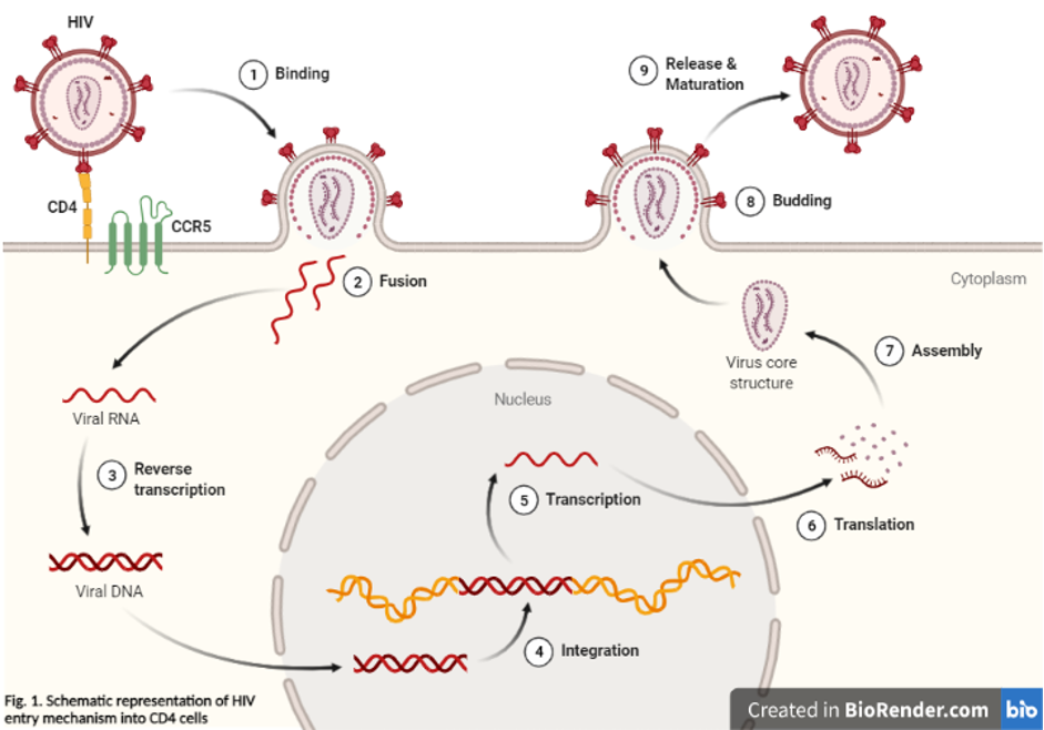
Rubella virus (RV) causes multisystem birth defects in the fetuses of infected women. To investigate the cellular basis of this pathology, we examined the cytopathic effect of RV in three permissive cell lines: Vero 76, RK13, and BHK21. Electron microscopy and the TUNEL assay showed that the cytopathic effect resulted from RV-induced programmed cell death (apoptosis) in all three cell lines, but the extent of apoptosis varied among these cells. At 48 h postinfection, the RK13 cell line showed the greatest number of apoptotic cells, the Vero 76 cell line was approximately 3-fold less, and BHK21 had very few. An increased multiplicity of infection and longer time postinfection were required for the BHK21 cell line to reach the level of apoptotic cells in Vero 76 at 48 h. Purified RV induced apoptosis in a dose-dependent fashion, but not UV-inactivated RV or virus-depleted culture supernatant. Specific inhibitors of the apoptosis-specific proteases caspases reduced RV-induced apoptosis and led to higher levels of RV components in infected cells. To address the role of regulatory proteins in RV-induced apoptosis, the antiapoptotic gene Bcl-2 or Bcl-XL was transfected into RK13 cells. Although a high level of Bcl-2 family proteins was expressed, no protection was observed from apoptosis induced by RV, Sindbis virus, or staurosporine in RK13 cells. In BHK21 cells, however, increased expression of Bcl-XL protected cells from apoptosis. The observed variability in apoptotic response to RV of these cell lines demonstrates that programmed cell death is dependent on the unique properties of each cell and may be indicative of how selective organ damage occurs in a congenital rubella syndrome fetus. (+info)Virus replication is the process by which a virus produces copies or reproduces itself inside a host cell. This involves several steps:
1. Attachment: The virus attaches to a specific receptor on the surface of the host cell.
2. Penetration: The viral genetic material enters the host cell, either by invagination of the cell membrane or endocytosis.
3. Uncoating: The viral genetic material is released from its protective coat (capsid) inside the host cell.
4. Replication: The viral genetic material uses the host cell's machinery to produce new viral components, such as proteins and nucleic acids.
5. Assembly: The newly synthesized viral components are assembled into new virus particles.
6. Release: The newly formed viruses are released from the host cell, often through lysis (breaking) of the cell membrane or by budding off the cell membrane.
The specific mechanisms and details of virus replication can vary depending on the type of virus. Some viruses, such as DNA viruses, use the host cell's DNA polymerase to replicate their genetic material, while others, such as RNA viruses, use their own RNA-dependent RNA polymerase or reverse transcriptase enzymes. Understanding the process of virus replication is important for developing antiviral therapies and vaccines.
A viral RNA (ribonucleic acid) is the genetic material found in certain types of viruses, as opposed to viruses that contain DNA (deoxyribonucleic acid). These viruses are known as RNA viruses. The RNA can be single-stranded or double-stranded and can exist as several different forms, such as positive-sense, negative-sense, or ambisense RNA. Upon infecting a host cell, the viral RNA uses the host's cellular machinery to translate the genetic information into proteins, leading to the production of new virus particles and the continuation of the viral life cycle. Examples of human diseases caused by RNA viruses include influenza, COVID-19 (SARS-CoV-2), hepatitis C, and polio.
Viral proteins are the proteins that are encoded by the viral genome and are essential for the viral life cycle. These proteins can be structural or non-structural and play various roles in the virus's replication, infection, and assembly process. Structural proteins make up the physical structure of the virus, including the capsid (the protein shell that surrounds the viral genome) and any envelope proteins (that may be present on enveloped viruses). Non-structural proteins are involved in the replication of the viral genome and modulation of the host cell environment to favor viral replication. Overall, a thorough understanding of viral proteins is crucial for developing antiviral therapies and vaccines.
RNA viruses are a type of virus that contain ribonucleic acid (RNA) as their genetic material, as opposed to deoxyribonucleic acid (DNA). RNA viruses replicate by using an enzyme called RNA-dependent RNA polymerase to transcribe and replicate their RNA genome.
There are several different groups of RNA viruses, including:
1. Negative-sense single-stranded RNA viruses: These viruses have a genome that is complementary to the mRNA and must undergo transcription to produce mRNA before translation can occur. Examples include influenza virus, measles virus, and rabies virus.
2. Positive-sense single-stranded RNA viruses: These viruses have a genome that can serve as mRNA and can be directly translated into protein after entry into the host cell. Examples include poliovirus, rhinoviruses, and coronaviruses.
3. Double-stranded RNA viruses: These viruses have a genome consisting of double-stranded RNA and use a complex replication strategy involving both transcription and reverse transcription. Examples include rotaviruses and reoviruses.
RNA viruses are known to cause a wide range of human diseases, ranging from the common cold to more severe illnesses such as hepatitis C, polio, and COVID-19. Due to their high mutation rates and ability to adapt quickly to new environments, RNA viruses can be difficult to control and treat with antiviral drugs or vaccines.
A cell line is a culture of cells that are grown in a laboratory for use in research. These cells are usually taken from a single cell or group of cells, and they are able to divide and grow continuously in the lab. Cell lines can come from many different sources, including animals, plants, and humans. They are often used in scientific research to study cellular processes, disease mechanisms, and to test new drugs or treatments. Some common types of human cell lines include HeLa cells (which come from a cancer patient named Henrietta Lacks), HEK293 cells (which come from embryonic kidney cells), and HUVEC cells (which come from umbilical vein endothelial cells). It is important to note that cell lines are not the same as primary cells, which are cells that are taken directly from a living organism and have not been grown in the lab.
Vaccinia virus is a large, complex DNA virus that belongs to the Poxviridae family. It is the virus used in the production of the smallpox vaccine. The vaccinia virus is not identical to the variola virus, which causes smallpox, but it is closely related and provides cross-protection against smallpox infection.
The vaccinia virus has a unique replication cycle that occurs entirely in the cytoplasm of infected cells, rather than in the nucleus like many other DNA viruses. This allows the virus to evade host cell defenses and efficiently produce new virions. The virus causes the formation of pocks or lesions on the skin, which contain large numbers of virus particles that can be transmitted to others through close contact.
Vaccinia virus has also been used as a vector for the delivery of genes encoding therapeutic proteins, vaccines against other infectious diseases, and cancer therapies. However, the use of vaccinia virus as a vector is limited by its potential to cause adverse reactions in some individuals, particularly those with weakened immune systems or certain skin conditions.
Viral DNA refers to the genetic material present in viruses that consist of DNA as their core component. Deoxyribonucleic acid (DNA) is one of the two types of nucleic acids that are responsible for storing and transmitting genetic information in living organisms. Viruses are infectious agents much smaller than bacteria that can only replicate inside the cells of other organisms, called hosts.
Viral DNA can be double-stranded (dsDNA) or single-stranded (ssDNA), depending on the type of virus. Double-stranded DNA viruses have a genome made up of two complementary strands of DNA, while single-stranded DNA viruses contain only one strand of DNA.
Examples of dsDNA viruses include Adenoviruses, Herpesviruses, and Poxviruses, while ssDNA viruses include Parvoviruses and Circoviruses. Viral DNA plays a crucial role in the replication cycle of the virus, encoding for various proteins necessary for its multiplication and survival within the host cell.
Molecular sequence data refers to the specific arrangement of molecules, most commonly nucleotides in DNA or RNA, or amino acids in proteins, that make up a biological macromolecule. This data is generated through laboratory techniques such as sequencing, and provides information about the exact order of the constituent molecules. This data is crucial in various fields of biology, including genetics, evolution, and molecular biology, allowing for comparisons between different organisms, identification of genetic variations, and studies of gene function and regulation.
Antiviral agents are a class of medications that are designed to treat infections caused by viruses. Unlike antibiotics, which target bacteria, antiviral agents interfere with the replication and infection mechanisms of viruses, either by inhibiting their ability to replicate or by modulating the host's immune response to the virus.
Antiviral agents are used to treat a variety of viral infections, including influenza, herpes simplex virus (HSV) infections, human immunodeficiency virus (HIV) infection, hepatitis B and C, and respiratory syncytial virus (RSV) infections.
These medications can be administered orally, intravenously, or topically, depending on the type of viral infection being treated. Some antiviral agents are also used for prophylaxis, or prevention, of certain viral infections.
It is important to note that antiviral agents are not effective against all types of viruses and may have significant side effects. Therefore, it is essential to consult with a healthcare professional before starting any antiviral therapy.
Virus assembly, also known as virion assembly, is the final stage in the virus life cycle where individual viral components come together to form a complete viral particle or virion. This process typically involves the self-assembly of viral capsid proteins around the viral genome (DNA or RNA) and, in enveloped viruses, the acquisition of a lipid bilayer membrane containing viral glycoproteins. The specific mechanisms and regulation of virus assembly vary among different viral families, but it is often directed by interactions between viral structural proteins and genomic nucleic acid.
Sindbis virus is an alphavirus that belongs to the Togaviridae family. It's named after the location where it was first isolated, in Sindbis, Egypt, in 1952. This virus is primarily transmitted by mosquitoes and can infect a wide range of animals, including birds and humans. In humans, Sindbis virus infection often causes a mild flu-like illness characterized by fever, rash, and joint pain. However, some people may develop more severe symptoms, such as neurological disorders, although this is relatively rare. There is no specific treatment for Sindbis virus infection, and management typically involves supportive care to alleviate symptoms.
Virus cultivation, also known as virus isolation or viral culture, is a laboratory method used to propagate and detect viruses by introducing them to host cells and allowing them to replicate. This process helps in identifying the specific virus causing an infection and studying its characteristics, such as morphology, growth pattern, and sensitivity to antiviral agents.
The steps involved in virus cultivation typically include:
1. Collection of a clinical sample (e.g., throat swab, blood, sputum) from the patient.
2. Preparation of the sample by centrifugation or filtration to remove cellular debris and other contaminants.
3. Inoculation of the prepared sample into susceptible host cells, which can be primary cell cultures, continuous cell lines, or embryonated eggs, depending on the type of virus.
4. Incubation of the inoculated cells under appropriate conditions to allow viral replication.
5. Observation for cytopathic effects (CPE), which are changes in the host cells caused by viral replication, such as cell rounding, shrinkage, or lysis.
6. Confirmation of viral presence through additional tests, like immunofluorescence assays, polymerase chain reaction (PCR), or electron microscopy.
Virus cultivation is a valuable tool in diagnostic virology, vaccine development, and research on viral pathogenesis and host-virus interactions. However, it requires specialized equipment, trained personnel, and biosafety measures due to the potential infectivity of the viruses being cultured.
Gene expression regulation, viral, refers to the processes that control the production of viral gene products, such as proteins and nucleic acids, during the viral life cycle. This can involve both viral and host cell factors that regulate transcription, RNA processing, translation, and post-translational modifications of viral genes.
Viral gene expression regulation is critical for the virus to replicate and produce progeny virions. Different types of viruses have evolved diverse mechanisms to regulate their gene expression, including the use of promoters, enhancers, transcription factors, RNA silencing, and epigenetic modifications. Understanding these regulatory processes can provide insights into viral pathogenesis and help in the development of antiviral therapies.
Vero cells are a line of cultured kidney epithelial cells that were isolated from an African green monkey (Cercopithecus aethiops) in the 1960s. They are named after the location where they were initially developed, the Vervet Research Institute in Japan.
Vero cells have the ability to divide indefinitely under certain laboratory conditions and are often used in scientific research, including virology, as a host cell for viruses to replicate. This allows researchers to study the characteristics of various viruses, such as their growth patterns and interactions with host cells. Vero cells are also used in the production of some vaccines, including those for rabies, polio, and Japanese encephalitis.
It is important to note that while Vero cells have been widely used in research and vaccine production, they can still have variations between different cell lines due to factors like passage number or culture conditions. Therefore, it's essential to specify the exact source and condition of Vero cells when reporting experimental results.
Virus receptors are specific molecules (commonly proteins) on the surface of host cells that viruses bind to in order to enter and infect those cells. This interaction between the virus and its receptor is a critical step in the infection process. Different types of viruses have different receptor requirements, and identifying these receptors can provide important insights into the biology of the virus and potential targets for antiviral therapies.
Defective viruses are viruses that have lost the ability to complete a full replication cycle and produce progeny virions independently. These viruses require the assistance of a helper virus, which provides the necessary functions for replication. Defective viruses can arise due to mutations, deletions, or other genetic changes that result in the loss of essential genes. They are often non-infectious and cannot cause disease on their own, but they may interfere with the replication of the helper virus and modulate the course of infection. Defective viruses can be found in various types of viruses, including retroviruses, bacteriophages, and DNA viruses.
Viral genes refer to the genetic material present in viruses that contains the information necessary for their replication and the production of viral proteins. In DNA viruses, the genetic material is composed of double-stranded or single-stranded DNA, while in RNA viruses, it is composed of single-stranded or double-stranded RNA.
Viral genes can be classified into three categories: early, late, and structural. Early genes encode proteins involved in the replication of the viral genome, modulation of host cell processes, and regulation of viral gene expression. Late genes encode structural proteins that make up the viral capsid or envelope. Some viruses also have structural genes that are expressed throughout their replication cycle.
Understanding the genetic makeup of viruses is crucial for developing antiviral therapies and vaccines. By targeting specific viral genes, researchers can develop drugs that inhibit viral replication and reduce the severity of viral infections. Additionally, knowledge of viral gene sequences can inform the development of vaccines that stimulate an immune response to specific viral proteins.
Virus shedding refers to the release of virus particles by an infected individual, who can then transmit the virus to others through various means such as respiratory droplets, fecal matter, or bodily fluids. This occurs when the virus replicates inside the host's cells and is released into the surrounding environment, where it can infect other individuals. The duration of virus shedding varies depending on the specific virus and the individual's immune response. It's important to note that some individuals may shed viruses even before they show symptoms, making infection control measures such as hand hygiene, mask-wearing, and social distancing crucial in preventing the spread of infectious diseases.
HIV-1 (Human Immunodeficiency Virus type 1) is a species of the retrovirus genus that causes acquired immunodeficiency syndrome (AIDS). It is primarily transmitted through sexual contact, exposure to infected blood or blood products, and from mother to child during pregnancy, childbirth, or breastfeeding. HIV-1 infects vital cells in the human immune system, such as CD4+ T cells, macrophages, and dendritic cells, leading to a decline in their numbers and weakening of the immune response over time. This results in the individual becoming susceptible to various opportunistic infections and cancers that ultimately cause death if left untreated. HIV-1 is the most prevalent form of HIV worldwide and has been identified as the causative agent of the global AIDS pandemic.
Hepatitis B virus (HBV) is a DNA virus that belongs to the Hepadnaviridae family and causes the infectious disease known as hepatitis B. This virus primarily targets the liver, where it can lead to inflammation and damage of the liver tissue. The infection can range from acute to chronic, with chronic hepatitis B increasing the risk of developing serious liver complications such as cirrhosis and liver cancer.
The Hepatitis B virus has a complex life cycle, involving both nuclear and cytoplasmic phases. It enters hepatocytes (liver cells) via binding to specific receptors and is taken up by endocytosis. The viral DNA is released into the nucleus, where it is converted into a covalently closed circular DNA (cccDNA) form, which serves as the template for viral transcription.
HBV transcribes several RNAs, including pregenomic RNA (pgRNA), which is used as a template for reverse transcription during virion assembly. The pgRNA is encapsidated into core particles along with the viral polymerase and undergoes reverse transcription to generate new viral DNA. This process occurs within the cytoplasm of the hepatocyte, resulting in the formation of immature virions containing partially double-stranded DNA.
These immature virions are then enveloped by host cell membranes containing HBV envelope proteins (known as surface antigens) to form mature virions that can be secreted from the hepatocyte and infect other cells. The virus can also integrate into the host genome, which may contribute to the development of hepatocellular carcinoma in chronic cases.
Hepatitis B is primarily transmitted through exposure to infected blood or bodily fluids containing the virus, such as through sexual contact, sharing needles, or from mother to child during childbirth. Prevention strategies include vaccination, safe sex practices, and avoiding needle-sharing behaviors. Treatment for hepatitis B typically involves antiviral medications that can help suppress viral replication and reduce the risk of liver damage.
Viral diseases are illnesses caused by the infection and replication of viruses in host organisms. These infectious agents are obligate parasites, meaning they rely on the cells of other living organisms to survive and reproduce. Viruses can infect various types of hosts, including animals, plants, and microorganisms, causing a wide range of diseases with varying symptoms and severity.
Once a virus enters a host cell, it takes over the cell's machinery to produce new viral particles, often leading to cell damage or death. The immune system recognizes the viral components as foreign and mounts an immune response to eliminate the infection. This response can result in inflammation, fever, and other symptoms associated with viral diseases.
Examples of well-known viral diseases include:
1. Influenza (flu) - caused by influenza A, B, or C viruses
2. Common cold - usually caused by rhinoviruses or coronaviruses
3. HIV/AIDS - caused by human immunodeficiency virus (HIV)
4. Measles - caused by measles morbillivirus
5. Hepatitis B and C - caused by hepatitis B virus (HBV) and hepatitis C virus (HCV), respectively
6. Herpes simplex - caused by herpes simplex virus type 1 (HSV-1) or type 2 (HSV-2)
7. Chickenpox and shingles - both caused by varicella-zoster virus (VZV)
8. Rabies - caused by rabies lyssavirus
9. Ebola - caused by ebolaviruses
10. COVID-19 - caused by severe acute respiratory syndrome coronavirus 2 (SARS-CoV-2)
Prevention and treatment strategies for viral diseases may include vaccination, antiviral medications, and supportive care to manage symptoms while the immune system fights off the infection.
DNA viruses are a type of virus that contain DNA (deoxyribonucleic acid) as their genetic material. These viruses replicate by using the host cell's machinery to synthesize new viral components, which are then assembled into new viruses and released from the host cell.
DNA viruses can be further classified based on the structure of their genomes and the way they replicate. For example, double-stranded DNA (dsDNA) viruses have a genome made up of two strands of DNA, while single-stranded DNA (ssDNA) viruses have a genome made up of a single strand of DNA.
Examples of DNA viruses include herpes simplex virus, varicella-zoster virus, human papillomavirus, and adenoviruses. Some DNA viruses are associated with specific diseases, such as cancer (e.g., human papillomavirus) or neurological disorders (e.g., herpes simplex virus).
It's important to note that while DNA viruses contain DNA as their genetic material, RNA viruses contain RNA (ribonucleic acid) as their genetic material. Both DNA and RNA viruses can cause a wide range of diseases in humans, animals, and plants.
A viral plaque assay is a laboratory technique used to measure the infectivity and concentration of viruses in a sample. This method involves infecting a monolayer of cells (usually in a petri dish or multi-well plate) with a known volume of a virus-containing sample, followed by overlaying the cells with a nutrient-agar medium to restrict viral spread and enable individual plaques to form.
After an incubation period that allows for viral replication and cell death, the cells are stained, and clear areas or "plaques" become visible in the monolayer. Each plaque represents a localized region of infected and lysed cells, caused by the progeny of a single infectious virus particle. The number of plaques is then counted, and the viral titer (infectious units per milliliter or PFU/mL) is calculated based on the dilution factor and volume of the original inoculum.
Viral plaque assays are essential for determining viral titers, assessing virus-host interactions, evaluating antiviral agents, and studying viral pathogenesis.
A base sequence in the context of molecular biology refers to the specific order of nucleotides in a DNA or RNA molecule. In DNA, these nucleotides are adenine (A), guanine (G), cytosine (C), and thymine (T). In RNA, uracil (U) takes the place of thymine. The base sequence contains genetic information that is transcribed into RNA and ultimately translated into proteins. It is the exact order of these bases that determines the genetic code and thus the function of the DNA or RNA molecule.
A plant virus is not defined in medical terms, as viruses are typically considered within the context of living organisms and plants are not classified as such. However, I can provide a general virology definition for you:
Plant viruses are small infectious agents that consist of nucleic acid (DNA or RNA) enclosed in a protein coat. They infect various plant species, causing a wide range of symptoms and diseases, which can result in significant economic losses in agriculture and horticulture. Plant viruses lack the ability to replicate outside a host cell, and they rely on the host's metabolic machinery for their reproduction. They can be transmitted through various means, such as insect vectors, seeds, or mechanical contact.
Simian Immunodeficiency Virus (SIV) is a retrovirus that primarily infects African non-human primates and is the direct ancestor of Human Immunodeficiency Virus type 2 (HIV-2). It is similar to HIV in its structure, replication strategy, and ability to cause an immunodeficiency disease in its host. SIV infection in its natural hosts is typically asymptomatic and non-lethal, but it can cause AIDS-like symptoms in other primate species. Research on SIV in its natural hosts has provided valuable insights into the mechanisms of HIV pathogenesis and potential strategies for prevention and treatment of AIDS.
A Cytopathic Effect (CPE) is a visible change in the cell or group of cells due to infection by a pathogen, such as a virus. When the cytopathic effect is caused specifically by a viral infection, it is referred to as a "Viral Cytopathic Effect" (VCPE).
The VCPE can include various changes in the cell's morphology, size, and structure, such as rounding, shrinkage, multinucleation, inclusion bodies, and formation of syncytia (multinucleated giant cells). These changes are often used to identify and characterize viruses in laboratory settings.
The VCPE is typically observed under a microscope after the virus has infected cell cultures, and it can help researchers determine the type of virus, the degree of infection, and the effectiveness of antiviral treatments. The severity and timing of the VCPE can vary depending on the specific virus and the type of cells that are infected.
Measles virus is a single-stranded, negative-sense RNA virus belonging to the genus Morbillivirus in the family Paramyxoviridae. It is the causative agent of measles, a highly contagious infectious disease characterized by fever, cough, runny nose, and a red, blotchy rash. The virus primarily infects the respiratory tract and then spreads throughout the body via the bloodstream.
The genome of the measles virus is approximately 16 kilobases in length and encodes for eight proteins: nucleocapsid (N), phosphoprotein (P), matrix protein (M), fusion protein (F), hemagglutinin (H), large protein (L), and two non-structural proteins, V and C. The H protein is responsible for binding to the host cell receptor CD150 (SLAM) and mediating viral entry, while the F protein facilitates fusion of the viral and host cell membranes.
Measles virus is transmitted through respiratory droplets and direct contact with infected individuals. The virus can remain airborne for up to two hours in a closed space, making it highly contagious. Measles is preventable through vaccination, which has led to significant reductions in the incidence of the disease worldwide.
Replication Protein A (RPA) is a single-stranded DNA binding protein complex that plays a crucial role in the process of DNA replication, repair, and recombination. In eukaryotic cells, RPA is composed of three subunits: RPA70, RPA32, and RPA14. The primary function of RPA is to coat single-stranded DNA (ssDNA) generated during these processes, protecting it from degradation, preventing the formation of secondary structures, and promoting the recruitment of other proteins involved in DNA metabolism.
RPA binds ssDNA with high affinity and specificity, forming a stable complex that protects the DNA from nucleases, chemical modifications, and other damaging agents. The protein also participates in the regulation of various enzymatic activities, such as helicase loading and activation, end processing, and polymerase processivity.
During DNA replication, RPA is essential for the initiation and elongation phases. It facilitates the assembly of the pre-replicative complex (pre-RC) at origins of replication, aids in the recruitment and activation of helicases, and promotes the switch from MCM2-7 helicase to polymerase processivity during DNA synthesis.
In addition to its role in DNA replication, RPA is involved in various DNA repair pathways, including nucleotide excision repair (NER), base excision repair (BER), mismatch repair (MMR), and double-strand break repair (DSBR). It also plays a critical role in meiotic recombination during sexual reproduction.
In summary, Replication Protein A (RPA) is a eukaryotic single-stranded DNA binding protein complex that protects, stabilizes, and regulates ssDNA during DNA replication, repair, and recombination processes.
A plasmid is a small, circular, double-stranded DNA molecule that is separate from the chromosomal DNA of a bacterium or other organism. Plasmids are typically not essential for the survival of the organism, but they can confer beneficial traits such as antibiotic resistance or the ability to degrade certain types of pollutants.
Plasmids are capable of replicating independently of the chromosomal DNA and can be transferred between bacteria through a process called conjugation. They often contain genes that provide resistance to antibiotics, heavy metals, and other environmental stressors. Plasmids have also been engineered for use in molecular biology as cloning vectors, allowing scientists to replicate and manipulate specific DNA sequences.
Plasmids are important tools in genetic engineering and biotechnology because they can be easily manipulated and transferred between organisms. They have been used to produce vaccines, diagnostic tests, and genetically modified organisms (GMOs) for various applications, including agriculture, medicine, and industry.
A virion is the complete, infectious form of a virus outside its host cell. It consists of the viral genome (DNA or RNA) enclosed within a protein coat called the capsid, which is often surrounded by a lipid membrane called the envelope. The envelope may contain viral proteins and glycoproteins that aid in attachment to and entry into host cells during infection. The term "virion" emphasizes the infectious nature of the virus particle, as opposed to non-infectious components like individual capsid proteins or naked viral genome.
'Influenza A Virus, H1N1 Subtype' is a specific subtype of the influenza A virus that causes flu in humans and animals. It contains certain proteins called hemagglutinin (H) and neuraminidase (N) on its surface, with this subtype specifically having H1 and N1 antigens. The H1N1 strain is well-known for causing the 2009 swine flu pandemic, which was a global outbreak of flu that resulted in significant morbidity and mortality. This subtype can also cause seasonal flu, although the severity and symptoms may vary. It is important to note that influenza viruses are constantly changing, and new strains or subtypes can emerge over time, requiring regular updates to vaccines to protect against them.
"Influenza A Virus, H5N1 Subtype" is a specific subtype of the Influenza A virus that is often found in avian species (birds) and can occasionally infect humans. The "H5N1" refers to the specific proteins (hemagglutinin and neuraminidase) found on the surface of the virus. This subtype has caused serious infections in humans, with high mortality rates, especially in cases where people have had close contact with infected birds. It does not commonly spread from person to person, but there is concern that it could mutate and adapt to efficiently transmit between humans, which would potentially cause a pandemic.
Simian Virus 40 (SV40) is a polyomavirus that is found in both monkeys and humans. It is a DNA virus that has been extensively studied in laboratory settings due to its ability to transform cells and cause tumors in animals. In fact, SV40 was discovered as a contaminant of poliovirus vaccines that were prepared using rhesus monkey kidney cells in the 1950s and 1960s.
SV40 is not typically associated with human disease, but there has been some concern that exposure to the virus through contaminated vaccines or other means could increase the risk of certain types of cancer, such as mesothelioma and brain tumors. However, most studies have failed to find a consistent link between SV40 infection and cancer in humans.
The medical community generally agrees that SV40 is not a significant public health threat, but researchers continue to study the virus to better understand its biology and potential impact on human health.
An amino acid sequence is the specific order of amino acids in a protein or peptide molecule, formed by the linking of the amino group (-NH2) of one amino acid to the carboxyl group (-COOH) of another amino acid through a peptide bond. The sequence is determined by the genetic code and is unique to each type of protein or peptide. It plays a crucial role in determining the three-dimensional structure and function of proteins.
An antigen is any substance that can stimulate an immune response, particularly the production of antibodies. Viral antigens are antigens that are found on or produced by viruses. They can be proteins, glycoproteins, or carbohydrates present on the surface or inside the viral particle.
Viral antigens play a crucial role in the immune system's recognition and response to viral infections. When a virus infects a host cell, it may display its antigens on the surface of the infected cell. This allows the immune system to recognize and target the infected cells for destruction, thereby limiting the spread of the virus.
Viral antigens are also important targets for vaccines. Vaccines typically work by introducing a harmless form of a viral antigen to the body, which then stimulates the production of antibodies and memory T-cells that can recognize and respond quickly and effectively to future infections with the actual virus.
It's worth noting that different types of viruses have different antigens, and these antigens can vary between strains of the same virus. This is why there are often different vaccines available for different viral diseases, and why flu vaccines need to be updated every year to account for changes in the circulating influenza virus strains.
'Cercopithecus aethiops' is the scientific name for the monkey species more commonly known as the green monkey. It belongs to the family Cercopithecidae and is native to western Africa. The green monkey is omnivorous, with a diet that includes fruits, nuts, seeds, insects, and small vertebrates. They are known for their distinctive greenish-brown fur and long tail. Green monkeys are also important animal models in biomedical research due to their susceptibility to certain diseases, such as SIV (simian immunodeficiency virus), which is closely related to HIV.
Viral activation, also known as viral reactivation or virus reactivation, refers to the process in which a latent or dormant virus becomes active and starts to replicate within a host cell. This can occur when the immune system is weakened or compromised, allowing the virus to evade the body's natural defenses and cause disease.
In some cases, viral activation can be triggered by certain environmental factors, such as stress, exposure to UV light, or infection with another virus. Once activated, the virus can cause symptoms similar to those seen during the initial infection, or it may lead to new symptoms depending on the specific virus and the host's immune response.
Examples of viruses that can remain dormant in the body and be reactivated include herpes simplex virus (HSV), varicella-zoster virus (VZV), cytomegalovirus (CMV), and Epstein-Barr virus (EBV). It is important to note that not all viruses can be reactivated, and some may remain dormant in the body indefinitely without causing any harm.
HeLa cells are a type of immortalized cell line used in scientific research. They are derived from a cancer that developed in the cervical tissue of Henrietta Lacks, an African-American woman, in 1951. After her death, cells taken from her tumor were found to be capable of continuous division and growth in a laboratory setting, making them an invaluable resource for medical research.
HeLa cells have been used in a wide range of scientific studies, including research on cancer, viruses, genetics, and drug development. They were the first human cell line to be successfully cloned and are able to grow rapidly in culture, doubling their population every 20-24 hours. This has made them an essential tool for many areas of biomedical research.
It is important to note that while HeLa cells have been instrumental in numerous scientific breakthroughs, the story of their origin raises ethical questions about informed consent and the use of human tissue in research.
Vesicular stomatitis Indiana virus (VSIV) is a single-stranded, negative-sense RNA virus that belongs to the family Rhabdoviridae and genus Vesiculovirus. It is the causative agent of vesicular stomatitis (VS), a viral disease that primarily affects horses and cattle, but can also infect other species including swine, sheep, goats, and humans.
The virus is transmitted through direct contact with infected animals or their saliva, as well as through insect vectors such as black flies and sandflies. The incubation period for VS ranges from 2 to 8 days, after which infected animals develop fever, lethargy, and vesicular lesions in the mouth, nose, and feet. These lesions can be painful and may cause difficulty eating or walking.
In humans, VSIV infection is typically asymptomatic or causes mild flu-like symptoms such as fever, muscle aches, and headache. Occasionally, individuals may develop vesicular lesions on their skin or mucous membranes, particularly if they have had contact with infected animals.
Diagnosis of VSIV infection is typically made through virus isolation from lesion exudates or blood, as well as through serological testing. Treatment is generally supportive and aimed at relieving symptoms, as there are no specific antiviral therapies available for VS. Prevention measures include vaccination of susceptible animals, vector control, and biosecurity measures to prevent the spread of infection between animals.
Virus latency, also known as viral latency, refers to a state of infection in which a virus remains dormant or inactive within a host cell for a period of time. During this phase, the virus does not replicate or cause any noticeable symptoms. However, under certain conditions such as stress, illness, or a weakened immune system, the virus can become reactivated and begin to produce new viruses, potentially leading to disease.
One well-known example of a virus that exhibits latency is the varicella-zoster virus (VZV), which causes chickenpox in children. After a person recovers from chickenpox, the virus remains dormant in the nervous system for years or even decades. In some cases, the virus can reactivate later in life, causing shingles, a painful rash that typically occurs on one side of the body.
Virus latency is an important concept in virology and infectious disease research, as it has implications for understanding the persistence of viral infections, developing treatments and vaccines, and predicting the risk of disease recurrence.
I'm sorry for any confusion, but "replicon" is not a term that has a widely accepted or specific medical definition. It is a term that is used in the field of molecular biology, where it refers to a segment of DNA that contains an origin of replication. The origin of replication is the site on the DNA molecule where the process of DNA replication is initiated. This concept is important in the fields of genetics and virology, but it is not a term that is commonly used in clinical medicine.
If you have any questions related to the medical field, I would be happy to try to help answer them for you!
West Nile Virus (WNV) is an Flavivirus, which is a type of virus that is spread by mosquitoes. It was first discovered in the West Nile district of Uganda in 1937 and has since been found in many countries throughout the world. WNV can cause a mild to severe illness known as West Nile fever.
Most people who become infected with WNV do not develop any symptoms, but some may experience fever, headache, body aches, joint pain, vomiting, diarrhea, or a rash. In rare cases, the virus can cause serious neurological illnesses such as encephalitis (inflammation of the brain) or meningitis (inflammation of the membranes surrounding the brain and spinal cord). These severe forms of the disease can be fatal, especially in older adults and people with weakened immune systems.
WNV is primarily transmitted to humans through the bite of infected mosquitoes, but it can also be spread through blood transfusions, organ transplants, or from mother to baby during pregnancy, delivery, or breastfeeding. There is no specific treatment for WNV, and most people recover on their own with rest and supportive care. However, hospitalization may be necessary in severe cases. Prevention measures include avoiding mosquito bites by using insect repellent, wearing long sleeves and pants, and staying indoors during peak mosquito activity hours.
Host-pathogen interactions refer to the complex and dynamic relationship between a living organism (the host) and a disease-causing agent (the pathogen). This interaction can involve various molecular, cellular, and physiological processes that occur between the two entities. The outcome of this interaction can determine whether the host will develop an infection or not, as well as the severity and duration of the illness.
During host-pathogen interactions, the pathogen may release virulence factors that allow it to evade the host's immune system, colonize tissues, and obtain nutrients for its survival and replication. The host, in turn, may mount an immune response to recognize and eliminate the pathogen, which can involve various mechanisms such as inflammation, phagocytosis, and the production of antimicrobial agents.
Understanding the intricacies of host-pathogen interactions is crucial for developing effective strategies to prevent and treat infectious diseases. This knowledge can help identify new targets for therapeutic interventions, inform vaccine design, and guide public health policies to control the spread of infectious agents.
A viral genome is the genetic material (DNA or RNA) that is present in a virus. It contains all the genetic information that a virus needs to replicate itself and infect its host. The size and complexity of viral genomes can vary greatly, ranging from a few thousand bases to hundreds of thousands of bases. Some viruses have linear genomes, while others have circular genomes. The genome of a virus also contains the information necessary for the virus to hijack the host cell's machinery and use it to produce new copies of the virus. Understanding the genetic makeup of viruses is important for developing vaccines and antiviral treatments.
A mutation is a permanent change in the DNA sequence of an organism's genome. Mutations can occur spontaneously or be caused by environmental factors such as exposure to radiation, chemicals, or viruses. They may have various effects on the organism, ranging from benign to harmful, depending on where they occur and whether they alter the function of essential proteins. In some cases, mutations can increase an individual's susceptibility to certain diseases or disorders, while in others, they may confer a survival advantage. Mutations are the driving force behind evolution, as they introduce new genetic variability into populations, which can then be acted upon by natural selection.
"Cells, cultured" is a medical term that refers to cells that have been removed from an organism and grown in controlled laboratory conditions outside of the body. This process is called cell culture and it allows scientists to study cells in a more controlled and accessible environment than they would have inside the body. Cultured cells can be derived from a variety of sources, including tissues, organs, or fluids from humans, animals, or cell lines that have been previously established in the laboratory.
Cell culture involves several steps, including isolation of the cells from the tissue, purification and characterization of the cells, and maintenance of the cells in appropriate growth conditions. The cells are typically grown in specialized media that contain nutrients, growth factors, and other components necessary for their survival and proliferation. Cultured cells can be used for a variety of purposes, including basic research, drug development and testing, and production of biological products such as vaccines and gene therapies.
It is important to note that cultured cells may behave differently than they do in the body, and results obtained from cell culture studies may not always translate directly to human physiology or disease. Therefore, it is essential to validate findings from cell culture experiments using additional models and ultimately in clinical trials involving human subjects.
Rabies is a viral disease that affects the nervous system of mammals, including humans. It's caused by the rabies virus (RV), which belongs to the family Rhabdoviridae and genus Lyssavirus. The virus has a bullet-shaped appearance under an electron microscope and is encased in a lipid envelope.
The rabies virus primarily spreads through the saliva of infected animals, usually via bites. Once inside the body, it travels along nerve fibers to the brain, where it multiplies rapidly and causes inflammation (encephalitis). The infection can lead to symptoms such as anxiety, confusion, hallucinations, seizures, paralysis, coma, and ultimately death if left untreated.
Rabies is almost always fatal once symptoms appear, but prompt post-exposure prophylaxis (PEP), which includes vaccination and sometimes rabies immunoglobulin, can prevent the disease from developing when administered after an exposure to a potentially rabid animal. Pre-exposure vaccination is also recommended for individuals at high risk of exposure, such as veterinarians and travelers visiting rabies-endemic areas.
Viral nonstructural proteins (NS) are viral proteins that are not part of the virion structure. They play various roles in the viral life cycle, such as replication of the viral genome, transcription, translation regulation, and modulation of the host cell environment to favor virus replication. These proteins are often produced in large quantities during infection and can manipulate or disrupt various cellular pathways to benefit the virus. They may also be involved in evasion of the host's immune response. The specific functions of viral nonstructural proteins vary depending on the type of virus.
DNA-binding proteins are a type of protein that have the ability to bind to DNA (deoxyribonucleic acid), the genetic material of organisms. These proteins play crucial roles in various biological processes, such as regulation of gene expression, DNA replication, repair and recombination.
The binding of DNA-binding proteins to specific DNA sequences is mediated by non-covalent interactions, including electrostatic, hydrogen bonding, and van der Waals forces. The specificity of binding is determined by the recognition of particular nucleotide sequences or structural features of the DNA molecule.
DNA-binding proteins can be classified into several categories based on their structure and function, such as transcription factors, histones, and restriction enzymes. Transcription factors are a major class of DNA-binding proteins that regulate gene expression by binding to specific DNA sequences in the promoter region of genes and recruiting other proteins to modulate transcription. Histones are DNA-binding proteins that package DNA into nucleosomes, the basic unit of chromatin structure. Restriction enzymes are DNA-binding proteins that recognize and cleave specific DNA sequences, and are widely used in molecular biology research and biotechnology applications.
"Influenza A Virus, H3N2 Subtype" is a specific subtype of the influenza A virus that causes respiratory illness and is known to circulate in humans and animals, including birds and pigs. The "H3N2" refers to the two proteins on the surface of the virus: hemagglutinin (H) and neuraminidase (N). In this subtype, the H protein is of the H3 variety and the N protein is of the N2 variety. This subtype has been responsible for several influenza epidemics and pandemics in humans, including the 1968 Hong Kong flu pandemic. It is one of the influenza viruses that are monitored closely by public health authorities due to its potential to cause significant illness and death, particularly in high-risk populations such as older adults, young children, and people with certain underlying medical conditions.
Genetic recombination is the process by which genetic material is exchanged between two similar or identical molecules of DNA during meiosis, resulting in new combinations of genes on each chromosome. This exchange occurs during crossover, where segments of DNA are swapped between non-sister homologous chromatids, creating genetic diversity among the offspring. It is a crucial mechanism for generating genetic variability and facilitating evolutionary change within populations. Additionally, recombination also plays an essential role in DNA repair processes through mechanisms such as homologous recombinational repair (HRR) and non-homologous end joining (NHEJ).
Hepacivirus is a genus of viruses in the family Flaviviridae. The most well-known member of this genus is Hepatitis C virus (HCV), which is a major cause of liver disease worldwide. HCV infection can lead to chronic hepatitis, cirrhosis, and liver cancer.
Hepaciviruses are enveloped viruses with a single-stranded, positive-sense RNA genome. They have a small icosahedral capsid and infect a variety of hosts, including humans, non-human primates, horses, and birds. The virus enters the host cell by binding to specific receptors on the cell surface and is then internalized through endocytosis.
HCV has a high degree of genetic diversity and is classified into seven major genotypes and numerous subtypes based on differences in its RNA sequence. This genetic variability can affect the virus's ability to evade the host immune response, making treatment more challenging.
In addition to HCV, other hepaciviruses have been identified in various animal species, including equine hepacivirus (EHCV), rodent hepacivirus (RHV), and bat hepacivirus (BtHepCV). These viruses are being studied to better understand the biology of hepaciviruses and their potential impact on human health.
Genetic transcription is the process by which the information in a strand of DNA is used to create a complementary RNA molecule. This process is the first step in gene expression, where the genetic code in DNA is converted into a form that can be used to produce proteins or functional RNAs.
During transcription, an enzyme called RNA polymerase binds to the DNA template strand and reads the sequence of nucleotide bases. As it moves along the template, it adds complementary RNA nucleotides to the growing RNA chain, creating a single-stranded RNA molecule that is complementary to the DNA template strand. Once transcription is complete, the RNA molecule may undergo further processing before it can be translated into protein or perform its functional role in the cell.
Transcription can be either "constitutive" or "regulated." Constitutive transcription occurs at a relatively constant rate and produces essential proteins that are required for basic cellular functions. Regulated transcription, on the other hand, is subject to control by various intracellular and extracellular signals, allowing cells to respond to changing environmental conditions or developmental cues.
RNA-dependent RNA polymerase, also known as RNA replicase, is an enzyme that catalyzes the production of RNA from an RNA template. It plays a crucial role in the replication of certain viruses, such as positive-strand RNA viruses and retroviruses, which use RNA as their genetic material. The enzyme uses the existing RNA strand as a template to create a new complementary RNA strand, effectively replicating the viral genome. This process is essential for the propagation of these viruses within host cells and is a target for antiviral therapies.
Respiratory Syncytial Viruses (RSV) are a common type of virus that cause respiratory infections, particularly in young children and older adults. They are responsible for inflammation and narrowing of the small airways in the lungs, leading to breathing difficulties and other symptoms associated with bronchiolitis and pneumonia.
The term "syncytial" refers to the ability of these viruses to cause infected cells to merge and form large multinucleated cells called syncytia, which is a characteristic feature of RSV infections. The virus spreads through respiratory droplets when an infected person coughs or sneezes, and it can also survive on surfaces for several hours, making transmission easy.
RSV infections are most common during the winter months and can cause mild to severe symptoms depending on factors such as age, overall health, and underlying medical conditions. While RSV is typically associated with respiratory illnesses in children, it can also cause significant disease in older adults and immunocompromised individuals. Currently, there is no vaccine available for RSV, but antiviral medications and supportive care are used to manage severe infections.
Cricetinae is a subfamily of rodents that includes hamsters, gerbils, and relatives. These small mammals are characterized by having short limbs, compact bodies, and cheek pouches for storing food. They are native to various parts of the world, particularly in Europe, Asia, and Africa. Some species are popular pets due to their small size, easy care, and friendly nature. In a medical context, understanding the biology and behavior of Cricetinae species can be important for individuals who keep them as pets or for researchers studying their physiology.
Orthomyxoviridae is a family of viruses that includes influenza A, B, and C viruses, which can cause respiratory infections in humans. Orthomyxoviridae infections are typically characterized by symptoms such as fever, cough, sore throat, runny or stuffy nose, muscle or body aches, headaches, and fatigue.
Influenza A and B viruses can cause seasonal epidemics of respiratory illness that occur mainly during the winter months in temperate climates. Influenza A viruses can also cause pandemics, which are global outbreaks of disease that occur when a new strain of the virus emerges to which there is little or no immunity in the human population.
Influenza C viruses are less common and typically cause milder illness than influenza A and B viruses. They do not cause epidemics and are not usually included in seasonal flu vaccines.
Orthomyxoviridae infections can be prevented through vaccination, good respiratory hygiene (such as covering the mouth and nose when coughing or sneezing), hand washing, and avoiding close contact with sick individuals. Antiviral medications may be prescribed to treat influenza A and B infections, particularly for people at high risk of complications, such as older adults, young children, pregnant women, and people with certain underlying medical conditions.
DNA replication timing refers to the specific point during the cell cycle when a particular segment or region of the DNA molecule is copied or replicated. The genome of an organism is composed of millions of base pairs of DNA, and not all of these regions are replicated at the same time. Instead, DNA replication is a highly regulated process that occurs in a specific order and pattern during the S phase of the cell cycle.
During DNA replication, the double helix structure of DNA is unwound, and each strand serves as a template for the synthesis of a new complementary strand. The timing of DNA replication can vary between different regions of the genome, with some regions replicating early in the S phase and others replicating later. This temporal organization of DNA replication is known as the DNA replication program or timing profile.
The regulation of DNA replication timing is critical for maintaining genomic stability and ensuring that all regions of the genome are accurately replicated before cell division. Abnormalities in DNA replication timing have been associated with various diseases, including cancer and developmental disorders. Therefore, understanding the mechanisms that control DNA replication timing is an important area of research in molecular biology and genetics.
Medical Definition of "Herpesvirus 1, Human" (also known as Human Herpesvirus 1 or HHV-1):
Herpesvirus 1, Human is a type of herpesvirus that primarily causes infection in humans. It is also commonly referred to as human herpesvirus 1 (HHV-1) or oral herpes. This virus is highly contagious and can be transmitted through direct contact with infected saliva, skin, or mucous membranes.
After initial infection, the virus typically remains dormant in the body's nerve cells and may reactivate later, causing recurrent symptoms. The most common manifestation of HHV-1 infection is oral herpes, characterized by cold sores or fever blisters around the mouth and lips. In some cases, HHV-1 can also cause other conditions such as encephalitis (inflammation of the brain) and keratitis (inflammation of the eye's cornea).
There is no cure for HHV-1 infection, but antiviral medications can help manage symptoms and reduce the severity and frequency of recurrent outbreaks.
Virulence, in the context of medicine and microbiology, refers to the degree or severity of damage or harm that a pathogen (like a bacterium, virus, fungus, or parasite) can cause to its host. It is often associated with the ability of the pathogen to invade and damage host tissues, evade or suppress the host's immune response, replicate within the host, and spread between hosts.
Virulence factors are the specific components or mechanisms that contribute to a pathogen's virulence, such as toxins, enzymes, adhesins, and capsules. These factors enable the pathogen to establish an infection, cause tissue damage, and facilitate its transmission between hosts. The overall virulence of a pathogen can be influenced by various factors, including host susceptibility, environmental conditions, and the specific strain or species of the pathogen.
Simplexvirus is a genus of viruses in the family Herpesviridae, subfamily Alphaherpesvirinae. This genus contains two species: Human alphaherpesvirus 1 (also known as HSV-1 or herpes simplex virus type 1) and Human alphaherpesvirus 2 (also known as HSV-2 or herpes simplex virus type 2). These viruses are responsible for causing various medical conditions, most commonly oral and genital herpes. They are characterized by their ability to establish lifelong latency in the nervous system and reactivate periodically to cause recurrent symptoms.
A genetic vector is a vehicle, often a plasmid or a virus, that is used to introduce foreign DNA into a host cell as part of genetic engineering or gene therapy techniques. The vector contains the desired gene or genes, along with regulatory elements such as promoters and enhancers, which are needed for the expression of the gene in the target cells.
The choice of vector depends on several factors, including the size of the DNA to be inserted, the type of cell to be targeted, and the efficiency of uptake and expression required. Commonly used vectors include plasmids, adenoviruses, retroviruses, and lentiviruses.
Plasmids are small circular DNA molecules that can replicate independently in bacteria. They are often used as cloning vectors to amplify and manipulate DNA fragments. Adenoviruses are double-stranded DNA viruses that infect a wide range of host cells, including human cells. They are commonly used as gene therapy vectors because they can efficiently transfer genes into both dividing and non-dividing cells.
Retroviruses and lentiviruses are RNA viruses that integrate their genetic material into the host cell's genome. This allows for stable expression of the transgene over time. Lentiviruses, a subclass of retroviruses, have the advantage of being able to infect non-dividing cells, making them useful for gene therapy applications in post-mitotic tissues such as neurons and muscle cells.
Overall, genetic vectors play a crucial role in modern molecular biology and medicine, enabling researchers to study gene function, develop new therapies, and modify organisms for various purposes.
Hemagglutinin (HA) glycoproteins are surface proteins found on influenza viruses. They play a crucial role in the virus's ability to infect and spread within host organisms.
The HAs are responsible for binding to sialic acid receptors on the host cell's surface, allowing the virus to attach and enter the cell. After endocytosis, the viral and endosomal membranes fuse, releasing the viral genome into the host cell's cytoplasm.
There are several subtypes of hemagglutinin (H1-H18) identified so far, with H1, H2, and H3 being common in human infections. The significant antigenic differences among these subtypes make them important targets for the development of influenza vaccines. However, due to their high mutation rate, new vaccine formulations are often required to match the circulating virus strains.
In summary, hemagglutinin glycoproteins on influenza viruses are essential for host cell recognition and entry, making them important targets for diagnosis, prevention, and treatment of influenza infections.
Orthomyxoviridae is a family of viruses that includes influenza A, B, and C viruses, which are the causative agents of flu in humans and animals. These viruses are enveloped, meaning they have a lipid membrane derived from the host cell, and have a single-stranded, negative-sense RNA genome. The genome is segmented, meaning it consists of several separate pieces of RNA, which allows for genetic reassortment or "shuffling" when two different strains infect the same cell, leading to the emergence of new strains.
The viral envelope contains two major glycoproteins: hemagglutinin (HA) and neuraminidase (NA). The HA protein is responsible for binding to host cells and facilitating entry into the cell, while NA helps release newly formed virus particles from infected cells by cleaving sialic acid residues on the host cell surface.
Orthomyxoviruses are known to cause respiratory infections in humans and animals, with influenza A viruses being the most virulent and capable of causing pandemics. Influenza B viruses typically cause less severe illness and are primarily found in humans, while influenza C viruses generally cause mild upper respiratory symptoms and are also mainly restricted to humans.
DNA helicases are a group of enzymes that are responsible for separating the two strands of DNA during processes such as replication and transcription. They do this by unwinding the double helix structure of DNA, using energy from ATP to break the hydrogen bonds between the base pairs. This allows other proteins to access the individual strands of DNA and carry out functions such as copying the genetic code or transcribing it into RNA.
During replication, DNA helicases help to create a replication fork, where the two strands of DNA are separated and new complementary strands are synthesized. In transcription, DNA helicases help to unwind the DNA double helix at the promoter region, allowing the RNA polymerase enzyme to bind and begin transcribing the DNA into RNA.
DNA helicases play a crucial role in maintaining the integrity of the genetic code and are essential for the normal functioning of cells. Defects in DNA helicases have been linked to various diseases, including cancer and neurological disorders.
A capsid is the protein shell that encloses and protects the genetic material of a virus. It is composed of multiple copies of one or more proteins that are arranged in a specific structure, which can vary in shape and symmetry depending on the type of virus. The capsid plays a crucial role in the viral life cycle, including protecting the viral genome from host cell defenses, mediating attachment to and entry into host cells, and assisting with the assembly of new virus particles during replication.
HIV (Human Immunodeficiency Virus) infection is a viral illness that progressively attacks and weakens the immune system, making individuals more susceptible to other infections and diseases. The virus primarily infects CD4+ T cells, a type of white blood cell essential for fighting off infections. Over time, as the number of these immune cells declines, the body becomes increasingly vulnerable to opportunistic infections and cancers.
HIV infection has three stages:
1. Acute HIV infection: This is the initial stage that occurs within 2-4 weeks after exposure to the virus. During this period, individuals may experience flu-like symptoms such as fever, fatigue, rash, swollen glands, and muscle aches. The virus replicates rapidly, and the viral load in the body is very high.
2. Chronic HIV infection (Clinical latency): This stage follows the acute infection and can last several years if left untreated. Although individuals may not show any symptoms during this phase, the virus continues to replicate at low levels, and the immune system gradually weakens. The viral load remains relatively stable, but the number of CD4+ T cells declines over time.
3. AIDS (Acquired Immunodeficiency Syndrome): This is the most advanced stage of HIV infection, characterized by a severely damaged immune system and numerous opportunistic infections or cancers. At this stage, the CD4+ T cell count drops below 200 cells/mm3 of blood.
It's important to note that with proper antiretroviral therapy (ART), individuals with HIV infection can effectively manage the virus, maintain a healthy immune system, and significantly reduce the risk of transmission to others. Early diagnosis and treatment are crucial for improving long-term health outcomes and reducing the spread of HIV.
Semliki Forest Virus (SFV) is an alphavirus in the Togaviridae family, which is primarily transmitted to vertebrates through mosquito vectors. The virus was initially isolated from mosquitoes in the Semliki Forest of Uganda and has since been found in various parts of Africa and Asia. SFV infection in humans can cause a mild febrile illness characterized by fever, headache, muscle pain, and rash. However, it is more commonly known for causing severe disease in animals, particularly non-human primates and cattle, where it can lead to encephalitis or hemorrhagic fever. SFV has also been used as a model organism in laboratory studies of virus replication and pathogenesis.
Neutralization tests are a type of laboratory assay used in microbiology and immunology to measure the ability of a substance, such as an antibody or antitoxin, to neutralize the activity of a toxin or infectious agent. In these tests, the substance to be tested is mixed with a known quantity of the toxin or infectious agent, and the mixture is then incubated under controlled conditions. After incubation, the mixture is tested for residual toxicity or infectivity using a variety of methods, such as cell culture assays, animal models, or biochemical assays.
The neutralization titer is then calculated based on the highest dilution of the test substance that completely neutralizes the toxin or infectious agent. Neutralization tests are commonly used in the diagnosis and evaluation of immune responses to vaccines, as well as in the detection and quantification of toxins and other harmful substances.
Examples of neutralization tests include the serum neutralization test for measles antibodies, the plaque reduction neutralization test (PRNT) for dengue virus antibodies, and the cytotoxicity neutralization assay for botulinum neurotoxins.
Protein binding, in the context of medical and biological sciences, refers to the interaction between a protein and another molecule (known as the ligand) that results in a stable complex. This process is often reversible and can be influenced by various factors such as pH, temperature, and concentration of the involved molecules.
In clinical chemistry, protein binding is particularly important when it comes to drugs, as many of them bind to proteins (especially albumin) in the bloodstream. The degree of protein binding can affect a drug's distribution, metabolism, and excretion, which in turn influence its therapeutic effectiveness and potential side effects.
Protein-bound drugs may be less available for interaction with their target tissues, as only the unbound or "free" fraction of the drug is active. Therefore, understanding protein binding can help optimize dosing regimens and minimize adverse reactions.
Transfection is a term used in molecular biology that refers to the process of deliberately introducing foreign genetic material (DNA, RNA or artificial gene constructs) into cells. This is typically done using chemical or physical methods, such as lipofection or electroporation. Transfection is widely used in research and medical settings for various purposes, including studying gene function, producing proteins, developing gene therapies, and creating genetically modified organisms. It's important to note that transfection is different from transduction, which is the process of introducing genetic material into cells using viruses as vectors.
DNA primers are short single-stranded DNA molecules that serve as a starting point for DNA synthesis. They are typically used in laboratory techniques such as the polymerase chain reaction (PCR) and DNA sequencing. The primer binds to a complementary sequence on the DNA template through base pairing, providing a free 3'-hydroxyl group for the DNA polymerase enzyme to add nucleotides and synthesize a new strand of DNA. This allows for specific and targeted amplification or analysis of a particular region of interest within a larger DNA molecule.
Parainfluenza Virus 1, Human (HPIV-1) is a type of respiratory virus that belongs to the family Paramyxoviridae and genus Respirovirus. It is one of the four serotypes of human parainfluenza viruses (HPIVs), which are important causes of acute respiratory infections in children, immunocompromised individuals, and the elderly.
HPIV-1 primarily infects the upper respiratory tract, causing symptoms such as cough, runny nose, sore throat, and fever. However, it can also cause lower respiratory tract infections, including bronchitis, bronchiolitis, and pneumonia, particularly in young children and infants.
HPIV-1 is transmitted through respiratory droplets or direct contact with infected individuals. The incubation period for HPIV-1 infection ranges from 2 to 7 days, after which symptoms can last for up to 10 days. There is no specific antiviral treatment available for HPIV-1 infections, and management typically involves supportive care such as hydration, fever reduction, and respiratory support if necessary.
Prevention measures include good hand hygiene, avoiding close contact with infected individuals, and practicing cough etiquette. Vaccines are not currently available for HPIV-1 infections, but research is ongoing to develop effective vaccines against these viruses.
Nucleic acid conformation refers to the three-dimensional structure that nucleic acids (DNA and RNA) adopt as a result of the bonding patterns between the atoms within the molecule. The primary structure of nucleic acids is determined by the sequence of nucleotides, while the conformation is influenced by factors such as the sugar-phosphate backbone, base stacking, and hydrogen bonding.
Two common conformations of DNA are the B-form and the A-form. The B-form is a right-handed helix with a diameter of about 20 Å and a pitch of 34 Å, while the A-form has a smaller diameter (about 18 Å) and a shorter pitch (about 25 Å). RNA typically adopts an A-form conformation.
The conformation of nucleic acids can have significant implications for their function, as it can affect their ability to interact with other molecules such as proteins or drugs. Understanding the conformational properties of nucleic acids is therefore an important area of research in molecular biology and medicine.
Viral load refers to the amount or quantity of virus (like HIV, Hepatitis C, SARS-CoV-2) present in an individual's blood or bodily fluids. It is often expressed as the number of virus copies per milliliter of blood or fluid. Monitoring viral load is important in managing and treating certain viral infections, as a higher viral load may indicate increased infectivity, disease progression, or response to treatment.
Antibodies, viral are proteins produced by the immune system in response to an infection with a virus. These antibodies are capable of recognizing and binding to specific antigens on the surface of the virus, which helps to neutralize or destroy the virus and prevent its replication. Once produced, these antibodies can provide immunity against future infections with the same virus.
Viral antibodies are typically composed of four polypeptide chains - two heavy chains and two light chains - that are held together by disulfide bonds. The binding site for the antigen is located at the tip of the Y-shaped structure, formed by the variable regions of the heavy and light chains.
There are five classes of antibodies in humans: IgA, IgD, IgE, IgG, and IgM. Each class has a different function and is distributed differently throughout the body. For example, IgG is the most common type of antibody found in the bloodstream and provides long-term immunity against viruses, while IgA is found primarily in mucous membranes and helps to protect against respiratory and gastrointestinal infections.
In addition to their role in the immune response, viral antibodies can also be used as diagnostic tools to detect the presence of a specific virus in a patient's blood or other bodily fluids.
Virus integration, in the context of molecular biology and virology, refers to the insertion of viral genetic material into the host cell's genome. This process is most commonly associated with retroviruses, such as HIV (Human Immunodeficiency Virus), which have an enzyme called reverse transcriptase that converts their RNA genome into DNA. This DNA can then integrate into the host's chromosomal DNA, becoming a permanent part of the host's genetic material.
This integration is a crucial step in the retroviral life cycle, allowing the virus to persist within the host cell and evade detection by the immune system. It also means that the viral genome can be passed on to daughter cells when the host cell divides.
However, it's important to note that not all viruses integrate their genetic material into the host's genome. Some viruses, like influenza, exist as separate entities within the host cell and do not become part of the host's DNA.
Viral core proteins are the structural proteins that make up the viral capsid or protein shell, enclosing and protecting the viral genome. These proteins play a crucial role in the assembly of the virion, assist in the infection process by helping to deliver the viral genome into the host cell, and may also have functions in regulating viral replication. The specific composition and structure of viral core proteins vary among different types of viruses.
Capsid proteins are the structural proteins that make up the capsid, which is the protective shell of a virus. The capsid encloses the viral genome and helps to protect it from degradation and detection by the host's immune system. Capsid proteins are typically arranged in a symmetrical pattern and can self-assemble into the capsid structure when exposed to the viral genome.
The specific arrangement and composition of capsid proteins vary between different types of viruses, and they play important roles in the virus's life cycle, including recognition and binding to host cells, entry into the cell, and release of the viral genome into the host cytoplasm. Capsid proteins can also serve as targets for antiviral therapies and vaccines.
Replication Protein C (RPC or RFC) is not a single protein but a complex of five different proteins, which are essential for the process of DNA replication in eukaryotic cells. The individual subunits of the RPC complex are designated as RFC1, RFC2, RFC3, RFC4, and RFC5.
The primary function of the RPC complex is to load the clamp protein, proliferating cell nuclear antigen (PCNA), onto DNA at the primer-template junction during DNA replication. PCNA acts as a sliding clamp that encircles the DNA duplex and tethers the DNA polymerase to the template, thereby increasing its processivity.
RPC also plays a role in various other cellular processes, including nucleotide excision repair, DNA damage bypass, and checkpoint control during DNA replication. Defects in RPC have been linked to several human genetic disorders, such as cerebro-oculo-facio-skeletal syndrome (COFS) and xeroderma pigmentosum complementation group E (XP-E).
HIV (Human Immunodeficiency Virus) is a species of lentivirus (a subgroup of retrovirus) that causes HIV infection and over time, HIV infection can lead to AIDS (Acquired Immunodeficiency Syndrome). This virus attacks the immune system, specifically the CD4 cells, also known as T cells, which are a type of white blood cell that helps coordinate the body's immune response. As HIV destroys these cells, the body becomes more vulnerable to other infections and diseases. It is primarily spread through bodily fluids like blood, semen, vaginal fluids, and breast milk.
It's important to note that while there is no cure for HIV, with proper medical care, HIV can be controlled. Treatment for HIV is called antiretroviral therapy (ART). If taken as prescribed, this medicine reduces the amount of HIV in the body to a very low level, which keeps the immune system working and prevents illness. This treatment also greatly reduces the risk of transmission.
Medical Definition of "Herpesvirus 4, Human" (Epstein-Barr Virus)
"Herpesvirus 4, Human," also known as Epstein-Barr virus (EBV), is a member of the Herpesviridae family and is one of the most common human viruses. It is primarily transmitted through saliva and is often referred to as the "kissing disease."
EBV is the causative agent of infectious mononucleosis (IM), also known as glandular fever, which is characterized by symptoms such as fatigue, sore throat, fever, and swollen lymph nodes. The virus can also cause other diseases, including certain types of cancer, such as Burkitt's lymphoma, Hodgkin's lymphoma, and nasopharyngeal carcinoma.
Once a person becomes infected with EBV, the virus remains in the body for the rest of their life, residing in certain white blood cells called B lymphocytes. In most people, the virus remains dormant and does not cause any further symptoms. However, in some individuals, the virus may reactivate, leading to recurrent or persistent symptoms.
EBV infection is diagnosed through various tests, including blood tests that detect antibodies against the virus or direct detection of the virus itself through polymerase chain reaction (PCR) assays. There is no cure for EBV infection, and treatment is generally supportive, focusing on relieving symptoms and managing complications. Prevention measures include practicing good hygiene, avoiding close contact with infected individuals, and not sharing personal items such as toothbrushes or drinking glasses.
A tumor virus infection is a condition in which a person's cells become cancerous or transformed due to the integration and disruption of normal cellular functions by a viral pathogen. These viruses are also known as oncoviruses, and they can cause tumors or cancer by altering the host cell's genetic material, promoting uncontrolled cell growth and division, evading immune surveillance, and inhibiting apoptosis (programmed cell death).
Examples of tumor viruses include:
1. DNA tumor viruses: These are double-stranded DNA viruses that can cause cancer in humans. Examples include human papillomavirus (HPV), hepatitis B virus (HBV), and Merkel cell polyomavirus (MCV).
2. RNA tumor viruses: Also known as retroviruses, these single-stranded RNA viruses can cause cancer in humans. Examples include human T-cell leukemia virus type 1 (HTLV-1) and human immunodeficiency virus (HIV).
Tumor virus infections are responsible for approximately 15-20% of all cancer cases worldwide, making them a significant public health concern. Prevention strategies, such as vaccination against HPV and HBV, have been shown to reduce the incidence of associated cancers.
BK virus, also known as BK polyomavirus, is a type of virus that belongs to the Polyomaviridae family. It is named after the initials of a patient in whom the virus was first isolated. The BK virus is a common infection in humans and is typically acquired during childhood. After the initial infection, the virus remains dormant in the body, often found in the urinary tract and kidneys.
In immunocompetent individuals, the virus usually does not cause any significant problems. However, in people with weakened immune systems, such as those who have undergone organ transplantation or have HIV/AIDS, BK virus can lead to severe complications. One of the most common manifestations of BK virus infection in immunocompromised individuals is hemorrhagic cystitis, a condition characterized by inflammation and bleeding in the bladder. In transplant recipients, BK virus can also cause nephropathy, leading to kidney damage or even failure.
There is no specific treatment for BK virus infection, but antiviral medications may be used to help control the virus's replication in some cases. Maintaining a strong immune system and monitoring viral load through regular testing are essential strategies for managing BK virus infections in immunocompromised individuals.
DNA-directed DNA polymerase is a type of enzyme that synthesizes new strands of DNA by adding nucleotides to an existing DNA template in a 5' to 3' direction. These enzymes are essential for DNA replication, repair, and recombination. They require a single-stranded DNA template, a primer with a free 3' hydroxyl group, and the four deoxyribonucleoside triphosphates (dNTPs) as substrates to carry out the polymerization reaction.
DNA polymerases also have proofreading activity, which allows them to correct errors that occur during DNA replication by removing mismatched nucleotides and replacing them with the correct ones. This helps ensure the fidelity of the genetic information passed from one generation to the next.
There are several different types of DNA polymerases, each with specific functions and characteristics. For example, DNA polymerase I is involved in both DNA replication and repair, while DNA polymerase III is the primary enzyme responsible for DNA replication in bacteria. In eukaryotic cells, DNA polymerase alpha, beta, gamma, delta, and epsilon have distinct roles in DNA replication, repair, and maintenance.
'Escherichia coli' (E. coli) is a type of gram-negative, facultatively anaerobic, rod-shaped bacterium that commonly inhabits the intestinal tract of humans and warm-blooded animals. It is a member of the family Enterobacteriaceae and one of the most well-studied prokaryotic model organisms in molecular biology.
While most E. coli strains are harmless and even beneficial to their hosts, some serotypes can cause various forms of gastrointestinal and extraintestinal illnesses in humans and animals. These pathogenic strains possess virulence factors that enable them to colonize and damage host tissues, leading to diseases such as diarrhea, urinary tract infections, pneumonia, and sepsis.
E. coli is a versatile organism with remarkable genetic diversity, which allows it to adapt to various environmental niches. It can be found in water, soil, food, and various man-made environments, making it an essential indicator of fecal contamination and a common cause of foodborne illnesses. The study of E. coli has contributed significantly to our understanding of fundamental biological processes, including DNA replication, gene regulation, and protein synthesis.
Viral interference is a phenomenon where the replication of one virus is inhibited or blocked by the presence of another virus. This can occur when two different viruses infect the same cell and compete for the cell's resources, such as nucleotides, energy, and replication machinery. As a result, the replication of one virus may be suppressed, allowing the other virus to predominate.
This phenomenon has been observed in both in vitro (laboratory) studies and in vivo (in the body) studies. It has been suggested that viral interference may play a role in the outcome of viral coinfections, where an individual is infected with more than one virus at the same time. Viral interference can also be exploited as a potential strategy for antiviral therapy, where one virus is used to inhibit the replication of another virus.
It's important to note that not all viruses interfere with each other, and the outcome of viral coinfections can depend on various factors such as the specific viruses involved, the timing and sequence of infection, and the host's immune response.
Mosaic viruses are a group of plant viruses that can cause mottled or mosaic patterns of discoloration on leaves, which is why they're named as such. These viruses infect a wide range of plants, including important crops like tobacco, tomatoes, and cucumbers. The infection can lead to various symptoms such as stunted growth, leaf deformation, reduced yield, or even plant death.
Mosaic viruses are typically spread by insects, such as aphids, that feed on the sap of infected plants and then transmit the virus to healthy plants. They can also be spread through contaminated seeds, tools, or contact with infected plant material. Once inside a plant, these viruses hijack the plant's cellular machinery to replicate themselves, causing damage to the host plant in the process.
It is important to note that mosaic viruses are not related to human or animal health; they only affect plants.
Viral structural proteins are the protein components that make up the viral particle or capsid, providing structure and stability to the virus. These proteins are encoded by the viral genome and are involved in the assembly of new virus particles during the replication cycle. They can be classified into different types based on their location and function, such as capsid proteins, matrix proteins, and envelope proteins. Capsid proteins form the protein shell that encapsulates the viral genome, while matrix proteins are located between the capsid and the envelope, and envelope proteins are embedded in the lipid bilayer membrane that surrounds some viruses.
Oncogenic viruses are a type of viruses that have the ability to cause cancer in host cells. They do this by integrating their genetic material into the DNA of the infected host cell, which can lead to the disruption of normal cellular functions and the activation of oncogenes (genes that have the potential to cause cancer). This can result in uncontrolled cell growth and division, ultimately leading to the formation of tumors. Examples of oncogenic viruses include human papillomavirus (HPV), hepatitis B virus (HBV), and human T-cell leukemia virus type 1 (HTLV-1). It is important to note that only a small proportion of viral infections lead to cancer, and the majority of cancers are not caused by viruses.
Myxoma virus (MYXV) is a member of the Poxviridae family, specifically in the Leporipoxvirus genus. It is a double-stranded DNA virus that naturally infects European rabbits (Oryctolagus cuniculus) and causes a fatal disease called myxomatosis. The virus is transmitted through insect vectors such as mosquitoes and fleas, and it replicates in the cytoplasm of infected cells.
Myxoma virus has been studied extensively as a model organism for viral pathogenesis and host-pathogen interactions. It has also been explored as a potential oncolytic virus for cancer therapy due to its ability to selectively infect and kill certain types of cancer cells while leaving normal cells unharmed. However, it is important to note that the use of Myxoma virus in humans is still experimental and requires further research and development before it can be considered safe and effective for therapeutic purposes.
The Mumps virus is a single-stranded, negative-sense RNA virus that belongs to the Paramyxoviridae family and Rubulavirus genus. It is the causative agent of mumps, an acute infectious disease characterized by painful swelling of the salivary glands, particularly the parotid glands.
The Mumps virus has a spherical or pleomorphic shape with a diameter of approximately 150-250 nanometers. It is surrounded by a lipid bilayer membrane derived from the host cell, which contains viral glycoproteins that facilitate attachment and entry into host cells.
The M protein, located beneath the envelope, plays a crucial role in virus assembly and budding. The genome of the Mumps virus consists of eight genes encoding nine proteins, including two major structural proteins (nucleocapsid protein and matrix protein) and several non-structural proteins involved in viral replication and pathogenesis.
Transmission of the Mumps virus occurs through respiratory droplets or direct contact with infected saliva. After infection, the incubation period ranges from 12 to 25 days, followed by a prodromal phase characterized by fever, headache, malaise, and muscle pain. The characteristic swelling of the parotid glands usually appears 1-3 days after the onset of symptoms.
Complications of mumps can include meningitis, encephalitis, orchitis, oophoritis, pancreatitis, and deafness. Prevention relies on vaccination with the measles-mumps-rubella (MMR) vaccine, which is highly effective in preventing mumps and its complications.
Viremia is a medical term that refers to the presence of viruses in the bloodstream. It occurs when a virus successfully infects a host and replicates within the body's cells, releasing new viral particles into the blood. This condition can lead to various clinical manifestations depending on the specific virus involved and the immune response of the infected individual. Some viral infections result in asymptomatic viremia, while others can cause severe illness or even life-threatening conditions. The detection of viremia is crucial for diagnosing certain viral infections and monitoring disease progression or treatment effectiveness.
Polymerase Chain Reaction (PCR) is a laboratory technique used to amplify specific regions of DNA. It enables the production of thousands to millions of copies of a particular DNA sequence in a rapid and efficient manner, making it an essential tool in various fields such as molecular biology, medical diagnostics, forensic science, and research.
The PCR process involves repeated cycles of heating and cooling to separate the DNA strands, allow primers (short sequences of single-stranded DNA) to attach to the target regions, and extend these primers using an enzyme called Taq polymerase, resulting in the exponential amplification of the desired DNA segment.
In a medical context, PCR is often used for detecting and quantifying specific pathogens (viruses, bacteria, fungi, or parasites) in clinical samples, identifying genetic mutations or polymorphisms associated with diseases, monitoring disease progression, and evaluating treatment effectiveness.
The cell nucleus is a membrane-bound organelle found in the eukaryotic cells (cells with a true nucleus). It contains most of the cell's genetic material, organized as DNA molecules in complex with proteins, RNA molecules, and histones to form chromosomes.
The primary function of the cell nucleus is to regulate and control the activities of the cell, including growth, metabolism, protein synthesis, and reproduction. It also plays a crucial role in the process of mitosis (cell division) by separating and protecting the genetic material during this process. The nuclear membrane, or nuclear envelope, surrounding the nucleus is composed of two lipid bilayers with numerous pores that allow for the selective transport of molecules between the nucleoplasm (nucleus interior) and the cytoplasm (cell exterior).
The cell nucleus is a vital structure in eukaryotic cells, and its dysfunction can lead to various diseases, including cancer and genetic disorders.
In the field of medicine, "time factors" refer to the duration of symptoms or time elapsed since the onset of a medical condition, which can have significant implications for diagnosis and treatment. Understanding time factors is crucial in determining the progression of a disease, evaluating the effectiveness of treatments, and making critical decisions regarding patient care.
For example, in stroke management, "time is brain," meaning that rapid intervention within a specific time frame (usually within 4.5 hours) is essential to administering tissue plasminogen activator (tPA), a clot-busting drug that can minimize brain damage and improve patient outcomes. Similarly, in trauma care, the "golden hour" concept emphasizes the importance of providing definitive care within the first 60 minutes after injury to increase survival rates and reduce morbidity.
Time factors also play a role in monitoring the progression of chronic conditions like diabetes or heart disease, where regular follow-ups and assessments help determine appropriate treatment adjustments and prevent complications. In infectious diseases, time factors are crucial for initiating antibiotic therapy and identifying potential outbreaks to control their spread.
Overall, "time factors" encompass the significance of recognizing and acting promptly in various medical scenarios to optimize patient outcomes and provide effective care.
Inclusion bodies, viral are typically described as intracellular inclusions that appear as a result of viral infections. These inclusion bodies consist of aggregates of virus-specific proteins, viral particles, or both, which accumulate inside the host cell's cytoplasm or nucleus during the replication cycle of certain viruses.
The presence of inclusion bodies can sometimes be observed through histological or cytological examination using various staining techniques. Different types of viruses may exhibit distinct morphologies and locations of these inclusion bodies, which can aid in the identification and diagnosis of specific viral infections. However, it is important to note that not all viral infections result in the formation of inclusion bodies, and their presence does not necessarily indicate active viral replication or infection.
Sendai virus, also known as murine parainfluenza virus or pneumonia virus of mice, is a species of paramyxovirus that primarily infects rodents. It is an enveloped, negative-sense, single-stranded RNA virus with a nonsegmented genome. The virus is named after the city of Sendai in Japan where it was first isolated in 1952.
Sendai virus is highly contagious and can cause respiratory illness in mice, rats, and other small rodents. It replicates in the respiratory epithelium, leading to inflammation and necrosis of the airways. The virus can also suppress the host's immune response, making infected animals more susceptible to secondary bacterial infections.
In laboratory settings, Sendai virus is sometimes used as a tool for studying viral pathogenesis, immunology, and gene therapy. It has been used as a vector for delivering genes into mammalian cells, including human cells, due to its ability to efficiently infect and transduce a wide range of cell types.
It's important to note that Sendai virus is not known to infect humans or cause disease in humans, and it is not considered a significant public health concern.
Influenza A virus is defined as a negative-sense, single-stranded, segmented RNA virus belonging to the family Orthomyxoviridae. It is responsible for causing epidemic and pandemic influenza in humans and is also known to infect various animal species, such as birds, pigs, horses, and seals. The viral surface proteins, hemagglutinin (HA) and neuraminidase (NA), are the primary targets for antiviral drugs and vaccines. There are 18 different HA subtypes and 11 known NA subtypes, which contribute to the diversity and antigenic drift of Influenza A viruses. The zoonotic nature of this virus allows for genetic reassortment between human and animal strains, leading to the emergence of novel variants with pandemic potential.
Phylogeny is the evolutionary history and relationship among biological entities, such as species or genes, based on their shared characteristics. In other words, it refers to the branching pattern of evolution that shows how various organisms have descended from a common ancestor over time. Phylogenetic analysis involves constructing a tree-like diagram called a phylogenetic tree, which depicts the inferred evolutionary relationships among organisms or genes based on molecular sequence data or other types of characters. This information is crucial for understanding the diversity and distribution of life on Earth, as well as for studying the emergence and spread of diseases.
In the context of cell biology, "S phase" refers to the part of the cell cycle during which DNA replication occurs. The "S" stands for synthesis, reflecting the active DNA synthesis that takes place during this phase. It is preceded by G1 phase (gap 1) and followed by G2 phase (gap 2), with mitosis (M phase) being the final stage of the cell cycle.
During S phase, the cell's DNA content effectively doubles as each chromosome is replicated to ensure that the two resulting daughter cells will have the same genetic material as the parent cell. This process is carefully regulated and coordinated with other events in the cell cycle to maintain genomic stability.
The Moloney murine leukemia virus (Mo-MLV) is a type of retrovirus, specifically a gammaretrovirus, that is commonly found in mice. It was first discovered and isolated by John Moloney in 1960. Mo-MLV is known to cause various types of cancerous conditions, particularly leukemia, in susceptible mouse strains.
Mo-MLV has a single-stranded RNA genome that is reverse transcribed into double-stranded DNA upon infection of the host cell. This viral DNA then integrates into the host's genome and utilizes the host's cellular machinery to produce new virus particles. The Mo-MLV genome encodes for several viral proteins, including gag (group-specific antigen), pol (polymerase), and env (envelope) proteins, which are essential for the replication cycle of the virus.
Mo-MLV is widely used in laboratory research as a model retrovirus to study various aspects of viral replication, gene therapy, and oncogenesis. It has also been engineered as a vector for gene delivery applications due to its ability to efficiently integrate into the host genome and deliver large DNA sequences. However, it is important to note that Mo-MLV and other retroviruses have the potential to cause insertional mutagenesis, which can lead to unintended genetic alterations and adverse effects in some cases.
"Gene products, GAG" refer to the proteins that are produced by the GAG (Group-specific Antigen) gene found in retroviruses, such as HIV (Human Immunodeficiency Virus). These proteins play a crucial role in the structure and function of the viral particle or virion.
The GAG gene encodes for a polyprotein that is cleaved by a protease into several individual proteins, including matrix (MA), capsid (CA), and nucleocapsid (NC) proteins. These proteins are involved in the formation of the viral core, which encloses the viral RNA genome and associated enzymes required for replication.
The MA protein is responsible for binding to the host cell membrane during viral entry, while the CA protein forms the capsid shell that surrounds the viral RNA and NC protein. The NC protein binds to the viral RNA and helps to package it into the virion during assembly. Overall, GAG gene products are essential for the life cycle of retroviruses and are important targets for antiretroviral therapy in HIV-infected individuals.
A viral attachment, in the context of virology, refers to the initial step in the infection process of a host cell by a virus. This involves the binding or adsorption of the viral particle to specific receptors on the surface of the host cell. The viral attachment proteins, often located on the viral envelope or capsid, recognize and interact with these receptors, leading to a close association between the virus and the host cell. This interaction is highly specific, as different viruses may target various cell types based on their unique receptor-binding preferences. Following attachment, the virus can enter the host cell and initiate the replication cycle, ultimately leading to the production of new viral particles and potential disease manifestations.
The JC (John Cunningham) virus, also known as human polyomavirus 2 (HPyV-2), is a type of double-stranded DNA virus that belongs to the Polyomaviridae family. It is named after the initials of the patient in whom it was first identified.
JC virus is a ubiquitous virus, meaning that it is commonly found in the general population worldwide. Most people get infected with JC virus during childhood and do not experience any symptoms. After the initial infection, the virus remains dormant in the kidneys and other organs of the body.
However, in individuals with weakened immune systems, such as those with HIV/AIDS or who have undergone organ transplantation, JC virus can reactivate and cause a serious brain infection called progressive multifocal leukoencephalopathy (PML). PML is a rare but often fatal disease that affects the white matter of the brain, causing cognitive decline, weakness, and paralysis.
There is currently no cure for PML, and treatment is focused on managing the underlying immune deficiency and controlling the symptoms of the disease.
Hepatitis A virus (HAV) is the causative agent of hepatitis A, a viral infection that causes inflammation of the liver. It is a small, non-enveloped, single-stranded RNA virus belonging to the Picornaviridae family and Hepatovirus genus. The virus primarily spreads through the fecal-oral route, often through contaminated food or water, or close contact with an infected person. After entering the body, HAV infects hepatocytes in the liver, leading to liver damage and associated symptoms such as jaundice, fatigue, abdominal pain, and nausea. The immune system eventually clears the infection, providing lifelong immunity against future HAV infections. Preventive measures include vaccination and practicing good hygiene to prevent transmission.
Deoxyribonucleic acid (DNA) is the genetic material present in the cells of organisms where it is responsible for the storage and transmission of hereditary information. DNA is a long molecule that consists of two strands coiled together to form a double helix. Each strand is made up of a series of four nucleotide bases - adenine (A), guanine (G), cytosine (C), and thymine (T) - that are linked together by phosphate and sugar groups. The sequence of these bases along the length of the molecule encodes genetic information, with A always pairing with T and C always pairing with G. This base-pairing allows for the replication and transcription of DNA, which are essential processes in the functioning and reproduction of all living organisms.
DNA Sequence Analysis is the systematic determination of the order of nucleotides in a DNA molecule. It is a critical component of modern molecular biology, genetics, and genetic engineering. The process involves determining the exact order of the four nucleotide bases - adenine (A), guanine (G), cytosine (C), and thymine (T) - in a DNA molecule or fragment. This information is used in various applications such as identifying gene mutations, studying evolutionary relationships, developing molecular markers for breeding, and diagnosing genetic diseases.
The process of DNA Sequence Analysis typically involves several steps, including DNA extraction, PCR amplification (if necessary), purification, sequencing reaction, and electrophoresis. The resulting data is then analyzed using specialized software to determine the exact sequence of nucleotides.
In recent years, high-throughput DNA sequencing technologies have revolutionized the field of genomics, enabling the rapid and cost-effective sequencing of entire genomes. This has led to an explosion of genomic data and new insights into the genetic basis of many diseases and traits.
Yellow fever virus (YFV) is an single-stranded RNA virus belonging to the Flaviviridae family, genus Flavivirus. It is primarily transmitted to humans through the bite of infected mosquitoes, most commonly Aedes and Haemagogus species. The virus is named for the jaundice that can occur in some patients, giving their skin and eyes a yellowish color.
Yellow fever is endemic in tropical regions of Africa and South America, with outbreaks occurring when large numbers of people are infected. After an incubation period of 3 to 6 days, symptoms typically begin with fever, chills, headache, back pain, and muscle aches. In more severe cases, the infection can progress to cause bleeding, organ failure, and death.
Prevention measures include vaccination, mosquito control, and personal protective measures such as wearing long sleeves and using insect repellent in areas where yellow fever is endemic or outbreaks are occurring.
Oncolytic viruses are a type of viruses that preferentially infect and kill cancer cells, while leaving normal cells relatively unharmed. These viruses can replicate inside the cancer cells, causing them to rupture and ultimately leading to their death. The release of new virus particles from the dead cancer cells allows the infection to spread to nearby cancer cells, resulting in a potential therapeutic effect.
Oncolytic viruses can be genetically modified to enhance their ability to target specific types of cancer cells and to increase their safety and efficacy. They may also be used in combination with other cancer therapies, such as chemotherapy or radiation therapy, to improve treatment outcomes. Oncolytic virus therapy is a promising area of cancer research, with several clinical trials underway to evaluate its potential benefits for patients with various types of cancer.
Simian Acquired Immunodeficiency Syndrome (SAIDS) is not recognized as a medical condition in humans. However, it is a disease that affects non-human primates like African green monkeys and sooty mangabeys. SAIDS is caused by the Simian Immunodeficiency Virus (SIV), which is similar to the Human Immunodeficiency Virus (HIV) that leads to Acquired Immunodeficiency Syndrome (AIDS) in humans.
In non-human primates, SIV infection can lead to a severe immunodeficiency state, characterized by the destruction of CD4+ T cells and impaired immune function, making the host susceptible to various opportunistic infections and cancers. However, it is important to note that most non-human primates infected with SIV do not develop SAIDS spontaneously, unlike humans who acquire HIV infection.
In summary, Simian Acquired Immunodeficiency Syndrome (SAIDS) is a disease affecting non-human primates due to Simian Immunodeficiency Virus (SIV) infection, characterized by immunodeficiency and susceptibility to opportunistic infections and cancers. It should not be confused with Human Immunodeficiency Virus Infection and Acquired Immunodeficiency Syndrome (HIV/AIDS) in humans.
"Ducks" is not a medical term. It is a common name used to refer to a group of birds that belong to the family Anatidae, which also includes swans and geese. Some ducks are hunted for their meat, feathers, or down, but they do not have any specific medical relevance. If you have any questions about a specific medical term or concept, I would be happy to help if you could provide more information!
Haplorhini is a term used in the field of primatology and physical anthropology to refer to a parvorder of simian primates, which includes humans, apes (both great and small), and Old World monkeys. The name "Haplorhini" comes from the Greek words "haploos," meaning single or simple, and "rhinos," meaning nose.
The defining characteristic of Haplorhini is the presence of a simple, dry nose, as opposed to the wet, fleshy noses found in other primates, such as New World monkeys and strepsirrhines (which include lemurs and lorises). The nostrils of haplorhines are located close together at the tip of the snout, and they lack the rhinarium or "wet nose" that is present in other primates.
Haplorhini is further divided into two infraorders: Simiiformes (which includes apes and Old World monkeys) and Tarsioidea (which includes tarsiers). These groups are distinguished by various anatomical and behavioral differences, such as the presence or absence of a tail, the structure of the hand and foot, and the degree of sociality.
Overall, Haplorhini is a group of primates that share a number of distinctive features related to their sensory systems, locomotion, and social behavior. Understanding the evolutionary history and diversity of this group is an important area of research in anthropology, biology, and psychology.
Cytomegalovirus (CMV) is a type of herpesvirus that can cause infection in humans. It is characterized by the enlargement of infected cells (cytomegaly) and is typically transmitted through close contact with an infected person, such as through saliva, urine, breast milk, or sexual contact.
CMV infection can also be acquired through organ transplantation, blood transfusions, or during pregnancy from mother to fetus. While many people infected with CMV experience no symptoms, it can cause serious complications in individuals with weakened immune systems, such as those undergoing cancer treatment or those who have HIV/AIDS.
In newborns, congenital CMV infection can lead to hearing loss, vision problems, and developmental delays. Pregnant women who become infected with CMV for the first time during pregnancy are at higher risk of transmitting the virus to their unborn child. There is no cure for CMV, but antiviral medications can help manage symptoms and reduce the risk of complications in severe cases.
Influenza, also known as the flu, is a highly contagious viral infection that attacks the respiratory system of humans. It is caused by influenza viruses A, B, or C and is characterized by the sudden onset of fever, chills, headache, muscle pain, sore throat, cough, runny nose, and fatigue. Influenza can lead to complications such as pneumonia, bronchitis, and ear infections, and can be particularly dangerous for young children, older adults, pregnant women, and people with weakened immune systems or chronic medical conditions. The virus is spread through respiratory droplets produced when an infected person coughs, sneezes, or talks, and can also survive on surfaces for a period of time. Influenza viruses are constantly changing, which makes it necessary to get vaccinated annually to protect against the most recent and prevalent strains.
In the context of medical and biological sciences, a "binding site" refers to a specific location on a protein, molecule, or cell where another molecule can attach or bind. This binding interaction can lead to various functional changes in the original protein or molecule. The other molecule that binds to the binding site is often referred to as a ligand, which can be a small molecule, ion, or even another protein.
The binding between a ligand and its target binding site can be specific and selective, meaning that only certain ligands can bind to particular binding sites with high affinity. This specificity plays a crucial role in various biological processes, such as signal transduction, enzyme catalysis, or drug action.
In the case of drug development, understanding the location and properties of binding sites on target proteins is essential for designing drugs that can selectively bind to these sites and modulate protein function. This knowledge can help create more effective and safer therapeutic options for various diseases.
Avian sarcoma viruses (ASVs) are a group of retroviruses that primarily infect birds and cause various types of tumors, particularly sarcomas. These viruses contain an oncogene, which is a gene that has the ability to transform normal cells into cancerous ones. The oncogene in ASVs is often derived from cellular genes called proto-oncogenes, which are normally involved in regulating cell growth and division.
ASVs can be divided into two main types: non-defective and defective. Non-defective ASVs contain a complete set of viral genes that allow them to replicate independently, while defective ASVs lack some of the necessary viral genes and require assistance from other viruses to replicate.
One well-known example of an avian sarcoma virus is the Rous sarcoma virus (RSV), which was first discovered in chickens by Peyton Rous in 1910. RSV causes a highly malignant form of sarcoma in chickens and has been extensively studied as a model system for cancer research. The oncogene in RSV is called v-src, which is derived from the normal cellular gene c-src.
Avian sarcoma viruses have contributed significantly to our understanding of the molecular mechanisms underlying cancer development and have provided valuable insights into the role of oncogenes in tumorigenesis.
Cowpox virus is a species of the Orthopoxvirus genus, which belongs to the Poxviridae family. It is a double-stranded DNA virus that primarily infects cows and occasionally other animals such as cats, dogs, and humans. The virus causes a mild disease in its natural host, cattle, characterized by the development of pustular lesions on the udder or teats.
In humans, cowpox virus infection can cause a localized skin infection, typically following contact with an infected animal or contaminated fomites. The infection is usually self-limiting and resolves within 1-2 weeks without specific treatment. However, in rare cases, the virus may spread to other parts of the body and cause more severe symptoms.
Historically, cowpox virus has played a significant role in medical research as it was used by Edward Jenner in 1796 to develop the first successful vaccine against smallpox. The similarity between the two viruses allowed for cross-protection, providing immunity to smallpox without exposing individuals to the more deadly disease. Smallpox has since been eradicated globally, and vaccination with cowpox virus is no longer necessary. However, understanding the biology of cowpox virus remains important due to its potential use as a model organism for studying poxvirus infections and developing countermeasures against related viruses.
Mutagenesis is the process by which the genetic material (DNA or RNA) of an organism is changed in a way that can alter its phenotype, or observable traits. These changes, known as mutations, can be caused by various factors such as chemicals, radiation, or viruses. Some mutations may have no effect on the organism, while others can cause harm, including diseases and cancer. Mutagenesis is a crucial area of study in genetics and molecular biology, with implications for understanding evolution, genetic disorders, and the development of new medical treatments.
In the context of medicine and pharmacology, "kinetics" refers to the study of how a drug moves throughout the body, including its absorption, distribution, metabolism, and excretion (often abbreviated as ADME). This field is called "pharmacokinetics."
1. Absorption: This is the process of a drug moving from its site of administration into the bloodstream. Factors such as the route of administration (e.g., oral, intravenous, etc.), formulation, and individual physiological differences can affect absorption.
2. Distribution: Once a drug is in the bloodstream, it gets distributed throughout the body to various tissues and organs. This process is influenced by factors like blood flow, protein binding, and lipid solubility of the drug.
3. Metabolism: Drugs are often chemically modified in the body, typically in the liver, through processes known as metabolism. These changes can lead to the formation of active or inactive metabolites, which may then be further distributed, excreted, or undergo additional metabolic transformations.
4. Excretion: This is the process by which drugs and their metabolites are eliminated from the body, primarily through the kidneys (urine) and the liver (bile).
Understanding the kinetics of a drug is crucial for determining its optimal dosing regimen, potential interactions with other medications or foods, and any necessary adjustments for special populations like pediatric or geriatric patients, or those with impaired renal or hepatic function.
Tobacco mosaic virus (TMV) is not typically considered in the context of medical definitions, as it's primarily known for its impact on plants rather than humans or animals. However, I will provide the botanical definition for your interest.
Tobacco mosaic virus (TMV) is a positive-sense single-stranded RNA virus that infects a wide range of plants, including tobacco, tomatoes, cucumbers, and many ornamental plants. The virus is named after its initial discovery in tobacco plants, which exhibit symptoms such as mosaic patterns of light and dark green on the leaves, leaf curling, and stunted growth. TMV is highly contagious and can be spread through mechanical means, such as touching infected plants or using contaminated tools. It's also one of the most well-studied viruses due to its impact on agriculture and its historical significance in early virology research.
Foot-and-Mouth Disease Virus (FMDV) is a single-stranded, positive-sense RNA virus belonging to the family Picornaviridae and the genus Aphthovirus. It is the causative agent of Foot-and-Mouth Disease (FMD), a highly contagious and severe viral disease that affects cloven-hoofed animals, including cattle, swine, sheep, goats, and buffalo. The virus can be transmitted through direct contact with infected animals or their bodily fluids, as well as through aerosolized particles in the air. FMDV has seven distinct serotypes (O, A, C, Asia 1, and South African Territories [SAT] 1, 2, and 3), and infection with one serotype does not provide cross-protection against other serotypes. The virus primarily targets the animal's epithelial tissues, causing lesions and blisters in and around the mouth, feet, and mammary glands. FMD is not a direct threat to human health but poses significant economic consequences for the global livestock industry due to its high infectivity and morbidity rates.
Gene deletion is a type of mutation where a segment of DNA, containing one or more genes, is permanently lost or removed from a chromosome. This can occur due to various genetic mechanisms such as homologous recombination, non-homologous end joining, or other types of genomic rearrangements.
The deletion of a gene can have varying effects on the organism, depending on the function of the deleted gene and its importance for normal physiological processes. If the deleted gene is essential for survival, the deletion may result in embryonic lethality or developmental abnormalities. However, if the gene is non-essential or has redundant functions, the deletion may not have any noticeable effects on the organism's phenotype.
Gene deletions can also be used as a tool in genetic research to study the function of specific genes and their role in various biological processes. For example, researchers may use gene deletion techniques to create genetically modified animal models to investigate the impact of gene deletion on disease progression or development.
A sequence deletion in a genetic context refers to the removal or absence of one or more nucleotides (the building blocks of DNA or RNA) from a specific region in a DNA or RNA molecule. This type of mutation can lead to the loss of genetic information, potentially resulting in changes in the function or expression of a gene. If the deletion involves a critical portion of the gene, it can cause diseases, depending on the role of that gene in the body. The size of the deleted sequence can vary, ranging from a single nucleotide to a large segment of DNA.
Messenger RNA (mRNA) is a type of RNA (ribonucleic acid) that carries genetic information copied from DNA in the form of a series of three-base code "words," each of which specifies a particular amino acid. This information is used by the cell's machinery to construct proteins, a process known as translation. After being transcribed from DNA, mRNA travels out of the nucleus to the ribosomes in the cytoplasm where protein synthesis occurs. Once the protein has been synthesized, the mRNA may be degraded and recycled. Post-transcriptional modifications can also occur to mRNA, such as alternative splicing and addition of a 5' cap and a poly(A) tail, which can affect its stability, localization, and translation efficiency.
I'm sorry for any confusion, but "Virus Physiological Phenomena" is not a widely recognized or established medical term or concept. It seems to be a combination of two concepts: "virus" and "physiological phenomena."
1. A virus is a small infectious agent that replicates inside the living cells of an organism. Viruses can cause many different types of illnesses, from the common cold to more serious diseases like HIV/AIDS or hepatitis.
2. Physiological phenomena refer to the functions and activities of living organisms and their parts, including cells, tissues, and organs.
If you're looking for information about how viruses affect physiological processes in the body, I would be happy to help provide some general information on that topic! However, it would be best to consult a specific medical text or expert for more detailed or specialized knowledge.
Lassa virus is an arenavirus that causes Lassa fever, a type of hemorrhagic fever. It is primarily transmitted to humans through contact with infected rodents or their urine and droppings. The virus can also be spread through person-to-person transmission via direct contact with the blood, urine, feces, or other bodily fluids of an infected person.
The virus was first discovered in 1969 in the town of Lassa in Nigeria, hence its name. It is endemic to West Africa and is a significant public health concern in countries such as Sierra Leone, Liberia, Guinea, and Nigeria. The symptoms of Lassa fever can range from mild to severe and may include fever, sore throat, muscle pain, chest pain, and vomiting. In severe cases, the virus can cause bleeding, organ failure, and death.
Prevention measures for Lassa fever include avoiding contact with rodents, storing food in rodent-proof containers, and practicing good hygiene. There is no vaccine available to prevent Lassa fever, but ribavirin, an antiviral drug, has been shown to be effective in treating the disease if administered early in the course of illness.
"Serial passage" is a term commonly used in the field of virology and microbiology. It refers to the process of repeatedly transmitting or passing a virus or other microorganism from one cultured cell line or laboratory animal to another, usually with the aim of adapting the microorganism to grow in that specific host system or to increase its virulence or pathogenicity. This technique is often used in research to study the evolution and adaptation of viruses and other microorganisms.
Species specificity is a term used in the field of biology, including medicine, to refer to the characteristic of a biological entity (such as a virus, bacterium, or other microorganism) that allows it to interact exclusively or preferentially with a particular species. This means that the biological entity has a strong affinity for, or is only able to infect, a specific host species.
For example, HIV is specifically adapted to infect human cells and does not typically infect other animal species. Similarly, some bacterial toxins are species-specific and can only affect certain types of animals or humans. This concept is important in understanding the transmission dynamics and host range of various pathogens, as well as in developing targeted therapies and vaccines.
Bluetongue virus (BTV) is an infectious agent that causes Bluetongue disease, a non-contagious viral disease affecting sheep and other ruminants. It is a member of the Orbivirus genus within the Reoviridae family. The virus is transmitted by biting midges of the Culicoides species and can infect various animals such as sheep, cattle, goats, and wild ruminants.
The virus has a double-stranded RNA genome and consists of ten segments that encode seven structural and four non-structural proteins. The clinical signs of Bluetongue disease in sheep include fever, salivation, swelling of the head and neck, nasal discharge, and respiratory distress, which can be severe or fatal. In contrast, cattle usually show milder symptoms or are asymptomatic, although they can serve as reservoirs for the virus.
Bluetongue virus is an important veterinary pathogen that has a significant economic impact on the global sheep industry. The disease is prevalent in many parts of the world, particularly in tropical and subtropical regions, but has also spread to temperate areas due to climate change and the movement of infected animals. Prevention and control measures include vaccination, insect control, and restricting the movement of infected animals.
A provirus is a form of the genetic material of a retrovirus that is integrated into the DNA of the host cell it has infected. Once integrated, the provirus is replicated along with the host's own DNA every time the cell divides, and it becomes a permanent part of the host's genome.
The process of integration involves the reverse transcription of the retroviral RNA genome into DNA by the enzyme reverse transcriptase, followed by the integration of the resulting double-stranded proviral DNA into the host chromosome by the enzyme integrase.
Proviruses can remain dormant and inactive for long periods of time, or they can become active and produce new viral particles that can infect other cells. In some cases, proviruses can also disrupt the normal functioning of host genes, leading to various diseases such as cancer.
Recombinant proteins are artificially created proteins produced through the use of recombinant DNA technology. This process involves combining DNA molecules from different sources to create a new set of genes that encode for a specific protein. The resulting recombinant protein can then be expressed, purified, and used for various applications in research, medicine, and industry.
Recombinant proteins are widely used in biomedical research to study protein function, structure, and interactions. They are also used in the development of diagnostic tests, vaccines, and therapeutic drugs. For example, recombinant insulin is a common treatment for diabetes, while recombinant human growth hormone is used to treat growth disorders.
The production of recombinant proteins typically involves the use of host cells, such as bacteria, yeast, or mammalian cells, which are engineered to express the desired protein. The host cells are transformed with a plasmid vector containing the gene of interest, along with regulatory elements that control its expression. Once the host cells are cultured and the protein is expressed, it can be purified using various chromatography techniques.
Overall, recombinant proteins have revolutionized many areas of biology and medicine, enabling researchers to study and manipulate proteins in ways that were previously impossible.
Respiratory Syncytial Virus (RSV) infections refer to the clinical illnesses caused by the Respiratory Syncytial Virus. RSV is a highly contagious virus that spreads through respiratory droplets, contact with infected surfaces, or direct contact with infected people. It primarily infects the respiratory tract, causing inflammation and damage to the cells lining the airways.
RSV infections can lead to a range of respiratory illnesses, from mild, cold-like symptoms to more severe conditions such as bronchiolitis (inflammation of the small airways in the lungs) and pneumonia (infection of the lung tissue). The severity of the infection tends to depend on factors like age, overall health status, and presence of underlying medical conditions.
In infants and young children, RSV is a leading cause of bronchiolitis and pneumonia, often resulting in hospitalization. In older adults, people with weakened immune systems, and those with chronic heart or lung conditions, RSV infections can also be severe and potentially life-threatening.
Symptoms of RSV infection may include runny nose, cough, sneezing, fever, wheezing, and difficulty breathing. Treatment typically focuses on managing symptoms and providing supportive care, although hospitalization and more aggressive interventions may be necessary in severe cases or for high-risk individuals. Preventive measures such as hand hygiene, wearing masks, and avoiding close contact with infected individuals can help reduce the spread of RSV.
Respiratory Syncytial Virus (RSV) is a highly contagious virus that causes infections in the respiratory system. In humans, it primarily affects the nose, throat, lungs, and bronchioles (the airways leading to the lungs). It is a major cause of lower respiratory tract infections and bronchiolitis (inflammation of the small airways in the lung) in young children, but can also infect older children and adults.
Human Respiratory Syncytial Virus (hRSV) belongs to the family Pneumoviridae and is an enveloped, single-stranded, negative-sense RNA virus. The viral envelope contains two glycoproteins: the G protein, which facilitates attachment to host cells, and the F protein, which mediates fusion of the viral and host cell membranes.
Infection with hRSV typically occurs through direct contact with respiratory droplets from an infected person or contaminated surfaces. The incubation period ranges from 2 to 8 days, after which symptoms such as runny nose, cough, sneezing, fever, and wheezing may appear. In severe cases, particularly in infants, young children, older adults, and individuals with weakened immune systems, hRSV can cause pneumonia or bronchiolitis, leading to hospitalization and, in rare cases, death.
Currently, there is no approved vaccine for hRSV; however, passive immunization with palivizumab, a monoclonal antibody, is available for high-risk infants to prevent severe lower respiratory tract disease caused by hRSV. Supportive care and prevention of complications are the mainstays of treatment for hRSV infections.
Chikungunya virus (CHIKV) is an alphavirus from the Togaviridae family that is transmitted to humans through the bite of infected mosquitoes, primarily Aedes aegypti and Aedes albopictus. The name "Chikungunya" is derived from a Makonde word meaning "to become contorted," which describes the stooped posture developed as a result of severe arthralgia (joint pain) that is a primary symptom of infection with this virus.
CHIKV infection typically causes a febrile illness, characterized by an abrupt onset of high fever, severe joint pain, muscle pain, headache, nausea, fatigue, and rash. While the symptoms are usually self-limiting and resolve within 10 days, some individuals may experience persistent or recurring joint pain for several months or even years after the initial infection.
There is no specific antiviral treatment available for Chikungunya virus infection, and management primarily focuses on relieving symptoms with rest, fluids, and over-the-counter pain relievers such as acetaminophen or nonsteroidal anti-inflammatory drugs (NSAIDs). Prevention measures include avoiding mosquito bites through the use of insect repellent, wearing long sleeves and pants, staying in air-conditioned or screened rooms, and eliminating standing water where mosquitoes breed.
Chikungunya virus is found primarily in Africa, Asia, and the Indian subcontinent, but it has also caused outbreaks in Europe and the Americas due to the spread of its vectors, Aedes aegypti and Aedes albopictus. The virus can cause large-scale epidemics, with millions of cases reported during outbreaks. There is currently no approved vaccine for Chikungunya virus infection.
Avian leukosis virus (ALV) is a type of retrovirus that primarily affects chickens and other birds. It is responsible for a group of diseases known as avian leukosis, which includes various types of tumors and immunosuppressive conditions. The virus is transmitted horizontally through the shedder's dander, feathers, and vertical transmission through infected eggs.
There are several subgroups of ALV (A, B, C, D, E, and J), each with different host ranges and pathogenicity. Some strains can cause rapid death in young chickens, while others may take years to develop clinical signs. The most common form of the disease is neoplastic, characterized by the development of various types of tumors such as lymphomas, myelomas, and sarcomas.
Avian leukosis virus infection can have significant economic impacts on the poultry industry due to decreased growth rates, increased mortality, and condemnation of infected birds at processing. Control measures include eradication programs, biosecurity practices, vaccination, and breeding for genetic resistance.
An amino acid substitution is a type of mutation in which one amino acid in a protein is replaced by another. This occurs when there is a change in the DNA sequence that codes for a particular amino acid in a protein. The genetic code is redundant, meaning that most amino acids are encoded by more than one codon (a sequence of three nucleotides). As a result, a single base pair change in the DNA sequence may not necessarily lead to an amino acid substitution. However, if a change does occur, it can have a variety of effects on the protein's structure and function, depending on the nature of the substituted amino acids. Some substitutions may be harmless, while others may alter the protein's activity or stability, leading to disease.
Electron microscopy (EM) is a type of microscopy that uses a beam of electrons to create an image of the sample being examined, resulting in much higher magnification and resolution than light microscopy. There are several types of electron microscopy, including transmission electron microscopy (TEM), scanning electron microscopy (SEM), and reflection electron microscopy (REM).
In TEM, a beam of electrons is transmitted through a thin slice of the sample, and the electrons that pass through the sample are focused to form an image. This technique can provide detailed information about the internal structure of cells, viruses, and other biological specimens, as well as the composition and structure of materials at the atomic level.
In SEM, a beam of electrons is scanned across the surface of the sample, and the electrons that are scattered back from the surface are detected to create an image. This technique can provide information about the topography and composition of surfaces, as well as the structure of materials at the microscopic level.
REM is a variation of SEM in which the beam of electrons is reflected off the surface of the sample, rather than scattered back from it. This technique can provide information about the surface chemistry and composition of materials.
Electron microscopy has a wide range of applications in biology, medicine, and materials science, including the study of cellular structure and function, disease diagnosis, and the development of new materials and technologies.
RNA-directed DNA polymerase is a type of enzyme that can synthesize DNA using an RNA molecule as a template. This process is called reverse transcription, and it is the mechanism by which retroviruses, such as HIV, replicate their genetic material. The enzyme responsible for this reaction in retroviruses is called reverse transcriptase.
Reverse transcriptase is an important target for antiretroviral therapy used to treat HIV infection and AIDS. In addition to its role in viral replication, RNA-directed DNA polymerase also has applications in molecular biology research, such as in the production of complementary DNA (cDNA) copies of RNA molecules for use in downstream applications like cloning and sequencing.
CD4-positive T-lymphocytes, also known as CD4+ T cells or helper T cells, are a type of white blood cell that plays a crucial role in the immune response. They express the CD4 receptor on their surface and help coordinate the immune system's response to infectious agents such as viruses and bacteria.
CD4+ T cells recognize and bind to specific antigens presented by antigen-presenting cells, such as dendritic cells or macrophages. Once activated, they can differentiate into various subsets of effector cells, including Th1, Th2, Th17, and Treg cells, each with distinct functions in the immune response.
CD4+ T cells are particularly important in the immune response to HIV (human immunodeficiency virus), which targets and destroys these cells, leading to a weakened immune system and increased susceptibility to opportunistic infections. The number of CD4+ T cells is often used as a marker of disease progression in HIV infection, with lower counts indicating more advanced disease.
Cell cycle proteins are a group of regulatory proteins that control the progression of the cell cycle, which is the series of events that take place in a eukaryotic cell leading to its division and duplication. These proteins can be classified into several categories based on their functions during different stages of the cell cycle.
The major groups of cell cycle proteins include:
1. Cyclin-dependent kinases (CDKs): CDKs are serine/threonine protein kinases that regulate key transitions in the cell cycle. They require binding to a regulatory subunit called cyclin to become active. Different CDK-cyclin complexes are activated at different stages of the cell cycle.
2. Cyclins: Cyclins are a family of regulatory proteins that bind and activate CDKs. Their levels fluctuate throughout the cell cycle, with specific cyclins expressed during particular phases. For example, cyclin D is important for the G1 to S phase transition, while cyclin B is required for the G2 to M phase transition.
3. CDK inhibitors (CKIs): CKIs are regulatory proteins that bind to and inhibit CDKs, thereby preventing their activation. CKIs can be divided into two main families: the INK4 family and the Cip/Kip family. INK4 family members specifically inhibit CDK4 and CDK6, while Cip/Kip family members inhibit a broader range of CDKs.
4. Anaphase-promoting complex/cyclosome (APC/C): APC/C is an E3 ubiquitin ligase that targets specific proteins for degradation by the 26S proteasome. During the cell cycle, APC/C regulates the metaphase to anaphase transition and the exit from mitosis by targeting securin and cyclin B for degradation.
5. Other regulatory proteins: Several other proteins play crucial roles in regulating the cell cycle, such as p53, a transcription factor that responds to DNA damage and arrests the cell cycle, and the polo-like kinases (PLKs), which are involved in various aspects of mitosis.
Overall, cell cycle proteins work together to ensure the proper progression of the cell cycle, maintain genomic stability, and prevent uncontrolled cell growth, which can lead to cancer.
Interferon-beta (IFN-β) is a type of cytokine - specifically, it's a protein that is produced and released by cells in response to stimulation by a virus or other foreign substance. It belongs to the interferon family of cytokines, which play important roles in the body's immune response to infection.
IFN-β has antiviral properties and helps to regulate the immune system. It works by binding to specific receptors on the surface of cells, which triggers a signaling cascade that leads to the activation of genes involved in the antiviral response. This results in the production of proteins that inhibit viral replication and promote the death of infected cells.
IFN-β is used as a medication for the treatment of certain autoimmune diseases, such as multiple sclerosis (MS). In MS, the immune system mistakenly attacks the protective coating around nerve fibers in the brain and spinal cord, causing inflammation and damage to the nerves. IFN-β has been shown to reduce the frequency and severity of relapses in people with MS, possibly by modulating the immune response and reducing inflammation.
It's important to note that while IFN-β is an important component of the body's natural defense system, it can also have side effects when used as a medication. Common side effects of IFN-β therapy include flu-like symptoms such as fever, chills, and muscle aches, as well as injection site reactions. More serious side effects are rare but can occur, so it's important to discuss the risks and benefits of this treatment with a healthcare provider.
Single-stranded DNA (ssDNA) is a form of DNA that consists of a single polynucleotide chain. In contrast, double-stranded DNA (dsDNA) consists of two complementary polynucleotide chains that are held together by hydrogen bonds.
In the double-helix structure of dsDNA, each nucleotide base on one strand pairs with a specific base on the other strand through hydrogen bonding: adenine (A) with thymine (T), and guanine (G) with cytosine (C). This base pairing provides stability to the double-stranded structure.
Single-stranded DNA, on the other hand, lacks this complementary base pairing and is therefore less stable than dsDNA. However, ssDNA can still form secondary structures through intrastrand base pairing, such as hairpin loops or cruciform structures.
Single-stranded DNA is found in various biological contexts, including viral genomes, transcription bubbles during gene expression, and in certain types of genetic recombination. It also plays a critical role in some laboratory techniques, such as polymerase chain reaction (PCR) and DNA sequencing.
Cell transformation, viral refers to the process by which a virus causes normal cells to become cancerous or tumorigenic. This occurs when the genetic material of the virus integrates into the DNA of the host cell and alters its regulation, leading to uncontrolled cell growth and division. Some viruses known to cause cell transformation include human papillomavirus (HPV), hepatitis B virus (HBV), and certain types of herpesviruses.
Parainfluenza Virus 3, Human (HPIV-3) is an enveloped, single-stranded RNA virus that belongs to the family Paramyxoviridae and genus Respirovirus. It is one of the four serotypes of human parainfluenza viruses (HPIVs), which are important causes of acute respiratory tract infections in infants, young children, and immunocompromised individuals.
HPIV-3 primarily infects the upper and lower respiratory tract, causing a wide range of clinical manifestations, from mild to severe respiratory illnesses. The incubation period for HPIV-3 infection is typically 3-7 days. In infants and young children, HPIV-3 can cause croup (laryngotracheobronchitis), bronchiolitis, and pneumonia, while in adults, it usually results in mild upper respiratory tract infections, such as the common cold.
The virus is transmitted through direct contact with infected respiratory secretions or contaminated surfaces, and infection can occur throughout the year but tends to peak during fall and winter months. Currently, there are no approved vaccines for HPIV-3; treatment is primarily supportive and focuses on managing symptoms and complications.
Site-directed mutagenesis is a molecular biology technique used to introduce specific and targeted changes to a specific DNA sequence. This process involves creating a new variant of a gene or a specific region of interest within a DNA molecule by introducing a planned, deliberate change, or mutation, at a predetermined site within the DNA sequence.
The methodology typically involves the use of molecular tools such as PCR (polymerase chain reaction), restriction enzymes, and/or ligases to introduce the desired mutation(s) into a plasmid or other vector containing the target DNA sequence. The resulting modified DNA molecule can then be used to transform host cells, allowing for the production of large quantities of the mutated gene or protein for further study.
Site-directed mutagenesis is a valuable tool in basic research, drug discovery, and biotechnology applications where specific changes to a DNA sequence are required to understand gene function, investigate protein structure/function relationships, or engineer novel biological properties into existing genes or proteins.
In genetics, sequence alignment is the process of arranging two or more DNA, RNA, or protein sequences to identify regions of similarity or homology between them. This is often done using computational methods to compare the nucleotide or amino acid sequences and identify matching patterns, which can provide insight into evolutionary relationships, functional domains, or potential genetic disorders. The alignment process typically involves adjusting gaps and mismatches in the sequences to maximize the similarity between them, resulting in an aligned sequence that can be visually represented and analyzed.
Neuraminidase is an enzyme that occurs on the surface of influenza viruses. It plays a crucial role in the life cycle of the virus by helping it to infect host cells and to spread from cell to cell within the body. Neuraminidase works by cleaving sialic acid residues from glycoproteins, allowing the virus to detach from infected cells and to move through mucus and other bodily fluids. This enzyme is a major target of antiviral drugs used to treat influenza, such as oseltamivir (Tamiflu) and zanamivir (Relenza). Inhibiting the activity of neuraminidase can help to prevent the spread of the virus within the body and reduce the severity of symptoms.
Tick-borne encephalitis (TBE) viruses are a group of related viruses that are primarily transmitted to humans through the bite of infected ticks. The main strains of TBE viruses include:
1. European tick-borne encephalitis virus (TBEV-Eu): This strain is found mainly in Europe and causes the majority of human cases of TBE. It is transmitted by the tick species Ixodes ricinus.
2. Siberian tick-borne encephalitis virus (TBEV-Sib): This strain is prevalent in Russia, Mongolia, and China, and is transmitted by the tick species Ixodes persulcatus.
3. Far Eastern tick-borne encephalitis virus (TBEV-FE): Also known as Russian spring-summer encephalitis (RSSE) virus, this strain is found in Russia, China, and Japan, and is transmitted by the tick species Ixodes persulcatus.
4. Louping ill virus (LIV): This strain is primarily found in the United Kingdom, Ireland, Portugal, and Spain, and is transmitted by the tick species Ixodes ricinus. It mainly affects sheep but can also infect humans.
5. Turkish sheep encephalitis virus (TSEV): This strain is found in Turkey and Greece and is primarily associated with ovine encephalitis, although it can occasionally cause human disease.
6. Negishi virus (NGS): This strain has been identified in Japan and Russia, but its role in human disease remains unclear.
TBE viruses are members of the Flaviviridae family and are closely related to other mosquito-borne flaviviruses such as West Nile virus, dengue virus, and Zika virus. The incubation period for TBE is usually 7-14 days after a tick bite, but it can range from 2 to 28 days. Symptoms of TBE include fever, headache, muscle pain, fatigue, and vomiting, followed by neurological symptoms such as meningitis (inflammation of the membranes surrounding the brain and spinal cord) or encephalitis (inflammation of the brain). Severe cases can lead to long-term complications or even death. No specific antiviral treatment is available for TBE, and management typically involves supportive care. Prevention measures include avoiding tick-infested areas, using insect repellents, wearing protective clothing, and promptly removing attached ticks. Vaccination is also recommended for individuals at high risk of exposure to TBE viruses.
Hepatitis Delta Virus (HDV) is not a traditional virus but rather a defective RNA particle that requires the assistance of the hepatitis B virus (HBV) to replicate. It's also known as delta agent or hepatitis D. HDV is a unique pathogen that only infects individuals who are already infected with HBV.
The virus causes a more severe form of viral hepatitis than HBV alone, leading to a higher risk of fulminant hepatitis (acute liver failure) and chronic hepatitis, which can progress to cirrhosis and hepatocellular carcinoma. HDV is primarily transmitted through percutaneous or sexual contact with infected blood or body fluids.
Prevention strategies include vaccination against HBV, which also prevents HDV infection, and avoiding high-risk behaviors such as intravenous drug use and unprotected sex with multiple partners. There is no specific treatment for HDV; however, antiviral therapy for HBV can help manage the infection.
I'm sorry for any confusion, but "Viruses, Unclassified" is not a recognized medical or scientific category. Generally, viruses are classified based on various characteristics such as genome structure, mode of replication, host range, and symptoms they cause. The International Committee on Taxonomy of Viruses (ICTV) is the organization responsible for the formal classification of viruses.
If you have any specific questions about certain unclassified viral entities or phenomena, I'd be happy to help if I can! Please provide more context so I can give a more accurate and helpful response.
Macrophages are a type of white blood cell that are an essential part of the immune system. They are large, specialized cells that engulf and destroy foreign substances, such as bacteria, viruses, parasites, and fungi, as well as damaged or dead cells. Macrophages are found throughout the body, including in the bloodstream, lymph nodes, spleen, liver, lungs, and connective tissues. They play a critical role in inflammation, immune response, and tissue repair and remodeling.
Macrophages originate from monocytes, which are a type of white blood cell produced in the bone marrow. When monocytes enter the tissues, they differentiate into macrophages, which have a larger size and more specialized functions than monocytes. Macrophages can change their shape and move through tissues to reach sites of infection or injury. They also produce cytokines, chemokines, and other signaling molecules that help coordinate the immune response and recruit other immune cells to the site of infection or injury.
Macrophages have a variety of surface receptors that allow them to recognize and respond to different types of foreign substances and signals from other cells. They can engulf and digest foreign particles, bacteria, and viruses through a process called phagocytosis. Macrophages also play a role in presenting antigens to T cells, which are another type of immune cell that helps coordinate the immune response.
Overall, macrophages are crucial for maintaining tissue homeostasis, defending against infection, and promoting wound healing and tissue repair. Dysregulation of macrophage function has been implicated in a variety of diseases, including cancer, autoimmune disorders, and chronic inflammatory conditions.
Porcine Respiratory and Reproductive Syndrome Virus (PRRSV) is an enveloped, positive-stranded RNA virus belonging to the Arteriviridae family. It is the causative agent of Porcine Respiratory and Reproductive Syndrome (PRRS), also known as "blue ear disease" or "porcine reproductive and respiratory syndrome."
The virus primarily affects pigs, causing a wide range of clinical signs including respiratory distress in young animals and reproductive failure in pregnant sows. The infection can lead to late-term abortions, stillbirths, premature deliveries, and weak or mummified fetuses. In growing pigs, PRRSV can cause pneumonia, which is often accompanied by secondary bacterial infections.
PRRSV has a tropism for cells of the monocyte-macrophage lineage, and it replicates within these cells, leading to the release of pro-inflammatory cytokines and the development of the clinical signs associated with the disease. The virus is highly infectious and can spread rapidly in susceptible pig populations, making it a significant concern for the swine industry worldwide.
It's important to note that PRRSV has two distinct genotypes: Type 1 (European) and Type 2 (North American). Both types have a high degree of genetic diversity, which can make controlling the virus challenging. Vaccination is available for PRRSV, but it may not provide complete protection against all strains of the virus, and it may not prevent infection or shedding. Therefore, biosecurity measures, such as strict sanitation and animal movement controls, are critical to preventing the spread of this virus in pig populations.
Protein biosynthesis is the process by which cells generate new proteins. It involves two major steps: transcription and translation. Transcription is the process of creating a complementary RNA copy of a sequence of DNA. This RNA copy, or messenger RNA (mRNA), carries the genetic information to the site of protein synthesis, the ribosome. During translation, the mRNA is read by transfer RNA (tRNA) molecules, which bring specific amino acids to the ribosome based on the sequence of nucleotides in the mRNA. The ribosome then links these amino acids together in the correct order to form a polypeptide chain, which may then fold into a functional protein. Protein biosynthesis is essential for the growth and maintenance of all living organisms.
Temperature, in a medical context, is a measure of the degree of hotness or coldness of a body or environment. It is usually measured using a thermometer and reported in degrees Celsius (°C), degrees Fahrenheit (°F), or kelvin (K). In the human body, normal core temperature ranges from about 36.5-37.5°C (97.7-99.5°F) when measured rectally, and can vary slightly depending on factors such as time of day, physical activity, and menstrual cycle. Elevated body temperature is a common sign of infection or inflammation, while abnormally low body temperature can indicate hypothermia or other medical conditions.
Promoter regions in genetics refer to specific DNA sequences located near the transcription start site of a gene. They serve as binding sites for RNA polymerase and various transcription factors that regulate the initiation of gene transcription. These regulatory elements help control the rate of transcription and, therefore, the level of gene expression. Promoter regions can be composed of different types of sequences, such as the TATA box and CAAT box, and their organization and composition can vary between different genes and species.
Also known as Varicella-zoster virus (VZV), Herpesvirus 3, Human is a species-specific alphaherpesvirus that causes two distinct diseases: chickenpox (varicella) during primary infection and herpes zoster (shingles) upon reactivation of latent infection.
Chickenpox is typically a self-limiting disease characterized by a generalized, pruritic vesicular rash, fever, and malaise. After resolution of the primary infection, VZV remains latent in the sensory ganglia and can reactivate later in life to cause herpes zoster, which is characterized by a unilateral, dermatomal vesicular rash and pain.
Herpesvirus 3, Human is highly contagious and spreads through respiratory droplets or direct contact with the chickenpox rash. Vaccination is available to prevent primary infection and reduce the risk of complications associated with chickenpox and herpes zoster.
Hepatitis C is a liver infection caused by the hepatitis C virus (HCV). It's primarily spread through contact with contaminated blood, often through sharing needles or other equipment to inject drugs. For some people, hepatitis C is a short-term illness but for most — about 75-85% — it becomes a long-term, chronic infection that can lead to serious health problems like liver damage, liver failure, and even liver cancer. The virus can infect and inflame the liver, causing symptoms like jaundice (yellowing of the skin and eyes), abdominal pain, fatigue, and dark urine. Many people with hepatitis C don't have any symptoms, so they might not know they have the infection until they experience complications. There are effective treatments available for hepatitis C, including antiviral medications that can cure the infection in most people. Regular testing is important to diagnose and treat hepatitis C early, before it causes serious health problems.
The Fluorescent Antibody Technique (FAT) is a type of immunofluorescence assay used in laboratory medicine and pathology for the detection and localization of specific antigens or antibodies in tissues, cells, or microorganisms. In this technique, a fluorescein-labeled antibody is used to selectively bind to the target antigen or antibody, forming an immune complex. When excited by light of a specific wavelength, the fluorescein label emits light at a longer wavelength, typically visualized as green fluorescence under a fluorescence microscope.
The FAT is widely used in diagnostic microbiology for the identification and characterization of various bacteria, viruses, fungi, and parasites. It has also been applied in the diagnosis of autoimmune diseases and certain cancers by detecting specific antibodies or antigens in patient samples. The main advantage of FAT is its high sensitivity and specificity, allowing for accurate detection and differentiation of various pathogens and disease markers. However, it requires specialized equipment and trained personnel to perform and interpret the results.
Vesiculovirus is a genus of enveloped, negative-stranded RNA viruses in the family Rhabdoviridae. They are known to cause vesicular diseases (hence the name) in both animals and humans, characterized by the formation of blisters or vesicles on the skin. The most well-known member of this genus is the vesicular stomatitis virus (VSV), which primarily affects cattle, horses, and pigs, causing oral and foot lesions. However, VSV can also infect humans, resulting in a flu-like illness. Other members of the Vesiculovirus genus include the Isfahan virus, Chandipura virus, and the Piry virus. These viruses are transmitted through insect vectors such as mosquitoes and sandflies, and can cause significant economic losses in the agricultural industry.
Trans-activators are proteins that increase the transcriptional activity of a gene or a set of genes. They do this by binding to specific DNA sequences and interacting with the transcription machinery, thereby enhancing the recruitment and assembly of the complexes needed for transcription. In some cases, trans-activators can also modulate the chromatin structure to make the template more accessible to the transcription machinery.
In the context of HIV (Human Immunodeficiency Virus) infection, the term "trans-activator" is often used specifically to refer to the Tat protein. The Tat protein is a viral regulatory protein that plays a critical role in the replication of HIV by activating the transcription of the viral genome. It does this by binding to a specific RNA structure called the Trans-Activation Response Element (TAR) located at the 5' end of all nascent HIV transcripts, and recruiting cellular cofactors that enhance the processivity and efficiency of RNA polymerase II, leading to increased viral gene expression.
I believe there might be a misunderstanding in your question. "Dogs" is not a medical term or condition. It is the common name for a domesticated carnivore of the family Canidae, specifically the genus Canis, which includes wolves, foxes, and other extant and extinct species of mammals. Dogs are often kept as pets and companions, and they have been bred in a wide variety of forms and sizes for different purposes, such as hunting, herding, guarding, assisting police and military forces, and providing companionship and emotional support.
If you meant to ask about a specific medical condition or term related to dogs, please provide more context so I can give you an accurate answer.
Per the Centers for Disease Control and Prevention (CDC), Norovirus is a highly contagious virus that often causes vomiting and diarrhea. It is a common cause of gastroenteritis, which is an inflammation of the stomach and intestines. This infection is often referred to as the "stomach flu," although it is not related to the influenza virus.
Norovirus spreads easily from person to person, through contaminated food or water, or by touching contaminated surfaces. Symptoms usually develop 12 to 48 hours after exposure and include nausea, vomiting, diarrhea, stomach pain, fever, and headache.
The Norwalk virus is named after Norwalk, Ohio, where an outbreak of the illness occurred in 1968. It was first identified during an investigation into an outbreak of gastroenteritis among school children. The virus was later renamed norovirus in 2002 to reflect its broader range of hosts and clinical manifestations.
It's important to note that while Norwalk virus is a common cause of viral gastroenteritis, there are many other viruses, bacteria, and parasites that can also cause similar symptoms. If you suspect you have norovirus or any other foodborne illness, it's important to seek medical attention and avoid preparing food for others until your symptoms have resolved.
Equine Infectious Anemia (EIA) is a viral disease that affects horses and other equine animals. The causative agent of this disease is the Equine Infectious Anemia Virus (EIAV), which belongs to the family Retroviridae and genus Lentivirus. This virus is primarily transmitted through the transfer of infected blood, most commonly through biting insects such as horseflies and deerflies.
The EIAV attacks the immune system of the infected animal, causing a variety of symptoms including fever, weakness, weight loss, anemia, and edema. The virus has a unique ability to integrate its genetic material into the host's DNA, which can lead to a lifelong infection. Some animals may become chronic carriers of the virus, showing no signs of disease but remaining infectious to others.
There is currently no cure for EIA, and infected animals must be isolated to prevent the spread of the disease. Vaccines are available in some countries, but they do not provide complete protection against infection and may only help reduce the severity of the disease. Regular testing and monitoring of equine populations are essential to control the spread of this virus.
Reverse Transcriptase Polymerase Chain Reaction (RT-PCR) is a laboratory technique used in molecular biology to amplify and detect specific DNA sequences. This technique is particularly useful for the detection and quantification of RNA viruses, as well as for the analysis of gene expression.
The process involves two main steps: reverse transcription and polymerase chain reaction (PCR). In the first step, reverse transcriptase enzyme is used to convert RNA into complementary DNA (cDNA) by reading the template provided by the RNA molecule. This cDNA then serves as a template for the PCR amplification step.
In the second step, the PCR reaction uses two primers that flank the target DNA sequence and a thermostable polymerase enzyme to repeatedly copy the targeted cDNA sequence. The reaction mixture is heated and cooled in cycles, allowing the primers to anneal to the template, and the polymerase to extend the new strand. This results in exponential amplification of the target DNA sequence, making it possible to detect even small amounts of RNA or cDNA.
RT-PCR is a sensitive and specific technique that has many applications in medical research and diagnostics, including the detection of viruses such as HIV, hepatitis C virus, and SARS-CoV-2 (the virus that causes COVID-19). It can also be used to study gene expression, identify genetic mutations, and diagnose genetic disorders.
"Macaca mulatta" is the scientific name for the Rhesus macaque, a species of monkey that is native to South, Central, and Southeast Asia. They are often used in biomedical research due to their genetic similarity to humans.
A nucleocapsid is a protein structure that encloses the genetic material (nucleic acid) of certain viruses. It is composed of proteins encoded by the virus itself, which are synthesized inside the host cell and then assemble around the viral genome to form a stable complex.
The nucleocapsid plays an important role in the viral life cycle. It protects the viral genome from degradation by host enzymes and helps to facilitate the packaging of the genome into new virus particles during assembly. Additionally, the nucleocapsid can also play a role in the regulation of viral gene expression and replication.
In some viruses, such as coronaviruses, the nucleocapsid is encased within an envelope derived from the host cell membrane, while in others, it exists as a naked capsid. The structure and composition of the nucleocapsid can vary significantly between different virus families.
Enterovirus B, Human (HEVB) is a type of enterovirus that infects humans. Enteroviruses are small viruses that belong to the Picornaviridae family and are named after the Greek word "pico" meaning small. They are further classified into several species, including Human Enterovirus B (HEV-B).
HEVB includes several serotypes, such as Coxsackievirus A9, A16, and B types, and Echoviruses. These viruses are typically transmitted through the fecal-oral route or respiratory droplets and can cause a range of illnesses, from mild symptoms like fever, rash, and sore throat to more severe diseases such as meningitis, myocarditis, and paralysis.
HEVB infections are common worldwide, and people of all ages can be affected. However, young children and individuals with weakened immune systems are at higher risk for severe illness. Prevention measures include good hygiene practices, such as washing hands frequently and avoiding close contact with sick individuals. There is no specific treatment for HEVB infections, and most cases resolve on their own within a few days to a week. However, hospitalization may be necessary for severe cases.
RNA virus infections refer to diseases or conditions caused by the invasion and replication of RNA (Ribonucleic acid) viruses in host cells. These viruses use RNA as their genetic material, which is different from DNA (Deoxyribonucleic acid) viruses. Upon entering a host cell, the RNA virus releases its genetic material, which then uses the host cell's machinery to produce new viral components and replicate. This process can lead to various outcomes, depending on the specific virus and the host's immune response:
1. Asymptomatic infection: Some RNA virus infections may not cause any noticeable symptoms and may only be discovered through diagnostic testing.
2. Acute infection: Many RNA viruses cause acute infections, characterized by the rapid onset of symptoms that typically last for a short period (days to weeks). Examples include the common cold (caused by rhinoviruses), influenza (caused by orthomyxoviruses), and some gastrointestinal infections (caused by noroviruses or rotaviruses).
3. Chronic infection: A few RNA viruses can establish chronic infections, where the virus persists in the host for an extended period, sometimes leading to long-term health complications. Examples include HIV (Human Immunodeficiency Virus), HCV (Hepatitis C Virus), and HTLV-1 (Human T-lymphotropic virus type 1).
4. Latent infection: Some RNA viruses, like herpesviruses, can establish latency in the host, where they remain dormant for extended periods but can reactivate under certain conditions, causing recurrent symptoms or diseases.
5. Oncogenic potential: Certain RNA viruses have oncogenic properties and can contribute to the development of cancer. For example, retroviruses like HTLV-1 can cause leukemia and lymphoma by integrating their genetic material into the host cell's DNA and altering gene expression.
Treatment for RNA virus infections varies depending on the specific virus and the severity of the infection. Antiviral medications, immunotherapy, and supportive care are common treatment strategies. Vaccines are also available to prevent some RNA virus infections, such as measles, mumps, rubella, influenza, and hepatitis A and B.
Hemagglutinins are glycoprotein spikes found on the surface of influenza viruses. They play a crucial role in the viral infection process by binding to sialic acid receptors on host cells, primarily in the respiratory tract. After attachment, hemagglutinins mediate the fusion of the viral and host cell membranes, allowing the viral genome to enter the host cell and initiate replication.
There are 18 different subtypes of hemagglutinin (H1-H18) identified in influenza A viruses, which naturally infect various animal species, including birds, pigs, and humans. The specificity of hemagglutinins for particular sialic acid receptors can influence host range and tissue tropism, contributing to the zoonotic potential of certain influenza A virus subtypes.
Hemagglutination inhibition (HI) assays are commonly used in virology and epidemiology to measure the antibody response to influenza viruses and determine vaccine effectiveness. In these assays, hemagglutinins bind to red blood cells coated with sialic acid receptors, forming a diffuse mat of cells that can be observed visually. The addition of specific antisera containing antibodies against the hemagglutinin prevents this binding and results in the formation of discrete buttons of red blood cells, indicating a positive HI titer and the presence of neutralizing antibodies.
Retroviridae is a family of viruses that includes human immunodeficiency virus (HIV) and other viruses that primarily use RNA as their genetic material. The name "retrovirus" comes from the fact that these viruses reverse transcribe their RNA genome into DNA, which then becomes integrated into the host cell's genome. This is a unique characteristic of retroviruses, as most other viruses use DNA as their genetic material.
Retroviruses can cause a variety of diseases in animals and humans, including cancer, neurological disorders, and immunodeficiency syndromes like AIDS. They have a lipid membrane envelope that contains glycoprotein spikes, which allow them to attach to and enter host cells. Once inside the host cell, the viral RNA is reverse transcribed into DNA by the enzyme reverse transcriptase, which is then integrated into the host genome by the enzyme integrase.
Retroviruses can remain dormant in the host genome for extended periods of time, and may be reactivated under certain conditions to produce new viral particles. This ability to integrate into the host genome has also made retroviruses useful tools in molecular biology, where they are used as vectors for gene therapy and other genetic manipulations.
Tobacco is not a medical term, but it refers to the leaves of the plant Nicotiana tabacum that are dried and fermented before being used in a variety of ways. Medically speaking, tobacco is often referred to in the context of its health effects. According to the World Health Organization (WHO), "tobacco" can also refer to any product prepared from the leaf of the tobacco plant for smoking, sucking, chewing or snuffing.
Tobacco use is a major risk factor for a number of diseases, including cancer, heart disease, stroke, lung disease, and various other medical conditions. The smoke produced by burning tobacco contains thousands of chemicals, many of which are toxic and can cause serious health problems. Nicotine, one of the primary active constituents in tobacco, is highly addictive and can lead to dependence.
Repetitive sequences in nucleic acid refer to repeated stretches of DNA or RNA nucleotide bases that are present in a genome. These sequences can vary in length and can be arranged in different patterns such as direct repeats, inverted repeats, or tandem repeats. In some cases, these repetitive sequences do not code for proteins and are often found in non-coding regions of the genome. They can play a role in genetic instability, regulation of gene expression, and evolutionary processes. However, certain types of repeat expansions have been associated with various neurodegenerative disorders and other human diseases.
A chick embryo refers to the developing organism that arises from a fertilized chicken egg. It is often used as a model system in biological research, particularly during the stages of development when many of its organs and systems are forming and can be easily observed and manipulated. The study of chick embryos has contributed significantly to our understanding of various aspects of developmental biology, including gastrulation, neurulation, organogenesis, and pattern formation. Researchers may use various techniques to observe and manipulate the chick embryo, such as surgical alterations, cell labeling, and exposure to drugs or other agents.
Adenoviridae is a family of viruses that includes many species that can cause various types of illnesses in humans and animals. These viruses are non-enveloped, meaning they do not have a lipid membrane, and have an icosahedral symmetry with a diameter of approximately 70-90 nanometers.
The genome of Adenoviridae is composed of double-stranded DNA, which contains linear chromosomes ranging from 26 to 45 kilobases in length. The family is divided into five genera: Mastadenovirus, Aviadenovirus, Atadenovirus, Siadenovirus, and Ichtadenovirus.
Human adenoviruses are classified under the genus Mastadenovirus and can cause a wide range of illnesses, including respiratory infections, conjunctivitis, gastroenteritis, and upper respiratory tract infections. Some serotypes have also been associated with more severe diseases such as hemorrhagic cystitis, hepatitis, and meningoencephalitis.
Adenoviruses are highly contagious and can be transmitted through respiratory droplets, fecal-oral route, or by contact with contaminated surfaces. They can also be spread through contaminated water sources. Infections caused by adenoviruses are usually self-limiting, but severe cases may require hospitalization and supportive care.
The cell cycle is a series of events that take place in a cell leading to its division and duplication. It consists of four main phases: G1 phase, S phase, G2 phase, and M phase.
During the G1 phase, the cell grows in size and synthesizes mRNA and proteins in preparation for DNA replication. In the S phase, the cell's DNA is copied, resulting in two complete sets of chromosomes. During the G2 phase, the cell continues to grow and produces more proteins and organelles necessary for cell division.
The M phase is the final stage of the cell cycle and consists of mitosis (nuclear division) and cytokinesis (cytoplasmic division). Mitosis results in two genetically identical daughter nuclei, while cytokinesis divides the cytoplasm and creates two separate daughter cells.
The cell cycle is regulated by various checkpoints that ensure the proper completion of each phase before progressing to the next. These checkpoints help prevent errors in DNA replication and division, which can lead to mutations and cancer.
Drug resistance, viral, refers to the ability of a virus to continue replicating in the presence of antiviral drugs that are designed to inhibit or stop its growth. This occurs when the virus mutates and changes its genetic makeup in such a way that the drug can no longer effectively bind to and inhibit the function of its target protein, allowing the virus to continue infecting host cells and causing disease.
Viral drug resistance can develop due to several factors, including:
1. Mutations in the viral genome that alter the structure or function of the drug's target protein.
2. Changes in the expression levels or location of the drug's target protein within the virus-infected cell.
3. Activation of alternative pathways that allow the virus to replicate despite the presence of the drug.
4. Increased efflux of the drug from the virus-infected cell, reducing its intracellular concentration and effectiveness.
Viral drug resistance is a significant concern in the treatment of viral infections such as HIV, hepatitis B and C, herpes simplex virus, and influenza. It can lead to reduced treatment efficacy, increased risk of treatment failure, and the need for more toxic or expensive drugs. Therefore, it is essential to monitor viral drug resistance during treatment and adjust therapy accordingly to ensure optimal outcomes.
Nucleocapsid proteins are structural proteins that are associated with the viral genome in many viruses. They play a crucial role in the formation and stability of the viral particle, also known as the virion. In particular, nucleocapsid proteins bind to the viral RNA or DNA genome and help to protect it from degradation by host cell enzymes. They also participate in the assembly and disassembly of the virion during the viral replication cycle.
In some viruses, such as coronaviruses, the nucleocapsid protein is also involved in regulating the transcription and replication of the viral genome. The nucleocapsid protein of SARS-CoV-2, for example, has been shown to interact with host cell proteins that are involved in the regulation of gene expression, which may contribute to the virus's ability to manipulate the host cell environment and evade the immune response.
Overall, nucleocapsid proteins are important components of many viruses and are often targeted by antiviral therapies due to their essential role in the viral replication cycle.
Restriction mapping is a technique used in molecular biology to identify the location and arrangement of specific restriction endonuclease recognition sites within a DNA molecule. Restriction endonucleases are enzymes that cut double-stranded DNA at specific sequences, producing fragments of various lengths. By digesting the DNA with different combinations of these enzymes and analyzing the resulting fragment sizes through techniques such as agarose gel electrophoresis, researchers can generate a restriction map - a visual representation of the locations and distances between recognition sites on the DNA molecule. This information is crucial for various applications, including cloning, genome analysis, and genetic engineering.
Mononuclear leukocytes are a type of white blood cells (leukocytes) that have a single, large nucleus. They include lymphocytes (B-cells, T-cells, and natural killer cells), monocytes, and dendritic cells. These cells play important roles in the body's immune system, including defending against infection and disease, and participating in immune responses and surveillance. Mononuclear leukocytes can be found in the bloodstream as well as in tissues throughout the body. They are involved in both innate and adaptive immunity, providing specific and nonspecific defense mechanisms to protect the body from harmful pathogens and other threats.
An open reading frame (ORF) is a continuous stretch of DNA or RNA sequence that has the potential to be translated into a protein. It begins with a start codon (usually "ATG" in DNA, which corresponds to "AUG" in RNA) and ends with a stop codon ("TAA", "TAG", or "TGA" in DNA; "UAA", "UAG", or "UGA" in RNA). The sequence between these two points is called a coding sequence (CDS), which, when transcribed into mRNA and translated into amino acids, forms a polypeptide chain.
In eukaryotic cells, ORFs can be located in either protein-coding genes or non-coding regions of the genome. In prokaryotic cells, multiple ORFs may be present on a single strand of DNA, often organized into operons that are transcribed together as a single mRNA molecule.
It's important to note that not all ORFs necessarily represent functional proteins; some may be pseudogenes or result from errors in genome annotation. Therefore, additional experimental evidence is typically required to confirm the expression and functionality of a given ORF.
African Swine Fever Virus (ASFV) is a large, double-stranded DNA virus that belongs to the Asfarviridae family. It is the causative agent of African swine fever (ASF), a highly contagious and deadly disease in domestic pigs and wild boars. The virus can be transmitted through direct contact with infected animals, contaminated feed, or fomites (inanimate objects).
ASFV infects cells of the monocyte-macrophage lineage and replicates in the cytoplasm of these cells. The virus causes a range of clinical signs, including fever, loss of appetite, hemorrhages, and death in severe cases. There is no effective vaccine or treatment available for ASF, and control measures rely on early detection, quarantine, and culling of infected animals to prevent the spread of the disease.
It's important to note that African swine fever virus is not a threat to human health, but it can have significant economic impacts on the pig industry due to high mortality rates in affected herds and trade restrictions imposed by countries to prevent the spread of the disease.
Nuclear proteins are a category of proteins that are primarily found in the nucleus of a eukaryotic cell. They play crucial roles in various nuclear functions, such as DNA replication, transcription, repair, and RNA processing. This group includes structural proteins like lamins, which form the nuclear lamina, and regulatory proteins, such as histones and transcription factors, that are involved in gene expression. Nuclear localization signals (NLS) often help target these proteins to the nucleus by interacting with importin proteins during active transport across the nuclear membrane.
Interferons (IFNs) are a group of signaling proteins made and released by host cells in response to the presence of pathogens such as viruses, bacteria, parasites, or tumor cells. They belong to the larger family of cytokines and are crucial for the innate immune system's defense against infections. Interferons exist in multiple forms, classified into three types: type I (alpha and beta), type II (gamma), and type III (lambda). These proteins play a significant role in modulating the immune response, inhibiting viral replication, regulating cell growth, and promoting apoptosis of infected cells. Interferons are used as therapeutic agents for various medical conditions, including certain viral infections, cancers, and autoimmune diseases.
DNA virus infections refer to diseases or conditions caused by the invasion and replication of DNA viruses in a host organism. DNA viruses are a type of virus that uses DNA as their genetic material. They can cause a variety of diseases, ranging from relatively mild illnesses to severe or life-threatening conditions.
Some examples of DNA viruses include herpes simplex virus (HSV), varicella-zoster virus (VZV), human papillomavirus (HPV), hepatitis B virus (HBV), and adenoviruses. These viruses can cause a range of diseases, including cold sores, genital herpes, chickenpox, shingles, cervical cancer, liver cancer, and respiratory infections.
DNA virus infections typically occur when the virus enters the body through a break in the skin or mucous membranes, such as those found in the eyes, nose, mouth, or genitals. Once inside the body, the virus infects cells and uses their machinery to replicate itself, often causing damage to the host cells in the process.
The symptoms of DNA virus infections can vary widely depending on the specific virus and the severity of the infection. Treatment may include antiviral medications, which can help to reduce the severity and duration of symptoms, as well as prevent the spread of the virus to others. In some cases, vaccines may be available to prevent DNA virus infections.
Myxovirus resistance proteins (MX proteins) are a family of large GTPases that play a crucial role in the innate immune response against various viral infections. They were initially discovered as interferon-induced genes that confer resistance to myxoviruses, such as influenza A virus.
There are two main types of MX proteins in humans, MX1 (MXA) and MX2 (MXB), which are encoded by the MX1 and MX2 genes, respectively. Both isoforms share a similar structure, consisting of an N-terminal GTPase domain, a middle domain, and a C-terminal dynamin-like domain. These domains enable MX proteins to hydrolyze GTP, oligomerize, and form higher-order structures that can inhibit viral replication.
MX1 primarily targets negative-strand RNA viruses, such as influenza A virus, vesicular stomatitis virus, and rabies virus, while MX2 has been shown to inhibit human immunodeficiency virus (HIV) and hepatitis B virus (HBV). The antiviral activity of MX proteins is mediated through their interaction with viral components, such as the nucleocapsid or polymerase complexes, leading to the inhibition of viral transcription, replication, or nuclear export.
In summary, Myxovirus resistance proteins are essential components of the innate immune system that provide broad-spectrum antiviral protection against various RNA and DNA viruses by directly targeting and inhibiting their replication processes.
Ebolavirus is a genus of viruses in the family Filoviridae, order Mononegavirales. It is named after the Ebola River in the Democratic Republic of Congo (formerly Zaire), where the virus was first identified in 1976. There are six species of Ebolavirus, four of which are known to cause disease in humans: Zaire ebolavirus, Sudan ebolavirus, Bundibugyo ebolavirus, and Tai Forest ebolavirus (formerly Cote d'Ivoire ebolavirus). The fifth species, Reston ebolavirus, is known to cause disease in non-human primates and pigs, but not in humans. The sixth and most recently identified species, Bombali ebolavirus, has not been associated with any human or animal diseases.
Ebolaviruses are enveloped, negative-sense, single-stranded RNA viruses that cause a severe and often fatal hemorrhagic fever in humans and non-human primates. The virus is transmitted to people from wild animals and spreads in the human population through human-to-human transmission. Fruit bats of the Pteropodidae family are considered to be the natural host of Ebolavirus.
The symptoms of Ebolavirus disease (EVD) typically include fever, severe headache, muscle pain, weakness, fatigue, and sore throat, followed by vomiting, diarrhea, rash, impaired kidney and liver function, and in some cases, both internal and external bleeding. The case fatality rate of EVD is variable but has been historically high, ranging from 25% to 90% in past outbreaks depending on the species and the quality of medical care. There are no licensed specific treatments or vaccines available for EVD, although several promising candidates are currently under development.
HIV Core Protein p24 is a structural protein that forms the cone-shaped core of the human immunodeficiency virus (HIV). It is one of the earliest and most abundant viral proteins produced during the replication cycle of HIV. The p24 antigen is often used as a marker for HIV infection in diagnostic tests, as its levels in the blood tend to correlate with the amount of virus present.
The core protein p24 plays a critical role in the assembly and infectivity of the virus. It helps to package the viral RNA and enzymes into the virion, and is also involved in the fusion of the viral and host cell membranes during infection. The p24 protein is produced by cleavage of a larger precursor protein called Gag, which is encoded by the HIV genome.
In addition to its role in the viral life cycle, p24 has also been the target of HIV vaccine development efforts, as antibodies against this protein can neutralize the virus and prevent infection. However, developing an effective HIV vaccine has proven to be a significant challenge due to the virus's ability to mutate and evade the immune system.
"Chickens" is a common term used to refer to the domesticated bird, Gallus gallus domesticus, which is widely raised for its eggs and meat. However, in medical terms, "chickens" is not a standard term with a specific definition. If you have any specific medical concern or question related to chickens, such as food safety or allergies, please provide more details so I can give a more accurate answer.
"Influenza A Virus, H7N7 Subtype" is a type of influenza virus that causes respiratory illness in humans and animals. The "H" and "N" in the name refer to two proteins on the surface of the virus, hemagglutinin (H) and neuraminidase (N), respectively. In this subtype, the H7 protein is combined with the N7 protein.
H7N7 viruses are primarily avian influenza viruses, meaning they naturally infect birds. However, they can occasionally infect other animals, including humans, and have caused sporadic human infections and outbreaks, mainly in people who have close contact with infected birds or their droppings.
H7N7 infections in humans can range from mild to severe respiratory illness, and some cases have resulted in death. However, human-to-human transmission of H7N7 viruses is rare. Public health authorities closely monitor H7N7 and other avian influenza viruses due to their potential to cause a pandemic if they acquire the ability to transmit efficiently between humans.
Interferon type I is a class of signaling proteins, also known as cytokines, that are produced and released by cells in response to the presence of pathogens such as viruses, bacteria, and parasites. These interferons play a crucial role in the body's innate immune system and help to establish an antiviral state in surrounding cells to prevent the spread of infection.
Interferon type I includes several subtypes, such as interferon-alpha (IFN-α), interferon-beta (IFN-β), and interferon-omega (IFN-ω). When produced, these interferons bind to specific receptors on the surface of nearby cells, triggering a cascade of intracellular signaling events that lead to the activation of genes involved in the antiviral response.
The activation of these genes results in the production of enzymes that inhibit viral replication and promote the destruction of infected cells. Interferon type I also enhances the adaptive immune response by promoting the activation and proliferation of immune cells such as T-cells and natural killer (NK) cells, which can directly target and eliminate infected cells.
Overall, interferon type I plays a critical role in the body's defense against viral infections and is an important component of the immune response to many different types of pathogens.
The "tat" gene in the Human Immunodeficiency Virus (HIV) produces the Tat protein, which is a regulatory protein that plays a crucial role in the replication of the virus. The Tat protein functions by enhancing the transcription of the viral genome, increasing the production of viral RNA and ultimately leading to an increase in the production of new virus particles. This protein is essential for the efficient replication of HIV and is a target for potential antiretroviral therapies.
Herpesvirus 1, Suid (Suid Herpesvirus 1 or SHV-1), also known as Pseudorabies Virus (PrV), is a species of the genus Varicellovirus in the subfamily Alphaherpesvirinae of the family Herpesviridae. It is a double-stranded DNA virus that primarily infects members of the Suidae family, including domestic pigs and wild boars. The virus can cause a range of symptoms known as Aujeszky's disease in these animals, which may include respiratory distress, neurological issues, and reproductive failures.
SHV-1 is highly contagious and can be transmitted through direct contact with infected animals or their secretions, as well as through aerosol transmission. Although it does not typically infect humans, there have been rare cases of human infection, usually resulting from exposure to infected pigs or their tissues. In these instances, the virus may cause mild flu-like symptoms or more severe neurological issues.
SHV-1 is an important pathogen in the swine industry and has significant economic implications due to its impact on animal health and production. Vaccination programs are widely used to control the spread of the virus and protect susceptible pig populations.
Nucleoproteins are complexes formed by the association of proteins with nucleic acids (DNA or RNA). These complexes play crucial roles in various biological processes, such as packaging and protecting genetic material, regulating gene expression, and replication and repair of DNA. In these complexes, proteins interact with nucleic acids through electrostatic, hydrogen bonding, and other non-covalent interactions, leading to the formation of stable structures that help maintain the integrity and function of the genetic material. Some well-known examples of nucleoproteins include histones, which are involved in DNA packaging in eukaryotic cells, and reverse transcriptase, an enzyme found in retroviruses that transcribes RNA into DNA.
Retroviridae infections refer to diseases caused by retroviruses, which are a type of virus that integrates its genetic material into the DNA of the host cell. This allows the virus to co-opt the cell's own machinery to produce new viral particles and infect other cells.
Some well-known retroviruses include human immunodeficiency virus (HIV), which causes AIDS, and human T-lymphotropic virus (HTLV), which can cause certain types of cancer and neurological disorders.
Retroviral infections can have a range of clinical manifestations depending on the specific virus and the host's immune response. HIV infection, for example, is characterized by progressive immunodeficiency that makes the infected individual susceptible to a wide range of opportunistic infections and cancers. HTLV infection, on the other hand, can cause adult T-cell leukemia/lymphoma or tropical spastic paraparesis, a neurological disorder.
Prevention and treatment strategies for retroviral infections depend on the specific virus but may include antiretroviral therapy (ART), vaccination, and behavioral modifications to reduce transmission risk.
Encephalitis viruses are a group of viruses that can cause encephalitis, which is an inflammation of the brain. Some of the most common encephalitis viruses include:
1. Herpes simplex virus (HSV) type 1 and 2: These viruses are best known for causing cold sores and genital herpes, but they can also cause encephalitis, particularly in newborns and individuals with weakened immune systems.
2. Varicella-zoster virus (VZV): This virus causes chickenpox and shingles, and it can also lead to encephalitis, especially in people who have had chickenpox.
3. Enteroviruses: These viruses are often responsible for summertime meningitis outbreaks and can occasionally cause encephalitis.
4. Arboviruses: These viruses are transmitted through the bites of infected mosquitoes, ticks, or other insects. Examples include West Nile virus, St. Louis encephalitis virus, Eastern equine encephalitis virus, and Western equine encephalitis virus.
5. Rabies virus: This virus is transmitted through the bite of an infected animal and can cause encephalitis in its later stages.
6. Measles virus: Although rare in developed countries due to vaccination, measles can still cause encephalitis as a complication of the infection.
7. Mumps virus: Like measles, mumps is preventable through vaccination, but it can also lead to encephalitis as a rare complication.
8. Cytomegalovirus (CMV): This virus is a member of the herpesvirus family and can cause encephalitis in people with weakened immune systems, such as those with HIV/AIDS or organ transplant recipients.
9. La Crosse virus: This arbovirus is primarily transmitted through the bites of infected eastern treehole mosquitoes and mainly affects children.
10. Powassan virus: Another arbovirus, Powassan virus is transmitted through the bites of infected black-legged ticks (also known as deer ticks) and can cause severe encephalitis.
It's important to note that many of these viruses are preventable through vaccination or by avoiding exposure to infected animals or mosquitoes. If you suspect you may have been exposed to one of these viruses, consult a healthcare professional for proper diagnosis and treatment.
Attenuated vaccines consist of live microorganisms that have been weakened (attenuated) through various laboratory processes so they do not cause disease in the majority of recipients but still stimulate an immune response. The purpose of attenuation is to reduce the virulence or replication capacity of the pathogen while keeping it alive, allowing it to retain its antigenic properties and induce a strong and protective immune response.
Examples of attenuated vaccines include:
1. Sabin oral poliovirus vaccine (OPV): This vaccine uses live but weakened polioviruses to protect against all three strains of the disease-causing poliovirus. The weakened viruses replicate in the intestine and induce an immune response, which provides both humoral (antibody) and cell-mediated immunity.
2. Measles, mumps, and rubella (MMR) vaccine: This combination vaccine contains live attenuated measles, mumps, and rubella viruses. It is given to protect against these three diseases and prevent their spread in the population.
3. Varicella (chickenpox) vaccine: This vaccine uses a weakened form of the varicella-zoster virus, which causes chickenpox. By introducing this attenuated virus into the body, it stimulates an immune response that protects against future infection with the wild-type virus.
4. Yellow fever vaccine: This live attenuated vaccine is used to prevent yellow fever, a viral disease transmitted by mosquitoes in tropical and subtropical regions of Africa and South America. The vaccine contains a weakened form of the yellow fever virus that cannot cause the disease but still induces an immune response.
5. Bacillus Calmette-Guérin (BCG) vaccine: This live attenuated vaccine is used to protect against tuberculosis (TB). It contains a weakened strain of Mycobacterium bovis, which does not cause TB in humans but stimulates an immune response that provides some protection against the disease.
Attenuated vaccines are generally effective at inducing long-lasting immunity and can provide robust protection against targeted diseases. However, they may pose a risk for individuals with weakened immune systems, as the attenuated viruses or bacteria could potentially cause illness in these individuals. Therefore, it is essential to consider an individual's health status before administering live attenuated vaccines.
Respirovirus is not typically used as a formal medical term in modern taxonomy. However, historically, it was used to refer to a genus of viruses within the family Paramyxoviridae, order Mononegavirales. This genus included several important human and animal pathogens that cause respiratory infections.
Human respiroviruses include:
1. Human parainfluenza virus (HPIV) types 1, 2, and 3: These viruses are a common cause of upper and lower respiratory tract infections, such as croup, bronchitis, and pneumonia, particularly in young children.
2. Sendai virus (also known as murine respirovirus): This virus primarily infects rodents but can occasionally cause mild respiratory illness in humans, especially those who work closely with these animals.
The term "respirovirus" is not officially recognized by the International Committee on Taxonomy of Viruses (ICTV) anymore, and these viruses are now classified under different genera within the subfamily Pneumovirinae: Human parainfluenza viruses 1 and 3 belong to the genus Orthorubulavirus, while Human parainfluenza virus 2 is placed in the genus Metapneumovirus.
The Origin Recognition Complex (ORC) is a protein complex in eukaryotic cells that plays a crucial role in the initiation of DNA replication. It specifically recognizes and binds to the origins of replication, which are specific sequences on the DNA molecule where replication begins. The ORC serves as a platform for the assembly of additional proteins required for the initiation of DNA replication, including the minichromosome maintenance (MCM) complex. This whole process is highly regulated and essential for the accurate duplication of genetic material during cell division.
Hepatitis viruses refer to a group of viral agents that primarily target the liver, causing inflammation and damage to hepatocytes (liver cells). This results in various clinical manifestations, ranging from an acute infection to a chronic, persistent infection. There are five main types of hepatitis viruses, named Hepatitis A, B, C, D, and E virus, each with distinct genetic material, modes of transmission, and disease severity.
1. Hepatitis A Virus (HAV): This is a single-stranded RNA virus that is primarily transmitted through the fecal-oral route, often via contaminated food or water. Infected individuals may experience symptoms such as jaundice, fatigue, abdominal pain, and loss of appetite. While most people recover completely within a few months, severe complications can occur in rare cases. A vaccine is available to prevent HAV infection.
2. Hepatitis B Virus (HBV): This is a double-stranded DNA virus that is primarily transmitted through contact with infected blood or bodily fluids, such as during sexual contact, sharing needles, or from mother to child during childbirth. HBV can cause both acute and chronic hepatitis, which may lead to severe liver complications like cirrhosis and liver cancer if left untreated. A vaccine is available to prevent HBV infection.
3. Hepatitis C Virus (HCV): This is a single-stranded RNA virus that is primarily transmitted through contact with infected blood, often through sharing needles or during medical procedures using contaminated equipment. Like HBV, HCV can cause both acute and chronic hepatitis, which may lead to severe liver complications if left untreated. No vaccine is currently available for HCV; however, antiviral treatments can cure the infection in many cases.
4. Hepatitis D Virus (HDV): This is a defective RNA virus that requires the presence of HBV to replicate and cause infection. HDV is primarily transmitted through contact with infected blood or bodily fluids, similar to HBV. Co-infection with both HBV and HDV can result in more severe liver disease compared to HBV infection alone. Antiviral treatments are available for HDV; however, a vaccine is not.
5. Hepatitis E Virus (HEV): This is a single-stranded RNA virus that primarily causes acute hepatitis and is usually transmitted through the fecal-oral route, often through contaminated food or water. In most cases, HEV infection resolves on its own without treatment. However, in pregnant women and individuals with weakened immune systems, HEV can cause severe liver complications. No vaccine is currently available for HEV in the United States; however, a vaccine has been approved in some countries.
Anti-HIV agents are a class of medications specifically designed to treat HIV (Human Immunodeficiency Virus) infection. These drugs work by interfering with various stages of the HIV replication cycle, preventing the virus from infecting and killing CD4+ T cells, which are crucial for maintaining a healthy immune system.
There are several classes of anti-HIV agents, including:
1. Nucleoside/Nucleotide Reverse Transcriptase Inhibitors (NRTIs): These drugs act as faulty building blocks that the virus incorporates into its genetic material, causing the replication process to halt. Examples include zidovudine (AZT), lamivudine (3TC), and tenofovir.
2. Non-nucleoside Reverse Transcriptase Inhibitors (NNRTIs): These medications bind directly to the reverse transcriptase enzyme, altering its shape and preventing it from functioning properly. Examples include efavirenz, nevirapine, and rilpivirine.
3. Protease Inhibitors (PIs): These drugs target the protease enzyme, which is responsible for cleaving viral polyproteins into functional components. By inhibiting this enzyme, PIs prevent the formation of mature, infectious virus particles. Examples include atazanavir, darunavir, and lopinavir.
4. Integrase Strand Transfer Inhibitors (INSTIs): These medications block the integrase enzyme, which is responsible for inserting the viral genetic material into the host cell's DNA. By inhibiting this step, INSTIs prevent the virus from establishing a permanent infection within the host cell. Examples include raltegravir, dolutegravir, and bictegravir.
5. Fusion/Entry Inhibitors: These drugs target different steps of the viral entry process, preventing HIV from infecting CD4+ T cells. Examples include enfuvirtide (T-20), maraviroc, and ibalizumab.
6. Post-Attachment Inhibitors: This class of medications prevents the virus from attaching to the host cell's receptors, thereby inhibiting infection. Currently, there is only one approved post-attachment inhibitor, fostemsavir.
Combination therapy using multiple classes of antiretroviral drugs has been shown to effectively suppress viral replication and improve clinical outcomes in people living with HIV. Regular adherence to the prescribed treatment regimen is crucial for maintaining an undetectable viral load and reducing the risk of transmission.
T-lymphocytes, also known as T-cells, are a type of white blood cell that plays a key role in the adaptive immune system's response to infection. They are produced in the bone marrow and mature in the thymus gland. There are several different types of T-cells, including CD4+ helper T-cells, CD8+ cytotoxic T-cells, and regulatory T-cells (Tregs).
CD4+ helper T-cells assist in activating other immune cells, such as B-lymphocytes and macrophages. They also produce cytokines, which are signaling molecules that help coordinate the immune response. CD8+ cytotoxic T-cells directly kill infected cells by releasing toxic substances. Regulatory T-cells help maintain immune tolerance and prevent autoimmune diseases by suppressing the activity of other immune cells.
T-lymphocytes are important in the immune response to viral infections, cancer, and other diseases. Dysfunction or depletion of T-cells can lead to immunodeficiency and increased susceptibility to infections. On the other hand, an overactive T-cell response can contribute to autoimmune diseases and chronic inflammation.
Innate immunity, also known as non-specific immunity or natural immunity, is the inherent defense mechanism that provides immediate protection against potentially harmful pathogens (like bacteria, viruses, fungi, and parasites) without the need for prior exposure. This type of immunity is present from birth and does not adapt to specific threats over time.
Innate immune responses involve various mechanisms such as:
1. Physical barriers: Skin and mucous membranes prevent pathogens from entering the body.
2. Chemical barriers: Enzymes, stomach acid, and lysozyme in tears, saliva, and sweat help to destroy or inhibit the growth of microorganisms.
3. Cellular responses: Phagocytic cells (neutrophils, monocytes, macrophages) recognize and engulf foreign particles and pathogens, while natural killer (NK) cells target and eliminate virus-infected or cancerous cells.
4. Inflammatory response: When an infection occurs, the innate immune system triggers inflammation to increase blood flow, recruit immune cells, and remove damaged tissue.
5. Complement system: A group of proteins that work together to recognize and destroy pathogens directly or enhance phagocytosis by coating them with complement components (opsonization).
Innate immunity plays a crucial role in initiating the adaptive immune response, which is specific to particular pathogens and provides long-term protection through memory cells. Both innate and adaptive immunity work together to maintain overall immune homeostasis and protect the body from infections and diseases.
Bunyamwera virus is an enveloped, single-stranded RNA virus that belongs to the family Peribunyaviridae and genus Orthobunyavirus. It was first isolated in 1943 from mosquitoes in the Bunyamwera district of Uganda. The viral genome consists of three segments: large (L), medium (M), and small (S).
The virus is primarily transmitted to vertebrates, including humans, through the bite of infected mosquitoes. It can cause a mild febrile illness in humans, characterized by fever, headache, muscle pain, and rash. However, Bunyamwera virus infection is usually asymptomatic or causes only mild symptoms in humans.
Bunyamwera virus has a wide host range, including mammals, birds, and mosquitoes, and is found in many parts of the world, particularly in tropical and subtropical regions. It is an important pathogen in veterinary medicine, causing disease in livestock such as cattle, sheep, and goats.
Research on Bunyamwera virus has contributed significantly to our understanding of the biology and ecology of bunyaviruses, which are a major cause of human and animal diseases worldwide.
RNA interference (RNAi) is a biological process in which RNA molecules inhibit the expression of specific genes. This process is mediated by small RNA molecules, including microRNAs (miRNAs) and small interfering RNAs (siRNAs), that bind to complementary sequences on messenger RNA (mRNA) molecules, leading to their degradation or translation inhibition.
RNAi plays a crucial role in regulating gene expression and defending against foreign genetic elements, such as viruses and transposons. It has also emerged as an important tool for studying gene function and developing therapeutic strategies for various diseases, including cancer and viral infections.
A genetic complementation test is a laboratory procedure used in molecular genetics to determine whether two mutated genes can complement each other's function, indicating that they are located at different loci and represent separate alleles. This test involves introducing a normal or wild-type copy of one gene into a cell containing a mutant version of the same gene, and then observing whether the presence of the normal gene restores the normal function of the mutated gene. If the introduction of the normal gene results in the restoration of the normal phenotype, it suggests that the two genes are located at different loci and can complement each other's function. However, if the introduction of the normal gene does not restore the normal phenotype, it suggests that the two genes are located at the same locus and represent different alleles of the same gene. This test is commonly used to map genes and identify genetic interactions in a variety of organisms, including bacteria, yeast, and animals.
Tertiary protein structure refers to the three-dimensional arrangement of all the elements (polypeptide chains) of a single protein molecule. It is the highest level of structural organization and results from interactions between various side chains (R groups) of the amino acids that make up the protein. These interactions, which include hydrogen bonds, ionic bonds, van der Waals forces, and disulfide bridges, give the protein its unique shape and stability, which in turn determines its function. The tertiary structure of a protein can be stabilized by various factors such as temperature, pH, and the presence of certain ions. Any changes in these factors can lead to denaturation, where the protein loses its tertiary structure and thus its function.
I'm happy to help! However, it seems there might be a slight misunderstanding in your question. RNA, or ribonucleic acid, is typically single-stranded. Unlike DNA, which is double-stranded and forms a double helix, RNA usually exists as a single strand of nucleotides.
There are exceptions to this rule, such as certain types of RNA molecules that can form double-stranded structures in specific contexts. For example:
1. Double-Stranded RNA (dsRNA) viruses: These viruses have genomes made entirely of RNA, which is double-stranded throughout or partially double-stranded. The dsRNA viruses include important pathogens such as rotaviruses and reoviruses.
2. Hairpin loops in RNA structures: Some single-stranded RNA molecules can fold back on themselves to form short double-stranded regions, called hairpin loops, within their overall structure. These are often found in ribosomal RNA (rRNA), transfer RNA (tRNA), and messenger RNA (mRNA) molecules.
So, while 'double-stranded RNA' is not a standard medical definition for RNA itself, there are specific instances where RNA can form double-stranded structures as described above.
Friend murine leukemia virus (F-MuLV) is a type of retrovirus that specifically infects mice. It was first discovered by Charlotte Friend in the 1950s and has since been widely used as a model system to study retroviral pathogenesis, oncogenesis, and immune responses.
F-MuLV is a complex retrovirus that contains several accessory genes, including gag, pol, env, and others. The virus can cause leukemia and other malignancies in susceptible mice, particularly when it is transmitted from mother to offspring through the milk.
The virus is also known to induce immunosuppression, which makes infected mice more susceptible to other infections and diseases. F-MuLV has been used extensively in laboratory research to investigate various aspects of retroviral biology, including viral entry, replication, gene expression, and host immune responses.
It is important to note that Friend murine leukemia virus only infects mice and is not known to cause any disease in humans or other animals.
Circular DNA is a type of DNA molecule that forms a closed loop, rather than the linear double helix structure commonly associated with DNA. This type of DNA is found in some viruses, plasmids (small extrachromosomal DNA molecules found in bacteria), and mitochondria and chloroplasts (organelles found in plant and animal cells).
Circular DNA is characterized by the absence of telomeres, which are the protective caps found on linear chromosomes. Instead, circular DNA has a specific sequence where the two ends join together, known as the origin of replication and the replication terminus. This structure allows for the DNA to be replicated efficiently and compactly within the cell.
Because of its circular nature, circular DNA is more resistant to degradation by enzymes that cut linear DNA, making it more stable in certain environments. Additionally, the ability to easily manipulate and clone circular DNA has made it a valuable tool in molecular biology and genetic engineering.
Cytoplasm is the material within a eukaryotic cell (a cell with a true nucleus) that lies between the nuclear membrane and the cell membrane. It is composed of an aqueous solution called cytosol, in which various organelles such as mitochondria, ribosomes, endoplasmic reticulum, Golgi apparatus, lysosomes, and vacuoles are suspended. Cytoplasm also contains a variety of dissolved nutrients, metabolites, ions, and enzymes that are involved in various cellular processes such as metabolism, signaling, and transport. It is where most of the cell's metabolic activities take place, and it plays a crucial role in maintaining the structure and function of the cell.
The HIV Long Terminal Repeat (LTR) is a regulatory region of the human immunodeficiency virus (HIV) genome that contains important sequences necessary for the transcription and replication of the virus. The LTR is divided into several functional regions, including the U3, R, and U5 regions.
The U3 region contains various transcription factor binding sites that regulate the initiation of viral transcription. The R region contains a promoter element that helps to recruit the enzyme RNA polymerase II for the transcription process. The U5 region contains signals required for the proper processing and termination of viral RNA transcription.
The LTR plays a crucial role in the life cycle of HIV, as it is involved in the integration of the viral genome into the host cell's DNA, allowing the virus to persist and replicate within the infected cell. Understanding the function and regulation of the HIV LTR has been an important area of research in the development of HIV therapies and potential vaccines.
Dengue virus (DENV) is a single-stranded, positive-sense RNA virus that belongs to the genus Flavivirus in the family Flaviviridae. It is primarily transmitted to humans through the bites of infected female mosquitoes, mainly Aedes aegypti and Aedes albopictus.
The DENV genome contains approximately 11,000 nucleotides and encodes three structural proteins (capsid, pre-membrane/membrane, and envelope) and seven non-structural proteins (NS1, NS2A, NS2B, NS3, NS4A, NS4B, and NS5). There are four distinct serotypes of DENV (DENV-1, DENV-2, DENV-3, and DENV-4), each of which can cause dengue fever, a mosquito-borne viral disease.
Infection with one serotype provides lifelong immunity against that particular serotype but only temporary and partial protection against the other three serotypes. Subsequent infections with different serotypes can increase the risk of developing severe dengue, such as dengue hemorrhagic fever or dengue shock syndrome, due to antibody-dependent enhancement (ADE) and original antigenic sin phenomena.
DENV is a significant public health concern in tropical and subtropical regions worldwide, with an estimated 390 million annual infections and approximately 100-400 million clinical cases. Preventive measures include vector control strategies to reduce mosquito populations and the development of effective vaccines against all four serotypes.
Sequence homology, amino acid, refers to the similarity in the order of amino acids in a protein or a portion of a protein between two or more species. This similarity can be used to infer evolutionary relationships and functional similarities between proteins. The higher the degree of sequence homology, the more likely it is that the proteins are related and have similar functions. Sequence homology can be determined through various methods such as pairwise alignment or multiple sequence alignment, which compare the sequences and calculate a score based on the number and type of matching amino acids.
A "gag gene product" in the context of Human Immunodeficiency Virus (HIV) refers to the proteins produced by the viral gag gene. The gag gene is one of the nine genes found in the HIV genome and it plays a crucial role in the viral replication cycle.
The gag gene encodes for the group-specific antigen (GAG) proteins, which are structural components of the virus. These proteins include matrix (MA), capsid (CA), and nucleocapsid (NC) proteins, as well as several smaller peptides. Together, these GAG proteins form the viral core, which encapsulates the viral RNA genome and enzymes necessary for replication.
The matrix protein is responsible for forming a layer underneath the viral envelope, while the capsid protein forms the inner shell of the viral core. The nucleocapsid protein binds to the viral RNA genome and protects it from degradation by host cell enzymes. Overall, the gag gene products are essential for the assembly and infectivity of HIV particles.
Virus inactivation is the process of reducing or eliminating the infectivity of a virus, making it no longer capable of replicating and causing infection. This can be achieved through various physical or chemical methods such as heat, radiation, chemicals (like disinfectants), or enzymes that damage the viral genome or disrupt the viral particle's structure.
It is important to note that virus inactivation does not necessarily mean complete destruction of the viral particles; it only implies that they are no longer infectious. The effectiveness of virus inactivation depends on factors such as the type and concentration of the virus, the inactivation method used, and the duration of exposure to the inactivating agent.
Virus inactivation is crucial in various settings, including healthcare, laboratory research, water treatment, food processing, and waste disposal, to prevent the spread of viral infections and ensure safety.
Polyomavirus is a type of double-stranded DNA virus that belongs to the family Polyomaviridae. These viruses are small, non-enveloped viruses with an icosahedral symmetry. They have a relatively simple structure and contain a circular genome.
Polyomaviruses are known to infect a wide range of hosts, including humans, animals, and birds. In humans, polyomaviruses can cause asymptomatic infections or lead to the development of various diseases, depending on the age and immune status of the host.
There are several types of human polyomaviruses, including:
* JC virus (JCV) and BK virus (BKV), which can cause severe disease in immunocompromised individuals, such as those with HIV/AIDS or organ transplant recipients. JCV is associated with progressive multifocal leukoencephalopathy (PML), a rare but often fatal demyelinating disease of the central nervous system, while BKV can cause nephropathy and hemorrhagic cystitis.
* Merkel cell polyomavirus (MCPyV), which is associated with Merkel cell carcinoma, a rare but aggressive form of skin cancer.
* Trichodysplasia spinulosa-associated polyomavirus (TSV), which is associated with trichodysplasia spinulosa, a rare skin disorder that affects immunocompromised individuals.
Polyomaviruses are typically transmitted through respiratory droplets or direct contact with infected bodily fluids. Once inside the host, they can establish latency in various tissues and organs, where they may remain dormant for long periods of time before reactivating under certain conditions, such as immunosuppression.
Prevention measures include good hygiene practices, such as handwashing and avoiding close contact with infected individuals. There are currently no vaccines available to prevent polyomavirus infections, although research is ongoing to develop effective vaccines against some of the more pathogenic human polyomaviruses.
Hydroxyurea is an antimetabolite drug that is primarily used in the treatment of myeloproliferative disorders such as chronic myelogenous leukemia (CML), essential thrombocythemia, and polycythemia vera. It works by interfering with the synthesis of DNA, which inhibits the growth of cancer cells.
In addition to its use in cancer therapy, hydroxyurea is also used off-label for the management of sickle cell disease. In this context, it helps to reduce the frequency and severity of painful vaso-occlusive crises by increasing the production of fetal hemoglobin (HbF), which decreases the formation of sickled red blood cells.
The medical definition of hydroxyurea is:
A hydantoin derivative and antimetabolite that inhibits ribonucleoside diphosphate reductase, thereby interfering with DNA synthesis. It has been used as an antineoplastic agent, particularly in the treatment of myeloproliferative disorders, and more recently for the management of sickle cell disease to reduce the frequency and severity of painful vaso-occlusive crises by increasing fetal hemoglobin production.
Culture techniques are methods used in microbiology to grow and multiply microorganisms, such as bacteria, fungi, or viruses, in a controlled laboratory environment. These techniques allow for the isolation, identification, and study of specific microorganisms, which is essential for diagnostic purposes, research, and development of medical treatments.
The most common culture technique involves inoculating a sterile growth medium with a sample suspected to contain microorganisms. The growth medium can be solid or liquid and contains nutrients that support the growth of the microorganisms. Common solid growth media include agar plates, while liquid growth media are used for broth cultures.
Once inoculated, the growth medium is incubated at a temperature that favors the growth of the microorganisms being studied. During incubation, the microorganisms multiply and form visible colonies on the solid growth medium or turbid growth in the liquid growth medium. The size, shape, color, and other characteristics of the colonies can provide important clues about the identity of the microorganism.
Other culture techniques include selective and differential media, which are designed to inhibit the growth of certain types of microorganisms while promoting the growth of others, allowing for the isolation and identification of specific pathogens. Enrichment cultures involve adding specific nutrients or factors to a sample to promote the growth of a particular type of microorganism.
Overall, culture techniques are essential tools in microbiology and play a critical role in medical diagnostics, research, and public health.
Nucleic acid hybridization is a process in molecular biology where two single-stranded nucleic acids (DNA, RNA) with complementary sequences pair together to form a double-stranded molecule through hydrogen bonding. The strands can be from the same type of nucleic acid or different types (i.e., DNA-RNA or DNA-cDNA). This process is commonly used in various laboratory techniques, such as Southern blotting, Northern blotting, polymerase chain reaction (PCR), and microarray analysis, to detect, isolate, and analyze specific nucleic acid sequences. The hybridization temperature and conditions are critical to ensure the specificity of the interaction between the two strands.
Viral tropism is the preference or susceptibility of certain cells, tissues, or organs for viral infection. It refers to the ability of a specific virus to infect and multiply in particular types of host cells, which is determined by the interaction between viral envelope proteins and specific receptors on the surface of the host cell. Understanding viral tropism is crucial in understanding the pathogenesis of viral infections and developing effective antiviral therapies and vaccines.
Bacterial DNA refers to the genetic material found in bacteria. It is composed of a double-stranded helix containing four nucleotide bases - adenine (A), thymine (T), guanine (G), and cytosine (C) - that are linked together by phosphodiester bonds. The sequence of these bases in the DNA molecule carries the genetic information necessary for the growth, development, and reproduction of bacteria.
Bacterial DNA is circular in most bacterial species, although some have linear chromosomes. In addition to the main chromosome, many bacteria also contain small circular pieces of DNA called plasmids that can carry additional genes and provide resistance to antibiotics or other environmental stressors.
Unlike eukaryotic cells, which have their DNA enclosed within a nucleus, bacterial DNA is present in the cytoplasm of the cell, where it is in direct contact with the cell's metabolic machinery. This allows for rapid gene expression and regulation in response to changing environmental conditions.
A genetic template refers to the sequence of DNA or RNA that contains the instructions for the development and function of an organism or any of its components. These templates provide the code for the synthesis of proteins and other functional molecules, and determine many of the inherited traits and characteristics of an individual. In this sense, genetic templates serve as the blueprint for life and are passed down from one generation to the next through the process of reproduction.
In molecular biology, the term "template" is used to describe the strand of DNA or RNA that serves as a guide or pattern for the synthesis of a complementary strand during processes such as transcription and replication. During transcription, the template strand of DNA is transcribed into a complementary RNA molecule, while during replication, each parental DNA strand serves as a template for the synthesis of a new complementary strand.
In genetic engineering and synthetic biology, genetic templates can be manipulated and modified to introduce new functions or alter existing ones in organisms. This is achieved through techniques such as gene editing, where specific sequences in the genetic template are targeted and altered using tools like CRISPR-Cas9. Overall, genetic templates play a crucial role in shaping the structure, function, and evolution of all living organisms.
DNA damage refers to any alteration in the structure or composition of deoxyribonucleic acid (DNA), which is the genetic material present in cells. DNA damage can result from various internal and external factors, including environmental exposures such as ultraviolet radiation, tobacco smoke, and certain chemicals, as well as normal cellular processes such as replication and oxidative metabolism.
Examples of DNA damage include base modifications, base deletions or insertions, single-strand breaks, double-strand breaks, and crosslinks between the two strands of the DNA helix. These types of damage can lead to mutations, genomic instability, and chromosomal aberrations, which can contribute to the development of diseases such as cancer, neurodegenerative disorders, and aging-related conditions.
The body has several mechanisms for repairing DNA damage, including base excision repair, nucleotide excision repair, mismatch repair, and double-strand break repair. However, if the damage is too extensive or the repair mechanisms are impaired, the cell may undergo apoptosis (programmed cell death) to prevent the propagation of potentially harmful mutations.
"Swine" is a common term used to refer to even-toed ungulates of the family Suidae, including domestic pigs and wild boars. However, in a medical context, "swine" often appears in the phrase "swine flu," which is a strain of influenza virus that typically infects pigs but can also cause illness in humans. The 2009 H1N1 pandemic was caused by a new strain of swine-origin influenza A virus, which was commonly referred to as "swine flu." It's important to note that this virus is not transmitted through eating cooked pork products; it spreads from person to person, mainly through respiratory droplets produced when an infected person coughs or sneezes.
CD8-positive T-lymphocytes, also known as CD8+ T cells or cytotoxic T cells, are a type of white blood cell that plays a crucial role in the adaptive immune system. They are named after the CD8 molecule found on their surface, which is a protein involved in cell signaling and recognition.
CD8+ T cells are primarily responsible for identifying and destroying virus-infected cells or cancerous cells. When activated, they release cytotoxic granules that contain enzymes capable of inducing apoptosis (programmed cell death) in the target cells. They also produce cytokines such as interferon-gamma, which can help coordinate the immune response and activate other immune cells.
CD8+ T cells are generated in the thymus gland and are a type of T cell, which is a lymphocyte that matures in the thymus and plays a central role in cell-mediated immunity. They recognize and respond to specific antigens presented on the surface of infected or cancerous cells in conjunction with major histocompatibility complex (MHC) class I molecules.
Overall, CD8+ T cells are an essential component of the immune system's defense against viral infections and cancer.
Severe Acute Respiratory Syndrome (SARS) is a viral respiratory illness caused by the SARS coronavirus (SARS-CoV). This virus is a member of the Coronaviridae family and is thought to be transmitted most readily through close person-to-person contact via respiratory droplets produced when an infected person coughs or sneezes.
The SARS outbreak began in southern China in 2002 and spread to several other countries before it was contained. The illness causes symptoms such as fever, chills, and body aches, which progress to a dry cough and sometimes pneumonia. Some people also report diarrhea. In severe cases, the illness can cause respiratory failure or death.
It's important to note that SARS is not currently a global health concern, as there have been no known cases since 2004. However, it remains a significant example of how quickly and widely a new infectious disease can spread in today's interconnected world.
Genetic variation refers to the differences in DNA sequences among individuals and populations. These variations can result from mutations, genetic recombination, or gene flow between populations. Genetic variation is essential for evolution by providing the raw material upon which natural selection acts. It can occur within a single gene, between different genes, or at larger scales, such as differences in the number of chromosomes or entire sets of chromosomes. The study of genetic variation is crucial in understanding the genetic basis of diseases and traits, as well as the evolutionary history and relationships among species.
Variola virus is the causative agent of smallpox, a highly contagious and deadly disease that was eradicated in 1980 due to a successful global vaccination campaign led by the World Health Organization (WHO). The virus belongs to the family Poxviridae and genus Orthopoxvirus. It is a large, enveloped, double-stranded DNA virus with a complex structure that includes a lipoprotein membrane and an outer protein layer called the lateral body.
The Variola virus has two main clinical forms: variola major and variola minor. Variola major is more severe and deadly, with a mortality rate of up to 30%, while variola minor is less severe and has a lower mortality rate. The virus is transmitted through direct contact with infected individuals or contaminated objects, such as clothing or bedding.
Smallpox was once a major public health threat worldwide, causing millions of deaths and severe illnesses. However, since its eradication, Variola virus has been kept in secure laboratories for research purposes only. The virus is considered a potential bioterrorism agent, and efforts are being made to develop new vaccines and antiviral treatments to protect against possible future outbreaks.
A cell line that is derived from tumor cells and has been adapted to grow in culture. These cell lines are often used in research to study the characteristics of cancer cells, including their growth patterns, genetic changes, and responses to various treatments. They can be established from many different types of tumors, such as carcinomas, sarcomas, and leukemias. Once established, these cell lines can be grown and maintained indefinitely in the laboratory, allowing researchers to conduct experiments and studies that would not be feasible using primary tumor cells. It is important to note that tumor cell lines may not always accurately represent the behavior of the original tumor, as they can undergo genetic changes during their time in culture.
Animal disease models are specialized animals, typically rodents such as mice or rats, that have been genetically engineered or exposed to certain conditions to develop symptoms and physiological changes similar to those seen in human diseases. These models are used in medical research to study the pathophysiology of diseases, identify potential therapeutic targets, test drug efficacy and safety, and understand disease mechanisms.
The genetic modifications can include knockout or knock-in mutations, transgenic expression of specific genes, or RNA interference techniques. The animals may also be exposed to environmental factors such as chemicals, radiation, or infectious agents to induce the disease state.
Examples of animal disease models include:
1. Mouse models of cancer: Genetically engineered mice that develop various types of tumors, allowing researchers to study cancer initiation, progression, and metastasis.
2. Alzheimer's disease models: Transgenic mice expressing mutant human genes associated with Alzheimer's disease, which exhibit amyloid plaque formation and cognitive decline.
3. Diabetes models: Obese and diabetic mouse strains like the NOD (non-obese diabetic) or db/db mice, used to study the development of type 1 and type 2 diabetes, respectively.
4. Cardiovascular disease models: Atherosclerosis-prone mice, such as ApoE-deficient or LDLR-deficient mice, that develop plaque buildup in their arteries when fed a high-fat diet.
5. Inflammatory bowel disease models: Mice with genetic mutations affecting intestinal barrier function and immune response, such as IL-10 knockout or SAMP1/YitFc mice, which develop colitis.
Animal disease models are essential tools in preclinical research, but it is important to recognize their limitations. Differences between species can affect the translatability of results from animal studies to human patients. Therefore, researchers must carefully consider the choice of model and interpret findings cautiously when applying them to human diseases.
Hendra virus (HeV) is an enveloped, negative-sense, single-stranded RNA virus that belongs to the genus Henipavirus in the family Paramyxoviridae. It was initially identified in 1994 during an outbreak of a mysterious disease affecting horses and humans in Hendra, a suburb of Brisbane, Australia. The natural host of this virus is the fruit bat (Pteropus spp.), also known as flying foxes.
HeV infection can cause severe respiratory and neurological diseases in various mammals, including horses, humans, and other domestic animals. Horses are considered the primary source of human infections, as they get infected after direct or indirect contact with body fluids (e.g., urine, saliva, or nasal discharge) from infected fruit bats. Human cases usually occur through close contact with infected horses or their bodily fluids during veterinary care, slaughtering, or other activities.
The incubation period in humans ranges from 5 to 16 days, followed by the onset of nonspecific influenza-like symptoms such as fever, cough, sore throat, and muscle pain. In severe cases, HeV can cause pneumonia, encephalitis, or both, with a high fatality rate (approximately 57%). No specific treatment or vaccine is currently available for humans; however, ribavirin has shown some efficacy in treating HeV infections in vitro and in animal models. Preventive measures include avoiding contact with infected horses and implementing strict biosecurity practices when handling potentially infected animals.
Poxviridae is a family of large, complex, double-stranded DNA viruses that includes many significant pathogens affecting humans and animals. The most well-known member of this family is the Variola virus, which causes smallpox in humans, a highly contagious and deadly disease that has been eradicated through global vaccination efforts. Other important human pathogens in this family include the Monkeypox virus, which can cause a smallpox-like illness, and the Molluscum contagiosum virus, which causes benign skin tumors.
Poxviruses have a unique ability to replicate in the cytoplasm of host cells, rather than in the nucleus like many other DNA viruses. They also have a complex structure, with a large, brick-shaped virion that contains a lateral body, a core, and an outer envelope. The genome of poxviruses is relatively large, ranging from 130 to 375 kilobases in length, and encodes many genes involved in viral replication, host immune evasion, and modulation of host cell processes.
Poxviridae is further divided into two subfamilies: Chordopoxvirinae, which includes viruses that infect vertebrates, and Entomopoxvirinae, which includes viruses that infect insects. The Chordopoxvirinae subfamily is divided into several genera, including Orthopoxvirus (which includes Variola, Monkeypox, and Vaccinia viruses), Parapoxvirus (which includes Orf virus and Bovine papular stomatitis virus), and Yatapoxvirus (which includes Yaba monkey tumor virus and Tanapox virus).
Overall, Poxviridae is a diverse family of viruses that pose significant public health and agricultural threats, and continue to be the subject of ongoing research and development efforts aimed at understanding their biology and developing new vaccines and therapies.
Hepatitis B is a viral infection that attacks the liver and can cause both acute and chronic disease. The virus is transmitted through contact with infected blood, semen, and other bodily fluids. It can also be passed from an infected mother to her baby at birth.
Acute hepatitis B infection lasts for a few weeks to several months and often causes no symptoms. However, some people may experience mild to severe flu-like symptoms, yellowing of the skin and eyes (jaundice), dark urine, and fatigue. Most adults with acute hepatitis B recover completely and develop lifelong immunity to the virus.
Chronic hepatitis B infection can lead to serious liver damage, including cirrhosis and liver cancer. People with chronic hepatitis B may experience long-term symptoms such as fatigue, joint pain, and depression. They are also at risk for developing liver failure and liver cancer.
Prevention measures include vaccination, safe sex practices, avoiding sharing needles or other drug injection equipment, and covering wounds and skin rashes. There is no specific treatment for acute hepatitis B, but chronic hepatitis B can be treated with antiviral medications to slow the progression of liver damage.
A ferret is a domesticated mammal that belongs to the weasel family, Mustelidae. The scientific name for the common ferret is Mustela putorius furo. Ferrets are native to Europe and have been kept as pets for thousands of years due to their playful and curious nature. They are small animals, typically measuring between 13-20 inches in length, including their tail, and weighing between 1.5-4 pounds.
Ferrets have a slender body with short legs, a long neck, and a pointed snout. They have a thick coat of fur that can vary in color from white to black, with many different patterns in between. Ferrets are known for their high level of activity and intelligence, and they require regular exercise and mental stimulation to stay healthy and happy.
Ferrets are obligate carnivores, which means that they require a diet that is high in protein and low in carbohydrates. They have a unique digestive system that allows them to absorb nutrients efficiently from their food, but it also means that they are prone to certain health problems if they do not receive proper nutrition.
Ferrets are social animals and typically live in groups. They communicate with each other using a variety of vocalizations, including barks, chirps, and purrs. Ferrets can be trained to use a litter box and can learn to perform simple tricks. With proper care and attention, ferrets can make loving and entertaining pets.
Small interfering RNA (siRNA) is a type of short, double-stranded RNA molecule that plays a role in the RNA interference (RNAi) pathway. The RNAi pathway is a natural cellular process that regulates gene expression by targeting and destroying specific messenger RNA (mRNA) molecules, thereby preventing the translation of those mRNAs into proteins.
SiRNAs are typically 20-25 base pairs in length and are generated from longer double-stranded RNA precursors called hairpin RNAs or dsRNAs by an enzyme called Dicer. Once generated, siRNAs associate with a protein complex called the RNA-induced silencing complex (RISC), which uses one strand of the siRNA (the guide strand) to recognize and bind to complementary sequences in the target mRNA. The RISC then cleaves the target mRNA, leading to its degradation and the inhibition of protein synthesis.
SiRNAs have emerged as a powerful tool for studying gene function and have shown promise as therapeutic agents for a variety of diseases, including viral infections, cancer, and genetic disorders. However, their use as therapeutics is still in the early stages of development, and there are challenges associated with delivering siRNAs to specific cells and tissues in the body.
"Saccharomyces cerevisiae" is not typically considered a medical term, but it is a scientific name used in the field of microbiology. It refers to a species of yeast that is commonly used in various industrial processes, such as baking and brewing. It's also widely used in scientific research due to its genetic tractability and eukaryotic cellular organization.
However, it does have some relevance to medical fields like medicine and nutrition. For example, certain strains of S. cerevisiae are used as probiotics, which can provide health benefits when consumed. They may help support gut health, enhance the immune system, and even assist in the digestion of certain nutrients.
In summary, "Saccharomyces cerevisiae" is a species of yeast with various industrial and potential medical applications.
A phenotype is the physical or biochemical expression of an organism's genes, or the observable traits and characteristics resulting from the interaction of its genetic constitution (genotype) with environmental factors. These characteristics can include appearance, development, behavior, and resistance to disease, among others. Phenotypes can vary widely, even among individuals with identical genotypes, due to differences in environmental influences, gene expression, and genetic interactions.
An enterovirus is a type of virus that primarily infects the gastrointestinal tract. There are over 100 different types of enteroviruses, including polioviruses, coxsackieviruses, echoviruses, and newer enteroviruses such as EV-D68 and EV-A71. These viruses are typically spread through close contact with an infected person, or by consuming food or water contaminated with the virus.
While many people infected with enteroviruses may not experience any symptoms, some may develop mild to severe illnesses such as hand, foot and mouth disease, herpangina, meningitis, encephalitis, myocarditis, and paralysis (in case of poliovirus). Infection can occur in people of all ages, but young children are more susceptible to infection and severe illness.
Prevention measures include practicing good hygiene, such as washing hands frequently with soap and water, avoiding close contact with sick individuals, and not sharing food or drinks with someone who is ill. There are also vaccines available to prevent poliovirus infection.
Viral regulatory and accessory proteins are a type of viral protein that play a role in the regulation of viral replication, gene expression, and host immune response. These proteins are not directly involved in the structural components of the virus but instead help to modulate the environment inside the host cell to facilitate viral replication and evade the host's immune system.
Regulatory proteins control various stages of the viral life cycle, such as transcription, translation, and genome replication. They may also interact with host cell regulatory proteins to alter their function and promote viral replication. Accessory proteins, on the other hand, are non-essential for viral replication but can enhance viral pathogenesis or modulate the host's immune response.
The specific functions of viral regulatory and accessory proteins vary widely among different viruses. For example, in human immunodeficiency virus (HIV), the Tat protein is a regulatory protein that activates transcription of the viral genome, while the Vpu protein is an accessory protein that downregulates the expression of CD4 receptors on host cells to prevent superinfection.
Understanding the functions of viral regulatory and accessory proteins is important for developing antiviral therapies and vaccines, as these proteins can be potential targets for inhibiting viral replication or modulating the host's immune response.
Genotype, in genetics, refers to the complete heritable genetic makeup of an individual organism, including all of its genes. It is the set of instructions contained in an organism's DNA for the development and function of that organism. The genotype is the basis for an individual's inherited traits, and it can be contrasted with an individual's phenotype, which refers to the observable physical or biochemical characteristics of an organism that result from the expression of its genes in combination with environmental influences.
It is important to note that an individual's genotype is not necessarily identical to their genetic sequence. Some genes have multiple forms called alleles, and an individual may inherit different alleles for a given gene from each parent. The combination of alleles that an individual inherits for a particular gene is known as their genotype for that gene.
Understanding an individual's genotype can provide important information about their susceptibility to certain diseases, their response to drugs and other treatments, and their risk of passing on inherited genetic disorders to their offspring.
Epstein-Barr virus (EBV) infections, also known as infectious mononucleosis or "mono," is a viral infection that most commonly affects adolescents and young adults. The virus is transmitted through saliva and other bodily fluids, and can cause a variety of symptoms including fever, sore throat, swollen lymph nodes, fatigue, and skin rash.
EBV is a member of the herpesvirus family and establishes lifelong latency in infected individuals. After the initial infection, the virus remains dormant in the body and can reactivate later in life, causing symptoms such as fatigue and swollen lymph nodes. In some cases, EBV infection has been associated with the development of certain types of cancer, such as Burkitt's lymphoma and nasopharyngeal carcinoma.
The diagnosis of EBV infections is typically made based on a combination of clinical symptoms and laboratory tests, such as blood tests that detect the presence of EBV antibodies or viral DNA. Treatment is generally supportive and aimed at alleviating symptoms, as there is no specific antiviral therapy for EBV infections.
Sigmodontinae is a subfamily of rodents, more specifically within the family Cricetidae. This group is commonly known as the New World rats and mice, and it includes over 300 species that are primarily found in North, Central, and South America. The members of Sigmodontinae vary greatly in size and habits, with some being arboreal while others live on the ground or burrow. Some species have specialized diets, such as eating insects or seeds, while others are more generalist feeders. This subfamily is also notable for its high degree of speciation and diversity, making it an interesting subject for evolutionary biologists and ecologists.
Ectromelia virus, also known as mousepox virus, is a species of Poxviridae family that specifically infects mice. It is the causative agent of a disease called ectromelia or mousepox, which is similar to smallpox in humans. The virus primarily affects the spleen, liver, and lungs of the host, leading to symptoms such as rash, fever, weight loss, and hind limb paralysis. Ectromelia virus has been used as a model organism to study poxvirus immunology and pathogenesis.
Bacterial chromosomes are typically circular, double-stranded DNA molecules that contain the genetic material of bacteria. Unlike eukaryotic cells, which have their DNA housed within a nucleus, bacterial chromosomes are located in the cytoplasm of the cell, often associated with the bacterial nucleoid.
Bacterial chromosomes can vary in size and structure among different species, but they typically contain all of the genetic information necessary for the survival and reproduction of the organism. They may also contain plasmids, which are smaller circular DNA molecules that can carry additional genes and can be transferred between bacteria through a process called conjugation.
One important feature of bacterial chromosomes is their ability to replicate rapidly, allowing bacteria to divide quickly and reproduce in large numbers. The replication of the bacterial chromosome begins at a specific origin point and proceeds in opposite directions until the entire chromosome has been copied. This process is tightly regulated and coordinated with cell division to ensure that each daughter cell receives a complete copy of the genetic material.
Overall, the study of bacterial chromosomes is an important area of research in microbiology, as understanding their structure and function can provide insights into bacterial genetics, evolution, and pathogenesis.
Proliferating Cell Nuclear Antigen (PCNA) is a protein that plays an essential role in the process of DNA replication and repair in eukaryotic cells. It functions as a cofactor for DNA polymerase delta, enhancing its activity during DNA synthesis. PCNA forms a sliding clamp around DNA, allowing it to move along the template and coordinate the actions of various enzymes involved in DNA metabolism.
PCNA is often used as a marker for cell proliferation because its levels increase in cells that are actively dividing or have been stimulated to enter the cell cycle. Immunostaining techniques can be used to detect PCNA and determine the proliferative status of tissues or cultures. In this context, 'proliferating' refers to the rapid multiplication of cells through cell division.
Human T-lymphotropic virus 1 (HTLV-1) is a complex retrovirus that infects CD4+ T lymphocytes and can cause adult T-cell leukemia/lymphoma (ATLL) and HTLV-1-associated myelopathy/tropical spastic paraparesis (HAM/TSP). The virus is primarily transmitted through breastfeeding, sexual contact, or contaminated blood products. After infection, the virus integrates into the host's genome and can remain latent for years or even decades before leading to disease. HTLV-1 is endemic in certain regions of the world, including Japan, the Caribbean, Central and South America, and parts of Africa.
Hepatitis E virus (HEV) is a single-stranded, positive-sense RNA virus that belongs to the family Hepeviridae and genus Orthohepevirus. It primarily infects the liver, causing acute hepatitis in humans. The virus is transmitted through the fecal-oral route, often through contaminated water or food sources. Ingestion of raw or undercooked pork or deer meat can also lead to HEV infection.
HEV infection typically results in self-limiting acute hepatitis, characterized by symptoms such as jaundice, fatigue, loss of appetite, abdominal pain, and dark urine. In some cases, particularly among pregnant women and individuals with weakened immune systems, HEV infection can lead to severe complications, including fulminant hepatic failure and death.
There are four main genotypes of HEV that infect humans: genotype 1 and 2 are primarily found in developing countries and are transmitted through contaminated water; genotype 3 and 4 are found worldwide and can be transmitted through both zoonotic and human-to-human routes.
Prevention measures include improving sanitation, access to clean water, and food safety practices. Currently, there is no specific antiviral treatment for HEV infection, but supportive care can help manage symptoms. A vaccine against HEV is available in China and has shown efficacy in preventing the disease.
A kidney, in medical terms, is one of two bean-shaped organs located in the lower back region of the body. They are essential for maintaining homeostasis within the body by performing several crucial functions such as:
1. Regulation of water and electrolyte balance: Kidneys help regulate the amount of water and various electrolytes like sodium, potassium, and calcium in the bloodstream to maintain a stable internal environment.
2. Excretion of waste products: They filter waste products from the blood, including urea (a byproduct of protein metabolism), creatinine (a breakdown product of muscle tissue), and other harmful substances that result from normal cellular functions or external sources like medications and toxins.
3. Endocrine function: Kidneys produce several hormones with important roles in the body, such as erythropoietin (stimulates red blood cell production), renin (regulates blood pressure), and calcitriol (activated form of vitamin D that helps regulate calcium homeostasis).
4. pH balance regulation: Kidneys maintain the proper acid-base balance in the body by excreting either hydrogen ions or bicarbonate ions, depending on whether the blood is too acidic or too alkaline.
5. Blood pressure control: The kidneys play a significant role in regulating blood pressure through the renin-angiotensin-aldosterone system (RAAS), which constricts blood vessels and promotes sodium and water retention to increase blood volume and, consequently, blood pressure.
Anatomically, each kidney is approximately 10-12 cm long, 5-7 cm wide, and 3 cm thick, with a weight of about 120-170 grams. They are surrounded by a protective layer of fat and connected to the urinary system through the renal pelvis, ureters, bladder, and urethra.
DNA repair is the process by which cells identify and correct damage to the DNA molecules that encode their genome. DNA can be damaged by a variety of internal and external factors, such as radiation, chemicals, and metabolic byproducts. If left unrepaired, this damage can lead to mutations, which may in turn lead to cancer and other diseases.
There are several different mechanisms for repairing DNA damage, including:
1. Base excision repair (BER): This process repairs damage to a single base in the DNA molecule. An enzyme called a glycosylase removes the damaged base, leaving a gap that is then filled in by other enzymes.
2. Nucleotide excision repair (NER): This process repairs more severe damage, such as bulky adducts or crosslinks between the two strands of the DNA molecule. An enzyme cuts out a section of the damaged DNA, and the gap is then filled in by other enzymes.
3. Mismatch repair (MMR): This process repairs errors that occur during DNA replication, such as mismatched bases or small insertions or deletions. Specialized enzymes recognize the error and remove a section of the newly synthesized strand, which is then replaced by new nucleotides.
4. Double-strand break repair (DSBR): This process repairs breaks in both strands of the DNA molecule. There are two main pathways for DSBR: non-homologous end joining (NHEJ) and homologous recombination (HR). NHEJ directly rejoins the broken ends, while HR uses a template from a sister chromatid to repair the break.
Overall, DNA repair is a crucial process that helps maintain genome stability and prevent the development of diseases caused by genetic mutations.
A dependovirus, also known as a dependent adenovirus or satellite adenovirus, is a type of virus that requires the presence of another virus, specifically an adenovirus, to replicate. Dependoviruses are small, non-enveloped viruses with a double-stranded DNA genome. They cannot complete their replication cycle without the help of an adenovirus, which provides necessary functions for the dependovirus to replicate.
Dependoviruses are clinically significant because they can cause disease in humans, particularly in individuals with weakened immune systems. In some cases, dependoviruses may also affect the severity and outcome of adenovirus infections. However, it is important to note that not all adenovirus infections are associated with dependovirus co-infections.
Borna Disease Virus (BoDV) is a negative-stranded RNA virus that belongs to the family Bornaviridae. It is the causative agent of Borna disease, a neurological disorder primarily affecting horses and sheep in Europe, although it has also been found in other mammals including cats, dogs, rabbits, and humans.
The virus is named after the town of Borna in Saxony, Germany, where an outbreak of the disease occurred in horses in the late 19th century. BoDV is unique among animal viruses because it can establish a persistent infection in the central nervous system (CNS) of its hosts and has been shown to have neurotropic properties.
In humans, BoDV infection has been linked to cases of encephalitis, a potentially life-threatening inflammation of the brain. However, human infections with BoDV are rare and often associated with close contact with infected animals or their tissues. There is currently no specific treatment for Borna disease or BoDV infection, and prevention efforts focus on reducing exposure to the virus through appropriate handling and care of infected animals.
Viral fusion proteins are specialized surface proteins found on the envelope of enveloped viruses. These proteins play a crucial role in the viral infection process by mediating the fusion of the viral membrane with the target cell membrane, allowing the viral genetic material to enter the host cell and initiate replication.
The fusion protein is often synthesized as an inactive precursor, which undergoes a series of conformational changes upon interaction with specific receptors on the host cell surface. This results in the exposure of hydrophobic fusion peptides or domains that insert into the target cell membrane, bringing the two membranes into close proximity and facilitating their merger.
A well-known example of a viral fusion protein is the gp120/gp41 complex found on the Human Immunodeficiency Virus (HIV). The gp120 subunit binds to CD4 receptors and chemokine coreceptors on the host cell surface, triggering conformational changes in the gp41 subunit that expose the fusion peptide and enable membrane fusion. Understanding the structure and function of viral fusion proteins is important for developing antiviral strategies and vaccines.
Oncolytic virotherapy is a type of cancer treatment that uses genetically modified viruses to selectively infect and destroy cancer cells, while leaving healthy cells unharmed. The virus used in oncolytic virotherapy can replicate inside cancer cells, causing them to rupture and release new viruses that can then infect nearby cancer cells.
The process continues in a cascading manner, leading to the destruction of many cancer cells in the treated area. Additionally, some oncolytic viruses can also stimulate an immune response against cancer cells, further enhancing their therapeutic effect. Oncolytic virotherapy is still an experimental treatment approach and is being studied in clinical trials for various types of cancer.
Molecular models are three-dimensional representations of molecular structures that are used in the field of molecular biology and chemistry to visualize and understand the spatial arrangement of atoms and bonds within a molecule. These models can be physical or computer-generated and allow researchers to study the shape, size, and behavior of molecules, which is crucial for understanding their function and interactions with other molecules.
Physical molecular models are often made up of balls (representing atoms) connected by rods or sticks (representing bonds). These models can be constructed manually using materials such as plastic or wooden balls and rods, or they can be created using 3D printing technology.
Computer-generated molecular models, on the other hand, are created using specialized software that allows researchers to visualize and manipulate molecular structures in three dimensions. These models can be used to simulate molecular interactions, predict molecular behavior, and design new drugs or chemicals with specific properties. Overall, molecular models play a critical role in advancing our understanding of molecular structures and their functions.
Antigens are substances that trigger an immune response in the body, leading to the production of antibodies. Antigens can be proteins, polysaccharides, or other molecules found on the surface of cells or viruses.
Viral antigens are antigens that are present on the surface of viruses. When a virus infects a cell, it may display viral antigens on the surface of the infected cell. This can alert the immune system to the presence of the virus and trigger an immune response.
Tumor antigens are antigens that are present on the surface of cancer cells. These antigens may be unique to the cancer cells, or they may be similar to antigens found on normal cells. Tumor antigens can be recognized by the immune system as foreign, leading to an immune response against the cancer cells.
It is important to note that not all viral infections lead to cancer, and not all tumors are caused by viruses. However, some viruses have been linked to an increased risk of certain types of cancer. For example, human papillomavirus (HPV) has been associated with an increased risk of cervical, anal, and oral cancers. In these cases, the virus may introduce viral antigens into the cells it infects, leading to an altered presentation of tumor antigens on the surface of the infected cells. This can potentially trigger an immune response against both the viral antigens and the tumor antigens, which may help to prevent or slow the growth of the cancer.
Molecular cloning is a laboratory technique used to create multiple copies of a specific DNA sequence. This process involves several steps:
1. Isolation: The first step in molecular cloning is to isolate the DNA sequence of interest from the rest of the genomic DNA. This can be done using various methods such as PCR (polymerase chain reaction), restriction enzymes, or hybridization.
2. Vector construction: Once the DNA sequence of interest has been isolated, it must be inserted into a vector, which is a small circular DNA molecule that can replicate independently in a host cell. Common vectors used in molecular cloning include plasmids and phages.
3. Transformation: The constructed vector is then introduced into a host cell, usually a bacterial or yeast cell, through a process called transformation. This can be done using various methods such as electroporation or chemical transformation.
4. Selection: After transformation, the host cells are grown in selective media that allow only those cells containing the vector to grow. This ensures that the DNA sequence of interest has been successfully cloned into the vector.
5. Amplification: Once the host cells have been selected, they can be grown in large quantities to amplify the number of copies of the cloned DNA sequence.
Molecular cloning is a powerful tool in molecular biology and has numerous applications, including the production of recombinant proteins, gene therapy, functional analysis of genes, and genetic engineering.
Giant cells are large, multinucleated cells that result from the fusion of monocytes or macrophages. They can be found in various types of inflammatory and degenerative lesions, including granulomas, which are a hallmark of certain diseases such as tuberculosis and sarcoidosis. There are several types of giant cells, including:
1. Langhans giant cells: These have a horseshoe-shaped or crescentic arrangement of nuclei around the periphery of the cell. They are typically found in granulomas associated with infectious diseases such as tuberculosis and histoplasmosis.
2. Foreign body giant cells: These form in response to the presence of foreign material, such as a splinter or suture, in tissue. The nuclei are usually scattered throughout the cell cytoplasm.
3. Touton giant cells: These are found in certain inflammatory conditions, such as xanthomatosis and granulomatous slack skin. They have a central core of lipid-laden histiocytes surrounded by a ring of nuclei.
4. Osteoclast giant cells: These are multinucleated cells responsible for bone resorption. They can be found in conditions such as giant cell tumors of bone and Paget's disease.
It is important to note that the presence of giant cells alone does not necessarily indicate a specific diagnosis, and their significance must be interpreted within the context of the overall clinical and pathological findings.
DNA replication is the biological process by which DNA makes an identical copy of itself during cell division. It is a fundamental mechanism that allows genetic information to be passed down from one generation of cells to the next. During DNA replication, each strand of the double helix serves as a template for the synthesis of a new complementary strand. This results in the creation of two identical DNA molecules. The enzymes responsible for DNA replication include helicase, which unwinds the double helix, and polymerase, which adds nucleotides to the growing strands.
A gene product is the biochemical material, such as a protein or RNA, that is produced by the expression of a gene. Env, short for "envelope," refers to a type of gene product that is commonly found in enveloped viruses. The env gene encodes the viral envelope proteins, which are crucial for the virus's ability to attach to and enter host cells during infection. These envelope proteins typically form a coat around the exterior of the virus and interact with receptors on the surface of the host cell, triggering the fusion or endocytosis processes that allow the viral genome to enter the host cell.
Therefore, in medical terms, 'Gene Products, env' specifically refers to the proteins or RNA produced by the env gene in enveloped viruses, which play a critical role in the virus's infectivity and pathogenesis.
Untranslated regions (UTRs) are sections of an mRNA molecule that do not contain information for protein synthesis. There are two types of UTRs: 5' UTR, which is located at the 5' end of the mRNA molecule, and 3' UTR, which is located at the 3' end.
The 5' UTR typically contains regulatory elements that control the translation of the mRNA into protein. These elements can affect the efficiency and timing of translation, as well as the stability of the mRNA molecule. The 5' UTR may also contain upstream open reading frames (uORFs), which are short sequences that can be translated into small peptides and potentially regulate the translation of the main coding sequence.
The length and sequence composition of the 5' UTR can have significant impacts on gene expression, and variations in these regions have been associated with various diseases, including cancer and neurological disorders. Therefore, understanding the structure and function of 5' UTRs is an important area of research in molecular biology and genetics.
Green Fluorescent Protein (GFP) is not a medical term per se, but a scientific term used in the field of molecular biology. GFP is a protein that exhibits bright green fluorescence when exposed to light, particularly blue or ultraviolet light. It was originally discovered in the jellyfish Aequorea victoria.
In medical and biological research, scientists often use recombinant DNA technology to introduce the gene for GFP into other organisms, including bacteria, plants, and animals, including humans. This allows them to track the expression and localization of specific genes or proteins of interest in living cells, tissues, or even whole organisms.
The ability to visualize specific cellular structures or processes in real-time has proven invaluable for a wide range of research areas, from studying the development and function of organs and organ systems to understanding the mechanisms of diseases and the effects of therapeutic interventions.
'Tumor cells, cultured' refers to the process of removing cancerous cells from a tumor and growing them in controlled laboratory conditions. This is typically done by isolating the tumor cells from a patient's tissue sample, then placing them in a nutrient-rich environment that promotes their growth and multiplication.
The resulting cultured tumor cells can be used for various research purposes, including the study of cancer biology, drug development, and toxicity testing. They provide a valuable tool for researchers to better understand the behavior and characteristics of cancer cells outside of the human body, which can lead to the development of more effective cancer treatments.
It is important to note that cultured tumor cells may not always behave exactly the same way as they do in the human body, so findings from cell culture studies must be validated through further research, such as animal models or clinical trials.
Lentivirus infections refer to the infectious disease caused by lentiviruses, a genus of retroviruses. These viruses are characterized by their ability to cause persistent and long-term infections, often leading to chronic diseases. They primarily target cells of the immune system, such as T-cells and macrophages, and can cause significant immunosuppression.
Lentiviruses have a slow replication cycle and can remain dormant in the host for extended periods. This makes them particularly effective at evading the host's immune response and can result in progressive damage to infected tissues over time.
One of the most well-known lentiviruses is the human immunodeficiency virus (HIV), which causes acquired immunodeficiency syndrome (AIDS). HIV infects and destroys CD4+ T-cells, leading to a weakened immune system and increased susceptibility to opportunistic infections.
Other examples of lentiviruses include simian immunodeficiency virus (SIV), feline immunodeficiency virus (FIV), and equine infectious anemia virus (EIAV). While these viruses primarily infect non-human animals, they are closely related to HIV and serve as important models for studying lentivirus infections and developing potential therapies.
Viral encephalitis is a medical condition characterized by inflammation of the brain caused by a viral infection. The infection can be caused by various types of viruses, such as herpes simplex virus, enteroviruses, arboviruses (transmitted through insect bites), or HIV.
The symptoms of viral encephalitis may include fever, headache, stiff neck, confusion, seizures, and altered level of consciousness. In severe cases, it can lead to brain damage, coma, or even death. The diagnosis is usually made based on clinical presentation, laboratory tests, and imaging studies such as MRI or CT scan. Treatment typically involves antiviral medications, supportive care, and management of complications.
Gammaherpesvirinae is a subfamily of herpesviruses, which are double-stranded DNA viruses that can establish lifelong infections in their hosts. Gammaherpesvirinae includes two genera: Lymphocryptovirus and Rhadinovirus.
Lymphocryptovirus genus contains the human herpesvirus 4 (HHV-4), also known as Epstein-Barr virus (EBV), which is a major cause of infectious mononucleosis and is associated with several malignancies, including Burkitt's lymphoma, Hodgkin's lymphoma, nasopharyngeal carcinoma, and gastric cancer.
Rhadinovirus genus contains the human herpesvirus 8 (HHV-8), also known as Kaposi's sarcoma-associated herpesvirus (KSHV), which is associated with several malignancies, including Kaposi's sarcoma, primary effusion lymphoma, and multicentric Castleman's disease.
Gammaherpesviruses primarily infect B cells and epithelial cells, and they can establish latency in their host cells, allowing them to evade the immune system and persist for the lifetime of the host. Infection with these viruses has been linked to various diseases, ranging from benign conditions such as infectious mononucleosis to malignancies such as lymphomas and carcinomas.
C57BL/6 (C57 Black 6) is an inbred strain of laboratory mouse that is widely used in biomedical research. The term "inbred" refers to a strain of animals where matings have been carried out between siblings or other closely related individuals for many generations, resulting in a population that is highly homozygous at most genetic loci.
The C57BL/6 strain was established in 1920 by crossing a female mouse from the dilute brown (DBA) strain with a male mouse from the black strain. The resulting offspring were then interbred for many generations to create the inbred C57BL/6 strain.
C57BL/6 mice are known for their robust health, longevity, and ease of handling, making them a popular choice for researchers. They have been used in a wide range of biomedical research areas, including studies of cancer, immunology, neuroscience, cardiovascular disease, and metabolism.
One of the most notable features of the C57BL/6 strain is its sensitivity to certain genetic modifications, such as the introduction of mutations that lead to obesity or impaired glucose tolerance. This has made it a valuable tool for studying the genetic basis of complex diseases and traits.
Overall, the C57BL/6 inbred mouse strain is an important model organism in biomedical research, providing a valuable resource for understanding the genetic and molecular mechanisms underlying human health and disease.
Bovine viral diarrhea (BVD) is a viral disease that primarily affects cattle, but can also infect other ruminants such as sheep and goats. The disease is caused by the bovine viral diarrhea virus (BVDV), which belongs to the family Flaviviridae and genus Pestivirus.
There are two biotypes of BVDV, type 1 and type 2, which can be further divided into various subtypes based on their genetic makeup. The virus can cause a range of clinical signs in infected animals, depending on the age and immune status of the animal, as well as the strain of the virus.
Acute infection with BVDV can cause fever, lethargy, loss of appetite, nasal discharge, and diarrhea, which can be severe and life-threatening in young calves. In addition, BVDV can cause reproductive problems such as abortion, stillbirth, and the birth of persistently infected (PI) calves. PI animals are those that were infected with BVDV in utero and have the virus continuously present in their bloodstream and other tissues throughout their lives. These animals serve as a source of infection for other cattle and can spread the virus to naive herds.
BVDV is transmitted through direct contact with infected animals or their bodily fluids, such as saliva, nasal secretions, and feces. The virus can also be spread indirectly through contaminated feed, water, and equipment. Prevention and control measures for BVDV include biosecurity practices, vaccination, and testing to identify and remove PI animals from herds.
Reoviridae is a family of double-stranded RNA viruses that are non-enveloped and have a segmented genome. The name "Reoviridae" is derived from Respiratory Enteric Orphan virus, as these viruses were initially discovered in respiratory and enteric (gastrointestinal) samples but did not appear to cause any specific diseases.
The family Reoviridae includes several important human pathogens such as rotaviruses, which are a major cause of severe diarrhea in young children worldwide, and orthoreoviruses, which can cause respiratory and systemic infections in humans. Additionally, many Reoviridae viruses infect animals, including birds, mammals, fish, and insects, and can cause a variety of diseases.
Reoviridae virions are typically composed of multiple protein layers that encase the genomic RNA segments. The family is divided into two subfamilies, Sedoreovirinae and Spinareovirinae, based on structural features and genome organization. Reoviruses have a complex replication cycle that involves multiple steps, including attachment to host cells, uncoating of the viral particle, transcription of the genomic RNA, translation of viral proteins, packaging of new virions, and release from infected cells.
'Gene expression regulation' refers to the processes that control whether, when, and where a particular gene is expressed, meaning the production of a specific protein or functional RNA encoded by that gene. This complex mechanism can be influenced by various factors such as transcription factors, chromatin remodeling, DNA methylation, non-coding RNAs, and post-transcriptional modifications, among others. Proper regulation of gene expression is crucial for normal cellular function, development, and maintaining homeostasis in living organisms. Dysregulation of gene expression can lead to various diseases, including cancer and genetic disorders.
"Spodoptera" is not a medical term, but a genus name in the insect family Noctuidae. It includes several species of moths commonly known as armyworms or cutworms due to their habit of consuming leaves and roots of various plants, causing significant damage to crops.
Some well-known species in this genus are Spodoptera frugiperda (fall armyworm), Spodoptera litura (tobacco cutworm), and Spodoptera exigua (beet armyworm). These pests can be a concern for medical entomology when they transmit pathogens or cause allergic reactions. For instance, their frass (feces) and shed skins may trigger asthma symptoms in susceptible individuals. However, the insects themselves are not typically considered medical issues unless they directly affect human health.
The nef gene in the Human Immunodeficiency Virus (HIV) encodes for the nef protein, which is a key regulatory protein for the virus. The nef gene products, which include the nef protein and its cleavage fragments, play several crucial roles in the viral life cycle and the pathogenesis of HIV infection.
The nef protein is a myristoylated, multifunctional type I transmembrane protein that localizes to the plasma membrane and endosomal compartments. It has been shown to have several effects on both viral replication and host cell functions:
1. Downregulation of CD4 receptor and major histocompatibility complex class I (MHC-I) molecules from the cell surface: By reducing the expression of these molecules, nef helps HIV to evade the immune response and enhances viral infectivity.
2. Enhancement of virion infectivity: Nef can increase the incorporation of viral envelope proteins into virions and promote their fusogenic activity, leading to more efficient infection of target cells.
3. Augmentation of viral replication: Nef contributes to the activation of signaling pathways that stimulate viral gene expression and support the establishment of viral reservoirs in infected cells.
4. Modulation of host cell signal transduction: Nef can interact with various host cell proteins, affecting their functions and contributing to HIV-induced immune dysfunction and disease progression.
The nef gene products are essential for efficient HIV replication and pathogenesis, making them potential targets for antiretroviral therapy and vaccine development.
Alphavirus infections refer to a group of diseases caused by viruses belonging to the Alphavirus genus of the Togaviridae family. These viruses are transmitted to humans through the bite of infected mosquitoes, and can cause a range of symptoms depending on the specific virus and the individual's immune response.
Some of the more common alphaviruses that cause human disease include:
* Chikungunya virus (CHIKV): This virus is transmitted by Aedes mosquitoes and can cause a fever, rash, and severe joint pain. While most people recover from CHIKV infection within a few weeks, some may experience long-term joint pain and inflammation.
* Eastern equine encephalitis virus (EEEV): This virus is transmitted by mosquitoes that feed on both birds and mammals, including humans. EEEV can cause severe neurological symptoms such as fever, headache, seizures, and coma. It has a high mortality rate of up to 30-50% in infected individuals.
* Western equine encephalitis virus (WEEV): This virus is also transmitted by mosquitoes that feed on both birds and mammals. WEEV can cause mild flu-like symptoms or more severe neurological symptoms such as fever, headache, and seizures. It has a lower mortality rate than EEEV but can still cause significant illness.
* Venezuelan equine encephalitis virus (VEEV): This virus is transmitted by mosquitoes that feed on horses and other mammals, including humans. VEEV can cause mild flu-like symptoms or more severe neurological symptoms such as fever, headache, and seizures. It is considered a potential bioterrorism agent due to its ability to cause severe illness and death in large populations.
There are no specific treatments for alphavirus infections other than supportive care to manage symptoms. Prevention measures include avoiding mosquito bites through the use of insect repellent, wearing long sleeves and pants, and staying indoors during peak mosquito hours. Public health efforts also focus on reducing mosquito populations through environmental controls such as eliminating standing water and using insecticides.
Virology is the study of viruses, their classification, and their effects on living organisms. It involves the examination of viral genetic material, viral replication, how viruses cause disease, and the development of antiviral drugs and vaccines to treat or prevent virus infections. Virologists study various types of viruses that can infect animals, plants, and microorganisms, as well as understand their evolution and transmission patterns.
Monoclonal antibodies are a type of antibody that are identical because they are produced by a single clone of cells. They are laboratory-produced molecules that act like human antibodies in the immune system. They can be designed to attach to specific proteins found on the surface of cancer cells, making them useful for targeting and treating cancer. Monoclonal antibodies can also be used as a therapy for other diseases, such as autoimmune disorders and inflammatory conditions.
Monoclonal antibodies are produced by fusing a single type of immune cell, called a B cell, with a tumor cell to create a hybrid cell, or hybridoma. This hybrid cell is then able to replicate indefinitely, producing a large number of identical copies of the original antibody. These antibodies can be further modified and engineered to enhance their ability to bind to specific targets, increase their stability, and improve their effectiveness as therapeutic agents.
Monoclonal antibodies have several mechanisms of action in cancer therapy. They can directly kill cancer cells by binding to them and triggering an immune response. They can also block the signals that promote cancer growth and survival. Additionally, monoclonal antibodies can be used to deliver drugs or radiation directly to cancer cells, increasing the effectiveness of these treatments while minimizing their side effects on healthy tissues.
Monoclonal antibodies have become an important tool in modern medicine, with several approved for use in cancer therapy and other diseases. They are continuing to be studied and developed as a promising approach to treating a wide range of medical conditions.
Respiroviruses are a genus of viruses in the family *Paramyxoviridae* that includes several important human pathogens, such as parainfluenza virus (PIV) types 1, 2, and 3, and human respiratory syncytial virus (HRSV). These viruses are primarily transmitted through respiratory droplets and direct contact with infected individuals.
Respirovirus infections mainly affect the respiratory tract and can cause a range of symptoms, from mild upper respiratory tract illness to severe lower respiratory tract infections. The severity of the disease depends on various factors, including the age and overall health status of the infected individual.
Parainfluenza viruses are a common cause of acute respiratory infections in children, particularly in those under five years old. They can lead to croup, bronchitis, pneumonia, and other respiratory tract complications. In adults, PIV infections are usually less severe but can still cause upper respiratory symptoms, such as the common cold.
Human respiratory syncytial virus is another important respirovirus that primarily affects young children, causing bronchiolitis and pneumonia. Reinfection with HRSV can occur throughout life, although subsequent infections are typically less severe than the initial infection. In older adults and individuals with compromised immune systems, HRSV infections can lead to serious complications, including pneumonia and exacerbation of chronic lung diseases.
Prevention strategies for respirovirus infections include good personal hygiene practices, such as frequent handwashing and covering the mouth and nose when coughing or sneezing. Vaccines are not available for most respiroviruses; however, research is ongoing to develop effective vaccines against these viruses, particularly HRSV.
Tombusvirus is a genus of viruses in the family Tombusviridae, order Tymovirales. These are positive-strand RNA viruses that infect a wide range of plants, causing various symptoms such as mosaic patterns, necrotic lesions, and stunting. The name "tombusvirus" is derived from the type species, Tomato bushy stunt virus (TBSV). TBSV has a 4.8 kb RNA genome that encodes for five proteins involved in replication, encapsidation, and movement within the host plant. Other notable tombusviruses include Cucumber necrosis virus (CNV) and Pelargonium leaf curl virus (PelLCV).
Feline Leukemia Virus (FeLV) is a retrovirus that primarily infects cats, causing a variety of diseases and disorders. It is the causative agent of feline leukemia, a name given to a syndrome characterized by a variety of symptoms such as lymphoma (cancer of the lymphatic system), anemia, immunosuppression, and reproductive disorders. FeLV is typically transmitted through close contact with infected cats, such as through saliva, nasal secretions, urine, and milk. It can also be spread through shared litter boxes and feeding dishes.
FeLV infects cells of the immune system, leading to a weakened immune response and making the cat more susceptible to other infections. The virus can also integrate its genetic material into the host's DNA, potentially causing cancerous changes in infected cells. FeLV is a significant health concern for cats, particularly those that are exposed to outdoor environments or come into contact with other cats. Vaccination and regular veterinary care can help protect cats from this virus.
Adenoviruses, Human: A group of viruses that commonly cause respiratory illnesses, such as bronchitis, pneumonia, and croup, in humans. They can also cause conjunctivitis (pink eye), cystitis (bladder infection), and gastroenteritis (stomach and intestinal infection).
Human adenoviruses are non-enveloped, double-stranded DNA viruses that belong to the family Adenoviridae. There are more than 50 different types of human adenoviruses, which can be classified into seven species (A-G). Different types of adenoviruses tend to cause specific illnesses, such as respiratory or gastrointestinal infections.
Human adenoviruses are highly contagious and can spread through close personal contact, respiratory droplets, or contaminated surfaces. They can also be transmitted through contaminated water sources. Some people may become carriers of the virus and experience no symptoms but still spread the virus to others.
Most human adenovirus infections are mild and resolve on their own within a few days to a week. However, some types of adenoviruses can cause severe illness, particularly in people with weakened immune systems, such as infants, young children, older adults, and individuals with HIV/AIDS or organ transplants.
There are no specific antiviral treatments for human adenovirus infections, but supportive care, such as hydration, rest, and fever reduction, can help manage symptoms. Preventive measures include practicing good hygiene, such as washing hands frequently, avoiding close contact with sick individuals, and not sharing personal items like towels or utensils.
A Structure-Activity Relationship (SAR) in the context of medicinal chemistry and pharmacology refers to the relationship between the chemical structure of a drug or molecule and its biological activity or effect on a target protein, cell, or organism. SAR studies aim to identify patterns and correlations between structural features of a compound and its ability to interact with a specific biological target, leading to a desired therapeutic response or undesired side effects.
By analyzing the SAR, researchers can optimize the chemical structure of lead compounds to enhance their potency, selectivity, safety, and pharmacokinetic properties, ultimately guiding the design and development of novel drugs with improved efficacy and reduced toxicity.
According to the medical definition, ultraviolet (UV) rays are invisible radiations that fall in the range of the electromagnetic spectrum between 100-400 nanometers. UV rays are further divided into three categories: UVA (320-400 nm), UVB (280-320 nm), and UVC (100-280 nm).
UV rays have various sources, including the sun and artificial sources like tanning beds. Prolonged exposure to UV rays can cause damage to the skin, leading to premature aging, eye damage, and an increased risk of skin cancer. UVA rays penetrate deeper into the skin and are associated with skin aging, while UVB rays primarily affect the outer layer of the skin and are linked to sunburns and skin cancer. UVC rays are the most harmful but fortunately, they are absorbed by the Earth's atmosphere and do not reach the surface.
Healthcare professionals recommend limiting exposure to UV rays, wearing protective clothing, using broad-spectrum sunscreen with an SPF of at least 30, and avoiding tanning beds to reduce the risk of UV-related health problems.
Madin-Darby Canine Kidney (MDCK) cells are a type of cell line that is derived from the kidney of a normal, healthy female cocker spaniel. They were first established in 1958 by researchers Madin and Darby. These cells are epithelial in origin and have the ability to form tight junctions, which makes them a popular choice for studying the transport of molecules across biological barriers.
MDCK cells are widely used in scientific research, particularly in the fields of cell biology, virology, and toxicology. They can be used to study various aspects of cell behavior, including cell adhesion, migration, differentiation, and polarization. Additionally, MDCK cells are susceptible to a variety of viruses, making them useful for studying viral replication and host-virus interactions.
In recent years, MDCK cells have also become an important tool in the development and production of vaccines. They can be used to produce large quantities of virus particles that can then be purified and used as vaccine antigens. Overall, Madin-Darby Canine Kidney cells are a valuable resource for researchers studying a wide range of biological phenomena.
Bacterial proteins are a type of protein that are produced by bacteria as part of their structural or functional components. These proteins can be involved in various cellular processes, such as metabolism, DNA replication, transcription, and translation. They can also play a role in bacterial pathogenesis, helping the bacteria to evade the host's immune system, acquire nutrients, and multiply within the host.
Bacterial proteins can be classified into different categories based on their function, such as:
1. Enzymes: Proteins that catalyze chemical reactions in the bacterial cell.
2. Structural proteins: Proteins that provide structural support and maintain the shape of the bacterial cell.
3. Signaling proteins: Proteins that help bacteria to communicate with each other and coordinate their behavior.
4. Transport proteins: Proteins that facilitate the movement of molecules across the bacterial cell membrane.
5. Toxins: Proteins that are produced by pathogenic bacteria to damage host cells and promote infection.
6. Surface proteins: Proteins that are located on the surface of the bacterial cell and interact with the environment or host cells.
Understanding the structure and function of bacterial proteins is important for developing new antibiotics, vaccines, and other therapeutic strategies to combat bacterial infections.
HIV-2 (Human Immunodeficiency Virus type 2) is a retrovirus that infects humans and can lead to the development of AIDS (Acquired Immunodeficiency Syndrome). It is closely related to HIV-1, which is the virus more commonly associated with AIDS worldwide. However, HIV-2 is primarily found in West Africa and is less efficiently transmitted than HIV-1, meaning it generally takes longer for the infection to progress to AIDS.
Like HIV-1, HIV-2 infects CD4+ T cells, a type of white blood cell that plays a central role in the immune response. Over time, the progressive loss of these cells weakens the immune system and leaves the individual susceptible to opportunistic infections and cancers.
While there are similarities between HIV-1 and HIV-2, there are also differences. For example, HIV-2 is less pathogenic than HIV-1, meaning it generally progresses more slowly and causes less severe disease. Additionally, HIV-2 is less responsive to some antiretroviral drugs used to treat HIV-1 infection.
It's important to note that both HIV-1 and HIV-2 can be transmitted through sexual contact, sharing of needles, and from mother to child during pregnancy, childbirth, or breastfeeding. Accurate diagnosis and appropriate medical care are crucial for managing either type of HIV infection and preventing its transmission to others.
"Satellite viruses" are a type of viruses that require the presence of another virus, known as a "helper virus," to complete their replication cycle. They lack certain genes that are essential for replication and therefore depend on the helper virus to provide these functions. Satellite viruses can either be satellite RNA or satellite DNA viruses, and they can affect plants, animals, and bacteria.
Satellite viruses can influence the severity of the disease caused by the helper virus, either increasing or decreasing it. They can also interfere with the replication of the helper virus and affect its transmission. The relationship between satellite viruses and their helper viruses is complex and can vary depending on the specific viruses involved.
It's important to note that the term "satellite virus" is not used consistently in the scientific literature, and some researchers may use it to refer to other types of dependent or defective viruses. Therefore, it's always a good idea to consult the original research when interpreting the use of this term.
Lymphocytic choriomeningitis virus (LCMV) is an Old World arenavirus that primarily infects rodents, particularly the house mouse (Mus musculus). The virus is harbored in these mice without causing any apparent disease, but they can shed the virus in their urine, droppings, and saliva.
Humans can contract LCMV through close contact with infected rodents or their excreta, inhalation of aerosolized virus, or ingestion of contaminated food or water. In humans, LCMV infection can cause a mild to severe illness called lymphocytic choriomeningitis (LCM), which primarily affects the meninges (the membranes surrounding the brain and spinal cord) and, less frequently, the brain and spinal cord itself.
The incubation period for LCMV infection is typically 1-2 weeks, after which symptoms may appear. Initial symptoms include fever, malaise, headache, muscle aches, and nausea. In some cases, the illness may progress to involve the meninges (meningitis), resulting in neck stiffness, light sensitivity, and altered mental status. In rare instances, LCMV infection can lead to encephalitis (inflammation of the brain) or myelitis (inflammation of the spinal cord), causing more severe neurological symptoms such as seizures, paralysis, or long-term neurological damage.
Most individuals who contract LCMV recover completely within a few weeks to months; however, immunocompromised individuals are at risk for developing severe and potentially fatal complications from the infection. Pregnant women infected with LCMV may also face an increased risk of miscarriage or fetal abnormalities.
Prevention measures include avoiding contact with rodents, especially house mice, and their excreta, maintaining good hygiene, and using appropriate personal protective equipment when handling potentially contaminated materials. There is no specific treatment for LCMV infection; management typically involves supportive care to alleviate symptoms and address complications as they arise.
Saccharomyces cerevisiae proteins are the proteins that are produced by the budding yeast, Saccharomyces cerevisiae. This organism is a single-celled eukaryote that has been widely used as a model organism in scientific research for many years due to its relatively simple genetic makeup and its similarity to higher eukaryotic cells.
The genome of Saccharomyces cerevisiae has been fully sequenced, and it is estimated to contain approximately 6,000 genes that encode proteins. These proteins play a wide variety of roles in the cell, including catalyzing metabolic reactions, regulating gene expression, maintaining the structure of the cell, and responding to environmental stimuli.
Many Saccharomyces cerevisiae proteins have human homologs and are involved in similar biological processes, making this organism a valuable tool for studying human disease. For example, many of the proteins involved in DNA replication, repair, and recombination in yeast have human counterparts that are associated with cancer and other diseases. By studying these proteins in yeast, researchers can gain insights into their function and regulation in humans, which may lead to new treatments for disease.
Transcription factors are proteins that play a crucial role in regulating gene expression by controlling the transcription of DNA to messenger RNA (mRNA). They function by binding to specific DNA sequences, known as response elements, located in the promoter region or enhancer regions of target genes. This binding can either activate or repress the initiation of transcription, depending on the properties and interactions of the particular transcription factor. Transcription factors often act as part of a complex network of regulatory proteins that determine the precise spatiotemporal patterns of gene expression during development, differentiation, and homeostasis in an organism.
Chromatin is the complex of DNA, RNA, and proteins that make up the chromosomes in the nucleus of a cell. It is responsible for packaging the long DNA molecules into a more compact form that fits within the nucleus. Chromatin is made up of repeating units called nucleosomes, which consist of a histone protein octamer wrapped tightly by DNA. The structure of chromatin can be altered through chemical modifications to the histone proteins and DNA, which can influence gene expression and other cellular processes.
A "reporter gene" is a type of gene that is linked to a gene of interest in order to make the expression or activity of that gene detectable. The reporter gene encodes for a protein that can be easily measured and serves as an indicator of the presence and activity of the gene of interest. Commonly used reporter genes include those that encode for fluorescent proteins, enzymes that catalyze colorimetric reactions, or proteins that bind to specific molecules.
In the context of genetics and genomics research, a reporter gene is often used in studies involving gene expression, regulation, and function. By introducing the reporter gene into an organism or cell, researchers can monitor the activity of the gene of interest in real-time or after various experimental treatments. The information obtained from these studies can help elucidate the role of specific genes in biological processes and diseases, providing valuable insights for basic research and therapeutic development.
"Genes x Environment" (GxE) is a term used in the field of genetics to describe the interaction between genetic factors and environmental influences on the development, expression, and phenotypic outcome of various traits, disorders, or diseases. This concept recognizes that both genes and environment play crucial roles in shaping an individual's health and characteristics, and that these factors do not act independently but rather interact with each other in complex ways.
GxE interactions can help explain why some individuals with a genetic predisposition for a particular disorder may never develop the condition, while others without such a predisposition might. The environmental factors involved in GxE interactions can include lifestyle choices (such as diet and exercise), exposure to toxins or pollutants, social experiences, and other external conditions that can influence gene expression and overall health outcomes.
Understanding GxE interactions is essential for developing personalized prevention and treatment strategies, as it allows healthcare providers to consider both genetic and environmental factors when assessing an individual's risk for various disorders or diseases.
Thymidine is a pyrimidine nucleoside that consists of a thymine base linked to a deoxyribose sugar by a β-N1-glycosidic bond. It plays a crucial role in DNA replication and repair processes as one of the four nucleosides in DNA, along with adenosine, guanosine, and cytidine. Thymidine is also used in research and clinical settings for various purposes, such as studying DNA synthesis or as a component of antiviral and anticancer therapies.
Biological models, also known as physiological models or organismal models, are simplified representations of biological systems, processes, or mechanisms that are used to understand and explain the underlying principles and relationships. These models can be theoretical (conceptual or mathematical) or physical (such as anatomical models, cell cultures, or animal models). They are widely used in biomedical research to study various phenomena, including disease pathophysiology, drug action, and therapeutic interventions.
Examples of biological models include:
1. Mathematical models: These use mathematical equations and formulas to describe complex biological systems or processes, such as population dynamics, metabolic pathways, or gene regulation networks. They can help predict the behavior of these systems under different conditions and test hypotheses about their underlying mechanisms.
2. Cell cultures: These are collections of cells grown in a controlled environment, typically in a laboratory dish or flask. They can be used to study cellular processes, such as signal transduction, gene expression, or metabolism, and to test the effects of drugs or other treatments on these processes.
3. Animal models: These are living organisms, usually vertebrates like mice, rats, or non-human primates, that are used to study various aspects of human biology and disease. They can provide valuable insights into the pathophysiology of diseases, the mechanisms of drug action, and the safety and efficacy of new therapies.
4. Anatomical models: These are physical representations of biological structures or systems, such as plastic models of organs or tissues, that can be used for educational purposes or to plan surgical procedures. They can also serve as a basis for developing more sophisticated models, such as computer simulations or 3D-printed replicas.
Overall, biological models play a crucial role in advancing our understanding of biology and medicine, helping to identify new targets for therapeutic intervention, develop novel drugs and treatments, and improve human health.
Western blotting is a laboratory technique used in molecular biology to detect and quantify specific proteins in a mixture of many different proteins. This technique is commonly used to confirm the expression of a protein of interest, determine its size, and investigate its post-translational modifications. The name "Western" blotting distinguishes this technique from Southern blotting (for DNA) and Northern blotting (for RNA).
The Western blotting procedure involves several steps:
1. Protein extraction: The sample containing the proteins of interest is first extracted, often by breaking open cells or tissues and using a buffer to extract the proteins.
2. Separation of proteins by electrophoresis: The extracted proteins are then separated based on their size by loading them onto a polyacrylamide gel and running an electric current through the gel (a process called sodium dodecyl sulfate-polyacrylamide gel electrophoresis or SDS-PAGE). This separates the proteins according to their molecular weight, with smaller proteins migrating faster than larger ones.
3. Transfer of proteins to a membrane: After separation, the proteins are transferred from the gel onto a nitrocellulose or polyvinylidene fluoride (PVDF) membrane using an electric current in a process called blotting. This creates a replica of the protein pattern on the gel but now immobilized on the membrane for further analysis.
4. Blocking: The membrane is then blocked with a blocking agent, such as non-fat dry milk or bovine serum albumin (BSA), to prevent non-specific binding of antibodies in subsequent steps.
5. Primary antibody incubation: A primary antibody that specifically recognizes the protein of interest is added and allowed to bind to its target protein on the membrane. This step may be performed at room temperature or 4°C overnight, depending on the antibody's properties.
6. Washing: The membrane is washed with a buffer to remove unbound primary antibodies.
7. Secondary antibody incubation: A secondary antibody that recognizes the primary antibody (often coupled to an enzyme or fluorophore) is added and allowed to bind to the primary antibody. This step may involve using a horseradish peroxidase (HRP)-conjugated or alkaline phosphatase (AP)-conjugated secondary antibody, depending on the detection method used later.
8. Washing: The membrane is washed again to remove unbound secondary antibodies.
9. Detection: A detection reagent is added to visualize the protein of interest by detecting the signal generated from the enzyme-conjugated or fluorophore-conjugated secondary antibody. This can be done using chemiluminescent, colorimetric, or fluorescent methods.
10. Analysis: The resulting image is analyzed to determine the presence and quantity of the protein of interest in the sample.
Western blotting is a powerful technique for identifying and quantifying specific proteins within complex mixtures. It can be used to study protein expression, post-translational modifications, protein-protein interactions, and more. However, it requires careful optimization and validation to ensure accurate and reproducible results.
A plant disease is a disorder that affects the normal growth and development of plants, caused by pathogenic organisms such as bacteria, viruses, fungi, parasites, or nematodes, as well as environmental factors like nutrient deficiencies, extreme temperatures, or physical damage. These diseases can cause various symptoms, including discoloration, wilting, stunted growth, necrosis, and reduced yield or productivity, which can have significant economic and ecological impacts.
A "gene product" is the biochemical material that results from the expression of a gene. This can include both RNA and protein molecules. In the case of the tat (transactivator of transcription) gene in human immunodeficiency virus (HIV), the gene product is a regulatory protein that plays a crucial role in the viral replication cycle.
The tat protein is a viral transactivator, which means it increases the transcription of HIV genes by interacting with various components of the host cell's transcription machinery. Specifically, tat binds to a complex called TAR (transactivation response element), which is located in the 5' untranslated region of all nascent HIV mRNAs. By binding to TAR, tat recruits and activates positive transcription elongation factor b (P-TEFb), which then phosphorylates the carboxy-terminal domain of RNA polymerase II, leading to efficient elongation of HIV transcripts.
The tat protein is essential for HIV replication, as it enhances viral gene expression and promotes the production of new virus particles. Inhibiting tat function has been a target for developing antiretroviral therapies against HIV infection.
Aphidicolin is an antimicrotubule agent that is specifically a inhibitor of DNA polymerase alpha. It is an antibiotic that is produced by the fungus Cephalosporium aphidicola and is used in research to study the cell cycle and DNA replication. In clinical medicine, it has been explored as a potential anticancer agent, although its use is not currently approved for this indication.
Reverse transcription is the enzymatic process by which an RNA molecule is copied into a DNA sequence. This process is performed by the reverse transcriptase enzyme, which synthesizes a complementary DNA (cDNA) strand using the RNA as a template. Reverse transcription occurs naturally in retroviruses, such as HIV, where it allows the viral RNA genome to be integrated into the host cell's DNA. This mechanism is also used in molecular biology techniques like cDNA cloning and gene expression analysis.
Sequence homology in nucleic acids refers to the similarity or identity between the nucleotide sequences of two or more DNA or RNA molecules. It is often used as a measure of biological relationship between genes, organisms, or populations. High sequence homology suggests a recent common ancestry or functional constraint, while low sequence homology may indicate a more distant relationship or different functions.
Nucleic acid sequence homology can be determined by various methods such as pairwise alignment, multiple sequence alignment, and statistical analysis. The degree of homology is typically expressed as a percentage of identical or similar nucleotides in a given window of comparison.
It's important to note that the interpretation of sequence homology depends on the biological context and the evolutionary distance between the sequences compared. Therefore, functional and experimental validation is often necessary to confirm the significance of sequence homology.
Dengue is a mosquito-borne viral infection that is primarily transmitted by the Aedes aegypti and Aedes albopictus species of mosquitoes. It is caused by one of four closely related dengue viruses (DENV 1, DENV 2, DENV 3, or DENV 4). The infection can cause a wide range of symptoms, ranging from mild fever and headache to severe flu-like illness, which is often characterized by the sudden onset of high fever, severe headache, muscle and joint pain, nausea, vomiting, and skin rash. In some cases, dengue can progress to more severe forms, such as dengue hemorrhagic fever or dengue shock syndrome, which can be life-threatening if not treated promptly and appropriately.
Dengue is prevalent in many tropical and subtropical regions around the world, particularly in urban and semi-urban areas with poor sanitation and inadequate mosquito control. There is no specific treatment for dengue, and prevention efforts focus on reducing mosquito populations and avoiding mosquito bites. Vaccines are available in some countries to prevent dengue infection, but they are not widely used due to limitations in their effectiveness and safety.
Cell fusion is the process by which two or more cells combine to form a single cell with a single nucleus, containing the genetic material from all of the original cells. This can occur naturally in certain biological processes, such as fertilization (when a sperm and egg cell fuse to form a zygote), muscle development (where multiple muscle precursor cells fuse together to create multinucleated muscle fibers), and during the formation of bone (where osteoclasts, the cells responsible for breaking down bone tissue, are multinucleated).
Cell fusion can also be induced artificially in laboratory settings through various methods, including chemical treatments, electrical stimulation, or viral vectors. Induced cell fusion is often used in research to create hybrid cells with unique properties, such as cybrid cells (cytoplasmic hybrids) and heterokaryons (nuclear hybrids). These hybrid cells can help scientists study various aspects of cell biology, genetics, and disease mechanisms.
In summary, cell fusion is the merging of two or more cells into one, resulting in a single cell with combined genetic material. This process occurs naturally during certain biological processes and can be induced artificially for research purposes.
Bovine Leukemia Virus (BLV) is a retrovirus that infects cattle and causes enzootic bovine leukosis, a neoplastic disease characterized by the proliferation of malignant B-lymphocytes. The virus primarily targets the animal's immune system, leading to a decrease in the number of white blood cells (leukopenia) and an increased susceptibility to other infections.
The virus is transmitted horizontally through close contact with infected animals or vertically from mother to offspring via infected milk or colostrum. The majority of BLV-infected cattle remain asymptomatic carriers, but a small percentage develop clinical signs such as lymphoma, weight loss, and decreased milk production.
BLV is closely related to human T-cell leukemia virus (HTLV), and both viruses belong to the Retroviridae family, genus Deltaretrovirus. However, it's important to note that BLV does not cause leukemia or any other neoplastic diseases in humans.
Terminal repeat sequences (TRS) are repetitive DNA sequences that are located at the termini or ends of chromosomes, plasmids, and viral genomes. They play a significant role in various biological processes such as genome replication, packaging, and integration. In eukaryotic cells, telomeres are the most well-known TRS, which protect the chromosome ends from degradation, fusion, and other forms of DNA damage.
Telomeres consist of repetitive DNA sequences (5'-TTAGGG-3' in vertebrates) that are several kilobases long, associated with a set of shelterin proteins that protect them from being recognized as double-strand breaks by the DNA repair machinery. With each cell division, telomeres progressively shorten due to the end replication problem, which can ultimately lead to cellular senescence or apoptosis.
In contrast, prokaryotic TRS are often found at the ends of plasmids and phages and are involved in DNA replication, packaging, and integration into host genomes. For example, the attP and attB sites in bacteriophage lambda are TRS that facilitate site-specific recombination during integration and excision from the host genome.
Overall, terminal repeat sequences are essential for maintaining genome stability and integrity in various organisms, and their dysfunction can lead to genomic instability, disease, and aging.
BALB/c is an inbred strain of laboratory mouse that is widely used in biomedical research. The strain was developed at the Institute of Cancer Research in London by Henry Baldwin and his colleagues in the 1920s, and it has since become one of the most commonly used inbred strains in the world.
BALB/c mice are characterized by their black coat color, which is determined by a recessive allele at the tyrosinase locus. They are also known for their docile and friendly temperament, making them easy to handle and work with in the laboratory.
One of the key features of BALB/c mice that makes them useful for research is their susceptibility to certain types of tumors and immune responses. For example, they are highly susceptible to developing mammary tumors, which can be induced by chemical carcinogens or viral infection. They also have a strong Th2-biased immune response, which makes them useful models for studying allergic diseases and asthma.
BALB/c mice are also commonly used in studies of genetics, neuroscience, behavior, and infectious diseases. Because they are an inbred strain, they have a uniform genetic background, which makes it easier to control for genetic factors in experiments. Additionally, because they have been bred in the laboratory for many generations, they are highly standardized and reproducible, making them ideal subjects for scientific research.
Recombinant DNA is a term used in molecular biology to describe DNA that has been created by combining genetic material from more than one source. This is typically done through the use of laboratory techniques such as molecular cloning, in which fragments of DNA are inserted into vectors (such as plasmids or viruses) and then introduced into a host organism where they can replicate and produce many copies of the recombinant DNA molecule.
Recombinant DNA technology has numerous applications in research, medicine, and industry, including the production of recombinant proteins for use as therapeutics, the creation of genetically modified organisms (GMOs) for agricultural or industrial purposes, and the development of new tools for genetic analysis and manipulation.
It's important to note that while recombinant DNA technology has many potential benefits, it also raises ethical and safety concerns, and its use is subject to regulation and oversight in many countries.
Immediate-early proteins (IEPs) are a class of regulatory proteins that play a crucial role in the early stages of gene expression in viral infection and cellular stress responses. These proteins are synthesized rapidly, without the need for new protein synthesis, after the induction of immediate-early genes (IEGs).
In the context of viral infection, IEPs are often the first proteins produced by the virus upon entry into the host cell. They function as transcription factors that bind to specific DNA sequences and regulate the expression of early and late viral genes required for replication and packaging of the viral genome.
IEPs can also be involved in modulating host cell signaling pathways, altering cell cycle progression, and inducing apoptosis (programmed cell death). Dysregulation of IEPs has been implicated in various diseases, including cancer and neurological disorders.
It is important to note that the term "immediate-early proteins" is primarily used in the context of viral infection, while in other contexts such as cellular stress responses or oncogene activation, these proteins may be referred to by different names, such as "early response genes" or "transcription factors."
A point mutation is a type of genetic mutation where a single nucleotide base (A, T, C, or G) in DNA is altered, deleted, or substituted with another nucleotide. Point mutations can have various effects on the organism, depending on the location of the mutation and whether it affects the function of any genes. Some point mutations may not have any noticeable effect, while others might lead to changes in the amino acids that make up proteins, potentially causing diseases or altering traits. Point mutations can occur spontaneously due to errors during DNA replication or be inherited from parents.
A conserved sequence in the context of molecular biology refers to a pattern of nucleotides (in DNA or RNA) or amino acids (in proteins) that has remained relatively unchanged over evolutionary time. These sequences are often functionally important and are highly conserved across different species, indicating strong selection pressure against changes in these regions.
In the case of protein-coding genes, the corresponding amino acid sequence is deduced from the DNA sequence through the genetic code. Conserved sequences in proteins may indicate structurally or functionally important regions, such as active sites or binding sites, that are critical for the protein's activity. Similarly, conserved non-coding sequences in DNA may represent regulatory elements that control gene expression.
Identifying conserved sequences can be useful for inferring evolutionary relationships between species and for predicting the function of unknown genes or proteins.
Centrifugation, Density Gradient is a medical laboratory technique used to separate and purify different components of a mixture based on their size, density, and shape. This method involves the use of a centrifuge and a density gradient medium, such as sucrose or cesium chloride, to create a stable density gradient within a column or tube.
The sample is carefully layered onto the top of the gradient and then subjected to high-speed centrifugation. During centrifugation, the particles in the sample move through the gradient based on their size, density, and shape, with heavier particles migrating faster and further than lighter ones. This results in the separation of different components of the mixture into distinct bands or zones within the gradient.
This technique is commonly used to purify and concentrate various types of biological materials, such as viruses, organelles, ribosomes, and subcellular fractions, from complex mixtures. It allows for the isolation of pure and intact particles, which can then be collected and analyzed for further study or use in downstream applications.
In summary, Centrifugation, Density Gradient is a medical laboratory technique used to separate and purify different components of a mixture based on their size, density, and shape using a centrifuge and a density gradient medium.
Acquired Immunodeficiency Syndrome (AIDS) is a chronic, life-threatening condition caused by the Human Immunodeficiency Virus (HIV). AIDS is the most advanced stage of HIV infection, characterized by the significant weakening of the immune system, making the person more susceptible to various opportunistic infections and cancers.
The medical definition of AIDS includes specific criteria based on CD4+ T-cell count or the presence of certain opportunistic infections and diseases. According to the Centers for Disease Control and Prevention (CDC), a person with HIV is diagnosed with AIDS when:
1. The CD4+ T-cell count falls below 200 cells per cubic millimeter of blood (mm3) - a normal range is typically between 500 and 1,600 cells/mm3.
2. They develop one or more opportunistic infections or cancers that are indicative of advanced HIV disease, regardless of their CD4+ T-cell count.
Some examples of these opportunistic infections and cancers include:
* Pneumocystis pneumonia (PCP)
* Candidiasis (thrush) affecting the esophagus, trachea, or lungs
* Cryptococcal meningitis
* Toxoplasmosis of the brain
* Cytomegalovirus disease
* Kaposi's sarcoma
* Non-Hodgkin's lymphoma
* Invasive cervical cancer
It is important to note that with appropriate antiretroviral therapy (ART), people living with HIV can maintain their CD4+ T-cell counts, suppress viral replication, and prevent the progression to AIDS. Early diagnosis and consistent treatment are crucial for managing HIV and improving life expectancy and quality of life.
Genetic models are theoretical frameworks used in genetics to describe and explain the inheritance patterns and genetic architecture of traits, diseases, or phenomena. These models are based on mathematical equations and statistical methods that incorporate information about gene frequencies, modes of inheritance, and the effects of environmental factors. They can be used to predict the probability of certain genetic outcomes, to understand the genetic basis of complex traits, and to inform medical management and treatment decisions.
There are several types of genetic models, including:
1. Mendelian models: These models describe the inheritance patterns of simple genetic traits that follow Mendel's laws of segregation and independent assortment. Examples include autosomal dominant, autosomal recessive, and X-linked inheritance.
2. Complex trait models: These models describe the inheritance patterns of complex traits that are influenced by multiple genes and environmental factors. Examples include heart disease, diabetes, and cancer.
3. Population genetics models: These models describe the distribution and frequency of genetic variants within populations over time. They can be used to study evolutionary processes, such as natural selection and genetic drift.
4. Quantitative genetics models: These models describe the relationship between genetic variation and phenotypic variation in continuous traits, such as height or IQ. They can be used to estimate heritability and to identify quantitative trait loci (QTLs) that contribute to trait variation.
5. Statistical genetics models: These models use statistical methods to analyze genetic data and infer the presence of genetic associations or linkage. They can be used to identify genetic risk factors for diseases or traits.
Overall, genetic models are essential tools in genetics research and medical genetics, as they allow researchers to make predictions about genetic outcomes, test hypotheses about the genetic basis of traits and diseases, and develop strategies for prevention, diagnosis, and treatment.
Fungal DNA refers to the genetic material present in fungi, which are a group of eukaryotic organisms that include microorganisms such as yeasts and molds, as well as larger organisms like mushrooms. The DNA of fungi, like that of all living organisms, is made up of nucleotides that are arranged in a double helix structure.
Fungal DNA contains the genetic information necessary for the growth, development, and reproduction of fungi. This includes the instructions for making proteins, which are essential for the structure and function of cells, as well as other important molecules such as enzymes and nucleic acids.
Studying fungal DNA can provide valuable insights into the biology and evolution of fungi, as well as their potential uses in medicine, agriculture, and industry. For example, researchers have used genetic engineering techniques to modify the DNA of fungi to produce drugs, biofuels, and other useful products. Additionally, understanding the genetic makeup of pathogenic fungi can help scientists develop new strategies for preventing and treating fungal infections.
DNA primase is a type of enzyme that plays a crucial role in the process of DNA replication. Its primary function is to synthesize short RNA segments, known as primers, that are required for the initiation of DNA synthesis.
In more detail, during DNA replication, an enzyme called helicase unwinds the double-stranded DNA molecule and creates a replication fork, where the two strands are separated. However, before DNA polymerase can add nucleotides to the new strand, it requires a free 3'-OH group to which it can add the next nucleotide. This free 3'-OH group is provided by the RNA primer synthesized by DNA primase.
DNA primase recognizes and binds to single-stranded DNA (ssDNA) at the replication fork, where it initiates the synthesis of an RNA primer. The primer consists of a short stretch of RNA nucleotides, typically around 10 bases long, that are added to the ssDNA template in a specific sequence. Once the RNA primer is in place, DNA polymerase can begin adding DNA nucleotides to the new strand, starting from the 3'-end of the RNA primer.
After DNA replication is complete, another enzyme called DNA polymerase I removes the RNA primers and replaces them with DNA nucleotides. The resulting gaps are then sealed by DNA ligase, which forms a phosphodiester bond between the adjacent nucleotides to create a continuous strand of DNA.
Overall, DNA primase is an essential enzyme that plays a critical role in the initiation and completion of DNA replication, ensuring the accurate duplication of genetic information from one generation to the next.
RNA helicases are a class of enzymes that are capable of unwinding RNA secondary structures using the energy derived from ATP hydrolysis. They play crucial roles in various cellular processes involving RNA, such as transcription, splicing, translation, ribosome biogenesis, and RNA degradation. RNA helicases can be divided into several superfamilies based on their sequence and structural similarities, with the two largest being superfamily 1 (SF1) and superfamily 2 (SF2). These enzymes typically contain conserved motifs that are involved in ATP binding and hydrolysis, as well as RNA binding. By unwinding RNA structures, RNA helicases facilitate the access of other proteins to their target RNAs, thereby enabling the coordinated regulation of RNA metabolism.
Hepatocytes are the predominant type of cells in the liver, accounting for about 80% of its cytoplasmic mass. They play a key role in protein synthesis, protein storage, transformation of carbohydrates, synthesis of cholesterol, bile salts and phospholipids, detoxification, modification, and excretion of exogenous and endogenous substances, initiation of formation and secretion of bile, and enzyme production. Hepatocytes are essential for the maintenance of homeostasis in the body.
An Enzyme-Linked Immunosorbent Assay (ELISA) is a type of analytical biochemistry assay used to detect and quantify the presence of a substance, typically a protein or peptide, in a liquid sample. It takes its name from the enzyme-linked antibodies used in the assay.
In an ELISA, the sample is added to a well containing a surface that has been treated to capture the target substance. If the target substance is present in the sample, it will bind to the surface. Next, an enzyme-linked antibody specific to the target substance is added. This antibody will bind to the captured target substance if it is present. After washing away any unbound material, a substrate for the enzyme is added. If the enzyme is present due to its linkage to the antibody, it will catalyze a reaction that produces a detectable signal, such as a color change or fluorescence. The intensity of this signal is proportional to the amount of target substance present in the sample, allowing for quantification.
ELISAs are widely used in research and clinical settings to detect and measure various substances, including hormones, viruses, and bacteria. They offer high sensitivity, specificity, and reproducibility, making them a reliable choice for many applications.
A "gene product" is a general term that refers to the biochemical material or molecule produced by a gene after it has been transcribed and translated. This can include proteins, RNA molecules, or other types of functional genetic material.
In the context of "nef," this refers to a specific protein encoded by the nef gene found in the human immunodeficiency virus (HIV), which causes AIDS. The nef gene is one of the nine genes present in the HIV genome, and it encodes for a protein that plays a crucial role in the viral replication cycle and the pathogenesis of HIV infection.
The nef protein has multiple functions, including downregulation of CD4 receptors on the surface of infected cells, which helps the virus evade the immune response. It also enhances viral infectivity and modulates various cell signaling pathways to promote viral replication and survival. The nef gene product is an important target for HIV research and potential therapeutic interventions.
CD4 antigens, also known as CD4 proteins or CD4 molecules, are a type of cell surface receptor found on certain immune cells, including T-helper cells and monocytes. They play a critical role in the immune response by binding to class II major histocompatibility complex (MHC) molecules on the surface of antigen-presenting cells and helping to activate T-cells. CD4 antigens are also the primary target of the human immunodeficiency virus (HIV), which causes AIDS, leading to the destruction of CD4-positive T-cells and a weakened immune system.
Acyclovir is an antiviral medication used for the treatment of infections caused by herpes simplex viruses (HSV) including genital herpes, cold sores, and shingles (varicella-zoster virus). It works by interfering with the replication of the virus's DNA, thereby preventing the virus from multiplying further. Acyclovir is available in various forms such as oral tablets, capsules, creams, and intravenous solutions.
The medical definition of 'Acyclovir' is:
Acyclovir (brand name Zovirax) is a synthetic nucleoside analogue that functions as an antiviral agent, specifically against herpes simplex viruses (HSV) types 1 and 2, varicella-zoster virus (VZV), and Epstein-Barr virus (EBV). Acyclovir is converted to its active form, acyclovir triphosphate, by viral thymidine kinase. This activated form then inhibits viral DNA polymerase, preventing further replication of the virus's DNA.
Acyclovir has a relatively low toxicity profile and is generally well-tolerated, although side effects such as nausea, vomiting, diarrhea, and headache can occur. In rare cases, more serious side effects such as kidney damage, seizures, or neurological problems may occur. It is important to take acyclovir exactly as directed by a healthcare provider and to report any unusual symptoms promptly.
"Cattle" is a term used in the agricultural and veterinary fields to refer to domesticated animals of the genus *Bos*, primarily *Bos taurus* (European cattle) and *Bos indicus* (Zebu). These animals are often raised for meat, milk, leather, and labor. They are also known as bovines or cows (for females), bulls (intact males), and steers/bullocks (castrated males). However, in a strict medical definition, "cattle" does not apply to humans or other animals.
Flow cytometry is a medical and research technique used to measure physical and chemical characteristics of cells or particles, one cell at a time, as they flow in a fluid stream through a beam of light. The properties measured include:
* Cell size (light scatter)
* Cell internal complexity (granularity, also light scatter)
* Presence or absence of specific proteins or other molecules on the cell surface or inside the cell (using fluorescent antibodies or other fluorescent probes)
The technique is widely used in cell counting, cell sorting, protein engineering, biomarker discovery and monitoring disease progression, particularly in hematology, immunology, and cancer research.
Spumavirus is actually referred to as " foamy virus" in medical terminology. It's a type of retrovirus, which means it uses RNA as its genetic material and has the ability to integrate its genetic material into the DNA of the host cell.
Spumaviruses are unique among retroviruses because they don't cause the same kind of diseases that other retroviruses do, like HIV. Instead, they're associated with a slow-growing, non-cancerous infection in various animal species, including cats and non-human primates. They're called "foamy viruses" because of the foamy or bubbly appearance of the infected cells when viewed under a microscope.
It's important to note that while spumaviruses can infect human cells in laboratory experiments, there's no evidence that they cause disease in humans.
Phosphorylation is the process of adding a phosphate group (a molecule consisting of one phosphorus atom and four oxygen atoms) to a protein or other organic molecule, which is usually done by enzymes called kinases. This post-translational modification can change the function, localization, or activity of the target molecule, playing a crucial role in various cellular processes such as signal transduction, metabolism, and regulation of gene expression. Phosphorylation is reversible, and the removal of the phosphate group is facilitated by enzymes called phosphatases.
Reverse Transcriptase Inhibitors (RTIs) are a class of antiretroviral drugs that are primarily used in the treatment and management of HIV (Human Immunodeficiency Virus) infection. They work by inhibiting the reverse transcriptase enzyme, which is essential for the replication of HIV.
HIV is a retrovirus, meaning it has an RNA genome and uses a unique enzyme called reverse transcriptase to convert its RNA into DNA. This process is necessary for the virus to integrate into the host cell's genome and replicate. Reverse Transcriptase Inhibitors interfere with this process by binding to the reverse transcriptase enzyme, preventing it from converting the viral RNA into DNA.
RTIs can be further divided into two categories: nucleoside/nucleotide reverse transcriptase inhibitors (NRTIs) and non-nucleoside reverse transcriptase inhibitors (NNRTIs). NRTIs are analogs of the building blocks of DNA, which get incorporated into the growing DNA chain during replication, causing termination of the chain. NNRTIs bind directly to the reverse transcriptase enzyme, causing a conformational change that prevents it from functioning.
By inhibiting the reverse transcriptase enzyme, RTIs can prevent the virus from replicating and reduce the viral load in an infected individual, thereby slowing down the progression of HIV infection and AIDS (Acquired Immunodeficiency Syndrome).
Viral envelope proteins are structural proteins found in the envelope that surrounds many types of viruses. These proteins play a crucial role in the virus's life cycle, including attachment to host cells, fusion with the cell membrane, and entry into the host cell. They are typically made up of glycoproteins and are often responsible for eliciting an immune response in the host organism. The exact structure and function of viral envelope proteins vary between different types of viruses.
I'm sorry for any confusion, but "Viral Hepatitis, Animal" is not a standard medical classification or definition. Hepatitis refers to inflammation of the liver, and viral hepatitis refers to inflammation caused by a virus. The term "animal" in this context doesn't provide a clear meaning.
However, it's worth noting that some animals can contract viral hepatitis, similar to humans. For instance, there are hepatitis A, B, and C-like viruses that have been identified in various animal species. These are typically not transmissible to humans.
If you're referring to a specific medical condition or context, could you please provide more details? I'd be happy to help further with more information.
The "vpr gene products" refer to the proteins produced by the vpr gene in the human immunodeficiency virus (HIV). The vpr gene is one of the accessory genes found in the HIV genome. It encodes for a viral protein, Vpr, which plays several roles during the viral replication cycle and infection process.
Vpr is a small, 96-amino acid protein that has multiple functions:
1. Nuclear localization: Vpr helps in the transport of the viral DNA into the nucleus of the infected cell by interacting with importin-α, a cellular protein responsible for nuclear import.
2. Cell cycle arrest: Vpr can induce G2 phase cell cycle arrest in infected cells, which may promote efficient viral replication and assembly.
3. Apoptosis (programmed cell death): Vpr has been shown to induce apoptosis in certain cell types, contributing to the cytopathic effects of HIV infection.
4. Virion packaging: Vpr is incorporated into newly assembled virions during the budding process, allowing it to be transmitted to neighboring cells during subsequent rounds of infection.
5. Transcriptional regulation: Vpr can interact with cellular proteins involved in transcriptional regulation, potentially modulating host gene expression and contributing to HIV pathogenesis.
Overall, vpr gene products play a significant role in the HIV replication cycle and contribute to viral pathogenesis by inducing cell cycle arrest, apoptosis, and altering host cell gene expression.
HIV Envelope Protein gp120 is a glycoprotein that is a major component of the outer envelope of the Human Immunodeficiency Virus (HIV). It plays a crucial role in the viral infection process. The "gp" stands for glycoprotein.
The gp120 protein is responsible for binding to CD4 receptors on the surface of human immune cells, particularly T-helper cells or CD4+ cells. This binding initiates the fusion process that allows the virus to enter and infect the cell.
After attachment, a series of conformational changes occur in the gp120 and another envelope protein, gp41, leading to the formation of a bridge between the viral and cell membranes, which ultimately results in the virus entering the host cell.
The gp120 protein is also one of the primary targets for HIV vaccine design due to its critical role in the infection process and its surface location, making it accessible to the immune system. However, its high variability and ability to evade the immune response have posed significant challenges in developing an effective HIV vaccine.
Arenaviridae is a family of viruses that includes several species known to cause disease in humans and animals. The name "Arenaviridae" comes from the Latin word "arena," meaning "sand," due to the sandy appearance of these viruses when viewed under an electron microscope.
The virions (complete virus particles) of Arenaviridae are typically enveloped, spherical or pleomorphic in shape, and measure between 50-300 nanometers in diameter. The genome of Arenaviridae viruses is composed of two single-stranded, negative-sense RNA segments called the L (large) segment and the S (small) segment. These segments encode for several viral proteins, including the glycoprotein (GP), nucleoprotein (NP), and the RNA-dependent RNA polymerase (L).
Arenaviridae viruses are primarily transmitted to humans through contact with infected rodents or their excreta. Some of the most well-known human pathogens in this family include Lassa fever virus, Junín virus, Machupo virus, and Guanarito virus, which can cause severe hemorrhagic fevers. Other Arenaviridae viruses, such as lymphocytic choriomeningitis virus (LCMV), can cause milder illnesses in humans, including fever, rash, and meningitis.
Prevention and control of Arenaviridae infections typically involve reducing exposure to infected rodents and their excreta, as well as the development of vaccines and antiviral therapies for specific viruses in this family.
Organophosphonates are a class of organic compounds characterized by the presence of a carbon-phosphorus bond. They contain a phosphonic acid group, which consists of a phosphorus atom bonded to four oxygen or nitrogen atoms, with one of those bonds being replaced by a carbon atom.
In a medical context, organophosphonates are commonly used as radiopharmaceuticals in diagnostic nuclear medicine procedures, such as bone scans. These compounds have the ability to bind to hydroxyapatite, the mineral component of bones, and can be labeled with radioactive isotopes for imaging purposes. They may also be used in therapeutic settings, including as treatments for conditions such as tumor-induced hypercalcemia and Paget's disease of bone.
It is important to note that organophosphonates are distinct from organophosphates, another class of compounds that contain a phosphorus atom bonded to three oxygen or sulfur atoms and one carbon atom. Organophosphates have been widely used as pesticides and chemical warfare agents, and can pose significant health risks due to their toxicity.
A nucleopolyhedrovirus (NPV) is a type of large, complex DNA virus that infects insects, particularly members of the order Lepidoptera (moths and butterflies). NPVs are characterized by their ability to produce multiple virions within a single polyhedral occlusion body, which provides protection for the virions in the environment and facilitates their transmission between hosts.
NPVs replicate in the nucleus of infected cells, where they induce the production of large quantities of viral proteins that ultimately lead to the lysis of the host cell. The virions are then released and can infect other cells or be transmitted to other insects. NPVs are important pathogens of many agricultural pests, and some species have been developed as biological control agents for use in integrated pest management programs.
Orf virus, also known as contagious ecthyma virus, is a member of the Parapoxvirus genus in the Poxviridae family. It primarily affects sheep and goats, causing a contagious skin disease characterized by papules, vesicles, pustules, and scabs, mainly on the mouth and legs. The virus can also infect humans, particularly those who handle infected animals or consume raw meat from an infected animal. In human cases, it typically causes a papular or pustular dermatitis, often on the hands, fingers, or forearms. The infection is usually self-limiting and resolves within 4-6 weeks without scarring.
Glycoproteins are complex proteins that contain oligosaccharide chains (glycans) covalently attached to their polypeptide backbone. These glycans are linked to the protein through asparagine residues (N-linked) or serine/threonine residues (O-linked). Glycoproteins play crucial roles in various biological processes, including cell recognition, cell-cell interactions, cell adhesion, and signal transduction. They are widely distributed in nature and can be found on the outer surface of cell membranes, in extracellular fluids, and as components of the extracellular matrix. The structure and composition of glycoproteins can vary significantly depending on their function and location within an organism.
RNA-binding proteins (RBPs) are a class of proteins that selectively interact with RNA molecules to form ribonucleoprotein complexes. These proteins play crucial roles in the post-transcriptional regulation of gene expression, including pre-mRNA processing, mRNA stability, transport, localization, and translation. RBPs recognize specific RNA sequences or structures through their modular RNA-binding domains, which can be highly degenerate and allow for the recognition of a wide range of RNA targets. The interaction between RBPs and RNA is often dynamic and can be regulated by various post-translational modifications of the proteins or by environmental stimuli, allowing for fine-tuning of gene expression in response to changing cellular needs. Dysregulation of RBP function has been implicated in various human diseases, including neurological disorders and cancer.
Hemagglutination inhibition (HI) tests are a type of serological assay used in medical laboratories to detect and measure the amount of antibodies present in a patient's serum. These tests are commonly used to diagnose viral infections, such as influenza or HIV, by identifying the presence of antibodies that bind to specific viral antigens and prevent hemagglutination (the agglutination or clumping together of red blood cells).
In an HI test, a small amount of the patient's serum is mixed with a known quantity of the viral antigen, which has been treated to attach to red blood cells. If the patient's serum contains antibodies that bind to the viral antigen, they will prevent the antigen from attaching to the red blood cells and inhibit hemagglutination. The degree of hemagglutination inhibition can be measured and used to estimate the amount of antibody present in the patient's serum.
HI tests are relatively simple and inexpensive to perform, but they have some limitations. For example, they may not detect early-stage infections before the body has had a chance to produce antibodies, and they may not be able to distinguish between different strains of the same virus. Nonetheless, HI tests remain an important tool for diagnosing viral infections and monitoring immune responses to vaccination or infection.
A gene product is the biochemical material, such as a protein or RNA, that is produced by the expression of a gene. "pol" in gene products usually refers to "polymerase," which is an enzyme that synthesizes DNA or RNA molecules by adding nucleotides one by one to a growing chain. Therefore, "gene products, pol" typically refer to the proteins that make up various types of RNA and DNA polymerases, which are involved in the transcription and replication of genetic material. These enzymes play crucial roles in many cellular processes, including gene expression, DNA repair, and cell division.
Adenine is a purine nucleotide base that is a fundamental component of DNA and RNA, the genetic material of living organisms. In DNA, adenine pairs with thymine via double hydrogen bonds, while in RNA, it pairs with uracil. Adenine is essential for the structure and function of nucleic acids, as well as for energy transfer reactions in cells through its role in the formation of adenosine triphosphate (ATP), the primary energy currency of the cell.
"Gag" is a term that refers to a group of genes found in retroviruses, a type of virus that includes HIV (human immunodeficiency virus). These genes encode proteins that play a crucial role in the replication and packaging of the viral genome into new virus particles.
The "gag" gene encodes a polyprotein, which is cleaved by viral proteases into several individual proteins during the maturation of the virus. The resulting proteins include matrix (MA), capsid (CA), and nucleocapsid (NC) proteins, as well as smaller peptides that help to facilitate the assembly and release of new virus particles.
The gag gene is an essential component of retroviruses, and its function has been extensively studied in order to better understand the replication cycle of these viruses and to develop potential therapies for retroviral infections.
CCR5 (C-C chemokine receptor type 5) is a type of protein found on the surface of certain white blood cells, including T-cells, macrophages, and dendritic cells. It belongs to the family of G protein-coupled receptors, which are involved in various cellular responses.
CCR5 acts as a co-receptor for HIV (Human Immunodeficiency Virus) entry into host cells, along with CD4. The virus binds to both CCR5 and CD4, leading to fusion of the viral and cell membranes and subsequent infection of the cell.
Individuals who have a genetic mutation that prevents CCR5 from functioning are resistant to HIV infection, highlighting its importance in the viral life cycle. Additionally, CCR5 antagonists have been developed as potential therapeutic agents for the treatment of HIV infection.
DNA-directed RNA polymerases are enzymes that synthesize RNA molecules using a DNA template in a process called transcription. These enzymes read the sequence of nucleotides in a DNA molecule and use it as a blueprint to construct a complementary RNA strand.
The RNA polymerase moves along the DNA template, adding ribonucleotides one by one to the growing RNA chain. The synthesis is directional, starting at the promoter region of the DNA and moving towards the terminator region.
In bacteria, there is a single type of RNA polymerase that is responsible for transcribing all types of RNA (mRNA, tRNA, and rRNA). In eukaryotic cells, however, there are three different types of RNA polymerases: RNA polymerase I, II, and III. Each type is responsible for transcribing specific types of RNA.
RNA polymerases play a crucial role in gene expression, as they link the genetic information encoded in DNA to the production of functional proteins. Inhibition or mutation of these enzymes can have significant consequences for cellular function and survival.
Rhabdoviruses are negative-sense, single-stranded RNA viruses that belong to the family Rhabdoviridae. They have a wide host range, including humans, and can cause various diseases.
Rhabdoviridae infections refer to the infectious diseases caused by rhabdoviruses. The most well-known member of this family is the rabies virus, which causes rabies, a fatal zoonotic disease that affects warm-blooded animals, including humans. Rabies is transmitted through the saliva of infected animals, usually via bites or scratches.
Other rhabdoviruses can also cause human diseases, such as:
1. Vesicular stomatitis virus (VSV): It primarily affects livestock, causing vesicular lesions in the mouth and on the feet. However, it can also infect humans, causing flu-like symptoms or a rash around the mouth and hands.
2. Chandipura virus: This rhabdovirus is associated with acute encephalitis, particularly in children. It is transmitted through mosquitoes and has been identified in several countries, including India and Nigeria.
3. Human basalotid fibroblast growth factor (bFGF) receptor-binding virus: This recently discovered rhabdovirus was found to be associated with a case of acute respiratory illness. More research is needed to understand its epidemiology, transmission, and clinical significance.
Prevention and control measures for Rhabdoviridae infections include vaccination against rabies, public education on avoiding contact with potentially infected animals, and personal protective measures such as wearing gloves when handling animals or their tissues.
Lymphocytes are a type of white blood cell that is an essential part of the immune system. They are responsible for recognizing and responding to potentially harmful substances such as viruses, bacteria, and other foreign invaders. There are two main types of lymphocytes: B-lymphocytes (B-cells) and T-lymphocytes (T-cells).
B-lymphocytes produce antibodies, which are proteins that help to neutralize or destroy foreign substances. When a B-cell encounters a foreign substance, it becomes activated and begins to divide and differentiate into plasma cells, which produce and secrete large amounts of antibodies. These antibodies bind to the foreign substance, marking it for destruction by other immune cells.
T-lymphocytes, on the other hand, are involved in cell-mediated immunity. They directly attack and destroy infected cells or cancerous cells. T-cells can also help to regulate the immune response by producing chemical signals that activate or inhibit other immune cells.
Lymphocytes are produced in the bone marrow and mature in either the bone marrow (B-cells) or the thymus gland (T-cells). They circulate throughout the body in the blood and lymphatic system, where they can be found in high concentrations in lymph nodes, the spleen, and other lymphoid organs.
Abnormalities in the number or function of lymphocytes can lead to a variety of immune-related disorders, including immunodeficiency diseases, autoimmune disorders, and cancer.
Synthetic vaccines are artificially produced, designed to stimulate an immune response and provide protection against specific diseases. Unlike traditional vaccines that are derived from weakened or killed pathogens, synthetic vaccines are created using synthetic components, such as synthesized viral proteins, DNA, or RNA. These components mimic the disease-causing agent and trigger an immune response without causing the actual disease. The use of synthetic vaccines offers advantages in terms of safety, consistency, and scalability in production, making them valuable tools for preventing infectious diseases.
Cell division is the process by which a single eukaryotic cell (a cell with a true nucleus) divides into two identical daughter cells. This complex process involves several stages, including replication of DNA, separation of chromosomes, and division of the cytoplasm. There are two main types of cell division: mitosis and meiosis.
Mitosis is the type of cell division that results in two genetically identical daughter cells. It is a fundamental process for growth, development, and tissue repair in multicellular organisms. The stages of mitosis include prophase, prometaphase, metaphase, anaphase, and telophase, followed by cytokinesis, which divides the cytoplasm.
Meiosis, on the other hand, is a type of cell division that occurs in the gonads (ovaries and testes) during the production of gametes (sex cells). Meiosis results in four genetically unique daughter cells, each with half the number of chromosomes as the parent cell. This process is essential for sexual reproduction and genetic diversity. The stages of meiosis include meiosis I and meiosis II, which are further divided into prophase, prometaphase, metaphase, anaphase, and telophase.
In summary, cell division is the process by which a single cell divides into two daughter cells, either through mitosis or meiosis. This process is critical for growth, development, tissue repair, and sexual reproduction in multicellular organisms.
Arboviruses are a group of viruses that are primarily transmitted to humans and animals through the bites of infected arthropods, such as mosquitoes, ticks, and sandflies. The term "arbovirus" is short for "arthropod-borne virus."
Arboviruses can cause a wide range of symptoms, depending on the specific virus and the individual host's immune response. Some common symptoms associated with arboviral infections include fever, headache, muscle and joint pain, rash, and fatigue. In severe cases, arboviral infections can lead to serious complications such as encephalitis (inflammation of the brain), meningitis (inflammation of the membranes surrounding the brain and spinal cord), or hemorrhagic fever (bleeding disorders).
There are hundreds of different arboviruses, and they are found in many parts of the world. Some of the most well-known arboviral diseases include dengue fever, chikungunya, Zika virus infection, West Nile virus infection, yellow fever, and Japanese encephalitis.
Prevention of arboviral infections typically involves avoiding mosquito bites and other arthropod vectors through the use of insect repellent, wearing long sleeves and pants, and staying indoors during peak mosquito feeding times. Public health efforts also focus on reducing vector populations through environmental management and the use of larvicides. Vaccines are available for some arboviral diseases, such as yellow fever and Japanese encephalitis.
Fibroblasts are specialized cells that play a critical role in the body's immune response and wound healing process. They are responsible for producing and maintaining the extracellular matrix (ECM), which is the non-cellular component present within all tissues and organs, providing structural support and biochemical signals for surrounding cells.
Fibroblasts produce various ECM proteins such as collagens, elastin, fibronectin, and laminins, forming a complex network of fibers that give tissues their strength and flexibility. They also help in the regulation of tissue homeostasis by controlling the turnover of ECM components through the process of remodeling.
In response to injury or infection, fibroblasts become activated and start to proliferate rapidly, migrating towards the site of damage. Here, they participate in the inflammatory response, releasing cytokines and chemokines that attract immune cells to the area. Additionally, they deposit new ECM components to help repair the damaged tissue and restore its functionality.
Dysregulation of fibroblast activity has been implicated in several pathological conditions, including fibrosis (excessive scarring), cancer (where they can contribute to tumor growth and progression), and autoimmune diseases (such as rheumatoid arthritis).
Southern blotting is a type of membrane-based blotting technique that is used in molecular biology to detect and locate specific DNA sequences within a DNA sample. This technique is named after its inventor, Edward M. Southern.
In Southern blotting, the DNA sample is first digested with one or more restriction enzymes, which cut the DNA at specific recognition sites. The resulting DNA fragments are then separated based on their size by gel electrophoresis. After separation, the DNA fragments are denatured to convert them into single-stranded DNA and transferred onto a nitrocellulose or nylon membrane.
Once the DNA has been transferred to the membrane, it is hybridized with a labeled probe that is complementary to the sequence of interest. The probe can be labeled with radioactive isotopes, fluorescent dyes, or chemiluminescent compounds. After hybridization, the membrane is washed to remove any unbound probe and then exposed to X-ray film (in the case of radioactive probes) or scanned (in the case of non-radioactive probes) to detect the location of the labeled probe on the membrane.
The position of the labeled probe on the membrane corresponds to the location of the specific DNA sequence within the original DNA sample. Southern blotting is a powerful tool for identifying and characterizing specific DNA sequences, such as those associated with genetic diseases or gene regulation.
Rift Valley fever virus (RVFV) is an arbovirus, a type of virus that is transmitted through the bite of infected arthropods such as mosquitoes and ticks. It belongs to the family Bunyaviridae and the genus Phlebovirus. The virus was first identified in 1930 during an investigation into a large epidemic of cattle deaths near Lake Naivasha in the Rift Valley of Kenya.
RVFV primarily affects animals, particularly sheep, goats, and cattle, causing severe illness and death in newborn animals and abortions in pregnant females. The virus can also infect humans, usually through contact with infected animal tissues or fluids, or through the bite of an infected mosquito. In humans, RVFV typically causes a self-limiting febrile illness, but in some cases, it can lead to more severe complications such as encephalitis (inflammation of the brain) and retinitis (inflammation of the retina), which can result in permanent vision loss.
RVFV is endemic to parts of Africa, particularly in the Rift Valley region, but it has also been found in other parts of the continent, as well as in Saudi Arabia and Yemen. The virus can be transmitted through the movement of infected animals or contaminated animal products, as well as through the spread of infected mosquitoes by wind or travel.
Prevention measures for RVFV include vaccination of livestock, use of personal protective equipment (PPE) when handling animals or their tissues, and avoidance of mosquito bites in areas where the virus is known to be present. There is currently no approved vaccine for humans, but several candidates are in development. Treatment for RVFV infection typically involves supportive care to manage symptoms and prevent complications.
Interferon-gamma (IFN-γ) is a soluble cytokine that is primarily produced by the activation of natural killer (NK) cells and T lymphocytes, especially CD4+ Th1 cells and CD8+ cytotoxic T cells. It plays a crucial role in the regulation of the immune response against viral and intracellular bacterial infections, as well as tumor cells. IFN-γ has several functions, including activating macrophages to enhance their microbicidal activity, increasing the presentation of major histocompatibility complex (MHC) class I and II molecules on antigen-presenting cells, stimulating the proliferation and differentiation of T cells and NK cells, and inducing the production of other cytokines and chemokines. Additionally, IFN-γ has direct antiproliferative effects on certain types of tumor cells and can enhance the cytotoxic activity of immune cells against infected or malignant cells.
A protoplast is not a term that is typically used in medical definitions, but rather it is a term commonly used in cell biology and botany. A protoplast refers to a plant or bacterial cell that has had its cell wall removed, leaving only the plasma membrane and the cytoplasmic contents, including organelles such as mitochondria, chloroplasts, ribosomes, and other cellular structures.
Protoplasts can be created through enzymatic or mechanical means to isolate the intracellular components for various research purposes, such as studying membrane transport, gene transfer, or cell fusion. In some cases, protoplasts may be used in medical research, particularly in areas related to plant pathology and genetic engineering of plants for medical applications.
An epitope is a specific region on the surface of an antigen (a molecule that can trigger an immune response) that is recognized by an antibody, B-cell receptor, or T-cell receptor. It is also commonly referred to as an antigenic determinant. Epitopes are typically composed of linear amino acid sequences or conformational structures made up of discontinuous amino acids in the antigen. They play a crucial role in the immune system's ability to differentiate between self and non-self molecules, leading to the targeted destruction of foreign substances like viruses and bacteria. Understanding epitopes is essential for developing vaccines, diagnostic tests, and immunotherapies.
Baculoviridae is a family of large, double-stranded DNA viruses that infect arthropods, particularly insects. The virions (virus particles) are enclosed in a rod-shaped or occlusion body called a polyhedron, which provides protection and stability in the environment. Baculoviruses have a wide host range within the order Lepidoptera (moths and butterflies), Hymenoptera (sawflies, bees, wasps, and ants), and Diptera (flies). They are important pathogens in agriculture and forestry, causing significant damage to insect pests.
The Baculoviridae family is divided into four genera: Alphabaculovirus, Betabaculovirus, Gammabaculovirus, and Deltabaculovirus. The two most well-studied and economically important genera are Alphabaculovirus (nuclear polyhedrosis viruses or NPVs) and Betabaculovirus (granulosis viruses or GVs).
Baculoviruses have a biphasic replication cycle, consisting of a budded phase and an occluded phase. During the budded phase, the virus infects host cells and produces enveloped virions that can spread to other cells within the insect. In the occluded phase, large numbers of non-enveloped virions are produced and encapsidated in a protein matrix called a polyhedron. These polyhedra accumulate in the infected insect's tissues, providing protection from environmental degradation and facilitating transmission to new hosts through oral ingestion or other means.
Baculoviruses have been extensively studied as models for understanding viral replication, gene expression, and host-pathogen interactions. They also have potential applications in biotechnology and pest control, including the production of recombinant proteins, gene therapy vectors, and environmentally friendly insecticides.
Respiratory Syncytial Virus (RSV), bovine refers to a species-specific strain of the Respiratory Syncytial Virus that primarily infects cattle. It is a member of the Pneumoviridae family and Orthopneumovirus genus. This virus is closely related to human RSV, and it can cause respiratory infections in young calves, leading to symptoms such as nasal discharge, coughing, difficulty breathing, and pneumonia.
Bovine RSV shares many similarities with its human counterpart, including the ability to form syncytia (multinucleated giant cells) in infected tissues. However, bovine RSV is not known to infect humans or cause disease in humans. It is primarily studied as a model organism for understanding the biology and pathogenesis of RSV infections in general.
Archaeal viruses are viruses that infect and replicate within archaea, which are single-celled microorganisms without a nucleus. These viruses have unique characteristics that distinguish them from bacterial and eukaryotic viruses. They often possess distinct morphologies, such as icosahedral or filamentous shapes, and their genomes can be composed of double-stranded DNA (dsDNA), single-stranded DNA (ssDNA), double-stranded RNA (dsRNA), or single-stranded RNA (ssRNA).
Archaeal viruses have evolved various strategies to hijack the host cell's machinery for replication, packaging, and release of new virus particles. Some archaeal viruses even encode their own proteins for transcription and translation, suggesting a more complex relationship with their hosts than previously thought. The study of archaeal viruses provides valuable insights into the evolution of viruses and their hosts and has implications for understanding the origins of life on Earth.
Hepatitis B Surface Antigens (HBsAg) are proteins found on the surface of the Hepatitis B virus. They are present in the blood of individuals infected with the Hepatitis B virus and are used as a marker for the presence of a current Hepatitis B infection. The detection of HBsAg in the blood indicates that an individual is infectious and can transmit the virus to others. It is typically used in diagnostic tests to detect and diagnose Hepatitis B infections, monitor treatment response, and assess the risk of transmission.
I am not aware of a medical definition for the term "birds." Birds are a group of warm-blooded vertebrates constituting the class Aves, characterized by feathers, toothless beaked jaws, the laying of hard-shelled eggs, and lightweight but strong skeletons. Some birds, such as pigeons and chickens, have been used in medical research, but the term "birds" itself does not have a specific medical definition.
I'm sorry for any confusion, but there seems to be a mistake in your question. "Arteritis Virus, Equine" is not a recognized medical term or virus in humans or animals. There is a condition called "Equine Viral Arteritis (EVA)," which is a viral disease that affects horses and other equine species. However, it does not affect humans.
Equine Viral Arteritis (EVA) is caused by the Equine Arteritis Virus (EAV). This virus primarily affects the respiratory system and can cause symptoms such as fever, lethargy, loss of appetite, and a runny nose in infected horses. In some cases, it may also lead to inflammation of the lining of blood vessels (vasculitis), which can result in abortion in pregnant mares or infertility in stallions.
It's essential to maintain proper biosecurity measures when dealing with horses, especially those that have been exposed to EVA, to prevent its spread and protect the health of other equine populations.
A two-hybrid system technique is a type of genetic screening method used in molecular biology to identify protein-protein interactions within an organism, most commonly baker's yeast (Saccharomyces cerevisiae) or Escherichia coli. The name "two-hybrid" refers to the fact that two separate proteins are being examined for their ability to interact with each other.
The technique is based on the modular nature of transcription factors, which typically consist of two distinct domains: a DNA-binding domain (DBD) and an activation domain (AD). In a two-hybrid system, one protein of interest is fused to the DBD, while the second protein of interest is fused to the AD. If the two proteins interact, the DBD and AD are brought in close proximity, allowing for transcriptional activation of a reporter gene that is linked to a specific promoter sequence recognized by the DBD.
The main components of a two-hybrid system include:
1. Bait protein (fused to the DNA-binding domain)
2. Prey protein (fused to the activation domain)
3. Reporter gene (transcribed upon interaction between bait and prey proteins)
4. Promoter sequence (recognized by the DBD when brought in proximity due to interaction)
The two-hybrid system technique has several advantages, including:
1. Ability to screen large libraries of potential interacting partners
2. High sensitivity for detecting weak or transient interactions
3. Applicability to various organisms and protein types
4. Potential for high-throughput analysis
However, there are also limitations to the technique, such as false positives (interactions that do not occur in vivo) and false negatives (lack of detection of true interactions). Additionally, the fusion proteins may not always fold or localize correctly, leading to potential artifacts. Despite these limitations, two-hybrid system techniques remain a valuable tool for studying protein-protein interactions and have contributed significantly to our understanding of various cellular processes.
Poliovirus is a human enterovirus, specifically a type of picornavirus, that is the causative agent of poliomyelitis (polio). It is a small, non-enveloped, single-stranded, positive-sense RNA virus. There are three serotypes of Poliovirus (types 1, 2 and 3) which can cause different degrees of severity in the disease. The virus primarily spreads through the fecal-oral route and infects the gastrointestinal tract, from where it can invade the nervous system and cause paralysis.
The Poliovirus has an icosahedral symmetry, with a diameter of about 30 nanometers. It contains a single stranded RNA genome which is encapsidated in a protein shell called capsid. The capsid is made up of 60 units of four different proteins (VP1, VP2, VP3 and VP4).
Poliovirus has been eradicated from most countries of the world through widespread vaccination with inactivated poliovirus vaccine (IPV) or oral poliovirus vaccine (OPV). However, it still remains endemic in a few countries and is considered a major public health concern.
Zidovudine is defined as an antiretroviral medication used to prevent and treat HIV/AIDS. It is a reverse transcriptase inhibitor (NRTI) that works by blocking the action of the reverse transcriptase enzyme, thereby preventing the virus from replicating in human cells.
Zidovudine is often used in combination with other antiretroviral drugs as part of highly active antiretroviral therapy (HAART) to manage HIV infection and reduce the risk of transmission. It is also used to prevent mother-to-child transmission of HIV during pregnancy, labor, delivery, and breastfeeding.
The most common side effects of zidovudine include headache, nausea, vomiting, and muscle pain. Prolonged use of zidovudine can lead to serious side effects such as anemia, neutropenia, and lactic acidosis. Therefore, regular monitoring of blood counts and liver function tests is necessary during treatment with this medication.
Protein-Serine-Threonine Kinases (PSTKs) are a type of protein kinase that catalyzes the transfer of a phosphate group from ATP to the hydroxyl side chains of serine or threonine residues on target proteins. This phosphorylation process plays a crucial role in various cellular signaling pathways, including regulation of metabolism, gene expression, cell cycle progression, and apoptosis. PSTKs are involved in many physiological and pathological processes, and their dysregulation has been implicated in several diseases, such as cancer, diabetes, and neurodegenerative disorders.
Medical Definition:
Mammary tumor virus, mouse (MMTV) is a type of retrovirus that specifically infects mice and is associated with the development of mammary tumors or breast cancer in these animals. The virus is primarily transmitted through mother's milk, leading to a high incidence of mammary tumors in female offspring.
MMTV contains an oncogene, which can integrate into the host's genome and induce uncontrolled cell growth and division, ultimately resulting in the formation of tumors. While MMTV is not known to infect humans, it has been a valuable model for studying retroviral pathogenesis and cancer biology.
Neutralizing antibodies are a type of antibody that defends against pathogens such as viruses or bacteria by neutralizing their ability to infect cells. They do this by binding to specific regions on the surface proteins of the pathogen, preventing it from attaching to and entering host cells. This renders the pathogen ineffective and helps to prevent or reduce the severity of infection. Neutralizing antibodies can be produced naturally in response to an infection or vaccination, or they can be generated artificially for therapeutic purposes.
Orthopoxvirus is a genus of large, complex, enveloped DNA viruses in the family Poxviridae. It includes several species that are significant human pathogens, such as Variola virus (which causes smallpox), Vaccinia virus (used in the smallpox vaccine and also known to cause cowpox and buffalopox), Monkeypox virus, and Camelpox virus. These viruses can cause a range of symptoms in humans, from mild rashes to severe disease and death, depending on the specific species and the immune status of the infected individual. Historically, smallpox was one of the most devastating infectious diseases known to humanity, but it was declared eradicated by the World Health Organization in 1980 due to a successful global vaccination campaign. However, other Orthopoxviruses continue to pose public health concerns and require ongoing surveillance and research.
I believe there may be some confusion in your question. "Rabbits" is a common name used to refer to the Lagomorpha species, particularly members of the family Leporidae. They are small mammals known for their long ears, strong legs, and quick reproduction.
However, if you're referring to "rabbits" in a medical context, there is a term called "rabbit syndrome," which is a rare movement disorder characterized by repetitive, involuntary movements of the fingers, resembling those of a rabbit chewing. It is also known as "finger-chewing chorea." This condition is usually associated with certain medications, particularly antipsychotics, and typically resolves when the medication is stopped or adjusted.
DNA restriction enzymes, also known as restriction endonucleases, are a type of enzyme that cut double-stranded DNA at specific recognition sites. These enzymes are produced by bacteria and archaea as a defense mechanism against foreign DNA, such as that found in bacteriophages (viruses that infect bacteria).
Restriction enzymes recognize specific sequences of nucleotides (the building blocks of DNA) and cleave the phosphodiester bonds between them. The recognition sites for these enzymes are usually palindromic, meaning that the sequence reads the same in both directions when facing the opposite strands of DNA.
Restriction enzymes are widely used in molecular biology research for various applications such as genetic engineering, genome mapping, and DNA fingerprinting. They allow scientists to cut DNA at specific sites, creating precise fragments that can be manipulated and analyzed. The use of restriction enzymes has been instrumental in the development of recombinant DNA technology and the Human Genome Project.
Tritium is not a medical term, but it is a term used in the field of nuclear physics and chemistry. Tritium (symbol: T or 3H) is a radioactive isotope of hydrogen with two neutrons and one proton in its nucleus. It is also known as heavy hydrogen or superheavy hydrogen.
Tritium has a half-life of about 12.3 years, which means that it decays by emitting a low-energy beta particle (an electron) to become helium-3. Due to its radioactive nature and relatively short half-life, tritium is used in various applications, including nuclear weapons, fusion reactors, luminous paints, and medical research.
In the context of medicine, tritium may be used as a radioactive tracer in some scientific studies or medical research, but it is not a term commonly used to describe a medical condition or treatment.
Rev (Regulator of Expression of Virion) gene products of the Human Immunodeficiency Virus (HIV) refer to the proteins encoded by the rev gene, which is one of the accessory genes of HIV. The rev protein plays a crucial role in the regulation of viral gene expression and replication.
During the early stages of HIV infection, the viral genome is transcribed into full-length RNA transcripts that serve as both messenger RNA (mRNA) for protein synthesis and genomic RNA for packaging into new virus particles. However, these full-length transcripts are unable to exit the nucleus and undergo translation due to their large size and the presence of intronic sequences.
The rev protein functions as a nuclear export factor that binds to specific Rev Response Elements (RRE) present within these full-length transcripts, allowing them to be transported out of the nucleus into the cytoplasm for translation and packaging. By regulating the nuclear export of viral RNA, rev ensures proper expression of viral genes required for virus replication and assembly.
Rev protein also plays a role in downregulating the production of early viral proteins, such as Tat and Nef, while promoting the expression of late viral proteins, like Env and Gag, which are necessary for virion assembly and release. This temporal regulation of gene expression is critical for efficient HIV replication and pathogenesis.
West Nile Fever is defined as a viral infection primarily transmitted to humans through the bite of infected mosquitoes. The virus responsible for this febrile illness, known as West Nile Virus (WNV), is maintained in nature between mosquito vectors and avian hosts. Although most individuals infected with WNV are asymptomatic, some may develop a mild, flu-like illness characterized by fever, headache, fatigue, body aches, skin rash, and swollen lymph glands. A minority of infected individuals, particularly the elderly and immunocompromised, may progress to severe neurological symptoms such as encephalitis (inflammation of the brain), meningitis (inflammation of the membranes surrounding the brain and spinal cord), or acute flaccid paralysis (sudden weakness in the limbs). The diagnosis is confirmed through laboratory tests, such as serological assays or nucleic acid amplification techniques. Treatment primarily focuses on supportive care, as there are no specific antiviral therapies available for West Nile Fever. Preventive measures include personal protection against mosquito bites and vector control strategies to reduce mosquito populations.
Parvoviridae is a family of small, non-enveloped viruses that infect a wide range of hosts, including humans, animals, and birds. These viruses have a single-stranded DNA genome and replicate in the nucleus of infected cells. They are resistant to heat, acid, and organic solvents, making them difficult to inactivate.
The family Parvoviridae is divided into two subfamilies: Parvovirinae and Densovirinae. Parvovirinae infect vertebrates, while Densovirinae infect invertebrates. The subfamily Parvovirinae includes several genera that infect various hosts, such as humans, dogs, cats, and primates.
Parvovirus B19 is a well-known member of this family that causes a variety of clinical manifestations in humans, including fifth disease (slapped cheek syndrome), arthralgia, and occasionally more severe diseases in immunocompromised individuals or those with certain hematological disorders.
In animals, parvoviruses can cause serious diseases such as canine parvovirus infection in dogs and feline panleukopenia in cats, which can be fatal if left untreated.
HEK293 cells, also known as human embryonic kidney 293 cells, are a line of cells used in scientific research. They were originally derived from human embryonic kidney cells and have been adapted to grow in a lab setting. HEK293 cells are widely used in molecular biology and biochemistry because they can be easily transfected (a process by which DNA is introduced into cells) and highly express foreign genes. As a result, they are often used to produce proteins for structural and functional studies. It's important to note that while HEK293 cells are derived from human tissue, they have been grown in the lab for many generations and do not retain the characteristics of the original embryonic kidney cells.
Oligodeoxyribonucleotides (ODNs) are relatively short, synthetic single-stranded DNA molecules. They typically contain 15 to 30 nucleotides, but can range from 2 to several hundred nucleotides in length. ODNs are often used as tools in molecular biology research for various applications such as:
1. Nucleic acid detection and quantification (e.g., real-time PCR)
2. Gene regulation (antisense, RNA interference)
3. Gene editing (CRISPR-Cas systems)
4. Vaccine development
5. Diagnostic purposes
Due to their specificity and affinity towards complementary DNA or RNA sequences, ODNs can be designed to target a particular gene or sequence of interest. This makes them valuable tools in understanding gene function, regulation, and interaction with other molecules within the cell.
DNA Polymerase III is a critical enzyme in the process of DNA replication in bacteria. It is responsible for synthesizing new strands of DNA by adding nucleotides to the growing chain, based on the template provided by the existing DNA strand. This enzyme has multiple subunits and possesses both polymerase and exonuclease activities. The polymerase activity adds nucleotides to the growing DNA strand, while the exonuclease activity proofreads and corrects any errors that occur during replication. Overall, DNA Polymerase III plays a crucial role in maintaining the accuracy and integrity of genetic information during bacterial cell division.
Fowlpox is a viral disease that primarily affects birds, particularly poultry such as chickens and turkeys. The Fowlpox virus belongs to the family Poxviridae and genus Avipoxvirus. It is transmitted through the bites of insects like mosquitoes or by direct contact with an infected bird.
The virus causes lesions on the skin (cutaneous form) or internal organs (diphtheritic form). Cutaneous form symptoms include wart-like growths or scabs on unfeathered areas such as the eyes, comb, wattles, and feet. Diphtheritic form symptoms are more severe and include difficulty breathing due to the formation of diphtheritic membranes in the upper respiratory tract and lungs.
Fowlpox is not generally a threat to human health but can lead to significant economic losses in poultry farming operations due to decreased egg production, reduced growth rates, and increased mortality. Vaccination programs are available to control and prevent fowlpox outbreaks in domestic birds.
Infectious Bursal Disease Virus (IBDV) is a highly contagious avian virus that primarily affects the bursa of Fabricius in young chickens, leading to an immunosuppressive disease known as Gumboro disease. The bursa of Fabricius is a vital organ for the development and maturation of B cells, which are crucial for the immune system's response to infections.
IBDV is a non-enveloped, double-stranded RNA virus belonging to the Birnaviridae family. It has two serotypes, with serotype 1 being responsible for the majority of outbreaks and being highly pathogenic, while serotype 2 is less virulent and causes mild or asymptomatic infections.
The virus targets and destroys the B cells in the bursa, leading to a weakened immune system that makes the affected chickens more susceptible to secondary bacterial and viral infections. The disease can cause significant economic losses in the poultry industry due to high mortality rates, decreased feed conversion efficiency, and reduced egg production.
Vaccination is an effective prevention strategy against IBDV, with both live and inactivated vaccines available for use in chickens. Good biosecurity measures, such as strict sanitation practices and limiting the movement of birds and people between farms, can also help prevent the spread of the virus.
Cytotoxic T-lymphocytes, also known as CD8+ T cells, are a type of white blood cell that plays a central role in the cell-mediated immune system. They are responsible for identifying and destroying virus-infected cells and cancer cells. When a cytotoxic T-lymphocyte recognizes a specific antigen presented on the surface of an infected or malignant cell, it becomes activated and releases toxic substances such as perforins and granzymes, which can create pores in the target cell's membrane and induce apoptosis (programmed cell death). This process helps to eliminate the infected or malignant cells and prevent the spread of infection or cancer.
Molecular weight, also known as molecular mass, is the mass of a molecule. It is expressed in units of atomic mass units (amu) or daltons (Da). Molecular weight is calculated by adding up the atomic weights of each atom in a molecule. It is a useful property in chemistry and biology, as it can be used to determine the concentration of a substance in a solution, or to calculate the amount of a substance that will react with another in a chemical reaction.
Viral matrix proteins are structural proteins that play a crucial role in the morphogenesis and life cycle of many viruses. They are often located between the viral envelope and the viral genome, serving as a scaffold for virus assembly and budding. These proteins also interact with other viral components, such as the viral genome, capsid proteins, and envelope proteins, to form an infectious virion. Additionally, matrix proteins can have regulatory functions, influencing viral transcription, replication, and host cell responses. The specific functions of viral matrix proteins vary among different virus families.
A cell-free system is a biochemical environment in which biological reactions can occur outside of an intact living cell. These systems are often used to study specific cellular processes or pathways, as they allow researchers to control and manipulate the conditions in which the reactions take place. In a cell-free system, the necessary enzymes, substrates, and cofactors for a particular reaction are provided in a test tube or other container, rather than within a whole cell.
Cell-free systems can be derived from various sources, including bacteria, yeast, and mammalian cells. They can be used to study a wide range of cellular processes, such as transcription, translation, protein folding, and metabolism. For example, a cell-free system might be used to express and purify a specific protein, or to investigate the regulation of a particular metabolic pathway.
One advantage of using cell-free systems is that they can provide valuable insights into the mechanisms of cellular processes without the need for time-consuming and resource-intensive cell culture or genetic manipulation. Additionally, because cell-free systems are not constrained by the limitations of a whole cell, they offer greater flexibility in terms of reaction conditions and the ability to study complex or transient interactions between biological molecules.
Overall, cell-free systems are an important tool in molecular biology and biochemistry, providing researchers with a versatile and powerful means of investigating the fundamental processes that underlie life at the cellular level.
Sarcoma viruses, murine, are a group of RNA viruses that primarily affect mice and other rodents. They are classified as type C retroviruses, which means they contain an envelope, have reverse transcriptase enzyme activity, and replicate through a DNA intermediate.
The murine sarcoma viruses (MSVs) are associated with the development of various types of tumors in mice, particularly fibrosarcomas, which are malignant tumors that originate from fibroblasts, the cells that produce collagen and other fibers in connective tissue.
The MSVs are closely related to the murine leukemia viruses (MLVs), and together they form a complex called the murine leukemia virus-related viruses (MLVRVs). The MLVRVs can undergo recombination events, leading to the generation of new viral variants with altered biological properties.
The MSVs are important tools in cancer research because they can transform normal cells into tumor cells in vitro and in vivo. The study of these viruses has contributed significantly to our understanding of the molecular mechanisms underlying cancer development and progression.
Recombinant fusion proteins are artificially created biomolecules that combine the functional domains or properties of two or more different proteins into a single protein entity. They are generated through recombinant DNA technology, where the genes encoding the desired protein domains are linked together and expressed as a single, chimeric gene in a host organism, such as bacteria, yeast, or mammalian cells.
The resulting fusion protein retains the functional properties of its individual constituent proteins, allowing for novel applications in research, diagnostics, and therapeutics. For instance, recombinant fusion proteins can be designed to enhance protein stability, solubility, or immunogenicity, making them valuable tools for studying protein-protein interactions, developing targeted therapies, or generating vaccines against infectious diseases or cancer.
Examples of recombinant fusion proteins include:
1. Etaglunatide (ABT-523): A soluble Fc fusion protein that combines the heavy chain fragment crystallizable region (Fc) of an immunoglobulin with the extracellular domain of the human interleukin-6 receptor (IL-6R). This fusion protein functions as a decoy receptor, neutralizing IL-6 and its downstream signaling pathways in rheumatoid arthritis.
2. Etanercept (Enbrel): A soluble TNF receptor p75 Fc fusion protein that binds to tumor necrosis factor-alpha (TNF-α) and inhibits its proinflammatory activity, making it a valuable therapeutic option for treating autoimmune diseases like rheumatoid arthritis, ankylosing spondylitis, and psoriasis.
3. Abatacept (Orencia): A fusion protein consisting of the extracellular domain of cytotoxic T-lymphocyte antigen 4 (CTLA-4) linked to the Fc region of an immunoglobulin, which downregulates T-cell activation and proliferation in autoimmune diseases like rheumatoid arthritis.
4. Belimumab (Benlysta): A monoclonal antibody that targets B-lymphocyte stimulator (BLyS) protein, preventing its interaction with the B-cell surface receptor and inhibiting B-cell activation in systemic lupus erythematosus (SLE).
5. Romiplostim (Nplate): A fusion protein consisting of a thrombopoietin receptor agonist peptide linked to an immunoglobulin Fc region, which stimulates platelet production in patients with chronic immune thrombocytopenia (ITP).
6. Darbepoetin alfa (Aranesp): A hyperglycosylated erythropoiesis-stimulating protein that functions as a longer-acting form of recombinant human erythropoietin, used to treat anemia in patients with chronic kidney disease or cancer.
7. Palivizumab (Synagis): A monoclonal antibody directed against the F protein of respiratory syncytial virus (RSV), which prevents RSV infection and is administered prophylactically to high-risk infants during the RSV season.
8. Ranibizumab (Lucentis): A recombinant humanized monoclonal antibody fragment that binds and inhibits vascular endothelial growth factor A (VEGF-A), used in the treatment of age-related macular degeneration, diabetic retinopathy, and other ocular disorders.
9. Cetuximab (Erbitux): A chimeric monoclonal antibody that binds to epidermal growth factor receptor (EGFR), used in the treatment of colorectal cancer and head and neck squamous cell carcinoma.
10. Adalimumab (Humira): A fully humanized monoclonal antibody that targets tumor necrosis factor-alpha (TNF-α), used in the treatment of various inflammatory diseases, including rheumatoid arthritis, psoriasis, and Crohn's disease.
11. Bevacizumab (Avastin): A recombinant humanized monoclonal antibody that binds to VEGF-A, used in the treatment of various cancers, including colorectal, lung, breast, and kidney cancer.
12. Trastuzumab (Herceptin): A humanized monoclonal antibody that targets HER2/neu receptor, used in the treatment of breast cancer.
13. Rituximab (Rituxan): A chimeric monoclonal antibody that binds to CD20 antigen on B cells, used in the treatment of non-Hodgkin's lymphoma and rheumatoid arthritis.
14. Palivizumab (Synagis): A humanized monoclonal antibody that binds to the F protein of respiratory syncytial virus, used in the prevention of respiratory syncytial virus infection in high-risk infants.
15. Infliximab (Remicade): A chimeric monoclonal antibody that targets TNF-α, used in the treatment of various inflammatory diseases, including Crohn's disease, ulcerative colitis, rheumatoid arthritis, and ankylosing spondylitis.
16. Natalizumab (Tysabri): A humanized monoclonal antibody that binds to α4β1 integrin, used in the treatment of multiple sclerosis and Crohn's disease.
17. Adalimumab (Humira): A fully human monoclonal antibody that targets TNF-α, used in the treatment of various inflammatory diseases, including rheumatoid arthritis, psoriatic arthritis, ankylosing spondylitis, Crohn's disease, and ulcerative colitis.
18. Golimumab (Simponi): A fully human monoclonal antibody that targets TNF-α, used in the treatment of rheumatoid arthritis, psoriatic arthritis, ankylosing spondylitis, and ulcerative colitis.
19. Certolizumab pegol (Cimzia): A PEGylated Fab' fragment of a humanized monoclonal antibody that targets TNF-α, used in the treatment of rheumatoid arthritis, psoriatic arthritis, ankylosing spondylitis, and Crohn's disease.
20. Ustekinumab (Stelara): A fully human monoclonal antibody that targets IL-12 and IL-23, used in the treatment of psoriasis, psoriatic arthritis, and Crohn's disease.
21. Secukinumab (Cosentyx): A fully human monoclonal antibody that targets IL-17A, used in the treatment of psoriasis, psoriatic arthritis, and ankylosing spondylitis.
22. Ixekizumab (Taltz): A fully human monoclonal antibody that targets IL-17A, used in the treatment of psoriasis and psoriatic arthritis.
23. Brodalumab (Siliq): A fully human monoclonal antibody that targets IL-17 receptor A, used in the treatment of psoriasis.
24. Sarilumab (Kevzara): A fully human monoclonal antibody that targets the IL-6 receptor, used in the treatment of rheumatoid arthritis.
25. Tocilizumab (Actemra): A humanized monoclonal antibody that targets the IL-6 receptor, used in the treatment of rheumatoid arthritis, systemic juvenile idiopathic arthritis, polyarticular juvenile idiopathic arthritis, giant cell arteritis, and chimeric antigen receptor T-cell-induced cytokine release syndrome.
26. Siltuximab (Sylvant): A chimeric monoclonal antibody that targets IL-6, used in the treatment of multicentric Castleman disease.
27. Satralizumab (Enspryng): A humanized monoclonal antibody that targets IL-6 receptor alpha, used in the treatment of neuromyelitis optica spectrum disorder.
28. Sirukumab (Plivensia): A human monoclonal antibody that targets IL-6, used in the treatment
Thymidine kinase (TK) is an enzyme that plays a crucial role in the synthesis of thymidine triphosphate (dTMP), a nucleotide required for DNA replication and repair. It catalyzes the phosphorylation of thymidine to thymidine monophosphate (dTMP) by transferring a phosphate group from adenosine triphosphate (ATP).
There are two major isoforms of thymidine kinase in humans: TK1 and TK2. TK1 is primarily found in the cytoplasm of proliferating cells, such as those involved in the cell cycle, while TK2 is located mainly in the mitochondria and is responsible for maintaining the dNTP pool required for mtDNA replication and repair.
Thymidine kinase activity has been used as a marker for cell proliferation, particularly in cancer cells, which often exhibit elevated levels of TK1 due to their high turnover rates. Additionally, measuring TK1 levels can help monitor the effectiveness of certain anticancer therapies that target DNA replication.
DNAB helicases are a type of enzyme that are essential for DNA replication. They function by unwinding the double-stranded DNA molecule into two single strands, creating a replication fork. This allows other enzymes to access the DNA and synthesize new strands. The "DNAB" designation refers to the fact that these helicases were first discovered in bacteria, although similar enzymes are found in all organisms.
DNAB helicases are large protein complexes that consist of several subunits. They use the energy from ATP hydrolysis to power the unwinding of the DNA helix. The unwound single strands of DNA are then coated with single-stranded binding proteins to prevent them from reannealing.
DNAB helicases play a critical role in initiating and maintaining the progression of the replication fork during DNA replication. Defects in DNAB helicases can lead to genomic instability and increased mutation rates, which can contribute to the development of cancer and other diseases.
Virus internalization, also known as viral entry, is the process by which a virus enters a host cell to infect it and replicate its genetic material. This process typically involves several steps:
1. Attachment: The viral envelope proteins bind to specific receptors on the surface of the host cell.
2. Entry: The virus then enters the host cell through endocytosis or membrane fusion, depending on the type of virus.
3. Uncoating: Once inside the host cell, the viral capsid is removed, releasing the viral genome into the cytoplasm.
4. Replication: The viral genome then uses the host cell's machinery to replicate itself and produce new viral particles.
It's important to note that the specific mechanisms of virus internalization can vary widely between different types of viruses, and are an active area of research in virology and infectious disease.
DNA Mutational Analysis is a laboratory test used to identify genetic variations or changes (mutations) in the DNA sequence of a gene. This type of analysis can be used to diagnose genetic disorders, predict the risk of developing certain diseases, determine the most effective treatment for cancer, or assess the likelihood of passing on an inherited condition to offspring.
The test involves extracting DNA from a patient's sample (such as blood, saliva, or tissue), amplifying specific regions of interest using polymerase chain reaction (PCR), and then sequencing those regions to determine the precise order of nucleotide bases in the DNA molecule. The resulting sequence is then compared to reference sequences to identify any variations or mutations that may be present.
DNA Mutational Analysis can detect a wide range of genetic changes, including single-nucleotide polymorphisms (SNPs), insertions, deletions, duplications, and rearrangements. The test is often used in conjunction with other diagnostic tests and clinical evaluations to provide a comprehensive assessment of a patient's genetic profile.
It is important to note that not all mutations are pathogenic or associated with disease, and the interpretation of DNA Mutational Analysis results requires careful consideration of the patient's medical history, family history, and other relevant factors.
Herpesviridae infections refer to diseases caused by the Herpesviridae family of double-stranded DNA viruses, which include herpes simplex virus type 1 (HSV-1), herpes simplex virus type 2 (HSV-2), varicella-zoster virus (VZV), cytomegalovirus (CMV), human herpesvirus 6 (HHV-6), human herpesvirus 7 (HHV-7), and human herpesvirus 8 (HHV-8). These viruses can cause a variety of clinical manifestations, ranging from mild skin lesions to severe systemic diseases.
After the initial infection, these viruses typically become latent in various tissues and may reactivate later in life, causing recurrent symptoms. The clinical presentation of Herpesviridae infections depends on the specific virus and the immune status of the host. Common manifestations include oral or genital ulcers (HSV-1 and HSV-2), chickenpox and shingles (VZV), mononucleosis (CMV), roseola (HHV-6), and Kaposi's sarcoma (HHV-8).
Preventive measures include avoiding close contact with infected individuals during the active phase of the infection, practicing safe sex, and avoiding sharing personal items that may come into contact with infectious lesions. Antiviral medications are available to treat Herpesviridae infections and reduce the severity and duration of symptoms.
Vpr is a protein that is encoded by the viral protein R (vpr) gene in the human immunodeficiency virus (HIV). The vpr gene is one of the accessory genes in HIV that are not essential for viral replication but contribute to the pathogenesis of the infection.
The Vpr protein plays a role in the regulation of the viral life cycle and the host cell response to infection. It can induce cell cycle arrest, promote nuclear import of the viral DNA, and enhance viral transcription. Additionally, Vpr has been shown to have pro-apoptotic activity, contributing to CD4+ T cell depletion and disease progression in HIV infection.
Vpr is also involved in the transport of the viral particle into the nucleus of non-dividing cells, such as macrophages, allowing for efficient replication in these cells. Overall, Vpr is an important virulence factor in HIV infection and has been a target for antiretroviral therapy development.
I'm sorry for any confusion, but "horses" is not a medical term. It is the common name given to the species *Equus ferus caballus*, which are large domesticated mammals used for transportation, work, and recreation. If you have any questions about horses or a related topic that you would like a medical perspective on, please let me know and I'd be happy to help!
Medical Definition of "Herpesvirus 2, Human" (also known as Human Herpesvirus 2 or HHV-2):
Herpesvirus 2, Human is a double-stranded DNA virus that belongs to the Herpesviridae family. It is one of the eight herpesviruses known to infect humans. HHV-2 is the primary cause of genital herpes, a sexually transmitted infection (STI) that affects the mucosal surfaces and skin around the genitals, rectum, or mouth.
The virus is typically transmitted through sexual contact with an infected person, and it can also be spread from mother to child during childbirth if the mother has active genital lesions. After initial infection, HHV-2 establishes latency in the sacral ganglia (a collection of nerve cells at the base of the spine) and may reactivate periodically, leading to recurrent outbreaks of genital herpes.
During both primary and recurrent infections, HHV-2 can cause painful blisters or ulcers on the skin or mucous membranes, as well as flu-like symptoms such as fever, swollen lymph nodes, and body aches. While there is no cure for genital herpes, antiviral medications can help manage symptoms, reduce outbreak frequency, and lower the risk of transmission to sexual partners.
It's important to note that HHV-2 infection can sometimes be asymptomatic or cause mild symptoms that go unnoticed, making it difficult to determine the exact prevalence of the virus in the population. According to the World Health Organization (WHO), an estimated 491 million people worldwide aged 15 years and older have HSV-2 infection, with a higher prevalence in women than men.
I'm sorry for any confusion, but "Macaca" is not a medical term. It is the name of a genus that includes several species of monkeys, commonly known as macaques. These primates are often used in biomedical research due to their similarities with humans in terms of genetics and physiology. If you have any questions related to medicine or health, I would be happy to try to help answer them.
The liver is a large, solid organ located in the upper right portion of the abdomen, beneath the diaphragm and above the stomach. It plays a vital role in several bodily functions, including:
1. Metabolism: The liver helps to metabolize carbohydrates, fats, and proteins from the food we eat into energy and nutrients that our bodies can use.
2. Detoxification: The liver detoxifies harmful substances in the body by breaking them down into less toxic forms or excreting them through bile.
3. Synthesis: The liver synthesizes important proteins, such as albumin and clotting factors, that are necessary for proper bodily function.
4. Storage: The liver stores glucose, vitamins, and minerals that can be released when the body needs them.
5. Bile production: The liver produces bile, a digestive juice that helps to break down fats in the small intestine.
6. Immune function: The liver plays a role in the immune system by filtering out bacteria and other harmful substances from the blood.
Overall, the liver is an essential organ that plays a critical role in maintaining overall health and well-being.
3' Untranslated Regions (3' UTRs) are segments of messenger RNA (mRNA) that do not code for proteins. They are located after the last exon, which contains the coding sequence for a protein, and before the poly-A tail in eukaryotic mRNAs.
The 3' UTR plays several important roles in regulating gene expression, including:
1. Stability of mRNA: The 3' UTR contains sequences that can bind to proteins that either stabilize or destabilize the mRNA, thereby controlling its half-life and abundance.
2. Localization of mRNA: Some 3' UTRs contain sequences that direct the localization of the mRNA to specific cellular compartments, such as the synapse in neurons.
3. Translation efficiency: The 3' UTR can also contain regulatory elements that affect the translation efficiency of the mRNA into protein. For example, microRNAs (miRNAs) can bind to complementary sequences in the 3' UTR and inhibit translation or promote degradation of the mRNA.
4. Alternative polyadenylation: The 3' UTR can also contain multiple alternative polyadenylation sites, which can lead to different lengths of the 3' UTR and affect gene expression.
Overall, the 3' UTR plays a critical role in post-transcriptional regulation of gene expression, and mutations or variations in the 3' UTR can contribute to human diseases.
Cyclophilin A is a type of intracellular protein that belongs to the immunophilin family. It has peptidyl-prolyl cis-trans isomerase activity, which means it helps in folding and assembling other proteins by catalyzing the cis-trans isomerization of proline residues.
Cyclophilin A is widely distributed in various tissues and cells, including immune cells such as T lymphocytes. It plays a crucial role in the immune system by binding to and activating the immunosuppressive drug cyclosporine A, which is used to prevent rejection of transplanted organs.
In addition to its role in protein folding and immunosuppression, Cyclophilin A has been implicated in various cellular processes such as signal transduction, gene expression, and apoptosis (programmed cell death). It also plays a role in viral replication, particularly of HIV-1, the virus that causes AIDS.
Helper viruses, also known as "auxiliary" or "satellite" viruses, are defective viruses that depend on the assistance of a second virus, called a helper virus, to complete their replication cycle. They lack certain genes that are essential for replication, and therefore require the helper virus to provide these functions.
Helper viruses are often found in cases of dual infection, where both the helper virus and the dependent virus infect the same cell. The helper virus provides the necessary enzymes and proteins for the helper virus to replicate, package its genome into new virions, and bud off from the host cell.
One example of a helper virus is the hepatitis B virus (HBV), which can serve as a helper virus for hepatitis D virus (HDV) infection. HDV is a defective RNA virus that requires the HBV surface antigen to form an envelope around its nucleocapsid and be transmitted to other cells. In the absence of HBV, HDV cannot replicate or cause disease.
Understanding the role of helper viruses in viral infections is important for developing effective treatments and vaccines against viral diseases.
Fluorescence microscopy is a type of microscopy that uses fluorescent dyes or proteins to highlight and visualize specific components within a sample. In this technique, the sample is illuminated with high-energy light, typically ultraviolet (UV) or blue light, which excites the fluorescent molecules causing them to emit lower-energy, longer-wavelength light, usually visible light in the form of various colors. This emitted light is then collected by the microscope and detected to produce an image.
Fluorescence microscopy has several advantages over traditional brightfield microscopy, including the ability to visualize specific structures or molecules within a complex sample, increased sensitivity, and the potential for quantitative analysis. It is widely used in various fields of biology and medicine, such as cell biology, neuroscience, and pathology, to study the structure, function, and interactions of cells and proteins.
There are several types of fluorescence microscopy techniques, including widefield fluorescence microscopy, confocal microscopy, two-photon microscopy, and total internal reflection fluorescence (TIRF) microscopy, each with its own strengths and limitations. These techniques can provide valuable insights into the behavior of cells and proteins in health and disease.
Reverse genetics is a term used in molecular biology that refers to the process of creating or modifying an organism's genetic material (DNA or RNA) to produce specific phenotypic traits or characteristics. In contrast to traditional forward genetics, where researchers start with an organism and identify the gene responsible for a particular trait, reverse genetics begins with a known gene or DNA sequence and creates an organism that expresses that gene.
In virology, reverse genetics is often used to study viruses by creating infectious clones of their genomes. This allows researchers to manipulate the virus's genetic material and study the effects of specific mutations on viral replication, pathogenesis, and host immune response. By using reverse genetics, scientists can gain insights into the function of individual genes and how they contribute to viral infection and disease.
Overall, reverse genetics is a powerful tool for understanding gene function and developing new strategies for treating genetic diseases or preventing viral infections.
Interferon-alpha (IFN-α) is a type I interferon, which is a group of signaling proteins made and released by host cells in response to the presence of viruses, parasites, and tumor cells. It plays a crucial role in the immune response against viral infections. IFN-α has antiviral, immunomodulatory, and anti-proliferative effects.
IFN-α is produced naturally by various cell types, including leukocytes (white blood cells), fibroblasts, and epithelial cells, in response to viral or bacterial stimulation. It binds to specific receptors on the surface of nearby cells, triggering a signaling cascade that leads to the activation of genes involved in the antiviral response. This results in the production of proteins that inhibit viral replication and promote the presentation of viral antigens to the immune system, enhancing its ability to recognize and eliminate infected cells.
In addition to its role in the immune response, IFN-α has been used as a therapeutic agent for various medical conditions, including certain types of cancer, chronic hepatitis B and C, and multiple sclerosis. However, its use is often limited by side effects such as flu-like symptoms, depression, and neuropsychiatric disorders.
Signal transduction is the process by which a cell converts an extracellular signal, such as a hormone or neurotransmitter, into an intracellular response. This involves a series of molecular events that transmit the signal from the cell surface to the interior of the cell, ultimately resulting in changes in gene expression, protein activity, or metabolism.
The process typically begins with the binding of the extracellular signal to a receptor located on the cell membrane. This binding event activates the receptor, which then triggers a cascade of intracellular signaling molecules, such as second messengers, protein kinases, and ion channels. These molecules amplify and propagate the signal, ultimately leading to the activation or inhibition of specific cellular responses.
Signal transduction pathways are highly regulated and can be modulated by various factors, including other signaling molecules, post-translational modifications, and feedback mechanisms. Dysregulation of these pathways has been implicated in a variety of diseases, including cancer, diabetes, and neurological disorders.
Herpesviridae is a family of large, double-stranded DNA viruses that includes several important pathogens affecting humans and animals. The herpesviruses are characterized by their ability to establish latency in infected host cells, allowing them to persist for the lifetime of the host and leading to recurrent episodes of disease.
The family Herpesviridae is divided into three subfamilies: Alphaherpesvirinae, Betaherpesvirinae, and Gammaherpesvirinae. Each subfamily includes several genera and species that infect various hosts, including humans, primates, rodents, birds, and reptiles.
Human herpesviruses include:
* Alphaherpesvirinae: Herpes simplex virus type 1 (HSV-1), Herpes simplex virus type 2 (HSV-2), and Varicella-zoster virus (VZV)
* Betaherpesvirinae: Human cytomegalovirus (HCMV), Human herpesvirus 6A (HHV-6A), Human herpesvirus 6B (HHV-6B), and Human herpesvirus 7 (HHV-7)
* Gammaherpesvirinae: Epstein-Barr virus (EBV) and Kaposi's sarcoma-associated herpesvirus (KSHV, also known as HHV-8)
These viruses are responsible for a wide range of clinical manifestations, from mild skin lesions to life-threatening diseases. Primary infections usually occur during childhood or adolescence and can be followed by recurrent episodes due to virus reactivation from latency.
Vesicular Stomatitis (VS) is a viral disease that primarily affects horses and cattle, but can also occasionally infect other species including swine, sheep, goats, llamas, alpacas, and humans. The virus causing VS belongs to the family Rhabdoviridae, genus Vesiculovirus, and is closely related to the viruses that cause rabies and Chandipura virus infection in humans.
The disease is characterized by the formation of vesicles (small fluid-filled blisters) on the oral mucosa (lining of the mouth), tongue, lips, nostrils, coronary bands (at the hooves), and teats. These lesions can cause pain, drooling, difficulty in swallowing, and reluctance to eat or drink. In severe cases, lameness may occur due to coronitis (inflammation of the coronary band).
VS is primarily transmitted through insect vectors such as mosquitoes, black flies, and sand flies, although direct contact with infected animals can also lead to transmission. The incubation period ranges from 2-8 days, after which the animal may start showing clinical signs. Most animals recover within 1-3 weeks, but the disease can result in significant economic losses due to reduced weight gain, decreased milk production, and temporary or permanent loss of ability to work in case of working animals.
Human infections with VS are rare and usually mild, causing flu-like symptoms such as fever, muscle aches, headache, and occasionally vesicular lesions on the hands, fingers, and mouth. No specific treatment is required for VS in humans or animals, but supportive care may be necessary to manage symptoms and prevent secondary bacterial infections.
VS is a reportable disease in many countries, including the United States, due to its potential to mimic other more serious vesicular diseases such as foot-and-mouth disease (FMD) and swine vesicular disease (SVD). Accurate diagnosis and prompt reporting are essential for implementing appropriate control measures and preventing the spread of the disease.
Genomic instability is a term used in genetics and molecular biology to describe a state of increased susceptibility to genetic changes or mutations in the genome. It can be defined as a condition where the integrity and stability of the genome are compromised, leading to an increased rate of DNA alterations such as point mutations, insertions, deletions, and chromosomal rearrangements.
Genomic instability is a hallmark of cancer cells and can also be observed in various other diseases, including genetic disorders and aging. It can arise due to defects in the DNA repair mechanisms, telomere maintenance, epigenetic regulation, or chromosome segregation during cell division. These defects can result from inherited genetic mutations, acquired somatic mutations, exposure to environmental mutagens, or age-related degenerative changes.
Genomic instability is a significant factor in the development and progression of cancer as it promotes the accumulation of oncogenic mutations that contribute to tumor initiation, growth, and metastasis. Therefore, understanding the mechanisms underlying genomic instability is crucial for developing effective strategies for cancer prevention, diagnosis, and treatment.
I'm happy to help! However, I believe there may be a slight misunderstanding in your question. "Tat" is not a term that is typically used in the context of genetics or genes. It is most commonly known as a protein called "Trans-activator of transcription" which is produced by the human immunodeficiency virus (HIV).
However, if you are referring to "genes," I can certainly provide a definition for that. Genes are segments of DNA that contain the instructions for the development and function of an organism. They are passed down from parents to offspring and encode the information necessary to build proteins, which carry out many of the functions necessary for the survival and reproduction of an organism.
If you meant something else by "tat" in the context of genetics, please provide more context or clarify your question, and I will do my best to help!
Molecular evolution is the process of change in the DNA sequence or protein structure over time, driven by mechanisms such as mutation, genetic drift, gene flow, and natural selection. It refers to the evolutionary study of changes in DNA, RNA, and proteins, and how these changes accumulate and lead to new species and diversity of life. Molecular evolution can be used to understand the history and relationships among different organisms, as well as the functional consequences of genetic changes.
Murine hepatitis virus (MHV) is a type of coronavirus that primarily infects laboratory mice. It is not related to the human hepatitis viruses A, B, C, D, or E. MHV causes a range of diseases in mice, including hepatitis (liver inflammation), encephalomyelitis (inflammation of the brain and spinal cord), and enteritis (inflammation of the intestine). The virus is transmitted through fecal-oral route and respiratory droplets. It's widely used in research to understand the pathogenesis, immunity, and molecular biology of coronaviruses.
The spleen is an organ in the upper left side of the abdomen, next to the stomach and behind the ribs. It plays multiple supporting roles in the body:
1. It fights infection by acting as a filter for the blood. Old red blood cells are recycled in the spleen, and platelets and white blood cells are stored there.
2. The spleen also helps to control the amount of blood in the body by removing excess red blood cells and storing platelets.
3. It has an important role in immune function, producing antibodies and removing microorganisms and damaged red blood cells from the bloodstream.
The spleen can be removed without causing any significant problems, as other organs take over its functions. This is known as a splenectomy and may be necessary if the spleen is damaged or diseased.
Phosphonoacetic acid (PAA) is not a naturally occurring substance, but rather a synthetic compound that is used in medical and scientific research. It is a colorless, crystalline solid that is soluble in water.
In a medical context, PAA is an inhibitor of certain enzymes that are involved in the replication of viruses, including HIV. It works by binding to the active site of these enzymes and preventing them from carrying out their normal functions. As a result, PAA has been studied as a potential antiviral agent, although it is not currently used as a medication.
It's important to note that while PAA has shown promise in laboratory studies, its safety and efficacy have not been established in clinical trials, and it is not approved for use as a drug by regulatory agencies such as the U.S. Food and Drug Administration (FDA).
"Nef" is an abbreviation for "negative regulatory factor," which is a protein encoded by the "nef" gene in the human immunodeficiency virus (HIV). The nef protein plays a role in the virulence and pathogenesis of HIV infection. It contributes to the degradation of CD4 receptors on the surface of immune cells, which are the primary targets of HIV, making it harder for the immune system to fight off the virus. Additionally, nef helps the virus evade the immune response by interfering with the presentation of viral antigens on the surface of infected cells. Overall, the nef gene and its protein product play important roles in the progression of HIV infection to AIDS.
Amino acid motifs are recurring patterns or sequences of amino acids in a protein molecule. These motifs can be identified through various sequence analysis techniques and often have functional or structural significance. They can be as short as two amino acids in length, but typically contain at least three to five residues.
Some common examples of amino acid motifs include:
1. Active site motifs: These are specific sequences of amino acids that form the active site of an enzyme and participate in catalyzing chemical reactions. For example, the catalytic triad in serine proteases consists of three residues (serine, histidine, and aspartate) that work together to hydrolyze peptide bonds.
2. Signal peptide motifs: These are sequences of amino acids that target proteins for secretion or localization to specific organelles within the cell. For example, a typical signal peptide consists of a positively charged n-region, a hydrophobic h-region, and a polar c-region that directs the protein to the endoplasmic reticulum membrane for translocation.
3. Zinc finger motifs: These are structural domains that contain conserved sequences of amino acids that bind zinc ions and play important roles in DNA recognition and regulation of gene expression.
4. Transmembrane motifs: These are sequences of hydrophobic amino acids that span the lipid bilayer of cell membranes and anchor transmembrane proteins in place.
5. Phosphorylation sites: These are specific serine, threonine, or tyrosine residues that can be phosphorylated by protein kinases to regulate protein function.
Understanding amino acid motifs is important for predicting protein structure and function, as well as for identifying potential drug targets in disease-associated proteins.
A "gene" is a basic unit of heredity in living organisms. It is a segment of DNA (deoxyribonucleic acid) that contains the instructions for the development and function of an organism. Genes are responsible for inherited traits, such as eye color, hair color, and height, as well as susceptibility to certain diseases.
"Pol" is short for "polymerase," which is an enzyme that helps synthesize DNA or RNA (ribonucleic acid). In the context of genes, "pol" often refers to "DNA polymerase," an enzyme that plays a crucial role in DNA replication and repair.
Therefore, "genes, pol" may refer to the genes involved in the regulation or function of DNA polymerases. These genes are essential for maintaining the integrity and stability of an organism's genome. Mutations in these genes can lead to various genetic disorders and cancer.
Vaccination is a simple, safe, and effective way to protect people against harmful diseases, before they come into contact with them. It uses your body's natural defenses to build protection to specific infections and makes your immune system stronger.
A vaccination usually contains a small, harmless piece of a virus or bacteria (or toxins produced by these germs) that has been made inactive or weakened so it won't cause the disease itself. This piece of the germ is known as an antigen. When the vaccine is introduced into the body, the immune system recognizes the antigen as foreign and produces antibodies to fight it.
If a person then comes into contact with the actual disease-causing germ, their immune system will recognize it and immediately produce antibodies to destroy it. The person is therefore protected against that disease. This is known as active immunity.
Vaccinations are important for both individual and public health. They prevent the spread of contagious diseases and protect vulnerable members of the population, such as young children, the elderly, and people with weakened immune systems who cannot be vaccinated or for whom vaccination is not effective.
"Marmota" is a genus of large ground squirrels that are native to North America and Eurasia. These animals, also known as woodchucks or whistle pigs, are well-known for their ability to hibernate during the winter months. They typically live in burrows that they dig themselves, and their diet consists mainly of grasses, leaves, and shrubs. Marmotas are social creatures and often live in colonies with a dominant male and several females. While "Marmota" is a valid term in medical literature, it is more commonly found in the fields of biology and zoology rather than medicine.
Post-translational protein processing refers to the modifications and changes that proteins undergo after their synthesis on ribosomes, which are complex molecular machines responsible for protein synthesis. These modifications occur through various biochemical processes and play a crucial role in determining the final structure, function, and stability of the protein.
The process begins with the translation of messenger RNA (mRNA) into a linear polypeptide chain, which is then subjected to several post-translational modifications. These modifications can include:
1. Proteolytic cleavage: The removal of specific segments or domains from the polypeptide chain by proteases, resulting in the formation of mature, functional protein subunits.
2. Chemical modifications: Addition or modification of chemical groups to the side chains of amino acids, such as phosphorylation (addition of a phosphate group), glycosylation (addition of sugar moieties), methylation (addition of a methyl group), acetylation (addition of an acetyl group), and ubiquitination (addition of a ubiquitin protein).
3. Disulfide bond formation: The oxidation of specific cysteine residues within the polypeptide chain, leading to the formation of disulfide bonds between them. This process helps stabilize the three-dimensional structure of proteins, particularly in extracellular environments.
4. Folding and assembly: The acquisition of a specific three-dimensional conformation by the polypeptide chain, which is essential for its function. Chaperone proteins assist in this process to ensure proper folding and prevent aggregation.
5. Protein targeting: The directed transport of proteins to their appropriate cellular locations, such as the nucleus, mitochondria, endoplasmic reticulum, or plasma membrane. This is often facilitated by specific signal sequences within the protein that are recognized and bound by transport machinery.
Collectively, these post-translational modifications contribute to the functional diversity of proteins in living organisms, allowing them to perform a wide range of cellular processes, including signaling, catalysis, regulation, and structural support.
Amantadine is an antiviral medication that is primarily used to prevent and treat certain types of influenza (flu). It works by stopping the virus from multiplying in your body. In addition to its antiviral properties, amantadine also has central nervous system (CNS) stimulant and dopaminergic effects, which make it useful in the treatment of Parkinson's disease and various movement disorders.
The medical definition of Amantadine is:
A synthetic symmetrical tricyclic amine used as an antiviral agent to treat and prevent influenza A infection and as an anti-parkinsonian drug to control extrapyramidal symptoms caused by neuroleptic agents. The antiviral effect may be due to interference with viral uncoating or replication. The anti-parkinsonian effect may be due to a combination of dopamine agonist and NMDA receptor antagonist properties. (Stedman's Medical Dictionary, 28th edition)
Please note that the use of Amantadine for various medical conditions should always be under the supervision of a healthcare professional, as they will consider potential benefits and risks and provide appropriate guidance.
A polyprotein is a long, continuous chain of amino acids that are produced through the translation of a single mRNA (messenger RNA) molecule. This occurs in some viruses, including retroviruses like HIV, where the viral genome contains instructions for the production of one or more polyproteins.
After the polyprotein is synthesized, it is cleaved into smaller, functional proteins by virus-encoded proteases. These individual proteins then assemble to form new virus particles. The concept of polyproteins is important in understanding viral replication and may provide targets for antiviral therapy.
Hepatitis B e antigen (HBeAg) is a protein produced by the hepatitis B virus (HBV) during its replication process. It can be found in the blood of individuals infected with HBV. The presence of HBeAg generally indicates that the virus is actively replicating in the liver and that the individual has high levels of viral load.
HBeAg is a serological marker used to assess the severity and activity of HBV infection, as well as the response to antiviral treatment. In particular, the disappearance of HBeAg from the blood (known as seroconversion) is often associated with a decrease in viral replication and an improvement in liver disease. However, the presence of HBeAg does not necessarily mean that the individual will develop symptoms or liver damage, as some people can remain asymptomatic carriers of the virus for many years.
It's important to note that not all HBV strains produce HBeAg, and some mutant strains may not produce detectable levels of this antigen even when the virus is actively replicating. Therefore, additional tests may be needed to confirm the presence or absence of HBV infection in these cases.
Duck hepatitis B virus (DHBV) is not a medical definition related to human health, but it is a species of hepatitis B virus that primarily infects various species of ducks and other Anseriformes (waterfowl). It is closely related to the human hepatitis B virus (HBV), but it is not known to infect humans or other mammals.
DHBV, like HBV, is a DNA virus that targets the liver and can cause both acute and chronic infections. The virus is transmitted through the fecal-oral route and primarily affects young ducklings. Infection with DHBV can lead to liver damage and death in infected birds.
Researchers study DHBV as a model system for understanding HBV infection and pathogenesis, due to their similarities in viral structure, replication strategy, and host-virus interactions. However, it is important to note that DHBV is not a human health concern and does not pose a risk of infection to humans or other mammals.
Complementary DNA (cDNA) is a type of DNA that is synthesized from a single-stranded RNA molecule through the process of reverse transcription. In this process, the enzyme reverse transcriptase uses an RNA molecule as a template to synthesize a complementary DNA strand. The resulting cDNA is therefore complementary to the original RNA molecule and is a copy of its coding sequence, but it does not contain non-coding regions such as introns that are present in genomic DNA.
Complementary DNA is often used in molecular biology research to study gene expression, protein function, and other genetic phenomena. For example, cDNA can be used to create cDNA libraries, which are collections of cloned cDNA fragments that represent the expressed genes in a particular cell type or tissue. These libraries can then be screened for specific genes or gene products of interest. Additionally, cDNA can be used to produce recombinant proteins in heterologous expression systems, allowing researchers to study the structure and function of proteins that may be difficult to express or purify from their native sources.
Lymphocyte activation is the process by which B-cells and T-cells (types of lymphocytes) become activated to perform effector functions in an immune response. This process involves the recognition of specific antigens presented on the surface of antigen-presenting cells, such as dendritic cells or macrophages.
The activation of B-cells leads to their differentiation into plasma cells that produce antibodies, while the activation of T-cells results in the production of cytotoxic T-cells (CD8+ T-cells) that can directly kill infected cells or helper T-cells (CD4+ T-cells) that assist other immune cells.
Lymphocyte activation involves a series of intracellular signaling events, including the binding of co-stimulatory molecules and the release of cytokines, which ultimately result in the expression of genes involved in cell proliferation, differentiation, and effector functions. The activation process is tightly regulated to prevent excessive or inappropriate immune responses that can lead to autoimmunity or chronic inflammation.
Hepatitis B virus (Woodchuck) refers to the hepadnavirus that naturally infects North American woodchucks (Marmota monax). This virus is closely related to the human Hepatitis B virus (HBV), and it is used as a model for studying HBV infection and related liver diseases in woodchucks. The woodchuck hepatitis virus (WHV) can cause both acute and chronic hepatitis, liver fibrosis, cirrhosis, and liver cancer in its natural host. The virus-host interactions and the disease progression in woodchucks closely mimic those observed in humans with HBV infection. Therefore, studies of WHV infection in woodchucks have contributed significantly to our understanding of HBV biology, host immune responses, and the development of novel therapies for HBV infection in humans.
Pseudorabies, also known as Aujeszky's disease, is a viral disease that primarily affects animals, particularly pigs, but can occasionally infect other mammals including dogs, cats, and humans. The disease is caused by the Suid herpesvirus 1 (SuHV-1) and is named "pseudorabies" because it can cause symptoms similar to rabies, such as neurological signs and aggression. However, it is not related to rabies and is caused by a different virus.
In pigs, the disease can cause a range of symptoms including respiratory distress, fever, neurological signs, and reproductive failure. In other animals, pseudorabies can cause severe neurological signs such as seizures, disorientation, and aggression.
Humans can become infected with pseudorabies through close contact with infected animals or their tissues, but it is rare and usually only occurs in people who work closely with pigs or other susceptible animals. In humans, the disease typically causes mild flu-like symptoms or a skin rash, but in rare cases, it can cause more severe neurological signs.
There is no specific treatment for pseudorabies, and prevention measures such as vaccination and biosecurity are critical to controlling the spread of the disease in animal populations.
Paramyxoviridae is a family of viruses that includes several important pathogens causing respiratory infections in humans and animals. According to the medical perspective, Paramyxoviridae infections refer to the diseases caused by these viruses.
Some notable human paramyxovirus infections include:
1. Respiratory Syncytial Virus (RSV) Infection: RSV is a common cause of respiratory tract infections, particularly in young children and older adults. It can lead to bronchiolitis and pneumonia, especially in infants and patients with compromised immune systems.
2. Measles (Rubeola): Measles is a highly contagious viral disease characterized by fever, cough, coryza (runny nose), conjunctivitis, and a maculopapular rash. It can lead to severe complications such as pneumonia, encephalitis, and even death, particularly in malnourished children and individuals with weakened immune systems.
3. Parainfluenza Virus Infection: Parainfluenza viruses are responsible for upper and lower respiratory tract infections, including croup, bronchitis, and pneumonia. They mainly affect young children but can also infect adults, causing mild to severe illnesses.
4. Mumps: Mumps is a contagious viral infection that primarily affects the salivary glands, causing painful swelling. It can lead to complications such as meningitis, encephalitis, deafness, and orchitis (inflammation of the testicles) in rare cases.
5. Human Metapneumovirus (HMPV) Infection: HMPV is a respiratory virus that can cause upper and lower respiratory tract infections, similar to RSV and parainfluenza viruses. It mainly affects young children and older adults, leading to bronchitis, pneumonia, and exacerbations of chronic lung diseases.
Prevention strategies for Paramyxoviridae infections include vaccination programs, practicing good personal hygiene, and implementing infection control measures in healthcare settings.
Cytokines are a broad and diverse category of small signaling proteins that are secreted by various cells, including immune cells, in response to different stimuli. They play crucial roles in regulating the immune response, inflammation, hematopoiesis, and cellular communication.
Cytokines mediate their effects by binding to specific receptors on the surface of target cells, which triggers intracellular signaling pathways that ultimately result in changes in gene expression, cell behavior, and function. Some key functions of cytokines include:
1. Regulating the activation, differentiation, and proliferation of immune cells such as T cells, B cells, natural killer (NK) cells, and macrophages.
2. Coordinating the inflammatory response by recruiting immune cells to sites of infection or tissue damage and modulating their effector functions.
3. Regulating hematopoiesis, the process of blood cell formation in the bone marrow, by controlling the proliferation, differentiation, and survival of hematopoietic stem and progenitor cells.
4. Modulating the development and function of the nervous system, including neuroinflammation, neuroprotection, and neuroregeneration.
Cytokines can be classified into several categories based on their structure, function, or cellular origin. Some common types of cytokines include interleukins (ILs), interferons (IFNs), tumor necrosis factors (TNFs), chemokines, colony-stimulating factors (CSFs), and transforming growth factors (TGFs). Dysregulation of cytokine production and signaling has been implicated in various pathological conditions, such as autoimmune diseases, chronic inflammation, cancer, and neurodegenerative disorders.
Insertional mutagenesis is a process of introducing new genetic material into an organism's genome at a specific location, which can result in a change or disruption of the function of the gene at that site. This technique is often used in molecular biology research to study gene function and regulation. The introduction of the foreign DNA is typically accomplished through the use of mobile genetic elements, such as transposons or viruses, which are capable of inserting themselves into the genome.
The insertion of the new genetic material can lead to a loss or gain of function in the affected gene, resulting in a mutation. This type of mutagenesis is called "insertional" because the mutation is caused by the insertion of foreign DNA into the genome. The effects of insertional mutagenesis can range from subtle changes in gene expression to the complete inactivation of a gene.
This technique has been widely used in genetic research, including the study of developmental biology, cancer, and genetic diseases. It is also used in the development of genetically modified organisms (GMOs) for agricultural and industrial applications.
Chromosome mapping, also known as physical mapping, is the process of determining the location and order of specific genes or genetic markers on a chromosome. This is typically done by using various laboratory techniques to identify landmarks along the chromosome, such as restriction enzyme cutting sites or patterns of DNA sequence repeats. The resulting map provides important information about the organization and structure of the genome, and can be used for a variety of purposes, including identifying the location of genes associated with genetic diseases, studying evolutionary relationships between organisms, and developing genetic markers for use in breeding or forensic applications.
Parvovirus is a type of virus that is known to cause diseases in various animals, including dogs and humans. The most common strain that infects humans is called Parvovirus B19. This particular strain is responsible for the illness known as Fifth disease, which primarily affects young children and causes symptoms such as fever, rash, and joint pain.
Parvovirus B19 spreads through respiratory droplets, such as when an infected person coughs or sneezes. It can also be transmitted through blood or contaminated objects. Once the virus enters the body, it typically targets and infects rapidly dividing cells, particularly those found in the bone marrow and the fetal heart.
In dogs, a different strain of parvovirus called Canine Parvovirus (CPV) is responsible for a highly contagious and often fatal gastrointestinal illness. CPV primarily affects puppies between 6 weeks and 6 months old, but older dogs can also be infected if they haven't been vaccinated.
It is essential to maintain good hygiene practices and ensure proper vaccination to prevent parvovirus infections in both humans and animals.
Membrane glycoproteins are proteins that contain oligosaccharide chains (glycans) covalently attached to their polypeptide backbone. They are integral components of biological membranes, spanning the lipid bilayer and playing crucial roles in various cellular processes.
The glycosylation of these proteins occurs in the endoplasmic reticulum (ER) and Golgi apparatus during protein folding and trafficking. The attached glycans can vary in structure, length, and composition, which contributes to the diversity of membrane glycoproteins.
Membrane glycoproteins can be classified into two main types based on their orientation within the lipid bilayer:
1. Type I (N-linked): These glycoproteins have a single transmembrane domain and an extracellular N-terminus, where the oligosaccharides are predominantly attached via asparagine residues (Asn-X-Ser/Thr sequon).
2. Type II (C-linked): These glycoproteins possess two transmembrane domains and an intracellular C-terminus, with the oligosaccharides linked to tryptophan residues via a mannose moiety.
Membrane glycoproteins are involved in various cellular functions, such as:
* Cell adhesion and recognition
* Receptor-mediated signal transduction
* Enzymatic catalysis
* Transport of molecules across membranes
* Cell-cell communication
* Immunological responses
Some examples of membrane glycoproteins include cell surface receptors (e.g., growth factor receptors, cytokine receptors), adhesion molecules (e.g., integrins, cadherins), and transporters (e.g., ion channels, ABC transporters).
Cross reactions, in the context of medical diagnostics and immunology, refer to a situation where an antibody or a immune response directed against one antigen also reacts with a different antigen due to similarities in their molecular structure. This can occur in allergy testing, where a person who is allergic to a particular substance may have a positive test result for a different but related substance because of cross-reactivity between them. For example, some individuals who are allergic to birch pollen may also have symptoms when eating certain fruits, such as apples, due to cross-reactive proteins present in both.
Genetic engineering, also known as genetic modification, is a scientific process where the DNA or genetic material of an organism is manipulated to bring about a change in its characteristics. This is typically done by inserting specific genes into the organism's genome using various molecular biology techniques. These new genes may come from the same species (cisgenesis) or a different species (transgenesis). The goal is to produce a desired trait, such as resistance to pests, improved nutritional content, or increased productivity. It's widely used in research, medicine, and agriculture. However, it's important to note that the use of genetically engineered organisms can raise ethical, environmental, and health concerns.
Influenza vaccines, also known as flu shots, are vaccines that protect against the influenza virus. Influenza is a highly contagious respiratory illness that can cause severe symptoms and complications, particularly in young children, older adults, pregnant women, and people with certain underlying health conditions.
Influenza vaccines contain inactivated or weakened viruses or pieces of the virus, which stimulate the immune system to produce antibodies that recognize and fight off the virus. The vaccine is typically given as an injection into the muscle, usually in the upper arm.
There are several different types of influenza vaccines available, including:
* Trivalent vaccines, which protect against three strains of the virus (two A strains and one B strain)
* Quadrivalent vaccines, which protect against four strains of the virus (two A strains and two B strains)
* High-dose vaccines, which contain a higher amount of antigen and are recommended for people aged 65 and older
* Adjuvanted vaccines, which contain an additional ingredient to boost the immune response and are also recommended for people aged 65 and older
* Cell-based vaccines, which are produced using cultured cells rather than eggs and may be recommended for people with egg allergies
It's important to note that influenza viruses are constantly changing, so the vaccine is updated each year to match the circulating strains. It's recommended that most people get vaccinated against influenza every year to stay protected.
Poultry diseases refer to a wide range of infectious and non-infectious disorders that affect domesticated birds, particularly those raised for meat, egg, or feather production. These diseases can be caused by various factors including viruses, bacteria, fungi, parasites, genetic predisposition, environmental conditions, and management practices.
Infectious poultry diseases are often highly contagious and can lead to significant economic losses in the poultry industry due to decreased production, increased mortality, and reduced quality of products. Some examples of infectious poultry diseases include avian influenza, Newcastle disease, salmonellosis, colibacillosis, mycoplasmosis, aspergillosis, and coccidiosis.
Non-infectious poultry diseases can be caused by factors such as poor nutrition, environmental stressors, and management issues. Examples of non-infectious poultry diseases include ascites, fatty liver syndrome, sudden death syndrome, and various nutritional deficiencies.
Prevention and control of poultry diseases typically involve a combination of biosecurity measures, vaccination programs, proper nutrition, good management practices, and monitoring for early detection and intervention. Rapid and accurate diagnosis of poultry diseases is crucial to implementing effective treatment and prevention strategies, and can help minimize the impact of disease outbreaks on both individual flocks and the broader poultry industry.
Gene expression is the process by which the information encoded in a gene is used to synthesize a functional gene product, such as a protein or RNA molecule. This process involves several steps: transcription, RNA processing, and translation. During transcription, the genetic information in DNA is copied into a complementary RNA molecule, known as messenger RNA (mRNA). The mRNA then undergoes RNA processing, which includes adding a cap and tail to the mRNA and splicing out non-coding regions called introns. The resulting mature mRNA is then translated into a protein on ribosomes in the cytoplasm through the process of translation.
The regulation of gene expression is a complex and highly controlled process that allows cells to respond to changes in their environment, such as growth factors, hormones, and stress signals. This regulation can occur at various stages of gene expression, including transcriptional activation or repression, RNA processing, mRNA stability, and translation. Dysregulation of gene expression has been implicated in many diseases, including cancer, genetic disorders, and neurological conditions.
"Cat" is a common name that refers to various species of small carnivorous mammals that belong to the family Felidae. The domestic cat, also known as Felis catus or Felis silvestris catus, is a popular pet and companion animal. It is a subspecies of the wildcat, which is found in Europe, Africa, and Asia.
Domestic cats are often kept as pets because of their companionship, playful behavior, and ability to hunt vermin. They are also valued for their ability to provide emotional support and therapy to people. Cats are obligate carnivores, which means that they require a diet that consists mainly of meat to meet their nutritional needs.
Cats are known for their agility, sharp senses, and predatory instincts. They have retractable claws, which they use for hunting and self-defense. Cats also have a keen sense of smell, hearing, and vision, which allow them to detect prey and navigate their environment.
In medical terms, cats can be hosts to various parasites and diseases that can affect humans and other animals. Some common feline diseases include rabies, feline leukemia virus (FeLV), feline immunodeficiency virus (FIV), and toxoplasmosis. It is important for cat owners to keep their pets healthy and up-to-date on vaccinations and preventative treatments to protect both the cats and their human companions.
Tropism, in the context of medicine and biology, refers to the growth or turning movement of an organism or its parts (like cells, roots, etc.) in response to an external stimulus such as light, gravity, touch, or chemical substances. This phenomenon is most commonly observed in plants, but it can also occur in certain types of animal cells. In a medical context, the term "tropism" is sometimes used to describe the preference of a virus or other infectious agent to attach to and invade specific types of cells in the body.
Gene silencing is a process by which the expression of a gene is blocked or inhibited, preventing the production of its corresponding protein. This can occur naturally through various mechanisms such as RNA interference (RNAi), where small RNAs bind to and degrade specific mRNAs, or DNA methylation, where methyl groups are added to the DNA molecule, preventing transcription. Gene silencing can also be induced artificially using techniques such as RNAi-based therapies, antisense oligonucleotides, or CRISPR-Cas9 systems, which allow for targeted suppression of gene expression in research and therapeutic applications.
DNA Polymerase I is a type of enzyme that plays a crucial role in DNA replication and repair in prokaryotic cells, such as bacteria. It is responsible for synthesizing new strands of DNA by adding nucleotides to the 3' end of an existing strand, using the complementary strand as a template.
DNA Polymerase I has several key functions during DNA replication:
1. **5' to 3' exonuclease activity:** It can remove nucleotides from the 5' end of a DNA strand in a process called excision repair, which helps to correct errors that may have occurred during DNA replication.
2. **3' to 5' exonuclease activity:** This enzyme can also proofread newly synthesized DNA by removing incorrect nucleotides from the 3' end of a strand, ensuring accurate replication.
3. **Polymerase activity:** DNA Polymerase I adds new nucleotides to the 3' end of an existing strand, extending the length of the DNA molecule during replication and repair processes.
4. **Pyrophosphorolysis:** It can reverse the polymerization reaction by removing a nucleotide from the 3' end of a DNA strand while releasing pyrophosphate, which is an important step in some DNA repair pathways.
In summary, DNA Polymerase I is a versatile enzyme involved in various aspects of DNA replication and repair, contributing to the maintenance of genetic information in prokaryotic cells.
Infectious Bronchitis Virus (IBV) is a single-stranded, enveloped RNA virus belonging to the genus Gammacoronavirus and family Coronaviridae. It is the causative agent of infectious bronchitis (IB), a highly contagious respiratory disease in birds, particularly in chickens. The virus primarily affects the upper respiratory tract, causing tracheitis, bronchitis, and sinusitis. In addition to respiratory issues, IBV can also lead to decreased egg production, poor growth rates, and impaired immune response in infected birds. Several serotypes and variants of IBV exist worldwide, making vaccine development and disease control challenging.
Rinderpest virus (RPV) is a species in the genus Morbillivirus and family Paramyxoviridae. It is an enveloped, negative-sense, single-stranded RNA virus that causes the highly contagious and often fatal disease called rinderpest in cattle, buffalo, and other even-toed ungulates (artiodactyls), including sheep, goats, and members of the deer family.
Historically, rinderpest has had devastating effects on livestock populations and has significantly impacted agricultural economies worldwide. The virus is primarily transmitted through direct contact with infected animals or their secretions and excretions. It mainly affects the respiratory and digestive systems of the host, causing symptoms such as fever, mouth sores, diarrhea, and severe weight loss.
Rinderpest was declared eradicated by the World Organization for Animal Health (OIE) in 2011, following a global effort to vaccinate animals and control the spread of the virus. It is one of only two viral diseases (the other being smallpox) that have been successfully eradicated through human intervention.
A disease outbreak is defined as the occurrence of cases of a disease in excess of what would normally be expected in a given time and place. It may affect a small and localized group or a large number of people spread over a wide area, even internationally. An outbreak may be caused by a new agent, a change in the agent's virulence or host susceptibility, or an increase in the size or density of the host population.
Outbreaks can have significant public health and economic impacts, and require prompt investigation and control measures to prevent further spread of the disease. The investigation typically involves identifying the source of the outbreak, determining the mode of transmission, and implementing measures to interrupt the chain of infection. This may include vaccination, isolation or quarantine, and education of the public about the risks and prevention strategies.
Examples of disease outbreaks include foodborne illnesses linked to contaminated food or water, respiratory infections spread through coughing and sneezing, and mosquito-borne diseases such as Zika virus and West Nile virus. Outbreaks can also occur in healthcare settings, such as hospitals and nursing homes, where vulnerable populations may be at increased risk of infection.
Down-regulation is a process that occurs in response to various stimuli, where the number or sensitivity of cell surface receptors or the expression of specific genes is decreased. This process helps maintain homeostasis within cells and tissues by reducing the ability of cells to respond to certain signals or molecules.
In the context of cell surface receptors, down-regulation can occur through several mechanisms:
1. Receptor internalization: After binding to their ligands, receptors can be internalized into the cell through endocytosis. Once inside the cell, these receptors may be degraded or recycled back to the cell surface in smaller numbers.
2. Reduced receptor synthesis: Down-regulation can also occur at the transcriptional level, where the expression of genes encoding for specific receptors is decreased, leading to fewer receptors being produced.
3. Receptor desensitization: Prolonged exposure to a ligand can lead to a decrease in receptor sensitivity or affinity, making it more difficult for the cell to respond to the signal.
In the context of gene expression, down-regulation refers to the decreased transcription and/or stability of specific mRNAs, leading to reduced protein levels. This process can be induced by various factors, including microRNA (miRNA)-mediated regulation, histone modification, or DNA methylation.
Down-regulation is an essential mechanism in many physiological processes and can also contribute to the development of several diseases, such as cancer and neurodegenerative disorders.
Poxviridae infections refer to diseases caused by the Poxviridae family of viruses, which are large, complex viruses with a double-stranded DNA genome. This family includes several pathogens that can infect humans, such as Variola virus (which causes smallpox), Vaccinia virus (used in the smallpox vaccine and can rarely cause infection), Monkeypox virus, and Cowpox virus.
These viruses typically cause skin lesions or pocks, hence the name "Poxviridae." The severity of the disease can vary depending on the specific virus and the immune status of the host. Smallpox, once a major global health threat, was declared eradicated by the World Health Organization in 1980 thanks to a successful vaccination campaign. However, other Poxviridae infections continue to pose public health concerns, particularly in regions with lower vaccination rates and where animal reservoirs exist.
Canine distemper virus (CDV) is a single-stranded RNA virus that belongs to the family Paramyxoviridae and causes a contagious and serious disease in dogs and other animals. The virus primarily affects the respiratory, gastrointestinal, and central nervous systems of infected animals.
The symptoms of canine distemper can vary widely depending on the age and immune status of the animal, as well as the strain of the virus. Initial signs may include fever, lethargy, loss of appetite, and discharge from the eyes and nose. As the disease progresses, affected animals may develop vomiting, diarrhea, pneumonia, and neurological symptoms such as seizures, muscle twitching, and paralysis.
Canine distemper is highly contagious and can be spread through direct contact with infected animals or their respiratory secretions. The virus can also be transmitted through contaminated objects such as food bowls, water dishes, and bedding.
Prevention of canine distemper is achieved through vaccination, which is recommended for all dogs as a core vaccine. It is important to keep dogs up-to-date on their vaccinations and to avoid contact with unfamiliar or unvaccinated animals. There is no specific treatment for canine distemper, and therapy is generally supportive, focusing on managing symptoms and preventing complications.
Hemagglutination is a process where red blood cells (RBCs) agglutinate or clump together. Viral hemagglutination refers to the ability of certain viruses to bind to and agglutinate RBCs. This is often due to viral surface proteins known as hemagglutinins, which can recognize and attach to specific receptors on the surface of RBCs.
In virology, viral hemagglutination assays are commonly used for virus identification and quantification. For example, the influenza virus is known to hemagglutinate chicken RBCs, and this property can be used to identify and titrate the virus in a sample. The hemagglutination titer is the highest dilution of a virus that still causes visible agglutination of RBCs. This information can be useful in understanding the viral load in a patient or during vaccine production.
'Toxic plants' refer to those species of plants that contain toxic substances capable of causing harmful effects or adverse health reactions in humans and animals when ingested, touched, or inhaled. These toxins can cause a range of symptoms from mild irritation to serious conditions such as organ failure, paralysis, or even death depending on the plant, the amount consumed, and the individual's sensitivity to the toxin.
Toxic plants may contain various types of toxins, including alkaloids, glycosides, proteins, resinous substances, and essential oils. Some common examples of toxic plants include poison ivy, poison oak, nightshade, hemlock, oleander, castor bean, and foxglove. It is important to note that some parts of a plant may be toxic while others are not, and the toxicity can also vary depending on the stage of growth or environmental conditions.
If you suspect exposure to a toxic plant, it is essential to seek medical attention immediately and, if possible, bring a sample of the plant for identification.
Membrane fusion is a fundamental biological process that involves the merging of two initially separate lipid bilayers, such as those surrounding cells or organelles, to form a single continuous membrane. This process plays a crucial role in various physiological events including neurotransmitter release, hormone secretion, fertilization, viral infection, and intracellular trafficking of proteins and lipids. Membrane fusion is tightly regulated and requires the participation of specific proteins called SNAREs (Soluble NSF Attachment Protein REceptors) and other accessory factors that facilitate the recognition, approximation, and merger of the membranes. The energy required to overcome the repulsive forces between the negatively charged lipid headgroups is provided by these proteins, which undergo conformational changes during the fusion process. Membrane fusion is a highly specific and coordinated event, ensuring that the correct membranes fuse at the right time and place within the cell.
Genetic transduction is a process in molecular biology that describes the transfer of genetic material from one bacterium to another by a viral vector called a bacteriophage (or phage). In this process, the phage infects one bacterium and incorporates a portion of the bacterial DNA into its own genetic material. When the phage then infects a second bacterium, it can transfer the incorporated bacterial DNA to the new host. This can result in the horizontal gene transfer (HGT) of traits such as antibiotic resistance or virulence factors between bacteria.
There are two main types of transduction: generalized and specialized. In generalized transduction, any portion of the bacterial genome can be packaged into the phage particle, leading to a random assortment of genetic material being transferred. In specialized transduction, only specific genes near the site where the phage integrates into the bacterial chromosome are consistently transferred.
It's important to note that genetic transduction is not to be confused with transformation or conjugation, which are other mechanisms of HGT in bacteria.
Experimental leukemia refers to the stage of research or clinical trials where new therapies, treatments, or diagnostic methods are being studied for leukemia. Leukemia is a type of cancer that affects the blood and bone marrow, leading to an overproduction of abnormal white blood cells.
In the experimental stage, researchers investigate various aspects of leukemia, such as its causes, progression, and potential treatments. They may conduct laboratory studies using cell cultures or animal models to understand the disease better and test new therapeutic approaches. Additionally, clinical trials may be conducted to evaluate the safety and efficacy of novel treatments in human patients with leukemia.
Experimental research in leukemia is crucial for advancing our understanding of the disease and developing more effective treatment strategies. It involves a rigorous and systematic process that adheres to ethical guidelines and scientific standards to ensure the validity and reliability of the findings.
Uridine is a nucleoside that consists of a pyrimidine base (uracil) linked to a pentose sugar (ribose). It is a component of RNA, where it pairs with adenine. Uridine can also be found in various foods such as beer, broccoli, yeast, and meat. In the body, uridine can be synthesized from orotate or from the breakdown of RNA. It has several functions, including acting as a building block for RNA, contributing to energy metabolism, and regulating cell growth and differentiation. Uridine is also available as a dietary supplement and has been studied for its potential benefits in various health conditions.
Exodeoxyribonucleases are a type of enzyme that cleave (break) nucleotides from the ends of DNA molecules. They are further classified into 5' exodeoxyribonucleases and 3' exodeoxyribonucleases based on the end of the DNA molecule they act upon.
5' Exodeoxyribonucleases remove nucleotides from the 5' end (phosphate group) of a DNA strand, while 3' exodeoxyribonucleases remove nucleotides from the 3' end (hydroxyl group) of a DNA strand.
These enzymes play important roles in various biological processes such as DNA replication, repair, and degradation. They are also used in molecular biology research for various applications such as DNA sequencing, cloning, and genetic engineering.
Transcriptional activation is the process by which a cell increases the rate of transcription of specific genes from DNA to RNA. This process is tightly regulated and plays a crucial role in various biological processes, including development, differentiation, and response to environmental stimuli.
Transcriptional activation occurs when transcription factors (proteins that bind to specific DNA sequences) interact with the promoter region of a gene and recruit co-activator proteins. These co-activators help to remodel the chromatin structure around the gene, making it more accessible for the transcription machinery to bind and initiate transcription.
Transcriptional activation can be regulated at multiple levels, including the availability and activity of transcription factors, the modification of histone proteins, and the recruitment of co-activators or co-repressors. Dysregulation of transcriptional activation has been implicated in various diseases, including cancer and genetic disorders.
A missense mutation is a type of point mutation in which a single nucleotide change results in the substitution of a different amino acid in the protein that is encoded by the affected gene. This occurs when the altered codon (a sequence of three nucleotides that corresponds to a specific amino acid) specifies a different amino acid than the original one. The function and/or stability of the resulting protein may be affected, depending on the type and location of the missense mutation. Missense mutations can have various effects, ranging from benign to severe, depending on the importance of the changed amino acid for the protein's structure or function.
Electrophoresis, polyacrylamide gel (EPG) is a laboratory technique used to separate and analyze complex mixtures of proteins or nucleic acids (DNA or RNA) based on their size and electrical charge. This technique utilizes a matrix made of cross-linked polyacrylamide, a type of gel, which provides a stable and uniform environment for the separation of molecules.
In this process:
1. The polyacrylamide gel is prepared by mixing acrylamide monomers with a cross-linking agent (bis-acrylamide) and a catalyst (ammonium persulfate) in the presence of a buffer solution.
2. The gel is then poured into a mold and allowed to polymerize, forming a solid matrix with uniform pore sizes that depend on the concentration of acrylamide used. Higher concentrations result in smaller pores, providing better resolution for separating smaller molecules.
3. Once the gel has set, it is placed in an electrophoresis apparatus containing a buffer solution. Samples containing the mixture of proteins or nucleic acids are loaded into wells on the top of the gel.
4. An electric field is applied across the gel, causing the negatively charged molecules to migrate towards the positive electrode (anode) while positively charged molecules move toward the negative electrode (cathode). The rate of migration depends on the size, charge, and shape of the molecules.
5. Smaller molecules move faster through the gel matrix and will migrate farther from the origin compared to larger molecules, resulting in separation based on size. Proteins and nucleic acids can be selectively stained after electrophoresis to visualize the separated bands.
EPG is widely used in various research fields, including molecular biology, genetics, proteomics, and forensic science, for applications such as protein characterization, DNA fragment analysis, cloning, mutation detection, and quality control of nucleic acid or protein samples.
DEAD-box RNA helicases are a family of proteins that are involved in unwinding RNA secondary structures and displacing proteins bound to RNA molecules. They get their name from the conserved amino acid sequence motif "DEAD" (Asp-Glu-Ala-Asp) found within their catalytic core, which is responsible for ATP-dependent helicase activity. These enzymes play crucial roles in various aspects of RNA metabolism, including pre-mRNA splicing, ribosome biogenesis, translation initiation, and RNA decay. DEAD-box helicases are also implicated in a number of human diseases, such as cancer and neurological disorders.
Apoptosis is a programmed and controlled cell death process that occurs in multicellular organisms. It is a natural process that helps maintain tissue homeostasis by eliminating damaged, infected, or unwanted cells. During apoptosis, the cell undergoes a series of morphological changes, including cell shrinkage, chromatin condensation, and fragmentation into membrane-bound vesicles called apoptotic bodies. These bodies are then recognized and engulfed by neighboring cells or phagocytic cells, preventing an inflammatory response. Apoptosis is regulated by a complex network of intracellular signaling pathways that involve proteins such as caspases, Bcl-2 family members, and inhibitors of apoptosis (IAPs).
I'm sorry for any confusion, but "Muromegalovirus" is not a recognized term in medical literature or in virology. It's possible there may be a misunderstanding or typo in your query. Murine gammaherpesvirus 68 (MHV-68) is a virus that infects rodents and is studied in laboratory settings to understand gammaherpesvirus biology and pathogenesis, including Kaposi's sarcoma-associated herpesvirus (KSHV) and Epstein-Barr virus (EBV), which are human pathogens. If you meant to ask about Murine cytomegalovirus (MCMV), that is a different virus and is a member of the betaherpesvirinae subfamily, which can cause serious disease in mice.
If you could provide more context or clarify your question, I would be happy to help further.
Virus uncoating is a stage in the viral replication cycle, following virus entry and penetration into the host cell. It refers to the process by which the viral genome is released from the protective protein shell (capsid) of the virion after it has entered the host cell. This allows the viral genome to gain access to the host cell's machinery and manipulate it for viral replication. The uncoating process can be induced by various factors, such as low pH, presence of certain enzymes, or exposure to reactive oxygen species, depending on the specific type of virus.
RNA (Ribonucleic Acid) is a single-stranded, linear polymer of ribonucleotides. It is a nucleic acid present in the cells of all living organisms and some viruses. RNAs play crucial roles in various biological processes such as protein synthesis, gene regulation, and cellular signaling. There are several types of RNA including messenger RNA (mRNA), ribosomal RNA (rRNA), transfer RNA (tRNA), small nuclear RNA (snRNA), microRNA (miRNA), and long non-coding RNA (lncRNA). These RNAs differ in their structure, function, and location within the cell.
Visna-maedi virus (VMV) is an retrovirus that belongs to the genus Lentivirus, which is part of the family Retroviridae. This virus is the causative agent of a slowly progressive, fatal disease in sheep known as maedi-visna. The term "visna" refers to a inflammatory disease of the central nervous system (CNS) and "maedi" refers to a progressive interstitial pneumonia.
The Visna-Maedi virus is closely related to the human immunodeficiency virus (HIV), which causes AIDS, as well as to other lentiviruses that affect animals such as caprine arthritis encephalitis virus (CAEV) and equine infectious anemia virus (EIAV).
Visna-maedi virus primarily targets the immune system cells, specifically monocytes/macrophages, leading to a weakened immune response in infected animals. This makes them more susceptible to other infections and diseases. The virus is transmitted through the respiratory route and infection can occur through inhalation of infectious aerosols or by ingestion of contaminated milk or colostrum from infected ewes.
There is no effective treatment or vaccine available for Visna-maedi virus infection, and control measures are focused on identifying and isolating infected animals to prevent the spread of the disease within sheep flocks.
Foot-and-mouth disease (FMD) is a highly contagious viral disease that affects cloven-hoofed animals, including cattle, sheep, goats, pigs, and buffalo. The virus can also infect wild animals like deer and antelope. FMD is not a direct threat to human health but may have significant economic impacts due to restrictions on trade and movement of infected animals.
The disease is characterized by fever, blister-like sores (vesicles) in the mouth, on the tongue, lips, gums, teats, and between the hooves. The vesicles can rupture, causing painful erosions that make it difficult for affected animals to eat, drink, or walk. In severe cases, FMD can lead to death, particularly among young animals.
The causative agent of foot-and-mouth disease is the foot-and-mouth disease virus (FMDV), which belongs to the Picornaviridae family and Aphthovirus genus. There are seven serotypes of FMDV: O, A, C, Asia 1, and South African Territories (SAT) 1, SAT 2, and SAT 3. Infection with one serotype does not provide cross-protection against other serotypes.
Prevention and control measures for foot-and-mouth disease include vaccination, quarantine, movement restrictions, disinfection, and culling of infected animals in severe outbreaks. Rapid detection and response are crucial to prevent the spread of FMD within and between countries.
Oligonucleotides are short sequences of nucleotides, the building blocks of DNA and RNA. They typically contain fewer than 100 nucleotides, and can be synthesized chemically to have specific sequences. Oligonucleotides are used in a variety of applications in molecular biology, including as probes for detecting specific DNA or RNA sequences, as inhibitors of gene expression, and as components of diagnostic tests and therapies. They can also be used in the study of protein-nucleic acid interactions and in the development of new drugs.
Transmission electron microscopy (TEM) is a type of microscopy in which an electron beam is transmitted through a ultra-thin specimen, interacting with it as it passes through. An image is formed from the interaction of the electrons with the specimen; the image is then magnified and visualized on a fluorescent screen or recorded on an electronic detector (or photographic film in older models).
TEM can provide high-resolution, high-magnification images that can reveal the internal structure of specimens including cells, viruses, and even molecules. It is widely used in biological and materials science research to investigate the ultrastructure of cells, tissues and materials. In medicine, TEM is used for diagnostic purposes in fields such as virology and bacteriology.
It's important to note that preparing a sample for TEM is a complex process, requiring specialized techniques to create thin (50-100 nm) specimens. These include cutting ultrathin sections of embedded samples using an ultramicrotome, staining with heavy metal salts, and positive staining or negative staining methods.
A chimera, in the context of medicine and biology, is a single organism that is composed of cells with different genetics. This can occur naturally in some situations, such as when fraternal twins do not fully separate in utero and end up sharing some organs or tissues. The term "chimera" can also refer to an organism that contains cells from two different species, which can happen in certain types of genetic research or medical treatments. For example, a patient's cells might be genetically modified in a lab and then introduced into their body to treat a disease; if some of these modified cells mix with the patient's original cells, the result could be a chimera.
It's worth noting that the term "chimera" comes from Greek mythology, where it referred to a fire-breathing monster that was part lion, part goat, and part snake. In modern scientific usage, the term has a specific technical meaning related to genetics and organisms, but it may still evoke images of fantastical creatures for some people.
HIV antibodies are proteins produced by the immune system in response to the presence of HIV (Human Immunodeficiency Virus) in the body. These antibodies are designed to recognize and bind to specific parts of the virus, known as antigens, in order to neutralize or eliminate it.
There are several types of HIV antibodies that can be produced, including:
1. Anti-HIV-1 and anti-HIV-2 antibodies: These are antibodies that specifically target the HIV-1 and HIV-2 viruses, respectively.
2. Antibodies to HIV envelope proteins: These antibodies recognize and bind to the outer envelope of the virus, which is covered in glycoprotein spikes that allow the virus to attach to and enter host cells.
3. Antibodies to HIV core proteins: These antibodies recognize and bind to the interior of the viral particle, where the genetic material of the virus is housed.
The presence of HIV antibodies in the blood can be detected through a variety of tests, including enzyme-linked immunosorbent assay (ELISA) and Western blot. A positive test result for HIV antibodies indicates that an individual has been infected with the virus, although it may take several weeks or months after infection for the antibodies to become detectable.
Monkeypox virus (MPXV) is a double-stranded DNA virus belonging to the Poxviridae family and Orthopoxvirus genus. It's the causative agent of monkeypox, a zoonotic disease with symptoms similar to smallpox but milder in nature. The virus was first discovered in 1958 in laboratory monkeys, hence its name.
There are two clades of MPXV: the Central African (Congo Basin) clade and the West African clade. The former is more severe and has a higher mortality rate, while the latter tends to cause less severe disease with lower fatality rates.
The virus is primarily transmitted to humans from infected animals such as rodents and primates, through direct contact with blood, bodily fluids, or rash material of an infected animal. Human-to-human transmission can occur via respiratory droplets, direct contact with lesions, or contaminated objects.
Monkeypox typically presents with fever, headache, muscle aches, swollen lymph nodes, and a distinctive rash that progresses from macules to papules, vesicles, pustules, and scabs before falling off. The incubation period ranges from 5-21 days, and the illness usually lasts for 2-4 weeks.
Vaccination against smallpox has been found to provide some cross-protection against monkeypox, but its efficacy wanes over time. Currently, there are no approved vaccines specifically for monkeypox, although research is ongoing to develop new vaccines and antiviral treatments for this disease.
Reticuloendotheliosis virus (REV) is not a single virus but a group of related viruses that can cause a variety of diseases in birds, including reticuloendotheliosis, lymphomas, and immunosuppression. These viruses belong to the family Retroviridae and the genus Gammaretrovirus. They have been identified in several bird species, including chickens, turkeys, quails, and pheasants.
Reticuloendotheliosis virus can cause a range of clinical signs, depending on the age and immune status of the infected bird. The virus primarily targets the reticuloendothelial system, which includes cells such as macrophages, lymphocytes, and endothelial cells. Infection with REV can lead to the development of tumors in various organs, including the liver, spleen, and bone marrow.
The virus is transmitted horizontally through direct contact with infected birds or their feces, as well as vertically from infected parents to their offspring. Control measures for reticuloendotheliosis include biosecurity practices, vaccination, and testing and culling of infected birds.
A gene product is the biochemical material, such as a protein or RNA, that is produced by the expression of a gene. The term "gene products, rev" is not a standard medical or scientific term, and its meaning is not immediately clear without additional context. However, "rev" is sometimes used in molecular biology to denote reverse orientation or transcription, so "gene products, rev" might refer to RNA molecules that are produced when a gene is transcribed in the opposite direction from what is typically observed.
It's important to note that not all genes produce protein products; some genes code for RNAs that have regulatory or structural functions, while others produce both proteins and RNA molecules. The study of gene products and their functions is an important area of research in molecular biology and genetics, as it can provide insights into the underlying mechanisms of genetic diseases and other biological processes.
Idoxuridine is an antiviral medication used primarily for the treatment of herpes simplex virus (HSV) infections of the eye, such as keratitis or dendritic ulcers. It works by interfering with the DNA replication of the virus, thereby inhibiting its ability to multiply and spread.
Idoxuridine is available as an ophthalmic solution (eye drops) and is typically applied directly to the affected eye every 1-2 hours while awake, for up to 2 weeks. Common side effects include local irritation, stinging, or burning upon application. Prolonged use of idoxuridine may lead to bacterial resistance or corneal toxicity, so it is important to follow your healthcare provider's instructions carefully when using this medication.
It is essential to note that idoxuridine is not commonly used today due to the development of more effective and less toxic antiviral agents for HSV infections.
Immunoprecipitation (IP) is a research technique used in molecular biology and immunology to isolate specific antigens or antibodies from a mixture. It involves the use of an antibody that recognizes and binds to a specific antigen, which is then precipitated out of solution using various methods, such as centrifugation or chemical cross-linking.
In this technique, an antibody is first incubated with a sample containing the antigen of interest. The antibody specifically binds to the antigen, forming an immune complex. This complex can then be captured by adding protein A or G agarose beads, which bind to the constant region of the antibody. The beads are then washed to remove any unbound proteins, leaving behind the precipitated antigen-antibody complex.
Immunoprecipitation is a powerful tool for studying protein-protein interactions, post-translational modifications, and signal transduction pathways. It can also be used to detect and quantify specific proteins in biological samples, such as cells or tissues, and to identify potential biomarkers of disease.
'Immune sera' refers to the serum fraction of blood that contains antibodies produced in response to an antigenic stimulus, such as a vaccine or an infection. These antibodies are proteins known as immunoglobulins, which are secreted by B cells (a type of white blood cell) and can recognize and bind to specific antigens. Immune sera can be collected from an immunized individual and used as a source of passive immunity to protect against infection or disease. It is often used in research and diagnostic settings to identify or measure the presence of specific antigens or antibodies.
Carrier proteins, also known as transport proteins, are a type of protein that facilitates the movement of molecules across cell membranes. They are responsible for the selective and active transport of ions, sugars, amino acids, and other molecules from one side of the membrane to the other, against their concentration gradient. This process requires energy, usually in the form of ATP (adenosine triphosphate).
Carrier proteins have a specific binding site for the molecule they transport, and undergo conformational changes upon binding, which allows them to move the molecule across the membrane. Once the molecule has been transported, the carrier protein returns to its original conformation, ready to bind and transport another molecule.
Carrier proteins play a crucial role in maintaining the balance of ions and other molecules inside and outside of cells, and are essential for many physiological processes, including nerve impulse transmission, muscle contraction, and nutrient uptake.
I'm sorry for any confusion, but there is no such thing as "Prostaglandins A." Prostaglandins are a group of lipid compounds that are derived enzymatically from fatty acids in the body, and they have diverse hormone-like effects in various tissues. They are typically classified into several groups based on their chemical structure, including prostaglandin D, E, F, I, and THC (tetrahydrocannabinol). Prostaglandin A is not a recognized subtype of prostaglandins.
If you have any questions about a specific type of prostaglandin or another medical topic, please don't hesitate to ask!
Hepatitis B core antigen (HBcAg) is a protein found in the core of the hepatitis B virus (HBV). It is present during active replication of the virus and plays a crucial role in the formation of the viral capsid or core. The antibodies produced against HBcAg (anti-HBc) can be detected in the blood, which serves as a marker for current or past HBV infection. It is important to note that HBcAg itself is not detectable in the blood because it is confined within the viral particle. However, during the serological testing of hepatitis B, the detection of anti-HBc IgM indicates a recent acute infection, while the presence of anti-HBc IgG suggests either a past resolved infection or an ongoing chronic infection.
The "vif" gene in the Human Immunodeficiency Virus (HIV) encodes for the Vif (Viral Infectivity Factor) protein. This protein is essential for the virus to infect and replicate within certain types of immune cells, particularly the CD4+ T-cells and cells of the macrophage lineage.
The Vif protein plays a crucial role in counteracting the host's antiviral defense mechanisms. Specifically, it targets and degrades a cellular protein called APOBEC3G (Apolipoprotein B mRNA Editing Enzyme Catalytic Polypeptide-like 3G), which would otherwise be incorporated into viral particles during the budding process. APOBEC3G has the ability to mutate the HIV genome, leading to the production of nonfunctional viral particles. By degrading APOBEC3G, Vif ensures the production of functional progeny virions and allows for efficient infection of new cells.
In summary, the Vif protein, encoded by the vif gene in HIV, is a critical factor that enables the virus to evade host immune defenses and maintain its replicative potential within susceptible cells.
Reassortant viruses are formed when two or more different strains of a virus infect the same cell and exchange genetic material, creating a new strain. This phenomenon is most commonly observed in segmented RNA viruses, such as influenza A and B viruses, where each strain may have a different combination of gene segments. When these reassortant viruses emerge, they can sometimes have altered properties, such as increased transmissibility or virulence, which can pose significant public health concerns. For example, pandemic influenza viruses often arise through the process of reassortment between human and animal strains.
Bromodeoxyuridine (BrdU) is a synthetic thymidine analog that can be incorporated into DNA during cell replication. It is often used in research and medical settings as a marker for cell proliferation or as a tool to investigate DNA synthesis and repair. When cells are labeled with BrdU and then examined using immunofluorescence or other detection techniques, the presence of BrdU can indicate which cells have recently divided or are actively synthesizing DNA.
In medical contexts, BrdU has been used in cancer research to study tumor growth and response to treatment. It has also been explored as a potential therapeutic agent for certain conditions, such as neurodegenerative diseases, where promoting cell proliferation and replacement of damaged cells may be beneficial. However, its use as a therapeutic agent is still experimental and requires further investigation.
Adenosine triphosphatases (ATPases) are a group of enzymes that catalyze the conversion of adenosine triphosphate (ATP) into adenosine diphosphate (ADP) and inorganic phosphate. This reaction releases energy, which is used to drive various cellular processes such as muscle contraction, transport of ions across membranes, and synthesis of proteins and nucleic acids.
ATPases are classified into several types based on their structure, function, and mechanism of action. Some examples include:
1. P-type ATPases: These ATPases form a phosphorylated intermediate during the reaction cycle and are involved in the transport of ions across membranes, such as the sodium-potassium pump and calcium pumps.
2. F-type ATPases: These ATPases are found in mitochondria, chloroplasts, and bacteria, and are responsible for generating a proton gradient across the membrane, which is used to synthesize ATP.
3. V-type ATPases: These ATPases are found in vacuolar membranes and endomembranes, and are involved in acidification of intracellular compartments.
4. A-type ATPases: These ATPases are found in the plasma membrane and are involved in various functions such as cell signaling and ion transport.
Overall, ATPases play a crucial role in maintaining the energy balance of cells and regulating various physiological processes.
African Swine Fever (ASF) is a highly contagious and deadly viral disease that affects both domestic and wild pigs. It is caused by the African swine fever virus (ASFV), which belongs to the Asfarviridae family. The disease is not zoonotic, meaning it does not infect or cause disease in humans.
Clinical signs of ASF can vary depending on the strain of the virus and the age and overall health status of the infected pig. However, common symptoms include high fever, loss of appetite, weakness, skin redness or blueness, diarrhea, vomiting, coughing, difficulty breathing, and abortion in pregnant sows. In severe cases, ASF can cause sudden death within a few days after infection.
ASF is transmitted through direct contact with infected pigs or their body fluids, as well as through contaminated feed, water, and fomites (inanimate objects). The virus can also be spread by soft ticks of the genus Ornithodoros, which can transmit the virus to wild suids such as warthogs and bushpigs.
There is no effective treatment or vaccine available for ASF, and control measures rely on early detection, quarantine, and culling of infected animals. Prevention measures include strict biosecurity protocols, restriction of pig movements, and proper disposal of carcasses and waste.
ASF is endemic in many African countries and has spread to other parts of the world, including Europe, Asia, and South America. It poses a significant threat to the global pork industry due to its high mortality rate and lack of effective control measures.
Regulatory sequences in nucleic acid refer to specific DNA or RNA segments that control the spatial and temporal expression of genes without encoding proteins. They are crucial for the proper functioning of cells as they regulate various cellular processes such as transcription, translation, mRNA stability, and localization. Regulatory sequences can be found in both coding and non-coding regions of DNA or RNA.
Some common types of regulatory sequences in nucleic acid include:
1. Promoters: DNA sequences typically located upstream of the gene that provide a binding site for RNA polymerase and transcription factors to initiate transcription.
2. Enhancers: DNA sequences, often located at a distance from the gene, that enhance transcription by binding to specific transcription factors and increasing the recruitment of RNA polymerase.
3. Silencers: DNA sequences that repress transcription by binding to specific proteins that inhibit the recruitment of RNA polymerase or promote chromatin compaction.
4. Intron splice sites: Specific nucleotide sequences within introns (non-coding regions) that mark the boundaries between exons (coding regions) and are essential for correct splicing of pre-mRNA.
5. 5' untranslated regions (UTRs): Regions located at the 5' end of an mRNA molecule that contain regulatory elements affecting translation efficiency, stability, and localization.
6. 3' untranslated regions (UTRs): Regions located at the 3' end of an mRNA molecule that contain regulatory elements influencing translation termination, stability, and localization.
7. miRNA target sites: Specific sequences in mRNAs that bind to microRNAs (miRNAs) leading to translational repression or degradation of the target mRNA.
Monocytes are a type of white blood cell that are part of the immune system. They are large cells with a round or oval shape and a nucleus that is typically indented or horseshoe-shaped. Monocytes are produced in the bone marrow and then circulate in the bloodstream, where they can differentiate into other types of immune cells such as macrophages and dendritic cells.
Monocytes play an important role in the body's defense against infection and tissue damage. They are able to engulf and digest foreign particles, microorganisms, and dead or damaged cells, which helps to clear them from the body. Monocytes also produce cytokines, which are signaling molecules that help to coordinate the immune response.
Elevated levels of monocytes in the bloodstream can be a sign of an ongoing infection, inflammation, or other medical conditions such as cancer or autoimmune disorders.
'Influenza A Virus, H7N9 Subtype' is a specific subtype of Influenza A virus that is known to primarily infect birds, but has also caused sporadic human infections in China since 2013. The 'H' and 'N' in the name refer to the proteins hemagglutinin (H) and neuraminidase (N), respectively, on the surface of the virus. In this subtype, the H7 and N9 proteins are found.
The H7N9 virus has caused serious illness in humans, with high fever, cough, and severe pneumonia being common symptoms. Some cases have resulted in death, particularly among those with underlying health conditions or weakened immune systems. The virus is not currently known to transmit efficiently from person to person, but there is concern that it could mutate and acquire the ability to spread more easily between humans, which could potentially lead to a pandemic.
It's important to note that seasonal flu vaccines do not provide protection against H7N9 virus, as it is antigenically distinct from seasonal influenza viruses. However, research and development efforts are ongoing to create a vaccine specifically for this subtype.
Vaccinia is actually not a medical term with a specific definition, but it refers to the virus used in the smallpox vaccine. The vaccinia virus is related to, but less harmful than, the variola virus that causes smallpox. When vaccinia virus is introduced into the skin, it leads to an immune response that protects against smallpox.
The term "vaccinia" also refers to the characteristic pockmark-like lesion that forms on the skin as part of the body's reaction to the vaccine. This lesion is a result of the infection and replication of the vaccinia virus in the skin cells, which triggers an immune response that helps protect against smallpox.
It's worth noting that while the smallpox vaccine is no longer routinely administered due to the eradication of smallpox, it may still be used in certain circumstances, such as in laboratory workers who handle the virus or in the event of a bioterrorism threat involving smallpox.
Protein precursors, also known as proproteins or prohormones, are inactive forms of proteins that undergo post-translational modification to become active. These modifications typically include cleavage of the precursor protein by specific enzymes, resulting in the release of the active protein. This process allows for the regulation and control of protein activity within the body. Protein precursors can be found in various biological processes, including the endocrine system where they serve as inactive hormones that can be converted into their active forms when needed.
Peptides are short chains of amino acid residues linked by covalent bonds, known as peptide bonds. They are formed when two or more amino acids are joined together through a condensation reaction, which results in the elimination of a water molecule and the formation of an amide bond between the carboxyl group of one amino acid and the amino group of another.
Peptides can vary in length from two to about fifty amino acids, and they are often classified based on their size. For example, dipeptides contain two amino acids, tripeptides contain three, and so on. Oligopeptides typically contain up to ten amino acids, while polypeptides can contain dozens or even hundreds of amino acids.
Peptides play many important roles in the body, including serving as hormones, neurotransmitters, enzymes, and antibiotics. They are also used in medical research and therapeutic applications, such as drug delivery and tissue engineering.
Porcine Reproductive and Respiratory Syndrome (PRRS) is a viral disease that affects pigs, causing reproductive failure in breeding herds and respiratory illness in young pigs. The disease is caused by the PRRS virus, which belongs to the family Arteriviridae.
In pregnant sows, PRRS can cause abortions, stillbirths, mummified fetuses, and weak or infertile offspring. In growing pigs, it can lead to pneumonia, reduced growth rates, and increased susceptibility to other infections. The virus is highly contagious and can spread rapidly within a herd through direct contact with infected pigs, aerosols, or contaminated fomites.
PRRS is a significant disease of global importance, causing substantial economic losses to the swine industry. Control measures include biosecurity practices, vaccination, and testing to detect and eliminate the virus from affected herds. However, there is no specific treatment for PRRS, and eradication of the virus from the pig population is unlikely due to its widespread distribution and ability to persist in infected animals and the environment.
Human Immunodeficiency Virus (HIV) Proteins refer to the different structural and non-structural proteins that are encoded by the HIV genome. These proteins play crucial roles in various stages of the viral life cycle, such as virus entry, replication, assembly, and release from infected host cells.
The major HIV proteins include:
1. Group-specific antigen (gag): A structural protein that forms the matrix, capsid, and nucleocapsid of the virion. It is involved in virus particle assembly and release.
2. Polymerase (pol): A multi-functional enzyme responsible for HIV replication, including reverse transcriptase activity, RNase H activity, and integrase activity. Reverse transcriptase converts the single-stranded viral RNA into double-stranded DNA, while integrase inserts this viral DNA into the host cell genome.
3. Envelope (env): A glycoprotein on the surface of the virion that mediates virus entry into host cells by binding to specific receptors and co-receptors on the target cell membrane, followed by fusion of the viral and host cell membranes. The envelope protein consists of two subunits: gp120 (the exterior domain) and gp41 (the transmembrane domain).
4. Accessory proteins: HIV encodes several accessory proteins that regulate various aspects of the viral life cycle, modulate host cell functions, and counteract the host immune response. These include Vif (viral infectivity factor), Vpr (viral protein R), Vpu (virion-associated protein unique for HIV-1), and Nef (negative regulatory factor).
5. Regulatory proteins: HIV encodes two regulatory proteins, Tat (transactivator of transcription) and Rev (regulator of expression of viral genes), that control the expression of viral genes during different stages of the viral life cycle. Tat is essential for efficient transcription of the viral genome, while Rev facilitates the export of fully spliced and partially spliced viral mRNAs from the nucleus to the cytoplasm.
Real-Time Polymerase Chain Reaction (RT-PCR) is a laboratory technique used in molecular biology to amplify and detect specific DNA sequences in real-time. It is a sensitive and specific method that allows for the quantification of target nucleic acids, such as DNA or RNA, through the use of fluorescent reporter molecules.
The RT-PCR process involves several steps: first, the template DNA is denatured to separate the double-stranded DNA into single strands. Then, primers (short sequences of DNA) specific to the target sequence are added and allowed to anneal to the template DNA. Next, a heat-stable enzyme called Taq polymerase adds nucleotides to the annealed primers, extending them along the template DNA until a new double-stranded DNA molecule is formed.
During each amplification cycle, fluorescent reporter molecules are added that bind specifically to the newly synthesized DNA. As more and more copies of the target sequence are generated, the amount of fluorescence increases in proportion to the number of copies present. This allows for real-time monitoring of the PCR reaction and quantification of the target nucleic acid.
RT-PCR is commonly used in medical diagnostics, research, and forensics to detect and quantify specific DNA or RNA sequences. It has been widely used in the diagnosis of infectious diseases, genetic disorders, and cancer, as well as in the identification of microbial pathogens and the detection of gene expression.
COS cells are a type of cell line that are commonly used in molecular biology and genetic research. The name "COS" is an acronym for "CV-1 in Origin," as these cells were originally derived from the African green monkey kidney cell line CV-1. COS cells have been modified through genetic engineering to express high levels of a protein called SV40 large T antigen, which allows them to efficiently take up and replicate exogenous DNA.
There are several different types of COS cells that are commonly used in research, including COS-1, COS-3, and COS-7 cells. These cells are widely used for the production of recombinant proteins, as well as for studies of gene expression, protein localization, and signal transduction.
It is important to note that while COS cells have been a valuable tool in scientific research, they are not without their limitations. For example, because they are derived from monkey kidney cells, there may be differences in the way that human genes are expressed or regulated in these cells compared to human cells. Additionally, because COS cells express SV40 large T antigen, they may have altered cell cycle regulation and other phenotypic changes that could affect experimental results. Therefore, it is important to carefully consider the choice of cell line when designing experiments and interpreting results.
An AIDS vaccine is a type of preventive vaccine that aims to stimulate the immune system to produce an effective response against the human immunodeficiency virus (HIV), which causes acquired immunodeficiency syndrome (AIDS). The goal of an AIDS vaccine is to induce the production of immune cells and proteins that can recognize and eliminate HIV-infected cells, thereby preventing the establishment of a persistent infection.
Despite decades of research, there is still no licensed AIDS vaccine available. This is due in part to the unique challenges posed by HIV, which has a high mutation rate and can rapidly evolve to evade the immune system's defenses. However, several promising vaccine candidates are currently being tested in clinical trials around the world, and researchers continue to explore new approaches and strategies for developing an effective AIDS vaccine.
Rodent-borne diseases are infectious diseases transmitted to humans (and other animals) by rodents, their parasites or by contact with rodent urine, feces, or saliva. These diseases can be caused by viruses, bacteria, fungi, or parasites. Some examples of rodent-borne diseases include Hantavirus Pulmonary Syndrome, Leptospirosis, Salmonellosis, Rat-bite fever, and Plague. It's important to note that rodents can also cause allergic reactions in some people through their dander, urine, or saliva. Proper sanitation, rodent control measures, and protective equipment when handling rodents can help prevent the spread of these diseases.
I believe there may be a slight confusion in your question. AIDS is a condition caused by the human immunodeficiency virus (HIV) infection, and it weakens the immune system, making people more susceptible to other infections and diseases. There is no vaccine for AIDS itself. However, there are vaccines being developed and tested to prevent HIV infection, which would help prevent AIDS from developing.
SAIDS is not a medical term. If you meant to ask about "HIV vaccines," I can provide a definition:
An HIV vaccine aims to stimulate the immune system to produce an effective response against the human immunodeficiency virus (HIV). An effective HIV vaccine would ideally prevent the initial infection or significantly reduce viral replication and disease progression in infected individuals. Currently, no licensed HIV vaccines are available, but research is ongoing to develop a protective vaccine against HIV infection.
Cytomegalovirus (CMV) infections are caused by the human herpesvirus 5 (HHV-5), a type of herpesvirus. The infection can affect people of all ages, but it is more common in individuals with weakened immune systems, such as those with HIV/AIDS or who have undergone organ transplantation.
CMV can be spread through close contact with an infected person's saliva, urine, blood, tears, semen, or breast milk. It can also be spread through sexual contact or by sharing contaminated objects, such as toys, eating utensils, or drinking glasses. Once a person is infected with CMV, the virus remains in their body for life and can reactivate later, causing symptoms to recur.
Most people who are infected with CMV do not experience any symptoms, but some may develop a mononucleosis-like illness, characterized by fever, fatigue, swollen glands, and sore throat. In people with weakened immune systems, CMV infections can cause more severe symptoms, including pneumonia, gastrointestinal disease, retinitis, and encephalitis.
Congenital CMV infection occurs when a pregnant woman passes the virus to her fetus through the placenta. This can lead to serious complications, such as hearing loss, vision loss, developmental delays, and mental disability.
Diagnosis of CMV infections is typically made through blood tests or by detecting the virus in bodily fluids, such as urine or saliva. Treatment depends on the severity of the infection and the patient's overall health. Antiviral medications may be prescribed to help manage symptoms and prevent complications.
Rhadinovirus is a type of gammaherpesvirus that can infect various animals, including humans. In humans, the rhadinovirus species includes the Kaposi's sarcoma-associated herpesvirus (KSHV) or human herpesvirus 8 (HHV-8). This virus is associated with several diseases, such as Kaposi's sarcoma, primary effusion lymphoma, and multicentric Castleman's disease, particularly in people with weakened immune systems. Rhadinoviruses are characterized by their complex genome structure and ability to establish latency in infected host cells.
Equine infectious anemia (EIA) is a viral disease that affects horses and other equine animals. It is caused by the Equine Infectious Anemia Virus (EIAV), which is transmitted through the bloodstream of infected animals, often through biting insects such as horseflies and deerflies.
The symptoms of EIA can vary widely, but often include fever, weakness, weight loss, anemia, and edema. In severe cases, the disease can cause death. There is no cure for EIA, and infected animals must be isolated to prevent the spread of the virus.
EIA is diagnosed through blood tests that detect the presence of antibodies to the virus. Horses that test positive for EIA are typically euthanized or permanently quarantined. Prevention measures include testing horses before they are bought, sold, or moved, as well as controlling insect populations and using insect repellents. Vaccines are not available for EIA in most countries.
Disease susceptibility, also known as genetic predisposition or genetic susceptibility, refers to the increased likelihood or risk of developing a particular disease due to inheriting specific genetic variations or mutations. These genetic factors can make an individual more vulnerable to certain diseases compared to those who do not have these genetic changes.
It is important to note that having a genetic predisposition does not guarantee that a person will definitely develop the disease. Other factors, such as environmental exposures, lifestyle choices, and additional genetic variations, can influence whether or not the disease will manifest. In some cases, early detection and intervention may help reduce the risk or delay the onset of the disease in individuals with a known genetic susceptibility.
Iridoviridae is a family of double-stranded DNA viruses that infect a wide range of hosts, including insects, fish, amphibians, and reptiles. The name "iridovirus" comes from the Greek word "iris," meaning rainbow, due to the characteristic iridescent coloration of infected insects' cuticles.
Iridoviruses are large, icosahedral virions with a diameter of approximately 120-300 nanometers. They have a complex internal structure, including a lipid membrane and several protein layers. The genome of iridoviruses is a circular, double-stranded DNA molecule that ranges in size from about 100 to 200 kilobases.
Iridoviruses can cause a variety of diseases in their hosts, including hemorrhagic septicemia, hepatopancreatic necrosis, and developmental abnormalities. Infection typically occurs through ingestion or injection of viral particles, and the virus replicates in the host's nuclei.
There are several genera within the family Iridoviridae, including Ranavirus, Lymphocystivirus, Megalocyivirus, and Iridovirus. Each genus has a specific host range and causes distinct clinical symptoms. For example, ranaviruses infect amphibians, reptiles, and fish, while lymphocystiviruses primarily infect teleost fish.
Iridoviruses are of interest to medical researchers because they have potential as biological control agents for pests and vectors of human diseases, such as mosquitoes and ticks. However, their use as biocontrol agents is still being studied, and there are concerns about the potential ecological impacts of releasing iridoviruses into the environment.
A cell membrane, also known as the plasma membrane, is a thin semi-permeable phospholipid bilayer that surrounds all cells in animals, plants, and microorganisms. It functions as a barrier to control the movement of substances in and out of the cell, allowing necessary molecules such as nutrients, oxygen, and signaling molecules to enter while keeping out harmful substances and waste products. The cell membrane is composed mainly of phospholipids, which have hydrophilic (water-loving) heads and hydrophobic (water-fearing) tails. This unique structure allows the membrane to be flexible and fluid, yet selectively permeable. Additionally, various proteins are embedded in the membrane that serve as channels, pumps, receptors, and enzymes, contributing to the cell's overall functionality and communication with its environment.
Hepatitis Delta Antigens (HDAg) are proteins found on the surface of the Hepatitis Delta Virus (HDV), a defective virus that requires the assistance of the Hepatitis B Virus (HBV) to replicate. There are two types of HDAg: small (S-HDAg) and large (L-HDAg). S-HDAg is a 195-amino acid protein that is essential for viral replication, while L-HDAg is a 214-amino acid protein that regulates the packaging of the viral genome into new virus particles. The presence of HDAg can be used to diagnose HDV infection and distinguish it from other forms of hepatitis.
Classical Swine Fever Virus (CSFV) is a positive-stranded RNA virus that belongs to the genus Pestivirus within the family Flaviviridae. It is the causative agent of Classical Swine Fever (CSF), also known as hog cholera, which is a highly contagious and severe disease in pigs. The virus is primarily transmitted through direct contact with infected animals or their body fluids, but it can also be spread through contaminated feed, water, and fomites.
CSFV infects pigs of all ages, causing a range of clinical signs that may include fever, loss of appetite, lethargy, weakness, diarrhea, vomiting, and respiratory distress. In severe cases, the virus can cause hemorrhages in various organs, leading to high mortality rates. CSF is a significant disease of economic importance in the swine industry, as it can result in substantial production losses and trade restrictions.
Prevention and control measures for CSF include vaccination, biosecurity practices, and stamping-out policies. Vaccines against CSF are available but may not provide complete protection or prevent the virus from shedding, making it essential to maintain strict biosecurity measures in pig farms. In some countries, stamping-out policies involve the rapid detection and elimination of infected herds to prevent the spread of the disease.
Encephalomyocarditis virus (EMCV) is a single-stranded, positive-sense RNA virus belonging to the family Picornaviridae and the genus Cardiovirus. It is a pathogen that can infect a wide range of hosts, including humans, causing encephalomyocarditis, a disease characterized by inflammation of both the brain (encephalitis) and heart (myocarditis).
EMCV infection typically occurs through the ingestion of contaminated food or water. The virus primarily targets organs with high cell turnover rates, such as the brain and heart. Infection can lead to a variety of symptoms, including fever, muscle weakness, neurological disorders, and cardiac dysfunction.
While human cases of EMCV infection are relatively rare, outbreaks have been reported in certain parts of the world, particularly in areas with poor sanitation and hygiene. In addition, EMCV has been identified as a potential bioterrorism agent due to its high virulence and ability to cause severe disease in humans.
Prevention measures include practicing good hygiene and food safety habits, such as washing hands frequently, cooking meat thoroughly, and avoiding contact with potentially contaminated water sources. There is currently no specific treatment for EMCV infection, and management typically involves supportive care to address symptoms and prevent complications.
Torque teno virus (TTV) is a single-stranded DNA virus that belongs to the family Anelloviridae. It was first identified in 1997 and has since been found to be present in the majority of human populations worldwide. The virus is classified into several genotypes and subtypes, with TTV being the prototype member of the genus Alphainellovirus.
TTV is a small virus, measuring only about 30-40 nanometers in diameter. It has a circular genome that ranges in size from 2.8 to 3.9 kilobases and encodes for several non-structural proteins involved in viral replication. The virus does not appear to cause any specific disease symptoms, but it has been associated with various clinical conditions such as liver disease, respiratory tract infections, and cancer.
TTV is primarily transmitted through the fecal-oral route, although other modes of transmission have also been suggested, including saliva, blood, and vertical transmission from mother to child during pregnancy or delivery. The virus has been detected in various body fluids, tissues, and organs, including blood, stool, respiratory secretions, and the liver.
The clinical significance of TTV infection remains unclear, as it is frequently found in both healthy individuals and those with various diseases. However, some studies have suggested that TTV viral load or genotype may be associated with certain clinical conditions, such as liver disease, transplant rejection, and cancer. Further research is needed to better understand the role of TTV in human health and disease.
The brain is the central organ of the nervous system, responsible for receiving and processing sensory information, regulating vital functions, and controlling behavior, movement, and cognition. It is divided into several distinct regions, each with specific functions:
1. Cerebrum: The largest part of the brain, responsible for higher cognitive functions such as thinking, learning, memory, language, and perception. It is divided into two hemispheres, each controlling the opposite side of the body.
2. Cerebellum: Located at the back of the brain, it is responsible for coordinating muscle movements, maintaining balance, and fine-tuning motor skills.
3. Brainstem: Connects the cerebrum and cerebellum to the spinal cord, controlling vital functions such as breathing, heart rate, and blood pressure. It also serves as a relay center for sensory information and motor commands between the brain and the rest of the body.
4. Diencephalon: A region that includes the thalamus (a major sensory relay station) and hypothalamus (regulates hormones, temperature, hunger, thirst, and sleep).
5. Limbic system: A group of structures involved in emotional processing, memory formation, and motivation, including the hippocampus, amygdala, and cingulate gyrus.
The brain is composed of billions of interconnected neurons that communicate through electrical and chemical signals. It is protected by the skull and surrounded by three layers of membranes called meninges, as well as cerebrospinal fluid that provides cushioning and nutrients.
I'm sorry for any confusion, but there seems to be a misunderstanding. Fibromas are benign tumors that develop in connective tissues, and they can affect various animals, including rabbits. However, there is no such thing as a "Fibroma Virus" in rabbits or any other animal. Fibromas can be caused by various factors, such as papillomavirus infection, but they are not themselves viruses. If you have any further questions or need clarification on a different topic, please don't hesitate to ask!
Coxsackievirus infections are a type of viral illness caused by Coxsackie A and B viruses, which belong to the family Picornaviridae. These viruses can cause a wide range of symptoms, depending on the specific strain and the age and overall health of the infected individual.
The most common types of Coxsackievirus infections are hand, foot, and mouth disease (HFMD) and herpangina. HFMD is characterized by fever, sore throat, and a rash that typically appears on the hands, feet, and mouth. Herpangina is similar but is usually marked by painful sores in the back of the mouth or throat.
Other possible symptoms of Coxsackievirus infections include:
* Fever
* Headache
* Muscle aches
* Fatigue
* Nausea and vomiting
* Abdominal pain
In some cases, Coxsackievirus infections can lead to more serious complications, such as meningitis (inflammation of the membranes surrounding the brain and spinal cord), myocarditis (inflammation of the heart muscle), or pleurodynia (also known as "devil's grip," a painful inflammation of the chest and abdominal muscles).
Coxsackievirus infections are typically spread through close contact with an infected person, such as through respiratory droplets or by touching contaminated surfaces. The viruses can also be spread through fecal-oral transmission.
There is no specific treatment for Coxsackievirus infections, and most people recover on their own within a week or two. However, severe cases may require hospitalization and supportive care, such as fluids and pain relief. Prevention measures include good hygiene practices, such as washing hands frequently and avoiding close contact with sick individuals.
Protein transport, in the context of cellular biology, refers to the process by which proteins are actively moved from one location to another within or between cells. This is a crucial mechanism for maintaining proper cell function and regulation.
Intracellular protein transport involves the movement of proteins within a single cell. Proteins can be transported across membranes (such as the nuclear envelope, endoplasmic reticulum, Golgi apparatus, or plasma membrane) via specialized transport systems like vesicles and transport channels.
Intercellular protein transport refers to the movement of proteins from one cell to another, often facilitated by exocytosis (release of proteins in vesicles) and endocytosis (uptake of extracellular substances via membrane-bound vesicles). This is essential for communication between cells, immune response, and other physiological processes.
It's important to note that any disruption in protein transport can lead to various diseases, including neurological disorders, cancer, and metabolic conditions.
DNA Polymerase II is a type of enzyme involved in DNA replication and repair in eukaryotic cells. It plays a crucial role in the process of proofreading and correcting errors that may occur during DNA synthesis.
During DNA replication, DNA polymerase II helps to fill in gaps or missing nucleotides behind the main replicative enzyme, DNA Polymerase epsilon. It also plays a significant role in repairing damaged DNA by removing and replacing incorrect or damaged nucleotides.
DNA Polymerase II is highly accurate and has a strong proofreading activity, which allows it to correct most of the errors that occur during DNA synthesis. This enzyme is also involved in the process of translesion synthesis, where it helps to bypass lesions or damage in the DNA template, allowing replication to continue.
Overall, DNA Polymerase II is an essential enzyme for maintaining genomic stability and preventing the accumulation of mutations in eukaryotic cells.
'Bird diseases' is a broad term that refers to the various medical conditions and infections that can affect avian species. These diseases can be caused by bacteria, viruses, fungi, parasites, or toxic substances and can affect pet birds, wild birds, and poultry. Some common bird diseases include:
1. Avian influenza (bird flu) - a viral infection that can cause respiratory symptoms, decreased appetite, and sudden death in birds.
2. Psittacosis (parrot fever) - a bacterial infection that can cause respiratory symptoms, fever, and lethargy in birds and humans who come into contact with them.
3. Aspergillosis - a fungal infection that can cause respiratory symptoms and weight loss in birds.
4. Candidiasis (thrush) - a fungal infection that can affect the mouth, crop, and other parts of the digestive system in birds.
5. Newcastle disease - a viral infection that can cause respiratory symptoms, neurological signs, and decreased egg production in birds.
6. Salmonellosis - a bacterial infection that can cause diarrhea, lethargy, and decreased appetite in birds and humans who come into contact with them.
7. Trichomoniasis - a parasitic infection that can affect the mouth, crop, and digestive system in birds.
8. Chlamydiosis (psittacosis) - a bacterial infection that can cause respiratory symptoms, lethargy, and decreased appetite in birds and humans who come into contact with them.
9. Coccidiosis - a parasitic infection that can affect the digestive system in birds.
10. Mycobacteriosis (avian tuberculosis) - a bacterial infection that can cause chronic weight loss, respiratory symptoms, and skin lesions in birds.
It is important to note that some bird diseases can be transmitted to humans and other animals, so it is essential to practice good hygiene when handling birds or their droppings. If you suspect your bird may be sick, it is best to consult with a veterinarian who specializes in avian medicine.
Flavivirus infections refer to a group of diseases caused by various viruses belonging to the Flaviviridae family, specifically within the genus Flavivirus. These viruses are primarily transmitted to humans through the bites of infected arthropods, such as mosquitoes and ticks.
Some well-known flavivirus infections include:
1. Dengue Fever: A mosquito-borne viral infection that is prevalent in tropical and subtropical regions worldwide. It can cause a wide range of symptoms, from mild flu-like illness to severe complications like dengue hemorrhagic fever and dengue shock syndrome.
2. Yellow Fever: A viral hemorrhagic disease transmitted by the Aedes and Haemagogus mosquitoes, primarily in Africa and South America. It can cause severe illness, including jaundice, bleeding, organ failure, and death.
3. Japanese Encephalitis: A mosquito-borne viral infection that is endemic to Southeast Asia and the Western Pacific. While most infections are asymptomatic or mild, a small percentage of cases can lead to severe neurological complications, such as encephalitis (inflammation of the brain) and meningitis (inflammation of the membranes surrounding the brain and spinal cord).
4. Zika Virus Infection: A mosquito-borne viral disease that has spread to many regions of the world, particularly in tropical and subtropical areas. Most Zika virus infections are mild or asymptomatic; however, infection during pregnancy can cause severe birth defects, such as microcephaly (abnormally small head size) and other neurological abnormalities in the developing fetus.
5. West Nile Virus Infection: A mosquito-borne viral disease that is endemic to North America, Europe, Africa, Asia, and Australia. Most infections are mild or asymptomatic; however, a small percentage of cases can lead to severe neurological complications, such as encephalitis, meningitis, and acute flaccid paralysis (sudden weakness in the arms and legs).
Prevention measures for these diseases typically involve avoiding mosquito bites through the use of insect repellent, wearing long sleeves and pants, staying indoors during peak mosquito hours, and removing standing water from around homes and businesses. Additionally, vaccines are available for some of these diseases, such as Japanese encephalitis and yellow fever, and should be considered for individuals traveling to areas where these diseases are common.
Reoviridae infections refer to diseases caused by the Reoviridae family of viruses, which are non-enveloped, double-stranded RNA viruses. These viruses are widespread and can infect a variety of hosts, including humans, animals, and insects. The infection typically causes mild respiratory or gastrointestinal symptoms in humans, such as cough, runny nose, sore throat, and diarrhea. In some cases, Reoviridae infections may also lead to more severe diseases, such as meningitis or encephalitis, particularly in immunocompromised individuals. However, it's worth noting that many Reoviridae infections are asymptomatic and do not cause any noticeable illness.
Reoviridae viruses include several genera, such as Orthoreovirus, Rotavirus, Coltivirus, and Orbivirus, among others. Some of the most well-known human pathogens in this family include Rotaviruses, which are a leading cause of severe diarrheal disease in young children worldwide, and Orthoreoviruses, which can cause respiratory illnesses.
Treatment for Reoviridae infections is generally supportive, focusing on managing symptoms such as fever, dehydration, and pain. Antiviral medications are not typically used to treat these infections. Prevention measures include good hygiene practices, such as handwashing and avoiding close contact with infected individuals, as well as vaccination against specific Reoviridae viruses, such as Rotavirus vaccines.
B-lymphocytes, also known as B-cells, are a type of white blood cell that plays a key role in the immune system's response to infection. They are responsible for producing antibodies, which are proteins that help to neutralize or destroy pathogens such as bacteria and viruses.
When a B-lymphocyte encounters a pathogen, it becomes activated and begins to divide and differentiate into plasma cells, which produce and secrete large amounts of antibodies specific to the antigens on the surface of the pathogen. These antibodies bind to the pathogen, marking it for destruction by other immune cells such as neutrophils and macrophages.
B-lymphocytes also have a role in presenting antigens to T-lymphocytes, another type of white blood cell involved in the immune response. This helps to stimulate the activation and proliferation of T-lymphocytes, which can then go on to destroy infected cells or help to coordinate the overall immune response.
Overall, B-lymphocytes are an essential part of the adaptive immune system, providing long-lasting immunity to previously encountered pathogens and helping to protect against future infections.
Medical Definition of "Herpesvirus 8, Human" (HHV-8):
Human Herpesvirus 8 (HHV-8), also known as Kaposi's Sarcoma-associated Herpesvirus (KSHV), is a DNA virus from the family of Herpesviridae. It is the causative agent of several malignancies, including Kaposi's sarcoma (KS), primary effusion lymphoma (PEL), and multicentric Castleman's disease (MCD). HHV-8 is primarily transmitted through saliva, sexual contact, or organ transplantation. In immunocompromised individuals, such as those with HIV/AIDS, the risk of HHV-8-associated malignancies significantly increases. The virus establishes latency in infected cells and can periodically reactivate, causing inflammation and potentially leading to the development of cancer.
Hepadnaviridae is a family of viruses that primarily infect the liver of various species, including humans. The most well-known member of this family is the hepatitis B virus (HBV), which causes serious human disease.
Hepadnaviridae infections, specifically HBV infection, can lead to a range of clinical manifestations, from acute self-limiting hepatitis to chronic liver inflammation, fibrosis, cirrhosis, and even hepatocellular carcinoma (liver cancer). The virus is transmitted through contact with infected blood or other bodily fluids, as well as during childbirth from an infected mother to her newborn.
The infection process begins when the viral envelope proteins bind to specific receptors on the surface of hepatocytes, allowing the virus to enter the cell. Once inside, the viral DNA is released and converted into a covalently closed circular DNA (cccDNA) form, which serves as the template for viral replication. The host's immune response plays a crucial role in controlling the infection, but in some cases, it may also contribute to liver damage.
Prevention measures include vaccination against HBV and safe practices to avoid exposure to infected blood or bodily fluids. Treatment options for chronic HBV infection include antiviral medications that can suppress viral replication and reduce the risk of liver complications.
A mutant protein is a protein that has undergone a genetic mutation, resulting in an altered amino acid sequence and potentially changed structure and function. These changes can occur due to various reasons such as errors during DNA replication, exposure to mutagenic substances, or inherited genetic disorders. The alterations in the protein's structure and function may have no significant effects, lead to benign phenotypic variations, or cause diseases, depending on the type and location of the mutation. Some well-known examples of diseases caused by mutant proteins include cystic fibrosis, sickle cell anemia, and certain types of cancer.
Zalcitabine (also known as ddC) is an antiretroviral medication used in the treatment of HIV infection. It belongs to a class of drugs called nucleoside reverse transcriptase inhibitors (NRTIs). Zalcitabine works by interfering with the replication of the virus, thus slowing down the progression of the disease.
The medical definition of Zalcitabine is: "A synthetic pyrimidine nucleoside analogue used as an antiretroviral agent in the treatment of HIV infection. It is converted to its active 5'-triphosphate form, which inhibits the activity of reverse transcriptase and results in chain termination."
It is important to note that Zalcitabine has been largely replaced by other antiretroviral drugs due to its significant side effects and the development of better treatment options.
Vertebrate viruses are a type of virus that primarily infect and replicate in vertebrates, which include animals such as mammals, birds, fish, reptiles, and amphibians. These viruses can cause a wide range of diseases, from mild symptoms to severe or even life-threatening conditions.
Vertebrate viruses are highly diverse and can be classified into different families based on their genetic material (DNA or RNA), structure, and replication strategy. Some examples of vertebrate viruses include influenza virus, human immunodeficiency virus (HIV), herpes simplex virus, rabies virus, and Zika virus.
Vertebrate viruses can enter the host cell through various mechanisms, such as binding to specific receptors on the cell surface or using cellular machinery to gain entry. Once inside the host cell, the virus takes over the cell's machinery to produce new viral particles, which can then infect other cells and spread throughout the body.
Vertebrate viruses have evolved complex mechanisms to evade the host immune system, such as suppressing the immune response or altering the expression of host genes. Understanding how vertebrate viruses interact with their hosts is crucial for developing effective antiviral therapies and vaccines.
Adenovirus E1A proteins are the early region 1A proteins encoded by adenoviruses, a group of viruses that commonly cause respiratory infections in humans. The E1A proteins play a crucial role in the regulation of the viral life cycle and host cell response. They function as transcriptional regulators, interacting with various cellular proteins to modulate gene expression and promote viral replication.
There are two major E1A protein isoforms, 289R and 243R, which differ in their amino-terminal regions due to alternative splicing of the E1A mRNA. The 289R isoform contains an additional 46 amino acids at its N-terminus compared to the 243R isoform. Both isoforms share conserved regions, including a strong transcriptional activation domain and a binding domain for cellular proteins involved in transcriptional regulation, such as retinoblastoma protein (pRb) and p300/CBP.
The interaction between E1A proteins and pRb is particularly important because it leads to the release of E2F transcription factors, which are essential for the initiation of viral DNA replication. By binding and inactivating pRb, E1A proteins promote the expression of cell cycle-regulated genes that facilitate viral replication in dividing cells.
In summary, adenovirus E1A proteins are multifunctional regulatory proteins involved in the control of viral gene expression and host cell response during adenovirus infection. They manipulate cellular transcription factors and pathways to create a favorable environment for viral replication.
White Spot Syndrome Virus 1 (WSSV-1) is not typically recognized as a human or mammalian pathogen. It is primarily known to affect crustaceans, particularly penaeid shrimps. WSSV-1 is a large double-stranded DNA virus from the family Nimaviridae and genus Whispovirus. The virus is highly virulent and can cause rapid death in infected animals, resulting in significant economic losses in aquaculture industries.
The name "White Spot Syndrome Virus" refers to the characteristic white spots that appear on the exoskeleton of infected shrimps before their death. It's essential to clarify that WSSV-1 is not a human health concern, and its medical definition is primarily relevant in the context of veterinary medicine and aquaculture.
Enterovirus infections are viral illnesses caused by enteroviruses, which are a type of picornavirus. These viruses commonly infect the gastrointestinal tract and can cause a variety of symptoms depending on the specific type of enterovirus and the age and overall health of the infected individual.
There are over 100 different types of enteroviruses, including polioviruses, coxsackieviruses, echoviruses, and newer enteroviruses such as EV-D68 and EV-A71. Some enterovirus infections may be asymptomatic or cause only mild symptoms, while others can lead to more severe illnesses.
Common symptoms of enterovirus infections include fever, sore throat, runny nose, cough, muscle aches, and skin rashes. In some cases, enteroviruses can cause more serious complications such as meningitis (inflammation of the membranes surrounding the brain and spinal cord), encephalitis (inflammation of the brain), myocarditis (inflammation of the heart muscle), and paralysis.
Enterovirus infections are typically spread through close contact with an infected person, such as through respiratory droplets or fecal-oral transmission. They can also be spread through contaminated surfaces or objects. Preventive measures include good hygiene practices, such as washing hands frequently and avoiding close contact with sick individuals.
There are no specific antiviral treatments for enterovirus infections, and most cases resolve on their own within a few days to a week. However, severe cases may require hospitalization and supportive care, such as fluids and medication to manage symptoms. Prevention efforts include vaccination against poliovirus and surveillance for emerging enteroviruses.
A viral vaccine is a biological preparation that introduces your body to a specific virus in a way that helps your immune system build up protection against the virus without causing the illness. Viral vaccines can be made from weakened or inactivated forms of the virus, or parts of the virus such as proteins or sugars. Once introduced to the body, the immune system recognizes the virus as foreign and produces an immune response, including the production of antibodies. These antibodies remain in the body and provide immunity against future infection with that specific virus.
Viral vaccines are important tools for preventing infectious diseases caused by viruses, such as influenza, measles, mumps, rubella, polio, hepatitis A and B, rabies, rotavirus, chickenpox, shingles, and some types of cancer. Vaccination programs have led to the control or elimination of many infectious diseases that were once common.
It's important to note that viral vaccines are not effective against bacterial infections, and separate vaccines must be developed for each type of virus. Additionally, because viruses can mutate over time, it is necessary to update some viral vaccines periodically to ensure continued protection.
Herpes Simplex is a viral infection caused by the Herpes Simplex Virus (HSV). There are two types of HSV: HSV-1 and HSV-2. Both types can cause sores or blisters on the skin or mucous membranes, but HSV-1 is typically associated with oral herpes (cold sores) and HSV-2 is usually linked to genital herpes. However, either type can infect any area of the body. The virus remains in the body for life and can reactivate periodically, causing recurrent outbreaks of lesions or blisters. It is transmitted through direct contact with infected skin or mucous membranes, such as during kissing or sexual activity.
Chromosomes are thread-like structures that exist in the nucleus of cells, carrying genetic information in the form of genes. They are composed of DNA and proteins, and are typically present in pairs in the nucleus, with one set inherited from each parent. In humans, there are 23 pairs of chromosomes for a total of 46 chromosomes. Chromosomes come in different shapes and forms, including sex chromosomes (X and Y) that determine the biological sex of an individual. Changes or abnormalities in the number or structure of chromosomes can lead to genetic disorders and diseases.
Rhabdoviridae is a family of negative-sense, single-stranded RNA viruses that include several important human and animal pathogens. The name "Rhabdoviridae" comes from the Greek word "rhabdos," meaning rod, which refers to the characteristic bullet shape of these virions.
The family Rhabdoviridae is divided into six genera: Vesiculovirus, Lyssavirus, Ephemerovirus, Novirhabdovirus, Cytorhabdovirus, and Sphericalvirus. The most well-known member of this family is the rabies virus, which belongs to the genus Lyssavirus.
Rhabdoviruses have a simple structure, consisting of an envelope surrounding a helical nucleocapsid that contains the RNA genome. The virions are typically 100-430 nm in length and 45-100 nm in diameter, with a central electron-dense core surrounded by a less dense matrix protein layer.
Rhabdoviruses infect a wide range of hosts, including mammals, birds, fish, reptiles, and insects. They typically cause acute infections characterized by fever, lethargy, and other nonspecific symptoms. In severe cases, rhabdovirus infections can lead to serious neurological disorders, such as encephalitis or meningitis, and can be fatal if left untreated.
Transmission of rhabdoviruses occurs through various routes, depending on the specific virus and host. For example, rabies virus is typically transmitted through the bite of an infected animal, while other rhabdoviruses may be spread through contact with contaminated bodily fluids or aerosols.
Prevention and control measures for rhabdovirus infections depend on the specific virus and host. For example, rabies vaccination is effective in preventing infection in humans and animals, while other rhabdoviruses may be controlled through quarantine measures, insect control, or antiviral therapy.
Hantavirus is an etiologic agent for several clinical syndromes, including hantavirus pulmonary syndrome (HPS) and hemorrhagic fever with renal syndrome (HFRS). It's a single-stranded RNA virus belonging to the family Bunyaviridae, genus Orthohantavirus.
These viruses are primarily transmitted to humans by inhalation of aerosolized excreta from infected rodents. The symptoms can range from flu-like illness to severe respiratory distress and renal failure, depending upon the specific hantavirus species. There are no known treatments for HFRS, but early recognition and supportive care can significantly improve outcomes. Ribavirin has been used in some cases of HPS with apparent benefit, although its general efficacy is not well-established
(References: CDC, NIH, WHO)
Sequence homology is a term used in molecular biology to describe the similarity between the nucleotide or amino acid sequences of two or more genes or proteins. It is a measure of the degree to which the sequences are related, indicating a common evolutionary origin.
In other words, sequence homology implies that the compared sequences have a significant number of identical or similar residues in the same order, suggesting that they share a common ancestor and have diverged over time through processes such as mutation, insertion, deletion, or rearrangement. The higher the degree of sequence homology, the more closely related the sequences are likely to be.
Sequence homology is often used to identify similarities between genes or proteins from different species, which can provide valuable insights into their functions, structures, and evolutionary relationships. It is commonly assessed using various bioinformatics tools and algorithms, such as BLAST (Basic Local Alignment Search Tool), Clustal Omega, and multiple sequence alignment (MSA) methods.
Luminescent proteins are a type of protein that emit light through a chemical reaction, rather than by absorbing and re-emitting light like fluorescent proteins. This process is called bioluminescence. The light emitted by luminescent proteins is often used in scientific research as a way to visualize and track biological processes within cells and organisms.
One of the most well-known luminescent proteins is Green Fluorescent Protein (GFP), which was originally isolated from jellyfish. However, GFP is actually a fluorescent protein, not a luminescent one. A true example of a luminescent protein is the enzyme luciferase, which is found in fireflies and other bioluminescent organisms. When luciferase reacts with its substrate, luciferin, it produces light through a process called oxidation.
Luminescent proteins have many applications in research, including as reporters for gene expression, as markers for protein-protein interactions, and as tools for studying the dynamics of cellular processes. They are also used in medical imaging and diagnostics, as well as in the development of new therapies.
C-X-C chemokine receptor type 4 (CXCR4) is a type of protein found on the surface of some cells, including white blood cells, and is a type of G protein-coupled receptor (GPCR). CXCR4 binds specifically to the chemokine ligand CXCL12 (also known as stromal cell-derived factor 1, or SDF-1), which plays a crucial role in the trafficking and homing of immune cells, particularly hematopoietic stem cells and lymphocytes. The binding of CXCL12 to CXCR4 triggers various intracellular signaling pathways that regulate cell migration, proliferation, survival, and differentiation.
In addition to its role in the immune system, CXCR4 has been implicated in several physiological and pathological processes, such as embryonic development, neurogenesis, angiogenesis, cancer metastasis, and HIV infection. In cancer, the overexpression of CXCR4 or increased levels of its ligand CXCL12 have been associated with poor prognosis, tumor growth, and metastasis in various types of malignancies, including breast, lung, prostate, colon, and ovarian cancers. In HIV infection, the CXCR4 coreceptor, together with CD4, facilitates viral entry into host cells, particularly during the later stages of the disease when the virus shifts its preference from CCR5 to CXCR4 as a coreceptor.
In summary, CXCR4 is a cell-surface receptor that binds specifically to the chemokine ligand CXCL12 and plays essential roles in immune cell trafficking, hematopoiesis, cancer metastasis, and HIV infection.
Avian myeloblastosis virus (AMV) is a type of retrovirus that primarily infects birds, particularly chickens. It is named after the disease it causes, avian myeloblastosis, which is a malignant condition affecting the bone marrow and blood cells of infected birds.
AMV is classified as an alpharetrovirus and has a single-stranded RNA genome. When the virus infects a host cell, its RNA genome is reverse transcribed into DNA, which then integrates into the host's chromosomal DNA. This integrated viral DNA, known as a provirus, can then direct the production of new virus particles.
AMV has been extensively studied as a model system for retroviruses and has contributed significantly to our understanding of their replication and pathogenesis. The virus is also used in laboratory research as a tool for generating genetically modified animals and for studying the regulation of gene expression. However, it is not known to infect or cause disease in humans or other mammals.
'Influenza A Virus, H5N2 Subtype' is a type of influenza virus that primarily infects birds, but has caused sporadic infections in humans who have had close contact with infected poultry or contaminated environments. The 'H5N2' refers to the specific subtype of the hemagglutinin (H) and neuraminidase (N) proteins found on the surface of the virus.
The H5N2 subtype has caused significant outbreaks in poultry populations, leading to substantial economic losses for the farming industry. While human infections with this subtype are rare, they can cause severe respiratory illness and have the potential to cause a pandemic if the virus were to acquire the ability to transmit efficiently from person to person.
It is important to note that seasonal influenza vaccines do not provide protection against H5N2 or other non-seasonal influenza viruses, highlighting the need for ongoing surveillance and research into new vaccine candidates.
The Respiratory System is a complex network of organs and tissues that work together to facilitate the process of breathing, which involves the intake of oxygen and the elimination of carbon dioxide. This system primarily includes the nose, throat (pharynx), voice box (larynx), windpipe (trachea), bronchi, bronchioles, lungs, and diaphragm.
The nostrils or mouth take in air that travels through the pharynx, larynx, and trachea into the lungs. Within the lungs, the trachea divides into two bronchi, one for each lung, which further divide into smaller tubes called bronchioles. At the end of these bronchioles are tiny air sacs known as alveoli where the exchange of gases occurs. Oxygen from the inhaled air diffuses through the walls of the alveoli into the bloodstream, while carbon dioxide, a waste product, moves from the blood to the alveoli and is exhaled out of the body.
The diaphragm, a large muscle that separates the chest from the abdomen, plays a crucial role in breathing by contracting and relaxing to change the volume of the chest cavity, thereby allowing air to flow in and out of the lungs. Overall, the Respiratory System is essential for maintaining life by providing the body's cells with the oxygen needed for metabolism and removing waste products like carbon dioxide.
Ectromelia, infectious, also known as mousepox, is a viral disease that primarily affects mice. It is caused by the ectromelia virus, which belongs to the Poxviridae family. The infection results in various symptoms such as skin lesions, rash, weight loss, and in severe cases, death.
The infection spreads through direct contact with infected mice or their excretions. It can also be transmitted through contaminated bedding, food, and water. In the lab setting, the virus can be transmitted through aerosolized particles, making it highly contagious in populations of mice.
The incubation period for ectromelia, infectious ranges from 5 to 10 days. The initial symptoms include a loss of appetite, lethargy, and hunched posture. As the infection progresses, a rash may develop on the ears, nose, and tail, which eventually spreads to the rest of the body. In severe cases, the rash can ulcerate and become necrotic, leading to the loss of limbs or digits.
There is no specific treatment for ectromelia, infectious. However, supportive care such as fluid therapy, nutritional support, and pain management can help manage the symptoms and improve outcomes. Prevention measures include maintaining good hygiene practices, quarantine of infected animals, and vaccination of susceptible populations.
While ectromelia, infectious is primarily a disease of mice, it has been used as a model for studying poxviruses and developing vaccines. The virus shares many similarities with variola virus, the causative agent of smallpox, making it a valuable tool for research.
I'm sorry for any confusion, but "goats" is not a term commonly used in medical definitions. It is a common noun referring to the domesticated animal species Capra aegagrus hircus. If you have any questions about a specific medical condition or term, please provide that and I would be happy to help.
Inbred strains of mice are defined as lines of mice that have been brother-sister mated for at least 20 consecutive generations. This results in a high degree of homozygosity, where the mice of an inbred strain are genetically identical to one another, with the exception of spontaneous mutations.
Inbred strains of mice are widely used in biomedical research due to their genetic uniformity and stability, which makes them useful for studying the genetic basis of various traits, diseases, and biological processes. They also provide a consistent and reproducible experimental system, as compared to outbred or genetically heterogeneous populations.
Some commonly used inbred strains of mice include C57BL/6J, BALB/cByJ, DBA/2J, and 129SvEv. Each strain has its own unique genetic background and phenotypic characteristics, which can influence the results of experiments. Therefore, it is important to choose the appropriate inbred strain for a given research question.
"vif" is an abbreviation for "virion-infectivity factor," which is a protein produced by certain viruses, including HIV (human immunodeficiency virus). The vif protein plays a crucial role in the viral replication process by neutralizing the host cell's defense mechanisms. Specifically, it targets and degrades a cellular protein called APOBEC3G, which would otherwise be incorporated into the viral particles and cause mutations in the viral DNA during reverse transcription. By counteracting APOBEC3G, vif ensures that the virus can replicate efficiently and avoids the creation of defective virions.
In the context of genes, "vif" refers to the genetic region within the HIV genome that encodes for the vif protein. This gene is essential for the virus's ability to evade the host immune system and establish a successful infection.
Bromovirus is a genus of viruses in the family Bromoviridae, order Picornavirales. These viruses have single-stranded, positive-sense RNA genomes and are transmitted by insects, primarily aphids. They infect a wide range of plants, causing various symptoms such as mosaic patterns on leaves, stunting, and reduced yield. The genus Bromovirus includes several important plant pathogens, including Alfalfa mosaic virus (AMV), Broad bean mottle virus (BBMV), and Cucumber mosaic virus (CMV).
Jurkat cells are a type of human immortalized T lymphocyte (a type of white blood cell) cell line that is commonly used in scientific research. They were originally isolated from the peripheral blood of a patient with acute T-cell leukemia. Jurkat cells are widely used as a model system to study T-cell activation, signal transduction, and apoptosis (programmed cell death). They are also used in the study of HIV infection and replication, as they can be infected with the virus and used to investigate viral replication and host cell responses.
Genetic enhancer elements are DNA sequences that increase the transcription of specific genes. They work by binding to regulatory proteins called transcription factors, which in turn recruit RNA polymerase II, the enzyme responsible for transcribing DNA into messenger RNA (mRNA). This results in the activation of gene transcription and increased production of the protein encoded by that gene.
Enhancer elements can be located upstream, downstream, or even within introns of the genes they regulate, and they can act over long distances along the DNA molecule. They are an important mechanism for controlling gene expression in a tissue-specific and developmental stage-specific manner, allowing for the precise regulation of gene activity during embryonic development and throughout adult life.
It's worth noting that genetic enhancer elements are often referred to simply as "enhancers," and they are distinct from other types of regulatory DNA sequences such as promoters, silencers, and insulators.
Confocal microscopy is a powerful imaging technique used in medical and biological research to obtain high-resolution, contrast-rich images of thick samples. This super-resolution technology provides detailed visualization of cellular structures and processes at various depths within a specimen.
In confocal microscopy, a laser beam focused through a pinhole illuminates a small spot within the sample. The emitted fluorescence or reflected light from this spot is then collected by a detector, passing through a second pinhole that ensures only light from the focal plane reaches the detector. This process eliminates out-of-focus light, resulting in sharp images with improved contrast compared to conventional widefield microscopy.
By scanning the laser beam across the sample in a raster pattern and collecting fluorescence at each point, confocal microscopy generates optical sections of the specimen. These sections can be combined to create three-dimensional reconstructions, allowing researchers to study cellular architecture and interactions within complex tissues.
Confocal microscopy has numerous applications in medical research, including studying protein localization, tracking intracellular dynamics, analyzing cell morphology, and investigating disease mechanisms at the cellular level. Additionally, it is widely used in clinical settings for diagnostic purposes, such as analyzing skin lesions or detecting pathogens in patient samples.
A potyvirus is a type of virus that belongs to the family Potyviridae and the genus Potyvirus. These viruses have single-stranded, positive-sense RNA genomes and are transmitted by various means, including mechanical transmission by insects, contact between plants, and contaminated seeds. Potyviruses are responsible for causing a number of important plant diseases, including those that affect crops such as potatoes, tomatoes, peppers, and tobacco. The virions (virus particles) of potyviruses are non-enveloped and flexuous rod-shaped, measuring about 680-900 nanometers in length. Some examples of potyviruses include Potato virus Y, Tobacco etch virus, and Peanut mottle virus.
Tick-borne encephalitis (TBE) is a viral infectious disease that causes inflammation of the brain (encephalitis). It is transmitted to humans through the bite of infected ticks, primarily of the Ixodes species. The TBE virus belongs to the family Flaviviridae and has several subtypes, with different geographical distributions.
The illness typically progresses in two stages:
1. An initial viremic phase, characterized by fever, headache, fatigue, muscle pain, and sometimes rash, which lasts about a week.
2. A second neurological phase, which occurs in approximately 20-30% of infected individuals, can manifest as meningitis (inflammation of the membranes surrounding the brain and spinal cord), encephalitis (inflammation of the brain), or meningoencephalitis (inflammation of both the brain and its membranes). Symptoms may include neck stiffness, severe headache, confusion, disorientation, seizures, and in severe cases, coma and long-term neurological complications.
Preventive measures include avoiding tick-infested areas, using insect repellents, wearing protective clothing, and promptly removing attached ticks. Vaccination is available and recommended for individuals living or traveling to TBE endemic regions. Treatment is primarily supportive, focusing on managing symptoms and addressing complications as they arise. There is no specific antiviral treatment for TBE.
Polyomavirus infections refer to the infectious diseases caused by polyomaviruses, a type of small, non-enveloped DNA viruses that are capable of infecting humans and animals. There are several different types of polyomaviruses that can cause infection, including JC virus (JCV), BK virus (BKV), KI virus (KIV), WU virus (WUV), and Merkel cell polyomavirus (MCPyV).
Infection with these viruses typically occurs during childhood and is usually asymptomatic or associated with mild respiratory illness. However, in immunocompromised individuals, such as those with HIV/AIDS or organ transplant recipients, polyomavirus infections can lead to more serious complications, including nephropathy (BKV), progressive multifocal leukoencephalopathy (JCV), and Merkel cell carcinoma (MCPyV).
Diagnosis of polyomavirus infections typically involves the detection of viral DNA or antigens in clinical samples, such as blood, urine, or tissue biopsies. Treatment is generally supportive and aimed at managing symptoms, although antiviral therapy may be used in some cases. Prevention strategies include good hygiene practices and avoiding close contact with individuals who are known to be infected.
RNA nucleotidyltransferases are a class of enzymes that catalyze the template-independent addition of nucleotides to the 3' end of RNA molecules, using nucleoside triphosphates as substrates. These enzymes play crucial roles in various biological processes, including RNA maturation, quality control, and regulation.
The reaction catalyzed by RNA nucleotidyltransferases involves the formation of a phosphodiester bond between the 3'-hydroxyl group of the RNA substrate and the alpha-phosphate group of the incoming nucleoside triphosphate. This results in the elongation of the RNA molecule by one or more nucleotides, depending on the specific enzyme and context.
Examples of RNA nucleotidyltransferases include poly(A) polymerases, which add poly(A) tails to mRNAs during processing, and terminal transferases, which are involved in DNA repair and V(D)J recombination in the immune system. These enzymes have been implicated in various diseases, including cancer and neurological disorders, making them potential targets for therapeutic intervention.
Cell survival refers to the ability of a cell to continue living and functioning normally, despite being exposed to potentially harmful conditions or treatments. This can include exposure to toxins, radiation, chemotherapeutic drugs, or other stressors that can damage cells or interfere with their normal processes.
In scientific research, measures of cell survival are often used to evaluate the effectiveness of various therapies or treatments. For example, researchers may expose cells to a particular drug or treatment and then measure the percentage of cells that survive to assess its potential therapeutic value. Similarly, in toxicology studies, measures of cell survival can help to determine the safety of various chemicals or substances.
It's important to note that cell survival is not the same as cell proliferation, which refers to the ability of cells to divide and multiply. While some treatments may promote cell survival, they may also inhibit cell proliferation, making them useful for treating diseases such as cancer. Conversely, other treatments may be designed to specifically target and kill cancer cells, even if it means sacrificing some healthy cells in the process.
Epithelial cells are types of cells that cover the outer surfaces of the body, line the inner surfaces of organs and glands, and form the lining of blood vessels and body cavities. They provide a protective barrier against the external environment, regulate the movement of materials between the internal and external environments, and are involved in the sense of touch, temperature, and pain. Epithelial cells can be squamous (flat and thin), cuboidal (square-shaped and of equal height), or columnar (tall and narrow) in shape and are classified based on their location and function.
Protein conformation refers to the specific three-dimensional shape that a protein molecule assumes due to the spatial arrangement of its constituent amino acid residues and their associated chemical groups. This complex structure is determined by several factors, including covalent bonds (disulfide bridges), hydrogen bonds, van der Waals forces, and ionic bonds, which help stabilize the protein's unique conformation.
Protein conformations can be broadly classified into two categories: primary, secondary, tertiary, and quaternary structures. The primary structure represents the linear sequence of amino acids in a polypeptide chain. The secondary structure arises from local interactions between adjacent amino acid residues, leading to the formation of recurring motifs such as α-helices and β-sheets. Tertiary structure refers to the overall three-dimensional folding pattern of a single polypeptide chain, while quaternary structure describes the spatial arrangement of multiple folded polypeptide chains (subunits) that interact to form a functional protein complex.
Understanding protein conformation is crucial for elucidating protein function, as the specific three-dimensional shape of a protein directly influences its ability to interact with other molecules, such as ligands, nucleic acids, or other proteins. Any alterations in protein conformation due to genetic mutations, environmental factors, or chemical modifications can lead to loss of function, misfolding, aggregation, and disease states like neurodegenerative disorders and cancer.
The AKR murine leukemia virus (AKR MLV) is a type of retrovirus that naturally infects mice of the AKR strain. It is a member of the gammaretrovirus genus and is closely related to other murine leukemia viruses (MLVs).
AKR MLV is transmitted horizontally through close contact with infected animals, as well as vertically from mother to offspring. The virus primarily infects hematopoietic cells, including lymphocytes and macrophages, and can cause a variety of diseases, most notably leukemia and lymphoma.
The AKR MLV genome contains three main structural genes: gag, pol, and env, which encode the viral matrix, capsid, nucleocapsid, reverse transcriptase, integrase, and envelope proteins, respectively. Additionally, the virus carries accessory genes, such as rex and sor, that play a role in regulating viral gene expression and replication.
AKR MLV has been extensively studied as a model system for retrovirus biology and pathogenesis, and its study has contributed significantly to our understanding of the mechanisms of retroviral infection, replication, and disease.
Immunization is defined medically as the process where an individual is made immune or resistant to an infectious disease, typically through the administration of a vaccine. The vaccine stimulates the body's own immune system to recognize and fight off the specific disease-causing organism, thereby preventing or reducing the severity of future infections with that organism.
Immunization can be achieved actively, where the person is given a vaccine to trigger an immune response, or passively, where antibodies are transferred to the person through immunoglobulin therapy. Immunizations are an important part of preventive healthcare and have been successful in controlling and eliminating many infectious diseases worldwide.
Minichromosome Maintenance Complex Component 2 (MCM2) is a protein that is a part of the minichromosome maintenance (MCM) complex, which is involved in the initiation and regulation of DNA replication. MCM2 is specifically a helicase that helps to unwind the DNA double helix during replication. It is essential for the proper duplication of genetic material and cell division. Abnormalities in MCM2 function have been implicated in various diseases, including cancer.
I'm sorry for any confusion, but "Active Transport, Cell Nucleus" is not a widely recognized or established medical term. Active transport typically refers to the energy-dependent process by which cells move molecules across their membranes against their concentration gradient. This process is facilitated by transport proteins and requires ATP as an energy source. However, this process primarily occurs in the cell membrane and not in the cell nucleus.
The cell nucleus, on the other hand, contains genetic material (DNA) and is responsible for controlling various cellular activities such as gene expression, replication, and repair. While there are transport processes that occur within the nucleus, they do not typically involve active transport in the same way that it occurs at the cell membrane.
Therefore, a medical definition of "Active Transport, Cell Nucleus" would not be applicable or informative in this context.
Adenovirus E1B proteins are proteins encoded by the early region 1B (E1B) gene of adenoviruses. There are two main E1B proteins, E1B-55kD and E1B-19kD, which play crucial roles during the viral life cycle and in tumorigenesis.
1. E1B-55kD: This protein is a potent transcriptional repressor that inhibits the expression of host cell genes involved in DNA damage response, apoptosis, and antiviral defense mechanisms. By doing so, it creates a favorable environment for viral replication and evades the host's immune surveillance. E1B-55kD also interacts with p53, a tumor suppressor protein, leading to its degradation and further contributing to oncogenesis.
2. E1B-19kD: This protein is involved in blocking apoptosis or programmed cell death, which would otherwise be triggered by the host's defense mechanisms during viral infection. E1B-19kD forms a complex with another adenoviral protein, E4orf6, and together they inhibit the activity of several pro-apoptotic proteins, thus promoting viral replication and persistence in the host cell.
In summary, Adenovirus E1B proteins are essential for the viral life cycle by counteracting host defense mechanisms, particularly through the inhibition of apoptosis and transcriptional repression. Additionally, their interaction with crucial cellular regulatory proteins like p53 contributes to oncogenic transformation in certain contexts.
Geminiviridae is a family of viruses that infect plants. The name "Geminiviridae" comes from the Latin word "geminus," meaning "twin," which refers to the characteristic twinned, icosahedral particles (capsids) that these viruses form during their replication process.
The members of Geminiviridae have a single-stranded DNA (ssDNA) genome, which is encapsidated within these twinned particles. The family is further divided into nine genera based on their genome organization, host range, and transmission vectors. These genera include:
1. Begomovirus
2. Mastrevirus
3. Curtovirus
4. Topocuvirus
5. Becurtovirus
6. Eragrovirus
7. Turncurtovirus
8. Capulavirus
9. Grablovirus
Geminiviruses are primarily transmitted by insect vectors, such as whiteflies and leafhoppers, although some can also be mechanically transmitted or spread through seed. They infect a wide range of economically important crops, causing significant yield losses and economic damage worldwide. Geminiviral infections can lead to various symptoms in plants, including stunting, leaf curling, mosaic patterns on leaves, and reduced fruit production.
I apologize, but there is no medical definition for "Arthritis-Encephalitis Virus, Caprine." The term "Caprine" refers to something related to or derived from goats. Arthritis-Encephalitis Viruses (AEV) are a group of viruses that can affect various animal species and cause joint inflammation (arthritis) and brain inflammation (encephalitis). However, I couldn't find any specific virus named "Arthritis-Encephalitis Virus, Caprine" in the medical literature.
There are several viruses that can affect goats and cause arthritis and encephalitis, such as CAEV (Caprine Arthritis-Encephalitis Virus) or PPRV (Peste des Petits Ruminants Virus). If you have any specific concerns about a particular virus affecting goats, please provide more context so I can give you a more accurate and helpful response.
I apologize for any confusion, but "Schizosaccharomyces" is not a medical term. It is a genus name in the field of microbiology and genetics, referring to a group of budding, tear-shaped yeasts that are widely used as model organisms in scientific research. The most well-known species within this genus is Schizosaccharomyces pombe, which has been extensively studied for its cell cycle regulation, DNA repair mechanisms, and other fundamental biological processes.
If you have any questions related to medical terminology or health-related topics, I would be happy to help.
Tobamovirus is a type of virus that primarily infects plants. It's named after the plant it was first discovered in, tobacco. This virus is known for its ability to cause mosaic patterns on leaves and reduce crop yields.
Tobamoviruses have a single-stranded, positive-sense RNA genome and are protected by an icosahedral capsid. They are transmitted through various means such as seeds, soil, contaminated tools, or contact with infected plants. Once inside the plant, they hijack the host's cellular machinery to replicate themselves and spread throughout the plant.
Common tobamoviruses include Tobacco mosaic virus (TMV), Tomato mosaic virus (ToMV), and Cucumber green mottle mosaic virus (CGMMV). These viruses can cause significant damage in agricultural settings, leading to substantial economic losses.
'Influenza A Virus, H1N2 Subtype' is a type of influenza virus that causes respiratory illness in humans and animals. The 'H' and 'N' in the name refer to two proteins on the surface of the virus, hemagglutinin (H) and neuraminidase (N), respectively. In this subtype, the specific forms are H1 and N2.
Influenza A viruses are divided into subtypes based on these surface proteins, and H1N2 is one of several subtypes that can infect humans. The H1N2 virus is known to have circulated in human populations since at least 2001, and it is thought to arise through the reassortment of genes from other influenza A viruses.
Like other influenza viruses, H1N2 can cause a range of symptoms including fever, cough, sore throat, runny or stuffy nose, muscle or body aches, headaches, and fatigue. In some cases, it can lead to more severe illnesses such as pneumonia and bronchitis, particularly in people with weakened immune systems, chronic medical conditions, or the elderly.
It is important to note that influenza viruses are constantly changing, and new subtypes and strains can emerge over time. This is why annual flu vaccinations are recommended to help protect against the most common circulating strains of the virus.
Swine diseases refer to a wide range of infectious and non-infectious conditions that affect pigs. These diseases can be caused by viruses, bacteria, fungi, parasites, or environmental factors. Some common swine diseases include:
1. Porcine Reproductive and Respiratory Syndrome (PRRS): a viral disease that causes reproductive failure in sows and respiratory problems in piglets and grower pigs.
2. Classical Swine Fever (CSF): also known as hog cholera, is a highly contagious viral disease that affects pigs of all ages.
3. Porcine Circovirus Disease (PCVD): a group of diseases caused by porcine circoviruses, including Porcine CircoVirus Associated Disease (PCVAD) and Postweaning Multisystemic Wasting Syndrome (PMWS).
4. Swine Influenza: a respiratory disease caused by type A influenza viruses that can infect pigs and humans.
5. Mycoplasma Hyopneumoniae: a bacterial disease that causes pneumonia in pigs.
6. Actinobacillus Pleuropneumoniae: a bacterial disease that causes severe pneumonia in pigs.
7. Salmonella: a group of bacteria that can cause food poisoning in humans and a variety of diseases in pigs, including septicemia, meningitis, and abortion.
8. Brachyspira Hyodysenteriae: a bacterial disease that causes dysentery in pigs.
9. Erysipelothrix Rhusiopathiae: a bacterial disease that causes erysipelas in pigs.
10. External and internal parasites, such as lice, mites, worms, and flukes, can also cause diseases in swine.
Prevention and control of swine diseases rely on good biosecurity practices, vaccination programs, proper nutrition, and management practices. Regular veterinary check-ups and monitoring are essential to detect and treat diseases early.
I believe there may be some confusion in your question. "Moths" are not a medical term, but rather they are a group of insects closely related to butterflies. They belong to the order Lepidoptera and are characterized by their scales covering their wings and body. If you have any questions about moths or if you meant to ask something else, please let me know!
Genetic therapy, also known as gene therapy, is a medical intervention that involves the use of genetic material, such as DNA or RNA, to treat or prevent diseases. It works by introducing functional genes into cells to replace missing or faulty ones caused by genetic disorders or mutations. The introduced gene is incorporated into the recipient's genome, allowing for the production of a therapeutic protein that can help manage the disease symptoms or even cure the condition.
There are several approaches to genetic therapy, including:
1. Replacing a faulty gene with a healthy one
2. Inactivating or "silencing" a dysfunctional gene causing a disease
3. Introducing a new gene into the body to help fight off a disease, such as cancer
Genetic therapy holds great promise for treating various genetic disorders, including cystic fibrosis, muscular dystrophy, hemophilia, and certain types of cancer. However, it is still an evolving field with many challenges, such as efficient gene delivery, potential immune responses, and ensuring the safety and long-term effectiveness of the therapy.
Ross River virus (RRV) is an infectious disease caused by the Ross River virus, which is a type of alphavirus. It is transmitted to humans through the bite of infected mosquitoes, primarily Aedes vigilax, Culex annulirostris, and Culex australicus in Australia.
RRV is endemic to Australia, Papua New Guinea, and some islands in the Pacific Ocean. The symptoms of RRV include fever, rash, joint pain and swelling, muscle aches, fatigue, and headache, which can last for several weeks to months. In severe cases, it can lead to chronic arthritis and other long-term complications.
There is no specific treatment for RRV, and management typically involves relieving symptoms with rest, fluids, and pain relief medications. Preventive measures include avoiding mosquito bites by using insect repellent, wearing protective clothing, and staying indoors during peak mosquito activity hours.
Rotavirus is a genus of double-stranded RNA virus in the Reoviridae family, which is a leading cause of severe diarrhea and gastroenteritis in young children and infants worldwide. The virus infects and damages the cells lining the small intestine, resulting in symptoms such as vomiting, watery diarrhea, abdominal cramps, and fever.
Rotavirus is highly contagious and can be spread through contact with infected individuals or contaminated surfaces, food, or water. The virus is typically transmitted via the fecal-oral route, meaning that it enters the body through the mouth after coming into contact with contaminated hands, objects, or food.
Rotavirus infections are often self-limiting and resolve within a few days to a week, but severe cases can lead to dehydration, hospitalization, and even death, particularly in developing countries where access to medical care and rehydration therapy may be limited. Fortunately, there are effective vaccines available that can prevent rotavirus infection and reduce the severity of symptoms in those who do become infected.
A CD4 lymphocyte count is a laboratory test that measures the number of CD4 T-cells (also known as CD4+ T-cells or helper T-cells) in a sample of blood. CD4 cells are a type of white blood cell that plays a crucial role in the body's immune response, particularly in fighting off infections caused by viruses and other pathogens.
CD4 cells express a protein on their surface called the CD4 receptor, which is used by human immunodeficiency virus (HIV) to infect and destroy these cells. As a result, people with HIV infection or AIDS often have low CD4 lymphocyte counts, which can make them more susceptible to opportunistic infections and other complications.
A normal CD4 lymphocyte count ranges from 500 to 1,200 cells per cubic millimeter of blood (cells/mm3) in healthy adults. A lower than normal CD4 count is often used as a marker for the progression of HIV infection and the development of AIDS. CD4 counts are typically monitored over time to assess the effectiveness of antiretroviral therapy (ART) and to guide clinical decision-making regarding the need for additional interventions, such as prophylaxis against opportunistic infections.
'Escherichia coli (E. coli) proteins' refer to the various types of proteins that are produced and expressed by the bacterium Escherichia coli. These proteins play a critical role in the growth, development, and survival of the organism. They are involved in various cellular processes such as metabolism, DNA replication, transcription, translation, repair, and regulation.
E. coli is a gram-negative, facultative anaerobe that is commonly found in the intestines of warm-blooded organisms. It is widely used as a model organism in scientific research due to its well-studied genetics, rapid growth, and ability to be easily manipulated in the laboratory. As a result, many E. coli proteins have been identified, characterized, and studied in great detail.
Some examples of E. coli proteins include enzymes involved in carbohydrate metabolism such as lactase, sucrase, and maltose; proteins involved in DNA replication such as the polymerases, single-stranded binding proteins, and helicases; proteins involved in transcription such as RNA polymerase and sigma factors; proteins involved in translation such as ribosomal proteins, tRNAs, and aminoacyl-tRNA synthetases; and regulatory proteins such as global regulators, two-component systems, and transcription factors.
Understanding the structure, function, and regulation of E. coli proteins is essential for understanding the basic biology of this important organism, as well as for developing new strategies for combating bacterial infections and improving industrial processes involving bacteria.
Superinfection is a medical term that refers to a secondary infection which occurs during or following the treatment of an initial infection. This second infection is often caused by a different microorganism that is resistant to the medication used to treat the first infection. Superinfections can occur in various parts of the body, such as the skin, respiratory system, gastrointestinal tract, or urinary tract, and are more common in individuals with weakened immune systems, chronic illnesses, or those who have been on antibiotics for an extended period.
Superinfections can lead to more severe complications, prolonged hospital stays, increased healthcare costs, and higher mortality rates if not promptly diagnosed and treated appropriately. Healthcare providers must be vigilant in monitoring patients' responses to treatment and recognizing signs of superinfection, such as worsening symptoms or the development of new ones, to ensure timely intervention and optimal patient outcomes.
A gene is a specific sequence of nucleotides in DNA that carries genetic information. Genes are the fundamental units of heredity and are responsible for the development and function of all living organisms. They code for proteins or RNA molecules, which carry out various functions within cells and are essential for the structure, function, and regulation of the body's tissues and organs.
Each gene has a specific location on a chromosome, and each person inherits two copies of every gene, one from each parent. Variations in the sequence of nucleotides in a gene can lead to differences in traits between individuals, including physical characteristics, susceptibility to disease, and responses to environmental factors.
Medical genetics is the study of genes and their role in health and disease. It involves understanding how genes contribute to the development and progression of various medical conditions, as well as identifying genetic risk factors and developing strategies for prevention, diagnosis, and treatment.
'Influenza A Virus, H7N3 Subtype' is a specific subtype of the Influenza A virus that is characterized by hemagglutinin protein 7 (H7) and neuraminidase protein 3 (N3) on its surface. This subtype has been known to cause outbreaks in poultry populations, and can occasionally infect humans who have close contact with infected birds. It has the potential to cause serious illness or even death, particularly in individuals with weakened immune systems. However, it is important to note that H7N3 influenza viruses are not currently circulating in humans and are not a direct threat to public health at this time.
A lung is a pair of spongy, elastic organs in the chest that work together to enable breathing. They are responsible for taking in oxygen and expelling carbon dioxide through the process of respiration. The left lung has two lobes, while the right lung has three lobes. The lungs are protected by the ribcage and are covered by a double-layered membrane called the pleura. The trachea divides into two bronchi, which further divide into smaller bronchioles, leading to millions of tiny air sacs called alveoli, where the exchange of gases occurs.
I'm sorry for any confusion, but "HN protein" is not a widely recognized or established medical term. HN is an abbreviation commonly used in virology to refer to the hemagglutinin-neuraminidase protein found on the surface of certain viruses, such as the paramyxoviridae family which includes viruses like parainfluenza and Hendra virus.
The HN protein plays a crucial role in the virus's ability to infect host cells. It helps the virus attach to and enter the host cell, and also assists in the release of new virus particles from infected cells. However, without more specific context, it's difficult to provide a more precise definition of "HN protein." If you have more details about the context in which this term was used, I'd be happy to try to provide a more specific answer.
Hepatovirus is a genus of viruses in the Picornaviridae family, and it's most notably represented by the Human Hepatitis A Virus (HAV). These viruses are non-enveloped, with a single-stranded, positive-sense RNA genome. They primarily infect hepatocytes, causing liver inflammation and disease, such as hepatitis. Transmission of hepatoviruses typically occurs through the fecal-oral route, often via contaminated food or water. The virus causes an acute infection that does not usually become chronic, and recovery is usually complete within a few weeks. Immunity after infection is solid and lifelong.
Protein kinases are a group of enzymes that play a crucial role in many cellular processes by adding phosphate groups to other proteins, a process known as phosphorylation. This modification can activate or deactivate the target protein's function, thereby regulating various signaling pathways within the cell. Protein kinases are essential for numerous biological functions, including metabolism, signal transduction, cell cycle progression, and apoptosis (programmed cell death). Abnormal regulation of protein kinases has been implicated in several diseases, such as cancer, diabetes, and neurological disorders.
Proteins are complex, large molecules that play critical roles in the body's functions. They are made up of amino acids, which are organic compounds that are the building blocks of proteins. Proteins are required for the structure, function, and regulation of the body's tissues and organs. They are essential for the growth, repair, and maintenance of body tissues, and they play a crucial role in many biological processes, including metabolism, immune response, and cellular signaling. Proteins can be classified into different types based on their structure and function, such as enzymes, hormones, antibodies, and structural proteins. They are found in various foods, especially animal-derived products like meat, dairy, and eggs, as well as plant-based sources like beans, nuts, and grains.
Chloramphenicol O-acetyltransferase is an enzyme that is encoded by the cat gene in certain bacteria. This enzyme is responsible for adding acetyl groups to chloramphenicol, which is an antibiotic that inhibits bacterial protein synthesis. When chloramphenicol is acetylated by this enzyme, it becomes inactivated and can no longer bind to the ribosome and prevent bacterial protein synthesis.
Bacteria that are resistant to chloramphenicol often have a plasmid-borne cat gene, which encodes for the production of Chloramphenicol O-acetyltransferase. This enzyme allows the bacteria to survive in the presence of chloramphenicol by rendering it ineffective. The transfer of this plasmid between bacteria can also confer resistance to other susceptible strains.
In summary, Chloramphenicol O-acetyltransferase is an enzyme that inactivates chloramphenicol by adding acetyl groups to it, making it an essential factor in bacterial resistance to this antibiotic.
Oseltamivir is an antiviral medication used to treat and prevent influenza A and B infections. It works by inhibiting the neuraminidase enzyme, which plays a crucial role in the replication of the influenza virus. By blocking this enzyme, oseltamivir prevents the virus from spreading within the body, thereby reducing the severity and duration of flu symptoms.
Oseltamivir is available as a phosphate salt, known as oseltamivir phosphate, which is converted into its active form, oseltamivir carboxylate, after oral administration. It is typically administered orally in the form of capsules or a powder for suspension.
It's important to note that oseltamivir is most effective when started within 48 hours of symptom onset. While it can reduce the duration of flu symptoms by about one to two days, it does not cure the infection and may not prevent serious complications in high-risk individuals, such as those with underlying medical conditions or weakened immune systems.
Common side effects of oseltamivir include nausea, vomiting, diarrhea, and headache. Serious side effects are rare but can include allergic reactions, skin rashes, and neuropsychiatric events like confusion, hallucinations, and abnormal behavior. Consult a healthcare professional for more detailed information about oseltamivir and its potential uses, benefits, and risks.
A nucleoside is a biochemical molecule that consists of a pentose sugar (a type of simple sugar with five carbon atoms) covalently linked to a nitrogenous base. The nitrogenous base can be one of several types, including adenine, guanine, cytosine, thymine, or uracil. Nucleosides are important components of nucleic acids, such as DNA and RNA, which are the genetic materials found in cells. They play a crucial role in various biological processes, including cell division, protein synthesis, and gene expression.
A coronavirus is a type of virus that causes respiratory illnesses, such as the common cold, and more severe diseases including Severe Acute Respiratory Syndrome (SARS) and Middle East Respiratory Syndrome (MERS). These viruses are typically spread through close contact with an infected person when they cough or sneeze. They can also spread by touching a surface or object that has the virus on it and then touching your own mouth, nose, or eyes.
Coronaviruses are named for the crown-like spikes on their surface. They are zoonotic, meaning they can be transmitted between animals and people. Common signs of infection include fever, cough, and shortness of breath. In more severe cases, infection can cause pneumonia, severe acute respiratory syndrome, kidney failure, and even death.
One of the most recently discovered coronaviruses is SARS-CoV-2, which causes the disease COVID-19. This virus was first identified in Wuhan, China in late 2019 and has since spread to become a global pandemic.
Alphaviruses are a genus of single-stranded, positive-sense RNA viruses that belong to the family Togaviridae. They are enveloped viruses and have a icosahedral symmetry with a diameter of approximately 70 nanometers. Alphaviruses are transmitted to vertebrates by mosquitoes and other arthropods, and can cause a range of diseases in humans and animals, including arthritis, encephalitis, and rash.
Some examples of alphaviruses that can infect humans include Chikungunya virus, Eastern equine encephalitis virus, Western equine encephalitis virus, Sindbis virus, and Venezuelan equine encephalitis virus. These viruses are usually found in tropical and subtropical regions around the world, and can cause outbreaks of disease in humans and animals.
Alphaviruses have a wide host range, including mammals, birds, reptiles, and insects. They replicate in the cytoplasm of infected cells and have a genome that encodes four non-structural proteins (nsP1 to nsP4) involved in viral replication, and five structural proteins (C, E3, E2, 6K, and E1) that form the virion.
Prevention and control of alphavirus infections rely on avoiding mosquito bites, using insect repellents, wearing protective clothing, and reducing mosquito breeding sites. There are no specific antiviral treatments available for alphavirus infections, but supportive care can help manage symptoms. Vaccines are available for some alphaviruses, such as Eastern equine encephalitis virus and Western equine encephalitis virus, but not for others, such as Chikungunya virus.
Filoviridae is a family of negative-sense, single-stranded RNA viruses that includes three genera: Ebolavirus, Marburgvirus, and Cuevavirus. These viruses are known to cause severe hemorrhagic fever in humans and nonhuman primates, with high fatality rates. The most well-known members of this family are Ebola virus and Marburg virus.
The virions of Filoviridae are filamentous, often having a "U," "6," or "hook" shape, and can be up to 14,000 nanometers in length. The genome of these viruses is non-segmented and contains seven genes that encode for structural proteins and enzymes necessary for replication.
Transmission of Filoviridae occurs through direct contact with infected bodily fluids or contaminated surfaces, and infection can result in a range of symptoms including fever, severe headache, muscle pain, weakness, fatigue, and hemorrhage. There are currently no approved vaccines or antiviral treatments for Filoviridae infections, although several are in development.
Immunoblotting, also known as western blotting, is a laboratory technique used in molecular biology and immunogenetics to detect and quantify specific proteins in a complex mixture. This technique combines the electrophoretic separation of proteins by gel electrophoresis with their detection using antibodies that recognize specific epitopes (protein fragments) on the target protein.
The process involves several steps: first, the protein sample is separated based on size through sodium dodecyl sulfate-polyacrylamide gel electrophoresis (SDS-PAGE). Next, the separated proteins are transferred onto a nitrocellulose or polyvinylidene fluoride (PVDF) membrane using an electric field. The membrane is then blocked with a blocking agent to prevent non-specific binding of antibodies.
After blocking, the membrane is incubated with a primary antibody that specifically recognizes the target protein. Following this, the membrane is washed to remove unbound primary antibodies and then incubated with a secondary antibody conjugated to an enzyme such as horseradish peroxidase (HRP) or alkaline phosphatase (AP). The enzyme catalyzes a colorimetric or chemiluminescent reaction that allows for the detection of the target protein.
Immunoblotting is widely used in research and clinical settings to study protein expression, post-translational modifications, protein-protein interactions, and disease biomarkers. It provides high specificity and sensitivity, making it a valuable tool for identifying and quantifying proteins in various biological samples.
Northern blotting is a laboratory technique used in molecular biology to detect and analyze specific RNA molecules (such as mRNA) in a mixture of total RNA extracted from cells or tissues. This technique is called "Northern" blotting because it is analogous to the Southern blotting method, which is used for DNA detection.
The Northern blotting procedure involves several steps:
1. Electrophoresis: The total RNA mixture is first separated based on size by running it through an agarose gel using electrical current. This separates the RNA molecules according to their length, with smaller RNA fragments migrating faster than larger ones.
2. Transfer: After electrophoresis, the RNA bands are denatured (made single-stranded) and transferred from the gel onto a nitrocellulose or nylon membrane using a technique called capillary transfer or vacuum blotting. This step ensures that the order and relative positions of the RNA fragments are preserved on the membrane, similar to how they appear in the gel.
3. Cross-linking: The RNA is then chemically cross-linked to the membrane using UV light or heat treatment, which helps to immobilize the RNA onto the membrane and prevent it from washing off during subsequent steps.
4. Prehybridization: Before adding the labeled probe, the membrane is prehybridized in a solution containing blocking agents (such as salmon sperm DNA or yeast tRNA) to minimize non-specific binding of the probe to the membrane.
5. Hybridization: A labeled nucleic acid probe, specific to the RNA of interest, is added to the prehybridization solution and allowed to hybridize (form base pairs) with its complementary RNA sequence on the membrane. The probe can be either a DNA or an RNA molecule, and it is typically labeled with a radioactive isotope (such as ³²P) or a non-radioactive label (such as digoxigenin).
6. Washing: After hybridization, the membrane is washed to remove unbound probe and reduce background noise. The washing conditions (temperature, salt concentration, and detergent concentration) are optimized based on the stringency required for specific hybridization.
7. Detection: The presence of the labeled probe is then detected using an appropriate method, depending on the type of label used. For radioactive probes, this typically involves exposing the membrane to X-ray film or a phosphorimager screen and analyzing the resulting image. For non-radioactive probes, detection can be performed using colorimetric, chemiluminescent, or fluorescent methods.
8. Data analysis: The intensity of the signal is quantified and compared to controls (such as housekeeping genes) to determine the relative expression level of the RNA of interest. This information can be used for various purposes, such as identifying differentially expressed genes in response to a specific treatment or comparing gene expression levels across different samples or conditions.
'Virus release' in a medical context typically refers to the point at which a virus that has infected a host cell causes that cell to rupture or disintegrate, releasing new viruses into the surrounding tissue or bodily fluids. This is a key step in the replication cycle of many viruses and can lead to the spread of infection throughout the body.
The process of virus release often follows a phase of viral replication inside the host cell, where the virus uses the cell's machinery to produce multiple copies of its genetic material and proteins. Once enough new viruses have been produced, they can cause the host cell membrane to break down, allowing the viruses to exit and infect other cells.
It is important to note that not all viruses follow this pattern of replication, and some may use alternative mechanisms such as budding or exocytosis to release new viruses from infected cells.
Ribavirin is an antiviral medication used in the treatment of certain viral infections, including hepatitis C and respiratory syncytial virus (RSV) infection. It works by interfering with viral replication, preventing the virus from multiplying within infected cells. Ribavirin is often used in combination with other antiviral drugs for more effective treatment.
It's important to note that ribavirin can have serious side effects and should only be used under the supervision of a healthcare professional. Additionally, it is not effective against all types of viral infections and its use should be based on a confirmed diagnosis and appropriate medical evaluation.
'Influenza A Virus, H7N1 Subtype' is a type of influenza virus that causes flu infections in animals and occasionally in humans. The H and N in the name refer to two proteins on the surface of the virus: hemagglutinin (H) and neuraminidase (N). In this subtype, the H7 protein binds to host cells and the N1 protein helps the virus to evade the immune system.
The H7N1 subtype is primarily a bird flu virus, but it has caused sporadic human infections, mainly in people who have had close contact with infected birds. Human-to-human transmission of this subtype is rare and not well understood. Infection with H7N1 can cause severe respiratory illness in humans, particularly in those with underlying health conditions.
It's important to note that influenza viruses are constantly changing and new strains can emerge through a process called antigenic shift or drift. Therefore, it is essential to monitor and study these viruses to better understand their potential impact on public health and to develop effective vaccines and treatments.
Hemadsorption is a medical procedure that involves the use of a device to remove certain substances, such as toxic byproducts or excess amounts of cytokines (proteins involved in immune responses), from the bloodstream. This is accomplished by passing the patient's blood through an external filter or adsorbent column, which contains materials that selectively bind to the target molecules. The clean blood is then returned to the patient's circulation.
Hemadsorption can be used as a supportive treatment in various clinical scenarios, such as poisoning, sepsis, and other critical illnesses, where rapid removal of harmful substances from the bloodstream may help improve the patient's condition and outcomes. However, its effectiveness and safety are still subjects of ongoing research and debate.
The trigeminal ganglion, also known as the semilunar or Gasserian ganglion, is a sensory ganglion (a cluster of nerve cell bodies) located near the base of the skull. It is a part of the trigeminal nerve (the fifth cranial nerve), which is responsible for sensation in the face and motor functions such as biting and chewing.
The trigeminal ganglion contains the cell bodies of sensory neurons that carry information from three major branches of the trigeminal nerve: the ophthalmic, maxillary, and mandibular divisions. These divisions provide sensation to different areas of the face, head, and oral cavity, including the skin, mucous membranes, muscles, and teeth.
Damage to the trigeminal ganglion or its nerve branches can result in various sensory disturbances, such as pain, numbness, or tingling in the affected areas. Conditions like trigeminal neuralgia, a disorder characterized by intense, stabbing facial pain, may involve the trigeminal ganglion and its associated nerves.
Lamivudine is an antiretroviral medication used in the treatment and management of HIV (Human Immunodeficiency Virus) infection and HBV (Hepatitis B Virus) infection. It is a nucleoside reverse transcriptase inhibitor (NRTI), which means it works by blocking the action of the reverse transcriptase enzyme that the viruses need to multiply. By doing this, Lamivudine helps to reduce the amount of the virus in the body, which in turn helps to slow down or prevent the damage that the virus can cause to the immune system and improve the patient's quality of life.
The medical definition of Lamivudine is: "A synthetic nucleoside analogue with activity against both HIV-1 and HBV. It is used in the treatment of HIV infection and AIDS, as well as chronic hepatitis B."
2',5'-Oligoadenylate synthetase (2'-5' OAS) is an enzyme that plays a crucial role in the innate immune response to viral infections. It is activated by double-stranded RNA, a molecular pattern often associated with viral replication. Once activated, 2'-5' OAS catalyzes the synthesis of 2'-5'-linked oligoadenylates, which then activate another enzyme called RNase L. RNase L degrades single-stranded RNA, thereby inhibiting viral replication and translation. This defense mechanism helps to limit the spread of viruses within the body. Additionally, 2'-5' OAS has been implicated in regulating cell death pathways and inflammatory responses.
Chromosomes in fungi are thread-like structures that contain genetic material, composed of DNA and proteins, present in the nucleus of a cell. Unlike humans and other eukaryotes that have a diploid number of chromosomes in their somatic cells, fungal chromosome numbers can vary widely between and within species.
Fungal chromosomes are typically smaller and fewer in number compared to those found in plants and animals. The chromosomal organization in fungi is also different from other eukaryotes. In many fungi, the chromosomes are condensed throughout the cell cycle, whereas in other eukaryotes, chromosomes are only condensed during cell division.
Fungi can have linear or circular chromosomes, depending on the species. For example, the model organism Saccharomyces cerevisiae (budding yeast) has a set of 16 small circular chromosomes, while other fungi like Neurospora crassa (red bread mold) and Aspergillus nidulans (a filamentous fungus) have linear chromosomes.
Fungal chromosomes play an essential role in the growth, development, reproduction, and survival of fungi. They carry genetic information that determines various traits such as morphology, metabolism, pathogenicity, and resistance to environmental stresses. Advances in genomic technologies have facilitated the study of fungal chromosomes, leading to a better understanding of their structure, function, and evolution.
Vif ( Viral Infectivity Factor) is a gene product of certain retroviruses, including HIV-1 and HIV-2. It is an accessory protein that plays a crucial role in the viral replication cycle by counteracting the host cell's antiviral defense mechanisms.
The primary function of Vif is to neutralize the host restriction factor APOBEC3G (Apolipoprotein B mRNA editing enzyme, catalytic polypeptide-like 3G), which would otherwise be incorporated into viral particles during budding and deaminate cytidine residues in the single-stranded DNA during reverse transcription. This results in hypermutation of the viral genome, leading to the production of nonfunctional viral proteins and ultimately inhibiting viral replication.
Vif binds to APOBEC3G and targets it for ubiquitination and subsequent degradation by the proteasome, thereby preventing its incorporation into virions and allowing efficient viral replication. Vif also interacts with other host factors involved in the ubiquitination pathway, such as CUL5 (Cullin 5) and ELOBC3 (Elongin B3), to form an E3 ubiquitin ligase complex that mediates APOBEC3G degradation.
In summary, Vif is a gene product of certain retroviruses that counteracts the host's antiviral defense mechanisms by neutralizing the restriction factor APOBEC3G and allowing efficient viral replication.
"Essential genes" refer to a category of genes that are vital for the survival or reproduction of an organism. They encode proteins that are necessary for fundamental biological processes, such as DNA replication, transcription, translation, and cell division. Mutations in essential genes often result in lethality or infertility, making them indispensable for the organism's existence. The identification and study of essential genes can provide valuable insights into the basic mechanisms of life and disease.
I'm happy to help! However, I believe there may be a slight misunderstanding in your question. "VPR" is not a term that is typically associated with genes in the context of medical or genetic definitions.
VPR can refer to the viral protein R of certain retroviruses, such as HIV-1. Vpr is a regulatory protein that plays a role in the replication and pathogenesis of the virus. It has been studied for its potential as a target for antiretroviral therapy.
If you meant to ask about a specific gene related to VPR or another medical topic, please provide more context or clarify your question, and I will do my best to help you find the information you are looking for!
Marek's disease is a highly contagious viral infection that primarily affects chickens and other members of the Galliformes order (which includes turkeys, quails, and pheasants). The disease is caused by the alphaherpesvirus known as Gallid herpesvirus 2 or Marek's disease virus (MDV).
The infection primarily targets the chicken's immune system, leading to various clinical manifestations such as:
1. T-cell lymphomas (cancerous growths) in the peripheral nerves, visceral organs, and skin. These tumors can cause paralysis, especially in the legs, and affect the bird's mobility and overall health.
2. Enlarged, pale, or discolored spleens and livers due to the proliferation of infected lymphocytes.
3. Lesions on the feather follicles, skin, and eyes (such as iritis, conjunctivitis, and blindness) caused by viral replication in these areas.
4. Immunosuppression, which makes affected birds more susceptible to secondary bacterial or viral infections, leading to a decline in overall health and production.
Marek's disease is primarily transmitted through the inhalation of dust particles containing infected dander or feather follicle epithelium. The virus can also be spread via contaminated equipment, clothing, and transportation vehicles.
Vaccination is an effective method to control Marek's disease in commercial poultry operations. However, the continuous evolution of more virulent strains poses a challenge for long-term protection and eradication efforts.
Immune evasion is a term used in immunology to describe the various strategies employed by pathogens (such as viruses, bacteria, parasites) to avoid or subvert the host's immune system. This can include mechanisms that allow the pathogen to directly inhibit or escape the actions of immune cells, like T cells and neutrophils, or to prevent the detection of their presence by masking themselves from the immune system.
For example, some viruses may change their surface proteins to avoid recognition by antibodies, while others may block the presentation of their antigens to T cells. Similarly, some bacteria can produce enzymes that degrade or modify components of the immune system, allowing them to evade detection and destruction.
Immune evasion is a major challenge in the development of effective vaccines and therapies for infectious diseases, as it allows pathogens to persist and cause chronic infections. Understanding the mechanisms of immune evasion can help researchers develop strategies to overcome these challenges and improve outcomes for patients.
RecQ helicases are a group of enzymes that belong to the RecQ family, which are named after the E. coli RecQ protein. These helicases play crucial roles in maintaining genomic stability by participating in various DNA metabolic processes such as DNA replication, repair, recombination, and transcription. They are highly conserved across different species, including bacteria, yeast, plants, and mammals.
In humans, there are five RecQ helicases: RECQL1, RECQL4, RECQL5, BLM (RecQ-like helicase), and WRN (Werner syndrome ATP-dependent helicase). Defects in these proteins have been linked to various genetic disorders. For instance, mutations in the BLM gene cause Bloom's syndrome, while mutations in the WRN gene lead to Werner syndrome, both of which are characterized by genomic instability and increased cancer predisposition.
RecQ helicases possess 3'-5' DNA helicase activity, unwinding double-stranded DNA into single strands, and can also perform other functions like branch migration, strand annealing, and removal of protein-DNA crosslinks. Their roles in DNA metabolism help prevent and resolve DNA damage, maintain proper chromosome segregation during cell division, and ensure the integrity of the genome.
I'm sorry for the confusion, but "Maus Elberfeld virus" doesn't seem to be a recognized term in current medical or scientific literature. It is possible that there may be some mistake or misunderstanding in the spelling or the name of the virus you are looking for.
Elberfeld is a city in Germany and "Maus" means mouse in German, so it might be referring to some virus affecting mice in Elberfeld. However, I cannot find any specific information about a virus with this exact name.
If you have more context or details, I'd be happy to help you try to find the information you need!
Bovine Virus Diarrhea-Mucosal Disease (BVD-MD) is a complex of diseases caused by the Bovine Virus Diarrhea virus (BVDV) and is a significant problem in the global cattle industry. The disease can manifest in various forms, from mild respiratory or reproductive issues to severe, life-threatening conditions such as mucosal disease.
Mucosal disease is the most acute form of BVD-MD and occurs when an animal that has been persistently infected (PI) with a specific strain of BVDV develops a secondary infection with a cytopathic biotype of the virus. PI animals are those that were infected in utero with BVDV before they developed immune competence, resulting in them shedding large amounts of the virus throughout their lives.
The secondary infection with the cytopathic biotype of BVDV causes extensive damage to the animal's lymphoid tissues and gastrointestinal tract, leading to severe clinical signs such as:
1. Profuse diarrhea
2. High fever (up to 41°C or 105.8°F)
3. Ulcerative lesions in the mouth, esophagus, and intestines
4. Severe dehydration
5. Depression and loss of appetite
6. Weight loss
7. Weakness
8. Increased respiratory rate
9. Swelling of the head, neck, and brisket
10. Death within 2-3 weeks after the onset of clinical signs
Morbidity and mortality rates in BVD-MD outbreaks can be high, causing significant economic losses for farmers due to decreased production, increased veterinary costs, and animal deaths. Prevention strategies include vaccination programs, biosecurity measures, and testing for PI animals to remove them from the herd.
Mitosis is a type of cell division in which the genetic material of a single cell, called the mother cell, is equally distributed into two identical daughter cells. It's a fundamental process that occurs in multicellular organisms for growth, maintenance, and repair, as well as in unicellular organisms for reproduction.
The process of mitosis can be broken down into several stages: prophase, prometaphase, metaphase, anaphase, and telophase. During prophase, the chromosomes condense and become visible, and the nuclear envelope breaks down. In prometaphase, the nuclear membrane is completely disassembled, and the mitotic spindle fibers attach to the chromosomes at their centromeres.
During metaphase, the chromosomes align at the metaphase plate, an imaginary line equidistant from the two spindle poles. In anaphase, sister chromatids are pulled apart by the spindle fibers and move toward opposite poles of the cell. Finally, in telophase, new nuclear envelopes form around each set of chromosomes, and the chromosomes decondense and become less visible.
Mitosis is followed by cytokinesis, a process that divides the cytoplasm of the mother cell into two separate daughter cells. The result of mitosis and cytokinesis is two genetically identical cells, each with the same number and kind of chromosomes as the original parent cell.
HIV Reverse Transcriptase is an enzyme that is encoded by the HIV-1 and HIV-2 viruses. It plays a crucial role in the replication cycle of the human immunodeficiency virus (HIV), which causes AIDS.
Reverse transcriptase is responsible for transcribing the viral RNA genome into DNA, a process known as reverse transcription. This allows the viral genetic material to integrate into the host cell's DNA and replicate along with it, leading to the production of new virus particles.
The enzyme has three distinct activities: a polymerase activity that synthesizes DNA using RNA as a template, an RNase H activity that degrades the RNA template during reverse transcription, and a DNA-dependent DNA polymerase activity that synthesizes DNA using a DNA template.
Reverse transcriptase inhibitors are a class of antiretroviral drugs used to treat HIV infection. They work by binding to and inhibiting the activity of the reverse transcriptase enzyme, thereby preventing the virus from replicating.
Dimerization is a process in which two molecules, usually proteins or similar structures, bind together to form a larger complex. This can occur through various mechanisms, such as the formation of disulfide bonds, hydrogen bonding, or other non-covalent interactions. Dimerization can play important roles in cell signaling, enzyme function, and the regulation of gene expression.
In the context of medical research and therapy, dimerization is often studied in relation to specific proteins that are involved in diseases such as cancer. For example, some drugs have been developed to target and inhibit the dimerization of certain proteins, with the goal of disrupting their function and slowing or stopping the progression of the disease.
Phosphoproteins are proteins that have been post-translationally modified by the addition of a phosphate group (-PO3H2) onto specific amino acid residues, most commonly serine, threonine, or tyrosine. This process is known as phosphorylation and is mediated by enzymes called kinases. Phosphoproteins play crucial roles in various cellular processes such as signal transduction, cell cycle regulation, metabolism, and gene expression. The addition or removal of a phosphate group can activate or inhibit the function of a protein, thereby serving as a switch to control its activity. Phosphoproteins can be detected and quantified using techniques such as Western blotting, mass spectrometry, and immunofluorescence.
Arbovirus encephalitis is a type of encephalitis (inflammation of the brain) caused by a group of viruses that are transmitted through the bite of infected arthropods, such as mosquitoes or ticks. The term "arbovirus" stands for "arthropod-borne virus."
There are many different types of arboviruses that can cause encephalitis, including:
* La Crosse virus
* St. Louis encephalitis virus
* West Nile virus
* Eastern equine encephalitis virus
* Western equine encephalitis virus
* Venezuelan equine encephalitis virus
The symptoms of arbovirus encephalitis can vary, but may include fever, headache, stiff neck, seizures, confusion, and weakness. In severe cases, it can lead to coma or death. Treatment typically involves supportive care to manage symptoms, as there is no specific antiviral treatment for most types of arbovirus encephalitis. Prevention measures include avoiding mosquito and tick bites, using insect repellent, and eliminating standing water where mosquitoes breed.
Viral pneumonia is a type of pneumonia caused by viral infection. It primarily affects the upper and lower respiratory tract, leading to inflammation of the alveoli (air sacs) in the lungs. This results in symptoms such as cough, difficulty breathing, fever, fatigue, and chest pain. Common viruses that can cause pneumonia include influenza virus, respiratory syncytial virus (RSV), and adenovirus. Viral pneumonia is often milder than bacterial pneumonia but can still be serious, especially in young children, older adults, and people with weakened immune systems. Treatment typically involves supportive care, such as rest, hydration, and fever reduction, while the body fights off the virus. In some cases, antiviral medications may be used to help manage symptoms and prevent complications.
Murine pneumonia virus (MPV) is not a widely recognized or officially established medical term. However, it may refer to the Pneumonia Virus of Mice (PVM), which is a pathogen that affects mice and can cause interstitial pneumonia.
PVM is an enveloped, single-stranded, negative-sense RNA virus belonging to the family Paramyxoviridae and the genus Pneumovirus. It primarily infects laboratory mice but has also been found in wild mouse populations. The virus replicates in the respiratory epithelium, leading to interstitial pneumonia and inflammation of the airways.
It is essential to note that Murine Pneumonia Virus should not be confused with Hantavirus Pulmonary Syndrome (HPS), which is also known as "mouse-related pulmonary syndrome." HPS is a severe, sometimes fatal, respiratory disease in humans caused by exposure to hantaviruses, which are found in rodents.
Rabies is a viral zoonotic disease that is typically transmitted through the saliva of infected animals, usually by a bite or scratch. The virus infects the central nervous system, causing encephalopathy and ultimately leading to death in both humans and animals if not treated promptly and effectively.
The rabies virus belongs to the Rhabdoviridae family, with a negative-sense single-stranded RNA genome. It is relatively fragile and cannot survive for long outside of its host, but it can be transmitted through contact with infected tissue or nerve cells.
Initial symptoms of rabies in humans may include fever, headache, and general weakness or discomfort. As the disease progresses, more specific symptoms appear, such as insomnia, anxiety, confusion, partial paralysis, excitation, hallucinations, agitation, hypersalivation (excessive saliva production), difficulty swallowing, and hydrophobia (fear of water).
Once clinical signs of rabies appear, the disease is almost always fatal. However, prompt post-exposure prophylaxis with rabies vaccine and immunoglobulin can prevent the onset of the disease if administered promptly after exposure. Preventive vaccination is also recommended for individuals at high risk of exposure to the virus, such as veterinarians, animal handlers, and travelers to areas where rabies is endemic.
Hepatocellular carcinoma (HCC) is the most common type of primary liver cancer in adults. It originates from the hepatocytes, which are the main functional cells of the liver. This type of cancer is often associated with chronic liver diseases such as cirrhosis caused by hepatitis B or C virus infection, alcohol abuse, non-alcoholic fatty liver disease (NAFLD), and aflatoxin exposure.
The symptoms of HCC can vary but may include unexplained weight loss, lack of appetite, abdominal pain or swelling, jaundice, and fatigue. The diagnosis of HCC typically involves imaging tests such as ultrasound, CT scan, or MRI, as well as blood tests to measure alpha-fetoprotein (AFP) levels. Treatment options for Hepatocellular carcinoma depend on the stage and extent of the cancer, as well as the patient's overall health and liver function. Treatment options may include surgery, radiation therapy, chemotherapy, targeted therapy, or liver transplantation.
Histones are highly alkaline proteins found in the chromatin of eukaryotic cells. They are rich in basic amino acid residues, such as arginine and lysine, which give them their positive charge. Histones play a crucial role in packaging DNA into a more compact structure within the nucleus by forming a complex with it called a nucleosome. Each nucleosome contains about 146 base pairs of DNA wrapped around an octamer of eight histone proteins (two each of H2A, H2B, H3, and H4). The N-terminal tails of these histones are subject to various post-translational modifications, such as methylation, acetylation, and phosphorylation, which can influence chromatin structure and gene expression. Histone variants also exist, which can contribute to the regulation of specific genes and other nuclear processes.
Virulence factors are characteristics or components of a microorganism, such as bacteria, viruses, fungi, or parasites, that contribute to its ability to cause damage or disease in a host organism. These factors can include various structures, enzymes, or toxins that allow the pathogen to evade the host's immune system, attach to and invade host tissues, obtain nutrients from the host, or damage host cells directly.
Examples of virulence factors in bacteria include:
1. Endotoxins: lipopolysaccharides found in the outer membrane of Gram-negative bacteria that can trigger a strong immune response and inflammation.
2. Exotoxins: proteins secreted by some bacteria that have toxic effects on host cells, such as botulinum toxin produced by Clostridium botulinum or diphtheria toxin produced by Corynebacterium diphtheriae.
3. Adhesins: structures that help the bacterium attach to host tissues, such as fimbriae or pili in Escherichia coli.
4. Capsules: thick layers of polysaccharides or proteins that surround some bacteria and protect them from the host's immune system, like those found in Streptococcus pneumoniae or Klebsiella pneumoniae.
5. Invasins: proteins that enable bacteria to invade and enter host cells, such as internalins in Listeria monocytogenes.
6. Enzymes: proteins that help bacteria obtain nutrients from the host by breaking down various molecules, like hemolysins that lyse red blood cells to release iron or hyaluronidases that degrade connective tissue.
Understanding virulence factors is crucial for developing effective strategies to prevent and treat infectious diseases caused by these microorganisms.
Immediate-early genes (IEGs) are a class of genes that respond rapidly to various extracellular signals and stimuli, including growth factors, hormones, neurotransmitters, and environmental stressors. In the context of genetics and molecular biology, IEGs do not directly code for proteins but instead encode regulatory transcription factors that control the expression of downstream genes involved in specific cellular processes such as proliferation, differentiation, survival, and apoptosis.
In the case of genes related to genetic material, 'Immediate-early' refers to a group of genes that are activated early in response to a stimulus, often within minutes, and before the activation of other genes known as delayed-early or late-response genes. These IEGs play crucial roles in initiating and coordinating complex cellular responses, including those related to development, learning, memory, and various disease states such as cancer and neurological disorders.
Examples of IEGs include the c-fos, c-jun, and egr-1 genes, which are widely studied in molecular biology and neuroscience research due to their rapid and transient response to stimuli and their involvement in various cellular processes.
Thymine is a pyrimidine nucleobase that is one of the four nucleobases in the nucleic acid double helix of DNA (the other three being adenine, guanine, and cytosine). It is denoted by the letter T in DNA notation and pairs with adenine via two hydrogen bonds. Thymine is not typically found in RNA, where uracil takes its place pairing with adenine. The structure of thymine consists of a six-membered ring (pyrimidine) fused to a five-membered ring containing two nitrogen atoms and a ketone group.
Checkpoint Kinase 2 (Chk2) is a serine/threonine protein kinase that plays a crucial role in the DNA damage response and the regulation of the cell cycle. It is activated by various types of DNA damage, including double-strand breaks, and phosphorylates several downstream targets involved in cell cycle arrest, DNA repair, and apoptosis. Chk2 is a key player in the G2/M checkpoint, which prevents cells with damaged DNA from entering mitosis and dividing. Mutations in the Chk2 gene have been associated with increased risk of cancer.
Luciferases are a class of enzymes that catalyze the oxidation of their substrates, leading to the emission of light. This bioluminescent process is often associated with certain species of bacteria, insects, and fish. The term "luciferase" comes from the Latin word "lucifer," which means "light bearer."
The most well-known example of luciferase is probably that found in fireflies, where the enzyme reacts with a compound called luciferin to produce light. This reaction requires the presence of oxygen and ATP (adenosine triphosphate), which provides the energy needed for the reaction to occur.
Luciferases have important applications in scientific research, particularly in the development of sensitive assays for detecting gene expression and protein-protein interactions. By labeling a protein or gene of interest with luciferase, researchers can measure its activity by detecting the light emitted during the enzymatic reaction. This allows for highly sensitive and specific measurements, making luciferases valuable tools in molecular biology and biochemistry.
Dactinomycin is an antineoplastic antibiotic, which means it is used to treat cancer. It is specifically used to treat certain types of testicular cancer, Wilms' tumor (a type of kidney cancer that occurs in children), and some gestational trophoblastic tumors (a type of tumor that can develop in the uterus after pregnancy). Dactinomycin works by interfering with the DNA in cancer cells, which prevents them from dividing and growing. It is often used in combination with other chemotherapy drugs as part of a treatment regimen.
Dactinomycin is administered intravenously (through an IV) and its use is usually limited to hospitals or specialized cancer treatment centers due to the need for careful monitoring during administration. Common side effects include nausea, vomiting, and hair loss. More serious side effects can include bone marrow suppression, which can lead to an increased risk of infection, and tissue damage at the site where the drug is injected. Dactinomycin can also cause severe allergic reactions in some people.
It's important to note that dactinomycin should only be used under the supervision of a qualified healthcare professional, as its use requires careful monitoring and management of potential side effects.
Repressor proteins are a type of regulatory protein in molecular biology that suppress the transcription of specific genes into messenger RNA (mRNA) by binding to DNA. They function as part of gene regulation processes, often working in conjunction with an operator region and a promoter region within the DNA molecule. Repressor proteins can be activated or deactivated by various signals, allowing for precise control over gene expression in response to changing cellular conditions.
There are two main types of repressor proteins:
1. DNA-binding repressors: These directly bind to specific DNA sequences (operator regions) near the target gene and prevent RNA polymerase from transcribing the gene into mRNA.
2. Allosteric repressors: These bind to effector molecules, which then cause a conformational change in the repressor protein, enabling it to bind to DNA and inhibit transcription.
Repressor proteins play crucial roles in various biological processes, such as development, metabolism, and stress response, by controlling gene expression patterns in cells.
Feline Immunodeficiency Virus (FIV) is a lentivirus that primarily affects felines, including domestic cats and wild cats. It is the feline equivalent of Human Immunodeficiency Virus (HIV). The virus attacks the immune system, specifically the CD4+ T-cells, leading to a decline in the immune function over time.
This makes the infected cat more susceptible to various secondary infections and diseases. It is usually transmitted through bite wounds from infected cats during fighting or mating. Mother to offspring transmission can also occur, either in utero, during birth, or through nursing.
There is no cure for FIV, but antiretroviral therapy can help manage the disease and improve the quality of life for infected cats. It's important to note that while FIV-positive cats can live normal lives for many years, they should be kept indoors to prevent transmission to other cats and to protect them from opportunistic infections.
Interferon Regulatory Factor-3 (IRF-3) is a transcription factor that plays a crucial role in the innate immune response. It is part of the Interferon Regulatory Factor family, which consists of several proteins involved in regulating the expression of genes related to the immune system.
IRF-3 is primarily known for its role in the production of type I interferons (IFNs), which are cytokines that help mediate the body's response to viral infections and other threats. When activated, IRF-3 translocates to the nucleus and binds to specific DNA sequences, promoting the expression of genes involved in the production of type I IFNs.
IRF-3 is typically kept in an inactive state in the cytoplasm of unstimulated cells. However, when a cell detects pathogen-associated molecular patterns (PAMPs) or danger-associated molecular patterns (DAMPs), signaling cascades are triggered that lead to the activation of IRF-3. This activation involves phosphorylation and dimerization of IRF-3, which then translocates to the nucleus and induces the expression of type I IFN genes.
Overall, Interferon Regulatory Factor-3 is a key player in the body's early defense against viral infections and other threats, helping to initiate the production of type I interferons and coordinate the immune response.
Protein interaction mapping is a research approach used to identify and characterize the physical interactions between different proteins within a cell or organism. This process often involves the use of high-throughput experimental techniques, such as yeast two-hybrid screening, mass spectrometry-based approaches, or protein fragment complementation assays, to detect and quantify the binding affinities of protein pairs. The resulting data is then used to construct a protein interaction network, which can provide insights into functional relationships between proteins, help elucidate cellular pathways, and inform our understanding of biological processes in health and disease.
Japanese Encephalitis Viruses (JEV) are part of the Flaviviridae family and belong to the genus Flavivirus. JEV is the leading cause of viral encephalitis in Asia, resulting in significant morbidity and mortality. The virus is primarily transmitted through the bite of infected Culex mosquitoes, particularly Culex tritaeniorhynchus and Culex vishnui complex.
JEV has a complex transmission cycle involving mosquito vectors, amplifying hosts (primarily pigs and wading birds), and dead-end hosts (humans). The virus is maintained in nature through a enzootic cycle between mosquitoes and amplifying hosts. Humans become infected when bitten by an infective mosquito, but they do not contribute to the transmission cycle.
The incubation period for JEV infection ranges from 5 to 15 days. Most infections are asymptomatic or result in mild symptoms such as fever, headache, and malaise. However, a small percentage of infected individuals develop severe neurological manifestations, including encephalitis, meningitis, and acute flaccid paralysis. The case fatality rate for JEV-induced encephalitis is approximately 20-30%, with up to half of the survivors experiencing long-term neurological sequelae.
There are no specific antiviral treatments available for Japanese encephalitis, and management primarily focuses on supportive care. Prevention strategies include vaccination, personal protective measures against mosquito bites, and vector control programs. JEV vaccines are available and recommended for travelers to endemic areas and for residents living in regions where the virus is circulating.
Ribonucleases (RNases) are a group of enzymes that catalyze the degradation of ribonucleic acid (RNA) molecules by hydrolyzing the phosphodiester bonds. These enzymes play crucial roles in various biological processes, such as RNA processing, turnover, and quality control. They can be classified into several types based on their specificities, mechanisms, and cellular localizations.
Some common classes of ribonucleases include:
1. Endoribonucleases: These enzymes cleave RNA internally, at specific sequences or structural motifs. Examples include RNase A, which targets single-stranded RNA; RNase III, which cuts double-stranded RNA at specific stem-loop structures; and RNase T1, which recognizes and cuts unpaired guanosine residues in RNA molecules.
2. Exoribonucleases: These enzymes remove nucleotides from the ends of RNA molecules. They can be further divided into 5'-3' exoribonucleases, which degrade RNA starting from the 5' end, and 3'-5' exoribonucleases, which start at the 3' end. Examples include Xrn1, a 5'-3' exoribonuclease involved in mRNA decay; and Dis3/RRP6, a 3'-5' exoribonuclease that participates in ribosomal RNA processing and degradation.
3. Specific ribonucleases: These enzymes target specific RNA molecules or regions with high precision. For example, RNase P is responsible for cleaving the 5' leader sequence of precursor tRNAs (pre-tRNAs) during their maturation; and RNase MRP is involved in the processing of ribosomal RNA and mitochondrial RNA molecules.
Dysregulation or mutations in ribonucleases have been implicated in various human diseases, such as neurological disorders, cancer, and viral infections. Therefore, understanding their functions and mechanisms is crucial for developing novel therapeutic strategies.
I'm sorry for any confusion, but "Schizosaccharomyces pombe proteins" is not a medical term or concept. Schizosaccharomyces pombe is a type of single-celled microorganism called a yeast, which is often used as a model organism in scientific research. Proteins are complex molecules that do most of the work in cells and are necessary for the structure, function, and regulation of the body's tissues and organs.
In the context of scientific research, "Schizosaccharomyces pombe proteins" would refer to the specific proteins found in or studied using this particular type of yeast. These proteins may have similarities to human proteins and can be used to help understand basic biological processes, as well as diseases that occur in humans. However, it is important to note that while research using model organisms like Schizosaccharomyces pombe has led to many important discoveries, the findings may not always translate directly to humans.
Arenaviruses, New World, are a group of viruses in the Arenaviridae family that primarily infect rodents and can cause disease in humans. They are named after the Latin word "arena" which means "sand" because of the sandy-like appearance of their virions when viewed under an electron microscope.
New World arenaviruses include several different species, such as Junín virus, Machupo virus, Guanarito virus, and Sabia virus, among others. These viruses are endemic to certain regions in the Americas, particularly in South America. They are transmitted to humans through close contact with infected rodents or their excreta, and can cause severe hemorrhagic fever with high fatality rates if left untreated.
Some New World arenaviruses, such as Junín virus and Machupo virus, have been associated with outbreaks of human disease in the past, while others, like Guanarito virus and Sabia virus, have caused sporadic cases of illness. There are currently no vaccines available for most New World arenaviruses, although research is ongoing to develop effective countermeasures against these viruses.
Minichromosome Maintenance Complex Component 3 (MCM3) is a protein that is a part of the minichromosome maintenance (MCM) complex, which is involved in the initiation and regulation of DNA replication. The MCM complex is made up of several different proteins, including MCM2-7, and helps to ensure that DNA replication occurs only once per cell cycle. MCM3 specifically plays a role in the loading and unloading of the MCM helicase onto DNA, helping to regulate the initiation of DNA replication. It is also involved in the cellular response to DNA damage and is considered a marker for actively proliferating cells.
Herpesvirus 1, Equid (EHV-1) is a DNA virus belonging to the family Herpesviridae and subfamily Alphaherpesvirinae. It is a species-specific virus that primarily infects horses, donkeys, and mules. The virus is also known as equine abortion virus, equine rhinitis virus type A, and equine herpesvirus 1.
EHV-1 can cause a range of clinical signs in infected animals, including respiratory disease, abortion in pregnant mares, and neurological disorders. The virus is primarily spread through direct contact with infected animals or their respiratory secretions, and it can also be spread through contaminated objects such as tack and feed buckets.
Once an animal is infected with EHV-1, the virus becomes latent in the nervous system and may reactivate later, causing recurrent disease. There is no cure for EHV-1 infection, but vaccines are available to help reduce the severity of clinical signs and prevent the spread of the virus.
Bovine Herpesvirus 1 (BoHV-1) is a species-specific virus that belongs to the family Herpesviridae, subfamily Alphaherpesvirinae, and genus Varicellovirus. This virus is the causative agent of Infectious Bovine Rhinotracheitis (IBR), which is a significant respiratory disease in cattle. The infection can also lead to reproductive issues, including abortions, stillbirths, and inflammation of the genital tract (infectious pustular vulvovaginitis) in cows and infertility in bulls.
The virus is primarily transmitted through direct contact with infected animals, their respiratory secretions, or contaminated objects. Once an animal is infected, BoHV-1 establishes a lifelong latency in the nervous system, from where it can periodically reactivate and shed the virus, even without showing any clinical signs. This makes eradication of the virus challenging in cattle populations.
Vaccines are available to control IBR, but they may not prevent infection or shedding entirely. Therefore, ongoing management practices, such as biosecurity measures and surveillance programs, are essential to minimize the impact of this disease on cattle health and productivity.
Cell extracts refer to the mixture of cellular components that result from disrupting or breaking open cells. The process of obtaining cell extracts is called cell lysis. Cell extracts can contain various types of molecules, such as proteins, nucleic acids (DNA and RNA), carbohydrates, lipids, and metabolites, depending on the methods used for cell disruption and extraction.
Cell extracts are widely used in biochemical and molecular biology research to study various cellular processes and pathways. For example, cell extracts can be used to measure enzyme activities, analyze protein-protein interactions, characterize gene expression patterns, and investigate metabolic pathways. In some cases, specific cellular components can be purified from the cell extracts for further analysis or application, such as isolating pure proteins or nucleic acids.
It is important to note that the composition of cell extracts may vary depending on the type of cells, the growth conditions, and the methods used for cell disruption and extraction. Therefore, it is essential to optimize the experimental conditions to obtain representative and meaningful results from cell extract studies.
Oncogene proteins, viral, are cancer-causing proteins that are encoded by the genetic material (DNA or RNA) of certain viruses. These viral oncogenes can be acquired through infection with retroviruses, such as human immunodeficiency virus (HIV), human T-cell leukemia virus (HTLV), and certain types of papillomaviruses and polyomaviruses.
When these viruses infect host cells, they can integrate their genetic material into the host cell's genome, leading to the expression of viral oncogenes. These oncogenes may then cause uncontrolled cell growth and division, ultimately resulting in the formation of tumors or cancers. The process by which viruses contribute to cancer development is complex and involves multiple steps, including the alteration of signaling pathways that regulate cell proliferation, differentiation, and survival.
Examples of viral oncogenes include the v-src gene found in the Rous sarcoma virus (RSV), which causes chicken sarcoma, and the E6 and E7 genes found in human papillomaviruses (HPVs), which are associated with cervical cancer and other anogenital cancers. Understanding viral oncogenes and their mechanisms of action is crucial for developing effective strategies to prevent and treat virus-associated cancers.
A gene is a segment of DNA that contains the instructions for the development and function of an organism. Genes are the basic units of inheritance, and they determine many of an individual's characteristics, such as eye color, hair color, and height.
In revised terminology, "genes" can be defined more specifically as a DNA sequence that codes for a functional RNA molecule or a protein. This includes both coding sequences (exons) and non-coding sequences (introns). The revised definition also acknowledges the role of regulatory elements, such as promoters and enhancers, which are DNA sequences that control the expression of genes.
Additionally, it is important to note that genes can exist in different forms, known as alleles, which can result in variations in traits among individuals. Some genes may also have multiple functions or be involved in complex genetic interactions, contributing to the complexity of genetics and inheritance.
Immunoglobulin G (IgG) is a type of antibody, which is a protective protein produced by the immune system in response to foreign substances like bacteria or viruses. IgG is the most abundant type of antibody in human blood, making up about 75-80% of all antibodies. It is found in all body fluids and plays a crucial role in fighting infections caused by bacteria, viruses, and toxins.
IgG has several important functions:
1. Neutralization: IgG can bind to the surface of bacteria or viruses, preventing them from attaching to and infecting human cells.
2. Opsonization: IgG coats the surface of pathogens, making them more recognizable and easier for immune cells like neutrophils and macrophages to phagocytose (engulf and destroy) them.
3. Complement activation: IgG can activate the complement system, a group of proteins that work together to help eliminate pathogens from the body. Activation of the complement system leads to the formation of the membrane attack complex, which creates holes in the cell membranes of bacteria, leading to their lysis (destruction).
4. Antibody-dependent cellular cytotoxicity (ADCC): IgG can bind to immune cells like natural killer (NK) cells and trigger them to release substances that cause target cells (such as virus-infected or cancerous cells) to undergo apoptosis (programmed cell death).
5. Immune complex formation: IgG can form immune complexes with antigens, which can then be removed from the body through various mechanisms, such as phagocytosis by immune cells or excretion in urine.
IgG is a critical component of adaptive immunity and provides long-lasting protection against reinfection with many pathogens. It has four subclasses (IgG1, IgG2, IgG3, and IgG4) that differ in their structure, function, and distribution in the body.
Gene knockdown techniques are methods used to reduce the expression or function of specific genes in order to study their role in biological processes. These techniques typically involve the use of small RNA molecules, such as siRNAs (small interfering RNAs) or shRNAs (short hairpin RNAs), which bind to and promote the degradation of complementary mRNA transcripts. This results in a decrease in the production of the protein encoded by the targeted gene.
Gene knockdown techniques are often used as an alternative to traditional gene knockout methods, which involve completely removing or disrupting the function of a gene. Knockdown techniques allow for more subtle and reversible manipulation of gene expression, making them useful for studying genes that are essential for cell survival or have redundant functions.
These techniques are widely used in molecular biology research to investigate gene function, genetic interactions, and disease mechanisms. However, it is important to note that gene knockdown can have off-target effects and may not completely eliminate the expression of the targeted gene, so results should be interpreted with caution.
I'm sorry for any confusion, but there seems to be a mistake in the terminology you provided. There is no virus known as "Herpesvirus 2, Gallid." However, I can provide some information on a related topic.
Gallid herpesvirus 2 (GaHV-2), also known as Marek's disease virus (MDV), is a member of the Herpesviridae family and is a significant pathogen in chickens. It causes Marek's disease, a lymphoproliferative disorder that results in various clinical manifestations such as T-cell lymphomas, neurological disorders, and immunosuppression. GaHV-2 is not known to infect or cause disease in humans or other mammalian species.
If you meant to ask about human herpesviruses, please let me know and provide the correct name so I can offer an accurate definition.
I'm sorry for any confusion, but "Mink" is not a medical term. It refers to a species of small, semiaquatic carnivorous mammals that are known for their sleek fur. They belong to the family Mustelidae, which also includes otters, weasels, and ferrets. If you have any questions about medical terminology or health-related topics, I'd be happy to help!
HIV Integrase is an enzyme that is produced by the human immunodeficiency virus (HIV). It plays a critical role in the life cycle of the virus, enabling it to integrate its genetic material into the DNA of the host cell. This process is essential for the virus to replicate and infect other cells.
The integrase enzyme works by cutting the viral DNA at specific sites, creating a break that allows the viral DNA to be inserted into the host cell's DNA. Once integrated, the viral DNA can use the host cell's machinery to produce new viruses, which can then infect other cells and continue the cycle of infection.
Inhibitors of HIV integrase are an important class of antiretroviral drugs used in the treatment of HIV infection. These drugs work by binding to the integrase enzyme and preventing it from functioning properly, thereby blocking the virus's ability to integrate its genetic material into host cells. This can help to reduce viral load and slow the progression of the disease.
Intranasal administration refers to the delivery of medication or other substances through the nasal passages and into the nasal cavity. This route of administration can be used for systemic absorption of drugs or for localized effects in the nasal area.
When a medication is administered intranasally, it is typically sprayed or dropped into the nostril, where it is absorbed by the mucous membranes lining the nasal cavity. The medication can then pass into the bloodstream and be distributed throughout the body for systemic effects. Intranasal administration can also result in direct absorption of the medication into the local tissues of the nasal cavity, which can be useful for treating conditions such as allergies, migraines, or pain in the nasal area.
Intranasal administration has several advantages over other routes of administration. It is non-invasive and does not require needles or injections, making it a more comfortable option for many people. Additionally, intranasal administration can result in faster onset of action than oral administration, as the medication bypasses the digestive system and is absorbed directly into the bloodstream. However, there are also some limitations to this route of administration, including potential issues with dosing accuracy and patient tolerance.
Potexvirus is a genus of viruses in the family Alphaflexiviridae. These are positive-sense single-stranded RNA viruses that infect a wide range of plants, causing various diseases such as mosaic, necrosis, and stunting. The name "Potexvirus" is derived from the type species potato virus X (PVX). The virions are flexuous rods, non-enveloped, and about 12-13 nm in diameter and 470-580 nm in length. The genome is approximately 6.4 kb in size and encodes five open reading frames (ORFs). The first ORF encodes the replicase protein, while the other four ORFs encode the triple gene block proteins involved in viral movement, a coat protein, and a small cysteine-rich protein of unknown function. Potexviruses are transmitted by mechanical contact or contaminated tools and seeds.
Newcastle disease virus (NDV) is a single-stranded, negative-sense RNA virus that belongs to the genus Avulavirus in the family Paramyxoviridae. It is the causative agent of Newcastle disease, a highly contagious and often fatal viral infection affecting birds and poultry worldwide. The virus can cause various clinical signs, including respiratory distress, neurological disorders, and decreased egg production, depending on the strain's virulence. NDV has zoonotic potential, but human infections are rare and typically result in mild, flu-like symptoms.
A "knockout" mouse is a genetically engineered mouse in which one or more genes have been deleted or "knocked out" using molecular biology techniques. This allows researchers to study the function of specific genes and their role in various biological processes, as well as potential associations with human diseases. The mice are generated by introducing targeted DNA modifications into embryonic stem cells, which are then used to create a live animal. Knockout mice have been widely used in biomedical research to investigate gene function, disease mechanisms, and potential therapeutic targets.
Murray Valley Encephalitis Virus (MVEV) is a type of arbovirus (arthropod-borne virus) that is primarily transmitted to humans through the bite of an infected mosquito. The virus is named after the Murray Valley region in Australia where it was first identified.
MVEV is the causative agent of Murray Valley encephalitis, a serious illness that can affect the brain and cause inflammation (encephalitis). The virus is found primarily in Australia, Papua New Guinea, and parts of Southeast Asia.
The transmission cycle of MVEV involves mosquitoes serving as vectors that transmit the virus between birds and mammals, including humans. Infection with MVEV can cause a range of symptoms, from mild fever and headache to severe neurological complications such as seizures, coma, and permanent brain damage. There is no specific treatment for Murray Valley encephalitis, and prevention efforts focus on reducing mosquito populations and avoiding mosquito bites in areas where the virus is known to be present.
Bacteriophage T4, also known as T4 phage, is a type of virus that infects and replicates within the bacterium Escherichia coli (E. coli). It is one of the most well-studied bacteriophages and has been used as a model organism in molecular biology research for many decades.
T4 phage has a complex structure, with an icosahedral head that contains its genetic material (DNA) and a tail that attaches to the host cell and injects the DNA inside. The T4 phage genome is around 169 kilobases in length and encodes approximately 289 proteins.
Once inside the host cell, the T4 phage DNA takes over the bacterial machinery to produce new viral particles. The host cell eventually lyses (bursts), releasing hundreds of new phages into the environment. T4 phage is a lytic phage, meaning that it only replicates through the lytic cycle and does not integrate its genome into the host's chromosome.
T4 phage has been used in various applications, including bacterial typing, phage therapy, and genetic engineering. Its study has contributed significantly to our understanding of molecular biology, genetics, and virology.
A precipitin test is a type of immunodiagnostic test used to detect and measure the presence of specific antibodies or antigens in a patient's serum. The test is based on the principle of antigen-antibody interaction, where the addition of an antigen to a solution containing its corresponding antibody results in the formation of an insoluble immune complex known as a precipitin.
In this test, a small amount of the patient's serum is added to a solution containing a known antigen or antibody. If the patient has antibodies or antigens that correspond to the added reagent, they will bind and form a visible precipitate. The size and density of the precipitate can be used to quantify the amount of antibody or antigen present in the sample.
Precipitin tests are commonly used in the diagnosis of various infectious diseases, autoimmune disorders, and allergies. They can also be used in forensic science to identify biological samples. However, they have largely been replaced by more modern immunological techniques such as enzyme-linked immunosorbent assays (ELISAs) and radioimmunoassays (RIAs).
Ribonucleoproteins (RNPs) are complexes composed of ribonucleic acid (RNA) and proteins. They play crucial roles in various cellular processes, including gene expression, RNA processing, transport, stability, and degradation. Different types of RNPs exist, such as ribosomes, spliceosomes, and signal recognition particles, each having specific functions in the cell.
Ribosomes are large RNP complexes responsible for protein synthesis, where messenger RNA (mRNA) is translated into proteins. They consist of two subunits: a smaller subunit containing ribosomal RNA (rRNA) and proteins that recognize the start codon on mRNA, and a larger subunit with rRNA and proteins that facilitate peptide bond formation during translation.
Spliceosomes are dynamic RNP complexes involved in pre-messenger RNA (pre-mRNA) splicing, where introns (non-coding sequences) are removed, and exons (coding sequences) are joined together to form mature mRNA. Spliceosomes consist of five small nuclear ribonucleoproteins (snRNPs), each containing a specific small nuclear RNA (snRNA) and several proteins, as well as numerous additional proteins.
Other RNP complexes include signal recognition particles (SRPs), which are responsible for targeting secretory and membrane proteins to the endoplasmic reticulum during translation, and telomerase, an enzyme that maintains the length of telomeres (the protective ends of chromosomes) by adding repetitive DNA sequences using its built-in RNA component.
In summary, ribonucleoproteins are essential complexes in the cell that participate in various aspects of RNA metabolism and protein synthesis.
eIF-2 kinase is a type of protein kinase that phosphorylates the alpha subunit of eukaryotic initiation factor-2 (eIF-2) at serine 51. This phosphorylation event inhibits the guanine nucleotide exchange factor eIF-2B, thereby preventing the recycling of eIF-2 and reducing global protein synthesis.
There are four main subtypes of eIF-2 kinases:
1. HRI (heme-regulated inhibitor) - responds to heme deficiency and oxidative stress
2. PERK (PKR-like endoplasmic reticulum kinase) - activated by ER stress and misfolded proteins in the ER
3. GCN2 (general control non-derepressible 2) - responds to amino acid starvation
4. PKR (double-stranded RNA-activated protein kinase) - activated by double-stranded RNA during viral infections
These eIF-2 kinases play crucial roles in regulating cellular responses to various stress conditions, such as the integrated stress response (ISR), which helps maintain cellular homeostasis and promote survival under adverse conditions.
The Fluorescent Antibody Technique (FAT), Indirect is a type of immunofluorescence assay used to detect the presence of specific antigens in a sample. In this method, the sample is first incubated with a primary antibody that binds to the target antigen. After washing to remove unbound primary antibodies, a secondary fluorescently labeled antibody is added, which recognizes and binds to the primary antibody. This indirect labeling approach allows for amplification of the signal, making it more sensitive than direct methods. The sample is then examined under a fluorescence microscope to visualize the location and amount of antigen based on the emitted light from the fluorescent secondary antibody. It's commonly used in diagnostic laboratories for detection of various bacteria, viruses, and other antigens in clinical specimens.
Cellular immunity, also known as cell-mediated immunity, is a type of immune response that involves the activation of immune cells, such as T lymphocytes (T cells), to protect the body against infected or damaged cells. This form of immunity is important for fighting off infections caused by viruses and intracellular bacteria, as well as for recognizing and destroying cancer cells.
Cellular immunity involves a complex series of interactions between various immune cells and molecules. When a pathogen infects a cell, the infected cell displays pieces of the pathogen on its surface in a process called antigen presentation. This attracts T cells, which recognize the antigens and become activated. Activated T cells then release cytokines, chemicals that help coordinate the immune response, and can directly attack and kill infected cells or help activate other immune cells to do so.
Cellular immunity is an important component of the adaptive immune system, which is able to learn and remember specific pathogens in order to mount a faster and more effective response upon subsequent exposure. This form of immunity is also critical for the rejection of transplanted organs, as the immune system recognizes the transplanted tissue as foreign and attacks it.
Membrane proteins are a type of protein that are embedded in the lipid bilayer of biological membranes, such as the plasma membrane of cells or the inner membrane of mitochondria. These proteins play crucial roles in various cellular processes, including:
1. Cell-cell recognition and signaling
2. Transport of molecules across the membrane (selective permeability)
3. Enzymatic reactions at the membrane surface
4. Energy transduction and conversion
5. Mechanosensation and signal transduction
Membrane proteins can be classified into two main categories: integral membrane proteins, which are permanently associated with the lipid bilayer, and peripheral membrane proteins, which are temporarily or loosely attached to the membrane surface. Integral membrane proteins can further be divided into three subcategories based on their topology:
1. Transmembrane proteins, which span the entire width of the lipid bilayer with one or more alpha-helices or beta-barrels.
2. Lipid-anchored proteins, which are covalently attached to lipids in the membrane via a glycosylphosphatidylinositol (GPI) anchor or other lipid modifications.
3. Monotopic proteins, which are partially embedded in the membrane and have one or more domains exposed to either side of the bilayer.
Membrane proteins are essential for maintaining cellular homeostasis and are targets for various therapeutic interventions, including drug development and gene therapy. However, their structural complexity and hydrophobicity make them challenging to study using traditional biochemical methods, requiring specialized techniques such as X-ray crystallography, nuclear magnetic resonance (NMR) spectroscopy, and single-particle cryo-electron microscopy (cryo-EM).
Hepatitis antigens are proteins or molecules present on the surface or inside the hepatitis viruses (hepatitis A, B, C, D, and E) that can stimulate an immune response in the body. These antigens are targeted by the immune system to produce antibodies to fight against the infection.
For example, the Hepatitis B surface antigen (HBsAg) is a protein found on the surface of the hepatitis B virus and its presence in the blood indicates an ongoing infection or evidence of past infection/vaccination. Similarly, the core antigen (HBcAg) is a protein found inside the hepatitis B virus and is a marker of active viral replication.
Detection of these antigens in clinical samples such as blood is useful for diagnosing hepatitis infections and monitoring the effectiveness of treatment.
Rubulavirus is a genus in the family Paramyxoviridae, order Mononegavirales. It includes several viruses that primarily cause respiratory infections in humans and animals. The most well-known rubulaviruses affecting humans are the human parainfluenza viruses (HPIV) 2 and 4, which can cause croup and bronchitis, and mumps virus, which causes mumps. These viruses are typically spread through respiratory droplets and direct contact with infected individuals. They have enveloped, non-segmented, negative-sense RNA genomes.
Ribonuclease H (RNase H) is an enzyme that specifically degrades the RNA portion of an RNA-DNA hybrid. It cleaves the phosphodiester bond between the ribose sugar and the phosphate group in the RNA strand, leaving the DNA strand intact. This enzyme plays a crucial role in several cellular processes, including DNA replication, repair, and transcription.
There are two main types of RNase H: type 1 and type 2. Type 1 RNase H is found in both prokaryotic and eukaryotic cells, while type 2 RNase H is primarily found in eukaryotes. The primary function of RNase H is to remove RNA primers that are synthesized during DNA replication. These RNA primers are replaced with DNA nucleotides by another enzyme called polymerase δ, leaving behind a gap in the DNA strand. RNase H then cleaves the RNA-DNA hybrid, allowing for the repair of the gap and the completion of DNA replication.
RNase H has also been implicated in the regulation of gene expression, as it can degrade RNA-DNA hybrids formed during transcription. This process, known as transcription-coupled RNA decay, helps to prevent the accumulation of aberrant RNA molecules and ensures proper gene expression.
In addition to its cellular functions, RNase H has been studied for its potential therapeutic applications. For example, inhibitors of RNase H have been shown to have antiviral activity against HIV-1, as they prevent the degradation of viral RNA during reverse transcription. On the other hand, activators of RNase H have been explored as a means to enhance the efficiency of RNA interference (RNAi) therapies by promoting the degradation of target RNA molecules.
A codon is a sequence of three adjacent nucleotides in DNA or RNA that specifies the insertion of a particular amino acid during protein synthesis, or signals the beginning or end of translation. In DNA, these triplets are read during transcription to produce a complementary mRNA molecule, which is then translated into a polypeptide chain during translation. There are 64 possible codons in the standard genetic code, with 61 encoding for specific amino acids and three serving as stop codons that signal the termination of protein synthesis.
Substrate specificity in the context of medical biochemistry and enzymology refers to the ability of an enzyme to selectively bind and catalyze a chemical reaction with a particular substrate (or a group of similar substrates) while discriminating against other molecules that are not substrates. This specificity arises from the three-dimensional structure of the enzyme, which has evolved to match the shape, charge distribution, and functional groups of its physiological substrate(s).
Substrate specificity is a fundamental property of enzymes that enables them to carry out highly selective chemical transformations in the complex cellular environment. The active site of an enzyme, where the catalysis takes place, has a unique conformation that complements the shape and charge distribution of its substrate(s). This ensures efficient recognition, binding, and conversion of the substrate into the desired product while minimizing unwanted side reactions with other molecules.
Substrate specificity can be categorized as:
1. Absolute specificity: An enzyme that can only act on a single substrate or a very narrow group of structurally related substrates, showing no activity towards any other molecule.
2. Group specificity: An enzyme that prefers to act on a particular functional group or class of compounds but can still accommodate minor structural variations within the substrate.
3. Broad or promiscuous specificity: An enzyme that can act on a wide range of structurally diverse substrates, albeit with varying catalytic efficiencies.
Understanding substrate specificity is crucial for elucidating enzymatic mechanisms, designing drugs that target specific enzymes or pathways, and developing biotechnological applications that rely on the controlled manipulation of enzyme activities.
Coronaviruses are a large family of viruses that can cause illnesses ranging from the common cold to more severe diseases such as pneumonia. The name "coronavirus" comes from the Latin word "corona," which means crown or halo, reflecting the distinctive appearance of the virus particles under electron microscopy, which have a crown-like structure due to the presence of spike proteins on their surface.
Coronaviruses are zoonotic, meaning they can be transmitted between animals and humans. Some coronaviruses are endemic in certain animal populations and occasionally jump to humans, causing outbreaks of new diseases. This is what happened with Severe Acute Respiratory Syndrome (SARS) in 2002-2003, Middle East Respiratory Syndrome (MERS) in 2012, and the most recent Coronavirus Disease 2019 (COVID-19), caused by SARS-CoV-2.
Coronavirus infections typically cause respiratory symptoms such as cough, shortness of breath, and fever. In severe cases, they can lead to pneumonia, acute respiratory distress syndrome (ARDS), and even death, especially in older adults or people with underlying medical conditions. Other symptoms may include fatigue, muscle aches, headache, sore throat, and gastrointestinal issues such as nausea, vomiting, and diarrhea.
Preventive measures for coronavirus infections include frequent hand washing, wearing face masks, practicing social distancing, avoiding close contact with sick individuals, and covering the mouth and nose when coughing or sneezing. There are currently vaccines available to prevent COVID-19, which have been shown to be highly effective in preventing severe illness, hospitalization, and death from the disease.
Mengovirus is a type of picornavirus, specifically a coxsackievirus A21, that is often used as a research reference material due to its ability to cause widespread cytopathic effects in cell cultures. It is named after the location where it was first isolated, the Mengo Hospital in Kampala, Uganda. This virus is not typically associated with human disease, but it has been used in laboratory studies of viral pathogenesis and host immune responses.
"Specific Pathogen-Free (SPF)" is a term used to describe animals or organisms that are raised and maintained in a controlled environment, free from specific pathogens (disease-causing agents) that could interfere with research outcomes or pose a risk to human or animal health. The "specific" part of the term refers to the fact that the exclusion of pathogens is targeted to those that are relevant to the particular organism or research being conducted.
To maintain an SPF status, animals are typically housed in specialized facilities with strict biosecurity measures, such as air filtration systems, quarantine procedures, and rigorous sanitation protocols. They are usually bred and raised in isolation from other animals, and their health status is closely monitored to ensure that they remain free from specific pathogens.
It's important to note that SPF does not necessarily mean "germ-free" or "sterile," as some microorganisms may still be present in the environment or on the animals themselves, even in an SPF facility. Instead, it means that the animals are free from specific pathogens that have been identified and targeted for exclusion.
In summary, Specific Pathogen-Free Organisms refer to animals or organisms that are raised and maintained in a controlled environment, free from specific disease-causing agents that are relevant to the research being conducted or human/animal health.
Cytidine deaminase is an enzyme that catalyzes the removal of an amino group from cytidine, converting it to uridine. This reaction is part of the process of RNA degradation and also plays a role in the immune response to viral infections.
Cytidine deaminase can be found in various organisms, including bacteria, humans, and other mammals. In humans, cytidine deaminase is encoded by the APOBEC3 gene family, which consists of several different enzymes that have distinct functions and expression patterns. Some members of this gene family are involved in the restriction of retroviruses, such as HIV-1, while others play a role in the regulation of endogenous retroelements and the modification of cellular RNA.
Mutations in cytidine deaminase genes have been associated with various diseases, including cancer and autoimmune disorders. For example, mutations in the APOBEC3B gene have been linked to an increased risk of breast cancer, while mutations in other members of the APOBEC3 family have been implicated in the development of lymphoma and other malignancies. Additionally, aberrant expression of cytidine deaminase enzymes has been observed in some autoimmune diseases, such as rheumatoid arthritis and systemic lupus erythematosus, suggesting a potential role for these enzymes in the pathogenesis of these conditions.
Enzyme inhibitors are substances that bind to an enzyme and decrease its activity, preventing it from catalyzing a chemical reaction in the body. They can work by several mechanisms, including blocking the active site where the substrate binds, or binding to another site on the enzyme to change its shape and prevent substrate binding. Enzyme inhibitors are often used as drugs to treat various medical conditions, such as high blood pressure, abnormal heart rhythms, and bacterial infections. They can also be found naturally in some foods and plants, and can be used in research to understand enzyme function and regulation.
Chronic Hepatitis B is a persistent infection of the liver caused by the hepatitis B virus (HBV), which can lead to chronic inflammation and scarring of the liver over time. It is defined as the presence of hepatitis B surface antigen (HBsAg) in the blood for more than six months.
The infection can be asymptomatic or may cause nonspecific symptoms such as fatigue, loss of appetite, nausea, and joint pain. A small percentage of people with chronic HBV infection may develop serious complications, including cirrhosis, liver failure, and liver cancer. Treatment options for chronic hepatitis B include antiviral medications that can help to suppress the virus and reduce the risk of liver damage. Vaccination is available to prevent hepatitis B infection.
Influenza Virus C is a type of influenza virus that causes respiratory illness in humans. It is one of the three types of influenza viruses, along with Influenza A and Influenza B, that are known to infect humans. However, Influenza Virus C is much less common than Influenza A and B and typically causes milder symptoms.
Influenza Virus C is an enveloped, negative-sense, single-stranded RNA virus that belongs to the family Orthomyxoviridae. It has a distinct antigenic structure from Influenza A and B viruses and is not typically associated with large outbreaks or epidemics.
Infection with Influenza Virus C can cause respiratory symptoms such as cough, sore throat, and fever. However, it is not known to cause severe illness or death in otherwise healthy individuals. Antiviral medications are generally not recommended for treatment of Influenza Virus C infections, but supportive care such as rest, hydration, and fever reduction can help alleviate symptoms.
It's worth noting that most people develop immunity to Influenza Virus C after infection, which provides protection against future infections with the same strain. However, new strains of Influenza Virus C can emerge over time, which may require updated vaccines to provide adequate protection.
"Macaca nemestrina," also known as the pig-tailed macaque, is not a medical term but a species name in biology. It refers to a specific species of monkey that is native to Southeast Asia. The pig-tailed macaque is a medium-sized monkey with a reddish-brown fur and a distinctive tail that resembles a pig's tail. They are omnivorous and live in social groups that can range from a few individuals to several hundred.
While "Macaca nemestrina" may not have a direct medical definition, these monkeys have been used as models in biomedical research due to their close genetic relationship with humans. Some studies involving pig-tailed macaques have contributed to our understanding of various human diseases and conditions, such as infectious diseases, neurological disorders, and reproductive health. However, it is important to note that the use of animals in research remains a controversial topic, and ethical considerations must be taken into account when conducting such studies.
Transmissible gastroenteritis virus (TGEV) is a porcine coronavirus that primarily affects the pig's intestinal tract, causing severe diarrhea, vomiting, and dehydration. The infection is highly contagious and can lead to significant mortality in young piglets. TGEV is transmitted through the fecal-oral route and can also be spread by contaminated fomites or aerosols. It primarily infects enterocytes in the small intestine, leading to villous atrophy and malabsorption of nutrients. There are no specific antiviral treatments for TGEV infection, and control measures typically focus on biosecurity, vaccination, and preventing the spread of the virus between herds.
3T3 cells are a type of cell line that is commonly used in scientific research. The name "3T3" is derived from the fact that these cells were developed by treating mouse embryo cells with a chemical called trypsin and then culturing them in a flask at a temperature of 37 degrees Celsius.
Specifically, 3T3 cells are a type of fibroblast, which is a type of cell that is responsible for producing connective tissue in the body. They are often used in studies involving cell growth and proliferation, as well as in toxicity tests and drug screening assays.
One particularly well-known use of 3T3 cells is in the 3T3-L1 cell line, which is a subtype of 3T3 cells that can be differentiated into adipocytes (fat cells) under certain conditions. These cells are often used in studies of adipose tissue biology and obesity.
It's important to note that because 3T3 cells are a type of immortalized cell line, they do not always behave exactly the same way as primary cells (cells that are taken directly from a living organism). As such, researchers must be careful when interpreting results obtained using 3T3 cells and consider any potential limitations or artifacts that may arise due to their use.
Geminin is a protein that plays a crucial role in the regulation of the cell cycle, specifically in the process of DNA replication. It functions as a regulatory protein that helps ensure the proper timing and completion of DNA replication before cell division occurs.
In more detail, Geminin binds to and inhibits the activity of several proteins involved in initiating DNA replication, such as CDT1 and CDC6. By doing so, it prevents the premature re-replication of DNA during the same cell cycle, which is essential for maintaining genomic stability.
Geminin is expressed in a cell cycle-dependent manner, with its levels peaking during the S and G2 phases, when DNA replication occurs, and declining during mitosis. This precise regulation of Geminin expression and activity helps coordinate the various stages of the cell cycle and ensures that DNA replication and cell division occur in a controlled and orderly fashion.
It's worth noting that deregulation of Geminin expression or function has been implicated in several human diseases, including cancer, where abnormal cell cycle control can contribute to uncontrolled cell growth and proliferation.
Dendritic cells (DCs) are a type of immune cell that play a critical role in the body's defense against infection and cancer. They are named for their dendrite-like projections, which they use to interact with and sample their environment. DCs are responsible for processing antigens (foreign substances that trigger an immune response) and presenting them to T cells, a type of white blood cell that plays a central role in the immune system's response to infection and cancer.
DCs can be found throughout the body, including in the skin, mucous membranes, and lymphoid organs. They are able to recognize and respond to a wide variety of antigens, including those from bacteria, viruses, fungi, and parasites. Once they have processed an antigen, DCs migrate to the lymph nodes, where they present the antigen to T cells. This interaction activates the T cells, which then go on to mount a targeted immune response against the invading pathogen or cancerous cells.
DCs are a diverse group of cells that can be divided into several subsets based on their surface markers and function. Some DCs, such as Langerhans cells and dermal DCs, are found in the skin and mucous membranes, where they serve as sentinels for invading pathogens. Other DCs, such as plasmacytoid DCs and conventional DCs, are found in the lymphoid organs, where they play a role in activating T cells and initiating an immune response.
Overall, dendritic cells are essential for the proper functioning of the immune system, and dysregulation of these cells has been implicated in a variety of diseases, including autoimmune disorders and cancer.
St. Louis Encephalitis Virus (SLEV) is a type of arbovirus (arthropod-borne virus) from the family Flaviviridae and genus Flavivirus. It is the causative agent of St. Louis encephalitis (SLE), a viral disease characterized by inflammation of the brain (encephalitis). The virus is primarily transmitted to humans through the bite of infected mosquitoes, particularly Culex spp.
The SLEV infection in humans is often asymptomatic or may cause mild flu-like symptoms such as fever, headache, nausea, and vomiting. However, in some cases, the virus can invade the central nervous system, leading to severe neurological manifestations like meningitis, encephalitis, seizures, and even coma or death. The risk of severe disease increases in older adults and people with weakened immune systems.
There is no specific antiviral treatment for SLE; management typically focuses on supportive care to alleviate symptoms and address complications. Prevention measures include avoiding mosquito bites, using insect repellents, and eliminating breeding sites for mosquitoes. Vaccines are not available for SLEV, but they have been developed and tested in the past, with potential for future use in high-risk populations during outbreaks.
Bacteriophages, often simply called phages, are viruses that infect and replicate within bacteria. They consist of a protein coat, called the capsid, that encases the genetic material, which can be either DNA or RNA. Bacteriophages are highly specific, meaning they only infect certain types of bacteria, and they reproduce by hijacking the bacterial cell's machinery to produce more viruses.
Once a phage infects a bacterium, it can either replicate its genetic material and create new phages (lytic cycle), or integrate its genetic material into the bacterial chromosome and replicate along with the bacterium (lysogenic cycle). In the lytic cycle, the newly formed phages are released by lysing, or breaking open, the bacterial cell.
Bacteriophages play a crucial role in shaping microbial communities and have been studied as potential alternatives to antibiotics for treating bacterial infections.
A dose-response relationship in the context of drugs refers to the changes in the effects or symptoms that occur as the dose of a drug is increased or decreased. Generally, as the dose of a drug is increased, the severity or intensity of its effects also increases. Conversely, as the dose is decreased, the effects of the drug become less severe or may disappear altogether.
The dose-response relationship is an important concept in pharmacology and toxicology because it helps to establish the safe and effective dosage range for a drug. By understanding how changes in the dose of a drug affect its therapeutic and adverse effects, healthcare providers can optimize treatment plans for their patients while minimizing the risk of harm.
The dose-response relationship is typically depicted as a curve that shows the relationship between the dose of a drug and its effect. The shape of the curve may vary depending on the drug and the specific effect being measured. Some drugs may have a steep dose-response curve, meaning that small changes in the dose can result in large differences in the effect. Other drugs may have a more gradual dose-response curve, where larger changes in the dose are needed to produce significant effects.
In addition to helping establish safe and effective dosages, the dose-response relationship is also used to evaluate the potential therapeutic benefits and risks of new drugs during clinical trials. By systematically testing different doses of a drug in controlled studies, researchers can identify the optimal dosage range for the drug and assess its safety and efficacy.
SCID mice is an acronym for Severe Combined Immunodeficiency mice. These are genetically modified mice that lack a functional immune system due to the mutation or knockout of several key genes required for immunity. This makes them ideal for studying the human immune system, infectious diseases, and cancer, as well as testing new therapies and treatments in a controlled environment without the risk of interference from the mouse's own immune system. SCID mice are often used in xenotransplantation studies, where human cells or tissues are transplanted into the mouse to study their behavior and interactions with the human immune system.
CD (cluster of differentiation) antigens are cell-surface proteins that are expressed on leukocytes (white blood cells) and can be used to identify and distinguish different subsets of these cells. They are important markers in the field of immunology and hematology, and are commonly used to diagnose and monitor various diseases, including cancer, autoimmune disorders, and infectious diseases.
CD antigens are designated by numbers, such as CD4, CD8, CD19, etc., which refer to specific proteins found on the surface of different types of leukocytes. For example, CD4 is a protein found on the surface of helper T cells, while CD8 is found on cytotoxic T cells.
CD antigens can be used as targets for immunotherapy, such as monoclonal antibody therapy, in which antibodies are designed to bind to specific CD antigens and trigger an immune response against cancer cells or infected cells. They can also be used as markers to monitor the effectiveness of treatments and to detect minimal residual disease (MRD) after treatment.
It's important to note that not all CD antigens are exclusive to leukocytes, some can be found on other cell types as well, and their expression can vary depending on the activation state or differentiation stage of the cells.
Junin virus is a type of arenavirus that causes Argentine hemorrhagic fever, a severe and often fatal disease endemic to Argentina. The virus is primarily transmitted to humans through contact with the excreta of infected rodents, particularly the dryland vole (Microtus parvulus).
The Junin virus has a lipid envelope and a single-stranded RNA genome that encodes for four structural proteins and several nonstructural proteins. The viral glycoproteins are responsible for receptor binding, membrane fusion, and host immune response evasion.
Argentine hemorrhagic fever caused by Junin virus is characterized by fever, muscle pain, headache, and gastrointestinal symptoms, which can progress to severe bleeding, shock, and multi-organ failure in severe cases. The virus has a high case fatality rate if left untreated, but antiviral therapy with ribavirin and immune plasma from convalescent patients has significantly improved survival rates.
Prevention measures include avoiding contact with rodents, using personal protective equipment during high-risk activities, and implementing rodent control programs in endemic areas. Vaccination with the Candid #1 vaccine has also been shown to be effective in preventing Argentine hemorrhagic fever caused by Junin virus.
A "cell line, transformed" is a type of cell culture that has undergone a stable genetic alteration, which confers the ability to grow indefinitely in vitro, outside of the organism from which it was derived. These cells have typically been immortalized through exposure to chemical or viral carcinogens, or by introducing specific oncogenes that disrupt normal cell growth regulation pathways.
Transformed cell lines are widely used in scientific research because they offer a consistent and renewable source of biological material for experimentation. They can be used to study various aspects of cell biology, including signal transduction, gene expression, drug discovery, and toxicity testing. However, it is important to note that transformed cells may not always behave identically to their normal counterparts, and results obtained using these cells should be validated in more physiologically relevant systems when possible.
Rubella virus is the sole member of the genus Rubivirus, within the family Togaviridae. It is a positive-sense single-stranded RNA virus that causes the disease rubella (German measles) in humans. The virus is typically transmitted through respiratory droplets and has an incubation period of 12-23 days.
Rubella virus infection during pregnancy, particularly during the first trimester, can lead to serious birth defects known as congenital rubella syndrome (CRS) in the developing fetus. The symptoms of CRS may include hearing impairment, eye abnormalities, heart defects, and developmental delays.
The virus was eradicated from the Americas in 2015 due to widespread vaccination programs. However, it still circulates in other parts of the world, and travelers can bring the virus back to regions where it has been eliminated. Therefore, maintaining high vaccination rates is crucial for preventing the spread of rubella and protecting vulnerable populations from CRS.
Minichromosome Maintenance Complex Component 7 (MCM7) is a protein that is a part of the minichromosome maintenance (MCM) complex, which is involved in the initiation and regulation of DNA replication. The MCM complex is made up of several different proteins, including MCM2-7, and plays a crucial role in the cell cycle by ensuring that DNA replication occurs only once per cell cycle. MCM7 has helicase activity, which helps to unwind the DNA double helix during replication. Defects in MCM7 have been associated with certain types of cancer.
Herpes Simplex Virus Protein Vmw65, also known as Infected Cell Protein 0 (ICP0), is a crucial regulatory protein of the Herpes Simplex Virus (HSV). It is a viral early protein, which means it becomes active during the initial stages of viral replication.
Vmw65 plays a significant role in the virus's ability to evade the host's immune response and promote viral replication. It functions as a transcriptional regulator, affecting the expression of various genes involved in the host's antiviral defense mechanisms. Vmw65 can induce the degradation of certain cellular proteins that inhibit viral replication and also enhance viral gene expression by promoting viral DNA synthesis.
The protein's name, Vmw65, is derived from its molecular weight (65 kilodaltons) and its initial discovery as a virus-induced membrane protein. However, it's now more commonly referred to as ICP0 due to its role as an immediate-early viral gene product that functions as a transcriptional regulatory protein.
Parvoviridae infections refer to diseases caused by viruses belonging to the Parvoviridae family. These viruses are known to infect a wide range of hosts, including humans, animals, and insects. The most well-known member of this family is the human parvovirus B19, which is responsible for a variety of clinical manifestations such as:
1. Erythema infectiosum (Fifth disease): A common childhood exanthem characterized by a "slapped cheek" rash and a lace-like rash on the extremities.
2. Transient aplastic crisis: A sudden and temporary halt in red blood cell production, which can lead to severe anemia in individuals with underlying hematologic disorders.
3. Hydrops fetalis: Intrauterine death due to severe anemia caused by parvovirus B19 infection in pregnant women, leading to heart failure and widespread fluid accumulation in the fetus.
Parvoviruses are small, non-enveloped viruses with a single-stranded DNA genome. They primarily infect and replicate within actively dividing cells, making them particularly harmful to rapidly proliferating tissues such as bone marrow and fetal tissues. In addition to parvovirus B19, other Parvoviridae family members can cause significant diseases in animals, including cats, dogs, and livestock.
Cattle diseases are a range of health conditions that affect cattle, which include but are not limited to:
1. Bovine Respiratory Disease (BRD): Also known as "shipping fever," BRD is a common respiratory illness in feedlot cattle that can be caused by several viruses and bacteria.
2. Bovine Viral Diarrhea (BVD): A viral disease that can cause a variety of symptoms, including diarrhea, fever, and reproductive issues.
3. Johne's Disease: A chronic wasting disease caused by the bacterium Mycobacterium avium subspecies paratuberculosis. It primarily affects the intestines and can cause severe diarrhea and weight loss.
4. Digital Dermatitis: Also known as "hairy heel warts," this is a highly contagious skin disease that affects the feet of cattle, causing lameness and decreased productivity.
5. Infectious Bovine Keratoconjunctivitis (IBK): Also known as "pinkeye," IBK is a common and contagious eye infection in cattle that can cause blindness if left untreated.
6. Salmonella: A group of bacteria that can cause severe gastrointestinal illness in cattle, including diarrhea, dehydration, and septicemia.
7. Leptospirosis: A bacterial disease that can cause a wide range of symptoms in cattle, including abortion, stillbirths, and kidney damage.
8. Blackleg: A highly fatal bacterial disease that causes rapid death in young cattle. It is caused by Clostridium chauvoei and vaccination is recommended for prevention.
9. Anthrax: A serious infectious disease caused by the bacterium Bacillus anthracis. Cattle can become infected by ingesting spores found in contaminated soil, feed or water.
10. Foot-and-Mouth Disease (FMD): A highly contagious viral disease that affects cloven-hooved animals, including cattle. It is characterized by fever and blisters on the feet, mouth, and teats. FMD is not a threat to human health but can have serious economic consequences for the livestock industry.
It's important to note that many of these diseases can be prevented or controlled through good management practices, such as vaccination, biosecurity measures, and proper nutrition. Regular veterinary care and monitoring are also crucial for early detection and treatment of any potential health issues in your herd.
A cucumovirus is a type of plant virus that belongs to the family Bromoviridae and the genus Cucumovirus. These viruses have a single-stranded, positive-sense RNA genome and are transmitted by various means, including mechanical inoculation, seed transmission, and insect vectors such as aphids.
Cucumoviruses infect a wide range of plants, causing symptoms such as mosaic patterns on leaves, stunted growth, and reduced yield. The type species of the genus Cucumovirus is cucumber mosaic virus (CMV), which is one of the most widespread and economically important plant viruses worldwide. Other important cucumoviruses include tomato aspermy virus (TAV) and peanut stunt virus (PSV).
Cucumoviruses have a tripartite genome, meaning that the RNA genome is divided into three segments, each of which encodes one or more viral proteins. The coat protein of cucumoviruses plays an important role in virus transmission by insect vectors and in the induction of symptoms in infected plants.
Preventing the spread of cucumoviruses involves using good hygiene practices, such as cleaning tools and equipment, removing infected plants, and using resistant plant varieties when available. There are no known treatments for plants infected with cucumoviruses, so prevention is key to managing these viruses in agricultural settings.
HIV Envelope Protein gp41 is a transmembrane protein that forms a part of the HIV envelope complex. It plays a crucial role in the viral fusion process, where it helps the virus to enter and infect the host cell. The "gp" stands for glycoprotein, indicating that the protein contains carbohydrate chains. The number 41 refers to its molecular weight, which is approximately 41 kilodaltons.
The gp41 protein exists as a trimer on the surface of the viral envelope and interacts with the host cell membrane during viral entry. It contains several functional domains, including an N-terminal fusion peptide, two heptad repeat regions (HR1 and HR2), a transmembrane domain, and a cytoplasmic tail. During viral fusion, the gp41 protein undergoes significant conformational changes, allowing the fusion peptide to insert into the host cell membrane. The HR1 and HR2 regions then interact to form a six-helix bundle structure, which brings the viral and host cell membranes together, facilitating membrane fusion and viral entry.
The gp41 protein is an important target for HIV vaccine development and antiretroviral therapy. Neutralizing antibodies that recognize and bind to specific epitopes on the gp41 protein can prevent viral entry and infection, while small molecule inhibitors that interfere with the formation of the six-helix bundle structure can also block viral fusion and replication.
Cytosine is one of the four nucleobases in the nucleic acid molecules DNA and RNA, along with adenine, guanine, and thymine (in DNA) or uracil (in RNA). The single-letter abbreviation for cytosine is "C."
Cytosine base pairs specifically with guanine through hydrogen bonding, forming a base pair. In DNA, the double helix consists of two complementary strands of nucleotides held together by these base pairs, such that the sequence of one strand determines the sequence of the other. This property is critical for DNA replication and transcription, processes that are essential for life.
Cytosine residues in DNA can undergo spontaneous deamination to form uracil, which can lead to mutations if not corrected by repair mechanisms. In RNA, cytosine can be methylated at the 5-carbon position to form 5-methylcytosine, a modification that plays a role in regulating gene expression and other cellular processes.
Flavivirus is a genus of viruses in the family Flaviviridae. They are enveloped, single-stranded, positive-sense RNA viruses that are primarily transmitted by arthropod vectors such as mosquitoes and ticks. Many flaviviruses cause significant disease in humans, including dengue fever, yellow fever, Japanese encephalitis, West Nile fever, and Zika fever. The name "flavivirus" is derived from the Latin word for "yellow," referring to the yellow fever virus, which was one of the first members of this genus to be discovered.
Microbial drug resistance is a significant medical issue that refers to the ability of microorganisms (such as bacteria, viruses, fungi, or parasites) to withstand or survive exposure to drugs or medications designed to kill them or limit their growth. This phenomenon has become a major global health concern, particularly in the context of bacterial infections, where it is also known as antibiotic resistance.
Drug resistance arises due to genetic changes in microorganisms that enable them to modify or bypass the effects of antimicrobial agents. These genetic alterations can be caused by mutations or the acquisition of resistance genes through horizontal gene transfer. The resistant microbes then replicate and multiply, forming populations that are increasingly difficult to eradicate with conventional treatments.
The consequences of drug-resistant infections include increased morbidity, mortality, healthcare costs, and the potential for widespread outbreaks. Factors contributing to the emergence and spread of microbial drug resistance include the overuse or misuse of antimicrobials, poor infection control practices, and inadequate surveillance systems.
To address this challenge, it is crucial to promote prudent antibiotic use, strengthen infection prevention and control measures, develop new antimicrobial agents, and invest in research to better understand the mechanisms underlying drug resistance.
A peptide fragment is a short chain of amino acids that is derived from a larger peptide or protein through various biological or chemical processes. These fragments can result from the natural breakdown of proteins in the body during regular physiological processes, such as digestion, or they can be produced experimentally in a laboratory setting for research or therapeutic purposes.
Peptide fragments are often used in research to map the structure and function of larger peptides and proteins, as well as to study their interactions with other molecules. In some cases, peptide fragments may also have biological activity of their own and can be developed into drugs or diagnostic tools. For example, certain peptide fragments derived from hormones or neurotransmitters may bind to receptors in the body and mimic or block the effects of the full-length molecule.
A Radioimmunoprecipitation Assay (RIA) is a highly sensitive laboratory technique used to measure the presence and concentration of specific antigens or antibodies in a sample. This technique combines the use of radioisotopes, immunochemistry, and precipitation reactions.
In an RIA, a known quantity of a radioactively labeled antigen (or hapten) is incubated with a sample containing an unknown amount of antibody (or vice versa). If the specific antigen-antibody pair is present in the sample, they will bind together to form an immune complex. This complex can then be selectively precipitated from the solution using a second antibody that recognizes and binds to the first antibody, thus forming an insoluble immune precipitate.
The amount of radioactivity present in the precipitate is directly proportional to the concentration of antigen or antibody in the sample. By comparing this value to a standard curve generated with known concentrations of antigen or antibody, the unknown concentration can be accurately determined. RIAs have been widely used in research and clinical settings for the quantification of various hormones, drugs, vitamins, and other biomolecules. However, due to safety concerns and regulatory restrictions associated with radioisotopes, non-radioactive alternatives like Enzyme-Linked Immunosorbent Assays (ELISAs) have become more popular in recent years.
Hepatitis D, also known as Delta hepatitis, is a viral infection of the liver that can only occur in people who have a current infection with the hepatitis B virus (HBV). It's caused by the hepatitis delta virus (HDV), which is a small, enveloped, single-stranded RNA virus.
HDV requires the presence of HBV for its replication and survival, so it can't infect someone who doesn't already have HBV. When both viruses are present, they can interact in ways that lead to more severe liver disease than either virus would cause alone.
Hepatitis D can be an acute or chronic infection, and it can range from mild to severe, with symptoms similar to those of other types of viral hepatitis, such as jaundice, fatigue, loss of appetite, nausea, vomiting, abdominal pain, and joint pain. In some cases, hepatitis D can lead to serious complications, including liver failure and death.
Hepatitis D is primarily spread through contact with infected blood or other bodily fluids, such as during sexual contact, sharing needles, or mother-to-child transmission during childbirth. It's preventable through vaccination against hepatitis B, which provides immunity to both viruses. There is no specific treatment for hepatitis D, but antiviral therapy for hepatitis B can help manage the infection and prevent complications.
Cysteine endopeptidases are a type of enzymes that cleave peptide bonds within proteins. They are also known as cysteine proteases or cysteine proteinases. These enzymes contain a catalytic triad consisting of three amino acids: cysteine, histidine, and aspartate. The thiol group (-SH) of the cysteine residue acts as a nucleophile and attacks the carbonyl carbon of the peptide bond, leading to its cleavage.
Cysteine endopeptidases play important roles in various biological processes, including protein degradation, cell signaling, and inflammation. They are involved in many physiological and pathological conditions, such as apoptosis, immune response, and cancer. Some examples of cysteine endopeptidases include cathepsins, caspases, and calpains.
It is important to note that these enzymes require a reducing environment to maintain the reduced state of their active site cysteine residue. Therefore, they are sensitive to oxidizing agents and inhibitors that target the thiol group. Understanding the structure and function of cysteine endopeptidases is crucial for developing therapeutic strategies that target these enzymes in various diseases.
Antisense RNA is a type of RNA molecule that is complementary to another RNA called sense RNA. In the context of gene expression, sense RNA is the RNA transcribed from a protein-coding gene, which serves as a template for translation into a protein. Antisense RNA, on the other hand, is transcribed from the opposite strand of the DNA and is complementary to the sense RNA.
Antisense RNA can bind to its complementary sense RNA through base-pairing, forming a double-stranded RNA structure. This interaction can prevent the sense RNA from being translated into protein or can target it for degradation by cellular machinery, thereby reducing the amount of protein produced from the gene. Antisense RNA can be used as a tool in molecular biology to study gene function or as a therapeutic strategy to silence disease-causing genes.
Genetic suppression is a concept in genetics that refers to the phenomenon where the expression or function of one gene is reduced or silenced by another gene. This can occur through various mechanisms such as:
* Allelic exclusion: When only one allele (version) of a gene is expressed, while the other is suppressed.
* Epigenetic modifications: Chemical changes to the DNA or histone proteins that package DNA can result in the suppression of gene expression.
* RNA interference: Small RNAs can bind to and degrade specific mRNAs (messenger RNAs), preventing their translation into proteins.
* Transcriptional repression: Proteins called transcription factors can bind to DNA and prevent the recruitment of RNA polymerase, which is necessary for gene transcription.
Genetic suppression plays a crucial role in regulating gene expression and maintaining proper cellular function. It can also contribute to diseases such as cancer when genes that suppress tumor growth are suppressed themselves.
Picornaviridae is a family of small, single-stranded RNA viruses that include several important human pathogens. Picornaviridae infections refer to the illnesses caused by these viruses.
The most well-known picornaviruses that cause human diseases are:
1. Enteroviruses: This genus includes poliovirus, coxsackieviruses, echoviruses, and enterovirus 71. These viruses can cause a range of illnesses, from mild symptoms like the common cold to more severe diseases such as meningitis, myocarditis, and paralysis (in the case of poliovirus).
2. Rhinoviruses: These are the most common cause of the common cold. They primarily infect the upper respiratory tract and usually cause mild symptoms like runny nose, sore throat, and cough.
3. Hepatitis A virus (HAV): This picornavirus is responsible for acute hepatitis A infection, which can cause jaundice, fatigue, abdominal pain, and loss of appetite.
Transmission of Picornaviridae infections typically occurs through direct contact with infected individuals or contaminated objects, respiratory droplets, or fecal-oral routes. Preventive measures include maintaining good personal hygiene, practicing safe food handling, and getting vaccinated against poliovirus and hepatitis A (if recommended). Treatment for most picornaviridae infections is generally supportive, focusing on relieving symptoms and ensuring proper hydration.
An epitope is a specific region on an antigen (a substance that triggers an immune response) that is recognized and bound by an antibody or a T-cell receptor. In the case of T-lymphocytes, which are a type of white blood cell that plays a central role in cell-mediated immunity, epitopes are typically presented on the surface of infected cells in association with major histocompatibility complex (MHC) molecules.
T-lymphocytes recognize and respond to epitopes through their T-cell receptors (TCRs), which are membrane-bound proteins that can bind to specific epitopes presented on the surface of infected cells. There are two main types of T-lymphocytes: CD4+ T-cells, also known as helper T-cells, and CD8+ T-cells, also known as cytotoxic T-cells.
CD4+ T-cells recognize epitopes presented in the context of MHC class II molecules, which are typically expressed on the surface of professional antigen-presenting cells such as dendritic cells, macrophages, and B-cells. CD4+ T-cells help to coordinate the immune response by producing cytokines that activate other immune cells.
CD8+ T-cells recognize epitopes presented in the context of MHC class I molecules, which are expressed on the surface of almost all nucleated cells. CD8+ T-cells are able to directly kill infected cells by releasing cytotoxic granules that contain enzymes that can induce apoptosis (programmed cell death) in the target cell.
In summary, epitopes are specific regions on antigens that are recognized and bound by T-lymphocytes through their T-cell receptors. CD4+ T-cells recognize epitopes presented in the context of MHC class II molecules, while CD8+ T-cells recognize epitopes presented in the context of MHC class I molecules.
Sensitivity and specificity are statistical measures used to describe the performance of a diagnostic test or screening tool in identifying true positive and true negative results.
* Sensitivity refers to the proportion of people who have a particular condition (true positives) who are correctly identified by the test. It is also known as the "true positive rate" or "recall." A highly sensitive test will identify most or all of the people with the condition, but may also produce more false positives.
* Specificity refers to the proportion of people who do not have a particular condition (true negatives) who are correctly identified by the test. It is also known as the "true negative rate." A highly specific test will identify most or all of the people without the condition, but may also produce more false negatives.
In medical testing, both sensitivity and specificity are important considerations when evaluating a diagnostic test. High sensitivity is desirable for screening tests that aim to identify as many cases of a condition as possible, while high specificity is desirable for confirmatory tests that aim to rule out the condition in people who do not have it.
It's worth noting that sensitivity and specificity are often influenced by factors such as the prevalence of the condition in the population being tested, the threshold used to define a positive result, and the reliability and validity of the test itself. Therefore, it's important to consider these factors when interpreting the results of a diagnostic test.
Coliphages are viruses that infect and replicate within certain species of bacteria that belong to the coliform group, particularly Escherichia coli (E. coli). These viruses are commonly found in water and soil environments and are frequently used as indicators of fecal contamination in water quality testing. Coliphages are not harmful to humans or animals, but their presence in water can suggest the potential presence of pathogenic bacteria or other microorganisms that may pose a health risk. There are two main types of coliphages: F-specific RNA coliphages and somatic (or non-F specific) DNA coliphages.
Ranavirus is a genus of double-stranded DNA viruses that infect amphibians, reptiles, and fish. It belongs to the family Iridoviridae and subfamily Ranavirinae. This virus can cause a disease known as ranaviral disease, which is characterized by hemorrhagic lesions, liver necrosis, and high mortality in infected animals. The virus can be transmitted through water, direct contact with infected animals, or consumption of infected prey. It is a significant concern for wildlife conservation and aquaculture.
Mononegavirales is an order of viruses that includes several families of negative-strand RNA viruses, such as Paramyxoviridae, Rhabdoviridae, and Filoviridae. These viruses are characterized by their single, non-segmented strand of RNA that is negative-sense, meaning it cannot be directly translated into protein by the host cell's machinery. Instead, a complementary positive-sense RNA must first be synthesized before protein production can occur.
The order Mononegavirales includes many important human and animal pathogens, such as measles virus, mumps virus, respiratory syncytial virus (RSV), rabies virus, Ebola virus, and Marburg virus. These viruses can cause a range of diseases, from mild respiratory infections to severe hemorrhagic fevers.
The virions of Mononegavirales are typically enveloped, with a helical capsid that surrounds the RNA genome. The genome is usually around 10-15 kilobases in length and encodes several proteins, including an RNA-dependent RNA polymerase that is responsible for replicating and transcribing the viral RNA.
Mononegavirales viruses are transmitted through various routes, including respiratory droplets, bodily fluids, and contact with infected animals or fomites. Prevention and control measures include vaccination, personal protective equipment (PPE), and infection control practices.
Nodaviridae is a family of small, non-enveloped viruses with icosahedral symmetry. The genome consists of two positive-sense, single-stranded RNA segments: RNA1 (3.1 kb) encodes the RNA-dependent RNA polymerase and RNA2 (1.4 kb) encodes the capsid protein. A subgenomic RNA3 is also produced from RNA1 during replication, which encodes a non-structural protein involved in viral replication. Nodaviruses infect insects and fish and can cause diseases such as encephalopathy and retinopathy in fish. They are transmitted horizontally through the fecal-oral route and vertically through the egg. Nodaviridae is a member of the order Picornavirales.
Circoviruses are a type of small, non-enveloped viruses that belong to the family Circoviridae. They have a single-stranded, circular DNA genome and can infect a wide range of hosts, including birds, pigs, and some mammals. Circoviruses are associated with various diseases in animals, such as porcine circovirus-associated disease (PCVAD) in pigs and beak and feather disease in birds. However, there is currently no evidence to suggest that circoviruses infect or cause disease in humans.
Chromosomal proteins, non-histone, are a diverse group of proteins that are associated with chromatin, the complex of DNA and histone proteins, but do not have the characteristic structure of histones. These proteins play important roles in various nuclear processes such as DNA replication, transcription, repair, recombination, and chromosome condensation and segregation during cell division. They can be broadly classified into several categories based on their functions, including architectural proteins, enzymes, transcription factors, and structural proteins. Examples of non-histone chromosomal proteins include high mobility group (HMG) proteins, poly(ADP-ribose) polymerases (PARPs), and condensins.
HIV Long-Term Survivors are individuals who have been living with HIV for an extended period, typically defined as 10 years or more after the initial diagnosis. Despite the challenges and health complications associated with HIV infection and treatment, these survivors have successfully navigated the complexities of managing their condition and maintaining their health over the long term.
It is important to note that HIV Long-Term Survivors face unique medical and psychosocial challenges, including an increased risk of age-related comorbidities, cognitive decline, and mental health issues such as depression and anxiety. As a result, ongoing medical care and support are essential for maintaining their overall health and wellbeing.
While the definition of HIV Long-Term Survivors may vary depending on the source, it is generally accepted that these individuals have demonstrated remarkable resilience and adaptability in living with HIV over an extended period. Their experiences provide valuable insights into the long-term effects of HIV infection and treatment, as well as the importance of providing comprehensive care and support to those living with this chronic condition.
"Pyrans" is not a term commonly used in medical definitions. It is a chemical term that refers to a class of heterocyclic compounds containing a six-membered ring with one oxygen atom and five carbon atoms. The name "pyran" comes from the fact that it contains a pyroline unit (two double-bonded carbons) and a ketone group (a carbon double-bonded to an oxygen).
While pyrans are not directly related to medical definitions, some of their derivatives have been studied for potential medicinal applications. For example, certain pyran derivatives have shown anti-inflammatory, antiviral, and anticancer activities in laboratory experiments. However, more research is needed before these compounds can be considered as potential therapeutic agents.
An allele is a variant form of a gene that is located at a specific position on a specific chromosome. Alleles are alternative forms of the same gene that arise by mutation and are found at the same locus or position on homologous chromosomes.
Each person typically inherits two copies of each gene, one from each parent. If the two alleles are identical, a person is said to be homozygous for that trait. If the alleles are different, the person is heterozygous.
For example, the ABO blood group system has three alleles, A, B, and O, which determine a person's blood type. If a person inherits two A alleles, they will have type A blood; if they inherit one A and one B allele, they will have type AB blood; if they inherit two B alleles, they will have type B blood; and if they inherit two O alleles, they will have type O blood.
Alleles can also influence traits such as eye color, hair color, height, and other physical characteristics. Some alleles are dominant, meaning that only one copy of the allele is needed to express the trait, while others are recessive, meaning that two copies of the allele are needed to express the trait.
Genetic transformation is the process by which an organism's genetic material is altered or modified, typically through the introduction of foreign DNA. This can be achieved through various techniques such as:
* Gene transfer using vectors like plasmids, phages, or artificial chromosomes
* Direct uptake of naked DNA using methods like electroporation or chemically-mediated transfection
* Use of genome editing tools like CRISPR-Cas9 to introduce precise changes into the organism's genome.
The introduced DNA may come from another individual of the same species (cisgenic), from a different species (transgenic), or even be synthetically designed. The goal of genetic transformation is often to introduce new traits, functions, or characteristics that do not exist naturally in the organism, or to correct genetic defects.
This technique has broad applications in various fields, including molecular biology, biotechnology, and medical research, where it can be used to study gene function, develop genetically modified organisms (GMOs), create cell lines for drug screening, and even potentially treat genetic diseases through gene therapy.
Neoplastic cell transformation is a process in which a normal cell undergoes genetic alterations that cause it to become cancerous or malignant. This process involves changes in the cell's DNA that result in uncontrolled cell growth and division, loss of contact inhibition, and the ability to invade surrounding tissues and metastasize (spread) to other parts of the body.
Neoplastic transformation can occur as a result of various factors, including genetic mutations, exposure to carcinogens, viral infections, chronic inflammation, and aging. These changes can lead to the activation of oncogenes or the inactivation of tumor suppressor genes, which regulate cell growth and division.
The transformation of normal cells into cancerous cells is a complex and multi-step process that involves multiple genetic and epigenetic alterations. It is characterized by several hallmarks, including sustained proliferative signaling, evasion of growth suppressors, resistance to cell death, enabling replicative immortality, induction of angiogenesis, activation of invasion and metastasis, reprogramming of energy metabolism, and evading immune destruction.
Neoplastic cell transformation is a fundamental concept in cancer biology and is critical for understanding the molecular mechanisms underlying cancer development and progression. It also has important implications for cancer diagnosis, prognosis, and treatment, as identifying the specific genetic alterations that underlie neoplastic transformation can help guide targeted therapies and personalized medicine approaches.
Medical Definition:
Murine leukemia virus (MLV) is a type of retrovirus that primarily infects and causes various types of malignancies such as leukemias and lymphomas in mice. It is a complex genus of viruses, with many strains showing different pathogenic properties.
MLV contains two identical single-stranded RNA genomes and has the ability to reverse transcribe its RNA into DNA upon infection, integrating this proviral DNA into the host cell's genome. This is facilitated by an enzyme called reverse transcriptase, which MLV carries within its viral particle.
The virus can be horizontally transmitted between mice through close contact with infected saliva, urine, or milk. Vertical transmission from mother to offspring can also occur either in-utero or through the ingestion of infected breast milk.
MLV has been extensively studied as a model system for retroviral pathogenesis and tumorigenesis, contributing significantly to our understanding of oncogenes and their role in cancer development. It's important to note that Murine Leukemia Virus does not infect humans.
A clone is a group of cells that are genetically identical to each other because they are derived from a common ancestor cell through processes such as mitosis or asexual reproduction. Therefore, the term "clone cells" refers to a population of cells that are genetic copies of a single parent cell.
In the context of laboratory research, cells can be cloned by isolating a single cell and allowing it to divide in culture, creating a population of genetically identical cells. This is useful for studying the behavior and characteristics of individual cell types, as well as for generating large quantities of cells for use in experiments.
It's important to note that while clone cells are genetically identical, they may still exhibit differences in their phenotype (physical traits) due to epigenetic factors or environmental influences.
A caulimovirus is a type of virus that primarily infects plants. It is a double-stranded DNA (dsDNA) virus, which means that its genetic material is composed of a pair of DNA strands. Caulimoviruses are named after the type species of the group, Cauliflower mosaic virus (CaMV).
Caulimoviruses are unique among dsDNA viruses because they replicate through an RNA intermediate, using a reverse transcriptase enzyme to produce DNA copies of their genome. This is similar to the way that retroviruses, which infect animals, replicate.
Caulimoviruses are relatively large viruses, with genomes ranging in size from about 7 to 8 kilobases (kb). They have a complex structure, with several proteins encoded by their genome that are involved in various aspects of the virus's replication and assembly.
Caulimoviruses infect a wide range of plant hosts, including many important crops such as cauliflower, cabbage, tomato, and pepper. They can cause serious diseases in these plants, leading to significant economic losses. There are no known caulimovirus infections of humans or other animals.
Aleutian Mink Disease Virus (AMDV) is a small, single-stranded, negative-sense RNA virus belonging to the family Parvoviridae and genus Amdoparvovirus. This virus primarily infects minks, causing a chronic wasting disease known as Aleutian Disease. The name of the virus comes from the Aleutian Islands of Alaska where the disease was first identified in mink farms during the 1940s.
The virus is highly host-specific and does not typically infect humans or other animals, except for some cases in wild and farmed foxes, raccoons, and dogs. The infection in these animals may lead to similar symptoms as observed in minks, such as weight loss, anemia, and immune suppression.
AMDV has a strong affinity for infecting cells of the monocyte-macrophage lineage, leading to chronic inflammation and immune complex deposition in various organs, including the kidneys, spleen, and liver. The infection can result in a spectrum of clinical signs, from subclinical to severe and fatal disease, depending on factors such as the age, genetics, and immune status of the host.
Diagnosis of AMDV infection is usually accomplished through serological tests, such as ELISA or hemagglutination inhibition assays, which detect antibodies against the virus in infected animals. Additionally, molecular techniques like PCR can be used to directly amplify and detect viral DNA in clinical samples.
There are no specific treatments for AMDV infection, and control measures primarily focus on preventing the spread of the virus through biosecurity practices, such as maintaining strict sanitation, quarantine procedures, and vaccination programs for susceptible animals.
Ataxia telangiectasia mutated (ATM) proteins are a type of protein that play a crucial role in the maintenance and repair of DNA in cells. The ATM gene produces these proteins, which are involved in several important cellular processes such as:
1. DNA damage response: When DNA is damaged, ATM proteins help to detect and respond to the damage by activating various signaling pathways that lead to DNA repair or apoptosis (programmed cell death) if the damage is too severe.
2. Cell cycle regulation: ATM proteins regulate the cell cycle by controlling checkpoints that ensure proper DNA replication and division. This helps prevent the propagation of cells with damaged DNA.
3. Telomere maintenance: ATM proteins help maintain telomeres, which are the protective caps at the ends of chromosomes. Telomeres shorten as cells divide, and when they become too short, cells can no longer divide and enter a state of senescence or die.
Mutations in the ATM gene can lead to Ataxia-telangiectasia (A-T), a rare inherited disorder characterized by neurological problems, immune system dysfunction, increased risk of cancer, and sensitivity to ionizing radiation. People with A-T have defective ATM proteins that cannot properly respond to DNA damage, leading to genomic instability and increased susceptibility to disease.
Adsorption is a process in which atoms, ions, or molecules from a gas, liquid, or dissolved solid accumulate on the surface of a material. This occurs because the particles in the adsorbate (the substance being adsorbed) have forces that attract them to the surface of the adsorbent (the material that the adsorbate is adhering to).
In medical terms, adsorption can refer to the use of materials with adsorptive properties to remove harmful substances from the body. For example, activated charcoal is sometimes used in the treatment of poisoning because it can adsorb a variety of toxic substances and prevent them from being absorbed into the bloodstream.
It's important to note that adsorption is different from absorption, which refers to the process by which a substance is taken up and distributed throughout a material or tissue.
Oligoribonucleotides are short, synthetic chains of ribonucleotides, which are the building blocks of RNA (ribonucleic acid). These chains typically contain fewer than 20 ribonucleotide units, and can be composed of all four types of nucleotides found in RNA: adenine (A), uracil (U), guanine (G), and cytosine (C). They are often used in research for various purposes, such as studying RNA function, regulating gene expression, or serving as potential therapeutic agents.
Glycosylation is the enzymatic process of adding a sugar group, or glycan, to a protein, lipid, or other organic molecule. This post-translational modification plays a crucial role in modulating various biological functions, such as protein stability, trafficking, and ligand binding. The structure and composition of the attached glycans can significantly influence the functional properties of the modified molecule, contributing to cell-cell recognition, signal transduction, and immune response regulation. Abnormal glycosylation patterns have been implicated in several disease states, including cancer, diabetes, and neurodegenerative disorders.
"Fish diseases" is a broad term that refers to various health conditions and infections affecting fish populations in aquaculture, ornamental fish tanks, or wild aquatic environments. These diseases can be caused by bacteria, viruses, fungi, parasites, or environmental factors such as water quality, temperature, and stress.
Some common examples of fish diseases include:
1. Bacterial diseases: Examples include furunculosis (caused by Aeromonas salmonicida), columnaris disease (caused by Flavobacterium columnare), and enteric septicemia of catfish (caused by Edwardsiella ictaluri).
2. Viral diseases: Examples include infectious pancreatic necrosis virus (IPNV) in salmonids, viral hemorrhagic septicemia virus (VHSV), and koi herpesvirus (KHV).
3. Fungal diseases: Examples include saprolegniasis (caused by Saprolegnia spp.) and cotton wool disease (caused by Aphanomyces spp.).
4. Parasitic diseases: Examples include ichthyophthirius multifiliis (Ich), costia, trichodina, and various worm infestations such as anchor worms (Lernaea spp.) and tapeworms (Diphyllobothrium spp.).
5. Environmental diseases: These are caused by poor water quality, temperature stress, or other environmental factors that weaken the fish's immune system and make them more susceptible to infections. Examples include osmoregulatory disorders, ammonia toxicity, and low dissolved oxygen levels.
It is essential to diagnose and treat fish diseases promptly to prevent their spread among fish populations and maintain healthy aquatic ecosystems. Preventative measures such as proper sanitation, water quality management, biosecurity practices, and vaccination can help reduce the risk of fish diseases in both farmed and ornamental fish settings.
Borna disease is a rare, infectious disease that affects the nervous system of animals, including horses, sheep, and goats. It is caused by the Borna disease virus (BDV) and is named after the town of Borna in Saxony, Germany, where an outbreak occurred in 1894.
In humans, there have been reports of a similar illness called "human bornavirus infection," but it is still not well understood and its relationship to animal bornavirus infections is unclear. The Centers for Disease Control and Prevention (CDC) states that "there is no evidence that BDV causes disease in humans."
Symptoms of Borna disease in animals can vary widely, depending on the species infected and other factors. In horses, the disease is often characterized by changes in behavior, such as increased aggression or fearfulness, loss of appetite, and difficulty coordinating movements. In severe cases, it can lead to paralysis and death.
There is no specific treatment for Borna disease, and prevention efforts focus on limiting the spread of the virus through measures such as quarantine and vaccination of susceptible animals.
Respiratory tract infections (RTIs) are infections that affect the respiratory system, which includes the nose, throat (pharynx), voice box (larynx), windpipe (trachea), bronchi, and lungs. These infections can be caused by viruses, bacteria, or, less commonly, fungi.
RTIs are classified into two categories based on their location: upper respiratory tract infections (URTIs) and lower respiratory tract infections (LRTIs). URTIs include infections of the nose, sinuses, throat, and larynx, such as the common cold, flu, laryngitis, and sinusitis. LRTIs involve the lower airways, including the bronchi and lungs, and can be more severe. Examples of LRTIs are pneumonia, bronchitis, and bronchiolitis.
Symptoms of RTIs depend on the location and cause of the infection but may include cough, congestion, runny nose, sore throat, difficulty breathing, wheezing, fever, fatigue, and chest pain. Treatment for RTIs varies depending on the severity and underlying cause of the infection. For viral infections, treatment typically involves supportive care to manage symptoms, while antibiotics may be prescribed for bacterial infections.
Preclinical drug evaluation refers to a series of laboratory tests and studies conducted to determine the safety and effectiveness of a new drug before it is tested in humans. These studies typically involve experiments on cells and animals to evaluate the pharmacological properties, toxicity, and potential interactions with other substances. The goal of preclinical evaluation is to establish a reasonable level of safety and understanding of how the drug works, which helps inform the design and conduct of subsequent clinical trials in humans. It's important to note that while preclinical studies provide valuable information, they may not always predict how a drug will behave in human subjects.
There is no medical definition or specific virus named "Encephalitis Virus, California." However, there are several viruses that can cause encephalitis (inflammation of the brain) and some of them have been identified in California. Some examples include:
1. West Nile Virus: A mosquito-borne virus that is the most common cause of encephalitis in the United States, including California.
2. St. Louis Encephalitis Virus: Another mosquito-borne virus that is less common but can cause encephalitis, particularly in older adults. It has been identified in California.
3. Californian serogroup viruses (La Crosse, Jamestown Canyon, Snowshoe Hare): These are transmitted through the bite of infected mosquitoes and have been known to cause encephalitis, particularly in children. They are named after California because they were first identified there.
4. Tick-borne encephalitis viruses: There are several tick-borne viruses that can cause encephalitis, including Powassan virus and deer tick virus. These have been reported in California but are rare.
It's important to note that any virus that causes an infection in the body has the potential to spread to the brain and cause encephalitis, so there are many other viruses that could potentially be associated with encephalitis in California or any other location.
Immunohistochemistry (IHC) is a technique used in pathology and laboratory medicine to identify specific proteins or antigens in tissue sections. It combines the principles of immunology and histology to detect the presence and location of these target molecules within cells and tissues. This technique utilizes antibodies that are specific to the protein or antigen of interest, which are then tagged with a detection system such as a chromogen or fluorophore. The stained tissue sections can be examined under a microscope, allowing for the visualization and analysis of the distribution and expression patterns of the target molecule in the context of the tissue architecture. Immunohistochemistry is widely used in diagnostic pathology to help identify various diseases, including cancer, infectious diseases, and immune-mediated disorders.
Coronaviridae is a family of enveloped, positive-sense, single-stranded RNA viruses. They are named for the crown-like (corona) appearance of their surface proteins. Coronaviruses infect a wide range of animals, including mammals and birds, and can cause respiratory, gastrointestinal, and neurological diseases. Some coronaviruses, such as Severe Acute Respiratory Syndrome Coronavirus (SARS-CoV) and Middle East Respiratory Syndrome Coronavirus (MERS-CoV), can cause severe and potentially fatal illness in humans. The most recent example is SARS-CoV-2, which causes COVID-19.
Protein interaction domains and motifs refer to specific regions or sequences within proteins that are involved in mediating interactions between two or more proteins. These elements can be classified into two main categories: domains and motifs.
Domains are structurally conserved regions of a protein that can fold independently and perform specific functions, such as binding to other molecules like DNA, RNA, or other proteins. They typically range from 25 to 500 amino acids in length and can be found in multiple copies within a single protein or shared among different proteins.
Motifs, on the other hand, are shorter sequences of 3-10 amino acids that mediate more localized interactions with other molecules. Unlike domains, motifs may not have well-defined structures and can be found in various contexts within a protein.
Together, these protein interaction domains and motifs play crucial roles in many biological processes, including signal transduction, gene regulation, enzyme function, and protein complex formation. Understanding the specificity and dynamics of these interactions is essential for elucidating cellular functions and developing therapeutic strategies.
"Macaca fascicularis" is the scientific name for the crab-eating macaque, also known as the long-tailed macaque. It's a species of monkey that is native to Southeast Asia. They are called "crab-eating" macaques because they are known to eat crabs and other crustaceans. These monkeys are omnivorous and their diet also includes fruits, seeds, insects, and occasionally smaller vertebrates.
Crab-eating macaques are highly adaptable and can be found in a wide range of habitats, including forests, grasslands, and wetlands. They are also known to live in close proximity to human settlements and are often considered pests due to their tendency to raid crops and steal food from humans.
These monkeys are social animals and live in large groups called troops. They have a complex social structure with a clear hierarchy and dominant males. Crab-eating macaques are also known for their intelligence and problem-solving abilities.
In medical research, crab-eating macaques are often used as animal models due to their close genetic relationship to humans. They are used in studies related to infectious diseases, neuroscience, and reproductive biology, among others.
Japanese Encephalitis Virus (JEV) is a type of flavivirus that is the causative agent of Japanese encephalitis, a mosquito-borne viral infection of the brain. The virus is primarily transmitted to humans through the bite of infected Culex species mosquitoes, particularly Culex tritaeniorhynchus and Culex gelidus.
JEV is endemic in many parts of Asia, including China, Japan, Korea, India, Nepal, Thailand, and Vietnam. It is estimated to cause around 68,000 clinical cases and 13,000-20,000 deaths each year. The virus is maintained in a transmission cycle between mosquitoes and vertebrate hosts, primarily pigs and wading birds.
Most JEV infections are asymptomatic or result in mild symptoms such as fever, headache, and muscle aches. However, in some cases, the infection can progress to severe encephalitis, which is characterized by inflammation of the brain, leading to neurological symptoms such as seizures, tremors, paralysis, and coma. The case fatality rate for Japanese encephalitis is estimated to be 20-30%, and around half of those who survive have significant long-term neurological sequelae.
Prevention of JEV infection includes the use of insect repellent, wearing protective clothing, and avoiding outdoor activities during peak mosquito feeding times. Vaccination is also an effective means of preventing Japanese encephalitis, and vaccines are available for travelers to endemic areas as well as for residents of those areas.
According to the World Health Organization (WHO), Marburgviruses are toxiviral hemorrhagic fever-causing agents that belong to the Filoviridae family, which also includes Ebolaviruses. These enveloped, non-segmented, negative-stranded RNA viruses cause a severe and often fatal illness in humans and non-human primates. The Marburg virus was initially discovered in 1967, after simultaneous outbreaks occurred in laboratories in Marburg and Frankfurt, Germany, and in Belgrade, Yugoslavia (now Serbia).
The virions of Marburgviruses are typically filamentous or U-shaped and measure approximately 80 nm in diameter. The genome consists of a single non-segmented, negative-sense RNA molecule that encodes seven structural proteins: nucleoprotein (NP), polymerase cofactor protein (VP35), matrix protein (VP40), glycoprotein (GP), transcription activator protein (VP30), RNA-dependent RNA polymerase (L), and a small hydrophobic protein (sVP24 or VP80).
Marburgviruses are primarily transmitted to humans through contact with the bodily fluids of infected animals, such as bats and non-human primates. Human-to-human transmission can occur via direct contact with infected individuals' blood, secretions, organs, or other bodily fluids, as well as through contaminated surfaces and materials.
The incubation period for Marburg virus disease (MVD) typically ranges from 2 to 21 days. Initial symptoms include fever, chills, headache, muscle aches, and general malaise. As the disease progresses, patients may develop severe watery diarrhea, abdominal pain, nausea, vomiting, and unexplained bleeding or bruising. In fatal cases, MVD can cause multi-organ failure, shock, and death, often within 7 to 14 days after symptom onset.
Currently, there are no approved vaccines or antiviral treatments specifically for Marburg virus infections. However, supportive care, such as fluid replacement, electrolyte management, and treatment of secondary infections, can help improve outcomes for MVD patients. Preventive measures, including the use of personal protective equipment (PPE) and proper infection control practices, are crucial to reducing the risk of transmission during outbreaks.
Minichromosome Maintenance (MCM) proteins are a group of highly conserved helicase proteins that play essential roles in the initiation and regulation of eukaryotic DNA replication. They are named after the discovery that they are associated with the minichromosomes of budding yeast.
In humans, there are six main MCM proteins (MCM2-7) that form a hexameric complex, which is loaded onto origins of replication during the G1 phase of the cell cycle. This complex functions as a helicase, unwinding double-stranded DNA to create single-stranded templates for the replication machinery.
MCMs are also involved in the regulation of the DNA replication process, ensuring that it is initiated only once per cell cycle and that it proceeds in a controlled and efficient manner. Dysregulation of MCM proteins has been implicated in various diseases, including cancer, where overexpression of these proteins can lead to genomic instability and increased rates of cell division.
A virus is a small infectious agent that replicates inside the living cells of an organism. It is not considered to be a living organism itself, as it lacks the necessary components to independently maintain its own metabolic functions. Viruses are typically composed of genetic material, either DNA or RNA, surrounded by a protein coat called a capsid. Some viruses also have an outer lipid membrane known as an envelope.
Viruses can infect all types of organisms, from animals and plants to bacteria and archaea. They cause various diseases by invading the host cell, hijacking its machinery, and using it to produce numerous copies of themselves, which can then infect other cells. The resulting infection and the immune response it triggers can lead to a range of symptoms, depending on the virus and the host organism.
Viruses are transmitted through various means, such as respiratory droplets, bodily fluids, contaminated food or water, and vectors like insects. Prevention methods include vaccination, practicing good hygiene, using personal protective equipment, and implementing public health measures to control their spread.
Inhibitory Concentration 50 (IC50) is a measure used in pharmacology, toxicology, and virology to describe the potency of a drug or chemical compound. It refers to the concentration needed to reduce the biological or biochemical activity of a given substance by half. Specifically, it is most commonly used in reference to the inhibition of an enzyme or receptor.
In the context of infectious diseases, IC50 values are often used to compare the effectiveness of antiviral drugs against a particular virus. A lower IC50 value indicates that less of the drug is needed to achieve the desired effect, suggesting greater potency and potentially fewer side effects. Conversely, a higher IC50 value suggests that more of the drug is required to achieve the same effect, indicating lower potency.
It's important to note that IC50 values can vary depending on the specific assay or experimental conditions used, so they should be interpreted with caution and in conjunction with other measures of drug efficacy.
NIH 3T3 cells are a type of mouse fibroblast cell line that was developed by the National Institutes of Health (NIH). The "3T3" designation refers to the fact that these cells were derived from embryonic Swiss mouse tissue and were able to be passaged (i.e., subcultured) more than three times in tissue culture.
NIH 3T3 cells are widely used in scientific research, particularly in studies involving cell growth and differentiation, signal transduction, and gene expression. They have also been used as a model system for studying the effects of various chemicals and drugs on cell behavior. NIH 3T3 cells are known to be relatively easy to culture and maintain, and they have a stable, flat morphology that makes them well-suited for use in microscopy studies.
It is important to note that, as with any cell line, it is essential to verify the identity and authenticity of NIH 3T3 cells before using them in research, as contamination or misidentification can lead to erroneous results.
HIV seropositivity is a term used to describe a positive result on an HIV antibody test. This means that the individual has developed antibodies against the Human Immunodeficiency Virus (HIV), indicating that they have been infected with the virus. However, it's important to note that this does not necessarily mean that the person has AIDS, as there can be a long period between HIV infection and the development of AIDS.
Liver neoplasms refer to abnormal growths in the liver that can be benign or malignant. Benign liver neoplasms are non-cancerous tumors that do not spread to other parts of the body, while malignant liver neoplasms are cancerous tumors that can invade and destroy surrounding tissue and spread to other organs.
Liver neoplasms can be primary, meaning they originate in the liver, or secondary, meaning they have metastasized (spread) to the liver from another part of the body. Primary liver neoplasms can be further classified into different types based on their cell of origin and behavior, including hepatocellular carcinoma, cholangiocarcinoma, and hepatic hemangioma.
The diagnosis of liver neoplasms typically involves a combination of imaging studies, such as ultrasound, CT scan, or MRI, and biopsy to confirm the type and stage of the tumor. Treatment options depend on the type and extent of the neoplasm and may include surgery, radiation therapy, chemotherapy, or liver transplantation.
Transgenic mice are genetically modified rodents that have incorporated foreign DNA (exogenous DNA) into their own genome. This is typically done through the use of recombinant DNA technology, where a specific gene or genetic sequence of interest is isolated and then introduced into the mouse embryo. The resulting transgenic mice can then express the protein encoded by the foreign gene, allowing researchers to study its function in a living organism.
The process of creating transgenic mice usually involves microinjecting the exogenous DNA into the pronucleus of a fertilized egg, which is then implanted into a surrogate mother. The offspring that result from this procedure are screened for the presence of the foreign DNA, and those that carry the desired genetic modification are used to establish a transgenic mouse line.
Transgenic mice have been widely used in biomedical research to model human diseases, study gene function, and test new therapies. They provide a valuable tool for understanding complex biological processes and developing new treatments for a variety of medical conditions.
Deoxyribonucleotides are the building blocks of DNA (deoxyribonucleic acid). They consist of a deoxyribose sugar, a phosphate group, and one of four nitrogenous bases: adenine (A), guanine (G), cytosine (C), or thymine (T). A deoxyribonucleotide is formed when a nucleotide loses a hydroxyl group from its sugar molecule. In DNA, deoxyribonucleotides link together to form a long, double-helix structure through phosphodiester bonds between the sugar of one deoxyribonucleotide and the phosphate group of another. The sequence of these nucleotides carries genetic information that is essential for the development and function of all known living organisms and many viruses.
I apologize for any confusion, but "insects" are not a medical term. Insects are small air-breathing arthropods that have a segmented body with six legs and usually have wings. They make up the largest group of animals on Earth, with over a million described species.
If you're looking for information about a specific medical condition or topic, please provide more details so I can offer a relevant response.
Organophosphorus compounds are a class of chemical substances that contain phosphorus bonded to organic compounds. They are used in various applications, including as plasticizers, flame retardants, pesticides (insecticides, herbicides, and nerve gases), and solvents. In medicine, they are also used in the treatment of certain conditions such as glaucoma. However, organophosphorus compounds can be toxic to humans and animals, particularly those that affect the nervous system by inhibiting acetylcholinesterase, an enzyme that breaks down the neurotransmitter acetylcholine. Exposure to these compounds can cause symptoms such as nausea, vomiting, muscle weakness, and in severe cases, respiratory failure and death.
Endonucleases are enzymes that cleave, or cut, phosphodiester bonds within a polynucleotide chain, specifically within the same molecule of DNA or RNA. They can be found in all living organisms and play crucial roles in various biological processes, such as DNA replication, repair, and recombination.
Endonucleases can recognize specific nucleotide sequences (sequence-specific endonucleases) or have no sequence preference (non-specific endonucleases). Some endonucleases generate sticky ends, overhangs of single-stranded DNA after cleavage, while others produce blunt ends without any overhang.
These enzymes are widely used in molecular biology techniques, such as restriction digestion, cloning, and genome editing (e.g., CRISPR-Cas9 system). Restriction endonucleases recognize specific DNA sequences called restriction sites and cleave the phosphodiester bonds at or near these sites, generating defined fragment sizes that can be separated by agarose gel electrophoresis. This property is essential for various applications in genetic engineering and biotechnology.
A gammaretrovirus is a type of retrovirus, which is a virus that contains RNA as its genetic material and uses the reverse transcriptase enzyme to produce DNA from its RNA genome. Gammaretroviruses are enveloped viruses, meaning they have a lipid membrane derived from the host cell. They are also classified as simple retroviruses because their genome only contains the genes gag, pol, and env.
Gammaretroviruses are known to cause diseases in animals, including leukemias and immunodeficiencies. One example of a gammaretrovirus is the feline leukemia virus (FeLV), which can cause a variety of symptoms in cats, including anemia, lymphoma, and immune suppression.
Gammaretroviruses have also been implicated in some human diseases, although they are not thought to be major causes of human disease. For example, the human T-cell leukemia virus type 1 (HTLV-1) is a retrovirus that is closely related to gammaretroviruses and can cause adult T-cell leukemia/lymphoma and tropical spastic paraparesis/ HTLV-associated myelopathy (TSP/HAM).
It's important to note that the classification of retroviruses has evolved over time, and some viruses that were once classified as gammaretroviruses are now considered to be part of other retrovirus genera.
A chromosome deletion is a type of genetic abnormality that occurs when a portion of a chromosome is missing or deleted. Chromosomes are thread-like structures located in the nucleus of cells that contain our genetic material, which is organized into genes.
Chromosome deletions can occur spontaneously during the formation of reproductive cells (eggs or sperm) or can be inherited from a parent. They can affect any chromosome and can vary in size, from a small segment to a large portion of the chromosome.
The severity of the symptoms associated with a chromosome deletion depends on the size and location of the deleted segment. In some cases, the deletion may be so small that it does not cause any noticeable symptoms. However, larger deletions can lead to developmental delays, intellectual disabilities, physical abnormalities, and various medical conditions.
Chromosome deletions are typically detected through a genetic test called karyotyping, which involves analyzing the number and structure of an individual's chromosomes. Other more precise tests, such as fluorescence in situ hybridization (FISH) or chromosomal microarray analysis (CMA), may also be used to confirm the diagnosis and identify the specific location and size of the deletion.
HIV Protease is a crucial enzyme that plays a significant role in the replication cycle of the Human Immunodeficiency Virus (HIV). It is responsible for cleaving or cutting specific long protein chains, produced during the translation of viral RNA, into smaller functional proteins. These proteins are essential for the formation of new virus particles.
The HIV Protease enzyme functions like a pair of molecular scissors, recognizing and cutting particular amino acid sequences in these polyprotein chains. By inhibiting this enzyme's activity with antiretroviral drugs known as protease inhibitors, the production of mature, infectious viral particles can be effectively prevented, which is a crucial component of highly active antiretroviral therapy (HAART) for managing HIV infection and reducing the risk of transmitting the virus to others.
A satellite RNA is a type of non-coding RNA that does not encode proteins but instead plays a role in the regulation of gene expression. It is so named because it can exist as a separate, smaller molecule that "satellites" around a larger RNA molecule called the helper RNA. Satellite RNAs are often associated with viruses and can affect their replication and packaging. They can also be found in some eukaryotic cells, where they may play a role in regulating the expression of certain genes or in the development of diseases such as cancer.
Human T-lymphotropic virus 2 (HTLV-2) is a retrovirus that primarily infects CD4+ T lymphocytes and other cells of the immune system. It is a deltaretrovirus closely related to HTLV-1, but with distinct biological properties and geographic distribution.
HTLV-2 infection is usually asymptomatic, although some individuals may develop neurological or skin disorders. However, the association between HTLV-2 and these diseases is not as clear as it is for HTLV-1 and adult T-cell leukemia/lymphoma or tropical spastic paraparesis/HTLV-1 associated myelopathy (TSP/HAM).
HTLV-2 is primarily transmitted through breastfeeding, sexual contact, and sharing of needles among injecting drug users. It is endemic in certain populations, particularly indigenous communities in the Americas, such as the Guaraní and Kayapó in Brazil, and the Navajo and Pima in the United States. Prevalence rates can reach up to 30% in some of these populations.
There is currently no vaccine or specific treatment for HTLV-2 infection, and prevention efforts focus on reducing transmission risks through education and harm reduction strategies.
Paramyxoviridae is a family of negative-sense, single-stranded RNA viruses that include several medically important pathogens. These viruses are characterized by their enveloped particles and helical symmetry. The paramyxoviruses can cause respiratory infections, neurological disorders, and other systemic diseases in humans, animals, and birds.
Some notable members of the Paramyxoviridae family include:
* Human respirovirus (also known as human parainfluenza virus): causes upper and lower respiratory tract infections in children and adults.
* Human orthopneumovirus (also known as respiratory syncytial virus, or RSV): a major cause of bronchiolitis and pneumonia in infants and young children.
* Measles morbillivirus: causes measles, a highly contagious viral disease characterized by fever, rash, and cough.
* Mumps virus: causes mumps, an acute infectious disease that primarily affects the salivary glands.
* Hendra virus and Nipah virus: zoonotic paramyxoviruses that can cause severe respiratory and neurological disease in humans and animals.
Effective vaccines are available for some paramyxoviruses, such as measles and mumps, but there are currently no approved vaccines for others, such as RSV and Nipah virus. Antiviral therapies are also limited, with only a few options available for the treatment of severe paramyxovirus infections.
Lymphoid tissue is a specialized type of connective tissue that is involved in the immune function of the body. It is composed of lymphocytes (a type of white blood cell), which are responsible for producing antibodies and destroying infected or cancerous cells. Lymphoid tissue can be found throughout the body, but it is particularly concentrated in certain areas such as the lymph nodes, spleen, tonsils, and Peyer's patches in the small intestine.
Lymphoid tissue provides a site for the activation, proliferation, and differentiation of lymphocytes, which are critical components of the adaptive immune response. It also serves as a filter for foreign particles, such as bacteria and viruses, that may enter the body through various routes. The lymphatic system, which includes lymphoid tissue, helps to maintain the health and integrity of the body by protecting it from infection and disease.
Cytosine deaminase is an enzyme that catalyzes the hydrolytic deamination of cytosine residues in DNA or deoxycytidine residues in RNA, converting them to uracil or uridine, respectively. This enzyme plays a role in the regulation of gene expression and is also involved in the defense against viral infections in some organisms. In humans, cytosine deamination in DNA can lead to mutations and has been implicated in the development of certain diseases, including cancer.
Chronic Hepatitis C is a liver infection caused by the hepatitis C virus (HCV) that lasts for more than six months. This long-term infection can lead to scarring of the liver (cirrhosis), which can cause serious health problems, such as liver failure or liver cancer, in some individuals. The infection is usually asymptomatic until complications arise, but it can be detected through blood tests that identify antibodies to the virus or viral RNA. Chronic hepatitis C is typically managed with antiviral therapy, which can help clear the virus from the body and reduce the risk of liver damage.
"Aedes" is a genus of mosquitoes that are known to transmit various diseases, including Zika virus, dengue fever, chikungunya, and yellow fever. These mosquitoes are typically found in tropical and subtropical regions around the world. They are distinguished by their black and white striped legs and thorax. Aedes aegypti is the most common species associated with disease transmission, although other species such as Aedes albopictus can also transmit diseases. It's important to note that only female mosquitoes bite and feed on blood, while males feed solely on nectar and plant juices.
Deoxyribonucleases (DNases) are a group of enzymes that cleave, or cut, the phosphodiester bonds in the backbone of deoxyribonucleic acid (DNA) molecules. DNases are classified based on their mechanism of action into two main categories: double-stranded DNases and single-stranded DNases.
Double-stranded DNases cleave both strands of the DNA duplex, while single-stranded DNases cleave only one strand. These enzymes play important roles in various biological processes, such as DNA replication, repair, recombination, and degradation. They are also used in research and clinical settings for applications such as DNA fragmentation analysis, DNA sequencing, and treatment of cystic fibrosis.
It's worth noting that there are many different types of DNases with varying specificities and activities, and the medical definition may vary depending on the context.
NF-κB (Nuclear Factor kappa-light-chain-enhancer of activated B cells) is a protein complex that plays a crucial role in regulating the immune response to infection and inflammation, as well as in cell survival, differentiation, and proliferation. It is composed of several subunits, including p50, p52, p65 (RelA), c-Rel, and RelB, which can form homodimers or heterodimers that bind to specific DNA sequences called κB sites in the promoter regions of target genes.
Under normal conditions, NF-κB is sequestered in the cytoplasm by inhibitory proteins known as IκBs (inhibitors of κB). However, upon stimulation by various signals such as cytokines, bacterial or viral products, and stress, IκBs are phosphorylated, ubiquitinated, and degraded, leading to the release and activation of NF-κB. Activated NF-κB then translocates to the nucleus, where it binds to κB sites and regulates the expression of target genes involved in inflammation, immunity, cell survival, and proliferation.
Dysregulation of NF-κB signaling has been implicated in various pathological conditions such as cancer, chronic inflammation, autoimmune diseases, and neurodegenerative disorders. Therefore, targeting NF-κB signaling has emerged as a potential therapeutic strategy for the treatment of these diseases.
Hepatitis A Virus, Human (HAV): A single-stranded, positive-sense RNA virus belonging to the Picornaviridae family, specifically the Hepatovirus genus. It is the causative agent of Hepatitis A, a viral infection that primarily affects the liver. The virus is typically transmitted through the fecal-oral route, often via contaminated food or water, or close contact with an infected individual. Following incubation (15-50 days), symptoms may include jaundice, fatigue, abdominal pain, loss of appetite, nausea, diarrhea, and fever. Most people recover completely within a few weeks; however, severe complications and death are possible, especially in individuals with preexisting liver disease. Prevention is primarily achieved through vaccination and practicing good hygiene.
Eastern equine encephalitis virus (EEEV) is a single-stranded RNA virus that belongs to the family Togaviridae and the genus Alphavirus. It is the causative agent of Eastern equine encephalitis (EEE), a rare but serious viral disease that can affect humans, horses, and some bird species.
EEEV is primarily transmitted through the bite of infected mosquitoes, particularly those belonging to the Culiseta and Coquillettidia genera. The virus is maintained in a transmission cycle between mosquitoes and wild birds, primarily passerine birds. Horses and humans are considered dead-end hosts, meaning they do not develop high enough levels of viremia to infect feeding mosquitoes and perpetuate the transmission cycle.
EEE is most commonly found in the eastern and Gulf Coast states of the United States, as well as in parts of Canada, Central and South America, and the Caribbean. The disease can cause severe neurological symptoms, including inflammation of the brain (encephalitis), meningitis, and neuritis. In severe cases, EEE can lead to seizures, coma, and death. There is no specific treatment for EEE, and prevention efforts focus on reducing mosquito populations and avoiding mosquito bites.
'Staining and labeling' are techniques commonly used in pathology, histology, cytology, and molecular biology to highlight or identify specific components or structures within tissues, cells, or molecules. These methods enable researchers and medical professionals to visualize and analyze the distribution, localization, and interaction of biological entities, contributing to a better understanding of diseases, cellular processes, and potential therapeutic targets.
Medical definitions for 'staining' and 'labeling' are as follows:
1. Staining: A process that involves applying dyes or stains to tissues, cells, or molecules to enhance their contrast and reveal specific structures or components. Stains can be categorized into basic stains (which highlight acidic structures) and acidic stains (which highlight basic structures). Common staining techniques include Hematoxylin and Eosin (H&E), which differentiates cell nuclei from the surrounding cytoplasm and extracellular matrix; special stains, such as PAS (Periodic Acid-Schiff) for carbohydrates or Masson's trichrome for collagen fibers; and immunostains, which use antibodies to target specific proteins.
2. Labeling: A process that involves attaching a detectable marker or tag to a molecule of interest, allowing its identification, quantification, or tracking within a biological system. Labels can be direct, where the marker is directly conjugated to the targeting molecule, or indirect, where an intermediate linker molecule is used to attach the label to the target. Common labeling techniques include fluorescent labels (such as FITC, TRITC, or Alexa Fluor), enzymatic labels (such as horseradish peroxidase or alkaline phosphatase), and radioactive labels (such as ³²P or ¹⁴C). Labeling is often used in conjunction with staining techniques to enhance the specificity and sensitivity of detection.
Together, staining and labeling provide valuable tools for medical research, diagnostics, and therapeutic development, offering insights into cellular and molecular processes that underlie health and disease.
Arterivirus infections are viral diseases caused by members of the Arteriviridae family, which includes several species that can infect a variety of animals. The most well-known arterivirus is the equine arteritis virus (EAV), which causes equine arteritis in horses. Other examples include the porcine reproductive and respiratory syndrome virus (PRRSV) in pigs, and simian hemorrhagic fever virus (SHFV) in non-human primates.
Arterivirus infections typically cause respiratory or reproductive symptoms, depending on the specific virus and host species. For example, EAV can cause respiratory disease, abortion, and infertility in horses, while PRRSV primarily affects the reproductive system of pigs, causing abortions, stillbirths, and weak piglets.
Transmission of arteriviruses typically occurs through direct contact with infected animals or their bodily fluids, such as respiratory droplets or semen. Some arteriviruses can also be transmitted vertically, from mother to offspring, during pregnancy or birth.
There are currently no specific treatments for arterivirus infections, and prevention efforts focus on biosecurity measures, such as quarantine and vaccination of susceptible animals.
Retroviridae is a family of viruses that includes HIV (Human Immunodeficiency Virus). Retroviridae proteins refer to the various structural and functional proteins that are encoded by the retroviral genome. These proteins can be categorized into three main groups:
1. Group-specific antigen (Gag) proteins: These proteins make up the viral matrix, capsid, and nucleocapsid. They are involved in the assembly of new virus particles.
2. Polymerase (Pol) proteins: These proteins include the reverse transcriptase, integrase, and protease enzymes. Reverse transcriptase is responsible for converting the viral RNA genome into DNA, which can then be integrated into the host cell's genome by the integrase enzyme. The protease enzyme is involved in processing the polyprotein precursors of Gag and Pol into their mature forms.
3. Envelope (Env) proteins: These proteins are responsible for the attachment and fusion of the virus to the host cell membrane. They are synthesized as a precursor protein, which is then cleaved by a host cell protease to form two distinct proteins - the surface unit (SU) and the transmembrane unit (TM). The SU protein contains the receptor-binding domain, while the TM protein forms the transmembrane anchor.
Retroviral proteins play crucial roles in various stages of the viral life cycle, including entry, reverse transcription, integration, transcription, translation, assembly, and release. Understanding the functions of these proteins is essential for developing effective antiretroviral therapies and vaccines against retroviral infections.
The G1 phase, or Gap 1 phase, is the first phase of the cell cycle, during which the cell grows in size and synthesizes mRNA and proteins in preparation for subsequent steps leading to mitosis. During this phase, the cell also checks its growth and makes sure that it is large enough to proceed through the cell cycle. If the cell is not large enough, it will arrest in the G1 phase until it has grown sufficiently. The G1 phase is followed by the S phase, during which DNA replication occurs.
Subacute sclerosing panencephalitis (SSPE) is not caused by a virus, but rather it is a slow, progressive, and fatal inflammatory disease of the central nervous system that is caused by a persistent infection with the measles virus. Therefore, there isn't a specific "SSPE virus," but rather SSPE is a complication of a measles virus infection.
In most cases, measles causes an acute infection that lasts for about 1-2 weeks and then resolves. However, in rare cases, the measles virus can persist in the body, particularly in immunocompromised individuals, and cause a progressive neurological disorder known as SSPE. The disease typically develops several years after the initial measles infection, and it is characterized by behavioral changes, intellectual deterioration, myoclonus (involuntary muscle jerks), seizures, and eventually coma and death.
It's important to note that vaccination against measles is highly effective in preventing both the acute infection and the development of SSPE.
Nucleic acid synthesis inhibitors are a class of antimicrobial, antiviral, or antitumor agents that block the synthesis of nucleic acids (DNA or RNA) by interfering with enzymes involved in their replication. These drugs can target various stages of nucleic acid synthesis, including DNA transcription, replication, and repair, as well as RNA transcription and processing.
Examples of nucleic acid synthesis inhibitors include:
1. Antibiotics like quinolones (e.g., ciprofloxacin), rifamycins (e.g., rifampin), and trimethoprim, which target bacterial DNA gyrase, RNA polymerase, or dihydrofolate reductase, respectively.
2. Antiviral drugs like reverse transcriptase inhibitors (e.g., zidovudine, lamivudine) and integrase strand transfer inhibitors (e.g., raltegravir), which target HIV replication by interfering with viral enzymes required for DNA synthesis.
3. Antitumor drugs like antimetabolites (e.g., methotrexate, 5-fluorouracil) and topoisomerase inhibitors (e.g., etoposide, doxorubicin), which interfere with DNA replication and repair in cancer cells.
These drugs have been widely used for treating various bacterial and viral infections, as well as cancers, due to their ability to selectively inhibit the growth of target cells without affecting normal cellular functions significantly. However, they may also cause side effects related to their mechanism of action or off-target effects on non-target cells.
The S phase cell cycle checkpoints are mechanisms that ensure the accurate and timely progression through the DNA synthesis (S) phase of the eukaryotic cell cycle. These checkpoints monitor the completion of DNA replication and the proper repair of any DNA damage before the cell moves on to the next phase, namely the mitosis (M) phase.
The S phase checkpoint is primarily focused on detecting and responding to DNA damage that may occur during the replication process. When DNA damage is detected, the checkpoint machinery triggers a series of events that lead to the activation of cell cycle arrest, DNA repair pathways, and/or apoptosis (programmed cell death) if the damage is too severe or cannot be repaired.
The primary components of the S phase checkpoint include sensors, transducers, and effectors. The sensors detect DNA damage or stalled replication forks, while the transducers transmit and amplify the signal to activate the effectors. The effectors then bring about cell cycle arrest, allowing time for repair or initiating apoptosis if necessary.
Overall, the S phase cell cycle checkpoints play a crucial role in maintaining genomic stability and preventing the propagation of cells with damaged DNA, which can lead to tumorigenesis and other diseases.
Cowpox is a mild and self-limiting viral disease that primarily affects cattle. Historically, it was known for providing immunity against smallpox in humans who came into contact with the virus. The cowpox virus belongs to the Poxviridae family and is closely related to the variola virus, which causes smallpox.
The infection in cows typically results in pustular lesions on their udders or teats. When a human milkmaid or farmer handling an infected animal develops cowpox, they usually experience flu-like symptoms and form pustular skin lesions, primarily on the hands and fingers. These lesions eventually scab over and heal within 1-2 months without medical intervention.
The transmission of cowpox from animals to humans is rare nowadays due to widespread vaccination against smallpox and improved hygiene practices in dairy farming. However, it remains an occupational hazard for those working closely with cattle or other susceptible animals.
Lymph nodes are small, bean-shaped organs that are part of the immune system. They are found throughout the body, especially in the neck, armpits, groin, and abdomen. Lymph nodes filter lymph fluid, which carries waste and unwanted substances such as bacteria, viruses, and cancer cells. They contain white blood cells called lymphocytes that help fight infections and diseases by attacking and destroying the harmful substances found in the lymph fluid. When an infection or disease is present, lymph nodes may swell due to the increased number of immune cells and fluid accumulation as they work to fight off the invaders.
Nuclear localization signals (NLSs) are specific short sequences of amino acids in a protein that serve as a targeting signal for nuclear import. They are recognized by import receptors, which facilitate the translocation of the protein through the nuclear pore complex and into the nucleus. NLSs typically contain one or more basic residues, such as lysine or arginine, and can be monopartite (a single stretch of basic amino acids) or bipartite (two stretches of basic amino acids separated by a spacer region). Once inside the nucleus, the protein can perform its specific function, such as regulating gene expression.
Togaviridae is a family of single-stranded, enveloped RNA viruses that includes several important pathogens affecting humans and animals. The most well-known member of this family is the genus Alphavirus, which includes viruses such as Chikungunya, Eastern equine encephalitis, Sindbis, O'nyong-nyong, Ross River, and Western equine encephalitis viruses.
Togaviridae infections typically cause symptoms such as fever, rash, arthralgia (joint pain), myalgia (muscle pain), and sometimes more severe manifestations like meningitis or encephalitis, depending on the specific virus and the host's immune status. The transmission of these viruses usually occurs through the bite of infected mosquitoes, although some members of this family can also be transmitted via other arthropod vectors or through contact with infected animals or their bodily fluids.
Prevention strategies for Togaviridae infections include using insect repellent, wearing protective clothing, and eliminating breeding sites for mosquitoes. Vaccines are available for some members of this family, such as the Eastern and Western equine encephalitis viruses, but not for others like Chikungunya virus. Treatment is generally supportive, focusing on relieving symptoms and managing complications.
Guanine is not a medical term per se, but it is a biological molecule that plays a crucial role in the body. Guanine is one of the four nucleobases found in the nucleic acids DNA and RNA, along with adenine, cytosine, and thymine (in DNA) or uracil (in RNA). Specifically, guanine pairs with cytosine via hydrogen bonds to form a base pair.
Guanine is a purine derivative, which means it has a double-ring structure. It is formed through the synthesis of simpler molecules in the body and is an essential component of genetic material. Guanine's chemical formula is C5H5N5O.
While guanine itself is not a medical term, abnormalities or mutations in genes that contain guanine nucleotides can lead to various medical conditions, including genetic disorders and cancer.
'Cercocebus atys' is the scientific name for the sooty mangabey, a species of old world monkey that is native to the forests of West Africa. They are known for their distinctive gray fur and dark faces, as well as their highly intelligent and social behavior. Sooty mangabeys are omnivorous, eating a diet that includes fruits, nuts, seeds, insects, and occasionally small vertebrates. They live in large groups called troops, which can include several males, females, and offspring. Sooty mangabeys are considered to be one of the most intelligent primate species, and have been observed using tools and engaging in complex social behaviors. Unfortunately, they are threatened by habitat loss and hunting, and are listed as vulnerable on the IUCN Red List.
Bunyaviridae is a family of viruses that includes several genera capable of causing human disease. These viruses are primarily transmitted to humans through the bite of infected arthropods, such as mosquitoes and ticks, or through contact with infected rodents or their excreta.
Some of the diseases caused by Bunyaviridae infections include:
1. Hantavirus Pulmonary Syndrome (HPS): This is a severe, sometimes fatal, respiratory disease caused by hantaviruses. It is transmitted to humans through contact with infected rodents or their urine and droppings.
2. Crimean-Congo Hemorrhagic Fever (CCHF): This is a serious and often fatal viral hemorrhagic fever caused by the CCHF virus. It is primarily transmitted to humans through the bite of infected ticks, but can also be spread through contact with the blood or tissue of infected animals.
3. Rift Valley Fever (RVF): This is a viral disease that primarily affects animals, but can also infect humans. It is transmitted to humans through contact with the blood or tissue of infected animals, or through the bite of infected mosquitoes.
4. La Crosse Encephalitis: This is a viral disease transmitted to humans through the bite of infected mosquitoes. It primarily affects children and can cause inflammation of the brain (encephalitis).
5. Toscana Virus Infection: This is a viral disease transmitted to humans through the bite of infected sandflies. It can cause symptoms such as fever, headache, and meningitis.
Prevention measures include avoiding contact with rodents and their excreta, using insect repellent and wearing protective clothing to prevent mosquito and tick bites, and seeking prompt medical attention if symptoms of a Bunyaviridae infection develop.
Bovine papillomavirus 1 (BPV-1) is a species of papillomavirus that primarily infects cattle, causing benign warts or papillomas in the skin and mucous membranes. It is not known to infect humans or cause disease in humans. BPV-1 is closely related to other papillomaviruses that can cause cancer in animals, but its role in human cancer is unclear.
BPV-1 is a double-stranded DNA virus that replicates in the nucleus of infected cells. It encodes several early and late proteins that are involved in viral replication and the transformation of host cells. BPV-1 has been extensively studied as a model system for understanding the molecular mechanisms of papillomavirus infection and oncogenesis.
In addition to its role in animal health, BPV-1 has also been used as a tool in biomedical research. For example, it can be used to transform cells in culture, providing a valuable resource for studying the properties of cancer cells and testing potential therapies. However, it is important to note that BPV-1 is not known to cause human disease and should not be used in any therapeutic context involving humans.
Peptide biosynthesis is the process by which cells synthesize peptides, short chains of amino acids. This process is mediated by enzymes called peptide synthetases, which catalyze the formation of peptide bonds between individual amino acids to create a longer chain. Peptide biosynthesis typically occurs through one of two pathways: ribosomal or non-ribosomal.
Ribosomal peptide biosynthesis involves the use of the cell's translational machinery, including the ribosome and transfer RNAs (tRNAs), to synthesize peptides from a messenger RNA (mRNA) template. This process is highly regulated and typically results in the production of small, linear peptides that are further modified by enzymes to create bioactive molecules such as hormones or neurotransmitters.
Non-ribosomal peptide biosynthesis (NRPS), on the other hand, is a more complex process that involves large multifunctional enzyme complexes called non-ribosomal peptide synthetases (NRPSs). These enzymes are capable of synthesizing a wide variety of structurally diverse peptides, including cyclic and branched peptides, as well as those containing non-proteinogenic amino acids. NRPSs typically consist of multiple modules, each responsible for adding a single amino acid to the growing peptide chain. The modular nature of NRPS systems allows for great diversity in the types of peptides that can be synthesized, making them important sources of bioactive molecules with potential therapeutic applications.
Ubiquitin-protein ligases, also known as E3 ubiquitin ligases, are a group of enzymes that play a crucial role in the ubiquitination process. Ubiquitination is a post-translational modification where ubiquitin molecules are attached to specific target proteins, marking them for degradation by the proteasome or for other regulatory functions.
Ubiquitin-protein ligases catalyze the final step in this process by binding to both the ubiquitin protein and the target protein, facilitating the transfer of ubiquitin from an E2 ubiquitin-conjugating enzyme to the target protein. There are several different types of ubiquitin-protein ligases, each with their own specificity for particular target proteins and regulatory functions.
Ubiquitin-protein ligases have been implicated in various cellular processes such as protein degradation, DNA repair, signal transduction, and regulation of the cell cycle. Dysregulation of ubiquitination has been associated with several diseases, including cancer, neurodegenerative disorders, and inflammatory responses. Therefore, understanding the function and regulation of ubiquitin-protein ligases is an important area of research in biology and medicine.
Flaviviridae is a family of viruses that includes many important human pathogens. According to the International Committee on Taxonomy of Viruses (ICTV), Flaviviridae is divided into four genera: Flavivirus, Hepacivirus, Pegivirus, and Pestivirus. These viruses are enveloped and have a single-stranded, positive-sense RNA genome.
1. Flavivirus genus includes several medically important viruses, such as dengue virus, yellow fever virus, Japanese encephalitis virus, West Nile virus, Zika virus, and tick-borne encephalitis virus. These viruses are primarily transmitted by arthropod vectors (mosquitoes or ticks) and can cause a wide range of symptoms, from mild febrile illness to severe hemorrhagic fever and neuroinvasive disease.
2. Hepacivirus genus contains hepatitis C virus (HCV), which is a major causative agent of viral hepatitis and liver diseases, such as cirrhosis and hepatocellular carcinoma. HCV is primarily transmitted through percutaneous exposure to infected blood or blood products, sexual contact, and mother-to-child transmission during childbirth.
3. Pegivirus genus includes pegiviruses (formerly known as GB viruses) that are associated with persistent infection in humans and other animals. While pegiviruses can cause acute illness, they are mostly linked to asymptomatic or mild infections.
4. Pestivirus genus contains several animal pathogens, such as bovine viral diarrhea virus (BVDV), Classical swine fever virus (CSFV), and border disease virus (BDV). These viruses can cause significant economic losses in the livestock industry due to reproductive failure, growth retardation, and immunosuppression.
In summary, Flaviviridae is a family of enveloped, single-stranded, positive-sense RNA viruses that includes several important human and animal pathogens. The family is divided into four genera: Flavivirus, Hepacivirus, Pegivirus, and Pestivirus.
Begomovirus is a genus of viruses in the family *Geminiviridae* that infect plants. These viruses are transmitted by insects, specifically whiteflies, and have circular, single-stranded DNA genomes. Begomoviruses cause various diseases in economically important crops, such as tomatoes, beans, cassava, and cotton, leading to significant yield losses worldwide. The name "Begomovirus" is derived from the type species *Bean golden mosaic virus*, which was isolated from beans in Mexico.
Alfalfa mosaic virus (AMV) is a plant-infecting virus that belongs to the family Bromoviridae and the genus Alfamovirus. It has a tripartite, single-stranded, positive-sense RNA genome. The virus is transmitted by various mechanical means, including sap transfer, as well as through seed and vegetative propagation. Insects such as aphids can also transmit the virus in a nonpersistent manner.
AMV infects a wide range of plant species, including many important agricultural crops like alfalfa, tobacco, tomatoes, beans, beets, and various ornamental plants. The virus causes a mosaic pattern of light and dark green areas on the leaves, along with other symptoms such as leaf curl, stunting, and reduced yield. There are no known treatments to cure infected plants, and control measures typically focus on preventing the spread of the virus through the use of disease-free seed and planting material, as well as controlling insect vectors.
CHO cells, or Chinese Hamster Ovary cells, are a type of immortalized cell line that are commonly used in scientific research and biotechnology. They were originally derived from the ovaries of a female Chinese hamster (Cricetulus griseus) in the 1950s.
CHO cells have several characteristics that make them useful for laboratory experiments. They can grow and divide indefinitely under appropriate conditions, which allows researchers to culture large quantities of them for study. Additionally, CHO cells are capable of expressing high levels of recombinant proteins, making them a popular choice for the production of therapeutic drugs, vaccines, and other biologics.
In particular, CHO cells have become a workhorse in the field of biotherapeutics, with many approved monoclonal antibody-based therapies being produced using these cells. The ability to genetically modify CHO cells through various methods has further expanded their utility in research and industrial applications.
It is important to note that while CHO cells are widely used in scientific research, they may not always accurately represent human cell behavior or respond to drugs and other compounds in the same way as human cells do. Therefore, results obtained using CHO cells should be validated in more relevant systems when possible.
Rhinovirus is a type of virus that belongs to the Picornaviridae family. It's one of the most common causes of the common cold in humans, responsible for around 10-40% of all adult cases and up to 80% of cases in children. The virus replicates in the upper respiratory tract, leading to symptoms such as nasal congestion, sneezing, sore throat, and cough.
Rhinovirus infections are typically mild and self-limiting, but they can be more severe or even life-threatening in people with weakened immune systems, such as those with HIV/AIDS or who are undergoing cancer treatment. There is no vaccine available to prevent rhinovirus infections, and treatment is generally supportive, focusing on relieving symptoms rather than targeting the virus itself.
The virus can be transmitted through respiratory droplets or direct contact with contaminated surfaces, and it's highly contagious. It can survive on surfaces for several hours, making hand hygiene and environmental disinfection important measures to prevent its spread.
Encephalomyelitis is a medical term that refers to inflammation of both the brain (encephalitis) and spinal cord (myelitis). This condition can be caused by various infectious agents, such as viruses, bacteria, fungi, or parasites, or it can be due to an autoimmune response where the body's own immune system attacks the nervous tissue.
The symptoms of encephalomyelitis can vary widely depending on the extent and location of the inflammation, but they may include fever, headache, stiff neck, seizures, muscle weakness, sensory changes, and difficulty with coordination or walking. In severe cases, encephalomyelitis can lead to permanent neurological damage or even death.
Treatment for encephalomyelitis typically involves addressing the underlying cause, such as administering antiviral medications for viral infections or immunosuppressive drugs for autoimmune reactions. Supportive care, such as pain management, physical therapy, and rehabilitation, may also be necessary to help manage symptoms and promote recovery.
Beta-galactosidase is an enzyme that catalyzes the hydrolysis of beta-galactosides into monosaccharides. It is found in various organisms, including bacteria, yeast, and mammals. In humans, it plays a role in the breakdown and absorption of certain complex carbohydrates, such as lactose, in the small intestine. Deficiency of this enzyme in humans can lead to a disorder called lactose intolerance. In scientific research, beta-galactosidase is often used as a marker for gene expression and protein localization studies.
HIV Envelope Protein gp160 is a precursor protein that is cleaved to form the two envelope glycoproteins, gp120 and gp41, on the surface of the Human Immunodeficiency Virus (HIV). The gp160 protein plays a crucial role in the viral life cycle as it mediates the attachment and fusion of the virus to the host cell membrane during infection.
The gp160 protein is composed of an extracellular domain, a transmembrane domain, and an intracellular domain. The extracellular domain contains several important regions that are involved in receptor binding and fusion activation. After the virus infects a host cell, the gp160 protein is cleaved by a protease enzyme into two separate proteins: gp120 and gp41.
The gp120 protein remains on the surface of the viral envelope and functions as the primary binding site for the CD4 receptor on the host cell surface, while gp41 spans the viral membrane and mediates the fusion of the viral and host cell membranes. Together, these proteins facilitate the entry of the viral genome into the host cell, which is a critical step in the HIV replication cycle.
Artificial bacterial chromosomes (ABCs) are synthetic replicons that are designed to function like natural bacterial chromosomes. They are created through the use of molecular biology techniques, such as recombination and cloning, to construct large DNA molecules that can stably replicate and segregate within a host bacterium.
ABCs are typically much larger than traditional plasmids, which are smaller circular DNA molecules that can also replicate in bacteria but have a limited capacity for carrying genetic information. ABCs can accommodate large DNA inserts, making them useful tools for cloning and studying large genes, gene clusters, or even entire genomes of other organisms.
There are several types of ABCs, including bacterial artificial chromosomes (BACs), P1-derived artificial chromosomes (PACs), and yeast artificial chromosomes (YACs). BACs are the most commonly used type of ABC and can accommodate inserts up to 300 kilobases (kb) in size. They have been widely used in genome sequencing projects, functional genomics studies, and protein production.
Overall, artificial bacterial chromosomes provide a powerful tool for manipulating and studying large DNA molecules in a controlled and stable manner within bacterial hosts.
Picornaviridae is a family of small, single-stranded RNA viruses that are non-enveloped and have an icosahedral symmetry. The name "picornavirus" is derived from "pico," meaning small, and "RNA." These viruses are responsible for a variety of human and animal diseases, including the common cold, poliomyelitis, hepatitis A, hand-foot-and-mouth disease, and myocarditis. The genome of picornaviruses is around 7.5 to 8.5 kilobases in length and encodes a single polyprotein that is processed into structural and nonstructural proteins by viral proteases. Picornaviridae includes several important genera, such as Enterovirus, Rhinovirus, Hepatovirus, Cardiovirus, Aphthovirus, and Erbovirus.
The endoplasmic reticulum (ER) is a network of interconnected tubules and sacs that are present in the cytoplasm of eukaryotic cells. It is a continuous membranous organelle that plays a crucial role in the synthesis, folding, modification, and transport of proteins and lipids.
The ER has two main types: rough endoplasmic reticulum (RER) and smooth endoplasmic reticulum (SER). RER is covered with ribosomes, which give it a rough appearance, and is responsible for protein synthesis. On the other hand, SER lacks ribosomes and is involved in lipid synthesis, drug detoxification, calcium homeostasis, and steroid hormone production.
In summary, the endoplasmic reticulum is a vital organelle that functions in various cellular processes, including protein and lipid metabolism, calcium regulation, and detoxification.
Lactate dehydrogenase-elevating virus (LDV) is an RNA virus that primarily infects mice. It is a member of the family Arteriviridae and is unique to murine species. LDV infection results in a persistent, chronic viremia without causing any overt signs of disease in the host. However, it is associated with a significant increase in serum lactate dehydrogenase (LDH) activity due to virus-induced damage to infected cells.
The virus infects various tissues and cell types, including macrophages and hepatocytes, and establishes a persistent infection by evading the host's immune response. LDV has been widely used as a model system for studying viral pathogenesis, persistence, and immunosuppression in mice.
It is important to note that Lactate dehydrogenase-elevating virus is not known to infect humans or other primates, and it is primarily studied in the context of basic research on viral infections and the immune response.
I apologize, but "sheep" is not a term that has a medical definition. It is a common animal with the scientific name Ovis aries. If you have any medical or health-related questions, I would be happy to try and help answer those for you.
An oligonucleotide probe is a short, single-stranded DNA or RNA molecule that contains a specific sequence of nucleotides designed to hybridize with a complementary sequence in a target nucleic acid (DNA or RNA). These probes are typically 15-50 nucleotides long and are used in various molecular biology techniques, such as polymerase chain reaction (PCR), DNA sequencing, microarray analysis, and blotting methods.
Oligonucleotide probes can be labeled with various reporter molecules, like fluorescent dyes or radioactive isotopes, to enable the detection of hybridized targets. The high specificity of oligonucleotide probes allows for the precise identification and quantification of target nucleic acids in complex biological samples, making them valuable tools in diagnostic, research, and forensic applications.
In a medical context, "hot temperature" is not a standard medical term with a specific definition. However, it is often used in relation to fever, which is a common symptom of illness. A fever is typically defined as a body temperature that is higher than normal, usually above 38°C (100.4°F) for adults and above 37.5-38°C (99.5-101.3°F) for children, depending on the source.
Therefore, when a medical professional talks about "hot temperature," they may be referring to a body temperature that is higher than normal due to fever or other causes. It's important to note that a high environmental temperature can also contribute to an elevated body temperature, so it's essential to consider both the body temperature and the environmental temperature when assessing a patient's condition.
Archaeal chromosomes refer to the genetic material present in Archaea, a domain of single-celled microorganisms. Like bacteria and eukaryotes, Archaea have their genetic material organized into a single circular chromosome, which is typically smaller than bacterial chromosomes. The archaeal chromosome contains all the genetic information necessary for the organism's survival, including genes coding for proteins, RNA molecules, and regulatory elements that control gene expression.
Archaeal chromosomes are structurally similar to bacterial chromosomes, with a histone-like protein called histone-like protein A (HLP) that helps compact the DNA into a more condensed form. However, archaeal chromosomes also share some features with eukaryotic chromosomes, such as the presence of nucleosome-like structures and the use of similar mechanisms for DNA replication and repair.
Overall, archaeal chromosomes are an important area of study in molecular biology, as they provide insights into the evolution and diversity of life on Earth.
Encephalitis is defined as inflammation of the brain parenchyma, which is often caused by viral infections but can also be due to bacterial, fungal, or parasitic infections, autoimmune disorders, or exposure to toxins. The infection or inflammation can cause various symptoms such as headache, fever, confusion, seizures, and altered consciousness, ranging from mild symptoms to severe cases that can lead to brain damage, long-term disabilities, or even death.
The diagnosis of encephalitis typically involves a combination of clinical evaluation, imaging studies (such as MRI or CT scans), and laboratory tests (such as cerebrospinal fluid analysis). Treatment may include antiviral medications, corticosteroids, immunoglobulins, and supportive care to manage symptoms and prevent complications.
Host-derived cellular factors are substances, such as cytokines, chemokines, and growth factors, that are produced by the cells of an organism (host) in response to various stimuli. These factors play a crucial role in modulating and regulating various biological processes, including immune responses, inflammation, tissue repair, and hematopoiesis. They can interact with pathogens, such as viruses and bacteria, and affect their replication, dissemination, and clearance. Understanding the role of host-derived cellular factors in disease pathogenesis is important for developing novel therapeutic strategies.
"Mesocricetus" is a genus of rodents, more commonly known as hamsters. It includes several species of hamsters that are native to various parts of Europe and Asia. The best-known member of this genus is the Syrian hamster, also known as the golden hamster or Mesocricetus auratus, which is a popular pet due to its small size and relatively easy care. These hamsters are burrowing animals and are typically solitary in the wild.
Epstein-Barr virus nuclear antigens (EBV NA) are proteins found inside the nucleus of cells that have been infected with the Epstein-Barr virus (EBV). EBV is a type of herpesvirus that is best known as the cause of infectious mononucleosis (also known as "mono" or "the kissing disease").
There are two main types of EBV NA: EBNA-1 and EBNA-2. These proteins play a role in the replication and survival of the virus within infected cells. They can be detected using laboratory tests, such as immunofluorescence assays or Western blotting, to help diagnose EBV infection or detect the presence of EBV-associated diseases, such as certain types of lymphoma and nasopharyngeal carcinoma.
EBNA-1 is essential for the maintenance and replication of the EBV genome within infected cells, while EBNA-2 activates viral gene expression and modulates the host cell's immune response to promote virus survival. Both proteins are considered potential targets for the development of antiviral therapies and vaccines against EBV infection.
Gene expression profiling is a laboratory technique used to measure the activity (expression) of thousands of genes at once. This technique allows researchers and clinicians to identify which genes are turned on or off in a particular cell, tissue, or organism under specific conditions, such as during health, disease, development, or in response to various treatments.
The process typically involves isolating RNA from the cells or tissues of interest, converting it into complementary DNA (cDNA), and then using microarray or high-throughput sequencing technologies to determine which genes are expressed and at what levels. The resulting data can be used to identify patterns of gene expression that are associated with specific biological states or processes, providing valuable insights into the underlying molecular mechanisms of diseases and potential targets for therapeutic intervention.
In recent years, gene expression profiling has become an essential tool in various fields, including cancer research, drug discovery, and personalized medicine, where it is used to identify biomarkers of disease, predict patient outcomes, and guide treatment decisions.
Ganciclovir is an antiviral medication used to prevent and treat cytomegalovirus (CMV) infections, particularly in individuals who have undergone organ transplants or have weakened immune systems due to conditions like HIV/AIDS. It works by inhibiting the replication of the virus, thereby reducing its ability to cause damage to the body's cells and tissues.
The medical definition of Ganciclovir is:
A synthetic nucleoside analogue with antiviral activity against herpesviruses, including cytomegalovirus (CMV). Ganciclovir is converted intracellularly to its active form, ganciclovir triphosphate, which inhibits viral DNA polymerase and subsequently prevents viral replication. It is primarily used for the prevention and treatment of CMV infections in immunocompromised patients, such as those who have undergone organ transplants or have HIV/AIDS. Ganciclovir is available in various formulations, including oral capsules, intravenous solution, and ocular implants.
Insect vectors are insects that transmit disease-causing pathogens (such as viruses, bacteria, parasites) from one host to another. They do this while feeding on the host's blood or tissues. The insects themselves are not infected by the pathogen but act as mechanical carriers that pass it on during their bite. Examples of diseases spread by insect vectors include malaria (transmitted by mosquitoes), Lyme disease (transmitted by ticks), and plague (transmitted by fleas). Proper prevention measures, such as using insect repellent and reducing standing water where mosquitoes breed, can help reduce the risk of contracting these diseases.
'Bacillus subtilis' is a gram-positive, rod-shaped bacterium that is commonly found in soil and vegetation. It is a facultative anaerobe, meaning it can grow with or without oxygen. This bacterium is known for its ability to form durable endospores during unfavorable conditions, which allows it to survive in harsh environments for long periods of time.
'Bacillus subtilis' has been widely studied as a model organism in microbiology and molecular biology due to its genetic tractability and rapid growth. It is also used in various industrial applications, such as the production of enzymes, antibiotics, and other bioproducts.
Although 'Bacillus subtilis' is generally considered non-pathogenic, there have been rare cases of infection in immunocompromised individuals. It is important to note that this bacterium should not be confused with other pathogenic species within the genus Bacillus, such as B. anthracis (causative agent of anthrax) or B. cereus (a foodborne pathogen).
Passive immunization is a type of temporary immunity that is transferred to an individual through the injection of antibodies produced outside of the body, rather than through the active production of antibodies in the body in response to vaccination or infection. This can be done through the administration of preformed antibodies, such as immune globulins, which contain a mixture of antibodies that provide immediate protection against specific diseases.
Passive immunization is often used in situations where individuals have been exposed to a disease and do not have time to develop their own active immune response, or in cases where individuals are unable to produce an adequate immune response due to certain medical conditions. It can also be used as a short-term measure to provide protection until an individual can receive a vaccination that will confer long-term immunity.
Passive immunization provides immediate protection against disease, but the protection is typically short-lived, lasting only a few weeks or months. This is because the transferred antibodies are gradually broken down and eliminated by the body over time. In contrast, active immunization confers long-term immunity through the production of memory cells that can mount a rapid and effective immune response upon re-exposure to the same pathogen in the future.
Western equine encephalitis virus (WEEV) is a type of viral encephalitis that is primarily transmitted by mosquitoes. It is caused by the western equine encephalitis virus, which belongs to the family Togaviridae and the genus Alphavirus.
WEEV is most commonly found in North America, particularly in the western and central regions of the United States and Canada. The virus is maintained in a natural cycle between mosquitoes and birds, but it can also infect horses and humans.
In humans, WEEV infection can cause mild flu-like symptoms or more severe neurological manifestations such as encephalitis, meningitis, and seizures. The virus is transmitted to humans through the bite of infected mosquitoes, particularly Culex tarsalis.
The incubation period for WEEV is typically 4-10 days, after which symptoms may appear suddenly or gradually. Mild cases of WEEV may be asymptomatic or may cause fever, headache, muscle aches, and fatigue. Severe cases may involve neck stiffness, disorientation, seizures, coma, and permanent neurological damage.
There is no specific treatment for WEEV, and management is primarily supportive. Prevention measures include the use of insect repellent, wearing long sleeves and pants, and avoiding outdoor activities during peak mosquito hours. Public health authorities may also implement mosquito control measures to reduce the risk of transmission.
Fungal proteins are a type of protein that is specifically produced and present in fungi, which are a group of eukaryotic organisms that include microorganisms such as yeasts and molds. These proteins play various roles in the growth, development, and survival of fungi. They can be involved in the structure and function of fungal cells, metabolism, pathogenesis, and other cellular processes. Some fungal proteins can also have important implications for human health, both in terms of their potential use as therapeutic targets and as allergens or toxins that can cause disease.
Fungal proteins can be classified into different categories based on their functions, such as enzymes, structural proteins, signaling proteins, and toxins. Enzymes are proteins that catalyze chemical reactions in fungal cells, while structural proteins provide support and protection for the cell. Signaling proteins are involved in communication between cells and regulation of various cellular processes, and toxins are proteins that can cause harm to other organisms, including humans.
Understanding the structure and function of fungal proteins is important for developing new treatments for fungal infections, as well as for understanding the basic biology of fungi. Research on fungal proteins has led to the development of several antifungal drugs that target specific fungal enzymes or other proteins, providing effective treatment options for a range of fungal diseases. Additionally, further study of fungal proteins may reveal new targets for drug development and help improve our ability to diagnose and treat fungal infections.
An alpharetrovirus is a type of retrovirus, which is a group of viruses that integrate their genetic material into the DNA of the host cell. Alpharetroviruses are characterized by their ability to cause persistent infections and are associated with various diseases in animals. One well-known example of an alpharetrovirus is the Rous sarcoma virus (RSV), which was the first retrovirus to be discovered and is known to cause cancer in chickens.
Alpharetroviruses have a complex structure, consisting of an outer envelope that contains glycoprotein spikes, and an inner core that contains the viral RNA genome and associated enzymes. The viral RNA genome contains three main genes: gag, pol, and env, which encode for the structural proteins, enzymes, and envelope proteins of the virus, respectively.
Alpharetroviruses are transmitted through various routes, including horizontal transmission (from host to host) and vertical transmission (from parent to offspring). They can cause a range of diseases, depending on the specific virus and the host species. In addition to RSV, other examples of alpharetroviruses include the avian leukosis virus, which causes tumors and immunosuppression in birds, and the Jaagsiekte sheep retrovirus, which causes a wasting disease in sheep.
It's worth noting that while alpharetroviruses are associated with diseases in animals, there are no known alpharetroviruses that infect humans. However, understanding the biology and behavior of these viruses in animal hosts can provide valuable insights into retroviral replication and pathogenesis, which may have implications for human health.
Nipah virus (NiV) is a zoonotic virus (it is transmitted from animals to humans) that causes severe illness in both humans and animals. It was first identified during an outbreak of disease in pigs and people in Malaysia and Singapore in 1998-1999.
The natural host of the virus are fruit bats of the Pteropodidae Family, Pteropus genus. Transmission to humans may occur through direct contact with infected bats or consumption of date palm sap contaminated by excretions or secretions from infected bats. Human-to-human transmission is also possible through close contact with people's secretions and excretions.
Infection with NiV can lead to a range of clinical presentations, from asymptomatic infection to acute respiratory illness and severe encephalitis (inflammation of the brain). The case fatality rate is estimated to be about 40-75% in humans. There is no vaccine available for either humans or animals. Prevention strategies include avoiding consumption of raw date palm sap, wearing protective clothing while handling infected animals or their contaminated materials, and practicing good hygiene.
Papillomaviridae is a family of small, non-enveloped DNA viruses that primarily infect the epithelial cells of mammals, birds, and reptiles. The name "papillomavirus" comes from the Latin word "papilla," which means nipple or small projection, reflecting the characteristic wart-like growths (papillomas) that these viruses can cause in infected host tissues.
The family Papillomaviridae includes more than 200 distinct papillomavirus types, with each type being defined by its specific DNA sequence. Human papillomaviruses (HPVs), which are the most well-studied members of this family, are associated with a range of diseases, from benign warts and lesions to malignant cancers such as cervical, anal, penile, vulvar, and oropharyngeal cancers.
Papillomaviruses have a circular, double-stranded DNA genome that is approximately 8 kbp in size. The viral genome encodes several early (E) proteins involved in viral replication and oncogenesis, as well as late (L) proteins that form the viral capsid. The life cycle of papillomaviruses is tightly linked to the differentiation program of their host epithelial cells, with productive infection occurring primarily in the differentiated layers of the epithelium.
In summary, Papillomaviridae is a family of DNA viruses that infect epithelial cells and can cause a variety of benign and malignant diseases. Human papillomaviruses are a significant public health concern due to their association with several cancer types.
Mitochondrial DNA (mtDNA) is the genetic material present in the mitochondria, which are specialized structures within cells that generate energy. Unlike nuclear DNA, which is present in the cell nucleus and inherited from both parents, mtDNA is inherited solely from the mother.
MtDNA is a circular molecule that contains 37 genes, including 13 genes that encode for proteins involved in oxidative phosphorylation, a process that generates energy in the form of ATP. The remaining genes encode for rRNAs and tRNAs, which are necessary for protein synthesis within the mitochondria.
Mutations in mtDNA can lead to a variety of genetic disorders, including mitochondrial diseases, which can affect any organ system in the body. These mutations can also be used in forensic science to identify individuals and establish biological relationships.
Antibody formation, also known as humoral immune response, is the process by which the immune system produces proteins called antibodies in response to the presence of a foreign substance (antigen) in the body. This process involves several steps:
1. Recognition: The antigen is recognized and bound by a type of white blood cell called a B lymphocyte or B cell, which then becomes activated.
2. Differentiation: The activated B cell undergoes differentiation to become a plasma cell, which is a type of cell that produces and secretes large amounts of antibodies.
3. Antibody production: The plasma cells produce and release antibodies, which are proteins made up of four polypeptide chains (two heavy chains and two light chains) arranged in a Y-shape. Each antibody has two binding sites that can recognize and bind to specific regions on the antigen called epitopes.
4. Neutralization or elimination: The antibodies bind to the antigens, neutralizing them or marking them for destruction by other immune cells. This helps to prevent the spread of infection and protect the body from harmful substances.
Antibody formation is an important part of the adaptive immune response, which allows the body to specifically recognize and respond to a wide variety of pathogens and foreign substances.
Serine endopeptidases are a type of enzymes that cleave peptide bonds within proteins (endopeptidases) and utilize serine as the nucleophilic amino acid in their active site for catalysis. These enzymes play crucial roles in various biological processes, including digestion, blood coagulation, and programmed cell death (apoptosis). Examples of serine endopeptidases include trypsin, chymotrypsin, thrombin, and elastase.
Antibody specificity refers to the ability of an antibody to bind to a specific epitope or antigenic determinant on an antigen. Each antibody has a unique structure that allows it to recognize and bind to a specific region of an antigen, typically a small portion of the antigen's surface made up of amino acids or sugar residues. This highly specific binding is mediated by the variable regions of the antibody's heavy and light chains, which form a pocket that recognizes and binds to the epitope.
The specificity of an antibody is determined by its unique complementarity-determining regions (CDRs), which are loops of amino acids located in the variable domains of both the heavy and light chains. The CDRs form a binding site that recognizes and interacts with the epitope on the antigen. The precise fit between the antibody's binding site and the epitope is critical for specificity, as even small changes in the structure of either can prevent binding.
Antibody specificity is important in immune responses because it allows the immune system to distinguish between self and non-self antigens. This helps to prevent autoimmune reactions where the immune system attacks the body's own cells and tissues. Antibody specificity also plays a crucial role in diagnostic tests, such as ELISA assays, where antibodies are used to detect the presence of specific antigens in biological samples.
Chicken anemia virus (CAV) is a small, non-enveloped DNA virus that belongs to the family *Circoviridae* and genus *Gyrovirus*. It primarily infects chickens and causes a variety of clinical signs, including severe anemia, immunosuppression, and runting in young birds.
The virus is highly contagious and can be spread through horizontal transmission via feces, contaminated equipment, or vertically from infected breeder hens to their offspring. CAV infection can lead to significant economic losses in the poultry industry due to decreased growth rates, increased mortality, and reduced egg production.
In addition to its impact on the poultry industry, CAV has also been used as a vector for gene delivery in biomedical research. Its small genome size and ability to infect a wide range of avian species make it an attractive candidate for vaccine development and gene therapy applications.
Herpes genitalis is a sexually transmitted infection caused by the herpes simplex virus (HSV), specifically HSV-2, and occasionally HSV-1. It primarily affects the genital area, but can also involve the anal region, thighs, and buttocks. The infection presents as painful fluid-filled blisters or lesions that may be accompanied by symptoms such as itching, tingling, or burning sensations in the affected area. After the initial outbreak, the virus remains dormant in the body and can reactivate periodically, causing recurrent episodes of genital herpes. It's important to note that while there is no cure for herpes genitalis, antiviral medications can help manage symptoms and reduce transmission risks.
Seroepidemiologic studies are a type of epidemiological study that measures the presence and levels of antibodies in a population's blood serum to investigate the prevalence, distribution, and transmission of infectious diseases. These studies help to identify patterns of infection and immunity within a population, which can inform public health policies and interventions.
Seroepidemiologic studies typically involve collecting blood samples from a representative sample of individuals in a population and testing them for the presence of antibodies against specific pathogens. The results are then analyzed to estimate the prevalence of infection and immunity within the population, as well as any factors associated with increased or decreased risk of infection.
These studies can provide valuable insights into the spread of infectious diseases, including emerging and re-emerging infections, and help to monitor the effectiveness of vaccination programs. Additionally, seroepidemiologic studies can also be used to investigate the transmission dynamics of infectious agents, such as identifying sources of infection or tracking the spread of antibiotic resistance.
Autoradiography is a medical imaging technique used to visualize and localize the distribution of radioactively labeled compounds within tissues or organisms. In this process, the subject is first exposed to a radioactive tracer that binds to specific molecules or structures of interest. The tissue is then placed in close contact with a radiation-sensitive film or detector, such as X-ray film or an imaging plate.
As the radioactive atoms decay, they emit particles (such as beta particles) that interact with the film or detector, causing chemical changes and leaving behind a visible image of the distribution of the labeled compound. The resulting autoradiogram provides information about the location, quantity, and sometimes even the identity of the molecules or structures that have taken up the radioactive tracer.
Autoradiography has been widely used in various fields of biology and medical research, including pharmacology, neuroscience, genetics, and cell biology, to study processes such as protein-DNA interactions, gene expression, drug metabolism, and neuronal connectivity. However, due to the use of radioactive materials and potential hazards associated with them, this technique has been gradually replaced by non-radioactive alternatives like fluorescence in situ hybridization (FISH) or immunofluorescence techniques.
Lymphocytic choriomeningitis (LCM) is a viral infectious disease caused by the lymphocytic choriomeningitis virus (LCMV). The infection primarily affects the membranes surrounding the brain and spinal cord (meninges), as well as the cerebrospinal fluid, brain, and spinal cord tissue. It is transmitted to humans through close contact with infected rodents, particularly the house mouse (Mus musculus) or its urine, feces, saliva, or nesting materials.
The symptoms of LCM can vary widely but often include fever, severe headache, stiff neck, sensitivity to light, and sometimes vomiting. In some cases, it may also cause muscle aches, joint pain, and rash. A more severe form of the disease can affect the brain and spinal cord, causing confusion, seizures, or even long-term neurological damage.
LCM is typically diagnosed based on symptoms, laboratory tests, and detection of LCMV in cerebrospinal fluid or blood. Treatment usually involves supportive care to manage symptoms, as there is no specific antiviral therapy available for this infection. Most people with LCM recover completely within a few weeks, but severe cases may require hospitalization and intensive care support.
Preventive measures include avoiding contact with rodents, especially their urine, feces, and saliva, and maintaining good hygiene practices such as washing hands thoroughly after handling animals or being in areas where rodents might be present.
In situ hybridization (ISH) is a molecular biology technique used to detect and localize specific nucleic acid sequences, such as DNA or RNA, within cells or tissues. This technique involves the use of a labeled probe that is complementary to the target nucleic acid sequence. The probe can be labeled with various types of markers, including radioisotopes, fluorescent dyes, or enzymes.
During the ISH procedure, the labeled probe is hybridized to the target nucleic acid sequence in situ, meaning that the hybridization occurs within the intact cells or tissues. After washing away unbound probe, the location of the labeled probe can be visualized using various methods depending on the type of label used.
In situ hybridization has a wide range of applications in both research and diagnostic settings, including the detection of gene expression patterns, identification of viral infections, and diagnosis of genetic disorders.
Bacteriophage phi X 174, also known as Phi X 174 or ΦX174, is a bacterial virus that infects the bacterium Escherichia coli (E. coli). It is a small, icosahedral-shaped virus with a diameter of about 30 nanometers and belongs to the family Podoviridae in the order Caudovirales.
Phi X 174 has a single-stranded DNA genome that is circular and consists of 5,386 base pairs. It is one of the smallest viruses known to infect bacteria, and its simplicity has made it a model system for studying bacteriophage biology and molecular biology.
Phi X 174 was first discovered in 1962 by American scientist S.E. Luria and his colleagues. It is able to infect E. coli cells that lack the F-pilus, a hair-like structure on the surface of the bacterial cell. Once inside the host cell, phi X 174 uses the host's machinery to replicate its DNA and produce new viral particles, which are then released from the host cell by lysis, causing the cell to burst open and release the new viruses.
Phi X 174 has been extensively studied for its unique biological properties, including its small size, simple genome, and ability to infect E. coli cells. It has also been used as a tool in molecular biology research, such as in the development of DNA sequencing techniques and the study of gene regulation.
Polyomaviridae is a family of small, non-enveloped DNA viruses that can infect various species, including humans. In humans, the most well-known polyomaviruses are JC virus (JCV) and BK virus (BKV), which can cause severe disease in individuals with weakened immune systems.
Polyomaviruses have a circular, double-stranded DNA genome that is encapsidated in an icosahedral capsid made up of 72 capsomeres. The virions are typically 40-45 nanometers in diameter.
In immunocompetent individuals, polyomavirus infection usually occurs during childhood and is asymptomatic or causes mild symptoms. However, in immunocompromised individuals, such as those with HIV/AIDS or organ transplant recipients, polyomaviruses can cause severe disease, including nephropathy (BKV) and progressive multifocal leukoencephalopathy (JCV).
It's worth noting that recent studies have identified several new human polyomaviruses, such as trichodysplasia spinulosa-associated polyomavirus (TSV) and Merkel cell polyomavirus (MCPyV), which have been linked to specific diseases. However, more research is needed to fully understand the clinical significance of these newly discovered viruses.
Callitrichinae is a subfamily of New World monkeys that includes marmosets and tamarins. These small primates are known for their claw-like nails (called "tegulae"), which they use for grooming and climbing, as well as their small size and social behavior. They are native to the forests of Central and South America. Some notable species in this subfamily include the common marmoset (Callithrix jacchus) and the golden lion tamarin (Leontopithecus rosalia).
Poly(I):C is a synthetic double-stranded RNA (dsRNA) molecule made up of polycytidylic acid (poly C) and polyinosinic acid (poly I), joined by a 1:1 ratio of their phosphodiester linkages. It is used in research as an immunostimulant, particularly to induce the production of interferons and other cytokines, and to activate immune cells such as natural killer (NK) cells, dendritic cells, and macrophages. Poly(I):C has been studied for its potential use in cancer immunotherapy and as a vaccine adjuvant. It can also induce innate antiviral responses and has been explored as an antiviral agent itself.
Respiratory Syncytial Virus (RSV) vaccines are immunizations designed to protect against the RSV infection, which is a major cause of respiratory tract illnesses in infants and young children worldwide. The virus can also cause serious illness in older adults and people with weakened immune systems.
There are currently no approved RSV vaccines available on the market, although several candidates are in various stages of development and clinical trials. Most of the vaccine candidates are aimed at preventing severe lower respiratory tract disease caused by RSV infection in infants and young children.
RSV vaccines typically work by stimulating the immune system to produce antibodies against the virus, which can help prevent infection or reduce the severity of symptoms if infection occurs. Some vaccine candidates use live-attenuated viruses, while others use inactivated viruses or viral proteins to induce an immune response.
While RSV vaccines have shown promise in clinical trials, developing a safe and effective vaccine has proven challenging due to the risk of vaccine-associated enhanced respiratory disease (VAERD), a rare but serious complication that can occur when certain types of RSV vaccines are given to people who have previously been infected with the virus. Therefore, ongoing research is focused on developing vaccines that can safely and effectively protect against RSV infection while minimizing the risk of VAERD.
RNA splicing is a post-transcriptional modification process in which the non-coding sequences (introns) are removed and the coding sequences (exons) are joined together in a messenger RNA (mRNA) molecule. This results in a continuous mRNA sequence that can be translated into a single protein. Alternative splicing, where different combinations of exons are included or excluded, allows for the creation of multiple proteins from a single gene.
Human Herpesvirus 6 (HHV-6) is a species of the Roseolovirus genus in the Herpesviridae family. It is a double-stranded DNA virus and is one of the human herpesviruses, which are a group of viruses that includes eight different types that can infect humans.
There are two variants of HHV-6, known as HHV-6A and HHV-6B. Both variants are closely related but have distinct biological properties and clinical manifestations. HHV-6B is the cause of exanthem subitum (also known as roseola infantum or sixth disease), a common childhood illness characterized by fever and rash, while HHV-6A has been associated with various diseases in immunocompromised individuals, such as encephalitis, pneumonitis, and bone marrow suppression.
HHV-6 is highly prevalent in the human population, with most people getting infected during early childhood. After the initial infection, the virus remains latent in the body for the rest of a person's life, and it can reactivate under certain conditions, such as immune suppression or stress. Reactivation of HHV-6 has been associated with various diseases, including encephalitis, seizures, and fatigue.
It is important to note that while HHV-6 infection is common, most people do not develop any symptoms or long-term complications. However, in some cases, the virus can cause significant illness, especially in immunocompromised individuals.
Tymoviruses are plant-infecting viruses that belong to the family Tymoviridae. These viruses have single, positive-stranded RNA genomes and are transmitted by insects, particularly beetles. The name "tymovirus" comes from the type species of this group, Turnip yellow mosaic virus (TYMV).
Tymoviruses cause a variety of symptoms in plants, including mosaic patterns, yellowing, and stunting. They have a wide host range and can infect many different plant species. The virions (virus particles) of tymoviruses are icosahedral in shape and measure about 30 nanometers in diameter.
Tymoviruses are important pathogens of crops and ornamental plants, and they can cause significant economic losses. There are currently no effective treatments for plant diseases caused by tymoviruses, so prevention through the use of resistant plant varieties and integrated pest management strategies is essential for controlling these diseases.
Up-regulation is a term used in molecular biology and medicine to describe an increase in the expression or activity of a gene, protein, or receptor in response to a stimulus. This can occur through various mechanisms such as increased transcription, translation, or reduced degradation of the molecule. Up-regulation can have important functional consequences, for example, enhancing the sensitivity or response of a cell to a hormone, neurotransmitter, or drug. It is a normal physiological process that can also be induced by disease or pharmacological interventions.
Ribonucleosides are organic compounds that consist of a nucleoside bound to a ribose sugar. Nucleosides are formed when a nitrogenous base (such as adenine, guanine, uracil, cytosine, or thymine) is attached to a sugar molecule (either ribose or deoxyribose) via a beta-glycosidic bond. In the case of ribonucleosides, the sugar component is D-ribose. Ribonucleosides play important roles in various biological processes, particularly in the storage, transfer, and expression of genetic information within cells. When ribonucleosides are phosphorylated, they become the building blocks of RNA (ribonucleic acid), a crucial biomolecule involved in protein synthesis and other cellular functions. Examples of ribonucleosides include adenosine, guanosine, uridine, cytidine, and inosine.
'Animal structures' is a broad term that refers to the various physical parts and organs that make up animals. These structures can include everything from the external features, such as skin, hair, and scales, to the internal organs and systems, such as the heart, lungs, brain, and digestive system.
Animal structures are designed to perform specific functions that enable the animal to survive, grow, and reproduce. For example, the heart pumps blood throughout the body, delivering oxygen and nutrients to the cells, while the lungs facilitate gas exchange between the animal and its environment. The brain serves as the control center of the nervous system, processing sensory information and coordinating motor responses.
Animal structures can be categorized into different systems based on their function, such as the circulatory system, respiratory system, nervous system, digestive system, and reproductive system. Each system is made up of various structures that work together to perform a specific function.
Understanding animal structures and how they function is essential for understanding animal biology and behavior. It also has important implications for human health, as many animals serve as models for studying human disease and developing new treatments.
An acute disease is a medical condition that has a rapid onset, develops quickly, and tends to be short in duration. Acute diseases can range from minor illnesses such as a common cold or flu, to more severe conditions such as pneumonia, meningitis, or a heart attack. These types of diseases often have clear symptoms that are easy to identify, and they may require immediate medical attention or treatment.
Acute diseases are typically caused by an external agent or factor, such as a bacterial or viral infection, a toxin, or an injury. They can also be the result of a sudden worsening of an existing chronic condition. In general, acute diseases are distinct from chronic diseases, which are long-term medical conditions that develop slowly over time and may require ongoing management and treatment.
Examples of acute diseases include:
* Acute bronchitis: a sudden inflammation of the airways in the lungs, often caused by a viral infection.
* Appendicitis: an inflammation of the appendix that can cause severe pain and requires surgical removal.
* Gastroenteritis: an inflammation of the stomach and intestines, often caused by a viral or bacterial infection.
* Migraine headaches: intense headaches that can last for hours or days, and are often accompanied by nausea, vomiting, and sensitivity to light and sound.
* Myocardial infarction (heart attack): a sudden blockage of blood flow to the heart muscle, often caused by a buildup of plaque in the coronary arteries.
* Pneumonia: an infection of the lungs that can cause coughing, chest pain, and difficulty breathing.
* Sinusitis: an inflammation of the sinuses, often caused by a viral or bacterial infection.
It's important to note that while some acute diseases may resolve on their own with rest and supportive care, others may require medical intervention or treatment to prevent complications and promote recovery. If you are experiencing symptoms of an acute disease, it is always best to seek medical attention to ensure proper diagnosis and treatment.
Two-dimensional (2D) gel electrophoresis is a type of electrophoretic technique used in the separation and analysis of complex protein mixtures. This method combines two types of electrophoresis – isoelectric focusing (IEF) and sodium dodecyl sulfate polyacrylamide gel electrophoresis (SDS-PAGE) – to separate proteins based on their unique physical and chemical properties in two dimensions.
In the first dimension, IEF separates proteins according to their isoelectric points (pI), which is the pH at which a protein carries no net electrical charge. The proteins are focused into narrow zones along a pH gradient established within a gel strip. In the second dimension, SDS-PAGE separates the proteins based on their molecular weights by applying an electric field perpendicular to the first dimension.
The separated proteins form distinct spots on the 2D gel, which can be visualized using various staining techniques. The resulting protein pattern provides valuable information about the composition and modifications of the protein mixture, enabling researchers to identify and compare different proteins in various samples. Two-dimensional gel electrophoresis is widely used in proteomics research, biomarker discovery, and quality control in protein production.
A lentivirus is a type of slow-acting retrovirus that can cause chronic diseases and cancers. The term "lentivirus" comes from the Latin word "lentus," which means slow. Lentiviruses are characterized by their ability to establish a persistent infection, during which they continuously produce new viral particles.
Lentiviruses have a complex genome that includes several accessory genes, in addition to the typical gag, pol, and env genes found in all retroviruses. These accessory genes play important roles in regulating the virus's replication cycle and evading the host's immune response.
One of the most well-known lentiviruses is the human immunodeficiency virus (HIV), which causes AIDS. Other examples include the feline immunodeficiency virus (FIV) and the simian immunodeficiency virus (SIV). Lentiviruses have also been used as vectors for gene therapy, as they can efficiently introduce new genes into both dividing and non-dividing cells.
Genetically modified plants (GMPs) are plants that have had their DNA altered through genetic engineering techniques to exhibit desired traits. These modifications can be made to enhance certain characteristics such as increased resistance to pests, improved tolerance to environmental stresses like drought or salinity, or enhanced nutritional content. The process often involves introducing genes from other organisms, such as bacteria or viruses, into the plant's genome. Examples of GMPs include Bt cotton, which has a gene from the bacterium Bacillus thuringiensis that makes it resistant to certain pests, and golden rice, which is engineered to contain higher levels of beta-carotene, a precursor to vitamin A. It's important to note that genetically modified plants are subject to rigorous testing and regulation to ensure their safety for human consumption and environmental impact before they are approved for commercial use.
Medical Definition of "Multiprotein Complexes" :
Multiprotein complexes are large molecular assemblies composed of two or more proteins that interact with each other to carry out specific cellular functions. These complexes can range from relatively simple dimers or trimers to massive structures containing hundreds of individual protein subunits. They are formed through a process known as protein-protein interaction, which is mediated by specialized regions on the protein surface called domains or motifs.
Multiprotein complexes play critical roles in many cellular processes, including signal transduction, gene regulation, DNA replication and repair, protein folding and degradation, and intracellular transport. The formation of these complexes is often dynamic and regulated in response to various stimuli, allowing for precise control of their function.
Disruption of multiprotein complexes can lead to a variety of diseases, including cancer, neurodegenerative disorders, and infectious diseases. Therefore, understanding the structure, composition, and regulation of these complexes is an important area of research in molecular biology and medicine.
Norovirus is a highly contagious virus that causes gastroenteritis, an inflammation of the stomach and intestines. It is often referred to as the "stomach flu" or "winter vomiting bug." Symptoms include nausea, vomiting, diarrhea, and abdominal pain. It can spread easily through contaminated food or water, contact with an infected person, or touching contaminated surfaces. Norovirus outbreaks are common in closed settings such as hospitals, nursing homes, schools, and cruise ships. The virus is hardy and can survive for weeks on surfaces, making it difficult to eliminate. It is also resistant to many disinfectants. There is no specific treatment for norovirus infection other than managing symptoms and staying hydrated. Vaccines are under development but not yet available.
Gene transfer techniques, also known as gene therapy, refer to medical procedures where genetic material is introduced into an individual's cells or tissues to treat or prevent diseases. This can be achieved through various methods:
1. **Viral Vectors**: The most common method uses modified viruses, such as adenoviruses, retroviruses, or lentiviruses, to carry the therapeutic gene into the target cells. The virus infects the cell and inserts the new gene into the cell's DNA.
2. **Non-Viral Vectors**: These include methods like electroporation (using electric fields to create pores in the cell membrane), gene guns (shooting gold particles coated with DNA into cells), or liposomes (tiny fatty bubbles that can enclose DNA).
3. **Direct Injection**: In some cases, the therapeutic gene can be directly injected into a specific tissue or organ.
The goal of gene transfer techniques is to supplement or replace a faulty gene with a healthy one, thereby correcting the genetic disorder. However, these techniques are still largely experimental and have their own set of challenges, including potential immune responses, issues with accurate targeting, and risks of mutations or cancer development.
The Central Nervous System (CNS) is the part of the nervous system that consists of the brain and spinal cord. It is called the "central" system because it receives information from, and sends information to, the rest of the body through peripheral nerves, which make up the Peripheral Nervous System (PNS).
The CNS is responsible for processing sensory information, controlling motor functions, and regulating various autonomic processes like heart rate, respiration, and digestion. The brain, as the command center of the CNS, interprets sensory stimuli, formulates thoughts, and initiates actions. The spinal cord serves as a conduit for nerve impulses traveling to and from the brain and the rest of the body.
The CNS is protected by several structures, including the skull (which houses the brain) and the vertebral column (which surrounds and protects the spinal cord). Despite these protective measures, the CNS remains vulnerable to injury and disease, which can have severe consequences due to its crucial role in controlling essential bodily functions.
I believe there may be a slight misunderstanding in your question. "Plant leaves" are not a medical term, but rather a general biological term referring to a specific organ found in plants.
Leaves are organs that are typically flat and broad, and they are the primary site of photosynthesis in most plants. They are usually green due to the presence of chlorophyll, which is essential for capturing sunlight and converting it into chemical energy through photosynthesis.
While leaves do not have a direct medical definition, understanding their structure and function can be important in various medical fields, such as pharmacognosy (the study of medicinal plants) or environmental health. For example, certain plant leaves may contain bioactive compounds that have therapeutic potential, while others may produce allergens or toxins that can impact human health.
Gene amplification is a process in molecular biology where a specific gene or set of genes are copied multiple times, leading to an increased number of copies of that gene within the genome. This can occur naturally in cells as a response to various stimuli, such as stress or exposure to certain chemicals, but it can also be induced artificially through laboratory techniques for research purposes.
In cancer biology, gene amplification is often associated with tumor development and progression, where the amplified genes can contribute to increased cell growth, survival, and drug resistance. For example, the overamplification of the HER2/neu gene in breast cancer has been linked to more aggressive tumors and poorer patient outcomes.
In diagnostic and research settings, gene amplification techniques like polymerase chain reaction (PCR) are commonly used to detect and analyze specific genes or genetic sequences of interest. These methods allow researchers to quickly and efficiently generate many copies of a particular DNA sequence, facilitating downstream analysis and detection of low-abundance targets.
HIV Protease Inhibitors are a class of antiretroviral medications used in the treatment of HIV infection. They work by blocking the activity of the HIV protease enzyme, which is necessary for the virus to replicate and infect new cells. By inhibiting this enzyme, the medication prevents the virus from maturing and assembling into new infectious particles.
HIV protease inhibitors are often used in combination with other antiretroviral drugs as part of a highly active antiretroviral therapy (HAART) regimen. This approach has been shown to effectively suppress viral replication, reduce the amount of virus in the bloodstream (viral load), and improve the health and longevity of people living with HIV.
Examples of HIV protease inhibitors include saquinavir, ritonavir, indinavir, nelfinavir, amprenavir, fosamprenavir, atazanavir, darunavir, and tipranavir. These medications are usually taken orally in the form of tablets or capsules, and may be prescribed alone or in combination with other antiretroviral drugs.
It is important to note that HIV protease inhibitors can have significant side effects, including gastrointestinal symptoms such as nausea, diarrhea, and abdominal pain, as well as metabolic changes such as increased cholesterol and triglyceride levels. Therefore, regular monitoring of liver function, lipid levels, and other health parameters is necessary to ensure safe and effective use of these medications.
A bacterial gene is a segment of DNA (or RNA in some viruses) that contains the genetic information necessary for the synthesis of a functional bacterial protein or RNA molecule. These genes are responsible for encoding various characteristics and functions of bacteria such as metabolism, reproduction, and resistance to antibiotics. They can be transmitted between bacteria through horizontal gene transfer mechanisms like conjugation, transformation, and transduction. Bacterial genes are often organized into operons, which are clusters of genes that are transcribed together as a single mRNA molecule.
It's important to note that the term "bacterial gene" is used to describe genetic elements found in bacteria, but not all genetic elements in bacteria are considered genes. For example, some DNA sequences may not encode functional products and are therefore not considered genes. Additionally, some bacterial genes may be plasmid-borne or phage-borne, rather than being located on the bacterial chromosome.
Infectious Hematopoietic Necrosis Virus (IHNV) is a species of negative-sense single-stranded RNA virus that belongs to the family Novirhabdoviridae. It is the causative agent of infectious hematopoietic necrosis (IHN), a serious and highly contagious disease in salmonid fish such as rainbow trout, sockeye salmon, and Atlantic salmon.
The virus primarily infects the hematopoietic tissue in the kidney, spleen, and liver of the host fish, leading to necrosis (cell death) and subsequent damage to the immune system. IHNV can cause significant mortality rates in infected fish populations, particularly in young fish, and poses a major threat to the aquaculture industry.
IHNV is transmitted horizontally through direct contact with infected fish or their bodily fluids, as well as vertically from infected broodstock to offspring. The virus can also be spread through contaminated water, equipment, and other fomites. Prevention and control measures include strict biosecurity protocols, vaccination of fish stocks, and the use of disinfectants to eliminate the virus from contaminated surfaces and equipment.
Endoribonucleases are enzymes that cleave RNA molecules internally, meaning they cut the phosphodiester bond between nucleotides within the RNA chain. These enzymes play crucial roles in various cellular processes, such as RNA processing, degradation, and quality control. Different endoribonucleases recognize specific sequences or structural features in RNA substrates, allowing them to target particular regions for cleavage. Some well-known examples of endoribonucleases include RNase III, RNase T1, and RNase A, each with distinct substrate preferences and functions.
A telomere is a region of repetitive DNA sequences found at the end of chromosomes, which protects the genetic data from damage and degradation during cell division. Telomeres naturally shorten as cells divide, and when they become too short, the cell can no longer divide and becomes senescent or dies. This natural process is associated with aging and various age-related diseases. The length of telomeres can also be influenced by various genetic and environmental factors, including stress, diet, and lifestyle.
Hydrogen-ion concentration, also known as pH, is a measure of the acidity or basicity of a solution. It is defined as the negative logarithm (to the base 10) of the hydrogen ion activity in a solution. The standard unit of measurement is the pH unit. A pH of 7 is neutral, less than 7 is acidic, and greater than 7 is basic.
In medical terms, hydrogen-ion concentration is important for maintaining homeostasis within the body. For example, in the stomach, a high hydrogen-ion concentration (low pH) is necessary for the digestion of food. However, in other parts of the body such as blood, a high hydrogen-ion concentration can be harmful and lead to acidosis. Conversely, a low hydrogen-ion concentration (high pH) in the blood can lead to alkalosis. Both acidosis and alkalosis can have serious consequences on various organ systems if not corrected.
Parainfluenza Virus 5 (PIV-5) is a species in the genus Respirovirus, within the family Paramyxoviridae. It is an enveloped, single-stranded, negative-sense RNA virus. Historically, it has been associated with respiratory tract infections in animals, particularly pigs (where it is also known as simian virus 5, SV-5), but its role in human disease remains unclear.
In humans, PIV-5 infections have been reported mainly in young children and are often asymptomatic or associated with mild upper respiratory symptoms. However, some studies suggest that PIV-5 may also be involved in more severe respiratory illnesses, such as pneumonia and bronchiolitis, although its clinical significance is still a matter of debate.
PIV-5 has a broad host range and can infect various animal species, including nonhuman primates, cattle, sheep, goats, and horses. In these animals, PIV-5 can cause respiratory diseases, as well as reproductive disorders and neurological symptoms. The virus is primarily transmitted through respiratory droplets and direct contact with infected secretions.
Research on PIV-5 is ongoing, focusing on its potential use as a vector for gene therapy and vaccine development due to its ability to infect various cell types and its relatively low pathogenicity in humans.
Simian Foamy Virus (SFV) is a type of retrovirus, specifically a member of the Spumavirus genus. It's also known as SFV or foamy virus because of the distinctive 'foamy' appearance of the infected cells in cell culture.
SFV is widespread among non-human primates, and it's believed to be non-pathogenic, meaning it doesn't cause disease in its natural hosts. However, it can infect other mammalian species, including humans, through close contact with bodily fluids such as saliva or blood.
In humans, SFV infection is usually asymptomatic and does not lead to any known diseases. Once a human is infected, the virus remains in the body for life, but it's believed to pose no significant health risk. It's primarily a research interest due to its use as a model retrovirus and its potential implications for understanding retroviral evolution and pathogenesis.
I understand you are asking for a medical definition of "Horse Diseases." However, it's important to note that there isn't a specific medical field dedicated to horse diseases as we typically categorize medical fields by human diseases and conditions. Veterinary medicine is the field responsible for studying, diagnosing, and treating diseases in animals, including horses.
Here's a general definition of 'Horse Diseases':
Horse diseases are health issues or medical conditions that affect equine species, particularly horses. These diseases can be caused by various factors such as bacterial, viral, fungal, or parasitic infections; genetic predispositions; environmental factors; and metabolic disorders. Examples of horse diseases include Strangles (Streptococcus equi), Equine Influenza, Equine Herpesvirus, West Nile Virus, Rabies, Potomac Horse Fever, Lyme Disease, and internal or external parasites like worms and ticks. Additionally, horses can suffer from musculoskeletal disorders such as arthritis, laminitis, and various injuries. Regular veterinary care, preventative measures, and proper management are crucial for maintaining horse health and preventing diseases.
Rous sarcoma virus (RSV) is an avian retrovirus that was first discovered by Peyton Rous in 1910. It is the cause of a type of cancer called avian sarcoma, which affects birds, particularly chickens. The virus is transmitted through the spread of infected cells or cell-free filtrates and can induce tumors at the site of infection.
RSV contains an RNA genome that is reverse transcribed into DNA upon entry into the host cell. This DNA then integrates into the host's chromosomal DNA, leading to a persistent infection. The virus encodes several oncogenes, including src, which play a crucial role in the transformation of infected cells and the development of cancer.
RSV has been extensively studied as a model system for retroviral-induced tumorigenesis and has contributed significantly to our understanding of the molecular mechanisms underlying cancer development.
Host specificity, in the context of medical and infectious diseases, refers to the tendency of a pathogen (such as a virus, bacterium, or parasite) to infect and cause disease only in specific host species or individuals with certain genetic characteristics. This means that the pathogen is not able to establish infection or cause illness in other types of hosts. Host specificity can be determined by various factors such as the ability of the pathogen to attach to and enter host cells, replicate within the host, evade the host's immune response, and obtain necessary nutrients from the host. Understanding host specificity is important for developing effective strategies to prevent and control infectious diseases.
Pneumovirus infections refer to respiratory illnesses caused by viruses belonging to the Pneumoviridae family, specifically human respirovirus (hRSV) and human metapneumovirus (hMPV). These viruses primarily infect the respiratory tract and can cause a wide range of symptoms, from mild upper respiratory tract infections to severe lower respiratory tract illnesses such as bronchiolitis and pneumonia.
Human respirovirus (hRSV) is a leading cause of bronchiolitis and pneumonia in infants and young children, while human metapneumovirus (hMPV) tends to infect older children and adults, causing similar respiratory symptoms. Both viruses can also cause more severe disease in immunocompromised individuals, the elderly, and those with underlying medical conditions.
Transmission of these viruses typically occurs through close contact with infected individuals or contaminated surfaces, and they are highly contagious. Preventive measures include good hygiene practices, such as frequent handwashing and avoiding close contact with sick individuals. Currently, there are no vaccines available to prevent pneumovirus infections, but antiviral treatments and supportive care can help manage the symptoms and reduce the risk of complications.
Cycloheximide is an antibiotic that is primarily used in laboratory settings to inhibit protein synthesis in eukaryotic cells. It is derived from the actinobacteria species Streptomyces griseus. In medical terms, it is not used as a therapeutic drug in humans due to its significant side effects, including liver toxicity and potential neurotoxicity. However, it remains a valuable tool in research for studying protein function and cellular processes.
The antibiotic works by binding to the 60S subunit of the ribosome, thereby preventing the transfer RNA (tRNA) from delivering amino acids to the growing polypeptide chain during translation. This inhibition of protein synthesis can be lethal to cells, making cycloheximide a useful tool in studying cellular responses to protein depletion or misregulation.
In summary, while cycloheximide has significant research applications due to its ability to inhibit protein synthesis in eukaryotic cells, it is not used as a therapeutic drug in humans because of its toxic side effects.
I'm happy to help! However, I believe there may be a slight misunderstanding in your question. "VPU" is not a term that is typically associated with genes in the context of human or general molecular biology. It is possible that you are referring to the "vpu" gene found in certain viruses, such as HIV-1 (Human Immunodeficiency Virus type 1).
The "vpu" gene in HIV-1 encodes a viral accessory protein called Vpu, which plays a crucial role in the viral life cycle and pathogenesis. Among its functions, Vpu downregulates the restriction factor CD4 on the host cell surface, promotes virion release from infected cells, and induces degradation of the restrictive factor BST-2/Tetherin.
If you were indeed referring to the "vpu" gene or protein in HIV-1 or a related context, I apologize for any confusion, and I'm glad to provide further information or clarification if needed. If you meant something different by "Genes, vpu," could you please provide more context or details? I want to ensure that I offer the most accurate and helpful response possible.
Immunologic memory, also known as adaptive immunity, refers to the ability of the immune system to recognize and mount a more rapid and effective response upon subsequent exposure to a pathogen or antigen that it has encountered before. This is a key feature of the vertebrate immune system and allows for long-term protection against infectious diseases.
Immunologic memory is mediated by specialized cells called memory T cells and B cells, which are produced during the initial response to an infection or immunization. These cells persist in the body after the pathogen has been cleared and can quickly respond to future encounters with the same or similar antigens. This rapid response leads to a more effective and efficient elimination of the pathogen, resulting in fewer symptoms and reduced severity of disease.
Immunologic memory is the basis for vaccines, which work by exposing the immune system to a harmless form of a pathogen or its components, inducing an initial response and generating memory cells that provide long-term protection against future infections.
Plum Pox Virus (PPV) is a member of the genus Potyvirus, which belongs to the family Potyviridae. It is a positive-sense single-stranded RNA virus that primarily infects stone fruit trees, including plums, peaches, nectarines, apricots, and cherries. The name "plum pox" comes from the characteristic symptoms observed in infected plum trees, which include pitting, discoloration, and deformation of the fruits, giving them a rough, pockmarked appearance similar to that of a plum.
The virus is primarily transmitted through the vector insects, such as aphids, that feed on the sap of infected plants. It can also be spread through grafting, budding, or contaminated tools and equipment. The incubation period for PPV can range from several weeks to several months, depending on the host plant and environmental conditions.
Plum Pox Virus is a significant concern for fruit growers worldwide, as it can cause substantial economic losses due to reduced fruit quality and yield. Currently, there are no effective treatments or cures for PPV infections, so prevention through the use of certified virus-free planting material and strict quarantine measures is essential to control its spread.
The Mason-Pfizer monkey virus (MPMV) is a type of retrovirus, specifically a betaretrovirus, that naturally infects certain species of primates. It was first discovered in 1966 and has been studied extensively due to its ability to cause immunodeficiency in its host, similar to the human immunodeficiency virus (HIV).
MPMV is not a significant threat to humans as it does not infect human cells efficiently. However, it has been used as a model system for studying retroviral replication and pathogenesis, which has contributed significantly to our understanding of HIV and other related viruses.
It's worth noting that MPMV should not be confused with SIV (Simian Immunodeficiency Virus), another primate virus that is more closely related to HIV and can infect humans under certain circumstances, causing a disease known as AIDS.
DNA topoisomerases are enzymes that modify the topological structure of DNA by regulating the number of twists or supercoils in the double helix. There are two main types of DNA topoisomerases: type I and type II.
Type I DNA topoisomerases function by cutting one strand of the DNA duplex, allowing the uncut strand to rotate around the break, and then resealing the break. This process can relieve both positive and negative supercoiling in DNA, as well as introduce single-stranded breaks into the DNA molecule.
Type I topoisomerases are further divided into three subtypes: type IA, type IB, and type IC. These subtypes differ in their mechanism of action and the structure of the active site tyrosine residue that makes the transient break in the DNA strand.
Overall, DNA topoisomerases play a crucial role in many cellular processes involving DNA, including replication, transcription, recombination, and chromosome segregation. Dysregulation of these enzymes has been implicated in various human diseases, including cancer and genetic disorders.
Artificial gene fusion refers to the creation of a new gene by joining together parts or whole sequences from two or more different genes. This is achieved through genetic engineering techniques, where the DNA segments are cut and pasted using enzymes called restriction endonucleases and ligases. The resulting artificial gene may encode for a novel protein with unique functions that neither of the parental genes possess. This approach has been widely used in biomedical research to study gene function, create new diagnostic tools, and develop gene therapies.
Superhelical DNA refers to a type of DNA structure that is formed when the double helix is twisted around itself. This occurs due to the presence of negative supercoiling, which results in an overtwisted state that can be described as having a greater number of helical turns than a relaxed circular DNA molecule.
Superhelical DNA is often found in bacterial and viral genomes, where it plays important roles in compacting the genome into a smaller volume and facilitating processes such as replication and transcription. The degree of supercoiling can affect the structure and function of DNA, with varying levels of supercoiling influencing the accessibility of specific regions of the genome to proteins and other regulatory factors.
Superhelical DNA is typically maintained in a stable state by topoisomerase enzymes, which introduce or remove twists in the double helix to regulate its supercoiling level. Changes in supercoiling can have significant consequences for cellular processes, as they can impact the expression of genes and the regulation of chromosome structure and function.
Deoxyribonucleosides are chemical compounds that constitute the basic building blocks of DNA, one of the two nucleic acids found in cells. They consist of a sugar molecule called deoxyribose, a nitrogenous base (either adenine, guanine, cytosine, or thymine), and a phosphate group.
The nitrogenous base is attached to the 1' carbon atom of the deoxyribose sugar, forming a glycosidic bond. The phosphate group is linked to the 5' carbon atom of the deoxyribose sugar through an ester linkage, creating a phosphodiester bond with another deoxyribonucleoside.
When multiple deoxyribonucleosides are joined together through their phosphate groups, they form a polynucleotide chain, which is the backbone of DNA. The sequence of nitrogenous bases along this chain encodes genetic information that determines the characteristics and functions of living organisms.
Deoxyribonucleosides play a crucial role in various biological processes, including DNA replication, repair, and transcription. They are also used as therapeutic agents for the treatment of certain genetic disorders and cancer.
CD46, also known as membrane cofactor protein (MCP), is a regulatory protein that plays a role in the immune system and helps to protect cells from complement activation. It is found on the surface of many different types of cells in the body, including cells of the immune system such as T cells and B cells, as well as cells of various other tissues such as epithelial cells and endothelial cells.
As an antigen, CD46 is a molecule that can be recognized by the immune system and stimulate an immune response. It is a type I transmembrane protein that consists of four distinct domains: two short cytoplasmic domains, a transmembrane domain, and a large extracellular domain. The extracellular domain contains several binding sites for complement proteins, which helps to regulate the activation of the complement system and prevent it from damaging host cells.
CD46 has been shown to play a role in protecting cells from complement-mediated damage, modulating immune responses, and promoting the survival and proliferation of certain types of immune cells. It is also thought to be involved in the development of some autoimmune diseases and may be a target for immunotherapy in the treatment of cancer.
Interferon receptors are cell surface proteins that bind to interferons, which are a group of signaling proteins made and released by host cells in response to the presence of viruses, parasites, or tumor cells. These receptors belong to the class II cytokine receptor family and are found on the membranes of many cell types, including leukocytes, fibroblasts, and endothelial cells.
There are two main types of interferon receptors: type I and type II. Type I interferon receptors (IFNAR) bind to type I interferons (IFN-α, IFN-β, and IFN-ω), while type II interferon receptors (IFNGR) bind to type II interferon (IFN-γ).
Once interferons bind to their respective receptors, they activate a signaling cascade that leads to the expression of genes involved in the immune response, such as those encoding antiviral proteins and cytokines. This helps to protect cells from viral infection and modulate the immune system's response to threats.
Interferon receptors play an essential role in the body's defense against infectious diseases and cancer. Dysregulation of interferon signaling has been implicated in various pathological conditions, including autoimmune disorders and viral infections that evade the immune system.
Ornithodoros is a genus of hard-bodied ticks that belong to the family Argasidae, also known as soft ticks. These ticks are characterized by their lack of a distinct rear end or capitulum, and they have a leathery cuticle that appears smooth and shiny when they are engorged with blood.
Ornithodoros ticks are known to be vectors of various diseases, including relapsing fever caused by Borrelia spp. They can transmit these pathogens through their saliva during feeding, which typically occurs at night. Ornithodoros ticks are also capable of surviving for long periods without food, making them efficient carriers and transmitters of disease-causing agents.
These ticks are often found in the nests or burrows of animals such as birds, reptiles, and mammals, where they feed on the host's blood. Some species of Ornithodoros ticks can also bite humans, causing skin irritation and other symptoms. It is important to take precautions when entering areas where these ticks may be present, such as wearing protective clothing and using insect repellent.
Base composition in genetics refers to the relative proportion of the four nucleotide bases (adenine, thymine, guanine, and cytosine) in a DNA or RNA molecule. In DNA, adenine pairs with thymine, and guanine pairs with cytosine, so the base composition is often expressed in terms of the ratio of adenine + thymine (A-T) to guanine + cytosine (G-C). This ratio can vary between species and even between different regions of the same genome. The base composition can provide important clues about the function, evolution, and structure of genetic material.
Gallid herpesvirus 1 (GaHV-1), also known as Marek's disease virus (MDV), is a member of the Herpesviridae family and specifically the Alphaherpesvirinae subfamily. It is a double-stranded DNA virus that primarily infects chickens and causes Marek's disease, a highly contagious neoplastic disease characterized by T-cell lymphomas in various organs of the chicken.
The virus is transmitted through the respiratory route and establishes latency in CD4+ T-lymphocytes. GaHV-1 has a complex genome, encoding for more than 100 open reading frames (ORFs), including several virulence factors that contribute to its oncogenic properties.
GaHV-1 infection can lead to various clinical manifestations, such as neurological signs, paralysis, and immunosuppression, in addition to the development of tumors. Vaccination is an effective control measure against Marek's disease, although new strains with increased virulence have emerged, requiring continuous monitoring and vaccine development efforts.
Influenza B virus is one of the primary types of influenza viruses that cause seasonal flu in humans. It's an enveloped, negative-sense, single-stranded RNA virus belonging to the family Orthomyxoviridae.
Influenza B viruses are typically found only in humans and circulate widely during the annual flu season. They mutate at a slower rate than Influenza A viruses, which means that immunity developed against one strain tends to provide protection against similar strains in subsequent seasons. However, they can still cause significant illness, especially among young children, older adults, and people with certain chronic medical conditions.
Influenza B viruses are divided into two lineages: Victoria and Yamagata. Vaccines are developed each year to target the most likely strains of Influenza A and B viruses that will circulate in the upcoming flu season.
Minichromosome Maintenance Complex Component 4 (MCM4) is a protein that is a part of the minichromosome maintenance (MCM) complex, which is involved in the initiation and regulation of DNA replication. The MCM complex is made up of several different proteins, including MCM2-7, and helps to ensure that DNA replication occurs only once per cell cycle. MCM4 has helicase activity, which means it can unwind double-stranded DNA during the replication process. It also plays a role in the regulation of the cell cycle and is essential for cell survival. Defects in MCM4 have been associated with certain types of cancer.
Bacteriophage lambda, often simply referred to as phage lambda, is a type of virus that infects the bacterium Escherichia coli (E. coli). It is a double-stranded DNA virus that integrates its genetic material into the bacterial chromosome as a prophage when it infects the host cell. This allows the phage to replicate along with the bacterium until certain conditions trigger the lytic cycle, during which new virions are produced and released by lysing, or breaking open, the host cell.
Phage lambda is widely studied in molecular biology due to its well-characterized life cycle and genetic structure. It has been instrumental in understanding various fundamental biological processes such as gene regulation, DNA recombination, and lysis-lysogeny decision.
A interferon alpha-beta receptor (IFNAR) is a cell surface receptor that binds to and mediates the effects of interferon-alpha (IFN-α) and interferon-beta (IFN-β), which are types of cytokines involved in the immune response. The IFNAR is a heterodimeric protein complex consisting of two subunits, IFNAR1 and IFNAR2, which are both transmembrane proteins.
The binding of IFN-α or IFN-β to the IFNAR leads to the activation of several intracellular signaling pathways, including the JAK-STAT (Janus kinase-signal transducer and activator of transcription) pathway. This results in the regulation of gene expression and the induction of various cellular responses such as antiviral activity, cell growth inhibition, and immune cell activation.
Abnormalities in the IFNAR signaling pathway have been implicated in several diseases, including viral infections, autoimmune disorders, and cancer.
Protein multimerization refers to the process where multiple protein subunits assemble together to form a complex, repetitive structure called a multimer or oligomer. This can involve the association of identical or similar protein subunits through non-covalent interactions such as hydrogen bonding, ionic bonding, and van der Waals forces. The resulting multimeric structures can have various shapes, sizes, and functions, including enzymatic activity, transport, or structural support. Protein multimerization plays a crucial role in many biological processes and is often necessary for the proper functioning of proteins within cells.
Oligonucleotide Array Sequence Analysis is a type of microarray analysis that allows for the simultaneous measurement of the expression levels of thousands of genes in a single sample. In this technique, oligonucleotides (short DNA sequences) are attached to a solid support, such as a glass slide, in a specific pattern. These oligonucleotides are designed to be complementary to specific target mRNA sequences from the sample being analyzed.
During the analysis, labeled RNA or cDNA from the sample is hybridized to the oligonucleotide array. The level of hybridization is then measured and used to determine the relative abundance of each target sequence in the sample. This information can be used to identify differences in gene expression between samples, which can help researchers understand the underlying biological processes involved in various diseases or developmental stages.
It's important to note that this technique requires specialized equipment and bioinformatics tools for data analysis, as well as careful experimental design and validation to ensure accurate and reproducible results.
Crimean-Congo Hemorrhagic Fever (CCHF) is a viral disease transmitted to humans through tick bites or contact with infected animal blood or tissues during and after slaughter. The virus belongs to the Nairovirus genus in the Bunyaviridae family. The disease was first identified in Crimea in 1944 and later in the Congo in 1956, hence the name Crimean-Congo Hemorrhagic Fever.
The CCHF virus causes severe illness with a case fatality rate of up to 40% in hospitalized patients. The symptoms include sudden onset of fever, muscle pain, headache, dizziness, neck pain and stiffness, back pain, sore eyes, and sensitivity to light. After a few days, patients may develop nausea, vomiting, diarrhea, abdominal pain, and bleeding from the mouth, nose, gums, and private parts.
There is no specific treatment or vaccine available for CCHF, but early supportive care with oral or intravenous fluids, analgesics, and antipyretics can significantly reduce mortality. Ribavirin has been used in the treatment of severe cases, but its efficacy is not fully proven. Preventive measures include avoiding tick bites, using protective clothing and gloves while handling animals or their tissues, and practicing good hygiene and food safety.
Chickenpox is a highly contagious viral infection caused by the varicella-zoster virus. It is characterized by an itchy, blister-like rash that typically covers the body and can also affect the mouth, eyes, and scalp. The rash progresses through various stages, from red bumps to fluid-filled blisters to scabs, before ultimately healing.
Chickenpox is usually a mild disease in children but can be more severe in adults, pregnant women, and individuals with weakened immune systems. Common symptoms include fever, fatigue, headache, and loss of appetite, which often precede the onset of the rash. The infection typically lasts about 1-2 weeks, and once a person has had chickenpox, they usually develop immunity to future infections.
A vaccine is available to prevent chickenpox, and it is routinely administered to children as part of their childhood vaccination schedule. In some cases, the vaccine may be recommended for adults who have not had chickenpox or been vaccinated previously.
Mammalian Orthoreovirus 3 (Reovirus 3) is a species in the Reoviridae family, Orthoreovirus genus. It is a non-enveloped, double-stranded RNA virus with a segmented genome. This virus is known to infect various mammals, including humans, and primarily targets the respiratory and gastrointestinal systems. However, it generally does not cause any noticeable symptoms or diseases in immunocompetent individuals. The virus has been studied for its potential use as an oncolytic agent in cancer therapy due to its ability to selectively infect and kill cancer cells.
Thymine nucleotides are biochemical components that play a crucial role in the structure and function of DNA (deoxyribonucleic acid), which is the genetic material present in living organisms. A thymine nucleotide consists of three parts: a sugar molecule called deoxyribose, a phosphate group, and a nitrogenous base called thymine.
Thymine is one of the four nucleobases in DNA, along with adenine, guanine, and cytosine. It specifically pairs with adenine through hydrogen bonding, forming a base pair that is essential for maintaining the structure and stability of the double helix. Thymine nucleotides are linked together by phosphodiester bonds between the sugar molecules of adjacent nucleotides, creating a long, linear polymer known as a DNA strand.
In summary, thymine nucleotides are building blocks of DNA that consist of deoxyribose, a phosphate group, and the nitrogenous base thymine, which pairs with adenine in the double helix structure.
Hairy leukoplakia is a medical condition characterized by the presence of white, thickened patches or lesions that develop on the tongue or inside of the cheek. It gets its name "hairy" from the appearance of tiny, hair-like projections on the surface of the lesions. This condition is most commonly associated with the Epstein-Barr virus (EBV) and is considered an oral manifestation of HIV/AIDS. However, it can also occur in individuals with a weakened immune system due to other causes. It's important to note that while hairy leukoplakia itself is not cancerous, it can indicate an increased risk for developing oral cancer.
Gene order, in the context of genetics and genomics, refers to the specific sequence or arrangement of genes along a chromosome. The order of genes on a chromosome is not random, but rather, it is highly conserved across species and is often used as a tool for studying evolutionary relationships between organisms.
The study of gene order has also provided valuable insights into genome organization, function, and regulation. For example, the clustering of genes that are involved in specific pathways or functions can provide information about how those pathways or functions have evolved over time. Similarly, the spatial arrangement of genes relative to each other can influence their expression levels and patterns, which can have important consequences for phenotypic traits.
Overall, gene order is an important aspect of genome biology that continues to be a focus of research in fields such as genomics, genetics, evolutionary biology, and bioinformatics.
Integrases are enzymes that are responsible for the integration of genetic material into a host's DNA. In particular, integrases play a crucial role in the life cycle of retroviruses, such as HIV (Human Immunodeficiency Virus). These viruses have an RNA genome, which must be reverse-transcribed into DNA before it can be integrated into the host's chromosomal DNA.
The integrase enzyme, encoded by the virus's pol gene, is responsible for this critical step in the retroviral replication cycle. It mediates the cutting and pasting of the viral cDNA into a specific site within the host cell's genome, leading to the formation of a provirus. This provirus can then be transcribed and translated by the host cell's machinery, resulting in the production of new virus particles.
Integrase inhibitors are an important class of antiretroviral drugs used in the treatment of HIV infection. They work by blocking the activity of the integrase enzyme, thereby preventing the integration of viral DNA into the host genome and halting the replication of the virus.
Coinfection is a term used in medicine to describe a situation where a person is infected with more than one pathogen (infectious agent) at the same time. This can occur when a person is infected with two or more viruses, bacteria, parasites, or fungi. Coinfections can complicate the diagnosis and treatment of infectious diseases, as the symptoms of each infection can overlap and interact with each other.
Coinfections are common in certain populations, such as people who are immunocompromised, have chronic illnesses, or live in areas with high levels of infectious agents. For example, a person with HIV/AIDS may be more susceptible to coinfections with tuberculosis, hepatitis, or pneumocystis pneumonia. Similarly, a person who has recently undergone an organ transplant may be at risk for coinfections with cytomegalovirus, Epstein-Barr virus, or other opportunistic pathogens.
Coinfections can also occur in people who are otherwise healthy but are exposed to multiple infectious agents at once, such as through travel to areas with high levels of infectious diseases or through close contact with animals that carry infectious agents. For example, a person who travels to a tropical area may be at risk for coinfections with malaria and dengue fever, while a person who works on a farm may be at risk for coinfections with influenza and Q fever.
Effective treatment of coinfections requires accurate diagnosis and appropriate antimicrobial therapy for each pathogen involved. In some cases, treating one infection may help to resolve the other, but in other cases, both infections may need to be treated simultaneously to achieve a cure. Preventing coinfections is an important part of infectious disease control, and can be achieved through measures such as vaccination, use of personal protective equipment, and avoidance of high-risk behaviors.
Culture media is a substance that is used to support the growth of microorganisms or cells in an artificial environment, such as a petri dish or test tube. It typically contains nutrients and other factors that are necessary for the growth and survival of the organisms being cultured. There are many different types of culture media, each with its own specific formulation and intended use. Some common examples include blood agar, which is used to culture bacteria; Sabouraud dextrose agar, which is used to culture fungi; and Eagle's minimum essential medium, which is used to culture animal cells.
Immunologic cytotoxicity refers to the damage or destruction of cells that occurs as a result of an immune response. This process involves the activation of immune cells, such as cytotoxic T cells and natural killer (NK) cells, which release toxic substances, such as perforins and granzymes, that can kill target cells.
In addition, antibodies produced by B cells can also contribute to immunologic cytotoxicity by binding to antigens on the surface of target cells and triggering complement-mediated lysis or antibody-dependent cellular cytotoxicity (ADCC) by activating immune effector cells.
Immunologic cytotoxicity plays an important role in the body's defense against viral infections, cancer cells, and other foreign substances. However, it can also contribute to tissue damage and autoimmune diseases if the immune system mistakenly targets healthy cells or tissues.
A transgene is a segment of DNA that has been artificially transferred from one organism to another, typically between different species, to introduce a new trait or characteristic. The term "transgene" specifically refers to the genetic material that has been transferred and has become integrated into the host organism's genome. This technology is often used in genetic engineering and biomedical research, including the development of genetically modified organisms (GMOs) for agricultural purposes or the creation of animal models for studying human diseases.
Transgenes can be created using various techniques, such as molecular cloning, where a desired gene is isolated, manipulated, and then inserted into a vector (a small DNA molecule, such as a plasmid) that can efficiently enter the host organism's cells. Once inside the cell, the transgene can integrate into the host genome, allowing for the expression of the new trait in the resulting transgenic organism.
It is important to note that while transgenes can provide valuable insights and benefits in research and agriculture, their use and release into the environment are subjects of ongoing debate due to concerns about potential ecological impacts and human health risks.
Viral eye infections are caused by viruses that invade different parts of the eye, leading to inflammation and irritation. Some common types of viral eye infections include conjunctivitis (pink eye), keratitis, and dendritic ulcers. These infections can cause symptoms such as redness, watering, soreness, sensitivity to light, and discharge. In some cases, viral eye infections can also lead to complications like corneal scarring and vision loss if left untreated. They are often highly contagious and can spread through contact with contaminated surfaces or respiratory droplets. Antiviral medications may be used to treat certain types of viral eye infections, but in many cases, the infection will resolve on its own over time. Preventive measures such as good hygiene and avoiding touching the eyes can help reduce the risk of viral eye infections.
'Influenza A Virus, H9N2 Subtype' is a type of influenza virus that causes respiratory illness in birds and occasionally in humans. It has been found to infect various animal species, including pigs, dogs, and horses. The H9N2 subtype has eight negative-sense RNA segments, encoding several proteins, such as hemagglutinin (H), neuraminidase (N), matrix protein (M), nucleoprotein (NP), nonstructural protein (NS), and three polymerase proteins (PA, PB1, and PB2).
The H9 hemagglutinin and N2 neuraminidase surface glycoproteins define the subtype of this influenza virus. The H9N2 viruses are known to have low pathogenicity in birds but can cause mild to moderate respiratory symptoms in humans, particularly those with occupational exposure to poultry or live bird markets.
H9N2 viruses have sporadically infected humans since their first identification in the 1960s and pose a pandemic threat due to their ability to reassort genetic material with other influenza A viruses, potentially creating new strains with increased transmissibility and pathogenicity for humans.
Wild animals are those species of animals that are not domesticated or tamed by humans and live in their natural habitats without regular human intervention. They can include a wide variety of species, ranging from mammals, birds, reptiles, amphibians, fish, to insects and other invertebrates.
Wild animals are adapted to survive in specific environments and have behaviors, physical traits, and social structures that enable them to find food, shelter, and mates. They can be found in various habitats such as forests, grasslands, deserts, oceans, rivers, and mountains. Some wild animals may come into contact with human populations, particularly in urban areas where their natural habitats have been destroyed or fragmented.
It is important to note that the term "wild" does not necessarily mean that an animal is aggressive or dangerous. While some wild animals can be potentially harmful to humans if provoked or threatened, many are generally peaceful and prefer to avoid contact with people. However, it is essential to respect their natural behaviors and habitats and maintain a safe distance from them to prevent any potential conflicts or harm to either party.
Immunodominant epitopes refer to specific regions or segments on an antigen (a molecule that can trigger an immune response) that are particularly effective at stimulating an immune response. These epitopes are often the parts of the antigen that are most recognized by the immune system, and as a result, they elicit a strong response from immune cells such as T-cells or B-cells.
In the context of T-cell responses, immunodominant epitopes are typically short peptide sequences (usually 8-15 amino acids long) that are presented to T-cells by major histocompatibility complex (MHC) molecules on the surface of antigen-presenting cells. The T-cell receptor recognizes and binds to these epitopes, triggering a cascade of immune responses aimed at eliminating the pathogen or foreign substance that contains the antigen.
In some cases, immunodominant epitopes may be the primary targets of vaccines or other immunotherapies, as they can elicit strong and protective immune responses. However, in other cases, immunodominant epitopes may also be associated with immune evasion or tolerance, where the immune system fails to mount an effective response against a pathogen or cancer cell. Understanding the properties and behavior of immunodominant epitopes is therefore crucial for developing effective vaccines and immunotherapies.
Salivary glands are exocrine glands that produce saliva, which is secreted into the oral cavity to keep the mouth and throat moist, aid in digestion by initiating food breakdown, and help maintain dental health. There are three major pairs of salivary glands: the parotid glands located in the cheeks, the submandibular glands found beneath the jaw, and the sublingual glands situated under the tongue. Additionally, there are numerous minor salivary glands distributed throughout the oral cavity lining. These glands release their secretions through a system of ducts into the mouth.
GB virus B (GBV-B) is not a widely accepted or used term in the medical community. It was initially identified as a possible new virus in the 1960s, during an investigation into hepatitis outbreaks among laboratory workers handling chimpanzee serum and feces. However, further research could not confirm its existence as a separate virus.
The name "GB virus B" comes from the initials of the person (G. Barker) who provided the original serum sample where the virus was first detected, and "B" to differentiate it from another related virus called GB virus A (GBV-A).
Currently, GBV-B is not considered a distinct human pathogen or a significant cause of any known diseases. It is not included in the list of human viruses by the International Committee on Taxonomy of Viruses (ICTV) and is not recognized as a medical condition by major health organizations such as the World Health Organization (WHO) or the Centers for Disease Control and Prevention (CDC).
Nucleotides are the basic structural units of nucleic acids, such as DNA and RNA. They consist of a nitrogenous base (adenine, guanine, cytosine, thymine or uracil), a pentose sugar (ribose in RNA and deoxyribose in DNA) and one to three phosphate groups. Nucleotides are linked together by phosphodiester bonds between the sugar of one nucleotide and the phosphate group of another, forming long chains known as polynucleotides. The sequence of these nucleotides determines the genetic information carried in DNA and RNA, which is essential for the functioning, reproduction and survival of all living organisms.
Endopeptidases are a type of enzyme that breaks down proteins by cleaving peptide bonds inside the polypeptide chain. They are also known as proteinases or endoproteinases. These enzymes work within the interior of the protein molecule, cutting it at specific points along its length, as opposed to exopeptidases, which remove individual amino acids from the ends of the protein chain.
Endopeptidases play a crucial role in various biological processes, such as digestion, blood coagulation, and programmed cell death (apoptosis). They are classified based on their catalytic mechanism and the structure of their active site. Some examples of endopeptidase families include serine proteases, cysteine proteases, aspartic proteases, and metalloproteases.
It is important to note that while endopeptidases are essential for normal physiological functions, they can also contribute to disease processes when their activity is unregulated or misdirected. For instance, excessive endopeptidase activity has been implicated in the pathogenesis of neurodegenerative disorders, cancer, and inflammatory conditions.
Severe Acute Respiratory Syndrome (SARS) is a viral respiratory illness characterized by fever, cough, shortness of breath, and sometimes severe pneumonia. It is caused by the SARS coronavirus (SARS-CoV).
The syndrome is considered severe due to its potential to cause rapid spread in communities and healthcare settings, and for its high case fatality rate. In the global outbreak of 2002-2003, approximately 8,000 people were infected and nearly 800 died. Since then, no large outbreaks have been reported, although there have been isolated cases linked to laboratory accidents or animal exposures.
SARS is transmitted through close contact with an infected person's respiratory droplets, such as when they cough or sneeze. It can also be spread by touching a surface contaminated with the virus and then touching the mouth, nose, or eyes. Healthcare workers and others in close contact with infected individuals are at higher risk of infection.
Preventive measures include good personal hygiene, such as frequent handwashing, wearing masks and other protective equipment when in close contact with infected individuals, and practicing respiratory etiquette (covering the mouth and nose when coughing or sneezing). Infected individuals should be isolated and receive appropriate medical care to help manage their symptoms and prevent transmission to others.
Pneumovirus is a genus of viruses in the family Pneumoviridae, order Mononegavirales. It includes several species that can cause respiratory infections in humans and animals. The most well-known species that infect humans is Human Respiratory Syncytial Virus (HRSV), which is a major cause of bronchiolitis and pneumonia in young children, the elderly, and immunocompromised individuals. Other human pneumoviruses include Human Metapneumovirus (HMPV) and Avian Metapneumovirus subtype C (AMPV-C). These viruses can cause similar respiratory symptoms, ranging from mild to severe.
Pneumoviruses are enveloped, negative-sense, single-stranded RNA viruses that replicate in the cytoplasm of infected cells. They have a nonsegmented genome and encode several structural proteins, including an attachment protein, fusion protein, matrix protein, and nucleocapsid protein. The virions are typically pleomorphic, with a diameter of 150-250 nm.
Transmission of pneumoviruses occurs through respiratory droplets or direct contact with contaminated surfaces. Preventive measures include good hygiene practices, such as hand washing and covering the mouth and nose when coughing or sneezing. There are currently no vaccines available for human pneumoviruses, but several candidates are in development. Treatment is primarily supportive and may include oxygen therapy, mechanical ventilation, and antiviral medications in severe cases.
'Culicidae' is the biological family that includes all species of mosquitoes. It consists of three subfamilies: Anophelinae, Culicinae, and Toxorhynchitinae. Mosquitoes are small, midge-like flies that are known for their ability to transmit various diseases to humans and other animals, such as malaria, yellow fever, dengue fever, and Zika virus. The medical importance of Culicidae comes from the fact that only female mosquitoes require blood meals to lay eggs, and during this process, they can transmit pathogens between hosts.
Hepatitis B antigens are proteins or particles present on the surface (HBsAg) or inside (HBcAg, HBeAg) the hepatitis B virus.
1. HBsAg (Hepatitis B surface antigen): This is a protein found on the outer surface of the hepatitis B virus. Its presence in the blood indicates an active infection with hepatitis B virus. It's also used as a marker to diagnose hepatitis B infection and monitor treatment response.
2. HBcAg (Hepatitis B core antigen): This is a protein found inside the hepatitis B virus core. It's not usually detected in the blood, but its antibodies (anti-HBc) are used to diagnose past or present hepatitis B infection.
3. HBeAg (Hepatitis B e antigen): This is a protein found inside the hepatitis B virus core and is associated with viral replication. Its presence in the blood indicates high levels of viral replication, increased infectivity, and higher risk of liver damage. It's used to monitor disease progression and treatment response.
These antigens play a crucial role in the diagnosis, management, and prevention of hepatitis B infection.
Coculture techniques refer to a type of experimental setup in which two or more different types of cells or organisms are grown and studied together in a shared culture medium. This method allows researchers to examine the interactions between different cell types or species under controlled conditions, and to study how these interactions may influence various biological processes such as growth, gene expression, metabolism, and signal transduction.
Coculture techniques can be used to investigate a wide range of biological phenomena, including the effects of host-microbe interactions on human health and disease, the impact of different cell types on tissue development and homeostasis, and the role of microbial communities in shaping ecosystems. These techniques can also be used to test the efficacy and safety of new drugs or therapies by examining their effects on cells grown in coculture with other relevant cell types.
There are several different ways to establish cocultures, depending on the specific research question and experimental goals. Some common methods include:
1. Mixed cultures: In this approach, two or more cell types are simply mixed together in a culture dish or flask and allowed to grow and interact freely.
2. Cell-layer cultures: Here, one cell type is grown on a porous membrane or other support structure, while the second cell type is grown on top of it, forming a layered coculture.
3. Conditioned media cultures: In this case, one cell type is grown to confluence and its culture medium is collected and then used to grow a second cell type. This allows the second cell type to be exposed to any factors secreted by the first cell type into the medium.
4. Microfluidic cocultures: These involve growing cells in microfabricated channels or chambers, which allow for precise control over the spatial arrangement and flow of nutrients, waste products, and signaling molecules between different cell types.
Overall, coculture techniques provide a powerful tool for studying complex biological systems and gaining insights into the mechanisms that underlie various physiological and pathological processes.
Macromolecular substances, also known as macromolecules, are large, complex molecules made up of repeating subunits called monomers. These substances are formed through polymerization, a process in which many small molecules combine to form a larger one. Macromolecular substances can be naturally occurring, such as proteins, DNA, and carbohydrates, or synthetic, such as plastics and synthetic fibers.
In the context of medicine, macromolecular substances are often used in the development of drugs and medical devices. For example, some drugs are designed to bind to specific macromolecules in the body, such as proteins or DNA, in order to alter their function and produce a therapeutic effect. Additionally, macromolecular substances may be used in the creation of medical implants, such as artificial joints and heart valves, due to their strength and durability.
It is important for healthcare professionals to have an understanding of macromolecular substances and how they function in the body, as this knowledge can inform the development and use of medical treatments.
Zinc fingers are a type of protein structural motif involved in specific DNA binding and, by extension, in the regulation of gene expression. They are so named because of their characteristic "finger-like" shape that is formed when a zinc ion binds to the amino acids within the protein. This structure allows the protein to interact with and recognize specific DNA sequences, thereby playing a crucial role in various biological processes such as transcription, repair, and recombination of genetic material.
Antiretroviral Therapy, Highly Active (HAART) is a medical treatment regimen used to manage HIV infection. It involves the combination of three or more antiretroviral drugs from at least two different classes, aiming to maximally suppress viral replication and prevent the development of drug resistance. The goal of HAART is to reduce the amount of HIV in the body to undetectable levels, preserve immune function, and improve quality of life for people living with HIV. Commonly used antiretroviral classes include nucleoside/nucleotide reverse transcriptase inhibitors (NRTIs), non-nucleoside reverse transcriptase inhibitors (NNRTIs), protease inhibitors (PIs), integrase strand transfer inhibitors (INSTIs), and fusion inhibitors.
Immunity, in medical terms, refers to the body's ability to resist or fight against harmful foreign substances or organisms such as bacteria, viruses, parasites, and fungi. This resistance is achieved through various mechanisms, including the production of antibodies, the activation of immune cells like T-cells and B-cells, and the release of cytokines and other chemical messengers that help coordinate the immune response.
There are two main types of immunity: innate immunity and adaptive immunity. Innate immunity is the body's first line of defense against infection and involves nonspecific mechanisms such as physical barriers (e.g., skin and mucous membranes), chemical barriers (e.g., stomach acid and enzymes), and inflammatory responses. Adaptive immunity, on the other hand, is specific to particular pathogens and involves the activation of T-cells and B-cells, which recognize and remember specific antigens (foreign substances that trigger an immune response). This allows the body to mount a more rapid and effective response to subsequent exposures to the same pathogen.
Immunity can be acquired through natural means, such as when a person recovers from an infection and develops immunity to that particular pathogen, or artificially, through vaccination. Vaccines contain weakened or inactivated forms of a pathogen or its components, which stimulate the immune system to produce a response without causing the disease. This response provides protection against future infections with that same pathogen.
Adaptor proteins are a type of protein that play a crucial role in intracellular signaling pathways by serving as a link between different components of the signaling complex. Specifically, "signal transducing adaptor proteins" refer to those adaptor proteins that are involved in signal transduction processes, where they help to transmit signals from the cell surface receptors to various intracellular effectors. These proteins typically contain modular domains that allow them to interact with multiple partners, thereby facilitating the formation of large signaling complexes and enabling the integration of signals from different pathways.
Signal transducing adaptor proteins can be classified into several families based on their structural features, including the Src homology 2 (SH2) domain, the Src homology 3 (SH3) domain, and the phosphotyrosine-binding (PTB) domain. These domains enable the adaptor proteins to recognize and bind to specific motifs on other signaling molecules, such as receptor tyrosine kinases, G protein-coupled receptors, and cytokine receptors.
One well-known example of a signal transducing adaptor protein is the growth factor receptor-bound protein 2 (Grb2), which contains an SH2 domain that binds to phosphotyrosine residues on activated receptor tyrosine kinases. Grb2 also contains an SH3 domain that interacts with proline-rich motifs on other signaling proteins, such as the guanine nucleotide exchange factor SOS. This interaction facilitates the activation of the Ras small GTPase and downstream signaling pathways involved in cell growth, differentiation, and survival.
Overall, signal transducing adaptor proteins play a critical role in regulating various cellular processes by modulating intracellular signaling pathways in response to extracellular stimuli. Dysregulation of these proteins has been implicated in various diseases, including cancer and inflammatory disorders.
Tombusviridae is a family of viruses in the order Picornavirales, characterized by having single-stranded, positive-sense RNA genomes. Members of this family typically infect plants and are transmitted by mechanical means or through contact with contaminated soil. The virions are non-enveloped and have icosahedral symmetry, with a diameter of about 30-34 nanometers. Tombusviruses are known to cause various symptoms in their host plants, including mottling, necrosis, and stunting. Some notable examples of tombusviruses include Tomato bushy stunt virus (TBSV) and Cucumber necrosis virus (CNV).
I believe there may be some confusion in your question. "Quail" is typically used to refer to a group of small birds that belong to the family Phasianidae and the subfamily Perdicinae. There is no established medical definition for "quail."
However, if you're referring to the verb "to quail," it means to shrink back, draw back, or cower, often due to fear or intimidation. In a medical context, this term could be used metaphorically to describe a patient's psychological response to a threatening situation, such as receiving a difficult diagnosis. But again, "quail" itself is not a medical term.
X-ray crystallography is a technique used in structural biology to determine the three-dimensional arrangement of atoms in a crystal lattice. In this method, a beam of X-rays is directed at a crystal and diffracts, or spreads out, into a pattern of spots called reflections. The intensity and angle of each reflection are measured and used to create an electron density map, which reveals the position and type of atoms in the crystal. This information can be used to determine the molecular structure of a compound, including its shape, size, and chemical bonds. X-ray crystallography is a powerful tool for understanding the structure and function of biological macromolecules such as proteins and nucleic acids.
Feline calicivirus (FCV) is a single-stranded, positive-sense RNA virus that belongs to the family Caliciviridae. It is a common pathogen in cats and can cause a variety of clinical signs, including upper respiratory disease, oral ulcers, pneumonia, and limping syndrome. FCV is highly contagious and can be spread through direct contact with infected cats or contaminated objects.
FCV infection typically causes mild to moderate symptoms, such as sneezing, nasal discharge, conjunctivitis, and ulcers in the mouth. However, some strains of the virus can cause more severe disease, including virulent systemic disease (VSD), which is characterized by severe pneumonia, jaundice, and multi-organ failure. VSD is a rare but often fatal complication of FCV infection.
There are several vaccines available to protect cats against FCV infection. However, because there are many different strains of the virus, vaccination may not prevent infection altogether, but it can reduce the severity of clinical signs and the risk of complications. It is important to note that some vaccinated cats can still become infected with FCV and shed the virus, so it is still possible for them to transmit the virus to other cats.
In addition to vaccination, good hygiene practices, such as regular cleaning and disinfection of surfaces and cages, can help prevent the spread of FCV in multi-cat environments. It is also important to isolate sick cats from healthy ones to reduce the risk of transmission.
Rad51 recombinase is a protein involved in the repair of double-stranded DNA breaks through homologous recombination, a process that helps maintain genomic stability. This protein forms a nucleoprotein filament on single-stranded DNA, facilitating the search for and invasion of homologous sequences in double-stranded DNA. Rad51 recombinase is highly conserved across various species, including humans, and plays a crucial role in preventing genetic disorders, cancer, and aging caused by DNA damage.
Myxomatosis, Infectious: A viral disease that primarily affects rabbits and hares. It is caused by the Myxoma virus, which belongs to the Poxviridae family. The disease is transmitted through direct contact with infected rabbits or through insect vectors such as mosquitoes and fleas.
The initial symptoms of myxomatosis include swelling of the eyelids, ears, and genital region. As the disease progresses, the rabbit may develop a high fever, difficulty breathing, and a bloody discharge from the nose and eyes. In severe cases, the rabbit may become blind, lose appetite, and become lethargic.
Myxomatosis is highly contagious and often fatal in wild rabbits, with mortality rates reaching up to 99%. However, domestic rabbits that have been vaccinated against the disease are generally resistant to infection. There is no specific treatment for myxomatosis, and efforts to control the spread of the disease typically focus on preventing transmission through insect vectors and limiting contact between infected and uninfected rabbits.
Single Nucleotide Polymorphism (SNP) is a type of genetic variation that occurs when a single nucleotide (A, T, C, or G) in the DNA sequence is altered. This alteration must occur in at least 1% of the population to be considered a SNP. These variations can help explain why some people are more susceptible to certain diseases than others and can also influence how an individual responds to certain medications. SNPs can serve as biological markers, helping scientists locate genes that are associated with disease. They can also provide information about an individual's ancestry and ethnic background.
Blood is the fluid that circulates in the body of living organisms, carrying oxygen and nutrients to the cells and removing carbon dioxide and other waste products. It is composed of red and white blood cells suspended in a liquid called plasma. The main function of blood is to transport oxygen from the lungs to the body's tissues and carbon dioxide from the tissues to the lungs. It also transports nutrients, hormones, and other substances to the cells and removes waste products from them. Additionally, blood plays a crucial role in the body's immune system by helping to fight infection and disease.
MicroRNAs (miRNAs) are a class of small non-coding RNAs, typically consisting of around 20-24 nucleotides, that play crucial roles in post-transcriptional regulation of gene expression. They primarily bind to the 3' untranslated region (3' UTR) of target messenger RNAs (mRNAs), leading to mRNA degradation or translational repression. MicroRNAs are involved in various biological processes, including development, differentiation, proliferation, and apoptosis, and have been implicated in numerous diseases, such as cancers and neurological disorders. They can be found in various organisms, from plants to animals, and are often conserved across species. MicroRNAs are usually transcribed from DNA sequences located in introns or exons of protein-coding genes or in intergenic regions. After transcription, they undergo a series of processing steps, including cleavage by ribonucleases Drosha and Dicer, to generate mature miRNA molecules capable of binding to their target mRNAs.
Poliomyelitis, also known as polio, is a highly infectious disease caused by a virus that invades the body through the mouth, usually from contaminated water or food. The virus multiplies in the intestine and can invade the nervous system, causing paralysis.
The medical definition of Poliomyelitis includes:
1. An acute viral infection caused by the poliovirus.
2. Characterized by inflammation of the gray matter of the spinal cord (poliomyelitis), leading to muscle weakness, and in some cases, paralysis.
3. The disease primarily affects children under 5 years of age.
4. Transmission occurs through the fecal-oral route or, less frequently, by respiratory droplets.
5. The virus enters the body via the mouth, multiplies in the intestines, and can invade the nervous system.
6. There are three types of poliovirus (types 1, 2, and 3), each capable of causing paralytic polio.
7. Infection with one type does not provide immunity to the other two types.
8. The disease has no cure, but vaccination can prevent it.
9. Two types of vaccines are available: inactivated poliovirus vaccine (IPV) and oral poliovirus vaccine (OPV).
10. Rare complications of OPV include vaccine-associated paralytic polio (VAPP) and circulating vaccine-derived polioviruses (cVDPVs).
Bacteriocin plasmids are autonomously replicating extrachromosomal genetic elements that carry the genes required for the biosynthesis, immunity, and regulation of bacteriocins. Bacteriocins are ribosomally synthesized antimicrobial peptides produced by bacteria to inhibit the growth of competing or closely related strains. These plasmids play a crucial role in the ecology and evolution of bacterial communities by providing a competitive advantage to the producing strain and promoting genetic diversity through horizontal gene transfer. Bacteriocin plasmids can be conjugative, mobilizable, or non-mobilizable, depending on their ability to self-transfer or require helper plasmids for transfer. They often contain additional genes encoding various functions, such as resistance to heavy metals, antibiotics, or other bacteriocins, which contribute to the fitness and adaptability of the host strain in diverse environments.
RNA probes are specialized biomolecules used in molecular biology to detect and localize specific RNA sequences within cells or tissues. They are typically single-stranded RNA molecules that have been synthesized with a modified nucleotide, such as digoxigenin or biotin, which can be detected using antibodies or streptavidin conjugates.
RNA probes are used in techniques such as in situ hybridization (ISH) and Northern blotting to identify the spatial distribution of RNA transcripts within cells or tissues, or to quantify the amount of specific RNA present in a sample. The probe is designed to be complementary to the target RNA sequence, allowing it to bind specifically to its target through base-pairing interactions.
RNA probes can be labeled with various reporter molecules, such as radioactive isotopes or fluorescent dyes, which enable their detection and visualization using techniques such as autoradiography or microscopy. The use of RNA probes has proven to be a valuable tool in the study of gene expression, regulation, and localization in various biological systems.
Phycodnaviridae is a family of large, double-stranded DNA viruses that infect various types of algae, including both photosynthetic and non-photosynthetic species. These viruses have a complex structure, with a capsid made up of multiple proteins and an outer lipid membrane. They are also known to contain various enzymes and other accessory proteins that are involved in the replication and packaging of their genomes.
Phycodnaviridae viruses are significant in marine ecosystems, where they play a role in regulating algal populations and contributing to nutrient cycling. Some members of this family have also been studied for their potential as sources of new genes and biomolecules with industrial or medical applications. However, it is important to note that these viruses can also cause harmful blooms or "red tides" in some aquatic environments, which can have negative impacts on fisheries and other marine resources.
A catalytic RNA, often referred to as a ribozyme, is a type of RNA molecule that has the ability to act as an enzyme and catalyze chemical reactions. These RNA molecules contain specific sequences and structures that allow them to bind to other molecules and accelerate chemical reactions without being consumed in the process.
Ribozymes play important roles in various biological processes, such as RNA splicing, translation regulation, and gene expression. One of the most well-known ribozymes is the self-splicing intron found in certain RNA molecules, which can excise itself from the host RNA and then ligase the flanking exons together.
The discovery of catalytic RNAs challenged the central dogma of molecular biology, which held that proteins were solely responsible for carrying out biological catalysis. The finding that RNA could also function as an enzyme opened up new avenues of research and expanded our understanding of the complexity and versatility of biological systems.
Herpesvirus 2, Saimiriine (SaHV-2) is a species of herpesvirus that primarily infects the primate species Saimiri sciureus, also known as the squirrel monkey. It is a member of the genus Rhadinovirus in the subfamily Gammaherpesvirinae. SaHV-2 has been associated with lymphoproliferative diseases and lymphomas in its natural host. The virus has a complex structure, consisting of an outer envelope, a protein layer called the capsid, and a DNA genome. It employs a sophisticated replication strategy to establish latency and evade the host's immune response.
It is important to note that SaHV-2 does not infect humans and is primarily studied in the context of comparative primatology and viral pathogenesis research.
Signal Transducer and Activator of Transcription 1 (STAT1) is a transcription factor that plays a crucial role in the regulation of gene expression in response to cytokines and interferons. It is activated through phosphorylation by Janus kinases (JAKs) upon binding of cytokines to their respective receptors. Once activated, STAT1 forms homodimers or heterodimers with other STAT family members, translocates to the nucleus, and binds to specific DNA sequences called gamma-activated sites (GAS) in the promoter regions of target genes. This results in the modulation of gene expression involved in various cellular processes such as immune responses, differentiation, apoptosis, and cell cycle control. STAT1 also plays a critical role in the antiviral response by mediating the transcription of interferon-stimulated genes (ISGs).
Biological adaptation is the process by which a organism becomes better suited to its environment over generations as a result of natural selection. It involves changes in an organism's structure, metabolism, or behavior that increase its fitness, or reproductive success, in a given environment. These changes are often genetic and passed down from one generation to the next through the process of inheritance.
Examples of biological adaptation include the development of camouflage in animals, the ability of plants to photosynthesize, and the development of antibiotic resistance in bacteria. Biological adaptation is an important concept in the field of evolutionary biology and helps to explain the diversity of life on Earth.
DNA ligases are enzymes that catalyze the formation of a phosphodiester bond between two compatible ends of DNA molecules, effectively joining or "ligating" them together. There are several types of DNA ligases found in nature, each with specific functions and preferences for the type of DNA ends they can seal.
The most well-known DNA ligase is DNA ligase I, which plays a crucial role in replicating and repairing DNA in eukaryotic cells. It seals nicks or gaps in double-stranded DNA during replication and participates in the final step of DNA excision repair by rejoining the repaired strand to the original strand.
DNA ligase IV, another important enzyme, is primarily involved in the repair of double-strand breaks through a process called non-homologous end joining (NHEJ). This pathway is essential for maintaining genome stability and preventing chromosomal abnormalities.
Bacterial DNA ligases, such as T4 DNA ligase, are often used in molecular biology techniques due to their ability to join various types of DNA ends with high efficiency. These enzymes have been instrumental in the development of recombinant DNA technology and gene cloning methods.
I'm sorry for any confusion, but "poultry" is not a medical term. It is a agricultural and culinary term that refers to domestic birds (such as chickens, ducks, geese, and turkeys) that are kept for their eggs, meat, or feathers. The study and care of these birds would fall under the field of veterinary medicine, but "poultry" itself is not a medical term.
Mimosine is not a medical term per se, but it is a chemical compound that has been studied in the context of biomedical research. Mimosine is an alkaloid found in certain plants, including the mimosa tree (Leucaena leucocephala). It has been shown to have various biological activities, such as anti-proliferative and cytotoxic effects on certain types of cells. However, it is not a term that is commonly used in medical diagnoses or treatments.
In terms of its chemical structure, mimosine is an amino acid that contains a pyrrolidone ring with a hydroxyl group at the 3-position and a carboxylic acid group at the 2-position. It can inhibit certain enzymes involved in DNA replication and repair, which may contribute to its anti-proliferative effects.
It's worth noting that mimosine has been studied for its potential therapeutic benefits, such as its ability to inhibit the growth of cancer cells. However, more research is needed to determine its safety and efficacy in humans before it can be considered a viable treatment option.
Classical Swine Fever (CSF), also known as Hog Cholera, is a highly contagious and often fatal viral disease in pigs that is caused by a Pestivirus. The virus can be spread through direct contact with infected pigs or their bodily fluids, as well as through contaminated feed, water, and objects.
Clinical signs of CSF include fever, loss of appetite, lethargy, reddening of the skin, vomiting, diarrhea, abortion in pregnant sows, and neurological symptoms such as tremors and weakness. The disease can cause significant economic losses in the swine industry due to high mortality rates, reduced growth rates, and trade restrictions.
Prevention and control measures include vaccination, biosecurity measures, quarantine, and stamping out infected herds. CSF is not considered a public health threat as it does not infect humans. However, it can have significant impacts on the swine industry and food security in affected regions.
Biological evolution is the change in the genetic composition of populations of organisms over time, from one generation to the next. It is a process that results in descendants differing genetically from their ancestors. Biological evolution can be driven by several mechanisms, including natural selection, genetic drift, gene flow, and mutation. These processes can lead to changes in the frequency of alleles (variants of a gene) within populations, resulting in the development of new species and the extinction of others over long periods of time. Biological evolution provides a unifying explanation for the diversity of life on Earth and is supported by extensive evidence from many different fields of science, including genetics, paleontology, comparative anatomy, and biogeography.
Molecular structure, in the context of biochemistry and molecular biology, refers to the arrangement and organization of atoms and chemical bonds within a molecule. It describes the three-dimensional layout of the constituent elements, including their spatial relationships, bond lengths, and angles. Understanding molecular structure is crucial for elucidating the functions and reactivities of biological macromolecules such as proteins, nucleic acids, lipids, and carbohydrates. Various experimental techniques, like X-ray crystallography, nuclear magnetic resonance (NMR) spectroscopy, and cryo-electron microscopy (cryo-EM), are employed to determine molecular structures at atomic resolution, providing valuable insights into their biological roles and potential therapeutic targets.
"Newborn animals" refers to the very young offspring of animals that have recently been born. In medical terminology, newborns are often referred to as "neonates," and they are classified as such from birth until about 28 days of age. During this time period, newborn animals are particularly vulnerable and require close monitoring and care to ensure their survival and healthy development.
The specific needs of newborn animals can vary widely depending on the species, but generally, they require warmth, nutrition, hydration, and protection from harm. In many cases, newborns are unable to regulate their own body temperature or feed themselves, so they rely heavily on their mothers for care and support.
In medical settings, newborn animals may be examined and treated by veterinarians to ensure that they are healthy and receiving the care they need. This can include providing medical interventions such as feeding tubes, antibiotics, or other treatments as needed to address any health issues that arise. Overall, the care and support of newborn animals is an important aspect of animal medicine and conservation efforts.
Bluetongue is a viral disease that primarily affects sheep and some species of cattle, goats, and wild ruminants. The disease is named for the bluish discoloration of the tongue that can occur in severe cases. It is transmitted by midges (Culicoides spp.) and occurs mainly in tropical and subtropical regions around the world.
The symptoms of Bluetongue include fever, salivation, difficulty breathing, swelling of the head and neck, and lameness. In severe cases, it can cause death. The disease is not contagious between animals and cannot be spread through contact or ingestion of contaminated feed or water.
There are several different serotypes of Bluetongue virus, and immunity to one serotype does not provide cross-protection against other serotypes. Vaccination is available for some serotypes in certain regions, but it may not be effective against all serotypes. Control measures typically focus on reducing exposure to midges through the use of insect repellents, screens, and other barriers.
'Avian influenza' refers to the infection caused by avian (bird) influenza A viruses. These viruses occur naturally among wild aquatic birds worldwide and can infect domestic poultry and other bird and animal species. Avian influenza viruses do not normally infect humans, but rare cases of human infection have occurred mainly after close contact with infected birds or heavily contaminated environments.
There are many different subtypes of avian influenza viruses based on two proteins on the surface of the virus: hemagglutinin (HA) and neuraminidase (NA). There are 16 known HA subtypes and 9 known NA subtypes, creating a vast number of possible combinations. Some of these combinations cause severe disease and death in birds (e.g., H5N1, H7N9), while others only cause mild illness (e.g., H9N2).
Most avian influenza viruses do not infect humans. However, some forms are zoonotic, meaning they can infect animals and humans. The risk to human health is generally low. When human infections with avian influenza viruses have occurred, most have resulted from direct contact with infected poultry or surfaces contaminated by their feces.
Avian influenza viruses have caused several pandemics in the past, including the 1918 Spanish flu (H1N1), which was an H1N1 virus containing genes of avian origin. The concern is that a highly pathogenic avian influenza virus could mutate to become easily transmissible from human to human, leading to another pandemic. This is one of the reasons why avian influenza viruses are closely monitored by public health authorities worldwide.
A carrier state is a condition in which a person carries and may be able to transmit a genetic disorder or infectious disease, but does not show any symptoms of the disease themselves. This occurs when an individual has a recessive allele for a genetic disorder or is infected with a pathogen, but does not have the necessary combination of genes or other factors required to develop the full-blown disease.
For example, in the case of cystic fibrosis, which is caused by mutations in the CFTR gene, a person who carries one normal allele and one mutated allele for the disease is considered a carrier. They do not have symptoms of cystic fibrosis themselves, but they can pass the mutated allele on to their offspring, who may then develop the disease if they inherit the mutation from both parents.
Similarly, in the case of infectious diseases, a person who is infected with a pathogen but does not show any symptoms may still be able to transmit the infection to others. This is known as being an asymptomatic carrier or a healthy carrier. For example, some people who are infected with hepatitis B virus (HBV) may not develop any symptoms of liver disease, but they can still transmit the virus to others through contact with their blood or other bodily fluids.
It's important to note that in some cases, carriers of certain genetic disorders or infectious diseases may have mild or atypical symptoms that do not meet the full criteria for a diagnosis of the disease. In these cases, they may be considered to have a "reduced penetrance" or "incomplete expression" of the disorder or infection.
ICR (Institute of Cancer Research) is a strain of albino Swiss mice that are widely used in scientific research. They are an outbred strain, which means that they have been bred to maintain maximum genetic heterogeneity. However, it is also possible to find inbred strains of ICR mice, which are genetically identical individuals produced by many generations of brother-sister mating.
Inbred ICR mice are a specific type of ICR mouse that has been inbred for at least 20 generations. This means that they have a high degree of genetic uniformity and are essentially genetically identical to one another. Inbred strains of mice are often used in research because their genetic consistency makes them more reliable models for studying biological phenomena and testing new therapies or treatments.
It is important to note that while inbred ICR mice may be useful for certain types of research, they do not necessarily represent the genetic diversity found in human populations. Therefore, it is important to consider the limitations of using any animal model when interpreting research findings and applying them to human health.
RNA caps are structures found at the 5' end of RNA molecules, including messenger RNA (mRNA), ribosomal RNA (rRNA), and transfer RNA (tRNA). These caps consist of a modified guanine nucleotide (called 7-methylguanosine) that is linked to the first nucleotide of the RNA chain through a triphosphate bridge. The RNA cap plays several important roles in regulating RNA metabolism, including protecting the RNA from degradation by exonucleases, promoting the recognition and binding of the RNA by ribosomes during translation, and modulating the stability and transport of the RNA within the cell.
Double-stranded DNA breaks (DSBs) refer to a type of damage that occurs in the DNA molecule when both strands of the double helix are severed or broken at the same location. This kind of damage is particularly harmful to cells because it can disrupt the integrity and continuity of the genetic material, potentially leading to genomic instability, mutations, and cell death if not properly repaired.
DSBs can arise from various sources, including exposure to ionizing radiation, chemical agents, free radicals, reactive oxygen species (ROS), and errors during DNA replication or repair processes. Unrepaired or incorrectly repaired DSBs have been implicated in numerous human diseases, such as cancer, neurodegenerative disorders, and premature aging.
Cells possess several mechanisms to repair double-stranded DNA breaks, including homologous recombination (HR) and non-homologous end joining (NHEJ). HR is a more accurate repair pathway that uses a homologous template, typically the sister chromatid, to restore the original DNA sequence. NHEJ, on the other hand, directly ligates the broken ends together, often resulting in small deletions or insertions at the break site and increased risk of errors. The choice between these two pathways depends on various factors, such as the cell cycle stage, the presence of nearby breaks, and the availability of repair proteins.
In summary, double-stranded DNA breaks are severe forms of DNA damage that can have detrimental consequences for cells if not properly repaired. Cells employ multiple mechanisms to address DSBs, with homologous recombination and non-homologous end joining being the primary repair pathways.
A disease reservoir refers to a population or group of living organisms, including humans, animals, and even plants, that can naturally carry and transmit a particular pathogen (disease-causing agent) without necessarily showing symptoms of the disease themselves. These hosts serve as a source of infection for other susceptible individuals, allowing the pathogen to persist and circulate within a community or environment.
Disease reservoirs can be further classified into:
1. **Primary (or Main) Reservoir**: This refers to the species that primarily harbors and transmits the pathogen, contributing significantly to its natural ecology and maintaining its transmission cycle. For example, mosquitoes are the primary reservoirs for many arboviruses like dengue, Zika, and chikungunya viruses.
2. **Amplifying Hosts**: These hosts can become infected with the pathogen and experience a high rate of replication, leading to an increased concentration of the pathogen in their bodies. This allows for efficient transmission to other susceptible hosts or vectors. For instance, birds are amplifying hosts for West Nile virus, as they can become viremic (have high levels of virus in their blood) and infect feeding mosquitoes that then transmit the virus to other animals and humans.
3. **Dead-end Hosts**: These hosts may become infected with the pathogen but do not contribute significantly to its transmission cycle, as they either do not develop sufficient quantities of the pathogen to transmit it or do not come into contact with potential vectors or susceptible hosts. For example, humans are dead-end hosts for many zoonotic diseases like rabies, as they cannot transmit the virus to other humans.
Understanding disease reservoirs is crucial in developing effective strategies for controlling and preventing infectious diseases, as it helps identify key species and environments that contribute to their persistence and transmission.
Zonal centrifugation is a type of centrifugation technique used in laboratory settings, particularly in the field of molecular biology and biochemistry. It involves the use of a specialized rotor with a radial gradient that allows for the separation of particles based on their size, density, and shape.
In zonal centrifugation, a sample is placed in a zone or sector of the rotor, which is then spun at high speeds to generate centrifugal force. This force causes the particles within the sample to migrate through the radial gradient towards the outer edge of the rotor, where they are separated based on their physical properties.
Zonal centrifugation is often used to purify subcellular fractions, such as organelles or membrane fragments, from complex biological samples. It can also be used to separate and concentrate viruses, ribosomes, and other large macromolecular complexes. The technique allows for high resolution separation of particles, making it a valuable tool in many areas of research.
Proteolysis is the biological process of breaking down proteins into smaller polypeptides or individual amino acids by the action of enzymes called proteases. This process is essential for various physiological functions, including digestion, protein catabolism, cell signaling, and regulation of numerous biological activities. Dysregulation of proteolysis can contribute to several pathological conditions, such as cancer, neurodegenerative diseases, and inflammatory disorders.
Orthoreovirus, avian refers to a type of orthoreovirus that primarily infects birds. Orthoreoviruses are non-enveloped, double-stranded RNA viruses belonging to the family Reoviridae. The avian orthoreoviruses are divided into three groups based on their host range and serological properties: orthoreovirus group 1 (avian reovirus), orthoreovirus group 2 (fiscal reovirus), and orthoreovirus group 3 (ptarmigan reovirus). Avian reoviruses are the most well-known and studied among these, causing various diseases in poultry, such as viral arthritis/tenosynovitis, runting-stunting syndrome, and enteric disease. They have a segmented genome consisting of 10 separate RNA segments that encode for several structural and non-structural proteins involved in virus replication, assembly, and pathogenesis.
Retroviridae proteins, oncogenic, refer to the proteins expressed by retroviruses that have the ability to transform normal cells into cancerous ones. These oncogenic proteins are typically encoded by viral genes known as "oncogenes," which are acquired through the process of transduction from the host cell's DNA during retroviral replication.
The most well-known example of an oncogenic retrovirus is the Human T-cell Leukemia Virus Type 1 (HTLV-1), which encodes the Tax and HBZ oncoproteins. These proteins manipulate various cellular signaling pathways, leading to uncontrolled cell growth and malignant transformation.
It is important to note that not all retroviruses are oncogenic, and only a small subset of them have been associated with cancer development in humans or animals.
Histocompatibility antigens, class I are proteins found on the surface of most cells in the body. They play a critical role in the immune system's ability to differentiate between "self" and "non-self." These antigens are composed of three polypeptides - two heavy chains and one light chain - and are encoded by genes in the major histocompatibility complex (MHC) on chromosome 6 in humans.
Class I MHC molecules present peptide fragments from inside the cell to CD8+ T cells, also known as cytotoxic T cells. This presentation allows the immune system to detect and destroy cells that have been infected by viruses or other intracellular pathogens, or that have become cancerous.
There are three main types of class I MHC molecules in humans: HLA-A, HLA-B, and HLA-C. The term "HLA" stands for human leukocyte antigen, which reflects the original identification of these proteins on white blood cells (leukocytes). The genes encoding these molecules are highly polymorphic, meaning there are many different variants in the population, and matching HLA types is essential for successful organ transplantation to minimize the risk of rejection.
Viral hepatitis in humans refers to inflammation of the liver caused by infection with viruses that primarily target the liver. There are five main types of human viral hepatitis, designated as Hepatitis A, B, C, D, and E virus (HAV, HBV, HCV, HDV, and HEV). These viruses can cause a range of illnesses, from acute self-limiting hepatitis to chronic hepatitis, which can lead to cirrhosis and liver cancer.
1. Hepatitis A virus (HAV) is typically spread through the fecal-oral route, often through contaminated food or water. It usually results in an acute self-limiting infection, but rarely can cause chronic hepatitis in individuals with weakened immune systems.
2. Hepatitis B virus (HBV) is primarily transmitted through contact with infected blood, semen, and other bodily fluids. It can lead to both acute and chronic hepatitis, which may result in cirrhosis and liver cancer if left untreated.
3. Hepatitis C virus (HCV) is predominantly spread through exposure to infected blood, such as through sharing needles or receiving contaminated blood transfusions. Chronic hepatitis C is common, and it can lead to serious liver complications like cirrhosis and liver cancer if not treated.
4. Hepatitis D virus (HDV) is an incomplete virus that requires the presence of HBV for its replication. HDV infection occurs only in individuals already infected with HBV, leading to more severe liver disease compared to HBV monoinfection.
5. Hepatitis E virus (HEV) is primarily transmitted through the fecal-oral route, often through contaminated food or water. It usually results in an acute self-limiting infection but can cause chronic hepatitis in pregnant women and individuals with weakened immune systems.
Prevention measures include vaccination for HAV and HBV, safe sex practices, avoiding sharing needles, and ensuring proper hygiene and sanitation to prevent fecal-oral transmission.
Nucleoside-triphosphatase (NTPase) is not a medical term per se, but rather a biochemical term. However, it is often used in the context of molecular biology and genetics, which are essential components of medical research and practice. Therefore, I will provide a definition related to these fields.
Nucleoside-triphosphatase (NTPase) refers to an enzyme that catalyzes the hydrolysis of nucleoside triphosphates (NTPs) into nucleoside diphosphates (NDPs) and inorganic phosphate (Pi). NTPs, such as adenosine triphosphate (ATP), guanosine triphosphate (GTP), cytidine triphosphate (CTP), and uridine triphosphate (UTP), are crucial for energy transfer in cells.
In the context of molecular biology, NTPases play essential roles in various cellular processes, including DNA replication, transcription, translation, and degradation. For example, DNA polymerase, an enzyme involved in DNA replication, is a type of NTPase that utilizes dNTPs (deoxynucleoside triphosphates) to synthesize new DNA strands. Similarly, RNA polymerase, which catalyzes the transcription of DNA into RNA, uses NTPs as substrates and has NTPase activity.
In summary, Nucleoside-triphosphatase (NTPase) is an enzyme that hydrolyzes nucleoside triphosphates (NTPs), releasing energy and playing a critical role in various cellular processes, including DNA replication, transcription, translation, and degradation.
Leporipoxvirus is a genus of viruses in the Poxviridae family, which includes double-stranded DNA viruses. This genus primarily consists of pathogens that infect rabbits and hares. Two well-known examples of Leporipoxviruses are myxoma virus and rabbit (hare) fibroma virus.
1. Myxoma Virus: It is the causative agent of myxomatosis, a often fatal disease in European rabbits (Oryctolagus cuniculus). The virus is transmitted through insect vectors, primarily mosquitoes and fleas. Infected rabbits develop skin lesions, swelling around the eyes and genitals, and eventually die due to internal organ failure.
2. Rabbit (Hare) Fibroma Virus: This Leporipoxvirus causes benign tumors called fibromas in rabbits and hares. The tumors typically develop on the skin or mucous membranes but can also occur internally. While these growths are not fatal, they can cause significant stress and discomfort for affected animals.
It is important to note that Leporipoxviruses do not pose a direct threat to humans as they primarily infect rabbits and hares. However, researchers study these viruses due to their potential applications in cancer therapy and vaccine development.
Lymphocyte depletion is a medical term that refers to the reduction in the number of lymphocytes (a type of white blood cell) in the body. Lymphocytes play a crucial role in the immune system, as they help to fight off infections and diseases.
Lymphocyte depletion can occur due to various reasons, including certain medical treatments such as chemotherapy or radiation therapy, immune disorders, viral infections, or bone marrow transplantation. This reduction in lymphocytes can make a person more susceptible to infections and diseases, as their immune system is weakened.
There are different types of lymphocytes, including T cells, B cells, and natural killer (NK) cells, and lymphocyte depletion can affect one or all of these types. In some cases, lymphocyte depletion may be temporary and resolve on its own or with treatment. However, in other cases, it may be more prolonged and require medical intervention to manage the associated risks and complications.
I could not find a specific medical definition for "Vaccines, DNA." However, I can provide you with some information about DNA vaccines.
DNA vaccines are a type of vaccine that uses genetically engineered DNA to stimulate an immune response in the body. They work by introducing a small piece of DNA into the body that contains the genetic code for a specific antigen (a substance that triggers an immune response). The cells of the body then use this DNA to produce the antigen, which prompts the immune system to recognize and attack it.
DNA vaccines have several advantages over traditional vaccines. They are relatively easy to produce, can be stored at room temperature, and can be designed to protect against a wide range of diseases. Additionally, because they use DNA to stimulate an immune response, DNA vaccines do not require the growth and culture of viruses or bacteria, which can make them safer than traditional vaccines.
DNA vaccines are still in the experimental stages, and more research is needed to determine their safety and effectiveness. However, they have shown promise in animal studies and are being investigated as a potential tool for preventing a variety of infectious diseases, including influenza, HIV, and cancer.
Foscarnet is an antiviral medication used to treat infections caused by viruses, particularly herpes simplex virus (HSV) and varicella-zoster virus (VZV). It is a pyrophosphate analog that inhibits viral DNA polymerase, preventing the replication of viral DNA.
Foscarnet is indicated for the treatment of severe HSV infections, such as mucocutaneous HSV in immunocompromised patients, and acyclovir-resistant HSV infections. It is also used to treat VZV infections, including shingles and varicella zoster virus (VZV) infection in immunocompromised patients.
Foscarnet is administered intravenously and its use requires careful monitoring of renal function and electrolyte levels due to the potential for nephrotoxicity and electrolyte imbalances. Common side effects include nausea, vomiting, diarrhea, and headache.
Central nervous system (CNS) diseases refer to medical conditions that primarily affect the brain and spinal cord. The CNS is responsible for controlling various functions in the body, including movement, sensation, cognition, and behavior. Therefore, diseases of the CNS can have significant impacts on a person's quality of life and overall health.
There are many different types of CNS diseases, including:
1. Infectious diseases: These are caused by viruses, bacteria, fungi, or parasites that infect the brain or spinal cord. Examples include meningitis, encephalitis, and polio.
2. Neurodegenerative diseases: These are characterized by progressive loss of nerve cells in the brain or spinal cord. Examples include Alzheimer's disease, Parkinson's disease, and Huntington's disease.
3. Structural diseases: These involve damage to the physical structure of the brain or spinal cord, such as from trauma, tumors, or stroke.
4. Functional diseases: These affect the function of the nervous system without obvious structural damage, such as multiple sclerosis and epilepsy.
5. Genetic disorders: Some CNS diseases are caused by genetic mutations, such as spinal muscular atrophy and Friedreich's ataxia.
Symptoms of CNS diseases can vary widely depending on the specific condition and the area of the brain or spinal cord that is affected. They may include muscle weakness, paralysis, seizures, loss of sensation, difficulty with coordination and balance, confusion, memory loss, changes in behavior or mood, and pain. Treatment for CNS diseases depends on the specific condition and may involve medications, surgery, rehabilitation therapy, or a combination of these approaches.
The palatine tonsils, also known as the "tonsils," are two masses of lymphoid tissue located on either side of the oropharynx, at the back of the throat. They are part of the immune system and play a role in protecting the body from inhaled or ingested pathogens. Each tonsil has a surface covered with crypts and follicles that contain lymphocytes, which help to filter out bacteria and viruses that enter the mouth and nose.
The palatine tonsils are visible through the mouth and can be seen during a routine physical examination. They vary in size, but typically are about the size of a large olive or almond. Swelling or inflammation of the tonsils is called tonsillitis, which can cause symptoms such as sore throat, difficulty swallowing, fever, and swollen lymph nodes in the neck. In some cases, enlarged tonsils may need to be removed through a surgical procedure called a tonsillectomy.
Endodeoxyribonucleases are a type of enzyme that cleave, or cut, phosphodiester bonds within the backbone of DNA molecules. These enzymes are also known as restriction endonucleases or simply restriction enzymes. They are called "restriction" enzymes because they were first discovered in bacteria, where they function to protect the organism from foreign DNA by cleaving and destroying invading viral DNA.
Endodeoxyribonucleases recognize specific sequences of nucleotides within the DNA molecule, known as recognition sites or restriction sites, and cut the phosphodiester bonds at specific locations within these sites. The cuts made by endodeoxyribonucleases can be either "sticky" or "blunt," depending on whether the enzyme leaves single-stranded overhangs or creates blunt ends at the site of cleavage, respectively.
Endodeoxyribonucleases are widely used in molecular biology research for various applications, including DNA cloning, genome mapping, and genetic engineering. They allow researchers to cut DNA molecules at specific sites, creating defined fragments that can be manipulated and recombined in a variety of ways.
"Xenopus proteins" refer to the proteins that are expressed or isolated from the Xenopus species, which are primarily used as model organisms in biological and biomedical research. The most commonly used Xenopus species for research are the African clawed frogs, Xenopus laevis and Xenopus tropicalis. These proteins play crucial roles in various cellular processes and functions, and they serve as valuable tools to study different aspects of molecular biology, developmental biology, genetics, and biochemistry.
Some examples of Xenopus proteins that are widely studied include:
1. Xenopus Histones: These are the proteins that package DNA into nucleosomes, which are the fundamental units of chromatin in eukaryotic cells. They play a significant role in gene regulation and epigenetic modifications.
2. Xenopus Cyclins and Cyclin-dependent kinases (CDKs): These proteins regulate the cell cycle and control cell division, differentiation, and apoptosis.
3. Xenopus Transcription factors: These proteins bind to specific DNA sequences and regulate gene expression during development and in response to various stimuli.
4. Xenopus Signaling molecules: These proteins are involved in intracellular signaling pathways that control various cellular processes, such as cell growth, differentiation, migration, and survival.
5. Xenopus Cytoskeletal proteins: These proteins provide structural support to the cells and regulate their shape, motility, and organization.
6. Xenopus Enzymes: These proteins catalyze various biochemical reactions in the cell, such as metabolic pathways, DNA replication, transcription, and translation.
Overall, Xenopus proteins are essential tools for understanding fundamental biological processes and have contributed significantly to our current knowledge of molecular biology, genetics, and developmental biology.
Maize streak virus (MSV) is a plant pathogenic virus that belongs to the family Geminiviridae and genus Mastrevirus. It is the causative agent of Maize streak disease, which is one of the most destructive diseases affecting maize crops in sub-Saharan Africa. The virus is transmitted by the leafhopper vector Cicadulina mbila and has a single-stranded DNA genome encapsidated in twinned icosahedral particles. MSV infection can result in significant yield losses, stunted growth, and reduced grain quality of maize plants.
Inactivated vaccines, also known as killed or non-live vaccines, are created by using a version of the virus or bacteria that has been grown in a laboratory and then killed or inactivated with chemicals, heat, or radiation. This process renders the organism unable to cause disease, but still capable of stimulating an immune response when introduced into the body.
Inactivated vaccines are generally considered safer than live attenuated vaccines since they cannot revert back to a virulent form and cause illness. However, they may require multiple doses or booster shots to maintain immunity because the immune response generated by inactivated vaccines is not as robust as that produced by live vaccines. Examples of inactivated vaccines include those for hepatitis A, rabies, and influenza (inactivated flu vaccine).
An immunocompromised host refers to an individual who has a weakened or impaired immune system, making them more susceptible to infections and decreased ability to fight off pathogens. This condition can be congenital (present at birth) or acquired (developed during one's lifetime).
Acquired immunocompromised states may result from various factors such as medical treatments (e.g., chemotherapy, radiation therapy, immunosuppressive drugs), infections (e.g., HIV/AIDS), chronic diseases (e.g., diabetes, malnutrition, liver disease), or aging.
Immunocompromised hosts are at a higher risk for developing severe and life-threatening infections due to their reduced immune response. Therefore, they require special consideration when it comes to prevention, diagnosis, and treatment of infectious diseases.
The Minute Virus of Mice (MVM) is a small, single-stranded DNA parvovirus that primarily infects laboratory mice. It was so named because of its extremely small size and the minimal cytopathic effect it causes in infected cells. MVM is not known to cause disease in humans or other animals. However, it has been used as a model system for studying parvovirus biology and pathogenesis due to its ability to efficiently infect and replicate in many types of mammalian cells. There are three strains of MVM (MVMp, MVMi, and MVMc) that vary in their host range and tissue tropism.
Biological transport refers to the movement of molecules, ions, or solutes across biological membranes or through cells in living organisms. This process is essential for maintaining homeostasis, regulating cellular functions, and enabling communication between cells. There are two main types of biological transport: passive transport and active transport.
Passive transport does not require the input of energy and includes:
1. Diffusion: The random movement of molecules from an area of high concentration to an area of low concentration until equilibrium is reached.
2. Osmosis: The diffusion of solvent molecules (usually water) across a semi-permeable membrane from an area of lower solute concentration to an area of higher solute concentration.
3. Facilitated diffusion: The assisted passage of polar or charged substances through protein channels or carriers in the cell membrane, which increases the rate of diffusion without consuming energy.
Active transport requires the input of energy (in the form of ATP) and includes:
1. Primary active transport: The direct use of ATP to move molecules against their concentration gradient, often driven by specific transport proteins called pumps.
2. Secondary active transport: The coupling of the movement of one substance down its electrochemical gradient with the uphill transport of another substance, mediated by a shared transport protein. This process is also known as co-transport or counter-transport.
"Nude mice" is a term used in the field of laboratory research to describe a strain of mice that have been genetically engineered to lack a functional immune system. Specifically, nude mice lack a thymus gland and have a mutation in the FOXN1 gene, which results in a failure to develop a mature T-cell population. This means that they are unable to mount an effective immune response against foreign substances or organisms.
The name "nude" refers to the fact that these mice also have a lack of functional hair follicles, resulting in a hairless or partially hairless phenotype. This feature is actually a secondary consequence of the same genetic mutation that causes their immune deficiency.
Nude mice are commonly used in research because their weakened immune system makes them an ideal host for transplanted tumors, tissues, and cells from other species, including humans. This allows researchers to study the behavior of these foreign substances in a living organism without the complication of an immune response. However, it's important to note that because nude mice lack a functional immune system, they must be kept in sterile conditions and are more susceptible to infection than normal mice.
Enzyme activation refers to the process by which an enzyme becomes biologically active and capable of carrying out its specific chemical or biological reaction. This is often achieved through various post-translational modifications, such as proteolytic cleavage, phosphorylation, or addition of cofactors or prosthetic groups to the enzyme molecule. These modifications can change the conformation or structure of the enzyme, exposing or creating a binding site for the substrate and allowing the enzymatic reaction to occur.
For example, in the case of proteolytic cleavage, an inactive precursor enzyme, known as a zymogen, is cleaved into its active form by a specific protease. This is seen in enzymes such as trypsin and chymotrypsin, which are initially produced in the pancreas as inactive precursors called trypsinogen and chymotrypsinogen, respectively. Once they reach the small intestine, they are activated by enteropeptidase, a protease that cleaves a specific peptide bond, releasing the active enzyme.
Phosphorylation is another common mechanism of enzyme activation, where a phosphate group is added to a specific serine, threonine, or tyrosine residue on the enzyme by a protein kinase. This modification can alter the conformation of the enzyme and create a binding site for the substrate, allowing the enzymatic reaction to occur.
Enzyme activation is a crucial process in many biological pathways, as it allows for precise control over when and where specific reactions take place. It also provides a mechanism for regulating enzyme activity in response to various signals and stimuli, such as hormones, neurotransmitters, or changes in the intracellular environment.
I'm sorry for any confusion, but "Avian Sarcoma" is not a widely recognized medical term. The term "sarcoma" is used in pathology to describe a type of cancer that develops from certain types of connective tissue (such as bone, cartilage, muscle, or blood vessels).
On the other hand, "avian" refers to birds or related to birds. In medical literature, avian sarcomas are sometimes used to describe sarcomas that occur in birds. However, specific types of avian sarcomas would be defined by the type of cell from which they originate (like a fibrosarcoma, osteosarcoma, etc.).
If you're asking about a specific medical condition or context, could you please provide more details? I'm here to help!
"Gene knockout techniques" refer to a group of biomedical research methods used in genetics and molecular biology to study the function of specific genes in an organism. These techniques involve introducing a deliberate, controlled genetic modification that results in the inactivation or "knockout" of a particular gene. This is typically achieved through various methods such as homologous recombination, where a modified version of the gene with inserted mutations is introduced into the organism's genome, replacing the original functional gene. The resulting organism, known as a "knockout mouse" or other model organisms, lacks the function of the targeted gene and can be used to study its role in biological processes, disease development, and potential therapeutic interventions.
Immunoenzyme techniques are a group of laboratory methods used in immunology and clinical chemistry that combine the specificity of antibody-antigen reactions with the sensitivity and amplification capabilities of enzyme reactions. These techniques are primarily used for the detection, quantitation, or identification of various analytes (such as proteins, hormones, drugs, viruses, or bacteria) in biological samples.
In immunoenzyme techniques, an enzyme is linked to an antibody or antigen, creating a conjugate. This conjugate then interacts with the target analyte in the sample, forming an immune complex. The presence and amount of this immune complex can be visualized or measured by detecting the enzymatic activity associated with it.
There are several types of immunoenzyme techniques, including:
1. Enzyme-linked Immunosorbent Assay (ELISA): A widely used method for detecting and quantifying various analytes in a sample. In ELISA, an enzyme is attached to either the capture antibody or the detection antibody. After the immune complex formation, a substrate is added that reacts with the enzyme, producing a colored product that can be measured spectrophotometrically.
2. Immunoblotting (Western blot): A method used for detecting specific proteins in a complex mixture, such as a protein extract from cells or tissues. In this technique, proteins are separated by gel electrophoresis and transferred to a membrane, where they are probed with an enzyme-conjugated antibody directed against the target protein.
3. Immunohistochemistry (IHC): A method used for detecting specific antigens in tissue sections or cells. In IHC, an enzyme-conjugated primary or secondary antibody is applied to the sample, and the presence of the antigen is visualized using a chromogenic substrate that produces a colored product at the site of the antigen-antibody interaction.
4. Immunofluorescence (IF): A method used for detecting specific antigens in cells or tissues by employing fluorophore-conjugated antibodies. The presence of the antigen is visualized using a fluorescence microscope.
5. Enzyme-linked immunosorbent assay (ELISA): A method used for detecting and quantifying specific antigens or antibodies in liquid samples, such as serum or culture supernatants. In ELISA, an enzyme-conjugated detection antibody is added after the immune complex formation, and a substrate is added that reacts with the enzyme to produce a colored product that can be measured spectrophotometrically.
These techniques are widely used in research and diagnostic laboratories for various applications, including protein characterization, disease diagnosis, and monitoring treatment responses.
Cell differentiation is the process by which a less specialized cell, or stem cell, becomes a more specialized cell type with specific functions and structures. This process involves changes in gene expression, which are regulated by various intracellular signaling pathways and transcription factors. Differentiation results in the development of distinct cell types that make up tissues and organs in multicellular organisms. It is a crucial aspect of embryonic development, tissue repair, and maintenance of homeostasis in the body.
Hepatitis B antibodies are proteins produced by the immune system in response to the presence of the Hepatitis B virus. There are two main types of Hepatitis B antibodies:
1. Hepatitis B surface antibody (anti-HBs): This is produced when a person has recovered from a Hepatitis B infection or has been successfully vaccinated against the virus. The presence of anti-HBs indicates immunity to Hepatitis B.
2. Hepatitis B core antibody (anti-HBC): This is produced during a Hepatitis B infection and remains present for life, even after the infection has been cleared. However, the presence of anti-HBC alone does not indicate immunity to Hepatitis B, as it can also be present in people who have a chronic Hepatitis B infection.
It's important to note that testing for Hepatitis B antibodies is typically done through blood tests and can help determine whether a person has been infected with the virus, has recovered from an infection, or has been vaccinated against it.
The vagina is the canal that joins the cervix (the lower part of the uterus) to the outside of the body. It also is known as the birth canal because babies pass through it during childbirth. The vagina is where sexual intercourse occurs and where menstrual blood exits the body. It has a flexible wall that can expand and retract. During sexual arousal, the vaginal walls swell with blood to become more elastic in order to accommodate penetration.
It's important to note that sometimes people use the term "vagina" to refer to the entire female genital area, including the external structures like the labia and clitoris. But technically, these are considered part of the vulva, not the vagina.
Eukaryotic cells are complex cells that characterize the cells of all living organisms except bacteria and archaea. They are typically larger than prokaryotic cells and contain a true nucleus and other membrane-bound organelles. The nucleus houses the genetic material, DNA, which is organized into chromosomes. Other organelles include mitochondria, responsible for energy production; chloroplasts, present in plant cells and responsible for photosynthesis; endoplasmic reticulum, involved in protein synthesis; Golgi apparatus, involved in the processing and transport of proteins and lipids; lysosomes, involved in digestion and waste disposal; and vacuoles, involved in storage and waste management. Eukaryotic cells also have a cytoskeleton made up of microtubules, intermediate filaments, and actin filaments that provide structure, support, and mobility to the cell.
Nuclear export signals (NES) are short, specific amino acid sequences that target proteins for transport from the nucleus to the cytoplasm through the nuclear pore complex. They are recognized by members of the karyopherin-β family, such as CRM1 (chromosome region maintenance 1), which bind to the NES and facilitate the translocation of the protein across the nuclear envelope. The NES typically consists of a leucine-rich motif, although other hydrophobic amino acids may also be present. Proteins containing NES are often involved in various cellular processes, including signal transduction, gene expression regulation, and cell cycle control.
Secondary protein structure refers to the local spatial arrangement of amino acid chains in a protein, typically described as regular repeating patterns held together by hydrogen bonds. The two most common types of secondary structures are the alpha-helix (α-helix) and the beta-pleated sheet (β-sheet). In an α-helix, the polypeptide chain twists around itself in a helical shape, with each backbone atom forming a hydrogen bond with the fourth amino acid residue along the chain. This forms a rigid rod-like structure that is resistant to bending or twisting forces. In β-sheets, adjacent segments of the polypeptide chain run parallel or antiparallel to each other and are connected by hydrogen bonds, forming a pleated sheet-like arrangement. These secondary structures provide the foundation for the formation of tertiary and quaternary protein structures, which determine the overall three-dimensional shape and function of the protein.
A gene product is the biochemical material, such as a protein or RNA, that is produced by the expression of a gene. Gene products are the result of the translation and transcription of genetic information encoded in DNA or RNA.
In the context of "tax," this term is not typically used in a medical definition of gene products. However, it may refer to the concept of taxing or regulating gene products in the context of genetic engineering or synthetic biology. This could involve imposing fees or restrictions on the production, use, or sale of certain gene products, particularly those that are genetically modified or engineered. The regulation of gene products is an important aspect of ensuring their safe and effective use in various applications, including medical treatments, agricultural production, and industrial processes.
Peptide hydrolases, also known as proteases or peptidases, are a group of enzymes that catalyze the hydrolysis of peptide bonds in proteins and peptides. They play a crucial role in various biological processes such as protein degradation, digestion, cell signaling, and regulation of various physiological functions. Based on their catalytic mechanism and the specificity for the peptide bond, they are classified into several types, including serine proteases, cysteine proteases, aspartic proteases, and metalloproteases. These enzymes have important clinical applications in the diagnosis and treatment of various diseases, such as cancer, viral infections, and inflammatory disorders.
Tumor suppressor proteins are a type of regulatory protein that helps control the cell cycle and prevent cells from dividing and growing in an uncontrolled manner. They work to inhibit tumor growth by preventing the formation of tumors or slowing down their progression. These proteins can repair damaged DNA, regulate gene expression, and initiate programmed cell death (apoptosis) if the damage is too severe for repair.
Mutations in tumor suppressor genes, which provide the code for these proteins, can lead to a decrease or loss of function in the resulting protein. This can result in uncontrolled cell growth and division, leading to the formation of tumors and cancer. Examples of tumor suppressor proteins include p53, Rb (retinoblastoma), and BRCA1/2.
Base pairing is a specific type of chemical bonding that occurs between complementary base pairs in the nucleic acid molecules DNA and RNA. In DNA, these bases are adenine (A), thymine (T), guanine (G), and cytosine (C). Adenine always pairs with thymine via two hydrogen bonds, while guanine always pairs with cytosine via three hydrogen bonds. This precise base pairing is crucial for the stability of the double helix structure of DNA and for the accurate replication and transcription of genetic information. In RNA, uracil (U) takes the place of thymine and pairs with adenine.
A pandemic is a global outbreak of a disease that spreads easily from person to person across a large region, such as multiple continents or worldwide. It is declared by the World Health Organization (WHO) when the spread of a disease poses a significant threat to the global population due to its severity and transmissibility.
Pandemics typically occur when a new strain of virus emerges that has not been previously seen in humans, for which there is little or no pre-existing immunity. This makes it difficult to control the spread of the disease, as people do not have natural protection against it. Examples of pandemics include the 1918 Spanish flu pandemic and the more recent COVID-19 pandemic caused by the SARS-CoV-2 virus.
During a pandemic, healthcare systems can become overwhelmed, and there may be significant social and economic disruption as governments take measures to slow the spread of the disease, such as travel restrictions, quarantines, and lockdowns. Effective vaccines and treatments are critical in controlling the spread of pandemics and reducing their impact on public health.
Parvovirus B19, Human is a single-stranded DNA virus that primarily infects humans. It belongs to the Parvoviridae family and Erbovirus genus. This virus is the causative agent of erythema infectiosum, also known as fifth disease, a mild, self-limiting illness characterized by a facial rash and occasionally joint pain or inflammation.
Parvovirus B19 has a strong tropism for erythroid progenitor cells in the bone marrow, where it replicates and causes temporary suppression of red blood cell production (aplastic crisis) in individuals with underlying hemolytic disorders such as sickle cell disease or spherocytosis.
Additionally, Parvovirus B19 can cause more severe complications in immunocompromised individuals, pregnant women, and fetuses. Infection during pregnancy may lead to hydrops fetalis, anemia, or even fetal death, particularly in the first and second trimesters. Transmission of the virus occurs primarily through respiratory droplets and occasionally via blood transfusions or vertical transmission from mother to fetus.
Nasal mucosa refers to the mucous membrane that lines the nasal cavity. It is a delicate, moist, and specialized tissue that contains various types of cells including epithelial cells, goblet cells, and glands. The primary function of the nasal mucosa is to warm, humidify, and filter incoming air before it reaches the lungs.
The nasal mucosa produces mucus, which traps dust, allergens, and microorganisms, preventing them from entering the respiratory system. The cilia, tiny hair-like structures on the surface of the epithelial cells, help move the mucus towards the back of the throat, where it can be swallowed or expelled.
The nasal mucosa also contains a rich supply of blood vessels and immune cells that help protect against infections and inflammation. It plays an essential role in the body's defense system by producing antibodies, secreting antimicrobial substances, and initiating local immune responses.
Carmovirus is a genus of viruses in the family *Tombusviridae*, which infect plants. The name "Carmovirus" is derived from the initials of the plant it was first isolated from, **C**harlock **A**rtichoke **M**osaic **Virus**. These viruses have a single-stranded, positive-sense RNA genome and are transmitted by beetles and through mechanical means such as contaminated tools or hands.
Carmoviruses cause symptoms such as mosaic patterns, leaf curling, and stunting in infected plants. They replicate in the cytoplasm of host cells and form viral inclusion bodies called **X**-**bodies**. Examples of Carmoviruses include:
* Carmovirus (CarMV)
* Cardamine chlorotic fleck virus (CCFV)
* Poplar mosaic virus (PopMV)
* Turnip crinkle virus (TCV)
It's important to note that medical professionals and researchers in human health may not encounter the term "Carmovirus" frequently, as it primarily relates to plant virology.
Organ specificity, in the context of immunology and toxicology, refers to the phenomenon where a substance (such as a drug or toxin) or an immune response primarily affects certain organs or tissues in the body. This can occur due to various reasons such as:
1. The presence of specific targets (like antigens in the case of an immune response or receptors in the case of drugs) that are more abundant in these organs.
2. The unique properties of certain cells or tissues that make them more susceptible to damage.
3. The way a substance is metabolized or cleared from the body, which can concentrate it in specific organs.
For example, in autoimmune diseases, organ specificity describes immune responses that are directed against antigens found only in certain organs, such as the thyroid gland in Hashimoto's disease. Similarly, some toxins or drugs may have a particular affinity for liver cells, leading to liver damage or specific drug interactions.
Exonucleases are a type of enzyme that cleaves nucleotides from the ends of a DNA or RNA molecule. They differ from endonucleases, which cut internal bonds within the nucleic acid chain. Exonucleases can be further classified based on whether they remove nucleotides from the 5' or 3' end of the molecule.
5' exonucleases remove nucleotides from the 5' end of the molecule, starting at the terminal phosphate group and working their way towards the interior of the molecule. This process releases nucleotide monophosphates (NMPs) as products.
3' exonucleases, on the other hand, remove nucleotides from the 3' end of the molecule, starting at the terminal hydroxyl group and working their way towards the interior of the molecule. This process releases nucleoside diphosphates (NDPs) as products.
Exonucleases play important roles in various biological processes, including DNA replication, repair, and degradation, as well as RNA processing and turnover. They are also used in molecular biology research for a variety of applications, such as DNA sequencing, cloning, and genome engineering.
Cyclin-dependent kinases (CDKs) are a family of serine/threonine protein kinases that play crucial roles in regulating the cell cycle, transcription, and other cellular processes. They are activated by binding to cyclin proteins, which accumulate and degrade at specific stages of the cell cycle. The activation of CDKs leads to phosphorylation of various downstream target proteins, resulting in the promotion or inhibition of different cell cycle events. Dysregulation of CDKs has been implicated in several human diseases, including cancer, and they are considered important targets for drug development.
Hep G2 cells are a type of human liver cancer cell line that were isolated from a well-differentiated hepatocellular carcinoma (HCC) in a patient with hepatitis C virus (HCV) infection. These cells have the ability to grow and divide indefinitely in culture, making them useful for research purposes. Hep G2 cells express many of the same markers and functions as normal human hepatocytes, including the ability to take up and process lipids and produce bile. They are often used in studies related to hepatitis viruses, liver metabolism, drug toxicity, and cancer biology. It is important to note that Hep G2 cells are tumorigenic and should be handled with care in a laboratory setting.
Uracil-DNA glycosylase (UDG) is an enzyme that plays a crucial role in the maintenance of genomic stability by removing uracil residues from DNA. These enzymes are essential because uracil can arise in DNA through the deamination of cytosine or through the misincorporation of dUMP during DNA replication. If left unrepaired, uracil can pair with adenine, leading to C:G to T:A transitions during subsequent rounds of replication.
UDGs initiate the base excision repair (BER) pathway by cleaving the N-glycosidic bond between the uracil base and the deoxyribose sugar, releasing the uracil base and creating an abasic site. The resulting apurinic/apyrimidinic (AP) site is then processed further by AP endonucleases, DNA polymerases, and ligases to complete the repair process.
There are several subtypes of UDGs that differ in their substrate specificity, cellular localization, and regulation. For example, some UDGs specifically remove uracil from single-stranded or double-stranded DNA, while others have broader substrate specificity and can also remove other damaged bases. Understanding the function and regulation of these enzymes is important for understanding the mechanisms that maintain genomic stability and prevent mutations.
Cell proliferation is the process by which cells increase in number, typically through the process of cell division. In the context of biology and medicine, it refers to the reproduction of cells that makes up living tissue, allowing growth, maintenance, and repair. It involves several stages including the transition from a phase of quiescence (G0 phase) to an active phase (G1 phase), DNA replication in the S phase, and mitosis or M phase, where the cell divides into two daughter cells.
Abnormal or uncontrolled cell proliferation is a characteristic feature of many diseases, including cancer, where deregulated cell cycle control leads to excessive and unregulated growth of cells, forming tumors that can invade surrounding tissues and metastasize to distant sites in the body.
Feline Acquired Immunodeficiency Syndrome (FAIDS) is a progressive immune disorder in cats caused by infection with the feline immunodeficiency virus (FIV). The virus attacks and weakens the cat's immune system, making it difficult for the animal to fight off other infections and diseases.
The initial infection with FIV may cause symptoms such as fever, swollen lymph nodes, and loss of appetite. However, many cats do not show any signs of illness for years after the initial infection. As the immune system becomes weaker over time, the cat becomes more susceptible to various secondary infections, cancers, and other diseases. Common symptoms in advanced stages of FAIDS include weight loss, chronic or recurring infections (such as respiratory, skin, or gastrointestinal infections), dental disease, anemia, and neurological disorders.
FAIDS is most commonly spread through bite wounds from infected cats, as the virus is present in their saliva. It can also be transmitted through sexual contact or from mother to kitten during pregnancy or nursing. There is no cure for FAIDS, but antiretroviral therapy (ART) can help manage the infection and slow down its progression. Supportive care, such as proper nutrition, regular veterinary check-ups, and monitoring for secondary infections, is essential for maintaining the cat's quality of life.
It is important to note that FIV is species-specific and cannot be transmitted from cats to humans or other animals, except non-human primates.
Plant viral movement proteins (MPs) are specialized proteins encoded by plant viruses that play a crucial role in the infection process. These proteins are responsible for the cell-to-cell movement of the virus, allowing it to spread throughout the infected plant. MPs facilitate the transport of viral genetic material from infected cells to neighboring uninfected cells, often through plasmodesmata, which are specialized channels that connect the cytoplasm of adjacent plant cells.
Movement proteins can increase the size exclusion limit (SEL) of plasmodesmata, creating a larger pore through which viral RNA or DNA can pass. They also form complexes with viral nucleic acids and other MPs to create movement protein-viral RNA/DNA complexes that are transported between cells. The precise mechanisms by which MPs function vary among different virus families, but their role in facilitating the spread of plant viruses is well established.
It's important to note that understanding the structure and function of plant viral movement proteins can provide valuable insights into plant-virus interactions and contribute to the development of novel strategies for controlling plant virus diseases.
Nucleic acid denaturation is the process of separating the two strands of a double-stranded DNA molecule, or unwinding the helical structure of an RNA molecule, by disrupting the hydrogen bonds that hold the strands together. This process is typically caused by exposure to high temperatures, changes in pH, or the presence of chemicals called denaturants.
Denaturation can also cause changes in the shape and function of nucleic acids. For example, it can disrupt the secondary and tertiary structures of RNA molecules, which can affect their ability to bind to other molecules and carry out their functions within the cell.
In molecular biology, nucleic acid denaturation is often used as a tool for studying the structure and function of nucleic acids. For example, it can be used to separate the two strands of a DNA molecule for sequencing or amplification, or to study the interactions between nucleic acids and other molecules.
It's important to note that denaturation is a reversible process, and under the right conditions, the double-stranded structure of DNA can be restored through a process called renaturation or annealing.
La Crosse virus (LACV) is an orthobunyavirus that belongs to the California serogroup and is the most common cause of pediatric arboviral encephalitis in the United States. It is named after La Crosse, Wisconsin, where it was first identified in 1963.
LACV is primarily transmitted through the bite of infected eastern treehole mosquitoes (Aedes triseriatus), which serve as the primary vector and amplifying host for the virus. The virus can also be found in other mosquito species, such as Aedes albopictus and Aedes japonicus.
The transmission cycle of LACV involves mosquitoes feeding on infected small mammals, particularly chipmunks and squirrels, which serve as the natural reservoirs for the virus. The virus then replicates in the salivary glands of the mosquito, making it possible to transmit the virus through their bite.
LACV infection can cause a range of symptoms, from mild flu-like illness to severe neurological complications such as encephalitis (inflammation of the brain) and meningitis (inflammation of the membranes surrounding the brain and spinal cord). Most cases occur in children under the age of 16, with peak transmission during summer months.
Preventive measures for LACV include using insect repellent, wearing protective clothing, eliminating standing water around homes to reduce mosquito breeding sites, and staying indoors during peak mosquito activity hours (dawn and dusk). There is currently no specific antiviral treatment available for LACV infection, and management typically involves supportive care to address symptoms.
Infectious pancreatic necrosis (IPN) is a viral disease that primarily affects young salmonid fish, such as salmon and trout. The IPN virus, also known as Salmonid alphavirus (SAV), is the causative agent of this disease. It is an enveloped, positive-sense single-stranded RNA virus belonging to the family Alphaflexiviridae and genus Alphavirus.
The IPN virus primarily targets the exocrine pancreas, leading to severe necrosis (tissue death) in infected fish. The infection can also spread to other organs, including the liver, kidney, and heart. Infected fish may exhibit various clinical signs such as lethargy, loss of appetite, darkening of the skin, abnormal swimming behavior, and exophthalmia (bulging eyes).
The IPN virus is highly contagious and can be transmitted horizontally through direct contact with infected fish or their bodily fluids. It can also be vertically transmitted from infected broodstock to their offspring. The disease can have significant economic impacts on the aquaculture industry, leading to high mortality rates in affected fish populations.
Prevention and control measures for IPN include vaccination of broodstock and fry, biosecurity practices, and quarantine procedures. There is no specific treatment for IPN, and antibiotics are generally not effective against viral infections. Supportive care, such as providing optimal water quality and nutrition, can help affected fish recover from the disease.
In medical terms, the skin is the largest organ of the human body. It consists of two main layers: the epidermis (outer layer) and dermis (inner layer), as well as accessory structures like hair follicles, sweat glands, and oil glands. The skin plays a crucial role in protecting us from external factors such as bacteria, viruses, and environmental hazards, while also regulating body temperature and enabling the sense of touch.
Flap endonucleases are a type of enzyme that are involved in the repair of damaged DNA. They are named for their ability to cleave or cut the "flaps" of single-stranded DNA that extend beyond the ends of double-stranded DNA. These flaps can occur as a result of DNA damage, such as oxidation or exposure to UV light, or during the normal process of DNA replication.
Flap endonucleases play an important role in several DNA repair pathways, including base excision repair and nucleotide excision repair. In these pathways, the enzyme recognizes and cleaves the flaps, allowing for the damaged or incorrect nucleotides to be removed and replaced with correct ones.
Flap endonucleases are highly conserved across different species, indicating their important role in maintaining genomic stability. Defects in these enzymes have been linked to increased susceptibility to cancer and other diseases associated with DNA damage.
The proteasome endopeptidase complex is a large protein complex found in the cells of eukaryotic organisms, as well as in archaea and some bacteria. It plays a crucial role in the degradation of damaged or unneeded proteins through a process called proteolysis. The proteasome complex contains multiple subunits, including both regulatory and catalytic particles.
The catalytic core of the proteasome is composed of four stacked rings, each containing seven subunits, forming a structure known as the 20S core particle. Three of these rings are made up of beta-subunits that contain the proteolytic active sites, while the fourth ring consists of alpha-subunits that control access to the interior of the complex.
The regulatory particles, called 19S or 11S regulators, cap the ends of the 20S core particle and are responsible for recognizing, unfolding, and translocating targeted proteins into the catalytic chamber. The proteasome endopeptidase complex can cleave peptide bonds in various ways, including hydrolysis of ubiquitinated proteins, which is an essential mechanism for maintaining protein quality control and regulating numerous cellular processes, such as cell cycle progression, signal transduction, and stress response.
In summary, the proteasome endopeptidase complex is a crucial intracellular machinery responsible for targeted protein degradation through proteolysis, contributing to various essential regulatory functions in cells.
There is no single medical definition for "Monkey Diseases." However, monkeys can carry and be infected with various diseases that are zoonotic, meaning they can be transmitted from animals to humans. Some examples include:
1. Simian Immunodeficiency Virus (SIV): A virus similar to Human Immunodeficiency Virus (HIV) that causes AIDS in monkeys. It is not typically harmful to monkeys but can cause AIDS in humans if transmitted, which is rare.
2. Herpes B Virus: Also known as Macacine herpesvirus 1 or Cercopithecine herpesvirus 1, it is a virus that commonly infects macaque monkeys. It can be transmitted to humans through direct contact with an infected monkey's saliva, eye fluid, or cerebrospinal fluid, causing a severe and potentially fatal illness called B encephalitis.
3. Tuberculosis (TB): Monkeys can contract and transmit tuberculosis to humans, although it is not common.
4. Simian Retrovirus (SRV): A virus that can infect both monkeys and great apes, causing immunodeficiency similar to HIV/AIDS in humans. It is not known to infect or cause disease in humans.
5. Various parasitic diseases: Monkeys can carry and transmit several parasites, including malaria-causing Plasmodium species, intestinal worms, and other parasites that can affect human health.
It's important to note that while monkeys can carry and transmit these diseases, the risk of transmission is generally low, and most cases occur in individuals who have close contact with monkeys, such as primatologists, zookeepers, or laboratory workers. Always follow safety guidelines when interacting with animals, including monkeys, to minimize the risk of disease transmission.
Hepatitis C antibodies are proteins produced by the immune system in response to an infection with the hepatitis C virus (HCV). Detection of these antibodies in the blood indicates a past or present HCV infection. However, it does not necessarily mean that the person is currently infected, as antibodies can persist for years even after the virus has been cleared from the body. Additional tests are usually needed to confirm whether the infection is still active and to guide treatment decisions.
"Lycopersicon esculentum" is the scientific name for the common red tomato. It is a species of fruit from the nightshade family (Solanaceae) that is native to western South America and Central America. Tomatoes are widely grown and consumed in many parts of the world as a vegetable, although they are technically a fruit. They are rich in nutrients such as vitamin C, potassium, and lycopene, which has been studied for its potential health benefits.
In medical terms, turbinates refer to the curled bone shelves that are present inside the nasal passages. They are covered by a mucous membrane and are responsible for warming, humidifying, and filtering the air that we breathe in through our nose. There are three pairs of turbinates in each nasal passage: inferior, middle, and superior turbinates. The inferior turbinate is the largest and most significant contributor to nasal airflow resistance. Inflammation or enlargement of the turbinates can lead to nasal congestion and difficulty breathing through the nose.
Post-transcriptional RNA processing refers to the modifications and regulations that occur on RNA molecules after the transcription of DNA into RNA. This process includes several steps:
1. 5' capping: The addition of a cap structure, usually a methylated guanosine triphosphate (GTP), to the 5' end of the RNA molecule. This helps protect the RNA from degradation and plays a role in its transport, stability, and translation.
2. 3' polyadenylation: The addition of a string of adenosine residues (poly(A) tail) to the 3' end of the RNA molecule. This process is important for mRNA stability, export from the nucleus, and translation initiation.
3. Intron removal and exon ligation: Eukaryotic pre-messenger RNAs (pre-mRNAs) contain intronic sequences that do not code for proteins. These introns are removed by a process called splicing, where the flanking exons are joined together to form a continuous mRNA sequence. Alternative splicing can lead to different mature mRNAs from a single pre-mRNA, increasing transcriptomic and proteomic diversity.
4. RNA editing: Specific nucleotide changes in RNA molecules that alter the coding potential or regulatory functions of RNA. This process is catalyzed by enzymes like ADAR (Adenosine Deaminases Acting on RNA) and APOBEC (Apolipoprotein B mRNA Editing Catalytic Polypeptide-like).
5. Chemical modifications: Various chemical modifications can occur on RNA nucleotides, such as methylation, pseudouridination, and isomerization. These modifications can influence RNA stability, localization, and interaction with proteins or other RNAs.
6. Transport and localization: Mature mRNAs are transported from the nucleus to the cytoplasm for translation. In some cases, specific mRNAs are localized to particular cellular compartments to ensure local protein synthesis.
7. Degradation: RNA molecules have finite lifetimes and undergo degradation by various ribonucleases (RNases). The rate of degradation can be influenced by factors such as RNA structure, modifications, or interactions with proteins.
Amino acids are organic compounds that serve as the building blocks of proteins. They consist of a central carbon atom, also known as the alpha carbon, which is bonded to an amino group (-NH2), a carboxyl group (-COOH), a hydrogen atom (H), and a variable side chain (R group). The R group can be composed of various combinations of atoms such as hydrogen, oxygen, sulfur, nitrogen, and carbon, which determine the unique properties of each amino acid.
There are 20 standard amino acids that are encoded by the genetic code and incorporated into proteins during translation. These include:
1. Alanine (Ala)
2. Arginine (Arg)
3. Asparagine (Asn)
4. Aspartic acid (Asp)
5. Cysteine (Cys)
6. Glutamine (Gln)
7. Glutamic acid (Glu)
8. Glycine (Gly)
9. Histidine (His)
10. Isoleucine (Ile)
11. Leucine (Leu)
12. Lysine (Lys)
13. Methionine (Met)
14. Phenylalanine (Phe)
15. Proline (Pro)
16. Serine (Ser)
17. Threonine (Thr)
18. Tryptophan (Trp)
19. Tyrosine (Tyr)
20. Valine (Val)
Additionally, there are several non-standard or modified amino acids that can be incorporated into proteins through post-translational modifications, such as hydroxylation, methylation, and phosphorylation. These modifications expand the functional diversity of proteins and play crucial roles in various cellular processes.
Amino acids are essential for numerous biological functions, including protein synthesis, enzyme catalysis, neurotransmitter production, energy metabolism, and immune response regulation. Some amino acids can be synthesized by the human body (non-essential), while others must be obtained through dietary sources (essential).
Nucleoside deaminases are a group of enzymes that catalyze the removal of an amino group (-NH2) from nucleosides, converting them to nucleosides with a modified base. This modification process is called deamination. Specifically, these enzymes convert cytidine and adenosine to uridine and inosine, respectively. Nucleoside deaminases play crucial roles in various biological processes, including the regulation of gene expression, immune response, and nucleic acid metabolism. Some nucleoside deaminases are also involved in the development of certain diseases and are considered as targets for drug design and discovery.
Exodeoxyribonuclease V, also known as RecJ or ExoV, is an enzyme that belongs to the family of exodeoxyribonucleases. It functions by removing nucleotides from the 3'-end of a DNA strand in a stepwise manner, leaving 5'-phosphate and 3'-hydroxyl groups after each cleavage event. Exodeoxyribonuclease V plays a crucial role in various DNA metabolic processes, including DNA repair, recombination, and replication. It is highly specific for double-stranded DNA substrates and requires the presence of a 5'-phosphate group at the cleavage site. Exodeoxyribonuclease V has been identified in several organisms, including bacteria and archaea, and its activity is tightly regulated to ensure proper maintenance and protection of genomic integrity.
"Gag-Pol" fusion proteins are a crucial component in the life cycle of retroviruses, such as HIV (Human Immunodeficiency Virus). These proteins are created through the joining of two viral gene products: the "gag" gene and the "pol" gene.
The "gag" gene encodes for structural proteins that make up the viral matrix and capsid, while the "pol" gene encodes for enzymes necessary for viral replication, including reverse transcriptase, integrase, and protease.
Through a process called ribosomal frameshifting or translational readthrough, the viral RNA genome is translated into a single large polyprotein that contains both Gag and Pol domains. This Gag-Pol fusion protein is then cleaved by the viral protease into its individual functional components, allowing for the assembly of new virus particles and the replication of the viral genome in the host cell.
The formation of Gag-Pol fusion proteins is essential for retroviral replication and represents a key target for antiretroviral therapy in the treatment of HIV infection.
Chemokine (C-C motif) ligand 4, also known as CCL4 or MIP-1β (Macrophage Inflammatory Protein-1β), is a small signaling protein that belongs to the chemokine family. Chemokines are a group of cytokines, or regulatory proteins, that play crucial roles in immunity and inflammation by directing the migration of various immune cells to sites of infection, injury, or tissue damage.
CCL4 is produced primarily by T cells, monocytes, macrophages, and dendritic cells. It exerts its functions by binding to specific chemokine receptors found on the surface of target cells, particularly CCR5 and CXCR3. The primary role of CCL4 is to recruit immune cells like T cells, eosinophils, and monocytes/macrophages to areas of inflammation or infection, where it contributes to the elimination of pathogens and facilitates tissue repair.
Aberrant regulation of chemokines, including CCL4, has been implicated in various disease conditions such as chronic inflammation, autoimmune disorders, and viral infections like HIV. In HIV infection, CCL4 plays a significant role in the viral replication and pathogenesis by acting as a co-receptor for virus entry into host cells.
Homologous recombination is a type of genetic recombination that occurs between two similar or identical (homologous) segments of DNA. It is a natural process that helps to maintain the stability of an organism's genome and plays a crucial role in DNA repair, particularly the repair of double-strand breaks.
In homologous recombination, the two DNA molecules exchange genetic information through a series of steps, including the formation of Holliday junctions, where the strands cross over and exchange partners. This process can result in new combinations of genetic material, which can increase genetic diversity and contribute to evolution.
Homologous recombination is also used in biotechnology and genetic engineering to introduce specific changes into DNA sequences or to create genetically modified organisms.
I believe there might be a slight confusion in your question. T-phages are not a medical term, but rather a term used in the field of molecular biology and virology. T-phages refer to specific bacteriophages (viruses that infect bacteria) that belong to the family of Podoviridae and have a tail structure with a contractile sheath.
To be more specific, T-even phages are a group of T-phages that include well-studied bacteriophages like T2, T4, and T6. These phages infect Escherichia coli bacteria and have been extensively researched to understand their life cycles, genetic material packaging, and molecular mechanisms of infection.
In summary, T-phages are not a medical term but rather refer to specific bacteriophages used in scientific research.
Minichromosome Maintenance Complex Component 6 (MCM6) is a protein that is a part of the minichromosome maintenance (MCM) complex, which is essential for the initiation and regulation of eukaryotic DNA replication. The MCM complex is composed of six related proteins (MCM2-7) that form a helicase responsible for unwinding DNA at the replication fork.
MCM6 plays a crucial role in the formation of the pre-replicative complex, which assembles at the origins of replication during the G1 phase of the cell cycle. MCM6, along with other MCM proteins, is loaded onto the origin of replication in an inactive form. Upon entry into the S phase, CDK (cyclin-dependent kinase) and DDK (DBF4-dependent kinase) phosphorylate MCM6 and other MCM components, activating the helicase activity and promoting DNA replication.
Mutations in MCM6 have been associated with certain genetic disorders, such as primordial dwarfism and Meier-Gorlin syndrome, which are characterized by growth retardation, developmental delays, and skeletal abnormalities.
Hepatitis antibodies are proteins produced by the immune system in response to an infection caused by a hepatitis virus. There are several types of hepatitis viruses, including A, B, C, D, and E, each with their own specific antibodies.
The presence of hepatitis antibodies in the blood can indicate a current or past infection with the corresponding hepatitis virus. For example, the detection of anti-HAV (hepatitis A virus) antibodies indicates a past infection or immunization against hepatitis A, while the detection of anti-HBs (hepatitis B surface antigen) antibodies indicates immunity due to vaccination or recovery from a hepatitis B infection.
It's important to note that some hepatitis antibodies may not provide immunity to future infections, and individuals can still be infected with the virus even if they have previously produced antibodies against it. Therefore, regular testing and vaccination are essential for preventing the spread of hepatitis viruses and protecting public health.
Vertical transmission of infectious diseases refers to the spread of an infection from an infected mother to her offspring during pregnancy, childbirth, or breastfeeding. This mode of transmission can occur through several pathways:
1. Transplacental transmission: The infection crosses the placenta and reaches the fetus while it is still in the womb. Examples include HIV, syphilis, and toxoplasmosis.
2. Intrauterine infection: The mother's infection causes direct damage to the developing fetus or its surrounding tissues, leading to complications such as congenital defects. Examples include rubella and cytomegalovirus (CMV).
3. Perinatal transmission: This occurs during childbirth when the infant comes into contact with the mother's infected genital tract or bodily fluids. Examples include group B streptococcus, herpes simplex virus (HSV), and hepatitis B.
4. Postnatal transmission: This occurs after birth, often through breastfeeding, when the infant ingests infected milk or comes into contact with the mother's contaminated bodily fluids. Examples include HIV and HTLV-I (human T-lymphotropic virus type I).
Vertical transmission is a significant concern in public health, as it can lead to severe complications, congenital disabilities, or even death in newborns. Preventive measures, such as prenatal screening, vaccination, and antimicrobial treatment, are crucial for reducing the risk of vertical transmission and ensuring better outcomes for both mothers and their offspring.
'C3H' is the name of an inbred strain of laboratory mice that was developed at the Jackson Laboratory in Bar Harbor, Maine. The mice are characterized by their uniform genetic background and have been widely used in biomedical research for many decades.
The C3H strain is particularly notable for its susceptibility to certain types of cancer, including mammary tumors and lymphomas. It also has a high incidence of age-related macular degeneration and other eye diseases. The strain is often used in studies of immunology, genetics, and carcinogenesis.
Like all inbred strains, the C3H mice are the result of many generations of brother-sister matings, which leads to a high degree of genetic uniformity within the strain. This makes them useful for studying the effects of specific genes or environmental factors on disease susceptibility and other traits. However, it also means that they may not always be representative of the genetic diversity found in outbred populations, including humans.
Regulator genes are a type of gene that regulates the activity of other genes in an organism. They do not code for a specific protein product but instead control the expression of other genes by producing regulatory proteins such as transcription factors, repressors, or enhancers. These regulatory proteins bind to specific DNA sequences near the target genes and either promote or inhibit their transcription into mRNA. This allows regulator genes to play a crucial role in coordinating complex biological processes, including development, differentiation, metabolism, and response to environmental stimuli.
There are several types of regulator genes, including:
1. Constitutive regulators: These genes are always active and produce regulatory proteins that control the expression of other genes in a consistent manner.
2. Inducible regulators: These genes respond to specific signals or environmental stimuli by producing regulatory proteins that modulate the expression of target genes.
3. Negative regulators: These genes produce repressor proteins that bind to DNA and inhibit the transcription of target genes, thereby reducing their expression.
4. Positive regulators: These genes produce activator proteins that bind to DNA and promote the transcription of target genes, thereby increasing their expression.
5. Master regulators: These genes control the expression of multiple downstream target genes involved in specific biological processes or developmental pathways.
Regulator genes are essential for maintaining proper gene expression patterns and ensuring normal cellular function. Mutations in regulator genes can lead to various diseases, including cancer, developmental disorders, and metabolic dysfunctions.
Guanidines are organic compounds that contain a guanidino group, which is a functional group with the formula -NH-C(=NH)-NH2. Guanidines can be found in various natural sources, including some animals, plants, and microorganisms. They also occur as byproducts of certain metabolic processes in the body.
In a medical context, guanidines are most commonly associated with the treatment of muscle weakness and neuromuscular disorders. The most well-known guanidine compound is probably guanidine hydrochloride, which has been used as a medication to treat conditions such as myasthenia gravis and Eaton-Lambert syndrome.
However, the use of guanidines as medications has declined in recent years due to their potential for toxicity and the development of safer and more effective treatments. Today, guanidines are mainly used in research settings to study various biological processes, including protein folding and aggregation, enzyme inhibition, and cell signaling.
Equine encephalomyelitis is a viral disease that affects the central nervous system (CNS) of horses and other equids such as donkeys and mules. The term "encephalomyelitis" refers to inflammation of both the brain (encephalitis) and spinal cord (myelitis). There are three main types of equine encephalomyelitis found in North America, each caused by a different virus: Eastern equine encephalomyelitis (EEE), Western equine encephalomyelitis (WEE), and Venezuelan equine encephalomyelitis (VEE).
EEE is the most severe form of the disease. It is transmitted to horses through the bite of infected mosquitoes, primarily Culiseta melanura and Coquillettidia perturbans. The virus multiplies in the horse's bloodstream and then spreads to the brain and spinal cord, causing inflammation and damage to nerve cells. Clinical signs of EEE include high fever, depression, loss of appetite, weakness, unsteady gait, muscle twitching, paralysis, and potentially death within 2-3 days after the onset of symptoms. The mortality rate for horses with EEE is approximately 75-90%.
WEE is less severe than EEE but can still cause significant illness in horses. It is also transmitted to horses through mosquito bites, primarily Culex tarsalis. Clinical signs of WEE include fever, depression, loss of appetite, muscle twitching, weakness, and unsteady gait. The mortality rate for horses with WEE is around 20-50%.
VEE is the least severe form of equine encephalomyelitis in horses, but it can still cause significant illness. It is primarily transmitted to horses through mosquito bites, mainly Culex (Melanoconion) spp., and also by direct contact with infected animals or their secretions. Clinical signs of VEE include fever, depression, loss of appetite, muscle twitching, weakness, and unsteady gait. The mortality rate for horses with VEE is around 5-20%.
Prevention measures for equine encephalomyelitis include vaccination, mosquito control, and avoiding exposure to infected animals or their secretions. There are vaccines available for EEE and WEE, which can provide protection against these diseases in horses. Mosquito control measures such as removing standing water, using insect repellents, and installing screens on windows and doors can help reduce the risk of mosquito-borne illnesses. Additionally, avoiding contact with infected animals or their secretions can help prevent the spread of VEE.
Tumor suppressor protein p53, also known as p53 or tumor protein p53, is a nuclear phosphoprotein that plays a crucial role in preventing cancer development and maintaining genomic stability. It does so by regulating the cell cycle and acting as a transcription factor for various genes involved in apoptosis (programmed cell death), DNA repair, and cell senescence (permanent cell growth arrest).
In response to cellular stress, such as DNA damage or oncogene activation, p53 becomes activated and accumulates in the nucleus. Activated p53 can then bind to specific DNA sequences and promote the transcription of target genes that help prevent the proliferation of potentially cancerous cells. These targets include genes involved in cell cycle arrest (e.g., CDKN1A/p21), apoptosis (e.g., BAX, PUMA), and DNA repair (e.g., GADD45).
Mutations in the TP53 gene, which encodes p53, are among the most common genetic alterations found in human cancers. These mutations often lead to a loss or reduction of p53's tumor suppressive functions, allowing cancer cells to proliferate uncontrollably and evade apoptosis. As a result, p53 has been referred to as "the guardian of the genome" due to its essential role in preventing tumorigenesis.
The trachea, also known as the windpipe, is a tube-like structure in the respiratory system that connects the larynx (voice box) to the bronchi (the two branches leading to each lung). It is composed of several incomplete rings of cartilage and smooth muscle, which provide support and flexibility. The trachea plays a crucial role in directing incoming air to the lungs during inspiration and outgoing air to the larynx during expiration.
Cyclophilins are a family of proteins that have peptidyl-prolyl isomerase activity, which means they help with the folding and functioning of other proteins in cells. They were first identified as binding proteins for the immunosuppressive drug cyclosporine A, hence their name.
Cyclophilins are found in various organisms, including humans, and play important roles in many cellular processes such as signal transduction, protein trafficking, and gene expression. In addition to their role in normal cell function, cyclophilins have also been implicated in several diseases, including viral infections, cancer, and neurodegenerative disorders.
In medicine, the most well-known use of cyclophilins is as a target for immunosuppressive drugs used in organ transplantation. Cyclosporine A and its derivatives work by binding to cyclophilins, which inhibits their activity and subsequently suppresses the immune response.
Inverted repeat sequences in a genetic context refer to a pattern of nucleotides (the building blocks of DNA or RNA) where a specific sequence appears in the reverse complementary orientation in the same molecule. This means that if you read the sequence from one end, it will be identical to the sequence read from the other end, but in the opposite direction.
For example, if a DNA segment is 5'-ATGCAT-3', an inverted repeat sequence would be 5'-GTACTC-3' on the same strand or its complementary sequence 3'-CAGTA-5' on the other strand.
These sequences can play significant roles in genetic regulation and expression, as they are often involved in forming hairpin or cruciform structures in single-stranded DNA or RNA molecules. They also have implications in genome rearrangements and stability, including deletions, duplications, and translocations.
Insect viruses, also known as entomoviruses, are viruses that specifically infect and replicate in insect hosts. These viruses can be found in various insect species, including those of medical and agricultural importance. Insect viruses can cause diseases in insect populations, leading to significant impacts on their growth, development, and survival. Some insect viruses have been studied as potential biological control agents for managing pest insects that affect crops or transmit diseases. Examples of insect viruses include Baculoviridae, Reoviridae, and Picornaviridae families.
"Xenopus" is not a medical term, but it is a genus of highly invasive aquatic frogs native to sub-Saharan Africa. They are often used in scientific research, particularly in developmental biology and genetics. The most commonly studied species is Xenopus laevis, also known as the African clawed frog.
In a medical context, Xenopus might be mentioned when discussing their use in research or as a model organism to study various biological processes or diseases.
Blepharitis is a common inflammatory condition that affects the eyelids, specifically the eyelash follicles and the edges of the eyelids (called the "eyelid margins"). It can cause symptoms such as redness, swelling, itching, burning, and a crusty or flaky buildup on the lashes. Blepharitis can be caused by a variety of factors, including bacterial infection, skin disorders like seborrheic dermatitis or rosacea, and meibomian gland dysfunction. It is often a chronic condition that requires ongoing treatment to manage symptoms and prevent recurrence.
Hepadnaviridae is a family of small, enveloped viruses that primarily infect the liver cells (hepatocytes) of various species, including humans. The name "Hepadnaviridae" is derived from "hepa" for hepatotrophic (liver-tropic) and "DNA" for the DNA genome of the viruses.
The most well-known member of this family is the human hepatitis B virus (HBV), which causes acute and chronic liver infections, leading to various clinical manifestations such as hepatitis, cirrhosis, and hepatocellular carcinoma.
Hepadnaviruses have a unique replication strategy that involves reverse transcription of an RNA intermediate, making them distinct from other DNA viruses. Their genome is partially double-stranded, with the minus strand being complete and the plus strand incomplete or absent. The genome encodes for four overlapping open reading frames (ORFs) that give rise to several viral proteins, including the surface antigen (HBsAg), core protein (HBcAg), polymerase, and X protein (HBx).
The family Hepadnaviridae includes several other members that infect animals, such as woodchuck hepatitis virus (WHV), ground squirrel hepatitis virus (GSHV), and duck hepatitis B virus (DHBV). These viruses serve as valuable models for understanding the biology and pathogenesis of HBV in humans.
Myocarditis is an inflammation of the myocardium, which is the middle layer of the heart wall. The myocardium is composed of cardiac muscle cells and is responsible for the heart's pumping function. Myocarditis can be caused by various infectious and non-infectious agents, including viruses, bacteria, fungi, parasites, autoimmune diseases, toxins, and drugs.
In myocarditis, the inflammation can damage the cardiac muscle cells, leading to decreased heart function, arrhythmias (irregular heart rhythms), and in severe cases, heart failure or even sudden death. Symptoms of myocarditis may include chest pain, shortness of breath, fatigue, palpitations, and swelling in the legs, ankles, or abdomen.
The diagnosis of myocarditis is often based on a combination of clinical presentation, laboratory tests, electrocardiogram (ECG), echocardiography, cardiac magnetic resonance imaging (MRI), and endomyocardial biopsy. Treatment depends on the underlying cause and severity of the disease and may include medications to support heart function, reduce inflammation, control arrhythmias, and prevent further damage to the heart muscle. In some cases, hospitalization and intensive care may be necessary.
Bunyaviridae is a family of enveloped, single-stranded RNA viruses that includes more than 350 different species. These viruses are named after the type species, Bunyamwera virus, which was first isolated in 1943 from mosquitoes in Uganda.
The genome of Bunyaviridae viruses is divided into three segments: large (L), medium (M), and small (S). The L segment encodes the RNA-dependent RNA polymerase, which is responsible for replication and transcription of the viral genome. The M segment encodes two glycoproteins that form the viral envelope and are involved in attachment and fusion to host cells. The S segment encodes the nucleocapsid protein, which packages the viral RNA, and a non-structural protein that is involved in modulation of the host immune response.
Bunyaviridae viruses are transmitted to humans and animals through arthropod vectors such as mosquitoes, ticks, and sandflies. Some members of this family can cause severe disease in humans, including Hantavirus pulmonary syndrome, Crimean-Congo hemorrhagic fever, and Rift Valley fever.
Prevention and control measures for Bunyaviridae viruses include avoiding contact with vectors, using insect repellent and wearing protective clothing, and implementing vector control programs. There are no specific antiviral treatments available for most Bunyaviridae infections, although ribavirin has been shown to be effective against some members of the family. Vaccines are available for a few Bunyaviridae viruses, such as Hantavirus and Crimean-Congo hemorrhagic fever virus, but they are not widely used due to limitations in production and distribution.
Dendritic keratitis is a specific form of keratitis, which is inflammation of the cornea. The term "dendritic" refers to the characteristic appearance of the lesion on the cornea, which resembles a branching tree or a dendrite.
Dendritic keratitis is most commonly caused by herpes simplex virus type 1 (HSV-1) infection, although other infectious and non-infectious etiologies can also produce similar lesions. The condition is characterized by the presence of a branching, dendrite-like ulcer on the corneal epithelium, often accompanied by symptoms such as eye pain, redness, photophobia (sensitivity to light), and tearing.
Treatment for dendritic keratitis typically involves antiviral medications to manage the underlying HSV-1 infection, as well as measures to promote corneal healing and reduce discomfort. It is essential to seek prompt medical attention if you suspect dendritic keratitis, as untreated or improperly managed cases can lead to serious complications, including corneal scarring, vision loss, and potential blindness.
Bacillus phages are viruses that infect and replicate within bacteria of the genus Bacillus. These phages, also known as bacteriophages or simply phages, are a type of virus that is specifically adapted to infect and multiply within bacteria. They use the bacterial cell's machinery to produce new copies of themselves, often resulting in the lysis (breakdown) of the bacterial cell. Bacillus phages are widely studied for their potential applications in biotechnology, medicine, and basic research.
Chemokine (C-C motif) ligand 5, also known as RANTES (Regulated on Activation, Normal T cell Expressed and Secreted), is a chemokine that plays a crucial role in the immune system. It is a small signaling protein that attracts and activates immune cells, such as leukocytes, to the sites of infection or inflammation. Chemokine CCL5 binds to specific receptors on the surface of target cells, including CCR1, CCR3, and CCR5, and triggers a cascade of intracellular signaling events that result in cell migration and activation.
Chemokine CCL5 is involved in various physiological and pathological processes, such as wound healing, immune surveillance, and inflammation. It has been implicated in the pathogenesis of several diseases, including HIV infection, rheumatoid arthritis, multiple sclerosis, and cancer. In HIV infection, Chemokine CCL5 can bind to and inhibit the entry of the virus into CD4+ T cells by blocking the interaction between the viral envelope protein gp120 and the chemokine receptor CCR5. However, in advanced stages of HIV infection, the virus may develop resistance to this inhibitory effect, leading to increased viral replication and disease progression.
Subcellular fractions refer to the separation and collection of specific parts or components of a cell, including organelles, membranes, and other structures, through various laboratory techniques such as centrifugation and ultracentrifugation. These fractions can be used in further biochemical and molecular analyses to study the structure, function, and interactions of individual cellular components. Examples of subcellular fractions include nuclear extracts, mitochondrial fractions, microsomal fractions (membrane vesicles), and cytosolic fractions (cytoplasmic extracts).
STAT2 (Signal Transducer and Activator of Transcription 2) is a protein that functions as a transcription factor. It is not a medical condition or diagnosis, but rather a component of the human body's immune response system. When activated through phosphorylation by receptor-associated kinases, STAT2 forms a complex with other proteins such as STAT1 and IRF9 to form the interferon-stimulated gene factor 3 (ISGF3) complex. This complex translocates to the nucleus and binds to specific DNA sequences, leading to the transcription of interferon-stimulated genes (ISGs). ISGs play crucial roles in the body's defense against viral infections by inhibiting various steps of the viral replication cycle.
Defects or mutations in STAT2 can lead to impaired immune responses and increased susceptibility to certain viral infections, such as herpes simplex virus encephalitis and severe acute respiratory syndrome coronavirus 2 (SARS-CoV-2). However, a medical definition would typically refer to a specific disease or condition associated with the protein, which is not the case for STAT2.
Immunoglobulin M (IgM) is a type of antibody that is primarily found in the blood and lymph fluid. It is the first antibody to be produced in response to an initial exposure to an antigen, making it an important part of the body's primary immune response. IgM antibodies are large molecules that are composed of five basic units, giving them a pentameric structure. They are primarily found on the surface of B cells as membrane-bound immunoglobulins (mlgM), where they function as receptors for antigens. Once an mlgM receptor binds to an antigen, it triggers the activation and differentiation of the B cell into a plasma cell that produces and secretes large amounts of soluble IgM antibodies.
IgM antibodies are particularly effective at agglutination (clumping) and complement activation, which makes them important in the early stages of an immune response to help clear pathogens from the bloodstream. However, they are not as stable or long-lived as other types of antibodies, such as IgG, and their levels tend to decline after the initial immune response has occurred.
In summary, Immunoglobulin M (IgM) is a type of antibody that plays a crucial role in the primary immune response to antigens by agglutination and complement activation. It is primarily found in the blood and lymph fluid, and it is produced by B cells after they are activated by an antigen.
N-Acetylneuraminic Acid (Neu5Ac) is an organic compound that belongs to the family of sialic acids. It is a common terminal sugar found on many glycoproteins and glycolipids on the surface of animal cells. Neu5Ac plays crucial roles in various biological processes, including cell recognition, signaling, and intercellular interactions. It is also involved in the protection against pathogens by serving as a barrier to prevent their attachment to host cells. Additionally, Neu5Ac has been implicated in several disease conditions, such as cancer and inflammation, due to its altered expression and metabolism.
Serotyping is a laboratory technique used to classify microorganisms, such as bacteria and viruses, based on the specific antigens or proteins present on their surface. It involves treating the microorganism with different types of antibodies and observing which ones bind to its surface. Each distinct set of antigens corresponds to a specific serotype, allowing for precise identification and characterization of the microorganism. This technique is particularly useful in epidemiology, vaccine development, and infection control.
A mucous membrane is a type of moist, protective lining that covers various body surfaces inside the body, including the respiratory, gastrointestinal, and urogenital tracts, as well as the inner surface of the eyelids and the nasal cavity. These membranes are composed of epithelial cells that produce mucus, a slippery secretion that helps trap particles, microorganisms, and other foreign substances, preventing them from entering the body or causing damage to tissues. The mucous membrane functions as a barrier against infection and irritation while also facilitating the exchange of gases, nutrients, and waste products between the body and its environment.
Chemokines are a family of small cytokines, or signaling proteins, that are secreted by cells and play an important role in the immune system. They are chemotactic, meaning they can attract and guide the movement of various immune cells to specific locations within the body. Chemokines do this by binding to G protein-coupled receptors on the surface of target cells, initiating a signaling cascade that leads to cell migration.
There are four main subfamilies of chemokines, classified based on the arrangement of conserved cysteine residues near the amino terminus: CXC, CC, C, and CX3C. Different chemokines have specific roles in inflammation, immune surveillance, hematopoiesis, and development. Dysregulation of chemokine function has been implicated in various diseases, including autoimmune disorders, infections, and cancer.
In summary, Chemokines are a group of signaling proteins that play a crucial role in the immune system by directing the movement of immune cells to specific locations within the body, thus helping to coordinate the immune response.
Deoxyuracil nucleotides are chemical compounds that are the building blocks of DNA. Specifically, they are the form of nucleotides that contain the sugar deoxyribose and the nucleobase deoxyuracil. In DNA, deoxyuracil nucleotides pair with deoxyadenosine nucleotides through base pairing.
Deoxyuracil is a nucleobase that is similar to thymine, but it lacks a methyl group. Thymine is the usual nucleobase that pairs with adenine in DNA, while uracil is typically found in RNA paired with adenine. However, in certain circumstances, such as during DNA repair or damage, deoxyuracil can be incorporated into DNA instead of thymine.
Deoxyuracil nucleotides are important for understanding DNA replication, repair, and mutation. Abnormalities in the incorporation or removal of deoxyuracil nucleotides can lead to genetic disorders, cancer, and other diseases.
Toll-like receptor 3 (TLR3) is a type of protein belonging to the family of Toll-like receptors, which are involved in the innate immune system's response to pathogens. TLR3 is primarily expressed on the surface of various cells including immune cells such as dendritic cells, macrophages, and epithelial cells.
TLR3 recognizes double-stranded RNA (dsRNA), a molecule found in certain viruses during their replication process. When TLR3 binds to dsRNA, it triggers a signaling cascade that leads to the activation of several transcription factors, including NF-κB and IRF3, which ultimately result in the production of proinflammatory cytokines and type I interferons (IFNs). These molecules play crucial roles in activating the immune response against viral infections.
In summary, TLR3 is a pattern recognition receptor that plays an essential role in the early detection and defense against viral pathogens by initiating innate immune responses upon recognizing double-stranded RNA.
Extrachromosomal inheritance refers to the transmission of genetic information that occurs outside of the chromosomes, which are the structures in the cell nucleus that typically contain and transmit genetic material. This type of inheritance is relatively rare and can involve various types of genetic elements, such as plasmids or transposons.
In extrachromosomal inheritance, these genetic elements can replicate independently of the chromosomes and be passed on to offspring through mechanisms other than traditional Mendelian inheritance. This can lead to non-Mendelian patterns of inheritance, where traits do not follow the expected dominant or recessive patterns.
One example of extrachromosomal inheritance is the transmission of mitochondrial DNA (mtDNA), which occurs in the cytoplasm of the cell rather than on the chromosomes. Mitochondria are organelles that produce energy for the cell, and they contain their own small circular genome that is inherited maternally. Mutations in mtDNA can lead to a variety of genetic disorders, including mitochondrial diseases.
Overall, extrachromosomal inheritance is an important area of study in genetics, as it can help researchers better understand the complex ways in which genetic information is transmitted and expressed in living organisms.
Aleutian Mink Disease (AMD) is a viral disease that primarily affects minks, particularly those of the Aleutian subspecies. The disease is caused by the parvovirus known as the Aleutian mink disease virus (ADMV).
The virus targets and infects the immune system's white blood cells, leading to a hyperactive immune response. This results in the production of excessive amounts of antibodies, a condition known as "autoimmune disease." The continued stimulation of the immune system can lead to damage and failure of various organs, including the liver and kidneys.
Clinical signs of AMD can vary widely but often include weight loss, anemia, jaundice, and neurological symptoms such as uncoordinated movements and tremors. The disease can be spread through direct contact with infected animals or their bodily fluids, as well as through contaminated equipment or surfaces.
It's worth noting that while the Aleutian Mink Disease primarily affects minks, there have been reports of related parvoviruses infecting other animal species, including humans. However, these viruses are not considered to be a significant public health concern at this time.
I'm sorry for any confusion, but "Lepidoptera" is not a medical term. It is a taxonomic order that includes moths and butterflies, which are insects known for their distinctive wing scales. This term is used in the field of biology, not medicine.
Venezuelan Equine Encephalitis Virus (VEEV) is a type of alphavirus that can cause encephalitis (inflammation of the brain) in horses and humans. It is primarily transmitted through the bite of infected mosquitoes, although it can also be spread through contact with contaminated food or water, or by aerosolization during laboratory work or in bioterrorism attacks.
VEEV infection can cause a range of symptoms in humans, from mild flu-like illness to severe encephalitis, which may result in permanent neurological damage or death. There are several subtypes of VEEV, some of which are more virulent than others. The virus is endemic in parts of Central and South America, but outbreaks can also occur in other regions, including the United States.
VEEV is considered a potential bioterrorism agent due to its ease of transmission through aerosolization and its high virulence. There are no specific treatments for VEEV infection, although supportive care can help manage symptoms. Prevention measures include avoiding mosquito bites in endemic areas, using personal protective equipment during laboratory work with the virus, and implementing strict biocontainment procedures in research settings.
I am not aware of a specific medical definition for the term "China." Generally, it is used to refer to:
1. The People's Republic of China (PRC), which is a country in East Asia. It is the most populous country in the world and the fourth largest by geographical area. Its capital city is Beijing.
2. In a historical context, "China" was used to refer to various dynasties and empires that existed in East Asia over thousands of years. The term "Middle Kingdom" or "Zhongguo" (中国) has been used by the Chinese people to refer to their country for centuries.
3. In a more general sense, "China" can also be used to describe products or goods that originate from or are associated with the People's Republic of China.
If you have a specific context in which you encountered the term "China" related to medicine, please provide it so I can give a more accurate response.
Luciferases are enzymes that catalyze the emission of light by a chemical reaction. Firefly luciferase is a specific type of luciferase that is found in fireflies and certain other insects. This enzyme catalyzes the oxidation of luciferin, a molecule that produces light when it is oxidized. The reaction also requires ATP (adenosine triphosphate) and oxygen. The light produced by this reaction is bioluminescence, which is light that is produced by a living organism. Firefly luciferase is widely used in research for a variety of purposes, including the detection of specific molecules and the study of gene expression.
Methyl methanesulfonate (MMS) is not a medication, but rather a chemical compound with the formula CH3SO3CH3. It's an alkylating agent that is used in laboratory settings for various research purposes, including as a methylating agent in biochemical and genetic studies.
MMS works by transferring its methyl group (CH3) to other molecules, which can result in the modification of DNA and other biological macromolecules. This property makes it useful in laboratory research, but it also means that MMS is highly reactive and toxic. Therefore, it must be handled with care and appropriate safety precautions.
It's important to note that MMS is not used as a therapeutic agent in medicine due to its high toxicity and potential to cause serious harm if mishandled or misused.
The G2 phase, also known as the "gap 2 phase," is a stage in the cell cycle that occurs after DNA replication (S phase) and before cell division (mitosis). During this phase, the cell prepares for mitosis by completing the synthesis of proteins and organelles needed for chromosome separation. The cell also checks for any errors or damage to the DNA before entering mitosis. This phase is a critical point in the cell cycle where proper regulation ensures the faithful transmission of genetic information from one generation of cells to the next. If significant DNA damage is detected during G2, the cell may undergo programmed cell death (apoptosis) instead of dividing.
Sialic acids are a family of nine-carbon sugars that are commonly found on the outermost surface of many cell types, particularly on the glycoconjugates of mucins in various secretions and on the glycoproteins and glycolipids of cell membranes. They play important roles in a variety of biological processes, including cell recognition, immune response, and viral and bacterial infectivity. Sialic acids can exist in different forms, with N-acetylneuraminic acid being the most common one in humans.
Herpes zoster, also known as shingles, is a viral infection that causes a painful rash. It's caused by the varicella-zoster virus, which also causes chickenpox. After you recover from chickenpox, the virus lies dormant in your nerve cells and can reactivate later in life as herpes zoster.
The hallmark symptom of herpes zoster is a unilateral, vesicular rash that occurs in a dermatomal distribution, which means it follows the path of a specific nerve. The rash usually affects one side of the body and can wrap around either the left or right side of your torso.
Before the rash appears, you may experience symptoms such as pain, tingling, or itching in the area where the rash will develop. Other possible symptoms include fever, headache, fatigue, and muscle weakness. The rash typically scabs over and heals within two to four weeks, but some people may continue to experience pain in the affected area for months or even years after the rash has healed. This is known as postherpetic neuralgia (PHN).
Herpes zoster is most common in older adults and people with weakened immune systems, although anyone who has had chickenpox can develop the condition. It's important to seek medical attention if you suspect you have herpes zoster, as early treatment with antiviral medications can help reduce the severity and duration of the rash and lower your risk of developing complications such as PHN.
Interphase is a phase in the cell cycle during which the cell primarily performs its functions of growth and DNA replication. It is the longest phase of the cell cycle, consisting of G1 phase (during which the cell grows and prepares for DNA replication), S phase (during which DNA replication occurs), and G2 phase (during which the cell grows further and prepares for mitosis). During interphase, the chromosomes are in their relaxed, extended form and are not visible under the microscope. Interphase is followed by mitosis, during which the chromosomes condense and separate to form two genetically identical daughter cells.
Genetic selection, also known as natural selection, is a fundamental mechanism of evolution. It refers to the process by which certain heritable traits become more or less common in a population over successive generations due to differential reproduction of organisms with those traits.
In genetic selection, traits that increase an individual's fitness (its ability to survive and reproduce) are more likely to be passed on to the next generation, while traits that decrease fitness are less likely to be passed on. This results in a gradual change in the distribution of traits within a population over time, leading to adaptation to the environment and potentially speciation.
Genetic selection can occur through various mechanisms, including viability selection (differential survival), fecundity selection (differences in reproductive success), and sexual selection (choices made by individuals during mating). The process of genetic selection is driven by environmental pressures, such as predation, competition for resources, and changes in the availability of food or habitat.
Hepatitis A is a viral infection that specifically targets the liver, causing inflammation and impaired function. This disease is caused by the hepatitis A virus (HAV), which spreads primarily through the fecal-oral route, often due to poor sanitation and hygiene. Individuals can become infected by consuming food or water contaminated with HAV or by coming into direct contact with an infected person's stool.
The symptoms of hepatitis A may include fatigue, loss of appetite, nausea, vomiting, abdominal pain, dark urine, clay-colored bowel movements, joint pain, and jaundice (yellowing of the skin and eyes). However, in some cases, particularly in children under six years old, the infection may be asymptomatic.
While hepatitis A can be unpleasant and cause serious complications, it is rarely fatal and most people recover completely within a few months. Preventive measures include vaccination, practicing good hygiene, and avoiding potentially contaminated food and water.
Infectious disease transmission refers to the spread of an infectious agent or pathogen from an infected person, animal, or contaminated object to another susceptible host. This can occur through various routes, including:
1. Contact transmission: Direct contact with an infected person or animal, such as through touching, kissing, or sexual contact.
2. Droplet transmission: Inhalation of respiratory droplets containing the pathogen, which are generated when an infected person coughs, sneezes, talks, or breathes heavily.
3. Airborne transmission: Inhalation of smaller particles called aerosols that can remain suspended in the air for longer periods and travel farther distances than droplets.
4. Fecal-oral transmission: Consuming food or water contaminated with fecal matter containing the pathogen, often through poor hygiene practices.
5. Vector-borne transmission: Transmission via an intermediate vector, such as a mosquito or tick, that becomes infected after feeding on an infected host and then transmits the pathogen to another host during a subsequent blood meal.
6. Vehicle-borne transmission: Consuming food or water contaminated with the pathogen through vehicles like soil, water, or fomites (inanimate objects).
Preventing infectious disease transmission is crucial in controlling outbreaks and epidemics. Measures include good personal hygiene, vaccination, use of personal protective equipment (PPE), safe food handling practices, and environmental disinfection.
Autophagy is a fundamental cellular process that involves the degradation and recycling of damaged or unnecessary cellular components, such as proteins and organelles. The term "autophagy" comes from the Greek words "auto" meaning self and "phagy" meaning eating. It is a natural process that occurs in all types of cells and helps maintain cellular homeostasis by breaking down and recycling these components.
There are several different types of autophagy, including macroautophagy, microautophagy, and chaperone-mediated autophagy (CMA). Macroautophagy is the most well-known form and involves the formation of a double-membraned vesicle called an autophagosome, which engulfs the cellular component to be degraded. The autophagosome then fuses with a lysosome, an organelle containing enzymes that break down and recycle the contents of the autophagosome.
Autophagy plays important roles in various cellular processes, including adaptation to starvation, removal of damaged organelles, clearance of protein aggregates, and regulation of programmed cell death (apoptosis). Dysregulation of autophagy has been implicated in a number of diseases, including cancer, neurodegenerative disorders, and infectious diseases.
The thymus gland is an essential organ of the immune system, located in the upper chest, behind the sternum and surrounding the heart. It's primarily active until puberty and begins to shrink in size and activity thereafter. The main function of the thymus gland is the production and maturation of T-lymphocytes (T-cells), which are crucial for cell-mediated immunity, helping to protect the body from infection and cancer.
The thymus gland provides a protected environment where immune cells called pre-T cells develop into mature T cells. During this process, they learn to recognize and respond appropriately to foreign substances while remaining tolerant to self-tissues, which is crucial for preventing autoimmune diseases.
Additionally, the thymus gland produces hormones like thymosin that regulate immune cell activities and contribute to the overall immune response.
'Frameshifting, ribosomal' refers to a type of genetic modification that occurs during translation, the process by which messenger RNA (mRNA) is translated into a protein. Specifically, frameshifting is a type of error or programmed change in the reading frame of the mRNA as it is being translated by the ribosome.
In ribosomal frameshifting, the ribosome shifts the reading frame of the mRNA by one or two nucleotides, resulting in an entirely different sequence of amino acids being incorporated into the growing polypeptide chain. This can lead to the production of a truncated or elongated protein, or a completely different protein altogether.
There are two types of ribosomal frameshifting: programmed -1 frameshifting and programmed +1 frameshifting. Programmed -1 frameshifting involves a -1 shift in the reading frame, resulting in the incorporation of a different set of three nucleotides (a codon) into the polypeptide chain. Programmed +1 frameshifting involves a +1 shift in the reading frame, with similar consequences.
Ribosomal frameshifting is a tightly regulated process that plays an important role in gene expression and can have significant consequences for protein function and cellular physiology. It is also implicated in certain genetic diseases and viral infections.
Polyomavirus transforming antigens refer to specific proteins expressed by polyomaviruses that can induce cellular transformation and lead to the development of cancer. These antigens are called large T antigen (T-Ag) and small t antigen (t-Ag). They manipulate key cellular processes, such as cell cycle regulation and DNA damage response, leading to uncontrolled cell growth and malignant transformation.
The large T antigen is a multifunctional protein that plays a crucial role in viral replication and transformation. It has several domains with different functions:
1. Origin binding domain (OBD): Binds to the viral origin of replication, initiating DNA synthesis.
2. Helicase domain: Unwinds double-stranded DNA during replication.
3. DNA binding domain: Binds to specific DNA sequences and acts as a transcriptional regulator.
4. Protein phosphatase 1 (PP1) binding domain: Recruits PP1 to promote viral DNA replication and inhibit host cell defense mechanisms.
5. p53-binding domain: Binds and inactivates the tumor suppressor protein p53, promoting cell cycle progression and preventing apoptosis.
6. Rb-binding domain: Binds to and inactivates the retinoblastoma protein (pRb), leading to deregulation of the cell cycle and uncontrolled cell growth.
The small t antigen shares a common N-terminal region with large T antigen but lacks some functional domains, such as the OBD and helicase domain. Small t antigen can also bind to and inactivate PP1 and pRb, contributing to transformation. However, its primary role is to stabilize large T antigen by preventing its proteasomal degradation.
Polyomavirus transforming antigens are associated with various human cancers, such as Merkel cell carcinoma (caused by Merkel cell polyomavirus) and some forms of brain tumors, sarcomas, and lymphomas (associated with simian virus 40).
Sandfly Fever Naples Virus (SFNV) is an single-stranded RNA virus that belongs to the family Bunyaviridae and genus Phlebovirus. It is the causative agent of sandfly fever, also known as "pappataci fever," a disease transmitted to humans through the bite of infected female sandflies (Phlebotomus spp.). The virus was first isolated in Naples, Italy, hence its name.
The incubation period for sandfly fever Naples virus infection is typically 3-5 days, after which patients may experience sudden onset of symptoms including high fever, chills, severe headache, muscle and joint pain, and a transient skin rash. The disease is usually self-limiting, with symptoms resolving within 7-10 days, although some cases may be more severe and require hospitalization. There is no specific treatment for sandfly fever Naples virus infection, and management is primarily supportive. Prevention measures include the use of insect repellent and protective clothing to reduce exposure to sandfly bites.
Protease inhibitors are a class of antiviral drugs that are used to treat infections caused by retroviruses, such as the human immunodeficiency virus (HIV), which is responsible for causing AIDS. These drugs work by blocking the activity of protease enzymes, which are necessary for the replication and multiplication of the virus within infected cells.
Protease enzymes play a crucial role in the life cycle of retroviruses by cleaving viral polyproteins into functional units that are required for the assembly of new viral particles. By inhibiting the activity of these enzymes, protease inhibitors prevent the virus from replicating and spreading to other cells, thereby slowing down the progression of the infection.
Protease inhibitors are often used in combination with other antiretroviral drugs as part of highly active antiretroviral therapy (HAART) for the treatment of HIV/AIDS. Common examples of protease inhibitors include saquinavir, ritonavir, indinavir, and atazanavir. While these drugs have been successful in improving the outcomes of people living with HIV/AIDS, they can also cause side effects such as nausea, diarrhea, headaches, and lipodystrophy (changes in body fat distribution).
Fungal genes refer to the genetic material present in fungi, which are eukaryotic organisms that include microorganisms such as yeasts and molds, as well as larger organisms like mushrooms. The genetic material of fungi is composed of DNA, just like in other eukaryotes, and is organized into chromosomes located in the nucleus of the cell.
Fungal genes are segments of DNA that contain the information necessary to produce proteins and RNA molecules required for various cellular functions. These genes are transcribed into messenger RNA (mRNA) molecules, which are then translated into proteins by ribosomes in the cytoplasm.
Fungal genomes have been sequenced for many species, revealing a diverse range of genes that encode proteins involved in various cellular processes such as metabolism, signaling, and regulation. Comparative genomic analyses have also provided insights into the evolutionary relationships among different fungal lineages and have helped to identify unique genetic features that distinguish fungi from other eukaryotes.
Understanding fungal genes and their functions is essential for advancing our knowledge of fungal biology, as well as for developing new strategies to control fungal pathogens that can cause diseases in humans, animals, and plants.
Birnaviridae is a family of viruses that includes several species known to cause infections in animals, including birds and fish. The most well-known member of this family is the infectious bursal disease virus (IBDV), which primarily affects young chickens and causes a highly contagious disease known as Gumboro disease.
Infection with IBDV can result in a range of symptoms, including diarrhea, depression, ruffled feathers, and decreased appetite. In severe cases, the virus can cause significant mortality in infected flocks. Other members of the Birnaviridae family include viruses that infect salmonids (such as infectious pancreatic necrosis virus) and other bird species.
Transmission of Birnaviridae viruses typically occurs through direct contact with infected animals or their feces, as well as through contaminated food and water sources. Prevention and control measures for these infections include good biosecurity practices, vaccination, and proper nutrition and management.
HIV antigens refer to the proteins present on the surface or within the human immunodeficiency virus (HIV), which can stimulate an immune response in the infected individual. These antigens are recognized by the host's immune system, specifically by CD4+ T cells and antibodies, leading to their activation and production. Two significant HIV antigens are the HIV-1 p24 antigen and the gp120/gp41 envelope proteins. The p24 antigen is a capsid protein found within the viral particle, while the gp120/gp41 complex forms the viral envelope and facilitates viral entry into host cells. Detection of HIV antigens in clinical settings, such as in the ELISA or Western blot tests, helps diagnose HIV infection and monitor disease progression.
I'm sorry for any confusion, but "Comovirus" is not a term commonly used in medical terminology. Comoviruses are actually a genus of viruses that belong to the family Secoviridae and order Picornavirales. These viruses typically infect plants and can cause various diseases in them. They are not known to infect humans or animals.
If you have any concerns about a specific medical term or condition, I would be happy to help if I can. Please provide me with more details so I can better assist you.
"Competitive binding" is a term used in pharmacology and biochemistry to describe the behavior of two or more molecules (ligands) competing for the same binding site on a target protein or receptor. In this context, "binding" refers to the physical interaction between a ligand and its target.
When a ligand binds to a receptor, it can alter the receptor's function, either activating or inhibiting it. If multiple ligands compete for the same binding site, they will compete to bind to the receptor. The ability of each ligand to bind to the receptor is influenced by its affinity for the receptor, which is a measure of how strongly and specifically the ligand binds to the receptor.
In competitive binding, if one ligand is present in high concentrations, it can prevent other ligands with lower affinity from binding to the receptor. This is because the higher-affinity ligand will have a greater probability of occupying the binding site and blocking access to the other ligands. The competition between ligands can be described mathematically using equations such as the Langmuir isotherm, which describes the relationship between the concentration of ligand and the fraction of receptors that are occupied by the ligand.
Competitive binding is an important concept in drug development, as it can be used to predict how different drugs will interact with their targets and how they may affect each other's activity. By understanding the competitive binding properties of a drug, researchers can optimize its dosage and delivery to maximize its therapeutic effect while minimizing unwanted side effects.
DNA nucleotidyltransferases are a class of enzymes that catalyze the addition of one or more nucleotides to the 3'-hydroxyl end of a DNA molecule. These enzymes play important roles in various biological processes, including DNA repair, recombination, and replication.
The reaction catalyzed by DNA nucleotidyltransferases involves the transfer of a nucleotide triphosphate (NTP) to the 3'-hydroxyl end of a DNA molecule, resulting in the formation of a phosphodiester bond and the release of pyrophosphate. The enzymes can add a single nucleotide or multiple nucleotides, depending on the specific enzyme and its function.
DNA nucleotidyltransferases are classified into several subfamilies based on their sequence similarity and function, including polymerases, terminal transferases, and primases. These enzymes have been extensively studied for their potential applications in biotechnology and medicine, such as in DNA sequencing, diagnostics, and gene therapy.
Bacteriophage T7 is a type of virus that infects and replicates within the bacterium Escherichia coli (E. coli). It is a double-stranded DNA virus that specifically recognizes and binds to the outer membrane of E. coli bacteria through its tail fibers. After attachment, the viral genome is injected into the host cell, where it hijacks the bacterial machinery to produce new phage particles. The rapid reproduction of T7 phages within the host cell often results in lysis, or rupture, of the bacterial cell, leading to the release of newly formed phage virions. Bacteriophage T7 is widely studied as a model system for understanding virus-host interactions and molecular biology.
I'm sorry for any confusion, but "Virus Physiological Processes" is not a widely recognized or established medical term or concept. Physiological processes typically refer to the functions and activities that occur within living organisms, like cells or organ systems. Viruses, however, are not considered alive in the traditional sense; they are obligate parasites that require host cells to replicate. Therefore, it's not typical to speak of physiological processes in relation to viruses.
If you have a more specific context or term related to virology or virus biology, I would be happy to help interpret or define that!
Hygromycin B is an antibiotic that is primarily used in research and agriculture. It is produced by the bacterium Streptomyces hygroscopicus and is active against both gram-positive and gram-negative bacteria, as well as some eukaryotic cells. In medicine, it is not commonly used due to its toxicity to mammalian cells.
In a laboratory setting, Hygromycin B is often used as a selection agent in molecular biology to ensure the growth of only those cells that have been genetically modified to express resistance to the antibiotic. This is typically achieved through the introduction of a gene that confers resistance to Hygromycin B.
In agriculture, it is used to control bacterial and fungal infections in plants. However, its use is restricted in some countries due to concerns about the development of antibiotic resistance and potential harm to non-target organisms.
Bacteriophage M13 is a type of bacterial virus that infects and replicates within the bacterium Escherichia coli (E. coli). It is a filamentous phage, meaning it has a long, thin, and flexible structure. The M13 phage specifically infects only the F pili of E. coli bacteria, which are hair-like appendages found on the surface of certain strains of E. coli.
Once inside the host cell, the M13 phage uses the bacterial machinery to produce new viral particles, or progeny phages, without killing the host cell. The phage genome is made up of a single-stranded circular DNA molecule that encodes for about 10 genes. These genes are involved in various functions such as replication, packaging, and assembly of the phage particles.
Bacteriophage M13 is widely used in molecular biology research due to its ability to efficiently incorporate foreign DNA sequences into its genome. This property has been exploited for a variety of applications, including DNA sequencing, gene cloning, and protein expression. The M13 phage can display foreign peptides or proteins on the surface of its coat protein, making it useful for screening antibodies or identifying ligands in phage display technology.
I apologize for any confusion, but "Varroidae" is not a medical term. It is actually a family of parasitic mites known as Varroidae that are closely associated with honey bees (Apis mellifera). The most well-known members of this family are the Varroa destructor and Varroa jacobsoni, which are external parasites that feed on the blood of honey bees and can cause significant harm to their colonies.
If you have any questions related to medical terminology or health-related topics, I would be happy to help!
Sarcoma viruses in cats, also known as feline sarcoma viruses (FeSVs), are a group of retroviruses that can cause tumors and other diseases in felines. There are two main types of FeSVs: the feline leukemia virus (FeLV)-related sarcoma viruses and the independent feline sarcoma viruses.
The FeLV-related sarcoma viruses are formed when a cat is infected with FeLV, and the FeLV genome integrates into the host's DNA in such a way that it becomes rearranged and acquires new oncogenic properties. These rearranged FeLV proviruses can then cause various types of tumors, including fibrosarcomas, lymphosarcomas, and leukemias.
The independent feline sarcoma viruses, on the other hand, are not associated with FeLV infection. They contain their own unique oncogenes that can induce the formation of fibrosarcomas, a type of soft tissue cancer. These viruses are typically transmitted through direct contact with an infected cat or its saliva and can cause rapidly growing tumors at the site of inoculation.
It is important to note that not all cats infected with FeSVs will develop tumors, and other factors such as the cat's age, immune status, and genetic background may also play a role in the development of disease.
Antibodies are proteins produced by the immune system in response to the presence of a foreign substance, such as a bacterium or virus. They are capable of identifying and binding to specific antigens (foreign substances) on the surface of these invaders, marking them for destruction by other immune cells. Antibodies are also known as immunoglobulins and come in several different types, including IgA, IgD, IgE, IgG, and IgM, each with a unique function in the immune response. They are composed of four polypeptide chains, two heavy chains and two light chains, that are held together by disulfide bonds. The variable regions of the heavy and light chains form the antigen-binding site, which is specific to a particular antigen.
Enterovirus A, Human is a type of enterovirus that infects humans. Enteroviruses are small, single-stranded RNA viruses that belong to the Picornaviridae family. There are over 100 different types of enteroviruses, and they are divided into several species, including Enterovirus A, B, C, D, and Rhinovirus.
Enterovirus A includes several important human pathogens, such as polioviruses (which have been largely eradicated thanks to vaccination efforts), coxsackieviruses, echoviruses, and enterovirus 71. These viruses are typically transmitted through the fecal-oral route or respiratory droplets and can cause a range of illnesses, from mild symptoms like fever, rash, and sore throat to more severe diseases such as meningitis, encephalitis, myocarditis, and paralysis.
Poliovirus, which is the most well-known member of Enterovirus A, was responsible for causing poliomyelitis, a highly infectious disease that can lead to irreversible paralysis. However, due to widespread vaccination programs, wild poliovirus transmission has been eliminated in many parts of the world, and only a few countries still report cases of polio caused by vaccine-derived viruses.
Coxsackieviruses and echoviruses can cause various symptoms, including fever, rash, mouth sores, muscle aches, and respiratory illnesses. In some cases, they can also lead to more severe diseases such as meningitis or myocarditis. Enterovirus 71 is a significant pathogen that can cause hand, foot, and mouth disease, which is a common childhood illness characterized by fever, sore throat, and rash on the hands, feet, and mouth. In rare cases, enterovirus 71 can also lead to severe neurological complications such as encephalitis and polio-like paralysis.
Prevention measures for enterovirus A infections include good hygiene practices, such as washing hands frequently, avoiding close contact with sick individuals, and practicing safe food handling. Vaccination is available for poliovirus and can help prevent the spread of vaccine-derived viruses. No vaccines are currently available for other enterovirus A infections, but research is ongoing to develop effective vaccines against these viruses.
"Suckling animals" refers to young mammals that are in the process of nursing from their mother's teats or nipples, typically for the purpose of obtaining milk and nutrition. This behavior is instinctual in newborn mammals and helps to establish a strong bond between the mother and offspring, as well as providing essential nutrients for growth and development.
The duration of suckling can vary widely among different species, ranging from just a few days or weeks in some animals to several months or even years in others. In many cases, suckling also helps to stimulate milk production in the mother, ensuring an adequate supply of milk for her offspring.
Examples of suckling animals include newborn humans, as well as young mammals such as puppies, kittens, piglets, lambs, calves, and fawns, among others.
Genetic predisposition to disease refers to an increased susceptibility or vulnerability to develop a particular illness or condition due to inheriting specific genetic variations or mutations from one's parents. These genetic factors can make it more likely for an individual to develop a certain disease, but it does not guarantee that the person will definitely get the disease. Environmental factors, lifestyle choices, and interactions between genes also play crucial roles in determining if a genetically predisposed person will actually develop the disease. It is essential to understand that having a genetic predisposition only implies a higher risk, not an inevitable outcome.
Pyrophosphatases are enzymes that catalyze the hydrolysis or cleavage of pyrophosphate (PPi) into two inorganic phosphate (Pi) molecules. This reaction is essential for many biochemical processes, such as energy metabolism and biosynthesis pathways, where pyrophosphate is generated as a byproduct. By removing the pyrophosphate, pyrophosphatases help drive these reactions forward and maintain the thermodynamic equilibrium.
There are several types of pyrophosphatases found in various organisms and cellular compartments, including:
1. Inorganic Pyrophosphatase (PPiase): This enzyme is widely distributed across all kingdoms of life and is responsible for hydrolyzing inorganic pyrophosphate into two phosphates. It plays a crucial role in maintaining the cellular energy balance by ensuring that the reverse reaction, the formation of pyrophosphate from two phosphates, does not occur spontaneously.
2. Nucleotide Pyrophosphatases: These enzymes hydrolyze the pyrophosphate bond in nucleoside triphosphates (NTPs) and deoxynucleoside triphosphates (dNTPs), converting them into nucleoside monophosphates (NMPs) or deoxynucleoside monophosphates (dNMPs). This reaction is important for regulating the levels of NTPs and dNTPs in cells, which are necessary for DNA and RNA synthesis.
3. ATPases and GTPases: These enzymes belong to a larger family of P-loop NTPases that use the energy released from pyrophosphate bond hydrolysis to perform mechanical work or transport ions across membranes. Examples include the F1F0-ATP synthase, which synthesizes ATP using a proton gradient, and various molecular motors like myosin, kinesin, and dynein, which move along cytoskeletal filaments.
Overall, pyrophosphatases are essential for maintaining cellular homeostasis by regulating the levels of nucleotides and providing energy for various cellular processes.
Serine is an amino acid, which is a building block of proteins. More specifically, it is a non-essential amino acid, meaning that the body can produce it from other compounds, and it does not need to be obtained through diet. Serine plays important roles in the body, such as contributing to the formation of the protective covering of nerve fibers (myelin sheath), helping to synthesize another amino acid called tryptophan, and taking part in the metabolism of fatty acids. It is also involved in the production of muscle tissues, the immune system, and the forming of cell structures. Serine can be found in various foods such as soy, eggs, cheese, meat, peanuts, lentils, and many others.
Chiroptera is the scientific order that includes all bat species. Bats are the only mammals capable of sustained flight, and they are distributed worldwide with the exception of extremely cold environments. They vary greatly in size, from the bumblebee bat, which weighs less than a penny, to the giant golden-crowned flying fox, which has a wingspan of up to 6 feet.
Bats play a crucial role in many ecosystems as pollinators and seed dispersers for plants, and they also help control insect populations. Some bat species are nocturnal and use echolocation to navigate and find food, while others are diurnal and rely on their vision. Their diet mainly consists of insects, fruits, nectar, and pollen, although a few species feed on blood or small vertebrates.
Unfortunately, many bat populations face significant threats due to habitat loss, disease, and wind turbine collisions, leading to declining numbers and increased conservation efforts.
Herpesvirus 4, Equid (also known as Equine Herpesvirus 4 or EHV-4) is a species of herpesvirus that primarily infects horses and other equids. It belongs to the family Herpesviridae, subfamily Alphaherpesvirinae, and genus Varicellovirus.
EHV-4 is a double-stranded DNA virus that causes respiratory disease in horses, often characterized by mild upper respiratory symptoms such as fever, nasal discharge, and coughing. The virus can also cause abortion in pregnant mares and neurological signs in some cases. EHV-4 is closely related to Herpesvirus 3, Equid (EHV-3) and Human Herpesvirus 3 (HHV-3 or Varicella-zoster virus), which causes chickenpox in humans.
Like other herpesviruses, EHV-4 establishes lifelong latency in infected horses, residing in the trigeminal ganglia and occasionally reactivating to cause recurrent disease. The virus is transmitted through direct contact with infected animals or contaminated objects such as tack and grooming equipment. While most infections are mild, EHV-4 can cause significant respiratory illness and economic losses in horse populations, particularly in young animals and those under stress.
Leukocytes, also known as white blood cells (WBCs), are a crucial component of the human immune system. They are responsible for protecting the body against infections and foreign substances. Leukocytes are produced in the bone marrow and circulate throughout the body in the bloodstream and lymphatic system.
There are several types of leukocytes, including:
1. Neutrophils - These are the most abundant type of leukocyte and are primarily responsible for fighting bacterial infections. They contain enzymes that can destroy bacteria.
2. Lymphocytes - These are responsible for producing antibodies and destroying virus-infected cells, as well as cancer cells. There are two main types of lymphocytes: B-lymphocytes and T-lymphocytes.
3. Monocytes - These are the largest type of leukocyte and help to break down and remove dead or damaged tissues, as well as microorganisms.
4. Eosinophils - These play a role in fighting parasitic infections and are also involved in allergic reactions and inflammation.
5. Basophils - These release histamine and other chemicals that cause inflammation in response to allergens or irritants.
An abnormal increase or decrease in the number of leukocytes can indicate an underlying medical condition, such as an infection, inflammation, or a blood disorder.
Hepatitis E is a viral infection that specifically affects the liver, caused by the hepatitis E virus (HEV). The disease is primarily transmitted through the fecal-oral route, often through contaminated water or food. It can also be spread through blood transfusions and vertical transmission from mother to fetus.
The incubation period for hepatitis E ranges from 2 to 10 weeks. Symptoms of the disease are similar to other types of viral hepatitis and may include jaundice (yellowing of the skin and eyes), fatigue, loss of appetite, abdominal pain, nausea, vomiting, joint pain, and dark urine.
In most cases, hepatitis E is a self-limiting disease, meaning that it resolves on its own within a few weeks to months. However, in some individuals, particularly those with weakened immune systems, the infection can lead to severe complications such as acute liver failure and death. Pregnant women, especially those in the third trimester, are at higher risk of developing severe disease and have a mortality rate of up to 25%.
Prevention measures include maintaining good hygiene practices, practicing safe food handling and preparation, and ensuring access to clean water sources. Currently, there is no specific treatment for hepatitis E, but supportive care can help manage symptoms. Vaccines are available in some countries to prevent the disease.
Seoul virus is a type of hantavirus that can cause a severe and sometimes fatal disease in humans called hemorrhagic fever with renal syndrome (HFRS). It is primarily carried by the brown or Norway rat (Rattus norvegicus) and is transmitted to humans through contact with infected rat urine, droppings, or saliva.
The virus can also be spread through aerosolized particles of rat excreta, making it possible for the virus to infect people who come into contact with contaminated dust or airborne particles. In addition, Seoul virus can be transmitted through the bite of an infected rat or by consuming food or water contaminated with rat urine or feces.
The symptoms of Seoul virus infection typically appear within 1-2 weeks after exposure and can include fever, chills, headache, muscle aches, nausea, and vomiting. In severe cases, the virus can cause damage to the blood vessels, leading to bleeding disorders, low blood pressure, and acute kidney failure.
Seoul virus is found worldwide, but it is most commonly reported in Asia. People who work in rat-infested environments, such as sewers, warehouses, and farms, are at increased risk of exposure to the virus. There is no specific treatment for Seoul virus infection, but supportive care, such as fluid replacement and management of complications, can improve outcomes. Prevention measures include avoiding contact with rats and their excreta, using personal protective equipment when working in rat-infested areas, and practicing good hygiene.
Rad52 is a DNA repair and recombination protein that plays a crucial role in the maintenance of genomic stability in cells. It is highly conserved across various species, including yeast, humans, and other mammals. The primary function of Rad52 is to facilitate the process of homologous recombination (HR), which is a critical DNA repair mechanism that helps to maintain the integrity of the genetic material in the event of double-strand breaks (DSBs) or other types of DNA damage.
Rad52 has several essential roles in HR:
1. Rad52 promotes the formation of ssDNA-Rad51 nucleoprotein filaments: Rad52 interacts with single-stranded DNA (ssDNA) generated during resection of DSBs, facilitating the recruitment and loading of the Rad51 recombinase onto the ssDNA. This Rad51-ssDNA nucleoprotein filament formation is a key step in HR, as it enables the search for homologous sequences and subsequent strand invasion.
2. Rad52 mediates DNA annealing: Rad52 can catalyze the annealing of complementary ssDNA molecules, promoting the reannealing of invaded strands during HR or facilitating the pairing of RPA-coated ssDNA with homologous duplex DNA.
3. Rad52 stimulates D-loop formation and extension: Rad52 can stimulate the extension of D-loops, which are three-stranded structures formed when a single-stranded DNA invades a double-stranded DNA molecule during HR. This process is essential for the subsequent steps of homology search and strand exchange.
4. Rad52 facilitates RPA displacement: Rad52 can displace replication protein A (RPA) from ssDNA, allowing Rad51 to bind and form nucleoprotein filaments. This is a critical step in HR, as RPA inhibits Rad51 binding to ssDNA.
5. Rad52 interacts with other DNA repair proteins: Rad52 interacts with various DNA repair proteins, including BRCA1, BRCA2, and the single-strand binding protein RPA, to coordinate HR and other DNA repair pathways.
In summary, Rad52 is a crucial player in homologous recombination (HR) and DNA damage response. It functions as a mediator of DNA annealing, D-loop formation, and RPA displacement, promoting efficient HR and maintaining genome stability.
Bovine Viral Diarrhea Virus 1 (BVDV-1) is a species of the Pestivirus genus within the Flaviviridae family. It is a small, enveloped, single-stranded RNA virus that primarily affects cattle, causing a wide range of clinical signs including diarrhea, fever, lethargy, respiratory and reproductive problems. The virus can be transmitted through direct contact with infected animals or their bodily fluids, as well as indirectly through contaminated objects or environments. BVDV-1 infection can result in acute or persistent infections, with the latter being particularly detrimental to the health and productivity of affected herds.
It's worth noting that while diarrhea is a common symptom of BVDV-1 infection, it is not exclusively associated with this virus, and other causes should also be considered when diagnosing and managing cases of diarrhea in cattle.
Paralysis is a loss of muscle function in part or all of your body. It can be localized, affecting only one specific area, or generalized, impacting multiple areas or even the entire body. Paralysis often occurs when something goes wrong with the way messages pass between your brain and muscles. In most cases, paralysis is caused by damage to the nervous system, especially the spinal cord. Other causes include stroke, trauma, infections, and various neurological disorders.
It's important to note that paralysis doesn't always mean a total loss of movement or feeling. Sometimes, it may just cause weakness or numbness in the affected area. The severity and extent of paralysis depend on the underlying cause and the location of the damage in the nervous system.
Macrophage Inflammatory Proteins (MIPs) are a group of chemokines, which are a type of signaling protein involved in immune responses and inflammation. Specifically, MIPs are chemotactic cytokines that attract monocytes, macrophages, and other immune cells to sites of infection or tissue damage. They play a crucial role in the recruitment and activation of these cells during the immune response.
There are several subtypes of MIPs, including MIP-1α, MIP-1β, and MIP-3α (also known as CCL3, CCL4, and CCL20, respectively). These proteins bind to specific G protein-coupled receptors on the surface of target cells, triggering a cascade of intracellular signaling events that lead to cell migration and activation.
MIPs have been implicated in a variety of inflammatory and immune-related conditions, including autoimmune diseases, cancer, and infectious diseases. They are also being studied as potential targets for the development of new therapies aimed at modulating the immune response in these conditions.
Cyclosporine is a medication that belongs to a class of drugs called immunosuppressants. It is primarily used to prevent the rejection of transplanted organs, such as kidneys, livers, and hearts. Cyclosporine works by suppressing the activity of the immune system, which helps to reduce the risk of the body attacking the transplanted organ.
In addition to its use in organ transplantation, cyclosporine may also be used to treat certain autoimmune diseases, such as rheumatoid arthritis and psoriasis. It does this by suppressing the overactive immune response that contributes to these conditions.
Cyclosporine is available in capsule, oral solution, and injectable forms. Common side effects of the medication include kidney problems, high blood pressure, tremors, headache, and nausea. Long-term use of cyclosporine can also increase the risk of certain types of cancer and infections.
It is important to note that cyclosporine should only be used under the close supervision of a healthcare provider, as it requires regular monitoring of blood levels and kidney function.
Endocytosis is the process by which cells absorb substances from their external environment by engulfing them in membrane-bound structures, resulting in the formation of intracellular vesicles. This mechanism allows cells to take up large molecules, such as proteins and lipids, as well as small particles, like bacteria and viruses. There are two main types of endocytosis: phagocytosis (cell eating) and pinocytosis (cell drinking). Phagocytosis involves the engulfment of solid particles, while pinocytosis deals with the uptake of fluids and dissolved substances. Other specialized forms of endocytosis include receptor-mediated endocytosis and caveolae-mediated endocytosis, which allow for the specific internalization of molecules through the interaction with cell surface receptors.
Nuclear factor 90 proteins (NF-90) are a family of ubiquitously expressed nuclear factors that play important roles in regulating gene expression. They were originally discovered as proteins that bind to the IL-6 response element in the promoter region of the acute phase genes. NF-90 proteins have since been shown to be involved in various cellular processes, including transcriptional regulation, RNA processing, and translation.
NF-90 proteins are composed of two subunits, NF-90A and NF-90B, which form a heterodimer that binds to DNA and RNA. They have multiple functional domains, including an N-terminal double-stranded RNA binding domain (dsRBD), a central dimerization domain, and a C-terminal glycine-rich region involved in protein-protein interactions.
NF-90 proteins are known to interact with various transcription factors, chromatin modifiers, and RNA-binding proteins, suggesting that they function as adaptors or scaffolds in the assembly of large protein complexes involved in gene regulation. They have been shown to regulate the expression of genes involved in inflammation, immune response, cell cycle, apoptosis, and stress response.
In addition to their role in transcriptional regulation, NF-90 proteins also play important roles in RNA metabolism. They bind to double-stranded RNA (dsRNA) and regulate the stability and translation of mRNAs encoding cytokines, growth factors, and other regulatory molecules. NF-90 proteins have been shown to interact with microRNAs (miRNAs), small non-coding RNAs that regulate gene expression by binding to target mRNAs, and modulate their activity.
Overall, NF-90 proteins are important regulators of gene expression at multiple levels, including transcriptional regulation, RNA processing, and translation. Dysregulation of NF-90 function has been implicated in various human diseases, including cancer, inflammation, and neurodegenerative disorders.
Hemagglutination tests are laboratory procedures used to detect the presence of antibodies or antigens in a sample, typically in blood serum. These tests rely on the ability of certain substances, such as viruses or bacteria, to agglutinate (clump together) red blood cells.
In a hemagglutination test, a small amount of the patient's serum is mixed with a known quantity of red blood cells that have been treated with a specific antigen. If the patient has antibodies against that antigen in their serum, they will bind to the antigens on the red blood cells and cause them to agglutinate. This clumping can be observed visually, indicating a positive test result.
Hemagglutination tests are commonly used to diagnose infectious diseases caused by viruses or bacteria that have hemagglutinating properties, such as influenza, parainfluenza, and HIV. They can also be used in blood typing and cross-matching before transfusions.
Sequence analysis in the context of molecular biology and genetics refers to the systematic examination and interpretation of DNA or protein sequences to understand their features, structures, functions, and evolutionary relationships. It involves using various computational methods and bioinformatics tools to compare, align, and analyze sequences to identify patterns, conserved regions, motifs, or mutations that can provide insights into molecular mechanisms, disease associations, or taxonomic classifications.
In a medical context, sequence analysis can be applied to diagnose genetic disorders, predict disease susceptibility, inform treatment decisions, and guide research in personalized medicine. For example, analyzing the sequence of a gene associated with a particular inherited condition can help identify the specific mutation responsible for the disorder, providing valuable information for genetic counseling and family planning. Similarly, comparing the sequences of pathogens from different patients can reveal drug resistance patterns or transmission dynamics, informing infection control strategies and therapeutic interventions.
Carbaryl is a carbamate pesticide that is used to control a wide variety of insects, including fleas, ticks, and mosquitoes. It works by inhibiting the action of an enzyme called cholinesterase, which is necessary for the proper functioning of the nervous system in insects. This leads to paralysis and death of the pests. Carbaryl is also used in some veterinary products to treat parasitic infestations. It can be found in various forms, such as powders, granules, and solutions, and can be applied to plants, animals, and indoor/outdoor surfaces. However, it can be harmful to non-target organisms, including humans, if not used properly. Therefore, it is important to follow the label instructions carefully when using carbaryl products.
A consensus sequence in genetics refers to the most common nucleotide (DNA or RNA) or amino acid at each position in a multiple sequence alignment. It is derived by comparing and analyzing several sequences of the same gene or protein from different individuals or organisms. The consensus sequence provides a general pattern or motif that is shared among these sequences and can be useful in identifying functional regions, conserved domains, or evolutionary relationships. However, it's important to note that not every sequence will exactly match the consensus sequence, as variations can occur naturally due to mutations or genetic differences among individuals.
"Mandrillus" is a genus of primates that includes two species: the mandrill (M. sphinx) and the drill (M. leucophaeus). These Old World monkeys are native to the rainforests of central Africa, particularly in Cameroon, Gabon, Equatorial Guinea, and Congo.
Mandrills are known for their distinctive appearance, with males having brightly colored faces and rear ends. They are also the largest and most sexually dimorphic monkeys, with males being significantly larger and more brightly colored than females.
Mandrills are primarily frugivorous, feeding on a diet that consists mainly of fruits, but they also eat other plant materials, insects, and occasionally small vertebrates. They live in large, hierarchical groups called troops, which can consist of several hundred individuals.
Mandrills have a complex social structure, with males competing for dominance and access to females. They are known for their loud, distinctive calls, which can be heard up to a mile away and are used to communicate with other members of their troop.
Overall, Mandrillus species are important indicators of the health and diversity of tropical rainforests in central Africa, and they play a critical role in seed dispersal and forest regeneration.
A mammalian embryo is the developing offspring of a mammal, from the time of implantation of the fertilized egg (blastocyst) in the uterus until the end of the eighth week of gestation. During this period, the embryo undergoes rapid cell division and organ differentiation to form a complex structure with all the major organs and systems in place. This stage is followed by fetal development, which continues until birth. The study of mammalian embryos is important for understanding human development, evolution, and reproductive biology.
Disease progression is the worsening or advancement of a medical condition over time. It refers to the natural course of a disease, including its development, the severity of symptoms and complications, and the impact on the patient's overall health and quality of life. Understanding disease progression is important for developing appropriate treatment plans, monitoring response to therapy, and predicting outcomes.
The rate of disease progression can vary widely depending on the type of medical condition, individual patient factors, and the effectiveness of treatment. Some diseases may progress rapidly over a short period of time, while others may progress more slowly over many years. In some cases, disease progression may be slowed or even halted with appropriate medical interventions, while in other cases, the progression may be inevitable and irreversible.
In clinical practice, healthcare providers closely monitor disease progression through regular assessments, imaging studies, and laboratory tests. This information is used to guide treatment decisions and adjust care plans as needed to optimize patient outcomes and improve quality of life.
I'm happy to help! However, I believe there might be a slight mistake in your question. "Gene products" is a general term that refers to the molecules produced by genes, including both RNA and proteins. On the other hand, "rex" is often used as an abbreviation for "regulated expression," which can be a part of a gene's name indicating its regulatory properties.
If you are referring to a specific genetic element or molecule named "rex," I would need more context or information to provide an accurate medical definition. Nonetheless, I hope this initial explanation helps! If you have further questions or need clarification, please let me know.
Arbovirus infections are a group of diseases caused by viruses that are transmitted to humans through the bites of infected arthropods, such as mosquitoes, ticks, and midges. "Arbo" is short for "arthropod-borne."
There are over 150 different Arboviruses, but only a few cause significant illness in humans. Some of the most common Arbovirus infections include:
* Dengue fever
* Chikungunya fever
* Yellow fever
* Zika virus infection
* Japanese encephalitis
* West Nile fever
* Tick-borne encephalitis
The symptoms of Arbovirus infections can vary widely, depending on the specific virus and the individual infected. Some people may experience only mild illness or no symptoms at all, while others may develop severe, life-threatening complications.
Common symptoms of Arbovirus infections include fever, headache, muscle and joint pain, rash, and fatigue. In more severe cases, Arbovirus infections can cause neurological problems such as meningitis (inflammation of the membranes surrounding the brain and spinal cord) or encephalitis (inflammation of the brain).
There is no specific treatment for most Arbovirus infections. Treatment is generally supportive, with fluids and medications to relieve symptoms. In severe cases, hospitalization may be necessary to manage complications such as dehydration or neurological problems.
Prevention of Arbovirus infections involves avoiding mosquito and tick bites, using insect repellent, wearing protective clothing, and eliminating breeding sites for mosquitoes and ticks. Vaccines are available to prevent some Arbovirus infections, such as yellow fever and Japanese encephalitis.
Immunocompetence is the condition of having a properly functioning immune system that can effectively respond to the presence of foreign substances, such as pathogens (like bacteria, viruses, and parasites) and other potentially harmful agents. It involves the ability of the immune system to recognize, attack, and eliminate these foreign substances while also maintaining tolerance to self-tissues and promoting tissue repair.
Immunocompetence is essential for overall health and wellbeing, as it helps protect the body from infections and diseases. Factors that can affect immunocompetence include age, genetics, stress, nutrition, sleep, and certain medical conditions or treatments (like chemotherapy or immunosuppressive drugs) that can weaken the immune system.
Adenosine Triphosphate (ATP) is a high-energy molecule that stores and transports energy within cells. It is the main source of energy for most cellular processes, including muscle contraction, nerve impulse transmission, and protein synthesis. ATP is composed of a base (adenine), a sugar (ribose), and three phosphate groups. The bonds between these phosphate groups contain a significant amount of energy, which can be released when the bond between the second and third phosphate group is broken, resulting in the formation of adenosine diphosphate (ADP) and inorganic phosphate. This process is known as hydrolysis and can be catalyzed by various enzymes to drive a wide range of cellular functions. ATP can also be regenerated from ADP through various metabolic pathways, such as oxidative phosphorylation or substrate-level phosphorylation, allowing for the continuous supply of energy to cells.
Avian leukosis is a group of viral diseases that primarily affect chickens and other birds. It is caused by retroviruses known as avian leukosis viruses (ALVs) and leads to a variety of clinical signs, including immunosuppression, growth retardation, and the development of tumors in various organs. The disease can be transmitted both horizontally (through direct contact with infected birds or their secretions) and vertically (from infected hens to their offspring through the egg).
There are several subgroups of ALVs, each associated with specific types of tumors and clinical manifestations. For example:
1. ALV-J (Japanese strain): This subgroup is responsible for myelocytomatosis, a condition characterized by the proliferation of immature blood cells in the bone marrow, leading to anemia, leukopenia, and enlarged spleens and livers.
2. ALV-A, ALV-B, and ALV-C (American strains): These subgroups are associated with various types of lymphoid tumors, such as B-cell and T-cell lymphomas, which can affect the bursa of Fabricius, thymus, spleen, and other organs.
3. ALV-E (European strain): This subgroup is linked to erythroblastosis, a condition in which there is an excessive proliferation of red blood cell precursors, resulting in the formation of tumors in the bone marrow and other organs.
Avian leukosis poses significant economic challenges for the poultry industry due to its impact on growth, feed conversion efficiency, and mortality rates. Additionally, some countries have regulations in place to prevent the spread of avian leukosis viruses through the trade of infected birds or their products. Prevention measures include strict biosecurity protocols, vaccination programs, and rigorous screening and eradication strategies for infected flocks.
A frameshift mutation is a type of genetic mutation that occurs when the addition or deletion of nucleotides in a DNA sequence is not divisible by three. Since DNA is read in groups of three nucleotides (codons), which each specify an amino acid, this can shift the "reading frame," leading to the insertion or deletion of one or more amino acids in the resulting protein. This can cause a protein to be significantly different from the normal protein, often resulting in a nonfunctional protein and potentially causing disease. Frameshift mutations are typically caused by insertions or deletions of nucleotides, but they can also result from more complex genetic rearrangements.
The kinetoplast is a unique structure found in the single, mitochondrion of certain protozoan parasites, including those of the genera Trypanosoma and Leishmania. It consists of a network of circular DNA molecules that are highly concentrated and tightly packed. These DNA molecules contain genetic information necessary for the functioning of the unique mitochondrion in these organisms.
The kinetoplast DNA (kDNA) is organized into thousands of maxicircles and minicircles, which vary in size and number depending on the species. Maxicircles are similar to mammalian mitochondrial DNA and encode proteins involved in oxidative phosphorylation, while minicircles contain sequences that code for guide RNAs involved in the editing of maxicircle transcripts.
The kDNA undergoes dynamic rearrangements during the life cycle of these parasites, which involves different morphological and metabolic forms. The study of kDNA has provided valuable insights into the biology and evolution of these important pathogens and has contributed to the development of novel therapeutic strategies.
I believe there may be a misunderstanding in your question. "Goat diseases" refers to illnesses that affect goats specifically. It does not mean diseases that are caused by goats or related to them in some way. Here are some examples of goat diseases:
1. Caprine Arthritis Encephalitis (CAE): A viral disease that affects goats, causing arthritis, pneumonia, and sometimes encephalitis.
2. Caseous Lymphadenitis (CL): A bacterial disease that causes abscesses in the lymph nodes of goats.
3. Contagious Caprine Pleuropneumonia (CCPP): A contagious respiratory disease caused by mycoplasma bacteria.
4. Johne's Disease: A chronic wasting disease caused by a type of bacterium called Mycobacterium avium subspecies paratuberculosis.
5. Pasteurellosis: A bacterial disease that can cause pneumonia, septicemia, and other infections in goats.
6. Salmonellosis: A bacterial disease caused by Salmonella bacteria, which can cause diarrhea, fever, and septicemia in goats.
7. Soremouth (Orf): A viral disease that causes sores and scabs around the mouth and nose of goats.
These are just a few examples of diseases that can affect goats. If you have any specific questions about goat health or diseases, I would recommend consulting with a veterinarian who specializes in small ruminants.
A chronic disease is a long-term medical condition that often progresses slowly over a period of years and requires ongoing management and care. These diseases are typically not fully curable, but symptoms can be managed to improve quality of life. Common chronic diseases include heart disease, stroke, cancer, diabetes, arthritis, and COPD (chronic obstructive pulmonary disease). They are often associated with advanced age, although they can also affect children and younger adults. Chronic diseases can have significant impacts on individuals' physical, emotional, and social well-being, as well as on healthcare systems and society at large.
Primatology is the study of primates, which includes humans and non-human primates such as monkeys, apes, and lemurs. Primate diseases refer to the range of infectious and non-infectious health conditions that affect these animals. These diseases can be caused by various factors including bacteria, viruses, parasites, fungi, genetics, environmental conditions, and human activities such as habitat destruction, hunting, and keeping primates as pets.
Examples of primate diseases include:
1. Retroviral infections: Primates are susceptible to retroviruses, including simian immunodeficiency virus (SIV) which is the precursor to human immunodeficiency virus (HIV).
2. Herpesviruses: Many primate species are infected with herpesviruses that can cause a range of diseases from mild skin infections to severe neurological disorders.
3. Tuberculosis: Primates can contract tuberculosis, which is caused by the bacterium Mycobacterium tuberculosis and can affect multiple organs.
4. Malaria: Primates are hosts to various species of Plasmodium parasites that cause malaria.
5. Hepatitis: Primates can be infected with hepatitis viruses, including hepatitis B and C.
6. Respiratory infections: Primates can suffer from respiratory infections caused by bacteria, viruses, or fungi.
7. Gastrointestinal diseases: Primates can develop gastrointestinal disorders due to bacterial, viral, or parasitic infections.
8. Neurological disorders: Primates can suffer from neurological conditions such as encephalitis and meningitis caused by various pathogens.
9. Reproductive diseases: Primates can experience reproductive health issues due to infectious agents or environmental factors.
10. Cancer: Primates, like humans, can develop cancer, which can be caused by genetic predisposition, viral infections, or environmental factors.
Understanding primate diseases is crucial for the conservation of endangered species, managing zoonotic diseases that can spread from animals to humans, and advancing medical research, particularly in the fields of infectious diseases and cancer.
Sin Nombre virus (SNV) is a type of hantavirus that was first identified in 1993 during an outbreak of severe respiratory illness in the Four Corners region of the southwestern United States. The name "Sin Nombre" means "without name" in Spanish and was given to the virus because it had not been previously identified or named.
SNV is primarily carried by deer mice (Peromyscus maniculatus) and can be transmitted to humans through contact with infected rodent urine, droppings, or saliva, or by inhaling aerosolized particles of the virus. The virus causes hantavirus pulmonary syndrome (HPS), a severe and sometimes fatal respiratory disease characterized by fever, muscle aches, coughing, and shortness of breath.
SNV is a single-stranded RNA virus that belongs to the family Bunyaviridae and the genus Hantavirus. It is a select agent, which means that it has the potential to pose a severe threat to public health and safety, and is therefore subject to strict regulations and controls by the Centers for Disease Control and Prevention (CDC) and other federal agencies.
I apologize for any confusion, but "plants" is not a term that has a medical definition. The term "plants" refers to a large and diverse group of organisms that obtain their energy through photosynthesis, which is the process of converting sunlight into chemical energy. Plants are typically characterized by having cells with cell walls containing cellulose, chloroplasts containing the pigment chlorophyll, and the ability to synthesize their own food through photosynthesis.
In a medical or biological context, you might be thinking of "plant-based" or "phytomedicine," which refer to the use of plants or plant extracts as a form of medicine or treatment. Phytomedicines have been used for thousands of years in many traditional systems of medicine, and some plant-derived compounds have been found to have therapeutic benefits in modern medicine as well. However, "plants" itself does not have a medical definition.
Drug synergism is a pharmacological concept that refers to the interaction between two or more drugs, where the combined effect of the drugs is greater than the sum of their individual effects. This means that when these drugs are administered together, they produce an enhanced therapeutic response compared to when they are given separately.
Drug synergism can occur through various mechanisms, such as:
1. Pharmacodynamic synergism - When two or more drugs interact with the same target site in the body and enhance each other's effects.
2. Pharmacokinetic synergism - When one drug affects the metabolism, absorption, distribution, or excretion of another drug, leading to an increased concentration of the second drug in the body and enhanced therapeutic effect.
3. Physiochemical synergism - When two drugs interact physically, such as when one drug enhances the solubility or permeability of another drug, leading to improved absorption and bioavailability.
It is important to note that while drug synergism can result in enhanced therapeutic effects, it can also increase the risk of adverse reactions and toxicity. Therefore, healthcare providers must carefully consider the potential benefits and risks when prescribing combinations of drugs with known or potential synergistic effects.
Survival analysis is a branch of statistics that deals with the analysis of time to event data. It is used to estimate the time it takes for a certain event of interest to occur, such as death, disease recurrence, or treatment failure. The event of interest is called the "failure" event, and survival analysis estimates the probability of not experiencing the failure event until a certain point in time, also known as the "survival" probability.
Survival analysis can provide important information about the effectiveness of treatments, the prognosis of patients, and the identification of risk factors associated with the event of interest. It can handle censored data, which is common in medical research where some participants may drop out or be lost to follow-up before the event of interest occurs.
Survival analysis typically involves estimating the survival function, which describes the probability of surviving beyond a certain time point, as well as hazard functions, which describe the instantaneous rate of failure at a given time point. Other important concepts in survival analysis include median survival times, restricted mean survival times, and various statistical tests to compare survival curves between groups.
Cell death is the process by which cells cease to function and eventually die. There are several ways that cells can die, but the two most well-known and well-studied forms of cell death are apoptosis and necrosis.
Apoptosis is a programmed form of cell death that occurs as a normal and necessary process in the development and maintenance of healthy tissues. During apoptosis, the cell's DNA is broken down into small fragments, the cell shrinks, and the membrane around the cell becomes fragmented, allowing the cell to be easily removed by phagocytic cells without causing an inflammatory response.
Necrosis, on the other hand, is a form of cell death that occurs as a result of acute tissue injury or overwhelming stress. During necrosis, the cell's membrane becomes damaged and the contents of the cell are released into the surrounding tissue, causing an inflammatory response.
There are also other forms of cell death, such as autophagy, which is a process by which cells break down their own organelles and proteins to recycle nutrients and maintain energy homeostasis, and pyroptosis, which is a form of programmed cell death that occurs in response to infection and involves the activation of inflammatory caspases.
Cell death is an important process in many physiological and pathological processes, including development, tissue homeostasis, and disease. Dysregulation of cell death can contribute to the development of various diseases, including cancer, neurodegenerative disorders, and autoimmune diseases.
T-lymphocyte subsets refer to distinct populations of T-cells, which are a type of white blood cell that plays a central role in cell-mediated immunity. The two main types of T-lymphocytes are CD4+ and CD8+ cells, which are defined by the presence or absence of specific proteins called cluster differentiation (CD) molecules on their surface.
CD4+ T-cells, also known as helper T-cells, play a crucial role in activating other immune cells, such as B-lymphocytes and macrophages, to mount an immune response against pathogens. They also produce cytokines that help regulate the immune response.
CD8+ T-cells, also known as cytotoxic T-cells, directly kill infected cells or tumor cells by releasing toxic substances such as perforins and granzymes.
The balance between these two subsets of T-cells is critical for maintaining immune homeostasis and mounting effective immune responses against pathogens while avoiding excessive inflammation and autoimmunity. Therefore, the measurement of T-lymphocyte subsets is essential in diagnosing and monitoring various immunological disorders, including HIV infection, cancer, and autoimmune diseases.
Oligopeptides are defined in medicine and biochemistry as short chains of amino acids, typically containing fewer than 20 amino acid residues. These small peptides are important components in various biological processes, such as serving as signaling molecules, enzyme inhibitors, or structural elements in some proteins. They can be found naturally in foods and may also be synthesized for use in medical research and therapeutic applications.
An ovum is the female reproductive cell, or gamete, produced in the ovaries. It is also known as an egg cell and is released from the ovary during ovulation. When fertilized by a sperm, it becomes a zygote, which can develop into a fetus. The ovum contains half the genetic material necessary to create a new individual.
"Cruciform DNA" is a term used to describe a specific conformation or structure that a double-stranded DNA molecule can adopt. It is so-called because the structure resembles the shape of a cross or crucifix.
This conformation arises when two inverted repeats of DNA sequence are located close to each other on the same DNA molecule, such that they can pair up and form a stable secondary structure. This results in the formation of a hairpin loop at each end of the inverted repeat sequences, with the loops pointing towards each other and the intervening sequences forming two arms that cross in the middle.
Cruciform structures are important in various biological processes, including DNA replication, repair, and recombination. However, they can also pose challenges to these processes, as the crossing of the DNA strands can create topological constraints that must be resolved before replication or transcription can proceed.
It's worth noting that cruciform structures are not stable in solution and are usually only observed under specific conditions, such as when the DNA is supercoiled or when negative supercoiling is introduced through the action of enzymes like topoisomerases.
Luteovirus is a genus of viruses in the family Tombusviridae, order Picornavirales. They are small, isometric (icosahedral), single-stranded, positive-sense RNA viruses that primarily infect plants. Luteoviruses are transmitted by aphids in a persistent but non-propagative manner, meaning the virus does not replicate within the insect vector.
These viruses cause various diseases in important agricultural crops, such as barley yellow dwarf virus (BYDV) and beet western yellows virus (BWYV). Luteovirus infections can lead to symptoms like yellowing, stunting, and reduced yield, which significantly impact crop production and quality. Due to their economic importance, luteoviruses have been extensively studied to understand their transmission, epidemiology, and molecular biology for the development of effective control strategies.
Central nervous system (CNS) viral diseases refer to medical conditions caused by the infection and replication of viruses within the brain or spinal cord. These viruses can cause a range of symptoms, depending on the specific virus and the location of the infection within the CNS. Some common examples of CNS viral diseases include:
1. Meningitis: This is an inflammation of the membranes surrounding the brain and spinal cord (meninges) caused by viruses such as enteroviruses, herpes simplex virus, or HIV. Symptoms may include fever, headache, stiff neck, and altered mental status.
2. Encephalitis: This is an inflammation of the brain parenchyma caused by viruses such as herpes simplex virus, West Nile virus, or rabies virus. Symptoms may include fever, headache, confusion, seizures, and focal neurologic deficits.
3. Poliomyelitis: This is a highly infectious disease caused by the poliovirus that can lead to paralysis of the muscles used for breathing, swallowing, and movement. It primarily affects children under 5 years old.
4. HIV-associated neurological disorders (HAND): HIV can cause various neurologic symptoms such as cognitive impairment, peripheral neuropathy, and myopathy.
5. Progressive multifocal leukoencephalopathy (PML): This is a rare but serious demyelinating disease of the CNS caused by the JC virus that primarily affects individuals with weakened immune systems, such as those with HIV/AIDS or those receiving immunosuppressive therapy.
Treatment for CNS viral diseases depends on the specific virus and may include antiviral medications, supportive care, and management of symptoms. Prevention measures such as vaccination, avoiding contact with infected individuals, and practicing good hygiene can help reduce the risk of these infections.
A protein subunit refers to a distinct and independently folding polypeptide chain that makes up a larger protein complex. Proteins are often composed of multiple subunits, which can be identical or different, that come together to form the functional unit of the protein. These subunits can interact with each other through non-covalent interactions such as hydrogen bonds, ionic bonds, and van der Waals forces, as well as covalent bonds like disulfide bridges. The arrangement and interaction of these subunits contribute to the overall structure and function of the protein.
"Cricetulus" is a genus of rodents that includes several species of hamsters. These small, burrowing animals are native to Asia and have a body length of about 8-15 centimeters, with a tail that is usually shorter than the body. They are characterized by their large cheek pouches, which they use to store food. Some common species in this genus include the Chinese hamster (Cricetulus griseus) and the Daurian hamster (Cricetulus dauuricus). These animals are often kept as pets or used in laboratory research.
Pestivirus is a genus of viruses in the family Flaviviridae, which are enveloped, single-stranded, positive-sense RNA viruses. There are several species within this genus that can cause disease in animals, including bovine viral diarrhea virus (BVDV) in cattle, border disease virus (BDV) in sheep, and classical swine fever virus (CSFV) in pigs. These viruses can cause a range of clinical signs, including respiratory and enteric diseases, reproductive failures, and immunosuppression. They are primarily spread through direct contact with infected animals or their bodily fluids, and can also be transmitted through contaminated fomites and semen. Prevention and control measures include vaccination, biosecurity practices, and testing and culling of infected animals.
Cell compartmentation, also known as intracellular compartmentalization, refers to the organization of cells into distinct functional and spatial domains. This is achieved through the separation of cellular components and biochemical reactions into membrane-bound organelles or compartments. Each compartment has its unique chemical composition and environment, allowing for specific biochemical reactions to occur efficiently and effectively without interfering with other processes in the cell.
Some examples of membrane-bound organelles include the nucleus, mitochondria, chloroplasts, endoplasmic reticulum, Golgi apparatus, lysosomes, peroxisomes, and vacuoles. These organelles have specific functions, such as energy production (mitochondria), protein synthesis and folding (endoplasmic reticulum and Golgi apparatus), waste management (lysosomes), and lipid metabolism (peroxisomes).
Cell compartmentation is essential for maintaining cellular homeostasis, regulating metabolic pathways, protecting the cell from potentially harmful substances, and enabling complex biochemical reactions to occur in a controlled manner. Dysfunction of cell compartmentation can lead to various diseases, including neurodegenerative disorders, cancer, and metabolic disorders.
Yellow fever is an acute viral hemorrhagic disease that's transmitted to humans through the bite of infected mosquitoes. The "yellow" in the name refers to the jaundice that can occur in some patients, resulting from liver damage caused by the virus. The disease is endemic in tropical regions of Africa and Central and South America.
The yellow fever virus is a single-stranded RNA virus that belongs to the Flaviviridae family, genus Flavivirus. It's closely related to other mosquito-borne viruses like dengue and Zika. The virus has three distinct geographical variants (West African, East African, and South American), each with different epidemiological patterns and clinical features.
The incubation period for yellow fever is typically 3 to 6 days after infection. The initial symptoms include fever, chills, headache, back pain, myalgia, and fatigue. Most patients recover after this initial phase, but around 15% of those infected enter a more severe phase characterized by high fever, jaundice, abdominal pain, vomiting, bleeding, and often rapid death within 7 to 10 days.
There is no specific treatment for yellow fever, and management is focused on supportive care, including fluid replacement, blood transfusions, and addressing any complications that arise. Prevention relies on vaccination and mosquito control measures. The yellow fever vaccine is safe and highly effective, providing immunity in 95% of those who receive it. A single dose offers lifelong protection in most individuals. Mosquito control efforts, such as reducing breeding sites and using insecticide-treated materials, can help prevent the spread of the virus in affected areas.
CD8 antigens are a type of protein found on the surface of certain immune cells called cytotoxic T lymphocytes or cytotoxic T cells. These cells play a critical role in the adaptive immune response, which is the specific and targeted response of the immune system to foreign substances (antigens) that invade the body.
CD8 antigens help cytotoxic T cells recognize and respond to infected or abnormal cells, such as those that have been infected by a virus or have become cancerous. When a cytotoxic T cell encounters a cell displaying a specific antigen bound to a CD8 molecule, it becomes activated and releases toxic substances that can kill the target cell.
CD8 antigens are also known as cluster of differentiation 8 antigens or CD8 receptors. They belong to a larger family of proteins called major histocompatibility complex class I (MHC class I) molecules, which present antigens to T cells and play a crucial role in the immune system's ability to distinguish between self and non-self.
The nuclear matrix is a complex network of fibrous proteins that forms the structural framework inside the nucleus of a cell. It is involved in various essential cellular processes, such as DNA replication, transcription, repair, and RNA processing. The nuclear matrix provides a platform for these activities by organizing and compacting chromatin, maintaining the spatial organization of the nucleus, and interacting with regulatory proteins and nuclear enzymes. It's crucial for preserving genome stability and regulating gene expression.
A "gene library" is not a recognized term in medical genetics or molecular biology. However, the closest concept that might be referred to by this term is a "genomic library," which is a collection of DNA clones that represent the entire genetic material of an organism. These libraries are used for various research purposes, such as identifying and studying specific genes or gene functions.
A closterovirus is a type of virus that primarily infects plants. These viruses are characterized by their long, flexuous (flexible) filamentous particles, which can be up to several thousand nanometers in length. Closteroviruses have a positive-sense single-stranded RNA genome and are transmitted by insect vectors, such as aphids.
Closteroviruses infect a wide range of plants, including important crops like citrus, beet, and grapevines. They can cause various symptoms in infected plants, such as stunting, leaf yellowing, and reduced yield. Some closteroviruses also have satellite RNAs or associated viruses that can affect the severity of the disease.
Examples of closteroviruses include citrus tristeza virus (CTV), beet yellows virus (BYV), and grapevine leafroll-associated virus 3 (GLRaV-3). Due to their economic importance, closteroviruses have been extensively studied, and significant efforts have been made to develop control strategies for these viruses.
A cohort study is a type of observational study in which a group of individuals who share a common characteristic or exposure are followed up over time to determine the incidence of a specific outcome or outcomes. The cohort, or group, is defined based on the exposure status (e.g., exposed vs. unexposed) and then monitored prospectively to assess for the development of new health events or conditions.
Cohort studies can be either prospective or retrospective in design. In a prospective cohort study, participants are enrolled and followed forward in time from the beginning of the study. In contrast, in a retrospective cohort study, researchers identify a cohort that has already been assembled through medical records, insurance claims, or other sources and then look back in time to assess exposure status and health outcomes.
Cohort studies are useful for establishing causality between an exposure and an outcome because they allow researchers to observe the temporal relationship between the two. They can also provide information on the incidence of a disease or condition in different populations, which can be used to inform public health policy and interventions. However, cohort studies can be expensive and time-consuming to conduct, and they may be subject to bias if participants are not representative of the population or if there is loss to follow-up.
Parainfluenza Virus 3, Bovine (PIV-3, Bovine) is a species-specific virus that belongs to the family Paramyxoviridae and genus Respirovirus. It primarily infects cattle and is one of the major causes of respiratory illness in young calves, known as bovine respiratory disease complex (BRDC). The virus is transmitted through direct contact with infected animals or contaminated fomites and mainly affects the upper and lower respiratory tract.
The Bovine Parainfluenza Virus 3 has a single-stranded, negative-sense RNA genome that encodes for several structural and non-structural proteins. The viral envelope contains two glycoprotein spikes: the hemagglutinin-neuraminidase (HN) protein and the fusion (F) protein. These proteins play crucial roles in the attachment, fusion, and entry of the virus into the host cell.
Clinical signs of Bovine Parainfluenza Virus 3 infection include coughing, nasal discharge, fever, difficulty breathing, and reduced appetite. In severe cases, it can lead to pneumonia, which may result in significant economic losses for the cattle industry. Although vaccines are available to control the spread of this virus, they might not always prevent infection or transmission but can help reduce the severity of clinical signs and minimize the impact on animal health and productivity.
Intracellular signaling peptides and proteins are molecules that play a crucial role in transmitting signals within cells, which ultimately lead to changes in cell behavior or function. These signals can originate from outside the cell (extracellular) or within the cell itself. Intracellular signaling molecules include various types of peptides and proteins, such as:
1. G-protein coupled receptors (GPCRs): These are seven-transmembrane domain receptors that bind to extracellular signaling molecules like hormones, neurotransmitters, or chemokines. Upon activation, they initiate a cascade of intracellular signals through G proteins and secondary messengers.
2. Receptor tyrosine kinases (RTKs): These are transmembrane receptors that bind to growth factors, cytokines, or hormones. Activation of RTKs leads to autophosphorylation of specific tyrosine residues, creating binding sites for intracellular signaling proteins such as adapter proteins, phosphatases, and enzymes like Ras, PI3K, and Src family kinases.
3. Second messenger systems: Intracellular second messengers are small molecules that amplify and propagate signals within the cell. Examples include cyclic adenosine monophosphate (cAMP), cyclic guanosine monophosphate (cGMP), diacylglycerol (DAG), inositol triphosphate (IP3), calcium ions (Ca2+), and nitric oxide (NO). These second messengers activate or inhibit various downstream effectors, leading to changes in cellular responses.
4. Signal transduction cascades: Intracellular signaling proteins often form complex networks of interacting molecules that relay signals from the plasma membrane to the nucleus. These cascades involve kinases (protein kinases A, B, C, etc.), phosphatases, and adapter proteins, which ultimately regulate gene expression, cell cycle progression, metabolism, and other cellular processes.
5. Ubiquitination and proteasome degradation: Intracellular signaling pathways can also control protein stability by modulating ubiquitin-proteasome degradation. E3 ubiquitin ligases recognize specific substrates and conjugate them with ubiquitin molecules, targeting them for proteasomal degradation. This process regulates the abundance of key signaling proteins and contributes to signal termination or amplification.
In summary, intracellular signaling pathways involve a complex network of interacting proteins that relay signals from the plasma membrane to various cellular compartments, ultimately regulating gene expression, metabolism, and other cellular processes. Dysregulation of these pathways can contribute to disease development and progression, making them attractive targets for therapeutic intervention.
Natural Killer (NK) cells are a type of lymphocyte, which are large granular innate immune cells that play a crucial role in the host's defense against viral infections and malignant transformations. They do not require prior sensitization to target and destroy abnormal cells, such as virus-infected cells or tumor cells. NK cells recognize their targets through an array of germline-encoded activating and inhibitory receptors that detect the alterations in the cell surface molecules of potential targets. Upon activation, NK cells release cytotoxic granules containing perforins and granzymes to induce target cell apoptosis, and they also produce a variety of cytokines and chemokines to modulate immune responses. Overall, natural killer cells serve as a critical component of the innate immune system, providing rapid and effective responses against infected or malignant cells.
The Kirsten murine sarcoma virus (KiMSV) is a type of retrovirus that can cause tumors in mice. It was first discovered in 1968 by Charlotte Kirsten and her colleagues. KiMSV is a complex retrovirus, which means that it contains additional genes beyond the standard gag, pol, and env genes found in simple retroviruses.
In particular, KiMSV contains an oncogene called v-Ki-ras, which encodes a protein that can transform cells and lead to cancer. This oncogene is derived from the host cell's c-Ki-ras gene, which is involved in normal cell signaling pathways. When the viral oncogene is expressed in infected cells, it can cause uncontrolled cell growth and division, leading to the formation of tumors.
KiMSV primarily causes fibrosarcomas, a type of cancer that arises from connective tissue cells called fibroblasts. However, it has also been shown to induce other types of tumors in mice, including leukemias and lymphomas.
While KiMSV is not known to infect humans or cause disease in humans, the study of this virus and its oncogene have provided important insights into the mechanisms of cancer development and progression. The v-Ki-ras oncogene, for example, has been found to be mutated and activated in many human cancers, including lung, colon, and pancreatic cancers.
Recombination is a natural process that occurs in cells to exchange genetic information between two similar or identical strands of DNA. This process helps to maintain the stability and diversity of the genome. RecA (RecA protein) is a type of recombinase enzyme found in bacteria, including Escherichia coli, that plays a crucial role in this process.
RecA recombinases are proteins that facilitate the exchange of genetic information between two DNA molecules by promoting homologous pairing and strand exchange. Homologous pairing is the alignment of similar or identical sequences of nucleotides on two different DNA molecules, while strand exchange refers to the physical transfer of one strand of DNA from one molecule to another.
RecA recombinases work by forming a nucleoprotein filament on single-stranded DNA (ssDNA) and then searching for complementary sequences on double-stranded DNA (dsDNA). Once a complementary sequence is found, the RecA protein facilitates the invasion of the ssDNA into the dsDNA, leading to strand exchange and the formation of a joint molecule. This joint molecule can then be used as a template for DNA replication or repair.
RecA recombinases have been extensively studied due to their importance in genetic recombination and DNA repair. They also have potential applications in biotechnology, such as in the development of genome engineering tools and methods for detecting and quantifying specific DNA sequences.
Herpetic keratitis is a specific type of keratitis (inflammation of the cornea) that is caused by herpes simplex virus (HSV) infection. It is further divided into two types: dendritic and disciform keratitis. Dendritic keratitis is characterized by the development of branching ulcers on the surface of the cornea, while disciform keratitis involves inflammation and opacity in the stroma (middle layer) of the cornea. Both types of herpetic keratitis can cause symptoms such as eye pain, redness, sensitivity to light, tearing, and blurred vision. If left untreated, herpetic keratitis can lead to serious complications, including blindness.
"Saguinus" is a genus of small, New World monkeys that are commonly known as tamarins. They are native to the forests of Central and South America. Tamarins have a slender body with long limbs, a specialized claw-like nail on their second digit of the foot, and a distinct coat coloration that varies between species. They primarily feed on fruits, insects, and exudates from trees. Tamarins are also known for their social structure, typically living in family groups consisting of a mated pair and their offspring.
Microbial sensitivity tests, also known as antibiotic susceptibility tests (ASTs) or bacterial susceptibility tests, are laboratory procedures used to determine the effectiveness of various antimicrobial agents against specific microorganisms isolated from a patient's infection. These tests help healthcare providers identify which antibiotics will be most effective in treating an infection and which ones should be avoided due to resistance. The results of these tests can guide appropriate antibiotic therapy, minimize the potential for antibiotic resistance, improve clinical outcomes, and reduce unnecessary side effects or toxicity from ineffective antimicrobials.
There are several methods for performing microbial sensitivity tests, including:
1. Disk diffusion method (Kirby-Bauer test): A standardized paper disk containing a predetermined amount of an antibiotic is placed on an agar plate that has been inoculated with the isolated microorganism. After incubation, the zone of inhibition around the disk is measured to determine the susceptibility or resistance of the organism to that particular antibiotic.
2. Broth dilution method: A series of tubes or wells containing decreasing concentrations of an antimicrobial agent are inoculated with a standardized microbial suspension. After incubation, the minimum inhibitory concentration (MIC) is determined by observing the lowest concentration of the antibiotic that prevents visible growth of the organism.
3. Automated systems: These use sophisticated technology to perform both disk diffusion and broth dilution methods automatically, providing rapid and accurate results for a wide range of microorganisms and antimicrobial agents.
The interpretation of microbial sensitivity test results should be done cautiously, considering factors such as the site of infection, pharmacokinetics and pharmacodynamics of the antibiotic, potential toxicity, and local resistance patterns. Regular monitoring of susceptibility patterns and ongoing antimicrobial stewardship programs are essential to ensure optimal use of these tests and to minimize the development of antibiotic resistance.
Affinity chromatography is a type of chromatography technique used in biochemistry and molecular biology to separate and purify proteins based on their biological characteristics, such as their ability to bind specifically to certain ligands or molecules. This method utilizes a stationary phase that is coated with a specific ligand (e.g., an antibody, antigen, receptor, or enzyme) that selectively interacts with the target protein in a sample.
The process typically involves the following steps:
1. Preparation of the affinity chromatography column: The stationary phase, usually a solid matrix such as agarose beads or magnetic beads, is modified by covalently attaching the ligand to its surface.
2. Application of the sample: The protein mixture is applied to the top of the affinity chromatography column, allowing it to flow through the stationary phase under gravity or pressure.
3. Binding and washing: As the sample flows through the column, the target protein selectively binds to the ligand on the stationary phase, while other proteins and impurities pass through. The column is then washed with a suitable buffer to remove any unbound proteins and contaminants.
4. Elution of the bound protein: The target protein can be eluted from the column using various methods, such as changing the pH, ionic strength, or polarity of the buffer, or by introducing a competitive ligand that displaces the bound protein.
5. Collection and analysis: The eluted protein fraction is collected and analyzed for purity and identity, often through techniques like SDS-PAGE or mass spectrometry.
Affinity chromatography is a powerful tool in biochemistry and molecular biology due to its high selectivity and specificity, enabling the efficient isolation of target proteins from complex mixtures. However, it requires careful consideration of the binding affinity between the ligand and the protein, as well as optimization of the elution conditions to minimize potential damage or denaturation of the purified protein.
Deoxyribonucleoproteins are complexes formed by the association of DNA (deoxyribonucleic acid) with proteins. These complexes play a crucial role in various cellular processes, including the packaging and protection of DNA within the cell, as well as the regulation of gene expression.
In particular, deoxyribonucleoproteins are important components of chromatin, which is the material that makes up chromosomes. Histone proteins are among the most abundant proteins found in chromatin, and they play a key role in compacting DNA into a more condensed form. Other non-histone proteins also associate with DNA to regulate various cellular processes, such as transcription, replication, and repair.
Deoxyribonucleoproteins can also be found in viruses, where they are often referred to as nucleocapsids. In these cases, the deoxyribonucleoprotein complex serves to protect the viral genome and facilitate its replication and transmission between host cells.
An "HIV enhancer" is not a widely recognized or used term in the field of medicine or virology. However, I can provide some context that might help you understand where this term could be coming from.
In the genome of HIV (the Human Immunodeficiency Virus), there are regulatory regions called enhancers that play a crucial role in controlling the transcription of the viral genes. These enhancers are DNA sequences that serve as binding sites for various proteins, including transcription factors, which regulate the initiation and efficiency of gene transcription.
In some cases, researchers might refer to an "HIV enhancer" when discussing specific regulatory elements within the HIV genome that enhance (up-regulate) viral replication or transcription. One well-known example is the long terminal repeat (LTR) region of HIV, which contains enhancers and promoters that are critical for viral gene expression.
However, it's essential to clarify the context in which the term "HIV enhancer" is being used, as it may not be universally understood without additional information. I would recommend consulting the source or author for a more precise definition if you encounter this term in a specific scientific context.
The intracellular space refers to the interior of a cell, specifically the area enclosed by the plasma membrane that is occupied by organelles, cytoplasm, and other cellular structures. It excludes the extracellular space, which is the area outside the cell surrounded by the plasma membrane. The intracellular space is where various metabolic processes, such as protein synthesis, energy production, and waste removal, occur. It is essential for maintaining the cell's structure, function, and survival.
Ribonucleotide Reductases (RNRs) are enzymes that play a crucial role in DNA synthesis and repair. They catalyze the conversion of ribonucleotides to deoxyribonucleotides, which are the building blocks of DNA. This process involves the reduction of the 2'-hydroxyl group of the ribose sugar to a hydrogen, resulting in the formation of deoxyribose.
RNRs are highly regulated and exist in various forms across different species. They are divided into three classes (I, II, and III) based on their structure, mechanism, and cofactor requirements. Class I RNRs are further divided into two subclasses (Ia and Ib), which differ in their active site architecture and regulation.
Class Ia RNRs, found in eukaryotes and some bacteria, contain a stable tyrosyl radical that acts as the catalytic center for hydrogen abstraction. Class Ib RNRs, found in many bacteria, use a pair of iron centers to perform the same function. Class II RNRs are present in some bacteria and archaea and utilize adenosine triphosphate (ATP) as a cofactor for reduction. Class III RNRs, found in anaerobic bacteria and archaea, use a unique mechanism involving a radical S-adenosylmethionine (SAM) cofactor to facilitate the reduction reaction.
RNRs are essential for DNA replication and repair, and their dysregulation has been linked to various diseases, including cancer and neurodegenerative disorders. Therefore, understanding the structure, function, and regulation of RNRs is of great interest in biochemistry, molecular biology, and medicine.
Floxuridine is a chemotherapeutic antimetabolite medication that is primarily used in the treatment of colon cancer. It is a fluorinated pyrimidine nucleoside analogue, which means it is similar in structure to the building blocks of DNA and RNA, and can be incorporated into these molecules during cell division, disrupting their normal function and preventing cell replication.
Floxuridine works by inhibiting the enzyme thymidylate synthase, which is necessary for the synthesis of thymidine, a nucleoside that is essential for DNA replication. By blocking this enzyme, floxuridine can prevent the growth and proliferation of cancer cells.
Floxuridine is often used in combination with other chemotherapy drugs as part of a treatment regimen for colon cancer. It may be administered intravenously or via continuous infusion, depending on the specific treatment plan. As with all chemotherapy drugs, floxuridine can have significant side effects, including nausea, vomiting, diarrhea, and myelosuppression (suppression of bone marrow function), which can lead to anemia, neutropenia, and thrombocytopenia.
Canarypox virus is a species of viruses in the family *Poxviridae*, subfamily *Chordopoxvirinae*, and genus *Avipoxvirus*. It primarily infects birds, particularly canaries, and causes a disease known as canarypox. The virus is not known to cause illness in humans or other mammals.
Canarypox virus has a double-stranded DNA genome and is relatively host-specific, meaning it does not easily infect species outside of its natural host range. However, it has been used as a vector for vaccine development in animals and has shown promise as a potential vector for recombinant vaccines in humans due to its ability to stimulate both humoral and cellular immune responses.
Recombinant canarypox viruses have been used to develop vaccines against various diseases, including rabies, equine encephalitis, and HIV. These vaccines work by inserting genetic material from the target pathogen into the canarypox virus genome, allowing the virus to express the foreign antigens and stimulate an immune response against them. However, it is important to note that these vaccines are still in the experimental stages and have not yet been approved for use in humans.
Cross-protection is a term used in immunology and vaccinology that refers to the ability of a vaccine or natural infection with one strain of a microorganism (such as a virus or bacteria) to provide protection against other, related strains. This occurs because the immune response elicited by the initial exposure also recognizes and targets certain common features present in the related strains.
In the context of vaccines, cross-protection can be an important factor in designing broadly protective vaccines that can cover multiple strains or serotypes of a pathogen, thus reducing the need for individual vaccines against each strain. However, the degree of cross-protection can vary depending on the specific microorganisms and antigens involved.
It's important to note that cross-protection is not always complete or long-lasting, and additional research may be needed to fully understand its mechanisms and limitations.
A prodrug is a pharmacologically inactive substance that, once administered, is metabolized into a drug that is active. Prodrugs are designed to improve the bioavailability or delivery of a drug, to minimize adverse effects, or to target the drug to specific sites in the body. The conversion of a prodrug to its active form typically occurs through enzymatic reactions in the liver or other tissues.
Prodrugs can offer several advantages over traditional drugs, including:
* Improved absorption: Some drugs have poor bioavailability due to their chemical properties, which make them difficult to absorb from the gastrointestinal tract. Prodrugs can be designed with improved absorption characteristics, allowing for more efficient delivery of the active drug to the body.
* Reduced toxicity: By masking the active drug's chemical structure, prodrugs can reduce its interactions with sensitive tissues and organs, thereby minimizing adverse effects.
* Targeted delivery: Prodrugs can be designed to selectively release the active drug in specific areas of the body, such as tumors or sites of infection, allowing for more precise and effective therapy.
Examples of prodrugs include:
* Aspirin (acetylsalicylic acid), which is metabolized to salicylic acid in the liver.
* Enalapril, an angiotensin-converting enzyme (ACE) inhibitor used to treat hypertension and heart failure, which is metabolized to enalaprilat in the liver.
* Codeine, an opioid analgesic, which is metabolized to morphine in the liver by the enzyme CYP2D6.
It's important to note that not all prodrugs are successful, and some may even have unintended consequences. For example, if a patient has a genetic variation that affects the activity of the enzyme responsible for converting the prodrug to its active form, the drug may not be effective or may produce adverse effects. Therefore, it's essential to consider individual genetic factors when prescribing prodrugs.
The nasopharynx is the uppermost part of the pharynx (throat), which is located behind the nose. It is a muscular cavity that serves as a passageway for air and food. The nasopharynx extends from the base of the skull to the lower border of the soft palate, where it continues as the oropharynx. Its primary function is to allow air to flow into the respiratory system through the nostrils while also facilitating the drainage of mucus from the nose into the throat. The nasopharynx contains several important structures, including the adenoids and the opening of the Eustachian tubes, which connect the middle ear to the back of the nasopharynx.
"Pan troglodytes" is the scientific name for a species of great apes known as the Common Chimpanzee. They are native to tropical rainforests in Western and Central Africa. Common Chimpanzees are our closest living relatives, sharing about 98.6% of our DNA. They are highly intelligent and social animals, capable of using tools, exhibiting complex behaviors, and displaying a range of emotions.
Here is a medical definition for 'Pan troglodytes':
The scientific name for the Common Chimpanzee species (genus Pan), a highly intelligent and social great ape native to tropical rainforests in Western and Central Africa. They are our closest living relatives, sharing approximately 98.6% of our DNA. Known for their complex behaviors, tool use, and emotional expression, Common Chimpanzees have been extensively studied in the fields of anthropology, psychology, and primatology to better understand human evolution and behavior.
Phlebovirus is a type of virus that belongs to the family Bunyaviridae. These viruses have a single-stranded, negative-sense RNA genome and are transmitted to humans through the bites of infected insects, such as sandflies or ticks. Some examples of diseases caused by Phleboviruses include sandfly fever, Toscana virus infection, and Rift Valley fever.
The term "Phlebovirus" comes from the Greek word "phleps," which means "vein," reflecting the viruses' tendency to cause febrile illnesses characterized by symptoms such as fever, headache, muscle pain, and rash. The virus was first identified in the 1960s and has since been found in many parts of the world, particularly in areas with warm climates where sandflies and ticks are more common.
Phleboviruses have a complex structure, consisting of three segments of RNA enclosed within a lipid membrane derived from the host cell. The viral membrane contains two glycoproteins, Gn and Gc, which are important for attachment to and entry into host cells. Once inside the cell, the virus uses its RNA-dependent RNA polymerase to replicate its genome and produce new virions, which can then infect other cells or be transmitted to a new host through the bite of an infected insect.
Prevention and treatment of Phlebovirus infections are focused on avoiding exposure to infected insects and reducing symptoms through supportive care. There are no specific antiviral treatments available for these infections, although research is ongoing to develop effective therapies. Vaccines are also being developed for some Phleboviruses, such as Rift Valley fever, which can cause severe illness and death in humans and animals.
A fungal genome refers to the complete set of genetic material or DNA present in the cells of a fungus. It includes all the genes and non-coding regions that are essential for the growth, development, and survival of the organism. The fungal genome is typically haploid, meaning it contains only one set of chromosomes, unlike diploid genomes found in many animals and plants.
Fungal genomes vary widely in size and complexity, ranging from a few megabases to hundreds of megabases. They contain several types of genetic elements such as protein-coding genes, regulatory regions, repetitive elements, and mobile genetic elements like transposons. The study of fungal genomes can provide valuable insights into the evolution, biology, and pathogenicity of fungi, and has important implications for medical research, agriculture, and industrial applications.
Lysosome-Associated Membrane Protein 3 (LAMP-3), also known as CD68, is a type I transmembrane glycoprotein that is primarily expressed on the lysosomal membranes of various cell types. It belongs to the LAMP family of proteins, which includes LAMP-1 and LAMP-2, that play crucial roles in maintaining the integrity and function of lysosomes.
LAMP-3 contains several structural features such as a large heavily glycosylated luminal domain, a transmembrane region, and a short cytoplasmic tail. The luminal domain is highly resistant to proteolysis due to its extensive glycosylation, which helps protect the lysosomal membrane from degradation by hydrolytic enzymes within the lysosome.
LAMP-3 has been found to be involved in various cellular processes, including antigen presentation, autophagy, and lysosomal exocytosis. It is also considered a marker for late endosomes/lysosomes and is often used as a tool to study the biology of these organelles.
In summary, Lysosome-Associated Membrane Protein 3 (LAMP-3) is a glycoprotein localized on lysosomal membranes that plays essential roles in maintaining lysosomal integrity and function while being involved in several cellular processes.
Eukaryotic Initiation Factor-2 (eIF-2) is a crucial protein complex in the process of protein synthesis, also known as translation, in eukaryotic cells. It plays a role in the initiation phase of translation, where it helps to recruit and position the initiator tRNA (tRNAiMet) at the start codon on the mRNA molecule.
The eIF-2 complex is made up of three subunits: α, β, and γ. Phosphorylation of the α subunit (eIF-2α) plays a regulatory role in protein synthesis. When eIF-2α is phosphorylated by one of several eIF-2 kinases in response to various stress signals, it leads to a decrease in global protein synthesis, allowing the cell to conserve resources and survive during times of stress. This process is known as the integrated stress response (ISR).
In summary, Eukaryotic Initiation Factor-2 (eIF-2) is a protein complex that plays a critical role in the initiation phase of protein synthesis in eukaryotic cells, and its activity can be regulated by phosphorylation of the α subunit.
Parainfluenza Virus 2, Human (HPIV-2) is a type of respiratory virus that belongs to the family Paramyxoviridae and genus Respirovirus. It is one of the four serotypes of human parainfluenza viruses (HPIVs), which also include HPIV-1, HPIV-3, and HPIV-4.
HPIV-2 primarily infects the upper respiratory tract and causes mild to moderate symptoms similar to those caused by other respiratory viruses. The infection can lead to inflammation of the nose, throat, and voice box (larynx), resulting in a runny nose, sore throat, cough, and hoarseness. In some cases, HPIV-2 can also cause croup, a condition characterized by a barking cough and stridor (high-pitched breathing sounds) due to inflammation of the upper airways.
HPIV-2 is highly contagious and spreads through respiratory droplets produced when an infected person talks, coughs, or sneezes. The virus can also be transmitted by touching contaminated surfaces and then touching the mouth, nose, or eyes. HPIV-2 infections are most common in infants and young children, but people of all ages can become infected.
There is no specific treatment for HPIV-2 infections, and management typically involves supportive care to alleviate symptoms. Preventive measures include good hygiene practices, such as frequent handwashing, covering the mouth and nose when coughing or sneezing, and avoiding close contact with sick individuals. Vaccines are not available for HPIV-2 infections, but research is ongoing to develop effective vaccines against these viruses.
Leukocyte reduction procedures are medical processes that aim to decrease the number of white blood cells (leukocytes) in a unit of blood or blood component, such as red blood cells or platelets. These procedures are often used during transfusions for patients who have heightened reactions to leukocytes, or to lower the risk of complications like febrile non-hemolytic transfusion reactions, allergic reactions, and transmission of certain infectious agents.
The most common method for leukocyte reduction is filtration, where the blood component passes through a specialized filter that captures and removes the white blood cells. This process can reduce the leukocyte count to less than 1 x 10^6 per unit, which is significantly lower than the typical 5-10 x 10^6 leukocytes per unit found in unprocessed components.
Leukocyte reduction procedures are beneficial for specific patient populations, such as those undergoing chemotherapy or bone marrow transplantation, and help improve overall transfusion safety and efficacy.
Hantavirus infections are a group of viral diseases caused by rodent-borne hantaviruses. These viruses are primarily transmitted to humans through the inhalation of aerosolized urine, droppings, or saliva from infected rodents, particularly the deer mouse, white-tailed mouse, and rice rat in North America.
There are several different types of hantavirus infections, including Hantavirus Pulmonary Syndrome (HPS) and Hemorrhagic Fever with Renal Syndrome (HFRS). HPS is more common in the Americas, while HFRS is more prevalent in Europe and Asia.
Symptoms of hantavirus infections can vary depending on the specific type of infection but may include fever, muscle aches, headache, fatigue, and coughing. In severe cases, hantavirus infections can lead to respiratory failure, shock, and even death.
Preventive measures include avoiding contact with rodents, sealing entry points to prevent their entry into homes or buildings, and using appropriate personal protective equipment when cleaning areas where rodents may have been present. Currently, there is no specific treatment for hantavirus infections, but early recognition and supportive care can improve outcomes.
A nucleosome is a basic unit of DNA packaging in eukaryotic cells, consisting of a segment of DNA coiled around an octamer of histone proteins. This structure forms a repeating pattern along the length of the DNA molecule, with each nucleosome resembling a "bead on a string" when viewed under an electron microscope. The histone octamer is composed of two each of the histones H2A, H2B, H3, and H4, and the DNA wraps around it approximately 1.65 times. Nucleosomes play a crucial role in compacting the large DNA molecule within the nucleus and regulating access to the DNA for processes such as transcription, replication, and repair.
Adenoviruses are a group of viruses that commonly cause respiratory infections, conjunctivitis, and gastroenteritis. The E2 proteins of adenoviruses are involved in the replication of the viral genome. Specifically, E2 consists of three proteins: E2a, E2b, and E2c.
E2a is a single-stranded DNA-binding protein that binds to the origin of replication on the viral genome and recruits other viral and cellular proteins necessary for replication. E2b is a DNA polymerase processivity factor that interacts with the viral DNA polymerase and increases its processivity, allowing for efficient synthesis of new viral DNA. E2c is a helicase that unwinds the double-stranded DNA at the replication fork, enabling the synthesis of new strands.
Together, these proteins play a critical role in the replication of adenoviruses and are important targets for the development of antiviral therapies.
Antisense oligonucleotides (ASOs) are short synthetic single stranded DNA-like molecules that are designed to complementarily bind to a specific RNA sequence through base-pairing, with the goal of preventing the translation of the target RNA into protein or promoting its degradation.
The antisense oligonucleotides work by hybridizing to the targeted messenger RNA (mRNA) molecule and inducing RNase H-mediated degradation, sterically blocking ribosomal translation, or modulating alternative splicing of the pre-mRNA.
ASOs have shown promise as therapeutic agents for various genetic diseases, viral infections, and cancers by specifically targeting disease-causing genes. However, their clinical application is still facing challenges such as off-target effects, stability, delivery, and potential immunogenicity.
Heterocyclic compounds are organic compounds that contain at least one atom within the ring structure, other than carbon, such as nitrogen, oxygen, sulfur or phosphorus. These compounds make up a large class of naturally occurring and synthetic materials, including many drugs, pigments, vitamins, and antibiotics. The presence of the heteroatom in the ring can have significant effects on the physical and chemical properties of the compound, such as its reactivity, stability, and bonding characteristics. Examples of heterocyclic compounds include pyridine, pyrimidine, and furan.
Polydnaviridae is a family of viruses that have a unique relationship with parasitic wasps in the order Hymenoptera. These viruses are double-stranded DNA viruses that are integrated into the genome of their wasp hosts and are transmitted vertically from one generation to the next through the eggs.
Polydnaviruses (PDVs) have a complex structure, consisting of multiple circular DNA molecules encapsidated in enveloped particles. They do not replicate in the wasp host but instead are produced in the calyx cells of the wasp's ovary and incorporated into the venom that is injected into the caterpillar or other insect host during oviposition.
Once inside the host, PDVs alter the host's immune system to prevent encapsulation and destruction of the wasp egg, allowing the wasp larva to develop within the host. The PDV genome also encodes various proteins that can manipulate the host's physiology and development, providing nutrients for the developing wasp larvae.
Overall, Polydnaviridae is a fascinating example of a virus-insect symbiosis that has evolved over millions of years to benefit both the wasp and the virus.
A catalytic domain is a portion or region within a protein that contains the active site, where the chemical reactions necessary for the protein's function are carried out. This domain is responsible for the catalysis of biological reactions, hence the name "catalytic domain." The catalytic domain is often composed of specific amino acid residues that come together to form the active site, creating a unique three-dimensional structure that enables the protein to perform its specific function.
In enzymes, for example, the catalytic domain contains the residues that bind and convert substrates into products through chemical reactions. In receptors, the catalytic domain may be involved in signal transduction or other regulatory functions. Understanding the structure and function of catalytic domains is crucial to understanding the mechanisms of protein function and can provide valuable insights for drug design and therapeutic interventions.
Chromosome fragile sites are specific locations along the length of a chromosome that are prone to breakage or rearrangement when exposed to certain chemicals or conditions, such as replication stress during cell division. These sites are often characterized by the presence of repetitive DNA sequences and proteins that help maintain the stability of the chromosome.
Fragile sites can be classified into two categories: common and rare. Common fragile sites are present in most individuals and are typically not associated with genetic disorders, while rare fragile sites are less common and may be linked to specific genetic conditions or increased risk for cancer.
When a chromosome breaks at a fragile site, it can lead to various genetic abnormalities such as deletions, duplications, inversions, or translocations of genetic material. These changes can have significant consequences on gene expression and function, potentially leading to developmental disorders, intellectual disability, cancer, or other health issues.
It is important to note that not all fragile sites will result in genetic abnormalities, as some may remain stable under normal conditions. However, certain factors such as environmental exposures, aging, or inherited genetic predispositions can increase the likelihood of chromosomal instability at fragile sites.
I'm sorry for any confusion, but "seasons" is not a term that has a medical definition. Seasons refer to the four divisions of the year (spring, summer, autumn or fall, and winter) based on the position of the earth in its orbit around the sun. If you have any questions related to health or medicine, I'd be happy to try to help answer those!
Interferon inducers are substances or agents that stimulate the production of interferons, which are a type of signaling protein released by host cells in response to the presence of viruses, bacteria, parasites, or other pathogens. Interferons play a crucial role in the immune system's defense against infections by inhibiting viral replication and promoting the activation of immune cells.
Interferon inducers can be synthetic or natural compounds that activate specific signaling pathways in the cell leading to the production of interferons. Examples of interferon inducers include:
1. Double-stranded RNA (dsRNA) analogs, such as polyinosinic-polycytidylic acid (Poly I:C), which mimic viral RNA and activate Toll-like receptor 3 (TLR3) and retinoic acid-inducible gene I (RIG-I) pathways.
2. Small molecule activators of cyclic GMP-AMP synthase (cGAS)-stimulator of interferon genes (STING) pathway, such as DMXAA and c-di-GMP, which activate the production of type I interferons in response to cytosolic DNA.
3. Protein kinase R (PKR) activators, such as dsRNA and certain viral proteins, which induce interferon production through the activation of PKR and eukaryotic initiation factor 2α (eIF2α).
4. Interferon regulatory factors (IRFs) activators, such as amycin and resveratrol, which directly activate IRFs leading to the induction of interferons.
Interferon inducers have potential therapeutic applications in the treatment of various diseases, including viral infections, cancer, and autoimmune disorders. However, their use is limited by potential side effects, such as inflammation and immune activation, which may lead to tissue damage and other adverse events.
Nepovirus is a genus of viruses in the family Secoviridae, order Picornavirales. They are non-enveloped, icosahedral viruses with a positive-sense single-stranded RNA genome. Nepoviruses infect a wide range of plants and are transmitted by nematodes or through seed transmission. The name "Nepovirus" is derived from "ne"matode "po"ssessing virus.
These viruses cause various symptoms in plants, including stunting, mosaic patterns on leaves, ringspots, and necrotic spots. Some Nepoviruses can also reduce crop yields significantly. Important species of Nepovirus include Tobacco ringspot virus (TRSV), Grapevine fanleaf virus (GFLV), Arabis mosaic virus (ArMV), and Tomato black ring virus (TBRV).
DNA transposable elements, also known as transposons or jumping genes, are mobile genetic elements that can change their position within a genome. They are composed of DNA sequences that include genes encoding the enzymes required for their own movement (transposase) and regulatory elements. When activated, the transposase recognizes specific sequences at the ends of the element and catalyzes the excision and reintegration of the transposable element into a new location in the genome. This process can lead to genetic variation, as the insertion of a transposable element can disrupt the function of nearby genes or create new combinations of gene regulatory elements. Transposable elements are widespread in both prokaryotic and eukaryotic genomes and are thought to play a significant role in genome evolution.
The CD4-CD8 ratio is a measurement of the relative numbers of two types of immune cells, CD4+ T cells (also known as helper T cells) and CD8+ T cells (also known as cytotoxic T cells), in the blood. The CD4-CD8 ratio is commonly used as a marker of immune function and health.
CD4+ T cells play an important role in the immune response by helping to coordinate the activity of other immune cells, producing chemical signals that activate them, and producing antibodies. CD8+ T cells are responsible for directly killing infected cells and tumor cells.
A normal CD4-CD8 ratio is typically between 1.0 and 3.0. A lower ratio may indicate an impaired immune system, such as in cases of HIV infection or other immunodeficiency disorders. A higher ratio may be seen in some viral infections, autoimmune diseases, or cancer. It's important to note that the CD4-CD8 ratio should be interpreted in conjunction with other laboratory and clinical findings for a more accurate assessment of immune function.
Amanitins are a type of bicyclic octapeptide toxin found in several species of mushrooms belonging to the Amanita genus, including the death cap (Amanita phalloides) and the destroying angel (Amanita virosa). These toxins are part of the group of compounds known as amatoxins.
Amanitins are highly toxic to humans and other animals, affecting the liver and kidneys in particular. They work by inhibiting RNA polymerase II, an enzyme that plays a crucial role in gene expression by transcribing DNA into messenger RNA (mRNA). This interference with protein synthesis can lead to severe damage to cells and tissues, potentially resulting in organ failure and death if left untreated.
Symptoms of amanitin poisoning typically appear in two phases. The first phase, which occurs within 6-24 hours after ingestion, includes gastrointestinal distress such as vomiting, diarrhea, and abdominal pain. This initial phase may subside for a short period, giving a false sense of recovery. However, the second phase, which can occur 3-7 days later, is characterized by liver and kidney damage, with symptoms such as jaundice, disorientation, seizures, coma, and ultimately, multiple organ failure if not treated promptly and effectively.
Treatment for amanitin poisoning usually involves supportive care, such as fluid replacement and addressing any complications that arise. In some cases, medications like silibinin (from milk thistle) or activated charcoal may be used to help reduce the absorption and toxicity of the amanitins. Additionally, liver transplantation might be considered in severe cases where organ failure is imminent. Prevention is key when it comes to amanitin poisoning, as there is no antidote available. Being able to identify and avoid potentially deadly mushrooms is essential for foragers and those who enjoy gathering wild fungi.
Cleavage and polyadenylation specificity factor (CPSF) is a protein complex that plays a crucial role in the post-transcriptional processing of messenger RNA (mRNA) in eukaryotic cells. The CPSF complex is responsible for recognizing and cleaving the pre-mRNA at a specific site, followed by the addition of a string of adenine nucleotides (polyadenylation) to the 3' end of the mRNA. This process is essential for the stability, transport, and translation of mature mRNAs. The CPSF complex consists of several subunits, including CPSF1, CPSF2, CPSF3, CPSF4, and others, each with distinct functions in the cleavage and polyadenylation process.
Mammals are a group of warm-blooded vertebrates constituting the class Mammalia, characterized by the presence of mammary glands (which produce milk to feed their young), hair or fur, three middle ear bones, and a neocortex region in their brain. They are found in a diverse range of habitats and come in various sizes, from tiny shrews to large whales. Examples of mammals include humans, apes, monkeys, dogs, cats, bats, mice, raccoons, seals, dolphins, horses, and elephants.
Caliciviridae is a family of single-stranded, positive-sense RNA viruses that includes several important pathogens causing gastrointestinal illness in humans and animals. The most well-known human calicivirus is norovirus, which is the leading cause of acute viral gastroenteritis worldwide.
Calicivirus infections typically cause symptoms such as vomiting, diarrhea, abdominal cramps, nausea, and fever. The infection is usually self-limiting and lasts for a few days, but in some cases, it can lead to dehydration, especially in young children, older adults, and people with weakened immune systems.
Norovirus is highly contagious and can spread through close contact with an infected person, consumption of contaminated food or water, or touching contaminated surfaces and then touching the mouth. Prevention measures include frequent handwashing, proper food handling and preparation, and cleaning and disinfection of contaminated surfaces.
There is no specific treatment for calicivirus infections, and antibiotics are not effective against viral infections. Treatment is generally supportive and includes hydration to replace lost fluids and electrolytes. In severe cases, hospitalization may be necessary for intravenous fluid replacement and monitoring.
Hantaan virus (HTNV) is a species of the genus Orthohantavirus, which causes hemorrhagic fever with renal syndrome (HFRS) in humans. This enveloped, single-stranded, negative-sense RNA virus is primarily transmitted to humans through contact with infected rodents or their excreta, particularly the striped field mouse (Apodemus agrarius) in Asia. The virus was initially isolated in 1976 from the Hantaan River area in Korea.
HTNV infection leads to a spectrum of clinical manifestations in HFRS, ranging from mild to severe forms. The symptoms often include fever, headache, muscle pain, nausea, vomiting, abdominal pain, and blurred vision. In severe cases, it can cause acute renal failure, hypotension, and hemorrhagic complications. The incubation period for HTNV infection typically ranges from 7 to 42 days.
Prevention strategies include avoiding contact with rodents, reducing rodent populations in living areas, using personal protective equipment when handling potentially infected materials, and ensuring proper food storage and waste disposal practices. No specific antiviral treatment is available for HFRS caused by HTNV; however, supportive care, such as fluid replacement and hemodialysis, can help manage severe symptoms and improve outcomes.
HIV integrase inhibitors are a class of antiretroviral medications used in the treatment and management of HIV infection. They work by blocking the action of integrase, an enzyme that the human immunodeficiency virus (HIV) uses to insert its genetic material into the DNA of host cells. By preventing this integration, HIV inhibitors help to stop the replication of the virus and reduce the viral load in the body.
Integrase inhibitors are often used in combination with other antiretroviral drugs as part of a highly active antiretroviral therapy (HAART) regimen. This approach has been shown to be effective in suppressing HIV replication, reducing the risk of disease progression, and improving the overall health and well-being of people living with HIV.
Some examples of integrase inhibitors include raltegravir (Isentress), elvitegravir (Vitekta), dolutegravir (Tivicay), and bictegravir (Biktarvy). These medications are usually taken orally, once or twice daily, and may be prescribed as part of a single-tablet regimen or in combination with other antiretroviral drugs.
It's important to note that while integrase inhibitors can be highly effective in managing HIV infection, they are not a cure for the disease. People living with HIV will need to continue taking their medications as prescribed, under the guidance of a healthcare provider, to maintain viral suppression and prevent drug resistance.
Cell surface receptors, also known as membrane receptors, are proteins located on the cell membrane that bind to specific molecules outside the cell, known as ligands. These receptors play a crucial role in signal transduction, which is the process of converting an extracellular signal into an intracellular response.
Cell surface receptors can be classified into several categories based on their structure and mechanism of action, including:
1. Ion channel receptors: These receptors contain a pore that opens to allow ions to flow across the cell membrane when they bind to their ligands. This ion flux can directly activate or inhibit various cellular processes.
2. G protein-coupled receptors (GPCRs): These receptors consist of seven transmembrane domains and are associated with heterotrimeric G proteins that modulate intracellular signaling pathways upon ligand binding.
3. Enzyme-linked receptors: These receptors possess an intrinsic enzymatic activity or are linked to an enzyme, which becomes activated when the receptor binds to its ligand. This activation can lead to the initiation of various signaling cascades within the cell.
4. Receptor tyrosine kinases (RTKs): These receptors contain intracellular tyrosine kinase domains that become activated upon ligand binding, leading to the phosphorylation and activation of downstream signaling molecules.
5. Integrins: These receptors are transmembrane proteins that mediate cell-cell or cell-matrix interactions by binding to extracellular matrix proteins or counter-receptors on adjacent cells. They play essential roles in cell adhesion, migration, and survival.
Cell surface receptors are involved in various physiological processes, including neurotransmission, hormone signaling, immune response, and cell growth and differentiation. Dysregulation of these receptors can contribute to the development of numerous diseases, such as cancer, diabetes, and neurological disorders.
Mucosal immunity refers to the immune system's defense mechanisms that are specifically adapted to protect the mucous membranes, which line various body openings such as the respiratory, gastrointestinal, and urogenital tracts. These membranes are constantly exposed to foreign substances, including potential pathogens, and therefore require a specialized immune response to maintain homeostasis and prevent infection.
Mucosal immunity is primarily mediated by secretory IgA (SIgA) antibodies, which are produced by B cells in the mucosa-associated lymphoid tissue (MALT). These antibodies can neutralize pathogens and prevent them from adhering to and invading the epithelial cells that line the mucous membranes.
In addition to SIgA, other components of the mucosal immune system include innate immune cells such as macrophages, dendritic cells, and neutrophils, which can recognize and respond to pathogens through pattern recognition receptors (PRRs). T cells also play a role in mucosal immunity, particularly in the induction of cell-mediated immunity against viruses and other intracellular pathogens.
Overall, mucosal immunity is an essential component of the body's defense system, providing protection against a wide range of potential pathogens while maintaining tolerance to harmless antigens present in the environment.
Henipavirus infections are caused by two paramyxoviruses, Hendra virus and Nipah virus. These viruses can cause severe illness in both humans and animals, particularly horses and pigs.
The natural hosts for these viruses are fruit bats (Pteropus spp.), also known as flying foxes. Transmission to humans can occur through direct contact with infected animals or their bodily fluids, consumption of contaminated food or drink, or through exposure to an environment contaminated with the virus.
Infection with Hendra virus can cause respiratory and neurological symptoms in humans, with a high fatality rate. Nipah virus infection can cause respiratory illness, fever, headache, dizziness, and altered consciousness, which can progress to encephalitis and coma. The case fatality rate for Nipah virus infection is estimated to be around 40-75%.
There are no specific treatments or vaccines available for henipavirus infections, and prevention efforts focus on reducing exposure to the viruses through public health measures such as avoiding contact with infected animals and their bodily fluids, practicing good hygiene and food safety, and implementing appropriate infection control practices.
Trypsin is a proteolytic enzyme, specifically a serine protease, that is secreted by the pancreas as an inactive precursor, trypsinogen. Trypsinogen is converted into its active form, trypsin, in the small intestine by enterokinase, which is produced by the intestinal mucosa.
Trypsin plays a crucial role in digestion by cleaving proteins into smaller peptides at specific arginine and lysine residues. This enzyme helps to break down dietary proteins into amino acids, allowing for their absorption and utilization by the body. Additionally, trypsin can activate other zymogenic pancreatic enzymes, such as chymotrypsinogen and procarboxypeptidases, thereby contributing to overall protein digestion.
A base pair mismatch is a type of mutation that occurs during the replication or repair of DNA, where two incompatible nucleotides pair up instead of the usual complementary bases (adenine-thymine or cytosine-guanine). This can result in the substitution of one base pair for another and may lead to changes in the genetic code, potentially causing errors in protein synthesis and possibly contributing to genetic disorders or diseases, including cancer.
Methylation, in the context of genetics and epigenetics, refers to the addition of a methyl group (CH3) to a molecule, usually to the nitrogenous base of DNA or to the side chain of amino acids in proteins. In DNA methylation, this process typically occurs at the 5-carbon position of cytosine residues that precede guanine residues (CpG sites) and is catalyzed by enzymes called DNA methyltransferases (DNMTs).
DNA methylation plays a crucial role in regulating gene expression, genomic imprinting, X-chromosome inactivation, and suppression of repetitive elements. Hypermethylation or hypomethylation of specific genes can lead to altered gene expression patterns, which have been associated with various human diseases, including cancer.
In summary, methylation is a fundamental epigenetic modification that influences genomic stability, gene regulation, and cellular function by introducing methyl groups to DNA or proteins.
Prevalence, in medical terms, refers to the total number of people in a given population who have a particular disease or condition at a specific point in time, or over a specified period. It is typically expressed as a percentage or a ratio of the number of cases to the size of the population. Prevalence differs from incidence, which measures the number of new cases that develop during a certain period.
A DNA probe is a single-stranded DNA molecule that contains a specific sequence of nucleotides, and is labeled with a detectable marker such as a radioisotope or a fluorescent dye. It is used in molecular biology to identify and locate a complementary sequence within a sample of DNA. The probe hybridizes (forms a stable double-stranded structure) with its complementary sequence through base pairing, allowing for the detection and analysis of the target DNA. This technique is widely used in various applications such as genetic testing, diagnosis of infectious diseases, and forensic science.
Chemokine (C-C motif) ligand 3 (CCL3), also known as macrophage inflammatory protein-1 alpha (MIP-1α), is a small signaling protein belonging to the chemokine family. Chemokines are a group of cytokines, or cell signaling molecules, that play important roles in immune responses and inflammation. They mediate their effects by interacting with specific receptors on the surface of target cells, leading to various biological responses such as chemotaxis (directed migration) of immune cells.
CCL3 is primarily produced by activated T cells, monocytes, macrophages, and other immune cells in response to infection or injury. It plays a crucial role in recruiting immune cells like monocytes, neutrophils, and dendritic cells to the sites of inflammation or infection. CCL3 also contributes to the activation and differentiation of immune cells, thereby participating in the regulation of adaptive immunity. Dysregulation of CCL3 has been implicated in several pathological conditions, including autoimmune diseases, chronic inflammation, and cancer.
Microbial viability is the ability of a microorganism to grow, reproduce and maintain its essential life functions. It can be determined through various methods such as cell growth in culture media, staining techniques that detect metabolic activity, or direct observation of active movement. In contrast, non-viable microorganisms are those that have been killed or inactivated and cannot replicate or cause further harm. The measurement of microbial viability is important in various fields such as medicine, food safety, water quality, and environmental monitoring to assess the effectiveness of disinfection and sterilization procedures, and to determine the presence and concentration of harmful bacteria in different environments.
Cell culture is a technique used in scientific research to grow and maintain cells from plants, animals, or humans in a controlled environment outside of their original organism. This environment typically consists of a sterile container called a cell culture flask or plate, and a nutrient-rich liquid medium that provides the necessary components for the cells' growth and survival, such as amino acids, vitamins, minerals, and hormones.
There are several different types of cell culture techniques used in research, including:
1. Adherent cell culture: In this technique, cells are grown on a flat surface, such as the bottom of a tissue culture dish or flask. The cells attach to the surface and spread out, forming a monolayer that can be observed and manipulated under a microscope.
2. Suspension cell culture: In suspension culture, cells are grown in liquid medium without any attachment to a solid surface. These cells remain suspended in the medium and can be agitated or mixed to ensure even distribution of nutrients.
3. Organoid culture: Organoids are three-dimensional structures that resemble miniature organs and are grown from stem cells or other progenitor cells. They can be used to study organ development, disease processes, and drug responses.
4. Co-culture: In co-culture, two or more different types of cells are grown together in the same culture dish or flask. This technique is used to study cell-cell interactions and communication.
5. Conditioned medium culture: In this technique, cells are grown in a medium that has been conditioned by previous cultures of other cells. The conditioned medium contains factors secreted by the previous cells that can influence the growth and behavior of the new cells.
Cell culture techniques are widely used in biomedical research to study cellular processes, develop drugs, test toxicity, and investigate disease mechanisms. However, it is important to note that cell cultures may not always accurately represent the behavior of cells in a living organism, and results from cell culture experiments should be validated using other methods.
Pregnancy is a physiological state or condition where a fertilized egg (zygote) successfully implants and grows in the uterus of a woman, leading to the development of an embryo and finally a fetus. This process typically spans approximately 40 weeks, divided into three trimesters, and culminates in childbirth. Throughout this period, numerous hormonal and physical changes occur to support the growing offspring, including uterine enlargement, breast development, and various maternal adaptations to ensure the fetus's optimal growth and well-being.
I must clarify that the term "Guinea Pigs" is not typically used in medical definitions. However, in colloquial or informal language, it may refer to people who are used as the first to try out a new medical treatment or drug. This is known as being a "test subject" or "in a clinical trial."
In the field of scientific research, particularly in studies involving animals, guinea pigs are small rodents that are often used as experimental subjects due to their size, cost-effectiveness, and ease of handling. They are not actually pigs from Guinea, despite their name's origins being unclear. However, they do not exactly fit the description of being used in human medical experiments.
Immunosuppression is a state in which the immune system's ability to mount an immune response is reduced, compromised or inhibited. This can be caused by certain medications (such as those used to prevent rejection of transplanted organs), diseases (like HIV/AIDS), or genetic disorders. As a result, the body becomes more susceptible to infections and cancer development. It's important to note that immunosuppression should not be confused with immunity, which refers to the body's ability to resist and fight off infections and diseases.
I'm happy to help! However, I believe there may be a slight mistake in your question. The abbreviation "cdc" is not typically associated with genetics or genes in the context of medical definitions.
If you meant to ask for a definition of "genes," here it is:
Genes are segments of DNA (deoxyribonucleic acid) that contain the instructions for the development, function, and reproduction of all living organisms. They are the basic units of heredity, passed down from one generation to the next. Genes encode specific proteins or RNA molecules that play critical roles in the structure, function, and regulation of the body's cells, tissues, and organs.
If you had a different term in mind, please let me know, and I will be happy to provide a definition for it!
Yeasts are single-celled microorganisms that belong to the fungus kingdom. They are characterized by their ability to reproduce asexually through budding or fission, and they obtain nutrients by fermenting sugars and other organic compounds. Some species of yeast can cause infections in humans, known as candidiasis or "yeast infections." These infections can occur in various parts of the body, including the skin, mouth, genitals, and internal organs. Common symptoms of a yeast infection may include itching, redness, irritation, and discharge. Yeast infections are typically treated with antifungal medications.
Chloramphenicol is an antibiotic medication that is used to treat a variety of bacterial infections. It works by inhibiting the ability of bacteria to synthesize proteins, which essential for their growth and survival. This helps to stop the spread of the infection and allows the body's immune system to clear the bacteria from the body.
Chloramphenicol is a broad-spectrum antibiotic, which means that it is effective against many different types of bacteria. It is often used to treat serious infections that have not responded to other antibiotics. However, because of its potential for serious side effects, including bone marrow suppression and gray baby syndrome, chloramphenicol is usually reserved for use in cases where other antibiotics are not effective or are contraindicated.
Chloramphenicol can be given by mouth, injection, or applied directly to the skin in the form of an ointment or cream. It is important to take or use chloramphenicol exactly as directed by a healthcare provider, and to complete the full course of treatment even if symptoms improve before all of the medication has been taken. This helps to ensure that the infection is fully treated and reduces the risk of antibiotic resistance.
HIV receptors are specific molecules found on the surface of certain human cells that the Human Immunodeficiency Virus (HIV) uses to enter and infect those cells. The two primary HIV receptors are CD4 and CCR5 or CXCR4 co-receptors.
1. CD4 Receptor: This is a glycoprotein found on the surface of helper T cells, macrophages, and dendritic cells. HIV first binds to the CD4 receptor via its envelope protein gp120. However, this binding alone is not sufficient for virus entry. The interaction between gp120 and CD4 triggers conformational changes in the viral envelope that expose the binding site for a co-receptor.
2. CCR5 or CXCR4 Co-receptors: These are chemokine receptors also found on the surface of certain cells, including helper T cells and macrophages. After HIV binds to the CD4 receptor, it interacts with either the CCR5 or CXCR4 co-receptor, which facilitates the fusion of the viral and cell membranes and the release of the viral genetic material into the host cell.
The specificity of HIV for these receptors plays a crucial role in its pathogenesis, as it determines which cells are susceptible to infection. Additionally, variations in the genes encoding these receptors can influence an individual's susceptibility to HIV infection and the rate of disease progression.
The Golgi apparatus, also known as the Golgi complex or simply the Golgi, is a membrane-bound organelle found in the cytoplasm of most eukaryotic cells. It plays a crucial role in the processing, sorting, and packaging of proteins and lipids for transport to their final destinations within the cell or for secretion outside the cell.
The Golgi apparatus consists of a series of flattened, disc-shaped sacs called cisternae, which are stacked together in a parallel arrangement. These stacks are often interconnected by tubular structures called tubules or vesicles. The Golgi apparatus has two main faces: the cis face, which is closest to the endoplasmic reticulum (ER) and receives proteins and lipids directly from the ER; and the trans face, which is responsible for sorting and dispatching these molecules to their final destinations.
The Golgi apparatus performs several essential functions in the cell:
1. Protein processing: After proteins are synthesized in the ER, they are transported to the cis face of the Golgi apparatus, where they undergo various post-translational modifications, such as glycosylation (the addition of sugar molecules) and sulfation. These modifications help determine the protein's final structure, function, and targeting.
2. Lipid modification: The Golgi apparatus also modifies lipids by adding or removing different functional groups, which can influence their properties and localization within the cell.
3. Protein sorting and packaging: Once proteins and lipids have been processed, they are sorted and packaged into vesicles at the trans face of the Golgi apparatus. These vesicles then transport their cargo to various destinations, such as lysosomes, plasma membrane, or extracellular space.
4. Intracellular transport: The Golgi apparatus serves as a central hub for intracellular trafficking, coordinating the movement of vesicles and other transport carriers between different organelles and cellular compartments.
5. Cell-cell communication: Some proteins that are processed and packaged in the Golgi apparatus are destined for secretion, playing crucial roles in cell-cell communication and maintaining tissue homeostasis.
In summary, the Golgi apparatus is a vital organelle involved in various cellular processes, including post-translational modification, sorting, packaging, and intracellular transport of proteins and lipids. Its proper functioning is essential for maintaining cellular homeostasis and overall organismal health.
Immunoelectron microscopy (IEM) is a specialized type of electron microscopy that combines the principles of immunochemistry and electron microscopy to detect and localize specific antigens within cells or tissues at the ultrastructural level. This technique allows for the visualization and identification of specific proteins, viruses, or other antigenic structures with a high degree of resolution and specificity.
In IEM, samples are first fixed, embedded, and sectioned to prepare them for electron microscopy. The sections are then treated with specific antibodies that have been labeled with electron-dense markers, such as gold particles or ferritin. These labeled antibodies bind to the target antigens in the sample, allowing for their visualization under an electron microscope.
There are several different methods of IEM, including pre-embedding and post-embedding techniques. Pre-embedding involves labeling the antigens before embedding the sample in resin, while post-embedding involves labeling the antigens after embedding. Post-embedding techniques are generally more commonly used because they allow for better preservation of ultrastructure and higher resolution.
IEM is a valuable tool in many areas of research, including virology, bacteriology, immunology, and cell biology. It can be used to study the structure and function of viruses, bacteria, and other microorganisms, as well as the distribution and localization of specific proteins and antigens within cells and tissues.
Molluscum contagiosum (MCV) is a DNA poxvirus that causes a common, benign, and generally self-limited skin infection known as molluscum contagiosum. This viral infection results in small, raised, pearly or flesh-colored bumps or papules on the skin. The lesions typically have a dimple or pit in the center and can appear anywhere on the body, but they are most commonly found in warm, moist areas such as the armpits, behind the knees, and in the groin area.
MCV is highly contagious and can spread through direct skin-to-skin contact, including sexual contact, or through contact with contaminated objects like towels or clothing. It is more common in children than adults, but in sexually active individuals, it often presents as a genital infection. The incubation period for MCV ranges from two weeks to several months, and the infection usually resolves on its own within 6-12 months, although treatment may be considered to expedite clearance or reduce transmission risk.
In the context of medical laboratory reporting, "R factors" refer to a set of values that describe the resistance of certain bacteria to different antibiotics. These factors are typically reported as R1, R2, R3, and so on, where each R factor corresponds to a specific antibiotic or class of antibiotics.
An R factor value of "1" indicates susceptibility to the corresponding antibiotic, while an R factor value of "R" (or "R-", depending on the laboratory's reporting practices) indicates resistance. An intermediate category may also be reported as "I" or "I-", indicating that the bacterium is intermediately sensitive to the antibiotic in question.
It's important to note that R factors are just one piece of information used to guide clinical decision-making around antibiotic therapy, and should be interpreted in conjunction with other factors such as the patient's clinical presentation, the severity of their infection, and any relevant guidelines or recommendations from infectious disease specialists.
Paramyxovirinae is a subfamily of viruses in the family Paramyxoviridae, order Mononegavirales. These viruses are enveloped, negative-sense, single-stranded RNA viruses that cause various diseases in animals and humans. The subfamily includes several important human pathogens such as:
1. Respiratory syncytial virus (RSV): A major cause of respiratory tract infections in infants, young children, and older adults.
2. Human metapneumovirus (HMPV): Another common cause of respiratory illness, particularly in children.
3. Parainfluenza viruses (PIVs): Responsible for upper and lower respiratory tract infections, including croup, bronchitis, and pneumonia.
4. Mumps virus: Causes the infectious disease mumps, characterized by swelling of the salivary glands.
5. Measles virus: A highly contagious virus that causes measles, a serious respiratory illness with characteristic rash.
6. Hendra virus and Nipah virus: Zoonotic viruses that can cause severe respiratory and neurological diseases in humans and animals.
These viruses share common structural and genetic features, such as an enveloped virion with a helical nucleocapsid, and a genome consisting of non-segmented, negative-sense single-stranded RNA. They also utilize similar replication strategies and have related gene arrangements.
Combination drug therapy is a treatment approach that involves the use of multiple medications with different mechanisms of action to achieve better therapeutic outcomes. This approach is often used in the management of complex medical conditions such as cancer, HIV/AIDS, and cardiovascular diseases. The goal of combination drug therapy is to improve efficacy, reduce the risk of drug resistance, decrease the likelihood of adverse effects, and enhance the overall quality of life for patients.
In combining drugs, healthcare providers aim to target various pathways involved in the disease process, which may help to:
1. Increase the effectiveness of treatment by attacking the disease from multiple angles.
2. Decrease the dosage of individual medications, reducing the risk and severity of side effects.
3. Slow down or prevent the development of drug resistance, a common problem in chronic diseases like HIV/AIDS and cancer.
4. Improve patient compliance by simplifying dosing schedules and reducing pill burden.
Examples of combination drug therapy include:
1. Antiretroviral therapy (ART) for HIV treatment, which typically involves three or more drugs from different classes to suppress viral replication and prevent the development of drug resistance.
2. Chemotherapy regimens for cancer treatment, where multiple cytotoxic agents are used to target various stages of the cell cycle and reduce the likelihood of tumor cells developing resistance.
3. Cardiovascular disease management, which may involve combining medications such as angiotensin-converting enzyme (ACE) inhibitors, beta-blockers, diuretics, and statins to control blood pressure, heart rate, fluid balance, and cholesterol levels.
4. Treatment of tuberculosis, which often involves a combination of several antibiotics to target different aspects of the bacterial life cycle and prevent the development of drug-resistant strains.
When prescribing combination drug therapy, healthcare providers must carefully consider factors such as potential drug interactions, dosing schedules, adverse effects, and contraindications to ensure safe and effective treatment. Regular monitoring of patients is essential to assess treatment response, manage side effects, and adjust the treatment plan as needed.
High-throughput screening (HTS) assays are a type of biochemical or cell-based assay that are designed to quickly and efficiently identify potential hits or active compounds from large libraries of chemicals or biological molecules. In HTS, automated equipment is used to perform the assay in a parallel or high-throughput format, allowing for the screening of thousands to millions of compounds in a relatively short period of time.
HTS assays typically involve the use of robotics, liquid handling systems, and detection technologies such as microplate readers, imagers, or flow cytometers. These assays are often used in drug discovery and development to identify lead compounds that modulate specific biological targets, such as enzymes, receptors, or ion channels.
HTS assays can be used to measure a variety of endpoints, including enzyme activity, binding affinity, cell viability, gene expression, and protein-protein interactions. The data generated from HTS assays are typically analyzed using statistical methods and bioinformatics tools to prioritize and optimize hit compounds for further development.
Overall, high-throughput screening assays are a powerful tool in modern drug discovery and development, enabling researchers to rapidly identify and characterize potential therapeutic agents with improved efficiency and accuracy.
Heterogeneous Nuclear Ribonucleoprotein K (hnRNP K) is a member of the family of heterogeneous nuclear ribonucleoproteins (hnRNPs), which are proteins that bind to RNA molecules in the nucleus of eukaryotic cells. These proteins play important roles in various aspects of RNA metabolism, including processing, transport, and stability.
Specifically, hnRNP K is a multifunctional protein that has been shown to participate in several cellular processes, such as transcription, splicing, mRNA stabilization, and translation. It can bind to both DNA and RNA molecules, and its binding affinity is influenced by various post-translational modifications, including phosphorylation, methylation, and acetylation.
hnRNP K has been implicated in the development and progression of several human diseases, including cancer, neurodegenerative disorders, and viral infections. Its expression levels and subcellular localization are often altered in these conditions, making it a potential target for therapeutic intervention.
Metapneumovirus is a type of virus that can cause respiratory infections in humans and animals. The human metapneumovirus (HMPV) is a leading cause of acute respiratory infection (ARI), particularly in young children, the elderly, and people with weakened immune systems. It is associated with a wide range of clinical manifestations, ranging from mild upper respiratory symptoms to severe bronchiolitis and pneumonia.
HMPV is an enveloped, single-stranded RNA virus that belongs to the Pneumoviridae family, subfamily Pneumovirinae, and genus Metapneumovirus. It was first identified in 2001, although it is believed to have been circulating in humans for at least 50 years before its discovery. HMPV is transmitted through respiratory droplets and direct contact with infected individuals or contaminated surfaces.
The incubation period of HMPV ranges from 3 to 6 days, after which symptoms such as cough, fever, nasal congestion, sore throat, and difficulty breathing may appear. In severe cases, HMPV can lead to bronchitis, bronchiolitis, or pneumonia, requiring hospitalization, especially in high-risk populations. Currently, there is no specific antiviral treatment for HMPV infections, and management typically involves supportive care, such as oxygen therapy, hydration, and respiratory support if necessary. Prevention measures include good hand hygiene, wearing masks, and avoiding close contact with infected individuals.
"Drug design" is the process of creating and developing a new medication or therapeutic agent to treat or prevent a specific disease or condition. It involves identifying potential targets within the body, such as proteins or enzymes that are involved in the disease process, and then designing small molecules or biologics that can interact with these targets to produce a desired effect.
The drug design process typically involves several stages, including:
1. Target identification: Researchers identify a specific molecular target that is involved in the disease process.
2. Lead identification: Using computational methods and high-throughput screening techniques, researchers identify small molecules or biologics that can interact with the target.
3. Lead optimization: Researchers modify the chemical structure of the lead compound to improve its ability to interact with the target, as well as its safety and pharmacokinetic properties.
4. Preclinical testing: The optimized lead compound is tested in vitro (in a test tube or petri dish) and in vivo (in animals) to evaluate its safety and efficacy.
5. Clinical trials: If the preclinical testing is successful, the drug moves on to clinical trials in humans to further evaluate its safety and efficacy.
The ultimate goal of drug design is to create a new medication that is safe, effective, and can be used to improve the lives of patients with a specific disease or condition.
A lymphocyte count is a laboratory test that measures the number of white blood cells called lymphocytes in a sample of blood. Lymphocytes are a vital part of the immune system and help fight off infections and diseases. A normal lymphocyte count ranges from 1,000 to 4,800 cells per microliter (µL) of blood for adults.
An abnormal lymphocyte count can indicate an infection, immune disorder, or blood cancer. A low lymphocyte count is called lymphopenia, while a high lymphocyte count is called lymphocytosis. The cause of an abnormal lymphocyte count should be investigated through further testing and clinical evaluation.
Hemagglutinins are proteins found on the surface of some viruses, including influenza viruses. They have the ability to bind to specific receptors on the surface of red blood cells, causing them to clump together (a process known as hemagglutination). This property is what allows certain viruses to infect host cells and cause disease. Hemagglutinins play a crucial role in the infection process of influenza viruses, as they facilitate the virus's entry into host cells by binding to sialic acid receptors on the surface of respiratory epithelial cells. There are 18 different subtypes of hemagglutinin (H1-H18) found in various influenza A viruses, and they are a major target of the immune response to influenza infection. Vaccines against influenza contain hemagglutinins from the specific strains of virus that are predicted to be most prevalent in a given season, and induce immunity by stimulating the production of antibodies that can neutralize the virus.
A blood donor is a person who voluntarily gives their own blood or blood components to be used for the benefit of another person in need. The blood donation process involves collecting the donor's blood, testing it for infectious diseases, and then storing it until it is needed by a patient. There are several types of blood donations, including:
1. Whole blood donation: This is the most common type of blood donation, where a donor gives one unit (about 450-500 milliliters) of whole blood. The blood is then separated into its components (red cells, plasma, and platelets) for transfusion to patients with different needs.
2. Double red cell donation: In this type of donation, the donor's blood is collected using a special machine that separates two units of red cells from the whole blood. The remaining plasma and platelets are returned to the donor during the donation process. This type of donation can be done every 112 days.
3. Platelet donation: A donor's blood is collected using a special machine that separates platelets from the whole blood. The red cells and plasma are then returned to the donor during the donation process. This type of donation can be done every seven days, up to 24 times a year.
4. Plasma donation: A donor's blood is collected using a special machine that separates plasma from the whole blood. The red cells and platelets are then returned to the donor during the donation process. This type of donation can be done every 28 days, up to 13 times a year.
Blood donors must meet certain eligibility criteria, such as being in good health, aged between 18 and 65 (in some countries, the upper age limit may vary), and weighing over 50 kg (110 lbs). Donors are also required to answer medical questionnaires and undergo a mini-physical examination before each donation. The frequency of blood donations varies depending on the type of donation and the donor's health status.
Cytarabine is a chemotherapeutic agent used in the treatment of various types of cancer, including leukemias and lymphomas. Its chemical name is cytosine arabinoside, and it works by interfering with the DNA synthesis of cancer cells, which ultimately leads to their death.
Cytarabine is often used in combination with other chemotherapy drugs and may be administered through various routes, such as intravenous (IV) or subcutaneous injection, or orally. The specific dosage and duration of treatment will depend on the type and stage of cancer being treated, as well as the patient's overall health status.
Like all chemotherapy drugs, cytarabine can cause a range of side effects, including nausea, vomiting, diarrhea, hair loss, and an increased risk of infection. It may also cause more serious side effects, such as damage to the liver, kidneys, or nervous system, and it is important for patients to be closely monitored during treatment to minimize these risks.
It's important to note that medical treatments should only be administered under the supervision of a qualified healthcare professional, and this information should not be used as a substitute for medical advice.
Cryo-electron microscopy (Cryo-EM) is a type of electron microscopy where the sample is studied at cryogenic temperatures, typically liquid nitrogen temperatures. This technique is used to investigate the structure and shape of biological molecules and complexes, viruses, and other nanoscale particles.
In Cryo-EM, the sample is rapidly frozen to preserve its natural structure and then imaged using a beam of electrons. The images are collected at different angles and then computationally combined to generate a 3D reconstruction of the sample. This technique allows researchers to visualize biological structures in their native environment with near-atomic resolution, providing valuable insights into their function and behavior.
Cryo-EM has become an increasingly popular tool in structural biology due to its ability to image large and complex structures that are difficult or impossible to crystallize for X-ray crystallography. It has been used to determine the structures of many important biological molecules, including membrane proteins, ribosomes, viruses, and protein complexes involved in various cellular processes.
Protein sorting signals, also known as sorting motifs or sorting determinants, are specific sequences or domains within a protein that determine its intracellular trafficking and localization. These signals can be found in the amino acid sequence of a protein and are recognized by various sorting machinery such as receptors, coat proteins, and transport vesicles. They play a crucial role in directing newly synthesized proteins to their correct destinations within the cell, including the endoplasmic reticulum (ER), Golgi apparatus, lysosomes, plasma membrane, or extracellular space.
There are several types of protein sorting signals, such as:
1. Signal peptides: These are short sequences of amino acids found at the N-terminus of a protein that direct it to the ER for translocation across the membrane and subsequent processing in the secretory pathway.
2. Transmembrane domains: Hydrophobic regions within a protein that span the lipid bilayer, often serving as anchors to tether proteins to specific organelle membranes or the plasma membrane.
3. Glycosylphosphatidylinositol (GPI) anchors: These are post-translational modifications added to the C-terminus of a protein, allowing it to be attached to the outer leaflet of the plasma membrane.
4. Endoplasmic reticulum retrieval signals: KDEL or KKXX-like sequences found at the C-terminus of proteins that direct their retrieval from the Golgi apparatus back to the ER.
5. Lysosomal targeting signals: Sequences within a protein, such as mannose 6-phosphate (M6P) residues or tyrosine-based motifs, that facilitate its recognition and transport to lysosomes.
6. Nuclear localization signals (NLS): Short sequences of basic amino acids that direct a protein to the nuclear pore complex for import into the nucleus.
7. Nuclear export signals (NES): Sequences rich in leucine residues that facilitate the export of proteins from the nucleus to the cytoplasm.
These various targeting and localization signals help ensure that proteins are delivered to their proper destinations within the cell, allowing for the coordinated regulation of cellular processes and functions.
Adenoviridae infections refer to diseases caused by members of the Adenoviridae family of viruses, which are non-enveloped, double-stranded DNA viruses. These viruses can infect a wide range of hosts, including humans, animals, and birds. In humans, adenovirus infections can cause a variety of symptoms, depending on the specific type of virus and the age and immune status of the infected individual.
Common manifestations of adenovirus infections in humans include:
1. Respiratory illness: Adenoviruses are a common cause of respiratory tract infections, such as bronchitis, pneumonia, and croup. They can also cause conjunctivitis (pink eye) and pharyngoconjunctival fever.
2. Gastrointestinal illness: Some types of adenoviruses can cause diarrhea, vomiting, and abdominal pain, particularly in children and immunocompromised individuals.
3. Genitourinary illness: Adenoviruses have been associated with urinary tract infections, hemorrhagic cystitis, and nephritis.
4. Eye infections: Epidemic keratoconjunctivitis is a severe form of conjunctivitis caused by certain adenovirus types.
5. Central nervous system infections: Adenoviruses have been linked to meningitis, encephalitis, and other neurological disorders, although these are rare.
Transmission of adenoviruses typically occurs through respiratory droplets, contaminated surfaces, or contaminated water. Preventive measures include good hygiene practices, such as handwashing and avoiding close contact with infected individuals. There is no specific treatment for adenovirus infections, but supportive care can help alleviate symptoms. In severe cases or in immunocompromised patients, antiviral therapy may be considered.
An "AT-rich sequence" in genetics refers to a region within DNA or RNA that has a high concentration of adenine (A) and thymine (T) base pairs. In DNA, adenine pairs with thymine via two hydrogen bonds, whereas cytosine (C) pairs with guanine (G) via three hydrogen bonds. Therefore, AT-rich sequences tend to have lower melting temperatures (the temperature at which the double-stranded structure separates into single strands) compared to GC-rich sequences. This property is exploited in various molecular biology techniques such as polymerase chain reaction (PCR), where increasing the AT content can lower the annealing temperature and make the reaction more efficient. However, AT-rich regions can also pose challenges in sequencing and assembly of genomic data due to their repetitive nature and lower complexity.
Immunologic adjuvants are substances that are added to a vaccine to enhance the body's immune response to the antigens contained in the vaccine. They work by stimulating the immune system and promoting the production of antibodies and activating immune cells, such as T-cells and macrophages, which help to provide a stronger and more sustained immune response to the vaccine.
Immunologic adjuvants can be derived from various sources, including bacteria, viruses, and chemicals. Some common examples include aluminum salts (alum), oil-in-water emulsions (such as MF59), and bacterial components (such as lipopolysaccharide or LPS).
The use of immunologic adjuvants in vaccines can help to improve the efficacy of the vaccine, particularly for vaccines that contain weak or poorly immunogenic antigens. They can also help to reduce the amount of antigen needed in a vaccine, which can be beneficial for vaccines that are difficult or expensive to produce.
It's important to note that while adjuvants can enhance the immune response to a vaccine, they can also increase the risk of adverse reactions, such as inflammation and pain at the injection site. Therefore, the use of immunologic adjuvants must be carefully balanced against their potential benefits and risks.
Gene expression regulation in bacteria refers to the complex cellular processes that control the production of proteins from specific genes. This regulation allows bacteria to adapt to changing environmental conditions and ensure the appropriate amount of protein is produced at the right time.
Bacteria have a variety of mechanisms for regulating gene expression, including:
1. Operon structure: Many bacterial genes are organized into operons, which are clusters of genes that are transcribed together as a single mRNA molecule. The expression of these genes can be coordinately regulated by controlling the transcription of the entire operon.
2. Promoter regulation: Transcription is initiated at promoter regions upstream of the gene or operon. Bacteria have regulatory proteins called sigma factors that bind to the promoter and recruit RNA polymerase, the enzyme responsible for transcribing DNA into RNA. The binding of sigma factors can be influenced by environmental signals, allowing for regulation of transcription.
3. Attenuation: Some operons have regulatory regions called attenuators that control transcription termination. These regions contain hairpin structures that can form in the mRNA and cause transcription to stop prematurely. The formation of these hairpins is influenced by the concentration of specific metabolites, allowing for regulation of gene expression based on the availability of those metabolites.
4. Riboswitches: Some bacterial mRNAs contain regulatory elements called riboswitches that bind small molecules directly. When a small molecule binds to the riboswitch, it changes conformation and affects transcription or translation of the associated gene.
5. CRISPR-Cas systems: Bacteria use CRISPR-Cas systems for adaptive immunity against viruses and plasmids. These systems incorporate short sequences from foreign DNA into their own genome, which can then be used to recognize and cleave similar sequences in invading genetic elements.
Overall, gene expression regulation in bacteria is a complex process that allows them to respond quickly and efficiently to changing environmental conditions. Understanding these regulatory mechanisms can provide insights into bacterial physiology and help inform strategies for controlling bacterial growth and behavior.
A mastadenovirus is a type of virus that belongs to the family Adenoviridae and the genus Mastadenovirus. These viruses are known to infect mammals, including humans, and can cause a variety of diseases such as respiratory infections, conjunctivitis, and gastroenteritis.
Human mastadenoviruses are typically associated with mild illnesses, although some strains can cause more severe disease, particularly in individuals with weakened immune systems. The virus is usually transmitted through respiratory droplets or contact with contaminated surfaces.
Mastadenoviruses are non-enveloped viruses, which means they do not have a lipid membrane surrounding their protein capsid. They contain a double-stranded DNA genome that encodes for several proteins involved in the virus's replication and assembly. The virus replicates in the nucleus of infected cells and can cause cell lysis or transformation, leading to various clinical manifestations.
Overall, mastadenoviruses are a significant cause of human and animal diseases, and understanding their biology and epidemiology is essential for developing effective prevention and treatment strategies.
Simian T-lymphotropic virus 1 (STLV-1) is a retrovirus that primarily infects Asian monkeys and apes. It is closely related to the human T-lymphotropic virus type 1 (HTLV-1), and there is evidence to suggest that STLV-1 may have been transmitted to humans through close contact with infected non-human primates, resulting in the emergence of HTLV-1.
Like HTLV-1, STLV-1 primarily infects CD4+ T lymphocytes and can cause a persistent infection. However, unlike HTLV-1, which is associated with several diseases including adult T-cell leukemia/lymphoma and tropical spastic paraparesis/HTLV-1-associated myelopathy (TSP/HAM), STLV-1 has not been definitively linked to any specific human diseases.
STLV-1 infection is typically asymptomatic in both monkeys and humans, but it can cause a range of clinical manifestations in some individuals, including lymphadenopathy, hepatitis, and neurological symptoms. The virus is primarily transmitted through contact with infected bodily fluids, such as blood, breast milk, and semen.
Research on STLV-1 is important for understanding the evolution and epidemiology of retroviruses, as well as for developing strategies to prevent transmission and manage related diseases in both humans and non-human primates.
Lysogeny is a process in the life cycle of certain viruses, known as bacteriophages or phages, which can infect bacteria. In lysogeny, the viral DNA integrates into the chromosome of the host bacterium and replicates along with it, remaining dormant and not producing any new virus particles. This state is called lysogeny or the lysogenic cycle.
The integrated viral DNA is known as a prophage. The bacterial cell that contains a prophage is called a lysogen. The lysogen can continue to grow and divide normally, passing the prophage onto its daughter cells during reproduction. This dormant state can last for many generations of the host bacterium.
However, under certain conditions such as DNA damage or exposure to UV radiation, the prophage can be induced to excise itself from the bacterial chromosome and enter the lytic cycle. In the lytic cycle, the viral DNA replicates rapidly, producing many new virus particles, which eventually leads to the lysis (breaking open) of the host cell and the release of the newly formed virions.
Lysogeny is an important mechanism for the spread and survival of bacteriophages in bacterial populations. It also plays a role in horizontal gene transfer between bacteria, as genes carried by prophages can be transferred to other bacteria during transduction.
Brefeldin A is a fungal metabolite that inhibits protein transport from the endoplasmic reticulum to the Golgi apparatus. It disrupts the organization of the Golgi complex and causes the redistribution of its proteins to the endoplasmic reticulum. Brefeldin A is used in research to study various cellular processes, including vesicular transport, protein trafficking, and signal transduction pathways. In medicine, it has been studied as a potential anticancer agent due to its ability to induce apoptosis (programmed cell death) in certain types of cancer cells. However, its clinical use is not yet approved.
Hemorrhagic disease virus (RDV) in rabbits refers to a highly virulent calicivirus that causes a severe and often fatal disease in rabbits. The disease is characterized by acute onset of fever, loss of appetite, lethargy, and various hemorrhagic symptoms such as bleeding from the nose, mouth, and rectum. In severe cases, it can lead to internal organs' necrosis and death within 12-36 hours after the onset of clinical signs.
There are two main strains of RDV: the European brown hare syndrome virus (EBHSV) and the rabbit hemorrhagic disease virus (RHDV). Both viruses are highly contagious and can be transmitted through direct contact with infected rabbits, their feces or urine, or contaminated objects. The virus can also be spread through insects such as flies and mosquitoes.
Preventive measures include vaccination, strict biosecurity protocols, and limiting exposure to wild rabbits and insects. There is no specific treatment for RDV infection, and antibiotics are generally not effective against the virus. Supportive care, such as fluid therapy and symptomatic treatment, may be provided to help alleviate clinical signs and improve the rabbit's chances of survival.
Interleukin-2 (IL-2) is a type of cytokine, which are signaling molecules that mediate and regulate immunity, inflammation, and hematopoiesis. Specifically, IL-2 is a growth factor for T cells, a type of white blood cell that plays a central role in the immune response. It is primarily produced by CD4+ T cells (also known as T helper cells) and stimulates the proliferation and differentiation of activated T cells, including effector T cells and regulatory T cells. IL-2 also has roles in the activation and function of other immune cells, such as B cells, natural killer cells, and dendritic cells. Dysregulation of IL-2 production or signaling can contribute to various pathological conditions, including autoimmune diseases, chronic infections, and cancer.
Deoxyribonuclease I (DNase I) is an enzyme that cleaves the phosphodiester bonds in the DNA molecule, breaking it down into smaller pieces. It is also known as DNase A or bovine pancreatic deoxyribonuclease. This enzyme specifically hydrolyzes the internucleotide linkages of DNA by cleaving the phosphodiester bond between the 3'-hydroxyl group of one deoxyribose sugar and the phosphate group of another, leaving 3'-phosphomononucleotides as products.
DNase I plays a crucial role in various biological processes, including DNA degradation during apoptosis (programmed cell death), DNA repair, and host defense against pathogens by breaking down extracellular DNA from invading microorganisms or damaged cells. It is widely used in molecular biology research for applications such as DNA isolation, removing contaminating DNA from RNA samples, and generating defined DNA fragments for cloning purposes. DNase I can be found in various sources, including bovine pancreas, human tears, and bacterial cultures.
Alpha karyopherins, also known as importin-α or karyopherin-α, are a family of transport receptors that play a crucial role in the nuclear transport of proteins. They facilitate the entry of specific proteins containing a nuclear localization signal (NLS) into the nucleus through the nuclear pore complex (NPC).
In this process, alpha karyopherins first bind to the NLS-containing protein in the cytoplasm. This complex then interacts with beta karyopherins (importin-β or karyopherin-β) and forms a trimeric complex. The trimeric complex is then transported through the NPC into the nucleus, where RanGTP binds to the importin-β component, causing dissociation of the complex. The alpha karyopherins, along with importin-β, are subsequently exported back to the cytoplasm via a separate nuclear export pathway for reuse in subsequent transport cycles.
There are several isoforms of alpha karyopherins, each recognizing specific NLS sequences and playing distinct roles in various cellular processes, such as gene regulation, DNA repair, and signal transduction. Dysregulation of alpha karyopherins has been implicated in several diseases, including cancer and neurodegenerative disorders.
Caliciviridae is a family of single-stranded, positive-sense RNA viruses that primarily infect animals, including humans. In humans, Caliciviridae causes gastroenteritis, commonly known as stomach flu, and is responsible for a significant portion of foodborne illnesses worldwide. The name "Caliciviridae" comes from the Latin word "calyx," meaning "cup," which refers to the cup-shaped depressions on the surface of some members of this virus family.
There are five genera within Caliciviridae that infect humans: Norovirus, Sapovirus, Lagovirus, Vesivirus, and Nebovirus. Among these, Norovirus is the most common cause of acute gastroenteritis in humans, accounting for approximately 90% of all cases.
Caliciviruses are small, non-enveloped viruses that range from 27 to 40 nanometers in diameter. They have a simple structure, consisting of a single protein shell (capsid) that encloses the RNA genome. The capsid proteins of Caliciviridae are organized into two major domains: the shell domain and the protruding domain. The protruding domain contains binding sites for host cell receptors and is responsible for eliciting an immune response in the host.
Caliciviruses are highly contagious and can be transmitted through various routes, including fecal-oral transmission, ingestion of contaminated food or water, and direct contact with infected individuals or surfaces. They are resistant to many common disinfectants and can survive for extended periods on environmental surfaces, making them difficult to eliminate from healthcare settings and other high-touch areas.
In addition to their medical importance, Caliciviridae also has significance in veterinary medicine, as several members of this family infect animals such as cats, dogs, pigs, and rabbits, causing a range of clinical symptoms from gastroenteritis to respiratory illnesses.
Deoxycytosine nucleotides are chemical compounds that are the building blocks of DNA, one of the two nucleic acids found in cells. Specifically, deoxycytosine nucleotides consist of a deoxyribose sugar, a phosphate group, and the nitrogenous base cytosine.
In DNA, deoxycytosine nucleotides pair with deoxyguanosine nucleotides through hydrogen bonding between the bases to form a stable structure that stores genetic information. The synthesis of deoxycytosine nucleotides is tightly regulated in cells to ensure proper replication and repair of DNA.
Disruptions in the regulation of deoxycytosine nucleotide metabolism can lead to various genetic disorders, including mitochondrial DNA depletion syndromes and cancer. Therefore, understanding the biochemistry and regulation of deoxycytosine nucleotides is crucial for developing effective therapies for these conditions.
Chromosome segregation is the process that occurs during cell division (mitosis or meiosis) where replicated chromosomes are separated and distributed equally into two daughter cells. Each chromosome consists of two sister chromatids, which are identical copies of genetic material. During chromosome segregation, these sister chromatids are pulled apart by a structure called the mitotic spindle and moved to opposite poles of the cell. This ensures that each new cell receives one copy of each chromosome, preserving the correct number and composition of chromosomes in the organism.
Radiation effects refer to the damages that occur in living tissues when exposed to ionizing radiation. These effects can be categorized into two types: deterministic and stochastic. Deterministic effects have a threshold dose below which the effect does not occur, and above which the severity of the effect increases with the dose. Examples include radiation-induced erythema, epilation, and organ damage. Stochastic effects, on the other hand, do not have a threshold dose, and the probability of the effect occurring increases with the dose. Examples include genetic mutations and cancer induction. The severity of the effect is not related to the dose in this case.
Molecular chaperones are a group of proteins that assist in the proper folding and assembly of other protein molecules, helping them achieve their native conformation. They play a crucial role in preventing protein misfolding and aggregation, which can lead to the formation of toxic species associated with various neurodegenerative diseases. Molecular chaperones are also involved in protein transport across membranes, degradation of misfolded proteins, and protection of cells under stress conditions. Their function is generally non-catalytic and ATP-dependent, and they often interact with their client proteins in a transient manner.
DNA topoisomerases are enzymes that regulate the topological state of DNA during various cellular processes such as replication, transcription, and repair. They do this by introducing temporary breaks in the DNA strands and allowing the strands to rotate around each other, thereby relieving torsional stress and supercoiling. Topoisomerases are classified into two types: type I and type II.
Type II topoisomerases are further divided into two subtypes: type IIA and type IIB. These enzymes function by forming a covalent bond with the DNA strands, cleaving them, and then passing another segment of DNA through the break before resealing the original strands. This process allows for the removal of both positive and negative supercoils from DNA as well as the separation of interlinked circular DNA molecules (catenanes) or knotted DNA structures.
Type II topoisomerases are essential for cell viability, and their dysfunction has been linked to various human diseases, including cancer and neurodegenerative disorders. They have also emerged as important targets for the development of anticancer drugs that inhibit their activity and induce DNA damage leading to cell death. Examples of type II topoisomerase inhibitors include etoposide, doxorubicin, and mitoxantrone.
Cysteine is a semi-essential amino acid, which means that it can be produced by the human body under normal circumstances, but may need to be obtained from external sources in certain conditions such as illness or stress. Its chemical formula is HO2CCH(NH2)CH2SH, and it contains a sulfhydryl group (-SH), which allows it to act as a powerful antioxidant and participate in various cellular processes.
Cysteine plays important roles in protein structure and function, detoxification, and the synthesis of other molecules such as glutathione, taurine, and coenzyme A. It is also involved in wound healing, immune response, and the maintenance of healthy skin, hair, and nails.
Cysteine can be found in a variety of foods, including meat, poultry, fish, dairy products, eggs, legumes, nuts, seeds, and some grains. It is also available as a dietary supplement and can be used in the treatment of various medical conditions such as liver disease, bronchitis, and heavy metal toxicity. However, excessive intake of cysteine may have adverse effects on health, including gastrointestinal disturbances, nausea, vomiting, and headaches.
Heterochromatin is a type of chromatin (the complex of DNA, RNA, and proteins that make up chromosomes) that is characterized by its tightly packed structure and reduced genetic activity. It is often densely stained with certain dyes due to its high concentration of histone proteins and other chromatin-associated proteins. Heterochromatin can be further divided into two subtypes: constitutive heterochromatin, which is consistently highly condensed and transcriptionally inactive throughout the cell cycle, and facultative heterochromatin, which can switch between a condensed, inactive state and a more relaxed, active state depending on the needs of the cell. Heterochromatin plays important roles in maintaining the stability and integrity of the genome by preventing the transcription of repetitive DNA sequences and protecting against the spread of transposable elements.
Togaviridae is a family of enveloped, single-stranded, positive-sense RNA viruses. It includes two genera: Alphavirus and Rubivirus. Alphaviruses are associated with arthritis and encephalitis in humans and animals, while Rubivirus contains only one species, the rubella virus, which is the causative agent of rubella (German measles). These viruses are usually transmitted through insect vectors such as mosquitoes.
Emerging communicable diseases are infections whose incidence has increased in the past two decades or threatens to increase in the near future. These diseases can be caused by new microbial agents, or by previously known agents that have newly acquired the ability to cause disease in humans. They may also result from changes in human demographics, behavior, or travel patterns, or from technological or environmental changes. Examples of emerging communicable diseases include COVID-19, Ebola virus disease, Zika virus infection, and West Nile fever.
Chromatids are defined as the individual strands that make up a duplicated chromosome. They are formed during the S phase of the cell cycle, when replication occurs and each chromosome is copied, resulting in two identical sister chromatids. These chromatids are connected at a region called the centromere and are held together by cohesin protein complexes until they are separated during mitosis or meiosis.
During mitosis, the sister chromatids are pulled apart by the mitotic spindle apparatus and distributed equally to each daughter cell. In meiosis, which is a type of cell division that occurs in the production of gametes (sex cells), homologous chromosomes pair up and exchange genetic material through a process called crossing over. After crossing over, each homologous chromosome consists of two recombinant chromatids that are separated during meiosis I, and then sister chromatids are separated during meiosis II.
Chromatids play an essential role in the faithful transmission of genetic information from one generation to the next, ensuring that each daughter cell or gamete receives a complete set of chromosomes with intact and functional genes.
Endosomes are membrane-bound compartments within eukaryotic cells that play a critical role in intracellular trafficking and sorting of various cargoes, including proteins and lipids. They are formed by the invagination of the plasma membrane during endocytosis, resulting in the internalization of extracellular material and cell surface receptors.
Endosomes can be classified into early endosomes, late endosomes, and recycling endosomes based on their morphology, molecular markers, and functional properties. Early endosomes are the initial sorting stations for internalized cargoes, where they undergo sorting and processing before being directed to their final destinations. Late endosomes are more acidic compartments that mature from early endosomes and are responsible for the transport of cargoes to lysosomes for degradation.
Recycling endosomes, on the other hand, are involved in the recycling of internalized cargoes back to the plasma membrane or to other cellular compartments. Endosomal sorting and trafficking are regulated by a complex network of molecular interactions involving various proteins, lipids, and intracellular signaling pathways.
Defects in endosomal function have been implicated in various human diseases, including neurodegenerative disorders, developmental abnormalities, and cancer. Therefore, understanding the mechanisms underlying endosomal trafficking and sorting is of great importance for developing therapeutic strategies to treat these conditions.
HTLV-I (Human T-lymphotropic virus type 1) infection is a viral infection that attacks the CD4+ T-cells (a type of white blood cell) and can lead to the development of various diseases, including Adult T-cell Leukemia/Lymphoma (ATLL) and HTLV-I Associated Myelopathy/Tropical Spastic Paraparesis (HAM/TSP). The virus is primarily transmitted through breastfeeding, sexual contact, or contaminated blood products. After infection, the virus becomes integrated into the host's DNA and can remain dormant for years, even decades, before leading to the development of disease. Most people infected with HTLV-I do not develop any symptoms, but a small percentage will go on to develop serious complications.
Gene dosage, in genetic terms, refers to the number of copies of a particular gene present in an organism's genome. Each gene usually has two copies (alleles) in diploid organisms, one inherited from each parent. An increase or decrease in the number of copies of a specific gene can lead to changes in the amount of protein it encodes, which can subsequently affect various biological processes and phenotypic traits.
For example, gene dosage imbalances have been associated with several genetic disorders, such as Down syndrome (trisomy 21), where an individual has three copies of chromosome 21 instead of the typical two copies, leading to developmental delays and intellectual disabilities. Similarly, in certain cases of cancer, gene amplification (an increase in the number of copies of a particular gene) can result in overexpression of oncogenes, contributing to tumor growth and progression.
An Electrophoretic Mobility Shift Assay (EMSA) is a laboratory technique used to detect and analyze protein-DNA interactions. In this assay, a mixture of proteins and fluorescently or radioactively labeled DNA probes are loaded onto a native polyacrylamide gel matrix and subjected to an electric field. The negatively charged DNA probe migrates towards the positive electrode, and the rate of migration (mobility) is dependent on the size and charge of the molecule. When a protein binds to the DNA probe, it forms a complex that has a different size and/or charge than the unbound probe, resulting in a shift in its mobility on the gel.
The EMSA can be used to identify specific protein-DNA interactions, determine the binding affinity of proteins for specific DNA sequences, and investigate the effects of mutations or post-translational modifications on protein-DNA interactions. The technique is widely used in molecular biology research, including studies of gene regulation, DNA damage repair, and epigenetic modifications.
In summary, Electrophoretic Mobility Shift Assay (EMSA) is a laboratory technique that detects and analyzes protein-DNA interactions by subjecting a mixture of proteins and labeled DNA probes to an electric field in a native polyacrylamide gel matrix. The binding of proteins to the DNA probe results in a shift in its mobility on the gel, allowing for the detection and analysis of specific protein-DNA interactions.
Viroids are the smallest known pathogens that can infect plants. They are similar to viruses in that they consist of nucleic acid, but unlike viruses, viroids do not contain protein and are not encapsidated within a protective coat. Instead, viroids are simply small, naked circles of RNA that can replicate inside plant cells by using the host's enzymes.
Viroids can cause various diseases in plants, such as stunting, leaf distortion, and reduced yield. They can be transmitted through seed, vegetative propagation, or mechanical means, such as grafting or pruning tools. Because of their small size and simple structure, viroids are difficult to detect and control, making them a significant challenge in plant pathology.
A nucleic acid heteroduplex is a double-stranded structure formed by the pairing of two complementary single strands of nucleic acids (DNA or RNA) that are derived from different sources. The term "hetero" refers to the fact that the two strands are not identical and come from different parents, genes, or organisms.
Heteroduplexes can form spontaneously during processes like genetic recombination, where DNA repair mechanisms may mistakenly pair complementary regions between two different double-stranded DNA molecules. They can also be generated intentionally in laboratory settings for various purposes, such as analyzing the similarity of DNA sequences or detecting mutations.
Heteroduplexes are often used in molecular biology techniques like polymerase chain reaction (PCR) and DNA sequencing, where they can help identify mismatches, insertions, deletions, or other sequence variations between the two parental strands. These variations can provide valuable information about genetic diversity, evolutionary relationships, and disease-causing mutations.
Interferon Regulatory Factor-7 (IRF-7) is a transcription factor that plays a crucial role in the induction of type I interferons (IFNs) and proinflammatory cytokines in response to viral infections. It belongs to the Interferon Regulatory Factor family, which consists of nine members (IRF-1 to IRF-9) that regulate various biological processes, including immune responses, cell growth, and differentiation.
IRF-7 is primarily expressed at low levels in most cells but can be strongly induced during viral infections. Once activated, IRF-7 forms a complex with other transcription factors, such as phosphorylated interferon response factors 3 (IRF-3) and activating transcription factor 2 (ATF-2/c-Jun), to bind to the promoter regions of type I IFN genes, including IFN-α and IFN-β. This binding leads to the transcriptional activation of these genes, resulting in the production of type I IFNs.
Type I IFNs are critical components of the innate immune response against viral infections, as they can induce an antiviral state in infected and neighboring cells by upregulating various interferon-stimulated genes (ISGs). These ISGs encode proteins that inhibit different stages of the viral life cycle, thereby preventing viral replication and spread.
In summary, Interferon Regulatory Factor-7 is a key transcription factor involved in the induction of type I interferons during viral infections, playing a critical role in the early innate immune response against pathogens.
Deoxyguanine nucleotides are chemical compounds that are the building blocks of DNA, one of the fundamental molecules of life. Specifically, deoxyguanine nucleotides contain a sugar molecule called deoxyribose, a phosphate group, and the nitrogenous base guanine.
Guanine is one of the four nitrogenous bases found in DNA, along with adenine, thymine, and cytosine. In DNA, guanine always pairs with cytosine through hydrogen bonding, forming a stable base pair that is crucial for maintaining the structure and integrity of the genetic code.
Deoxyguanine nucleotides are synthesized in cells during the process of DNA replication, which occurs prior to cell division. During replication, the double helix structure of DNA is unwound, and each strand serves as a template for the synthesis of a new complementary strand. Deoxyguanine nucleotides are added to the growing chain of nucleotides by an enzyme called DNA polymerase, which catalyzes the formation of a phosphodiester bond between the deoxyribose sugar of one nucleotide and the phosphate group of the next.
Abnormalities in the synthesis or metabolism of deoxyguanine nucleotides can lead to genetic disorders and cancer. For example, mutations in genes that encode enzymes involved in the synthesis of deoxyguanine nucleotides have been linked to inherited diseases such as xeroderma pigmentosum and Bloom syndrome, which are characterized by increased sensitivity to sunlight and a predisposition to cancer. Additionally, defects in the repair of damaged deoxyguanine nucleotides can lead to the accumulation of mutations and contribute to the development of cancer.
Myristic acid is not typically considered a medical term, but it is a scientific term related to the field of medicine. It is a type of fatty acid that is found in some foods and in the human body. Medically, it may be relevant in discussions of nutrition, metabolism, or lipid disorders.
Here's a definition of myristic acid from a biological or chemical perspective:
Myristic acid is a saturated fatty acid with the chemical formula CH3(CH2)12CO2H. It is a 14-carbon atom chain with a carboxyl group at one end and a methyl group at the other. Myristic acid occurs naturally in some foods, such as coconut oil, palm kernel oil, and dairy products. It is also found in the structural lipids of living cells, where it plays a role in cell signaling and membrane dynamics.
Feline panleukopenia virus (FPV) is a highly contagious and deadly viral disease that primarily affects domestic cats, as well as other members of the Felidae family. It is also known as feline parvovirus or feline distemper. The virus attacks the rapidly dividing cells in the body, including those found in the intestines, bone marrow, and fetus.
The primary mode of transmission of FPV is through direct contact with infected cats or their feces. The virus can also be spread indirectly through contaminated objects such as clothing, food bowls, and litter boxes. FPV is resistant to many disinfectants and can survive in the environment for long periods, making it a challenging disease to control.
The symptoms of FPV include severe vomiting, diarrhea (often containing blood), loss of appetite, high fever, lethargy, and sudden death. The virus can also cause abortion in pregnant cats. Diagnosis is typically made based on clinical signs and laboratory tests that detect the presence of the virus in feces or other bodily fluids.
Treatment for FPV is primarily supportive, as there are no antiviral medications available to treat the disease. Treatment may include fluid therapy, nutritional support, antibiotics to prevent secondary bacterial infections, and medication to control vomiting and diarrhea. The prognosis for cats with FPV is guarded, and many die despite aggressive treatment.
Prevention of FPV is through vaccination, which is recommended for all cats. Kittens should receive their first FPV vaccine at 6-8 weeks of age, followed by boosters every 3-4 weeks until they are 16-20 weeks old. Adult cats should be vaccinated annually or as recommended by a veterinarian. It is also important to practice good hygiene and sanitation to prevent the spread of FPV in multi-cat households or shelters.
African horse sickness (AHS) is a viral disease that primarily affects horses, mules, and donkeys. It is caused by the African horse sickness virus (AHSV), which belongs to the family Reoviridae and the genus Orbivirus. There are nine serotypes of AHSV (AHSV-1 to AHSV-9) that have been identified.
The AHSV is transmitted by certain species of Culicoides midges, also known as biting midges or no-see-ums. The virus replicates in the midgut of the insect and then spreads to other tissues, including the salivary glands. When an infected midge bites a susceptible animal, the virus is transmitted through the saliva.
The disease is characterized by fever, respiratory distress, and edema (fluid accumulation) in various parts of the body, including the lungs, belly, and under the skin. The severity of the disease can vary depending on the species and age of the animal, as well as the strain of the virus. In severe cases, the disease can be fatal.
There is no specific treatment for AHS, and prevention relies on vaccination and control of the midge population. The disease is endemic in many parts of Africa, but it has also occurred in other parts of the world, including Spain, Portugal, and South America. It is considered a List B disease by the World Organization for Animal Health (OIE), which means that it is important to monitor and control its spread in order to protect international trade in animals and animal products.
"Salmo salar" is the scientific name for the Atlantic salmon, which is a species of ray-finned fish belonging to the family Salmonidae. This anadromous fish is born in freshwater, migrates to the sea as a juvenile, then returns to freshwater to reproduce. The Atlantic salmon is highly valued for its nutritional content and is a popular choice for food worldwide. It's also an important species for recreational fishing and aquaculture.
Chronic hepatitis is a type of liver inflammation that lasts for more than six months and can lead to scarring of the liver (cirrhosis), liver failure, and even liver cancer in some cases. It can be caused by various factors, including viral infections such as Hepatitis B and C, autoimmune disorders, alcohol abuse, and non-alcoholic fatty liver disease. The symptoms of chronic hepatitis may include fatigue, loss of appetite, nausea, vomiting, abdominal pain, joint pain, dark urine, and jaundice (yellowing of the skin and eyes). Treatment for chronic hepatitis depends on the underlying cause and may include antiviral medications, immunosuppressive drugs, or lifestyle changes.
Ribosomes are complex macromolecular structures composed of ribonucleic acid (RNA) and proteins that play a crucial role in protein synthesis within cells. They serve as the site for translation, where messenger RNA (mRNA) is translated into a specific sequence of amino acids to create a polypeptide chain, which eventually folds into a functional protein.
Ribosomes consist of two subunits: a smaller subunit and a larger subunit. These subunits are composed of ribosomal RNA (rRNA) molecules and proteins. In eukaryotic cells, the smaller subunit is denoted as the 40S subunit, while the larger subunit is referred to as the 60S subunit. In prokaryotic cells, these subunits are named the 30S and 50S subunits, respectively. The ribosome's overall structure resembles a "doughnut" or a "cotton reel," with grooves and binding sites for various factors involved in protein synthesis.
Ribosomes can be found floating freely within the cytoplasm of cells or attached to the endoplasmic reticulum (ER) membrane, forming part of the rough ER. Membrane-bound ribosomes are responsible for synthesizing proteins that will be transported across the ER and ultimately secreted from the cell or inserted into the membrane. In contrast, cytoplasmic ribosomes synthesize proteins destined for use within the cytoplasm or organelles.
In summary, ribosomes are essential components of cells that facilitate protein synthesis by translating mRNA into functional polypeptide chains. They can be found in various cellular locations and exist as either free-floating entities or membrane-bound structures.
Methyltransferases are a class of enzymes that catalyze the transfer of a methyl group (-CH3) from a donor molecule to an acceptor molecule, which is often a protein, DNA, or RNA. This transfer of a methyl group can modify the chemical and physical properties of the acceptor molecule, playing a crucial role in various cellular processes such as gene expression, signal transduction, and DNA repair.
In biochemistry, methyltransferases are classified based on the type of donor molecule they use for the transfer of the methyl group. The most common methyl donor is S-adenosylmethionine (SAM), a universal methyl group donor found in many organisms. Methyltransferases that utilize SAM as a cofactor are called SAM-dependent methyltransferases.
Abnormal regulation or function of methyltransferases has been implicated in several diseases, including cancer and neurological disorders. Therefore, understanding the structure, function, and regulation of these enzymes is essential for developing targeted therapies to treat these conditions.
Alanine transaminase (ALT) is a type of enzyme found primarily in the cells of the liver and, to a lesser extent, in the cells of other tissues such as the heart, muscles, and kidneys. Its primary function is to catalyze the reversible transfer of an amino group from alanine to another alpha-keto acid, usually pyruvate, to form pyruvate and another amino acid, usually glutamate. This process is known as the transamination reaction.
When liver cells are damaged or destroyed due to various reasons such as hepatitis, alcohol abuse, nonalcoholic fatty liver disease, or drug-induced liver injury, ALT is released into the bloodstream. Therefore, measuring the level of ALT in the blood is a useful diagnostic tool for evaluating liver function and detecting liver damage. Normal ALT levels vary depending on the laboratory, but typically range from 7 to 56 units per liter (U/L) for men and 6 to 45 U/L for women. Elevated ALT levels may indicate liver injury or disease, although other factors such as muscle damage or heart disease can also cause elevations in ALT.
Tumor Necrosis Factor-alpha (TNF-α) is a cytokine, a type of small signaling protein involved in immune response and inflammation. It is primarily produced by activated macrophages, although other cell types such as T-cells, natural killer cells, and mast cells can also produce it.
TNF-α plays a crucial role in the body's defense against infection and tissue injury by mediating inflammatory responses, activating immune cells, and inducing apoptosis (programmed cell death) in certain types of cells. It does this by binding to its receptors, TNFR1 and TNFR2, which are found on the surface of many cell types.
In addition to its role in the immune response, TNF-α has been implicated in the pathogenesis of several diseases, including autoimmune disorders such as rheumatoid arthritis, inflammatory bowel disease, and psoriasis, as well as cancer, where it can promote tumor growth and metastasis.
Therapeutic agents that target TNF-α, such as infliximab, adalimumab, and etanercept, have been developed to treat these conditions. However, these drugs can also increase the risk of infections and other side effects, so their use must be carefully monitored.
A case-control study is an observational research design used to identify risk factors or causes of a disease or health outcome. In this type of study, individuals with the disease or condition (cases) are compared with similar individuals who do not have the disease or condition (controls). The exposure history or other characteristics of interest are then compared between the two groups to determine if there is an association between the exposure and the disease.
Case-control studies are often used when it is not feasible or ethical to conduct a randomized controlled trial, as they can provide valuable insights into potential causes of diseases or health outcomes in a relatively short period of time and at a lower cost than other study designs. However, because case-control studies rely on retrospective data collection, they are subject to biases such as recall bias and selection bias, which can affect the validity of the results. Therefore, it is important to carefully design and conduct case-control studies to minimize these potential sources of bias.
Solubility is a fundamental concept in pharmaceutical sciences and medicine, which refers to the maximum amount of a substance (solute) that can be dissolved in a given quantity of solvent (usually water) at a specific temperature and pressure. Solubility is typically expressed as mass of solute per volume or mass of solvent (e.g., grams per liter, milligrams per milliliter). The process of dissolving a solute in a solvent results in a homogeneous solution where the solute particles are dispersed uniformly throughout the solvent.
Understanding the solubility of drugs is crucial for their formulation, administration, and therapeutic effectiveness. Drugs with low solubility may not dissolve sufficiently to produce the desired pharmacological effect, while those with high solubility might lead to rapid absorption and short duration of action. Therefore, optimizing drug solubility through various techniques like particle size reduction, salt formation, or solubilization is an essential aspect of drug development and delivery.
RNA Sequence Analysis is a branch of bioinformatics that involves the determination and analysis of the nucleotide sequence of Ribonucleic Acid (RNA) molecules. This process includes identifying and characterizing the individual RNA molecules, determining their functions, and studying their evolutionary relationships.
RNA Sequence Analysis typically involves the use of high-throughput sequencing technologies to generate large datasets of RNA sequences, which are then analyzed using computational methods. The analysis may include comparing the sequences to reference databases to identify known RNA molecules or discovering new ones, identifying patterns and features in the sequences, such as motifs or domains, and predicting the secondary and tertiary structures of the RNA molecules.
RNA Sequence Analysis has many applications in basic research, including understanding gene regulation, identifying novel non-coding RNAs, and studying evolutionary relationships between organisms. It also has practical applications in clinical settings, such as diagnosing and monitoring diseases, developing new therapies, and personalized medicine.
Saccharomycetales is an order of fungi that are commonly known as "true yeasts." They are characterized by their single-celled growth and ability to reproduce through budding or fission. These organisms are widely distributed in nature and can be found in a variety of environments, including soil, water, and on the surfaces of plants and animals.
Many species of Saccharomycetales are used in industrial processes, such as the production of bread, beer, and wine. They are also used in biotechnology to produce various enzymes, vaccines, and other products. Some species of Saccharomycetales can cause diseases in humans and animals, particularly in individuals with weakened immune systems. These infections, known as candidiasis or thrush, can affect various parts of the body, including the skin, mouth, and genital area.
Deoxyribonucleases, Type II Site-Specific are a type of enzymes that cleave phosphodiester bonds in DNA molecules at specific recognition sites. They are called "site-specific" because they cut DNA at particular sequences, rather than at random or nonspecific locations. These enzymes belong to the class of endonucleases and play crucial roles in various biological processes such as DNA recombination, repair, and restriction.
Type II deoxyribonucleases are further classified into several subtypes based on their cofactor requirements, recognition site sequences, and cleavage patterns. The most well-known examples of Type II deoxyribonucleases are the restriction endonucleases, which recognize specific DNA motifs in double-stranded DNA and cleave them, generating sticky ends or blunt ends. These enzymes are widely used in molecular biology research for various applications such as genetic engineering, cloning, and genome analysis.
It is important to note that the term "Deoxyribonucleases, Type II Site-Specific" refers to a broad category of enzymes with similar properties and functions, rather than a specific enzyme or family of enzymes. Therefore, providing a concise medical definition for this term can be challenging, as it covers a wide range of enzymes with distinct characteristics and applications.
Zanamivir is an antiviral medication used to treat and prevent influenza A and B infections. It works by blocking the action of influenza viral neuraminidase, which helps the virus to spread and infect other cells. By inhibiting this enzyme, zanamivir prevents the virus from replicating and thus reduces the severity and duration of flu symptoms.
Zanamivir is available as an inhalation powder and is usually administered using a device called a diskhaler. It is important to note that zanamivir is not effective against other viral or bacterial infections, and it should be used as soon as possible after the onset of flu symptoms for the best results.
As with any medication, zanamivir can have side effects, including respiratory problems such as bronchospasm, cough, and shortness of breath. It may also cause nausea, vomiting, and headaches. People with a history of respiratory disorders, such as asthma or chronic obstructive pulmonary disease (COPD), should use zanamivir with caution, as it may exacerbate these conditions.
Zanamivir is not recommended for people with severe allergies to any ingredient in the medication, and it should be used with caution in pregnant or breastfeeding women, children under seven years of age, and people with kidney or liver disease. It is important to consult a healthcare provider before taking zanamivir or any other medication.
Lysine is an essential amino acid, which means that it cannot be synthesized by the human body and must be obtained through the diet. Its chemical formula is (2S)-2,6-diaminohexanoic acid. Lysine is necessary for the growth and maintenance of tissues in the body, and it plays a crucial role in the production of enzymes, hormones, and antibodies. It is also essential for the absorption of calcium and the formation of collagen, which is an important component of bones and connective tissue. Foods that are good sources of lysine include meat, poultry, fish, eggs, and dairy products.
"Poly A" is an abbreviation for "poly(A) tail" or "polyadenylation." It refers to the addition of multiple adenine (A) nucleotides to the 3' end of eukaryotic mRNA molecules during the process of transcription. This poly(A) tail plays a crucial role in various aspects of mRNA metabolism, including stability, transport, and translation. The length of the poly(A) tail can vary from around 50 to 250 nucleotides depending on the cell type and developmental stage.
DNA repair enzymes are a group of enzymes that are responsible for identifying and correcting damage to the DNA molecule. These enzymes play a critical role in maintaining the integrity of an organism's genetic material, as they help to ensure that the information stored in DNA is accurately transmitted during cell division and reproduction.
There are several different types of DNA repair enzymes, each responsible for correcting specific types of damage. For example, base excision repair enzymes remove and replace damaged or incorrect bases, while nucleotide excision repair enzymes remove larger sections of damaged DNA and replace them with new nucleotides. Other types of DNA repair enzymes include mismatch repair enzymes, which correct errors that occur during DNA replication, and double-strand break repair enzymes, which are responsible for fixing breaks in both strands of the DNA molecule.
Defects in DNA repair enzymes have been linked to a variety of diseases, including cancer, neurological disorders, and premature aging. For example, individuals with xeroderma pigmentosum, a rare genetic disorder characterized by an increased risk of skin cancer, have mutations in genes that encode nucleotide excision repair enzymes. Similarly, defects in mismatch repair enzymes have been linked to hereditary nonpolyposis colorectal cancer, a type of colon cancer that is inherited and tends to occur at a younger age than sporadic colon cancer.
Overall, DNA repair enzymes play a critical role in maintaining the stability and integrity of an organism's genetic material, and defects in these enzymes can have serious consequences for human health.
Secondary immunization, also known as "anamnestic response" or "booster," refers to the enhanced immune response that occurs upon re-exposure to an antigen, having previously been immunized or infected with the same pathogen. This response is characterized by a more rapid and robust production of antibodies and memory cells compared to the primary immune response. The secondary immunization aims to maintain long-term immunity against infectious diseases and improve vaccine effectiveness. It usually involves administering additional doses of a vaccine or booster shots after the initial series of immunizations, which helps reinforce the immune system's ability to recognize and combat specific pathogens.
Adaptive immunity is a specific type of immune response that involves the activation of immune cells, such as T-lymphocytes and B-lymphocytes, to recognize and respond to specific antigens. This type of immunity is called "adaptive" because it can change over time to better recognize and respond to particular threats.
Adaptive immunity has several key features that distinguish it from innate immunity, which is the other main type of immune response. One of the most important features of adaptive immunity is its ability to specifically recognize and target individual antigens. This is made possible by the presence of special receptors on T-lymphocytes and B-lymphocytes that can bind to specific proteins or other molecules on the surface of invading pathogens.
Another key feature of adaptive immunity is its ability to "remember" previous encounters with antigens. This allows the immune system to mount a more rapid and effective response when it encounters the same antigen again in the future. This is known as immunological memory, and it is the basis for vaccination, which exposes the immune system to a harmless form of an antigen in order to stimulate the production of immunological memory and protect against future infection.
Overall, adaptive immunity plays a crucial role in protecting the body against infection and disease, and it is an essential component of the overall immune response.
I'm not aware of a widely recognized or established medical term called "F factor." It is possible that it could be a term specific to certain medical specialties, research, or publications. In order to provide an accurate and helpful response, I would need more context or information about where you encountered this term.
If you meant to ask about the F-plasmid, which is sometimes referred to as the "F factor" in bacteriology, it is a type of plasmid that can be found in certain strains of bacteria and carries genes related to conjugation (the process by which bacteria transfer genetic material between each other). The F-plasmid can exist as an independent circular DNA molecule or integrate into the chromosome of the host bacterium.
If this is not the term you were looking for, please provide more context so I can give a better answer.
Arenavirus is a type of virus that belongs to the family Arenaviridae. These viruses are enveloped and have a single-stranded, bi-segmented RNA genome. They are named after the Latin word "arena" which means "sand" because their virions contain ribosomes which resemble sand granules when viewed under an electron microscope.
Arenaviruses are primarily associated with rodents and can cause chronic infection in their natural hosts. Some arenaviruses can also infect humans and other animals, causing severe hemorrhagic fevers. Examples of human diseases caused by arenaviruses include Lassa fever, Argentine hemorrhagic fever, Bolivian hemorrhagic fever, and Venezuelan hemorrhagic fever.
These viruses are typically transmitted to humans through contact with infected rodents or their excreta, but some can also be spread from person to person through close contact with an infected individual's blood or other bodily fluids. There are currently no vaccines available for most arenaviruses, and treatment is primarily supportive, focusing on managing symptoms and complications.
Intracellular membranes refer to the membrane structures that exist within a eukaryotic cell (excluding bacteria and archaea, which are prokaryotic and do not have intracellular membranes). These membranes compartmentalize the cell, creating distinct organelles or functional regions with specific roles in various cellular processes.
Major types of intracellular membranes include:
1. Nuclear membrane (nuclear envelope): A double-membraned structure that surrounds and protects the genetic material within the nucleus. It consists of an outer and inner membrane, perforated by nuclear pores that regulate the transport of molecules between the nucleus and cytoplasm.
2. Endoplasmic reticulum (ER): An extensive network of interconnected tubules and sacs that serve as a major site for protein folding, modification, and lipid synthesis. The ER has two types: rough ER (with ribosomes on its surface) and smooth ER (without ribosomes).
3. Golgi apparatus/Golgi complex: A series of stacked membrane-bound compartments that process, sort, and modify proteins and lipids before they are transported to their final destinations within the cell or secreted out of the cell.
4. Lysosomes: Membrane-bound organelles containing hydrolytic enzymes for breaking down various biomolecules (proteins, carbohydrates, lipids, and nucleic acids) in the process called autophagy or from outside the cell via endocytosis.
5. Peroxisomes: Single-membrane organelles involved in various metabolic processes, such as fatty acid oxidation and detoxification of harmful substances like hydrogen peroxide.
6. Vacuoles: Membrane-bound compartments that store and transport various molecules, including nutrients, waste products, and enzymes. Plant cells have a large central vacuole for maintaining turgor pressure and storing metabolites.
7. Mitochondria: Double-membraned organelles responsible for generating energy (ATP) through oxidative phosphorylation and other metabolic processes, such as the citric acid cycle and fatty acid synthesis.
8. Chloroplasts: Double-membraned organelles found in plant cells that convert light energy into chemical energy during photosynthesis, producing oxygen and organic compounds (glucose) from carbon dioxide and water.
9. Endoplasmic reticulum (ER): A network of interconnected membrane-bound tubules involved in protein folding, modification, and transport; it is divided into two types: rough ER (with ribosomes on the surface) and smooth ER (without ribosomes).
10. Nucleus: Double-membraned organelle containing genetic material (DNA) and associated proteins involved in replication, transcription, RNA processing, and DNA repair. The nuclear membrane separates the nucleoplasm from the cytoplasm and contains nuclear pores for transporting molecules between the two compartments.
Molecular epidemiology is a branch of epidemiology that uses laboratory techniques to identify and analyze the genetic material (DNA, RNA) of pathogens or host cells to understand their distribution, transmission, and disease associations in populations. It combines molecular biology methods with epidemiological approaches to investigate the role of genetic factors in disease occurrence and outcomes. This field has contributed significantly to the identification of infectious disease outbreaks, tracking the spread of antibiotic-resistant bacteria, understanding the transmission dynamics of viruses, and identifying susceptible populations for targeted interventions.
Cell fractionation is a laboratory technique used to separate different cellular components or organelles based on their size, density, and other physical properties. This process involves breaking open the cell (usually through homogenization), and then separating the various components using various methods such as centrifugation, filtration, and ultracentrifugation.
The resulting fractions can include the cytoplasm, mitochondria, nuclei, endoplasmic reticulum, Golgi apparatus, lysosomes, peroxisomes, and other organelles. Each fraction can then be analyzed separately to study the biochemical and functional properties of the individual components.
Cell fractionation is a valuable tool in cell biology research, allowing scientists to study the structure, function, and interactions of various cellular components in a more detailed and precise manner.
Cyclins are a family of regulatory proteins that play a crucial role in the cell cycle, which is the series of events that take place as a cell grows, divides, and produces two daughter cells. They are called cyclins because their levels fluctuate or cycle during the different stages of the cell cycle.
Cyclins function as subunits of serine/threonine protein kinase complexes, forming an active enzyme that adds phosphate groups to other proteins, thereby modifying their activity. This post-translational modification is a critical mechanism for controlling various cellular processes, including the regulation of the cell cycle.
There are several types of cyclins (A, B, D, and E), each of which is active during specific phases of the cell cycle:
1. Cyclin D: Expressed in the G1 phase, it helps to initiate the cell cycle by activating cyclin-dependent kinases (CDKs) that promote progression through the G1 restriction point.
2. Cyclin E: Active during late G1 and early S phases, it forms a complex with CDK2 to regulate the transition from G1 to S phase, where DNA replication occurs.
3. Cyclin A: Expressed in the S and G2 phases, it associates with both CDK2 and CDK1 to control the progression through the S and G2 phases and entry into mitosis (M phase).
4. Cyclin B: Active during late G2 and M phases, it partners with CDK1 to regulate the onset of mitosis by controlling the breakdown of the nuclear envelope, chromosome condensation, and spindle formation.
The activity of cyclins is tightly controlled through several mechanisms, including transcriptional regulation, protein degradation, and phosphorylation/dephosphorylation events. Dysregulation of cyclin expression or function can lead to uncontrolled cell growth and proliferation, which are hallmarks of cancer.
Sister chromatid exchange (SCE) is a type of genetic recombination that takes place between two identical sister chromatids during the DNA repair process in meiosis or mitosis. It results in an exchange of genetic material between the two chromatids, creating a new combination of genes on each chromatid. This event is a normal part of cell division and helps to increase genetic variability within a population. However, an increased rate of SCEs can also be indicative of exposure to certain genotoxic agents or conditions that cause DNA damage.
The Fluorescent Antibody Technique (FAT), Direct is a type of immunofluorescence assay used in laboratory diagnostic tests. It is a method for identifying and locating specific antigens in cells or tissues by using fluorescent-labeled antibodies that directly bind to the target antigen.
In this technique, a sample (such as a tissue section or cell smear) is prepared and then treated with a fluorescently labeled primary antibody that specifically binds to the antigen of interest. After washing away unbound antibodies, the sample is examined under a fluorescence microscope. If the antigen is present in the sample, it will be visible as distinct areas of fluorescence, allowing for the direct visualization and localization of the antigen within the cells or tissues.
Direct FAT is commonly used in diagnostic laboratories to identify and diagnose various infectious diseases, including bacterial, viral, and fungal infections. It can also be used to detect specific proteins or antigens in research and clinical settings.
According to the World Health Organization (WHO), Rotavirus is the most common cause of severe diarrhea among children under 5 years of age. It is responsible for around 215,000 deaths among children in this age group each year.
Rotavirus infection causes inflammation of the stomach and intestines, resulting in symptoms such as vomiting, watery diarrhea, and fever. The virus is transmitted through the fecal-oral route, often through contaminated hands, food, or water. It can also be spread through respiratory droplets when an infected person coughs or sneezes.
Rotavirus infections are highly contagious and can spread rapidly in communities, particularly in settings where children are in close contact with each other, such as child care centers and schools. The infection is usually self-limiting and resolves within a few days, but severe cases can lead to dehydration and require hospitalization.
Prevention measures include good hygiene practices, such as handwashing with soap and water, safe disposal of feces, and rotavirus vaccination. The WHO recommends the inclusion of rotavirus vaccines in national immunization programs to reduce the burden of severe diarrhea caused by rotavirus infection.
Arabinofuranosyluracil (AraU) is a nucleoside analogue, which means it is a synthetic compound similar to the building blocks of DNA and RNA. AraU is formed by combining the sugar arabinose with the nucleobase uracil. Nucleoside analogues like AraU are often used in cancer chemotherapy and antiviral therapy because they can interfere with the replication of DNA and RNA, disrupting the growth or replication of cancer cells or viruses.
In the context of medical research and treatment, AraU has been studied for its potential use as an anticancer and antiviral agent. However, it is not currently approved for use as a medication in humans. Like many nucleoside analogues, AraU can have toxic effects on normal cells as well as cancerous or virus-infected cells, which limits its usefulness as a therapeutic agent.
Neoplasms are abnormal growths of cells or tissues in the body that serve no physiological function. They can be benign (non-cancerous) or malignant (cancerous). Benign neoplasms are typically slow growing and do not spread to other parts of the body, while malignant neoplasms are aggressive, invasive, and can metastasize to distant sites.
Neoplasms occur when there is a dysregulation in the normal process of cell division and differentiation, leading to uncontrolled growth and accumulation of cells. This can result from genetic mutations or other factors such as viral infections, environmental exposures, or hormonal imbalances.
Neoplasms can develop in any organ or tissue of the body and can cause various symptoms depending on their size, location, and type. Treatment options for neoplasms include surgery, radiation therapy, chemotherapy, immunotherapy, and targeted therapy, among others.
Reproducibility of results in a medical context refers to the ability to obtain consistent and comparable findings when a particular experiment or study is repeated, either by the same researcher or by different researchers, following the same experimental protocol. It is an essential principle in scientific research that helps to ensure the validity and reliability of research findings.
In medical research, reproducibility of results is crucial for establishing the effectiveness and safety of new treatments, interventions, or diagnostic tools. It involves conducting well-designed studies with adequate sample sizes, appropriate statistical analyses, and transparent reporting of methods and findings to allow other researchers to replicate the study and confirm or refute the results.
The lack of reproducibility in medical research has become a significant concern in recent years, as several high-profile studies have failed to produce consistent findings when replicated by other researchers. This has led to increased scrutiny of research practices and a call for greater transparency, rigor, and standardization in the conduct and reporting of medical research.
Cyclin-Dependent Kinase 2 (CDK2) is a type of enzyme that plays a crucial role in the regulation of the cell cycle, which is the process by which cells grow and divide. CDK2 is activated when it binds to a regulatory subunit called a cyclin.
During the cell cycle, CDK2 helps to control the progression from the G1 phase to the S phase, where DNA replication occurs. Specifically, CDK2 phosphorylates various target proteins that are involved in the regulation of DNA replication and the initiation of mitosis, which is the process of cell division.
CDK2 activity is tightly regulated through a variety of mechanisms, including phosphorylation, dephosphorylation, and protein degradation. Dysregulation of CDK2 activity has been implicated in various human diseases, including cancer. Therefore, CDK2 is an important target for the development of therapies aimed at treating these diseases.
Chromosomes are thread-like structures that contain genetic material, i.e., DNA and proteins, present in the nucleus of human cells. In humans, there are 23 pairs of chromosomes, for a total of 46 chromosomes, in each diploid cell. Twenty-two of these pairs are called autosomal chromosomes, which come in identical pairs and contain genes that determine various traits unrelated to sex.
The last pair is referred to as the sex chromosomes (X and Y), which determines a person's biological sex. Females have two X chromosomes (46, XX), while males possess one X and one Y chromosome (46, XY). Chromosomes vary in size, with the largest being chromosome 1 and the smallest being the Y chromosome.
Human chromosomes are typically visualized during mitosis or meiosis using staining techniques that highlight their banding patterns, allowing for identification of specific regions and genes. Chromosomal abnormalities can lead to various genetic disorders, including Down syndrome (trisomy 21), Turner syndrome (monosomy X), and Klinefelter syndrome (XXY).
Mutagens are physical or chemical agents that can cause permanent changes in the structure of genetic material, including DNA and chromosomes, leading to mutations. These mutations can be passed down to future generations and may increase the risk of cancer and other diseases. Examples of mutagens include ultraviolet (UV) radiation, tobacco smoke, and certain chemicals found in industrial settings. It is important to note that not all mutations are harmful, but some can have negative effects on health and development.
Neurons, also known as nerve cells or neurocytes, are specialized cells that constitute the basic unit of the nervous system. They are responsible for receiving, processing, and transmitting information and signals within the body. Neurons have three main parts: the dendrites, the cell body (soma), and the axon. The dendrites receive signals from other neurons or sensory receptors, while the axon transmits these signals to other neurons, muscles, or glands. The junction between two neurons is called a synapse, where neurotransmitters are released to transmit the signal across the gap (synaptic cleft) to the next neuron. Neurons vary in size, shape, and structure depending on their function and location within the nervous system.
Deoxyadenosine is a chemical compound that is a component of DNA, one of the nucleic acids that make up the genetic material of living organisms. Specifically, deoxyadenosine is a nucleoside, which is a molecule consisting of a sugar (in this case, deoxyribose) bonded to a nitrogenous base (in this case, adenine).
Deoxyribonucleosides like deoxyadenosine are the building blocks of DNA, along with phosphate groups. In DNA, deoxyadenosine pairs with thymidine via hydrogen bonds to form one of the four rungs in the twisted ladder structure of the double helix.
It is important to note that there is a similar compound called adenosine, which contains an extra oxygen atom on the sugar molecule (making it a ribonucleoside) and is a component of RNA, another nucleic acid involved in protein synthesis and other cellular processes.
Virosomes are artificially created structures that consist of viral envelopes, which have been stripped of their genetic material, combined with liposomes. They maintain the ability to fuse with cell membranes and can be used as delivery systems for vaccines or drugs, as they can carry foreign proteins or nucleic acids into cells. This makes them useful in the development of novel vaccine strategies and targeted therapy.
Mass spectrometry (MS) is an analytical technique used to identify and quantify the chemical components of a mixture or compound. It works by ionizing the sample, generating charged molecules or fragments, and then measuring their mass-to-charge ratio in a vacuum. The resulting mass spectrum provides information about the molecular weight and structure of the analytes, allowing for identification and characterization.
In simpler terms, mass spectrometry is a method used to determine what chemicals are present in a sample and in what quantities, by converting the chemicals into ions, measuring their masses, and generating a spectrum that shows the relative abundances of each ion type.
HSP70 heat-shock proteins are a family of highly conserved molecular chaperones that play a crucial role in protein folding and protection against stress-induced damage. They are named after the fact that they were first discovered in response to heat shock, but they are now known to be produced in response to various stressors, such as oxidative stress, inflammation, and exposure to toxins.
HSP70 proteins bind to exposed hydrophobic regions of unfolded or misfolded proteins, preventing their aggregation and assisting in their proper folding. They also help target irreversibly damaged proteins for degradation by the proteasome. In addition to their role in protein homeostasis, HSP70 proteins have been shown to have anti-inflammatory and immunomodulatory effects, making them a subject of interest in various therapeutic contexts.
Medical Definition:
"Risk factors" are any attribute, characteristic or exposure of an individual that increases the likelihood of developing a disease or injury. They can be divided into modifiable and non-modifiable risk factors. Modifiable risk factors are those that can be changed through lifestyle choices or medical treatment, while non-modifiable risk factors are inherent traits such as age, gender, or genetic predisposition. Examples of modifiable risk factors include smoking, alcohol consumption, physical inactivity, and unhealthy diet, while non-modifiable risk factors include age, sex, and family history. It is important to note that having a risk factor does not guarantee that a person will develop the disease, but rather indicates an increased susceptibility.
The pol gene of the Human Immunodeficiency Virus (HIV) encodes for several essential enzymes that play a crucial role in the viral replication cycle. These enzymes include:
1. Reverse Transcriptase (RT): This enzyme transcribes the viral RNA genome into DNA, which then gets integrated into the host cell's genome.
2. Integrase (IN): This enzyme integrates the viral DNA into the host cell's genome, allowing for the establishment of a persistent infection.
3. Protease (PR): This enzyme cleaves the polyproteins produced by the translation of the viral RNA into individual functional proteins required for the formation of new virus particles.
Inhibitors targeting these enzymes are commonly used in highly active antiretroviral therapy (HAART) to suppress HIV replication and manage AIDS.
"Spleen Focus-Forming Virus" (SFFV) is not a widely used medical term, but it is a term from the field of virology. SFFV is a type of retrovirus that primarily infects mice and causes erythroleukemia, a cancer of the blood-forming organs. The virus is called "Spleen Focus-Forming" because when it infects mice, it initially replicates in the spleen and forms distinct foci or clusters of infected cells.
The virus contains an oncogene called v-abl, which is a cancer-causing gene that contributes to the development of leukemia in infected animals. SFFV is closely related to another retrovirus called Friend Virus (FV), and together they are referred to as the FV complex. These viruses have been extensively studied as models for retroviral-induced leukemogenesis and have provided valuable insights into the mechanisms of cancer development.
Single-strand specific DNA and RNA endonucleases are enzymes that cleave or cut single-stranded DNA or RNA molecules at specific sites, leaving a free 3'-hydroxyl group and a 5'-phosphate group on the resulting fragments. These enzymes recognize and bind to particular nucleotide sequences or structural motifs in single-stranded nucleic acids, making them useful tools for various molecular biology techniques such as DNA and RNA mapping, sequencing, and manipulation.
Examples of single-strand specific endonucleases include S1 nuclease (specific to single-stranded DNA), mung bean nuclease (specific to single-stranded DNA with a preference for 3'-overhangs), and RNase A (specific to single-stranded RNA). These enzymes have distinct substrate specificities, cleavage patterns, and optimal reaction conditions, which should be carefully considered when selecting them for specific applications.
Quaternary protein structure refers to the arrangement and interaction of multiple folded protein molecules in a multi-subunit complex. These subunits can be identical or different forms of the same protein or distinctly different proteins that associate to form a functional complex. The quaternary structure is held together by non-covalent interactions, such as hydrogen bonds, ionic bonds, and van der Waals forces. Understanding quaternary structure is crucial for comprehending the function, regulation, and assembly of many protein complexes involved in various cellular processes.
I apologize for any confusion, but "Penaeidae" is not a medical term. It is actually the scientific name of a family of crustaceans, specifically marine decapods, commonly known as prawns or shrimps. If you have any questions about medical terminology or concepts, I would be happy to help with those instead.
I'm sorry for any confusion, but "Poly U" is not a recognized medical term or abbreviation in the English language. It could potentially refer to Polytechnic University or Hong Kong Polytechnic University, but it does not have a specific medical connotation. If you have more context or information, I'd be happy to help further!
Untranslated regions (UTRs) are segments of messenger RNA (mRNA) that do not contain information for the synthesis of proteins. They are located at the 5' end (5' UTR) and 3' end (3' UTR) of the mRNA, outside of the coding sequence (CDS). The 5' UTR contains regulatory elements that control translation initiation, while the 3' UTR contains sequences involved in mRNA stability, localization, and translation efficiency. These regions do not code for proteins but play a crucial role in post-transcriptional regulation of gene expression.
Telomere-binding proteins are specialized proteins that bind to the telomeres, which are the repetitive DNA sequences found at the ends of chromosomes. These proteins play a crucial role in protecting the structural integrity and stability of chromosomes by preventing the degradation of telomeres during cell division and preventing the chromosomes from being recognized as damaged or broken.
One of the most well-known telomere-binding proteins is called TRF2 (telomeric repeat-binding factor 2), which helps to maintain the structure of the telomere "T-loop" and prevent the activation of DNA repair mechanisms that can lead to chromosomal instability. Another important telomere-binding protein is called POT1 (protection of telomeres 1), which specifically binds to the single-stranded overhang of the telomere and helps to regulate the activity of telomerase, an enzyme that adds DNA repeats to the ends of chromosomes during cell division.
Mutations in telomere-binding proteins have been linked to a variety of human diseases, including premature aging disorders, cancer, and bone marrow failure syndromes. Therefore, understanding the function and regulation of these proteins is an important area of research in molecular biology and genetics.
Physiological adaptation refers to the changes or modifications that occur in an organism's biological functions or structures as a result of environmental pressures or changes. These adaptations enable the organism to survive and reproduce more successfully in its environment. They can be short-term, such as the constriction of blood vessels in response to cold temperatures, or long-term, such as the evolution of longer limbs in animals that live in open environments.
In the context of human physiology, examples of physiological adaptation include:
1. Acclimatization: The process by which the body adjusts to changes in environmental conditions, such as altitude or temperature. For example, when a person moves to a high-altitude location, their body may produce more red blood cells to compensate for the lower oxygen levels, leading to improved oxygen delivery to tissues.
2. Exercise adaptation: Regular physical activity can lead to various physiological adaptations, such as increased muscle strength and endurance, enhanced cardiovascular function, and improved insulin sensitivity.
3. Hormonal adaptation: The body can adjust hormone levels in response to changes in the environment or internal conditions. For instance, during prolonged fasting, the body releases stress hormones like cortisol and adrenaline to help maintain energy levels and prevent muscle wasting.
4. Sensory adaptation: Our senses can adapt to different stimuli over time. For example, when we enter a dark room after being in bright sunlight, it takes some time for our eyes to adjust to the new light level. This process is known as dark adaptation.
5. Aging-related adaptations: As we age, various physiological changes occur that help us adapt to the changing environment and maintain homeostasis. These include changes in body composition, immune function, and cognitive abilities.
"Outbred strains" of animals in a medical context refers to populations of animals that are not genetically identical or inbred. These animals are derived from matings between individuals from different genetic backgrounds and are characterized by a high degree of genetic variability. This genetic diversity is maintained through random mating and selection, allowing for a wide range of phenotypic traits to be expressed within the population.
Outbred strains are often used in biomedical research as they provide a more genetically diverse background compared to inbred or genetically modified animal models. This genetic diversity can help to better represent human populations and improve the translatability of research findings to clinical applications. Additionally, outbred animals may be less susceptible to certain experimental artifacts that can arise from the use of highly inbred strains, such as reduced immune function or increased susceptibility to disease.
Examples of commonly used outbred animal models include the Sprague-Dawley rat and the Swiss Webster mouse. These animals are widely used in a variety of research areas, including toxicology, pharmacology, behavioral studies, and basic biomedical research.
Arenaviridae infections are viral illnesses caused by members of the Arenaviridae family of viruses, which include several Old World and New World arenaviruses. These viruses are primarily transmitted to humans through contact with infected rodents or their excreta.
Old World arenaviruses include Lassa fever virus, Lymphocytic choriomeningitis virus (LCMV), and Lujo virus, among others. They are endemic in Africa and can cause severe hemorrhagic fever with high mortality rates.
New World arenaviruses, found mainly in the Americas, include Junin virus, Machupo virus, Guanarito virus, and Sabia virus. These viruses can cause hemorrhagic fever as well, although their severity varies.
In general, Arenaviridae infections can present with a wide range of symptoms, from mild flu-like illness to severe hemorrhagic fever, depending on the specific virus and the individual's immune status. Treatment typically involves supportive care, while some viruses have specific antiviral therapies available. Prevention measures include avoiding contact with rodents and their excreta, as well as implementing public health interventions to control rodent populations in endemic areas.
Dideoxynucleosides are a type of modified nucleoside used in the treatment of certain viral infections, such as HIV and HBV. These compounds lack a hydroxyl group (-OH) at the 3'-carbon position of the sugar moiety, which prevents them from being further metabolized into DNA.
When incorporated into a growing DNA chain during reverse transcription, dideoxynucleosides act as chain terminators, inhibiting viral replication. Common examples of dideoxynucleosides include zidovudine (AZT), didanosine (ddI), stavudine (d4T), and lamivudine (3TC). These drugs are often used in combination with other antiretroviral agents to form highly active antiretroviral therapy (HAART) regimens for the treatment of HIV infection.
Chenopodium quinoa is commonly known as "quinoa." It is not a true grass or cereal grain, but rather a pseudocereal that is closely related to beets and spinach. Quinoa is native to the Andean region of South America and has been cultivated and consumed for thousands of years by indigenous peoples in this region.
Quinoa is a highly nutritious food that is rich in protein, fiber, vitamins, minerals, and antioxidants. It contains all nine essential amino acids, making it a complete protein source. Quinoa is also gluten-free, which makes it a popular alternative to wheat and other grains for people with celiac disease or gluten intolerance.
The seeds of the quinoa plant are typically cooked and consumed as a grain, and they have a mild, nutty flavor and a fluffy texture when cooked. Quinoa can be used in a variety of dishes, including salads, pilafs, stir-fries, and breakfast cereals. It is also commonly used as a stuffing for vegetables or meat dishes.
Quinoa has gained popularity in recent years due to its numerous health benefits and versatility in cooking. It is now widely available in grocery stores and health food stores around the world.
'Caulobacter crescentus' is a gram-negative, oligotrophic aquatic bacterium that is commonly found in freshwater environments. It is known for its distinctive curved or "crescent" shape and the presence of a holdfast structure at one end, which allows it to attach to surfaces. 'Caulobacter crescentus' has a complex life cycle involving two distinct cell types: swarmer cells, which are motile and can swim in search of new surfaces to colonize, and stalked cells, which are non-motile and have a long, thin stalk that extends from the holdfast end. This bacterium is often used as a model organism for studying cell differentiation, asymmetric cell division, and the regulation of gene expression in response to environmental signals.
Leucine is an essential amino acid, meaning it cannot be produced by the human body and must be obtained through the diet. It is one of the three branched-chain amino acids (BCAAs), along with isoleucine and valine. Leucine is critical for protein synthesis and muscle growth, and it helps to regulate blood sugar levels, promote wound healing, and produce growth hormones.
Leucine is found in various food sources such as meat, dairy products, eggs, and certain plant-based proteins like soy and beans. It is also available as a dietary supplement for those looking to increase their intake for athletic performance or muscle recovery purposes. However, it's important to consult with a healthcare professional before starting any new supplement regimen.
Nuclear Factor I (NFI) transcription factors are a family of transcriptional regulatory proteins that bind to specific DNA sequences and play crucial roles in the regulation of gene expression. They are involved in various biological processes, including cell growth, differentiation, and development. NFI transcription factors recognize and bind to the consensus sequence TTGGC(N)5GCCAA, where N represents any nucleotide. In humans, there are four known members of the NFI family (NFIA, NFIB, NFIC, and NFIX), each with distinct expression patterns and functions. These factors can act as both activators and repressors of transcription, depending on the context and interacting proteins.
Lymphoma is a type of cancer that originates from the white blood cells called lymphocytes, which are part of the immune system. These cells are found in various parts of the body such as the lymph nodes, spleen, bone marrow, and other organs. Lymphoma can be classified into two main types: Hodgkin lymphoma (HL) and non-Hodgkin lymphoma (NHL).
HL is characterized by the presence of a specific type of abnormal lymphocyte called Reed-Sternberg cells, while NHL includes a diverse group of lymphomas that lack these cells. The symptoms of lymphoma may include swollen lymph nodes, fever, night sweats, weight loss, and fatigue.
The exact cause of lymphoma is not known, but it is believed to result from genetic mutations in the lymphocytes that lead to uncontrolled cell growth and division. Exposure to certain viruses, chemicals, and radiation may increase the risk of developing lymphoma. Treatment options for lymphoma depend on various factors such as the type and stage of the disease, age, and overall health of the patient. Common treatments include chemotherapy, radiation therapy, immunotherapy, and stem cell transplantation.
According to the United States Food and Drug Administration (FDA), biological products are "products that are made from or contain a living organism or its derivatives, such as vaccines, blood and blood components, cells, genes, tissues, and proteins." These products can be composed of sugars, proteins, nucleic acids, or complex combinations of these substances, and they can come from many sources, including humans, animals, microorganisms, or plants.
Biological products are often used to diagnose, prevent, or treat a wide range of medical conditions, and they can be administered in various ways, such as through injection, inhalation, or topical application. Because biological products are derived from living organisms, their manufacturing processes can be complex and must be tightly controlled to ensure the safety, purity, and potency of the final product.
It's important to note that biological products are not the same as drugs, which are chemically synthesized compounds. While drugs are designed to interact with specific targets in the body, such as enzymes or receptors, biological products can have more complex and varied mechanisms of action, making them potentially more difficult to characterize and regulate.
The Abelson murine leukemia virus (Abelson murine leukemia virus, or A-MuLV) is a type of retrovirus that can cause cancer in mice. It was first discovered in 1970 and has since been widely studied as a model system for understanding the mechanisms of retroviral infection and cancer development.
A-MuLV is named after Peter Nowell and David A. Harrison, who first described the virus and its ability to cause leukemia in mice. The virus contains an oncogene called "v-abl," which encodes a tyrosine kinase enzyme that can activate various signaling pathways involved in cell growth and division. When the v-abl oncogene is integrated into the genome of an infected mouse cell, it can cause uncontrolled cell growth and division, leading to the development of leukemia.
A-MuLV has been used extensively in laboratory research to study the molecular mechanisms of cancer development and to develop new therapies for treating cancer. It has also been used as a tool for introducing specific genetic modifications into mouse cells, allowing researchers to study the effects of those modifications on cell behavior and function.
A newborn infant is a baby who is within the first 28 days of life. This period is also referred to as the neonatal period. Newborns require specialized care and attention due to their immature bodily systems and increased vulnerability to various health issues. They are closely monitored for signs of well-being, growth, and development during this critical time.
"Intraperitoneal injection" is a medical term that refers to the administration of a substance or medication directly into the peritoneal cavity, which is the space between the lining of the abdominal wall and the organs contained within it. This type of injection is typically used in clinical settings for various purposes, such as delivering chemotherapy drugs, anesthetics, or other medications directly to the abdominal organs.
The procedure involves inserting a needle through the abdominal wall and into the peritoneal cavity, taking care to avoid any vital structures such as blood vessels or nerves. Once the needle is properly positioned, the medication can be injected slowly and carefully to ensure even distribution throughout the cavity.
It's important to note that intraperitoneal injections are typically reserved for situations where other routes of administration are not feasible or effective, as they carry a higher risk of complications such as infection, bleeding, or injury to surrounding organs. As with any medical procedure, it should only be performed by trained healthcare professionals under appropriate clinical circumstances.
Gibbon Ape Leukemia Virus (GaLV) is not exactly a "leukemia virus" in the sense that it directly causes leukemia in humans. Instead, GaLV is a type of retrovirus that primarily infects gibbons and some other non-human primates. It's important to note that GaLV is not known to infect or cause disease in healthy human beings.
GaLV has four subtypes (A, B, C, and D), with A and B being the most well-studied. These viruses have a close genetic relationship with certain human retroviruses, such as Human T-cell Leukemia Virus types I and II (HTLV-I/II). Although GaLV is not known to cause leukemia or any other diseases in humans directly, it has served as an important model for understanding the biology and pathogenesis of retroviruses, including those that can cause leukemia and other malignancies in humans.
The term "leukemia virus" is often used to describe retroviruses that can cause leukemia or lymphoma, such as HTLV-I/II and Human Immunodeficiency Virus (HIV). GaLV does not fit into this category for humans, but it's essential to understand its role in the context of retroviral research and comparative primatology.
Methionine is an essential amino acid, which means that it cannot be synthesized by the human body and must be obtained through the diet. It plays a crucial role in various biological processes, including:
1. Protein synthesis: Methionine is one of the building blocks of proteins, helping to create new proteins and maintain the structure and function of cells.
2. Methylation: Methionine serves as a methyl group donor in various biochemical reactions, which are essential for DNA synthesis, gene regulation, and neurotransmitter production.
3. Antioxidant defense: Methionine can be converted to cysteine, which is involved in the formation of glutathione, a potent antioxidant that helps protect cells from oxidative damage.
4. Homocysteine metabolism: Methionine is involved in the conversion of homocysteine back to methionine through a process called remethylation, which is essential for maintaining normal homocysteine levels and preventing cardiovascular disease.
5. Fat metabolism: Methionine helps facilitate the breakdown and metabolism of fats in the body.
Foods rich in methionine include meat, fish, dairy products, eggs, and some nuts and seeds.
Phosphorus radioisotopes are radioactive isotopes or variants of the element phosphorus that emit radiation. Phosphorus has several radioisotopes, with the most common ones being phosphorus-32 (^32P) and phosphorus-33 (^33P). These radioisotopes are used in various medical applications such as cancer treatment and diagnostic procedures.
Phosphorus-32 has a half-life of approximately 14.3 days and emits beta particles, making it useful for treating certain types of cancer, such as leukemia and lymphoma. It can also be used in brachytherapy, a type of radiation therapy that involves placing a radioactive source close to the tumor.
Phosphorus-33 has a shorter half-life of approximately 25.4 days and emits both beta particles and gamma rays. This makes it useful for diagnostic procedures, such as positron emission tomography (PET) scans, where the gamma rays can be detected and used to create images of the body's internal structures.
It is important to note that handling and using radioisotopes requires specialized training and equipment to ensure safety and prevent radiation exposure.
In situ hybridization, fluorescence (FISH) is a type of molecular cytogenetic technique used to detect and localize the presence or absence of specific DNA sequences on chromosomes through the use of fluorescent probes. This technique allows for the direct visualization of genetic material at a cellular level, making it possible to identify chromosomal abnormalities such as deletions, duplications, translocations, and other rearrangements.
The process involves denaturing the DNA in the sample to separate the double-stranded molecules into single strands, then adding fluorescently labeled probes that are complementary to the target DNA sequence. The probe hybridizes to the complementary sequence in the sample, and the location of the probe is detected by fluorescence microscopy.
FISH has a wide range of applications in both clinical and research settings, including prenatal diagnosis, cancer diagnosis and monitoring, and the study of gene expression and regulation. It is a powerful tool for identifying genetic abnormalities and understanding their role in human disease.
Puumala virus (PUUV) is an RNA virus that belongs to the Hantavirus genus in the Bunyaviridae family. It is the most common cause of nephropathia epidemica (NE), also known as hemorrhagic fever with renal syndrome (HFRS), in Europe. The virus is primarily transmitted to humans through contact with infected rodent urine, droppings, or saliva, particularly from the bank vole (Myodes glareolus). The symptoms of NE caused by PUUV include fever, headache, muscle pain, nausea, and vomiting, which can progress to acute kidney injury in severe cases. Preventive measures include avoiding contact with rodents and their excreta, as well as ensuring proper ventilation when cleaning areas where rodents may be present.
Proto-oncogene proteins are normal cellular proteins that play crucial roles in various cellular processes, such as signal transduction, cell cycle regulation, and apoptosis (programmed cell death). They are involved in the regulation of cell growth, differentiation, and survival under physiological conditions.
When proto-oncogene proteins undergo mutations or aberrations in their expression levels, they can transform into oncogenic forms, leading to uncontrolled cell growth and division. These altered proteins are then referred to as oncogene products or oncoproteins. Oncogenic mutations can occur due to various factors, including genetic predisposition, environmental exposures, and aging.
Examples of proto-oncogene proteins include:
1. Ras proteins: Involved in signal transduction pathways that regulate cell growth and differentiation. Activating mutations in Ras genes are found in various human cancers.
2. Myc proteins: Regulate gene expression related to cell cycle progression, apoptosis, and metabolism. Overexpression of Myc proteins is associated with several types of cancer.
3. EGFR (Epidermal Growth Factor Receptor): A transmembrane receptor tyrosine kinase that regulates cell proliferation, survival, and differentiation. Mutations or overexpression of EGFR are linked to various malignancies, such as lung cancer and glioblastoma.
4. Src family kinases: Intracellular tyrosine kinases that regulate signal transduction pathways involved in cell proliferation, survival, and migration. Dysregulation of Src family kinases is implicated in several types of cancer.
5. Abl kinases: Cytoplasmic tyrosine kinases that regulate various cellular processes, including cell growth, differentiation, and stress responses. Aberrant activation of Abl kinases, as seen in chronic myelogenous leukemia (CML), leads to uncontrolled cell proliferation.
Understanding the roles of proto-oncogene proteins and their dysregulation in cancer development is essential for developing targeted cancer therapies that aim to inhibit or modulate these aberrant signaling pathways.
Rinderpest is a highly contagious viral disease that primarily affects cattle and buffalo, although it can also infect other species such as sheep, goats, and deer. The virus responsible for rinderpest is a member of the Morbillivirus genus, which includes measles in humans and canine distemper in dogs.
The term "Rinderpest" comes from the German word "Rind," meaning cattle, and "Pest," meaning plague or pestilence. Historically, rinderpest has had devastating effects on livestock populations, causing significant economic losses and threatening food security in many parts of the world.
The disease is characterized by fever, oral lesions, diarrhea, and rapid weight loss, often leading to death within a week of infection. Transmission typically occurs through direct contact with infected animals or their secretions, such as nasal discharge, saliva, or feces. The virus can also be spread via contaminated feed, water, and fomites (inanimate objects).
In 2011, the Food and Agriculture Organization of the United Nations declared rinderpest eradicated, making it the first viral disease to be eliminated through human efforts. This achievement was largely due to extensive vaccination campaigns, improved surveillance, and strict quarantine measures. However, maintaining vigilance against potential re-emergence remains crucial, as the virus still exists in some laboratory collections.
The eye is the organ of sight, primarily responsible for detecting and focusing on visual stimuli. It is a complex structure composed of various parts that work together to enable vision. Here are some of the main components of the eye:
1. Cornea: The clear front part of the eye that refracts light entering the eye and protects the eye from harmful particles and microorganisms.
2. Iris: The colored part of the eye that controls the amount of light reaching the retina by adjusting the size of the pupil.
3. Pupil: The opening in the center of the iris that allows light to enter the eye.
4. Lens: A biconvex structure located behind the iris that further refracts light and focuses it onto the retina.
5. Retina: A layer of light-sensitive cells (rods and cones) at the back of the eye that convert light into electrical signals, which are then transmitted to the brain via the optic nerve.
6. Optic Nerve: The nerve that carries visual information from the retina to the brain.
7. Vitreous: A clear, gel-like substance that fills the space between the lens and the retina, providing structural support to the eye.
8. Conjunctiva: A thin, transparent membrane that covers the front of the eye and the inner surface of the eyelids.
9. Extraocular Muscles: Six muscles that control the movement of the eye, allowing for proper alignment and focus.
The eye is a remarkable organ that allows us to perceive and interact with our surroundings. Various medical specialties, such as ophthalmology and optometry, are dedicated to the diagnosis, treatment, and management of various eye conditions and diseases.
RNA stability refers to the duration that a ribonucleic acid (RNA) molecule remains intact and functional within a cell before it is degraded or broken down into its component nucleotides. Various factors can influence RNA stability, including:
1. Primary sequence: Certain sequences in the RNA molecule may be more susceptible to degradation by ribonucleases (RNases), enzymes that break down RNA.
2. Secondary structure: The formation of stable secondary structures, such as hairpins or stem-loop structures, can protect RNA from degradation.
3. Presence of RNA-binding proteins: Proteins that bind to RNA can either stabilize or destabilize the RNA molecule, depending on the type and location of the protein-RNA interaction.
4. Chemical modifications: Modifications to the RNA nucleotides, such as methylation, can increase RNA stability by preventing degradation.
5. Subcellular localization: The subcellular location of an RNA molecule can affect its stability, with some locations providing more protection from ribonucleases than others.
6. Cellular conditions: Changes in cellular conditions, such as pH or temperature, can also impact RNA stability.
Understanding RNA stability is important for understanding gene regulation and the function of non-coding RNAs, as well as for developing RNA-based therapeutic strategies.
"Xenopus laevis" is not a medical term itself, but it refers to a specific species of African clawed frog that is often used in scientific research, including biomedical and developmental studies. Therefore, its relevance to medicine comes from its role as a model organism in laboratories.
In a broader sense, Xenopus laevis has contributed significantly to various medical discoveries, such as the understanding of embryonic development, cell cycle regulation, and genetic research. For instance, the Nobel Prize in Physiology or Medicine was awarded in 1963 to John R. B. Gurdon and Sir Michael J. Bishop for their discoveries concerning the genetic mechanisms of organism development using Xenopus laevis as a model system.
There are many diseases that can affect cats, and the specific medical definitions for these conditions can be quite detailed and complex. However, here are some common categories of feline diseases and examples of each:
1. Infectious diseases: These are caused by viruses, bacteria, fungi, or parasites. Examples include:
* Feline panleukopenia virus (FPV), also known as feline parvovirus, which can cause severe gastrointestinal symptoms and death in kittens.
* Feline calicivirus (FCV), which can cause upper respiratory symptoms such as sneezing and nasal discharge.
* Feline leukemia virus (FeLV), which can suppress the immune system and lead to a variety of secondary infections and diseases.
* Bacterial infections, such as those caused by Pasteurella multocida or Bartonella henselae, which can cause abscesses or other symptoms.
2. Neoplastic diseases: These are cancerous conditions that can affect various organs and tissues in cats. Examples include:
* Lymphoma, which is a common type of cancer in cats that can affect the lymph nodes, spleen, liver, and other organs.
* Fibrosarcoma, which is a type of soft tissue cancer that can arise from fibrous connective tissue.
* Squamous cell carcinoma, which is a type of skin cancer that can be caused by exposure to sunlight or tobacco smoke.
3. Degenerative diseases: These are conditions that result from the normal wear and tear of aging or other factors. Examples include:
* Osteoarthritis, which is a degenerative joint disease that can cause pain and stiffness in older cats.
* Dental disease, which is a common condition in cats that can lead to tooth loss, gum inflammation, and other problems.
* Heart disease, such as hypertrophic cardiomyopathy (HCM), which is a thickening of the heart muscle that can lead to congestive heart failure.
4. Hereditary diseases: These are conditions that are inherited from a cat's parents and are present at birth or develop early in life. Examples include:
* Polycystic kidney disease (PKD), which is a genetic disorder that causes cysts to form in the kidneys and can lead to kidney failure.
* Hypertrophic cardiomyopathy (HCM), which can be inherited as an autosomal dominant trait in some cats.
* Progressive retinal atrophy (PRA), which is a group of genetic disorders that cause degeneration of the retina and can lead to blindness.
In a medical or scientific context, "Primates" is a biological order that includes various species of mammals, such as humans, apes, monkeys, and prosimians (like lemurs and lorises). This group is characterized by several distinct features, including:
1. A forward-facing eye position, which provides stereoscopic vision and depth perception.
2. Nails instead of claws on most digits, except for the big toe in some species.
3. A rotating shoulder joint that allows for a wide range of motion in the arms.
4. A complex brain with a well-developed cortex, which is associated with higher cognitive functions like problem-solving and learning.
5. Social structures and behaviors, such as living in groups and exhibiting various forms of communication.
Understanding primates is essential for medical and biological research since many human traits, diseases, and behaviors have their origins within this group.
CDC2 protein kinase, also known as cell division cycle 2 or CDK1, is a type of enzyme that plays a crucial role in the regulation of the cell cycle. The cell cycle is the series of events that cells undergo as they grow, replicate their DNA, and divide into two daughter cells.
CDC2 protein kinase is a member of the cyclin-dependent kinase (CDK) family, which are serine/threonine protein kinases that are activated by binding to regulatory subunits called cyclins. CDC2 protein kinase is primarily associated with the regulation of the G2 phase and the entry into mitosis, the stage of the cell cycle where nuclear and cytoplasmic division occur.
CDC2 protein kinase functions by phosphorylating various target proteins, which alters their activity and contributes to the coordination of the different events that occur during the cell cycle. The activity of CDC2 protein kinase is tightly regulated through a variety of mechanisms, including phosphorylation and dephosphorylation, as well as the binding and destruction of cyclin subunits.
Dysregulation of CDC2 protein kinase has been implicated in various human diseases, including cancer, where uncontrolled cell division can lead to the formation of tumors. Therefore, understanding the regulation and function of CDC2 protein kinase is an important area of research in molecular biology and medicine.
Purine nucleosides are fundamental components of nucleic acids, which are the genetic materials found in all living organisms. A purine nucleoside is composed of a purine base (either adenine or guanine) linked to a sugar molecule, specifically ribose in the case of purine nucleosides.
The purine base and sugar moiety are joined together through a glycosidic bond at the 1' position of the sugar. These nucleosides play crucial roles in various biological processes, including energy transfer, signal transduction, and as precursors for the biosynthesis of DNA and RNA.
In the human body, purine nucleosides can be derived from the breakdown of endogenous nucleic acids or through the dietary intake of nucleoproteins. They are further metabolized to form uric acid, which is eventually excreted in the urine. Elevated levels of uric acid in the body can lead to the formation of uric acid crystals and contribute to the development of gout or kidney stones.
Hydrolysis is a chemical process, not a medical one. However, it is relevant to medicine and biology.
Hydrolysis is the breakdown of a chemical compound due to its reaction with water, often resulting in the formation of two or more simpler compounds. In the context of physiology and medicine, hydrolysis is a crucial process in various biological reactions, such as the digestion of food molecules like proteins, carbohydrates, and fats. Enzymes called hydrolases catalyze these hydrolysis reactions to speed up the breakdown process in the body.
Myristic acid is not typically considered in the context of medical definitions, but it is instead more commonly referred to in the fields of chemistry and biochemistry. However, I can provide you with some relevant information about myristic acid that may be helpful.
Myristic acid is a type of saturated fatty acid, which means it contains no double bonds between its carbon atoms. The chemical formula for myristic acid is CH3(CH2)12COOH. It has a 14-carbon chain and is named after the nutmeg tree (Myristica fragrans), from which it was first isolated. Myristic acid occurs naturally in various plant and animal sources, including coconut oil, palm kernel oil, butterfat, and breast milk.
In a medical context, myristic acid is sometimes discussed due to its potential role in health and disease. For instance, some studies have suggested that high intake of myristic acid may contribute to an increased risk of cardiovascular disease, as it can raise levels of low-density lipoprotein (LDL) cholesterol, also known as "bad" cholesterol. However, more research is needed to fully understand the health implications of myristic acid consumption.
It's worth noting that medical definitions typically focus on specific substances or processes related to human health, disease, and treatment. Myristic acid, while an essential component in biochemistry, may not have a direct medical definition due to its broader relevance in chemistry and food science.
Lectins are a type of proteins that bind specifically to carbohydrates and have been found in various plant and animal sources. They play important roles in biological recognition events, such as cell-cell adhesion, and can also be involved in the immune response. Some lectins can agglutinate certain types of cells or precipitate glycoproteins, while others may have a more direct effect on cellular processes. In some cases, lectins from plants can cause adverse effects in humans if ingested, such as digestive discomfort or allergic reactions.
A human genome is the complete set of genetic information contained within the 23 pairs of chromosomes found in the nucleus of most human cells. It includes all of the genes, which are segments of DNA that contain the instructions for making proteins, as well as non-coding regions of DNA that regulate gene expression and provide structural support to the chromosomes.
The human genome contains approximately 3 billion base pairs of DNA and is estimated to contain around 20,000-25,000 protein-coding genes. The sequencing of the human genome was completed in 2003 as part of the Human Genome Project, which has had a profound impact on our understanding of human biology, disease, and evolution.
'Inbred AKR mice' is a strain of laboratory mice used in biomedical research. The 'AKR' designation stands for "Akita Radioactive," referring to the location where this strain was first developed in Akita, Japan. These mice are inbred, meaning that they have been produced by many generations of brother-sister matings, resulting in a genetically homogeneous population with minimal genetic variation.
Inbred AKR mice are known for their susceptibility to certain types of leukemia and lymphoma, making them valuable models for studying these diseases and testing potential therapies. They also develop age-related cataracts and have a higher incidence of diabetes than some other strains.
It is important to note that while inbred AKR mice are widely used in research, their genetic uniformity may limit the applicability of findings to more genetically diverse human populations.
Cytotoxicity tests, immunologic are a group of laboratory assays used to measure the immune-mediated damage or destruction (cytotoxicity) of cells. These tests are often used in medical research and clinical settings to evaluate the potential toxicity of drugs, biological agents, or environmental factors on specific types of cells.
Immunologic cytotoxicity tests typically involve the use of immune effector cells, such as cytotoxic T lymphocytes (CTLs) or natural killer (NK) cells, which can recognize and kill target cells that express specific antigens on their surface. The tests may also involve the use of antibodies or other immune molecules that can bind to target cells and trigger complement-mediated cytotoxicity.
There are several types of immunologic cytotoxicity tests, including:
1. Cytotoxic T lymphocyte (CTL) assays: These tests measure the ability of CTLs to recognize and kill target cells that express specific antigens. The test involves incubating target cells with CTLs and then measuring the amount of cell death or damage.
2. Natural killer (NK) cell assays: These tests measure the ability of NK cells to recognize and kill target cells that lack self-antigens or express stress-induced antigens. The test involves incubating target cells with NK cells and then measuring the amount of cell death or damage.
3. Antibody-dependent cellular cytotoxicity (ADCC) assays: These tests measure the ability of antibodies to bind to target cells and recruit immune effector cells, such as NK cells or macrophages, to mediate cell lysis. The test involves incubating target cells with antibodies and then measuring the amount of cell death or damage.
4. Complement-dependent cytotoxicity (CDC) assays: These tests measure the ability of complement proteins to bind to target cells and form a membrane attack complex that leads to cell lysis. The test involves incubating target cells with complement proteins and then measuring the amount of cell death or damage.
Immunologic cytotoxicity tests are important tools in immunology, cancer research, and drug development. They can help researchers understand how immune cells recognize and kill infected or damaged cells, as well as how to develop new therapies that enhance or inhibit these processes.
Epithelium is the tissue that covers the outer surface of the body, lines the internal cavities and organs, and forms various glands. It is composed of one or more layers of tightly packed cells that have a uniform shape and size, and rest on a basement membrane. Epithelial tissues are avascular, meaning they do not contain blood vessels, and are supplied with nutrients by diffusion from the underlying connective tissue.
Epithelial cells perform a variety of functions, including protection, secretion, absorption, excretion, and sensation. They can be classified based on their shape and the number of cell layers they contain. The main types of epithelium are:
1. Squamous epithelium: composed of flat, scalelike cells that fit together like tiles on a roof. It forms the lining of blood vessels, air sacs in the lungs, and the outermost layer of the skin.
2. Cuboidal epithelium: composed of cube-shaped cells with equal height and width. It is found in glands, tubules, and ducts.
3. Columnar epithelium: composed of tall, rectangular cells that are taller than they are wide. It lines the respiratory, digestive, and reproductive tracts.
4. Pseudostratified epithelium: appears stratified or layered but is actually made up of a single layer of cells that vary in height. The nuclei of these cells appear at different levels, giving the tissue a stratified appearance. It lines the respiratory and reproductive tracts.
5. Transitional epithelium: composed of several layers of cells that can stretch and change shape to accommodate changes in volume. It is found in the urinary bladder and ureters.
Epithelial tissue provides a barrier between the internal and external environments, protecting the body from physical, chemical, and biological damage. It also plays a crucial role in maintaining homeostasis by regulating the exchange of substances between the body and its environment.
Mitochondria are specialized structures located inside cells that convert the energy from food into ATP (adenosine triphosphate), which is the primary form of energy used by cells. They are often referred to as the "powerhouses" of the cell because they generate most of the cell's supply of chemical energy. Mitochondria are also involved in various other cellular processes, such as signaling, differentiation, and apoptosis (programmed cell death).
Mitochondria have their own DNA, known as mitochondrial DNA (mtDNA), which is inherited maternally. This means that mtDNA is passed down from the mother to her offspring through the egg cells. Mitochondrial dysfunction has been linked to a variety of diseases and conditions, including neurodegenerative disorders, diabetes, and aging.
Uracil is not a medical term, but it is a biological molecule. Medically or biologically, uracil can be defined as one of the four nucleobases in the nucleic acid of RNA (ribonucleic acid) that is linked to a ribose sugar by an N-glycosidic bond. It forms base pairs with adenine in double-stranded RNA and DNA. Uracil is a pyrimidine derivative, similar to thymine found in DNA, but it lacks the methyl group (-CH3) that thymine has at the 5 position of its ring.
I'm sorry for any confusion, but "Mardivirus" is not a recognized term in medical or scientific literature. It seems like there might be a spelling mistake or it could be a term that is specific to certain context which I am not aware of. If you have more information or context about where this term came from, I would be happy to help you further.
Phosphorus isotopes are different forms of the element phosphorus that have different numbers of neutrons in their atomic nuclei, while the number of protons remains the same. The most common and stable isotope of phosphorus is 31P, which contains 15 protons and 16 neutrons. However, there are also several other isotopes of phosphorus that exist, including 32P and 33P, which are radioactive and have 15 protons and 17 or 18 neutrons, respectively. These radioactive isotopes are often used in medical research and treatment, such as in the form of radiopharmaceuticals to diagnose and treat various diseases.
Demyelinating diseases are a group of disorders that are characterized by damage to the myelin sheath, which is the protective covering surrounding nerve fibers in the brain, optic nerves, and spinal cord. Myelin is essential for the rapid transmission of nerve impulses, and its damage results in disrupted communication between the brain and other parts of the body.
The most common demyelinating disease is multiple sclerosis (MS), where the immune system mistakenly attacks the myelin sheath. Other demyelinating diseases include:
1. Acute Disseminated Encephalomyelitis (ADEM): An autoimmune disorder that typically follows a viral infection or vaccination, causing widespread inflammation and demyelination in the brain and spinal cord.
2. Neuromyelitis Optica (NMO) or Devic's Disease: A rare autoimmune disorder that primarily affects the optic nerves and spinal cord, leading to severe vision loss and motor disability.
3. Transverse Myelitis: Inflammation of the spinal cord causing damage to both sides of one level (segment) of the spinal cord, resulting in various neurological symptoms such as muscle weakness, numbness, or pain, depending on which part of the spinal cord is affected.
4. Guillain-Barré Syndrome: An autoimmune disorder that causes rapid-onset muscle weakness, often beginning in the legs and spreading to the upper body, including the face and breathing muscles. It occurs when the immune system attacks the peripheral nerves' myelin sheath.
5. Central Pontine Myelinolysis (CPM): A rare neurological disorder caused by rapid shifts in sodium levels in the blood, leading to damage to the myelin sheath in a specific area of the brainstem called the pons.
These diseases can result in various symptoms, such as muscle weakness, numbness, vision loss, difficulty with balance and coordination, and cognitive impairment, depending on the location and extent of the demyelination. Treatment typically focuses on managing symptoms, modifying the immune system's response, and promoting nerve regeneration and remyelination when possible.
A genome is the complete set of genetic material (DNA, or in some viruses, RNA) present in a single cell of an organism. It includes all of the genes, both coding and noncoding, as well as other regulatory elements that together determine the unique characteristics of that organism. The human genome, for example, contains approximately 3 billion base pairs and about 20,000-25,000 protein-coding genes.
The term "genome" was first coined by Hans Winkler in 1920, derived from the word "gene" and the suffix "-ome," which refers to a complete set of something. The study of genomes is known as genomics.
Understanding the genome can provide valuable insights into the genetic basis of diseases, evolution, and other biological processes. With advancements in sequencing technologies, it has become possible to determine the entire genomic sequence of many organisms, including humans, and use this information for various applications such as personalized medicine, gene therapy, and biotechnology.
Morphogenesis is a term used in developmental biology and refers to the process by which cells give rise to tissues and organs with specific shapes, structures, and patterns during embryonic development. This process involves complex interactions between genes, cells, and the extracellular environment that result in the coordinated movement and differentiation of cells into specialized functional units.
Morphogenesis is a dynamic and highly regulated process that involves several mechanisms, including cell proliferation, death, migration, adhesion, and differentiation. These processes are controlled by genetic programs and signaling pathways that respond to environmental cues and regulate the behavior of individual cells within a developing tissue or organ.
The study of morphogenesis is important for understanding how complex biological structures form during development and how these processes can go awry in disease states such as cancer, birth defects, and degenerative disorders.
Ebola Hemorrhagic Fever (EHF) is a severe, often fatal illness in humans. It is one of the five identified subtypes of the Ebolavirus. The virus is transmitted to people from wild animals and spreads in the human population through human-to-human transmission.
The early symptoms include sudden onset of fever, fatigue, muscle pain, headache and sore throat. This is followed by vomiting, diarrhea, rash, symptoms of impaired kidney and liver function, and in some cases, both internal and external bleeding.
Laboratory findings include low white blood cell and platelet counts and elevated liver enzymes.
The virus is introduced into the human population through close contact with the blood, secretions, organs or other bodily fluids of infected animals such as fruit bats, porcupines and non-human primates. Then it spreads in communities through human-to-human transmission via direct contact (through broken skin or mucous membranes) with the blood, secretions, organs or other bodily fluids of infected people, and with surfaces and materials contaminated with these fluids.
Healthcare workers have frequently been infected while treating patients with suspected or confirmed EVD due to a lack of adequate infection prevention and control measures.
There are currently no approved specific antiviral drugs or vaccines for Ebola. Several promising treatments and vaccine candidates are being evaluated.
'DBA' is an abbreviation for 'Database of Genotypes and Phenotypes,' but in the context of "Inbred DBA mice," it refers to a specific strain of laboratory mice that have been inbred for many generations. The DBA strain is one of the oldest inbred strains, and it was established in 1909 by C.C. Little at the Bussey Institute of Harvard University.
The "Inbred DBA" mice are genetically identical mice that have been produced by brother-sister matings for more than 20 generations. This extensive inbreeding results in a homozygous population, where all members of the strain have the same genetic makeup. The DBA strain is further divided into several sub-strains, including DBA/1, DBA/2, and DBA/J, among others.
DBA mice are known for their black coat color, which can fade to gray with age, and they exhibit a range of phenotypic traits that make them useful for research purposes. For example, DBA mice have a high incidence of retinal degeneration, making them a valuable model for studying eye diseases. They also show differences in behavior, immune response, and susceptibility to various diseases compared to other inbred strains.
In summary, "Inbred DBA" mice are a specific strain of laboratory mice that have been inbred for many generations, resulting in a genetically identical population with distinct phenotypic traits. They are widely used in biomedical research to study various diseases and biological processes.
Histochemistry is the branch of pathology that deals with the microscopic localization of cellular or tissue components using specific chemical reactions. It involves the application of chemical techniques to identify and locate specific biomolecules within tissues, cells, and subcellular structures. This is achieved through the use of various staining methods that react with specific antigens or enzymes in the sample, allowing for their visualization under a microscope. Histochemistry is widely used in diagnostic pathology to identify different types of tissues, cells, and structures, as well as in research to study cellular and molecular processes in health and disease.
HIV seronegativity is a term used to describe a person who has tested negative for HIV (Human Immunodeficiency Virus) antibodies in their blood. This means that the individual does not show evidence of current or past infection with HIV, which can cause AIDS (Acquired Immune Deficiency Syndrome). However, it's important to note that there is a window period after initial infection during which a person may test negative for HIV antibodies, even though they are indeed infected. This window period typically lasts between 2-6 weeks but can extend up to 3 months in some cases. Therefore, if someone believes they have been exposed to HIV, they should consider getting tested again after this window period has passed.
Interleukins (ILs) are a group of naturally occurring proteins that are important in the immune system. They are produced by various cells, including immune cells like lymphocytes and macrophages, and they help regulate the immune response by facilitating communication between different types of cells. Interleukins can have both pro-inflammatory and anti-inflammatory effects, depending on the specific interleukin and the context in which it is produced. They play a role in various biological processes, including the development of immune responses, inflammation, and hematopoiesis (the formation of blood cells).
There are many different interleukins that have been identified, and they are numbered according to the order in which they were discovered. For example, IL-1, IL-2, IL-3, etc. Each interleukin has a specific set of functions and targets certain types of cells. Dysregulation of interleukins has been implicated in various diseases, including autoimmune disorders, infections, and cancer.
"CBA" is an abbreviation for a specific strain of inbred mice that were developed at the Cancer Research Institute in London. The "Inbred CBA" mice are genetically identical individuals within the same strain, due to many generations of brother-sister matings. This results in a homozygous population, making them valuable tools for research because they reduce variability and increase reproducibility in experimental outcomes.
The CBA strain is known for its susceptibility to certain diseases, such as autoimmune disorders and cancer, which makes it a popular choice for researchers studying those conditions. Additionally, the CBA strain has been widely used in studies related to transplantation immunology, infectious diseases, and genetic research.
It's important to note that while "Inbred CBA" mice are a well-established and useful tool in biomedical research, they represent only one of many inbred strains available for scientific investigation. Each strain has its own unique characteristics and advantages, depending on the specific research question being asked.
Ubiquitin is a small protein that is present in most tissues in the body. It plays a critical role in regulating many important cellular processes, such as protein degradation and DNA repair. Ubiquitin can attach to other proteins in a process called ubiquitination, which can target the protein for degradation or modify its function.
Ubiquitination involves a series of enzymatic reactions that ultimately result in the attachment of ubiquitin molecules to specific lysine residues on the target protein. The addition of a single ubiquitin molecule is called monoubiquitination, while the addition of multiple ubiquitin molecules is called polyubiquitination.
Polyubiquitination can serve as a signal for proteasomal degradation, where the target protein is broken down into its component amino acids by the 26S proteasome complex. Monoubiquitination and other forms of ubiquitination can also regulate various cellular processes, such as endocytosis, DNA repair, and gene expression.
Dysregulation of ubiquitin-mediated protein degradation has been implicated in a variety of diseases, including cancer, neurodegenerative disorders, and inflammatory conditions.
Deoxyribonuclease (DNase) HindIII is a type of enzyme that cleaves, or cuts, DNA at specific sequences. The name "HindIII" refers to the fact that this particular enzyme was first isolated from the bacterium Haemophilus influenzae strain Rd (Hin) and it cuts at the restriction site 5'-A/AGCTT-3'.
DNase HindIII recognizes and binds to the palindromic sequence "AAGCTT" in double-stranded DNA, and then cleaves each strand of the DNA molecule at specific points within that sequence. This results in the production of two fragments of DNA with sticky ends: 5'-phosphate and 3'-hydroxyl groups. These sticky ends can then be joined together by another enzyme, DNA ligase, to form new combinations of DNA molecules.
DNase HindIII is widely used in molecular biology research for various purposes, such as restriction mapping, cloning, and genetic engineering. It is also used in diagnostic tests to detect specific sequences of DNA in clinical samples.
Carbon isotopes are variants of the chemical element carbon that have different numbers of neutrons in their atomic nuclei. The most common and stable isotope of carbon is carbon-12 (^{12}C), which contains six protons and six neutrons. However, carbon can also come in other forms, known as isotopes, which contain different numbers of neutrons.
Carbon-13 (^{13}C) is a stable isotope of carbon that contains seven neutrons in its nucleus. It makes up about 1.1% of all carbon found on Earth and is used in various scientific applications, such as in tracing the metabolic pathways of organisms or in studying the age of fossilized materials.
Carbon-14 (^{14}C), also known as radiocarbon, is a radioactive isotope of carbon that contains eight neutrons in its nucleus. It is produced naturally in the atmosphere through the interaction of cosmic rays with nitrogen gas. Carbon-14 has a half-life of about 5,730 years, which makes it useful for dating organic materials, such as archaeological artifacts or fossils, up to around 60,000 years old.
Carbon isotopes are important in many scientific fields, including geology, biology, and medicine, and are used in a variety of applications, from studying the Earth's climate history to diagnosing medical conditions.
DNA tumor viruses are a group of viruses that contain double-stranded DNA (dsDNA) as their genetic material and are capable of causing cancer in humans. These viruses typically infect specific cell types and can alter the normal regulation of the cell cycle, leading to uncontrolled cell growth and tumor formation.
There are several families of DNA tumor viruses, including:
1. Papillomaviridae: This family includes human papillomavirus (HPV), which is associated with cervical, anal, and oropharyngeal cancers.
2. Herpesviridae: This family includes Epstein-Barr virus (EBV), which is associated with Burkitt's lymphoma, Hodgkin's lymphoma, and nasopharyngeal carcinoma.
3. Adenoviridae: This family includes adenoviruses, which can cause respiratory infections and are associated with some rare forms of cancer.
4. Polyomaviridae: This family includes simian virus 40 (SV40) and Merkel cell polyomavirus (MCV), which are associated with certain types of cancer.
5. Hepadnaviridae: This family includes hepatitis B virus (HBV), which is associated with liver cancer.
DNA tumor viruses can cause cancer by several mechanisms, including integrating their genetic material into the host cell's DNA, expressing oncogenes that promote cell growth and division, and inhibiting tumor suppressor genes that regulate the cell cycle. Understanding how these viruses contribute to cancer development is important for developing preventative measures and treatments for virus-associated cancers.
Alanine is an alpha-amino acid that is used in the biosynthesis of proteins. The molecular formula for alanine is C3H7NO2. It is a non-essential amino acid, which means that it can be produced by the human body through the conversion of other nutrients, such as pyruvate, and does not need to be obtained directly from the diet.
Alanine is classified as an aliphatic amino acid because it contains a simple carbon side chain. It is also a non-polar amino acid, which means that it is hydrophobic and tends to repel water. Alanine plays a role in the metabolism of glucose and helps to regulate blood sugar levels. It is also involved in the transfer of nitrogen between tissues and helps to maintain the balance of nitrogen in the body.
In addition to its role as a building block of proteins, alanine is also used as a neurotransmitter in the brain and has been shown to have a calming effect on the nervous system. It is found in many foods, including meats, poultry, fish, eggs, dairy products, and legumes.
Feces are the solid or semisolid remains of food that could not be digested or absorbed in the small intestine, along with bacteria and other waste products. After being stored in the colon, feces are eliminated from the body through the rectum and anus during defecation. Feces can vary in color, consistency, and odor depending on a person's diet, health status, and other factors.
Potyviridae is a family of viruses that infect plants. The members of this family have single-stranded, positive-sense RNA genomes and flexuous filamentous particles. The genome is encapsidated in a capsid made up of a single coat protein. The Potyviridae family includes several important plant pathogens, such as the potato virus Y (PVY), tobacco etch virus (TEV), and soybean mosaic virus (SMV). These viruses can cause significant economic losses in agriculture by reducing crop yields and quality. They are transmitted by various means, including mechanical transmission through sap, contact with contaminated tools or hands, and by insect vectors such as aphids.
Dominant genes refer to the alleles (versions of a gene) that are fully expressed in an individual's phenotype, even if only one copy of the gene is present. In dominant inheritance patterns, an individual needs only to receive one dominant allele from either parent to express the associated trait. This is in contrast to recessive genes, where both copies of the gene must be the recessive allele for the trait to be expressed. Dominant genes are represented by uppercase letters (e.g., 'A') and recessive genes by lowercase letters (e.g., 'a'). If an individual inherits one dominant allele (A) from either parent, they will express the dominant trait (A).
Respiratory mucosa refers to the mucous membrane that lines the respiratory tract, including the nose, throat, bronchi, and lungs. It is a specialized type of tissue that is composed of epithelial cells, goblet cells, and glands that produce mucus, which helps to trap inhaled particles such as dust, allergens, and pathogens.
The respiratory mucosa also contains cilia, tiny hair-like structures that move rhythmically to help propel the mucus and trapped particles out of the airways and into the upper part of the throat, where they can be swallowed or coughed up. This defense mechanism is known as the mucociliary clearance system.
In addition to its role in protecting the respiratory tract from harmful substances, the respiratory mucosa also plays a crucial role in immune function by containing various types of immune cells that help to detect and respond to pathogens and other threats.
Ubiquitination is a post-translational modification process in which a ubiquitin protein is covalently attached to a target protein. This process plays a crucial role in regulating various cellular functions, including protein degradation, DNA repair, and signal transduction. The addition of ubiquitin can lead to different outcomes depending on the number and location of ubiquitin molecules attached to the target protein. Monoubiquitination (the attachment of a single ubiquitin molecule) or multiubiquitination (the attachment of multiple ubiquitin molecules) can mark proteins for degradation by the 26S proteasome, while specific types of ubiquitination (e.g., K63-linked polyubiquitination) can serve as a signal for nonproteolytic functions such as endocytosis, autophagy, or DNA repair. Ubiquitination is a highly regulated process that involves the coordinated action of three enzymes: E1 ubiquitin-activating enzyme, E2 ubiquitin-conjugating enzyme, and E3 ubiquitin ligase. Dysregulation of ubiquitination has been implicated in various diseases, including cancer, neurodegenerative disorders, and inflammatory conditions.
Dideoxynucleotides are analogs of nucleotides, which are the building blocks of DNA and RNA. In a nucleotide, there is a sugar molecule (deoxyribose in DNA and ribose in RNA) attached to a phosphate group and one of four nitrogenous bases (adenine, guanine, cytosine, or thymine in DNA; adenine, guanine, cytosine, or uracil in RNA).
In a dideoxynucleotide, there are two fewer oxygen molecules on the sugar component. Specifically, instead of having a hydroxyl group (-OH) at both the 2' and 3' carbons of the sugar, a dideoxynucleotide has a hydrogen atom (-H) at the 3' carbon and a hydroxyl or another group at the 2' carbon.
Dideoxynucleotides are used in scientific research and medical diagnostics, most notably in the Sanger method of DNA sequencing. In this process, DNA polymerase adds nucleotides to a single-stranded DNA template during replication. When a dideoxynucleotide is incorporated into the growing DNA chain, it acts as a terminator because there is no 3' hydroxyl group for the next nucleotide to be added. By running multiple reactions with different dideoxynucleotides and comparing the lengths of the resulting DNA fragments, researchers can determine the sequence of the template DNA.
Dideoxynucleotides are also used as antiretroviral drugs in the treatment of HIV infection. They inhibit the reverse transcriptase enzyme that HIV uses to convert its RNA genome into DNA, thus preventing the virus from replicating. Examples of dideoxynucleoside analog reverse transcriptase inhibitors (ddNRTIs) include zidovudine (AZT), didanosine (ddI), stavudine (d4T), and lamivudine (3TC).
Infectious Mononucleosis, also known as "mono" or the "kissing disease," is a common infectious illness caused by the Epstein-Barr virus (EBV). It primarily affects adolescents and young adults. The medical definition of Infectious Mononucleosis includes the following signs and symptoms:
1. Infection: Infectious Mononucleosis is an infection that spreads through saliva, hence the nickname "kissing disease." It can also be transmitted through sharing food, drinks, or personal items such as toothbrushes or utensils with an infected person.
2. Incubation period: The incubation period for Infectious Mononucleosis is typically 4-6 weeks after exposure to the virus.
3. Symptoms: Common symptoms of Infectious Mononucleosis include fever, sore throat (often severe and may resemble strep throat), fatigue, swollen lymph nodes (particularly in the neck and armpits), and skin rash (in some cases).
4. Diagnosis: The diagnosis of Infectious Mononucleosis is typically made based on a combination of clinical symptoms, physical examination findings, and laboratory test results. A complete blood count (CBC) may reveal an increased number of white blood cells, particularly atypical lymphocytes. Additionally, the Paul-Bunnell or Monospot test can detect heterophile antibodies, which are present in about 85% of cases after the first week of illness.
5. Treatment: There is no specific antiviral treatment for Infectious Mononucleosis. Management typically involves supportive care, such as rest, hydration, and pain relief for symptoms like sore throat and fever.
6. Complications: Although most cases of Infectious Mononucleosis resolve without significant complications, some individuals may experience complications such as splenomegaly (enlarged spleen), hepatitis, or neurological issues. Rarely, the virus can cause more severe complications like myocarditis (inflammation of the heart muscle) or hemolytic anemia (destruction of red blood cells).
7. Prevention: Preventing Infectious Mononucleosis is difficult since it is primarily spread through respiratory droplets and saliva. However, practicing good hygiene, such as covering the mouth and nose when coughing or sneezing and avoiding sharing personal items like utensils or drinking glasses, can help reduce the risk of transmission.
"Terminator regions" is a term used in molecular biology and genetics to describe specific sequences within DNA that control the termination of transcription, which is the process of creating an RNA copy of a sequence of DNA. These regions are also sometimes referred to as "transcription termination sites."
In the context of genetic terminators, the term "terminator" refers to the sequence of nucleotides that signals the end of the gene and the beginning of the termination process. The terminator region typically contains a specific sequence of nucleotides that recruits proteins called termination factors, which help to disrupt the transcription bubble and release the newly synthesized RNA molecule from the DNA template.
It's important to note that there are different types of terminators in genetics, including "Rho-dependent" and "Rho-independent" terminators, which differ in their mechanisms for terminating transcription. Rho-dependent terminators rely on the action of a protein called Rho, while Rho-independent terminators form a stable hairpin structure that causes the transcription machinery to stall and release the RNA.
In summary, "Terminator regions" in genetics are specific sequences within DNA that control the termination of transcription by signaling the end of the gene and recruiting proteins or forming structures that disrupt the transcription bubble and release the newly synthesized RNA molecule.
Aphids, also known as plant lice, are small sap-sucking insects that belong to the superfamily Aphidoidea in the order Hemiptera. They are soft-bodied and pear-shaped, with most species measuring less than 1/8 inch (3 millimeters) long.
Aphids feed on a wide variety of plants by inserting their needle-like mouthparts into the plant's vascular system to extract phloem sap. This feeding can cause stunted growth, yellowing, curling, or distortion of leaves and flowers, and may even lead to the death of the plant in severe infestations.
Aphids reproduce rapidly and can produce several generations per year. Many species give birth to live young (nymphs) rather than laying eggs, which allows them to increase their population numbers quickly. Aphids also have a complex life cycle that may involve sexual reproduction, parthenogenesis (reproduction without fertilization), and winged or wingless forms.
Aphids are an important pest in agriculture and horticulture, causing significant damage to crops and ornamental plants. They can also transmit plant viruses and produce honeydew, a sticky substance that attracts ants and supports the growth of sooty mold fungi.
Controlling aphids may involve cultural practices such as pruning, watering, and removing weeds; biological control using natural enemies such as lady beetles, lacewings, and parasitic wasps; or chemical control using insecticides.
Mutation rate is the frequency at which spontaneous or induced genetic changes (mutations) occur in an organism's DNA or RNA. It is typically measured as the number of mutations per unit of time, such as per generation, per cell division, or per base pair. Mutation rates can vary widely depending on factors such as the specific gene or genomic region involved, the type of mutation (e.g., point mutation, insertion, deletion), and the environmental conditions.
Mutations can have a range of effects on an organism's fitness, from neutral to deleterious to beneficial. A high mutation rate can increase genetic diversity within a population but may also increase the risk of harmful mutations that can lead to diseases or reduced viability. Conversely, a low mutation rate can reduce genetic variation and limit the potential for adaptation to changing environments.
Phytohemagglutinins (PHA) are a type of lectin, specifically a mitogen, found in certain plants such as red kidney beans, white kidney beans, and butter beans. They have the ability to agglutinate erythrocytes (red blood cells) and stimulate the proliferation of lymphocytes (a type of white blood cell). PHA is often used in medical research and diagnostics as a means to study immune system function, particularly the activation and proliferation of T-cells. It's also used in some immunological assays. However, it should be noted that ingesting large amounts of raw or undercooked beans containing high levels of PHA can cause adverse gastrointestinal symptoms due to their ability to interact with the cells lining the digestive tract.
Oncogenes are genes that have the potential to cause cancer. They can do this by promoting cell growth and division (cellular proliferation), preventing cell death (apoptosis), or enabling cells to invade surrounding tissue and spread to other parts of the body (metastasis). Oncogenes can be formed when normal genes, called proto-oncogenes, are mutated or altered in some way. This can happen as a result of exposure to certain chemicals or radiation, or through inherited genetic mutations. When activated, oncogenes can contribute to the development of cancer by causing cells to divide and grow in an uncontrolled manner.
Caulobacter is a genus of gram-negative, aerobic, aquatic bacteria that are characterized by the presence of a polar stalk or attachment structure. These bacteria are commonly found in freshwater and marine environments and play an important role in organic matter decomposition and nutrient cycling. The stalk of Caulobacter contains adhesins that allow the bacterium to attach to surfaces, while the unstalked portion can move using flagella.
Caulobacter has a complex life cycle involving two distinct cell types: a swarmer cell and a stalked cell. Swarmer cells are motile and have a single polar flagellum that they use to search for new surfaces to attach to. Once they find a suitable surface, they differentiate into stalked cells by synthesizing a stalk structure at the site of attachment. The stalked cells then replicate their DNA and divide asymmetrically to produce a new swarmer cell and a new stalked cell.
Caulobacter is an important model organism for studying bacterial cell differentiation, motility, and surface adhesion. It has also been studied as a potential source of novel enzymes and bioactive compounds with applications in biotechnology and medicine.
Integration Host Factors (IHF) are small, DNA-binding proteins that play a crucial role in the organization and regulation of DNA in many bacteria. They function by binding to specific sequences of DNA and causing a bend or kink in the double helix. This bending of the DNA brings distant regions of the genome into close proximity, allowing for interactions between different regulatory elements and facilitating various DNA transactions such as transcription, replication, and repair. IHF also plays a role in protecting the genome from damage by preventing the invasion of foreign DNA and promoting the specific recognition of bacterial chromosomal sites during partitioning. Overall, IHF is an essential protein that helps regulate gene expression and maintain genomic stability in bacteria.
I apologize, but "Allolevivirus" is not a recognized term in medical or virological nomenclature. It is possible that you may have misspelled or are thinking of a different virus name. If you meant "Levivirus," this is a type of small, naked RNA virus that infects bacteria and does not infect humans or animals. If you have any other questions or need clarification on a different topic, please let me know!
The Smallpox vaccine is not a live virus vaccine but is instead made from a vaccinia virus, which is a virus related to the variola virus (the virus that causes smallpox). The vaccinia virus used in the vaccine does not cause smallpox, but it does cause a milder illness with symptoms such as a fever and a rash of pustules or blisters at the site of inoculation.
The smallpox vaccine was first developed by Edward Jenner in 1796 and is one of the oldest vaccines still in use today. It has been highly effective in preventing smallpox, which was once a major cause of death and disability worldwide. In fact, smallpox was declared eradicated by the World Health Organization (WHO) in 1980, thanks in large part to the widespread use of the smallpox vaccine.
Despite the eradication of smallpox, the smallpox vaccine is still used today in certain circumstances. For example, it may be given to laboratory workers who handle the virus or to military personnel who may be at risk of exposure to the virus. The vaccine may also be used as an emergency measure in the event of a bioterrorism attack involving smallpox.
It is important to note that the smallpox vaccine is not without risks and can cause serious side effects, including a severe allergic reaction (anaphylaxis), encephalitis (inflammation of the brain), and myocarditis (inflammation of the heart muscle). As a result, it is only given to people who are at high risk of exposure to the virus and who have been determined to be good candidates for vaccination by a healthcare professional.
Tunicamycin is not a medical condition or disease, but rather a bacterial antibiotic and a research tool used in biochemistry and cell biology. It is produced by certain species of bacteria, including Streptomyces lysosuperificus and Streptomyces chartreusis.
Tunicamycin works by inhibiting the enzyme that catalyzes the first step in the biosynthesis of N-linked glycoproteins, which are complex carbohydrates that are attached to proteins during their synthesis. This leads to the accumulation of misfolded proteins and endoplasmic reticulum (ER) stress, which can ultimately result in cell death.
In medical research, tunicamycin is often used to study the role of N-linked glycoproteins in various biological processes, including protein folding, quality control, and trafficking. It has also been explored as a potential therapeutic agent for cancer and other diseases, although its use as a drug is limited by its toxicity to normal cells.
Interleukin-6 (IL-6) is a cytokine, a type of protein that plays a crucial role in communication between cells, especially in the immune system. It is produced by various cells including T-cells, B-cells, fibroblasts, and endothelial cells in response to infection, injury, or inflammation.
IL-6 has diverse effects on different cell types. In the immune system, it stimulates the growth and differentiation of B-cells into plasma cells that produce antibodies. It also promotes the activation and survival of T-cells. Moreover, IL-6 plays a role in fever induction by acting on the hypothalamus to raise body temperature during an immune response.
In addition to its functions in the immune system, IL-6 has been implicated in various physiological processes such as hematopoiesis (the formation of blood cells), bone metabolism, and neural development. However, abnormal levels of IL-6 have also been associated with several diseases, including autoimmune disorders, chronic inflammation, and cancer.
Chromatin Immunoprecipitation (ChIP) is a molecular biology technique used to analyze the interaction between proteins and DNA in the cell. It is a powerful tool for studying protein-DNA binding, such as transcription factor binding to specific DNA sequences, histone modification, and chromatin structure.
In ChIP assays, cells are first crosslinked with formaldehyde to preserve protein-DNA interactions. The chromatin is then fragmented into small pieces using sonication or other methods. Specific antibodies against the protein of interest are added to precipitate the protein-DNA complexes. After reversing the crosslinking, the DNA associated with the protein is purified and analyzed using PCR, sequencing, or microarray technologies.
ChIP assays can provide valuable information about the regulation of gene expression, epigenetic modifications, and chromatin structure in various biological processes and diseases, including cancer, development, and differentiation.
Skin diseases of viral origin are conditions that affect the skin caused by viral infections. These infections can lead to various symptoms such as rashes, blisters, papules, and skin lesions. Some common examples of viral skin diseases include:
1. Herpes Simplex Virus (HSV) infection: This causes cold sores or genital herpes, which are characterized by small, painful blisters on the skin.
2. Varicella-zoster virus (VZV) infection: This causes chickenpox and shingles, which are characterized by itchy, fluid-filled blisters on the skin.
3. Human Papillomavirus (HPV) infection: This causes warts, which are small, rough growths on the skin.
4. Molluscum contagiosum: This is a viral infection that causes small, raised, and pearly white bumps on the skin.
5. Measles: This is a highly contagious viral disease characterized by fever, cough, runny nose, and a rash that spreads all over the body.
6. Rubella: Also known as German measles, this viral infection causes a red rash on the face and neck that spreads to the rest of the body.
Viral skin diseases can be spread through direct contact with an infected person or contaminated objects, such as towels or bedding. Some viral skin diseases can be prevented through vaccination, while others can be treated with antiviral medications or other therapies.
Apoptosis regulatory proteins are a group of proteins that play an essential role in the regulation and execution of apoptosis, also known as programmed cell death. This process is a normal part of development and tissue homeostasis, allowing for the elimination of damaged or unnecessary cells. The balance between pro-apoptotic and anti-apoptotic proteins determines whether a cell will undergo apoptosis.
Pro-apoptotic proteins, such as BAX, BID, and PUMA, promote apoptosis by neutralizing or counteracting the effects of anti-apoptotic proteins or by directly activating the apoptotic pathway. These proteins can be activated in response to various stimuli, including DNA damage, oxidative stress, and activation of the death receptor pathway.
Anti-apoptotic proteins, such as BCL-2, BCL-XL, and MCL-1, inhibit apoptosis by binding and neutralizing pro-apoptotic proteins or by preventing the release of cytochrome c from the mitochondria, which is a key step in the intrinsic apoptotic pathway.
Dysregulation of apoptosis regulatory proteins has been implicated in various diseases, including cancer, neurodegenerative disorders, and autoimmune diseases. Therefore, understanding the role of these proteins in apoptosis regulation is crucial for developing new therapeutic strategies to treat these conditions.
Water microbiology is not a formal medical term, but rather a branch of microbiology that deals with the study of microorganisms found in water. It involves the identification, enumeration, and characterization of bacteria, viruses, parasites, and other microscopic organisms present in water sources such as lakes, rivers, oceans, groundwater, drinking water, and wastewater.
In a medical context, water microbiology is relevant to public health because it helps to assess the safety of water supplies for human consumption and recreational activities. It also plays a critical role in understanding and preventing waterborne diseases caused by pathogenic microorganisms that can lead to illnesses such as diarrhea, skin infections, and respiratory problems.
Water microbiologists use various techniques to study water microorganisms, including culturing, microscopy, genetic analysis, and biochemical tests. They also investigate the ecology of these organisms, their interactions with other species, and their response to environmental factors such as temperature, pH, and nutrient availability.
Overall, water microbiology is a vital field that helps ensure the safety of our water resources and protects public health.
Cell separation is a process used to separate and isolate specific cell types from a heterogeneous mixture of cells. This can be accomplished through various physical or biological methods, depending on the characteristics of the cells of interest. Some common techniques for cell separation include:
1. Density gradient centrifugation: In this method, a sample containing a mixture of cells is layered onto a density gradient medium and then centrifuged. The cells are separated based on their size, density, and sedimentation rate, with denser cells settling closer to the bottom of the tube and less dense cells remaining near the top.
2. Magnetic-activated cell sorting (MACS): This technique uses magnetic beads coated with antibodies that bind to specific cell surface markers. The labeled cells are then passed through a column placed in a magnetic field, which retains the magnetically labeled cells while allowing unlabeled cells to flow through.
3. Fluorescence-activated cell sorting (FACS): In this method, cells are stained with fluorochrome-conjugated antibodies that recognize specific cell surface or intracellular markers. The stained cells are then passed through a laser beam, which excites the fluorophores and allows for the detection and sorting of individual cells based on their fluorescence profile.
4. Filtration: This simple method relies on the physical size differences between cells to separate them. Cells can be passed through filters with pore sizes that allow smaller cells to pass through while retaining larger cells.
5. Enzymatic digestion: In some cases, cells can be separated by enzymatically dissociating tissues into single-cell suspensions and then using various separation techniques to isolate specific cell types.
These methods are widely used in research and clinical settings for applications such as isolating immune cells, stem cells, or tumor cells from biological samples.
CD81 is a type of protein that is found on the surface of certain cells in the human body. It is a member of the tetraspanin family of proteins, which are involved in various cellular processes including cell adhesion, motility, and activation. CD81 has been shown to be important in the function of the immune system, particularly in the regulation of T cells.
CD81 is also known as a potential antigen, which means that it can stimulate an immune response when introduced into the body. Specifically, CD81 can bind to another protein called CD19, and this interaction has been shown to be important for the activation and survival of B cells, which are a type of white blood cell involved in the production of antibodies.
In some cases, CD81 may be targeted by the immune system in certain autoimmune diseases or during rejection of transplanted organs. Additionally, CD81 has been identified as a potential target for cancer immunotherapy, as it is overexpressed on some types of cancer cells and can help to inhibit the anti-tumor immune response.
DNA footprinting is a laboratory technique used to identify specific DNA-protein interactions and map the binding sites of proteins on a DNA molecule. This technique involves the use of enzymes or chemicals that can cleave the DNA strand, but are prevented from doing so when a protein is bound to the DNA. By comparing the pattern of cuts in the presence and absence of the protein, researchers can identify the regions of the DNA where the protein binds.
The process typically involves treating the DNA-protein complex with a chemical or enzymatic agent that cleaves the DNA at specific sequences or sites. After the reaction is stopped, the DNA is separated into single strands and analyzed using techniques such as gel electrophoresis to visualize the pattern of cuts. The regions of the DNA where protein binding has occurred are protected from cleavage and appear as gaps or "footprints" in the pattern of cuts.
DNA footprinting is a valuable tool for studying gene regulation, as it can provide insights into how proteins interact with specific DNA sequences to control gene expression. It can also be used to study protein-DNA interactions involved in processes such as DNA replication, repair, and recombination.
2-Aminopurine is a fluorescent purine analog, which means it is a compound that is similar in structure to the naturally occurring molecule called purines, which are building blocks of DNA and RNA. 2-Aminopurine is used in research to study the structure and function of nucleic acids (DNA and RNA) due to its fluorescent properties. It can be incorporated into oligonucleotides (short stretches of nucleic acids) to allow for the monitoring of interactions between nucleic acids, such as during DNA replication or transcription. The fluorescence of 2-Aminopurine changes upon excitation with light and can be used to detect structural changes in nucleic acids or to measure the distance between two fluorophores.
Proto-oncogene proteins c-bcl-2 are a group of proteins that play a role in regulating cell death (apoptosis). The c-bcl-2 gene produces one of these proteins, which helps to prevent cells from undergoing apoptosis. This protein is located on the membrane of mitochondria and endoplasmic reticulum and it can inhibit the release of cytochrome c, a key player in the activation of caspases, which are enzymes that trigger apoptosis.
In normal cells, the regulation of c-bcl-2 protein helps to maintain a balance between cell proliferation and cell death, ensuring proper tissue homeostasis. However, when the c-bcl-2 gene is mutated or its expression is dysregulated, it can contribute to cancer development by allowing cancer cells to survive and proliferate. High levels of c-bcl-2 protein have been found in many types of cancer, including leukemia, lymphoma, and carcinomas, and are often associated with a poor prognosis.
Alveolar macrophages are a type of macrophage (a large phagocytic cell) that are found in the alveoli of the lungs. They play a crucial role in the immune defense system of the lungs by engulfing and destroying any foreign particles, such as dust, microorganisms, and pathogens, that enter the lungs through the process of inhalation. Alveolar macrophages also produce cytokines, which are signaling molecules that help to coordinate the immune response. They are important for maintaining the health and function of the lungs by removing debris and preventing infection.
"Lippia" is a genus of plants in the verbena family, Verbenaceae. Some species of Lippia are used in traditional medicine and have been studied for their potential medicinal properties. For example, Lippia citriodora (also known as lemon balm) has been used in herbal medicine for its calming and sedative effects. Lippia graveolens (also known as Mexican oregano) is used as a spice and has been studied for its potential anti-inflammatory and antimicrobial properties. However, it's important to note that the use of these plants as medicine should be done under the guidance of a healthcare professional, as they can interact with other medications and have potential side effects.
Lassa fever is an acute viral hemorrhagic fever caused by the Lassa virus. It is primarily transmitted to humans through contact with infected rodents or their excreta, and it can also spread from person to person via bodily fluids. The symptoms of Lassa fever typically include fever, sore throat, muscle pain, chest pain, headache, and vomiting. In severe cases, the disease can cause bleeding from the mouth and nose, as well as complications such as deafness and encephalitis. Lassa fever is endemic to West Africa, particularly in Nigeria, Guinea, Liberia, and Sierra Leone.
Didanosine is a medication used to treat HIV (human immunodeficiency virus) infection. It is an antiretroviral drug, specifically a nucleoside reverse transcriptase inhibitor (NRTI), that works by interfering with the replication of the virus in the body. Didanosine is often used in combination with other antiretroviral drugs as part of highly active antiretroviral therapy (HAART) to help control HIV infection and reduce the risk of HIV-related illnesses.
The medical definition of 'Didanosine' is:
A synthetic nucleoside analogue that inhibits the reverse transcriptase activity of the human immunodeficiency virus (HIV). It is converted in vivo to the active metabolite dideoxyadenosine triphosphate, which competitively inhibits HIV DNA polymerase and has antiviral properties. The drug is used in the treatment of HIV infection and AIDS.
Rubulavirus infections refer to a group of viral illnesses caused by members of the Rubulavirus genus, which is part of the Paramyxoviridae family. The most well-known rubulavirus is the mumps virus, which causes mumps, a contagious disease characterized by swelling of the salivary glands, fever, and pain while chewing or swallowing. Other rubulaviruses include parainfluenza viruses 1 and 3, which can cause respiratory illnesses such as bronchitis and pneumonia. Rubulavirus infections are typically spread through respiratory droplets or direct contact with infected individuals. Vaccination is available for some rubulavirus infections, such as mumps.
Distemper is a highly contagious viral disease that primarily affects dogs, but can also infect other animals such as cats, ferrets, and raccoons. It is caused by a paramyxovirus and is characterized by respiratory, gastrointestinal, and neurological symptoms.
The respiratory symptoms of distemper include coughing, sneezing, and nasal discharge. Gastrointestinal symptoms may include vomiting and diarrhea. Neurological symptoms can include seizures, twitching, and paralysis. Distemper is often fatal, especially in puppies and young dogs that have not been vaccinated.
The virus is spread through direct contact with infected animals or their bodily fluids, such as saliva and urine. It can also be spread through the air, making it highly contagious in areas where large numbers of unvaccinated animals are housed together, such as animal shelters and kennels.
Prevention is key in protecting against distemper, and vaccination is recommended for all dogs. Puppies should receive their first distemper vaccine at six to eight weeks of age, followed by booster shots every three to four weeks until they are 16 weeks old. Adult dogs should receive a distemper booster shot every one to three years, depending on their risk of exposure.
Holliday junction resolvases are a type of enzyme that are involved in the process of genetic recombination. They are named after Robin Holliday, who first proposed the existence of a structure called a Holliday junction during genetic recombination.
A Holliday junction is a four-way DNA structure that forms when two DNA molecules exchange genetic material during recombination. The junction is held together by hydrogen bonds between complementary base pairs, and it can move along the DNA molecules through a process called branch migration.
Holliday junction resolvases are responsible for cleaving the DNA strands at the Holliday junction, resolving the structure into two separate DNA molecules. They do this by introducing nicks in the phosphodiester backbone of the DNA strands on either side of the junction and then joining the broken ends together. This results in the exchange of genetic material between the two original DNA molecules.
There are several different types of Holliday junction resolvases, including the bacterial RuvC and RecU enzymes, as well as the eukaryotic Flap endonuclease 1 (FEN1) and XPF/ERCC1 complexes. These enzymes have different specificities for cleaving the DNA strands at the Holliday junction, but they all play important roles in ensuring that genetic recombination occurs accurately and efficiently.
Polysaccharides are complex carbohydrates consisting of long chains of monosaccharide units (simple sugars) bonded together by glycosidic linkages. They can be classified based on the type of monosaccharides and the nature of the bonds that connect them.
Polysaccharides have various functions in living organisms. For example, starch and glycogen serve as energy storage molecules in plants and animals, respectively. Cellulose provides structural support in plants, while chitin is a key component of fungal cell walls and arthropod exoskeletons.
Some polysaccharides also have important roles in the human body, such as being part of the extracellular matrix (e.g., hyaluronic acid) or acting as blood group antigens (e.g., ABO blood group substances).
Serologic tests are laboratory tests that detect the presence or absence of antibodies or antigens in a patient's serum (the clear liquid that separates from clotted blood). These tests are commonly used to diagnose infectious diseases, as well as autoimmune disorders and other medical conditions.
In serologic testing for infectious diseases, a sample of the patient's blood is collected and allowed to clot. The serum is then separated from the clot and tested for the presence of antibodies that the body has produced in response to an infection. The test may be used to identify the specific type of infection or to determine whether the infection is active or has resolved.
Serologic tests can also be used to diagnose autoimmune disorders, such as rheumatoid arthritis and lupus, by detecting the presence of antibodies that are directed against the body's own tissues. These tests can help doctors confirm a diagnosis and monitor the progression of the disease.
It is important to note that serologic tests are not always 100% accurate and may produce false positive or false negative results. Therefore, they should be interpreted in conjunction with other clinical findings and laboratory test results.
GTP-binding proteins, also known as G proteins, are a family of molecular switches present in many organisms, including humans. They play a crucial role in signal transduction pathways, particularly those involved in cellular responses to external stimuli such as hormones, neurotransmitters, and sensory signals like light and odorants.
G proteins are composed of three subunits: α, β, and γ. The α-subunit binds GTP (guanosine triphosphate) and acts as the active component of the complex. When a G protein-coupled receptor (GPCR) is activated by an external signal, it triggers a conformational change in the associated G protein, allowing the α-subunit to exchange GDP (guanosine diphosphate) for GTP. This activation leads to dissociation of the G protein complex into the GTP-bound α-subunit and the βγ-subunit pair. Both the α-GTP and βγ subunits can then interact with downstream effectors, such as enzymes or ion channels, to propagate and amplify the signal within the cell.
The intrinsic GTPase activity of the α-subunit eventually hydrolyzes the bound GTP to GDP, which leads to re-association of the α and βγ subunits and termination of the signal. This cycle of activation and inactivation makes G proteins versatile signaling elements that can respond quickly and precisely to changing environmental conditions.
Defects in G protein-mediated signaling pathways have been implicated in various diseases, including cancer, neurological disorders, and cardiovascular diseases. Therefore, understanding the function and regulation of GTP-binding proteins is essential for developing targeted therapeutic strategies.
An antigen-antibody complex is a type of immune complex that forms when an antibody binds to a specific antigen. An antigen is any substance that triggers an immune response, while an antibody is a protein produced by the immune system to neutralize or destroy foreign substances like antigens.
When an antibody binds to an antigen, it forms a complex that can be either soluble or insoluble. Soluble complexes are formed when the antigen is small and can move freely through the bloodstream. Insoluble complexes, on the other hand, are formed when the antigen is too large to move freely, such as when it is part of a bacterium or virus.
The formation of antigen-antibody complexes plays an important role in the immune response. Once formed, these complexes can be recognized and cleared by other components of the immune system, such as phagocytes, which help to prevent further damage to the body. However, in some cases, the formation of large numbers of antigen-antibody complexes can lead to inflammation and tissue damage, contributing to the development of certain autoimmune diseases.
Chlorella is a type of single-celled, green freshwater microalgae that is rich in nutrients, including proteins, vitamins, minerals, and chlorophyll. It is often marketed as a dietary supplement or health food because of its high nutritional content. Chlorella contains all the essential amino acids, making it a complete protein source, and is also rich in antioxidants, such as vitamin C, beta-carotene, and various phytochemicals.
Chlorella has been studied for its potential health benefits, including its ability to support immune function, detoxify heavy metals from the body, improve digestion, and reduce chronic inflammation. However, more research is needed to confirm these potential benefits and determine safe and effective dosages. It's important to note that chlorella supplements are not regulated by the FDA, so it's crucial to choose reputable brands and consult with a healthcare provider before taking any new supplements.
"Evaluation studies" is a broad term that refers to the systematic assessment or examination of a program, project, policy, intervention, or product. The goal of an evaluation study is to determine its merits, worth, and value by measuring its effects, efficiency, and impact. There are different types of evaluation studies, including formative evaluations (conducted during the development or implementation of a program to provide feedback for improvement), summative evaluations (conducted at the end of a program to determine its overall effectiveness), process evaluations (focusing on how a program is implemented and delivered), outcome evaluations (assessing the short-term and intermediate effects of a program), and impact evaluations (measuring the long-term and broad consequences of a program).
In medical contexts, evaluation studies are often used to assess the safety, efficacy, and cost-effectiveness of new treatments, interventions, or technologies. These studies can help healthcare providers make informed decisions about patient care, guide policymakers in developing evidence-based policies, and promote accountability and transparency in healthcare systems. Examples of evaluation studies in medicine include randomized controlled trials (RCTs) that compare the outcomes of a new treatment to those of a standard or placebo treatment, observational studies that examine the real-world effectiveness and safety of interventions, and economic evaluations that assess the costs and benefits of different healthcare options.
Deltaretroviruses are a genus of retroviruses that include human T-lymphotropic virus (HTLV) types 1 and 2, bovine leukemia virus (BLV), and simian T-lymphotropic viruses. These viruses are characterized by their ability to cause persistent infections and can lead to the development of various diseases such as adult T-cell leukemia/lymphoma (ATLL) and tropical spastic paraparesis/HTLV-associated myelopathy (TSP/HAM).
The genome of deltaretroviruses contains two copies of single-stranded RNA, which are reverse transcribed into double-stranded DNA during the replication process. The viral DNA is then integrated into the host cell's genome, leading to a lifelong infection.
Deltaretroviruses primarily infect CD4+ T cells and other immune cells, and transmission typically occurs through bodily fluids such as breast milk, blood, and sexual contact. Prevention measures include avoiding high-risk behaviors, screening blood products, and implementing strict infection control practices in healthcare settings.
Polyethylene glycols (PEGs) are a family of synthetic, water-soluble polymers with a wide range of molecular weights. They are commonly used in the medical field as excipients in pharmaceutical formulations due to their ability to improve drug solubility, stability, and bioavailability. PEGs can also be used as laxatives to treat constipation or as bowel cleansing agents prior to colonoscopy examinations. Additionally, some PEG-conjugated drugs have been developed for use in targeted cancer therapies.
In a medical context, PEGs are often referred to by their average molecular weight, such as PEG 300, PEG 400, PEG 1500, and so on. Higher molecular weight PEGs tend to be more viscous and have longer-lasting effects in the body.
It's worth noting that while PEGs are generally considered safe for use in medical applications, some people may experience allergic reactions or hypersensitivity to these compounds. Prolonged exposure to high molecular weight PEGs has also been linked to potential adverse effects, such as decreased fertility and developmental toxicity in animal studies. However, more research is needed to fully understand the long-term safety of PEGs in humans.
I'm not aware of any recognized medical term or condition specifically referred to as "turkeys." The term "turkey" is most commonly used in a non-medical context to refer to the large, bird-like domesticated fowl native to North America, scientifically known as Meleagris gallopavo.
However, if you are referring to a medical condition called "turkey neck," it is a colloquial term used to describe sagging or loose skin around the neck area, which can resemble a turkey's wattle. This condition is not a formal medical diagnosis but rather a descriptive term for an aesthetic concern some people may have about their appearance.
If you meant something else by "turkeys," please provide more context so I can give you a more accurate answer.
Genetic markers are specific segments of DNA that are used in genetic mapping and genotyping to identify specific genetic locations, diseases, or traits. They can be composed of short tandem repeats (STRs), single nucleotide polymorphisms (SNPs), restriction fragment length polymorphisms (RFLPs), or variable number tandem repeats (VNTRs). These markers are useful in various fields such as genetic research, medical diagnostics, forensic science, and breeding programs. They can help to track inheritance patterns, identify genetic predispositions to diseases, and solve crimes by linking biological evidence to suspects or victims.
Nucleotide mapping is not a widely recognized medical term, but it is commonly used in the field of molecular biology and genetics. It generally refers to the process of determining the precise order of nucleotides (adenine, thymine, guanine, and cytosine) in a DNA or RNA molecule using various sequencing techniques.
Mapping the nucleotide sequence is crucial for understanding the genetic makeup and function of an organism, identifying genetic variations associated with diseases, developing diagnostic tests, and designing personalized treatments. The term "nucleotide mapping" may also be used to describe the alignment of short DNA or RNA sequences to a reference genome to identify their location and any potential mutations.
Valine is an essential amino acid, meaning it cannot be produced by the human body and must be obtained through diet. It is a hydrophobic amino acid, with a branched side chain, and is necessary for the growth, repair, and maintenance of tissues in the body. Valine is also important for muscle metabolism, and is often used by athletes as a supplement to enhance physical performance. Like other essential amino acids, valine must be obtained through foods such as meat, fish, dairy products, and legumes.
Nuclear antigens are proteins or other molecules found in the nucleus of a cell that can stimulate an immune response and produce antibodies when they are recognized as foreign by the body's immune system. These antigens are normally located inside the cell and are not typically exposed to the immune system, but under certain circumstances, such as during cell death or damage, they may be released and become targets of the immune system.
Nuclear antigens can play a role in the development of some autoimmune diseases, such as systemic lupus erythematosus (SLE), where the body's immune system mistakenly attacks its own cells and tissues. In SLE, nuclear antigens such as double-stranded DNA and nucleoproteins are common targets of the abnormal immune response.
Testing for nuclear antigens is often used in the diagnosis and monitoring of autoimmune diseases. For example, a positive test for anti-double-stranded DNA antibodies is a specific indicator of SLE and can help confirm the diagnosis. However, it's important to note that not all people with SLE will have positive nuclear antigen tests, and other factors must also be considered in making a diagnosis.
Bromouracil is a chemical compound that is used in the synthesis of DNA. It is a brominated derivative of uracil, which is one of the nucleobases found in RNA. Bromouracil can be incorporated into DNA during replication in place of thymine, another nucleobase. This can lead to mutations in the DNA because bromouracil behaves differently from thymine in certain chemical reactions.
Bromouracil is not typically found in living organisms and is not considered to be a normal part of the genetic material. It may be used in research settings to study the mechanisms of DNA replication and mutation. In clinical medicine, bromouracil has been used in the treatment of psoriasis, a skin condition characterized by red, scaly patches. However, its use in this context is not common.
It is important to note that bromouracil can have toxic effects and should be handled with care. It can cause irritation to the skin and eyes, and prolonged exposure may lead to more serious health problems. If you have any questions about bromouracil or its use, it is best to speak with a healthcare professional or a qualified scientist.
Acetylation is a chemical process that involves the addition of an acetyl group (-COCH3) to a molecule. In the context of medical biochemistry, acetylation often refers to the post-translational modification of proteins, where an acetyl group is added to the amino group of a lysine residue in a protein by an enzyme called acetyltransferase. This modification can alter the function or stability of the protein and plays a crucial role in regulating various cellular processes such as gene expression, DNA repair, and cell signaling. Acetylation can also occur on other types of molecules, including lipids and carbohydrates, and has important implications for drug metabolism and toxicity.
A cloaca is a common cavity or channel in some animals, including many birds and reptiles, that serves as the combined endpoint for the digestive, urinary, and reproductive systems. Feces, urine, and in some cases, eggs are all expelled through this single opening. In humans and other mammals, these systems have separate openings. Anatomical anomalies can result in a human born with a cloaca, which is very rare and typically requires surgical correction.
Deoxycytidine monophosphate (dCMP) is a nucleotide that is a building block of DNA. It consists of the sugar deoxyribose, the base cytosine, and one phosphate group. Nucleotides like dCMP are linked together through the phosphate groups to form long chains of DNA. In this way, dCMP plays an essential role in the structure and function of DNA, including the storage and transmission of genetic information.
Adenovirus E1 proteins are the earliest expressed and most critical proteins in the replication cycle of adenoviruses. The "E" stands for "early," indicating that these proteins are produced before the viral DNA begins to replicate. The E1 proteins play a crucial role in regulating the viral life cycle, altering cellular processes to support efficient viral replication, and inhibiting the host's antiviral responses.
The adenovirus E1 proteins are divided into two groups: E1A and E1B.
1. E1A proteins: These proteins are involved in transactivating various viral and cellular promoters, which leads to the expression of early and late viral genes. They also interact with several cellular proteins to alter the host cell cycle, promote cell growth, and inhibit apoptosis (programmed cell death). E1A proteins are essential for efficient viral replication and can transform cells in culture, contributing to adenovirus-induced tumorigenesis in certain animal models.
2. E1B proteins: These proteins have multiple functions during the viral life cycle. E1B 55kDa protein is a potent inhibitor of apoptosis and contributes to efficient viral replication by preventing premature cell death. It also interacts with several cellular proteins, including tumor suppressor p53, to modulate their functions. The E1B 19kDa protein, on the other hand, is a DNA-binding protein that plays a role in viral mRNA processing and export from the nucleus.
Together, adenovirus E1 proteins are essential for successful viral replication and manipulate host cellular processes to create a favorable environment for viral propagation. Understanding their functions has contributed significantly to our knowledge of viral pathogenesis and cancer biology.
Treatment outcome is a term used to describe the result or effect of medical treatment on a patient's health status. It can be measured in various ways, such as through symptoms improvement, disease remission, reduced disability, improved quality of life, or survival rates. The treatment outcome helps healthcare providers evaluate the effectiveness of a particular treatment plan and make informed decisions about future care. It is also used in clinical research to compare the efficacy of different treatments and improve patient care.
I'm happy to help, but I want to clarify that the term "sheep diseases" refers to a wide range of health issues that can affect sheep populations. It's not a single medical condition with a specific definition. Here are some examples of common diseases that can affect sheep:
1. Ovine Johne's Disease (OJD): A chronic, infectious disease caused by the bacterium Mycobacterium avium subspecies paratuberculosis. It affects the intestines and can cause weight loss, diarrhea, and death.
2. Footrot: A highly contagious bacterial infection that affects the feet of sheep, causing lameness, swelling, and pain. It's caused by the bacteria Dichelobacter nodosus.
3. Caseous Lymphadenitis (CL): A chronic infectious disease caused by the bacterium Corynebacterium pseudotuberculosis. It affects the lymph nodes and can cause abscesses, weight loss, and death.
4. Contagious Ecthyma (Orf): A highly contagious viral infection that affects the skin and mucous membranes of sheep, causing sores and lesions.
5. Mastitis: An inflammation of the mammary gland in sheep, usually caused by a bacterial infection. It can cause decreased milk production, fever, and loss of appetite.
6. Pneumonia: A respiratory infection that can affect sheep, causing coughing, difficulty breathing, and fever. It can be caused by various bacteria or viruses.
7. Enterotoxemia: A potentially fatal disease caused by the overproduction of toxins in the intestines of sheep, usually due to a bacterial infection with Clostridium perfringens.
8. Polioencephalomalacia (PEM): A neurological disorder that affects the brain of sheep, causing symptoms such as blindness, circling, and seizures. It's often caused by a thiamine deficiency or excessive sulfur intake.
9. Toxoplasmosis: A parasitic infection that can affect sheep, causing abortion, stillbirth, and neurological symptoms.
10. Blue tongue: A viral disease that affects sheep, causing fever, respiratory distress, and mouth ulcers. It's transmitted by insect vectors and is often associated with climate change.
Diterpenes are a class of naturally occurring compounds that are composed of four isoprene units, which is a type of hydrocarbon. They are synthesized by a wide variety of plants and animals, and are found in many different types of organisms, including fungi, insects, and marine organisms.
Diterpenes have a variety of biological activities and are used in medicine for their therapeutic effects. Some diterpenes have anti-inflammatory, antimicrobial, and antiviral properties, and are used to treat a range of conditions, including respiratory infections, skin disorders, and cancer.
Diterpenes can be further classified into different subgroups based on their chemical structure and biological activity. Some examples of diterpenes include the phytocannabinoids found in cannabis plants, such as THC and CBD, and the paclitaxel, a diterpene found in the bark of the Pacific yew tree that is used to treat cancer.
It's important to note that while some diterpenes have therapeutic potential, others may be toxic or have adverse effects, so it is essential to use them under the guidance and supervision of a healthcare professional.
Tissue culture techniques refer to the methods used to maintain and grow cells, tissues or organs from multicellular organisms in an artificial environment outside of the living body, called an in vitro culture. These techniques are widely used in various fields such as biology, medicine, and agriculture for research, diagnostics, and therapeutic purposes.
The basic components of tissue culture include a sterile growth medium that contains nutrients, growth factors, and other essential components to support the growth of cells or tissues. The growth medium is often supplemented with antibiotics to prevent contamination by microorganisms. The cells or tissues are cultured in specialized containers called culture vessels, which can be plates, flasks, or dishes, depending on the type and scale of the culture.
There are several types of tissue culture techniques, including:
1. Monolayer Culture: In this technique, cells are grown as a single layer on a flat surface, allowing for easy observation and manipulation of individual cells.
2. Organoid Culture: This method involves growing three-dimensional structures that resemble the organization and function of an organ in vivo.
3. Co-culture: In co-culture, two or more cell types are grown together to study their interactions and communication.
4. Explant Culture: In this technique, small pieces of tissue are cultured to maintain the original structure and organization of the cells within the tissue.
5. Primary Culture: This refers to the initial culture of cells directly isolated from a living organism. These cells can be further subcultured to generate immortalized cell lines.
Tissue culture techniques have numerous applications, such as studying cell behavior, drug development and testing, gene therapy, tissue engineering, and regenerative medicine.
A disease vector is a living organism that transmits infectious pathogens from one host to another. These vectors can include mosquitoes, ticks, fleas, and other arthropods that carry viruses, bacteria, parasites, or other disease-causing agents. The vector becomes infected with the pathogen after biting an infected host, and then transmits the infection to another host through its saliva or feces during a subsequent blood meal.
Disease vectors are of particular concern in public health because they can spread diseases rapidly and efficiently, often over large geographic areas. Controlling vector-borne diseases requires a multifaceted approach that includes reducing vector populations, preventing bites, and developing vaccines or treatments for the associated diseases.
Nalidixic acid is an antimicrobial agent, specifically a synthetic quinolone derivative. It is primarily used for the treatment of urinary tract infections caused by susceptible strains of gram-negative bacteria, such as Escherichia coli, Proteus mirabilis, and Klebsiella pneumoniae.
Nalidixic acid works by inhibiting bacterial DNA gyrase, an enzyme necessary for DNA replication. This leads to the prevention of DNA synthesis and ultimately results in bacterial cell death. However, its use has become limited due to the emergence of resistance and the availability of more effective antimicrobials.
It is essential to note that nalidixic acid is not typically used as a first-line treatment for urinary tract infections or any other type of infection. It should only be used when other antibiotics are not suitable due to resistance, allergies, or other factors. Additionally, the drug's potential side effects, such as gastrointestinal disturbances, headaches, and dizziness, may limit its use in some patients.
The lac operon is a genetic regulatory system found in the bacteria Escherichia coli that controls the expression of genes responsible for the metabolism of lactose as a source of energy. It consists of three structural genes (lacZ, lacY, and lacA) that code for enzymes involved in lactose metabolism, as well as two regulatory elements: the lac promoter and the lac operator.
The lac repressor protein, produced by the lacI gene, binds to the lac operator sequence when lactose is not present, preventing RNA polymerase from transcribing the structural genes. When lactose is available, it is converted into allolactose, which acts as an inducer and binds to the lac repressor protein, causing a conformational change that prevents it from binding to the operator sequence. This allows RNA polymerase to bind to the promoter and transcribe the structural genes, leading to the production of enzymes necessary for lactose metabolism.
In summary, the lac operon is a genetic regulatory system in E. coli that controls the expression of genes involved in lactose metabolism based on the availability of lactose as a substrate.
Bone marrow is the spongy tissue found inside certain bones in the body, such as the hips, thighs, and vertebrae. It is responsible for producing blood-forming cells, including red blood cells, white blood cells, and platelets. There are two types of bone marrow: red marrow, which is involved in blood cell production, and yellow marrow, which contains fatty tissue.
Red bone marrow contains hematopoietic stem cells, which can differentiate into various types of blood cells. These stem cells continuously divide and mature to produce new blood cells that are released into the circulation. Red blood cells carry oxygen throughout the body, white blood cells help fight infections, and platelets play a crucial role in blood clotting.
Bone marrow also serves as a site for immune cell development and maturation. It contains various types of immune cells, such as lymphocytes, macrophages, and dendritic cells, which help protect the body against infections and diseases.
Abnormalities in bone marrow function can lead to several medical conditions, including anemia, leukopenia, thrombocytopenia, and various types of cancer, such as leukemia and multiple myeloma. Bone marrow aspiration and biopsy are common diagnostic procedures used to evaluate bone marrow health and function.
"Cercopithecus" is a genus of Old World monkeys that are commonly known as guenons. These monkeys are native to Africa and are characterized by their colorful fur, long tails, and distinctive facial features. They are agile animals that live in a variety of habitats, including forests, savannas, and mountains.
The term "Cercopithecus" is derived from the Greek words "kerkos," meaning tail, and "pithekos," meaning ape or monkey. This name reflects the long tails that are characteristic of these monkeys.
There are several species of guenons within the genus "Cercopithecus," including the vervet monkey, the grivet, the tantalus monkey, and the de Brazza's monkey, among others. These monkeys are important members of their ecosystems and play a key role in seed dispersal and forest regeneration. They are also popular subjects of research due to their complex social structures and behaviors.
The cornea is the clear, dome-shaped surface at the front of the eye. It plays a crucial role in focusing vision. The cornea protects the eye from harmful particles and microorganisms, and it also serves as a barrier against UV light. Its transparency allows light to pass through and get focused onto the retina. The cornea does not contain blood vessels, so it relies on tears and the fluid inside the eye (aqueous humor) for nutrition and oxygen. Any damage or disease that affects its clarity and shape can significantly impact vision and potentially lead to blindness if left untreated.
The cell nucleus is a membrane-bound organelle found in the eukaryotic cells (cells with a true nucleus). It contains most of the cell's genetic material, organized as DNA molecules bound to hist proteins, forming chromosomes. The nuclear membrane, also known as the nuclear envelope, consists of two lipid bilayers perforated by nuclear pores that regulate the transport of molecules between the nucleus and the cytoplasm.
The cell nucleus has several structures with essential functions:
1. Chromosomes: These are thread-like structures made up of DNA, hist proteins, and RNA. They carry genetic information in the form of genes and are responsible for inheritance.
2. Nucleolus: A prominent structure within the nucleus, the nucleolus is the site of ribosome biogenesis. It assembles ribosomal subunits, which are then transported to the cytoplasm for protein synthesis.
3. Nuclear matrix/nuclear lamina: A network of proteins that provides structural support and anchorage for chromosomes, the nucleolus, and other nuclear components. It is located directly inside the inner nuclear membrane.
4. Nuclear pores: These are large protein complexes embedded in the nuclear membrane that regulate the exchange of molecules between the nucleus and cytoplasm. They allow the passage of ions, small molecules, and proteins while preventing the uncontrolled release of genetic material.
5. Heterochromatin and euchromatin: These are different forms of chromatin (chromosomal material) with distinct functions. Heterochromatin is highly condensed and transcriptionally inactive, whereas euchromatin is less condensed and more accessible for gene transcription.
Together, these structures within the cell nucleus play crucial roles in maintaining genome stability, regulating gene expression, and ensuring proper cell function.
Bacterial RNA refers to the genetic material present in bacteria that is composed of ribonucleic acid (RNA). Unlike higher organisms, bacteria contain a single circular chromosome made up of DNA, along with smaller circular pieces of DNA called plasmids. These bacterial genetic materials contain the information necessary for the growth and reproduction of the organism.
Bacterial RNA can be divided into three main categories: messenger RNA (mRNA), ribosomal RNA (rRNA), and transfer RNA (tRNA). mRNA carries genetic information copied from DNA, which is then translated into proteins by the rRNA and tRNA molecules. rRNA is a structural component of the ribosome, where protein synthesis occurs, while tRNA acts as an adapter that brings amino acids to the ribosome during protein synthesis.
Bacterial RNA plays a crucial role in various cellular processes, including gene expression, protein synthesis, and regulation of metabolic pathways. Understanding the structure and function of bacterial RNA is essential for developing new antibiotics and other therapeutic strategies to combat bacterial infections.
A dose-response relationship in immunology refers to the quantitative relationship between the dose or amount of an antigen (a substance that triggers an immune response) and the magnitude or strength of the resulting immune response. Generally, as the dose of an antigen increases, the intensity and/or duration of the immune response also increase, up to a certain point. This relationship helps in determining the optimal dosage for vaccines and immunotherapies, ensuring sufficient immune activation while minimizing potential adverse effects.
Ribosomal DNA (rDNA) refers to the specific regions of DNA in a cell that contain the genes for ribosomal RNA (rRNA). Ribosomes are complex structures composed of proteins and rRNA, which play a crucial role in protein synthesis by translating messenger RNA (mRNA) into proteins.
In humans, there are four types of rRNA molecules: 18S, 5.8S, 28S, and 5S. These rRNAs are encoded by multiple copies of rDNA genes that are organized in clusters on specific chromosomes. In humans, the majority of rDNA genes are located on the short arms of acrocentric chromosomes 13, 14, 15, 21, and 22.
Each cluster of rDNA genes contains both transcribed and non-transcribed spacer regions. The transcribed regions contain the genes for the four types of rRNA, while the non-transcribed spacers contain regulatory elements that control the transcription of the rRNA genes.
The number of rDNA copies varies between species and even within individuals of the same species. The copy number can also change during development and in response to environmental factors. Variations in rDNA copy number have been associated with various diseases, including cancer and neurological disorders.
I'm sorry for any confusion, but "Uukuniemi virus" is not a commonly used medical term in the English language. It is a term from virology, referring to a specific type of virus discovered in Finland. Uukuniemi virus is a type of hantavirus, which can cause hemorrhagic fever with renal syndrome in humans. However, it's primarily a term used in research and not something that would typically be used in a medical diagnosis or treatment context. If you have any more specific questions about virology or infectious diseases, I'd be happy to try and help answer them!
Reticuloendotheliosis viruses in avian species refer to a group of viruses that cause a type of lymphoma known as reticuloendotheliosis or avian lymphoproliferative disease. These viruses are classified under the genus Gammaretrovirus, family Retroviridae. There are several subgroups within this virus, including the AEV (Avian Erythroblastosis Virus), REV (Reticuloendotheliosis Virus), and SRV (Spleen Necrosis Virus).
These viruses primarily affect birds, particularly chickens, turkeys, and other avian species. The infection can lead to a variety of clinical signs, including immunosuppression, lymphoma, anemia, and various neoplastic (tumor) conditions. Transmission typically occurs horizontally through the respiratory route or vertically from infected parents to offspring.
Diagnosis of reticuloendotheliosis viruses in avian species is often made by detecting viral antigens, RNA, or DNA in affected tissues or by measuring antibodies against the virus in serum samples. Treatment is generally supportive, focusing on addressing secondary infections and managing clinical signs. Prevention strategies include good biosecurity practices, vaccination, and avoiding the introduction of infected birds into a flock.
Influenza A Virus, H7N2 Subtype is a subtype of Influenza A virus that contains hemagglutinin (H) protein type 7 and neuraminidase (N) protein type 2. It is known to infect birds, but can occasionally cause disease in humans who have close contact with infected birds or contaminated environments. Human infections with H7N2 subtype are rare and typically result in mild illness, although there is potential for this subtype to mutate and cause more severe disease or become easily transmissible between humans. It is included in the group of viruses that are being monitored for potential pandemic threats by public health authorities.
Adenoviruses are a group of viruses that commonly cause respiratory infections, conjunctivitis, and gastroenteritis. The E3 region of the adenovirus genome encodes several proteins that play important roles in the virus's life cycle and its interactions with the host cell.
The E3 proteins include:
1. E3-10.4K: This protein helps to prevent the infected cell from undergoing programmed cell death (apoptosis), allowing the virus to continue replicating.
2. E3-14.7K: This protein inhibits the host cell's antiviral response by blocking the activation of certain immune signaling pathways.
3. E3-14.5K: This protein helps to prevent the infected cell from presenting viral antigens on its surface, which would otherwise alert the immune system to the infection.
4. E3-19K: This protein helps to stabilize the virion and protect it from being broken down by host cell enzymes.
5. E3-gp19K: This protein is involved in the transport of newly synthesized viral proteins to the endoplasmic reticulum, where they can be assembled into new virions.
6. E3-RID: This protein helps to protect the virus from being neutralized by antibodies produced by the host's immune system.
Overall, the E3 proteins play important roles in helping the adenovirus evade the host's immune response and establish a successful infection.
Coronaviridae is a family of enveloped, positive-sense RNA viruses that cause various diseases in animals and humans. Human coronavirus infections most commonly result in mild to moderate upper respiratory tract illnesses, such as the common cold. However, two highly pathogenic coronaviruses have emerged in the past two decades: Severe Acute Respiratory Syndrome Coronavirus (SARS-CoV) and Middle East Respiratory Syndrome Coronavirus (MERS-CoV). These viruses can cause severe and potentially fatal respiratory illnesses.
In general, coronaviruses are transmitted through respiratory droplets produced when an infected person coughs, sneezes, or talks. In some cases, people may become infected by touching a surface contaminated with the virus and then touching their mouth, nose, or eyes. Preventive measures include frequent handwashing, avoiding close contact with sick individuals, and practicing good respiratory etiquette (e.g., covering coughs and sneezes).
Treatment for coronavirus infections is primarily supportive, focusing on relieving symptoms and managing complications. For severe cases of SARS-CoV and MERS-CoV infections, antiviral medications and supportive care in an intensive care unit may be necessary. Vaccines have been developed to protect against SARS-CoV-2, the virus that causes COVID-19, and are being distributed globally.
A codon is a sequence of three adjacent nucleotides in DNA or RNA that specifies a particular amino acid during the process of protein synthesis, or codes for the termination of translation. In DNA, these triplets are read in a 5' to 3' direction, while in mRNA, they are read in a 5' to 3' direction as well. There are 64 possible codons (4^3) in the genetic code, and 61 of them specify amino acids. The remaining three codons, UAA, UAG, and UGA, are terminator or stop codons that signal the end of protein synthesis.
Terminator codons, also known as nonsense codons, do not code for any amino acids. Instead, they cause the release of the newly synthesized polypeptide chain from the ribosome, which is the complex machinery responsible for translating the genetic code into a protein. This process is called termination or translation termination.
In prokaryotic cells, termination occurs when a release factor recognizes and binds to the stop codon in the A site of the ribosome. This triggers the hydrolysis of the peptidyl-tRNA bond, releasing the completed polypeptide chain from the tRNA and the ribosome. In eukaryotic cells, a similar process occurs, but it involves different release factors and additional steps to ensure accurate termination.
In summary, a codon is a sequence of three adjacent nucleotides in DNA or RNA that specifies an amino acid or signals the end of protein synthesis. Terminator codons are specific codons that do not code for any amino acids and instead signal the end of translation, leading to the release of the newly synthesized polypeptide chain from the ribosome.
Deoxyuridine is a chemical compound that is a component of DNA. It is a nucleoside, which means it consists of a sugar (deoxyribose) linked to a nitrogenous base (uracil). In the case of deoxyuridine, the uracil is not methylated, which differentiates it from thymidine.
Deoxyuridine can be converted into deoxyuridine monophosphate (dUMP) by the enzyme thymidine kinase. The dUMP can then be converted into deoxythymidine triphosphate (dTTP), which is a building block of DNA, through a series of reactions involving other enzymes.
Deoxyuridine has been used in research and medicine as a marker for DNA synthesis and repair. It can also be used to inhibit the growth of certain types of cells, such as cancer cells, by disrupting their DNA synthesis.
The nuclear envelope is a complex and double-membrane structure that surrounds the eukaryotic cell's nucleus. It consists of two distinct membranes: the outer nuclear membrane, which is continuous with the endoplasmic reticulum (ER) membrane, and the inner nuclear membrane, which is closely associated with the chromatin and nuclear lamina.
The nuclear envelope serves as a selective barrier between the nucleus and the cytoplasm, controlling the exchange of materials and information between these two cellular compartments. Nuclear pore complexes (NPCs) are embedded in the nuclear envelope at sites where the inner and outer membranes fuse, forming aqueous channels that allow for the passive or active transport of molecules, such as ions, metabolites, and RNA-protein complexes.
The nuclear envelope plays essential roles in various cellular processes, including DNA replication, transcription, RNA processing, and chromosome organization. Additionally, it is dynamically regulated during the cell cycle, undergoing disassembly and reformation during mitosis to facilitate equal distribution of genetic material between daughter cells.
Circoviridae is a family of small, non-enveloped viruses that infect a wide range of hosts, including animals and birds. The infection caused by circoviruses in animals and birds can result in a variety of symptoms depending on the species infected and the particular circovirus involved.
In pigs, circovirus type 2 (PCV2) is the most well-known member of this family and is associated with a number of clinical conditions, collectively known as porcine circovirus diseases (PCVD). These conditions include postweaning multisystemic wasting syndrome (PMWS), porcine dermatitis and nephropathy syndrome (PDNS), and reproductive failure.
In birds, circoviruses can cause various symptoms such as runting and stunting, feather abnormalities, and immunosuppression, leading to secondary infections. The most well-known avian circovirus is the beak and feather disease virus (BFDV), which infects psittacine birds, including parrots, causing beak deformities, feather loss, and immune suppression.
However, it's important to note that circoviruses are also found in humans, but currently, there is no evidence that human circovirus infections cause disease.
In general, circoviridae infections can be diagnosed through various laboratory tests such as PCR, sequencing, and serology. Treatment typically involves supportive care and management of secondary infections, as there are no specific antiviral therapies available for circovirus infections. Prevention strategies include good biosecurity practices, vaccination, and avoidance of contact with infected animals or their feces.
Hepatitis C antigens refer to the proteins present on the surface of the hepatitis C virus (HCV). The most commonly studied and clinically relevant antigen is the core protein, which plays a crucial role in the viral replication process. Detection of HCV antigens in serum or plasma can indicate an ongoing infection, as they appear during the early stages of infection and usually persist until the development of a humoral immune response, which leads to the production of antibodies against these antigens.
The detection of HCV core antigen (HCVcAg) has been used as an alternative diagnostic marker for HCV infection, especially in resource-limited settings where nucleic acid testing (NAT), such as polymerase chain reaction (PCR) for HCV RNA, might not be readily available. However, the sensitivity and specificity of HCVcAg detection are generally lower than those of NAT methods. Nonetheless, it remains a valuable tool in monitoring treatment response and disease progression in individuals with chronic hepatitis C infection.
Archaeal DNA refers to the genetic material present in archaea, a domain of single-celled microorganisms lacking a nucleus. Like bacteria, archaea have a single circular chromosome that contains their genetic information. However, archaeal DNA is significantly different from bacterial and eukaryotic DNA in terms of its structure and composition.
Archaeal DNA is characterized by the presence of unique modifications such as methylation patterns, which help distinguish it from other types of DNA. Additionally, archaea have a distinct set of genes involved in DNA replication, repair, and recombination, many of which are more similar to those found in eukaryotes than bacteria.
One notable feature of archaeal DNA is its resistance to environmental stressors such as extreme temperatures, pH levels, and salt concentrations. This allows archaea to thrive in some of the most inhospitable environments on Earth, including hydrothermal vents, acidic hot springs, and highly saline lakes.
Overall, the study of archaeal DNA has provided valuable insights into the evolutionary history of life on Earth and the unique adaptations that allow these organisms to survive in extreme conditions.
Gene expression regulation in fungi refers to the complex cellular processes that control the production of proteins and other functional gene products in response to various internal and external stimuli. This regulation is crucial for normal growth, development, and adaptation of fungal cells to changing environmental conditions.
In fungi, gene expression is regulated at multiple levels, including transcriptional, post-transcriptional, translational, and post-translational modifications. Key regulatory mechanisms include:
1. Transcription factors (TFs): These proteins bind to specific DNA sequences in the promoter regions of target genes and either activate or repress their transcription. Fungi have a diverse array of TFs that respond to various signals, such as nutrient availability, stress, developmental cues, and quorum sensing.
2. Chromatin remodeling: The organization and compaction of DNA into chromatin can influence gene expression. Fungi utilize ATP-dependent chromatin remodeling complexes and histone modifying enzymes to alter chromatin structure, thereby facilitating or inhibiting the access of transcriptional machinery to genes.
3. Non-coding RNAs: Small non-coding RNAs (sncRNAs) play a role in post-transcriptional regulation of gene expression in fungi. These sncRNAs can guide RNA-induced transcriptional silencing (RITS) complexes to specific target loci, leading to the repression of gene expression through histone modifications and DNA methylation.
4. Alternative splicing: Fungi employ alternative splicing mechanisms to generate multiple mRNA isoforms from a single gene, thereby increasing proteome diversity. This process can be regulated by RNA-binding proteins that recognize specific sequence motifs in pre-mRNAs and promote or inhibit splicing events.
5. Protein stability and activity: Post-translational modifications (PTMs) of proteins, such as phosphorylation, ubiquitination, and sumoylation, can influence their stability, localization, and activity. These PTMs play a crucial role in regulating various cellular processes, including signal transduction, stress response, and cell cycle progression.
Understanding the complex interplay between these regulatory mechanisms is essential for elucidating the molecular basis of fungal development, pathogenesis, and drug resistance. This knowledge can be harnessed to develop novel strategies for combating fungal infections and improving agricultural productivity.
Chemokine receptors are a type of G protein-coupled receptor (GPCR) that bind to chemokines, which are small signaling proteins involved in immune cell trafficking and inflammation. These receptors play a crucial role in the regulation of immune responses, hematopoiesis, and development. Chemokine receptors are expressed on the surface of various cells, including leukocytes, endothelial cells, and fibroblasts. Upon binding to their respective chemokines, these receptors activate intracellular signaling pathways that lead to cell migration, activation, or proliferation. There are several subfamilies of chemokine receptors, including CXCR, CCR, CX3CR, and XCR, each with distinct specificities for different chemokines. Dysregulation of chemokine receptor signaling has been implicated in various pathological conditions, such as autoimmune diseases, cancer, and viral infections.
Harvey murine sarcoma virus (HMSV) is a type of retrovirus, specifically a sarcoma virus that was first isolated from mice. It is named after J. Harvey, who discovered the virus in 1964. HMSV is closely related to Moloney murine leukemia virus (M-MuLV).
HMSV is a complex retrovirus, which contains several accessory genes that are not required for replication but contribute to viral pathogenesis and oncogenic transformation. The most well-known oncogene carried by HMSV is v-src, which encodes the pp60v-src protein tyrosine kinase. This oncogene was the first cellular oncogene (c-src) to be discovered, and it plays a crucial role in the transformation of cells and the development of sarcomas in infected mice.
HMSV infection typically occurs through the direct introduction of viral particles into susceptible tissues or by the transfer of infected cells. Once inside the host, HMSV integrates its genetic material into the host cell's DNA, leading to the expression of viral genes and the production of new virus particles. The activation of the v-src oncogene results in uncontrolled cell growth and division, ultimately leading to the formation of tumors.
In summary, Harvey murine sarcoma virus is a retrovirus that carries the v-src oncogene, causing uncontrolled cell growth and leading to the development of sarcomas in infected mice.
A neoplasm is a tumor or growth that is formed by an abnormal and excessive proliferation of cells, which can be benign or malignant. Neoplasm proteins are therefore any proteins that are expressed or produced in these neoplastic cells. These proteins can play various roles in the development, progression, and maintenance of neoplasms.
Some neoplasm proteins may contribute to the uncontrolled cell growth and division seen in cancer, such as oncogenic proteins that promote cell cycle progression or inhibit apoptosis (programmed cell death). Others may help the neoplastic cells evade the immune system, allowing them to proliferate undetected. Still others may be involved in angiogenesis, the formation of new blood vessels that supply the tumor with nutrients and oxygen.
Neoplasm proteins can also serve as biomarkers for cancer diagnosis, prognosis, or treatment response. For example, the presence or level of certain neoplasm proteins in biological samples such as blood or tissue may indicate the presence of a specific type of cancer, help predict the likelihood of cancer recurrence, or suggest whether a particular therapy will be effective.
Overall, understanding the roles and behaviors of neoplasm proteins can provide valuable insights into the biology of cancer and inform the development of new diagnostic and therapeutic strategies.
Herpesvirus vaccines are immunizations designed to protect against infections caused by herpesviruses. These viruses include herpes simplex virus type 1 (HSV-1), which primarily causes oral herpes, and herpes simplex virus type 2 (HSV-2), which primarily causes genital herpes. Additionally, other herpesviruses such as varicella-zoster virus (VZV), which causes chickenpox and shingles, and cytomegalovirus (CMV), which can cause serious complications in newborns and immunocompromised individuals, are also targeted by herpesvirus vaccines.
Herpesvirus vaccines work by exposing the immune system to a weakened or inactivated form of the virus, or to specific viral proteins, which triggers an immune response. This response includes the production of antibodies and activation of T-cells that recognize and attack the virus if it enters the body in the future.
Currently, there are vaccines available for HSV-1 and HSV-2, but they are not widely used. The only FDA-approved herpesvirus vaccine is for VZV, which is marketed as Varivax and prevents chickenpox and reduces the risk of shingles. There are also several experimental vaccines in development for other herpesviruses, including HSV-1, HSV-2, and CMV.
Herpesvirus 1, Cercopithecine is a species of the genus *Cercopithec herpesvirus* within the subfamily *Betaherpesvirinae*, family *Herpesviridae*. This virus is also known as Herpesvirus simiae or B-virus. It is closely related to human cytomegalovirus and human herpesvirus 6 and 7.
The primary host of this virus is the Asian macaque monkey, including rhesus macaques, pig-tailed macaques, and Formosan rock macaques. The virus can cause a spectrum of diseases in these monkeys, ranging from mild skin lesions to severe systemic illness with neurological involvement.
In humans, infection with Cercopithecine herpesvirus 1 is rare but can occur through contact with infected macaque monkeys or their saliva and other bodily fluids. Infection in humans can lead to serious complications, including encephalitis, meningitis, and myelitis, which can be fatal if not treated promptly. Therefore, it is essential to take strict precautions when handling macaque monkeys or their tissues to prevent transmission of this virus to humans.
Novobiocin is an antibiotic derived from the actinomycete species Streptomyces niveus. It belongs to the class of drugs known as aminocoumarins, which function by inhibiting bacterial DNA gyrase, thereby preventing DNA replication and transcription. Novobiocin has activity against a narrow range of gram-positive bacteria, including some strains of Staphylococcus aureus (particularly those resistant to penicillin and methicillin), Streptococcus pneumoniae, and certain mycobacteria. It is used primarily in the treatment of serious staphylococcal infections and is administered orally or intravenously.
It's important to note that Novobiocin has been largely replaced by other antibiotics due to its narrow spectrum of activity, potential for drug interactions, and adverse effects. It is not widely used in clinical practice today.
Y-box-binding protein 1 (YB-1) is a multifunctional protein that belongs to the family of cold shock proteins. It binds to the Y-box DNA sequence, which is a cis-acting element found in the promoter regions of various genes. YB-1 plays a crucial role in several cellular processes such as transcription, translation, DNA repair, and nucleocytoplasmic shuttling.
YB-1 has been implicated in the regulation of gene expression in response to different stimuli, including stress, growth factors, and differentiation signals. It can function both as a transcriptional activator and repressor, depending on the cellular context and interacting partners. YB-1 is also involved in the regulation of mRNA stability, translation, and localization.
In addition to its role in normal cellular processes, YB-1 has been implicated in various pathological conditions, including cancer, neurodegenerative diseases, and viral infections. For instance, elevated levels of YB-1 have been found in several types of cancer, where it can promote tumor growth, invasion, and drug resistance.
Overall, YB-1 is a versatile protein that plays a critical role in the regulation of gene expression at multiple levels, and its dysregulation has been associated with various diseases.
Antigen presentation is the process by which certain cells in the immune system, known as antigen presenting cells (APCs), display foreign or abnormal proteins (antigens) on their surface to other immune cells, such as T-cells. This process allows the immune system to recognize and mount a response against harmful pathogens, infected or damaged cells.
There are two main types of antigen presentation: major histocompatibility complex (MHC) class I and MHC class II presentation.
1. MHC class I presentation: APCs, such as dendritic cells, macrophages, and B-cells, process and load antigens onto MHC class I molecules, which are expressed on the surface of almost all nucleated cells in the body. The MHC class I-antigen complex is then recognized by CD8+ T-cells (cytotoxic T-cells), leading to the destruction of infected or damaged cells.
2. MHC class II presentation: APCs, particularly dendritic cells and B-cells, process and load antigens onto MHC class II molecules, which are mainly expressed on the surface of professional APCs. The MHC class II-antigen complex is then recognized by CD4+ T-cells (helper T-cells), leading to the activation of other immune cells, such as B-cells and macrophages, to eliminate the pathogen or damaged cells.
In summary, antigen presentation is a crucial step in the adaptive immune response, allowing for the recognition and elimination of foreign or abnormal substances that could potentially harm the body.
Astrocytes are a type of star-shaped glial cell found in the central nervous system (CNS), including the brain and spinal cord. They play crucial roles in supporting and maintaining the health and function of neurons, which are the primary cells responsible for transmitting information in the CNS.
Some of the essential functions of astrocytes include:
1. Supporting neuronal structure and function: Astrocytes provide structural support to neurons by ensheathing them and maintaining the integrity of the blood-brain barrier, which helps regulate the entry and exit of substances into the CNS.
2. Regulating neurotransmitter levels: Astrocytes help control the levels of neurotransmitters in the synaptic cleft (the space between two neurons) by taking up excess neurotransmitters and breaking them down, thus preventing excessive or prolonged activation of neuronal receptors.
3. Providing nutrients to neurons: Astrocytes help supply energy metabolites, such as lactate, to neurons, which are essential for their survival and function.
4. Modulating synaptic activity: Through the release of various signaling molecules, astrocytes can modulate synaptic strength and plasticity, contributing to learning and memory processes.
5. Participating in immune responses: Astrocytes can respond to CNS injuries or infections by releasing pro-inflammatory cytokines and chemokines, which help recruit immune cells to the site of injury or infection.
6. Promoting neuronal survival and repair: In response to injury or disease, astrocytes can become reactive and undergo morphological changes that aid in forming a glial scar, which helps contain damage and promote tissue repair. Additionally, they release growth factors and other molecules that support the survival and regeneration of injured neurons.
Dysfunction or damage to astrocytes has been implicated in several neurological disorders, including Alzheimer's disease, Parkinson's disease, amyotrophic lateral sclerosis (ALS), and multiple sclerosis (MS).
Protein synthesis inhibitors are a class of medications or chemical substances that interfere with the process of protein synthesis in cells. Protein synthesis is the biological process by which cells create proteins, essential components for the structure, function, and regulation of tissues and organs. This process involves two main stages: transcription and translation.
Translation is the stage where the genetic information encoded in messenger RNA (mRNA) is translated into a specific sequence of amino acids, resulting in a protein molecule. Protein synthesis inhibitors work by targeting various components of the translation machinery, such as ribosomes, transfer RNAs (tRNAs), or translation factors, thereby preventing or disrupting the formation of new proteins.
These inhibitors have clinical applications in treating various conditions, including bacterial and viral infections, cancer, and autoimmune disorders. Some examples of protein synthesis inhibitors include:
1. Antibiotics: Certain antibiotics, like tetracyclines, macrolides, aminoglycosides, and chloramphenicol, target bacterial ribosomes and inhibit their ability to synthesize proteins, thereby killing or inhibiting the growth of bacteria.
2. Antiviral drugs: Protein synthesis inhibitors are used to treat viral infections by targeting various stages of the viral replication cycle, including protein synthesis. For example, ribavirin is an antiviral drug that can inhibit viral RNA-dependent RNA polymerase and mRNA capping, which are essential for viral protein synthesis.
3. Cancer therapeutics: Some chemotherapeutic agents target rapidly dividing cancer cells by interfering with their protein synthesis machinery. For instance, puromycin is an aminonucleoside antibiotic that can be incorporated into elongating polypeptide chains during translation, causing premature termination and inhibiting overall protein synthesis in cancer cells.
4. Immunosuppressive drugs: Protein synthesis inhibitors are also used as immunosuppressants to treat autoimmune disorders and prevent organ rejection after transplantation. For example, tacrolimus and cyclosporine bind to and inhibit the activity of calcineurin, a protein phosphatase that plays a crucial role in T-cell activation and cytokine production.
In summary, protein synthesis inhibitors are valuable tools for treating various diseases, including bacterial and viral infections, cancer, and autoimmune disorders. By targeting the protein synthesis machinery of pathogens or abnormal cells, these drugs can selectively inhibit their growth and proliferation while minimizing harm to normal cells.
Progressive multifocal leukoencephalopathy (PML) is a rare and serious demyelinating disease of the central nervous system that affects the white matter of the brain. It's caused by the reactivation of the John Cunningham virus (JCV) in immunocompromised individuals, such as those with HIV/AIDS, organ transplants, or hematologic malignancies.
In PML, the JCV infects and destroys the oligodendrocytes, which are the cells responsible for producing myelin, the fatty substance that insulates and protects nerve fibers. This results in multiple areas of focal demyelination throughout the brain, leading to progressive neurological symptoms such as cognitive decline, motor weakness, vision loss, and speech difficulties.
PML is a medical emergency, and prompt diagnosis and treatment of the underlying immunodeficiency are crucial for improving outcomes. Unfortunately, there is no specific treatment for PML itself, but restoring immune function can help slow or stop the progression of the disease.
Medical Definition:
Microtubule-associated proteins (MAPs) are a diverse group of proteins that bind to microtubules, which are key components of the cytoskeleton in eukaryotic cells. MAPs play crucial roles in regulating microtubule dynamics and stability, as well as in mediating interactions between microtubules and other cellular structures. They can be classified into several categories based on their functions, including:
1. Microtubule stabilizers: These MAPs promote the assembly of microtubules and protect them from disassembly by enhancing their stability. Examples include tau proteins and MAP2.
2. Microtubule dynamics regulators: These MAPs modulate the rate of microtubule polymerization and depolymerization, allowing for dynamic reorganization of the cytoskeleton during cell division and other processes. Examples include stathmin and XMAP215.
3. Microtubule motor proteins: These MAPs use energy from ATP hydrolysis to move along microtubules, transporting various cargoes within the cell. Examples include kinesin and dynein.
4. Adapter proteins: These MAPs facilitate interactions between microtubules and other cellular structures, such as membranes, organelles, or signaling molecules. Examples include MAP4 and CLASPs.
Dysregulation of MAPs has been implicated in several diseases, including neurodegenerative disorders like Alzheimer's disease (where tau proteins form abnormal aggregates called neurofibrillary tangles) and cancer (where altered microtubule dynamics can contribute to uncontrolled cell division).
Peste-des-petits-ruminants (PPR) virus is a negative-stranded, enveloped RNA virus that belongs to the genus Morbillivirus within the family Paramyxoviridae. It is the causative agent of Peste-des-petits-ruminants (PPR), also known as sheep and goat plague, which is a highly contagious disease affecting small ruminants such as sheep and goats. The virus is closely related to the rinderpest virus, which was declared eradicated in 2011.
The PPR virus primarily targets the respiratory and gastrointestinal systems of infected animals, causing symptoms such as fever, nasal discharge, coughing, diarrhea, and mouth ulcers. The disease can be severe and often results in high mortality rates, particularly in young animals.
PPR is a significant threat to food security and the livelihoods of smallholder farmers in many parts of the world, particularly in Africa and Asia. Vaccination programs have been implemented to control the spread of the virus and reduce its impact on susceptible populations.
Progressive interstitial pneumonia of sheep, also known as ovine progressive pneumonic dyspnea (OPPD), is a contagious and fatal disease that affects the respiratory system of sheep. It is caused by the bacterium Mycoplasma ovipneumoniae.
The disease is characterized by inflammation and fibrosis of the interstitial tissue of the lungs, which leads to progressive difficulty in breathing, coughing, and weight loss. The infection can also spread to the air sacs (alveoli) of the lungs, causing pus-filled lesions and further compromising lung function.
OPPD is a chronic disease that can take several months to progress from initial infection to death. It is highly contagious and can be spread through direct contact with infected animals or contaminated equipment. The disease is most commonly seen in sheep that are under stress, such as those that have been transported or housed in close quarters.
Prevention and control measures for OPPD include good biosecurity practices, such as quarantine and testing of new animals before introducing them to a flock, as well as vaccination of susceptible animals. Treatment is generally not effective once clinical signs appear, and affected animals usually need to be euthanized to prevent further spread of the disease.
A replication origin is a specific location in a DNA molecule where the process of DNA replication is initiated. It serves as the starting point for the synthesis of new strands of DNA during cell division. The origin of replication contains regulatory elements and sequences that are recognized by proteins, which then recruit and assemble the necessary enzymes to start the replication process. In eukaryotic cells, replication origins are often found in clusters, with multiple origins scattered throughout each chromosome.
Monensin is a type of antibiotic known as a polyether ionophore, which is used primarily in the veterinary field for the prevention and treatment of coccidiosis, a parasitic disease caused by protozoa in animals. It works by selectively increasing the permeability of cell membranes to sodium ions, leading to disruption of the ion balance within the cells of the parasite and ultimately causing its death.
In addition to its use as an animal antibiotic, monensin has also been studied for its potential effects on human health, including its ability to lower cholesterol levels and improve insulin sensitivity in type 2 diabetes. However, it is not currently approved for use in humans due to concerns about toxicity and potential side effects.
I'm sorry for any confusion, but "North America" is not a medical term. It is a geographical term that refers to the third largest continent in the world, which includes countries such as the United States, Canada, and Mexico, among others. If you have any questions related to medical terminology or health topics, I'd be happy to help with those!
"Cell count" is a medical term that refers to the process of determining the number of cells present in a given volume or sample of fluid or tissue. This can be done through various laboratory methods, such as counting individual cells under a microscope using a specialized grid called a hemocytometer, or using automated cell counters that use light scattering and electrical impedance techniques to count and classify different types of cells.
Cell counts are used in a variety of medical contexts, including hematology (the study of blood and blood-forming tissues), microbiology (the study of microscopic organisms), and pathology (the study of diseases and their causes). For example, a complete blood count (CBC) is a routine laboratory test that includes a white blood cell (WBC) count, red blood cell (RBC) count, hemoglobin level, hematocrit value, and platelet count. Abnormal cell counts can indicate the presence of various medical conditions, such as infections, anemia, or leukemia.
Mamastrovirus is a genus of viruses in the family Astroviridae, which infect mammals. These non-enveloped, single-stranded, positive-sense RNA viruses are responsible for gastroenteritis in various mammalian species, including humans. The name "mamastrovirus" is derived from "mammal astrovirus."
Human mastastroviruses (HAstV) are further divided into eight major serotypes (HAstV-1 to HAstV-8), with additional genotypes and variants identified. Infection usually occurs through the fecal-oral route, leading to symptoms such as diarrhea, vomiting, abdominal pain, and fever. While mastastrovirus infections are often self-limiting, they can cause severe dehydration and other complications, particularly in young children, immunocompromised individuals, and the elderly.
Research into mamastroviruses continues to advance our understanding of their epidemiology, pathogenesis, and potential therapeutic targets for treating astrovirus-induced gastroenteritis.
Toll-like receptor 7 (TLR7) is a type of protein belonging to the family of Toll-like receptors, which are involved in the innate immune system's response to pathogens. TLR7 is primarily expressed on endosomal membranes of various immune cells, including dendritic cells, B cells, and macrophages. It recognizes single-stranded RNA molecules from viruses, thereby activating signaling pathways that lead to the production of proinflammatory cytokines and type I interferons. This response is crucial for initiating an effective immune response against viral infections.
Novirhabdovirus is not a medical term, but a taxonomic designation for a genus of viruses within the family Rhabdoviridae. These viruses are characterized by having a bullet-shaped virion and a single-stranded, negative-sense RNA genome. They infect a variety of hosts, including fish and birds, and can cause serious diseases. However, they are not known to infect humans or pose a direct threat to human health.
Bovine coronavirus (BCoV) is a species of coronavirus that infects cattle and other animals such as yaks, deer, and occasionally humans. It is an enveloped, single-stranded, positive-sense RNA virus belonging to the genus Betacoronavirus in the family Coronaviridae.
BCoV primarily causes respiratory and enteric diseases in cattle, resulting in symptoms such as pneumonia, coughing, diarrhea, and decreased appetite. The virus is transmitted through direct contact with infected animals or their feces, contaminated food, water, or fomites.
In humans, BCoV infection is rare but has been associated with respiratory illnesses in people working closely with cattle, such as farmers, abattoir workers, and veterinarians. The symptoms of human BCoV infection are similar to those caused by other coronaviruses, including fever, cough, and shortness of breath.
Prevention measures for BCoV include good hygiene practices, wearing personal protective equipment when working with cattle, and vaccination of animals against the virus. There is currently no specific treatment or vaccine available for human BCoV infection.
Deoxyadenine nucleotides are the chemical components that make up DNA, one of the building blocks of life. Specifically, deoxyadenine nucleotides contain a sugar molecule called deoxyribose, a phosphate group, and the nitrogenous base adenine. Adenine always pairs with thymine in DNA through hydrogen bonding. Together, these components form the building blocks of the genetic code that determines many of an organism's traits and characteristics.
Concanavalin A (Con A) is a type of protein known as a lectin, which is found in the seeds of the plant Canavalia ensiformis, also known as jack bean. It is often used in laboratory settings as a tool to study various biological processes, such as cell division and the immune response, due to its ability to bind specifically to certain sugars on the surface of cells. Con A has been extensively studied for its potential applications in medicine, including as a possible treatment for cancer and viral infections. However, more research is needed before these potential uses can be realized.
DNA mismatch repair (MMR) is a cellular process that helps to correct errors that occur during DNA replication and recombination. This mechanism plays a critical role in maintaining the stability of the genome by reducing the rate of mutations.
The MMR system recognizes and repairs base-base mismatches and small insertions or deletions (indels) that can arise due to slippage of DNA polymerase during replication. The process involves several proteins, including MutSα or MutSβ, which recognize the mismatch, and MutLα, which acts as a endonuclease to cleave the DNA near the mismatch. Excision of the mismatched region is then carried out by exonucleases, followed by resynthesis of the repaired strand using the correct template.
Defects in MMR genes have been linked to various human diseases, including hereditary nonpolyposis colorectal cancer (HNPCC) and other types of cancer. In HNPCC, mutations in MMR genes lead to an accumulation of mutations in critical genes, which can ultimately result in the development of cancer.
The term "DNA, neoplasm" is not a standard medical term or concept. DNA refers to deoxyribonucleic acid, which is the genetic material present in the cells of living organisms. A neoplasm, on the other hand, is a tumor or growth of abnormal tissue that can be benign (non-cancerous) or malignant (cancerous).
In some contexts, "DNA, neoplasm" may refer to genetic alterations found in cancer cells. These genetic changes can include mutations, amplifications, deletions, or rearrangements of DNA sequences that contribute to the development and progression of cancer. Identifying these genetic abnormalities can help doctors diagnose and treat certain types of cancer more effectively.
However, it's important to note that "DNA, neoplasm" is not a term that would typically be used in medical reports or research papers without further clarification. If you have any specific questions about DNA changes in cancer cells or neoplasms, I would recommend consulting with a healthcare professional or conducting further research on the topic.
Genetic conjugation is a type of genetic transfer that occurs between bacterial cells. It involves the process of one bacterium (the donor) transferring a piece of its DNA to another bacterium (the recipient) through direct contact or via a bridge-like connection called a pilus. This transferred DNA may contain genes that provide the recipient cell with new traits, such as antibiotic resistance or virulence factors, which can make the bacteria more harmful or difficult to treat. Genetic conjugation is an important mechanism for the spread of antibiotic resistance and other traits among bacterial populations.
Cardiovirus infections refer to diseases caused by viruses belonging to the Cardiovirus genus of the Picornaviridae family. These viruses are small, single-stranded, positive-sense RNA viruses that infect a wide range of hosts, including humans, animals, and birds.
In humans, the most common cardiovirus is the human enterovirus 71 (HEV71), which primarily causes hand, foot, and mouth disease (HFMD). HFMD is a mild, self-limiting illness characterized by fever, sore throat, and rash on the hands, feet, and mouth. However, in some cases, HEV71 infection can lead to severe neurological complications such as encephalitis, meningitis, and acute flaccid paralysis.
Another important cardiovirus is the Theiler's murine encephalomyelitis virus (TMEV), which primarily infects mice and causes a biphasic disease characterized by an initial phase of flaccid paralysis followed by a second phase of chronic demyelination. TMEV has been used as a model to study the mechanisms of viral-induced demyelination and has provided valuable insights into the pathogenesis of multiple sclerosis.
Cardiovirus infections are typically diagnosed through the detection of viral RNA or antigens in clinical specimens such as stool, throat swabs, or cerebrospinal fluid. Treatment is generally supportive and aimed at managing symptoms, as there are no specific antiviral therapies available for cardiovirus infections. Prevention measures include good hygiene practices, such as handwashing and avoiding close contact with infected individuals.
Immunophenotyping is a medical laboratory technique used to identify and classify cells, usually in the context of hematologic (blood) disorders and malignancies (cancers), based on their surface or intracellular expression of various proteins and antigens. This technique utilizes specific antibodies tagged with fluorochromes, which bind to the target antigens on the cell surface or within the cells. The labeled cells are then analyzed using flow cytometry, allowing for the detection and quantification of multiple antigenic markers simultaneously.
Immunophenotyping helps in understanding the distribution of different cell types, their subsets, and activation status, which can be crucial in diagnosing various hematological disorders, immunodeficiencies, and distinguishing between different types of leukemias, lymphomas, and other malignancies. Additionally, it can also be used to monitor the progression of diseases, evaluate the effectiveness of treatments, and detect minimal residual disease (MRD) during follow-up care.
Multienzyme complexes are specialized protein structures that consist of multiple enzymes closely associated or bound together, often with other cofactors and regulatory subunits. These complexes facilitate the sequential transfer of substrates along a series of enzymatic reactions, also known as a metabolic pathway. By keeping the enzymes in close proximity, multienzyme complexes enhance reaction efficiency, improve substrate specificity, and maintain proper stoichiometry between different enzymes involved in the pathway. Examples of multienzyme complexes include the pyruvate dehydrogenase complex, the citrate synthase complex, and the fatty acid synthetase complex.
Organ culture techniques refer to the methods used to maintain or grow intact organs or pieces of organs under controlled conditions in vitro, while preserving their structural and functional characteristics. These techniques are widely used in biomedical research to study organ physiology, pathophysiology, drug development, and toxicity testing.
Organ culture can be performed using a variety of methods, including:
1. Static organ culture: In this method, the organs or tissue pieces are placed on a porous support in a culture dish and maintained in a nutrient-rich medium. The medium is replaced periodically to ensure adequate nutrition and removal of waste products.
2. Perfusion organ culture: This method involves perfusing the organ with nutrient-rich media, allowing for better distribution of nutrients and oxygen throughout the tissue. This technique is particularly useful for studying larger organs such as the liver or kidney.
3. Microfluidic organ culture: In this approach, microfluidic devices are used to create a controlled microenvironment for organ cultures. These devices allow for precise control over the flow of nutrients and waste products, as well as the application of mechanical forces.
Organ culture techniques can be used to study various aspects of organ function, including metabolism, secretion, and response to drugs or toxins. Additionally, these methods can be used to generate three-dimensional tissue models that better recapitulate the structure and function of intact organs compared to traditional two-dimensional cell cultures.
"Rodentia" is not a medical term, but a taxonomic category in biology. It refers to the largest order of mammals, comprising over 40% of all mammal species. Commonly known as rodents, this group includes mice, rats, hamsters, gerbils, guinea pigs, squirrels, prairie dogs, capybaras, beavers, and many others.
While "Rodentia" itself is not a medical term, certain conditions or issues related to rodents can have medical implications. For instance, rodents are known to carry and transmit various diseases that can affect humans, such as hantavirus, leptospirosis, salmonellosis, and lymphocytic choriomeningitis (LCMV). Therefore, understanding the biology and behavior of rodents is important in the context of public health and preventive medicine.
Rabies vaccines are medical products that contain antigens of the rabies virus, which stimulate an immune response in individuals who receive them. The purpose of rabies vaccines is to prevent the development of rabies, a viral disease that is almost always fatal once symptoms appear.
There are two primary types of rabies vaccines available:
1. Pre-exposure prophylaxis (PrEP) vaccines: These vaccines are given to individuals who are at high risk of coming into contact with the rabies virus, such as veterinarians, animal handlers, and travelers visiting areas where rabies is common. The vaccine series typically consists of three doses given over a period of 28 days.
2. Post-exposure prophylaxis (PEP) vaccines: These vaccines are administered to individuals who have already been exposed to the rabies virus, usually through a bite or scratch from an infected animal. The vaccine series typically consists of four doses given over a period of 14 days, along with a dose of rabies immune globulin (RIG) to provide immediate protection while the immune system responds to the vaccine.
Both types of rabies vaccines are highly effective at preventing the disease, but it is essential to receive them as soon as possible after exposure or before potential exposure, as the virus can be fatal if left untreated.
Structural models in medicine and biology are theoretical or physical representations used to explain the arrangement, organization, and relationship of various components or parts of a living organism or its systems. These models can be conceptual, graphical, mathematical, or computational and are used to understand complex biological structures and processes, such as molecular interactions, cell signaling pathways, organ system functions, and whole-body physiology. Structural models help researchers and healthcare professionals form hypotheses, design experiments, interpret data, and develop interventions for various medical conditions and diseases.
A genetic locus (plural: loci) is a specific location on a chromosome where a particular gene or DNA sequence is found. It is the precise position where a specific genetic element, such as a gene or marker, is located on a chromsomere. This location is defined in terms of its relationship to other genetic markers and features on the same chromosome. Genetic loci can be used in linkage and association studies to identify the inheritance patterns and potential relationships between genes and various traits or diseases.
Cardiovirus is a genus of positive-stranded RNA viruses that belong to the family Picornaviridae. These viruses are known to cause mild illnesses in humans, such as fever and respiratory symptoms, and can also cause diseases in animals, including myocarditis (inflammation of the heart muscle) and encephalitis (inflammation of the brain).
Cardioviruses are characterized by their small size, non-enveloped structure, and icosahedral symmetry. They infect host cells by binding to specific receptors on the cell surface and releasing their RNA genome into the cytoplasm. The viral RNA then uses the host cell's machinery to translate its genetic information into proteins, which are necessary for the virus to replicate and assemble new virions.
There are two main species of cardioviruses that infect humans: human cardiovirus A (HCVA) and human cardiovirus B (HCVB). HCVA is also known as Saffold virus and is typically associated with mild respiratory illness, while HCVB has been linked to cases of meningitis and encephalitis. However, more research is needed to fully understand the clinical significance of these viruses in humans.
Gastroenteritis is not a medical condition itself, but rather a symptom-based description of inflammation in the gastrointestinal tract, primarily involving the stomach and intestines. It's often referred to as "stomach flu," although it's not caused by influenza virus.
Medically, gastroenteritis is defined as an inflammation of the mucous membrane of the stomach and intestines, usually resulting in symptoms such as diarrhea, abdominal cramps, nausea, vomiting, fever, and dehydration. This condition can be caused by various factors, including viral (like rotavirus or norovirus), bacterial (such as Salmonella, Shigella, or Escherichia coli), or parasitic infections, food poisoning, allergies, or the use of certain medications.
Gastroenteritis is generally self-limiting and resolves within a few days with proper hydration and rest. However, severe cases may require medical attention to prevent complications like dehydration, which can be particularly dangerous for young children, older adults, and individuals with weakened immune systems.
"STAT" stands for Signal Transducers and Activators of Transcription. STAT transcription factors are a family of proteins that play a crucial role in the signal transduction of various cytokines and growth factors in cells. They are activated by receptor-associated tyrosine kinases, which phosphorylate and activate STATs, leading to their dimerization and translocation into the nucleus. Once in the nucleus, these dimers bind to specific DNA sequences and regulate the transcription of target genes, thereby mediating various cellular responses such as proliferation, differentiation, and apoptosis. "STAT Transcription Factors" refer to the activated form of STAT proteins that function as transcription factors in the nucleus.
"Bronchi" are a pair of airways in the respiratory system that branch off from the trachea (windpipe) and lead to the lungs. They are responsible for delivering oxygen-rich air to the lungs and removing carbon dioxide during exhalation. The right bronchus is slightly larger and more vertical than the left, and they further divide into smaller branches called bronchioles within the lungs. Any abnormalities or diseases affecting the bronchi can impact lung function and overall respiratory health.
Computational biology is a branch of biology that uses mathematical and computational methods to study biological data, models, and processes. It involves the development and application of algorithms, statistical models, and computational approaches to analyze and interpret large-scale molecular and phenotypic data from genomics, transcriptomics, proteomics, metabolomics, and other high-throughput technologies. The goal is to gain insights into biological systems and processes, develop predictive models, and inform experimental design and hypothesis testing in the life sciences. Computational biology encompasses a wide range of disciplines, including bioinformatics, systems biology, computational genomics, network biology, and mathematical modeling of biological systems.
Hemorrhagic Fever with Renal Syndrome (HFRS) is a group of clinically similar diseases caused by several distinct but related orthohantaviruses. The viruses are primarily transmitted to humans through inhalation of aerosols contaminated with excreta of infected rodents.
The clinical presentation of HFRS includes four phases: febrile, hypotensive, oliguric (decreased urine output), and polyuric (increased urine output). The febrile phase is characterized by fever, headache, myalgia, and abdominal pain. In the hypotensive phase, patients may experience a sudden drop in blood pressure, shock, and acute kidney injury leading to oliguria. The oliguric phase can last for days to weeks, followed by a polyuric phase where urine output increases significantly.
Additional symptoms of HFRS may include nausea, vomiting, conjunctival injection (redness), photophobia (sensitivity to light), and petechial rash (small red or purple spots on the skin caused by bleeding under the skin). In severe cases, HFRS can lead to acute renal failure, hypovolemic shock, and even death.
The severity of HFRS varies depending on the specific virus causing the infection. The most severe form of HFRS is caused by the Hantaaan virus, which has a mortality rate of up to 15%. Other viruses that can cause HFRS include Dobrava-Belgrade, Seoul, and Puumala viruses, with lower mortality rates ranging from less than 1% to about 5%.
Prevention measures for HFRS include reducing exposure to rodents and their excreta through proper food storage, waste disposal, and rodent control. Vaccines are available in some countries to prevent HFRS caused by specific viruses.
Chromosomal instability is a term used in genetics to describe a type of genetic alteration where there are abnormalities in the number or structure of chromosomes within cells. Chromosomes are thread-like structures that contain our genetic material, and they usually exist in pairs in the nucleus of a cell.
Chromosomal instability can arise due to various factors, including errors in DNA replication or repair, problems during cell division, or exposure to environmental mutagens. This instability can lead to an increased frequency of chromosomal abnormalities, such as deletions, duplications, translocations, or changes in the number of chromosomes.
Chromosomal instability is associated with several human diseases, including cancer. In cancer cells, chromosomal instability can contribute to tumor heterogeneity, drug resistance, and disease progression. It is also observed in certain genetic disorders, such as Down syndrome, where an extra copy of chromosome 21 is present, and in some rare inherited syndromes, such as Bloom syndrome and Fanconi anemia, which are characterized by a high risk of cancer and other health problems.
Arginine is an α-amino acid that is classified as a semi-essential or conditionally essential amino acid, depending on the developmental stage and health status of the individual. The adult human body can normally synthesize sufficient amounts of arginine to meet its needs, but there are certain circumstances, such as periods of rapid growth or injury, where the dietary intake of arginine may become necessary.
The chemical formula for arginine is C6H14N4O2. It has a molecular weight of 174.20 g/mol and a pKa value of 12.48. Arginine is a basic amino acid, which means that it contains a side chain with a positive charge at physiological pH levels. The side chain of arginine is composed of a guanidino group, which is a functional group consisting of a nitrogen atom bonded to three methyl groups.
In the body, arginine plays several important roles. It is a precursor for the synthesis of nitric oxide, a molecule that helps regulate blood flow and immune function. Arginine is also involved in the detoxification of ammonia, a waste product produced by the breakdown of proteins. Additionally, arginine can be converted into other amino acids, such as ornithine and citrulline, which are involved in various metabolic processes.
Foods that are good sources of arginine include meat, poultry, fish, dairy products, nuts, seeds, and legumes. Arginine supplements are available and may be used for a variety of purposes, such as improving exercise performance, enhancing wound healing, and boosting immune function. However, it is important to consult with a healthcare provider before taking arginine supplements, as they can interact with certain medications and have potential side effects.
Cyclin B is a type of cyclin protein that regulates the cell cycle, specifically the transition from G2 phase to mitosis (M phase) in eukaryotic cells. Cyclin B binds and activates cyclin-dependent kinase 1 (CDK1), forming the complex known as M-phase promoting factor (MPF). This complex triggers the events leading to cell division, such as chromosome condensation, nuclear envelope breakdown, and spindle formation. The levels of cyclin B increase during the G2 phase and are degraded by the anaphase-promoting complex/cyclosome (APC/C) at the onset of anaphase, allowing the cell cycle to progress into the next phase.
Tospovirus is a type of virus that belongs to the family Bunyaviridae and the genus Tospovirus. It is transmitted by thrips, small insects that feed on plant sap. Tospoviruses are important pathogens of plants and can cause serious diseases in a wide range of crops, including vegetables, fruits, and ornamental plants.
Tospoviruses have a tripartite negative-stranded RNA genome, consisting of large (L), medium (M), and small (S) segments, which encode the RNA-dependent RNA polymerase, two glycoproteins, and the nucleocapsid protein, respectively. The M segment also encodes a nonstructural protein called NSm, which is involved in viral movement within the plant.
The most well-known tospovirus is the Tomato spotted wilt virus (TSWV), which infects over 800 host plants and causes significant economic losses worldwide. Other important tospoviruses include Groundnut ringspot virus (GRSV), Impatiens necrotic spot virus (INSV), and Watermelon silver mottle virus (WSMoV).
Tospovirus infections can cause a variety of symptoms in plants, including leaf spots, ring spots, necrosis, stunting, and reduced yield. There are no known cures for tospovirus infections, and control measures typically focus on preventing the spread of the virus through the use of resistant plant varieties, cultural practices, and insecticides to reduce thrips populations.
"Legionella pneumophila" is a species of Gram-negative, aerobic bacteria that are commonly found in freshwater environments such as lakes and streams. It can also be found in man-made water systems like hot tubs, cooling towers, and decorative fountains. This bacterium is the primary cause of Legionnaires' disease, a severe form of pneumonia, and Pontiac fever, a milder illness resembling the flu. Infection typically occurs when people inhale tiny droplets of water containing the bacteria. It is not transmitted from person to person.
Genetic polymorphism refers to the occurrence of multiple forms (called alleles) of a particular gene within a population. These variations in the DNA sequence do not generally affect the function or survival of the organism, but they can contribute to differences in traits among individuals. Genetic polymorphisms can be caused by single nucleotide changes (SNPs), insertions or deletions of DNA segments, or other types of genetic rearrangements. They are important for understanding genetic diversity and evolution, as well as for identifying genetic factors that may contribute to disease susceptibility in humans.
Immunoglobulin A (IgA) is a type of antibody that plays a crucial role in the immune function of the human body. It is primarily found in external secretions, such as saliva, tears, breast milk, and sweat, as well as in mucous membranes lining the respiratory and gastrointestinal tracts. IgA exists in two forms: a monomeric form found in serum and a polymeric form found in secretions.
The primary function of IgA is to provide immune protection at mucosal surfaces, which are exposed to various environmental antigens, such as bacteria, viruses, parasites, and allergens. By doing so, it helps prevent the entry and colonization of pathogens into the body, reducing the risk of infections and inflammation.
IgA functions by binding to antigens present on the surface of pathogens or allergens, forming immune complexes that can neutralize their activity. These complexes are then transported across the epithelial cells lining mucosal surfaces and released into the lumen, where they prevent the adherence and invasion of pathogens.
In summary, Immunoglobulin A (IgA) is a vital antibody that provides immune defense at mucosal surfaces by neutralizing and preventing the entry of harmful antigens into the body.
A xenograft model antitumor assay is a type of preclinical cancer research study that involves transplanting human tumor cells or tissues into an immunodeficient mouse. This model allows researchers to study the effects of various treatments, such as drugs or immune therapies, on human tumors in a living organism.
In this assay, human tumor cells or tissues are implanted into the mouse, typically under the skin or in another organ, where they grow and form a tumor. Once the tumor has established, the mouse is treated with the experimental therapy, and the tumor's growth is monitored over time. The response of the tumor to the treatment is then assessed by measuring changes in tumor size or weight, as well as other parameters such as survival rate and metastasis.
Xenograft model antitumor assays are useful for evaluating the efficacy and safety of new cancer therapies before they are tested in human clinical trials. They provide valuable information on how the tumors respond to treatment, drug pharmacokinetics, and toxicity, which can help researchers optimize dosing regimens and identify potential side effects. However, it is important to note that xenograft models have limitations, such as differences in tumor biology between mice and humans, and may not always predict how well a therapy will work in human patients.
Medical Definition:
Lethal Dose 50 (LD50) is a standard measurement in toxicology that refers to the estimated amount or dose of a substance, which if ingested, injected, inhaled, or absorbed through the skin by either human or animal, would cause death in 50% of the test population. It is expressed as the mass of a substance per unit of body weight (mg/kg, μg/kg, etc.). LD50 values are often used to compare the toxicity of different substances and help determine safe dosage levels.
Werner Syndrome is a rare, autosomal recessive genetic disorder characterized by the appearance of premature aging. It's often referred to as "progeria of the adult" or "adult progeria." The syndrome is caused by mutations in the WRN gene, which provides instructions for making a protein involved in repairing damaged DNA and maintaining the stability of the genetic information.
The symptoms typically begin in a person's late teens or early twenties and may include:
- Short stature
- Premature graying and loss of hair
- Skin changes, such as scleroderma (a thickening and hardening of the skin) and ulcers
- Voice changes
- Type 2 diabetes
- Cataracts
- Atherosclerosis (the buildup of fats, cholesterol, and other substances in and on the artery walls)
- Increased risk of cancer
The life expectancy of individuals with Werner Syndrome is typically around 45 to 50 years. It's important to note that while there are similarities between Werner Syndrome and other forms of progeria, such as Hutchinson-Gilford Progeria Syndrome, they are distinct conditions with different genetic causes and clinical features.
Antigens are substances (usually proteins) on the surface of cells, viruses, fungi, or bacteria that the immune system recognizes as foreign and mounts a response against.
Differentiation in the context of T-lymphocytes refers to the process by which immature T-cells mature and develop into different types of T-cells with specific functions, such as CD4+ helper T-cells or CD8+ cytotoxic T-cells.
T-lymphocytes, also known as T-cells, are a type of white blood cell that plays a central role in cell-mediated immunity. They are produced in the bone marrow and mature in the thymus gland. Once mature, they circulate throughout the body in search of foreign antigens to attack and destroy.
Therefore, 'Antigens, Differentiation, T-Lymphocyte' refers to the process by which T-lymphocytes mature and develop the ability to recognize and respond to specific foreign antigens.
C-type lectins are a family of proteins that contain one or more carbohydrate recognition domains (CRDs) with a characteristic pattern of conserved sequence motifs. These proteins are capable of binding to specific carbohydrate structures in a calcium-dependent manner, making them important in various biological processes such as cell adhesion, immune recognition, and initiation of inflammatory responses.
C-type lectins can be further classified into several subfamilies based on their structure and function, including selectins, collectins, and immunoglobulin-like receptors. They play a crucial role in the immune system by recognizing and binding to carbohydrate structures on the surface of pathogens, facilitating their clearance by phagocytic cells. Additionally, C-type lectins are involved in various physiological processes such as cell development, tissue repair, and cancer progression.
It is important to note that some C-type lectins can also bind to self-antigens and contribute to autoimmune diseases. Therefore, understanding the structure and function of these proteins has important implications for developing new therapeutic strategies for various diseases.
Bacterial transformation is a natural process by which exogenous DNA is taken up and incorporated into the genome of a bacterial cell. This process was first discovered in 1928 by Frederick Griffith, who observed that dead virulent bacteria could transfer genetic material to live avirulent bacteria, thereby conferring new properties such as virulence to the recipient cells.
The uptake of DNA by bacterial cells typically occurs through a process called "competence," which can be either naturally induced under certain environmental conditions or artificially induced in the laboratory using various methods. Once inside the cell, the exogenous DNA may undergo recombination with the host genome, resulting in the acquisition of new genes or the alteration of existing ones.
Bacterial transformation has important implications for both basic research and biotechnology. It is a powerful tool for studying gene function and for engineering bacteria with novel properties, such as the ability to produce valuable proteins or degrade environmental pollutants. However, it also poses potential risks in the context of genetic engineering and biocontainment, as transformed bacteria may be able to transfer their newly acquired genes to other organisms in the environment.
Theilovirus is not typically considered a separate virus in modern virology. Instead, it is now classified as a genotype (genotype 3) of the human parechovirus (HPeV), which belongs to the family Picornaviridae. HPeVs are small, non-enveloped, single-stranded RNA viruses that can cause various clinical manifestations, ranging from mild respiratory or gastrointestinal symptoms to severe neurological diseases in infants and young children.
Historically, Theilovirus was first identified as a separate virus in 1958 by H. Theil and K. Maassab, isolated from the feces of healthy children. It was initially classified as a member of the Enterovirus genus but was later reclassified as a distinct genus, Theilovirus, in 1999. However, subsequent genetic analysis revealed that Theilovirus is closely related to HPeVs, and it is now considered a genotype within the HPeV species.
In summary, Theilovirus is not a separate medical term or virus but rather a historical name for what is now classified as human parechovirus genotype 3 (HPeV3).
Recombinases are enzymes that catalyze the process of recombination between two or more DNA molecules by breaking and rejoining their strands. They play a crucial role in various biological processes such as DNA repair, genetic recombination during meiosis, and site-specific genetic modifications.
Recombinases recognize and bind to specific DNA sequences, called recognition sites or crossover sites, where they cleave the phosphodiester bonds of the DNA backbone, forming a Holliday junction intermediate. The recombinase then catalyzes the exchange of strands between the two DNA molecules at the junction and subsequently ligates the broken ends to form new phosphodiester bonds, resulting in the recombination of the DNA molecules.
There are several types of recombinases, including serine recombinases, tyrosine recombinases, and lambda integrase. These enzymes differ in their recognition sites, catalytic mechanisms, and biological functions. Recombinases have important applications in molecular biology and genetic engineering, such as generating targeted DNA deletions or insertions, constructing genetic circuits, and developing gene therapy strategies.
Enzyme induction is a process by which the activity or expression of an enzyme is increased in response to some stimulus, such as a drug, hormone, or other environmental factor. This can occur through several mechanisms, including increasing the transcription of the enzyme's gene, stabilizing the mRNA that encodes the enzyme, or increasing the translation of the mRNA into protein.
In some cases, enzyme induction can be a beneficial process, such as when it helps the body to metabolize and clear drugs more quickly. However, in other cases, enzyme induction can have negative consequences, such as when it leads to the increased metabolism of important endogenous compounds or the activation of harmful procarcinogens.
Enzyme induction is an important concept in pharmacology and toxicology, as it can affect the efficacy and safety of drugs and other xenobiotics. It is also relevant to the study of drug interactions, as the induction of one enzyme by a drug can lead to altered metabolism and effects of another drug that is metabolized by the same enzyme.
"Intramuscular injections" refer to a medical procedure where a medication or vaccine is administered directly into the muscle tissue. This is typically done using a hypodermic needle and syringe, and the injection is usually given into one of the large muscles in the body, such as the deltoid (shoulder), vastus lateralis (thigh), or ventrogluteal (buttock) muscles.
Intramuscular injections are used for a variety of reasons, including to deliver medications that need to be absorbed slowly over time, to bypass stomach acid and improve absorption, or to ensure that the medication reaches the bloodstream quickly and directly. Common examples of medications delivered via intramuscular injection include certain vaccines, antibiotics, and pain relievers.
It is important to follow proper technique when administering intramuscular injections to minimize pain and reduce the risk of complications such as infection or injury to surrounding tissues. Proper site selection, needle length and gauge, and injection technique are all critical factors in ensuring a safe and effective intramuscular injection.
Endogenous retroviruses (ERVs) are DNA sequences that have integrated into the genome of germ cells and are therefore passed down from parent to offspring through generations. These sequences are the remnants of ancient retroviral infections, where the retrovirus has become a permanent part of the host's genetic material.
Retroviruses are RNA viruses that replicate by reverse transcribing their RNA genome into DNA and integrating it into the host cell's genome. When this integration occurs in the germ cells, the retroviral DNA becomes a permanent part of the host organism's genome and is passed down to future generations.
Over time, many ERVs have accumulated mutations that render them unable to produce infectious viral particles. However, some ERVs remain capable of producing functional viral proteins and RNA, and may even be able to produce infectious viral particles under certain conditions. These active ERVs can play a role in various biological processes, both beneficial and detrimental, such as regulating gene expression, contributing to genome instability, and potentially causing disease.
It is estimated that up to 8% of the human genome consists of endogenous retroviral sequences, making them an important component of our genetic makeup.
Intergenic DNA refers to the stretches of DNA that are located between genes. These regions do not contain coding sequences for proteins or RNA and thus were once thought to be "junk" DNA with no function. However, recent research has shown that intergenic DNA can play important roles in the regulation of gene expression, chromosome structure and stability, and other cellular processes. Intergenic DNA may contain various types of regulatory elements such as enhancers, silencers, insulators, and promoters that control the transcription of nearby genes. Additionally, intergenic DNA can also include repetitive sequences, transposable elements, and other non-coding RNAs that have diverse functions in the cell.
'Drosophila melanogaster' is the scientific name for a species of fruit fly that is commonly used as a model organism in various fields of biological research, including genetics, developmental biology, and evolutionary biology. Its small size, short generation time, large number of offspring, and ease of cultivation make it an ideal subject for laboratory studies. The fruit fly's genome has been fully sequenced, and many of its genes have counterparts in the human genome, which facilitates the understanding of genetic mechanisms and their role in human health and disease.
Here is a brief medical definition:
Drosophila melanogaster (droh-suh-fih-luh meh-lon-guh-ster): A species of fruit fly used extensively as a model organism in genetic, developmental, and evolutionary research. Its genome has been sequenced, revealing many genes with human counterparts, making it valuable for understanding genetic mechanisms and their role in human health and disease.
'Crithidia fasciculata' is a species of protozoan parasites belonging to the order Trypanosomatida and family Trypanosomatidae. These unicellular organisms are commonly found in the intestinal tracts of insects, particularly mosquitoes and other blood-sucking dipterans. They are non-pathogenic to humans but have been widely used as a model organism in scientific research, particularly in the fields of molecular biology, genetics, and cell biology.
The cells of 'Crithidia fasciculata' are elongated and slender, typically measuring 15-30 micrometers in length and 2-3 micrometers in width. They possess a single flagellum that emerges from the anterior end of the cell and is used for locomotion. The cells also contain a distinct kinetoplast, a unique structure found within the mitochondrion that contains DNA.
'Crithidia fasciculata' has been used as a model organism to study various aspects of trypanosome biology, including the mechanisms of gene expression, protein trafficking, and cell division. Additionally, it has been used in studies on the development of new drugs and therapies for treating trypanosomiasis, a group of diseases caused by infection with parasites of the genus Trypanosoma.
I'm sorry for any confusion, but "Ficusin" is not a recognized term in medical or scientific literature. It appears to be a made-up name. Ficus is a genus of plants that includes many species commonly known as fig trees, but there is no specific "Ficusin" compound derived from this plant or used in medical contexts. If you have more information about where you encountered this term or if it's related to a specific context, I would be happy to help further!
Chromatin assembly and disassembly refer to the processes by which chromatin, the complex of DNA, histone proteins, and other molecules that make up chromosomes, is organized within the nucleus of a eukaryotic cell.
Chromatin assembly refers to the process by which DNA wraps around histone proteins to form nucleosomes, which are then packed together to form higher-order structures. This process is essential for compacting the vast amount of genetic material contained within the cell nucleus and for regulating gene expression. Chromatin assembly is mediated by a variety of protein complexes, including the histone chaperones and ATP-dependent chromatin remodeling enzymes.
Chromatin disassembly, on the other hand, refers to the process by which these higher-order structures are disassembled during cell division, allowing for the equal distribution of genetic material to daughter cells. This process is mediated by phosphorylation of histone proteins by kinases, which leads to the dissociation of nucleosomes and the decondensation of chromatin.
Both Chromatin assembly and disassembly are dynamic and highly regulated processes that play crucial roles in the maintenance of genome stability and the regulation of gene expression.
Guanosine is a nucleoside that consists of a guanine base linked to a ribose sugar molecule through a beta-N9-glycosidic bond. It plays a crucial role in various biological processes, such as serving as a building block for DNA and RNA during replication and transcription. Guanosine triphosphate (GTP) and guanosine diphosphate (GDP) are important energy carriers and signaling molecules involved in intracellular regulation. Additionally, guanosine has been studied for its potential role as a neuroprotective agent and possible contribution to cell-to-cell communication.
"Response elements" is a term used in molecular biology, particularly in the study of gene regulation. Response elements are specific DNA sequences that can bind to transcription factors, which are proteins that regulate gene expression. When a transcription factor binds to a response element, it can either activate or repress the transcription of the nearby gene.
Response elements are often found in the promoter region of genes and are typically short, conserved sequences that can be recognized by specific transcription factors. The binding of a transcription factor to a response element can lead to changes in chromatin structure, recruitment of co-activators or co-repressors, and ultimately, the regulation of gene expression.
Response elements are important for many biological processes, including development, differentiation, and response to environmental stimuli such as hormones, growth factors, and stress. The specificity of transcription factor binding to response elements allows for precise control of gene expression in response to changing conditions within the cell or organism.
Cytidine is a nucleoside, which consists of the sugar ribose and the nitrogenous base cytosine. It is an important component of RNA (ribonucleic acid), where it pairs with guanosine via hydrogen bonding to form a base pair. Cytidine can also be found in some DNA (deoxyribonucleic acid) sequences, particularly in viral DNA and in mitochondrial DNA.
Cytidine can be phosphorylated to form cytidine monophosphate (CMP), which is a nucleotide that plays a role in various biochemical reactions in the body. CMP can be further phosphorylated to form cytidine diphosphate (CDP) and cytidine triphosphate (CTP), which are involved in the synthesis of lipids, glycogen, and other molecules.
Cytidine is also available as a dietary supplement and has been studied for its potential benefits in treating various health conditions, such as liver disease and cancer. However, more research is needed to confirm these potential benefits and establish safe and effective dosages.
Kaposi sarcoma (KS) is a type of cancer that causes abnormal growths in the skin, lymph nodes, or other organs. It is caused by the Kaposi sarcoma-associated herpesvirus (KSHV), also known as human herpesvirus 8 (HHV8). There are several forms of KS, including:
1. Classic KS: This form primarily affects older men of Mediterranean, Middle Eastern, or Ashkenazi Jewish descent. It tends to progress slowly and mainly involves the skin.
2. Endemic KS: Found in parts of Africa, this form predominantly affects children and young adults, regardless of their HIV status.
3. Immunosuppression-associated KS: This form is more aggressive and occurs in people with weakened immune systems due to organ transplantation or other causes.
4. Epidemic KS (AIDS-related KS): This is the most common form of KS, seen primarily in people with HIV/AIDS. The widespread use of antiretroviral therapy (ART) has significantly reduced its incidence.
KS lesions can appear as red, purple, or brown spots on the skin and may also affect internal organs such as the lungs, lymph nodes, or gastrointestinal tract. Symptoms vary depending on the location of the lesions but often include fever, fatigue, weight loss, and swelling in the legs or abdomen. Treatment options depend on the extent and severity of the disease and may involve local therapies (e.g., radiation, topical treatments), systemic therapies (e.g., chemotherapy, immunotherapy), or a combination of these approaches.
Oral administration is a route of giving medications or other substances by mouth. This can be in the form of tablets, capsules, liquids, pastes, or other forms that can be swallowed. Once ingested, the substance is absorbed through the gastrointestinal tract and enters the bloodstream to reach its intended target site in the body. Oral administration is a common and convenient route of medication delivery, but it may not be appropriate for all substances or in certain situations, such as when rapid onset of action is required or when the patient has difficulty swallowing.
An immunoassay is a biochemical test that measures the presence or concentration of a specific protein, antibody, or antigen in a sample using the principles of antibody-antigen reactions. It is commonly used in clinical laboratories to diagnose and monitor various medical conditions such as infections, hormonal disorders, allergies, and cancer.
Immunoassays typically involve the use of labeled reagents, such as enzymes, radioisotopes, or fluorescent dyes, that bind specifically to the target molecule. The amount of label detected is proportional to the concentration of the target molecule in the sample, allowing for quantitative analysis.
There are several types of immunoassays, including enzyme-linked immunosorbent assay (ELISA), radioimmunoassay (RIA), fluorescence immunoassay (FIA), and chemiluminescent immunoassay (CLIA). Each type has its own advantages and limitations, depending on the sensitivity, specificity, and throughput required for a particular application.
Bacteriophage phi 6, also known as Phi 6 or Pseudomonas phage Phi 6, is a double-stranded RNA virus that infects and replicates within the bacterium Pseudomonas syringae. It is a member of the family Cystoviridae and has an icosahedral head and a tail structure, which allows it to attach to and inject its genetic material into the host cell. Bacteriophage phi 6 is often used as a model system for studying RNA replication and transcription, as well as for understanding the mechanisms of virus-host interactions. It has also been studied as a potential candidate for use in phage therapy, which is the use of bacteriophages to treat bacterial infections.
Viral hepatitis vaccines are vaccines that prevent infection caused by various hepatitis viruses, including hepatitis A and B. These vaccines contain antigens that stimulate the immune system to produce antibodies that protect against infection with the corresponding virus. The vaccines are typically administered through injection and may require multiple doses for full protection.
The hepatitis A vaccine is made from inactivated hepatitis A virus, while the hepatitis B vaccine is made from recombinant hepatitis B surface antigen. Both vaccines have been shown to be highly effective in preventing infection and reducing the risk of complications associated with viral hepatitis, such as liver disease and liver cancer.
It's important to note that there are no vaccines available for other types of viral hepatitis, such as hepatitis C, D, or E. Prevention strategies for these types of viral hepatitis typically involve measures to reduce exposure to the virus, such as safe injection practices and avoiding high-risk behaviors like sharing needles or having unprotected sex with infected individuals.
Rifampin is an antibiotic medication that belongs to the class of drugs known as rifamycins. It works by inhibiting bacterial DNA-dependent RNA polymerase, thereby preventing bacterial growth and multiplication. Rifampin is used to treat a variety of infections caused by bacteria, including tuberculosis, Haemophilus influenzae, Neisseria meningitidis, and Legionella pneumophila. It is also used to prevent meningococcal disease in people who have been exposed to the bacteria.
Rifampin is available in various forms, including tablets, capsules, and injectable solutions. The medication is usually taken two to four times a day, depending on the type and severity of the infection being treated. Rifampin may be given alone or in combination with other antibiotics.
It is important to note that rifampin can interact with several other medications, including oral contraceptives, anticoagulants, and anti-seizure drugs, among others. Therefore, it is essential to inform your healthcare provider about all the medications you are taking before starting treatment with rifampin.
Rifampin may cause side effects such as nausea, vomiting, diarrhea, dizziness, headache, and changes in the color of urine, tears, sweat, and saliva to a reddish-orange color. These side effects are usually mild and go away on their own. However, if they persist or become bothersome, it is important to consult your healthcare provider.
In summary, rifampin is an antibiotic medication used to treat various bacterial infections and prevent meningococcal disease. It works by inhibiting bacterial DNA-dependent RNA polymerase, preventing bacterial growth and multiplication. Rifampin may interact with several other medications, and it can cause side effects such as nausea, vomiting, diarrhea, dizziness, headache, and changes in the color of body fluids.
Newcastle Disease is a highly contagious viral disease caused by the Newcastle Disease Virus (NDV). It primarily affects birds and poultry, causing severe respiratory, neurological, and gastrointestinal symptoms. The virus can also infect mammals, including humans, but human cases are relatively rare and usually result in mild or asymptomatic infections.
In birds, the disease can cause significant mortality, especially in young chickens. Symptoms may include respiratory distress, depression, greenish diarrhea, muscle tremors, twisting of the neck (torticollis), paralysis, and decreased egg production. The virus is transmitted through direct contact with infected birds or their feces, as well as through contaminated food, water, and equipment.
In humans, Newcastle Disease typically results in conjunctivitis, mild respiratory symptoms, or influenza-like illness. It is not considered a significant public health concern, but proper biosecurity measures should be taken to prevent transmission between birds and humans. Vaccination programs are widely used to control the disease in poultry populations.
I couldn't find a medical definition for "Depression, Chemical" as it is not a recognized medical term. However, I can provide you with information about chemical imbalances in the brain that are associated with depression.
Major depressive disorder (MDD), commonly referred to as depression, is believed to be caused by a combination of genetic, environmental, and physiological factors. While there is no definitive evidence that depression is solely caused by a "chemical imbalance," neurotransmitter irregularities in the brain are associated with depressive symptoms. Neurotransmitters are chemical messengers that transmit signals in the brain and other parts of the body. Some of the primary neurotransmitters involved in mood regulation include serotonin, norepinephrine, and dopamine.
In depression, it is thought that there may be alterations in the functioning of these neurotransmitter systems, leading to an imbalance. For example:
1. Serotonin: Low levels of serotonin are associated with depressive symptoms. Selective serotonin reuptake inhibitors (SSRIs), a common class of antidepressants, work by increasing the availability of serotonin in the synapse (the space between neurons) to improve communication between brain cells.
2. Norepinephrine: Imbalances in norepinephrine levels can contribute to depressive symptoms and anxiety. Norepinephrine reuptake inhibitors (NRIs), tricyclic antidepressants (TCAs), and monoamine oxidase inhibitors (MAOIs) are medications that target norepinephrine to help alleviate depression.
3. Dopamine: Deficiencies in dopamine can lead to depressive symptoms, anhedonia (the inability to feel pleasure), and motivation loss. Some antidepressants, like bupropion, work by increasing dopamine levels in the brain.
In summary, while "Chemical Depression" is not a recognized medical term, chemical imbalances in neurotransmitter systems are associated with depressive symptoms. However, depression is a complex disorder that cannot be solely attributed to a single cause or a simple chemical imbalance. It is essential to consider multiple factors when diagnosing and treating depression.
Telomerase is an enzyme that adds repetitive DNA sequences (telomeres) to the ends of chromosomes, which are lost during each cell division due to the incomplete replication of the ends of linear chromosomes. Telomerase is not actively present in most somatic cells, but it is highly expressed in germ cells and stem cells, allowing them to divide indefinitely. However, in many types of cancer cells, telomerase is abnormally activated, which leads to the maintenance or lengthening of telomeres, contributing to their unlimited replicative potential and tumorigenesis.
Interleukin-10 (IL-10) is an anti-inflammatory cytokine that plays a crucial role in the modulation of immune responses. It is produced by various cell types, including T cells, macrophages, and dendritic cells. IL-10 inhibits the production of pro-inflammatory cytokines, such as TNF-α, IL-1, IL-6, IL-8, and IL-12, and downregulates the expression of costimulatory molecules on antigen-presenting cells. This results in the suppression of T cell activation and effector functions, which ultimately helps to limit tissue damage during inflammation and promote tissue repair. Dysregulation of IL-10 has been implicated in various pathological conditions, including chronic infections, autoimmune diseases, and cancer.
Deltaretroviruses are a genus of retroviruses that can cause chronic infections in humans and animals. The two main deltaretroviruses that infect humans are the Human T-cell Leukemia Virus type 1 (HTLV-1) and Human T-cell Leukemia Virus type 2 (HTLV-2).
HTLV-1 is primarily transmitted through breastfeeding, sexual contact, and contaminated blood products. It can cause several diseases, including Adult T-cell Leukemia/Lymphoma (ATLL) and a neurological disorder called HTLV-1 Associated Myelopathy/Tropical Spastic Paraparesis (HAM/TSP).
HTLV-2 is primarily transmitted through intravenous drug use and sexual contact. While it has been associated with some diseases, such as neurological disorders and rare cases of leukemia, the link between HTLV-2 and disease is not as clear as it is for HTLV-1.
Deltaretrovirus infections can be diagnosed through blood tests that detect antibodies to the viruses or through genetic testing to detect the virus itself. There is currently no cure for deltaretrovirus infections, but antiretroviral therapy (ART) may help manage the infection and reduce the risk of transmission.
It's important to note that deltaretrovirus infections are relatively rare, and most people who are infected do not develop symptoms or disease. However, if you believe you may have been exposed to these viruses, it is important to speak with a healthcare provider for further evaluation and testing.
Experimental neoplasms refer to abnormal growths or tumors that are induced and studied in a controlled laboratory setting, typically in animals or cell cultures. These studies are conducted to understand the fundamental mechanisms of cancer development, progression, and potential treatment strategies. By manipulating various factors such as genetic mutations, environmental exposures, and pharmacological interventions, researchers can gain valuable insights into the complex processes underlying neoplasm formation and identify novel targets for cancer therapy. It is important to note that experimental neoplasms may not always accurately represent human cancers, and further research is needed to translate these findings into clinically relevant applications.
I'm sorry for any confusion, but "Thailand" is not a medical term. It is a country located in Southeast Asia. If you have any questions about medical terms or concepts, I would be happy to help answer those for you!
 Citrus tristeza virus replication signal