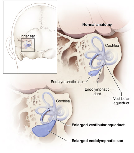
Vestibular Aqueduct
Cerebral Aqueduct
Endolymphatic Sac
Endolymphatic Duct
Hearing Loss, Sensorineural
Cochlear Aqueduct
Temporal Bone
Goiter
Ear Neoplasms
Vestibular Diseases
Membrane Transport Proteins
Hearing Loss
Hearing Loss, Conductive
Hydrocephalus
Anion Transport Proteins
Cerebral Ventriculography
Pneumoencephalography
Tomography, X-Ray Computed
Contralateral deafness following unilateral suboccipital brain tumor surgery in a patient with large vestibular aqueduct--case report. (1/45)
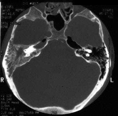
A 68-year-old female developed contralateral deafness following extirpation of a left cerebellopontine angle epidermoid cyst. Computed tomography showed that large vestibular aqueduct was present. This unusual complication may have been caused by an abrupt pressure change after cerebrospinal fluid release, which was transmitted through the large vestibular aqueduct and resulted in cochlear damage. (+info)Temporal bone computed tomography findings in bilateral sensorineural hearing loss. (2/45)
AIM: To examine the yield of computed tomography (CT) of the temporal bones when investigating sensorineural hearing loss (SNHL) and to identify factors associated with CT findings. METHODS: Retrospective analysis of 116 consecutively investigated children with bilateral SNHL at the audiology department of Great Ormond Street Hospital, London. Main outcome measures were CT results, hearing loss parameters, history, and clinical examination. RESULTS: A total of 33 (28.4%) CT scans were identified as abnormal. Children with profound and/or progressive hearing loss and/or craniofacial abnormalities were more likely to have an abnormal CT scan and together accounted for 25 abnormal CT scans. Sex, consanguineous parents, or family history of SNHL were not associated with CT findings. Dilated vestibular aqueduct was significantly correlated with the presence of progressive SNHL. CONCLUSIONS: All children with SNHL should undergo radiological investigation of the petrous bones/inner ear; abnormalities are more likely to be found in cases with craniofacial abnormalities, or profound or progressive hearing loss. The decision whether to perform a CT or magnetic resonance imaging will depend on scanner availability, expertise, and management considerations, but cochlear implant candidates will require both. (+info)Enlarged vestibular aqueduct: a radiological marker of pendred syndrome, and mutation of the PDS gene. (3/45)
Although the textbook view of Pendred syndrome is that of an autosomal recessive condition characterized by deafness and goitre, it is increasingly clear that not all such patients present this classical clinical picture. Malformations of the inner ear, specifically enlargement of the vestibular aqueduct, are common in Pendred syndrome and mutations in the PDS (Pendred Syndrome) gene have been recorded in patients presenting with deafness and vestibular aqueduct dilatation only, without other features of Pendred syndrome. Since this is the most common radiological malformation of the cochlea in deaf patients, we investigated what proportion of such cases were due to mutation of the PDS gene. We assessed 57 patients referred with radiological evidence of vestibular aqueduct enlargement, by history, clinical examination, perchlorate discharge test and molecular analysis of the PDS locus. Forty-one patients (72%) had unequivocal evidence of Pendred syndrome. The finding of a single heterozygous mutation at the PDS gene in a further eight was strongly suggestive of a critical role for pendrin, the protein product of the PDS gene, in the generation of enlarged vestibular aqueducts in at least 86% (49/57 cases) of patients with this radiological malformation. Securing the diagnosis of Pendred syndrome may be difficult, especially in the single case. Goitre is an inconstant finding, and the perchlorate discharge test, although helpful, is of diagnostic value only if abnormal. Enlargement of the vestibular aqueduct should be considered as the most likely presentation of Pendred syndrome and should prompt specific investigation of that diagnostic possibility. Pendred syndrome might henceforth be recharacterized as deafness with enlargement of the vestibular aqueduct, which is sometimes associated with goitre. (+info)Phenotypes associated with replacement of His by Arg in the Pendred syndrome gene. (4/45)
BACKGROUND: Pendred syndrome is often associated with inner ear malformations, especially enlarged vestibular aqueduct (EVA). Recently, mutations in the Pendred syndrome gene (PDS) have been reported in patients with EVA, in addition to those with classical Pendred syndrome. OBJECTIVE: The aim of this study was to investigate the genotype-phenotype correlations of PDS. METHODS: Each of the 21 exons and flanking splice regions of PDS was analysed by direct DNA sequencing in nine patients with EVA; allele-specific amplification was performed to confirm the mutation. Genetic analyses were compared with thyroid function tests, perchlorate discharge tests, thyroid volume and pure-tone audiogram. Magnetic resonance imaging was used to determine the volume of the endolymphatic duct and sac of each patient. RESULTS: A missense mutation, H723R, was identified in the homozygous state in three patients and in the heterozygous state in another three. Although none of the patients had goitre, increased serum thyroglobulin and an abnormal degree of iodide release were correlated with the number of mutant alleles identified. However, there was no relationship between the degree of hearing loss and the number of mutant alleles. CONCLUSION: The present study reveals that the number of mutant alleles correlates with the degree of subclinical thyroid abnormality, but not with the degree of hearing loss in Japanese patients with the PDS missense mutation H723R. (+info)Lack of pendrin expression leads to deafness and expansion of the endolymphatic compartment in inner ears of Foxi1 null mutant mice. (5/45)
Mice that lack the winged helix/forkhead gene Foxi1 (also known as Fkh10) are deaf and display shaker/waltzer behavior, an indication of disturbed balance. While Foxi1 is expressed in the entire otic vesicle at E9.5, it becomes gradually restricted to the endolymphatic duct/sac epithelium and at E16.5 Foxi1 expression in the inner ear is confined to this epithelium. Histological sections, paintfill experiments and whole-mount hybridizations reveal no abnormality in inner ear development of Foxi1(-/-) mice before E13.5. Between E13.5 and E16.5 the membranous labyrinth of inner ears from null mutants starts to expand as can be seen in histological sections, paint-fill experiments and three-dimensional reconstruction. Postnatally, inner ears of Foxi1(-/-) mice are extremely expanded, and large irregular cavities, compressing the cerebellum and the otherwise normal middle ear, have replaced the delicate compartments of the wild-type inner ear. This phenotype resembles that of the human sensorineural deafness syndrome Pendred syndrome, caused by mutations in the PDS gene. In situ hybridization of Foxi1(-/-) endolymphatic duct/sac epithelium shows a complete lack of the transcript encoding the chloride/iodide transporter pendrin. Based on this, we would like to suggest that Foxi1 is an upstream regulator of pendrin and that the phenotype seen in Foxi1 null mice is, at least in part, due to defective pendrin-mediated chloride ion resorption in the endolymphatic duct/sac epithelium. We show that this regulation could be mediated by absence of a specific endolymphatic cell type--FORE (forkhead related) cells--expressing Foxi1, Pds, Coch and Jag1. Thus, mutations in FOXI1 could prove to cause a Pendred syndrome-like human deafness. (+info)Distribution and frequencies of PDS (SLC26A4) mutations in Pendred syndrome and nonsyndromic hearing loss associated with enlarged vestibular aqueduct: a unique spectrum of mutations in Japanese. (6/45)
Molecular diagnosis makes a substantial contribution to precise diagnosis, subclassification, prognosis, and selection of therapy. Mutations in the PDS (SLC26A4) gene are known to be responsible for both Pendred syndrome and nonsyndromic hearing loss associated with enlarged vestibular aqueduct, and the molecular confirmation of the PDS gene has become important in the diagnosis of these conditions. In the present study, PDS mutation analysis confirmed that PDS mutations were present and significantly responsible in 90% of Pendred families, and in 78.1% of families with nonsyndromic hearing loss associated with enlarged vestibular aqueduct. Furthermore, variable phenotypic expression by the same combination of mutations indicated that these two conditions are part of a continuous category of disease. Interestingly, the PDS mutation spectrum in Japanese, including the seven novel mutations revealed by this study, is very different from that found in Caucasians. Of the novel mutations detected, 53% were the H723R mutation, suggesting a possible founder effect. Ethnic background is therefore presumably important and should be noted when genetic testing is being performed. The PDS gene mutation spectrum in Japanese may be representative of those in Eastern Asian populations and its elucidation is expected to facilitate the molecular diagnosis of a variety of diseases. (+info)Relationship between the external aperture and hearing loss in large vestibular aqueduct syndrome. (7/45)
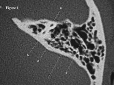
BACKGROUND: Large vestibular aqueduct syndrome (LVAS) is a major cause of hearing loss in childhood. This study aimed at measuring external aperture of enlargement of the vestibular aqueduct (EVA) and analyzing relationship between the size of external aperture and hearing loss. METHODS: Diagnostic criteria of LVAS were based on hearing loss and CT images. CT images of temporal bone of 100 LVAS patients were collected and 60 control subjects were reviewed retrospectively in the past 10 years. A battery of audiometric and vestibular function tests were performed. The width of the vestibular aqueduct (VA) was measured on axial CT images of the temporal bone. RESULTS: One hundred patients (65 men, 35 women) were diagnosed as having the isolated EVA. Hearing loss mostly occurred in early childhood. The diagnosis age of LVAS was 7.7 years on average. The causes of hearing loss could not be confirmed by initial consult. Typically, audiometric curve is the high-frequency down-sloping configuration. 92% of the cases had severe or profound sonsorineural hearing loss (SNHL). The mean size of the external aperture was (7.5 +/- 1.2) mm in present LVAS. Statistical analysis showed that the degree of hearing loss is unrelated to the width of VA. CONCLUSIONS: LVAS is a distinct clinical entity characterized by fluctuating, progressive SNHL. The degree of hearing loss is unrelated to the size of external aperture of VA. The protective management and hearing aid have become the main therapies. The cochlear implantation might be performed if the hearing loss affected learning at school. (+info)SLC26A4 gene is frequently involved in nonsyndromic hearing impairment with enlarged vestibular aqueduct in Caucasian populations. (8/45)
Sensorineural hearing loss is the most frequent sensory deficit of childhood and is of genetic origin in up to 75% of cases. It has been shown that mutations of the SLC26A4 (PDS) gene were involved in syndromic deafness characterized by congenital sensorineural hearing impairment and goitre (Pendred's syndrome), as well as in congenital isolated deafness (DFNB4). While the prevalence of SLC26A4 mutations in Pendred's syndrome is clearly established, it remains to be studied in large cohorts of patients with nonsyndromic deafness and detailed clinical informations. In this report, 109 patients from 100 unrelated families, aged from 1 to 32 years (median age: 10 years), with nonsyndromic deafness and enlarged vestibular aqueduct, were genotyped for SLC26A4 using DHPLC molecular screening and sequencing. In all, 91 allelic variants were observed in 100 unrelated families, of which 19 have never been reported. The prevalence of SLC26A4 mutations was 40% (40/100), with biallelic mutation in 24% (24/100), while six families were homozygous. All patients included in this series had documented deafness, associated with EVA and without any evidence of syndromic disease. Among patients with SLC26A4 biallelic mutations, deafness was more severe, fluctuated more than in patients with no mutation. In conclusion, the incidence of SLC26A4 mutations is high in patients with isolated deafness and enlarged vestibular aqueduct and could represent up to 4% of nonsyndromic hearing impairment. SLC26A4 could be the second most frequent gene implicated in nonsyndromic deafness after GJB2, in this Caucasian population. (+info)The vestibular aqueduct is a bony canal that runs from the inner ear to the brain. It contains a membranous duct, called the endolymphatic duct, which is filled with a fluid called endolymph. The vestibular aqueduct plays a role in the maintenance of balance and hearing by regulating the pressure and composition of the endolymph. Abnormalities or damage to the vestibular aqueduct can lead to conditions such as endolymphatic hydrops, which can cause symptoms like vertigo, dizziness, and hearing loss.
The cerebral aqueduct, also known as the aqueduct of Sylvius, is a narrow canal that connects the third and fourth ventricles (cavities) of the brain. It allows for the flow of cerebrospinal fluid (CSF) from the third ventricle to the fourth ventricle. The cerebral aqueduct is a critical component of the ventricular system of the brain, and any obstruction or abnormality in this region can result in an accumulation of CSF and increased pressure within the brain, which can lead to serious neurological symptoms and conditions such as hydrocephalus.
The endolymphatic sac is a small, fluid-filled structure that is part of the inner ear. It is located near the vestibular aqueduct and is responsible for maintaining the balance of fluids in the inner ear. The endolymphatic sac also plays a role in the resorption of endolymph, which is the fluid that fills the membranous labyrinth of the inner ear. Disorders of the endolymphatic sac can lead to conditions such as Meniere's disease, which is characterized by vertigo, hearing loss, and tinnitus.
The endolymphatic duct is a narrow canal in the inner ear that is part of the membranous labyrinth. It connects the utricle and saccule (two sensory structures in the vestibular system responsible for detecting changes in head position and movement) to the endolymphatic sac (a dilated portion of the duct that helps regulate the volume and pressure of endolymph, a fluid found within the membranous labyrinth).
The endolymphatic duct plays a crucial role in maintaining the balance and homeostasis of the inner ear by allowing the absorption and circulation of endolymph. Disorders or abnormalities in this region can lead to various vestibular and hearing dysfunctions, such as Meniere's disease, endolymphatic hydrops, and other inner ear disorders.
Sensorineural hearing loss (SNHL) is a type of hearing impairment that occurs due to damage to the inner ear (cochlea) or to the nerve pathways from the inner ear to the brain. It can be caused by various factors such as aging, exposure to loud noises, genetics, certain medical conditions (like diabetes and heart disease), and ototoxic medications.
SNHL affects the ability of the hair cells in the cochlea to convert sound waves into electrical signals that are sent to the brain via the auditory nerve. As a result, sounds may be perceived as muffled, faint, or distorted, making it difficult to understand speech, especially in noisy environments.
SNHL is typically permanent and cannot be corrected with medication or surgery, but hearing aids or cochlear implants can help improve communication and quality of life for those affected.
The cochlear aqueduct is a small canal that runs from the inner ear to the brain. It contains a fluid called perilymph, which helps to protect and cushion the structures of the inner ear. The cochlear aqueduct also serves as a passageway for the endolymphatic duct and sac, which are involved in the regulation of the inner ear's fluid balance.
Anomalies or abnormalities of the cochlear aqueduct can lead to hearing problems, balance disorders, and other symptoms. For example, a large or dilated cochlear aqueduct may be associated with an increased risk of meningitis, a serious infection of the membranes surrounding the brain and spinal cord. In some cases, surgical closure of the cochlear aqueduct may be necessary to prevent recurrent meningitis or other complications.
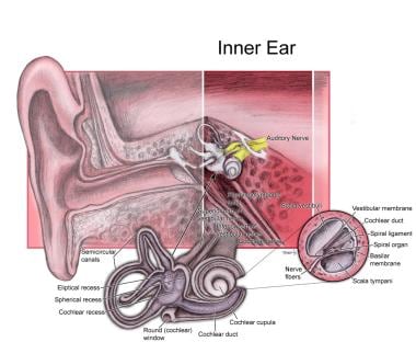
The temporal bone is a paired bone that is located on each side of the skull, forming part of the lateral and inferior walls of the cranial cavity. It is one of the most complex bones in the human body and has several important structures associated with it. The main functions of the temporal bone include protecting the middle and inner ear, providing attachment for various muscles of the head and neck, and forming part of the base of the skull.
The temporal bone is divided into several parts, including the squamous part, the petrous part, the tympanic part, and the styloid process. The squamous part forms the lateral portion of the temporal bone and articulates with the parietal bone. The petrous part is the most medial and superior portion of the temporal bone and contains the inner ear and the semicircular canals. The tympanic part forms the lower and anterior portions of the temporal bone and includes the external auditory meatus or ear canal. The styloid process is a long, slender projection that extends downward from the inferior aspect of the temporal bone and serves as an attachment site for various muscles and ligaments.
The temporal bone plays a crucial role in hearing and balance, as it contains the structures of the middle and inner ear, including the oval window, round window, cochlea, vestibule, and semicircular canals. The stapes bone, one of the three bones in the middle ear, is entirely encased within the petrous portion of the temporal bone. Additionally, the temporal bone contains important structures for facial expression and sensation, including the facial nerve, which exits the skull through the stylomastoid foramen, a small opening in the temporal bone.
Goiter is a medical term that refers to an enlarged thyroid gland. The thyroid gland is a small, butterfly-shaped gland located in the front of your neck below the larynx or voice box. It produces hormones that regulate your body's metabolism, growth, and development.
Goiter can vary in size and may be visible as a swelling at the base of the neck. It can be caused by several factors, including iodine deficiency, autoimmune disorders, thyroid cancer, pregnancy, or the use of certain medications. Depending on the underlying cause and the severity of the goiter, treatment options may include medication, surgery, or radioactive iodine therapy.
Ear neoplasms refer to abnormal growths or tumors that occur in the ear. These growths can be benign (non-cancerous) or malignant (cancerous) and can affect any part of the ear, including the outer ear, middle ear, inner ear, and the ear canal.
Benign ear neoplasms are typically slow-growing and do not spread to other parts of the body. Examples include exostoses, osteomas, and ceruminous adenomas. These types of growths are usually removed surgically for cosmetic reasons or if they cause discomfort or hearing problems.
Malignant ear neoplasms, on the other hand, can be aggressive and may spread to other parts of the body. Examples include squamous cell carcinoma, basal cell carcinoma, and adenoid cystic carcinoma. These types of tumors often require more extensive treatment, such as surgery, radiation therapy, and chemotherapy.
It is important to note that any new growth or change in the ear should be evaluated by a healthcare professional to determine the nature of the growth and develop an appropriate treatment plan.
Vestibular diseases are a group of disorders that affect the vestibular system, which is responsible for maintaining balance and spatial orientation. The vestibular system includes the inner ear and parts of the brain that process sensory information related to movement and position.
These diseases can cause symptoms such as vertigo (a spinning sensation), dizziness, imbalance, nausea, and visual disturbances. Examples of vestibular diseases include:
1. Benign paroxysmal positional vertigo (BPPV): a condition in which small crystals in the inner ear become dislodged and cause brief episodes of vertigo triggered by changes in head position.
2. Labyrinthitis: an inner ear infection that can cause sudden onset of vertigo, hearing loss, and tinnitus (ringing in the ears).
3. Vestibular neuronitis: inflammation of the vestibular nerve that causes severe vertigo, nausea, and imbalance but typically spares hearing.
4. Meniere's disease: a disorder characterized by recurrent episodes of vertigo, tinnitus, hearing loss, and a feeling of fullness in the affected ear.
5. Vestibular migraine: a type of migraine that includes vestibular symptoms such as dizziness, imbalance, and disorientation.
6. Superior canal dehiscence syndrome: a condition in which there is a thinning or absence of bone over the superior semicircular canal in the inner ear, leading to vertigo, sound- or pressure-induced dizziness, and hearing loss.
7. Bilateral vestibular hypofunction: reduced function of both vestibular systems, causing chronic imbalance, unsteadiness, and visual disturbances.
Treatment for vestibular diseases varies depending on the specific diagnosis but may include medication, physical therapy, surgery, or a combination of these approaches.
Membrane transport proteins are specialized biological molecules, specifically integral membrane proteins, that facilitate the movement of various substances across the lipid bilayer of cell membranes. They are responsible for the selective and regulated transport of ions, sugars, amino acids, nucleotides, and other molecules into and out of cells, as well as within different cellular compartments. These proteins can be categorized into two main types: channels and carriers (or pumps). Channels provide a passive transport mechanism, allowing ions or small molecules to move down their electrochemical gradient, while carriers actively transport substances against their concentration gradient, requiring energy usually in the form of ATP. Membrane transport proteins play a crucial role in maintaining cell homeostasis, signaling processes, and many other physiological functions.
Hearing loss is a partial or total inability to hear sounds in one or both ears. It can occur due to damage to the structures of the ear, including the outer ear, middle ear, inner ear, or nerve pathways that transmit sound to the brain. The degree of hearing loss can vary from mild (difficulty hearing soft sounds) to severe (inability to hear even loud sounds). Hearing loss can be temporary or permanent and may be caused by factors such as exposure to loud noises, genetics, aging, infections, trauma, or certain medical conditions. It is important to note that hearing loss can have significant impacts on a person's communication abilities, social interactions, and overall quality of life.
Conductive hearing loss is a type of hearing loss that occurs when there is a problem with the outer or middle ear. Sound waves are not able to transmit efficiently through the ear canal to the eardrum and the small bones in the middle ear, resulting in a reduction of sound that reaches the inner ear. Causes of conductive hearing loss may include earwax buildup, fluid in the middle ear, a middle ear infection, a hole in the eardrum, or problems with the tiny bones in the middle ear. This type of hearing loss can often be treated through medical intervention or surgery.
A syndrome, in medical terms, is a set of symptoms that collectively indicate or characterize a disease, disorder, or underlying pathological process. It's essentially a collection of signs and/or symptoms that frequently occur together and can suggest a particular cause or condition, even though the exact physiological mechanisms might not be fully understood.
For example, Down syndrome is characterized by specific physical features, cognitive delays, and other developmental issues resulting from an extra copy of chromosome 21. Similarly, metabolic syndromes like diabetes mellitus type 2 involve a group of risk factors such as obesity, high blood pressure, high blood sugar, and abnormal cholesterol or triglyceride levels that collectively increase the risk of heart disease, stroke, and diabetes.
It's important to note that a syndrome is not a specific diagnosis; rather, it's a pattern of symptoms that can help guide further diagnostic evaluation and management.
Deafness is a hearing loss that is so severe that it results in significant difficulty in understanding or comprehending speech, even when using hearing aids. It can be congenital (present at birth) or acquired later in life due to various causes such as disease, injury, infection, exposure to loud noises, or aging. Deafness can range from mild to profound and may affect one ear (unilateral) or both ears (bilateral). In some cases, deafness may be accompanied by tinnitus, which is the perception of ringing or other sounds in the ears.
Deaf individuals often use American Sign Language (ASL) or other forms of sign language to communicate. Some people with less severe hearing loss may benefit from hearing aids, cochlear implants, or other assistive listening devices. Deafness can have significant social, educational, and vocational implications, and early intervention and appropriate support services are critical for optimal development and outcomes.
Hydrocephalus is a medical condition characterized by an abnormal accumulation of cerebrospinal fluid (CSF) within the brain, leading to an increase in intracranial pressure and potentially causing damage to the brain tissues. This excessive buildup of CSF can result from either overproduction or impaired absorption of the fluid, which typically causes the ventricles (fluid-filled spaces) inside the brain to expand and put pressure on surrounding brain structures.
The condition can be congenital, present at birth due to genetic factors or abnormalities during fetal development, or acquired later in life as a result of injuries, infections, tumors, or other disorders affecting the brain's ability to regulate CSF flow and absorption. Symptoms may vary depending on age, severity, and duration but often include headaches, vomiting, balance problems, vision issues, cognitive impairment, and changes in behavior or personality.
Treatment for hydrocephalus typically involves surgically implanting a shunt system that diverts the excess CSF from the brain to another part of the body where it can be absorbed, such as the abdominal cavity. In some cases, endoscopic third ventriculostomy (ETV) might be an alternative treatment option, creating a new pathway for CSF flow within the brain. Regular follow-ups with neurosurgeons and other healthcare professionals are essential to monitor the condition and make any necessary adjustments to the treatment plan.
Anion transport proteins are specialized membrane transport proteins that facilitate the movement of negatively charged ions, known as anions, across biological membranes. These proteins play a crucial role in maintaining ionic balance and regulating various physiological processes within the body.
There are several types of anion transport proteins, including:
1. Cl-/HCO3- exchangers (also known as anion exchangers or band 3 proteins): These transporters facilitate the exchange of chloride (Cl-) and bicarbonate (HCO3-) ions across the membrane. They are widely expressed in various tissues, including the red blood cells, gastrointestinal tract, and kidneys, where they help regulate pH, fluid balance, and electrolyte homeostasis.
2. Sulfate permeases: These transporters facilitate the movement of sulfate ions (SO42-) across membranes. They are primarily found in the epithelial cells of the kidneys, intestines, and choroid plexus, where they play a role in sulfur metabolism and absorption.
3. Cl- channels: These proteins form ion channels that allow chloride ions to pass through the membrane. They are involved in various physiological processes, such as neuronal excitability, transepithelial fluid transport, and cell volume regulation.
4. Cation-chloride cotransporters: These transporters move both cations (positively charged ions) and chloride anions together across the membrane. They are involved in regulating neuronal excitability, cell volume, and ionic balance in various tissues.
Dysfunction of anion transport proteins has been implicated in several diseases, such as cystic fibrosis (due to mutations in the CFTR Cl- channel), distal renal tubular acidosis (due to defects in Cl-/HCO3- exchangers), and some forms of epilepsy (due to abnormalities in cation-chloride cotransporters).
Cerebral ventriculography is a medical imaging technique that involves the injection of a contrast material into the cerebral ventricles, which are fluid-filled spaces within the brain. The purpose of this procedure is to produce detailed images of the ventricular system and the surrounding structures in order to diagnose and evaluate various neurological conditions, such as hydrocephalus (excessive accumulation of cerebrospinal fluid in the ventricles), tumors, or other abnormalities that may be causing obstruction or compression of the ventricular system.
The procedure typically involves inserting a thin, flexible tube called a catheter into the lateral ventricle of the brain through a small hole drilled in the skull. The contrast material is then injected through the catheter and X-ray images are taken as the contrast material flows through the ventricular system. These images can help to identify any abnormalities or blockages that may be present.
Cerebral ventriculography has largely been replaced by non-invasive imaging techniques, such as computed tomography (CT) and magnetic resonance imaging (MRI), which provide similar information without the need for invasive procedures. However, cerebral ventriculography may still be used in certain cases where these other methods are not sufficient to make a definitive diagnosis.
Pneumoencephalography is a diagnostic procedure that is rarely used today, due to the development of less invasive techniques. It involves the introduction of air or another gas into the ventricular system or subarachnoid space of the brain, followed by X-ray imaging to visualize the structures and any abnormalities within the intracranial cavity.
The primary purpose of this procedure was to diagnose conditions affecting the brain's ventricles, such as hydrocephalus, tumors, or inflammation. The introduction of air into the cranium allowed for better visualization of these structures and any potential abnormalities. However, due to its invasive nature, risks associated with the procedure, and the availability of non-invasive imaging techniques like CT and MRI scans, pneumoencephalography has fallen out of favor in modern medicine.
X-ray computed tomography (CT or CAT scan) is a medical imaging method that uses computer-processed combinations of many X-ray images taken from different angles to produce cross-sectional (tomographic) images (virtual "slices") of the body. These cross-sectional images can then be used to display detailed internal views of organs, bones, and soft tissues in the body.
The term "computed tomography" is used instead of "CT scan" or "CAT scan" because the machines take a series of X-ray measurements from different angles around the body and then use a computer to process these data to create detailed images of internal structures within the body.
CT scanning is a noninvasive, painless medical test that helps physicians diagnose and treat medical conditions. CT imaging provides detailed information about many types of tissue including lung, bone, soft tissue and blood vessels. CT examinations can be performed on every part of the body for a variety of reasons including diagnosis, surgical planning, and monitoring of therapeutic responses.
In computed tomography (CT), an X-ray source and detector rotate around the patient, measuring the X-ray attenuation at many different angles. A computer uses this data to construct a cross-sectional image by the process of reconstruction. This technique is called "tomography". The term "computed" refers to the use of a computer to reconstruct the images.
CT has become an important tool in medical imaging and diagnosis, allowing radiologists and other physicians to view detailed internal images of the body. It can help identify many different medical conditions including cancer, heart disease, lung nodules, liver tumors, and internal injuries from trauma. CT is also commonly used for guiding biopsies and other minimally invasive procedures.
In summary, X-ray computed tomography (CT or CAT scan) is a medical imaging technique that uses computer-processed combinations of many X-ray images taken from different angles to produce cross-sectional images of the body. It provides detailed internal views of organs, bones, and soft tissues in the body, allowing physicians to diagnose and treat medical conditions.
Eye manifestations refer to any changes or abnormalities in the eye that can be observed or detected. These manifestations can be related to various medical conditions, diseases, or disorders affecting the eye or other parts of the body. They can include structural changes, such as swelling or bulging of the eye, as well as functional changes, such as impaired vision or sensitivity to light. Examples of eye manifestations include cataracts, glaucoma, diabetic retinopathy, macular degeneration, and uveitis.
The subcommissural organ (SCO) is a small neuroendocrine gland located at the caudal end of the third ventricle in the brain. It is situated in the vicinity of the posterior commissure, hence its name. The SCO is primarily composed of ependymal cells and produces a variety of neuropeptides and proteins that are released into the cerebrospinal fluid (CSF).
The main function of the subcommissural organ is to secrete a glycoprotein called SCO-spondin, which plays a role in the formation and maintenance of the cerebral aqueduct and the rostral part of the central canal of the spinal cord. The CSF flow through these structures is facilitated by the presence of SCO-spondin, which has been shown to have adhesive properties that help prevent the collapse of these narrow channels.
Dysfunction or abnormalities in the subcommissural organ may contribute to various neurological disorders, such as hydrocephalus and other conditions associated with impaired CSF flow. However, further research is needed to fully understand the role of this intriguing structure in brain physiology and pathology.
 Vestibular aqueduct
Enlarged vestibular aqueduct
List of OMIM disorder codes
Semicircular canals
FOXI1
Endolymphatic duct
Pendrin
Vestibule of the ear
Conductive hearing loss
Homeobox protein SIX1
Pendred syndrome
Odobenocetops
Endolymphatic sac tumor
Marfanoid
Mondini dysplasia
Subarcuate fossa
Endolymphatic sac
List of syndromes
Eva
List of MeSH codes (A09)
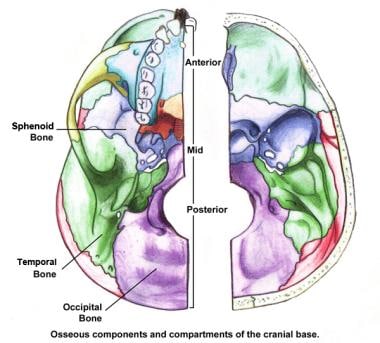
Base of skull
The Traveling Awareness Bears
Tokarahia
Aqueduct
Medial longitudinal fasciculus
Cochlear
Perilymph
List of MeSH codes (A08)
Fourth ventricle
Mumps
Vestibular aqueduct - Wikipedia
Retrospective Analysis of the Association of a Small Vestibular Aqueduct with Cochleovestibular Symptoms in a Large, Single...
 Large Vestibular Aqueduct Syndrome
Audiologic presentation of enlargement of the vestibular aqueduct according to the SLC26A4 genotypes - Fingerprint - Korea...
 Skull Base, Petrous Apex, Tumors: Practice Essentials, History of the Procedure, Problem
 Similar articles for PMID: 20301515 - Search Results - PubMed
 Hearing Loss: Pendred Syndrome (SLC26A4)
 Evaluation of the Thyroid in Patients With Hearing Loss and Enlarged Vestibular Aqueducts | Endocrinology | JAMA Otolaryngology...
 Pendred syndrome: MedlinePlus Genetics
 Mutation:genetics<...
 Clinical aspects of hereditary hearing loss | Genetics in Medicine
Otosclerosis: Practice Essentials, History of the Procedure, Epidemiology
Wade Chien, M.D., FACS | NIDCD
 2.2. Causes of deafness - BATOD
 Meet Sidney Mautte - Drum Corps Associates
Dylan Chan | UCSF Profiles
 Unilateral Hearing Loss And Academic Performance Literature Reviews | WePapers
Transvenous embolization of dural fistulas involving the transverse and sigmoid sinuses. | American Journal of Neuroradiology
 Tampa General's Auditory-Verbal Therapy Offers Hope for Young Deaf Patients | Tampa General Hospital
 News | Northeast Ohio Medical University | NEOMED
 Etiologies and Treatment Options for Sudden Sensorineural Hearing Loss | The Hearing Review
 American Cochlear Implant Alliance Task Force Guidelines for... : Ear and Hearing
Browse Items · NEOMED Bibliography Database
 head and neck anatomy.ppt
 semicircular canal - Ontology Browser - Rat Genome Database
duct of olfactory gland - Ontology Browser - Rat Genome Database
 Namespace
 Kenneth Grundfast | Profiles RNS
 Pendred syndrome | Contact
 Kay W. Chang, MD's Profile | Stanford Profiles
Associated with enlarged vestibular aqueduct2
- Enlargement of the vestibular aqueduct to greater than 2 mm is associated with enlarged vestibular aqueduct syndrome, a disease entity that is associated with one-sided hearing loss in children. (wikipedia.org)
- Usami SAbe SWeston MDShinkawa HVan Camp GKimberling WJ Non-syndromic hearing loss associated with enlarged vestibular aqueduct is caused by PDS mutations. (jamanetwork.com)
Syndrome8
- Large vestibular aqueduct syndrome (LVAS), also known as enlarged vestibular aqueduct (EVA) or large endolymphatic sac anomaly (LESA), refers to the presence of congenital sensorineural hearing loss with an enlarged vestibular aqueduct due to enlargement of the endolymphatic duct . (harmonyhearing.com.au)
- Valvassori GEClemis JD The large vestibular aqueduct syndrome. (jamanetwork.com)
- SLC26A4/PDS genotype-phenotype correlation in hearing loss with enlargement of the vestibular aqueduct (EVA): evidence that Pendred syndrome and non-syndromic EVA are distinct clinical and genetic entities. (jamanetwork.com)
- An inner ear abnormality called an enlarged vestibular aqueduct (EVA) is a characteristic feature of Pendred syndrome. (medlineplus.gov)
- Phenotypes of SLC26A4 gene mutations: Pendred syndrome and hypoacusis with enlarged vestibular aqueduct. (nel.edu)
- This paper presents the current views, regarding the pathomechanisms, which lead to the development of pathological symptoms in the enlargement of the vestibular aqueduct syndrome (EVAS) and the Pendred syndrome (PS). (nel.edu)
- The five-year-old was diagnosed with bilateral profound hearing loss due to Mondini malformation and enlarged vestibular aqueduct (EVA) syndrome. (tgh.org)
- also known as vestibular aqueduct syndrome (EVA). (bioparadigms.org)
Large vestibular2
- Although large vestibular aqueducts are a congenital condition, hearing loss may not be present from birth. (harmonyhearing.com.au)
- Vestibular-evoked myogenic potentials in three patients with large vestibular aqueduct. (omeka.net)
Enlargement4
- Measured thyroid volume (T vol ) of patients with enlargement of the vestibular aqueduct and 2 (black squares), 1 (white triangles), or no (light blue circles) mutant alleles of SLC26A4 . (jamanetwork.com)
- Hypo-functional SLC26A4 variants associated with nonsyndromic hearing loss and enlargement of the vestibular aqueduct: genotype-phenotype correlation or coincidental polymorphism? (jamanetwork.com)
- SLC26A4 genotypes and phenotypes associated with enlargement of the vestibular aqueduct. (medlineplus.gov)
- Disorders of homeostasis in labyrinth fluids are responsible for abnormalities of its structure, such as enlargement of the vestibular aqueduct and of the endolymph sac. (nel.edu)
Otosclerosis1
- Some examples of this are microtia, otosclerosis, and enlarged vestibular aqueducts (EVA). (batod.org.uk)
Cochlear1
- Though she passed a newborn hearing screen, she would later be diagnosed with enlarged vestibular aqueduct and mild cochlear dysplasia. (neomed.edu)
Deafness1
- Some cases progress to profound deafness, some include vestibular losses or difficulties, and other cases lead to neither. (harmonyhearing.com.au)
Bony canal1
- The vestibular aqueduct is a bony canal that connects the inner ear with the inside of the skull. (medlineplus.gov)
Anomaly1
- An enlarged vestibular aqueduct (LVA) is a common congenital inner ear anomaly responsible for some unusual vestibular and audiological symptoms. (omeka.net)
Symptoms5
- This study correlates the presence of a small vestibular aqueduct with cochleovestibular symptoms. (ajnr.org)
- We suggest that the finding of a small vestibular aqueduct on CT could be reported by radiologists as a possible finding in Ménière disease, but it remains of uncertain, and potentially unlikely, clinical importance in the absence of symptoms of Ménière disease. (ajnr.org)
- In those persons with related vestibular symptoms, treatment may include vestibular rehabilitation therapy. (harmonyhearing.com.au)
- Vestibular symptoms may also appear early, but are much more difficult to identify in the very young. (harmonyhearing.com.au)
- Hearing loss may be associated with vestibular symptoms. (hearingreview.com)
Auditory1
- Dr. René H Gifford will discuss recent work on electric-acoustic integration in children and adults, and Dr. Sharon Cushing will discuss her work as a clinician on 3-D auditory and vestibular effects. (aro.org)
SLC26A41
- SLC26A4 gene is frequently involved in nonsyndromic hearing impairment with enlarged vestibular aqueduct in Caucasian populations. (jamanetwork.com)
Mondini1
- The combination of an enlarged vestibular aqueduct and an abnormally shaped cochlea is known as Mondini malformation. (medlineplus.gov)
Audiologic1
- In addition to a complete medical history and physical examination, the diagnostic process for uncovering EVA usually involves audiologic and vestibular testing as well as radiologic assessment. (harmonyhearing.com.au)
Correlation1
- While patients with Ménière disease were proportionately more likely to have a small vestibular aqueduct than patients without Ménière disease, the small vestibular aqueduct was more frequently seen in patients without Ménière disease and had no correlation with hearing loss, vertigo, dizziness, or aural fullness. (ajnr.org)
Dysfunction1
- Some affected individuals also have problems with balance caused by dysfunction of the vestibular system, which is the part of the inner ear that helps maintain the body's balance and orientation. (medlineplus.gov)
Bilateral1
- Sidney was diagnosed with moderate to severe bilateral hearing loss due to enlarged vestibular aqueducts. (dcacorps.org)
Inner ear3
- Vestibular aqueducts are narrow, bony canals that travel from the inner ear to deep inside the skull. (harmonyhearing.com.au)
- Running through each vestibular aqueduct is a fluid-filled tube called the endolymphatic duct, which connects the inner ear to a balloon-shaped structure called the endolymphatic sac. (harmonyhearing.com.au)
- The otolithic organ as a receptor of vestibular hearing revealed by vestibular-evoked myogenic potentials in patients with inner ear anomalies. (omeka.net)
Cochlea2
- These organs are the cochlea, which detects sound waves and turns them into nerve signals, and the vestibular labyrinth, which detects movement and gravity. (harmonyhearing.com.au)
- and many viral infections that affect the cochlea (and sometimes the vestibular apparatus). (msdmanuals.com)
Clinical2
- If the vestibular aqueduct is larger in size, and the clinical presentation is consistent, the diagnosis can be made. (wikipedia.org)
- however, the prevalence and clinical importance of small vestibular aqueducts remain unclear in patients without Ménière disease. (ajnr.org)
Disease1
- Five of 12 patients with Ménière disease (5 ears) had a small vestibular aqueduct. (ajnr.org)
Hearing loss2
- 001). There was no statistical difference between the small vestibular aqueduct cohort and the cohort with normal vestibular aqueducts (0.3-0.7 mm) regarding tinnitus ( P = .06), hearing loss ( P = .88), vertigo ( P = .26), dizziness ( P = .83), and aural fullness ( P = .61). (ajnr.org)
- No relationship exists between how large the aqueduct is and the amount of hearing loss a person may sustain. (harmonyhearing.com.au)
System3
- PS also may affect the vestibular system, which controls balance and some individuals with PS will show some vestibular weakness when their balance system is tested. (contact.org.uk)
- I began studying the vestibular system during my dissertation research at the Università di Pavia with Professors Ivo Prigioni and GianCarlo Russo. (aro.org)
- My research focuses on characterizing the biophysics of synaptic transmission between hair cells and primary afferents in the vestibular system. (aro.org)
Patients2
Enlarged Vestibular Aqueduct Syndrome3
- Enlargement of the vestibular aqueduct to greater than 2 mm is associated with enlarged vestibular aqueduct syndrome, a disease entity that is associated with one-sided hearing loss in children. (wikipedia.org)
- Correlation analysis of genotypes, auditory function, and vestibular size in Chinese children with enlarged vestibular aqueduct syndrome. (nih.gov)
- It is called EVAS, enlarged vestibular aqueduct syndrome. (multipleexperiences.org)
Dizziness7
- Causes of dizziness, vertigo and disequilibrium can be hard to identify, but can indicate a problem in your vestibular (inner ear) system. (vestibular.org)
- By contrast, dizziness can be a primary sign of a vestibular disorder in addition to a broad array of cardiovascular, neurological, metabolic, vision, and psychological problems. (vestibular.org)
- When the vestibular system malfunctions, it can no longer help resolve moments of sensory conflict, resulting in symptoms such as dizziness, vertigo, and disequilibrium. (vestibular.org)
- Two of the patients complained about vertigo and dizziness but vestibular assessments of the patients showed normal results. (omeka.net)
- Though the NMT/SO MdDS subtype report identical symptoms to the MT MdDS subtype, recently the Barany Society has suggested that NMT/SO patients should perhaps be classified as a separate vestibular entity (Persistent Postural-Perceptual Dizziness), given the vastly different onset causes and potential differences in the underlying mechanisms. (vestibular.org)
- There are different types of vestibular disorders, and they can cause overwhelming vertigo, dizziness, and other symptoms, which cannot be seen and only be felt. (neuroequilibrium.in)
- Individuals suffering from vestibular disorders can experience one or more symptoms like fatigue, dizziness, unsteadiness, jumping vision, hearing loss, vomiting, and tinnitus , which is ringing sound in the ears. (neuroequilibrium.in)
Neuritis4
- Vestibular neuritis and labyrinthitis may also cause vertigo. (ajnr.org)
- A person experiencing a viral infection like measles or chickenpox can experience vestibular neuritis . (neuroequilibrium.in)
- Inner ear infections that cause vestibular neuritis or labyrinthitis are usually viral rather than bacterial. (medlink.com)
- Some of the viruses that have been associated with vestibular neuritis or labyrinthitis include herpes viruses (such as the ones that cause cold sores or chicken pox and shingles), influenza, measles, rubella, mumps, polio, hepatitis, and Epstein-Barr. (medlink.com)
Abnormalities1
- However, compensating for vestibular system abnormalities is more problematic. (vestibular.org)
Malformation1
- This study will try to identify and understand the genetic factors that lead to an inner ear malformation called "enlarged vestibular aqueducts", that can be associated with hearing loss. (nih.gov)
Auditory and vestibular2
- The Department of Otolaryngology at Johns Hopkins provides great opportunities for collaborative work with other auditory and vestibular researchers with different expertise. (aro.org)
- Dr. René H Gifford will discuss recent work on electric-acoustic integration in children and adults, and Dr. Sharon Cushing will discuss her work as a clinician on 3-D auditory and vestibular effects. (aro.org)
Endolymphatic sac1
- High jugular bulb (HJB), one of the most common anatomical variant in temporal bone, has been reported to be more common in MD, and it has been suggested to put pressure on the endolymphatic sac (ES) and distal vestibular aqueduct (VA), which contains the endolymphatic duct and the infratemporal endolymphatic sac, resulting in endolymphatic hydrops and Meniere-like symptoms (5,6). (researchsquare.com)
Vertigo2
- Vertigo is caused by a disturbed vestibular system and is subdivided into peripheral vertigo (due to failure of the end organs) or central vertigo (due to failure of the vestibular nerves or central connections to the brainstem and cerebellum). (ajnr.org)
- People suffering from vestibular disorders can experience sudden attacks of tinnitus, vertigo, hearing loss, or feeling of fullness at any time. (neuroequilibrium.in)
Vestibule1
- At the hinder part of the medial wall of the vestibule is the orifice of the vestibular aqueduct, which extends to the posterior surface of the petrous portion of the temporal bone. (wikipedia.org)
Myogenic potentials3
- We assessed vestibular otolith function in children with ADHD and controls using the subjective visual vertical (SVV) bucket test and cervical vestibular-evoked myogenic potentials (cVEMPs). (frontiersin.org)
- Vestibular-evoked myogenic potentials in three patients with large vestibular aqueduct. (omeka.net)
- To our knowledge this is the first report of saccular malfunction in three patients with LVA by means of vestibular evoked myogenic potentials. (omeka.net)
Symptoms6
- Hearing loss may be associated with vestibular symptoms. (hearingreview.com)
- An enlarged vestibular aqueduct (LVA) is a common congenital inner ear anomaly responsible for some unusual vestibular and audiological symptoms. (omeka.net)
- What are symptoms of a Vestibular problem? (physiotherapyvictoria.ca)
- The numbers of symptoms associated with vestibular dysfunctions are as varied as the number of causes. (physiotherapyvictoria.ca)
- There are a multitude of associated symptoms which are typical of most vestibular disorders, such as imbalance, unsteadiness, cognitive slowing, visual-motion sensitivity, brain fog and anxiety 3 . (vestibular.org)
- He felt that my symptoms due to my EVA (enlarged vestibular aqueduct). (trixie-dixie.com)
Labyrinthitis1
- In serous labyrinthitis, bacteria that have infected the middle ear or the bone surrounding the inner ear produce toxins that invade the inner ear via the oval or round windows and inflame the cochlea, the vestibular system, or both. (medlink.com)
APPARATUS2
- Despite extensive studies on hearing and the vestibular apparatus, saccular function is not studied. (omeka.net)
- and many viral infections that affect the cochlea (and sometimes the vestibular apparatus). (msdmanuals.com)
Peripheral1
- Peripheral vestibular tests in MdDS patients are usually unremarkable, though brain imaging studies have demonstrated changes in the brain metabolism and functional brain connections. (vestibular.org)
Bony3
- Vestibular aqueducts are narrow, bony canals that travel from the inner ear to inside the skull. (nih.gov)
- The vestibular aqueduct is a bony canal that connects the inner ear with the inside of the skull. (medlineplus.gov)
- The bony, narrow canals within the inner ear that connects to the skull are known as vestibular aqueducts. (neuroequilibrium.in)
Medial wall1
- [ citation needed ] here, the prominence of the facial canal (or prominence of the aqueduct of Fallopius ) upon the medial wall indicates the position of the superior portion of the facial canal. (cloudfront.net)
Brainstem1
- These findings suggest that vestibular brainstem reflexes are altered in a subset of children with ADHD. (frontiersin.org)
Sensory3
- Just as a courtroom judge must rule between two sides presenting competing evidence, the vestibular system serves as the tie-breaker between conflicting forms of sensory information. (vestibular.org)
- We use the vestibular periphery as a sensory system model in mice. (aro.org)
- The term neuronitis (damage to the sensory neurons of the vestibular ganglion) is also used. (medlink.com)
Disorder2
- It is also quite possible that a person may have a combination of problems, such as a degenerative vestibular disorder along with a visual deficit such as cataracts or a neurological disorder such as a stroke. (vestibular.org)
- This defect can result in vestibular disorder, but it is possible to cure the defect through a surgical process. (neuroequilibrium.in)
Genetic2
- Other illnesses, as well as genetic and environmental factors, may also cause or contribute to vestibular disorders. (vestibular.org)
- They have enlarged vestibular aqueducts, a genetic condition that Kirk has too. (thriveglobal.com)
Cerebellum1
- However, in addition to the cerebellum, the vestibular system is one of the most important neural networks involved in the control of balance and gait. (frontiersin.org)
Rehabilitation2
- What is Vestibular Rehabilitation? (physiotherapyvictoria.ca)
- What will my first visit to VOR for Vestibular Rehabilitation involve? (physiotherapyvictoria.ca)
Occur2
- 13 Gadolinium enhancement of the labyrinthine structures or vestibular nerves may also occur and should not be mistaken for hemorrhage. (ajnr.org)
- Vestibular disorders can occur from inner-ear infections. (neuroequilibrium.in)
Cognitive1
- Individuals with vestibular disorders can suffer from cognitive impacts, like poor concentration, difficulty in recalling things, difficulty reading printed text, as well as impaired mental stamina. (neuroequilibrium.in)
Evaluate2
- CT or MR imaging, or both, may be used to evaluate the vestibular aqueduct, endolymphatic duct, and sac and to rule out associated infectious or neoplastic disease. (ajnr.org)
- The principal aim of this study is to evaluate vestibular otolith function in ADHD and matched control children. (frontiersin.org)
Organs1
- The balance function involves the vestibular organs. (medlink.com)
Anatomy1
- anatomy) A structure conveying fluid, such as the cerebral aqueduct or vestibular aqueduct. (ss-media.org)
Integrates1
- The brain integrates balance signals sent through the vestibular nerve from the right ear and the left ear. (medlink.com)
Balance1
- Acoustic Neuroma is a tumor in the inner ear and a form of the vestibular It is non-cancerous and expands slowly, but can squeeze your nerves, which control your balance and hearing. (neuroequilibrium.in)