Uterine Artery
Uterus
Uterine Artery Embolization
Placental Circulation
Ultrasonography, Doppler
Pre-Eclampsia
Pulsatile Flow
Pregnancy
Ultrasonography, Doppler, Pulsed
Ultrasonography, Doppler, Color
Ultrasonography, Prenatal
Pulmonary Artery
Leiomyoma
Pregnancy Trimester, Second
Umbilical Arteries
Pregnancy, Animal
Fetal Growth Retardation
Mesenteric Arteries
Carotid Arteries
Pregnancy Trimester, First
Basilar Artery
Iliac Artery
Vertebral Artery
Coronary Artery Bypass
Pregnancy, High-Risk
Sheep
Blood Flow Velocity
Embolization, Therapeutic
Hypertension, Pregnancy-Induced
Vasoconstriction
Radial Artery
Mammary Arteries
Vascular Resistance
Carotid Artery, Internal
Uterine Hemorrhage
Vasodilation
Subclavian Artery
Pregnancy-Associated Plasma Protein-A
Placental Insufficiency
Gestational Age
Carotid Artery Diseases
Splenic Artery
Brachial Artery
Middle Cerebral Artery
Pregnancy Outcome
Abruptio Placentae
Hepatic Artery
Carotid Artery, Common
Prospective Studies
Pregnancy Trimester, Third
Celiac Artery
Ophthalmic Artery
Pregnancy Complications, Cardiovascular
Mesenteric Artery, Superior
Endothelium, Vascular
Pregnancy, Ectopic
Renal Artery Obstruction
Thoracic Arteries
Temporal Arteries
Treatment Outcome
Sensitivity and Specificity
Bronchial Arteries
Placenta
Infant, Small for Gestational Age
Popliteal Artery
Ulnar Artery
Intrauterine Devices, Copper
Predictive Value of Tests
Diethylcarbamazine
Coronary Angiography
Rheology
Carotid Artery, External
Norepinephrine
Nitric Oxide
Phenylephrine
Laser-Doppler Flowmetry
Altitude
Phorbol 12,13-Dibutyrate
Receptor Activity-Modifying Proteins
Placentation
Pregnancy Proteins
Carotid Artery Injuries
Arterial Occlusive Diseases
Receptors, Adrenergic, alpha-1
Postpartum Hemorrhage
Test Anxiety Scale
Fetal Distress
Estradiol
Potassium Chloride
Coronary Disease
Nitric Oxide Synthase
Crown-Rump Length
Reference Values
Muscle Contraction
Hemodynamics
Infarction, Middle Cerebral Artery
Pregnancy Complications
Axillary Artery
Muscle Tonus
Polyvinyl Alcohol
Heart Rate, Fetal
Enzyme Inhibitors
Retrospective Studies
Retinal Artery Occlusion
Acetylcholine
Tomography, X-Ray Computed
Sheep, Domestic
Stents
Indomethacin
Carotid Artery Thrombosis
Nitric Oxide Synthase Type III
Risk Factors
Rats, Sprague-Dawley
Follow-Up Studies
Trophoblastic Neoplasms
ROC Curve
Aneurysm
Dose-Response Relationship, Drug
Dilatation and Curettage
NG-Nitroarginine Methyl Ester
Carotid Stenosis
Chorionic Gonadotropin, beta Subunit, Human
Papio anubis
Nitroprusside
Guinea Pigs
Progesterone
Cerebral Angiography
Endometrium
Mephentermine
Magnetic Resonance Angiography
Radiology, Interventional
Swine
Magnetic Resonance Imaging, Interventional
Maxillary Artery
Ephedrine
Biological Markers
Angioplasty, Balloon
Laparoscopy
Carotid Artery, Internal, Dissection
Postoperative Hemorrhage
Risk Assessment
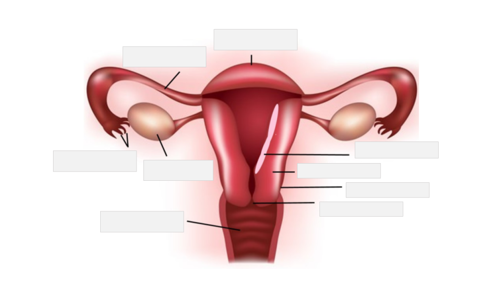
Ovary
Constriction, Pathologic
Analysis of Variance
Case-Control Studies
Trophoblasts
Cyclooxygenase Inhibitors
Pregnancy, Abdominal
Coronary Artery Bypass, Off-Pump
Internal Mammary-Coronary Artery Anastomosis
Dogs
Fetus
Observer Variation
Ultrasonography
Myocardial Infarction
Vertebral Artery Dissection
Disease Models, Animal
Chorionic Villi
Follicular Phase
Calcium
Mesenteric Artery, Inferior
Relaxin
Trophoblast-mediated spiral artery remodelling: a role for apoptosis. (1/165)
(+info)Oxygen as modulator of trophoblast invasion. (2/165)
(+info)First-trimester uterine artery Doppler indices in the prediction of small-for-gestational age pregnancy and intrauterine growth restriction. (3/165)
(+info)Role of uterine artery Doppler in interpreting low PAPP-A values in first-trimester screening for Down syndrome in pregnancies at high risk of impaired placentation. (4/165)
(+info)Fatal pulmonary embolus after uterine artery fibroid embolisation. (5/165)
(+info)Correlation between the Doppler velocimetry findings of the uterine arteries during the first and second trimesters of pregnancy. (6/165)
(+info)Local uteroplacental influences are responsible for the induction of uterine artery myogenic tone during rat pregnancy. (7/165)
(+info)Placental size and the prediction of severe early-onset intrauterine growth restriction in women with low pregnancy-associated plasma protein-A. (8/165)
(+info)The uterine artery is a paired branch of the internal iliac (hip) artery that supplies blood to the uterus and vagina. It anastomoses (joins) with the ovarian artery to form a rich vascular network that nourishes the female reproductive organs. The right and left uterine arteries run along the sides of the uterus, where they divide into several branches to supply oxygenated blood and nutrients to the myometrium (uterine muscle), endometrium (lining), and cervix. These arteries undergo significant changes in size and structure during pregnancy to accommodate the growing fetus and placenta, making them crucial for maintaining a healthy pregnancy.
Arteries are blood vessels that carry oxygenated blood away from the heart to the rest of the body. They have thick, muscular walls that can withstand the high pressure of blood being pumped out of the heart. Arteries branch off into smaller vessels called arterioles, which further divide into a vast network of tiny capillaries where the exchange of oxygen, nutrients, and waste occurs between the blood and the body's cells. After passing through the capillary network, deoxygenated blood collects in venules, then merges into veins, which return the blood back to the heart.
The uterus, also known as the womb, is a hollow, muscular organ located in the female pelvic cavity, between the bladder and the rectum. It has a thick, middle layer called the myometrium, which is composed of smooth muscle tissue, and an inner lining called the endometrium, which provides a nurturing environment for the fertilized egg to develop into a fetus during pregnancy.
The uterus is where the baby grows and develops until it is ready for birth through the cervix, which is the lower, narrow part of the uterus that opens into the vagina. The uterus plays a critical role in the menstrual cycle as well, by shedding its lining each month if pregnancy does not occur.
Uterine artery embolization (UAE) is a minimally invasive procedure used to treat certain conditions related to the uterus and uterine fibroids. The procedure involves blocking or reducing the blood flow to the fibroids, causing them to shrink and alleviating symptoms such as heavy menstrual bleeding, pain, and pressure.
During the procedure, an interventional radiologist makes a small incision in the groin area and inserts a catheter into the femoral artery. The catheter is then guided to the uterine arteries using fluoroscopic imaging. Once in place, tiny particles are injected through the catheter to block or reduce the blood flow to the fibroids. This process may be performed on one or both uterine arteries, depending on the location and size of the fibroids.
UAE is typically an outpatient procedure, and most women can return home the same day. Recovery time varies but is generally shorter than that of a hysterectomy, which is the surgical removal of the uterus. Potential risks associated with UAE include infection, bleeding, damage to nearby organs, and premature menopause in some cases. However, these complications are relatively rare.
Placental circulation refers to the specialized circulatory system that develops during pregnancy to allow for the exchange of nutrients, oxygen, and waste products between the mother's blood and the fetal blood in the placenta. The placenta is a highly vascular organ that grows within the uterus and is connected to the developing fetus via the umbilical cord.
In the maternal side of the placenta, the spiral arteries branch into smaller vessels called the intervillous spaces, where they come in close contact with the fetal blood vessels within the villi (finger-like projections) of the placenta. The intervillous spaces are filled with maternal blood that flows around the villi, allowing for the exchange of gases and nutrients between the two circulations.
On the fetal side, the umbilical cord contains two umbilical arteries that carry oxygen-depleted blood from the fetus to the placenta, and one umbilical vein that returns oxygenated blood back to the fetus. The umbilical arteries branch into smaller vessels within the villi, where they exchange gases and nutrients with the maternal blood in the intervillous spaces.
Overall, the placental circulation is a crucial component of fetal development, allowing for the growing fetus to receive the necessary oxygen and nutrients to support its growth and development.
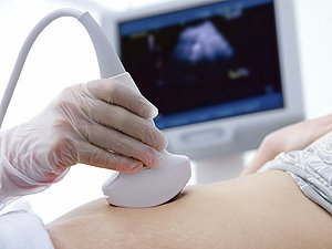
Ultrasonography, Doppler refers to a non-invasive diagnostic medical procedure that uses high-frequency sound waves to create real-time images of the movement of blood flow through vessels, tissues, or heart valves. The Doppler effect is used to measure the frequency shift of the ultrasound waves as they bounce off moving red blood cells, which allows for the calculation of the speed and direction of blood flow. This technique is commonly used to diagnose and monitor various conditions such as deep vein thrombosis, carotid artery stenosis, heart valve abnormalities, and fetal heart development during pregnancy. It does not use radiation or contrast agents and is considered safe with minimal risks.
Pre-eclampsia is a pregnancy-related disorder, typically characterized by the onset of high blood pressure (hypertension) and damage to organs, such as the kidneys, after the 20th week of pregnancy. It is often accompanied by proteinuria, which is the presence of excess protein in the urine. Pre-eclampsia can lead to serious complications for both the mother and the baby if left untreated or unmanaged.
The exact causes of pre-eclampsia are not fully understood, but it is believed that placental issues, genetic factors, and immune system problems may contribute to its development. Risk factors include first-time pregnancies, history of pre-eclampsia in previous pregnancies, chronic hypertension, obesity, older age (35 or older), and assisted reproductive technology (ART) pregnancies.
Pre-eclampsia can progress to a more severe form called eclampsia, which is characterized by the onset of seizures. HELLP syndrome, another severe complication, involves hemolysis (breaking down of red blood cells), elevated liver enzymes, and low platelet count.
Early detection and management of pre-eclampsia are crucial to prevent severe complications. Regular prenatal care, including frequent blood pressure checks and urine tests, can help identify early signs of the condition. Treatment typically involves close monitoring, medication to lower blood pressure, corticosteroids to promote fetal lung maturity, and, in some cases, delivery of the baby if the mother's or baby's health is at risk.
Pulsatile flow is a type of fluid flow that occurs in a rhythmic, wave-like pattern, typically seen in the cardiovascular system. It refers to the periodic variation in the volume or velocity of a fluid (such as blood) that is caused by the regular beating of the heart. In pulsatile flow, there are periods of high flow followed by periods of low or no flow, which creates a distinct pattern on a graph or tracing. This type of flow is important for maintaining proper function and health in organs and tissues throughout the body.
Pregnancy is a physiological state or condition where a fertilized egg (zygote) successfully implants and grows in the uterus of a woman, leading to the development of an embryo and finally a fetus. This process typically spans approximately 40 weeks, divided into three trimesters, and culminates in childbirth. Throughout this period, numerous hormonal and physical changes occur to support the growing offspring, including uterine enlargement, breast development, and various maternal adaptations to ensure the fetus's optimal growth and well-being.
Ultrasonography, Doppler, Pulsed is a type of diagnostic ultrasound technique that uses the Doppler effect to measure blood flow in the body. In this technique, short bursts of ultrasound are emitted and then listened for as they bounce back off moving red blood cells. By analyzing the frequency shift of the returning sound waves, the velocity and direction of blood flow can be determined. This information is particularly useful in evaluating conditions such as deep vein thrombosis, carotid artery stenosis, and fetal heart abnormalities. Pulsed Doppler ultrasonography provides more detailed information about blood flow than traditional color Doppler imaging, making it a valuable tool for diagnosing and monitoring various medical conditions.
Ultrasonography, Doppler, color is a type of diagnostic ultrasound technique that uses the Doppler effect to produce visual images of blood flow in vessels and the heart. The Doppler effect is the change in frequency or wavelength of a wave in relation to an observer who is moving relative to the source of the wave. In this context, it refers to the change in frequency of the ultrasound waves as they reflect off moving red blood cells.
In color Doppler ultrasonography, different colors are used to represent the direction and speed of blood flow. Red typically represents blood flowing toward the transducer (the device that sends and receives sound waves), while blue represents blood flowing away from the transducer. The intensity or brightness of the color is proportional to the velocity of blood flow.
Color Doppler ultrasonography is often used in conjunction with grayscale ultrasound imaging, which provides information about the structure and composition of tissues. Together, these techniques can help diagnose a wide range of conditions, including heart disease, blood clots, and abnormalities in blood flow.
Prenatal ultrasonography, also known as obstetric ultrasound, is a medical diagnostic procedure that uses high-frequency sound waves to create images of the developing fetus, placenta, and amniotic fluid inside the uterus. It is a non-invasive and painless test that is widely used during pregnancy to monitor the growth and development of the fetus, detect any potential abnormalities or complications, and determine the due date.
During the procedure, a transducer (a small handheld device) is placed on the mother's abdomen and moved around to capture images from different angles. The sound waves travel through the mother's body and bounce back off the fetus, producing echoes that are then converted into electrical signals and displayed as images on a screen.
Prenatal ultrasonography can be performed at various stages of pregnancy, including early pregnancy to confirm the pregnancy and detect the number of fetuses, mid-pregnancy to assess the growth and development of the fetus, and late pregnancy to evaluate the position of the fetus and determine if it is head down or breech. It can also be used to guide invasive procedures such as amniocentesis or chorionic villus sampling.
Overall, prenatal ultrasonography is a valuable tool in modern obstetrics that helps ensure the health and well-being of both the mother and the developing fetus.
The pulmonary artery is a large blood vessel that carries deoxygenated blood from the right ventricle of the heart to the lungs for oxygenation. It divides into two main branches, the right and left pulmonary arteries, which further divide into smaller vessels called arterioles, and then into a vast network of capillaries in the lungs where gas exchange occurs. The thin walls of these capillaries allow oxygen to diffuse into the blood and carbon dioxide to diffuse out, making the blood oxygen-rich before it is pumped back to the left side of the heart through the pulmonary veins. This process is crucial for maintaining proper oxygenation of the body's tissues and organs.
Leiomyoma is a benign (non-cancerous) tumor that originates from the smooth muscle cells. It most commonly occurs in the uterus, where it is also known as a fibroid, but can also develop in other parts of the body such as the skin, gastrointestinal tract, and genitourinary system. Leiomyomas are typically slow-growing and often cause no symptoms, although they can lead to various complications depending on their size and location. Treatment options for leiomyomas include surveillance, medication, or surgical removal.
The second trimester of pregnancy is the period between the completion of 12 weeks (the end of the first trimester) and 26 weeks (the beginning of the third trimester) of gestational age. It is often considered the most comfortable period for many pregnant women as the risk of miscarriage decreases significantly, and the symptoms experienced during the first trimester, such as nausea and fatigue, typically improve.
During this time, the uterus expands above the pubic bone, allowing more space for the growing fetus. The fetal development in the second trimester includes significant growth in size and weight, formation of all major organs, and the beginning of movement sensations that the mother can feel. Additionally, the fetus starts to hear, swallow and kick, and the skin is covered with a protective coating called vernix.
Prenatal care during this period typically includes regular prenatal appointments to monitor the mother's health and the baby's growth and development. These appointments may include measurements of the uterus, fetal heart rate monitoring, and screening tests for genetic disorders or other potential issues.
The umbilical arteries are a pair of vessels that develop within the umbilical cord during fetal development. They carry oxygenated and nutrient-rich blood from the mother to the developing fetus through the placenta. These arteries arise from the internal iliac arteries in the fetus and pass through the umbilical cord to connect with the two umbilical veins within the placenta. After birth, the umbilical arteries become ligaments (the medial umbilical ligaments) that run along the inner abdominal wall.
"Animal pregnancy" is not a term that is typically used in medical definitions. However, in biological terms, animal pregnancy refers to the condition where a fertilized egg (or eggs) implants and develops inside the reproductive tract of a female animal, leading to the birth of offspring (live young).
The specific details of animal pregnancy can vary widely between different species, with some animals exhibiting phenomena such as placental development, gestation periods, and hormonal changes that are similar to human pregnancy, while others may have very different reproductive strategies.
It's worth noting that the study of animal pregnancy and reproduction is an important area of biological research, as it can provide insights into fundamental mechanisms of embryonic development, genetics, and evolution.
Fetal growth retardation, also known as intrauterine growth restriction (IUGR), is a condition in which a fetus fails to grow at the expected rate during pregnancy. This can be caused by various factors such as maternal health problems, placental insufficiency, chromosomal abnormalities, and genetic disorders. The fetus may be smaller than expected for its gestational age, have reduced movement, and may be at risk for complications during labor and delivery. It is important to monitor fetal growth and development closely throughout pregnancy to detect any potential issues early on and provide appropriate medical interventions.
The mesenteric arteries are the arteries that supply oxygenated blood to the intestines. There are three main mesenteric arteries: the superior mesenteric artery, which supplies blood to the small intestine (duodenum to two-thirds of the transverse colon) and large intestine (cecum, ascending colon, and the first part of the transverse colon); the inferior mesenteric artery, which supplies blood to the distal third of the transverse colon, descending colon, sigmoid colon, and rectum; and the middle colic artery, which is a branch of the superior mesenteric artery that supplies blood to the transverse colon. These arteries are important in maintaining adequate blood flow to the intestines to support digestion and absorption of nutrients.
The carotid arteries are a pair of vital blood vessels in the human body that supply oxygenated blood to the head and neck. Each person has two common carotid arteries, one on each side of the neck, which branch off from the aorta, the largest artery in the body.
The right common carotid artery originates from the brachiocephalic trunk, while the left common carotid artery arises directly from the aortic arch. As they ascend through the neck, they split into two main branches: the internal and external carotid arteries.
The internal carotid artery supplies oxygenated blood to the brain, eyes, and other structures within the skull, while the external carotid artery provides blood to the face, scalp, and various regions of the neck.
Maintaining healthy carotid arteries is crucial for overall cardiovascular health and preventing serious conditions like stroke, which can occur when the arteries become narrowed or blocked due to the buildup of plaque or fatty deposits (atherosclerosis). Regular check-ups with healthcare professionals may include monitoring carotid artery health through ultrasound or other imaging techniques.
Uterine neoplasms refer to abnormal growths in the uterus, which can be benign (non-cancerous) or malignant (cancerous). These growths can originate from different types of cells within the uterus, leading to various types of uterine neoplasms. The two main categories of uterine neoplasms are endometrial neoplasms and uterine sarcomas.
Endometrial neoplasms develop from the endometrium, which is the inner lining of the uterus. Most endometrial neoplasms are classified as endometrioid adenocarcinomas, arising from glandular cells in the endometrium. Other types include serous carcinoma, clear cell carcinoma, and mucinous carcinoma.
Uterine sarcomas, on the other hand, are less common and originate from the connective tissue (stroma) or muscle (myometrium) of the uterus. Uterine sarcomas can be further divided into several subtypes, such as leiomyosarcoma, endometrial stromal sarcoma, and undifferentiated uterine sarcoma.
Uterine neoplasms can cause various symptoms, including abnormal vaginal bleeding or discharge, pelvic pain, and difficulty urinating or having bowel movements. The diagnosis typically involves a combination of imaging tests (such as ultrasound, CT, or MRI scans) and tissue biopsies to determine the type and extent of the neoplasm. Treatment options depend on the type, stage, and patient's overall health but may include surgery, radiation therapy, chemotherapy, or hormone therapy.
Cerebral arteries refer to the blood vessels that supply oxygenated blood to the brain. These arteries branch off from the internal carotid arteries and the vertebral arteries, which combine to form the basilar artery. The major cerebral arteries include:
1. Anterior cerebral artery (ACA): This artery supplies blood to the frontal lobes of the brain, including the motor and sensory cortices responsible for movement and sensation in the lower limbs.
2. Middle cerebral artery (MCA): The MCA is the largest of the cerebral arteries and supplies blood to the lateral surface of the brain, including the temporal, parietal, and frontal lobes. It is responsible for providing blood to areas involved in motor function, sensory perception, speech, memory, and vision.
3. Posterior cerebral artery (PCA): The PCA supplies blood to the occipital lobe, which is responsible for visual processing, as well as parts of the temporal and parietal lobes.
4. Anterior communicating artery (ACoA) and posterior communicating arteries (PComAs): These are small arteries that connect the major cerebral arteries, forming an important circulatory network called the Circle of Willis. The ACoA connects the two ACAs, while the PComAs connect the ICA with the PCA and the basilar artery.
These cerebral arteries play a crucial role in maintaining proper brain function by delivering oxygenated blood to various regions of the brain. Any damage or obstruction to these arteries can lead to serious neurological conditions, such as strokes or transient ischemic attacks (TIAs).
The renal artery is a pair of blood vessels that originate from the abdominal aorta and supply oxygenated blood to each kidney. These arteries branch into several smaller vessels that provide blood to the various parts of the kidneys, including the renal cortex and medulla. The renal arteries also carry nutrients and other essential components needed for the normal functioning of the kidneys. Any damage or blockage to the renal artery can lead to serious consequences, such as reduced kidney function or even kidney failure.
The femoral artery is the major blood vessel that supplies oxygenated blood to the lower extremity of the human body. It is a continuation of the external iliac artery and becomes the popliteal artery as it passes through the adductor hiatus in the adductor magnus muscle of the thigh.
The femoral artery is located in the femoral triangle, which is bound by the sartorius muscle anteriorly, the adductor longus muscle medially, and the biceps femoris muscle posteriorly. It can be easily palpated in the groin region, making it a common site for taking blood samples, measuring blood pressure, and performing surgical procedures such as femoral artery catheterization and bypass grafting.
The femoral artery gives off several branches that supply blood to the lower limb, including the deep femoral artery, the superficial femoral artery, and the profunda femoris artery. These branches provide blood to the muscles, bones, skin, and other tissues of the leg, ankle, and foot.
The first trimester of pregnancy is defined as the period of gestational development that extends from conception (fertilization of the egg by sperm) to the end of the 13th week. This critical phase marks significant transformations in both the mother's body and the growing embryo/fetus.
During the first trimester, the fertilized egg implants into the uterine lining (implantation), initiating a series of complex interactions leading to the formation of the placenta - an organ essential for providing nutrients and oxygen to the developing fetus while removing waste products. Simultaneously, the embryo undergoes rapid cell division and differentiation, giving rise to various organs and systems. By the end of the first trimester, most major structures are present, although they continue to mature and grow throughout pregnancy.
The mother may experience several physiological changes during this time, including:
- Morning sickness (nausea and vomiting)
- Fatigue
- Breast tenderness
- Frequent urination
- Food aversions or cravings
- Mood swings
Additionally, hormonal shifts can cause various symptoms and prepare the body for potential changes in lactation, posture, and pelvic alignment as pregnancy progresses. Regular prenatal care is crucial during this period to monitor both maternal and fetal wellbeing, identify any potential complications early on, and provide appropriate guidance and support throughout the pregnancy.
The basilar artery is a major blood vessel that supplies oxygenated blood to the brainstem and cerebellum. It is formed by the union of two vertebral arteries at the lower part of the brainstem, near the junction of the medulla oblongata and pons.
The basilar artery runs upward through the center of the brainstem and divides into two posterior cerebral arteries at the upper part of the brainstem, near the midbrain. The basilar artery gives off several branches that supply blood to various parts of the brainstem, including the pons, medulla oblongata, and midbrain, as well as to the cerebellum.
The basilar artery is an important part of the circle of Willis, a network of arteries at the base of the brain that ensures continuous blood flow to the brain even if one of the arteries becomes blocked or narrowed.
The iliac arteries are major branches of the abdominal aorta, the large artery that carries oxygen-rich blood from the heart to the rest of the body. The iliac arteries divide into two branches, the common iliac arteries, which further bifurcate into the internal and external iliac arteries.
The internal iliac artery supplies blood to the lower abdomen, pelvis, and the reproductive organs, while the external iliac artery provides blood to the lower extremities, including the legs and feet. Together, the iliac arteries play a crucial role in circulating blood throughout the body, ensuring that all tissues and organs receive the oxygen and nutrients they need to function properly.
Regional blood flow (RBF) refers to the rate at which blood flows through a specific region or organ in the body, typically expressed in milliliters per minute per 100 grams of tissue (ml/min/100g). It is an essential physiological parameter that reflects the delivery of oxygen and nutrients to tissues while removing waste products. RBF can be affected by various factors such as metabolic demands, neural regulation, hormonal influences, and changes in blood pressure or vascular resistance. Measuring RBF is crucial for understanding organ function, diagnosing diseases, and evaluating the effectiveness of treatments.
The vertebral artery is a major blood vessel that supplies oxygenated blood to the brain and upper spinal cord. It arises from the subclavian artery, then ascends through the transverse processes of several cervical vertebrae before entering the skull through the foramen magnum. Inside the skull, it joins with the opposite vertebral artery to form the basilar artery, which supplies blood to the brainstem and cerebellum. The vertebral artery also gives off several important branches that supply blood to various regions of the brainstem and upper spinal cord.
Coronary artery bypass surgery, also known as coronary artery bypass grafting (CABG), is a surgical procedure used to improve blood flow to the heart in patients with severe coronary artery disease. This condition occurs when the coronary arteries, which supply oxygen-rich blood to the heart muscle, become narrowed or blocked due to the buildup of fatty deposits, called plaques.
During CABG surgery, a healthy blood vessel from another part of the body is grafted, or attached, to the coronary artery, creating a new pathway for oxygen-rich blood to flow around the blocked or narrowed portion of the artery and reach the heart muscle. This bypass helps to restore normal blood flow and reduce the risk of angina (chest pain), shortness of breath, and other symptoms associated with coronary artery disease.
There are different types of CABG surgery, including traditional on-pump CABG, off-pump CABG, and minimally invasive CABG. The choice of procedure depends on various factors, such as the patient's overall health, the number and location of blocked arteries, and the presence of other medical conditions.
It is important to note that while CABG surgery can significantly improve symptoms and quality of life in patients with severe coronary artery disease, it does not cure the underlying condition. Lifestyle modifications, such as regular exercise, a healthy diet, smoking cessation, and medication therapy, are essential for long-term management and prevention of further progression of the disease.
High-risk pregnancy is a term used to describe a situation where the mother or the fetus has an increased risk of developing complications during pregnancy, labor, delivery, or in the postpartum period. These risks may be due to pre-existing medical conditions in the mother, such as diabetes, hypertension, heart disease, kidney disease, autoimmune disorders, or infectious diseases like HIV/AIDS. Other factors that can contribute to a high-risk pregnancy include advanced maternal age (35 years and older), obesity, multiple gestations (twins, triplets, etc.), fetal growth restriction, placental issues, and a history of previous pregnancy complications or preterm labor.
High-risk pregnancies require specialized care and monitoring by healthcare professionals, often involving maternal-fetal medicine specialists, obstetricians, perinatologists, and neonatologists. Regular prenatal care, frequent checkups, ultrasound monitoring, and sometimes additional testing and interventions may be necessary to ensure the best possible outcomes for both the mother and the baby.
I apologize, but "sheep" is not a term that has a medical definition. It is a common animal with the scientific name Ovis aries. If you have any medical or health-related questions, I would be happy to try and help answer those for you.
Blood flow velocity is the speed at which blood travels through a specific part of the vascular system. It is typically measured in units of distance per time, such as centimeters per second (cm/s) or meters per second (m/s). Blood flow velocity can be affected by various factors, including cardiac output, vessel diameter, and viscosity of the blood. Measuring blood flow velocity is important in diagnosing and monitoring various medical conditions, such as heart disease, stroke, and peripheral vascular disease.
Therapeutic embolization is a medical procedure that involves intentionally blocking or obstructing blood vessels to stop excessive bleeding or block the flow of blood to a tumor or abnormal tissue. This is typically accomplished by injecting small particles, such as microspheres or coils, into the targeted blood vessel through a catheter, which is inserted into a larger blood vessel and guided to the desired location using imaging techniques like X-ray or CT scanning. The goal of therapeutic embolization is to reduce the size of a tumor, control bleeding, or block off abnormal blood vessels that are causing problems.
Pregnancy-induced hypertension (PIH), also known as gestational hypertension, is a condition characterized by the new onset of high blood pressure (≥140 mm Hg systolic or ≥90 mm Hg diastolic) after 20 weeks of pregnancy in a woman who was normotensive before. It can sometimes progress to more severe conditions like preeclampsia and eclampsia, which are associated with damage to other organ systems such as the liver and kidneys.
PIH is typically classified into two types:
1. Gestational hypertension: This is when a woman develops high blood pressure after 20 weeks of pregnancy without any protein in the urine or evidence of damage to other organ systems. Women with gestational hypertension are at increased risk for preeclampsia and may require closer monitoring.
2. Preeclampsia: This is a more severe form of PIH, characterized by high blood pressure and proteinuria (≥0.3 g in a 24-hour urine collection) after 20 weeks of pregnancy. Preeclampsia can also involve damage to other organ systems, such as the liver, kidneys, or brain, and may progress to eclampsia, a life-threatening condition characterized by seizures.
The exact causes of PIH are not fully understood, but it is thought to be related to problems with the development and function of the blood vessels that supply the placenta. Risk factors for developing PIH include first-time pregnancies, obesity, older age, a history of chronic hypertension or kidney disease, and carrying multiples (twins, triplets, etc.).
Treatment for PIH depends on the severity of the condition and the gestational age of the pregnancy. In mild cases, close monitoring of blood pressure, urine protein levels, and fetal growth may be sufficient. More severe cases may require medication to lower blood pressure, corticosteroids to promote fetal lung maturity, or early delivery of the baby to prevent further complications.
Vasoconstriction is a medical term that refers to the narrowing of blood vessels due to the contraction of the smooth muscle in their walls. This process decreases the diameter of the lumen (the inner space of the blood vessel) and reduces blood flow through the affected vessels. Vasoconstriction can occur throughout the body, but it is most noticeable in the arterioles and precapillary sphincters, which control the amount of blood that flows into the capillary network.
The autonomic nervous system, specifically the sympathetic division, plays a significant role in regulating vasoconstriction through the release of neurotransmitters like norepinephrine (noradrenaline). Various hormones and chemical mediators, such as angiotensin II, endothelin-1, and serotonin, can also induce vasoconstriction.
Vasoconstriction is a vital physiological response that helps maintain blood pressure and regulate blood flow distribution in the body. However, excessive or prolonged vasoconstriction may contribute to several pathological conditions, including hypertension, stroke, and peripheral vascular diseases.
The radial artery is a key blood vessel in the human body, specifically a part of the peripheral arterial system. Originating from the brachial artery in the upper arm, the radial artery travels down the arm and crosses over the wrist, where it can be palpated easily. It then continues into the hand, dividing into several branches to supply blood to the hand's tissues and digits.
The radial artery is often used for taking pulse readings due to its easy accessibility at the wrist. Additionally, in medical procedures such as coronary angiography or bypass surgery, the radial artery can be utilized as a site for catheter insertion. This allows healthcare professionals to examine the heart's blood vessels and assess cardiovascular health.
The mammary arteries are a set of blood vessels that supply oxygenated blood to the mammary glands, which are the structures in female breasts responsible for milk production during lactation. The largest mammary artery, also known as the internal thoracic or internal mammary artery, originates from the subclavian artery and descends along the inner side of the chest wall. It then branches into several smaller arteries that supply blood to the breast tissue. These include the anterior and posterior intercostal arteries, lateral thoracic artery, and pectoral branches. The mammary arteries are crucial in maintaining the health and function of the breast tissue, and any damage or blockage to these vessels can lead to various breast-related conditions or diseases.
Vascular resistance is a measure of the opposition to blood flow within a vessel or a group of vessels, typically expressed in units of mmHg/(mL/min) or sometimes as dynes*sec/cm^5. It is determined by the diameter and length of the vessels, as well as the viscosity of the blood flowing through them. In general, a decrease in vessel diameter, an increase in vessel length, or an increase in blood viscosity will result in an increase in vascular resistance, while an increase in vessel diameter, a decrease in vessel length, or a decrease in blood viscosity will result in a decrease in vascular resistance. Vascular resistance is an important concept in the study of circulation and cardiovascular physiology because it plays a key role in determining blood pressure and blood flow within the body.
Menorrhagia is a medical term used to describe abnormally heavy or prolonged menstrual periods. It's often characterized by the loss of an excessive amount of menstrual blood (usually more than 80 ml) and can last longer than normal, typically over seven days. This condition can have significant impacts on a woman's quality of life, causing fatigue, distress, and restrictions in daily activities due to the need for frequent pad or tampon changes.
The causes of menorrhagia are varied and can include hormonal imbalances, uterine fibroids or polyps, endometrial hyperplasia, pelvic inflammatory disease, pregnancy complications, certain medications, and underlying medical conditions such as coagulopathies or thyroid disorders. In some cases, the cause may remain undetermined even after a thorough evaluation.
Treatment options for menorrhagia depend on the underlying cause and range from medication management with hormonal therapies, nonsteroidal anti-inflammatory drugs (NSAIDs), or tranexamic acid to procedural interventions like endometrial ablation, hysteroscopic resection of polyps or fibroids, or ultimately hysterectomy in severe cases. It is essential for individuals experiencing menorrhagia to consult with their healthcare provider to determine the best course of action based on their specific situation and medical history.
The internal carotid artery is a major blood vessel that supplies oxygenated blood to the brain. It originates from the common carotid artery and passes through the neck, entering the skull via the carotid canal in the temporal bone. Once inside the skull, it branches into several smaller vessels that supply different parts of the brain with blood.
The internal carotid artery is divided into several segments: cervical, petrous, cavernous, clinoid, and supraclinoid. Each segment has distinct clinical significance in terms of potential injury or disease. The most common conditions affecting the internal carotid artery include atherosclerosis, which can lead to stroke or transient ischemic attack (TIA), and dissection, which can cause severe headache, neck pain, and neurological symptoms.
It's important to note that any blockage or damage to the internal carotid artery can have serious consequences, as it can significantly reduce blood flow to the brain and lead to permanent neurological damage or even death. Therefore, regular check-ups and screening tests are recommended for individuals at high risk of developing vascular diseases.
Uterine hemorrhage, also known as uterine bleeding or gynecological bleeding, is an abnormal loss of blood from the uterus. It can occur in various clinical settings such as menstruation (known as menorrhagia), postpartum period (postpartum hemorrhage), or in non-pregnant women (dysfunctional uterine bleeding). The bleeding may be light to heavy, intermittent or continuous, and can be accompanied by symptoms such as pain, dizziness, or fainting. Uterine hemorrhage is a common gynecological problem that can have various underlying causes, including hormonal imbalances, structural abnormalities, coagulopathies, and malignancies. It is important to seek medical attention if experiencing heavy or prolonged uterine bleeding to determine the cause and receive appropriate treatment.
Vasodilation is the widening or increase in diameter of blood vessels, particularly the involuntary relaxation of the smooth muscle in the tunica media (middle layer) of the arteriole walls. This results in an increase in blood flow and a decrease in vascular resistance. Vasodilation can occur due to various physiological and pathophysiological stimuli, such as local metabolic demands, neural signals, or pharmacological agents. It plays a crucial role in regulating blood pressure, tissue perfusion, and thermoregulation.
The subclavian artery is a major blood vessel that supplies the upper limb and important structures in the neck and head. It arises from the brachiocephalic trunk (in the case of the right subclavian artery) or directly from the aortic arch (in the case of the left subclavian artery).
The subclavian artery has several branches, including:
1. The vertebral artery, which supplies blood to the brainstem and cerebellum.
2. The internal thoracic artery (also known as the mammary artery), which supplies blood to the chest wall, breast, and anterior mediastinum.
3. The thyrocervical trunk, which gives rise to several branches that supply the neck, including the inferior thyroid artery, the suprascapular artery, and the transverse cervical artery.
4. The costocervical trunk, which supplies blood to the neck and upper back, including the posterior chest wall and the lower neck muscles.
The subclavian artery is a critical vessel in maintaining adequate blood flow to the upper limb, and any blockage or damage to this vessel can lead to significant morbidity, including arm pain, numbness, weakness, or even loss of function.
Pregnancy-associated plasma protein-A (PAPP-A) is a protease that is often used as a biomarker in early pregnancy. It is a protein that is produced by the placenta and can be detected in the mother's bloodstream during pregnancy.
In early pregnancy, low levels of PAPP-A may indicate an increased risk for certain complications, such as preeclampsia or fetal growth restriction. High levels of PAPP-A, on the other hand, may be associated with an increased risk of chromosomal abnormalities, such as Down syndrome.
It is important to note that while PAPP-A levels can provide valuable information about the health of a pregnancy, they are just one piece of the puzzle and should be considered in conjunction with other factors, such as maternal age, medical history, and ultrasound results. Your healthcare provider will use this information along with other tests to assess your risk for certain complications and develop an appropriate plan of care.
Placental insufficiency is a condition in which the placenta does not provide adequate nutrients and oxygen to the developing fetus. This can occur due to various reasons, such as poor placental development, damage to the placenta, or problems with the blood flow to the placenta. As a result, the fetus may receive less oxygen and nutrients than it needs for proper growth and development, which can lead to a range of complications, including low birth weight, preterm birth, and developmental delays.
The medical definition of placental insufficiency is: "a condition in which the placenta fails to provide adequate support to the developing fetus, resulting in impaired fetal growth and development." This condition can be diagnosed through various tests, such as ultrasound, fetal monitoring, and blood tests, and may require close monitoring and management throughout pregnancy to ensure the best possible outcomes for both the mother and the baby.
Gestational age is the length of time that has passed since the first day of the last menstrual period (LMP) in pregnant women. It is the standard unit used to estimate the age of a pregnancy and is typically expressed in weeks. This measure is used because the exact date of conception is often not known, but the start of the last menstrual period is usually easier to recall.
It's important to note that since ovulation typically occurs around two weeks after the start of the LMP, gestational age is approximately two weeks longer than fetal age, which is the actual time elapsed since conception. Medical professionals use both gestational and fetal age to track the development and growth of the fetus during pregnancy.
Carotid artery diseases refer to conditions that affect the carotid arteries, which are the major blood vessels that supply oxygen-rich blood to the head and neck. The most common type of carotid artery disease is atherosclerosis, which occurs when fatty deposits called plaques build up in the inner lining of the arteries.
These plaques can cause the arteries to narrow or become blocked, reducing blood flow to the brain and increasing the risk of stroke. Other carotid artery diseases include carotid artery dissection, which occurs when there is a tear in the inner lining of the artery, and fibromuscular dysplasia, which is a condition that affects the muscle and tissue in the walls of the artery.
Symptoms of carotid artery disease may include neck pain or pulsations, transient ischemic attacks (TIAs) or "mini-strokes," and strokes. Treatment options for carotid artery disease depend on the severity and type of the condition but may include lifestyle changes, medications, endarterectomy (a surgical procedure to remove plaque from the artery), or angioplasty and stenting (procedures to open blocked arteries using a balloon and stent).
The splenic artery is the largest branch of the celiac trunk, which arises from the abdominal aorta. It supplies blood to the spleen and several other organs in the upper left part of the abdomen. The splenic artery divides into several branches that ultimately form a network of capillaries within the spleen. These capillaries converge to form the main venous outflow, the splenic vein, which drains into the hepatic portal vein.
The splenic artery is a vital structure in the human body, and any damage or blockage can lead to serious complications, including splenic infarction (reduced blood flow to the spleen) or splenic rupture (a surgical emergency that can be life-threatening).
The brachial artery is a major blood vessel in the upper arm. It supplies oxygenated blood to the muscles and tissues of the arm, forearm, and hand. The brachial artery originates from the axillary artery at the level of the shoulder joint and runs down the medial (inner) aspect of the arm, passing through the cubital fossa (the depression on the anterior side of the elbow) where it can be palpated during a routine blood pressure measurement. At the lower end of the forearm, the brachial artery bifurcates into the radial and ulnar arteries, which further divide into smaller vessels to supply the hand and fingers.
The Middle Cerebral Artery (MCA) is one of the main blood vessels that supplies oxygenated blood to the brain. It arises from the internal carotid artery and divides into several branches, which supply the lateral surface of the cerebral hemisphere, including the frontal, parietal, and temporal lobes.
The MCA is responsible for providing blood flow to critical areas of the brain, such as the primary motor and sensory cortices, Broca's area (associated with speech production), Wernicke's area (associated with language comprehension), and the visual association cortex.
Damage to the MCA or its branches can result in a variety of neurological deficits, depending on the specific location and extent of the injury. These may include weakness or paralysis on one side of the body, sensory loss, language impairment, and visual field cuts.
Pregnancy outcome refers to the final result or status of a pregnancy, including both the health of the mother and the newborn baby. It can be categorized into various types such as:
1. Live birth: The delivery of one or more babies who show signs of life after separation from their mother.
2. Stillbirth: The delivery of a baby who has died in the womb after 20 weeks of pregnancy.
3. Miscarriage: The spontaneous loss of a pregnancy before the 20th week.
4. Abortion: The intentional termination of a pregnancy before the fetus can survive outside the uterus.
5. Ectopic pregnancy: A pregnancy that develops outside the uterus, usually in the fallopian tube, which is not viable and requires medical attention.
6. Preterm birth: The delivery of a baby before 37 weeks of gestation, which can lead to various health issues for the newborn.
7. Full-term birth: The delivery of a baby between 37 and 42 weeks of gestation.
8. Post-term pregnancy: The delivery of a baby after 42 weeks of gestation, which may increase the risk of complications for both mother and baby.
The pregnancy outcome is influenced by various factors such as maternal age, health status, lifestyle habits, genetic factors, and access to quality prenatal care.
A smooth muscle within the vascular system refers to the involuntary, innervated muscle that is found in the walls of blood vessels. These muscles are responsible for controlling the diameter of the blood vessels, which in turn regulates blood flow and blood pressure. They are called "smooth" muscles because their individual muscle cells do not have the striations, or cross-striped patterns, that are observed in skeletal and cardiac muscle cells. Smooth muscle in the vascular system is controlled by the autonomic nervous system and by hormones, and can contract or relax slowly over a period of time.
Abruptio placentae, also known as placental abruption, is a medical condition that occurs when the placenta separates from the uterus before the baby is born. The placenta is an organ that develops in the uterus during pregnancy to provide oxygen and nutrients to the growing fetus.
In abruptio placentae, the separation of the placenta from the uterus can cause bleeding, which can be serious or life-threatening for both the mother and the baby. The severity of the condition depends on how much of the placenta has separated from the uterus and how much bleeding has occurred.
Abruptio placentae can cause a range of symptoms, including vaginal bleeding, abdominal pain, contractions, and fetal distress. In severe cases, it can lead to preterm labor, low birth weight, and even stillbirth. The exact cause of abruptio placentae is not always known, but risk factors include high blood pressure, smoking, cocaine use, trauma to the abdomen, and advanced maternal age. Treatment may involve hospitalization, bed rest, medication to prevent contractions, or delivery of the baby if the pregnancy is at term.
Vasoconstrictor agents are substances that cause the narrowing of blood vessels by constricting the smooth muscle in their walls. This leads to an increase in blood pressure and a decrease in blood flow. They work by activating the sympathetic nervous system, which triggers the release of neurotransmitters such as norepinephrine and epinephrine that bind to alpha-adrenergic receptors on the smooth muscle cells of the blood vessel walls, causing them to contract.
Vasoconstrictor agents are used medically for a variety of purposes, including:
* Treating hypotension (low blood pressure)
* Controlling bleeding during surgery or childbirth
* Relieving symptoms of nasal congestion in conditions such as the common cold or allergies
Examples of vasoconstrictor agents include phenylephrine, oxymetazoline, and epinephrine. It's important to note that prolonged use or excessive doses of vasoconstrictor agents can lead to rebound congestion and other adverse effects, so they should be used with caution and under the guidance of a healthcare professional.
The hepatic artery is a branch of the celiac trunk or abdominal aorta that supplies oxygenated blood to the liver. It typically divides into two main branches, the right and left hepatic arteries, which further divide into smaller vessels to supply different regions of the liver. The hepatic artery also gives off branches to supply other organs such as the gallbladder, pancreas, and duodenum.
It's worth noting that there is significant variability in the anatomy of the hepatic artery, with some individuals having additional branches or variations in the origin of the vessel. This variability can have implications for surgical procedures involving the liver and surrounding organs.
The common carotid artery is a major blood vessel in the neck that supplies oxygenated blood to the head and neck. It originates from the brachiocephalic trunk or the aortic arch and divides into the internal and external carotid arteries at the level of the upper border of the thyroid cartilage. The common carotid artery is an important structure in the circulatory system, and any damage or blockage to it can have serious consequences, including stroke.
Prospective studies, also known as longitudinal studies, are a type of cohort study in which data is collected forward in time, following a group of individuals who share a common characteristic or exposure over a period of time. The researchers clearly define the study population and exposure of interest at the beginning of the study and follow up with the participants to determine the outcomes that develop over time. This type of study design allows for the investigation of causal relationships between exposures and outcomes, as well as the identification of risk factors and the estimation of disease incidence rates. Prospective studies are particularly useful in epidemiology and medical research when studying diseases with long latency periods or rare outcomes.
Coronary vessels refer to the network of blood vessels that supply oxygenated blood and nutrients to the heart muscle, also known as the myocardium. The two main coronary arteries are the left main coronary artery and the right coronary artery.
The left main coronary artery branches off into the left anterior descending artery (LAD) and the left circumflex artery (LCx). The LAD supplies blood to the front of the heart, while the LCx supplies blood to the side and back of the heart.
The right coronary artery supplies blood to the right lower part of the heart, including the right atrium and ventricle, as well as the back of the heart.
Coronary vessel disease (CVD) occurs when these vessels become narrowed or blocked due to the buildup of plaque, leading to reduced blood flow to the heart muscle. This can result in chest pain, shortness of breath, or a heart attack.
Angiography is a medical procedure in which an x-ray image is taken to visualize the internal structure of blood vessels, arteries, or veins. This is done by injecting a radiopaque contrast agent (dye) into the blood vessel using a thin, flexible catheter. The dye makes the blood vessels visible on an x-ray image, allowing doctors to diagnose and treat various medical conditions such as blockages, narrowing, or malformations of the blood vessels.
There are several types of angiography, including:
* Cardiac angiography (also called coronary angiography) - used to examine the blood vessels of the heart
* Cerebral angiography - used to examine the blood vessels of the brain
* Peripheral angiography - used to examine the blood vessels in the limbs or other parts of the body.
Angiography is typically performed by a radiologist, cardiologist, or vascular surgeon in a hospital setting. It can help diagnose conditions such as coronary artery disease, aneurysms, and peripheral arterial disease, among others.
The third trimester of pregnancy is the final stage of pregnancy that lasts from week 29 until birth, which typically occurs around the 40th week. During this period, the fetus continues to grow and mature, gaining weight rapidly. The mother's body also prepares for childbirth by dilating the cervix and producing milk in preparation for breastfeeding. Regular prenatal care is crucial during this time to monitor the health of both the mother and the developing fetus, as well as to prepare for delivery.
The celiac artery, also known as the anterior abdominal aortic trunk, is a major artery that originates from the abdominal aorta and supplies oxygenated blood to the foregut, which includes the stomach, liver, spleen, pancreas, and upper part of the duodenum. It branches into three main branches: the left gastric artery, the splenic artery, and the common hepatic artery. The celiac artery plays a crucial role in providing blood to these vital organs, and any disruption or damage to it can lead to serious health consequences.
The ophthalmic artery is the first branch of the internal carotid artery, which supplies blood to the eye and its adnexa. It divides into several branches that provide oxygenated blood to various structures within the eye, including the retina, optic nerve, choroid, iris, ciliary body, and cornea. Any blockage or damage to the ophthalmic artery can lead to serious vision problems or even blindness.
Vasodilator agents are pharmacological substances that cause the relaxation or widening of blood vessels by relaxing the smooth muscle in the vessel walls. This results in an increase in the diameter of the blood vessels, which decreases vascular resistance and ultimately reduces blood pressure. Vasodilators can be further classified based on their site of action:
1. Systemic vasodilators: These agents cause a generalized relaxation of the smooth muscle in the walls of both arteries and veins, resulting in a decrease in peripheral vascular resistance and preload (the volume of blood returning to the heart). Examples include nitroglycerin, hydralazine, and calcium channel blockers.
2. Arterial vasodilators: These agents primarily affect the smooth muscle in arterial vessel walls, leading to a reduction in afterload (the pressure against which the heart pumps blood). Examples include angiotensin-converting enzyme (ACE) inhibitors, angiotensin receptor blockers (ARBs), and direct vasodilators like sodium nitroprusside.
3. Venous vasodilators: These agents primarily affect the smooth muscle in venous vessel walls, increasing venous capacitance and reducing preload. Examples include nitroglycerin and other organic nitrates.
Vasodilator agents are used to treat various cardiovascular conditions such as hypertension, heart failure, angina, and pulmonary arterial hypertension. It is essential to monitor their use carefully, as excessive vasodilation can lead to orthostatic hypotension, reflex tachycardia, or fluid retention.
Cardiovascular complications in pregnancy refer to conditions that affect the heart and blood vessels, which can arise during pregnancy, childbirth, or after delivery. These complications can be pre-existing or new-onset and can range from mild to severe, potentially threatening the life of both the mother and the fetus. Some examples of cardiovascular complications in pregnancy include:
1. Hypertension disorders: This includes chronic hypertension (high blood pressure before pregnancy), gestational hypertension (high blood pressure that develops after 20 weeks of pregnancy), and preeclampsia/eclampsia (a pregnancy-specific disorder characterized by high blood pressure, proteinuria, and potential organ damage).
2. Cardiomyopathy: A condition in which the heart muscle becomes weakened, leading to an enlarged heart and reduced pumping efficiency. Peripartum cardiomyopathy is a specific type that occurs during pregnancy or in the months following delivery.
3. Arrhythmias: Irregularities in the heart's rhythm, such as tachycardia (rapid heartbeat) or bradycardia (slow heartbeat), can occur during pregnancy and may require medical intervention.
4. Valvular heart disease: Pre-existing valve disorders, like mitral stenosis or aortic insufficiency, can worsen during pregnancy due to increased blood volume and cardiac output. Additionally, new valve issues might develop during pregnancy.
5. Venous thromboembolism (VTE): Pregnancy increases the risk of developing blood clots in the veins, particularly deep vein thrombosis (DVT) or pulmonary embolism (PE).
6. Ischemic heart disease: Although rare, coronary artery disease and acute coronary syndrome can occur during pregnancy, especially in women with risk factors such as obesity, diabetes, or smoking history.
7. Heart failure: Severe cardiac dysfunction leading to fluid accumulation, shortness of breath, and reduced exercise tolerance may develop due to any of the above conditions or other underlying heart diseases.
Early recognition, monitoring, and appropriate management of these cardiovascular complications in pregnancy are crucial for maternal and fetal well-being.
The superior mesenteric artery (SMA) is a major artery that supplies oxygenated blood to the intestines, specifically the lower part of the duodenum, jejunum, ileum, cecum, ascending colon, and the first and second parts of the transverse colon. It originates from the abdominal aorta, located just inferior to the pancreas, and passes behind the neck of the pancreas before dividing into several branches to supply the intestines. The SMA is an essential vessel in the digestive system, providing blood flow for nutrient absorption and overall gut function.
The endothelium is a thin layer of simple squamous epithelial cells that lines the interior surface of blood vessels, lymphatic vessels, and heart chambers. The vascular endothelium, specifically, refers to the endothelial cells that line the blood vessels. These cells play a crucial role in maintaining vascular homeostasis by regulating vasomotor tone, coagulation, platelet activation, inflammation, and permeability of the vessel wall. They also contribute to the growth and repair of the vascular system and are involved in various pathological processes such as atherosclerosis, hypertension, and diabetes.
Ectopic pregnancy is a type of abnormal pregnancy that occurs outside the uterine cavity. The most common site for an ectopic pregnancy is the fallopian tube, accounting for about 95% of cases. This condition is also known as tubal pregnancy. Other less common sites include the ovary, cervix, and abdominal cavity.
In a normal pregnancy, the fertilized egg travels down the fallopian tube and implants itself in the lining of the uterus. However, in an ectopic pregnancy, the fertilized egg implants and starts to develop somewhere other than the uterus. The growing embryo cannot survive outside the uterus, and if left untreated, an ectopic pregnancy can cause life-threatening bleeding due to the rupture of the fallopian tube or other organs.
Symptoms of ectopic pregnancy may include abdominal pain, vaginal bleeding, shoulder pain, lightheadedness, fainting, and in severe cases, shock. Diagnosis is usually made through a combination of medical history, physical examination, ultrasound, and blood tests to measure the levels of human chorionic gonadotropin (hCG), a hormone produced during pregnancy.
Treatment for ectopic pregnancy depends on several factors, including the location, size, and growth rate of the ectopic mass, as well as the patient's overall health and desire for future pregnancies. Treatment options may include medication to stop the growth of the embryo or surgery to remove the ectopic tissue. In some cases, both methods may be used together. Early diagnosis and treatment can help prevent serious complications and improve the chances of preserving fertility in future pregnancies.
Renal artery obstruction is a medical condition that refers to the blockage or restriction of blood flow in the renal artery, which is the main vessel that supplies oxygenated and nutrient-rich blood to the kidneys. This obstruction can be caused by various factors, such as blood clots, atherosclerosis (the buildup of fats, cholesterol, and other substances in and on the artery walls), emboli (tiny particles or air bubbles that travel through the bloodstream and lodge in smaller vessels), or compressive masses like tumors.
The obstruction can lead to reduced kidney function, hypertension, and even kidney failure in severe cases. Symptoms may include high blood pressure, proteinuria (the presence of protein in the urine), hematuria (blood in the urine), and a decrease in kidney function as measured by serum creatinine levels. Diagnosis typically involves imaging studies like Doppler ultrasound, CT angiography, or magnetic resonance angiography to visualize the renal artery and assess the extent of the obstruction. Treatment options may include medications to control blood pressure and reduce kidney damage, as well as invasive procedures like angioplasty and stenting or surgical intervention to remove the obstruction and restore normal blood flow to the kidneys.
The Thoracic Arteries are branches of the aorta that supply oxygenated blood to the thoracic region of the body. The pair of arteries originate from the descending aorta and divide into several smaller branches, including intercostal arteries that supply blood to the muscles between the ribs, and posterior intercostal arteries that supply blood to the back and chest wall. Other branches of the thoracic arteries include the superior phrenic arteries, which supply blood to the diaphragm, and the bronchial arteries, which supply blood to the lungs. These arteries play a crucial role in maintaining the health and function of the chest and respiratory system.
Ligation, in the context of medical terminology, refers to the process of tying off a part of the body, usually blood vessels or tissue, with a surgical suture or another device. The goal is to stop the flow of fluids such as blood or other substances within the body. It is commonly used during surgeries to control bleeding or to block the passage of fluids, gases, or solids in various parts of the body.
Temporal arteries are the paired set of arteries that run along the temples on either side of the head. They are branches of the external carotid artery and play a crucial role in supplying oxygenated blood to the scalp and surrounding muscles. One of the most common conditions associated with temporal arteries is Temporal Arteritis (also known as Giant Cell Arteritis), which is an inflammation of these arteries that can lead to serious complications like vision loss if not promptly diagnosed and treated.
Treatment outcome is a term used to describe the result or effect of medical treatment on a patient's health status. It can be measured in various ways, such as through symptoms improvement, disease remission, reduced disability, improved quality of life, or survival rates. The treatment outcome helps healthcare providers evaluate the effectiveness of a particular treatment plan and make informed decisions about future care. It is also used in clinical research to compare the efficacy of different treatments and improve patient care.
Sensitivity and specificity are statistical measures used to describe the performance of a diagnostic test or screening tool in identifying true positive and true negative results.
* Sensitivity refers to the proportion of people who have a particular condition (true positives) who are correctly identified by the test. It is also known as the "true positive rate" or "recall." A highly sensitive test will identify most or all of the people with the condition, but may also produce more false positives.
* Specificity refers to the proportion of people who do not have a particular condition (true negatives) who are correctly identified by the test. It is also known as the "true negative rate." A highly specific test will identify most or all of the people without the condition, but may also produce more false negatives.
In medical testing, both sensitivity and specificity are important considerations when evaluating a diagnostic test. High sensitivity is desirable for screening tests that aim to identify as many cases of a condition as possible, while high specificity is desirable for confirmatory tests that aim to rule out the condition in people who do not have it.
It's worth noting that sensitivity and specificity are often influenced by factors such as the prevalence of the condition in the population being tested, the threshold used to define a positive result, and the reliability and validity of the test itself. Therefore, it's important to consider these factors when interpreting the results of a diagnostic test.
The bronchial arteries are a pair of arteries that originate from the descending thoracic aorta and supply oxygenated blood to the bronchi, bronchioles, and connected tissues within the lungs. They play a crucial role in providing nutrients and maintaining the health of the airways in the respiratory system. The bronchial arteries also help in the defense mechanism of the lungs by delivering immune cells and participating in the process of angiogenesis (the formation of new blood vessels) during lung injury or repair.
The placenta is an organ that develops in the uterus during pregnancy and provides oxygen and nutrients to the growing baby through the umbilical cord. It also removes waste products from the baby's blood. The placenta attaches to the wall of the uterus, and the baby's side of the placenta contains many tiny blood vessels that connect to the baby's circulatory system. This allows for the exchange of oxygen, nutrients, and waste between the mother's and baby's blood. After the baby is born, the placenta is usually expelled from the uterus in a process called afterbirth.
Small for Gestational Age (SGA) is a term used in pediatrics to describe newborn infants who are smaller in size than expected for the number of weeks they have been in the womb. It is typically defined as a baby whose weight is below the 10th percentile for its gestational age. SGA can be further classified into two categories: constitutionally small (also known as physiologically small) and pathologically small. Constitutionally small infants are those who are genetically predisposed to being smaller, while pathologically small infants have a growth restriction due to factors such as placental insufficiency, maternal hypertension, or chromosomal abnormalities.
It is important to note that SGA is not the same as premature birth. Premature babies are those born before 37 weeks of gestation, regardless of their size. However, a baby can be both premature and SGA.
The popliteal artery is the continuation of the femoral artery that passes through the popliteal fossa, which is the area behind the knee. It is the major blood vessel that supplies oxygenated blood to the lower leg and foot. The popliteal artery divides into the anterior tibial artery and the tibioperoneal trunk at the lower border of the popliteus muscle. Any damage or blockage to this artery can result in serious health complications, including reduced blood flow to the leg and foot, which may lead to pain, cramping, numbness, or even tissue death (gangrene) if left untreated.
The Ulnar Artery is a major blood vessel that supplies the forearm, hand, and fingers with oxygenated blood. It originates from the brachial artery in the upper arm and travels down the medial (towards the body's midline) side of the forearm, passing through the Guyon's canal at the wrist before branching out to supply the hand and fingers.
The ulnar artery provides blood to the palmar aspect of the hand and the ulnar side of the little finger and half of the ring finger. It also contributes to the formation of the deep palmar arch, which supplies blood to the deep structures of the hand. The ulnar artery is an important structure in the circulatory system, providing critical blood flow to the upper limb.
An Intrauterine Device (IUD) is a small, T-shaped device that is inserted into the uterus to prevent pregnancy. The copper IUD is a type of long-acting reversible contraception (LARC) that releases copper ions, which are toxic to sperm and egg, preventing fertilization. It is one of the most effective forms of birth control available, with a failure rate of less than 1%.
The copper IUD can be used by women who have previously given birth as well as those who have not. It can be inserted up to five days after unprotected intercourse as emergency contraception to prevent pregnancy. Once inserted, the copper IUD can remain in place for up to ten years, although it can be removed at any time if a woman wants to become pregnant or for other reasons.
Copper IUDs are also used as an effective treatment for heavy menstrual bleeding and can be used to manage endometriosis-associated pain. Common side effects of copper IUDs include heavier and longer menstrual periods, cramping during insertion, and irregular periods during the first few months after insertion. However, these side effects usually subside over time.
It is important to note that while copper IUDs are highly effective at preventing pregnancy, they do not protect against sexually transmitted infections (STIs). Therefore, it is still recommended to use condoms or other barrier methods of protection during sexual activity to reduce the risk of STIs.
The Predictive Value of Tests, specifically the Positive Predictive Value (PPV) and Negative Predictive Value (NPV), are measures used in diagnostic tests to determine the probability that a positive or negative test result is correct.
Positive Predictive Value (PPV) is the proportion of patients with a positive test result who actually have the disease. It is calculated as the number of true positives divided by the total number of positive results (true positives + false positives). A higher PPV indicates that a positive test result is more likely to be a true positive, and therefore the disease is more likely to be present.
Negative Predictive Value (NPV) is the proportion of patients with a negative test result who do not have the disease. It is calculated as the number of true negatives divided by the total number of negative results (true negatives + false negatives). A higher NPV indicates that a negative test result is more likely to be a true negative, and therefore the disease is less likely to be present.
The predictive value of tests depends on the prevalence of the disease in the population being tested, as well as the sensitivity and specificity of the test. A test with high sensitivity and specificity will generally have higher predictive values than a test with low sensitivity and specificity. However, even a highly sensitive and specific test can have low predictive values if the prevalence of the disease is low in the population being tested.
Diethylcarbamazine (DECT or DEC) is an anti-parasitic medication used to treat infections caused by roundworms, including lymphatic filariasis (elephantiasis) and river blindness (onchocerciasis). It works by killing the parasitic worms, thus helping to prevent the progression of these diseases.
Diethylcarbamazine is typically available as a prescription oral medication in the form of tablets or capsules. The dosage and duration of treatment will depend on the type and severity of the infection being treated. It's important to note that DEC should only be taken under the supervision of a healthcare professional, as it may have side effects and potential drug interactions.
Medical Citation:
"Diethylcarbamazine." National Center for Biotechnology Information. PubChem Compound Database. U.S. National Library of Medicine. . Accessed on April 18, 2023.
Coronary angiography is a medical procedure that uses X-ray imaging to visualize the coronary arteries, which supply blood to the heart muscle. During the procedure, a thin, flexible catheter is inserted into an artery in the arm or groin and threaded through the blood vessels to the heart. A contrast dye is then injected through the catheter, and X-ray images are taken as the dye flows through the coronary arteries. These images can help doctors diagnose and treat various heart conditions, such as blockages or narrowing of the arteries, that can lead to chest pain or heart attacks. It is also known as coronary arteriography or cardiac catheterization.
Blood pressure is the force exerted by circulating blood on the walls of the blood vessels. It is measured in millimeters of mercury (mmHg) and is given as two figures:
1. Systolic pressure: This is the pressure when the heart pushes blood out into the arteries.
2. Diastolic pressure: This is the pressure when the heart rests between beats, allowing it to fill with blood.
Normal blood pressure for adults is typically around 120/80 mmHg, although this can vary slightly depending on age, sex, and other factors. High blood pressure (hypertension) is generally considered to be a reading of 130/80 mmHg or higher, while low blood pressure (hypotension) is usually defined as a reading below 90/60 mmHg. It's important to note that blood pressure can fluctuate throughout the day and may be affected by factors such as stress, physical activity, and medication use.
Rheology is not a term that is specific to medicine, but rather it is a term used in the field of physics to describe the flow and deformation of matter. It specifically refers to the study of how materials flow or deform under various stresses or strains. This concept can be applied to various medical fields such as studying the flow properties of blood (hematology), understanding the movement of tissues and organs during surgical procedures, or analyzing the mechanical behavior of biological materials like bones and cartilages.
The external carotid artery is a major blood vessel in the neck that supplies oxygenated blood to the structures of the head and neck, excluding the brain. It originates from the common carotid artery at the level of the upper border of the thyroid cartilage, then divides into several branches that supply various regions of the head and neck, including the face, scalp, ears, and neck muscles.
The external carotid artery has eight branches:
1. Superior thyroid artery: Supplies blood to the thyroid gland, larynx, and surrounding muscles.
2. Ascending pharyngeal artery: Supplies blood to the pharynx, palate, and meninges of the brain.
3. Lingual artery: Supplies blood to the tongue and floor of the mouth.
4. Facial artery: Supplies blood to the face, nose, lips, and palate.
5. Occipital artery: Supplies blood to the scalp and muscles of the neck.
6. Posterior auricular artery: Supplies blood to the ear and surrounding muscles.
7. Maxillary artery: Supplies blood to the lower face, nasal cavity, palate, and meninges of the brain.
8. Superficial temporal artery: Supplies blood to the scalp, face, and temporomandibular joint.
The external carotid artery is an essential structure for maintaining adequate blood flow to the head and neck, and any damage or blockage can lead to serious medical conditions such as stroke or tissue necrosis.
Norepinephrine, also known as noradrenaline, is a neurotransmitter and a hormone that is primarily produced in the adrenal glands and is released into the bloodstream in response to stress or physical activity. It plays a crucial role in the "fight-or-flight" response by preparing the body for action through increasing heart rate, blood pressure, respiratory rate, and glucose availability.
As a neurotransmitter, norepinephrine is involved in regulating various functions of the nervous system, including attention, perception, motivation, and arousal. It also plays a role in modulating pain perception and responding to stressful or emotional situations.
In medical settings, norepinephrine is used as a vasopressor medication to treat hypotension (low blood pressure) that can occur during septic shock, anesthesia, or other critical illnesses. It works by constricting blood vessels and increasing heart rate, which helps to improve blood pressure and perfusion of vital organs.
Nitric oxide (NO) is a molecule made up of one nitrogen atom and one oxygen atom. In the body, it is a crucial signaling molecule involved in various physiological processes such as vasodilation, immune response, neurotransmission, and inhibition of platelet aggregation. It is produced naturally by the enzyme nitric oxide synthase (NOS) from the amino acid L-arginine. Inhaled nitric oxide is used medically to treat pulmonary hypertension in newborns and adults, as it helps to relax and widen blood vessels, improving oxygenation and blood flow.
A hysterectomy is a surgical procedure that involves the removal of the uterus (womb). Depending on the specific medical condition and necessity, a hysterectomy may also include the removal of the ovaries, fallopian tubes, and surrounding tissues. There are different types of hysterectomies, including:
1. Total hysterectomy: The uterus and cervix are removed.
2. Supracervical (or subtotal) hysterectomy: Only the upper part of the uterus is removed, leaving the cervix intact.
3. Radical hysterectomy: This procedure involves removing the uterus, cervix, surrounding tissues, and the upper part of the vagina. It is typically performed in cases of cervical cancer.
4. Oophorectomy: The removal of one or both ovaries can be performed along with a hysterectomy depending on the patient's medical condition and age.
5. Salpingectomy: The removal of one or both fallopian tubes can also be performed along with a hysterectomy if needed.
The reasons for performing a hysterectomy may include but are not limited to: uterine fibroids, heavy menstrual bleeding, endometriosis, adenomyosis, pelvic prolapse, cervical or uterine cancer, and chronic pelvic pain. The choice of the type of hysterectomy depends on the patient's medical condition, age, and personal preferences.
Phenylephrine is a medication that belongs to the class of drugs known as sympathomimetic amines. It primarily acts as an alpha-1 adrenergic receptor agonist, which means it stimulates these receptors, leading to vasoconstriction (constriction of blood vessels). This effect can be useful in various medical situations, such as:
1. Nasal decongestion: When applied topically in the nose, phenylephrine causes constriction of the blood vessels in the nasal passages, which helps to relieve congestion and swelling. It is often found in over-the-counter (OTC) cold and allergy products.
2. Ocular circulation: In ophthalmology, phenylephrine is used to dilate the pupils before eye examinations. The increased pressure from vasoconstriction helps to open up the pupil, allowing for a better view of the internal structures of the eye.
3. Hypotension management: In some cases, phenylephrine may be given intravenously to treat low blood pressure (hypotension) during medical procedures like spinal anesthesia or septic shock. The vasoconstriction helps to increase blood pressure and improve perfusion of vital organs.
It is essential to use phenylephrine as directed, as improper usage can lead to adverse effects such as increased heart rate, hypertension, arrhythmias, and rebound congestion (when used as a nasal decongestant). Always consult with a healthcare professional for appropriate guidance on using this medication.
Laser-Doppler flowmetry (LDF) is a non-invasive, investigative technique used to measure microcirculatory blood flow in real time. It is based on the principle of the Doppler effect, which describes the change in frequency or wavelength of light or sound waves as they encounter a moving object or reflect off a moving surface.
In LDF, a low-power laser beam is directed at the skin or other transparent tissue. The light penetrates the tissue and scatters off the moving red blood cells within the microvasculature. As the light scatters, it undergoes a slight frequency shift due to the movement of the red blood cells. This frequency shift is then detected by a photodetector, which converts it into an electrical signal. The magnitude of this signal is directly proportional to the speed and concentration of the moving red blood cells, providing a measure of microcirculatory blood flow.
LDF has various clinical applications, including the assessment of skin perfusion in patients with peripheral arterial disease, burn injuries, and flaps used in reconstructive surgery. It can also be used to study the effects of drugs or other interventions on microcirculation in research settings.
Altitude is the height above a given level, especially mean sea level. In medical terms, altitude often refers to high altitude, which is generally considered to be 1500 meters (about 5000 feet) or more above sea level. At high altitudes, the air pressure is lower and there is less oxygen available, which can lead to altitude sickness in some people. Symptoms of altitude sickness can include headache, dizziness, shortness of breath, and fatigue. It's important for people who are traveling to high altitudes to allow themselves time to adjust to the lower oxygen levels and to watch for signs of altitude sickness.
A uterine contraction is a rhythmic, involuntary muscle tightening that occurs in the uterus. These contractions are primarily caused by the activation of smooth muscle cells within the uterine wall, known as myometrial cells. They play a crucial role in various reproductive processes, including menstruation, implantation of a fertilized egg, and childbirth (labor).
During labor, strong and frequent uterine contractions help to dilate the cervix and efface (thin) the lower part of the uterus. As the contractions become more intense and regular, they assist in moving the baby down through the birth canal, ultimately resulting in delivery. Uterine contractions are regulated by a complex interplay of hormones, neurotransmitters, and other signaling molecules, ensuring proper coordination and timing throughout the reproductive process.
Phorbol 12,13-dibutyrate (PDB) is not a medical term per se, but a chemical compound used in scientific research. It's a type of phorbol ester, which are tumor promoters and active components of croton oil. PDB is often used as a biochemical tool to study cell signaling pathways, particularly those involving protein kinase C (PKC) activation.
Medically, it may be mentioned in research or clinical studies related to cellular processes, cancer, or inflammation. However, it is not something that a patient would typically encounter in a medical setting.
Receptor Activity-Modifying Proteins (RAMPs) are a family of single-pass transmembrane proteins that play a crucial role in modulating the function of certain G protein-coupled receptors (GPCRs). RAMPs were first identified in 1998, and since then, they have been shown to interact with and alter the pharmacological properties, ligand specificity, trafficking, and signal transduction of class II GPCRs, including calcitonin gene-related peptide (CGRP) receptors and adrenomedullin receptors.
There are three known isoforms of RAMPs: RAMP1, RAMP2, and RAMP3. These isoforms can form heterodimers with class II GPCRs, leading to the formation of distinct receptor complexes with unique functional properties. The interaction between RAMPs and GPCRs can result in the generation of novel signaling pathways, which may have implications for various physiological processes and diseases.
RAMPs are involved in several biological functions, such as regulating vasodilation, inflammation, nociception, and cell proliferation. Dysregulation of RAMP-mediated signaling has been implicated in the pathogenesis of various disorders, including migraine, cardiovascular diseases, and cancer. Therefore, understanding the molecular mechanisms underlying RAMP function is essential for developing novel therapeutic strategies targeting these diseases.
Placentation is the process by which the placenta, an organ that provides nutrients and oxygen to the developing fetus and removes waste products, is formed and develops during pregnancy. It involves the attachment of the fertilized egg (embryo) to the uterine wall and the development of specialized structures that facilitate the exchange of gases, nutrients, and waste between the mother and the fetus.
In humans, placentation begins when the embryo implants into the endometrium, or the lining of the uterus, about 6-10 days after fertilization. The outer layer of the embryo, called the trophoblast, invades the endometrial tissue and forms a structure called the placenta.
The placenta consists of both maternal and fetal tissues. The fetal portion of the placenta is derived from the chorionic villi, which are finger-like projections that develop on the surface of the embryo and increase the surface area for exchange. The maternal portion of the placenta is made up of modified endometrial tissue called decidua.
The placenta grows and develops throughout pregnancy, providing a vital connection between the mother and fetus. Proper placentation is essential for a healthy pregnancy and fetal development. Abnormalities in placentation can lead to complications such as preeclampsia, preterm labor, and intrauterine growth restriction.
"Pregnancy proteins" is not a standard medical term, but it may refer to specific proteins that are produced or have increased levels during pregnancy. Two common pregnancy-related proteins are:
1. Human Chorionic Gonadotropin (hCG): A hormone produced by the placenta shortly after fertilization. It is often detected in urine or blood tests to confirm pregnancy. Its primary function is to maintain the corpus luteum, which produces progesterone and estrogen during early pregnancy until the placenta takes over these functions.
2. Pregnancy-Specific beta-1 Glycoprotein (SP1): A protein produced by the placental trophoblasts during pregnancy. Its function is not well understood, but it may play a role in implantation, placentation, and protection against the mother's immune system. SP1 levels increase throughout pregnancy and are used as a marker for fetal growth and well-being.
These proteins have clinical significance in monitoring pregnancy progression, detecting potential complications, and diagnosing certain pregnancy-related conditions.
Carotid artery injuries refer to damages or traumas that affect the carotid arteries, which are a pair of major blood vessels located in the neck that supply oxygenated blood to the head and neck. These injuries can occur due to various reasons such as penetrating or blunt trauma, iatrogenic causes (during medical procedures), or degenerative diseases.
Carotid artery injuries can be categorized into three types:
1. Blunt carotid injury (BCI): This type of injury is caused by a sudden and severe impact to the neck, which can result in intimal tears, dissection, or thrombosis of the carotid artery. BCIs are commonly seen in motor vehicle accidents, sports-related injuries, and assaults.
2. Penetrating carotid injury: This type of injury is caused by a foreign object that penetrates the neck and damages the carotid artery. Examples include gunshot wounds, stab wounds, or other sharp objects that pierce the skin and enter the neck.
3. Iatrogenic carotid injury: This type of injury occurs during medical procedures such as endovascular interventions, surgical procedures, or the placement of central lines.
Symptoms of carotid artery injuries may include:
* Stroke or transient ischemic attack (TIA)
* Neurological deficits such as hemiparesis, aphasia, or visual disturbances
* Bleeding from the neck or mouth
* Pulsatile mass in the neck
* Hypotension or shock
* Loss of consciousness
Diagnosis of carotid artery injuries may involve imaging studies such as computed tomography angiography (CTA), magnetic resonance angiography (MRA), or conventional angiography. Treatment options include endovascular repair, surgical repair, or anticoagulation therapy, depending on the severity and location of the injury.
Arterial occlusive diseases are medical conditions characterized by the blockage or narrowing of the arteries, which can lead to a reduction in blood flow to various parts of the body. This reduction in blood flow can cause tissue damage and may result in serious complications such as tissue death (gangrene), organ dysfunction, or even death.
The most common cause of arterial occlusive diseases is atherosclerosis, which is the buildup of plaque made up of fat, cholesterol, calcium, and other substances in the inner lining of the artery walls. Over time, this plaque can harden and narrow the arteries, restricting blood flow. Other causes of arterial occlusive diseases include blood clots, emboli (tiny particles that travel through the bloodstream and lodge in smaller vessels), inflammation, trauma, and certain inherited conditions.
Symptoms of arterial occlusive diseases depend on the location and severity of the blockage. Common symptoms include:
* Pain, cramping, or fatigue in the affected limb, often triggered by exercise and relieved by rest (claudication)
* Numbness, tingling, or weakness in the affected limb
* Coldness or discoloration of the skin in the affected area
* Slow-healing sores or wounds on the toes, feet, or legs
* Erectile dysfunction in men
Treatment for arterial occlusive diseases may include lifestyle changes such as quitting smoking, exercising regularly, and eating a healthy diet. Medications to lower cholesterol, control blood pressure, prevent blood clots, or manage pain may also be prescribed. In severe cases, surgical procedures such as angioplasty, stenting, or bypass surgery may be necessary to restore blood flow.
Alpha-1 adrenergic receptors (also known as α1-adrenoreceptors) are a type of G protein-coupled receptor that binds catecholamines, such as norepinephrine and epinephrine. These receptors are primarily found in the smooth muscle of various organs, including the vasculature, heart, liver, kidneys, gastrointestinal tract, and genitourinary system.
When an alpha-1 adrenergic receptor is activated by a catecholamine, it triggers a signaling cascade that leads to the activation of phospholipase C, which in turn activates protein kinase C and increases intracellular calcium levels. This ultimately results in smooth muscle contraction, increased heart rate and force of contraction, and vasoconstriction.
Alpha-1 adrenergic receptors are also found in the central nervous system, where they play a role in regulating wakefulness, attention, and anxiety. There are three subtypes of alpha-1 adrenergic receptors (α1A, α1B, and α1D), each with distinct physiological roles and pharmacological properties.
In summary, alpha-1 adrenergic receptors are a type of G protein-coupled receptor that binds catecholamines and mediates various physiological responses, including smooth muscle contraction, increased heart rate and force of contraction, vasoconstriction, and regulation of wakefulness and anxiety.
Postpartum hemorrhage (PPH) is a significant obstetrical complication defined as the loss of more than 500 milliliters of blood within the first 24 hours after childbirth, whether it occurs vaginally or through cesarean section. It can also be defined as a blood loss of more than 1000 mL in relation to the amount of blood lost during the procedure and the patient's baseline hematocrit level.
Postpartum hemorrhage is classified into two types: primary (early) PPH, which occurs within the first 24 hours after delivery, and secondary (late) PPH, which happens between 24 hours and 12 weeks postpartum. The most common causes of PPH are uterine atony, trauma to the genital tract, retained placental tissue, and coagulopathy.
Uterine atony is the inability of the uterus to contract effectively after delivery, leading to excessive bleeding. Trauma to the genital tract can occur during childbirth, causing lacerations or tears that may result in bleeding. Retained placental tissue refers to the remnants of the placenta left inside the uterus, which can cause infection and heavy bleeding. Coagulopathy is a condition where the blood has difficulty clotting, leading to uncontrolled bleeding.
Symptoms of PPH include excessive vaginal bleeding, low blood pressure, increased heart rate, decreased urine output, and signs of shock such as confusion, rapid breathing, and pale skin. Treatment for PPH includes uterotonics, manual removal of retained placental tissue, repair of genital tract lacerations, blood transfusions, and surgery if necessary.
Preventing PPH involves proper antenatal care, monitoring high-risk pregnancies, active management of the third stage of labor, and prompt recognition and treatment of any bleeding complications during or after delivery.
I cannot specifically provide a medical definition for "Test Anxiety Scale," as it is not a widely recognized or established medical term. However, I can give you some information about the concept of test anxiety and its measurement.
Test anxiety is a type of performance anxiety that occurs when an individual experiences excessive fear, worry, or stress before, during, or after taking tests or exams. It can negatively impact their cognitive functioning, memory recall, and overall academic performance. Test anxiety may manifest as physical symptoms (e.g., headaches, rapid heartbeat, sweating) and/or psychological symptoms (e.g., racing thoughts, feelings of panic, low self-esteem).
A Test Anxiety Scale is a standardized psychometric instrument designed to measure the severity of test anxiety experienced by an individual. These scales typically consist of a series of questions or statements that assess various aspects of test anxiety, such as cognitive worry, physical symptoms, and affective reactions. Respondents are asked to rate their agreement with each item on a Likert-type scale (e.g., 1 = strongly disagree, 5 = strongly agree). The total score provides an indication of the individual's overall test anxiety level.
Examples of Test Anxiety Scales include:
1. Sarason's Test Anxiety Scale (STAS)
2. The Test Anxiety Inventory (TAI)
3. The Cognitive and Somatic Anxiety Questionnaire (CSAQ)
4. The Westside Test Anxiety Scale (WTAS)
5. The Reactions to Tests Scale (RTS)
These scales are often used in research and clinical settings to assess the effectiveness of interventions aimed at reducing test anxiety or to identify individuals who may benefit from such interventions.
In the field of medicine, "time factors" refer to the duration of symptoms or time elapsed since the onset of a medical condition, which can have significant implications for diagnosis and treatment. Understanding time factors is crucial in determining the progression of a disease, evaluating the effectiveness of treatments, and making critical decisions regarding patient care.
For example, in stroke management, "time is brain," meaning that rapid intervention within a specific time frame (usually within 4.5 hours) is essential to administering tissue plasminogen activator (tPA), a clot-busting drug that can minimize brain damage and improve patient outcomes. Similarly, in trauma care, the "golden hour" concept emphasizes the importance of providing definitive care within the first 60 minutes after injury to increase survival rates and reduce morbidity.
Time factors also play a role in monitoring the progression of chronic conditions like diabetes or heart disease, where regular follow-ups and assessments help determine appropriate treatment adjustments and prevent complications. In infectious diseases, time factors are crucial for initiating antibiotic therapy and identifying potential outbreaks to control their spread.
Overall, "time factors" encompass the significance of recognizing and acting promptly in various medical scenarios to optimize patient outcomes and provide effective care.
A vaginal hysterectomy is a surgical procedure in which the uterus is removed through the vagina. During this type of hysterectomy, an incision is made in the vagina, and the uterus is separated from its attachments and then removed. The cervix may also be removed during a vaginal hysterectomy. This procedure is typically used to treat conditions such as uterine fibroids, endometriosis, pelvic support problems, and gynecologic cancer.
It's important to note that after a hysterectomy, a woman will no longer have menstrual periods or be able to become pregnant. Additionally, the removal of the ovaries (oophorectomy) during a hysterectomy can lead to surgical menopause and associated symptoms such as hot flashes, vaginal dryness, and mood changes.
As with any surgery, there are risks involved with vaginal hysterectomy, including infection, bleeding, injury to surrounding organs, and reactions to anesthesia. It is important for patients to discuss the potential benefits and risks of this procedure with their healthcare provider before making a decision about treatment.
Anoxia is a medical condition that refers to the absence or complete lack of oxygen supply in the body or a specific organ, tissue, or cell. This can lead to serious health consequences, including damage or death of cells and tissues, due to the vital role that oxygen plays in supporting cellular metabolism and energy production.
Anoxia can occur due to various reasons, such as respiratory failure, cardiac arrest, severe blood loss, carbon monoxide poisoning, or high altitude exposure. Prolonged anoxia can result in hypoxic-ischemic encephalopathy, a serious condition that can cause brain damage and long-term neurological impairments.
Medical professionals use various diagnostic tests, such as blood gas analysis, pulse oximetry, and electroencephalography (EEG), to assess oxygen levels in the body and diagnose anoxia. Treatment for anoxia typically involves addressing the underlying cause, providing supplemental oxygen, and supporting vital functions, such as breathing and circulation, to prevent further damage.
Fetal distress is a term used to describe situations where a fetus is experiencing problems during labor or delivery that are causing significant physiological changes. These changes may include an abnormal heart rate, decreased oxygen levels, or the presence of meconium (the baby's first stool) in the amniotic fluid. Fetal distress can be caused by a variety of factors, such as problems with the umbilical cord, placental abruption, maternal high blood pressure, or prolonged labor. It is important to monitor fetal well-being during labor and delivery to detect and address any signs of fetal distress promptly. Treatment may include changing the mother's position, administering oxygen, giving intravenous fluids, or performing an emergency cesarean section.
Estradiol is a type of estrogen, which is a female sex hormone. It is the most potent and dominant form of estrogen in humans. Estradiol plays a crucial role in the development and maintenance of secondary sexual characteristics in women, such as breast development and regulation of the menstrual cycle. It also helps maintain bone density, protect the lining of the uterus, and is involved in cognition and mood regulation.
Estradiol is produced primarily by the ovaries, but it can also be synthesized in smaller amounts by the adrenal glands and fat cells. In men, estradiol is produced from testosterone through a process called aromatization. Abnormal levels of estradiol can contribute to various health issues, such as hormonal imbalances, infertility, osteoporosis, and certain types of cancer.
Potassium chloride is an essential electrolyte that is often used in medical settings as a medication. It's a white, crystalline salt that is highly soluble in water and has a salty taste. In the body, potassium chloride plays a crucial role in maintaining fluid and electrolyte balance, nerve function, and muscle contraction.
Medically, potassium chloride is commonly used to treat or prevent low potassium levels (hypokalemia) in the blood. Hypokalemia can occur due to various reasons such as certain medications, kidney diseases, vomiting, diarrhea, or excessive sweating. Potassium chloride is available in various forms, including tablets, capsules, and liquids, and it's usually taken by mouth.
It's important to note that potassium chloride should be used with caution and under the supervision of a healthcare provider, as high levels of potassium (hyperkalemia) can be harmful and even life-threatening. Hyperkalemia can cause symptoms such as muscle weakness, irregular heartbeat, and cardiac arrest.
Coronary artery disease, often simply referred to as coronary disease, is a condition in which the blood vessels that supply oxygen-rich blood to the heart become narrowed or blocked due to the buildup of fatty deposits called plaques. This can lead to chest pain (angina), shortness of breath, or in severe cases, a heart attack.
The medical definition of coronary artery disease is:
A condition characterized by the accumulation of atheromatous plaques in the walls of the coronary arteries, leading to decreased blood flow and oxygen supply to the myocardium (heart muscle). This can result in symptoms such as angina pectoris, shortness of breath, or arrhythmias, and may ultimately lead to myocardial infarction (heart attack) or heart failure.
Risk factors for coronary artery disease include age, smoking, high blood pressure, high cholesterol, diabetes, obesity, physical inactivity, and a family history of the condition. Lifestyle changes such as quitting smoking, exercising regularly, eating a healthy diet, and managing stress can help reduce the risk of developing coronary artery disease. Medical treatments may include medications to control blood pressure, cholesterol levels, or irregular heart rhythms, as well as procedures such as angioplasty or bypass surgery to improve blood flow to the heart.
Oligomenorrhea is a medical term used to describe infrequent menstrual periods, where the cycle length is more than 35 days but less than 68 days. It's considered a menstrual disorder and can affect people of reproductive age. The causes of oligomenorrhea are varied, including hormonal imbalances, polycystic ovary syndrome (PCOS), thyroid disorders, excessive exercise, significant weight loss or gain, and stress. In some cases, it may not cause any other symptoms, but in others, it can be associated with infertility, hirsutism (excessive hair growth), acne, or obesity. Treatment depends on the underlying cause and may include lifestyle modifications, hormonal medications, or surgery in rare cases.
Nitric Oxide Synthase (NOS) is a group of enzymes that catalyze the production of nitric oxide (NO) from L-arginine. There are three distinct isoforms of NOS, each with different expression patterns and functions:
1. Neuronal Nitric Oxide Synthase (nNOS or NOS1): This isoform is primarily expressed in the nervous system and plays a role in neurotransmission, synaptic plasticity, and learning and memory processes.
2. Inducible Nitric Oxide Synthase (iNOS or NOS2): This isoform is induced by various stimuli such as cytokines, lipopolysaccharides, and hypoxia in a variety of cells including immune cells, endothelial cells, and smooth muscle cells. iNOS produces large amounts of NO, which functions as a potent effector molecule in the immune response, particularly in the defense against microbial pathogens.
3. Endothelial Nitric Oxide Synthase (eNOS or NOS3): This isoform is constitutively expressed in endothelial cells and produces low levels of NO that play a crucial role in maintaining vascular homeostasis by regulating vasodilation, inhibiting platelet aggregation, and preventing smooth muscle cell proliferation.
Overall, NOS plays an essential role in various physiological processes, including neurotransmission, immune response, cardiovascular function, and respiratory regulation. Dysregulation of NOS activity has been implicated in several pathological conditions such as hypertension, atherosclerosis, neurodegenerative diseases, and inflammatory disorders.
Crown-rump length (CRL) is a medical measurement used in obstetrics to estimate the age of a developing fetus. It refers to the length from the top of the head (crown) to the bottom of the buttocks (rump). This measurement is typically taken during an ultrasound examination in the first trimester of pregnancy, between 8 and 13 weeks of gestation.
The CRL is used to calculate the estimated due date and to monitor fetal growth and development. It can also help identify potential issues or abnormalities in fetal development. As the pregnancy progresses, other measurements such as head circumference, abdominal circumference, and femur length are used to assess fetal growth and development.
Reference values, also known as reference ranges or reference intervals, are the set of values that are considered normal or typical for a particular population or group of people. These values are often used in laboratory tests to help interpret test results and determine whether a patient's value falls within the expected range.
The process of establishing reference values typically involves measuring a particular biomarker or parameter in a large, healthy population and then calculating the mean and standard deviation of the measurements. Based on these statistics, a range is established that includes a certain percentage of the population (often 95%) and excludes extreme outliers.
It's important to note that reference values can vary depending on factors such as age, sex, race, and other demographic characteristics. Therefore, it's essential to use reference values that are specific to the relevant population when interpreting laboratory test results. Additionally, reference values may change over time due to advances in measurement technology or changes in the population being studied.
Muscle contraction is the physiological process in which muscle fibers shorten and generate force, leading to movement or stability of a body part. This process involves the sliding filament theory where thick and thin filaments within the sarcomeres (the functional units of muscles) slide past each other, facilitated by the interaction between myosin heads and actin filaments. The energy required for this action is provided by the hydrolysis of adenosine triphosphate (ATP). Muscle contractions can be voluntary or involuntary, and they play a crucial role in various bodily functions such as locomotion, circulation, respiration, and posture maintenance.
Hemodynamics is the study of how blood flows through the cardiovascular system, including the heart and the vascular network. It examines various factors that affect blood flow, such as blood volume, viscosity, vessel length and diameter, and pressure differences between different parts of the circulatory system. Hemodynamics also considers the impact of various physiological and pathological conditions on these variables, and how they in turn influence the function of vital organs and systems in the body. It is a critical area of study in fields such as cardiology, anesthesiology, and critical care medicine.
Middle Cerebral Artery (MCA) infarction is a type of ischemic stroke that occurs when there is an obstruction in the blood supply to the middle cerebral artery, which is one of the major blood vessels that supplies oxygenated blood to the brain. The MCA supplies blood to a large portion of the brain, including the motor and sensory cortex, parts of the temporal and parietal lobes, and the basal ganglia.
An infarction is the death of tissue due to the lack of blood supply, which can lead to damage or loss of function in the affected areas of the brain. Symptoms of MCA infarction may include weakness or numbness on one side of the body, difficulty speaking or understanding speech, vision problems, and altered levels of consciousness.
MCA infarctions can be caused by various factors, including embolism (a blood clot that travels to the brain from another part of the body), thrombosis (a blood clot that forms in the MCA itself), or stenosis (narrowing of the artery due to atherosclerosis or other conditions). Treatment for MCA infarction may include medications to dissolve blood clots, surgery to remove the obstruction, or rehabilitation to help regain lost function.
Pregnancy complications refer to any health problems that arise during pregnancy which can put both the mother and the baby at risk. These complications may occur at any point during the pregnancy, from conception until childbirth. Some common pregnancy complications include:
1. Gestational diabetes: a type of diabetes that develops during pregnancy in women who did not have diabetes before becoming pregnant.
2. Preeclampsia: a pregnancy complication characterized by high blood pressure and damage to organs such as the liver or kidneys.
3. Placenta previa: a condition where the placenta covers the cervix, which can cause bleeding and may require delivery via cesarean section.
4. Preterm labor: when labor begins before 37 weeks of gestation, which can lead to premature birth and other complications.
5. Intrauterine growth restriction (IUGR): a condition where the fetus does not grow at a normal rate inside the womb.
6. Multiple pregnancies: carrying more than one baby, such as twins or triplets, which can increase the risk of premature labor and other complications.
7. Rh incompatibility: a condition where the mother's blood type is different from the baby's, which can cause anemia and jaundice in the newborn.
8. Pregnancy loss: including miscarriage, stillbirth, or ectopic pregnancy, which can be emotionally devastating for the parents.
It is important to monitor pregnancy closely and seek medical attention promptly if any concerning symptoms arise. With proper care and management, many pregnancy complications can be treated effectively, reducing the risk of harm to both the mother and the baby.
The axillary artery is a major blood vessel in the upper limb. It is the continuation of the subclavian artery and begins at the lateral border of the first rib, where it becomes the brachial artery. The axillary artery supplies oxygenated blood to the upper extremity, chest wall, and breast.
The axillary artery is divided into three parts based on the surrounding structures:
1. First part: From its origin at the lateral border of the first rib to the medial border of the pectoralis minor muscle. It lies deep to the clavicle and is covered by the scalene muscles, the anterior and middle scalene being the most important. The branches arising from this portion are the superior thoracic artery and the thyrocervical trunk.
2. Second part: Behind the pectoralis minor muscle. The branches arising from this portion are the lateral thoracic artery and the subscapular artery.
3. Third part: After leaving the lower border of the pectoralis minor muscle, it becomes the brachial artery. The branches arising from this portion are the anterior circumflex humeral artery and the posterior circumflex humeral artery.
The axillary artery is a common site for surgical interventions such as angioplasty and stenting to treat peripheral arterial disease, as well as for bypass grafting in cases of severe atherosclerosis or occlusion.
The myometrium is the middle and thickest layer of the uterine wall, composed mainly of smooth muscle cells. It is responsible for the strong contractions during labor and can also contribute to bleeding during menstruation or childbirth. The myometrium is able to stretch and expand to accommodate a growing fetus and then contract during labor to help push the baby out. It also plays a role in maintaining the structure and shape of the uterus, and in protecting the internal organs within the pelvic cavity.
Muscle tonus, also known as muscle tone, refers to the continuous and passive partial contraction of the muscles, which helps to maintain posture and stability. It is the steady state of slight tension that is present in resting muscles, allowing them to quickly respond to stimuli and move. This natural state of mild contraction is maintained by the involuntary activity of the nervous system and can be affected by factors such as injury, disease, or exercise.
It's important to note that muscle tone should not be confused with muscle "tone" in the context of physical appearance or body sculpting, which refers to the amount of muscle definition and leanness seen in an individual's physique.
I'm sorry for any confusion, but "Polyvinyl Alcohol" is not a medical term. It is a chemical compound used in various industrial and commercial applications, including the production of adhesives, paints, and medical devices. Polyvinyl Alcohol is a type of synthetic polymer made from the polymerization of vinyl acetate monomer, followed by alcoholysis to replace the acetate groups with hydroxyl groups.
In a medical context, Polyvinyl Alcohol might be used in certain medical devices or applications, such as contact lenses, eye drops, and drug delivery systems, due to its biocompatibility and resistance to protein absorption. However, it is not a term commonly used to describe a medical condition or treatment.
Fetal heart rate (FHR) is the number of times a fetus's heart beats in one minute. It is measured through the use of a fetoscope, Doppler ultrasound device, or cardiotocograph (CTG). A normal FHR ranges from 120 to 160 beats per minute (bpm), although it can vary throughout pregnancy and is usually faster than an adult's heart rate. Changes in the FHR pattern may indicate fetal distress, hypoxia, or other conditions that require medical attention. Regular monitoring of FHR during pregnancy, labor, and delivery helps healthcare providers assess fetal well-being and ensure a safe outcome for both the mother and the baby.
Enzyme inhibitors are substances that bind to an enzyme and decrease its activity, preventing it from catalyzing a chemical reaction in the body. They can work by several mechanisms, including blocking the active site where the substrate binds, or binding to another site on the enzyme to change its shape and prevent substrate binding. Enzyme inhibitors are often used as drugs to treat various medical conditions, such as high blood pressure, abnormal heart rhythms, and bacterial infections. They can also be found naturally in some foods and plants, and can be used in research to understand enzyme function and regulation.
Retrospective studies, also known as retrospective research or looking back studies, are a type of observational study that examines data from the past to draw conclusions about possible causal relationships between risk factors and outcomes. In these studies, researchers analyze existing records, medical charts, or previously collected data to test a hypothesis or answer a specific research question.
Retrospective studies can be useful for generating hypotheses and identifying trends, but they have limitations compared to prospective studies, which follow participants forward in time from exposure to outcome. Retrospective studies are subject to biases such as recall bias, selection bias, and information bias, which can affect the validity of the results. Therefore, retrospective studies should be interpreted with caution and used primarily to generate hypotheses for further testing in prospective studies.
Retinal artery occlusion (RAO) is a medical condition characterized by the blockage or obstruction of the retinal artery, which supplies oxygenated blood to the retina. This blockage typically occurs due to embolism (a small clot or debris that travels to the retinal artery), thrombosis (blood clot formation in the artery), or vasculitis (inflammation of the blood vessels).
There are two types of retinal artery occlusions:
1. Central Retinal Artery Occlusion (CRAO): This type occurs when the main retinal artery is obstructed, affecting the entire inner layer of the retina. It can lead to severe and sudden vision loss in the affected eye.
2. Branch Retinal Artery Occlusion (BRAO): This type affects a branch of the retinal artery, causing visual field loss in the corresponding area. Although it is less severe than CRAO, it can still result in noticeable vision impairment.
Immediate medical attention is crucial for both types of RAO to improve the chances of recovery and minimize potential damage to the eye and vision. Treatment options may include medications, laser therapy, or surgery, depending on the underlying cause and the severity of the condition.
Acetylcholine is a neurotransmitter, a type of chemical messenger that transmits signals across a chemical synapse from one neuron (nerve cell) to another "target" neuron, muscle cell, or gland cell. It is involved in both peripheral and central nervous system functions.
In the peripheral nervous system, acetylcholine acts as a neurotransmitter at the neuromuscular junction, where it transmits signals from motor neurons to activate muscles. Acetylcholine also acts as a neurotransmitter in the autonomic nervous system, where it is involved in both the sympathetic and parasympathetic systems.
In the central nervous system, acetylcholine plays a role in learning, memory, attention, and arousal. Disruptions in cholinergic neurotransmission have been implicated in several neurological disorders, including Alzheimer's disease, Parkinson's disease, and myasthenia gravis.
Acetylcholine is synthesized from choline and acetyl-CoA by the enzyme choline acetyltransferase and is stored in vesicles at the presynaptic terminal of the neuron. When a nerve impulse arrives, the vesicles fuse with the presynaptic membrane, releasing acetylcholine into the synapse. The acetylcholine then binds to receptors on the postsynaptic membrane, triggering a response in the target cell. Acetylcholine is subsequently degraded by the enzyme acetylcholinesterase, which terminates its action and allows for signal transduction to be repeated.
X-ray computed tomography (CT or CAT scan) is a medical imaging method that uses computer-processed combinations of many X-ray images taken from different angles to produce cross-sectional (tomographic) images (virtual "slices") of the body. These cross-sectional images can then be used to display detailed internal views of organs, bones, and soft tissues in the body.
The term "computed tomography" is used instead of "CT scan" or "CAT scan" because the machines take a series of X-ray measurements from different angles around the body and then use a computer to process these data to create detailed images of internal structures within the body.
CT scanning is a noninvasive, painless medical test that helps physicians diagnose and treat medical conditions. CT imaging provides detailed information about many types of tissue including lung, bone, soft tissue and blood vessels. CT examinations can be performed on every part of the body for a variety of reasons including diagnosis, surgical planning, and monitoring of therapeutic responses.
In computed tomography (CT), an X-ray source and detector rotate around the patient, measuring the X-ray attenuation at many different angles. A computer uses this data to construct a cross-sectional image by the process of reconstruction. This technique is called "tomography". The term "computed" refers to the use of a computer to reconstruct the images.
CT has become an important tool in medical imaging and diagnosis, allowing radiologists and other physicians to view detailed internal images of the body. It can help identify many different medical conditions including cancer, heart disease, lung nodules, liver tumors, and internal injuries from trauma. CT is also commonly used for guiding biopsies and other minimally invasive procedures.
In summary, X-ray computed tomography (CT or CAT scan) is a medical imaging technique that uses computer-processed combinations of many X-ray images taken from different angles to produce cross-sectional images of the body. It provides detailed internal views of organs, bones, and soft tissues in the body, allowing physicians to diagnose and treat medical conditions.
Muscle relaxation, in a medical context, refers to the process of reducing tension and promoting relaxation in the skeletal muscles. This can be achieved through various techniques, including progressive muscle relaxation (PMR), where individuals consciously tense and then release specific muscle groups in a systematic manner.
PMR has been shown to help reduce anxiety, stress, and muscle tightness, and improve overall well-being. It is often used as a complementary therapy in conjunction with other treatments for conditions such as chronic pain, headaches, and insomnia.
Additionally, muscle relaxation can also be facilitated through pharmacological interventions, such as the use of muscle relaxant medications. These drugs work by inhibiting the transmission of signals between nerves and muscles, leading to a reduction in muscle tone and spasticity. They are commonly used to treat conditions such as multiple sclerosis, cerebral palsy, and spinal cord injuries.
A domestic sheep (Ovis aries) is not a medical term, but it is an animal species that humans keep and breed for a variety of purposes, including meat, wool, and milk production. While the term "sheep" may appear in medical contexts, such as in discussions of zoonotic diseases (diseases transmissible between animals and humans), the specific definition you are looking for is not medical in nature. Domestic sheep are social herbivores that prefer to eat short grasses and can be found in various parts of the world. They have been domesticated for thousands of years, making them one of the earliest animals to be domesticated by humans.
A stent is a small mesh tube that's used to treat narrow or weak arteries. Arteries are blood vessels that carry blood away from your heart to other parts of your body. A stent is placed in an artery as part of a procedure called angioplasty. Angioplasty restores blood flow through narrowed or blocked arteries by inflating a tiny balloon inside the blocked artery to widen it.
The stent is then inserted into the widened artery to keep it open. The stent is usually made of metal, but some are coated with medication that is slowly and continuously released to help prevent the formation of scar tissue in the artery. This can reduce the chance of the artery narrowing again.
Stents are also used in other parts of the body, such as the neck (carotid artery) and kidneys (renal artery), to help maintain blood flow and prevent blockages. They can also be used in the urinary system to treat conditions like ureteropelvic junction obstruction or narrowing of the urethra.
Indomethacin is a non-steroidal anti-inflammatory drug (NSAID) that is commonly used to reduce pain, inflammation, and fever. It works by inhibiting the activity of certain enzymes in the body, including cyclooxygenase (COX), which plays a role in producing prostaglandins, chemicals involved in the inflammatory response.
Indomethacin is available in various forms, such as capsules, suppositories, and injectable solutions, and is used to treat a wide range of conditions, including rheumatoid arthritis, osteoarthritis, ankylosing spondylitis, gout, and bursitis. It may also be used to relieve pain and reduce fever in other conditions, such as dental procedures or after surgery.
Like all NSAIDs, indomethacin can have side effects, including stomach ulcers, bleeding, and kidney damage, especially when taken at high doses or for long periods of time. It may also increase the risk of heart attack and stroke. Therefore, it is important to use indomethacin only as directed by a healthcare provider and to report any unusual symptoms or side effects promptly.
Carotid artery thrombosis is a medical condition characterized by the formation of a blood clot (thrombus) inside the carotid artery, which is one of the major blood vessels that supplies oxygenated blood to the head and neck. This condition can lead to serious complications such as a stroke or transient ischemic attack (TIA), also known as a "mini-stroke," if the clot dislodges and travels to the brain, blocking the flow of blood and oxygen.
Carotid artery thrombosis can result from various factors, including atherosclerosis (the buildup of fats, cholesterol, and other substances in the artery walls), hypertension (high blood pressure), diabetes, smoking, and genetic predisposition. Symptoms may include neck pain or stiffness, weakness or numbness in the face or limbs, difficulty speaking or understanding speech, vision problems, and sudden severe headaches. Diagnosis typically involves imaging tests such as ultrasound, CT angiography, or MRI angiography. Treatment options may include anticoagulant or antiplatelet medications, endovascular procedures to remove the clot, or surgery to clean out the artery (carotid endarterectomy).
Nitric Oxide Synthase Type III (NOS-III), also known as endothelial Nitric Oxide Synthase (eNOS), is an enzyme responsible for the production of nitric oxide (NO) in the endothelium, the lining of blood vessels. This enzyme catalyzes the conversion of L-arginine to L-citrulline, producing NO as a byproduct. The release of NO from eNOS plays an important role in regulating vascular tone and homeostasis, including the relaxation of smooth muscle cells in the blood vessel walls, inhibition of platelet aggregation, and modulation of immune function. Mutations or dysfunction in NOS-III can contribute to various cardiovascular diseases such as hypertension, atherosclerosis, and erectile dysfunction.
Medical Definition:
"Risk factors" are any attribute, characteristic or exposure of an individual that increases the likelihood of developing a disease or injury. They can be divided into modifiable and non-modifiable risk factors. Modifiable risk factors are those that can be changed through lifestyle choices or medical treatment, while non-modifiable risk factors are inherent traits such as age, gender, or genetic predisposition. Examples of modifiable risk factors include smoking, alcohol consumption, physical inactivity, and unhealthy diet, while non-modifiable risk factors include age, sex, and family history. It is important to note that having a risk factor does not guarantee that a person will develop the disease, but rather indicates an increased susceptibility.
Sprague-Dawley rats are a strain of albino laboratory rats that are widely used in scientific research. They were first developed by researchers H.H. Sprague and R.C. Dawley in the early 20th century, and have since become one of the most commonly used rat strains in biomedical research due to their relatively large size, ease of handling, and consistent genetic background.
Sprague-Dawley rats are outbred, which means that they are genetically diverse and do not suffer from the same limitations as inbred strains, which can have reduced fertility and increased susceptibility to certain diseases. They are also characterized by their docile nature and low levels of aggression, making them easier to handle and study than some other rat strains.
These rats are used in a wide variety of research areas, including toxicology, pharmacology, nutrition, cancer, and behavioral studies. Because they are genetically diverse, Sprague-Dawley rats can be used to model a range of human diseases and conditions, making them an important tool in the development of new drugs and therapies.
Meningeal arteries refer to the branches of the major cerebral arteries that supply blood to the meninges, which are the protective membranes covering the brain and spinal cord. These arteries include:
1. The middle meningeal artery, a branch of the maxillary artery, which supplies the dura mater in the cranial cavity.
2. The anterior and posterior meningeal arteries, branches of the internal carotid and vertebral arteries, respectively, that supply blood to the dura mater in the anterior and posterior cranial fossae.
3. The vasorum nervorum, small arteries that arise from the spinal branch of the ascending cervical artery and supply the spinal meninges.
These arteries play a crucial role in maintaining the health and integrity of the meninges and the central nervous system they protect.
Follow-up studies are a type of longitudinal research that involve repeated observations or measurements of the same variables over a period of time, in order to understand their long-term effects or outcomes. In medical context, follow-up studies are often used to evaluate the safety and efficacy of medical treatments, interventions, or procedures.
In a typical follow-up study, a group of individuals (called a cohort) who have received a particular treatment or intervention are identified and then followed over time through periodic assessments or data collection. The data collected may include information on clinical outcomes, adverse events, changes in symptoms or functional status, and other relevant measures.
The results of follow-up studies can provide important insights into the long-term benefits and risks of medical interventions, as well as help to identify factors that may influence treatment effectiveness or patient outcomes. However, it is important to note that follow-up studies can be subject to various biases and limitations, such as loss to follow-up, recall bias, and changes in clinical practice over time, which must be carefully considered when interpreting the results.
Trophoblastic neoplasms are a group of rare tumors that originate from the trophoblast, which is the outer layer of cells that surrounds a developing embryo and helps to form the placenta during pregnancy. These tumors can be benign or malignant and are characterized by their ability to produce human chorionic gonadotropin (hCG), a hormone that is normally produced during pregnancy.
There are several types of trophoblastic neoplasms, including:
1. Hydatidiform mole: A benign growth that forms in the uterus when a fertilized egg implants but does not develop into a normal embryo. There are two types of hydatidiform moles: complete and partial. Complete moles have no fetal tissue, while partial moles have some fetal tissue.
2. Invasive mole: A malignant form of hydatidiform mole that invades the uterine wall and may spread to other parts of the body.
3. Choriocarcinoma: A rapidly growing and highly invasive malignant tumor that can arise from a hydatidiform mole, a normal pregnancy, or an ectopic pregnancy. It can spread quickly to other parts of the body, such as the lungs, liver, and brain.
4. Placental site trophoblastic tumor (PSTT): A rare type of trophoblastic neoplasm that arises from the cells that attach the placenta to the uterine wall. It is usually slow-growing but can be aggressive in some cases.
5. Epithelioid trophoblastic tumor (ETT): Another rare type of trophoblastic neoplasm that arises from the cells that form the placental villi. It is typically low-grade and has a good prognosis, but it can recur in some cases.
The treatment for trophoblastic neoplasms depends on the type and stage of the tumor. Treatment options may include surgery, chemotherapy, radiation therapy, or a combination of these approaches. Regular monitoring of hCG levels is also important to ensure that the tumor has been completely removed and to detect any recurrence early.
Placental diseases, also known as placental pathologies, refer to a group of conditions that affect the development and function of the placenta during pregnancy. The placenta is an organ that develops in the uterus during pregnancy and provides oxygen and nutrients to the developing fetus while removing waste products.
Placental diseases can have serious consequences for both the mother and the fetus, including preterm labor, growth restriction, stillbirth, and long-term health problems for the child. Some common placental diseases include:
1. Placental abruption: This occurs when the placenta separates from the uterine wall before delivery, causing bleeding and potentially harming the fetus.
2. Placental previa: This is a condition where the placenta implants in the lower part of the uterus, covering the cervix. It can cause bleeding and may require cesarean delivery.
3. Preeclampsia: This is a pregnancy-related disorder characterized by high blood pressure and damage to organs such as the liver and kidneys. Placental dysfunction is thought to play a role in its development.
4. Intrauterine growth restriction (IUGR): This occurs when the fetus does not grow properly due to poor placental function, leading to low birth weight and potential health problems.
5. Chorioamnionitis: This is an infection of the membranes surrounding the fetus, which can lead to preterm labor and other complications.
6. Placental infarction: This occurs when a portion of the placenta dies due to a lack of blood flow, which can lead to growth restriction or stillbirth.
Prompt diagnosis and treatment of placental diseases are essential for ensuring the best possible outcomes for both the mother and the fetus.
A Receiver Operating Characteristic (ROC) curve is a graphical representation used in medical decision-making and statistical analysis to illustrate the performance of a binary classifier system, such as a diagnostic test or a machine learning algorithm. It's a plot that shows the tradeoff between the true positive rate (sensitivity) and the false positive rate (1 - specificity) for different threshold settings.
The x-axis of an ROC curve represents the false positive rate (the proportion of negative cases incorrectly classified as positive), while the y-axis represents the true positive rate (the proportion of positive cases correctly classified as positive). Each point on the curve corresponds to a specific decision threshold, with higher points indicating better performance.
The area under the ROC curve (AUC) is a commonly used summary measure that reflects the overall performance of the classifier. An AUC value of 1 indicates perfect discrimination between positive and negative cases, while an AUC value of 0.5 suggests that the classifier performs no better than chance.
ROC curves are widely used in healthcare to evaluate diagnostic tests, predictive models, and screening tools for various medical conditions, helping clinicians make informed decisions about patient care based on the balance between sensitivity and specificity.
An aneurysm is a localized, balloon-like bulge in the wall of a blood vessel. It occurs when the pressure inside the vessel causes a weakened area to swell and become enlarged. Aneurysms can develop in any blood vessel, but they are most common in arteries at the base of the brain (cerebral aneurysm) and the main artery carrying blood from the heart to the rest of the body (aortic aneurysm).
Aneurysms can be classified as saccular or fusiform, depending on their shape. A saccular aneurysm is a round or oval bulge that projects from the side of a blood vessel, while a fusiform aneurysm is a dilated segment of a blood vessel that is uniform in width and involves all three layers of the arterial wall.
The size and location of an aneurysm can affect its risk of rupture. Generally, larger aneurysms are more likely to rupture than smaller ones. Aneurysms located in areas with high blood pressure or where the vessel branches are also at higher risk of rupture.
Ruptured aneurysms can cause life-threatening bleeding and require immediate medical attention. Symptoms of a ruptured aneurysm may include sudden severe headache, neck stiffness, nausea, vomiting, blurred vision, or loss of consciousness. Unruptured aneurysms may not cause any symptoms and are often discovered during routine imaging tests for other conditions.
Treatment options for aneurysms depend on their size, location, and risk of rupture. Small, unruptured aneurysms may be monitored with regular imaging tests to check for growth or changes. Larger or symptomatic aneurysms may require surgical intervention, such as clipping or coiling, to prevent rupture and reduce the risk of complications.
A dose-response relationship in the context of drugs refers to the changes in the effects or symptoms that occur as the dose of a drug is increased or decreased. Generally, as the dose of a drug is increased, the severity or intensity of its effects also increases. Conversely, as the dose is decreased, the effects of the drug become less severe or may disappear altogether.
The dose-response relationship is an important concept in pharmacology and toxicology because it helps to establish the safe and effective dosage range for a drug. By understanding how changes in the dose of a drug affect its therapeutic and adverse effects, healthcare providers can optimize treatment plans for their patients while minimizing the risk of harm.
The dose-response relationship is typically depicted as a curve that shows the relationship between the dose of a drug and its effect. The shape of the curve may vary depending on the drug and the specific effect being measured. Some drugs may have a steep dose-response curve, meaning that small changes in the dose can result in large differences in the effect. Other drugs may have a more gradual dose-response curve, where larger changes in the dose are needed to produce significant effects.
In addition to helping establish safe and effective dosages, the dose-response relationship is also used to evaluate the potential therapeutic benefits and risks of new drugs during clinical trials. By systematically testing different doses of a drug in controlled studies, researchers can identify the optimal dosage range for the drug and assess its safety and efficacy.
Dilatation and Curettage (D&C) is a medical procedure commonly performed on the uterus. The term "dilatation" refers to the widening or opening of the cervix, which is the lower part of the uterus that opens into the vagina. This is achieved using dilators, which are gradually inserted into the cervical canal to stretch it open.
The term "curettage" refers to the scraping or suctioning out of tissue from the lining of the uterus (endometrium). A curette, a long, loop-shaped surgical instrument, is used to scrape the lining, or suction equipment may be used to remove the tissue.
A D&C procedure is typically performed to diagnose and treat various conditions affecting the uterus, such as abnormal uterine bleeding, heavy menstrual periods, endometrial hyperplasia, or to remove residual tissue after a miscarriage or abortion. It's usually a minor surgical procedure that can be done in a hospital, clinic, or doctor's office, and is often performed under local anesthesia, conscious sedation, or general anesthesia depending on the situation and patient preference.
A myoma, also known as a leiomyoma or fibroid, is a benign (noncancerous) tumor that originates from the smooth muscle cells in the wall of a visceral organ. The term "myoma" is often used to describe these growths when they occur in the uterus, where they are typically referred to as uterine fibroids. Uterine fibroids can vary in size, shape, and location within the uterine wall. They are quite common, especially among women of reproductive age, and may not always cause symptoms. However, in some cases, they can lead to issues such as heavy menstrual bleeding, pelvic pain, or infertility. Myomas can also occur in other organs, like the gastrointestinal tract, but they are most frequently found in the uterus.
NG-Nitroarginine Methyl Ester (L-NAME) is not a medication, but rather a research chemical used in scientific studies. It is an inhibitor of nitric oxide synthase, an enzyme that synthesizes nitric oxide, a molecule involved in the relaxation of blood vessels.
Therefore, L-NAME is often used in experiments to investigate the role of nitric oxide in various physiological and pathophysiological processes. It is important to note that the use of L-NAME in humans is not approved for therapeutic purposes due to its potential side effects, which can include hypertension, decreased renal function, and decreased cerebral blood flow.
Carotid stenosis is a medical condition that refers to the narrowing or constriction of the lumen (inner space) of the carotid artery. The carotid arteries are major blood vessels that supply oxygenated blood to the head and neck. Carotid stenosis usually results from the buildup of plaque, made up of fat, cholesterol, calcium, and other substances, on the inner walls of the artery. This process is called atherosclerosis.
As the plaque accumulates, it causes the artery to narrow, reducing blood flow to the brain. Severe carotid stenosis can increase the risk of stroke, as a clot or debris from the plaque can break off and travel to the brain, blocking a smaller blood vessel and causing tissue damage or death.
Carotid stenosis is typically diagnosed through imaging tests such as ultrasound, CT angiography, or MRI angiography. Treatment options may include lifestyle modifications (such as quitting smoking, controlling blood pressure, and managing cholesterol levels), medications to reduce the risk of clots, or surgical procedures like endarterectomy or stenting to remove or bypass the blockage.
Chorionic Gonadotropin, beta Subunit, Human (β-hCG) is a protein that is produced by the placenta during pregnancy. It is a component of human chorionic gonadotropin (hCG), which is a hormone that is composed of two subunits: alpha and beta. The β-hCG subunit is specific to hCG and is not found in other hormones, making it a useful marker for pregnancy and certain medical conditions.
During early pregnancy, the levels of β-hCG increase rapidly and can be detected in the blood and urine. This has led to the development of pregnancy tests that detect the presence of β-hCG to confirm pregnancy. In addition to its role in pregnancy, β-hCG is also used as a tumor marker for certain types of cancer, such as germ cell tumors and choriocarcinoma.
Elevated levels of β-hCG may indicate the presence of a molar pregnancy, a condition in which a fertilized egg implants in the uterus but does not develop properly. In some cases, a molar pregnancy can become cancerous and require treatment. Therefore, monitoring β-hCG levels during pregnancy is important for detecting any potential complications.
Uterine diseases refer to a range of medical conditions that affect the uterus, which is the reproductive organ in females where fetal development occurs. These diseases can be categorized into structural abnormalities, infectious diseases, and functional disorders. Here are some examples:
1. Structural abnormalities: These include congenital malformations such as septate uterus or bicornuate uterus, as well as acquired conditions like endometrial polyps, fibroids (benign tumors of the muscular wall), and adenomyosis (where the endometrial tissue grows into the muscular wall).
2. Infectious diseases: The uterus can be affected by various infections, including bacterial, viral, fungal, or parasitic agents. Examples include pelvic inflammatory disease (PID), tuberculosis, and candidiasis.
3. Functional disorders: These are conditions that affect the normal functioning of the uterus without any apparent structural abnormalities or infections. Examples include dysmenorrhea (painful periods), menorrhagia (heavy periods), and endometriosis (where the endometrial tissue grows outside the uterus).
4. Malignant diseases: Uterine cancer, including endometrial cancer and cervical cancer, are significant health concerns for women.
5. Other conditions: Miscarriage, ectopic pregnancy, and infertility can also be considered as uterine diseases since they involve the abnormal functioning or structural issues of the uterus.
"Papio anubis" is the scientific name for the Olive Baboon, which is a species of Old World monkey found in savannas, open woodlands, and hills in East Africa. The term "Papio" refers to the genus of baboons, while "anubis" is the specific name for this particular species.
The Olive Baboon is named for its distinctive olive-gray fur, which can vary in color depending on the subspecies. They have a distinct dog-like face with a pink or red area around their mouths and noses. Adult males typically have a large, rough cheek pad on either side of their faces, which they use to display dominance during social interactions.
Olive Baboons are highly social animals that live in large troops consisting of several adult males, females, and their offspring. They have a complex social hierarchy based on age, size, and rank, and engage in various behaviors such as grooming, playing, and communication to maintain social bonds.
While "Papio anubis" is a medical or scientific term, it is not typically used in clinical settings. However, understanding the behavior and ecology of primates like Olive Baboons can provide valuable insights into human evolution, behavior, and disease transmission.
nitroprusside (ni-troe-rus-ide)
A rapid-acting vasodilator used in the management of severe hypertension, acute heart failure, and to reduce afterload in patients undergoing cardiac surgery. It is a potent arterial and venous dilator that decreases preload and afterload, thereby reducing myocardial oxygen demand. Nitroprusside is metabolized to cyanide, which must be monitored closely during therapy to prevent toxicity.
Pharmacologic class: Peripheral vasodilators
Therapeutic class: Antihypertensives, Vasodilators
Medical Categories: Cardiovascular Drugs, Hypertension Agents
I must clarify that the term "Guinea Pigs" is not typically used in medical definitions. However, in colloquial or informal language, it may refer to people who are used as the first to try out a new medical treatment or drug. This is known as being a "test subject" or "in a clinical trial."
In the field of scientific research, particularly in studies involving animals, guinea pigs are small rodents that are often used as experimental subjects due to their size, cost-effectiveness, and ease of handling. They are not actually pigs from Guinea, despite their name's origins being unclear. However, they do not exactly fit the description of being used in human medical experiments.
Veins are blood vessels that carry deoxygenated blood from the tissues back to the heart. They have a lower pressure than arteries and contain valves to prevent the backflow of blood. Veins have a thin, flexible wall with a larger lumen compared to arteries, allowing them to accommodate more blood volume. The color of veins is often blue or green due to the absorption characteristics of light and the reduced oxygen content in the blood they carry.
Progesterone is a steroid hormone that is primarily produced in the ovaries during the menstrual cycle and in pregnancy. It plays an essential role in preparing the uterus for implantation of a fertilized egg and maintaining the early stages of pregnancy. Progesterone works to thicken the lining of the uterus, creating a nurturing environment for the developing embryo.
During the menstrual cycle, progesterone is produced by the corpus luteum, a temporary structure formed in the ovary after an egg has been released from a follicle during ovulation. If pregnancy does not occur, the levels of progesterone will decrease, leading to the shedding of the uterine lining and menstruation.
In addition to its reproductive functions, progesterone also has various other effects on the body, such as helping to regulate the immune system, supporting bone health, and potentially influencing mood and cognition. Progesterone can be administered medically in the form of oral pills, intramuscular injections, or vaginal suppositories for various purposes, including hormone replacement therapy, contraception, and managing certain gynecological conditions.
Cerebral angiography is a medical procedure that involves taking X-ray images of the blood vessels in the brain after injecting a contrast dye into them. This procedure helps doctors to diagnose and treat various conditions affecting the blood vessels in the brain, such as aneurysms, arteriovenous malformations, and stenosis (narrowing of the blood vessels).
During the procedure, a catheter is inserted into an artery in the leg and threaded through the body to the blood vessels in the neck or brain. The contrast dye is then injected through the catheter, and X-ray images are taken to visualize the blood flow through the brain's blood vessels.
Cerebral angiography provides detailed images of the blood vessels in the brain, allowing doctors to identify any abnormalities or blockages that may be causing symptoms or increasing the risk of stroke. Based on the results of the cerebral angiography, doctors can develop a treatment plan to address these issues and prevent further complications.
The endometrium is the innermost layer of the uterus, which lines the uterine cavity and has a critical role in the menstrual cycle and pregnancy. It is composed of glands and blood vessels that undergo cyclic changes under the influence of hormones, primarily estrogen and progesterone. During the menstrual cycle, the endometrium thickens in preparation for a potential pregnancy. If fertilization does not occur, it will break down and be shed, resulting in menstruation. In contrast, if implantation takes place, the endometrium provides essential nutrients to support the developing embryo and placenta throughout pregnancy.
Mephentermine is a sympathomimetic amine, which is a type of medication that stimulates the sympathetic nervous system. It is primarily used as a short-acting nasal decongestant and bronchodilator to relieve symptoms associated with conditions such as allergies, sinus congestion, and bronchial asthma.
Mephentermine works by narrowing blood vessels in the nasal passages and lungs, which helps to reduce swelling and congestion. It also relaxes muscles in the airways, making it easier to breathe.
It is important to note that mephentermine is a controlled substance due to its potential for abuse and dependence. Therefore, it should only be used under the supervision of a healthcare provider and according to their instructions.
Magnetic Resonance Angiography (MRA) is a non-invasive medical imaging technique that uses magnetic fields and radio waves to create detailed images of the blood vessels or arteries within the body. It is a type of Magnetic Resonance Imaging (MRI) that focuses specifically on the circulatory system.
MRA can be used to diagnose and evaluate various conditions related to the blood vessels, such as aneurysms, stenosis (narrowing of the vessel), or the presence of plaques or tumors. It can also be used to plan for surgeries or other treatments related to the vascular system. The procedure does not use radiation and is generally considered safe, although people with certain implants like pacemakers may not be able to have an MRA due to safety concerns.
Interventional radiology (IR) is a subspecialty of radiology that uses minimally invasive image-guided procedures to diagnose and treat various medical conditions. The main goal of interventional radiology is to offer patients less invasive options for treatment, which can result in smaller incisions, reduced recovery time, and fewer complications compared to traditional open surgeries.
Interventional radiologists use a variety of imaging techniques, such as X-rays, fluoroscopy, computed tomography (CT), magnetic resonance imaging (MRI), and ultrasound, to guide catheters, wires, needles, and other small instruments through the body to target specific areas. These targeted interventions can be used for both diagnostic and therapeutic purposes, including:
1. Biopsies: Obtaining tissue samples from organs or tumors to determine a diagnosis.
2. Drainage procedures: Removing fluid from abscesses, cysts, or blocked areas to alleviate symptoms and promote healing.
3. Stent placements: Opening narrowed or obstructed blood vessels, bile ducts, or airways using small mesh tubes called stents.
4. Embolization: Blocking abnormal blood vessels or reducing blood flow to tumors, aneurysms, or other problematic areas.
5. Tumor ablation: Destroying tumors using heat (radiofrequency ablation, microwave ablation), cold (cryoablation), or other energy sources.
6. Pain management: Treating chronic pain by targeting specific nerves and blocking their transmission of pain signals.
7. Vascular access: Creating secure pathways to blood vessels for dialysis, chemotherapy, or other long-term treatments.
8. Aneurysm repair: Reinforcing weakened or bulging blood vessel walls using coils, stents, or flow diverters.
9. Vertebroplasty and kyphoplasty: Stabilizing fractured vertebrae in the spine to alleviate pain and improve mobility.
10. Uterine fibroid embolization: Reducing the size and symptoms of uterine fibroids by blocking their blood supply.
These are just a few examples of interventional radiology procedures. The field is constantly evolving, with new techniques and technologies being developed to improve patient care and outcomes. Interventional radiologists work closely with other medical specialists to provide minimally invasive treatment options for a wide range of conditions.
Omega-N-Methylarginine (also known as NG, NG-dimethyl-L-arginine) is not a commonly used medical term and it's not a well-known compound in medicine. However, it is a form of methylated arginine that can be found in the body.
Methylated arginines are a group of compounds that are generated through the post-translational modification of proteins by enzymes called protein arginine methyltransferases (PRMTs). These modifications play important roles in various cellular processes, including gene expression and signal transduction.
Omega-N-Methylarginine is a specific type of methylated arginine that has two methyl groups attached to the nitrogen atom at the end of the side chain (omega position) of the amino acid arginine. It can be formed by the action of PRMTs on proteins, and it may have various biological functions in the body. However, its specific medical significance is not well-established, and more research is needed to fully understand its role in health and disease.
A newborn infant is a baby who is within the first 28 days of life. This period is also referred to as the neonatal period. Newborns require specialized care and attention due to their immature bodily systems and increased vulnerability to various health issues. They are closely monitored for signs of well-being, growth, and development during this critical time.
A Cesarean section, often referred to as a C-section, is a surgical procedure used to deliver a baby. It involves making an incision through the mother's abdomen and uterus to remove the baby. This procedure may be necessary when a vaginal delivery would put the mother or the baby at risk.
There are several reasons why a C-section might be recommended, including:
* The baby is in a breech position (feet first) or a transverse position (sideways) and cannot be turned to a normal head-down position.
* The baby is too large to safely pass through the mother's birth canal.
* The mother has a medical condition, such as heart disease or high blood pressure, that could make vaginal delivery risky.
* The mother has an infection, such as HIV or herpes, that could be passed to the baby during a vaginal delivery.
* The labor is not progressing and there are concerns about the health of the mother or the baby.
C-sections are generally safe for both the mother and the baby, but like any surgery, they do carry some risks. These can include infection, bleeding, blood clots, and injury to nearby organs. In addition, women who have a C-section are more likely to experience complications in future pregnancies, such as placenta previa or uterine rupture.
If you have questions about whether a C-section is necessary for your delivery, it's important to discuss your options with your healthcare provider.
Leiomyomatosis is a medical term that refers to the benign growth (non-cancerous) of smooth muscle cells, which form tumors known as leiomyomas or fibroids. These growths can occur in various parts of the body, including the skin, uterus, gastrointestinal tract, and other organs.
The term "leiomyomatosis" is often used to describe a condition where multiple smooth muscle tumors develop in a single organ or throughout the body. For example:
1. Cutaneous leiomyomatosis - Multiple benign tumors of the smooth muscle in the skin.
2. Uterine leiomyomatosis - Multiple fibroids in the uterus, also known as uterine fibroids or myomas.
3. Gastrointestinal stromal tumor (GIST) leiomyomatosis - Multiple benign smooth muscle tumors in the gastrointestinal tract.
4. Disseminated peritoneal leiomyomatosis - Multiple benign smooth muscle tumors spread across the peritoneum, the lining of the abdominal cavity.
These conditions are usually not cancerous but can cause various symptoms depending on their location and size. Treatment options may include surveillance, medication, or surgical removal of the tumors.
"Swine" is a common term used to refer to even-toed ungulates of the family Suidae, including domestic pigs and wild boars. However, in a medical context, "swine" often appears in the phrase "swine flu," which is a strain of influenza virus that typically infects pigs but can also cause illness in humans. The 2009 H1N1 pandemic was caused by a new strain of swine-origin influenza A virus, which was commonly referred to as "swine flu." It's important to note that this virus is not transmitted through eating cooked pork products; it spreads from person to person, mainly through respiratory droplets produced when an infected person coughs or sneezes.
The vagina is the canal that joins the cervix (the lower part of the uterus) to the outside of the body. It also is known as the birth canal because babies pass through it during childbirth. The vagina is where sexual intercourse occurs and where menstrual blood exits the body. It has a flexible wall that can expand and retract. During sexual arousal, the vaginal walls swell with blood to become more elastic in order to accommodate penetration.
It's important to note that sometimes people use the term "vagina" to refer to the entire female genital area, including the external structures like the labia and clitoris. But technically, these are considered part of the vulva, not the vagina.
Interventional Magnetic Resonance Imaging (MRI) is a medical imaging technique that combines the diagnostic capabilities of MRI with minimally invasive image-guided procedures. It uses a strong magnetic field, radio waves, and computer software to produce detailed images of the body's internal structures and soft tissues.
In interventional MRI, the technology is used in real-time to guide the placement of needles, catheters, or other medical instruments for diagnostic or therapeutic purposes. This can include biopsies, tumor ablations, or targeted drug deliveries. The primary advantage of interventional MRI over traditional interventional radiology techniques is its ability to provide high-resolution imaging without the use of radiation, making it a safer option for certain patients. However, it requires specialized equipment and trained personnel to perform these procedures.
The maxillary artery is a branch of the external carotid artery that supplies the deep structures of the face and head. It originates from the external carotid artery just below the neck of the mandible and passes laterally to enter the parotid gland. Within the gland, it gives off several branches, including the deep auricular, anterior tympanic, and middle meningeal arteries.
After leaving the parotid gland, the maxillary artery travels through the infratemporal fossa, where it gives off several more branches, including the inferior alveolar, buccinator, and masseteric arteries. These vessels supply blood to the teeth, gums, and muscles of mastication.
The maxillary artery also gives off the sphenopalatine artery, which supplies the nasal cavity, nasopharynx, and palate. Additionally, it provides branches that supply the meninges, dura mater, and brain. Overall, the maxillary artery plays a critical role in providing blood flow to many structures in the head and neck region.
Ephedrine is a medication that stimulates the nervous system and is used to treat low blood pressure, asthma, and nasal congestion. It works by narrowing the blood vessels and increasing heart rate, which can help to increase blood pressure and open up the airways in the lungs. Ephedrine may also be used as a bronchodilator to treat COPD (chronic obstructive pulmonary disease).
Ephedrine is available in various forms, including tablets, capsules, and solutions for injection. It is important to follow the instructions of a healthcare provider when taking ephedrine, as it can have side effects such as rapid heart rate, anxiety, headache, and dizziness. Ephedrine should not be used by people with certain medical conditions, such as heart disease, high blood pressure, or narrow-angle glaucoma, and it should not be taken during pregnancy or breastfeeding without consulting a healthcare provider.
In addition to its medical uses, ephedrine has been used as a performance-enhancing drug and is banned by many sports organizations. It can also be found in some over-the-counter cold and allergy medications, although these products are required to carry warnings about the potential for misuse and addiction.
A biological marker, often referred to as a biomarker, is a measurable indicator that reflects the presence or severity of a disease state, or a response to a therapeutic intervention. Biomarkers can be found in various materials such as blood, tissues, or bodily fluids, and they can take many forms, including molecular, histologic, radiographic, or physiological measurements.
In the context of medical research and clinical practice, biomarkers are used for a variety of purposes, such as:
1. Diagnosis: Biomarkers can help diagnose a disease by indicating the presence or absence of a particular condition. For example, prostate-specific antigen (PSA) is a biomarker used to detect prostate cancer.
2. Monitoring: Biomarkers can be used to monitor the progression or regression of a disease over time. For instance, hemoglobin A1c (HbA1c) levels are monitored in diabetes patients to assess long-term blood glucose control.
3. Predicting: Biomarkers can help predict the likelihood of developing a particular disease or the risk of a negative outcome. For example, the presence of certain genetic mutations can indicate an increased risk for breast cancer.
4. Response to treatment: Biomarkers can be used to evaluate the effectiveness of a specific treatment by measuring changes in the biomarker levels before and after the intervention. This is particularly useful in personalized medicine, where treatments are tailored to individual patients based on their unique biomarker profiles.
It's important to note that for a biomarker to be considered clinically valid and useful, it must undergo rigorous validation through well-designed studies, including demonstrating sensitivity, specificity, reproducibility, and clinical relevance.
Angioplasty, balloon refers to a medical procedure used to widen narrowed or obstructed blood vessels, particularly the coronary arteries that supply blood to the heart muscle. This procedure is typically performed using a catheter-based technique, where a thin, flexible tube called a catheter is inserted into an artery, usually through the groin or wrist, and guided to the site of the narrowing or obstruction in the coronary artery.
Once the catheter reaches the affected area, a small balloon attached to the tip of the catheter is inflated, which compresses the plaque against the artery wall and stretches the artery, thereby restoring blood flow. The balloon is then deflated and removed, along with the catheter.
Balloon angioplasty is often combined with the placement of a stent, a small metal mesh tube that helps to keep the artery open and prevent it from narrowing again. This procedure is known as percutaneous coronary intervention (PCI) or coronary angioplasty and stenting.
Overall, balloon angioplasty is a relatively safe and effective treatment for coronary artery disease, although complications such as bleeding, infection, or re-narrowing of the artery can occur in some cases.
Coronary circulation refers to the circulation of blood in the coronary vessels, which supply oxygenated blood to the heart muscle (myocardium) and drain deoxygenated blood from it. The coronary circulation system includes two main coronary arteries - the left main coronary artery and the right coronary artery - that branch off from the aorta just above the aortic valve. These arteries further divide into smaller branches, which supply blood to different regions of the heart muscle.
The left main coronary artery divides into two branches: the left anterior descending (LAD) artery and the left circumflex (LCx) artery. The LAD supplies blood to the front and sides of the heart, while the LCx supplies blood to the back and sides of the heart. The right coronary artery supplies blood to the lower part of the heart, including the right ventricle and the bottom portion of the left ventricle.
The veins that drain the heart muscle include the great cardiac vein, the middle cardiac vein, and the small cardiac vein, which merge to form the coronary sinus. The coronary sinus empties into the right atrium, allowing deoxygenated blood to enter the right side of the heart and be pumped to the lungs for oxygenation.
Coronary circulation is essential for maintaining the health and function of the heart muscle, as it provides the necessary oxygen and nutrients required for proper contraction and relaxation of the myocardium. Any disruption or blockage in the coronary circulation system can lead to serious consequences, such as angina, heart attack, or even death.
Laparoscopy is a surgical procedure that involves the insertion of a laparoscope, which is a thin tube with a light and camera attached to it, through small incisions in the abdomen. This allows the surgeon to view the internal organs without making large incisions. It's commonly used to diagnose and treat various conditions such as endometriosis, ovarian cysts, infertility, and appendicitis. The advantages of laparoscopy over traditional open surgery include smaller incisions, less pain, shorter hospital stays, and quicker recovery times.
A carotid artery, internal, dissection is a medical condition that affects the internal carotid artery, which is a major blood vessel in the neck that supplies oxygenated blood to the brain. In this condition, there is a separation (dissection) of the layers of the artery wall, causing blood to accumulate in the space between the layers. This can lead to narrowing or blockage of the artery, reducing blood flow to the brain and increasing the risk of stroke. Internal carotid artery dissection can be caused by trauma, high blood pressure, connective tissue disorders, or spontaneously. Symptoms may include neck pain, headache, facial pain, visual disturbances, weakness or numbness in the arms or legs, difficulty speaking or understanding speech, and dizziness or loss of balance.
Postoperative hemorrhage is a medical term that refers to bleeding that occurs after a surgical procedure. This condition can range from minor oozing to severe, life-threatening bleeding. Postoperative hemorrhage can occur soon after surgery or even several days later, as the surgical site begins to heal.
The causes of postoperative hemorrhage can vary, but some common factors include:
1. Inadequate hemostasis during surgery: This means that all bleeding was not properly controlled during the procedure, leading to bleeding after surgery.
2. Blood vessel injury: During surgery, blood vessels may be accidentally cut or damaged, causing bleeding after the procedure.
3. Coagulopathy: This is a condition in which the body has difficulty forming blood clots, increasing the risk of postoperative hemorrhage.
4. Use of anticoagulant medications: Medications that prevent blood clots can increase the risk of bleeding after surgery.
5. Infection: An infection at the surgical site can cause inflammation and bleeding.
Symptoms of postoperative hemorrhage may include swelling, pain, warmth, or discoloration around the surgical site, as well as signs of shock such as rapid heartbeat, low blood pressure, and confusion. Treatment for postoperative hemorrhage depends on the severity of the bleeding and may include medications to control bleeding, transfusions of blood products, or additional surgery to stop the bleeding.
Risk assessment in the medical context refers to the process of identifying, evaluating, and prioritizing risks to patients, healthcare workers, or the community related to healthcare delivery. It involves determining the likelihood and potential impact of adverse events or hazards, such as infectious diseases, medication errors, or medical devices failures, and implementing measures to mitigate or manage those risks. The goal of risk assessment is to promote safe and high-quality care by identifying areas for improvement and taking action to minimize harm.
An ovary is a part of the female reproductive system in which ova or eggs are produced through the process of oogenesis. They are a pair of solid, almond-shaped structures located one on each side of the uterus within the pelvic cavity. Each ovary measures about 3 to 5 centimeters in length and weighs around 14 grams.
The ovaries have two main functions: endocrine (hormonal) function and reproductive function. They produce and release eggs (ovulation) responsible for potential fertilization and development of an embryo/fetus during pregnancy. Additionally, they are essential in the production of female sex hormones, primarily estrogen and progesterone, which regulate menstrual cycles, sexual development, and reproduction.
During each menstrual cycle, a mature egg is released from one of the ovaries into the fallopian tube, where it may be fertilized by sperm. If not fertilized, the egg, along with the uterine lining, will be shed, leading to menstruation.
Fetal development is the process in which a fertilized egg grows and develops into a fetus, which is a developing human being from the end of the eighth week after conception until birth. This complex process involves many different stages, including:
1. Fertilization: The union of a sperm and an egg to form a zygote.
2. Implantation: The movement of the zygote into the lining of the uterus, where it will begin to grow and develop.
3. Formation of the embryo: The development of the basic structures of the body, including the neural tube (which becomes the brain and spinal cord), heart, gastrointestinal tract, and sensory organs.
4. Differentiation of tissues and organs: The process by which different cells and tissues become specialized to perform specific functions.
5. Growth and maturation: The continued growth and development of the fetus, including the formation of bones, muscles, and other tissues.
Fetal development is a complex and highly regulated process that involves the interaction of genetic and environmental factors. Proper nutrition, prenatal care, and avoidance of harmful substances such as tobacco, alcohol, and drugs are important for ensuring healthy fetal development.
Pathological constriction refers to an abnormal narrowing or tightening of a body passage or organ, which can interfere with the normal flow of blood, air, or other substances through the area. This constriction can occur due to various reasons such as inflammation, scarring, or abnormal growths, and can affect different parts of the body, including blood vessels, airways, intestines, and ureters. Pathological constriction can lead to a range of symptoms and complications depending on its location and severity, and may require medical intervention to correct.
Analysis of Variance (ANOVA) is a statistical technique used to compare the means of two or more groups and determine whether there are any significant differences between them. It is a way to analyze the variance in a dataset to determine whether the variability between groups is greater than the variability within groups, which can indicate that the groups are significantly different from one another.
ANOVA is based on the concept of partitioning the total variance in a dataset into two components: variance due to differences between group means (also known as "between-group variance") and variance due to differences within each group (also known as "within-group variance"). By comparing these two sources of variance, ANOVA can help researchers determine whether any observed differences between groups are statistically significant, or whether they could have occurred by chance.
ANOVA is a widely used technique in many areas of research, including biology, psychology, engineering, and business. It is often used to compare the means of two or more experimental groups, such as a treatment group and a control group, to determine whether the treatment had a significant effect. ANOVA can also be used to compare the means of different populations or subgroups within a population, to identify any differences that may exist between them.
A case-control study is an observational research design used to identify risk factors or causes of a disease or health outcome. In this type of study, individuals with the disease or condition (cases) are compared with similar individuals who do not have the disease or condition (controls). The exposure history or other characteristics of interest are then compared between the two groups to determine if there is an association between the exposure and the disease.
Case-control studies are often used when it is not feasible or ethical to conduct a randomized controlled trial, as they can provide valuable insights into potential causes of diseases or health outcomes in a relatively short period of time and at a lower cost than other study designs. However, because case-control studies rely on retrospective data collection, they are subject to biases such as recall bias and selection bias, which can affect the validity of the results. Therefore, it is important to carefully design and conduct case-control studies to minimize these potential sources of bias.
Trophoblasts are specialized cells that make up the outer layer of a blastocyst, which is a hollow ball of cells that forms in the earliest stages of embryonic development. In humans, this process occurs about 5-6 days after fertilization. The blastocyst consists of an inner cell mass (which will eventually become the embryo) and an outer layer of trophoblasts.
Trophoblasts play a crucial role in implantation, which is the process by which the blastocyst attaches to and invades the lining of the uterus. Once implanted, the trophoblasts differentiate into two main layers: the cytotrophoblasts (which are closer to the inner cell mass) and the syncytiotrophoblasts (which form a multinucleated layer that is in direct contact with the maternal tissues).
The cytotrophoblasts proliferate and fuse to form the syncytiotrophoblasts, which have several important functions. They secrete enzymes that help to degrade and remodel the extracellular matrix of the uterine lining, allowing the blastocyst to implant more deeply. They also form a barrier between the maternal and fetal tissues, helping to protect the developing embryo from the mother's immune system.
Additionally, trophoblasts are responsible for the formation of the placenta, which provides nutrients and oxygen to the developing fetus and removes waste products. The syncytiotrophoblasts in particular play a key role in this process by secreting hormones such as human chorionic gonadotropin (hCG), which helps to maintain pregnancy, and by forming blood vessels that allow for the exchange of nutrients and waste between the mother and fetus.
Abnormalities in trophoblast development or function can lead to a variety of pregnancy-related complications, including preeclampsia, intrauterine growth restriction, and gestational trophoblastic diseases such as hydatidiform moles and choriocarcinomas.
Cyclooxygenase (COX) inhibitors are a class of drugs that work by blocking the activity of cyclooxygenase enzymes, which are involved in the production of prostaglandins. Prostaglandins are hormone-like substances that play a role in inflammation, pain, and fever.
There are two main types of COX enzymes: COX-1 and COX-2. COX-1 is produced continuously in various tissues throughout the body and helps maintain the normal function of the stomach and kidneys, among other things. COX-2, on the other hand, is produced in response to inflammation and is involved in the production of prostaglandins that contribute to pain, fever, and inflammation.
COX inhibitors can be non-selective, meaning they block both COX-1 and COX-2, or selective, meaning they primarily block COX-2. Non-selective COX inhibitors include drugs such as aspirin, ibuprofen, and naproxen, while selective COX inhibitors are often referred to as coxibs and include celecoxib (Celebrex) and rofecoxib (Vioxx).
COX inhibitors are commonly used to treat pain, inflammation, and fever. However, long-term use of non-selective COX inhibitors can increase the risk of gastrointestinal side effects such as ulcers and bleeding, while selective COX inhibitors may be associated with an increased risk of cardiovascular events such as heart attack and stroke. It is important to talk to a healthcare provider about the potential risks and benefits of COX inhibitors before using them.
"Abdominal pregnancy" is a type of ectopic pregnancy, which is a rare and potentially life-threatening condition in which a fertilized egg implants outside the uterus. In an abdominal pregnancy, the embryo implants and grows in the abdominal cavity, rather than in the fallopian tube or uterus.
Abdominal pregnancies can be caused by various factors, including previous abdominal surgeries, pelvic inflammatory disease, or a history of ectopic pregnancies. They are often difficult to diagnose and can pose significant risks to both the mother and the fetus, as they can lead to internal bleeding, infection, and other complications.
Treatment for abdominal pregnancy typically involves surgery to remove the embryo and repair any damage to surrounding organs. In some cases, it may be possible to preserve the fallopian tube and uterus, but in others, removal of one or both may be necessary. It is important for women who have had an ectopic pregnancy to receive appropriate follow-up care and counseling to reduce their risk of future ectopic pregnancies.
Coronary artery bypass, off-pump refers to a surgical procedure used to treat coronary artery disease (CAD), which is the narrowing or blockage of the coronary arteries due to the buildup of fatty deposits called plaques. This procedure is also known as off-pump coronary artery bypass (OPCAB) or beating heart bypass surgery.
In a coronary artery bypass, off-pump procedure, the surgeon creates a new pathway for blood to flow around the blocked or narrowed portion of the coronary artery using a healthy blood vessel from another part of the body, such as the chest wall (internal mammary artery) or the leg (saphenous vein). This allows oxygen-rich blood to bypass the blockage and reach the heart muscle directly.
The key difference between on-pump and off-pump coronary artery bypass surgery is that in an off-pump procedure, the heart continues to beat during the operation, and no heart-lung machine (cardiopulmonary bypass) is used. This approach has several potential advantages over on-pump CABG, including reduced risks of bleeding, stroke, and kidney failure. However, it may not be suitable for all patients, particularly those with complex or extensive coronary artery disease.
Overall, coronary artery bypass, off-pump surgery is a safe and effective treatment option for many patients with CAD, and can help improve symptoms, quality of life, and long-term outcomes.
Internal mammary-coronary artery anastomosis is a surgical procedure in which the internal mammary artery (IMA) is connected to the coronary artery of the heart. This type of surgery, also known as internal thoracic artery-coronary artery bypass grafting (ITA CABG), is performed to improve blood flow to the heart muscle and reduce symptoms of coronary artery disease such as angina and shortness of breath.
The IMA is a small artery that branches off the subclavian artery and runs along the inside of the chest wall. It has several advantages over other conduits used for bypass grafting, including its size, length, and excellent long-term patency rates. The procedure involves harvesting the IMA through a small incision in the chest wall and then sewing it to the coronary artery using fine sutures.
The internal mammary-coronary artery anastomosis can be performed as a single bypass graft or in combination with other conduits such as the saphenous vein. The choice of conduit and number of grafts depends on various factors, including the location and severity of coronary artery disease, patient's age and overall health status.
Overall, internal mammary-coronary artery anastomosis is a safe and effective surgical procedure that has been shown to improve symptoms, quality of life, and survival in patients with coronary artery disease.
I believe there might be a misunderstanding in your question. "Dogs" is not a medical term or condition. It is the common name for a domesticated carnivore of the family Canidae, specifically the genus Canis, which includes wolves, foxes, and other extant and extinct species of mammals. Dogs are often kept as pets and companions, and they have been bred in a wide variety of forms and sizes for different purposes, such as hunting, herding, guarding, assisting police and military forces, and providing companionship and emotional support.
If you meant to ask about a specific medical condition or term related to dogs, please provide more context so I can give you an accurate answer.
Coronary stenosis is a medical condition that refers to the narrowing of the coronary arteries, which supply oxygen-rich blood to the heart muscle. This narrowing is typically caused by the buildup of plaque, made up of fat, cholesterol, and other substances, on the inner walls of the arteries. Over time, as the plaque hardens and calcifies, it can cause the artery to become narrowed or blocked, reducing blood flow to the heart muscle.
Coronary stenosis can lead to various symptoms and complications, including chest pain (angina), shortness of breath, irregular heart rhythms (arrhythmias), and heart attacks. Treatment options for coronary stenosis may include lifestyle changes, medications, medical procedures such as angioplasty or bypass surgery, or a combination of these approaches. Regular check-ups and diagnostic tests, such as stress testing or coronary angiography, can help detect and monitor coronary stenosis over time.
Non-steroidal abortifacient agents are medications or substances that can cause abortion by interfering with the normal functioning of the hormones in the reproductive system. These agents do not contain steroids and work primarily by preventing the implantation of a fertilized egg in the uterus or by causing the shedding of the uterine lining, leading to the termination of an early pregnancy.
Examples of non-steroidal abortifacient agents include:
1. Mifepristone (RU-486): This medication works by blocking the action of progesterone, a hormone necessary for maintaining pregnancy. When used in combination with another medication called misoprostol, it can cause an abortion during the early stages of pregnancy.
2. Misoprostol: This medication is primarily used to prevent and treat stomach ulcers but can also be used as an abortifacient agent. It causes uterine contractions and cervical dilation, leading to the expulsion of the contents of the uterus.
3. High-dose estrogen and progestin: These hormones can interfere with the normal functioning of the reproductive system and cause an early abortion when taken in high doses.
4. Herbal remedies: Certain herbs, such as pennyroyal, tansy, and savin, have been used traditionally as abortifacient agents. However, their effectiveness and safety are not well-established, and they can cause serious side effects or even death when taken in large quantities.
It is important to note that the use of non-steroidal abortifacient agents for the purpose of inducing an abortion should only be done under the supervision of a licensed healthcare provider, as there are potential risks and complications associated with their use. Additionally, some of these agents may be restricted or illegal in certain jurisdictions, so it is essential to comply with local laws and regulations regarding their use.
Vascular surgical procedures are operations that are performed to treat conditions and diseases related to the vascular system, which includes the arteries, veins, and capillaries. These procedures can be invasive or minimally invasive and are often used to treat conditions such as peripheral artery disease, carotid artery stenosis, aortic aneurysms, and venous insufficiency.
Some examples of vascular surgical procedures include:
* Endarterectomy: a procedure to remove plaque buildup from the inside of an artery
* Bypass surgery: creating a new path for blood to flow around a blocked or narrowed artery
* Angioplasty and stenting: using a balloon to open a narrowed artery and placing a stent to keep it open
* Aneurysm repair: surgically repairing an aneurysm, a weakened area in the wall of an artery that has bulged out and filled with blood
* Embolectomy: removing a blood clot from a blood vessel
* Thrombectomy: removing a blood clot from a vein
These procedures are typically performed by vascular surgeons, who are trained in the diagnosis and treatment of vascular diseases.
Vascular patency is a term used in medicine to describe the state of a blood vessel (such as an artery or vein) being open, unobstructed, and allowing for the normal flow of blood. It is an important concept in the treatment and management of various cardiovascular conditions, such as peripheral artery disease, coronary artery disease, and deep vein thrombosis.
Maintaining vascular patency can help prevent serious complications like tissue damage, organ dysfunction, or even death. This may involve medical interventions such as administering blood-thinning medications to prevent clots, performing procedures to remove blockages, or using devices like stents to keep vessels open. Regular monitoring of vascular patency is also crucial for evaluating the effectiveness of treatments and adjusting care plans accordingly.
A fetus is the developing offspring in a mammal, from the end of the embryonic period (approximately 8 weeks after fertilization in humans) until birth. In humans, the fetal stage of development starts from the eleventh week of pregnancy and continues until childbirth, which is termed as full-term pregnancy at around 37 to 40 weeks of gestation. During this time, the organ systems become fully developed and the body grows in size. The fetus is surrounded by the amniotic fluid within the amniotic sac and is connected to the placenta via the umbilical cord, through which it receives nutrients and oxygen from the mother. Regular prenatal care is essential during this period to monitor the growth and development of the fetus and ensure a healthy pregnancy and delivery.
Observer variation, also known as inter-observer variability or measurement agreement, refers to the difference in observations or measurements made by different observers or raters when evaluating the same subject or phenomenon. It is a common issue in various fields such as medicine, research, and quality control, where subjective assessments are involved.
In medical terms, observer variation can occur in various contexts, including:
1. Diagnostic tests: Different radiologists may interpret the same X-ray or MRI scan differently, leading to variations in diagnosis.
2. Clinical trials: Different researchers may have different interpretations of clinical outcomes or adverse events, affecting the consistency and reliability of trial results.
3. Medical records: Different healthcare providers may document medical histories, physical examinations, or treatment plans differently, leading to inconsistencies in patient care.
4. Pathology: Different pathologists may have varying interpretations of tissue samples or laboratory tests, affecting diagnostic accuracy.
Observer variation can be minimized through various methods, such as standardized assessment tools, training and calibration of observers, and statistical analysis of inter-rater reliability.
Ultrasonography, also known as sonography, is a diagnostic medical procedure that uses high-frequency sound waves (ultrasound) to produce dynamic images of organs, tissues, or blood flow inside the body. These images are captured in real-time and can be used to assess the size, shape, and structure of various internal structures, as well as detect any abnormalities such as tumors, cysts, or inflammation.
During an ultrasonography procedure, a small handheld device called a transducer is placed on the patient's skin, which emits and receives sound waves. The transducer sends high-frequency sound waves into the body, and these waves bounce back off internal structures and are recorded by the transducer. The recorded data is then processed and transformed into visual images that can be interpreted by a medical professional.
Ultrasonography is a non-invasive, painless, and safe procedure that does not use radiation like other imaging techniques such as CT scans or X-rays. It is commonly used to diagnose and monitor conditions in various parts of the body, including the abdomen, pelvis, heart, blood vessels, and musculoskeletal system.
Myocardial infarction (MI), also known as a heart attack, is a medical condition characterized by the death of a segment of heart muscle (myocardium) due to the interruption of its blood supply. This interruption is most commonly caused by the blockage of a coronary artery by a blood clot formed on the top of an atherosclerotic plaque, which is a buildup of cholesterol and other substances in the inner lining of the artery.
The lack of oxygen and nutrients supply to the heart muscle tissue results in damage or death of the cardiac cells, causing the affected area to become necrotic. The extent and severity of the MI depend on the size of the affected area, the duration of the occlusion, and the presence of collateral circulation.
Symptoms of a myocardial infarction may include chest pain or discomfort, shortness of breath, nausea, lightheadedness, and sweating. Immediate medical attention is necessary to restore blood flow to the affected area and prevent further damage to the heart muscle. Treatment options for MI include medications, such as thrombolytics, antiplatelet agents, and pain relievers, as well as procedures such as percutaneous coronary intervention (PCI) or coronary artery bypass grafting (CABG).
Vertebral artery dissection is a medical condition that involves a tear in the inner lining (the tunica intima) of the vertebral artery, one of the major blood vessels supplying oxygenated blood to the brain. This tear allows blood to enter the vessel wall, creating a false lumen and leading to narrowing or blockage of the true lumen. The dissection can occur spontaneously or following trauma to the neck, and it can result in decreased blood flow to the brainstem and cerebellum, potentially causing symptoms such as headache, neck pain, dizziness, vertigo, double vision, difficulty swallowing, slurred speech, and weakness or numbness on one side of the body. Vertebral artery dissection is a serious condition that requires prompt medical attention and management to prevent potential complications such as stroke.
Gynecologic surgical procedures refer to the operations that are performed on the female reproductive system and related organs. These surgeries can be either minimally invasive or open procedures, depending on the condition and the patient's health status.
The indications for gynecologic surgical procedures may include but are not limited to:
1. Diagnosis and treatment of various benign and malignant conditions such as uterine fibroids, ovarian cysts, endometriosis, and cancers of the reproductive organs.
2. Management of abnormal uterine bleeding, pelvic pain, and infertility.
3. Treatment of ectopic pregnancies and miscarriages.
4. Pelvic organ prolapse repair.
5. Sterilization procedures such as tubal ligation.
6. Investigation and treatment of suspicious lesions or abnormal Pap smears.
Some common gynecologic surgical procedures include hysterectomy (removal of the uterus), oophorectomy (removal of the ovary), salpingectomy (removal of the fallopian tube), cystectomy (removal of a cyst), myomectomy (removal of fibroids while preserving the uterus), and endometrial ablation (destruction of the lining of the uterus).
Minimally invasive surgical techniques such as laparoscopy and hysteroscopy have gained popularity in recent years due to their advantages over traditional open surgeries, including smaller incisions, less postoperative pain, quicker recovery times, and reduced risk of complications.
Animal disease models are specialized animals, typically rodents such as mice or rats, that have been genetically engineered or exposed to certain conditions to develop symptoms and physiological changes similar to those seen in human diseases. These models are used in medical research to study the pathophysiology of diseases, identify potential therapeutic targets, test drug efficacy and safety, and understand disease mechanisms.
The genetic modifications can include knockout or knock-in mutations, transgenic expression of specific genes, or RNA interference techniques. The animals may also be exposed to environmental factors such as chemicals, radiation, or infectious agents to induce the disease state.
Examples of animal disease models include:
1. Mouse models of cancer: Genetically engineered mice that develop various types of tumors, allowing researchers to study cancer initiation, progression, and metastasis.
2. Alzheimer's disease models: Transgenic mice expressing mutant human genes associated with Alzheimer's disease, which exhibit amyloid plaque formation and cognitive decline.
3. Diabetes models: Obese and diabetic mouse strains like the NOD (non-obese diabetic) or db/db mice, used to study the development of type 1 and type 2 diabetes, respectively.
4. Cardiovascular disease models: Atherosclerosis-prone mice, such as ApoE-deficient or LDLR-deficient mice, that develop plaque buildup in their arteries when fed a high-fat diet.
5. Inflammatory bowel disease models: Mice with genetic mutations affecting intestinal barrier function and immune response, such as IL-10 knockout or SAMP1/YitFc mice, which develop colitis.
Animal disease models are essential tools in preclinical research, but it is important to recognize their limitations. Differences between species can affect the translatability of results from animal studies to human patients. Therefore, researchers must carefully consider the choice of model and interpret findings cautiously when applying them to human diseases.
Chorionic villi are finger-like projections of the chorion, which is the outermost extraembryonic membrane in a developing embryo. These structures are composed of both fetal and maternal tissues and play a crucial role in the early stages of pregnancy by providing a site for exchange of nutrients and waste products between the mother and the developing fetus.
Chorionic villi contain fetal blood vessels that are surrounded by stromal cells, trophoblasts, and connective tissue. They are formed during the process of implantation, when the fertilized egg attaches to the uterine wall. The chorionic villi continue to grow and multiply as the placenta develops, eventually forming a highly vascular and specialized organ that supports fetal growth and development throughout pregnancy.
One important function of chorionic villi is to serve as the site for the production of human chorionic gonadotropin (hCG), a hormone that can be detected in the mother's blood and urine during early pregnancy. This hormone plays a critical role in maintaining pregnancy by signaling the corpus luteum to continue producing progesterone, which helps to prevent menstruation and support fetal growth.
Abnormalities in chorionic villi can lead to various pregnancy complications, such as miscarriage, stillbirth, or intrauterine growth restriction. For this reason, chorionic villus sampling (CVS) is a diagnostic procedure that may be performed during early pregnancy to obtain fetal cells for genetic testing and diagnosis of chromosomal abnormalities or other genetic disorders.
The follicular phase is a term used in reproductive endocrinology, which refers to the first part of the menstrual cycle. This phase begins on the first day of menstruation and lasts until ovulation. During this phase, several follicles in the ovaries begin to mature under the influence of follicle-stimulating hormone (FSH) released by the pituitary gland.
Typically, one follicle becomes dominant and continues to mature, while the others regress. The dominant follicle produces increasing amounts of estrogen, which causes the lining of the uterus to thicken in preparation for a possible pregnancy. The follicular phase can vary in length, but on average it lasts about 14 days.
It's important to note that the length and characteristics of the follicular phase can provide valuable information in diagnosing various reproductive disorders, such as polycystic ovary syndrome (PCOS) or thyroid dysfunction.
Calcium is an essential mineral that is vital for various physiological processes in the human body. The medical definition of calcium is as follows:
Calcium (Ca2+) is a crucial cation and the most abundant mineral in the human body, with approximately 99% of it found in bones and teeth. It plays a vital role in maintaining structural integrity, nerve impulse transmission, muscle contraction, hormonal secretion, blood coagulation, and enzyme activation.
Calcium homeostasis is tightly regulated through the interplay of several hormones, including parathyroid hormone (PTH), calcitonin, and vitamin D. Dietary calcium intake, absorption, and excretion are also critical factors in maintaining optimal calcium levels in the body.
Hypocalcemia refers to low serum calcium levels, while hypercalcemia indicates high serum calcium levels. Both conditions can have detrimental effects on various organ systems and require medical intervention to correct.
The Inferior Mesenteric Artery (IMA) is a major artery that supplies blood to the distal portion of the large intestine, including the descending colon, sigmoid colon, and rectum. It originates from the abdominal aorta, typically at the level of the third lumbar vertebra (L3), and descends anteriorly to the left psoas major muscle before crossing the iliac crest and entering the pelvis.
Once in the pelvis, the IMA divides into several branches, including the left colic artery, which supplies the descending colon; the sigmoidal branches, which supply the sigmoid colon; and the superior rectal artery, which supplies the upper part of the rectum. The inferior mesenteric artery plays a crucial role in maintaining blood flow to the distal gut and is often evaluated during surgical procedures involving the abdomen or pelvis.
Relaxin is a hormone produced by the ovaries and, during pregnancy, also by the placenta and the fetal membranes. Its primary function is to relax the uterus and pelvic joints in preparation for childbirth, hence its name. It does this by softening the connective tissues and increasing their elasticity, which allows them to stretch more easily. Relaxin also plays a role in the cardiovascular system during pregnancy, helping to maintain healthy blood pressure levels.
Additionally, relaxin has been shown to have effects on other parts of the body, such as reducing muscle stiffness and joint pain, increasing flexibility, and potentially even playing a role in bone metabolism. However, more research is needed to fully understand all of its functions and potential therapeutic uses.
Lipoxygenase inhibitors are a class of compounds that block the activity of lipoxygenase enzymes. These enzymes are involved in the metabolism of arachidonic acid and other polyunsaturated fatty acids, leading to the production of leukotrienes and other inflammatory mediators. By inhibiting lipoxygenase, these compounds can help reduce inflammation and may have potential therapeutic applications in the treatment of various diseases, including asthma, atherosclerosis, and cancer. Some examples of lipoxygenase inhibitors include nordihydroguaiaretic acid (NDGA), zileuton, and baicalein.
The luteal phase is the second half of the menstrual cycle, starting from ovulation (release of an egg from the ovaries) and lasting until the start of the next menstruation. This phase typically lasts around 12-14 days in a regular 28-day menstrual cycle. During this phase, the remains of the dominant follicle that released the egg transform into the corpus luteum, which produces progesterone and some estrogen to support the implantation of a fertilized egg and maintain the early stages of pregnancy. If pregnancy does not occur, the corpus luteum degenerates, leading to a drop in hormone levels and the start of a new menstrual cycle.
Epoprostenol is a medication that belongs to a class of drugs called prostaglandins. It is a synthetic analog of a natural substance in the body called prostacyclin, which widens blood vessels and has anti-platelet effects. Epoprostenol is used to treat pulmonary arterial hypertension (PAH), a condition characterized by high blood pressure in the arteries that supply blood to the lungs.
Epoprostenol works by relaxing the smooth muscle in the walls of the pulmonary arteries, which reduces the resistance to blood flow and lowers the pressure within these vessels. This helps improve symptoms such as shortness of breath, fatigue, and chest pain, and can also prolong survival in people with PAH.
Epoprostenol is administered continuously through a small pump that delivers the medication directly into the bloodstream. It is a potent vasodilator, which means it can cause a sudden drop in blood pressure if not given carefully. Therefore, it is usually started in a hospital setting under close medical supervision.
Common side effects of epoprostenol include headache, flushing, jaw pain, nausea, vomiting, diarrhea, and muscle or joint pain. More serious side effects can include bleeding, infection at the site of the catheter, and an allergic reaction to the medication.
Multiple pregnancy is a type of gestation where more than one fetus is carried simultaneously in the uterus. The most common forms of multiple pregnancies are twins (two fetuses), triplets (three fetuses), and quadruplets (four fetuses). Multiple pregnancies can occur when a single fertilized egg splits into two or more embryos (monozygotic) or when more than one egg is released and gets fertilized during ovulation (dizygotic). The risk of multiple pregnancies increases with the use of assisted reproductive technologies, such as in vitro fertilization. Multiple pregnancies are associated with higher risks for both the mother and the fetuses, including preterm labor, low birth weight, and other complications.
Arteriovenous malformations (AVMs) are abnormal tangles of blood vessels that directly connect arteries and veins, bypassing the capillary system. This results in a high-flow and high-pressure circulation in the affected area. AVMs can occur anywhere in the body but are most common in the brain and spine. They can vary in size and may cause symptoms such as headaches, seizures, or bleeding in the brain. In some cases, AVMs may not cause any symptoms and may only be discovered during imaging tests for other conditions. Treatment options include surgery, radiation therapy, or embolization to reduce the flow of blood through the malformation and prevent complications.
Serotonin, also known as 5-hydroxytryptamine (5-HT), is a monoamine neurotransmitter that is found primarily in the gastrointestinal (GI) tract, blood platelets, and the central nervous system (CNS) of humans and other animals. It is produced by the conversion of the amino acid tryptophan to 5-hydroxytryptophan (5-HTP), and then to serotonin.
In the CNS, serotonin plays a role in regulating mood, appetite, sleep, memory, learning, and behavior, among other functions. It also acts as a vasoconstrictor, helping to regulate blood flow and blood pressure. In the GI tract, it is involved in peristalsis, the contraction and relaxation of muscles that moves food through the digestive system.
Serotonin is synthesized and stored in serotonergic neurons, which are nerve cells that use serotonin as their primary neurotransmitter. These neurons are found throughout the brain and spinal cord, and they communicate with other neurons by releasing serotonin into the synapse, the small gap between two neurons.
Abnormal levels of serotonin have been linked to a variety of disorders, including depression, anxiety, schizophrenia, and migraines. Medications that affect serotonin levels, such as selective serotonin reuptake inhibitors (SSRIs), are commonly used to treat these conditions.
Digital subtraction angiography (DSA) is a medical imaging technique used to visualize the blood vessels and blood flow within the body. It combines the use of X-ray technology with digital image processing to produce detailed images of the vascular system.
In DSA, a contrast agent is injected into the patient's bloodstream through a catheter, which is typically inserted into an artery in the leg and guided to the area of interest using fluoroscopy. As the contrast agent flows through the blood vessels, X-ray images are taken at multiple time points.
The digital subtraction process involves taking a baseline image without contrast and then subtracting it from subsequent images taken with contrast. This allows for the removal of background structures and noise, resulting in clearer images of the blood vessels. DSA can be used to diagnose and evaluate various vascular conditions, such as aneurysms, stenosis, and tumors, and can also guide interventional procedures such as angioplasty and stenting.
Cerebrovascular circulation refers to the network of blood vessels that supply oxygenated blood and nutrients to the brain tissue, and remove waste products. It includes the internal carotid arteries, vertebral arteries, circle of Willis, and the intracranial arteries that branch off from them.
The internal carotid arteries and vertebral arteries merge to form the circle of Willis, a polygonal network of vessels located at the base of the brain. The anterior cerebral artery, middle cerebral artery, posterior cerebral artery, and communicating arteries are the major vessels that branch off from the circle of Willis and supply blood to different regions of the brain.
Interruptions or abnormalities in the cerebrovascular circulation can lead to various neurological conditions such as stroke, transient ischemic attack (TIA), and vascular dementia.
Calcitonin gene-related peptide (CGRP) is a neurotransmitter and vasodilator peptide that is widely distributed in the nervous system. It is encoded by the calcitonin gene, which also encodes calcitonin and catestatin. CGRP is produced and released by sensory nerves and plays important roles in pain transmission, modulation of inflammation, and regulation of blood flow.
CGRP exists as two forms, α-CGRP and β-CGRP, which differ slightly in their amino acid sequences but have similar biological activities. α-CGRP is found primarily in the central and peripheral nervous systems, while β-CGRP is expressed mainly in the gastrointestinal tract.
CGRP exerts its effects by binding to specific G protein-coupled receptors, which are widely distributed in various tissues, including blood vessels, smooth muscles, and sensory neurons. Activation of CGRP receptors leads to increased intracellular cyclic AMP levels, activation of protein kinase A, and subsequent relaxation of vascular smooth muscle, resulting in vasodilation.
CGRP has been implicated in several clinical conditions, including migraine, cluster headache, and inflammatory pain. Inhibition of CGRP signaling has emerged as a promising therapeutic strategy for the treatment of these disorders.
A chemical sympathectomy is a medical procedure that involves the use of chemicals to interrupt the function of the sympathetic nervous system. The sympathetic nervous system is a part of the autonomic nervous system that regulates various involuntary physiological responses, such as heart rate, blood pressure, and sweating.
In a chemical sympathectomy, an anesthetic or neurolytic agent is injected into or around the sympathetic nerve trunks to block the transmission of nerve impulses. This procedure can be performed to treat various medical conditions, such as hyperhidrosis (excessive sweating), Raynaud's phenomenon, and certain types of pain.
The effects of a chemical sympathectomy are usually temporary, lasting several months to a year or more, depending on the type of agent used and the specific technique employed. Potential complications of this procedure include nerve damage, bleeding, infection, and puncture of surrounding organs.
Hypertension is a medical term used to describe abnormally high blood pressure in the arteries, often defined as consistently having systolic blood pressure (the top number in a blood pressure reading) over 130 mmHg and/or diastolic blood pressure (the bottom number) over 80 mmHg. It is also commonly referred to as high blood pressure.
Hypertension can be classified into two types: primary or essential hypertension, which has no identifiable cause and accounts for about 95% of cases, and secondary hypertension, which is caused by underlying medical conditions such as kidney disease, hormonal disorders, or use of certain medications.
If left untreated, hypertension can lead to serious health complications such as heart attack, stroke, heart failure, and chronic kidney disease. Therefore, it is important for individuals with hypertension to manage their condition through lifestyle modifications (such as healthy diet, regular exercise, stress management) and medication if necessary, under the guidance of a healthcare professional.
An intrauterine device (IUD) is a small, T-shaped birth control device that is inserted into the uterus to prevent pregnancy. A medicated IUD is a type of IUD that contains hormones, which are released slowly over time to provide additional benefits beyond just contraception.
There are two types of medicated IUDs available in the US market: levonorgestrel-releasing intrauterine system (LNG-IUS) and the copper intrauterine device (Cu-IUD). The LNG-IUS releases a progestin hormone called levonorgestrel, which thickens cervical mucus to prevent sperm from reaching the egg, thins the lining of the uterus to make it less likely for a fertilized egg to implant, and can also inhibit ovulation in some women. The Cu-IUD is non-hormonal and works by releasing copper ions that create a toxic environment for sperm, preventing them from reaching the egg.
Medicated IUDs are highly effective at preventing pregnancy, with typical use failure rates of less than 1% per year. They can remain in place for several years, depending on the brand, and can be removed at any time by a healthcare provider if a woman wants to become pregnant or experience side effects. Common side effects of medicated IUDs may include irregular menstrual bleeding, cramping, and spotting between periods, although these tend to improve over time.
An animal model in medicine refers to the use of non-human animals in experiments to understand, predict, and test responses and effects of various biological and chemical interactions that may also occur in humans. These models are used when studying complex systems or processes that cannot be easily replicated or studied in human subjects, such as genetic manipulation or exposure to harmful substances. The choice of animal model depends on the specific research question being asked and the similarities between the animal's and human's biological and physiological responses. Examples of commonly used animal models include mice, rats, rabbits, guinea pigs, and non-human primates.
A "false positive reaction" in medical testing refers to a situation where a diagnostic test incorrectly indicates the presence of a specific condition or disease in an individual who does not actually have it. This occurs when the test results give a positive outcome, while the true health status of the person is negative or free from the condition being tested for.
False positive reactions can be caused by various factors including:
1. Presence of unrelated substances that interfere with the test result (e.g., cross-reactivity between similar molecules).
2. Low specificity of the test, which means it may detect other conditions or irrelevant factors as positive.
3. Contamination during sample collection, storage, or analysis.
4. Human errors in performing or interpreting the test results.
False positive reactions can have significant consequences, such as unnecessary treatments, anxiety, and increased healthcare costs. Therefore, it is essential to confirm any positive test result with additional tests or clinical evaluations before making a definitive diagnosis.
Contraceptive agents, female, are medications or devices specifically designed to prevent pregnancy in women. They work by interfering with the normal process of ovulation, fertilization, or implantation of a fertilized egg in the uterus. Some common examples of female contraceptive agents include:
1. Hormonal methods: These include combined oral contraceptives (COCs), progestin-only pills, patches, vaginal rings, and hormonal implants. They contain synthetic forms of the female hormones estrogen and/or progesterone, which work by preventing ovulation, thickening cervical mucus to make it harder for sperm to reach the egg, or thinning the lining of the uterus to prevent implantation of a fertilized egg.
2. Intrauterine devices (IUDs): These are small, T-shaped devices made of plastic or copper that are inserted into the uterus by a healthcare provider. They release hormones or copper ions that interfere with sperm movement and prevent fertilization or implantation.
3. Barrier methods: These include condoms, diaphragms, cervical caps, and sponges. They work by physically preventing sperm from reaching the egg.
4. Emergency contraception: This includes medications such as Plan B or Ella, which can be taken up to 5 days after unprotected sex to prevent pregnancy. They work by delaying ovulation or preventing fertilization of the egg.
5. Fertility awareness-based methods (FABMs): These involve tracking a woman's menstrual cycle and avoiding sexual intercourse during her fertile window. Some FABMs also involve using barrier methods during this time.
It is important to note that different contraceptive agents have varying levels of effectiveness, side effects, and risks. Women should consult with their healthcare provider to determine the best method for their individual needs and circumstances.
Calcitonin receptor-like protein (CRLR) is a gene that encodes for a protein involved in the regulation of various physiological functions, including cell proliferation, differentiation, and apoptosis. CRLR itself does not bind to calcitonin or any other ligands, but it serves as a necessary component for the formation of functional receptors for certain peptide hormones, such as calcitonin gene-related peptide (CGRP) and adrenomedullin.
These hormones play important roles in regulating blood pressure, inflammation, and nociception (the perception of pain). CRLR forms a complex with receptor activity-modifying proteins (RAMPs) to create functional receptors for CGRP and adrenomedullin. The specific combination of CRLR and RAMPs determines the type of receptor formed, which in turn dictates the ligand specificity and downstream signaling pathways activated.
Mutations in the CRLR gene have been associated with various diseases, including familial hypocalciuric hypercalcemia (FHH) type 3, a rare inherited disorder characterized by elevated serum calcium levels and decreased urinary calcium excretion. Additionally, dysregulation of CRLR-mediated signaling pathways has been implicated in the development and progression of several types of cancer, neurodegenerative disorders, and cardiovascular diseases.
Arteriosclerosis is a general term that describes the hardening and stiffening of the artery walls. It's a progressive condition that can occur as a result of aging, or it may be associated with certain risk factors such as high blood pressure, high cholesterol, diabetes, smoking, and a sedentary lifestyle.
The process of arteriosclerosis involves the buildup of plaque, made up of fat, cholesterol, calcium, and other substances, in the inner lining of the artery walls. Over time, this buildup can cause the artery walls to thicken and harden, reducing the flow of oxygen-rich blood to the body's organs and tissues.
Arteriosclerosis can affect any of the body's arteries, but it is most commonly found in the coronary arteries that supply blood to the heart, the cerebral arteries that supply blood to the brain, and the peripheral arteries that supply blood to the limbs. When arteriosclerosis affects the coronary arteries, it can lead to heart disease, angina, or heart attack. When it affects the cerebral arteries, it can lead to stroke or transient ischemic attack (TIA). When it affects the peripheral arteries, it can cause pain, numbness, or weakness in the limbs, and in severe cases, gangrene and amputation.
Nitro-L-arginine or Nitroarginine is not a medical term per se, but it is a chemical compound that is sometimes used in medical research and experiments. It is a salt of nitric acid and L-arginine, an amino acid that is important for the functioning of the body.
Nitroarginine is known to inhibit the production of nitric oxide, a molecule that plays a role in various physiological processes such as blood flow regulation, immune response, and neurotransmission. As a result, nitroarginine has been used in research to study the effects of reduced nitric oxide levels on different systems in the body.
It's worth noting that nitroarginine is not approved for use as a medication in humans, and its use is generally limited to laboratory settings.
Methyldopa is a centrally acting antihypertensive drug, which means it works in the brain to lower blood pressure. It is a synthetic derivative of the amino acid L-DOPA and acts as a false neurotransmitter, mimicking the action of norepinephrine in the brain. This results in decreased sympathetic outflow from the central nervous system, leading to vasodilation and reduced blood pressure. Methyldopa is used primarily for the treatment of hypertension (high blood pressure) and is available in oral formulations.
Ionophores are compounds that have the ability to form complexes with ions and facilitate their transportation across biological membranes. They can be either organic or inorganic molecules, and they play important roles in various physiological processes, including ion homeostasis, signal transduction, and antibiotic activity. In medicine and research, ionophores are used as tools to study ion transport, modulate cellular functions, and as therapeutic agents, especially in the treatment of bacterial and fungal infections.
6-Ketoprostaglandin F1 alpha, also known as prostaglandin H1A, is a stable metabolite of prostaglandin F2alpha (PGF2alpha). It is a type of eicosanoid, which is a signaling molecule made by the enzymatic or non-enzymatic oxidation of arachidonic acid or other polyunsaturated fatty acids. Prostaglandins are a subclass of eicosanoids and have diverse hormone-like effects in various tissues, including smooth muscle contraction, vasodilation, and modulation of inflammation.
6-Ketoprostaglandin F1 alpha is formed by the oxidation of PGF2alpha by 15-hydroxyprostaglandin dehydrogenase (15-PGDH), an enzyme that metabolizes prostaglandins and thromboxanes. It has been used as a biomarker for the measurement of PGF2alpha production in research settings, but it does not have any known physiological activity.
An intracranial aneurysm is a localized, blood-filled dilation or bulging in the wall of a cerebral artery within the skull (intracranial). These aneurysms typically occur at weak points in the arterial walls, often at branching points where the vessel divides into smaller branches. Over time, the repeated pressure from blood flow can cause the vessel wall to weaken and balloon out, forming a sac-like structure. Intracranial aneurysms can vary in size, ranging from a few millimeters to several centimeters in diameter.
There are three main types of intracranial aneurysms:
1. Saccular (berry) aneurysm: This is the most common type, characterized by a round or oval shape with a narrow neck and a bulging sac. They usually develop at branching points in the arteries due to congenital weaknesses in the vessel wall.
2. Fusiform aneurysm: These aneurysms have a dilated segment along the length of the artery, forming a cigar-shaped or spindle-like structure. They are often caused by atherosclerosis and can affect any part of the cerebral arteries.
3. Dissecting aneurysm: This type occurs when there is a tear in the inner lining (intima) of the artery, allowing blood to flow between the layers of the vessel wall. It can lead to narrowing or complete blockage of the affected artery and may cause subarachnoid hemorrhage if it ruptures.
Intracranial aneurysms can be asymptomatic and discovered incidentally during imaging studies for other conditions. However, when they grow larger or rupture, they can lead to severe complications such as subarachnoid hemorrhage, stroke, or even death. Treatment options include surgical clipping, endovascular coiling, or flow diversion techniques to prevent further growth and potential rupture of the aneurysm.
Cyclic guanosine monophosphate (cGMP) is a important second messenger molecule that plays a crucial role in various biological processes within the human body. It is synthesized from guanosine triphosphate (GTP) by the enzyme guanylyl cyclase.
Cyclic GMP is involved in regulating diverse physiological functions, such as smooth muscle relaxation, cardiovascular function, and neurotransmission. It also plays a role in modulating immune responses and cellular growth and differentiation.
In the medical field, changes in cGMP levels or dysregulation of cGMP-dependent pathways have been implicated in various disease states, including pulmonary hypertension, heart failure, erectile dysfunction, and glaucoma. Therefore, pharmacological agents that target cGMP signaling are being developed as potential therapeutic options for these conditions.
Meclofenamic acid is a type of non-steroidal anti-inflammatory drug (NSAID) that is commonly used to relieve pain, reduce inflammation, and lower fever. It works by inhibiting the activity of certain enzymes in the body, such as cyclooxygenase (COX), which are involved in the production of prostaglandins, chemicals that contribute to inflammation and pain.
Meclofenamic acid is often used to treat a variety of conditions, including menstrual cramps, arthritis, and other types of musculoskeletal pain. It may also be used to reduce fever and relieve symptoms associated with colds and flu.
Like other NSAIDs, meclofenamic acid can have side effects, such as stomach ulcers, bleeding, and kidney or liver problems. It should be taken under the guidance of a healthcare provider, who can monitor for potential adverse effects and adjust the dosage accordingly.
A false aneurysm, also known as a pseudoaneurysm, is a type of aneurysm that occurs when there is a leakage or rupture of blood from a blood vessel into the surrounding tissues, creating a pulsating hematoma or collection of blood. Unlike true aneurysms, which involve a localized dilation or bulging of the blood vessel wall, false aneurysms do not have a complete covering of all three layers of the arterial wall (intima, media, and adventitia). Instead, they are typically covered by only one or two layers, such as the intima and adventitia, or by surrounding tissues like connective tissue or fascia.
False aneurysms can result from various factors, including trauma, infection, iatrogenic causes (such as medical procedures), or degenerative changes in the blood vessel wall. They are more common in arteries than veins and can occur in any part of the body. If left untreated, false aneurysms can lead to serious complications such as rupture, thrombosis, distal embolization, or infection. Treatment options for false aneurysms include surgical repair, endovascular procedures, or observation with regular follow-up imaging.
Reproducibility of results in a medical context refers to the ability to obtain consistent and comparable findings when a particular experiment or study is repeated, either by the same researcher or by different researchers, following the same experimental protocol. It is an essential principle in scientific research that helps to ensure the validity and reliability of research findings.
In medical research, reproducibility of results is crucial for establishing the effectiveness and safety of new treatments, interventions, or diagnostic tools. It involves conducting well-designed studies with adequate sample sizes, appropriate statistical analyses, and transparent reporting of methods and findings to allow other researchers to replicate the study and confirm or refute the results.
The lack of reproducibility in medical research has become a significant concern in recent years, as several high-profile studies have failed to produce consistent findings when replicated by other researchers. This has led to increased scrutiny of research practices and a call for greater transparency, rigor, and standardization in the conduct and reporting of medical research.
Calcinosis is a medical condition characterized by the abnormal deposit of calcium salts in various tissues of the body, commonly under the skin or in the muscles and tendons. These calcium deposits can form hard lumps or nodules that can cause pain, inflammation, and restricted mobility. Calcinosis can occur as a complication of other medical conditions, such as autoimmune disorders, kidney disease, and hypercalcemia (high levels of calcium in the blood). In some cases, the cause of calcinosis may be unknown. Treatment for calcinosis depends on the underlying cause and may include medications to manage calcium levels, physical therapy, and surgical removal of large deposits.
Heart rate is the number of heartbeats per unit of time, often expressed as beats per minute (bpm). It can vary significantly depending on factors such as age, physical fitness, emotions, and overall health status. A resting heart rate between 60-100 bpm is generally considered normal for adults, but athletes and individuals with high levels of physical fitness may have a resting heart rate below 60 bpm due to their enhanced cardiovascular efficiency. Monitoring heart rate can provide valuable insights into an individual's health status, exercise intensity, and response to various treatments or interventions.
Three-dimensional (3D) imaging in medicine refers to the use of technologies and techniques that generate a 3D representation of internal body structures, organs, or tissues. This is achieved by acquiring and processing data from various imaging modalities such as X-ray computed tomography (CT), magnetic resonance imaging (MRI), ultrasound, or confocal microscopy. The resulting 3D images offer a more detailed visualization of the anatomy and pathology compared to traditional 2D imaging techniques, allowing for improved diagnostic accuracy, surgical planning, and minimally invasive interventions.
In 3D imaging, specialized software is used to reconstruct the acquired data into a volumetric model, which can be manipulated and viewed from different angles and perspectives. This enables healthcare professionals to better understand complex anatomical relationships, detect abnormalities, assess disease progression, and monitor treatment response. Common applications of 3D imaging include neuroimaging, orthopedic surgery planning, cancer staging, dental and maxillofacial reconstruction, and interventional radiology procedures.
Potassium channel blockers are a class of medications that work by blocking potassium channels, which are proteins in the cell membrane that control the movement of potassium ions into and out of cells. By blocking these channels, potassium channel blockers can help to regulate electrical activity in the heart, making them useful for treating certain types of cardiac arrhythmias (irregular heart rhythms).
There are several different types of potassium channel blockers, including:
1. Class III antiarrhythmic drugs: These medications, such as amiodarone and sotalol, are used to treat and prevent serious ventricular arrhythmias (irregular heart rhythms that originate in the lower chambers of the heart).
2. Calcium channel blockers: While not strictly potassium channel blockers, some calcium channel blockers also have effects on potassium channels. These medications, such as diltiazem and verapamil, are used to treat hypertension (high blood pressure), angina (chest pain), and certain types of arrhythmias.
3. Non-selective potassium channel blockers: These medications, such as 4-aminopyridine and tetraethylammonium, have a broader effect on potassium channels and are used primarily in research settings to study the electrical properties of cells.
It's important to note that potassium channel blockers can have serious side effects, particularly when used in high doses or in combination with other medications that affect heart rhythms. They should only be prescribed by a healthcare provider who is familiar with their use and potential risks.
An Intrauterine Device (IUD) is a long-acting, reversible contraceptive device that is inserted into the uterus to prevent pregnancy. It is a small T-shaped piece of flexible plastic with strings attached to it for removal. There are two types of IUDs available: hormonal and copper. Hormonal IUDs release progestin, which thickens cervical mucus and thins the lining of the uterus, preventing sperm from reaching and fertilizing an egg. Copper IUDs, on the other hand, produce an inflammatory reaction in the uterus that is toxic to sperm and eggs, preventing fertilization.
IUDs are more than 99% effective at preventing pregnancy and can remain in place for several years, depending on the type. They are easily removable by a healthcare provider if a woman wants to become pregnant or choose another form of contraception. IUDs do not protect against sexually transmitted infections (STIs), so it is important to use condoms in addition to an IUD for protection against STIs.
In summary, Intrauterine Devices are small, T-shaped plastic devices that are inserted into the uterus to prevent pregnancy. They come in two types: hormonal and copper, both of which work by preventing fertilization. IUDs are highly effective, long-acting, and reversible forms of contraception.
Myosin light chains are regulatory proteins that bind to the myosin head region of myosin molecules, which are involved in muscle contraction. There are two types of myosin light chains, essential and regulatory, that have different functions. The essential light chains are necessary for the assembly and stability of the myosin filaments, while the regulatory light chains control the calcium-sensitive activation of the myosin ATPase activity during muscle contraction. Phosphorylation of the regulatory light chains plays a critical role in regulating muscle contraction and relaxation.
Ovariectomy is a surgical procedure in which one or both ovaries are removed. It is also known as "ovary removal" or "oophorectomy." This procedure is often performed as a treatment for various medical conditions, including ovarian cancer, endometriosis, uterine fibroids, and pelvic pain. Ovariectomy can also be part of a larger surgical procedure called an hysterectomy, in which the uterus is also removed.
In some cases, an ovariectomy may be performed as a preventative measure for individuals at high risk of developing ovarian cancer. This is known as a prophylactic ovariectomy. After an ovariectomy, a person will no longer have menstrual periods and will be unable to become pregnant naturally. Hormone replacement therapy may be recommended in some cases to help manage symptoms associated with the loss of hormones produced by the ovaries.
Catheterization is a medical procedure in which a catheter (a flexible tube) is inserted into the body to treat various medical conditions or for diagnostic purposes. The specific definition can vary depending on the area of medicine and the particular procedure being discussed. Here are some common types of catheterization:
1. Urinary catheterization: This involves inserting a catheter through the urethra into the bladder to drain urine. It is often performed to manage urinary retention, monitor urine output in critically ill patients, or assist with surgical procedures.
2. Cardiac catheterization: A procedure where a catheter is inserted into a blood vessel, usually in the groin or arm, and guided to the heart. This allows for various diagnostic tests and treatments, such as measuring pressures within the heart chambers, assessing blood flow, or performing angioplasty and stenting of narrowed coronary arteries.
3. Central venous catheterization: A catheter is inserted into a large vein, typically in the neck, chest, or groin, to administer medications, fluids, or nutrition, or to monitor central venous pressure.
4. Peritoneal dialysis catheterization: A catheter is placed into the abdominal cavity for individuals undergoing peritoneal dialysis, a type of kidney replacement therapy.
5. Neurological catheterization: In some cases, a catheter may be inserted into the cerebrospinal fluid space (lumbar puncture) or the brain's ventricular system (ventriculostomy) to diagnose or treat various neurological conditions.
These are just a few examples of catheterization procedures in medicine. The specific definition and purpose will depend on the medical context and the particular organ or body system involved.
Galectins are a family of animal lectins (carbohydrate-binding proteins) that bind specifically to beta-galactosides. They play important roles in various biological processes, including inflammation, immune response, cancer progression, and development. Galectins are widely distributed in various tissues and organ systems, and they can be found both intracellularly and extracellularly.
There are 15 known mammalian galectins, which are classified into three groups based on their structure: prototype (Gal-1, -2, -5, -7, -10, -13, -14, and -16), chimera-type (Gal-3), and tandem-repeat type (Gal-4, -6, -8, -9, and -12). Each galectin has a unique set of functions, but they often work together to regulate cellular processes.
Abnormal expression or function of galectins has been implicated in various diseases, including cancer, fibrosis, and autoimmune disorders. Therefore, galectins are considered potential targets for the development of new therapeutic strategies.
Postoperative complications refer to any unfavorable condition or event that occurs during the recovery period after a surgical procedure. These complications can vary in severity and may include, but are not limited to:
1. Infection: This can occur at the site of the incision or inside the body, such as pneumonia or urinary tract infection.
2. Bleeding: Excessive bleeding (hemorrhage) can lead to a drop in blood pressure and may require further surgical intervention.
3. Blood clots: These can form in the deep veins of the legs (deep vein thrombosis) and can potentially travel to the lungs (pulmonary embolism).
4. Wound dehiscence: This is when the surgical wound opens up, which can lead to infection and further complications.
5. Pulmonary issues: These include atelectasis (collapsed lung), pneumonia, or respiratory failure.
6. Cardiovascular problems: These include abnormal heart rhythms (arrhythmias), heart attack, or stroke.
7. Renal failure: This can occur due to various reasons such as dehydration, blood loss, or the use of certain medications.
8. Pain management issues: Inadequate pain control can lead to increased stress, anxiety, and decreased mobility.
9. Nausea and vomiting: These can be caused by anesthesia, opioid pain medication, or other factors.
10. Delirium: This is a state of confusion and disorientation that can occur in the elderly or those with certain medical conditions.
Prompt identification and management of these complications are crucial to ensure the best possible outcome for the patient.
Vasoactive Intestinal Peptide (VIP) receptors are a type of G-protein coupled receptor found in various tissues and organs throughout the body, including the heart, blood vessels, lungs, gastrointestinal tract, and nervous system. These receptors bind to VIP, a neuropeptide that acts as a potent vasodilator, increasing blood flow and reducing vascular resistance.
There are two main types of VIP receptors: VPAC1 and VPAC2. Both receptor subtypes have similar structures and functions, but they differ in their distribution throughout the body and their sensitivity to different ligands. For example, VPAC1 is more abundant in the heart, lungs, and gastrointestinal tract, while VPAC2 is more prevalent in the nervous system and endocrine organs.
VIP receptors play important roles in regulating various physiological processes, including cardiovascular function, smooth muscle relaxation, neurotransmission, and immune response. Abnormalities in VIP signaling have been implicated in a variety of diseases, including inflammatory disorders, neurological conditions, and cancer.
In summary, Vasoactive Intestinal Peptide (VIP) receptors are a type of G-protein coupled receptor that bind to the neuropeptide VIP and play important roles in regulating various physiological processes throughout the body.
Transposition of the Great Vessels is a congenital heart defect in which the two main vessels that carry blood from the heart to the rest of the body are switched in position. Normally, the aorta arises from the left ventricle and carries oxygenated blood to the body, while the pulmonary artery arises from the right ventricle and carries deoxygenated blood to the lungs. In transposition of the great vessels, the aorta arises from the right ventricle and the pulmonary artery arises from the left ventricle. This results in oxygen-poor blood being pumped to the body and oxygen-rich blood being recirculated back to the lungs, which can lead to serious health problems and is often fatal if not corrected through surgery soon after birth.
Estrogens are a group of steroid hormones that are primarily responsible for the development and regulation of female sexual characteristics and reproductive functions. They are also present in lower levels in males. The main estrogen hormone is estradiol, which plays a key role in promoting the growth and development of the female reproductive system, including the uterus, fallopian tubes, and breasts. Estrogens also help regulate the menstrual cycle, maintain bone density, and have important effects on the cardiovascular system, skin, hair, and cognitive function.
Estrogens are produced primarily by the ovaries in women, but they can also be produced in smaller amounts by the adrenal glands and fat cells. In men, estrogens are produced from the conversion of testosterone, the primary male sex hormone, through a process called aromatization.
Estrogen levels vary throughout a woman's life, with higher levels during reproductive years and lower levels after menopause. Estrogen therapy is sometimes used to treat symptoms of menopause, such as hot flashes and vaginal dryness, or to prevent osteoporosis in postmenopausal women. However, estrogen therapy also carries risks, including an increased risk of certain cancers, blood clots, and stroke, so it is typically recommended only for women who have a high risk of these conditions.
Levonorgestrel is a synthetic form of the natural hormone progesterone, which is used in various forms of birth control and emergency contraceptives. It works by preventing ovulation (the release of an egg from the ovaries), thickening cervical mucus to make it harder for sperm to reach the egg, and thinning the lining of the uterus to make it less likely for a fertilized egg to implant.
Medically, Levonorgestrel is classified as a progestin and is available in various forms, including oral tablets, intrauterine devices (IUDs), and emergency contraceptive pills. It may also be used to treat endometriosis, irregular menstrual cycles, and heavy menstrual bleeding.
It's important to note that while Levonorgestrel is a highly effective form of birth control when used correctly, it does not protect against sexually transmitted infections (STIs). Therefore, condoms should still be used during sexual activity if there is any risk of STI transmission.
Vertebrobasilar insufficiency (VBI) is a medical condition characterized by inadequate blood flow to the vertebral and basilar arteries, which supply oxygenated blood to the brainstem and cerebellum. These arteries arise from the subclavian arteries and merge to form the basilar artery, which supplies critical structures in the posterior circulation of the brain.
VBI is often caused by atherosclerosis, or the buildup of plaque in the arterial walls, leading to narrowing (stenosis) or occlusion of these vessels. Other causes include embolism, arterial dissection, and vasculitis. The decreased blood flow can result in various neurological symptoms, such as dizziness, vertigo, imbalance, difficulty swallowing, slurred speech, visual disturbances, and even transient ischemic attacks (TIAs) or strokes.
Diagnosis of VBI typically involves a combination of clinical evaluation, imaging studies like MRA or CTA, and sometimes cerebral angiography to assess the extent and location of vascular narrowing or occlusion. Treatment options may include lifestyle modifications, medications to manage risk factors (such as hypertension, diabetes, or high cholesterol), antiplatelet therapy, or surgical interventions like endarterectomy or stenting in severe cases.
**Prazosin** is an antihypertensive drug, which belongs to the class of medications called alpha-blockers. It works by relaxing the muscles in the blood vessels, which helps to lower blood pressure and improve blood flow. Prazosin is primarily used to treat high blood pressure (hypertension), but it may also be used for the management of symptoms related to enlarged prostate (benign prostatic hyperplasia).
In a medical definition context:
Prazosin: A selective α1-adrenergic receptor antagonist, used in the treatment of hypertension and benign prostatic hyperplasia. It acts by blocking the action of norepinephrine on the smooth muscle of blood vessels, resulting in vasodilation and decreased peripheral vascular resistance. This leads to a reduction in blood pressure and an improvement in urinary symptoms associated with an enlarged prostate.
Menstruation is the regular, cyclical shedding of the uterine lining (endometrium) in women and female individuals of reproductive age, accompanied by the discharge of blood and other materials from the vagina. It typically occurs every 21 to 35 days and lasts for approximately 2-7 days. This process is a part of the menstrual cycle, which is under the control of hormonal fluctuations involving follicle-stimulating hormone (FSH), luteinizing hormone (LH), estrogen, and progesterone.
The menstrual cycle can be divided into three main phases:
1. Menstruation phase: The beginning of the cycle is marked by the start of menstrual bleeding, which signals the breakdown and shedding of the endometrium due to the absence of pregnancy and low levels of estrogen and progesterone. This phase typically lasts for 2-7 days.
2. Proliferative phase: After menstruation, under the influence of rising estrogen levels, the endometrium starts to thicken and regenerate. The uterine lining becomes rich in blood vessels and glands, preparing for a potential pregnancy. This phase lasts from day 5 until around day 14 of an average 28-day cycle.
3. Secretory phase: Following ovulation (release of an egg from the ovaries), which usually occurs around day 14, increased levels of progesterone cause further thickening and maturation of the endometrium. The glands in the lining produce nutrients to support a fertilized egg. If pregnancy does not occur, both estrogen and progesterone levels will drop, leading to menstruation and the start of a new cycle.
Understanding menstruation is essential for monitoring reproductive health, identifying potential issues such as irregular periods or menstrual disorders, and planning family planning strategies.
Coronary balloon angioplasty is a minimally invasive medical procedure used to widen narrowed or obstructed coronary arteries (the blood vessels that supply oxygen-rich blood to the heart muscle) and improve blood flow to the heart. This procedure is typically performed in conjunction with the insertion of a stent, a small mesh tube that helps keep the artery open.
During coronary balloon angioplasty, a thin, flexible catheter with a deflated balloon at its tip is inserted into a blood vessel, usually through a small incision in the groin or arm. The catheter is then guided to the narrowed or obstructed section of the coronary artery. Once in position, the balloon is inflated to compress the plaque against the artery wall and widen the lumen (the inner space) of the artery. This helps restore blood flow to the heart muscle.
The procedure is typically performed under local anesthesia and conscious sedation to minimize discomfort. Coronary balloon angioplasty is a relatively safe and effective treatment for many people with coronary artery disease, although complications such as bleeding, infection, or re-narrowing of the artery (restenosis) can occur in some cases.
Nitroglycerin, also known as glyceryl trinitrate, is a medication used primarily for the treatment of angina pectoris (chest pain due to coronary artery disease) and hypertensive emergencies (severe high blood pressure). It belongs to a class of drugs called nitrates or organic nitrites.
Nitroglycerin works by relaxing and dilating the smooth muscle in blood vessels, which leads to decreased workload on the heart and increased oxygen delivery to the myocardium (heart muscle). This results in reduced symptoms of angina and improved cardiac function during hypertensive emergencies.
The drug is available in various forms, including sublingual tablets, sprays, transdermal patches, ointments, and intravenous solutions. The choice of formulation depends on the specific clinical situation and patient needs. Common side effects of nitroglycerin include headache, dizziness, and hypotension (low blood pressure).
Contrast media are substances that are administered to a patient in order to improve the visibility of internal body structures or processes in medical imaging techniques such as X-rays, CT scans, MRI scans, and ultrasounds. These media can be introduced into the body through various routes, including oral, rectal, or intravenous administration.
Contrast media work by altering the appearance of bodily structures in imaging studies. For example, when a patient undergoes an X-ray examination, contrast media can be used to highlight specific organs, tissues, or blood vessels, making them more visible on the resulting images. In CT and MRI scans, contrast media can help to enhance the differences between normal and abnormal tissues, allowing for more accurate diagnosis and treatment planning.
There are several types of contrast media available, each with its own specific properties and uses. Some common examples include barium sulfate, which is used as a contrast medium in X-ray studies of the gastrointestinal tract, and iodinated contrast media, which are commonly used in CT scans to highlight blood vessels and other structures.
While contrast media are generally considered safe, they can sometimes cause adverse reactions, ranging from mild symptoms such as nausea or hives to more serious complications such as anaphylaxis or kidney damage. As a result, it is important for healthcare providers to carefully evaluate each patient's medical history and individual risk factors before administering contrast media.
A ruptured aneurysm is a serious medical condition that occurs when the wall of an artery or a blood vessel weakens and bulges out, forming an aneurysm, which then bursts, causing bleeding into the surrounding tissue. This can lead to internal hemorrhage, organ damage, and even death, depending on the location and severity of the rupture.
Ruptured aneurysms are often caused by factors such as high blood pressure, smoking, aging, and genetic predisposition. They can occur in any part of the body but are most common in the aorta (the largest artery in the body) and the cerebral arteries (in the brain).
Symptoms of a ruptured aneurysm may include sudden and severe pain, weakness or paralysis, difficulty breathing, confusion, loss of consciousness, and shock. Immediate medical attention is required to prevent further complications and increase the chances of survival. Treatment options for a ruptured aneurysm may include surgery, endovascular repair, or medication to manage symptoms and prevent further bleeding.
Estrogen Receptor beta (ER-β) is a protein that is encoded by the gene ESR2 in humans. It belongs to the family of nuclear receptors, which are transcription factors that regulate gene expression in response to hormonal signals. ER-β is one of two main estrogen receptors, the other being Estrogen Receptor alpha (ER-α), and it plays an important role in mediating the effects of estrogens in various tissues, including the breast, uterus, bone, brain, and cardiovascular system.
Estrogens are steroid hormones that play a critical role in the development and maintenance of female reproductive and sexual function. They also have important functions in other tissues, such as maintaining bone density and promoting cognitive function. ER-β is widely expressed in many tissues, including those outside of the reproductive system, suggesting that it may have diverse physiological roles beyond estrogen-mediated reproduction.
ER-β has been shown to have both overlapping and distinct functions from ER-α, and its expression patterns differ between tissues. For example, in the breast, ER-β is expressed at higher levels in normal tissue compared to cancerous tissue, suggesting that it may play a protective role against breast cancer development. In contrast, in the uterus, ER-β has been shown to have anti-proliferative effects and may protect against endometrial cancer.
Overall, ER-β is an important mediator of estrogen signaling and has diverse physiological roles in various tissues. Understanding its functions and regulation may provide insights into the development of novel therapies for a range of diseases, including cancer, osteoporosis, and cardiovascular disease.
Blood vessel prosthesis implantation is a surgical procedure in which an artificial blood vessel, also known as a vascular graft or prosthetic graft, is inserted into the body to replace a damaged or diseased native blood vessel. The prosthetic graft can be made from various materials such as Dacron (polyester), PTFE (polytetrafluoroethylene), or bovine/human tissue.
The implantation of a blood vessel prosthesis is typically performed to treat conditions that cause narrowing or blockage of the blood vessels, such as atherosclerosis, aneurysms, or traumatic injuries. The procedure may be used to bypass blocked arteries in the legs (peripheral artery disease), heart (coronary artery bypass surgery), or neck (carotid endarterectomy). It can also be used to replace damaged veins for hemodialysis access in patients with kidney failure.
The success of blood vessel prosthesis implantation depends on various factors, including the patient's overall health, the location and extent of the vascular disease, and the type of graft material used. Possible complications include infection, bleeding, graft thrombosis (clotting), and graft failure, which may require further surgical intervention or endovascular treatments.
Maternal age is a term used to describe the age of a woman at the time she becomes pregnant or gives birth. It is often used in medical and epidemiological contexts to discuss the potential risks, complications, and outcomes associated with pregnancy and childbirth at different stages of a woman's reproductive years.
Advanced maternal age typically refers to women who become pregnant or give birth at 35 years of age or older. This group faces an increased risk for certain chromosomal abnormalities, such as Down syndrome, and other pregnancy-related complications, including gestational diabetes, preeclampsia, and cesarean delivery.
On the other end of the spectrum, adolescent pregnancies (those that occur in women under 20 years old) also come with their own set of potential risks and complications, such as preterm birth, low birth weight, and anemia.
It's important to note that while maternal age can influence pregnancy outcomes, many other factors – including genetics, lifestyle choices, and access to quality healthcare – can also play a significant role in determining the health of both mother and baby during pregnancy and childbirth.
I believe there may be some confusion in your question. "Rabbits" is a common name used to refer to the Lagomorpha species, particularly members of the family Leporidae. They are small mammals known for their long ears, strong legs, and quick reproduction.
However, if you're referring to "rabbits" in a medical context, there is a term called "rabbit syndrome," which is a rare movement disorder characterized by repetitive, involuntary movements of the fingers, resembling those of a rabbit chewing. It is also known as "finger-chewing chorea." This condition is usually associated with certain medications, particularly antipsychotics, and typically resolves when the medication is stopped or adjusted.
Arginine is an α-amino acid that is classified as a semi-essential or conditionally essential amino acid, depending on the developmental stage and health status of the individual. The adult human body can normally synthesize sufficient amounts of arginine to meet its needs, but there are certain circumstances, such as periods of rapid growth or injury, where the dietary intake of arginine may become necessary.
The chemical formula for arginine is C6H14N4O2. It has a molecular weight of 174.20 g/mol and a pKa value of 12.48. Arginine is a basic amino acid, which means that it contains a side chain with a positive charge at physiological pH levels. The side chain of arginine is composed of a guanidino group, which is a functional group consisting of a nitrogen atom bonded to three methyl groups.
In the body, arginine plays several important roles. It is a precursor for the synthesis of nitric oxide, a molecule that helps regulate blood flow and immune function. Arginine is also involved in the detoxification of ammonia, a waste product produced by the breakdown of proteins. Additionally, arginine can be converted into other amino acids, such as ornithine and citrulline, which are involved in various metabolic processes.
Foods that are good sources of arginine include meat, poultry, fish, dairy products, nuts, seeds, and legumes. Arginine supplements are available and may be used for a variety of purposes, such as improving exercise performance, enhancing wound healing, and boosting immune function. However, it is important to consult with a healthcare provider before taking arginine supplements, as they can interact with certain medications and have potential side effects.
Premature obstetric labor, also known as preterm labor, is defined as regular contractions leading to cervical changes that begin before 37 weeks of gestation. This condition can result in premature birth and potentially complications for the newborn, depending on how early the delivery occurs. It's important to note that premature labor requires medical attention and intervention to try to stop or delay it, if possible, to allow for further fetal development.
The thoracic aorta is the segment of the largest artery in the human body (the aorta) that runs through the chest region (thorax). The thoracic aorta begins at the aortic arch, where it branches off from the ascending aorta, and extends down to the diaphragm, where it becomes the abdominal aorta.
The thoracic aorta is divided into three parts: the ascending aorta, the aortic arch, and the descending aorta. The ascending aorta rises from the left ventricle of the heart and is about 2 inches (5 centimeters) long. The aortic arch curves backward and to the left, giving rise to the brachiocephalic trunk, the left common carotid artery, and the left subclavian artery. The descending thoracic aorta runs downward through the chest, passing through the diaphragm to become the abdominal aorta.
The thoracic aorta supplies oxygenated blood to the upper body, including the head, neck, arms, and chest. It plays a critical role in maintaining blood flow and pressure throughout the body.
In the field of medicine, twins are defined as two offspring produced by the same pregnancy. They can be either monozygotic (identical) or dizygotic (fraternal). Monozygotic twins develop from a single fertilized egg that splits into two separate embryos, resulting in individuals who share identical genetic material. Dizygotic twins, on the other hand, result from the fertilization of two separate eggs by two different sperm cells, leading to siblings who share about 50% of their genetic material, similar to non-twin siblings.
 Uterine artery
 Uterine artery embolization: MedlinePlus Medical Encyclopedia
 WikiGenes - Uterine Artery
 "Vascular responses of aortic, renal, and uterine arteries in suramin-i" by SEBİLE GÜLER ÇEKİÇ, EDİP GÜVENÇ ÇEKİÇ et al.
 History of Uterine Artery Embolization
 Fetal Growth Restriction: Practice Essentials, Causes of Fetal Growth Restriction, Perinatal Implications
 Uterine artery embolization for treatment of leiomyomata: long-term outcomes from the FIBROID Registry.
 IAPS Video 6: Transecting the Uterine Artery (2 Techniques)
 Live from Spectrum: The pros and cons of uterine artery embolization
 Nerve Block Eases Pain from Uterine Artery Embolization Procedures | Anesthesia Experts
 Why Uterine Artery Embolization is good option for uterine fibroids Treatment | Medanta
 Interventionell-radiologische Therapie des Uterusmyoms durch Embolisation<...
 Uterine Fibroid Treatment | Sutter Health
 Modern alternatives to hysterectomy | Main Line Health
 Phaneuf Uterine Artery Forceps Surgical Instruments - MediKrebs Corp. USA Leading Surgical Instruments Manufacturer
 Outcome of pregnancy in patients with unilaterally increased uterine artery resistance in second trimester
 August 2002 - Volume 100 - Issue 2 : Obstetrics & Gynecology
 High-end arteriolar resistance limits uterine artery blood flow and restricts fetal growth in preeclampsia and gestational...
 Tufft Surgical Instruments - DR. PAILYS''''S TVUAC SET - TRANSVAGINAL UTERINE ARTERY CLAMP SET Authorized Wholesale Dealer from...
 View of Acute thrombosis of left subclavian artery in association with abnormal uterine bleeding: an interesting case report