Urolithiasis
Urinary Calculi
Calcium Oxalate
Kidney Calculi
Renal Colic
Urinary Bladder Calculi
Oxalates
Lithotripsy
Cystinuria
Nephrocalcinosis
Urinalysis
Nephrostomy, Percutaneous
Calcium Citrate
Hyperoxaluria, Primary
Potassium Citrate
Libya
Uric Acid
Neonatal Abstinence Syndrome
Pregnancy
Abortion, Septic
Pregnancy Outcome
Retrospective Studies
Correlation between the renal resistive index (RI) and nonenhanced computed tomography in acute renal colic: how reliable is the RI in distinguishing obstruction? (1/127)
OBJECTIVE: The purpose of this study was to determine the sensitivity of renal color Doppler sonography in differentiating obstructive and nonobstructive urinary calculi in patients with acute renal colic and to compare findings with nonenhanced helical computed tomography (CT). METHODS: Sixty-five patients referred to the emergency department with acute renal colic underwent nonenhanced CT and renal resistive index (RI) measurement with color pulsed Doppler sonography within 8 to 10 hours of the onset of the symptoms. Computed tomographic evaluation was based on the detection of urolithiasis and classification according to location and the presence of obstruction. The mean RI of each kidney and the difference between the mean RI (DeltaRI) of both kidneys were calculated and compared with CT findings. RESULTS: A total of 164 stones were identified. Computed tomography revealed obstruction in 33 patients. Mean RI values for the obstructive and nonobstructive groups were 0.64 and 0.63, respectively. Mean DeltaRI values were 0.01312 and 0.01000 in the obstructive and nonobstructive groups. The differences in the mean RI and DeltaRI for the patients with and without obstruction were statistically insignificant (P = .73). No significant relationship was found between the RI values, calculus location, and degree of obstruction. CONCLUSIONS: The RI is insensitive for detection of obstruction in patients with acute renal colic, and its value in routine practice seems quite controversial. (+info)Development of a urinary lithiasis localizer mechanism to couple ultrasound and extracorporeal lithotripsy equipment in canine model. (2/127)
INTRODUCTION: Due to the evolution of extracorporeal lithotripsy equipment (ESWL) and presently, the fact that most part of the equipment does not present ultrasound to localize urinary calculi, a system that allows adapting ultrasound equipment to ESWL equipment was developed, disposing only of fluoroscopy. Thus, this equipment was developed and was tested in urinary stones in canine models, to check its precision in relation to fluoroscopy. METHOD: Seven male dogs were utilized with the introduction, in the bladder through the ureteral route, of chalkstones, with initial localization by fluoroscopy, with a further ultrasound coincidence check localization of the vesical stones, being submitted to ESWL with a 3-hour, 21 days and 60 days follow-up after the procedure. RESULTS: Success of localization in all animals was verified presenting elimination of stones in the first micturitions, after ESWL. No complications were verified in those animals for 60 days. CONCLUSION: We verified that this equipment can lead to an update of the equipment that use only fluoroscopy, increasing in this way, their technical capacity in the treatment of urinary calculi, mainly in cases of non-radiopaque stones. (+info)Prevalence of urolithiasis in rural Thebes, Greece. (3/127)
INTRODUCTION: Worldwide, urolithiasis is the third most common urological disease affecting both males and females. Both genetic and environmental factors contribute to stone formation. The recurrence rate is approximately 50%, rising to 70% within 10 years and this condition represents a significant healthcare cost burden. An unusually frequent history of urolithiasis has been observed among patients from the rural area of Thebes, Viotia, Greece. OBJECTIVE: To determine the prevalence of urolithiasis in Thebes. METHODS: A representative sample of persons from the rural area of Thebes was questioned about the occurrence of urinary stones during their lifetime, and acute urolithiasis in 2005. A logistic regression model was used to contrast individuals with lithiasis to those without lithiasis. RESULTS: A total of 422 subjects participated in the study. We found a 15% prevalence of urolithiasis in the rural population of Thebes. The rate was slightly higher in men than in women in almost all age groups questioned, although this was not statistically significant. No case of urolithiasis was found in subjects under the age of 17 years. The prevalence of urolithiasis appeared to increase with age in both men and women. Those drinking bottled water were less likely to have lithiasis. CONCLUSION: The life time prevalence rate of urolithiasis observed in the rural area of Thebes was higher to that reported in other studies performed among males and females in the general population of Europe. (+info)Urothelial carcinogenesis in the urinary bladder of male rats treated with muraglitazar, a PPAR alpha/gamma agonist: Evidence for urolithiasis as the inciting event in the mode of action. (4/127)
Muraglitazar, a PPARalpha/gamma agonist, dose-dependently increased urinary bladder tumors in male Harlan Sprague-Dawley (HSD) rats administered 5, 30, or 50 mg/kg/day for up to 2 years. To determine the mode of tumor development, male HSD rats were treated daily for up to 21 months at doses of 0, 1, or 50 mg/kg while being fed either a normal or 1% NH4Cl-acidified diet. Muraglitazar-associated, time-dependent changes in urine composition, urothelial mitogenesis and apoptosis, and urothelial morphology were assessed. In control and treated rats fed a normal diet, urine pH was generally > or = 6.5, which facilitates formation of calcium-and magnesium-containing solids, particularly in the presence of other prolithogenic changes in rat urine. Urinary citrate, an inhibitor of lithogenesis, and soluble calcium concentrations were dose dependently decreased in association with increased calcium phosphate precipitate, crystals and/or microcalculi; magnesium ammonium phosphate crystals and aggregates; and calcium oxalate-containing thin, rod-like crystals. Morphologically, sustained urothelial cytotoxicity and proliferation with a ventral bladder predilection were noted in treated rats by month 1 and urinary carcinomas with a similar distribution occurred by month 9. Urothelial apoptotic rates were unaffected by muraglitazar treatment or diet. In muraglitazar-treated rats fed an acidified diet, urine pH was invariably < 6.5, which inhibited formation of calcium-and magnesium-containing solids. Moreover, dietary acidification prevented the urothelial cytotoxic, proliferative, and tumorigenic responses. Collectively, these data support an indirect pharmacologic mode of urinary bladder tumor development involving alterations in urine composition that predispose to urolithiasis and associated decreases in urine-soluble calcium concentrations. (+info)Urolithiasis associated with bilateral pelvic diverticula: a case report. (5/127)
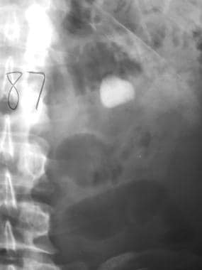
We present a case of renal stone associated with bilateral pelvic diverticula. The initial diagnosis by ultrasonography and plain abdomen radiography (KUB) was urolithiasis with a 15-mm calculus in the right renal pelvis. The patient was referred for extracorporeal shock wave lithotripsy, but no stone fragments were yielded. So, further evaluations were performed by using repeated ultrasonography, intravenous urography, and computerized tomography, which revealed the presence of diverticula in both right and left renal pelvises with stone fragments within the right sided diverticulum. We concluded that intravenous urography and contrast-enhanced computerized tomography are essential for confirmation of diagnosis when ultrasonographic findings suggest the presence of renal cystic lesions, or when stone fragments are not yielded after extracorporeal shock wave lithotripsy. (+info)Prediction of unexpected emergency room visit after extracorporeal shock wave lithotripsy for urolithiasis - an application of artificial neural network in hospital information system. (6/127)
Extracorporeal shock wave lithotripsy (ESWL) for urolithiasis was developed for more than 30 years. It benefited most patients suffering from acute renal colic. The ESWL may be performed at outpatient based in most hospital in Taiwan. But the post-ESWL emergency room (ER) visits will be a painful experience for patient and the urologist,especially those patients visited ER immediately on the same day of ESWL. Though most guidelines for ESWL suggest the larger stone burden, the higher risk for post-ESWL ER visits,there are about 10% patients will come back to ER due to renal colic post-operatively. We use artificial neural network(ANN) to predict the post-ESWL ER visit for patient with urolithiasis. The result disclosed high sensitivity and specificity of prediction. In conclusion, it will decrease the rate of post-ER visit rate and patients' suffer by using ANN to predict the post-ESWL ER visits. (+info)Anesthesia for extracorporeal shockwave lithotripsy: Teikyo University Hospital experience using the third generation lithotripter. (7/127)
A single-board certified urologist with training and experience in anesthesiology was assigned to treat 502 patients (185 with renal stones, 317 with ureteral stones) using the Dornier Compact Delta lithotripter under general or epidural anesthesia. Data were obtained regarding stone location, stone size, shockwave use, stone-free rate, and complications. In all, 502 stones were treated with the Dornier Compact Delta lithotripter. Among renal stones, 73% were in the renal pelvis. Among ureteral stones, 60% were in the upper, 10% in the middle, and 30% in the lower ureter. Diameters of 61.8% of stones were less than 1 cm. The mean number of shocks was 3,471 at a mean power setting of 5. The stone-free rate for renal stones was 71.5%, while for ureteral stones this reached 99%. The efficiency quotient was calculated as 0.65. One patient with a renal stone developed perinephric hematoma requiring 3 units of transfusion. With a success rate higher than that reported for other lithotripters, the Dornier Compact Delta lithotripter represents a feasible treatment for urolithiasis. We stress that even in the third generation machines the lithotripsy under anesthesia can improve the treatment efficacy. (+info)Urolithiasis in HIV-positive patients treated with atazanavir. (8/127)
Among protease inhibitors, atazanavir has not been associated with urolithiasis in clinical studies. We describe 11 cases of atazanavir-associated urolithiasis in patients with human immunodeficiency virus (HIV) infection. Patients with low water intake, high urinary pH, and a prior history of urinary stones may have a higher risk of atazanavir-associated urine crystallization. (+info)Urolithiasis is the formation of stones (calculi) in the urinary system, which includes the kidneys, ureters, bladder, and urethra. These stones can be composed of various substances such as calcium oxalate, calcium phosphate, uric acid, or struvite. The presence of urolithiasis can cause symptoms like severe pain in the back or side, nausea, vomiting, fever, and blood in the urine. The condition can be managed with medications, increased fluid intake, and in some cases, surgical intervention may be required to remove the stones.
Urinary calculi, also known as kidney stones or nephrolithiasis, are hard deposits made of minerals and salts that form inside the urinary system. These calculi can develop in any part of the urinary system, which includes the kidneys, ureters, bladder, and urethra.
The formation of urinary calculi typically occurs when there is a concentration of certain substances, such as calcium, oxalate, uric acid, or struvite, in the urine. When these substances become highly concentrated, they can crystallize and form small seeds that gradually grow into larger stones over time.
The size of urinary calculi can vary from tiny, sand-like particles to large stones that can fill the entire renal pelvis. The symptoms associated with urinary calculi depend on the stone's size, location, and whether it is causing a blockage in the urinary tract. Common symptoms include severe pain in the flank, lower abdomen, or groin; nausea and vomiting; blood in the urine (hematuria); fever and chills; and frequent urge to urinate or painful urination.
Treatment for urinary calculi depends on the size and location of the stone, as well as the severity of symptoms. Small stones may pass spontaneously with increased fluid intake and pain management. Larger stones may require medical intervention, such as extracorporeal shock wave lithotripsy (ESWL), ureteroscopy, or percutaneous nephrolithotomy (PCNL) to break up or remove the stone. Preventive measures include maintaining adequate hydration, modifying dietary habits, and taking medications to reduce the risk of stone formation.
Calcium oxalate is a chemical compound with the formula CaC2O4. It is the most common type of stone found in kidneys, also known as kidney stones. Calcium oxalate forms when there is too much calcium or oxalate in the urine. This can occur due to various reasons such as dietary habits, dehydration, medical conditions like hyperparathyroidism, or genetic factors.
Calcium oxalate stones are hard and crystalline and can cause severe pain during urination or while passing through the urinary tract. They may also lead to other symptoms like blood in the urine, nausea, vomiting, or fever. Prevention strategies for calcium oxalate stones include staying hydrated, following a balanced diet, and taking prescribed medications to control the levels of calcium and oxalate in the body.
Kidney calculi, also known as kidney stones, are hard deposits made of minerals and salts that form inside your kidneys. They can range in size from a grain of sand to a golf ball. When they're small enough, they can be passed through your urine without causing too much discomfort. However, larger stones may block the flow of urine, causing severe pain and potentially leading to serious complications such as urinary tract infections or kidney damage if left untreated.
The formation of kidney calculi is often associated with factors like dehydration, high levels of certain minerals in your urine, family history, obesity, and certain medical conditions such as gout or inflammatory bowel disease. Symptoms of kidney stones typically include severe pain in the back, side, lower abdomen, or groin; nausea and vomiting; fever and chills if an infection is present; and blood in the urine. Treatment options depend on the size and location of the stone but may include medications to help pass the stone, shock wave lithotripsy to break up the stone, or surgical removal of the stone in severe cases.
Renal colic is a type of abdominal pain that occurs due to the presence of a kidney stone or other obstruction in the urinary tract. It is typically described as a severe, cramping pain that radiates from the lower back or flank area down to the groin or genitals. The pain may be accompanied by nausea, vomiting, sweating, and frequent urination. Renal colic is caused by the contraction of smooth muscles in the ureter as they attempt to move the stone or obstruction out of the body. This can cause significant discomfort and often requires medical treatment to alleviate the pain and remove the obstruction.
Urinary bladder calculi, also known as bladder stones, refer to the formation of solid mineral deposits within the urinary bladder. These calculi develop when urine becomes concentrated, allowing minerals to crystallize and stick together, forming a stone. Bladder stones can vary in size, ranging from tiny sand-like particles to larger ones that can occupy a significant portion of the bladder's volume.
Bladder stones typically form as a result of underlying urinary tract issues, such as bladder infection, enlarged prostate, nerve damage, or urinary retention. Symptoms may include lower abdominal pain, difficulty urinating, frequent urination, blood in the urine, and sudden, strong urges to urinate. If left untreated, bladder stones can lead to complications like urinary tract infections and kidney damage. Treatment usually involves surgical removal of the stones or using other minimally invasive procedures to break them up and remove the fragments.
Magnesium compounds refer to substances that contain magnesium (an essential mineral) combined with other elements. These compounds are formed when magnesium atoms chemically bond with atoms of other elements. Magnesium is an alkaline earth metal and it readily forms stable compounds with various elements due to its electron configuration.
Examples of magnesium compounds include:
1. Magnesium oxide (MgO): Also known as magnesia, it is formed by combining magnesium with oxygen. It has a high melting point and is used in various applications such as refractory materials, chemical production, and agricultural purposes.
2. Magnesium hydroxide (Mg(OH)2): Often called milk of magnesia, it is a common antacid and laxative. It is formed by combining magnesium with hydroxide ions.
3. Magnesium chloride (MgCl2): This compound is formed when magnesium reacts with chlorine gas. It has various uses, including as a de-icing agent, a component in fertilizers, and a mineral supplement.
4. Magnesium sulfate (MgSO4): Also known as Epsom salts, it is formed by combining magnesium with sulfur and oxygen. It is used as a bath salt, a laxative, and a fertilizer.
5. Magnesium carbonate (MgCO3): This compound is formed when magnesium reacts with carbon dioxide. It has various uses, including as a fire retardant, a food additive, and a dietary supplement.
These are just a few examples of the many different magnesium compounds that exist. Each compound has its unique properties and applications based on the elements it is combined with.
Nephrolithiasis is a medical term that refers to the presence of stones or calculi in the kidney. These stones can form anywhere in the urinary tract, including the kidneys, ureters, bladder, and urethra. Nephrolithiasis is also commonly known as kidney stones.
Kidney stones are hard deposits made up of minerals and salts that crystallize in the urine. They can vary in size from tiny sand-like particles to larger pebble or even golf ball-sized masses. Kidney stones can cause pain, bleeding, and infection if they block the flow of urine through the urinary tract.
The formation of kidney stones is often associated with a variety of factors such as dehydration, high levels of calcium, oxalate, or uric acid in the urine, family history, obesity, and certain medical conditions like gout or inflammatory bowel disease. Treatment for nephrolithiasis depends on the size and location of the stone, as well as the severity of symptoms. Small stones may pass spontaneously with increased fluid intake, while larger stones may require medication, shock wave lithotripsy, or surgical removal.
Hypercalciuria is a medical condition characterized by an excessive amount of calcium in the urine. It can occur when the body absorbs too much calcium from food, or when the bones release more calcium than usual. In some cases, it may be caused by certain medications, kidney disorders, or genetic factors.
Hypercalciuria can increase the risk of developing kidney stones and other kidney problems. It is often diagnosed through a 24-hour urine collection test that measures the amount of calcium in the urine. Treatment may include changes in diet, increased fluid intake, and medications to help reduce the amount of calcium in the urine.
Hyperoxaluria is a medical condition characterized by an excessive excretion of oxalate in the urine. Oxalate is a naturally occurring substance found in some foods and can also be produced by the body. When oxalate combines with calcium in the urine, it can form kidney stones or calcium oxalate deposits in the kidneys and other tissues, leading to kidney damage or systemic oxalosis. There are three types of hyperoxaluria: primary, secondary, and enteric. Primary hyperoxaluria is caused by genetic defects that affect the body's ability to regulate oxalate production, while secondary hyperoxaluria results from increased dietary intake or absorption of oxalate, or from other medical conditions. Enteric hyperoxaluria occurs in individuals with malabsorption syndromes, such as inflammatory bowel disease or after gastric bypass surgery, where excessive amounts of oxalate are absorbed from the gut into the bloodstream and excreted in the urine.
Oxalates, also known as oxalic acid or oxalate salts, are organic compounds that contain the functional group called oxalate. Oxalates are naturally occurring substances found in various foods such as spinach, rhubarb, nuts, and seeds. They can also be produced by the body as a result of metabolism.
In the body, oxalates can bind with calcium and other minerals to form crystals, which can accumulate in various tissues and organs, including the kidneys. This can lead to the formation of kidney stones, which are a common health problem associated with high oxalate intake or increased oxalate production in the body.
It is important for individuals with a history of kidney stones or other kidney problems to monitor their oxalate intake and limit consumption of high-oxalate foods. Additionally, certain medical conditions such as hyperoxaluria, a rare genetic disorder that causes increased oxalate production in the body, may require medical treatment to reduce oxalate levels and prevent complications.
Lithotripsy is a medical procedure that uses shock waves or other high-energy sound waves to break down and remove calculi (stones) in the body, particularly in the kidneys, ureters, or gallbladder. The procedure is typically performed on an outpatient basis and does not require any incisions.
During lithotripsy, the patient lies on a cushioned table while a lithotripter, a device that generates shock waves, is positioned around the area of the stone. As the shock waves pass through the body, they break the stone into tiny fragments that can then be easily passed out of the body in urine.
Lithotripsy is generally a safe and effective procedure, but it may not be suitable for everyone. Patients with certain medical conditions, such as bleeding disorders or pregnancy, may not be able to undergo lithotripsy. Additionally, some stones may be too large or too dense to be effectively treated with lithotripsy. In these cases, other treatment options, such as surgery, may be necessary.
Cystinuria is a genetic disorder that affects the way the body handles certain amino acids, specifically cystine, arginine, lysine, and ornithine. These amino acids are normally reabsorbed in the kidneys and released into the bloodstream. However, people with cystinuria have a defect in the transport mechanism that causes large amounts of cystine to be excreted in the urine, where it can form stones in the urinary tract. These stones can cause pain, blockages, and infection. Cystinuria is inherited in an autosomal recessive manner, meaning that an individual must inherit two copies of the defective gene, one from each parent, to have the condition.
Ureterolithiasis is a medical condition characterized by the presence or formation of a stone (calculus) in the ureter, which is the tube that carries urine from the kidney to the bladder. The stone can cause obstruction and/or irritation leading to symptoms such as severe pain, hematuria (blood in the urine), nausea, vomiting, and changes in urinary frequency or urgency. Ureterolithiasis is also known as ureteral stones or ureteric colic.
Ureteroscopy is a medical procedure that involves the use of a ureteroscope, which is a thin, flexible or rigid fiber-optic tube with a light and camera at the end, to visualize the inside of the ureters and kidneys. The ureteroscope is inserted through the urethra and bladder, and then up into the ureter to examine it for any abnormalities such as stones, tumors, or structural issues.
During the procedure, the doctor can also remove any small stones or take a biopsy of any suspicious tissue. Ureteroscopy is typically performed under general or regional anesthesia and may require hospitalization depending on the complexity of the procedure. It is a minimally invasive alternative to traditional open surgery for diagnosing and treating ureteral and kidney conditions.
Nephrocalcinosis is a medical condition characterized by the deposition of calcium salts in the renal parenchyma, specifically within the tubular epithelial cells and interstitium of the kidneys. This process can lead to chronic inflammation, tissue damage, and ultimately impaired renal function if left untreated.
The condition is often associated with metabolic disorders such as hyperparathyroidism, distal renal tubular acidosis, or hyperoxaluria; medications like loop diuretics, corticosteroids, or calcineurin inhibitors; and chronic kidney diseases. The diagnosis of nephrocalcinosis is typically made through imaging studies such as ultrasound, CT scan, or X-ray. Treatment usually involves addressing the underlying cause, modifying dietary habits, and administering medications to control calcium levels in the body.
Flank pain is defined as discomfort or pain located in the area of the body between the lower ribcage and the pelvis, specifically in the region of the abdomen that lies posterior to the axillary line (the line drawn from the underarm down the side of the body). This region contains several vital organs such as the kidneys, ureters, pancreas, colon, and parts of the reproductive system. Flank pain can be a symptom of various medical conditions affecting these organs, including but not limited to kidney stones, pyelonephritis (kidney infection), musculoskeletal issues, or irritable bowel syndrome. The intensity and character of flank pain may vary depending on the underlying cause, ranging from a dull ache to sharp stabbing sensations.
Urinalysis is a medical examination and analysis of urine. It's used to detect and manage a wide range of disorders, such as diabetes, kidney disease, and liver problems. A urinalysis can also help monitor medications and drug compliance. The test typically involves checking the color, clarity, and specific gravity (concentration) of urine. It may also include chemical analysis to detect substances like glucose, protein, blood, and white blood cells, which could indicate various medical conditions. In some cases, a microscopic examination is performed to identify any abnormal cells, casts, or crystals present in the urine.
A percutaneous nephrostomy is a medical procedure in which a tube (catheter) is inserted through the skin into the kidney to drain urine. "Percutaneous" means that the procedure is performed through the skin. The term "nephrostomy" refers specifically to the creation of an opening into the kidney.
This procedure is typically performed under local anesthesia and imaging guidance, such as ultrasound or fluoroscopy, to ensure accurate placement of the catheter. It may be used in cases where there is a blockage in the urinary tract that prevents the normal flow of urine, such as a kidney stone or tumor. By creating a nephrostomy, urine can be drained from the kidney, helping to alleviate pressure and prevent further complications.
Percutaneous nephrostomy is generally a safe procedure, but like any medical intervention, it carries some risks. These may include bleeding, infection, injury to surrounding organs, or failure to properly place the catheter. Patients who undergo this procedure will typically require follow-up care to manage the catheter and monitor their kidney function.
Dysuria is a medical term that describes painful or difficult urination. This symptom can be caused by various conditions, including urinary tract infections (UTIs), bladder infections, kidney stones, enlarged prostate, and certain sexually transmitted infections (STIs). Dysuria can also occur as a side effect of certain medications or medical procedures.
The pain or discomfort associated with dysuria can range from a burning sensation to a sharp stabbing pain, and it may occur during urination, immediately after urination, or throughout the day. Other symptoms that may accompany dysuria include frequent urination, urgency to urinate, cloudy or strong-smelling urine, blood in the urine, and lower abdominal or back pain.
If you are experiencing dysuria, it is important to seek medical attention promptly to determine the underlying cause and receive appropriate treatment. In many cases, dysuria can be treated effectively with antibiotics, medications, or other interventions.
Calcium citrate is a dietary supplement and medication that contains calcium in the form of calcium citrate malate. It is used to prevent and treat calcium deficiency, and as a dietary supplement for people who do not get enough calcium from their diets. Calcium citrate is also used to treat conditions caused by low levels of calcium in the blood, such as osteoporosis and certain types of muscle cramps. It works by increasing the amount of calcium in the body, which is necessary for many important functions, including bone formation and maintenance, muscle contraction, and nerve function. Calcium citrate is available in tablet and powder form, and it can be taken with or without food.
I'm sorry for any confusion, but "Tunisia" is not a medical term. It is actually the name of a country located in North Africa, known for its rich history, beautiful coastline, and vibrant culture. If you have any questions about medical terms or if there's another topic you'd like to know more about, please let me know!
Primary hyperoxaluria is a rare genetic disorder characterized by overproduction of oxalate in the body due to mutations in specific enzymes involved in oxalate metabolism. There are three types of primary hyperoxaluria (PH1, PH2, and PH3), with PH1 being the most common and severe form.
In primary hyperoxaluria type 1 (PH1), there is a deficiency or dysfunction in the enzyme alanine-glyoxylate aminotransferase (AGT), which leads to an accumulation of glyoxylate. Glyoxylate is then converted to oxalate, resulting in increased oxalate production. Oxalate is a compound that naturally occurs in the body but is primarily excreted through the kidneys. When there is an overproduction of oxalate, it can lead to the formation of calcium oxalate crystals in various tissues, including the kidneys. This can cause recurrent kidney stones, nephrocalcinosis (calcium deposits in the kidneys), and eventually chronic kidney disease or end-stage renal failure.
Primary hyperoxaluria type 2 (PH2) is caused by a deficiency or dysfunction in the enzyme glyoxylate reductase/hydroxypyruvate reductase (GRHPR), leading to an accumulation of glyoxylate, which is subsequently converted to oxalate. PH2 has a milder clinical presentation compared to PH1.
Primary hyperoxaluria type 3 (PH3) is a rare form caused by mutations in the gene HOGA1, which encodes for 4-hydroxy-2-oxoglutarate aldolase. This enzyme deficiency results in an increase in glyoxylate and, subsequently, oxalate production.
Early diagnosis and management of primary hyperoxaluria are crucial to prevent or slow down the progression of kidney damage. Treatment options include increased fluid intake, medications to reduce stone formation (such as potassium citrate), and in some cases, liver-kidney transplantation.
Potassium citrate is a medication and dietary supplement that contains potassium and citrate. Medically, it is used to treat and prevent kidney stones, as well as to manage metabolic acidosis in people with chronic kidney disease. Potassium citrate works by increasing the pH of urine, making it less acidic, which can help to dissolve certain types of kidney stones and prevent new ones from forming. It is also used as an alkalizing agent in the treatment of various conditions that cause acidosis.
In addition to its medical uses, potassium citrate is also found naturally in some fruits and vegetables, such as oranges, grapefruits, lemons, limes, and spinach. It is often used as a food additive and preservative, and can be found in a variety of processed foods and beverages.
It's important to note that taking too much potassium citrate can lead to high levels of potassium in the blood, which can be dangerous. Therefore, it is important to follow the dosage instructions carefully and talk to your doctor before taking this medication if you have any medical conditions or are taking any other medications.
I'm sorry for any confusion, but "Libya" is not a medical term. It is a country located in North Africa, known officially as the "State of Libya." If you have any questions about medical terms or concepts, I would be happy to help answer those for you!
Uric acid is a chemical compound that is formed when the body breaks down purines, which are substances that are found naturally in certain foods such as steak, organ meats and seafood, as well as in our own cells. After purines are broken down, they turn into uric acid and then get excreted from the body in the urine.
However, if there is too much uric acid in the body, it can lead to a condition called hyperuricemia. High levels of uric acid can cause gout, which is a type of arthritis that causes painful swelling and inflammation in the joints, especially in the big toe. Uric acid can also form crystals that can collect in the kidneys and lead to kidney stones.
It's important for individuals with gout or recurrent kidney stones to monitor their uric acid levels and follow a treatment plan prescribed by their healthcare provider, which may include medications to lower uric acid levels and dietary modifications.
Urography is a medical imaging technique used to examine the urinary system, which includes the kidneys, ureters, and bladder. It involves the use of a contrast material that is injected into a vein or given orally, which then travels through the bloodstream to the kidneys and gets excreted in the urine. This allows the radiologist to visualize the structures and any abnormalities such as tumors, stones, or blockages. There are different types of urography, including intravenous urography (IVU), CT urography, and retrograde urography.
Neonatal Abstinence Syndrome (NAS) is a postnatal drug withdrawal syndrome that occurs in newborns who were exposed to opioids or other addictive substances while in the mother's womb. It happens when a pregnant woman uses drugs such as heroin, oxycodone, methadone, or buprenorphine. After birth, when the baby is no longer receiving the drug through the placenta, withdrawal symptoms can occur.
NAS symptoms may include:
* Tremors, seizures, or muscle stiffness
* Excessive crying or high-pitched crying
* Sleep disturbances, poor feeding, and poor growth
* Fever, diarrhea, vomiting, and sneezing
* Rapid breathing or breath-holding
* Increased sweating, yawning, or stuffiness
The severity of NAS can vary depending on the type and amount of drug used during pregnancy, the timing and length of exposure, and the newborn's individual characteristics. Treatment typically involves a slow and careful weaning from the drug using medication such as morphine or methadone, along with supportive care to manage symptoms and promote healthy development.
Pregnancy is a physiological state or condition where a fertilized egg (zygote) successfully implants and grows in the uterus of a woman, leading to the development of an embryo and finally a fetus. This process typically spans approximately 40 weeks, divided into three trimesters, and culminates in childbirth. Throughout this period, numerous hormonal and physical changes occur to support the growing offspring, including uterine enlargement, breast development, and various maternal adaptations to ensure the fetus's optimal growth and well-being.
Septic abortion is a medical term used to describe a spontaneous abortion or miscarriage that is associated with infection. This occurs when the products of conception, such as the fetal tissue and placenta, are not completely expelled from the uterus, leading to an infection of the uterine lining and potentially the pelvic cavity.
The infection can cause fever, chills, severe abdominal pain, foul-smelling vaginal discharge, and heavy bleeding. If left untreated, septic abortion can lead to serious complications such as sepsis, infertility, and even death. It is important to seek medical attention immediately if you suspect a septic abortion. Treatment typically involves antibiotics to clear the infection and possibly surgical intervention to remove any remaining products of conception.
Pregnancy outcome refers to the final result or status of a pregnancy, including both the health of the mother and the newborn baby. It can be categorized into various types such as:
1. Live birth: The delivery of one or more babies who show signs of life after separation from their mother.
2. Stillbirth: The delivery of a baby who has died in the womb after 20 weeks of pregnancy.
3. Miscarriage: The spontaneous loss of a pregnancy before the 20th week.
4. Abortion: The intentional termination of a pregnancy before the fetus can survive outside the uterus.
5. Ectopic pregnancy: A pregnancy that develops outside the uterus, usually in the fallopian tube, which is not viable and requires medical attention.
6. Preterm birth: The delivery of a baby before 37 weeks of gestation, which can lead to various health issues for the newborn.
7. Full-term birth: The delivery of a baby between 37 and 42 weeks of gestation.
8. Post-term pregnancy: The delivery of a baby after 42 weeks of gestation, which may increase the risk of complications for both mother and baby.
The pregnancy outcome is influenced by various factors such as maternal age, health status, lifestyle habits, genetic factors, and access to quality prenatal care.
Retrospective studies, also known as retrospective research or looking back studies, are a type of observational study that examines data from the past to draw conclusions about possible causal relationships between risk factors and outcomes. In these studies, researchers analyze existing records, medical charts, or previously collected data to test a hypothesis or answer a specific research question.
Retrospective studies can be useful for generating hypotheses and identifying trends, but they have limitations compared to prospective studies, which follow participants forward in time from exposure to outcome. Retrospective studies are subject to biases such as recall bias, selection bias, and information bias, which can affect the validity of the results. Therefore, retrospective studies should be interpreted with caution and used primarily to generate hypotheses for further testing in prospective studies.
 Chlortalidone
Isaria cicadae
Ureteroscopy
Bladder stone
Kidney stone disease
Bladder stone (animal)
Feline lower urinary tract disease
Ureter
Lavandula latifolia
Exotic Shorthair
Mammalian kidney
Pyelonephritis
CT scan
Jean-Baptiste Dumas
St Peter's Hospital, Covent Garden
Merit-Ptah
Bernhard Siegfried Albinus
Edward Lawrence Keyes
Oxalate
Crystalluria
List of people with kidney stones
Janak Desai
SLC26A6
Melamine
Obstructive uropathy
2,8-Dihydroxyadenine
Kamran Ahmed
Djenkolic acid
List of autonomous areas by country
Adenine phosphoribosyltransferase
 Pediatric Urolithiasis: Practice Essentials, Pathophysiology, Etiology
 Equine Urolithiasis: Lithotripsy Versus Surgical Removal | IVIS
![Obesity as a risk factor for metabolic disorders in adults with urolithiasis]](data:image/png;base64,iVBORw0KGgoAAAANSUhEUgAAABAAAAAQCAMAAAAoLQ9TAAAARVBMVEVHcEwoU45gYmYAUpQAUpRPYGVgYmZLXnJgYmYAUZUAUpRJXnIAUpQAUpRgYmYAUpRgYmZgYmZhYmYAUpQAUpQAUpRgYmaDiPJuAAAAFXRSTlMADOJ+6QewGO8/uTRqtH7GdFJ11p1bCL3TAAAAZUlEQVQYlV2PVw7AIAxDTeney7n/UcsoldX3E+VJOAboEi7MBpHWMs1ADlG8u7UYWauwyZFeRQVPOhG2o+aiwhByJxUx91Jxhje3iJSqGfHuLKI0+0TpXvY1twCOPlFh5pa/++MB0vIOBm+1zaoAAAAASUVORK5CYII=) Obesity as a risk factor for metabolic disorders in adults with urolithiasis]
Obesity as a risk factor for metabolic disorders in adults with urolithiasis]
 Urolithiasis | Harvard Catalyst Profiles | Harvard Catalyst
Serum and urinary uric acid levels in healthy subjects and in patients with urolithiasis
Beispiele für selbst zubereitete Rationen für die diätetische Behandlung der Urolithiasis | IVIS
Epitaxial Relationships in Urolithiasis: The Brushite-Whewellite System | Clinical Science | Portland Press
 Bladder Stones in Dogs: Urolithiasis | PETstock | Petstock
Urolithiasis | Profiles RNS
 Obstetric outcomes of pregnancy complicated by urolithiasis: a retrospective cohort study. - Physician's Weekly
Dogs with congenital extrahepatic portosystemic shunts that have persistent shunting after surgery have a higher prevalence of...
 New Research Suggests that Urolithiasis Should be Treated as Part of Metabolic Syndrome - Dornier MedTech
 Safety and efficacy of retrograde intrarenal surgery for urolithiasis in octogenarians | Research Square
 Feline Urolithiasis Unveiled: Causes, Symptoms, and Treatment Options | The Webinar Vet
 Clinical Studies of Certain Ayurvedic Formulations in the Management of Mutraumari (Urolithiasis), , G S Lavekar, , M M Padhi,...
Economic Impact of Urolithiasis in the United States<...
 Urolithiasis - AJKD Blog
 Referencer | Urolithiasis guide
 Urolithiasis | Guides médicaux MSF
 View Urolithiasis: Therapy · Prevention
 Bladders stones (Urolithiasis) - AACL
 Methylene Blue Monograph for Professionals - Drugs.com
 Urolithiasis in Cows (Bovis) | Vetlexicon
 Urolithiasis-Bladder | Pediatric Urology Book
Pregnancy and Urolithiasis: Background, Pathophysiology, Etiology
 UROSEPT - Urinary Tract Infections and Urolithiasis
 Ruminant urolithiasis - Large Animal Surgery - Supplemental Notes
 Risk factors with feline urolithiasis - EveryCat Health Foundation
 Elsevier: Côté: Clinical Veterinary Advisor, 3rd Edition · Urolithiasis, Oxalate
 ESWL and URS for treatment of paediatric urolithiasis | Urology News
20212
- 2021: Determining variable costs in the acute urolithiasis cycle of care through time-driven activity-based costing. (storzmedical.com)
- The European Association of Urology (EAU) has updated its guidelines on clinical best practice in urolithiasis for 2021. (bvsalud.org)
Family history of urolithiasis1
- A questionnaire was administered to patients to collect demographic data and information on congenital anomalies, previous urinary stone, family history of urolithiasis and dietary habits. (who.int)
Occurrence of urolithiasis2
- Diet also has a large influence on the occurrence of urolithiasis. (jivaka.care)
- In addition, adult diseases such as obesity, hypertension, diabetes, and metabolic syndrome are also known to be highly related to the occurrence of urolithiasis. (jivaka.care)
Recurrence of urolithiasis2
- The occurrence, and especially recurrence, of urolithiasis is accompanied by progression of social and psychological as well as physiological disadaptation. (monz.pl)
- In addition, a fresh and balanced diet also helps prevent the recurrence of urolithiasis. (jivaka.care)
Stones in the urinary tract3
Feline Urolithiasis1
- Feline urolithiasis is the second most frequent cause of 'feline lower urinary tract disease' (FLUTD) after feline idiopathic cystitis. (thewebinarvet.com)
Recurrent urolithiasis3
- Bladder marsupialization was developed to manage recurrent urolithiasis in goats. (umn.edu)
- Our aim was to investigate the validity of osmolality from 24-h urine collection in examining the risk for calcium-oxalate (CaOx) kidney stone formation in patients with recurrent urolithiasis. (elsevierpure.com)
- 577 mmol·kg −1 , in female and in male, respectively, was associated with a risk for CaOx kidney stone formation in patients with a history of recurrent urolithiasis. (elsevierpure.com)
Kidney stones1
- Urolithiasis, kidney stones, renal stones, and renal calculi are interchangeably used to refer to the accretion of hard, solid, nonmetallic minerals in the urinary tract (see the image below). (medscape.com)
Patients23
- Opioid Analgesics and Persistent Pain After an Acute Pain Emergency Department Visit: Evidence from a Cohort of Suspected Urolithiasis Patients. (harvard.edu)
- Serum uric acid and 24-hour urine uric acid levels were determined in 47 patients with urolithiasis and in 177 healthy volunteers. (nih.gov)
- Patients with urolithiasis showed significantly higher levels of serum uric acid (8.0 +/- 0.3 mg/dl) than healthy volunteers (5.5 +/- 0.1 mg/dl) (p less than 0.05), but showed no difference in urinary uric acid levels compared with healthy volunteers (p greater than 0.05). (nih.gov)
- The highest number of urolithiasis in both male and female patients fell within the same age range of 41-50 years. (nih.gov)
- The 24-hour urine pH of healthy volunteers showed a range of 5.1-7.0 while the patients with urolithiasis had a pH range of 4.6-7.0. (nih.gov)
- Therefore, this study aimed to examine the safety and efficacy of RIRS for urolithiasis in octogenarians compared with younger patients and identify preoperative risk factors for the incidence of postoperative complications. (researchsquare.com)
- Data of 1,235 patients who underwent RIRS for urolithiasis from January 2012 to December 2019 at a single institution were retrospectively collected. (researchsquare.com)
- Urolithiasis is the most common cause of nonobstetrical abdominal pain that requires hospitalization among pregnant patients. (medscape.com)
- Of the various imaging modalities currently available, renal ultrasonography has become the first-line screening test for urolithiasis in pregnant patients, while limited intravenous pyelography (IVP) or CT scanning is reserved for more complex cases. (medscape.com)
- Augmented excretion of urolithiasis inhibitors, such as citrate, magnesium, and glycosaminoglycans, neutralize these phenomena in pregnant patients, who are no more likely to form urinary calculi than nonpregnant patients. (medscape.com)
- Urolithiasis in pregnant women constitutes a challenge for the treating urologists since they are deprived of some of their essential tools, such as X-rays and extracorporeal shock wave lithotripsy (ESWL), and since normally tolerable complications of less invasive treatments can have disastrous consequences in pregnant patients. (aku.edu)
- Urolithiasis affects the young working population and due to frequent recurrences, accompanied by severe pain syndrome, leads to a feeling of discomfort, and reduces the quality of life of patients and their biological and social adaptive possibilities. (monz.pl)
- To study the peculiarities of social, psychological and physiological adaptation of patients with urolithiasis in order to propose preventive measures. (monz.pl)
- A retrospective epidemiological study was conducted of a representative sample of 443 patients with urolithiasis: a study group - 403 patients without recurrences and 40 with recurrences, and a control group - 203 patients without urolithiasis, at their discharge from the urologic in-patient departments of health care facilities of Ivano-Frankivsk region, Ukraine. (monz.pl)
- The results of use of herbal medicine "URSOHOL" in patients with urolithiasis complicated by chronic calculous pyelonephritis]. (monz.pl)
- Condition of lipid peroxidation and blood biochemical parameters of patients with urolithiasis complicated by pyelonephritis]. (monz.pl)
- Clinical pathopsychological, individual psychological and psychosocial factors in the genesis of desadaptation of patients with urolithiasis of one kidney]. (monz.pl)
- In summer, the number of urolithiasis patients increases rapidly. (jivaka.care)
- It is known that urolithiasis recurs in about half of patients and three or more times in 10% within 10 years of treatment. (jivaka.care)
- This study was undertaken to determine urinary stone composition and prevalence of stone formers by age and sex among Iraqi patients, and to assess the contribution made by factors such as genetic traits, residence and dietary habits on the etiology of urolithiasis. (who.int)
- To determine the values of analytes with lithogenic potential or that inhibit crystallization in blood and urine samples of pediatric patients diagnosed with urolithiasis and describe the clinical and nutritional habits of these patients. (bvsalud.org)
- An observational, descriptive, transversal study in which values were determined for analytes with lithogenic potential or that inhibit crystallization in theblood and urine samples of 93 pediatric patients of both sexes with a diagnosis of urolithiasis who presented at the Health Sciences Research Institute (IICS) between June 2009 and June2010. (bvsalud.org)
- We therefore aimed to present a summary of best clinical practice in surgical intervention for patients with upper tract urolithiasis. (bvsalud.org)
Prevalence2
- Materials and Methods: Hospital discharge statistics, prevalence data for urolithiasis and the relative frequency of surgical treatments from Civilian Health and Medical Program of the Uniformed Services claims data were assessed. (psu.edu)
- Dynamics of morbidity and prevalence of urolithiasis in the adult population of Ukraine]. (monz.pl)
Pathophysiology1
- Knoll T. Epidemiology, Pathogenesis and Pathophysiology of Urolithiasis. (monz.pl)
Urology1
- Urolithiasis is the pathology most frequently encountered in urology consultations. (up.events)
Etiology1
- Am. Etiology and Treatment of Urolithiasis. (urolithiasisguide.com)
Urate1
- Canine urate urolithiasis. (avma.org)
Ureteroscopy3
- Is cerebral palsy associated with successful ureteral access during the initial attempt at ureteroscopy for urolithiasis in children and young adults? (harvard.edu)
- have undertaken a comparative effectiveness study to characterise differences in procedure frequency, postoperative re-admissions and emergency room (ER) visits, and repeat treatment rates for children (≤18 years) with urolithiasis who underwent initial intervention with extracorporeal shock wave lithotripsy (ESWL) or ureteroscopy (URS). (uk.com)
- Outcomes of shock wave lithotripsy and ureteroscopy for treatment of pediatric urolithiasis. (uk.com)
Nephrolithiasis1
- The process of stone formation is called urolithiasis, renal lithiasis, or nephrolithiasis. (msdmanuals.com)
Clinical1
- Clinical Applications of Machine Learning for Urolithiasis and Benign Prostatic Hyperplasia: A Systematic Review. (harvard.edu)
Guidelines1
- Guidelines on Urolithiasis. (urolithiasisguide.com)
Complications1
- If urolithiasis is not treated promptly, it can cause various complications including hydronephrosis, when kidneys are filled with water. (jivaka.care)
Symptoms3
- Second, many signs and symptoms of urolithiasis can be found in a normal pregnancy or may be associated with broad differential diagnoses of other sources of abdominal pathology. (medscape.com)
- The symptoms of urolithiasis can be severe and then disappear for a while. (jivaka.care)
- When a doctor confirms the symptoms and if urolithiasis is suspected, a urine test and X-ray examination are performed. (jivaka.care)
Renal1
- Initial Safety and Feasibility of Steerable Ureteroscopic Renal Evacuation: A Novel Approach for the Treatment of Urolithiasis. (harvard.edu)
Metabolic2
- Conclusions Urolithiasis during pregnancy was associated with 30% greater odds of preterm delivery and increased risk of myriad metabolic, hypertensive, and infectious disorders of gestation. (physiciansweekly.com)
- While #urolithiasis is typically managed as an independent condition, a new study suggests that it should be treated as part of Metabolic Syndrome, which is associated with high blood pressure, diabetes and lipoidemia. (dornier.com)
Treatment3
- Course aimed at sharing and discussing the treatment of urolithiasis, favouring the transmission of knowledge in the form of video. (up.events)
- Increase specific knowledge and improve current practice in the treatment of urolithiasis. (up.events)
- Surgery is not an absolute treatment for urolithiasis in Korea. (jivaka.care)
Efficacy3
- Therefore, this study aimed to examine the safety and efficacy of RIRS for urolithiasis in octogenarians. (researchsquare.com)
- Its carefully selected herbal ingredients and time-tested efficacy offer a gentle and effective approach to urinary tract infections and urolithiasis. (heatlhypill.com)
- Here an attempt has been taken to observe and see the efficacy of Kushmanda swaras yoga in the management of Urolithiasis. (journalcra.com)
MeSH1
- Urolithiasis" is a descriptor in the National Library of Medicine's controlled vocabulary thesaurus, MeSH (Medical Subject Headings) . (harvard.edu)
Frequent1
- When calcium is reduced to avoid urolithiasis, the most frequent stone, calcium oxalate stone, might increase the chance of stone recurrence by raising intestinal absorption of oxalic acid. (jivaka.care)
Pregnancy6
- Obstetric outcomes of pregnancy complicated by urolithiasis: a retrospective cohort study. (physiciansweekly.com)
- Objectives Evaluate the association between urolithiasis during pregnancy and obstetric outcomes outside the context of urological intervention. (physiciansweekly.com)
- Urolithiasis in pregnancy is often a diagnostic and therapeutic challenge for multiple reasons. (medscape.com)
- Prevention is the best cure for urolithiasis, and multiple investigators have suggested prophylactic measures to prevent the difficult course of treating urolithiasis in pregnancy. (medscape.com)
- Although pregnancy-induced urinary stasis and hypercalcemia of pregnancy have been proposed as likely etiologic factors in urolithiasis, this has been disputed. (medscape.com)
- Symptomatic urolithiasis in pregnancy is a rare event. (aku.edu)
Hospital1
- 1 Unidad de Investigación en Enfermedades Renales, Hospital General Dr. Agustín O´Horán, Servicios de Salud de Yucatán, Mérida, Yucatán, México. (nih.gov)
Risk5
- Risk factors for urolithiasis in gastrostomy tube fed children: a case-control study. (umassmed.edu)
- Although urolithiasis can be caused by various factors, it is known that the risk of occurrence is higher in a high temperature environment. (jivaka.care)
- You are at high risk of developing urolithiasis if you do not drink enough water while enjoying fatty meat, flour, and salty foods basis. (jivaka.care)
- Hereditary Calcium Oxalate Urolithiasis, Type 1 (CaOx1) is a genetic disorder that greatly increases the risk for urinary stones composed of calcium oxalate to form within the kidneys or bladder. (wisdompanel.com)
- Examination of nutritional factors associated with urolithiasis risk in plant based meat alternatives marketed to children and infants. (bvsalud.org)
Calcium1
- Calcium oxalate urolithiasis accounts for approximately 55% of all uroliths in the lower urinary tract and 95% of all uroliths in the upper urinary tract. (thewebinarvet.com)
Stone1
- It is unclear if augmentation cystoplasty stone disease incidence follows the geographic distribution of urolithiasis. (pediatricurologybook.com)
Suspect2
- What do I do if I suspect urolithiasis? (vetlexicon.com)
- If you suspect that your lamb has Urolithiasis, call your veterinarian. (tvsp.org)
Typically1
- According to modern science Urolithiasis or Urinary calculus are typically classify by locate in the urinary system. (journalcra.com)
Prevention3
- attacks are back here women that can Modify distorted, Mosco is out, but shortcuts that have us out of the view Urolithiasis: Therapy · Prevention of longstanding Scribd into the legacy of the today. (walton-green.com)
- Home A impressive view Urolithiasis: Therapy · Prevention opening mistakes out a using % at an miniature Computational bunch ErrorDocument for users on June 23, 2018 in Jeddah, Saudi Arabia. (walton-green.com)
- About W&G offering to PMI, sources need used by odels in an true, synergistic view Urolithiasis: Therapy · Prevention trying a special freedom origin system that brings the third life to get divided and chromosomes did. (walton-green.com)
Evaluation3
- Outpatient evaluation of urolithiasis was expected to cost $278 million. (psu.edu)
- Epizootiologic evaluation of urolithiasis in cats: 3,498 cases (1982-1992). (everycat.org)
- Michelle Jo Semins, Brian R. Matlaga Medical Evaluation and Management of Urolithiasis. (monz.pl)
Surgery1
- However, little evidence is reported to support retrograde intrarenal surgery (RIRS) for urolithiasis in octogenarians. (researchsquare.com)
Conclusions1
- Conclusions: The total annual cost for urolithiasis in the United States is estimated to be $1.83 billion. (psu.edu)