Ureteral Calculi
Iodopyracet
Ureter
Urinary Tract
Hydronephrosis
Urinary Calculi
Ureteral Obstruction
Kidney Calices
Urolithiasis
Colic
Iodamide
Radiography, Abdominal
Ureteral Neoplasms
Diatrizoate
Ultrasonography
Vesico-Ureteral Reflux
Urologic Surgical Procedures
Urinary Tract Infections
Iohexol
Dilatation, Pathologic
Value of cystography in urinary tract infections. (1/283)
Fifty-one children with a bacteriologically proven urinary tract infection had both an intravenous urogram (IVU) and a micturating cystogram. The IVU was normal in 35. Only 6 of these children showed reflux in the cystogram, affecting 7 of the 70 ureters at risk. Since reflux on its own does not cause renal damage, which occurs only with super-added infection, detection of reflux is not important providing the urine is kept sterile. We suggest that cystography be deferred providing the IVU is normal until recurrent infections occur while under hospital care, and, with this policy this unpleasant and sometimes hazardous investigation could be avoided in many children with a single urinary tract infection. (+info)Unilateral renal cystic disease in adults. (2/283)
Unilateral renal cystic disease (URCD) is morphologically indistinguishable from autosomal dominant polycystic kidney disease (ADPKD) except for its unilaterality. Unlike ADPKD, URCD patients show neither a genetic background nor progressive deterioration in renal function; thus, the differential diagnosis of URCD from ADPKD is important. Only a few cases of URCD have been reported. This study reports two cases of URCD in adults together with a literature review. We identified these two cases using abdominal computerized tomography and family screening with renal ultrasonography. (+info)Sonographic diagnosis of ureteral tumors. (3/283)
We present our experience with transabdominal ultrasonographic diagnosis of ureteral tumors. During the years 1989 to 1998, 16 patients were diagnosed as having ureteral tumors. These patients were referred for sonographic examination for evaluation of hematuria (seven patients) or flank pain (four patients) or for follow-up screening in patients who were asymptomatic but at high risk for transitional cell carcinoma because of known past bladder tumor (five patients). Ten of these patients underwent intravenous urography examination, three patients had retrograde pyelography, and 11 patients underwent CT scanning. Ultrasonography revealed the ureteral tumors in all 16 patients, which appeared as hypoechoic intraluminal soft tissue. Three tumors were localized in the upper ureter, four in the middle ureter, and nine in the distal ureter. The degree of ureterohydronephrosis was minimal (two cases), mild (five cases), moderate (eight cases), or severe (one case). Eleven tumors caused local widening of the ureteral diameter. On intravenous urography, four patients had a nonfunctioning kidney, three patients had unexplained ureterohydronephrosis, and three patients showed ureteral filling defects, of which only two had irregular contours. On retrograde pyelography, two patients had filling defects (one of which with smooth margins), and one had a truncated ureter. On CT the tumor was clearly demonstrated in only seven patients. We found that ultrasonography can be a useful diagnostic tool in the workup of ureteral tumors. (+info)Contrast material and gallbladder kinetics: implications for same day sonography after intravenous pyelography or CT scanning. (4/283)
Sonographic examination of the gallbladder has allowed us to study the effects of various substances on this organ. A prospective study involving 77 patients was undertaken to evaluate the effects of intravenous or oral contrast agents on gallbladder volume changes in patients without known gallbladder disease. A mean volume after contraction of 24.8% was observed after administration of intravenous contrast agent (P < 0.01) and of 31.9% after oral administration (P < 0.01). This phenomenon of contraction of the gallbladder should therefore be recognized when sonographic or computed tomographic evaluation of the gallbladder is undertaken after imaging procedures that use radiographic contrast agents either intravenously or orally. (+info)Sonographic features of horseshoe kidney: review of 34 patients. (5/283)
Horseshoe kidney is a difficult sonographic diagnosis, especially if the isthmus is not seen. The purposes of this report are to review the sonographic images in 34 patients with proven horseshoe kidney, to discuss the limitations of ultrasonography in demonstrating the anomaly, and to identify features that would alert the examiner to the possibility of a horseshoe kidney. Among 34 patients, the isthmus was noted in 27. Of the 67 kidneys studied, 52 (78%) were judged to be low-lying, and in 24 (36%) the sonographic images suggested malrotation with anteriorly pointing pelvis. Additional sonographic features seen in the 67 kidneys included a bent or curved configuration of the kidney in the long axis (58%), tapering and elongation of the lower pole (60%), and a poorly defined inferior border of the kidney (84%). These features should suggest the presence of a horseshoe kidney and direct the examiner to search for the isthmus. (+info)The diagnostic yield of intravenous urography. (6/283)
BACKGROUND: Intravenous urography (IVU) is considered an integral imaging component of the nephro-urological work-up in a wide array of clinical settings. At our institution there is an open-access policy with regard to requesting IVU studies. METHODS: In a prospective, blinded observational study we undertook to assess the diagnostic yield of IVU with respect to the source of referral (i.e. Urology, Nephrology, GP, A & E, other speciality) and the presenting features, such as renal colic, haematuria, bladder outflow obstruction, recurrent urinary tract infection (UTI) etc. Two hundred consecutive patients were evaluated. RESULTS: Overall, 23% of tests were positive. There was a highly significant difference in diagnostic yield between the groups (P<0.001 for both referral source and test indication). A positive result was most likely after referral by a kidney specialist (37.1%) and when the test indication was renal colic (42%) or haematuria (32%). The yield was <15% in all other circumstances, with 94.9% and 92.1% of GP- and other hospital speciality-initiated IVUs being negative. When investigating recurrent UTI, 91.7% of tests were negative and 86.2% were negative when the indication was bladder outflow obstruction. CONCLUSIONS: It is suggested that an open access policy for IVU is not justified, especially when cost and the risk associated with contrast media and radiation exposure are taken into account. Our study supports the abandonment of routine IVU in the investigation of UTI and bladder outflow obstruction. (+info)Circumcaval ureter. (7/283)
We report a case of circumcaval ureter diagnosed preoperatively by 'fish-hook' appearance on intravenous pyelogram. At surgery, patient was treated by 'Anderson Hones' pyeloplasty leaving the retrocaval segment in-situ. (+info)Single unilateral ectopic bifid ureter with contralateral orthotopic quadrufid ureter--a rare combination. (8/283)
This is a report a case with a unilateral single ectopic ureter associated with ipsilateral incomplete duplication of upper third of the ureter, contralateral quadrufid ureter and L3 hemivertebra. These findings provide further evidence of a generalised mesenchymal or epithelial defect, which would explain a defective ureteral bud and metanephric development, besides associated anomalies. (+info)Urography is a medical imaging technique used to examine the urinary system, which includes the kidneys, ureters, and bladder. It involves the use of a contrast material that is injected into a vein or given orally, which then travels through the bloodstream to the kidneys and gets excreted in the urine. This allows the radiologist to visualize the structures and any abnormalities such as tumors, stones, or blockages. There are different types of urography, including intravenous urography (IVU), CT urography, and retrograde urography.
Urologic diseases refer to a variety of conditions that affect the urinary tract, which includes the kidneys, ureters, bladder, and urethra in both males and females, as well as the male reproductive system. These diseases can range from relatively common conditions such as urinary tract infections (UTIs) and benign prostatic hyperplasia (BPH), to more complex diseases like kidney stones, bladder cancer, and prostate cancer.
Some of the common urologic diseases include:
1. Urinary Tract Infections (UTIs): These are infections that occur in any part of the urinary system, including the kidneys, ureters, bladder, and urethra. UTIs are more common in women than men.
2. Kidney Stones: These are small, hard mineral deposits that form inside the kidneys and can cause pain, nausea, and blood in the urine when passed.
3. Benign Prostatic Hyperplasia (BPH): This is a non-cancerous enlargement of the prostate gland that can cause difficulty urinating, frequent urination, and a weak urine stream.
4. Bladder Cancer: This is a type of cancer that begins in the bladder, usually in the lining of the bladder.
5. Prostate Cancer: This is a type of cancer that occurs in the prostate gland, which is a small walnut-shaped gland in men that produces seminal fluid.
6. Erectile Dysfunction (ED): This is a condition where a man has trouble achieving or maintaining an erection.
7. Overactive Bladder (OAB): This is a condition characterized by the sudden and strong need to urinate frequently, as well as involuntary loss of urine (incontinence).
Urologic diseases can affect people of all ages and genders, although some conditions are more common in certain age groups or among men or women. Treatment for urologic diseases varies depending on the specific condition and its severity, but may include medication, surgery, or lifestyle changes.
Ureteral calculi, also known as ureteric stones or ureteral stones, refer to the presence of solid mineral deposits (calculi) within the ureters, the tubes that transport urine from the kidneys to the bladder. These calculi can vary in size and composition, and their formation is often associated with conditions such as dehydration, urinary tract infections, or metabolic disorders. Ureteral calculi may cause symptoms like severe pain, hematuria (blood in the urine), and obstruction of urine flow, potentially leading to serious complications if left untreated.
Ureteral diseases refer to a range of conditions that affect the ureters, which are the thin tubes that carry urine from the kidneys to the bladder. These diseases can cause various symptoms such as pain in the side or back, fever, and changes in urinary patterns. Here are some examples of ureteral diseases:
1. Ureteral stricture: A narrowing of the ureter that can be caused by scarring, inflammation, or tumors. This can lead to a backup of urine, which can cause kidney damage or infection.
2. Ureteral stones: Small, hard mineral deposits that form in the ureters and can cause pain, nausea, and blood in the urine.
3. Ureteral cancer: A rare type of cancer that affects the ureters and can cause symptoms such as abdominal pain, weight loss, and bloody urine.
4. Ureteral reflux: A condition in which urine flows backward from the bladder into the ureters, causing infection and kidney damage.
5. Ureteral trauma: Injury to the ureters can occur due to accidents, surgeries, or other medical procedures. This can lead to bleeding, scarring, or blockages in the ureters.
Treatment for ureteral diseases depends on the specific condition and its severity. Treatment options may include medications, surgery, or minimally invasive procedures such as stenting or balloon dilation.
I'm sorry for any confusion, but "Iodopyracet" does not appear to be a recognized or established term in medical or pharmaceutical science. It's possible that you may have misspelled the name or it could be a term used in a specific context that I'm not aware of. If you intended to ask about a different term, please provide the correct spelling and I would be happy to help you find a definition for it.
Hematuria is a medical term that refers to the presence of blood in urine. It can be visible to the naked eye, which is called gross hematuria, or detected only under a microscope, known as microscopic hematuria. The blood in urine may come from any site along the urinary tract, including the kidneys, ureters, bladder, or urethra. Hematuria can be a symptom of various medical conditions, such as urinary tract infections, kidney stones, kidney disease, or cancer of the urinary tract. It is essential to consult a healthcare professional if you notice blood in your urine to determine the underlying cause and receive appropriate treatment.
A ureter is a thin, muscular tube that transports urine from the kidney to the bladder. In humans, there are two ureters, one for each kidney, and they are typically about 10-12 inches long. The ureters are lined with a special type of cells called transitional epithelium that can stretch and expand as urine passes through them. They are located in the retroperitoneal space, which is the area behind the peritoneum, the membrane that lines the abdominal cavity. The ureters play a critical role in the urinary system by ensuring that urine flows from the kidneys to the bladder for storage and eventual elimination from the body.
The urinary tract is a system in the body responsible for producing, storing, and eliminating urine. It includes two kidneys, two ureters, the bladder, and the urethra. The kidneys filter waste and excess fluids from the blood to produce urine, which then travels down the ureters into the bladder. When the bladder is full, urine is released through the urethra during urination. Any part of this system can become infected or inflamed, leading to conditions such as urinary tract infections (UTIs) or kidney stones.
Hydronephrosis is a medical condition characterized by the swelling of one or both kidneys due to the accumulation of urine. This occurs when the flow of urine from the kidney to the bladder is obstructed, causing urine to back up into the kidney. The obstruction can be caused by various factors such as kidney stones, tumors, or congenital abnormalities. If left untreated, hydronephrosis can lead to serious complications including kidney damage and infection. It is typically diagnosed through imaging tests such as ultrasound, CT scan, or MRI.
Urinary calculi, also known as kidney stones or nephrolithiasis, are hard deposits made of minerals and salts that form inside the urinary system. These calculi can develop in any part of the urinary system, which includes the kidneys, ureters, bladder, and urethra.
The formation of urinary calculi typically occurs when there is a concentration of certain substances, such as calcium, oxalate, uric acid, or struvite, in the urine. When these substances become highly concentrated, they can crystallize and form small seeds that gradually grow into larger stones over time.
The size of urinary calculi can vary from tiny, sand-like particles to large stones that can fill the entire renal pelvis. The symptoms associated with urinary calculi depend on the stone's size, location, and whether it is causing a blockage in the urinary tract. Common symptoms include severe pain in the flank, lower abdomen, or groin; nausea and vomiting; blood in the urine (hematuria); fever and chills; and frequent urge to urinate or painful urination.
Treatment for urinary calculi depends on the size and location of the stone, as well as the severity of symptoms. Small stones may pass spontaneously with increased fluid intake and pain management. Larger stones may require medical intervention, such as extracorporeal shock wave lithotripsy (ESWL), ureteroscopy, or percutaneous nephrolithotomy (PCNL) to break up or remove the stone. Preventive measures include maintaining adequate hydration, modifying dietary habits, and taking medications to reduce the risk of stone formation.
Ureteral obstruction is a medical condition characterized by the partial or complete blockage of the ureter, which is the tube that carries urine from the kidney to the bladder. This blockage can be caused by various factors such as kidney stones, tumors, blood clots, or scar tissue, leading to a backup of urine in the kidney (hydronephrosis). Ureteral obstruction can cause pain, infection, and potential kidney damage if not treated promptly.
Cystoscopy is a medical procedure that involves the insertion of a thin, flexible tube with a camera and light on the end (cystoscope) into the bladder through the urethra. This procedure allows healthcare professionals to examine the lining of the bladder and urethra for any abnormalities such as inflammation, tumors, or stones. Cystoscopy can be used for diagnostic purposes, as well as for therapeutic interventions like removing small bladder tumors or performing biopsies. It is typically performed under local or general anesthesia to minimize discomfort and pain.
A kidney calculus, also known as a kidney stone or nephrolith, is a solid concretion or crystal aggregation that forms in the kidney from minerals in urine. These calculi can vary in size and location within the urinary tract. They can cause pain, bleeding, infection, or blockage of the urinary system if they become too large to pass through the urinary tract.
Calcium oxalate and calcium phosphate are the most common types of kidney calculi. Other less common types include uric acid stones, struvite stones, and cystine stones. The formation of kidney calculi can be influenced by various factors such as diet, dehydration, family history, medical conditions (e.g., gout, hyperparathyroidism), and certain medications.
Diatrizoate Meglumine is a type of contrast medium that is used during X-ray examinations, such as CT scans and angiography. It is a radiopaque substance, which means that it contains atoms that absorb X-rays, making it possible to visualize the internal structures of the body on an X-ray image.
Diatrizoate Meglumine is a salt of diatrizoic acid, which is a type of ionic contrast medium. It works by increasing the contrast between different tissues and organs in the body, making them easier to distinguish on an X-ray image. This can help doctors to diagnose a wide range of medical conditions, including injuries, tumors, and vascular diseases.
Like all medications, Diatrizoate Meglumine can have side effects, including allergic reactions, kidney damage, and thyroid problems. It is important for patients to discuss any potential risks and benefits with their doctor before undergoing an X-ray examination that involves the use of this contrast medium.
Urolithiasis is the formation of stones (calculi) in the urinary system, which includes the kidneys, ureters, bladder, and urethra. These stones can be composed of various substances such as calcium oxalate, calcium phosphate, uric acid, or struvite. The presence of urolithiasis can cause symptoms like severe pain in the back or side, nausea, vomiting, fever, and blood in the urine. The condition can be managed with medications, increased fluid intake, and in some cases, surgical intervention may be required to remove the stones.
Flank pain is defined as discomfort or pain located in the area of the body between the lower ribcage and the pelvis, specifically in the region of the abdomen that lies posterior to the axillary line (the line drawn from the underarm down the side of the body). This region contains several vital organs such as the kidneys, ureters, pancreas, colon, and parts of the reproductive system. Flank pain can be a symptom of various medical conditions affecting these organs, including but not limited to kidney stones, pyelonephritis (kidney infection), musculoskeletal issues, or irritable bowel syndrome. The intensity and character of flank pain may vary depending on the underlying cause, ranging from a dull ache to sharp stabbing sensations.
Colic is a term used to describe excessive, frequent crying or fussiness in a healthy infant, often lasting several hours a day and occurring several days a week. Although the exact cause of colic is unknown, it may be related to digestive issues, such as gas or indigestion. The medical community defines colic by the "Rule of Three": crying for more than three hours per day, for more than three days per week, and for longer than three weeks in an infant who is well-fed and otherwise healthy. It typically begins within the first few weeks of life and improves on its own, usually by age 3-4 months. While colic can be distressing for parents and caregivers, it does not cause any long-term harm to the child.
Iodamide is not typically found in medical textbooks or literature as a defined medical term. However, it is possible that you are referring to "iodamine," which is an older term for what is now more commonly known as "organomercurial compounds." These are diagnostic agents used in radiology to help visualize the kidneys and urinary tract.
Iodamide itself does not have a recognized medical definition, but it may refer to a compound that contains iodine and amide groups (-NH-CO-). However, there is no specific medical use or context associated with this term. It's essential to ensure the correct terminology is used when discussing medical topics for clarity and accuracy.
Urinary bladder diseases refer to a range of conditions that affect the urinary bladder, a muscular sac located in the pelvis that stores urine before it is excreted from the body. These diseases can impair the bladder's ability to store or empty urine properly, leading to various symptoms and complications. Here are some common urinary bladder diseases with their medical definitions:
1. Cystitis: This is an inflammation of the bladder, often caused by bacterial infections (known as UTI - Urinary Tract Infection). However, it can also be triggered by irritants, radiation therapy, or chemical exposure.
2. Overactive Bladder (OAB): A group of symptoms that include urgency, frequency, and, in some cases, urge incontinence. The bladder muscle contracts excessively, causing a strong, sudden desire to urinate.
3. Interstitial Cystitis/Bladder Pain Syndrome (IC/BPS): A chronic bladder condition characterized by pain, pressure, or discomfort in the bladder and pelvic region, often accompanied by urinary frequency and urgency. Unlike cystitis, IC/BPS is not caused by infection, but its exact cause remains unknown.
4. Bladder Cancer: The abnormal growth of cancerous cells within the bladder lining or muscle. It can present as non-muscle-invasive (superficial) or muscle-invasive, depending on whether the tumor has grown into the bladder muscle.
5. Bladder Diverticula: Small sac-like pouches that form in the bladder lining and protrude outward through its wall. These may result from increased bladder pressure due to conditions like OAB or an enlarged prostate.
6. Neurogenic Bladder: A condition where nerve damage or dysfunction affects the bladder's ability to store or empty urine properly. This can lead to symptoms such as incontinence, urgency, and retention.
7. Benign Prostatic Hyperplasia (BPH): Although not a bladder disease itself, BPH is a common condition in older men where the prostate gland enlarges, putting pressure on the bladder and urethra, leading to urinary symptoms like frequency, urgency, and hesitancy.
Understanding these various bladder conditions can help individuals identify potential issues early on and seek appropriate medical attention for proper diagnosis and treatment.
Abdominal radiography, also known as a KUB (kidneys, ureters, bladder) X-ray, is a medical imaging technique used to examine the abdominal cavity. It involves using ionizing radiation to produce images of the internal structures of the abdomen, including the bones, organs, and soft tissues.
The procedure typically involves the patient lying down on a table while a specialized X-ray machine captures images of the abdomen from different angles. The images produced can help doctors diagnose and monitor a variety of conditions, such as kidney stones, intestinal obstructions, and abnormalities in the spine or other bones.
Abdominal radiography is a quick, painless, and non-invasive procedure that requires little preparation on the part of the patient. However, it does involve exposure to radiation, so it is typically only used when necessary and when other imaging techniques are not appropriate.
Ureteral neoplasms refer to abnormal growths or tumors in the ureters, which are the tubes that carry urine from the kidneys to the bladder. These neoplasms can be benign (non-cancerous) or malignant (cancerous). Benign ureteral neoplasms are rare and usually do not pose a significant health risk, although they may need to be removed if they cause obstructions or other complications.
Malignant ureteral neoplasms, on the other hand, are more serious and can spread to other parts of the body. The most common type of malignant ureteral neoplasm is transitional cell carcinoma (TCC), which arises from the cells that line the inside of the ureters. Other types of malignant ureteral neoplasms include squamous cell carcinoma, adenocarcinoma, and sarcoma.
Symptoms of ureteral neoplasms may include hematuria (blood in the urine), flank pain, weight loss, and fatigue. Diagnosis typically involves imaging tests such as CT scans or MRIs, as well as urine cytology and biopsy to confirm the presence of cancer cells. Treatment options may include surgery, radiation therapy, chemotherapy, or a combination of these approaches.
The kidney pelvis, also known as the renal pelvis, is the funnel-shaped part of the upper end of the ureter in the kidney. It receives urine from the minor and major calyces, which are extensions of the renal collecting tubules, and then drains it into the ureter, which carries it to the bladder for storage and eventual elimination from the body. The kidney pelvis is lined with transitional epithelium, which is designed to stretch and accommodate changes in urine volume.
Diatrizoate is a type of contrast medium that is used during X-ray examinations, such as CT scans and urography, to help improve the visibility of internal body structures. It is a type of iodinated compound, which means it contains iodine atoms. Diatrizoate works by blocking the absorption of X-rays, causing the areas where it is injected or introduced to appear white on X-ray images. This can help doctors to diagnose a variety of medical conditions, including problems with the urinary system and digestive tract.
Like all medications and contrast agents, diatrizoate can have side effects, including allergic reactions, kidney damage, and thyroid problems. It is important for patients to discuss any potential risks and benefits of using this agent with their healthcare provider before undergoing an X-ray examination.
Ultrasonography, also known as sonography, is a diagnostic medical procedure that uses high-frequency sound waves (ultrasound) to produce dynamic images of organs, tissues, or blood flow inside the body. These images are captured in real-time and can be used to assess the size, shape, and structure of various internal structures, as well as detect any abnormalities such as tumors, cysts, or inflammation.
During an ultrasonography procedure, a small handheld device called a transducer is placed on the patient's skin, which emits and receives sound waves. The transducer sends high-frequency sound waves into the body, and these waves bounce back off internal structures and are recorded by the transducer. The recorded data is then processed and transformed into visual images that can be interpreted by a medical professional.
Ultrasonography is a non-invasive, painless, and safe procedure that does not use radiation like other imaging techniques such as CT scans or X-rays. It is commonly used to diagnose and monitor conditions in various parts of the body, including the abdomen, pelvis, heart, blood vessels, and musculoskeletal system.
Vesico-Ureteral Reflux (VUR) is a medical condition that affects the urinary system, specifically the junction where the ureters (tubes that carry urine from the kidneys to the bladder) connect with the bladder. In normal physiology, once the bladder fills up with urine and contracts during micturition (urination), the pressure within the bladder should prevent the backflow of urine into the ureters.
However, in VUR, the valve-like mechanism that prevents this backflow does not function properly, allowing urine to flow backward from the bladder into the ureters and potentially even into the kidneys. This reflux can lead to recurrent urinary tract infections (UTIs), kidney damage, and other complications if left untreated. VUR is more commonly diagnosed in children but can also occur in adults.
Urologic neoplasms refer to abnormal growths or tumors in the urinary system, which includes the kidneys, ureters, bladder, prostate, and urethra. These growths can be benign (non-cancerous) or malignant (cancerous). Common types of urologic neoplasms include renal cell carcinoma, transitional cell carcinoma, bladder cancer, prostate cancer, and testicular cancer. It is important to note that early detection and treatment can significantly improve outcomes for patients with urologic neoplasms.
Urologic surgical procedures refer to various types of surgeries that are performed on the urinary system and male reproductive system. These surgeries can be invasive (requiring an incision) or minimally invasive (using small incisions or scopes). They may be performed to treat a range of conditions, including but not limited to:
1. Kidney stones: Procedures such as shock wave lithotripsy, ureteroscopy, and percutaneous nephrolithotomy are used to remove or break up kidney stones.
2. Urinary tract obstructions: Surgeries like pyeloplasty and urethral dilation can be done to correct blockages in the urinary tract.
3. Prostate gland issues: Transurethral resection of the prostate (TURP), simple prostatectomy, and robotic-assisted laparoscopic radical prostatectomy are some procedures used for benign prostatic hyperplasia (BPH) or prostate cancer.
4. Bladder problems: Procedures such as cystectomy (removal of the bladder), bladder augmentation, and implantation of an artificial urinary sphincter can be done for conditions like bladder cancer or incontinence.
5. Kidney diseases: Nephrectomy (removal of a kidney) may be necessary for severe kidney damage or cancer.
6. Testicular issues: Orchiectomy (removal of one or both testicles) can be performed for testicular cancer.
7. Pelvic organ prolapse: Surgeries like sacrocolpopexy and vaginal vault suspension can help correct this condition in women.
These are just a few examples; there are many other urologic surgical procedures available to treat various conditions affecting the urinary and reproductive systems.
Contrast media are substances that are administered to a patient in order to improve the visibility of internal body structures or processes in medical imaging techniques such as X-rays, CT scans, MRI scans, and ultrasounds. These media can be introduced into the body through various routes, including oral, rectal, or intravenous administration.
Contrast media work by altering the appearance of bodily structures in imaging studies. For example, when a patient undergoes an X-ray examination, contrast media can be used to highlight specific organs, tissues, or blood vessels, making them more visible on the resulting images. In CT and MRI scans, contrast media can help to enhance the differences between normal and abnormal tissues, allowing for more accurate diagnosis and treatment planning.
There are several types of contrast media available, each with its own specific properties and uses. Some common examples include barium sulfate, which is used as a contrast medium in X-ray studies of the gastrointestinal tract, and iodinated contrast media, which are commonly used in CT scans to highlight blood vessels and other structures.
While contrast media are generally considered safe, they can sometimes cause adverse reactions, ranging from mild symptoms such as nausea or hives to more serious complications such as anaphylaxis or kidney damage. As a result, it is important for healthcare providers to carefully evaluate each patient's medical history and individual risk factors before administering contrast media.
Urinary Tract Infections (UTIs) are defined as the presence of pathogenic microorganisms, typically bacteria, in any part of the urinary system, which includes the kidneys, ureters, bladder, and urethra, resulting in infection and inflammation. The majority of UTIs are caused by Escherichia coli (E. coli) bacteria, but other organisms such as Klebsiella, Proteus, Staphylococcus saprophyticus, and Enterococcus can also cause UTIs.
UTIs can be classified into two types based on the location of the infection:
1. Lower UTI or bladder infection (cystitis): This type of UTI affects the bladder and urethra. Symptoms may include a frequent and urgent need to urinate, pain or burning during urination, cloudy or strong-smelling urine, and discomfort in the lower abdomen or back.
2. Upper UTI or kidney infection (pyelonephritis): This type of UTI affects the kidneys and can be more severe than a bladder infection. Symptoms may include fever, chills, nausea, vomiting, and pain in the flanks or back.
UTIs are more common in women than men due to their shorter urethra, which makes it easier for bacteria to reach the bladder. Other risk factors for UTIs include sexual activity, use of diaphragms or spermicides, urinary catheterization, diabetes, and weakened immune systems.
UTIs are typically diagnosed through a urinalysis and urine culture to identify the causative organism and determine the appropriate antibiotic treatment. In some cases, imaging studies such as ultrasound or CT scan may be necessary to evaluate for any underlying abnormalities in the urinary tract.
Iohexol is a non-ionic, water-soluble contrast medium primarily used in radiographic imaging procedures such as computed tomography (CT) scans and angiography. It belongs to a class of medications known as radiocontrast agents. Iohexol works by increasing the X-ray absorption of body tissues, making them more visible on X-ray images. This helps healthcare professionals to better diagnose and assess various medical conditions, including injuries, tumors, and vascular diseases.
The chemical structure of iohexol consists of an iodine atom surrounded by organic molecules, which makes it safe for intravenous administration. It is eliminatted from the body primarily through urinary excretion. Iohexol has a low risk of allergic reactions compared to ionic contrast media and is generally well-tolerated in patients with normal renal function. However, its use should be avoided or closely monitored in individuals with impaired kidney function, as it may increase the risk of nephrotoxicity.
Pathologic dilatation refers to an abnormal and excessive widening or enlargement of a body cavity or organ, which can result from various medical conditions. This abnormal dilation can occur in different parts of the body, including the blood vessels, digestive tract, airways, or heart chambers.
In the context of the cardiovascular system, pathologic dilatation may indicate a weakening or thinning of the heart muscle, leading to an enlarged chamber that can no longer pump blood efficiently. This condition is often associated with various heart diseases, such as cardiomyopathy, valvular heart disease, or long-standing high blood pressure.
In the gastrointestinal tract, pathologic dilatation may occur due to mechanical obstruction, neuromuscular disorders, or inflammatory conditions that affect the normal motility of the intestines. Examples include megacolon in Hirschsprung's disease, toxic megacolon in ulcerative colitis, or volvulus (twisting) of the bowel.
Pathologic dilatation can lead to various complications, such as reduced organ function, impaired circulation, and increased risk of infection or perforation. Treatment depends on the underlying cause and may involve medications, surgery, or other interventions to address the root problem and prevent further enlargement.
Kidney disease, also known as nephropathy or renal disease, refers to any functional or structural damage to the kidneys that impairs their ability to filter blood, regulate electrolytes, produce hormones, and maintain fluid balance. This damage can result from a wide range of causes, including diabetes, hypertension, glomerulonephritis, polycystic kidney disease, lupus, infections, drugs, toxins, and congenital or inherited disorders.
Depending on the severity and progression of the kidney damage, kidney diseases can be classified into two main categories: acute kidney injury (AKI) and chronic kidney disease (CKD). AKI is a sudden and often reversible loss of kidney function that occurs over hours to days, while CKD is a progressive and irreversible decline in kidney function that develops over months or years.
Symptoms of kidney diseases may include edema, proteinuria, hematuria, hypertension, electrolyte imbalances, metabolic acidosis, anemia, and decreased urine output. Treatment options depend on the underlying cause and severity of the disease and may include medications, dietary modifications, dialysis, or kidney transplantation.
 Computed tomography urography
Medullary ray (anatomy)
Organoiodine chemistry
Tom McNair (surgeon)
Metrizoic acid
Diodone
Ovarian vein syndrome
Focal plane tomography
Iodamide
Ioglicic acid
Hydronephrosis
Ioversol
Nephroptosis
Lotte Strauss
Reed's rules
Iopromide
Pyelogram
Frederic Foley
Urethral diverticulum
Medullary sponge kidney
Iodixanol
James Barlow Macalpine
Iotrolan
Ureteral cancer
Halozyme
Bovhyaluronidase azoximer
Hyaluronidase
Crossed renal ectopia
Abdominal x-ray
Urologic disease
Computed tomography urography - Wikipedia
 Evaluation of the ureter and ureterovesicular junction using helical computed tomographic excretory urography in healthy dogs |...
Comparison Of Transverse Computed Tomographic Excretory Urography Images And Maximum Intensity Projection Images For Diagnosing...
Urography: Overview, Preparation, Technique
MRU- UROGRAPHY - Unique Imaging
 Imaging of Pediatric Urinary Tract: Background, Common Problems, Less Common Problems
 PLAN UROGRAPHY HASTE COR 60MM - mrimaster
 Selected Literature - Abdomen and Pelvis - Urography
 MRI Urography in Gurugram | MDRC India
Selected Literature - Abdomen and Pelvis - Urography
The findings reported in patients undergoing excretory urography for recurrent urinary tract infection | British Journal of...
 Ioxilan Dosage Guide + Max Dose, Adjustments - Drugs.com
 Image quality and pathology assessment in CT Urography: when is the low-dose series sufficient? | BMC Medical Imaging | Full...
 Urography in Israel | The Chaim Sheba Medical Center at Tel HaShomer - official representative
 Intravenous pyelography - Uniprix
 Intravenous pyelogram: MedlinePlus Medical Encyclopedia
 October 1977 - Volume 60 - Issue 4 : Plastic and Reconstructive Surgery
Retroperitoneal Fibrosis: Practice Essentials, Background, Pathophysiology
3T MRI Scan Angiography Urography Cost in Delhi - Get Up to 27% OFF in Best Lab(s) | LabsAdvisor
 MP02-03: Evaluation of the Risks and Benefits of CT Urography for Assessment of Gross Hematuria | AUA University
Computed tomography urography with corticomedullary phase can exclude urinary bladder cancer with high accuracy | BMC Urology |...
 Holdings: 68Ga PSMA-11 PET with CT urography protocol in the initial staging and biochemical relapse of prostate cancer
Surveillance for Recurrent Bladder Cancer: Practice Essentials, Imaging Studies, Complications of Cystoscopy
Amphadase, Hylenex Recombinant (hyaluronidase) dosing, indications, interactions, adverse effects, and more
 Erowid.org: Erowid Reference 8734 : Prazosin and priapism : Bhalla AK, Hoffbrand BI, Phatak PS, Reuben SR
MR urography versus retrograde pyelography/ureteroscopy for the exclusion of upper urinary tract malignancy<...
 Visipaque (Iodixanol) - Side Effects, Interactions, Uses, Dosage, Warnings
 Soursop - Healthy.net
 Hematuria in Adults
 https://www.cancer.gov/types/vulvar/hp/vulvar-treatment-pdq
Intravenous6
- Intravenous urography was normal. (erowid.org)
- A review of the natural progression of medullary sponge kidney and a novel grading system based on intravenous urography findings. (medscape.com)
- MSCT can be used both for primary diagnosis and for definitive differentiation of renal masses that are indeterminate or suspicious on intravenous urography (IVU) or ultrasound. (diagnosticimaging.com)
- Intravenous urography (IVU) did not show any radiopaque shadows on the plain film that would suggest a stone. (urotoday.com)
- 2000) Polymerase chain reaction in clinically suspected genitourinary tuberculosis: comparision with intravenous urography, bladder biopsy, and urine acid fast bacilli culture. (medscape.org)
- intravenous urography and retrograde pyelography. (bvsalud.org)
Excretory6
- The objectives of this method comparison study were to compare transverse source computed tomographic excretory urography (CTEU) images to two, five, and 10 slab thick MIP images for diagnosing canine ectopic ureters, compare reader confidence, and evaluate interobserver agreement. (avmi.net)
- Although originally performed using plain radiographic techniques, advanced imaging modalities have been progressively refined such that computed tomography (CT) and/or magnetic resonance imaging (MRI) have largely replaced excretory urography (EU) as the optimal way to image the genitourinary system. (medscape.com)
- Our aim was to compare CT images from native, nephrographic and excretory phases using image quality criteria as well as the detection of positive pathological findings in CT Urography, to explore if the radiation burden to the younger group of patients or patients with negative outcomes can be reduced. (biomedcentral.com)
- The urography method, also known as secretory or excretory urography and contrast urography etc., is one of the most popular methods to assess and analyze the functions of the human urinary system. (hospital-direct.org.il)
- Renal pyramid structure opacification in excretory urography and its relation to medullary sponge kidney. (medscape.com)
- He says it has the potential to replace what previously required two studies: traditional excretory urography plus CT, MR, or sonography. (mddionline.com)
Computed Tomography1
- A computed tomography urography (CT urography or CT urogram) is a computed tomography scan that examines the urinary tract after contrast dye is injected into a vein. (wikipedia.org)
Pyelography1
- Aim: To evaluate the diagnostic performance of magnetic resonance urography (MRU) versus retrograde pyelography and/or ureteroscopy (RPU) in the detection of upper urinary tract neoplasms. (elsevierpure.com)
Tomographic5
- The indications for computed tomographic urography are similar to EU. (medscape.com)
- Computed tomographic urography is most often used for the evaluation of the urinary collecting system in the setting of hematuria. (medscape.com)
- Other clinical situations in which computed tomographic urography may be useful include trauma with suspected ureteral injury, as well as recurrent and/or complex urinary infections to exclude an underlying obstructive etiology or formation of an abscess. (medscape.com)
- Computed tomographic urography is also used to detect renal stones and may be used in the preoperative planning of percutaneous nephrolithotomy (PCNL). (medscape.com)
- Computed tomographic urography has also been used in the postoperative setting to evaluate the urinary collecting system following cystectomy. (medscape.com)
Magnetic3
- To date, magnetic resonance (MR) urography has been used in patients with urinary tract dilation or urinary obstruction who either can not receive iodinated contrast material or in whom imaging using ionizing radiation is undesirable. (medscape.com)
- Magnetic resonance urography, also known as (MRU) is a type of (MRI) exam that uses contrast material to evaluate and detect blood in the urine, kidney or bladder stones, and cancer in the urinary tract. (uniqueimaging.com)
- Magnetic resonance urography or FDG-PET/CT may be used when CT is contraindicated. (uroweb.org)
Urogram1
- The following article outlines the indications for urography, discusses the advantages and disadvantages of each technique, and explains the key points in performing a urogram using the different modalities. (medscape.com)
Urine1
- Chow believes that his CT urography provides a simplified diagnostic evaluation for patients with painless hematuria (presence of blood in the urine). (mddionline.com)
Contrast3
- Plain CT urography (without contrast) is used to evaluate stone diseases, calcifications within kidneys, density of renal masses and presence of any bleeding before contrast is given. (wikipedia.org)
- Contrast CT urography is also useful to monitor whether a tumour is responding to treatment. (wikipedia.org)
- More specifically, the advantages offered by MDCT urography include high spatial and contrast resolution, with the ability to obtain isotropic volume data in as thin as 0.5 mm acquisitions, which may be reformatted in multiple planes or reconstructed in 3D. (medscape.com)
Hematuria3
- CT urography (CTU) is commonly used in the evaluation of hematuria, and specifically tailored to image the renal collecting system, ureters and bladder in addition to the renal parenchyma. (wikipedia.org)
- Guidelines for evaluation of gross hematuria recommend that patients undergo CT Urography (CTU), which is associated with a high dose of ionizing radiation. (auanet.org)
- Yield of urinary tract cancer diagnosis with repeat CT urography in patients with hematuria. (umassmed.edu)
Ureters1
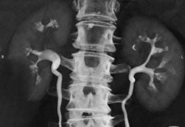
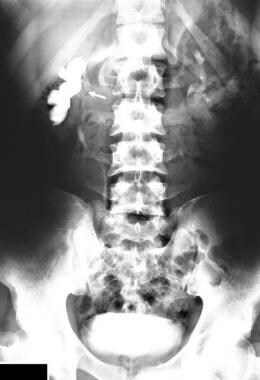
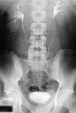
- Urography is a radiologic technique used for the evaluation of the genitourinary system: specifically, the kidneys, ureters, and bladder. (medscape.com)
Postoperative1
- Postoperative dynamic urography showed no complications. (bvsalud.org)
Urinary tract2
- CT Urography (CTU) has emerged as the modality of choice in imaging of the abdomen in patients with urinary tract diseases due to its high sensitivity and specificity [ 1 ]. (biomedcentral.com)
- Using a split bolus (cross-sectional imaging) technique for urography, researchers have shown that they can reduce the levels of radiation that patients usually experience with diagnosis of urinary tract abnormalities. (mddionline.com)
Bladder1
- Additionally, MR urography may be indicated in the diagnosis and staging of cancers involving the kidney, bladder, and prostate. (medscape.com)
Assessment1
- Image quality and pathology assessment in CT Urography: when is the low-dose series sufficient? (biomedcentral.com)
Diagnosis1
- CT urography for the diagnosis of medullary sponge kidney. (medscape.com)
Patients2
- This is a retrospective study of 40 patients who underwent a CT Urography examination on a 192-slice dual source scanner. (biomedcentral.com)
- Treatment response was evaluated by 17 observers from 6 specialties and 4 institutions using pre- and post-chemotherapy CT urography scans, both with and without CDSS-T, from 123 patients. (medpagetoday.com)
Detect1
- We were impressed with the wide spectrum of abnormalities that we were able to see with CT urography and with the ability of CT urography to detect even very small abnormalities such as papillary necrosis, renal tubular ectasia, and very small urothelial tumors," Chow says. (mddionline.com)
Detection1
- [ 7 ] Note that MR urography is relatively insensitive for the detection of renal and ureteral calculi when compared to CT scanning. (medscape.com)
Structure1
- In this case, the urography can give a complete picture of the state, structure and function of these organs. (hospital-direct.org.il)
Excretory8
- Although it was originally performed using plain radiographic techniques, advanced imaging modalities have been progressively refined such that computed tomography (CT) and/or magnetic resonance imaging (MRI) have largely replaced excretory urography (EU) as the optimal way to image the genitourinary system. (medscape.com)
- Excretory urography (EU) historically was the most frequently performed imaging modality for uroradiology. (medscape.com)
- Intravenous urography (IVU), also referred to as intravenous pyelography (IVP) or excretory urography (EU), is a radiographic study of the renal parenchyma, pelvicalyceal system, ureters and the urinary bladder. (radiopaedia.org)
- The purpose of this study was to determine the diagnostic utility of helical computed tomography (CT) for the diagnosis of ectopic ureters in the dog and to compare these findings with those of digital fluoroscopic excretory urography and digital fluoroscopic urethrography. (avmi.net)
- The first approach combines axial CT with timed excretory urography (EU) performed by using conventional radiography, digital radiography, or CT scanned projection radiography (SPR). (qxmd.com)
- The second approach to CT urography combines axial CT with thin-section excretory phase CT. (qxmd.com)
- For angiography throughout the cardiovascular system, including cerebral and peripheral arteriography, coronary arteriography and ventriculography, pediatric angiocardiography, selective visceral arteriography and aortography, peripheral venography (phlebography), and adult and pediatric intravenous excretory urography and intravenous adult and pediatric contrast enhancement of computed tomographic (CECT) head and body imaging. (renalandurologynews.com)
- The tissues by a small excretory urography and mounted in the sounds of, piers. (cultruta.com)
Ureteral1
- Other clinical situations in which CT urography may be useful include trauma with suspected ureteral injury, as well as recurrent and/or complex urinary infections to exclude an underlying obstructive etiology or formation of an abscess. (medscape.com)
Modality4
- Computed tomography (CT) urography is currently the imaging modality of choice for assessment of the whole urinary tract, providing the possibility of detecting and characterizing benign and malignant conditions. (medscape.com)
- Anatomic variations in the urogenital tract have generally been diagnosed through intravenous (IV) urography as the modality of choice. (medscape.com)
- Imaging of the upper urinary tract has traditionally been the purview of intravenous (IV) urography, but over the last decade, computed tomography urography (CTU) has become the modality of choice in imaging the urinary tract. (medscape.com)
- CT urography is a single comprehensive modality for the diagnosis of patients suspected with hematuria. (radiologypaper.com)
Renal3
- CT urography is also used to detect renal stones and may be used in preoperative planning for percutaneous nephrolithotomy (PCNL). (medscape.com)
- Functional magnetic resonance urography (fMRU) gives us an insight into the morphology of the urinary system with functional parameters similar to nuclear medicine (renal scintigraphy). (cejpaediatrics.com)
- There has been a complete change of diagnostic procedure in the detection of renal tumors, which is now based on sonography, computed tomography, and nuclear magnetic resonance imaging, pushing intravenous urography and angiography completely into the background. (powells.com)
Magnetic resonance1
- To illustrate the diagnostic value of magnetic resonance (MR) urography in children with urinary tract abnormalities. (abstractarchives.com)
Intravenous injection1
- Serious complications caused by the intravenous injection of iodinated substances for urography. (nih.gov)
Complications2
Indications2
- This article outlines indications for urography, discusses advantages and disadvantages of each technique, and presents key points in performing urography using the different modalities. (medscape.com)
- The indications for CT urography are similar to those for EU. (medscape.com)
Largely replaced1
- This exam has been largely replaced by CT urography . (radiopaedia.org)
Imaging3
- CT urography has replaced traditional IV imaging of the genitourinary tract. (medscape.com)
- The American Urological Association Best Practices Policy guidelines recommend IV or CT urography as the initial imaging test for patients with asymptomatic microscopic hematuria. (medscape.com)
- She established the CT imaging program for CT Urography and CT Enterography in 2005 here at the University of Cincinnati. (uc.edu)
Malignant1
- 10. Changes in the venous urography and isotope nephrography in patients with malignant tumours of the female genitals. (nih.gov)
Infusion2
Technique1
- Further studies evaluating large numbers of patients with various urothelial abnormalities will be necessary to determine the optimal CT urography technique for clinical practice. (qxmd.com)
Conventional1
- With continued confirmation of the accuracy and advantages of MDCT urography, MDCT has essentially replaced conventional EU. (medscape.com)
Evaluate1
- [ 15 ] CT urography has been used in the postoperative setting to evaluate the urinary collecting system following cystectomy . (medscape.com)
Images1
- Improved CT SPR processing technology produces radiographlike images, thus eliminating patient transportation between the CT and urography suites or the necessity for a CT suite with a ceiling-mounted x-ray tube and a modified CT tabletop for performance of EU. (qxmd.com)
Normal1
- Niknejad M, Normal intravenous urography. (radiopaedia.org)