Uridine Diphosphate Sugars
Uridine Diphosphate Glucose
Adenosine Diphosphate Glucose
Uridine Diphosphate Glucose Dehydrogenase
Chromatography, Paper
Glucose
Uridine Diphosphate
Glucose-1-Phosphate Adenylyltransferase
Uridine Diphosphate N-Acetylglucosamine
Glucuronosyltransferase
Glucosyltransferases
Uracil Nucleotides
Uridine Diphosphate Xylose
Uridine Diphosphate Galactose
Uridine Diphosphate Glucuronic Acid
Uridine Phosphorylase
Glucuronates
UDPglucose 4-Epimerase
Uridine Kinase
Nitrophenols
Xylose
Carbohydrate Epimerases
Crigler-Najjar Syndrome
Uridine Triphosphate
Galactosemias
Phenolphthaleins
Galactose
Lactose Synthase
Microsomes, Liver
Glucose Tolerance Test
Alkyl and Aryl Transferases
Carbon Isotopes
Hyperbilirubinemia, Hereditary
Liver
Adenosine Diphosphate
Uridine Diphosphate N-Acetylgalactosamine
Nucleoside-Diphosphate Kinase
Glucose Intolerance
Uridine Monophosphate
Hemiterpenes
Hexosamines
Chromatography, Thin Layer
Manganese
Digitonin
Electrophoresis
Nucleotides
Enterobacter
Glucose Oxidase
Glucose Transporter Type 1
Glucosamine
NM23 Nucleoside Diphosphate Kinases
A protein-glucan intermediate during paramylon synthesis. (1/357)
A sodium deoxycholate extract containing glucosyltransferase activity was obtained from a particulate preparation from Euglena gracilis. It transferred glucose from UDP-[14C]glucose into material that was precipitated by trichloroacetic acid. This material released beta-(1 leads to 3)-glucan oligosaccharides into solution on incubation with weak acid, weak alkali and beta-(1 leads to 3)-glucosidase. The products of the incubation of the deoxycholate extract with UDP-[14C]glucose were analysed by sodium dodecyl sulphate/polyacrylamide-gel electrophoresis. Radioactive bands were obtained that had the properties of beta-(1 leads to 3)-glucan covalently linked to protein by a bond labile to weak acid. High-molecular-weight material containing a beta-(1 leads to 3)-glucan was also shown to be present by gel filtration. The bond linking glucan to aglycone is possibly a pyrophosphate linkage. It is proposed that in Euglena gracilis beta-(1 leads to 3)-glucan (paramylon) is synthesized on a protein primer. (+info)Formation of lipid-linked sugar compounds in Halobacterium salinarium. Presumed intermediates in glycoprotein synthesis. (2/357)
The ability of bacitracin to inhibit the growth of Halobacterium salinarium suggested that glycosylation of the major envelope component, a high molecular weight glycoprotein, might occur via a pathway involving lipid intermediates. This report demonstrates that the cells have enzymatic activities for formation of lipid-linked sugar compounds having the expected properties of such intermediates. Whole cell homogenate catalyzed the transfer of sugar from UDP-glucose, GDP-mannose, and UDP-N-acetyglucosamine to endogenous lipid acceptors. Two lipid products were formed from UDP-glucose, two from GDP-mannose, and one from UDP-N-acetylglucosamine. Characterization of the partially purified lipids by ion exchange chromatography, thin layer chromatography, and mild acid and base hydrolysis showed the major product in each case to have the properties expected for polyisoprenyl phosphoglucose, polyisoprenyl phosphomannose, and polyisoprenyl pyrophospho-N-acetylglucosamine. Estimates of chain length by thin layer chromatography indicate that the lipid has 11 to 12 isoprene identity as a C55-60-polyisoprenyl pyrophospho-N-acetylglucosamine. The N-acetylglucosamine transferase, present in cell envelope preparations, was partially characterized. The enzyme was found to be extremely halophilic, specifically requiring a high concentration of KCl. Optimum activity was obtained at 4 m KCl and partial substitution of K+ by Na+ resulted in a decrease in activity. (+info)Stimulation of collagen galactosyltransferase and glucosyltransferase activities by lysophosphatidylcholine. (3/357)
Lysophosphatidylcholine stimulated the activities of collagen galactosyl- and glucosyl-transferases in chick-embryo extract and its particulate fractions in vitro, whereas essentially no stimulation was noted in the high-speed supernatant, where the enzymes are soluble and membrane-free. The stimulatory effect of lysophosphatidylcholine was masked by 0.1% Triton X-100. In kinetic experiments lysophosphatidylcholine raised the maximum velocities with respect to the substrates and co-substrates, whereas no changes were observed in the apparant Km values. Phospholipase A preincubation of the chick-embryo extract resulted in stimulation of both transferase activities, probably gy generating lysophosphatides from endogenous phospholipids. No stimulation by lysophosphatidylcholine was found when tested with 500-fold-purified glycosyltransferase. The results suggest that collagen glycosyltransferases must be associated with the membrane structures of the cell in order to be stimulated by lysophosphatidylcholine. Lysophosphatidylcholine could have some regulatory significance in vivo, since its concentration in the cell is comparable with that which produced marked stimulation in vitro. (+info)Studies on the mechanism of collagen glucosyltransferase reaction. (4/357)
The mechanism of collagen glucosyltransferase reaction was studied with enzyme preparations purified about 2500-5000-fold from extract of homogenate of whole chick embryos. Data obtained in experiments on initial velocity and inhibition kinetics of the reaction were consistent with an ordered mechanism in which the substrates are bound to the enzyme in the following order: Mn2+, UDP-glucose and collagen substrate, the addition of Mn2+ being at thermodynamic equilibrium and the binding site of the UDP-glucose to the enzyme not being the same as that for Mn2+ and collagen substrate. Only one metal co-factor seems to be involved in the reaction. The collagen substrate can probably also react in some conditions with enzyme-Mn2+ and with enzyme-Mn2+-UDP, and the UDP with the free enzyme, but in all these instances dead-end complexes are formed. Evidence is presented for an ordered release of the products in the following order: glucosylated collagen, UDP and Mn2+, in which Mn2+ need not leave the enzyme during each catalytic cycle. (+info)Orotate decreases the inhibitory effect of ethanol on galactose elimination in the perfused rat liver. (5/357)
1. The galactose-elimination rate in perfused livers from starved rats was decreased in the presence of ethanol (2-28mM) to one-third of the control values. Orotate injections partly reversed the effect of ethanol, so that the galactose-elimination rate was about two-thirds of the control values. Orotate alone had no effect on the galactose-elimination rate. 2. Ethanol increased [galactose 1-phosphate] and [UDP-galactose], and decreased (UDP-glucose] and [UTP], both with and without orotate. Orotate increased [UTP], [UDP-galactose], both with and without ethanol. The increase of [galactose 1-phosphate] in the presence of ethanol was inhibited by orotate. Orotate alone had no appreciable effect on [galactose 1-phosphate]. 3. Both the effect of ethanol and that of orotate on the galactose-elimination rate can be accounted for by assuming inhibition of galactokinase by galactose 1-phosphate with Ki about 0.2mM, the inhibition being either non-competitive or uncompetitive. 4. The primary effect of ethanol seems to be inhibition of UDP-glucose epimerase (EC 5.1.3.2), followed by accumulation of UDP-galactose, trapping of UDP-glucose and increase of [galactose 1-phosphate]. Orotate decreased the effect of ethanol, probably by increasing [UDP-glucose]. (+info)Incorporation of [14C]glucose from UDP[14C]glucose into a phosphoglycolipid by cell-free particulate systems of a moderately halophilic gram-negative bacterium, Pseudomonas halosaccharolytica ATCC 29423. (6/357)
A glucosyl group from uridine diphosphate [U-14C]glucose is incorporated into a phosphoglycolipid, probably a glucosylphosphatidylglycerol, by a disrupted membrane enzyme preparation from a gram-negative, moderately halophilic bacterium, Pseudomonas halosaccharolytica ATCC 29423. The conversion of [14C]phosphatidylglycerol into phosphoglycolipid by the particulate preparation was also enhanced in the presence of non-labelled UDP-glucose. A chemical degradation study of labelled phosphoglycolipid showed the bulk of the radioactivity from UDP[U-14C]glucose to be associated with the glucose moiety, which also appeared to be attached to the hydroxyl group of a second glycerol. (+info)Uridine diphosphate-glucose transport into the endoplasmic reticulum of Saccharomyces cerevisiae: in vivo and in vitro evidence. (7/357)
It has been proposed that synthesis of beta-1,6-glucan, one of Saccharomyces cerevisiae cell wall components, is initiated by a uridine diphosphate (UDP)-glucose-dependent reaction in the lumen of the endoplasmic reticulum (ER). Because this sugar nucleotide is not synthesized in the lumen of the ER, we have examined whether or not UDP-glucose can be transported across the ER membrane. We have detected transport of this sugar nucleotide into the ER in vivo and into ER-containing microsomes in vitro. Experiments with ER-containing microsomes showed that transport of UDP-glucose was temperature dependent and saturable with an apparent Km of 46 microM and a Vmax of 200 pmol/mg protein/3 min. Transport was substrate specific because UDP-N-acetylglucosamine did not enter these vesicles. Demonstration of UDP-glucose transport into the ER lumen in vivo was accomplished by functional expression of Schizosaccharomyces pombe UDP-glucose:glycoprotein glucosyltransferase (GT) in S. cerevisiae, which is devoid of this activity. Monoglucosylated protein-linked oligosaccharides were detected in alg6 or alg5 mutant cells, which transfer Man9GlcNAc2 to protein; glucosylation was dependent on the inhibition of glucosidase II or the disruption of the gene encoding this enzyme. Although S. cerevisiae lacks GT, it contains Kre5p, a protein with significant homology and the same size and subcellular location as GT. Deletion mutants, kre5Delta, lack cell wall beta-1,6 glucan and grow very slowly. Expression of S. pombe GT in kre5Delta mutants did not complement the slow-growth phenotype, indicating that both proteins have different functions in spite of their similarities. (+info)Multi-enzymatic glucosylation using Eucalyptus UDP-glucosyltransferase coupled UDPglucose-fermentation by bakers' yeast. (8/357)
The enzymatic synthesis of glucoside compounds using a membrane-associated UDP-glucosyltransferase fraction from Eucalyptus perriniana cultured cells as a water-insoluble catalyst (N. Nakajima, et. al., J. Ferment. Bioeng., 84 (5), pp. 455-460, 1997) has been effectively done by coupling UDPglucose-fermentation by bakers' yeast. For example, beta-thujaplicin (hinokitiol) and p-aminobenzoic acid were converted respectively to their corresponding beta-D-monoglucosides with the conversion rate of around 24-26% by the multi-enzymatic system with UDPglucose as a glucose donor, which is produced by yeast cells from glucose and 5'-UMP. Addition of either cellobiose, a substrate of beta-glucosidase, or DL-1,2-anhydro-myo-inositol, an inhibitor for the enzyme in the reaction mixture, could increased the yield of these beta-D-monoglucosides. This new enzymatic system could also be used for the synthesis of flavonoid glucosides such as isoquercitrin (quercetin 3-O-beta-D-glucoside). (+info)Uridine diphosphate sugars (UDP-sugars) are nucleotide sugars that play a crucial role in the biosynthesis of glycans, which are complex carbohydrates found on the surface of many cell types. UDP-sugars consist of a uridine diphosphate molecule linked to a sugar moiety, such as glucose, galactose, or xylose. These molecules serve as activated donor substrates for glycosyltransferases, enzymes that catalyze the transfer of sugar residues to acceptor molecules, including proteins and other carbohydrates. UDP-sugars are essential for various biological processes, such as cell recognition, signaling, and protein folding. Dysregulation of UDP-sugar metabolism has been implicated in several diseases, including cancer and congenital disorders of glycosylation.
Uridine Diphosphate Glucose (UDP-glucose) is a nucleotide sugar that plays a crucial role in the synthesis and metabolism of carbohydrates in the body. It is formed from uridine triphosphate (UTP) and glucose-1-phosphate through the action of the enzyme UDP-glucose pyrophosphorylase.
UDP-glucose serves as a key intermediate in various biochemical pathways, including glycogen synthesis, where it donates glucose molecules to form glycogen, a large polymeric storage form of glucose found primarily in the liver and muscles. It is also involved in the biosynthesis of other carbohydrate-containing compounds such as proteoglycans and glycolipids.
Moreover, UDP-glucose is an essential substrate for the enzyme glucosyltransferase, which is responsible for adding glucose molecules to various acceptor molecules during the process of glycosylation. This post-translational modification is critical for the proper folding and functioning of many proteins.
Overall, UDP-glucose is a vital metabolic intermediate that plays a central role in carbohydrate metabolism and protein function.
Adenosine diphosphate glucose (ADP-glucose) is a key intermediate in the biosynthesis of glycogen, which is a complex carbohydrate that serves as a primary form of energy storage in animals, fungi, and bacteria. In this process, ADP-glucose is formed from glucose-1-phosphate and adenosine triphosphate (ATP) through the action of the enzyme ADP-glucose pyrophosphorylase. Once synthesized, ADP-glucose is then used as a substrate for the enzyme glycogen synthase, which catalyzes the addition of glucose units to an existing glycogen molecule, leading to its growth and expansion. This pathway plays a crucial role in regulating cellular energy metabolism and maintaining glucose homeostasis within the body.
Uridine Diphosphate (UDP) Glucose Dehydrogenase is an enzyme that plays a role in carbohydrate metabolism. Its systematic name is UDP-glucose:NAD+ oxidoreductase, and it catalyzes the following chemical reaction:
UDP-glucose + NAD+ -> UDP-glucuronate + NADH + H+
This enzyme helps convert UDP-glucose into UDP-glucuronate, which is a crucial component in the biosynthesis of various substances in the body, such as glycosaminoglycans and other glyconjugates. The reaction also results in the reduction of NAD+ to NADH, which is an essential coenzyme in numerous metabolic processes.
UDP-glucose dehydrogenase is widely distributed in various tissues, including the liver, kidney, and intestine. Deficiencies or mutations in this enzyme can lead to several metabolic disorders, such as glucosuria and hypermethioninemia.
Paper chromatography is a type of chromatography technique that involves the separation and analysis of mixtures based on their components' ability to migrate differently upon capillary action on a paper medium. This simple and cost-effective method utilizes a paper, typically made of cellulose, as the stationary phase. The sample mixture is applied as a small spot near one end of the paper, and then the other end is dipped into a developing solvent or a mixture of solvents (mobile phase) in a shallow container.
As the mobile phase moves up the paper by capillary action, components within the sample mixture separate based on their partition coefficients between the stationary and mobile phases. The partition coefficient describes how much a component prefers to be in either the stationary or mobile phase. Components with higher partition coefficients in the mobile phase will move faster and further than those with lower partition coefficients.
Once separation is complete, the paper is dried and can be visualized under ultraviolet light or by using chemical reagents specific for the components of interest. The distance each component travels from the origin (point of application) and its corresponding solvent front position are measured, allowing for the calculation of Rf values (retardation factors). Rf is a dimensionless quantity calculated as the ratio of the distance traveled by the component to the distance traveled by the solvent front.
Rf = (distance traveled by component) / (distance traveled by solvent front)
Paper chromatography has been widely used in various applications, such as:
1. Identification and purity analysis of chemical compounds in pharmaceuticals, forensics, and research laboratories.
2. Separation and detection of amino acids, sugars, and other biomolecules in biological samples.
3. Educational purposes to demonstrate the principles of chromatography and separation techniques.
Despite its limitations, such as lower resolution compared to high-performance liquid chromatography (HPLC) and less compatibility with volatile or nonpolar compounds, paper chromatography remains a valuable tool for quick, qualitative analysis in various fields.
Glucose is a simple monosaccharide (or single sugar) that serves as the primary source of energy for living organisms. It's a fundamental molecule in biology, often referred to as "dextrose" or "grape sugar." Glucose has the molecular formula C6H12O6 and is vital to the functioning of cells, especially those in the brain and nervous system.
In the body, glucose is derived from the digestion of carbohydrates in food, and it's transported around the body via the bloodstream to cells where it can be used for energy. Cells convert glucose into a usable form through a process called cellular respiration, which involves a series of metabolic reactions that generate adenosine triphosphate (ATP)—the main currency of energy in cells.
Glucose is also stored in the liver and muscles as glycogen, a polysaccharide (multiple sugar) that can be broken down back into glucose when needed for energy between meals or during physical activity. Maintaining appropriate blood glucose levels is crucial for overall health, and imbalances can lead to conditions such as diabetes mellitus.
Uridine diphosphate (UDP) is a nucleotide diphosphate that consists of a pyrophosphate group, a ribose sugar, and the nucleobase uracil. It plays a crucial role as a coenzyme in various biosynthetic reactions, including the synthesis of glycogen, proteoglycans, and other polysaccharides. UDP is also involved in the detoxification of bilirubin, an end product of hemoglobin breakdown, by converting it to a water-soluble form that can be excreted through the bile. Additionally, UDP serves as a precursor for the synthesis of other nucleotides and their derivatives.
Glucose-1-phosphate adenylyltransferase, also known as ADP-glucose pyrophosphorylase or AGPase, is an enzyme that plays a crucial role in carbohydrate metabolism, specifically in the synthesis of starch. It catalyzes the reaction between ATP and glucose-1-phosphate to produce ADP-glucose and pyrophosphate. This reaction is the first committed step in the biosynthetic pathway of starch in plants, algae, and some bacteria. In humans, defects in this enzyme can lead to a rare genetic disorder called glycogen storage disease type Ib.
Uridine Diphosphate N-Acetylglucosamine (UDP-GlcNAc) is not a medical term per se, but rather a biochemical term. It is a form of nucleotide sugar that plays a crucial role in several biochemical processes in the human body.
To provide a more detailed definition: UDP-GlcNAc is a nucleotide sugar that serves as a donor substrate for various glycosyltransferases involved in the biosynthesis of glycoproteins, proteoglycans, and glycolipids. It is a key component in the process of N-linked and O-linked glycosylation, which are important post-translational modifications of proteins that occur within the endoplasmic reticulum and Golgi apparatus. UDP-GlcNAc also plays a role in the biosynthesis of hyaluronic acid, a major component of the extracellular matrix.
Abnormal levels or functioning of UDP-GlcNAc have been implicated in various disease states, including cancer and diabetes. However, it is not typically used as a diagnostic marker or therapeutic target in clinical medicine.
Nucleotidyltransferases are a class of enzymes that catalyze the transfer of nucleotides to an acceptor molecule, such as RNA or DNA. These enzymes play crucial roles in various biological processes, including DNA replication, repair, and recombination, as well as RNA synthesis and modification.
The reaction catalyzed by nucleotidyltransferases typically involves the donation of a nucleoside triphosphate (NTP) to an acceptor molecule, resulting in the formation of a phosphodiester bond between the nucleotides. The reaction can be represented as follows:
NTP + acceptor → NMP + pyrophosphate
where NTP is the nucleoside triphosphate donor and NMP is the nucleoside monophosphate product.
There are several subclasses of nucleotidyltransferases, including polymerases, ligases, and terminases. These enzymes have distinct functions and substrate specificities, but all share the ability to transfer nucleotides to an acceptor molecule.
Examples of nucleotidyltransferases include DNA polymerase, RNA polymerase, reverse transcriptase, telomerase, and ligase. These enzymes are essential for maintaining genome stability and function, and their dysregulation has been implicated in various diseases, including cancer and neurodegenerative disorders.
Glucuronosyltransferase (UDP-glucuronosyltransferase) is an enzyme belonging to the family of glycosyltransferases. It plays a crucial role in the process of biotransformation and detoxification of various endogenous and exogenous substances, including drugs, hormones, and environmental toxins, in the liver and other organs.
The enzyme functions by transferring a glucuronic acid moiety from a donor molecule, uridine diphosphate glucuronic acid (UDP-GlcUA), to an acceptor molecule, which can be a variety of hydrophobic compounds. This reaction results in the formation of a more water-soluble glucuronide conjugate, facilitating the excretion of the substrate through urine or bile.
There are multiple isoforms of glucuronosyltransferase, classified into two main families: UGT1 and UGT2. These isoforms exhibit different substrate specificities and tissue distributions, allowing for a wide range of compounds to be metabolized through the glucuronidation pathway.
In summary, Glucuronosyltransferase is an essential enzyme in the detoxification process, facilitating the elimination of various substances from the body by conjugating them with a glucuronic acid moiety.
Glucosyltransferases (GTs) are a group of enzymes that catalyze the transfer of a glucose molecule from an activated donor to an acceptor molecule, resulting in the formation of a glycosidic bond. These enzymes play crucial roles in various biological processes, including the biosynthesis of complex carbohydrates, cell wall synthesis, and protein glycosylation. In some cases, GTs can also contribute to bacterial pathogenesis by facilitating the attachment of bacteria to host tissues through the formation of glucans, which are polymers of glucose molecules.
GTs can be classified into several families based on their sequence similarities and catalytic mechanisms. The donor substrates for GTs are typically activated sugars such as UDP-glucose, TDP-glucose, or GDP-glucose, which serve as the source of the glucose moiety that is transferred to the acceptor molecule. The acceptor can be a wide range of molecules, including other sugars, proteins, lipids, or small molecules.
In the context of human health and disease, GTs have been implicated in various pathological conditions, such as cancer, inflammation, and microbial infections. For example, some GTs can modify proteins on the surface of cancer cells, leading to increased cell proliferation, migration, and invasion. Additionally, GTs can contribute to bacterial resistance to antibiotics by modifying the structure of bacterial cell walls or by producing biofilms that protect bacteria from host immune responses and antimicrobial agents.
Overall, Glucosyltransferases are essential enzymes involved in various biological processes, and their dysregulation has been associated with several human diseases. Therefore, understanding the structure, function, and regulation of GTs is crucial for developing novel therapeutic strategies to target these enzymes and treat related pathological conditions.
Uracil nucleotides are chemical compounds that play a crucial role in the synthesis, repair, and replication of DNA and RNA. Specifically, uracil nucleotides refer to the group of molecules that contain the nitrogenous base uracil, which is linked to a ribose sugar through a beta-glycosidic bond. This forms the nucleoside uridine, which can then be phosphorylated to create the uracil nucleotide.
Uracil nucleotides are important in the formation of RNA, where uracil base pairs with adenine through two hydrogen bonds during transcription. However, uracil is not typically found in DNA, and its presence in DNA can indicate damage or mutation. When uracil is found in DNA, it is usually the result of a process called deamination, where the nitrogenous base cytosine is spontaneously converted to uracil. This can lead to errors during replication, as uracil will pair with adenine instead of guanine, leading to a C-to-T or G-to-A mutation.
To prevent this type of mutation, cells have enzymes called uracil DNA glycosylases that recognize and remove uracil from DNA. This initiates the base excision repair pathway, which removes the damaged nucleotide and replaces it with a correct one. Overall, uracil nucleotides are essential for proper cellular function, but their misincorporation into DNA can have serious consequences for genome stability.
Uridine Diphosphate Xylose (UDP-Xylose) is not a medical term per se, but rather a biochemical term. It is the molecule that serves as the starting point for the biosynthesis of plant polysaccharides, such as xyloglucans and xylans, which are important components of the plant cell wall.
UDP-Xylose is a nucleotide sugar, meaning it consists of a sugar molecule (xylose) linked to a nucleotide (uridine diphosphate or UDP). It is synthesized in the cytoplasm of plant cells through the action of enzymes that transfer xylose from UDP-glucose to UTP.
In medicine, UDP-Xylose may be used as a substrate for enzyme assays or as a tool for studying carbohydrate metabolism in plants and microorganisms. However, it is not a substance that is typically used in medical treatments or interventions.
Uridine Diphosphate Galactose (UDP-galactose) is a nucleotide sugar that plays a crucial role in the biosynthesis of glycans, proteoglycans, and glycolipids. It is formed from uridine diphosphate glucose (UDP-glucose) through the action of the enzyme UDP-glucose 4'-epimerase.
In the body, UDP-galactose serves as a galactosyl donor in various metabolic pathways, including lactose synthesis in the mammary gland and the addition of galactose residues to proteoglycans and glycoproteins in the Golgi apparatus. Defects in the metabolism of UDP-galactose have been linked to several genetic disorders, such as galactosemia, which can result in serious health complications if left untreated.
Uridine Diphosphate Glucuronic Acid (UDP-Glucuronic Acid) is not a medical term per se, but rather a biochemical term. It is a compound that plays an essential role in the detoxification process in the liver. UDP-Glucuronic Acid is a nucleotide sugar derivative that combines with toxins, drugs, and other substances to form glucuronides, which are then excreted through urine or bile. This process is known as glucuronidation, and it helps make the substances more water-soluble and easier for the body to eliminate.
Uridine phosphorylase is an enzyme that plays a role in the metabolism of nucleosides, specifically uridine. The medical definition of 'uridine phosphorylase' is:
An enzyme (EC 2.4.2.3) involved in the reversible phosphorolysis of uridine to uracil and ribose-1-phosphate. This enzyme also catalyzes the phosphorolytic cleavage of other pyrimidine nucleosides, such as cytidine and thymidine, into their respective bases and ribose-1-phosphate. Uridine phosphorylase has a role in the salvage pathway of pyrimidine nucleotide biosynthesis and is found in various tissues, including the liver, intestines, and blood cells. Deficiency or dysfunction of uridine phosphorylase can lead to impaired nucleotide metabolism and may be associated with certain medical conditions, such as hereditary orotic aciduria.
Glucuronates are not a medical term per se, but they refer to salts or esters of glucuronic acid, a organic compound that is a derivative of glucose. In the context of medical and biological sciences, glucuronidation is a common detoxification process in which glucuronic acid is conjugated to a wide variety of molecules, including drugs, hormones, and environmental toxins, to make them more water-soluble and facilitate their excretion from the body through urine or bile.
The process of glucuronidation is catalyzed by enzymes called UDP-glucuronosyltransferases (UGTs), which are found in various tissues, including the liver, intestines, and kidneys. The resulting glucuronides can be excreted directly or further metabolized before excretion.
Therefore, "glucuronates" can refer to the chemical compounds that result from this process of conjugation with glucuronic acid, as well as the therapeutic potential of enhancing or inhibiting glucuronidation for various clinical applications.
UDP-glucose 4-epimerase (UGE) is an enzyme that catalyzes the reversible interconversion of UDP-galactose and UDP-glucose, two important nucleotide sugars involved in carbohydrate metabolism. This enzyme plays a crucial role in maintaining the balance between these two molecules, which are essential for the synthesis of various glycoconjugates, such as glycoproteins and proteoglycans. UGE is widely distributed in nature and has been identified in various organisms, including humans. In humans, deficiency or mutations in this enzyme can lead to a rare genetic disorder known as galactosemia, which is characterized by an impaired ability to metabolize the sugar galactose, resulting in several health issues.
Uridine kinase is an enzyme that phosphorylates the pyrimidine nucleoside uridine to produce uridine monophosphate (UMP). This reaction plays a crucial role in the salvage pathway of pyrimidine nucleotide synthesis, which recycles nucleosides generated from the degradation of RNA.
The human genome encodes two isoforms of uridine kinase, UCK1 and UCK2, which share a high degree of sequence similarity but have distinct tissue expression patterns and subcellular localization. UCK1 is primarily expressed in the liver and kidney, while UCK2 is more widely expressed in various tissues.
Uridine kinase activity has been implicated in several physiological processes, including the regulation of intracellular nucleotide pools, the biosynthesis of glycosaminoglycans and proteoglycans, and the modulation of antiviral responses. Dysregulation of uridine kinase activity has been associated with various pathological conditions, such as cancer, viral infections, and neurological disorders.
Nitrophenols are organic compounds that contain a hydroxyl group (-OH) attached to a phenyl ring (aromatic hydrocarbon) and one or more nitro groups (-NO2). They have the general structure R-C6H4-NO2, where R represents the hydroxyl group.
Nitrophenols are known for their distinctive yellow to brown color and can be found in various natural sources such as plants and microorganisms. Some common nitrophenols include:
* p-Nitrophenol (4-nitrophenol)
* o-Nitrophenol (2-nitrophenol)
* m-Nitrophenol (3-nitrophenol)
These compounds are used in various industrial applications, including dyes, pharmaceuticals, and agrochemicals. However, they can also be harmful to human health and the environment, as some nitrophenols have been identified as potential environmental pollutants and may pose risks to human health upon exposure.
Xylose is a type of sugar that is commonly found in plants and wood. In the context of medical definitions, xylose is often used in tests to assess the function of the small intestine. The most common test is called the "xylose absorption test," which measures the ability of the small intestine to absorb this sugar.
In this test, a patient is given a small amount of xylose to drink, and then several blood and/or urine samples are collected over the next few hours. The amount of xylose that appears in these samples is measured and used to determine how well the small intestine is absorbing nutrients.
Abnormal results on a xylose absorption test can indicate various gastrointestinal disorders, such as malabsorption syndromes, celiac disease, or bacterial overgrowth in the small intestine.
Carbohydrate epimerases are a group of enzymes that catalyze the interconversion of specific stereoisomers (epimers) of carbohydrates by the reversible oxidation and reduction of carbon atoms, usually at the fourth or fifth position. These enzymes play important roles in the biosynthesis and modification of various carbohydrate-containing molecules, such as glycoproteins, proteoglycans, and glycolipids, which are involved in numerous biological processes including cell recognition, signaling, and adhesion.
The reaction catalyzed by carbohydrate epimerases involves the transfer of a hydrogen atom and a proton between two adjacent carbon atoms, leading to the formation of new stereochemical configurations at these positions. This process can result in the conversion of one epimer into another, thereby expanding the structural diversity of carbohydrates and their derivatives.
Carbohydrate epimerases are classified based on the type of substrate they act upon and the specific stereochemical changes they induce. Some examples include UDP-glucose 4-epimerase, which interconverts UDP-glucose and UDP-galactose; UDP-N-acetylglucosamine 2-epimerase, which converts UDP-N-acetylglucosamine to UDP-N-acetylmannosamine; and GDP-fucose synthase, which catalyzes the conversion of GDP-mannose to GDP-fucose.
Understanding the function and regulation of carbohydrate epimerases is crucial for elucidating their roles in various biological processes and developing strategies for targeting them in therapeutic interventions.
Crigler-Najjar Syndrome is a rare inherited genetic disorder that affects the metabolism of bilirubin, a yellow pigment produced when hemoglobin breaks down. This condition is characterized by high levels of unconjugated bilirubin in the blood, which can lead to jaundice, kernicterus, and neurological damage if left untreated.
There are two types of Crigler-Najjar Syndrome: Type I and Type II.
Type I is the more severe form, and it is caused by a mutation in the UGT1A1 gene, which encodes for an enzyme responsible for conjugating bilirubin. People with this type of Crigler-Najjar Syndrome have little to no functional enzyme activity, leading to very high levels of unconjugated bilirubin in the blood. This form is usually diagnosed in infancy and requires regular phototherapy or a liver transplant to prevent neurological damage.
Type II is a milder form of the disorder, caused by a mutation that results in reduced enzyme activity but not complete loss of function. People with this type of Crigler-Najjar Syndrome usually have milder symptoms and may not require regular phototherapy or a liver transplant, although they may still be at risk for neurological damage if their bilirubin levels become too high.
Both types of Crigler-Najjar Syndrome are inherited in an autosomal recessive manner, meaning that an individual must inherit two copies of the mutated gene (one from each parent) to develop the condition.
Blood glucose, also known as blood sugar, is the concentration of glucose in the blood. Glucose is a simple sugar that serves as the main source of energy for the body's cells. It is carried to each cell through the bloodstream and is absorbed into the cells with the help of insulin, a hormone produced by the pancreas.
The normal range for blood glucose levels in humans is typically between 70 and 130 milligrams per deciliter (mg/dL) when fasting, and less than 180 mg/dL after meals. Levels that are consistently higher than this may indicate diabetes or other metabolic disorders.
Blood glucose levels can be measured through a variety of methods, including fingerstick blood tests, continuous glucose monitoring systems, and laboratory tests. Regular monitoring of blood glucose levels is important for people with diabetes to help manage their condition and prevent complications.
Uridine Triphosphate (UTP) is a nucleotide that plays a crucial role in the synthesis and repair of DNA and RNA. It consists of a nitrogenous base called uracil, a pentose sugar (ribose), and three phosphate groups. UTP is one of the four triphosphates used in the biosynthesis of RNA during transcription, where it donates its uracil base to the growing RNA chain. Additionally, UTP serves as an energy source and a substrate in various biochemical reactions within the cell, including phosphorylation processes and the synthesis of glycogen and other molecules.
Galactosemia is a rare metabolic disorder that affects the body's ability to metabolize the simple sugar galactose, which is found in milk and other dairy products. It is caused by deficiency or complete absence of one of the three enzymes needed to convert galactose into glucose:
1. Galactokinase (GALK) deficiency - also known as Galactokinase galactosemia, is a milder form of the disorder.
2. Galactose-1-phosphate uridylyltransferase (GALT) deficiency - the most common and severe form of classic galactosemia.
3. Galactose epimerase (GALE) deficiency - also known as Epimerase deficiency galactosemia, is a rare and milder form of the disorder.
The most severe form of the disorder, GALT deficiency, can lead to serious health problems such as cataracts, liver damage, mental retardation, and sepsis if left untreated. Treatment typically involves removing galactose from the diet, which requires avoiding all milk and dairy products. Early diagnosis and treatment are crucial for improving outcomes in individuals with galactosemia.
Phenolphthalein is not strictly a medical term, but it is a chemical compound that has been used in medical contexts. It's primarily known for its use as an acid-base indicator in chemistry and medical laboratory tests. Here's the general definition:
Phenolphthalein is a crystalline compound, commonly available as a colorless powder or clear liquid. It is used as a pH indicator, turning pink to purple in basic solutions (pH above 8.2) and colorless in acidic solutions (pH below 8.2). This property makes it useful in various applications, such as titrations and monitoring the pH of chemical reactions or solutions.
In a medical context, phenolphthalein has historically been used as an active ingredient in certain over-the-counter laxatives. However, due to concerns about potential carcinogenicity and other side effects, its use in pharmaceuticals has been largely discontinued or restricted in many countries, including the United States.
Galactose is a simple sugar or monosaccharide that is a constituent of lactose, the disaccharide found in milk and dairy products. It's structurally similar to glucose but with a different chemical structure, and it plays a crucial role in various biological processes.
Galactose can be metabolized in the body through the action of enzymes such as galactokinase, galactose-1-phosphate uridylyltransferase, and UDP-galactose 4'-epimerase. Inherited deficiencies in these enzymes can lead to metabolic disorders like galactosemia, which can cause serious health issues if not diagnosed and treated promptly.
In summary, Galactose is a simple sugar that plays an essential role in lactose metabolism and other biological processes.
Lactose synthase is an enzyme composed of two subunits: a regulatory subunit, β-1,4-galactosyltransferase (β-1,4-GT), and a catalytic subunit, α-lactalbumin. This enzyme plays a crucial role in lactose biosynthesis during milk production in mammals. By catalyzing the transfer of a galactose molecule from UDP-galactose to glucose, lactose synthase generates lactose (or milk sugar), which is essential for providing energy and growth to newborns. The activity of lactose synthase is primarily regulated by α-lactalbumin, which modifies the substrate specificity of β-1,4-GT, allowing it to use glucose as an acceptor instead of other glycoconjugates.
Microsomes, liver refers to a subcellular fraction of liver cells (hepatocytes) that are obtained during tissue homogenization and subsequent centrifugation. These microsomal fractions are rich in membranous structures known as the endoplasmic reticulum (ER), particularly the rough ER. They are involved in various important cellular processes, most notably the metabolism of xenobiotics (foreign substances) including drugs, toxins, and carcinogens.
The liver microsomes contain a variety of enzymes, such as cytochrome P450 monooxygenases, that are crucial for phase I drug metabolism. These enzymes help in the oxidation, reduction, or hydrolysis of xenobiotics, making them more water-soluble and facilitating their excretion from the body. Additionally, liver microsomes also host other enzymes involved in phase II conjugation reactions, where the metabolites from phase I are further modified by adding polar molecules like glucuronic acid, sulfate, or acetyl groups.
In summary, liver microsomes are a subcellular fraction of liver cells that play a significant role in the metabolism and detoxification of xenobiotics, contributing to the overall protection and maintenance of cellular homeostasis within the body.
Hexosyltransferases are a group of enzymes that catalyze the transfer of a hexose (a type of sugar molecule made up of six carbon atoms) from a donor molecule to an acceptor molecule. This transfer results in the formation of a glycosidic bond between the two molecules.
Hexosyltransferases are involved in various biological processes, including the biosynthesis of complex carbohydrates, such as glycoproteins and glycolipids, which play important roles in cell recognition, signaling, and communication. These enzymes can transfer a variety of hexose sugars, including glucose, galactose, mannose, fucose, and N-acetylglucosamine, to different acceptor molecules, such as proteins, lipids, or other carbohydrates.
Hexosyltransferases are classified based on the type of donor molecule they use, the type of sugar they transfer, and the type of glycosidic bond they form. Some examples of hexosyltransferases include:
* Glycosyltransferases (GTs): These enzymes transfer a sugar from an activated donor molecule, such as a nucleotide sugar, to an acceptor molecule. GTs are involved in the biosynthesis of various glycoconjugates, including proteoglycans, glycoproteins, and glycolipids.
* Fucosyltransferases (FUTs): These enzymes transfer fucose, a type of hexose sugar, to an acceptor molecule. FUTs are involved in the biosynthesis of various glycoconjugates, including blood group antigens and Lewis antigens.
* Galactosyltransferases (GALTs): These enzymes transfer galactose, another type of hexose sugar, to an acceptor molecule. GALTs are involved in the biosynthesis of various glycoconjugates, including lactose in milk and gangliosides in the brain.
* Mannosyltransferases (MTs): These enzymes transfer mannose, a type of hexose sugar, to an acceptor molecule. MTs are involved in the biosynthesis of various glycoconjugates, including N-linked glycoproteins and yeast cell walls.
Hexosyltransferases play important roles in many biological processes, including cell recognition, signaling, and adhesion. Dysregulation of these enzymes has been implicated in various diseases, such as cancer, inflammation, and neurodegenerative disorders. Therefore, understanding the mechanisms of hexosyltransferases is crucial for developing new therapeutic strategies.
In the context of medicine and pharmacology, "kinetics" refers to the study of how a drug moves throughout the body, including its absorption, distribution, metabolism, and excretion (often abbreviated as ADME). This field is called "pharmacokinetics."
1. Absorption: This is the process of a drug moving from its site of administration into the bloodstream. Factors such as the route of administration (e.g., oral, intravenous, etc.), formulation, and individual physiological differences can affect absorption.
2. Distribution: Once a drug is in the bloodstream, it gets distributed throughout the body to various tissues and organs. This process is influenced by factors like blood flow, protein binding, and lipid solubility of the drug.
3. Metabolism: Drugs are often chemically modified in the body, typically in the liver, through processes known as metabolism. These changes can lead to the formation of active or inactive metabolites, which may then be further distributed, excreted, or undergo additional metabolic transformations.
4. Excretion: This is the process by which drugs and their metabolites are eliminated from the body, primarily through the kidneys (urine) and the liver (bile).
Understanding the kinetics of a drug is crucial for determining its optimal dosing regimen, potential interactions with other medications or foods, and any necessary adjustments for special populations like pediatric or geriatric patients, or those with impaired renal or hepatic function.
A Glucose Tolerance Test (GTT) is a medical test used to diagnose prediabetes, type 2 diabetes, and gestational diabetes. It measures how well your body is able to process glucose, which is a type of sugar.
During the test, you will be asked to fast (not eat or drink anything except water) for at least eight hours before the test. Then, a healthcare professional will take a blood sample to measure your fasting blood sugar level. After that, you will be given a sugary drink containing a specific amount of glucose. Your blood sugar levels will be measured again after two hours and sometimes also after one hour.
The results of the test will indicate how well your body is able to process the glucose and whether you have normal, impaired, or diabetic glucose tolerance. If your blood sugar levels are higher than normal but not high enough to be diagnosed with diabetes, you may have prediabetes, which means that you are at increased risk of developing type 2 diabetes in the future.
It is important to note that a Glucose Tolerance Test should be performed under the supervision of a healthcare professional, as high blood sugar levels can be dangerous if not properly managed.
Alkyl and aryl transferases are a group of enzymes that catalyze the transfer of alkyl or aryl groups from one molecule to another. These enzymes play a role in various biological processes, including the metabolism of drugs and other xenobiotics, as well as the biosynthesis of certain natural compounds.
Alkyl transferases typically catalyze the transfer of methyl or ethyl groups, while aryl transferases transfer larger aromatic rings. These enzymes often use cofactors such as S-adenosylmethionine (SAM) or acetyl-CoA to donate the alkyl or aryl group to a recipient molecule.
Examples of alkyl and aryl transferases include:
1. Methyltransferases: enzymes that transfer methyl groups from SAM to various acceptor molecules, such as DNA, RNA, proteins, and small molecules.
2. Histone methyltransferases: enzymes that methylate specific residues on histone proteins, which can affect chromatin structure and gene expression.
3. N-acyltransferases: enzymes that transfer acetyl or other acyl groups to amino groups in proteins or small molecules.
4. O-acyltransferases: enzymes that transfer acyl groups to hydroxyl groups in lipids, steroids, and other molecules.
5. Arylsulfatases: enzymes that remove sulfate groups from aromatic rings, releasing an alcohol and sulfate.
6. Glutathione S-transferases (GSTs): enzymes that transfer the tripeptide glutathione to electrophilic centers in xenobiotics and endogenous compounds, facilitating their detoxification and excretion.
Polyisoprenyl phosphates are a type of organic compound that play a crucial role in the biosynthesis of various essential biomolecules in cells. They are formed by the addition of isoprene units, which are five-carbon molecules with a branched structure, to a phosphate group.
In medical terms, polyisoprenyl phosphates are primarily known for their role as intermediates in the biosynthesis of dolichols and farnesylated proteins. Dolichols are long-chain isoprenoids that function as lipid carriers in the synthesis of glycoproteins, which are proteins that contain carbohydrate groups attached to them. Farnesylated proteins, on the other hand, are proteins that have been modified with a farnesyl group, which is a 15-carbon isoprenoid. This modification plays a role in the localization and function of certain proteins within the cell.
Abnormalities in the biosynthesis of polyisoprenyl phosphates and their downstream products have been implicated in various diseases, including cancer, neurological disorders, and genetic syndromes. Therefore, understanding the biology and regulation of these compounds is an active area of research with potential therapeutic implications.
Carbon isotopes are variants of the chemical element carbon that have different numbers of neutrons in their atomic nuclei. The most common and stable isotope of carbon is carbon-12 (^{12}C), which contains six protons and six neutrons. However, carbon can also come in other forms, known as isotopes, which contain different numbers of neutrons.
Carbon-13 (^{13}C) is a stable isotope of carbon that contains seven neutrons in its nucleus. It makes up about 1.1% of all carbon found on Earth and is used in various scientific applications, such as in tracing the metabolic pathways of organisms or in studying the age of fossilized materials.
Carbon-14 (^{14}C), also known as radiocarbon, is a radioactive isotope of carbon that contains eight neutrons in its nucleus. It is produced naturally in the atmosphere through the interaction of cosmic rays with nitrogen gas. Carbon-14 has a half-life of about 5,730 years, which makes it useful for dating organic materials, such as archaeological artifacts or fossils, up to around 60,000 years old.
Carbon isotopes are important in many scientific fields, including geology, biology, and medicine, and are used in a variety of applications, from studying the Earth's climate history to diagnosing medical conditions.
Hyperbilirubinemia is a condition characterized by an excess of bilirubin in the blood. Bilirubin is a yellowish substance produced by the liver when it breaks down old red blood cells. Normally, bilirubin is processed by the liver and excreted through the bile ducts and into the digestive system. However, if there is a problem with the liver or the bile ducts, bilirubin can build up in the blood, causing hyperbilirubinemia.
Hereditary hyperbilirubinemia refers to forms of the condition that are caused by genetic mutations. There are several types of hereditary hyperbilirubinemia, including:
1. Dubin-Johnson syndrome: This is a rare autosomal recessive disorder characterized by chronic conjugated hyperbilirubinemia and a dark brownish-black pigmentation of the liver. It is caused by mutations in the MRP2 gene, which provides instructions for making a protein that helps to remove bilirubin from the liver cells into the bile ducts.
2. Rotor syndrome: This is another rare autosomal recessive disorder characterized by chronic conjugated hyperbilirubinemia. It is caused by mutations in the SLCO1B1 and SLCO1B3 genes, which provide instructions for making proteins that help to transport bilirubin into the liver cells.
3. Crigler-Najjar syndrome: This is a rare autosomal recessive disorder characterized by severe unconjugated hyperbilirubinemia. It is caused by mutations in the UGT1A1 gene, which provides instructions for making an enzyme that helps to conjugate bilirubin in the liver.
4. Gilbert syndrome: This is a common autosomal recessive disorder characterized by mild unconjugated hyperbilirubinemia. It is caused by mutations in the UGT1A1 gene, but to a lesser degree than Crigler-Najjar syndrome.
In general, hereditary hyperbilirubinemias are managed with close monitoring of bilirubin levels and may require treatment with phototherapy or exchange transfusion in severe cases. In some cases, liver transplantation may be necessary.
The liver is a large, solid organ located in the upper right portion of the abdomen, beneath the diaphragm and above the stomach. It plays a vital role in several bodily functions, including:
1. Metabolism: The liver helps to metabolize carbohydrates, fats, and proteins from the food we eat into energy and nutrients that our bodies can use.
2. Detoxification: The liver detoxifies harmful substances in the body by breaking them down into less toxic forms or excreting them through bile.
3. Synthesis: The liver synthesizes important proteins, such as albumin and clotting factors, that are necessary for proper bodily function.
4. Storage: The liver stores glucose, vitamins, and minerals that can be released when the body needs them.
5. Bile production: The liver produces bile, a digestive juice that helps to break down fats in the small intestine.
6. Immune function: The liver plays a role in the immune system by filtering out bacteria and other harmful substances from the blood.
Overall, the liver is an essential organ that plays a critical role in maintaining overall health and well-being.
Adenosine diphosphate (ADP) is a chemical compound that plays a crucial role in energy transfer within cells. It is a nucleotide, which consists of a adenosine molecule (a sugar molecule called ribose attached to a nitrogenous base called adenine) and two phosphate groups.
In the cell, ADP functions as an intermediate in the conversion of energy from one form to another. When a high-energy phosphate bond in ADP is broken, energy is released and ADP is converted to adenosine triphosphate (ATP), which serves as the main energy currency of the cell. Conversely, when ATP donates a phosphate group to another molecule, it is converted back to ADP, releasing energy for the cell to use.
ADP also plays a role in blood clotting and other physiological processes. In the coagulation cascade, ADP released from damaged red blood cells can help activate platelets and initiate the formation of a blood clot.
Uridine Diphosphate N-Acetylgalactosamine (UDP-GalNAc) is not a medical term per se, but rather a biochemical term. It is used in the medical and scientific fields to describe a specific type of molecule called a nucleotide sugar. UDP-GalNAc plays a crucial role in the process of protein glycosylation, which is the attachment of carbohydrate structures (glycans) to proteins.
To provide a more detailed definition: UDP-GalNAc is a nucleotide sugar composed of uridine diphosphate (UDP), a molecule called N-acetylgalactosamine (GalNAc), and several phosphate groups. It serves as the donor substrate for the addition of N-acetylgalactosamine to serine or threonine residues on proteins during the initial step of O-linked glycosylation, a common post-translational modification in eukaryotic cells. This process is essential for various biological functions, including protein folding, stability, and cell recognition.
Bilirubin is a yellowish pigment that is produced by the liver when it breaks down old red blood cells. It is a normal byproduct of hemoglobin metabolism and is usually conjugated (made water-soluble) in the liver before being excreted through the bile into the digestive system. Elevated levels of bilirubin can cause jaundice, a yellowing of the skin and eyes. Increased bilirubin levels may indicate liver disease or other medical conditions such as gallstones or hemolysis. It is also measured to assess liver function and to help diagnose various liver disorders.
Nucleoside-diphosphate kinase (NDK) is an enzyme that plays a crucial role in the regulation of intracellular levels of nucleoside triphosphates and diphosphates. These nucleotides are essential for various cellular processes, including DNA replication, transcription, translation, and energy metabolism.
NDK catalyzes the transfer of a phosphate group from a nucleoside triphosphate (most commonly ATP or GTP) to a nucleoside diphosphate (NDP), converting it into a nucleoside triphosphate (NTP). The reaction can be summarized as follows:
NTP + NDP ↔ NDP + NTP
The enzyme has several isoforms, which are differentially expressed in various tissues and cellular compartments. In humans, there are nine known isoforms of NDK, classified into three subfamilies: NM23-H (NME1), NM23-H2 (NME2), and NME4-8. These isoforms share a conserved catalytic core but differ in their regulatory domains and cellular localization.
NDK has been implicated in several physiological processes, such as cell proliferation, differentiation, and survival. Dysregulation of NDK activity has been associated with various pathological conditions, including cancer, neurodegenerative diseases, and viral infections.
Glucose intolerance is a condition in which the body has difficulty processing and using glucose, or blood sugar, effectively. This results in higher than normal levels of glucose in the blood after eating, particularly after meals that are high in carbohydrates. Glucose intolerance can be an early sign of developing diabetes, specifically type 2 diabetes, and it may also indicate other metabolic disorders such as prediabetes or insulin resistance.
In a healthy individual, the pancreas produces insulin to help regulate blood sugar levels by facilitating glucose uptake in muscles, fat tissue, and the liver. When someone has glucose intolerance, their body may not produce enough insulin, or their cells may have become less responsive to insulin (insulin resistance), leading to impaired glucose metabolism.
Glucose intolerance can be diagnosed through various tests, including the oral glucose tolerance test (OGTT) and hemoglobin A1c (HbA1c) test. Treatment for glucose intolerance often involves lifestyle modifications such as weight loss, increased physical activity, and a balanced diet with reduced sugar and refined carbohydrate intake. In some cases, medication may be prescribed to help manage blood sugar levels more effectively.
Uridine Monophosphate (UMP) is a nucleotide that is a constituent of RNA (Ribonucleic Acid). It consists of a nitrogenous base called Uridine, linked to a sugar molecule (ribose) and a phosphate group. UMP plays a crucial role in various biochemical reactions within the body, including energy transfer and cellular metabolism. It is also involved in the synthesis of other nucleotides and serves as an important precursor in the production of genetic material during cell division.
I'm sorry for any confusion, but "Hemiterpenes" is not a recognized term in medical or biochemistry terminology. The term "terpene" does refer to a large class of naturally occurring organic hydrocarbons, which are synthesized in various plants and animals. They are built from repeating units of isoprene (a five-carbon molecule), and can be further classified into monoterpenes (two isoprene units), sesquiterpenes (three isoprene units), diterpenes (four isoprene units), and so on.
However, the prefix "hemi-" means "half," which doesn't have a clear application in this context. It's possible there may be a misunderstanding or a typo in your question. If you meant to ask about a specific type of compound or a concept related to terpenes, please provide more context so I can give a more accurate answer.
Hexosamines are amino sugars that are formed by the substitution of an amino group (-NH2) for a hydroxyl group (-OH) in a hexose sugar. The most common hexosamine is N-acetylglucosamine (GlcNAc), which is derived from glucose. Other hexosamines include galactosamine, mannosamine, and fucosamine.
Hexosamines play important roles in various biological processes, including the formation of glycosaminoglycans, proteoglycans, and glycoproteins. These molecules are involved in many cellular functions, such as cell signaling, cell adhesion, and protein folding. Abnormalities in hexosamine metabolism have been implicated in several diseases, including diabetes, cancer, and neurodegenerative disorders.
Thin-layer chromatography (TLC) is a type of chromatography used to separate, identify, and quantify the components of a mixture. In TLC, the sample is applied as a small spot onto a thin layer of adsorbent material, such as silica gel or alumina, which is coated on a flat, rigid support like a glass plate. The plate is then placed in a developing chamber containing a mobile phase, typically a mixture of solvents.
As the mobile phase moves up the plate by capillary action, it interacts with the stationary phase and the components of the sample. Different components of the mixture travel at different rates due to their varying interactions with the stationary and mobile phases, resulting in distinct spots on the plate. The distance each component travels can be measured and compared to known standards to identify and quantify the components of the mixture.
TLC is a simple, rapid, and cost-effective technique that is widely used in various fields, including forensics, pharmaceuticals, and research laboratories. It allows for the separation and analysis of complex mixtures with high resolution and sensitivity, making it an essential tool in many analytical applications.
Manganese is not a medical condition, but it's an essential trace element that is vital for human health. Here is the medical definition of Manganese:
Manganese (Mn) is a trace mineral that is present in tiny amounts in the body. It is found mainly in bones, the liver, kidneys, and pancreas. Manganese helps the body form connective tissue, bones, blood clotting factors, and sex hormones. It also plays a role in fat and carbohydrate metabolism, calcium absorption, and blood sugar regulation. Manganese is also necessary for normal brain and nerve function.
The recommended dietary allowance (RDA) for manganese is 2.3 mg per day for adult men and 1.8 mg per day for adult women. Good food sources of manganese include nuts, seeds, legumes, whole grains, green leafy vegetables, and tea.
In some cases, exposure to high levels of manganese can cause neurological symptoms similar to Parkinson's disease, a condition known as manganism. However, this is rare and usually occurs in people who are occupationally exposed to manganese dust or fumes, such as welders.
Digitonin is a type of saponin, which is a natural substance found in some plants. It is often used in laboratory settings as a detergent to disrupt cell membranes and make it easier to study the contents of cells. Digitonin specifically binds to cholesterol in cell membranes, making it a useful tool for studying cholesterol-rich structures such as lipid rafts. It is not used as a medication in humans.
Electrophoresis is a laboratory technique used in the field of molecular biology and chemistry to separate charged particles, such as DNA, RNA, or proteins, based on their size and charge. This technique uses an electric field to drive the movement of these charged particles through a medium, such as gel or liquid.
In electrophoresis, the sample containing the particles to be separated is placed in a matrix, such as a gel or a capillary tube, and an electric current is applied. The particles in the sample have a net charge, either positive or negative, which causes them to move through the matrix towards the oppositely charged electrode.
The rate at which the particles move through the matrix depends on their size and charge. Larger particles move more slowly than smaller ones, and particles with a higher charge-to-mass ratio move faster than those with a lower charge-to-mass ratio. By comparing the distance that each particle travels in the matrix, researchers can identify and quantify the different components of a mixture.
Electrophoresis has many applications in molecular biology and medicine, including DNA sequencing, genetic fingerprinting, protein analysis, and diagnosis of genetic disorders.
Nucleotides are the basic structural units of nucleic acids, such as DNA and RNA. They consist of a nitrogenous base (adenine, guanine, cytosine, thymine or uracil), a pentose sugar (ribose in RNA and deoxyribose in DNA) and one to three phosphate groups. Nucleotides are linked together by phosphodiester bonds between the sugar of one nucleotide and the phosphate group of another, forming long chains known as polynucleotides. The sequence of these nucleotides determines the genetic information carried in DNA and RNA, which is essential for the functioning, reproduction and survival of all living organisms.
Enterobacter is a genus of gram-negative, facultatively anaerobic, rod-shaped bacteria that are commonly found in the environment, including in soil, water, and the gastrointestinal tracts of humans and animals. These bacteria are members of the family Enterobacteriaceae and are known to cause a variety of infections in humans, particularly in healthcare settings.
Enterobacter species are capable of causing a range of infections, including urinary tract infections, pneumonia, bacteremia, and wound infections. They are often resistant to multiple antibiotics, which can make treatment challenging. Infections with Enterobacter are typically treated with broad-spectrum antibiotics that are effective against gram-negative bacteria.
It's worth noting that while Enterobacter species can cause infections, they are also a normal part of the microbiota found in the human gut and usually do not cause harm in healthy individuals. However, if the bacterium enters the bloodstream or other sterile sites in the body, it can cause infection and illness.
Glucose oxidase (GOD) is an enzyme that catalyzes the oxidation of D-glucose to D-glucono-1,5-lactone, while reducing oxygen to hydrogen peroxide in the process. This reaction is a part of the metabolic pathway in some organisms that convert glucose into energy. The systematic name for this enzyme is D-glucose:oxygen 1-oxidoreductase.
Glucose oxidase is commonly found in certain fungi, such as Aspergillus niger, and it has various applications in industry, medicine, and research. For instance, it's used in the production of glucose sensors for monitoring blood sugar levels, in the detection and quantification of glucose in food and beverages, and in the development of biosensors for environmental monitoring.
It's worth noting that while glucose oxidase has many applications, it should not be confused with glutathione peroxidase, another enzyme involved in the reduction of hydrogen peroxide to water.
Glucose Transporter Type 1 (GLUT1) is a specific type of protein called a glucose transporter, which is responsible for facilitating the transport of glucose across the blood-brain barrier and into the brain cells. It is encoded by the SLC2A1 gene and is primarily found in the endothelial cells of the blood-brain barrier, as well as in other tissues such as the erythrocytes (red blood cells), placenta, and kidney.
GLUT1 plays a critical role in maintaining normal glucose levels in the brain, as it is the main mechanism for glucose uptake into the brain. Disorders of GLUT1 can lead to impaired glucose transport, which can result in neurological symptoms such as seizures, developmental delay, and movement disorders. These disorders are known as GLUT1 deficiency syndromes.
Glucosamine is a natural compound found in the body, primarily in the fluid around joints. It is a building block of cartilage, which is the tissue that cushions bones and allows for smooth joint movement. Glucosamine can also be produced in a laboratory and is commonly sold as a dietary supplement.
Medical definitions of glucosamine describe it as a type of amino sugar that plays a crucial role in the formation and maintenance of cartilage, ligaments, tendons, and other connective tissues. It is often used as a supplement to help manage osteoarthritis symptoms, such as pain, stiffness, and swelling in the joints, by potentially reducing inflammation and promoting cartilage repair.
There are different forms of glucosamine available, including glucosamine sulfate, glucosamine hydrochloride, and N-acetyl glucosamine. Glucosamine sulfate is the most commonly used form in supplements and has been studied more extensively than other forms. While some research suggests that glucosamine may provide modest benefits for osteoarthritis symptoms, its effectiveness remains a topic of ongoing debate among medical professionals.
NM23 nucleoside diphosphate kinases are a group of proteins that play a role in regulating cellular functions, including signal transduction, cell proliferation, and differentiation. They are named after the NM23 gene that encodes them, which was initially identified as a potential metastasis suppressor.
NM23 nucleoside diphosphate kinases have the ability to transfer phosphate groups between nucleoside diphosphates (NDPs) and nucleoside triphosphates (NTPs), thereby maintaining the balance of these molecules in cells. This enzymatic activity is important for various cellular processes, such as DNA replication, repair, and transcription.
There are several isoforms of NM23 nucleoside diphosphate kinases, including NM23-H1, NM23-H2, and NM23-H4, which differ in their tissue distribution and functions. While the role of NM23 as a metastasis suppressor has been debated, recent studies suggest that it may be involved in regulating cell motility and invasion through its effects on actin dynamics and microtubule organization.
Overall, NM23 nucleoside diphosphate kinases are important regulators of cellular homeostasis and have been implicated in various physiological and pathological processes, including cancer metastasis, inflammation, and neurodegenerative diseases.
Phenols, also known as phenolic acids or phenol derivatives, are a class of chemical compounds consisting of a hydroxyl group (-OH) attached to an aromatic hydrocarbon ring. In the context of medicine and biology, phenols are often referred to as a type of antioxidant that can be found in various foods and plants.
Phenols have the ability to neutralize free radicals, which are unstable molecules that can cause damage to cells and contribute to the development of chronic diseases such as cancer, heart disease, and neurodegenerative disorders. Some common examples of phenolic compounds include gallic acid, caffeic acid, ferulic acid, and ellagic acid, among many others.
Phenols can also have various pharmacological activities, including anti-inflammatory, antimicrobial, and analgesic effects. However, some phenolic compounds can also be toxic or irritating to the body in high concentrations, so their use as therapeutic agents must be carefully monitored and controlled.
Substrate specificity in the context of medical biochemistry and enzymology refers to the ability of an enzyme to selectively bind and catalyze a chemical reaction with a particular substrate (or a group of similar substrates) while discriminating against other molecules that are not substrates. This specificity arises from the three-dimensional structure of the enzyme, which has evolved to match the shape, charge distribution, and functional groups of its physiological substrate(s).
Substrate specificity is a fundamental property of enzymes that enables them to carry out highly selective chemical transformations in the complex cellular environment. The active site of an enzyme, where the catalysis takes place, has a unique conformation that complements the shape and charge distribution of its substrate(s). This ensures efficient recognition, binding, and conversion of the substrate into the desired product while minimizing unwanted side reactions with other molecules.
Substrate specificity can be categorized as:
1. Absolute specificity: An enzyme that can only act on a single substrate or a very narrow group of structurally related substrates, showing no activity towards any other molecule.
2. Group specificity: An enzyme that prefers to act on a particular functional group or class of compounds but can still accommodate minor structural variations within the substrate.
3. Broad or promiscuous specificity: An enzyme that can act on a wide range of structurally diverse substrates, albeit with varying catalytic efficiencies.
Understanding substrate specificity is crucial for elucidating enzymatic mechanisms, designing drugs that target specific enzymes or pathways, and developing biotechnological applications that rely on the controlled manipulation of enzyme activities.
 Uridine diphosphate glucose
UTP-glucose-1-phosphate uridylyltransferase
UTP-monosaccharide-1-phosphate uridylyltransferase
Starch synthase
Luis Federico Leloir
Leloir pathway
UDP-glucose 4-epimerase
Lipopolysaccharide glucosyltransferase I
Rosalind Kornfeld
Galactose 1-phosphate
Cyanohydrin beta-glucosyltransferase
GPR17
Linamarin synthase
David Sidney Feingold
Galactosaminogalactan
UDPG
Glycogen storage disease type 0
C15H24N2O17P2
Sucrose-phosphate synthase
P-Coumaric acid
UDP-glucose 6-dehydrogenase
P2Y receptor
List of MeSH codes (D08)
Abscisic acid
Ranwel Caputto
Cellulose synthase (UDP-forming)
Barakat-Perenthaler syndrome
Uridine diphosphate glucuronic acid
Glycogen synthase
Uridine
UDPG3
- UDPG=uridine diphosphate-glucose. (medscape.com)
- The level of uridine diphosphate glucose (UDPG) in the cell supernatant was measured by ELISA. (bvsalud.org)
- For instance, when UDP picks up a glucose molecule, it becomes converted into UDPG (Uridine Diphosphate Glucose). (biologydiscussion.com)
Sucrose7
- UDP-glucose can also be used as a precursor of sucrose, lipopolysaccharides and glycosphingolipids. (wikipedia.org)
- Sucrose plus UDP (or ADP) becomes D-fructose plus UDP-glucose (or ADP-glucose), which is then available for cell wall (or starch) biosynthesis. (unl.edu)
- This enzyme catalyzes the synthesis of sucrose 6-phosphate from fructose 6-phosphate and uridine 5'-diphosphate-glucose, a key regulatory step of sucrose metabolism. (unl.edu)
- This enzyme produces sucrose 6-phosphate and UDP from UDP-glucose and D-fructose 6-phosphate, and may be encoded near the gene for fructokinase. (unl.edu)
- Sucrose synthases catalyze the synthesis of sucrose from UDP-glucose and fructose. (unl.edu)
- Although UDP is generally considered to be the preferred nucleoside diphosphate for sucrose synthase, numerous studies have shown that ADP serves as an effective acceptor molecule to produce ADP-glucose. (expasy.org)
- Sucrose synthase has a dual role in producing both UDP-glucose (necessary for cell wall and glycoprotein biosynthesis) and ADP- glucose (necessary for starch biosynthesis). (expasy.org)
Flavin adenine dinu1
- Bacterial RNA polymerase (RNA Pol) can initiate transcription in vitro by accepting nucleotide metabolites capped with flavin adenine dinucleotide (FAD), uridine diphosphate glucose (UDP-Glc), and uridine diphosphate N-acetylglucosamine (UDP-GlcNAc). (biosyn.com)
Sugars1
- GALM (glactose mutorotase, aldose1-epimerase) catalyzes the interconversion of the alpha and the beta anomers of hexose sugars like glucose and galactose and is not common. (medscape.com)
Transfer Glucose from Udp-Glucose1
- Alpha-Lactalbumin (La) Stimulates Milk Beta-1,4-Galactosyltransferase I (Beta 4Gal-T1) to Transfer Glucose from Udp-Glucose to N-Acetylglucosamine. (expasy.org)
Glycogen6
- UDP-glucose is a precursor of glycogen and can be converted into UDP-galactose and UDP-glucuronic acid, which can then be used as substrates by the enzymes that make polysaccharides containing galactose and glucuronic acid. (wikipedia.org)
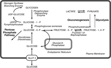
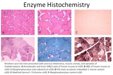
- [ 1 , 2 ] In healthy individuals, myophosphorylase initiates glycogen breakdown by removing 1,4-glucosyl groups from glycogen with the release of glucose-1-phosphate (see image below). (medscape.com)
- Most patients with McArdle disease have undetectable myophosphorylase activity and, thus, are unable to utilize energy (release glucose) from their glycogen stores in muscles. (medscape.com)
- During anaerobic exercise (eg, weightlifting, sprinting), the myophosphorylase in the skeletal muscles converts glycogen to glucose, which enters the glycolysis pathway to produce ATP anaerobically. (medscape.com)
- Thereafter, the duration and intensity of exercise determines the type of fuel source used by the skeletal muscles (eg, anaerobic glycolysis, blood glucose, muscle glycogen followed by aerobic glycolysis and fatty acid oxidation). (medscape.com)
- The glycogen phosphorylase can be converted into glucose 6-phosphate via phosphoglucomutase which is responsible for catalyzing the reversible reaction. (microbiologynote.com)
Fructose 6-phospha1
- Phosphoglucose isomerase (PGI) is a glycolytic enzyme that converts glucose-6-phosphate to fructose-6-phosphate, a key precursor of fungal cell wall biosynthesis. (diamond.ac.uk)
Metabolism7
- UDP-glucose is used in nucleotide sugar metabolism as an activated form of glucose, a substrate for enzymes called glucosyltransferases. (wikipedia.org)
- The absorption and initial metabolism of glucose by the acanthocephalan Polymorphus minutus have been studied in vitro under conditions designed to approximate to those in vivo . (biologists.com)
- The other aim was to expand the work of Crompton & Ward (1967 a ) , who found that lactic and succinic acids were the main excretory products from the carbohydrate metabolism of P. minutus , by analyzing extracts of the worms for other metabolites originating from glucose. (biologists.com)
- BACKGROUND: Fibroblast growth factor (FGF) 15/19, which is expressed in and secreted from the distal ileum, can regulate hepatic glucose metabolism in an endocrine manner. (bvsalud.org)
- The team found that UDP-glucose 4-epimerase (Uge5), an enzyme known for its involvement in galactose metabolism, also regulates the expression of degrading enzyme genes in A. aculeatus. (asiaresearchnews.com)
- Uridine diphosphate (UDP)-glucose 4-epimerase (Uge5) is well known as an enzyme involved in galactose metabolism. (asiaresearchnews.com)
- Galactosemia Galactosemia is a carbohydrate metabolism disorder caused by inherited deficiencies in enzymes that convert galactose to glucose. (msdmanuals.com)
Biosynthesis1
- Mechanistically, metabolic reprogramming in PCK1-loss hepatoma cells led to oxaloacetate accumulation and increased de novo uridine triphosphate synthesis contributing to uridine diphosphate-N-acetylglucosamine (UDP-GlcNAc) biosynthesis. (mcw.edu)
Enzyme5
- This enzyme catalyzes conversion of galactose-1-phosphate and UDP glucose to UDP galactose and glucose-1-phosphate. (medscape.com)
- Salmonella typhimurium gal E mutants used to produce the vaccine are effectively avirulent and highly protective but lack the specific UDP-galactose 4-epimerase enzyme which allows for the normal synthesis of UDP-galactose from UDP-glucose 12 . (drugbank.com)
- MS = An enzyme that catalyzes the transfer of UMP from UDPglucose to galactose 1-phosphate, forming UDPgalactose and glucose 1-phosphate. (doctorinternet.com)
- Given that glucose availability influences the use of uridine-derived ribose, we hypothesized that a glucose-depleted microenvironment triggers [pancreatic cancer cells] to upregulate UPP1 [the gene that makes an enzyme that breaks down uridine] as a compensatory response. (acsh.org)
- UPP1 codes for (synthesizes) the enzyme uridine phosphorylase (UP), also known as uridine-uracil phosphoribosyltransferase, which is responsible for the metabolic breakdown of uridine, a process that generates energy-rich metabolites that the cells can use. (acsh.org)
Nucleoside diphosphate1
- An improved method for the isolation of some nucleoside diphosphate sugar from yeast. (neglectedscience.com)
Nucleotide3
- Uridine diphosphate glucose (uracil-diphosphate glucose, UDP-glucose) is a nucleotide sugar. (wikipedia.org)
- DNA Nucleoside Nucleotide Oligonucleotide RNA TDP-glucose Uracil Uridine diphosphate Rademacher T, Parekh R, Dwek R (1988). (wikipedia.org)
- 2. Maxwell, E.S. and de Robichon-Szulmajster, H. Purification of uridine diphosphate galactose-4-epimerase from yeast and the identification of protein-bound diphosphopyridine nucleotide. (qmul.ac.uk)
Enzymes3
- The deduced amino acid sequences of these ORFs showed a high degree of homology to the deduced amino acid sequences of the rfbC, rfbD, rfbA, and rfbB genes of Salmonella typhimurium LT2, which have been shown to encode a set of enzymes responsible for conversion of glucose 1-phosphate to dTDP-rhamnose. (uni-bielefeld.de)
- Uridine diphosphate-glucuronosyltransferases (UGTs) are phase 2 conjugation enzymes mainly located in the endoplasmic reticulum (ER) of the liver and many other tissues, and can be recovered in artificial ER membrane preparations (microsomes). (mdpi.com)
- Dextrins and maltose are converted to glucose by enzymes in the border of the intestinal brush (the microvilli that resemble fingers of the epithelial cells in the intestinal tract, which dramatically increase the surface area of the surface of the intestine). (microbiologynote.com)
Pathway2
- Regardless, although it is predominantly used as a pathway to generate glucose fuel for the human body, galactose is involved as an ingredient in some commonly used vaccines and non-prescription products. (drugbank.com)
- The glucose 6-phosphate that is formed during the phosphoglucomutase process can be absorbed into glycolysis or a different pathway, such as the pentose phosphate pathway. (microbiologynote.com)
Phosphate2
- 4) glycosidic linkage, which connects to Iast the two sugar residues on a non-reducing end, creating glucose 1-phosphate as well as the polymer, which is one that is a glucose unit smaller. (microbiologynote.com)
- PhosphoroLyses conserves a portion of the energy contained in the glycosidic bond within the phosphate ester of glucose 1-phosphate. (microbiologynote.com)
Ribose3
Metabolites1
- After 4 hr. incubation with radioactive glucose the radioactive label has been detected in a variety of metabolites extracted from the worms. (biologists.com)
Monophosphate2
- Dolichol monophosphate glucose: an intermediate in glucose transfer in liver. (neglectedscience.com)
- Glucose transfer from doliochol monophosphate glucose: the product formed with endogenous microsomal acceptor. (neglectedscience.com)
Undergo glycolysis2
- In the process of somatostatin secretion, glucose must first undergo glycolysis in the mitochondrion to increase ATP in the cell. (smpdb.ca)
- In the majority of organisms, other hexoses than glucose are able to undergo glycolysis following transformation into the phosphorylated form. (microbiologynote.com)
Uracil1
- Conidia were made by developing strains on great moderate with or without uracil and uridine for 48?h in 37C. (thetechnoant.info)
Form of glucose1
- It is also evident from the literature that, since these helminths do not possess an alimentary tract, most of their dietary carbohydrate must be absorbed through their body surfaces in the form of glucose present in the lumen of their hosts' intestine. (biologists.com)
Derivatives1
- This is carrier of sugar molecules such as glucose, galactose or their derivatives. (biologydiscussion.com)
Monosaccharide1
- Galactose is a naturally occurring monosaccharide that forms the disaccharide lactose when combined with glucose (another monosaccharide) 6 . (drugbank.com)
Donor1
- Chitin, accounting for about 10C20% from the cell wall structure [9], is normally synthesized by chitin synthases that make use of UDP (uridine diphosphate)CGlcNAc as the glucose donor. (thetechnoant.info)
Metabolic1
- Such glucose is, of course, ultimately relied upon and used as the primary metabolic fuel for humans in a variety of biological reactions. (drugbank.com)
Amino acids1
- Nervous and muscle tissues preferentially oxidize ketones over fatty acids, glucose, or amino acids under normal conditions. (medscape.com)
Concentrations1
- The acanthocephalan, Polymorphus minutus , inhabits a region of the small intestine of ducks where a considerable range of glucose concentrations has been measured ( Crompton, 1966 ). (biologists.com)
Carrier2
- The results of the investigation of the absorption of glucose are compatible with the assumption that a carrier system is involved in the transport of glucose. (biologists.com)
- The carrier system is half-saturated at an external glucose concentration of about 0·25 mg./ml. and is tending towards full saturation when the external glucose concentration reaches 2·0 mg./ml. (biologists.com)
Reaction2
- The kit distinguishes 5-mC from 5-hmC by the addition of glucose to the hydroxyl group of 5-hmC via an enzymatic reaction utilizing T4 β-glucosyltransferase (T4-BGT). (neb.com)
- Step I: DNA Glucosylation Reaction with T4 β-glucosyltransferase (T4-BGT) Genomic DNA of interest is treated with T4-BGT, adding a glucose moeity to 5-hydroxymethylcytosine. (neb.com)
Carbohydrates2
- Glucose and galactose increase postprandially and several other carbohydrates contained in milk do not. (medscape.com)
- Numerous carbohydrates, including glucose, meet their catabolic end in $ycolysis after being transformed into glycolytic intermediates. (microbiologynote.com)
Liver1
- The brain, heart, skeletal muscle, and liver depend on ketone bodies or on glucose for energy and for carbon chains to synthesize cellular proteins (see the images below). (medscape.com)
Found3
- The rate of glucose absorption, at a given glucose concentration, has been found to fall when the worms have been acclimatized to that concentration. (biologists.com)
- It is most probable that P. minutus will be found to have a requirement for carbohydrate similar to that of M. dubius , and the primary aim of the work reported in this paper was to investigate the absorption of glucose by P. minutus in vitro , under conditions designed to simulate those in the worm's environment. (biologists.com)
- Subsequently, when lactose or small amounts of free galactose found in various common dairy products (and other foods) are consumed, the hydrolysis of lactose to glucose and galactose occurs and galactose is itself further metabolized to generate glucose 6 . (drugbank.com)
Brain1
- Many of these chemicals are synthesized in the brain, in part or wholly from glucose or ketone bodies. (medscape.com)
Fuel5
- Pancreatic Cancer Cells Deprived of Glucose Find Another Fuel. (acsh.org)
- A new study in the journal Nature examines what happens when pancreatic cancer cells are deprived of glucose, their normal fuel. (acsh.org)
- It's because cancer cells are perfectly willing to switch to another fuel source - uridine (Figure 1). (acsh.org)
- L) Glucose, the primary fuel of the body. (acsh.org)
- R) The chemical structure of uridine diphosphate, an alternate fuel for pancreatic cancer cells. (acsh.org)