Umbilical Arteries
Single Umbilical Artery
Ultrasonography, Prenatal
Umbilical Veins
Ultrasonography, Doppler
Fetal Growth Retardation
Placental Circulation
Pregnancy
Heart Rate, Fetal
Blood Flow Velocity
Gestational Age
Ultrasonography, Doppler, Pulsed
Umbilical Cord
Fetal Distress
Pulsatile Flow
Fetal Monitoring
Placental Insufficiency
Fetus
Fetal Diseases
Ultrasonography, Doppler, Color
Pulmonary Artery
Pregnancy Outcome
Fetal Blood
Pregnancy Trimester, Third
Middle Cerebral Artery
Carotid Arteries
Placenta
Uterine Artery
Pregnancy Trimester, Second
Hernia, Umbilical
Mesenteric Arteries
Fetofetal Transfusion
Fetal Heart
Basilar Artery
Hallermann's Syndrome
Pre-Eclampsia
Endothelium, Vascular
Pregnancy, High-Risk
Infant, Small for Gestational Age
Apgar Score
Iliac Artery
Prospective Studies
Vertebral Artery
Coronary Artery Bypass
Pregnancy Trimester, First
Radial Artery
Uterus
Mammary Arteries
Vascular Resistance
Human Umbilical Vein Endothelial Cells
Carotid Artery, Internal
Subclavian Artery
Cardiotocography
Fetal Weight
Maternal-Fetal Exchange
Carotid Artery Diseases
Umbilicus
Chorion
Splenic Artery
Brachial Artery
Abnormalities, Multiple
Dihydralazine
Pregnancy, Prolonged
Hepatic Artery
Carotid Artery, Common
Ultrasonography
Cardiovascular Abnormalities
Laser-Doppler Flowmetry
Fetal Movement
Cells, Cultured
Reference Values
Endothelial Cells
Oligohydramnios
Prenatal Diagnosis
Vasoconstriction
Birth Weight
Pregnancy Complications, Cardiovascular
Vasodilation
Celiac Artery
Sensitivity and Specificity
HSP20 Heat-Shock Proteins
Ophthalmic Artery
Mesenteric Artery, Superior
Anesthesia, Obstetrical
Serotonin
Mechanical Phenomena
Catheterization, Peripheral
Hemodynamics
Pregnancy Complications
Predictive Value of Tests
Pregnancy in Diabetics
Renal Artery Obstruction
Thoracic Arteries
Retrospective Studies
Temporal Arteries
Cord Blood Stem Cell Transplantation
Twins
Bronchial Arteries
Isoprostanes
Popliteal Artery
Prostaglandins, Synthetic
Heat shock protein expression in umbilical artery smooth muscle. (1/593)
Postpartum vasospasm in the umbilical arteries may be due to impaired vasorelaxation secondary to alterations in the expression of heat shock proteins. The contractile responses of pre- and full-term bovine umbilical artery smooth muscles were determined in a muscle bath. Heat shock protein expression was determined in bovine and human arterial tissues using western blotting with specific antisera. Full-term bovine and human umbilical artery smooth muscle was refractory to relaxation induced by the nitric oxide donor, sodium nitroprusside. This impaired vasorelaxation was associated with the expression of the inducible form of the heat shock protein, HSP70i, and increases in the expression of the small heat shock protein, HSP27. Small heat shock proteins have been implicated in modulating contraction and relaxation responses in vascular smooth muscles. Thus, alterations in heat shock protein expression may play a role in umbilical artery vasospasm. (+info)Strong induction of members of the chitinase family of proteins in atherosclerosis: chitotriosidase and human cartilage gp-39 expressed in lesion macrophages. (2/593)
Atherosclerosis is initiated by the infiltration of monocytes into the subendothelial space of the vessel wall and subsequent lipid accumulation of the activated macrophages. The molecular mechanisms involved in the anomalous behavior of macrophages in atherogenesis have only partially been disclosed. Chitotriosidase and human cartilage gp-39 (HC gp-39) are members of the chitinase family of proteins and are expressed in lipid-laden macrophages accumulated in various organs during Gaucher disease. In addition, as shown in this study, chitotriosidase and HC gp-39 can be induced with distinct kinetics in cultured macrophages. We investigated the expression of these chitinase-like genes in the human atherosclerotic vessel wall by in situ hybridizations on atherosclerotic specimens derived from femoral artery (4 specimens), aorta (4 specimens), iliac artery (3 specimens), carotid artery (4 specimens), and coronary artery (1 specimen), as well as 5 specimens derived from apparently normal vascular tissue. We show for the first time that chitotriosidase and HC gp-39 expression was strongly upregulated in distinct subsets of macrophages in the atherosclerotic plaque. The expression patterns of chitotriosidase and HC gp-39 were compared and shown to be different from the patterns observed for the extracellular matrix protein osteopontin and the macrophage marker tartrate-resistant acid phosphatase. Our data emphasize the remarkable phenotypic variation among macrophages present in the atherosclerotic lesion. Furthermore, chitotriosidase enzyme activity was shown to be elevated up to 55-fold in extracts of atherosclerotic tissue. Although a function for chitotriosidase and HC gp-39 has not been identified, we hypothesize a role in cell migration and tissue remodeling during atherogenesis. (+info)Thromboatheromatous complications of umbilical arterial catheterization in the newborn period. Clinicopathological study. (3/593)
Severe catheter-related thromboatheromatous lesions were found at necropsy in 33 of 56 infants who had umbilical arterial catheters passed during life. In infants dying within 8 days of insertion of the catheter, varying degrees of thrombosis of the aorta and its major branches were seen. With increasing thrombosis and aging of the thrombus, fatty deposits were seen first within the thrombus, and then in the intima and media. In addition there was evidence of proliferation of medial smooth muscle cells and of disruption of the medial architecture below the thrombus, characterized by the presence of abundant mucopolysaccharide. In infants who survived longer, varying degrees of organization of the thrombus could be traced, leading eventually to raised fibrous plaques with lipid and occasionally calcification. The lesions in the older infants were similar in many respects to experimental thromboatheromatous lesions produced in rabbits, and to some lesions of artheroma occurring spontaneously in humans. A wide variety of embolic phenomena were found, with features suggesting asynchrony of embolic episodes. The presence of thrombotic lesions could not be related to birthweight, Apgar scores at 1 and 5 minutes, age at catheterization, duration of catheterization, underlying disease process, age at death or the presence of hypothermia, acidosis, or anomalies in coagulation tests. There is a need for less hazardous methods of monitoring arterial oxygen tension. (+info)Circulatory changes induced by isovolumic increase in red cell mass in fetal lambs. (4/593)
AIM: To verify whether extra uterine changes in total peripheral vascular resistance and cardiac output, caused by raised haematocrit, occur in fetal life and if they can be documented using conventional ultrasound techniques. METHODS: An exchange transfusion with packed red cells was performed on five fetal lambs at 140 days of gestation (weight 3.44, SD 0.48 kg); three others were used as controls. The haematocrit was raised from 44 +/- 3 to 64 (SD2)%. RESULTS: Body temperature, blood gas, and pH remained within normal limits. Blood viscosity increased from 5.3 (0.3) to 9.6 (1.6) cps. Combined cardiac output fell to 30% of its initial value. The pulsatility index (PI) remained unchanged in the umbilical artery (0.66, SD 0.1) and descending aorta (1.3, SD 0.3). A significant positive correlation was found between haematocrit and PI only in the carotid artery (r = 0.67, p < 0.01). CONCLUSION: In the fetus, as in adults, an increase in blood viscosity is associated with a fall in cardiac output. However, the low resistance and the relative inertia of the placental vascular bed blunt the velocimetric changes that could be induced in the lower body vascular system by an increase in resistance. Such changes were observed only in the carotid artery. These results could be of interest in the Doppler monitoring of human fetuses at risk of an abnormal increase in their haematocrit. (+info)Characteristics of blood flow in intrauterine growth-restricted fetuses with hypercoiled cord. (5/593)
OBJECTIVE: To clarify the characteristics of fetoplacental blood flow of growth-restricted fetuses with hypercoiled umbilical cord. SUBJECTS: Eight growth-restricted fetuses with hypercoiled cord. METHODS: Flow velocity waveforms of the umbilical cord artery and vein, fetal abdominal aorta and fetal inferior vena cava were analyzed. RESULTS: The resistance index in the umbilical artery in the hypercoiled cases was lower than that in normal fetuses. Early-diastolic reversed flow was observed in the abdominal aorta in some cases. In all cases, umbilical venous pulsation was observed in the entire cord until delivery. In one case, fetal heart failure occurred, resulting in pre-mature delivery. An atrophic type of single umbilical artery was observed in four cases. CONCLUSION: Fetal blood flow disturbance caused by a hypercoiled umbilical cord may be a cause of growth restriction. (+info)Prenatal diagnosis of a lean umbilical cord: a simple marker for the fetus at risk of being small for gestational age at birth. (6/593)
OBJECTIVE: The purpose of this study was to investigate whether the prenatal diagnosis of a 'lean' umbilical cord in otherwise normal fetuses identifies fetuses at risk of being small for gestational age (SGA) at birth and of having distress in labor. The umbilical cord was defined as lean when its cross-sectional area on ultrasound examination was below the 10th centile for gestational age. METHOD: Pregnant women undergoing routine sonographic examination were included in the study. Inclusion criteria were gestational age greater than 20 weeks, intact membranes, and singleton gestation. The sonographic cross-sectional area of the umbilical cord was measured in a plane adjacent to the insertion into the fetal abdomen. Umbilical artery Doppler waveforms were recorded during fetal apnea and fetal anthropometric parameters were measured. RESULTS: During the study period, 860 patients met the inclusion criteria, of whom 3.6% delivered a SGA infant. The proportion of SGA infants was higher among fetuses who had a lean umbilical cord on ultrasound examination than among those with a normal umbilical cord (11.5% vs. 2.6%, p < 0.05). Fetuses with a lean cord had a risk 4.4-fold higher of being SGA at birth than those with a normal umbilical cord. After 25 weeks of gestation, this risk was 12.4 times higher when the umbilical cord was lean than when it was of normal size. The proportion of fetuses with meconium-stained amniotic fluid at delivery was higher among fetuses with a lean cord than among those with a normal umbilical cord (14.6% vs. 3.1%, p < 0.001). The proportion of infants who had a 5-min Apgar score < 7 was higher among those who had a lean cord than among those with normal umbilical cord (5.2% vs. 1.3%, p < 0.05). Considering only patients admitted in labor with intact membranes and who delivered an appropriate-for-gestational-age infant, the proportion of fetuses who had oligohydramnios at the time of delivery was higher among those who had a lean cord than among those with a normal umbilical cord (17.6% versus 1.3%, p < 0.01). CONCLUSION: We conclude that fetuses with a lean umbilical cord have an increased risk of being small for gestational age at birth and of having signs of distress at the time of delivery. (+info)Early prenatal diagnosis of cord entanglement in monoamniotic multiple pregnancies. (7/593)
OBJECTIVES: Cord entanglement is a severe complication in monoamniotic multiple pregnancies. Three cases were reviewed to determine how early ultrasound diagnosis might improve counselling and management. METHODS: In two monoamniotic twin and one dichorionic diamniotic triplet pregnancies, cord entanglement was detected between 10 and 18 gestational weeks by color Doppler and pulsed Doppler velocimetry. Pregnancies were followed up on a weekly basis with special observation of fetal behavior and use of color Doppler velocimetry. RESULTS: In Case 1, a monoamniotic twin pregnancy with cord entanglement close to the umbilical insertions was diagnosed at 10 weeks. Longitudinal follow-up showed intrauterine death of both twins at 15 weeks. In Case 2, entanglement of the umbilical cords of two monoamniotic triplets within a dichorionic diamniotic triplet pregnancy was diagnosed at 10 weeks. The pregnancy continued uneventfully until 35 weeks when cord entanglement was confirmed at Cesarean section. All triplets have since developed normally. In Case 3, monoamniotic twins were diagnosed at 18 weeks. Color Doppler detected side-by-side insertion of the umbilical cords and Doppler velocimetry suggested an entanglement at the chorionic plate. The pregnancy was complicated by polyhydramnios. Cesarean section at 36 weeks confirmed cord entanglement at the chorionic plate. Postnatal computer angiography and morphological examination of the placenta showed the presence of superficial artery-to-artery and vein-to-vein anastomoses and of deep arteriovenous shunts. The development of the twins was uneventful. CONCLUSIONS: Diagnosis of cord entanglement is feasible early in gestation. Future protocols are proposed to document the gestational age at detection, the location, and the Doppler flow patterns and to facilitate the assessment of short- and long-term development. (+info)Abnormal ductus venosus blood flow: a clue to umbilical cord complication. (8/593)
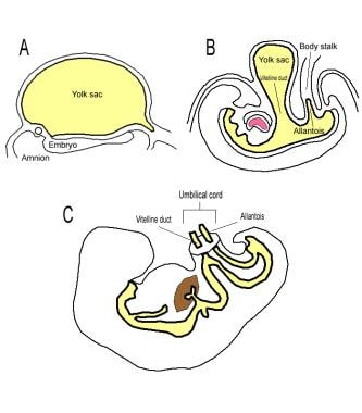
We report a case of umbilical cord complication causing, fetal hypoxemia and acidemia. At 30 weeks of gestation, the patient was referred because of slightly increased amniotic fluid volume and a non-reactive cardiotocogram. Biometry was appropriate for gestational age. Umbilical artery and fetal aortic Doppler findings were normal, whereas diastolic blood flow velocities in the middle cerebral artery were increased and the ductus venosus showed severely abnormal flow velocity waveforms with reversal of flow during atrial contraction. Since other reasons for fetal hypoxemia could be excluded, careful examination of the umbilical cord was performed. Traction of the hypercoiled umbilical cord due to its course around the fetal neck and shoulders was suspected. Cesarean section confirmed the sonographic findings and fetal blood gases revealed fetal acidemia. This case indicates that investigation of fetal venous blood flow may also help to identify fetal jeopardy due to reasons other than increased placental vascular resistance. (+info)The umbilical arteries are a pair of vessels that develop within the umbilical cord during fetal development. They carry oxygenated and nutrient-rich blood from the mother to the developing fetus through the placenta. These arteries arise from the internal iliac arteries in the fetus and pass through the umbilical cord to connect with the two umbilical veins within the placenta. After birth, the umbilical arteries become ligaments (the medial umbilical ligaments) that run along the inner abdominal wall.
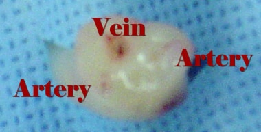
Single umbilical artery (SUA) is a congenital abnormality characterized by the presence of only one umbilical artery in the developing fetus, instead of the usual two. The umbilical cord typically contains two umbilical arteries and one umbilical vein, which are responsible for carrying oxygenated and nutrient-rich blood from the placenta to the growing fetus, as well as transporting deoxygenated and waste-laden blood back to the placenta.
The occurrence of a single umbilical artery can be an isolated finding or associated with other structural or chromosomal abnormalities. The exact cause of SUA is not fully understood, but it has been linked to factors such as maternal age, diabetes, and genetic predisposition.
During prenatal ultrasound examinations, the detection of a single umbilical artery might prompt further evaluation for potential associated anomalies or genetic disorders. In some cases, SUA may not have any significant consequences on fetal development or pregnancy outcomes; however, it can increase the risk for complications such as intrauterine growth restriction, preterm birth, and stillbirth. Therefore, close monitoring of the pregnancy is often recommended when a single umbilical artery is identified.
Prenatal ultrasonography, also known as obstetric ultrasound, is a medical diagnostic procedure that uses high-frequency sound waves to create images of the developing fetus, placenta, and amniotic fluid inside the uterus. It is a non-invasive and painless test that is widely used during pregnancy to monitor the growth and development of the fetus, detect any potential abnormalities or complications, and determine the due date.
During the procedure, a transducer (a small handheld device) is placed on the mother's abdomen and moved around to capture images from different angles. The sound waves travel through the mother's body and bounce back off the fetus, producing echoes that are then converted into electrical signals and displayed as images on a screen.
Prenatal ultrasonography can be performed at various stages of pregnancy, including early pregnancy to confirm the pregnancy and detect the number of fetuses, mid-pregnancy to assess the growth and development of the fetus, and late pregnancy to evaluate the position of the fetus and determine if it is head down or breech. It can also be used to guide invasive procedures such as amniocentesis or chorionic villus sampling.
Overall, prenatal ultrasonography is a valuable tool in modern obstetrics that helps ensure the health and well-being of both the mother and the developing fetus.
The umbilical veins are blood vessels in the umbilical cord that carry oxygenated and nutrient-rich blood from the mother to the developing fetus during pregnancy. There are typically two umbilical veins, one of which usually degenerates and becomes obliterated, leaving a single functional vein. This remaining vein is known as the larger umbilical vein or the venous duct. It enters the fetal abdomen through the umbilicus and passes through the liver, where it branches off to form the portal sinus. Ultimately, the blood from the umbilical vein mixes with the blood from the inferior vena cava and is pumped to the heart through the right atrium.
It's important to note that after birth, the umbilical veins are no longer needed and undergo involution, becoming the ligamentum teres in the adult.
Ultrasonography, Doppler refers to a non-invasive diagnostic medical procedure that uses high-frequency sound waves to create real-time images of the movement of blood flow through vessels, tissues, or heart valves. The Doppler effect is used to measure the frequency shift of the ultrasound waves as they bounce off moving red blood cells, which allows for the calculation of the speed and direction of blood flow. This technique is commonly used to diagnose and monitor various conditions such as deep vein thrombosis, carotid artery stenosis, heart valve abnormalities, and fetal heart development during pregnancy. It does not use radiation or contrast agents and is considered safe with minimal risks.
Fetal growth retardation, also known as intrauterine growth restriction (IUGR), is a condition in which a fetus fails to grow at the expected rate during pregnancy. This can be caused by various factors such as maternal health problems, placental insufficiency, chromosomal abnormalities, and genetic disorders. The fetus may be smaller than expected for its gestational age, have reduced movement, and may be at risk for complications during labor and delivery. It is important to monitor fetal growth and development closely throughout pregnancy to detect any potential issues early on and provide appropriate medical interventions.
Placental circulation refers to the specialized circulatory system that develops during pregnancy to allow for the exchange of nutrients, oxygen, and waste products between the mother's blood and the fetal blood in the placenta. The placenta is a highly vascular organ that grows within the uterus and is connected to the developing fetus via the umbilical cord.
In the maternal side of the placenta, the spiral arteries branch into smaller vessels called the intervillous spaces, where they come in close contact with the fetal blood vessels within the villi (finger-like projections) of the placenta. The intervillous spaces are filled with maternal blood that flows around the villi, allowing for the exchange of gases and nutrients between the two circulations.
On the fetal side, the umbilical cord contains two umbilical arteries that carry oxygen-depleted blood from the fetus to the placenta, and one umbilical vein that returns oxygenated blood back to the fetus. The umbilical arteries branch into smaller vessels within the villi, where they exchange gases and nutrients with the maternal blood in the intervillous spaces.
Overall, the placental circulation is a crucial component of fetal development, allowing for the growing fetus to receive the necessary oxygen and nutrients to support its growth and development.
Arteries are blood vessels that carry oxygenated blood away from the heart to the rest of the body. They have thick, muscular walls that can withstand the high pressure of blood being pumped out of the heart. Arteries branch off into smaller vessels called arterioles, which further divide into a vast network of tiny capillaries where the exchange of oxygen, nutrients, and waste occurs between the blood and the body's cells. After passing through the capillary network, deoxygenated blood collects in venules, then merges into veins, which return the blood back to the heart.
Pregnancy is a physiological state or condition where a fertilized egg (zygote) successfully implants and grows in the uterus of a woman, leading to the development of an embryo and finally a fetus. This process typically spans approximately 40 weeks, divided into three trimesters, and culminates in childbirth. Throughout this period, numerous hormonal and physical changes occur to support the growing offspring, including uterine enlargement, breast development, and various maternal adaptations to ensure the fetus's optimal growth and well-being.
Fetal heart rate (FHR) is the number of times a fetus's heart beats in one minute. It is measured through the use of a fetoscope, Doppler ultrasound device, or cardiotocograph (CTG). A normal FHR ranges from 120 to 160 beats per minute (bpm), although it can vary throughout pregnancy and is usually faster than an adult's heart rate. Changes in the FHR pattern may indicate fetal distress, hypoxia, or other conditions that require medical attention. Regular monitoring of FHR during pregnancy, labor, and delivery helps healthcare providers assess fetal well-being and ensure a safe outcome for both the mother and the baby.
Blood flow velocity is the speed at which blood travels through a specific part of the vascular system. It is typically measured in units of distance per time, such as centimeters per second (cm/s) or meters per second (m/s). Blood flow velocity can be affected by various factors, including cardiac output, vessel diameter, and viscosity of the blood. Measuring blood flow velocity is important in diagnosing and monitoring various medical conditions, such as heart disease, stroke, and peripheral vascular disease.
Gestational age is the length of time that has passed since the first day of the last menstrual period (LMP) in pregnant women. It is the standard unit used to estimate the age of a pregnancy and is typically expressed in weeks. This measure is used because the exact date of conception is often not known, but the start of the last menstrual period is usually easier to recall.
It's important to note that since ovulation typically occurs around two weeks after the start of the LMP, gestational age is approximately two weeks longer than fetal age, which is the actual time elapsed since conception. Medical professionals use both gestational and fetal age to track the development and growth of the fetus during pregnancy.
Ultrasonography, Doppler, Pulsed is a type of diagnostic ultrasound technique that uses the Doppler effect to measure blood flow in the body. In this technique, short bursts of ultrasound are emitted and then listened for as they bounce back off moving red blood cells. By analyzing the frequency shift of the returning sound waves, the velocity and direction of blood flow can be determined. This information is particularly useful in evaluating conditions such as deep vein thrombosis, carotid artery stenosis, and fetal heart abnormalities. Pulsed Doppler ultrasonography provides more detailed information about blood flow than traditional color Doppler imaging, making it a valuable tool for diagnosing and monitoring various medical conditions.
The umbilical cord is a flexible, tube-like structure that connects the developing fetus to the placenta in the uterus during pregnancy. It arises from the abdomen of the fetus and transports essential nutrients, oxygen, and blood from the mother's circulation to the growing baby. Additionally, it carries waste products, such as carbon dioxide, from the fetus back to the placenta for elimination. The umbilical cord is primarily composed of two arteries (the umbilical arteries) and one vein (the umbilical vein), surrounded by a protective gelatinous substance called Wharton's jelly, and enclosed within a fibrous outer covering known as the umbilical cord coating. Following birth, the umbilical cord is clamped and cut, leaving behind the stump that eventually dries up and falls off, resulting in the baby's belly button.
Fetal distress is a term used to describe situations where a fetus is experiencing problems during labor or delivery that are causing significant physiological changes. These changes may include an abnormal heart rate, decreased oxygen levels, or the presence of meconium (the baby's first stool) in the amniotic fluid. Fetal distress can be caused by a variety of factors, such as problems with the umbilical cord, placental abruption, maternal high blood pressure, or prolonged labor. It is important to monitor fetal well-being during labor and delivery to detect and address any signs of fetal distress promptly. Treatment may include changing the mother's position, administering oxygen, giving intravenous fluids, or performing an emergency cesarean section.
Pulsatile flow is a type of fluid flow that occurs in a rhythmic, wave-like pattern, typically seen in the cardiovascular system. It refers to the periodic variation in the volume or velocity of a fluid (such as blood) that is caused by the regular beating of the heart. In pulsatile flow, there are periods of high flow followed by periods of low or no flow, which creates a distinct pattern on a graph or tracing. This type of flow is important for maintaining proper function and health in organs and tissues throughout the body.
Fetal monitoring is a procedure used during labor and delivery to assess the well-being of the fetus. It involves the use of electronic devices to measure and record the fetal heart rate and uterine contractions. The information obtained from fetal monitoring can help healthcare providers identify any signs of fetal distress, such as a decreased fetal heart rate, which may indicate the need for interventions or an emergency cesarean delivery.
There are two main types of fetal monitoring: external and internal. External fetal monitoring involves placing sensors on the mother's abdomen to detect the fetal heart rate and uterine contractions. Internal fetal monitoring, which is typically used during high-risk deliveries, involves inserting an electrode into the fetus' scalp to measure the fetal heart rate more accurately.
Fetal monitoring can provide valuable information about the fetus's well-being during labor and delivery, but it is important to note that it has limitations and may not always detect fetal distress in a timely manner. Therefore, healthcare providers must use their clinical judgment and other assessment tools, such as fetal movement counting and visual examination of the fetus, to ensure the safe delivery of the baby.
Placental insufficiency is a condition in which the placenta does not provide adequate nutrients and oxygen to the developing fetus. This can occur due to various reasons, such as poor placental development, damage to the placenta, or problems with the blood flow to the placenta. As a result, the fetus may receive less oxygen and nutrients than it needs for proper growth and development, which can lead to a range of complications, including low birth weight, preterm birth, and developmental delays.
The medical definition of placental insufficiency is: "a condition in which the placenta fails to provide adequate support to the developing fetus, resulting in impaired fetal growth and development." This condition can be diagnosed through various tests, such as ultrasound, fetal monitoring, and blood tests, and may require close monitoring and management throughout pregnancy to ensure the best possible outcomes for both the mother and the baby.
A fetus is the developing offspring in a mammal, from the end of the embryonic period (approximately 8 weeks after fertilization in humans) until birth. In humans, the fetal stage of development starts from the eleventh week of pregnancy and continues until childbirth, which is termed as full-term pregnancy at around 37 to 40 weeks of gestation. During this time, the organ systems become fully developed and the body grows in size. The fetus is surrounded by the amniotic fluid within the amniotic sac and is connected to the placenta via the umbilical cord, through which it receives nutrients and oxygen from the mother. Regular prenatal care is essential during this period to monitor the growth and development of the fetus and ensure a healthy pregnancy and delivery.
Cerebral arteries refer to the blood vessels that supply oxygenated blood to the brain. These arteries branch off from the internal carotid arteries and the vertebral arteries, which combine to form the basilar artery. The major cerebral arteries include:
1. Anterior cerebral artery (ACA): This artery supplies blood to the frontal lobes of the brain, including the motor and sensory cortices responsible for movement and sensation in the lower limbs.
2. Middle cerebral artery (MCA): The MCA is the largest of the cerebral arteries and supplies blood to the lateral surface of the brain, including the temporal, parietal, and frontal lobes. It is responsible for providing blood to areas involved in motor function, sensory perception, speech, memory, and vision.
3. Posterior cerebral artery (PCA): The PCA supplies blood to the occipital lobe, which is responsible for visual processing, as well as parts of the temporal and parietal lobes.
4. Anterior communicating artery (ACoA) and posterior communicating arteries (PComAs): These are small arteries that connect the major cerebral arteries, forming an important circulatory network called the Circle of Willis. The ACoA connects the two ACAs, while the PComAs connect the ICA with the PCA and the basilar artery.
These cerebral arteries play a crucial role in maintaining proper brain function by delivering oxygenated blood to various regions of the brain. Any damage or obstruction to these arteries can lead to serious neurological conditions, such as strokes or transient ischemic attacks (TIAs).
Fetal diseases are medical conditions or abnormalities that affect a fetus during pregnancy. These diseases can be caused by genetic factors, environmental influences, or a combination of both. They can range from mild to severe and may impact various organ systems in the developing fetus. Examples of fetal diseases include congenital heart defects, neural tube defects, chromosomal abnormalities such as Down syndrome, and infectious diseases such as toxoplasmosis or rubella. Fetal diseases can be diagnosed through prenatal testing, including ultrasound, amniocentesis, and chorionic villus sampling. Treatment options may include medication, surgery, or delivery of the fetus, depending on the nature and severity of the disease.
Ultrasonography, Doppler, color is a type of diagnostic ultrasound technique that uses the Doppler effect to produce visual images of blood flow in vessels and the heart. The Doppler effect is the change in frequency or wavelength of a wave in relation to an observer who is moving relative to the source of the wave. In this context, it refers to the change in frequency of the ultrasound waves as they reflect off moving red blood cells.
In color Doppler ultrasonography, different colors are used to represent the direction and speed of blood flow. Red typically represents blood flowing toward the transducer (the device that sends and receives sound waves), while blue represents blood flowing away from the transducer. The intensity or brightness of the color is proportional to the velocity of blood flow.
Color Doppler ultrasonography is often used in conjunction with grayscale ultrasound imaging, which provides information about the structure and composition of tissues. Together, these techniques can help diagnose a wide range of conditions, including heart disease, blood clots, and abnormalities in blood flow.
The pulmonary artery is a large blood vessel that carries deoxygenated blood from the right ventricle of the heart to the lungs for oxygenation. It divides into two main branches, the right and left pulmonary arteries, which further divide into smaller vessels called arterioles, and then into a vast network of capillaries in the lungs where gas exchange occurs. The thin walls of these capillaries allow oxygen to diffuse into the blood and carbon dioxide to diffuse out, making the blood oxygen-rich before it is pumped back to the left side of the heart through the pulmonary veins. This process is crucial for maintaining proper oxygenation of the body's tissues and organs.
Pregnancy outcome refers to the final result or status of a pregnancy, including both the health of the mother and the newborn baby. It can be categorized into various types such as:
1. Live birth: The delivery of one or more babies who show signs of life after separation from their mother.
2. Stillbirth: The delivery of a baby who has died in the womb after 20 weeks of pregnancy.
3. Miscarriage: The spontaneous loss of a pregnancy before the 20th week.
4. Abortion: The intentional termination of a pregnancy before the fetus can survive outside the uterus.
5. Ectopic pregnancy: A pregnancy that develops outside the uterus, usually in the fallopian tube, which is not viable and requires medical attention.
6. Preterm birth: The delivery of a baby before 37 weeks of gestation, which can lead to various health issues for the newborn.
7. Full-term birth: The delivery of a baby between 37 and 42 weeks of gestation.
8. Post-term pregnancy: The delivery of a baby after 42 weeks of gestation, which may increase the risk of complications for both mother and baby.
The pregnancy outcome is influenced by various factors such as maternal age, health status, lifestyle habits, genetic factors, and access to quality prenatal care.
Fetal blood refers to the blood circulating in a fetus during pregnancy. It is essential for the growth and development of the fetus, as it carries oxygen and nutrients from the placenta to the developing tissues and organs. Fetal blood also removes waste products, such as carbon dioxide, from the fetal tissues and transports them to the placenta for elimination.
Fetal blood has several unique characteristics that distinguish it from adult blood. For example, fetal hemoglobin (HbF) is the primary type of hemoglobin found in fetal blood, whereas adults primarily have adult hemoglobin (HbA). Fetal hemoglobin has a higher affinity for oxygen than adult hemoglobin, which allows it to more efficiently extract oxygen from the maternal blood in the placenta.
Additionally, fetal blood contains a higher proportion of reticulocytes (immature red blood cells) and nucleated red blood cells compared to adult blood. These differences reflect the high turnover rate of red blood cells in the developing fetus and the need for rapid growth and development.
Examination of fetal blood can provide important information about the health and well-being of the fetus during pregnancy. For example, fetal blood sampling (also known as cordocentesis or percutaneous umbilical blood sampling) can be used to diagnose genetic disorders, infections, and other conditions that may affect fetal development. However, this procedure carries risks, including preterm labor, infection, and fetal loss, and is typically only performed when there is a significant risk of fetal compromise or when other diagnostic tests have been inconclusive.
A newborn infant is a baby who is within the first 28 days of life. This period is also referred to as the neonatal period. Newborns require specialized care and attention due to their immature bodily systems and increased vulnerability to various health issues. They are closely monitored for signs of well-being, growth, and development during this critical time.
The third trimester of pregnancy is the final stage of pregnancy that lasts from week 29 until birth, which typically occurs around the 40th week. During this period, the fetus continues to grow and mature, gaining weight rapidly. The mother's body also prepares for childbirth by dilating the cervix and producing milk in preparation for breastfeeding. Regular prenatal care is crucial during this time to monitor the health of both the mother and the developing fetus, as well as to prepare for delivery.
The Middle Cerebral Artery (MCA) is one of the main blood vessels that supplies oxygenated blood to the brain. It arises from the internal carotid artery and divides into several branches, which supply the lateral surface of the cerebral hemisphere, including the frontal, parietal, and temporal lobes.
The MCA is responsible for providing blood flow to critical areas of the brain, such as the primary motor and sensory cortices, Broca's area (associated with speech production), Wernicke's area (associated with language comprehension), and the visual association cortex.
Damage to the MCA or its branches can result in a variety of neurological deficits, depending on the specific location and extent of the injury. These may include weakness or paralysis on one side of the body, sensory loss, language impairment, and visual field cuts.
The carotid arteries are a pair of vital blood vessels in the human body that supply oxygenated blood to the head and neck. Each person has two common carotid arteries, one on each side of the neck, which branch off from the aorta, the largest artery in the body.
The right common carotid artery originates from the brachiocephalic trunk, while the left common carotid artery arises directly from the aortic arch. As they ascend through the neck, they split into two main branches: the internal and external carotid arteries.
The internal carotid artery supplies oxygenated blood to the brain, eyes, and other structures within the skull, while the external carotid artery provides blood to the face, scalp, and various regions of the neck.
Maintaining healthy carotid arteries is crucial for overall cardiovascular health and preventing serious conditions like stroke, which can occur when the arteries become narrowed or blocked due to the buildup of plaque or fatty deposits (atherosclerosis). Regular check-ups with healthcare professionals may include monitoring carotid artery health through ultrasound or other imaging techniques.
The placenta is an organ that develops in the uterus during pregnancy and provides oxygen and nutrients to the growing baby through the umbilical cord. It also removes waste products from the baby's blood. The placenta attaches to the wall of the uterus, and the baby's side of the placenta contains many tiny blood vessels that connect to the baby's circulatory system. This allows for the exchange of oxygen, nutrients, and waste between the mother's and baby's blood. After the baby is born, the placenta is usually expelled from the uterus in a process called afterbirth.
The femoral artery is the major blood vessel that supplies oxygenated blood to the lower extremity of the human body. It is a continuation of the external iliac artery and becomes the popliteal artery as it passes through the adductor hiatus in the adductor magnus muscle of the thigh.
The femoral artery is located in the femoral triangle, which is bound by the sartorius muscle anteriorly, the adductor longus muscle medially, and the biceps femoris muscle posteriorly. It can be easily palpated in the groin region, making it a common site for taking blood samples, measuring blood pressure, and performing surgical procedures such as femoral artery catheterization and bypass grafting.
The femoral artery gives off several branches that supply blood to the lower limb, including the deep femoral artery, the superficial femoral artery, and the profunda femoris artery. These branches provide blood to the muscles, bones, skin, and other tissues of the leg, ankle, and foot.
The uterine artery is a paired branch of the internal iliac (hip) artery that supplies blood to the uterus and vagina. It anastomoses (joins) with the ovarian artery to form a rich vascular network that nourishes the female reproductive organs. The right and left uterine arteries run along the sides of the uterus, where they divide into several branches to supply oxygenated blood and nutrients to the myometrium (uterine muscle), endometrium (lining), and cervix. These arteries undergo significant changes in size and structure during pregnancy to accommodate the growing fetus and placenta, making them crucial for maintaining a healthy pregnancy.
The renal artery is a pair of blood vessels that originate from the abdominal aorta and supply oxygenated blood to each kidney. These arteries branch into several smaller vessels that provide blood to the various parts of the kidneys, including the renal cortex and medulla. The renal arteries also carry nutrients and other essential components needed for the normal functioning of the kidneys. Any damage or blockage to the renal artery can lead to serious consequences, such as reduced kidney function or even kidney failure.
The second trimester of pregnancy is the period between the completion of 12 weeks (the end of the first trimester) and 26 weeks (the beginning of the third trimester) of gestational age. It is often considered the most comfortable period for many pregnant women as the risk of miscarriage decreases significantly, and the symptoms experienced during the first trimester, such as nausea and fatigue, typically improve.
During this time, the uterus expands above the pubic bone, allowing more space for the growing fetus. The fetal development in the second trimester includes significant growth in size and weight, formation of all major organs, and the beginning of movement sensations that the mother can feel. Additionally, the fetus starts to hear, swallow and kick, and the skin is covered with a protective coating called vernix.
Prenatal care during this period typically includes regular prenatal appointments to monitor the mother's health and the baby's growth and development. These appointments may include measurements of the uterus, fetal heart rate monitoring, and screening tests for genetic disorders or other potential issues.
Fetal hypoxia is a medical condition that refers to a reduced level of oxygen supply to the fetus. This can occur due to various reasons, such as maternal health problems, complications during pregnancy or delivery, or issues with the placenta. Prolonged fetal hypoxia can lead to serious complications, including brain damage and even fetal death. It is important for healthcare providers to closely monitor fetal oxygen levels during pregnancy and delivery to ensure the well-being of the fetus.
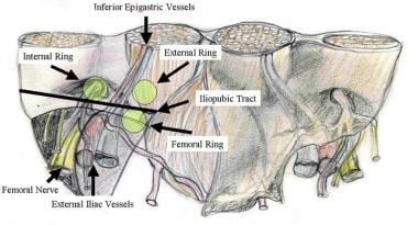
An umbilical hernia is a type of hernia that occurs at the umbilicus, or belly button. It results from a protrusion of abdominal contents through a weakened area in the abdominal wall surrounding the navel. This condition is common in newborns and infants, especially premature babies, due to incomplete closure of the abdominal muscles during development.
In most cases, umbilical hernias in children close on their own by age 3-4 or by the time they reach school age. However, if the hernia is still present after this age, surgical intervention may be required to prevent potential complications such as incarceration (where the herniated tissue becomes trapped and cannot be pushed back in) or strangulation (where the blood supply to the herniated tissue is cut off, leading to tissue death).
Adults can also develop umbilical hernias, often as a result of increased pressure in the abdomen due to obesity, pregnancy, heavy lifting, or persistent coughing. Umbilical hernias in adults are generally more likely to require surgical repair due to the higher risk of complications.
The mesenteric arteries are the arteries that supply oxygenated blood to the intestines. There are three main mesenteric arteries: the superior mesenteric artery, which supplies blood to the small intestine (duodenum to two-thirds of the transverse colon) and large intestine (cecum, ascending colon, and the first part of the transverse colon); the inferior mesenteric artery, which supplies blood to the distal third of the transverse colon, descending colon, sigmoid colon, and rectum; and the middle colic artery, which is a branch of the superior mesenteric artery that supplies blood to the transverse colon. These arteries are important in maintaining adequate blood flow to the intestines to support digestion and absorption of nutrients.
Fetofetal transfusion is a medical condition that can occur in pregnancies with multiple fetuses, such as twins or higher-order multiples. It refers to the transfer of blood from one fetus (donor) to another (recipient) through anastomotic connections in their shared placenta.
In some cases, these anastomoses can result in an imbalance in blood flow between the fetuses, leading to a net transfer of blood from one fetus to the other. This situation is more likely to occur when there is a significant weight or size difference between the fetuses, known as twin-to-twin transfusion syndrome (TTTS).
In TTTS, the recipient fetus receives an excess of blood, which can lead to high-output cardiac failure, hydrops, and potential intrauterine demise. Meanwhile, the donor fetus may become anemic, growth-restricted, and at risk for hypovolemia and intrauterine demise as well. Fetofetal transfusion can be diagnosed through ultrasound evaluation and managed with various interventions, including laser ablation of anastomotic vessels or fetoscopic surgery, depending on the severity and gestational age at diagnosis.
The fetal heart is the cardiovascular organ that develops in the growing fetus during pregnancy. It starts to form around 22 days after conception and continues to develop throughout the first trimester. By the end of the eighth week of gestation, the fetal heart has developed enough to pump blood throughout the body.
The fetal heart is similar in structure to the adult heart but has some differences. It is smaller and more compact, with a four-chambered structure that includes two atria and two ventricles. The fetal heart also has unique features such as the foramen ovale, which is a hole between the right and left atria that allows blood to bypass the lungs, and the ductus arteriosus, a blood vessel that connects the pulmonary artery to the aorta and diverts blood away from the lungs.
The fetal heart is responsible for pumping oxygenated blood from the placenta to the rest of the body and returning deoxygenated blood back to the placenta for re-oxygenation. The rate of the fetal heartbeat is faster than that of an adult, typically ranging from 120 to 160 beats per minute. Fetal heart rate monitoring is a common method used during pregnancy and childbirth to assess the health and well-being of the developing fetus.
The basilar artery is a major blood vessel that supplies oxygenated blood to the brainstem and cerebellum. It is formed by the union of two vertebral arteries at the lower part of the brainstem, near the junction of the medulla oblongata and pons.
The basilar artery runs upward through the center of the brainstem and divides into two posterior cerebral arteries at the upper part of the brainstem, near the midbrain. The basilar artery gives off several branches that supply blood to various parts of the brainstem, including the pons, medulla oblongata, and midbrain, as well as to the cerebellum.
The basilar artery is an important part of the circle of Willis, a network of arteries at the base of the brain that ensures continuous blood flow to the brain even if one of the arteries becomes blocked or narrowed.
Hallermann-Streiff syndrome is a rare genetic disorder characterized by a distinctive combination of skeletal, craniofacial, and skin abnormalities. The main features include a bird-like face with a prominent forehead, small chin, and beaked nose; widely spaced eyes (hypertelorism) with a short eyelid fissure; a thin beak-shaped upper jaw (maxilla); underdeveloped cheekbones (malar hypoplasia); and a small receding lower jaw (micrognathia).
Individuals with Hallermann-Streiff syndrome often have sparse hair, eyebrows, and eyelashes; thin skin; and an increased risk of developing cataracts and other eye abnormalities. They may also have dental anomalies, such as missing or malformed teeth, and a high-arched palate.
Hallermann-Streiff syndrome is caused by mutations in the GJA1 gene, which provides instructions for making a protein called connexin 43. This protein is important for the normal development and function of various tissues, including the bones and skin. The exact role of connexin 43 in the development of Hallermann-Streiff syndrome is not well understood.
Hallermann-Streiff syndrome is inherited in an autosomal recessive manner, which means that an individual must inherit two copies of the mutated gene (one from each parent) to develop the condition.
Pre-eclampsia is a pregnancy-related disorder, typically characterized by the onset of high blood pressure (hypertension) and damage to organs, such as the kidneys, after the 20th week of pregnancy. It is often accompanied by proteinuria, which is the presence of excess protein in the urine. Pre-eclampsia can lead to serious complications for both the mother and the baby if left untreated or unmanaged.
The exact causes of pre-eclampsia are not fully understood, but it is believed that placental issues, genetic factors, and immune system problems may contribute to its development. Risk factors include first-time pregnancies, history of pre-eclampsia in previous pregnancies, chronic hypertension, obesity, older age (35 or older), and assisted reproductive technology (ART) pregnancies.
Pre-eclampsia can progress to a more severe form called eclampsia, which is characterized by the onset of seizures. HELLP syndrome, another severe complication, involves hemolysis (breaking down of red blood cells), elevated liver enzymes, and low platelet count.
Early detection and management of pre-eclampsia are crucial to prevent severe complications. Regular prenatal care, including frequent blood pressure checks and urine tests, can help identify early signs of the condition. Treatment typically involves close monitoring, medication to lower blood pressure, corticosteroids to promote fetal lung maturity, and, in some cases, delivery of the baby if the mother's or baby's health is at risk.
The endothelium is a thin layer of simple squamous epithelial cells that lines the interior surface of blood vessels, lymphatic vessels, and heart chambers. The vascular endothelium, specifically, refers to the endothelial cells that line the blood vessels. These cells play a crucial role in maintaining vascular homeostasis by regulating vasomotor tone, coagulation, platelet activation, inflammation, and permeability of the vessel wall. They also contribute to the growth and repair of the vascular system and are involved in various pathological processes such as atherosclerosis, hypertension, and diabetes.
High-risk pregnancy is a term used to describe a situation where the mother or the fetus has an increased risk of developing complications during pregnancy, labor, delivery, or in the postpartum period. These risks may be due to pre-existing medical conditions in the mother, such as diabetes, hypertension, heart disease, kidney disease, autoimmune disorders, or infectious diseases like HIV/AIDS. Other factors that can contribute to a high-risk pregnancy include advanced maternal age (35 years and older), obesity, multiple gestations (twins, triplets, etc.), fetal growth restriction, placental issues, and a history of previous pregnancy complications or preterm labor.
High-risk pregnancies require specialized care and monitoring by healthcare professionals, often involving maternal-fetal medicine specialists, obstetricians, perinatologists, and neonatologists. Regular prenatal care, frequent checkups, ultrasound monitoring, and sometimes additional testing and interventions may be necessary to ensure the best possible outcomes for both the mother and the baby.
Small for Gestational Age (SGA) is a term used in pediatrics to describe newborn infants who are smaller in size than expected for the number of weeks they have been in the womb. It is typically defined as a baby whose weight is below the 10th percentile for its gestational age. SGA can be further classified into two categories: constitutionally small (also known as physiologically small) and pathologically small. Constitutionally small infants are those who are genetically predisposed to being smaller, while pathologically small infants have a growth restriction due to factors such as placental insufficiency, maternal hypertension, or chromosomal abnormalities.
It is important to note that SGA is not the same as premature birth. Premature babies are those born before 37 weeks of gestation, regardless of their size. However, a baby can be both premature and SGA.
The digestive system is a complex series of organs and glands that process food. Abnormalities in the digestive system can refer to a wide range of conditions that affect any part of the system, including the esophagus, stomach, small intestine, large intestine, liver, pancreas, and gallbladder. These abnormalities can be present at birth (congenital) or acquired later in life due to various factors such as infection, inflammation, injury, or disease.
Some examples of digestive system abnormalities include:
1. Gastroesophageal Reflux Disease (GERD): A condition where the stomach acid flows back into the esophagus, causing heartburn and damage to the esophageal lining.
2. Peptic Ulcers: Open sores that develop on the lining of the stomach or duodenum, often caused by bacterial infections or long-term use of nonsteroidal anti-inflammatory drugs (NSAIDs).
3. Inflammatory Bowel Disease (IBD): A group of chronic inflammatory conditions of the intestine, including Crohn's disease and ulcerative colitis.
4. Irritable Bowel Syndrome (IBS): A functional gastrointestinal disorder characterized by abdominal pain, bloating, and altered bowel habits.
5. Celiac Disease: An autoimmune disorder where the ingestion of gluten leads to damage in the small intestine.
6. Diverticulosis: The presence of small pouches or sacs that form on the lining of the intestine, which can become inflamed or infected (diverticulitis).
7. Hiatal Hernia: A condition where a portion of the stomach protrudes through the diaphragm into the chest cavity.
8. Hepatitis: Inflammation of the liver, often caused by viral infections or toxins.
9. Cirrhosis: A chronic liver disease characterized by scarring and loss of liver function, often due to long-term alcohol abuse or hepatitis.
10. Gallstones: Small, hard deposits that form in the gallbladder and can cause pain and inflammation.
These are just a few examples of gastrointestinal disorders, and there are many others. If you are experiencing symptoms such as abdominal pain, bloating, diarrhea, constipation, or difficulty swallowing, it is important to speak with your healthcare provider to determine the cause and develop an appropriate treatment plan.
The Apgar score is a quick assessment of the physical condition of a newborn infant, assessed by measuring heart rate, respiratory effort, muscle tone, reflex irritability, and skin color. It is named after Virginia Apgar, an American anesthesiologist who developed it in 1952. The score is usually given at one minute and five minutes after birth, with a possible range of 0 to 10. Scores of 7 and above are considered normal, while scores of 4-6 indicate moderate distress, and scores below 4 indicate severe distress. The Apgar score can provide important information for making decisions about the need for resuscitation or other medical interventions after birth.
Fetal death, also known as stillbirth or intrauterine fetal demise, is defined as the death of a fetus at 20 weeks of gestation or later. The criteria for defining fetal death may vary slightly by country and jurisdiction, but in general, it refers to the loss of a pregnancy after the point at which the fetus is considered viable outside the womb.
Fetal death can occur for a variety of reasons, including chromosomal abnormalities, placental problems, maternal health conditions, infections, and umbilical cord accidents. In some cases, the cause of fetal death may remain unknown.
The diagnosis of fetal death is typically made through ultrasound or other imaging tests, which can confirm the absence of a heartbeat or movement in the fetus. Once fetal death has been diagnosed, medical professionals will work with the parents to determine the best course of action for managing the pregnancy and delivering the fetus. This may involve waiting for labor to begin naturally, inducing labor, or performing a cesarean delivery.
Experiencing a fetal death can be a very difficult and emotional experience for parents, and it is important for them to receive supportive care from their healthcare providers, family members, and friends. Grief counseling and support groups may also be helpful in coping with the loss.
The iliac arteries are major branches of the abdominal aorta, the large artery that carries oxygen-rich blood from the heart to the rest of the body. The iliac arteries divide into two branches, the common iliac arteries, which further bifurcate into the internal and external iliac arteries.
The internal iliac artery supplies blood to the lower abdomen, pelvis, and the reproductive organs, while the external iliac artery provides blood to the lower extremities, including the legs and feet. Together, the iliac arteries play a crucial role in circulating blood throughout the body, ensuring that all tissues and organs receive the oxygen and nutrients they need to function properly.
Regional blood flow (RBF) refers to the rate at which blood flows through a specific region or organ in the body, typically expressed in milliliters per minute per 100 grams of tissue (ml/min/100g). It is an essential physiological parameter that reflects the delivery of oxygen and nutrients to tissues while removing waste products. RBF can be affected by various factors such as metabolic demands, neural regulation, hormonal influences, and changes in blood pressure or vascular resistance. Measuring RBF is crucial for understanding organ function, diagnosing diseases, and evaluating the effectiveness of treatments.
Prospective studies, also known as longitudinal studies, are a type of cohort study in which data is collected forward in time, following a group of individuals who share a common characteristic or exposure over a period of time. The researchers clearly define the study population and exposure of interest at the beginning of the study and follow up with the participants to determine the outcomes that develop over time. This type of study design allows for the investigation of causal relationships between exposures and outcomes, as well as the identification of risk factors and the estimation of disease incidence rates. Prospective studies are particularly useful in epidemiology and medical research when studying diseases with long latency periods or rare outcomes.
The vertebral artery is a major blood vessel that supplies oxygenated blood to the brain and upper spinal cord. It arises from the subclavian artery, then ascends through the transverse processes of several cervical vertebrae before entering the skull through the foramen magnum. Inside the skull, it joins with the opposite vertebral artery to form the basilar artery, which supplies blood to the brainstem and cerebellum. The vertebral artery also gives off several important branches that supply blood to various regions of the brainstem and upper spinal cord.
Coronary artery bypass surgery, also known as coronary artery bypass grafting (CABG), is a surgical procedure used to improve blood flow to the heart in patients with severe coronary artery disease. This condition occurs when the coronary arteries, which supply oxygen-rich blood to the heart muscle, become narrowed or blocked due to the buildup of fatty deposits, called plaques.
During CABG surgery, a healthy blood vessel from another part of the body is grafted, or attached, to the coronary artery, creating a new pathway for oxygen-rich blood to flow around the blocked or narrowed portion of the artery and reach the heart muscle. This bypass helps to restore normal blood flow and reduce the risk of angina (chest pain), shortness of breath, and other symptoms associated with coronary artery disease.
There are different types of CABG surgery, including traditional on-pump CABG, off-pump CABG, and minimally invasive CABG. The choice of procedure depends on various factors, such as the patient's overall health, the number and location of blocked arteries, and the presence of other medical conditions.
It is important to note that while CABG surgery can significantly improve symptoms and quality of life in patients with severe coronary artery disease, it does not cure the underlying condition. Lifestyle modifications, such as regular exercise, a healthy diet, smoking cessation, and medication therapy, are essential for long-term management and prevention of further progression of the disease.
The first trimester of pregnancy is defined as the period of gestational development that extends from conception (fertilization of the egg by sperm) to the end of the 13th week. This critical phase marks significant transformations in both the mother's body and the growing embryo/fetus.
During the first trimester, the fertilized egg implants into the uterine lining (implantation), initiating a series of complex interactions leading to the formation of the placenta - an organ essential for providing nutrients and oxygen to the developing fetus while removing waste products. Simultaneously, the embryo undergoes rapid cell division and differentiation, giving rise to various organs and systems. By the end of the first trimester, most major structures are present, although they continue to mature and grow throughout pregnancy.
The mother may experience several physiological changes during this time, including:
- Morning sickness (nausea and vomiting)
- Fatigue
- Breast tenderness
- Frequent urination
- Food aversions or cravings
- Mood swings
Additionally, hormonal shifts can cause various symptoms and prepare the body for potential changes in lactation, posture, and pelvic alignment as pregnancy progresses. Regular prenatal care is crucial during this period to monitor both maternal and fetal wellbeing, identify any potential complications early on, and provide appropriate guidance and support throughout the pregnancy.
Placental diseases, also known as placental pathologies, refer to a group of conditions that affect the development and function of the placenta during pregnancy. The placenta is an organ that develops in the uterus during pregnancy and provides oxygen and nutrients to the developing fetus while removing waste products.
Placental diseases can have serious consequences for both the mother and the fetus, including preterm labor, growth restriction, stillbirth, and long-term health problems for the child. Some common placental diseases include:
1. Placental abruption: This occurs when the placenta separates from the uterine wall before delivery, causing bleeding and potentially harming the fetus.
2. Placental previa: This is a condition where the placenta implants in the lower part of the uterus, covering the cervix. It can cause bleeding and may require cesarean delivery.
3. Preeclampsia: This is a pregnancy-related disorder characterized by high blood pressure and damage to organs such as the liver and kidneys. Placental dysfunction is thought to play a role in its development.
4. Intrauterine growth restriction (IUGR): This occurs when the fetus does not grow properly due to poor placental function, leading to low birth weight and potential health problems.
5. Chorioamnionitis: This is an infection of the membranes surrounding the fetus, which can lead to preterm labor and other complications.
6. Placental infarction: This occurs when a portion of the placenta dies due to a lack of blood flow, which can lead to growth restriction or stillbirth.
Prompt diagnosis and treatment of placental diseases are essential for ensuring the best possible outcomes for both the mother and the fetus.
The radial artery is a key blood vessel in the human body, specifically a part of the peripheral arterial system. Originating from the brachial artery in the upper arm, the radial artery travels down the arm and crosses over the wrist, where it can be palpated easily. It then continues into the hand, dividing into several branches to supply blood to the hand's tissues and digits.
The radial artery is often used for taking pulse readings due to its easy accessibility at the wrist. Additionally, in medical procedures such as coronary angiography or bypass surgery, the radial artery can be utilized as a site for catheter insertion. This allows healthcare professionals to examine the heart's blood vessels and assess cardiovascular health.
The uterus, also known as the womb, is a hollow, muscular organ located in the female pelvic cavity, between the bladder and the rectum. It has a thick, middle layer called the myometrium, which is composed of smooth muscle tissue, and an inner lining called the endometrium, which provides a nurturing environment for the fertilized egg to develop into a fetus during pregnancy.
The uterus is where the baby grows and develops until it is ready for birth through the cervix, which is the lower, narrow part of the uterus that opens into the vagina. The uterus plays a critical role in the menstrual cycle as well, by shedding its lining each month if pregnancy does not occur.
The mammary arteries are a set of blood vessels that supply oxygenated blood to the mammary glands, which are the structures in female breasts responsible for milk production during lactation. The largest mammary artery, also known as the internal thoracic or internal mammary artery, originates from the subclavian artery and descends along the inner side of the chest wall. It then branches into several smaller arteries that supply blood to the breast tissue. These include the anterior and posterior intercostal arteries, lateral thoracic artery, and pectoral branches. The mammary arteries are crucial in maintaining the health and function of the breast tissue, and any damage or blockage to these vessels can lead to various breast-related conditions or diseases.
Vascular resistance is a measure of the opposition to blood flow within a vessel or a group of vessels, typically expressed in units of mmHg/(mL/min) or sometimes as dynes*sec/cm^5. It is determined by the diameter and length of the vessels, as well as the viscosity of the blood flowing through them. In general, a decrease in vessel diameter, an increase in vessel length, or an increase in blood viscosity will result in an increase in vascular resistance, while an increase in vessel diameter, a decrease in vessel length, or a decrease in blood viscosity will result in a decrease in vascular resistance. Vascular resistance is an important concept in the study of circulation and cardiovascular physiology because it plays a key role in determining blood pressure and blood flow within the body.
A smooth muscle within the vascular system refers to the involuntary, innervated muscle that is found in the walls of blood vessels. These muscles are responsible for controlling the diameter of the blood vessels, which in turn regulates blood flow and blood pressure. They are called "smooth" muscles because their individual muscle cells do not have the striations, or cross-striped patterns, that are observed in skeletal and cardiac muscle cells. Smooth muscle in the vascular system is controlled by the autonomic nervous system and by hormones, and can contract or relax slowly over a period of time.
Human Umbilical Vein Endothelial Cells (HUVECs) are a type of primary cells that are isolated from the umbilical cord vein of human placenta. These cells are naturally equipped with endothelial properties and functions, making them an essential tool in biomedical research. HUVECs line the interior surface of blood vessels and play a crucial role in the regulation of vascular function, including angiogenesis (the formation of new blood vessels), coagulation, and permeability. Due to their accessibility and high proliferation rate, HUVECs are widely used in various research areas such as vascular biology, toxicology, drug development, and gene therapy.
The internal carotid artery is a major blood vessel that supplies oxygenated blood to the brain. It originates from the common carotid artery and passes through the neck, entering the skull via the carotid canal in the temporal bone. Once inside the skull, it branches into several smaller vessels that supply different parts of the brain with blood.
The internal carotid artery is divided into several segments: cervical, petrous, cavernous, clinoid, and supraclinoid. Each segment has distinct clinical significance in terms of potential injury or disease. The most common conditions affecting the internal carotid artery include atherosclerosis, which can lead to stroke or transient ischemic attack (TIA), and dissection, which can cause severe headache, neck pain, and neurological symptoms.
It's important to note that any blockage or damage to the internal carotid artery can have serious consequences, as it can significantly reduce blood flow to the brain and lead to permanent neurological damage or even death. Therefore, regular check-ups and screening tests are recommended for individuals at high risk of developing vascular diseases.
The subclavian artery is a major blood vessel that supplies the upper limb and important structures in the neck and head. It arises from the brachiocephalic trunk (in the case of the right subclavian artery) or directly from the aortic arch (in the case of the left subclavian artery).
The subclavian artery has several branches, including:
1. The vertebral artery, which supplies blood to the brainstem and cerebellum.
2. The internal thoracic artery (also known as the mammary artery), which supplies blood to the chest wall, breast, and anterior mediastinum.
3. The thyrocervical trunk, which gives rise to several branches that supply the neck, including the inferior thyroid artery, the suprascapular artery, and the transverse cervical artery.
4. The costocervical trunk, which supplies blood to the neck and upper back, including the posterior chest wall and the lower neck muscles.
The subclavian artery is a critical vessel in maintaining adequate blood flow to the upper limb, and any blockage or damage to this vessel can lead to significant morbidity, including arm pain, numbness, weakness, or even loss of function.
Cardiotocography (CTG) is a technical means of monitoring the fetal heart rate and uterine contractions during pregnancy, particularly during labor. It provides visual information about the fetal heart rate pattern and the frequency and intensity of uterine contractions. This helps healthcare providers assess the well-being of the fetus and the progression of labor.
The cardiotocograph records two main traces:
1. Fetal heart rate (FHR): It is recorded using an ultrasound transducer placed on the mother's abdomen. The normal fetal heart rate ranges from 120 to 160 beats per minute. Changes in the FHR pattern may indicate fetal distress, hypoxia, or other complications.
2. Uterine contractions: They are recorded using a pressure sensor (toco) placed on the mother's abdomen. The intensity and frequency of uterine contractions can be assessed to evaluate the progression of labor and the effect of contractions on fetal oxygenation.
Cardiotocography is widely used in obstetrics as a non-invasive method for monitoring fetal well-being during pregnancy and labor. However, it should always be interpreted cautiously by healthcare professionals, considering other factors like maternal and fetal conditions, medical history, and clinical presentation. Overinterpretation or misinterpretation of CTG traces can lead to unnecessary interventions or delays in recognizing actual fetal distress.
Fetal weight is the calculated weight of a fetus during pregnancy, typically estimated through ultrasound measurements. It is a crucial indicator of fetal growth and development throughout pregnancy. The weight is determined by measuring various parameters such as the head circumference, abdominal circumference, and femur length, which are then used in conjunction with specific formulas to estimate the fetal weight. Regular monitoring of fetal weight helps healthcare providers assess fetal health, identify potential growth restrictions or abnormalities, and determine appropriate delivery timing. Low fetal weight can indicate intrauterine growth restriction (IUGR), while high fetal weight might suggest macrosomia, both of which may require specialized care and management.
A Cesarean section, often referred to as a C-section, is a surgical procedure used to deliver a baby. It involves making an incision through the mother's abdomen and uterus to remove the baby. This procedure may be necessary when a vaginal delivery would put the mother or the baby at risk.
There are several reasons why a C-section might be recommended, including:
* The baby is in a breech position (feet first) or a transverse position (sideways) and cannot be turned to a normal head-down position.
* The baby is too large to safely pass through the mother's birth canal.
* The mother has a medical condition, such as heart disease or high blood pressure, that could make vaginal delivery risky.
* The mother has an infection, such as HIV or herpes, that could be passed to the baby during a vaginal delivery.
* The labor is not progressing and there are concerns about the health of the mother or the baby.
C-sections are generally safe for both the mother and the baby, but like any surgery, they do carry some risks. These can include infection, bleeding, blood clots, and injury to nearby organs. In addition, women who have a C-section are more likely to experience complications in future pregnancies, such as placenta previa or uterine rupture.
If you have questions about whether a C-section is necessary for your delivery, it's important to discuss your options with your healthcare provider.
Maternal-fetal exchange, also known as maternal-fetal transport or placental transfer, refers to the physiological process by which various substances are exchanged between the mother and fetus through the placenta. This exchange includes the transfer of oxygen and nutrients from the mother's bloodstream to the fetal bloodstream, as well as the removal of waste products and carbon dioxide from the fetal bloodstream to the mother's bloodstream.
The process occurs via passive diffusion, facilitated diffusion, and active transport mechanisms across the placental barrier, which is composed of fetal capillary endothelial cells, the extracellular matrix, and the syncytiotrophoblast layer of the placenta. The maternal-fetal exchange is crucial for the growth, development, and survival of the fetus throughout pregnancy.
Carotid artery diseases refer to conditions that affect the carotid arteries, which are the major blood vessels that supply oxygen-rich blood to the head and neck. The most common type of carotid artery disease is atherosclerosis, which occurs when fatty deposits called plaques build up in the inner lining of the arteries.
These plaques can cause the arteries to narrow or become blocked, reducing blood flow to the brain and increasing the risk of stroke. Other carotid artery diseases include carotid artery dissection, which occurs when there is a tear in the inner lining of the artery, and fibromuscular dysplasia, which is a condition that affects the muscle and tissue in the walls of the artery.
Symptoms of carotid artery disease may include neck pain or pulsations, transient ischemic attacks (TIAs) or "mini-strokes," and strokes. Treatment options for carotid artery disease depend on the severity and type of the condition but may include lifestyle changes, medications, endarterectomy (a surgical procedure to remove plaque from the artery), or angioplasty and stenting (procedures to open blocked arteries using a balloon and stent).
The umbilicus, also known as the navel, is the scar left on the abdominal wall after the removal of the umbilical cord in a newborn. The umbilical cord connects the developing fetus to the placenta in the uterus during pregnancy, providing essential nutrients and oxygen while removing waste products. After birth, the cord is clamped and cut, leaving behind a small stump that eventually dries up and falls off, leaving the umbilicus. In adults, it typically appears as a slight depression or dimple on the abdomen.
The chorion is the outermost fetal membrane that surrounds the developing conceptus (the embryo or fetus and its supporting structures). It forms early in pregnancy as an extraembryonic structure, meaning it arises from cells that will not become part of the actual body of the developing organism. The chorion plays a crucial role in pregnancy by contributing to the formation of the placenta, which provides nutrients and oxygen to the growing embryo/fetus and removes waste products.
One of the most important functions of the chorion is to produce human chorionic gonadotropin (hCG), a hormone that signals the presence of pregnancy and maintains the corpus luteum, a temporary endocrine structure in the ovary that produces progesterone during early pregnancy. Progesterone is essential for preparing the uterus for implantation and maintaining the pregnancy.
The chorion consists of two layers: an inner cytotrophoblast layer and an outer syncytiotrophoblast layer. The cytotrophoblast layer is made up of individual cells, while the syncytiotrophoblast layer is a multinucleated mass of fused cytotrophoblast cells. These layers interact with the maternal endometrium (the lining of the uterus) to form the placenta and facilitate exchange between the mother and the developing fetus.
In summary, the chorion is a vital extraembryonic structure in pregnancy that contributes to the formation of the placenta, produces hCG, and interacts with the maternal endometrium to support fetal development.
The splenic artery is the largest branch of the celiac trunk, which arises from the abdominal aorta. It supplies blood to the spleen and several other organs in the upper left part of the abdomen. The splenic artery divides into several branches that ultimately form a network of capillaries within the spleen. These capillaries converge to form the main venous outflow, the splenic vein, which drains into the hepatic portal vein.
The splenic artery is a vital structure in the human body, and any damage or blockage can lead to serious complications, including splenic infarction (reduced blood flow to the spleen) or splenic rupture (a surgical emergency that can be life-threatening).
The brachial artery is a major blood vessel in the upper arm. It supplies oxygenated blood to the muscles and tissues of the arm, forearm, and hand. The brachial artery originates from the axillary artery at the level of the shoulder joint and runs down the medial (inner) aspect of the arm, passing through the cubital fossa (the depression on the anterior side of the elbow) where it can be palpated during a routine blood pressure measurement. At the lower end of the forearm, the brachial artery bifurcates into the radial and ulnar arteries, which further divide into smaller vessels to supply the hand and fingers.
'Abnormalities, Multiple' is a broad term that refers to the presence of two or more structural or functional anomalies in an individual. These abnormalities can be present at birth (congenital) or can develop later in life (acquired). They can affect various organs and systems of the body and can vary greatly in severity and impact on a person's health and well-being.
Multiple abnormalities can occur due to genetic factors, environmental influences, or a combination of both. Chromosomal abnormalities, gene mutations, exposure to teratogens (substances that cause birth defects), and maternal infections during pregnancy are some of the common causes of multiple congenital abnormalities.
Examples of multiple congenital abnormalities include Down syndrome, Turner syndrome, and VATER/VACTERL association. Acquired multiple abnormalities can result from conditions such as trauma, infection, degenerative diseases, or cancer.
The medical evaluation and management of individuals with multiple abnormalities depend on the specific abnormalities present and their impact on the individual's health and functioning. A multidisciplinary team of healthcare professionals is often involved in the care of these individuals to address their complex needs.
Dihydralazine is a medication that belongs to a class of drugs called vasodilators. It works by relaxing the muscles in the walls of blood vessels, which causes the vessels to widen and allows for increased blood flow. Dihydralazine is primarily used to treat high blood pressure (hypertension), although it may also be used to manage heart failure.
The medical definition of Dihydralazine can be described as:
A synthetic pyridine derivative and a direct-acting vasodilator, which selectively relaxes arteriolar smooth muscle. It is used in the treatment of severe hypertension and chronic heart failure. The mechanism of its action is not fully understood, but it appears to block calcium channels and to result in the stimulation of nitric oxide release.
Prolonged pregnancy, also known as post-term pregnancy, is a medical condition defined as a pregnancy that continues beyond 42 weeks (294 days) of gestation from the first day of the last menstrual period. It is important to note that this definition is based on the estimated date of delivery and not the actual conception date. Prolonged pregnancies are associated with increased risks for both the mother and the fetus, including stillbirth, meconium aspiration, fetal distress, and difficulty during labor and delivery. Therefore, healthcare providers closely monitor pregnant women who reach 41 weeks of gestation to ensure timely delivery if necessary.
The hepatic artery is a branch of the celiac trunk or abdominal aorta that supplies oxygenated blood to the liver. It typically divides into two main branches, the right and left hepatic arteries, which further divide into smaller vessels to supply different regions of the liver. The hepatic artery also gives off branches to supply other organs such as the gallbladder, pancreas, and duodenum.
It's worth noting that there is significant variability in the anatomy of the hepatic artery, with some individuals having additional branches or variations in the origin of the vessel. This variability can have implications for surgical procedures involving the liver and surrounding organs.
The common carotid artery is a major blood vessel in the neck that supplies oxygenated blood to the head and neck. It originates from the brachiocephalic trunk or the aortic arch and divides into the internal and external carotid arteries at the level of the upper border of the thyroid cartilage. The common carotid artery is an important structure in the circulatory system, and any damage or blockage to it can have serious consequences, including stroke.
Ultrasonography, also known as sonography, is a diagnostic medical procedure that uses high-frequency sound waves (ultrasound) to produce dynamic images of organs, tissues, or blood flow inside the body. These images are captured in real-time and can be used to assess the size, shape, and structure of various internal structures, as well as detect any abnormalities such as tumors, cysts, or inflammation.
During an ultrasonography procedure, a small handheld device called a transducer is placed on the patient's skin, which emits and receives sound waves. The transducer sends high-frequency sound waves into the body, and these waves bounce back off internal structures and are recorded by the transducer. The recorded data is then processed and transformed into visual images that can be interpreted by a medical professional.
Ultrasonography is a non-invasive, painless, and safe procedure that does not use radiation like other imaging techniques such as CT scans or X-rays. It is commonly used to diagnose and monitor conditions in various parts of the body, including the abdomen, pelvis, heart, blood vessels, and musculoskeletal system.
Cardiovascular abnormalities refer to structural or functional anomalies in the heart or blood vessels. These abnormalities can be present at birth (congenital) or acquired later in life. They can affect the heart's chambers, valves, walls, or blood vessels, leading to various complications such as heart failure, stroke, or even death if left untreated.
Examples of congenital cardiovascular abnormalities include:
1. Septal defects - holes in the walls separating the heart's chambers (atrial septal defect, ventricular septal defect)
2. Valvular stenosis or insufficiency - narrowing or leakage of the heart valves
3. Patent ductus arteriosus - a persistent opening between the aorta and pulmonary artery
4. Coarctation of the aorta - narrowing of the aorta
5. Tetralogy of Fallot - a combination of four heart defects, including ventricular septal defect, overriding aorta, pulmonary stenosis, and right ventricular hypertrophy
Examples of acquired cardiovascular abnormalities include:
1. Atherosclerosis - the buildup of plaque in the arteries, leading to narrowing or blockage
2. Cardiomyopathy - disease of the heart muscle, causing it to become enlarged, thickened, or stiffened
3. Hypertension - high blood pressure, which can damage the heart and blood vessels over time
4. Myocardial infarction (heart attack) - damage to the heart muscle due to blocked blood supply
5. Infective endocarditis - infection of the inner lining of the heart chambers and valves
These abnormalities can be diagnosed through various tests, such as echocardiography, electrocardiogram (ECG), stress testing, cardiac catheterization, or magnetic resonance imaging (MRI). Treatment options depend on the type and severity of the abnormality and may include medications, medical procedures, or surgery.
Laser-Doppler flowmetry (LDF) is a non-invasive, investigative technique used to measure microcirculatory blood flow in real time. It is based on the principle of the Doppler effect, which describes the change in frequency or wavelength of light or sound waves as they encounter a moving object or reflect off a moving surface.
In LDF, a low-power laser beam is directed at the skin or other transparent tissue. The light penetrates the tissue and scatters off the moving red blood cells within the microvasculature. As the light scatters, it undergoes a slight frequency shift due to the movement of the red blood cells. This frequency shift is then detected by a photodetector, which converts it into an electrical signal. The magnitude of this signal is directly proportional to the speed and concentration of the moving red blood cells, providing a measure of microcirculatory blood flow.
LDF has various clinical applications, including the assessment of skin perfusion in patients with peripheral arterial disease, burn injuries, and flaps used in reconstructive surgery. It can also be used to study the effects of drugs or other interventions on microcirculation in research settings.
Coronary vessels refer to the network of blood vessels that supply oxygenated blood and nutrients to the heart muscle, also known as the myocardium. The two main coronary arteries are the left main coronary artery and the right coronary artery.
The left main coronary artery branches off into the left anterior descending artery (LAD) and the left circumflex artery (LCx). The LAD supplies blood to the front of the heart, while the LCx supplies blood to the side and back of the heart.
The right coronary artery supplies blood to the right lower part of the heart, including the right atrium and ventricle, as well as the back of the heart.
Coronary vessel disease (CVD) occurs when these vessels become narrowed or blocked due to the buildup of plaque, leading to reduced blood flow to the heart muscle. This can result in chest pain, shortness of breath, or a heart attack.
Fetal movement, also known as quickening, refers to the first perceived movements of the fetus in the uterus during pregnancy. These movements are often described as a fluttering sensation in the lower abdomen and are usually felt by pregnant individuals between 18 and 25 weeks of gestation, although they may occur earlier or later depending on various factors such as the position of the placenta and whether it is a first-time pregnancy.
Fetal movements are an important sign of fetal well-being, and pregnant individuals are typically advised to monitor them regularly starting from around 28 weeks of gestation. A decrease in fetal movement or the absence of fetal movement for an extended period may indicate a problem and should be reported to a healthcare provider immediately.
Fetal movements can be described as kicks, rolls, jabs, or turns, and they become stronger and more frequent as the pregnancy progresses. By 32 weeks of gestation, most fetuses move around 10 times per hour, and by 37 weeks, they typically move around 30 times per day. However, it is important to note that every fetus has its own pattern of movements, and what is normal for one may not be normal for another.
"Cells, cultured" is a medical term that refers to cells that have been removed from an organism and grown in controlled laboratory conditions outside of the body. This process is called cell culture and it allows scientists to study cells in a more controlled and accessible environment than they would have inside the body. Cultured cells can be derived from a variety of sources, including tissues, organs, or fluids from humans, animals, or cell lines that have been previously established in the laboratory.
Cell culture involves several steps, including isolation of the cells from the tissue, purification and characterization of the cells, and maintenance of the cells in appropriate growth conditions. The cells are typically grown in specialized media that contain nutrients, growth factors, and other components necessary for their survival and proliferation. Cultured cells can be used for a variety of purposes, including basic research, drug development and testing, and production of biological products such as vaccines and gene therapies.
It is important to note that cultured cells may behave differently than they do in the body, and results obtained from cell culture studies may not always translate directly to human physiology or disease. Therefore, it is essential to validate findings from cell culture experiments using additional models and ultimately in clinical trials involving human subjects.
Reference values, also known as reference ranges or reference intervals, are the set of values that are considered normal or typical for a particular population or group of people. These values are often used in laboratory tests to help interpret test results and determine whether a patient's value falls within the expected range.
The process of establishing reference values typically involves measuring a particular biomarker or parameter in a large, healthy population and then calculating the mean and standard deviation of the measurements. Based on these statistics, a range is established that includes a certain percentage of the population (often 95%) and excludes extreme outliers.
It's important to note that reference values can vary depending on factors such as age, sex, race, and other demographic characteristics. Therefore, it's essential to use reference values that are specific to the relevant population when interpreting laboratory test results. Additionally, reference values may change over time due to advances in measurement technology or changes in the population being studied.
Endothelial cells are the type of cells that line the inner surface of blood vessels, lymphatic vessels, and heart chambers. They play a crucial role in maintaining vascular homeostasis by controlling vasomotor tone, coagulation, platelet activation, and inflammation. Endothelial cells also regulate the transport of molecules between the blood and surrounding tissues, and contribute to the maintenance of the structural integrity of the vasculature. They are flat, elongated cells with a unique morphology that allows them to form a continuous, nonthrombogenic lining inside the vessels. Endothelial cells can be isolated from various tissues and cultured in vitro for research purposes.
Oligohydramnios is a medical condition that refers to an abnormally low amount of amniotic fluid surrounding the fetus in the uterus during pregnancy. The amniotic fluid is essential for the protection and development of the fetus, including lung maturation and joint mobility. Oligohydramnios is often diagnosed through ultrasound measurements of the pocket depth of the amniotic fluid and is defined as an amniotic fluid index (AFI) of less than 5 cm or a single deepest pocket (SDP) of less than 2 cm after 24 weeks of gestation.
The condition can be caused by various factors, such as fetal growth restriction, maternal high blood pressure, placental insufficiency, rupture of membranes, and genetic disorders. Oligohydramnios may increase the risk of complications during pregnancy and childbirth, including preterm labor, fetal distress, and stillbirth. The management of oligohydramnios depends on the underlying cause and gestational age, and may include close monitoring, delivery, or treatment of the underlying condition.
Blood circulation, also known as cardiovascular circulation, refers to the process by which blood is pumped by the heart and circulated throughout the body through a network of blood vessels, including arteries, veins, and capillaries. This process ensures that oxygen and nutrients are delivered to cells and tissues, while waste products and carbon dioxide are removed.
The circulation of blood can be divided into two main parts: the pulmonary circulation and the systemic circulation. The pulmonary circulation involves the movement of blood between the heart and the lungs, where it picks up oxygen and releases carbon dioxide. The systemic circulation refers to the movement of blood between the heart and the rest of the body, delivering oxygen and nutrients to cells and tissues while picking up waste products for removal.
The heart plays a central role in blood circulation, acting as a pump that contracts and relaxes to move blood through the body. The contraction of the heart's left ventricle pushes oxygenated blood into the aorta, which then branches off into smaller arteries that carry blood throughout the body. The blood then flows through capillaries, where it exchanges oxygen and nutrients for waste products and carbon dioxide with surrounding cells and tissues. The deoxygenated blood is then collected in veins, which merge together to form larger vessels that eventually return the blood back to the heart's right atrium. From there, the blood is pumped into the lungs to pick up oxygen and release carbon dioxide, completing the cycle of blood circulation.
Prenatal diagnosis is the medical testing of fetuses, embryos, or pregnant women to detect the presence or absence of certain genetic disorders or birth defects. These tests can be performed through various methods such as chorionic villus sampling (CVS), amniocentesis, or ultrasound. The goal of prenatal diagnosis is to provide early information about the health of the fetus so that parents and healthcare providers can make informed decisions about pregnancy management and newborn care. It allows for early intervention, treatment, or planning for the child's needs after birth.
Vasoconstriction is a medical term that refers to the narrowing of blood vessels due to the contraction of the smooth muscle in their walls. This process decreases the diameter of the lumen (the inner space of the blood vessel) and reduces blood flow through the affected vessels. Vasoconstriction can occur throughout the body, but it is most noticeable in the arterioles and precapillary sphincters, which control the amount of blood that flows into the capillary network.
The autonomic nervous system, specifically the sympathetic division, plays a significant role in regulating vasoconstriction through the release of neurotransmitters like norepinephrine (noradrenaline). Various hormones and chemical mediators, such as angiotensin II, endothelin-1, and serotonin, can also induce vasoconstriction.
Vasoconstriction is a vital physiological response that helps maintain blood pressure and regulate blood flow distribution in the body. However, excessive or prolonged vasoconstriction may contribute to several pathological conditions, including hypertension, stroke, and peripheral vascular diseases.
The Ductus Arteriosus is a fetal blood vessel that connects the pulmonary trunk (the artery that carries blood from the heart to the lungs) and the aorta (the largest artery in the body, which carries oxygenated blood from the heart to the rest of the body). This vessel allows most of the blood from the right ventricle of the fetal heart to bypass the lungs, as the fetus receives oxygen through the placenta rather than breathing air.
After birth, with the first breaths, the blood oxygen level increases and the pressure in the lungs rises. As a result, the circulation in the newborn's body changes, and the Ductus Arteriosus is no longer needed. Within the first few days or weeks of life, this vessel usually closes spontaneously, turning into a fibrous cord called the Ligamentum Arteriosum.
Persistent Patency of the Ductus Arteriosus (PDA) occurs when the Ductus Arteriosus does not close after birth, which can lead to various complications such as heart failure and pulmonary hypertension. This condition is often seen in premature infants and may require medical intervention or surgical closure of the vessel.
Birth weight refers to the first weight of a newborn infant, usually taken immediately after birth. It is a critical vital sign that indicates the baby's health status and is used as a predictor for various short-term and long-term health outcomes.
Typically, a full-term newborn's weight ranges from 5.5 to 8.8 pounds (2.5 to 4 kg), although normal birth weights can vary significantly based on factors such as gestational age, genetics, maternal health, and nutrition. Low birth weight is defined as less than 5.5 pounds (2.5 kg), while high birth weight is greater than 8.8 pounds (4 kg).
Low birth weight babies are at a higher risk for various medical complications, including respiratory distress syndrome, jaundice, infections, and developmental delays. High birth weight babies may face challenges with delivery, increased risk of obesity, and potential metabolic issues later in life. Regular prenatal care is essential to monitor fetal growth and ensure a healthy pregnancy and optimal birth weight for the baby.
Cardiovascular complications in pregnancy refer to conditions that affect the heart and blood vessels, which can arise during pregnancy, childbirth, or after delivery. These complications can be pre-existing or new-onset and can range from mild to severe, potentially threatening the life of both the mother and the fetus. Some examples of cardiovascular complications in pregnancy include:
1. Hypertension disorders: This includes chronic hypertension (high blood pressure before pregnancy), gestational hypertension (high blood pressure that develops after 20 weeks of pregnancy), and preeclampsia/eclampsia (a pregnancy-specific disorder characterized by high blood pressure, proteinuria, and potential organ damage).
2. Cardiomyopathy: A condition in which the heart muscle becomes weakened, leading to an enlarged heart and reduced pumping efficiency. Peripartum cardiomyopathy is a specific type that occurs during pregnancy or in the months following delivery.
3. Arrhythmias: Irregularities in the heart's rhythm, such as tachycardia (rapid heartbeat) or bradycardia (slow heartbeat), can occur during pregnancy and may require medical intervention.
4. Valvular heart disease: Pre-existing valve disorders, like mitral stenosis or aortic insufficiency, can worsen during pregnancy due to increased blood volume and cardiac output. Additionally, new valve issues might develop during pregnancy.
5. Venous thromboembolism (VTE): Pregnancy increases the risk of developing blood clots in the veins, particularly deep vein thrombosis (DVT) or pulmonary embolism (PE).
6. Ischemic heart disease: Although rare, coronary artery disease and acute coronary syndrome can occur during pregnancy, especially in women with risk factors such as obesity, diabetes, or smoking history.
7. Heart failure: Severe cardiac dysfunction leading to fluid accumulation, shortness of breath, and reduced exercise tolerance may develop due to any of the above conditions or other underlying heart diseases.
Early recognition, monitoring, and appropriate management of these cardiovascular complications in pregnancy are crucial for maternal and fetal well-being.
Vasodilation is the widening or increase in diameter of blood vessels, particularly the involuntary relaxation of the smooth muscle in the tunica media (middle layer) of the arteriole walls. This results in an increase in blood flow and a decrease in vascular resistance. Vasodilation can occur due to various physiological and pathophysiological stimuli, such as local metabolic demands, neural signals, or pharmacological agents. It plays a crucial role in regulating blood pressure, tissue perfusion, and thermoregulation.
The celiac artery, also known as the anterior abdominal aortic trunk, is a major artery that originates from the abdominal aorta and supplies oxygenated blood to the foregut, which includes the stomach, liver, spleen, pancreas, and upper part of the duodenum. It branches into three main branches: the left gastric artery, the splenic artery, and the common hepatic artery. The celiac artery plays a crucial role in providing blood to these vital organs, and any disruption or damage to it can lead to serious health consequences.
Sensitivity and specificity are statistical measures used to describe the performance of a diagnostic test or screening tool in identifying true positive and true negative results.
* Sensitivity refers to the proportion of people who have a particular condition (true positives) who are correctly identified by the test. It is also known as the "true positive rate" or "recall." A highly sensitive test will identify most or all of the people with the condition, but may also produce more false positives.
* Specificity refers to the proportion of people who do not have a particular condition (true negatives) who are correctly identified by the test. It is also known as the "true negative rate." A highly specific test will identify most or all of the people without the condition, but may also produce more false negatives.
In medical testing, both sensitivity and specificity are important considerations when evaluating a diagnostic test. High sensitivity is desirable for screening tests that aim to identify as many cases of a condition as possible, while high specificity is desirable for confirmatory tests that aim to rule out the condition in people who do not have it.
It's worth noting that sensitivity and specificity are often influenced by factors such as the prevalence of the condition in the population being tested, the threshold used to define a positive result, and the reliability and validity of the test itself. Therefore, it's important to consider these factors when interpreting the results of a diagnostic test.
HSP20, or heat shock protein 20, is a member of the small heat shock protein (sHSP) family. These proteins are characterized by their low molecular weight (12-43 kDa) and are named "heat shock" proteins due to their increased expression in response to elevated temperatures and other stressful conditions. HSP20 is specifically involved in protecting cells from stress-induced damage and promoting cell survival.
HSP20 functions as a chaperone, helping to maintain the proper folding and stability of other proteins in the cell. It can bind to misfolded or unfolded proteins, preventing their aggregation and assisting in their refolding. HSP20 has also been shown to have anti-apoptotic properties, meaning it helps prevent programmed cell death in response to stress.
HSP20 is expressed in a variety of tissues, including the heart, where it plays a role in protecting against ischemic injury and promoting recovery after a heart attack. It has been suggested that increasing the expression of HSP20 may have therapeutic potential for treating various cardiovascular diseases.
The ophthalmic artery is the first branch of the internal carotid artery, which supplies blood to the eye and its adnexa. It divides into several branches that provide oxygenated blood to various structures within the eye, including the retina, optic nerve, choroid, iris, ciliary body, and cornea. Any blockage or damage to the ophthalmic artery can lead to serious vision problems or even blindness.
The superior mesenteric artery (SMA) is a major artery that supplies oxygenated blood to the intestines, specifically the lower part of the duodenum, jejunum, ileum, cecum, ascending colon, and the first and second parts of the transverse colon. It originates from the abdominal aorta, located just inferior to the pancreas, and passes behind the neck of the pancreas before dividing into several branches to supply the intestines. The SMA is an essential vessel in the digestive system, providing blood flow for nutrient absorption and overall gut function.
Obstetrical anesthesia refers to the use of anesthetic techniques and medications during childbirth or obstetrical procedures. The goal is to provide pain relief and comfort to the birthing person while ensuring the safety of both the mother and the baby. There are different types of obstetrical anesthesia, including:
1. Local anesthesia: Injection of a local anesthetic agent to numb a specific area, such as the perineum (the area between the vagina and the anus) during childbirth.
2. Regional anesthesia: Numbing a larger region of the body using techniques like spinal or epidural anesthesia. These methods involve injecting local anesthetic agents near the spinal cord to block nerve impulses, providing pain relief in the lower half of the body.
3. General anesthesia: Using inhaled gases or intravenous medications to render the birthing person unconscious during cesarean sections (C-sections) or other surgical procedures related to childbirth.
The choice of anesthetic technique depends on various factors, including the type of delivery, the mother's medical history, and the preferences of both the mother and the healthcare team. Obstetrical anesthesia requires specialized training and expertise to ensure safe and effective pain management during labor and delivery.
Serotonin, also known as 5-hydroxytryptamine (5-HT), is a monoamine neurotransmitter that is found primarily in the gastrointestinal (GI) tract, blood platelets, and the central nervous system (CNS) of humans and other animals. It is produced by the conversion of the amino acid tryptophan to 5-hydroxytryptophan (5-HTP), and then to serotonin.
In the CNS, serotonin plays a role in regulating mood, appetite, sleep, memory, learning, and behavior, among other functions. It also acts as a vasoconstrictor, helping to regulate blood flow and blood pressure. In the GI tract, it is involved in peristalsis, the contraction and relaxation of muscles that moves food through the digestive system.
Serotonin is synthesized and stored in serotonergic neurons, which are nerve cells that use serotonin as their primary neurotransmitter. These neurons are found throughout the brain and spinal cord, and they communicate with other neurons by releasing serotonin into the synapse, the small gap between two neurons.
Abnormal levels of serotonin have been linked to a variety of disorders, including depression, anxiety, schizophrenia, and migraines. Medications that affect serotonin levels, such as selective serotonin reuptake inhibitors (SSRIs), are commonly used to treat these conditions.
I'm sorry for any confusion, but "mechanical phenomena" is not a standard medical term. The term "mechanical phenomenon" is more commonly used in the fields of physics and engineering to refer to events or processes that involve physical forces and movements, such as the movement of a lever or the flow of a fluid due to pressure differences.
If you have any questions about a specific medical concept or condition, I would be happy to try to help you with that instead!
Peripheral catheterization is a medical procedure that involves the insertion of a thin, flexible tube (catheter) into a peripheral vein, which is a blood vessel located outside of the chest and abdomen. This type of catheterization is typically performed to administer medications, fluids, or nutritional support, or to monitor various physiological parameters such as central venous pressure.
Peripheral catheters are usually inserted into veins in the hands or arms, although they can also be placed in other peripheral veins. The procedure is typically performed using aseptic technique to minimize the risk of infection. Once the catheter is in place, it may be secured with a dressing or suture to prevent movement and dislodgement.
Peripheral catheterization is a relatively safe and common procedure that is routinely performed in hospitals, clinics, and other healthcare settings. However, like any medical procedure, it carries a small risk of complications such as infection, bleeding, or damage to the vein or surrounding tissues.
Hemodynamics is the study of how blood flows through the cardiovascular system, including the heart and the vascular network. It examines various factors that affect blood flow, such as blood volume, viscosity, vessel length and diameter, and pressure differences between different parts of the circulatory system. Hemodynamics also considers the impact of various physiological and pathological conditions on these variables, and how they in turn influence the function of vital organs and systems in the body. It is a critical area of study in fields such as cardiology, anesthesiology, and critical care medicine.
Pregnancy complications refer to any health problems that arise during pregnancy which can put both the mother and the baby at risk. These complications may occur at any point during the pregnancy, from conception until childbirth. Some common pregnancy complications include:
1. Gestational diabetes: a type of diabetes that develops during pregnancy in women who did not have diabetes before becoming pregnant.
2. Preeclampsia: a pregnancy complication characterized by high blood pressure and damage to organs such as the liver or kidneys.
3. Placenta previa: a condition where the placenta covers the cervix, which can cause bleeding and may require delivery via cesarean section.
4. Preterm labor: when labor begins before 37 weeks of gestation, which can lead to premature birth and other complications.
5. Intrauterine growth restriction (IUGR): a condition where the fetus does not grow at a normal rate inside the womb.
6. Multiple pregnancies: carrying more than one baby, such as twins or triplets, which can increase the risk of premature labor and other complications.
7. Rh incompatibility: a condition where the mother's blood type is different from the baby's, which can cause anemia and jaundice in the newborn.
8. Pregnancy loss: including miscarriage, stillbirth, or ectopic pregnancy, which can be emotionally devastating for the parents.
It is important to monitor pregnancy closely and seek medical attention promptly if any concerning symptoms arise. With proper care and management, many pregnancy complications can be treated effectively, reducing the risk of harm to both the mother and the baby.
The Predictive Value of Tests, specifically the Positive Predictive Value (PPV) and Negative Predictive Value (NPV), are measures used in diagnostic tests to determine the probability that a positive or negative test result is correct.
Positive Predictive Value (PPV) is the proportion of patients with a positive test result who actually have the disease. It is calculated as the number of true positives divided by the total number of positive results (true positives + false positives). A higher PPV indicates that a positive test result is more likely to be a true positive, and therefore the disease is more likely to be present.
Negative Predictive Value (NPV) is the proportion of patients with a negative test result who do not have the disease. It is calculated as the number of true negatives divided by the total number of negative results (true negatives + false negatives). A higher NPV indicates that a negative test result is more likely to be a true negative, and therefore the disease is less likely to be present.
The predictive value of tests depends on the prevalence of the disease in the population being tested, as well as the sensitivity and specificity of the test. A test with high sensitivity and specificity will generally have higher predictive values than a test with low sensitivity and specificity. However, even a highly sensitive and specific test can have low predictive values if the prevalence of the disease is low in the population being tested.
'Pregnancy in Diabetics' refers to the condition where an individual with pre-existing diabetes mellitus becomes pregnant. This can be further categorized into two types:
1. Pre-gestational diabetes: This is when a woman is diagnosed with diabetes before she becomes pregnant. It includes both Type 1 and Type 2 diabetes. Proper control of blood sugar levels prior to conception and during pregnancy is crucial to reduce the risk of complications for both the mother and the baby.
2. Gestational diabetes: This is when a woman develops high blood sugar levels during pregnancy, typically in the second or third trimester. While it usually resolves after delivery, women with gestational diabetes have a higher risk of developing Type 2 diabetes later in life. Proper management of gestational diabetes is essential to ensure a healthy pregnancy and reduce the risk of complications for both the mother and the baby.
Vasoconstrictor agents are substances that cause the narrowing of blood vessels by constricting the smooth muscle in their walls. This leads to an increase in blood pressure and a decrease in blood flow. They work by activating the sympathetic nervous system, which triggers the release of neurotransmitters such as norepinephrine and epinephrine that bind to alpha-adrenergic receptors on the smooth muscle cells of the blood vessel walls, causing them to contract.
Vasoconstrictor agents are used medically for a variety of purposes, including:
* Treating hypotension (low blood pressure)
* Controlling bleeding during surgery or childbirth
* Relieving symptoms of nasal congestion in conditions such as the common cold or allergies
Examples of vasoconstrictor agents include phenylephrine, oxymetazoline, and epinephrine. It's important to note that prolonged use or excessive doses of vasoconstrictor agents can lead to rebound congestion and other adverse effects, so they should be used with caution and under the guidance of a healthcare professional.
Renal artery obstruction is a medical condition that refers to the blockage or restriction of blood flow in the renal artery, which is the main vessel that supplies oxygenated and nutrient-rich blood to the kidneys. This obstruction can be caused by various factors, such as blood clots, atherosclerosis (the buildup of fats, cholesterol, and other substances in and on the artery walls), emboli (tiny particles or air bubbles that travel through the bloodstream and lodge in smaller vessels), or compressive masses like tumors.
The obstruction can lead to reduced kidney function, hypertension, and even kidney failure in severe cases. Symptoms may include high blood pressure, proteinuria (the presence of protein in the urine), hematuria (blood in the urine), and a decrease in kidney function as measured by serum creatinine levels. Diagnosis typically involves imaging studies like Doppler ultrasound, CT angiography, or magnetic resonance angiography to visualize the renal artery and assess the extent of the obstruction. Treatment options may include medications to control blood pressure and reduce kidney damage, as well as invasive procedures like angioplasty and stenting or surgical intervention to remove the obstruction and restore normal blood flow to the kidneys.
The aorta is the largest artery in the human body, which originates from the left ventricle of the heart and carries oxygenated blood to the rest of the body. It can be divided into several parts, including the ascending aorta, aortic arch, and descending aorta. The ascending aorta gives rise to the coronary arteries that supply blood to the heart muscle. The aortic arch gives rise to the brachiocephalic, left common carotid, and left subclavian arteries, which supply blood to the head, neck, and upper extremities. The descending aorta travels through the thorax and abdomen, giving rise to various intercostal, visceral, and renal arteries that supply blood to the chest wall, organs, and kidneys.
The Thoracic Arteries are branches of the aorta that supply oxygenated blood to the thoracic region of the body. The pair of arteries originate from the descending aorta and divide into several smaller branches, including intercostal arteries that supply blood to the muscles between the ribs, and posterior intercostal arteries that supply blood to the back and chest wall. Other branches of the thoracic arteries include the superior phrenic arteries, which supply blood to the diaphragm, and the bronchial arteries, which supply blood to the lungs. These arteries play a crucial role in maintaining the health and function of the chest and respiratory system.
Retrospective studies, also known as retrospective research or looking back studies, are a type of observational study that examines data from the past to draw conclusions about possible causal relationships between risk factors and outcomes. In these studies, researchers analyze existing records, medical charts, or previously collected data to test a hypothesis or answer a specific research question.
Retrospective studies can be useful for generating hypotheses and identifying trends, but they have limitations compared to prospective studies, which follow participants forward in time from exposure to outcome. Retrospective studies are subject to biases such as recall bias, selection bias, and information bias, which can affect the validity of the results. Therefore, retrospective studies should be interpreted with caution and used primarily to generate hypotheses for further testing in prospective studies.
Temporal arteries are the paired set of arteries that run along the temples on either side of the head. They are branches of the external carotid artery and play a crucial role in supplying oxygenated blood to the scalp and surrounding muscles. One of the most common conditions associated with temporal arteries is Temporal Arteritis (also known as Giant Cell Arteritis), which is an inflammation of these arteries that can lead to serious complications like vision loss if not promptly diagnosed and treated.
Diastole is the phase of the cardiac cycle during which the heart muscle relaxes and the chambers of the heart fill with blood. It follows systole, the phase in which the heart muscle contracts and pumps blood out to the body. In a normal resting adult, diastole lasts for approximately 0.4-0.5 seconds during each heartbeat. The period of diastole is divided into two phases: early diastole and late diastole. During early diastole, the ventricles fill with blood due to the pressure difference between the atria and ventricles. During late diastole, the atrioventricular valves close, and the ventricles continue to fill with blood due to the relaxation of the ventricular muscle and the compliance of the ventricular walls. The duration and pressure changes during diastole are important for maintaining adequate cardiac output and blood flow to the body.
In the field of medicine, "time factors" refer to the duration of symptoms or time elapsed since the onset of a medical condition, which can have significant implications for diagnosis and treatment. Understanding time factors is crucial in determining the progression of a disease, evaluating the effectiveness of treatments, and making critical decisions regarding patient care.
For example, in stroke management, "time is brain," meaning that rapid intervention within a specific time frame (usually within 4.5 hours) is essential to administering tissue plasminogen activator (tPA), a clot-busting drug that can minimize brain damage and improve patient outcomes. Similarly, in trauma care, the "golden hour" concept emphasizes the importance of providing definitive care within the first 60 minutes after injury to increase survival rates and reduce morbidity.
Time factors also play a role in monitoring the progression of chronic conditions like diabetes or heart disease, where regular follow-ups and assessments help determine appropriate treatment adjustments and prevent complications. In infectious diseases, time factors are crucial for initiating antibiotic therapy and identifying potential outbreaks to control their spread.
Overall, "time factors" encompass the significance of recognizing and acting promptly in various medical scenarios to optimize patient outcomes and provide effective care.
Cord blood stem cell transplantation is a medical procedure that involves the infusion of stem cells derived from the umbilical cord blood into a patient. These stem cells, specifically hematopoietic stem cells, have the ability to differentiate into various types of blood cells, including red and white blood cells and platelets.
Cord blood stem cell transplantation is often used as a treatment for patients with various malignant and non-malignant disorders, such as leukemia, lymphoma, sickle cell disease, and metabolic disorders. The procedure involves collecting cord blood from the umbilical cord and placenta after the birth of a baby, processing and testing it for compatibility with the recipient's immune system, and then infusing it into the patient through a vein in a process similar to a blood transfusion.
The advantages of using cord blood stem cells include their availability, low risk of transmission of infectious diseases, and reduced risk of graft-versus-host disease compared to other sources of hematopoietic stem cells, such as bone marrow or peripheral blood. However, the number of stem cells in a cord blood unit is generally lower than that found in bone marrow or peripheral blood, which can limit its use in some patients, particularly adults.
Overall, cord blood stem cell transplantation is an important and promising area of regenerative medicine, offering hope for patients with a wide range of disorders.
In the field of medicine, twins are defined as two offspring produced by the same pregnancy. They can be either monozygotic (identical) or dizygotic (fraternal). Monozygotic twins develop from a single fertilized egg that splits into two separate embryos, resulting in individuals who share identical genetic material. Dizygotic twins, on the other hand, result from the fertilization of two separate eggs by two different sperm cells, leading to siblings who share about 50% of their genetic material, similar to non-twin siblings.
The bronchial arteries are a pair of arteries that originate from the descending thoracic aorta and supply oxygenated blood to the bronchi, bronchioles, and connected tissues within the lungs. They play a crucial role in providing nutrients and maintaining the health of the airways in the respiratory system. The bronchial arteries also help in the defense mechanism of the lungs by delivering immune cells and participating in the process of angiogenesis (the formation of new blood vessels) during lung injury or repair.
Isoprostanes are a type of prostaglandin-like compounds that are produced in the body through the free radical-catalyzed peroxidation of arachidonic acid, a polyunsaturated fatty acid found in cell membranes. They are formed in situ on phospholipids and then released as free isoprostanes by the action of phospholipases.
Isoprostanes are chemically stable and can be measured in various biological fluids such as urine, blood, and breath to assess oxidative stress in the body. They have been used as biomarkers for lipid peroxidation and oxidative damage in several diseases, including atherosclerosis, cancer, neurodegenerative disorders, and respiratory diseases.
Isoprostanes can also activate various signaling pathways and contribute to the development of inflammation, vasoconstriction, platelet aggregation, and other physiological responses. Therefore, they play a significant role in the pathogenesis of several diseases associated with oxidative stress and inflammation.
The popliteal artery is the continuation of the femoral artery that passes through the popliteal fossa, which is the area behind the knee. It is the major blood vessel that supplies oxygenated blood to the lower leg and foot. The popliteal artery divides into the anterior tibial artery and the tibioperoneal trunk at the lower border of the popliteus muscle. Any damage or blockage to this artery can result in serious health complications, including reduced blood flow to the leg and foot, which may lead to pain, cramping, numbness, or even tissue death (gangrene) if left untreated.
Synthetic prostaglandins are human-made versions of prostaglandins, which are naturally occurring hormone-like substances in the body that play many roles in health and disease. Prostaglandins are produced in various tissues throughout the body and have diverse effects, such as regulating blood flow, promoting inflammation, causing muscle contraction or relaxation, and modulating pain perception.
Synthetic prostaglandins are developed to mimic the effects of natural prostaglandins and are used for therapeutic purposes in medical treatments. They can be chemically synthesized or derived from animal tissues. Synthetic prostaglandins have been used in various clinical settings, including:
1. Induction of labor: Synthetic prostaglandin E1 (dinoprostone) and prostaglandin E2 (misoprostol) are used to ripen the cervix and induce labor in pregnant women.
2. Abortion: Misoprostol is used off-label for early pregnancy termination, often in combination with mifepristone.
3. Prevention of nonsteroidal anti-inflammatory drug (NSAID)-induced gastric ulcers: Misoprostol is sometimes prescribed to protect the stomach lining from developing ulcers due to long-term NSAID use.
4. Treatment of postpartum hemorrhage: Synthetic prostaglandins like carboprost (15-methyl prostaglandin F2α) and dinoprostone are used to manage severe bleeding after childbirth.
5. Management of dysmenorrhea: Misoprostol is sometimes prescribed for the treatment of painful periods or menstrual cramps.
6. Treatment of erectile dysfunction: Alprostadil, a synthetic prostaglandin E1, can be used as an intracavernosal injection or urethral suppository to treat erectile dysfunction.
It is important to note that while synthetic prostaglandins mimic the effects of natural prostaglandins, they may also have additional or different properties and potential side effects. Therefore, their use should be under the guidance and supervision of a healthcare professional.
'Diseases in Twins' is a field of study that focuses on the similarities and differences in the occurrence, development, and outcomes of diseases among twins. This research can provide valuable insights into the genetic and environmental factors that contribute to various medical conditions.
Twins can be classified into two types: monozygotic (identical) and dizygotic (fraternal). Monozygotic twins share 100% of their genes, while dizygotic twins share about 50%, similar to non-twin siblings. By comparing the concordance rates (the likelihood of both twins having the same disease) between monozygotic and dizygotic twins, researchers can estimate the heritability of a particular disease.
Studying diseases in twins also helps understand the role of environmental factors. When both twins develop the same disease, but they are discordant for certain risk factors (e.g., one twin smokes and the other does not), it suggests that the disease may have a stronger genetic component. On the other hand, when both twins share similar risk factors and develop the disease, it implies that environmental factors play a significant role.
Diseases in Twins research has contributed to our understanding of various medical conditions, including infectious diseases, cancer, mental health disorders, and developmental disorders. This knowledge can lead to better prevention strategies, early detection methods, and more targeted treatments for these diseases.
 Umbilical artery
Single umbilical artery
Human umbilical artery endothelial cells
Placenta
Infant
Chorionic villi
Superior vesical artery
Acid-base disorder
Umbilical cord
Stillbirth
Umbilical vein
Percutaneous umbilical cord blood sampling
Aortic valvuloplasty
RUNX1
Chorionic vessels
Arterial resistivity index
Ureter
Seminal vesicles
Vesical arteries
Krebs-Henseleit solution
Sirenomelia
Fetal membranes
Vesiculodeferential artery
Allantois
Artery
Human umbilical vein endothelial cell
Fetal scalp blood testing
Medial umbilical ligament
Massive perivillous fibrin deposition
Alan Clemetson
Umbilical artery - Wikipedia
 Umbilical Cord Complications: Overview, Cord Length, Single Umbilical Artery
Medical Science Monitor | Ramifications of umbilical arteries on the chorionic plate of human placenta - Article abstract ...
 Umbilical arteries (embryology)
Umbilical artery perforation: a potentially life-threatening complication of umbilical artery catheterisation | BMJ Case Reports
Absent or reversed end diastolic flow velocity in the umbilical artery and necrotising enterocolitis. | Archives of Disease in...
 Imaging Diagnosis-Aortic Aneurysm And Ureteral Obstruction Secondary To Umbilical Artery Abscessation In A 5-Week-Old Foal |...
 The incidence and clinical characteristics of placental insufficiency among high-risk pregnancies with normal umbilical artery...
Umbilical Arteries | Profiles RNS
 Umbilical Artery - Earth's Lab
 Single Umbilical Artery (SUA) - Abel Speaks
UVC and UAC - Umbilical venous catheter and umbilical artery catheter
 Perinatal outcomes of small for gestational age neonates born with an isolated single umbilical artery - Fingerprint
- Ben...
Perinatal outcomes of small for gestational age neonates born with an isolated single umbilical artery - Fingerprint
- Ben...
Intrauterine growth restriction with abnormal umbilical artery Dopplers: A harbinger for preeclampsia
Changes in umbilical artery flow velocity waveforms following maternal administration of betamethasone<...
 Doppler ultrasonography in the middle cerebral artery and the umbilical artery of piglets: an exploratory study
Table - Severe Neurologic Disorders in 2 Fetuses with Zika Virus Infection, Colombia - Volume 23, Number 6-June 2017 - Emerging...
 Re: Ratio of umbilical and cerebral artery pulsatility indices in assessment of fetal risk: numerator and denominator matter
Preeclampsia: Practice Essentials, Overview, Pathophysiology
 Placental pathology and neonatal outcome in small for gestational age pregnancies with and without abnormal umbilical artery...
 Plasmodium falciparum parasitaemia in the first half of pregnancy, uterine and umbilical artery blood flow, and foetal growth:...
Identificación inmunohistoquímica de antígenos receptores de angiotensina II en arteria umbilical de alpaca (Vicugna pacos)<...
The relation between of platelet counts on umbilical artery doppler parameters and perinatal mortality in hellp syndrome Hellp...
 Correlation of maternal body mass index with umbilical artery Doppler in pregnancies complicated by fetal growth restriction...
 Information Technology in Bio- and Medical Informatics: 7th International Conference, ITBAM 2016, Porto, Portugal, September 5...
 Left Pulmonary Artery Function, Anatomy & Diagram | Body Maps
Pregnancy
 Trisomy 13: MedlinePlus Medical Encyclopedia
 CALCULATORS ON-LINE CENTER PART I: I-N - Martindale Center
 Severe early onset pre-eclampsia: prognostic value of ultrasound and Doppler assessment | Journal of Perinatology
Doppler18
- Traditionally fetal wellbeing in suspected SGA was assessed by umbilical artery Doppler (UAD) resistance index (RI) or pulsatility index (PI) and if found to be within the normal range, it was seen as a reassuring sign that no significant placental disease is present. (sun.ac.za)
- However, studies have shown that the pathophysiology of early- and late onset FGR are different and that late onset placental insufficiency (LOPI) may be present even in the presence of a normal umbilical artery Doppler result. (sun.ac.za)
- Impaired fetoplacental angiogenesis in growth-restricted fetuses with abnormal umbilical artery doppler velocimetry is mediated by aryl hydrocarbon receptor nuclear translocator (ARNT). (rush.edu)
- It is often caused by placental-insufficiency and can be diagnosed with Doppler velocimetry measurements in the umbilical and middle cerebral arteries. (uu.nl)
- With color flow Doppler the arteries were located in two fetuses per sow. (uu.nl)
- Objective To determine the accuracy of ophthalmic artery Doppler in pregnancy for the prediction of pre-eclampsia (PE). (metu.edu.tr)
- Plasmodium falciparum parasitaemia in the first half of pregnancy, uterine and umbilical artery blood flow, and foetal growth: a longitudinal Doppler ultrasound study. (espkinshasa.net)
- Correlation of maternal body mass index with umbilical artery Doppler. (rcsi.com)
- Absent or reversed end-diastolic blood flow in the umbilical artery and abnormal Doppler cerebroplacental ratio - cognitive, neurological and somatic development at 3-6 years. (nature.com)
- Normal spectral Doppler waveform of umbilical artery and vein in a near-term fetus. (medscape.com)
- Spectral Doppler waveform of umbilical artery in intrauterine growth retardation (IUGR) demonstrates loss of diastolic flow. (medscape.com)
- For those found to have growth restriction, the ACR notes that duplex Doppler velocimetry of the umbilical artery and a biophysical profile are usually appropropriate. (medscape.com)
- The most frequently studied vessel in Doppler velocimetry is the umbilical artery because of its accessibility and association with fetal outcome. (medscape.com)
- If the estimated fetal weight is below the 10th percentile for gestational age, they suggest that further evaluation be considered, such as amniotic fluid assessment and Doppler blood flow studies of the umbilical artery. (medscape.com)
- Melchiorre et al found that there is a significant relationship between first-trimester uterine artery Doppler resistance indices (RI) and the subsequent delivery of neonates who are small for gestational age (SGA) or have intrauterine growth restriction. (medscape.com)
- They found, however, that the sensitivity of first-trimester uterine artery Doppler is greater for SGA with preeclampsia than it is for IUGR alone and noted that this difference could be the result of different underlying placental abnormalities that are detected variably on first-trimester uterine artery Doppler evaluation. (medscape.com)
- AIM: To determine the Doppler indices of the umbilical arteries in normal singleton pregnancy with a view to generating local reference ranges. (bvsalud.org)
- CONCLUSION: A normogram of umbilical artery Doppler indices was constructed, which showed that the indices decreased with gestational age. (bvsalud.org)
Placenta23
- The umbilical arteries supply deoxygenated blood from the fetus to the placenta. (wikipedia.org)
- Inside the placenta, the umbilical arteries connect with each other at a distance of approximately 5 mm from the cord insertion in what is called the Hyrtl anastomosis. (wikipedia.org)
- [ 16 ] The incidence can be overestimated with gross examination of the cord, especially if the portion close to the placenta is examined, because the arteries may fuse close to the placenta. (medscape.com)
- The umbilical arteries are developmental vessels which pass bilaterally from the dorsal aortae to the placenta. (gpnotebook.com)
- In the fetus, it travels inside the umbilical cord to the placenta as well as therefore is the connection to the maternal circulation. (earthslab.com)
- The umbilical artery brings deoxygenated blood and waste items via the fetus to the placenta. (earthslab.com)
- The umbilical cord brings oxygenated blood as well as nutrients via the placenta to the fetus via the abdomen, where the navel develops. (earthslab.com)
- The vein brings oxygen and nutrient-rich blood via the placenta to the fetus, and the arteries bring deoxygenated and nutrient-depleted blood far from the fetus. (earthslab.com)
- The umbilical cord connects the developing baby to the placenta. (abelspeaks.org)
- The fetus is connected by the umbilical cord to the placenta. (uhhospitals.org)
- Waste products and carbon dioxide from the fetus are sent back through the umbilical cord and placenta to the mother's circulation to be removed. (uhhospitals.org)
- This blood then enters the umbilical arteries and flows into the placenta. (uhhospitals.org)
- The vein carries oxygen and nutrients from the placenta to the baby, and the two arteries carry waste from the baby to the placenta. (wkw.com)
- Potentially umbilical cord problems and conditions include the umbilical cord being too short or too long, not connecting properly to the placenta, or becoming knotted or compressed. (wkw.com)
- Vasa previa occurs when one or more of the blood vessels from the umbilical cord or placenta cross the cervix. (wkw.com)
- This means that the vessels are not protected by the Wharton's jelly in the umbilical cord or the tissue in the placenta. (wkw.com)
- You may also be at risk if your baby's umbilical cord doesn't connect properly to the placenta, or if you have other placenta problems like placenta previa. (wkw.com)
- The umbilical cord passes through the amnion to reach the placenta. (ehd.org)
- Oxygenated, nutrient-rich blood from the placenta is carried to the fetus by the umbilical vein. (heart.org)
- Some blood from the aorta flows to the two umbilical arteries and re-enters the placenta, where carbon dioxide and other waste products from the fetus are taken up and enter the maternal circulation. (heart.org)
- The placenta accepts the blood without oxygen from the fetus through the umbilical arteries. (heart.org)
- In the placenta the blood picks up oxygen and returns to the fetus via a third vessel in the umbilical cord (umbilical vein). (heart.org)
- This allows for the oxygen-poor blood to leave the fetus through the umbilical arteries and get back to the placenta to pick up oxygen. (heart.org)
Fetal13
- Subsequently, they branch into chorionic arteries or intraplacental fetal arteries. (wikipedia.org)
- Chapter 72 - Regulation of Umbilical Blood Flow", Fetal and Neonatal Physiology (Third Edition), W.B. Saunders, pp. 748-758, doi:10.1016/b978-0-7216-9654-6.50075-8, ISBN 978-0-7216-9654-6, retrieved 2020-11-16 Fetal and maternal blood circulation systems From Online course in embryology for medicine students. (wikipedia.org)
- Umbilical cord abnormalities are numerous, ranging from false knots, which have no clinical significance, to vasa previa, which often leads to fetal death. (medscape.com)
- Single umbilical arteries are associated more commonly with fetal anomalies than normal umbilical cords. (medscape.com)
- Does chorionic villus sampling compromise fetal umbilical blood flow? (rush.edu)
- Usually there are three blood vessels in the umbilical cord: one vein (which carries oxygen and nutrients from mom to baby) and two arteries (which move fetal waste from the baby to mom). (abelspeaks.org)
- Observations included fetal weight estimation, growth pattern, amniotic fluid volume, appearance of the heart and bowel, pulsatility index of the uterine, umbilical and middle cerebral arteries and ductus venosus. (nature.com)
- Umbilical vein varix (UVV) is a very rare cord anomaly associated with intrauterine fetal death and fetal anomaly. (hindawi.com)
- Umbilical vein varix (UVV), variceal dilatation of the umbilical vein, is one of the rare umbilical anomalies associated with intrauterine fetal death and fetal malformations [ 1 , 2 ]. (hindawi.com)
- At 23 weeks of gestation, fetal screening ultrasonography showed two normal umbilical arteries and abnormal bean-like dilation of the umbilical vein (Figure 1 ), which was diagnosed as extra-abdominal UVV. (hindawi.com)
- Because of fetal immaturity and the risk of umbilical blood flow interruption, we hospitalized her after obtaining informed consent and performed ultrasound screening once every two days to obtain a profile of the umbilical cord blood flow and ensure fetal well-being. (hindawi.com)
- No fetal abnormalities such as anemic changes (hydrops or an increase of the PSV of the middle cerebral artery) were suspected. (hindawi.com)
- Fetoplacental units, also based on fetal sex, nearest the cervix were removed, the umbilical vein and artery were separated and cut close to the pup. (cdc.gov)
Usually two umbilical arteries2
- There are usually two umbilical arteries present together with one umbilical vein in the umbilical cord. (wikipedia.org)
- In humans, there are usually two umbilical arteries but sometimes one. (rush.edu)
Catheter7
- A catheter may be inserted into one of the umbilical arteries of critically ill babies for drawing blood for testing. (wikipedia.org)
- An umbilical venous catheter is used for vascular access in a neonate. (radiologytutor.com)
- An umbilical artery catheter (UAC) is passed through either one of the umbilical arteries and descends to the iliac artery before ascending to the aorta. (radiologytutor.com)
- The second radiograph demonstrates an NG tube, but also an umbilical venous catheter with its tip within the right portal vein. (radiologytutor.com)
- Weigh the risks and benefits of placing a central venous device at a recommended site to reduce infectious complications against the risk for mechanical complications (e.g., pneumothorax, subclavian artery puncture, subclavian vein laceration, subclavian vein stenosis, hemothorax, thrombosis, air embolism, and catheter misplacement). (cdc.gov)
- With respect to renovascular disease, umbilical artery catheter-associated thromboembolism affecting the aorta, the renal arteries, or both probably is the most common cause of hypertension observed in the typical NICU. (medscape.com)
- Following Neal's report, the association between umbilical arterial catheter-associated thrombi and the development of hypertension was confirmed by several other groups of investigators. (medscape.com)
Medial umbilica1
- A portion obliterates to become the medial umbilical ligament (not to be confused with the median umbilical ligament, a different structure that represents the remnant of the embryonic urachus). (wikipedia.org)
Arterial8
- Abdominal ultrasonography revealed left hydronephrosis, hydroureter, and omphaloarteritis of the left umbilical artery with abscess formation that communicated with an arterial structure. (avmi.net)
- Specialized arterial vessels in the umbilical cord. (rush.edu)
- The male baby was born with a birth weight of 1,744 g with Apgar scores of 8 (1 minute) and 9 (5 minutes), and umbilical arterial pH of 7.340. (hindawi.com)
- Hypertension can be observed in various situations in the modern NICU and is especially common in infants who have undergone umbilical arterial catheterization. (medscape.com)
- A 1992 study by Singh and colleagues clearly demonstrated that hypertension was considerably more common in infants with BPD, patent ductus arteriosus, or intraventricular hemorrhage or in those who had indwelling umbilical arterial catheters. (medscape.com)
- Approximately 9% of the infants in their series who had indwelling umbilical arterial catheters developed hypertension. (medscape.com)
- In 1972, Neal et al were the first investigators to demonstrate an association between the use of umbilical arterial catheters and development of arterial thrombi. (medscape.com)
- Other vascular abnormalities may also lead to hypertension in the newborn, including idiopathic arterial calcification and renal artery stenosis secondary to congenital rubella infection. (medscape.com)
Uterine artery1
- There were differential effects of early pregnancy malaria parasitaemia on uterine artery resistance by nutritional status, with decreased uterine artery resistance among nourished women with early pregnancy malaria and increased uterine artery resistance among undernourished women with early pregnancy malaria. (espkinshasa.net)
Indices1
- Linear mixed effect models estimated the effect of early pregnancy malaria parasitaemia on uterine and umbilical artery resistance indices. (espkinshasa.net)
Aorta1
- The ductus arteriosus moves blood from the pulmonary artery to the aorta. (uhhospitals.org)
Growth-restricted fetuses1
- Cerebral and umbilical vascular resistance response to vibroacoustic stimulation in growth-restricted fetuses. (rush.edu)
Vein varix1
- Ultrasonogram at 28 weeks of gestation shows enlargement of the thrombus or hematoma (measured 29 × 24 mm) beside the umbilical vein varix (UVV). (hindawi.com)
Absent4
- Occasionally, one umbilical artery is absent, with the left artery absent more commonly than the right. (medscape.com)
- Absent or reversed end diastolic flow velocity in the umbilical artery and necrotising enterocolitis. (bmj.com)
- This ultrasound finding is called a two-vessel cord but may also be called a single umbilical artery (SUA) because one of the two arteries is absent. (abelspeaks.org)
- 5) and abnormal umbilical artery Dopplers (absent or reverse end-diastolic flow) had modest predictive value for perinatal mortality. (medscape.com)
Internal iliac ar1
- The umbilical arteries are actually the latter of the internal iliac arteries (anterior division of). (wikipedia.org)
Pregnancy7
- [ 15 ] and there appears to be an association between isolated single umbilical artery and an increased risk for small-for-gestational-age (SGA) infants and pregnancy-induced hypertension (but not for spontaneous preterm birth). (medscape.com)
- The umbilical cord kinds around the fifth week of pregnancy and also can mature to 20 inches long at full-term. (earthslab.com)
- Among primigravidae, early pregnancy malaria parasitaemia decreased umbilical artery resistance in the late third trimester, likely reflecting adaptive villous angiogenesis. (espkinshasa.net)
- Early pregnancy malaria parasitaemia affects uterine and umbilical artery blood flow, possibly due to alterations in placentation and angiogenesis, respectively. (espkinshasa.net)
- The umbilical cord begins to form around the fourth week of pregnancy and typically grows to around 22 to 24 inches long. (wkw.com)
- This can occur during pregnancy or during labor, but it typically occurs when the umbilical cord enters the birth canal before your baby. (wkw.com)
- Umbilical cord knots are knots in the umbilical cord that are formed during delivery when a baby with a nuchal cord is pulled through the loop, or during pregnancy when the baby moves around. (wkw.com)
Ductus2
- In males, it may also give rise to the artery to the ductus deferens which can be supplied by the inferior vesical artery in some individuals. (wikipedia.org)
- The umbilical vein is found with the two umbilical arteries and travels to the inferior vena cava (IVC) via the left portal vein and ductus venosus. (radiologytutor.com)
Angiotensin2
Pulmonary arteries1
- The umbilical arteries are one of two arteries in the human body, that carry deoxygenated blood, the other being the pulmonary arteries. (wikipedia.org)
Right ventricle2
- The pulmonary artery, or pulmonary trunk, originates from the bottom of the right ventricle of the heart. (healthline.com)
- This less oxygenated blood is pumped from the right ventricle into the pulmonary artery. (uhhospitals.org)
Blood Flow4
- Resistance to blood flow decreases during development as the artery grows wider. (wikipedia.org)
- The objective of this analysis was to examine the effect of malaria parasitaemia prior to 20 weeks' gestation on subsequent changes in uterine and umbilical artery blood flow and intrauterine growth restriction. (espkinshasa.net)
- Data were analysed from 548 antenatal visits after 20 weeks' gestation of 128 women, which included foetal biometric measures and interrogation of uterine and umbilical artery blood flow. (espkinshasa.net)
- Peak systolic velocity (PSV) near the UVV was partially increased up to about 100 cm/s, and blood flow was not detected in one of the umbilical arteries at 28 weeks of gestation. (hindawi.com)
Complications2
- This article outlines the risk factors for known umbilical cord complications and the available courses of action to avert their associated morbidity and mortality. (medscape.com)
- There are several types of umbilical cord conditions, and each come with their own set of complications. (wkw.com)
Gestation3
- In a twin gestation study, umbilical cord coiling was not associated with zygosity (ie, coiling did not appear to be genetically influenced by zygosity). (medscape.com)
- Because the PSV of the UVV showed a sudden rapid increase up to about 150 cm/s at 32 weeks of gestation, she underwent emergent cesarean section on the same day to avoid sudden umbilical cord occlusion. (hindawi.com)
- Ultrasonogram at 23 weeks of gestation shows abnormal bean-like dilation of the umbilical vein. (hindawi.com)
Single7
- Single umbilical artery Gordon, Z. (wikipedia.org)
- The umbilical cord normally contains two arteries and a single vein. (medscape.com)
- Single umbilical artery occurs in less than 1% of cords in singletons and 5% of cords in at least one twin. (medscape.com)
- Single umbilical arteries are found twice as often in White women than in Black and Japanese women. (medscape.com)
- The infant may have a single umbilical artery at birth. (medlineplus.gov)
- Single umbilical artery is when the umbilical cord contains only two blood vessels, or when one artery in the umbilical cord is missing. (wkw.com)
- no single umbilical artery was seen. (bvsalud.org)
Oxygen4
- The main pulmonary artery is responsible for transporting oxygen-depleted blood away from the heart and back toward the lungs. (healthline.com)
- Along with the umbilical arteries, these are the sole arteries in the body that transport oxygen-depleted blood. (healthline.com)
- Through the blood vessels in the umbilical cord, the fetus gets all needed nutrition and oxygen. (uhhospitals.org)
- At birth, the umbilical cord is clamped, and the baby no longer gets oxygen and nutrients from the mother. (uhhospitals.org)
Neonatal3
- This is a common procedure in neonatal intensive care, and can often be performed until 2 weeks after birth (when the arteries start to decay too much). (wikipedia.org)
- ABSTRACT A prospective cross-sectional study was carried out to determine the relationship and predic- tive value of umbilical cord blood pH for adverse neonatal outcomes. (who.int)
- Other renovascular problems that may lead to neonatal hypertension include renal venous thrombosis (RVT) and renal artery stenosis secondary to fibromuscular dysplasia (FMD). (medscape.com)
Infusions2
- The umbilical arteries are typically not suitable for infusions. (wikipedia.org)
- Higgins N, Fitzgerald PC, van Dyk D, Dyer RA, Rodriguez N, McCarthy RJ, Wong CA. The Effect of Prophylactic Phenylephrine and Ephedrine Infusions on Umbilical Artery Blood pH in Women With Preeclampsia Undergoing Cesarean Delivery With Spinal Anesthesia: A Randomized, Double-Blind Trial. (rush.edu)
Cysts1
- Umbilical cord cysts are sacs of fluid in the umbilical cord. (wkw.com)
Ultrasound2
- During a second trimester ultrasound, the umbilical cord is routinely examined. (abelspeaks.org)
- Umbilical arteries were assessed on ultrasound, with the subjects in the supine position. (bvsalud.org)
Vascular1
- Although potential predisposing factors, such as duration of line placement and line position (low versus high), have been studied, these studies have not been conclusive, leading to the assumption that the cause of hypertension in such cases is related to thrombus formation at the time of line placement, which is probably related to disruption of the vascular endothelium of the umbilical artery. (medscape.com)
Birth3
- The umbilical artery regresses after birth. (wikipedia.org)
- In uncommon cases, the umbilical cord might not establish properly, or some other issue happens throughout birth. (earthslab.com)
- Umbilical cord conditions are not considered birth injuries, as most of these conditions occur due to causes that can't be prevented. (wkw.com)
Flows1
- The enriched blood flows through the umbilical cord to the liver and splits into three branches. (uhhospitals.org)
Inferior1
- The umbilical cord goes into the fetus via the abdomen and develops into two branches: one accompanies the hepatic portal vein in the liver , as well as the other links to the heart via the inferior vena cava . (earthslab.com)
Cord blood2
Connects the deve1
- The umbilical cord is a narrow tube that connects the developing baby to the uterus of the mother. (wkw.com)
Enters1
- Blood coming back from the fetus also enters the right atrium through the superior vena cava and coronary arteries. (heart.org)