Ultraviolet Therapy
Ultraviolet Rays
Spectrophotometry, Ultraviolet
Radiation Effects
PUVA Therapy
Erythema
Sunscreening Agents
Microscopy, Ultraviolet
HIV associated eosinophilic folliculitis--differential diagnosis and management. (1/159)
Eosinophilic folliculitis (EF) is a chronic, intensely pruritic condition of unknown pathogenesis that causes marked morbidity in those HIV patients whom it affects. There is a wide differential diagnosis of itchy skin conditions in HIV which are amenable to different treatments. It is therefore essential to take a biopsy of each suspected case and examine multiple sections of the biopsy to confirm or refute a diagnosis of EF. Treatment of EF can be difficult but we hope that by suggesting a rational approach to this and considering possible therapeutic options more patients may be helped with this troublesome dermatosis. (+info)UVA-1 cold light treatment of SLE: a double blind, placebo controlled crossover trial. (2/159)
OBJECTIVE: Treatment of patients with systemic lupus erythematosus (SLE) often implies strong drugs with possibly serious side effects. Thus there is a need for new immunosuppressive treatments. Long wave ultraviolet A (UVA-1) cold light therapy is an anti-inflammatory, immunomodulatory treatment with a possible systemic effect and few side effects. In the current study low dose UVA-1 cold light treatment was tested to determine whether it reduces disease activity in SLE. METHODS: Eleven patients with SLE were treated with UVA-1 cold light treatment and a placebo light treatment in a double blind, placebo controlled, crossover study. In two consecutive 12 week periods the patients were treated in the first three weeks with UVA-1 and placebo treatment or vice versa. The primary variables were the SLE Disease Activity Index (SLEDAI) and SLE Activity Measure (SLAM). RESULTS: The mean SLAM and SLEDAI showed a significant decrease of 30.4% (p=0.0005) and 37.9% (p=0.016) respectively after three weeks of UVA-1 and a non-significant decline of 9.3% (p=0.43) and 12.2% (p=0.54) respectively after three weeks of placebo treatment. In this small trial the difference in reduction of the disease activity indices during UVA-1 compared with during placebo treatment failed to reach the conventional border of significance (p=0.07). The total score of quality of life measure RAND-36 did not improve significantly, but the subscore for vitality did improve. CONCLUSION: Low dose UVA-1 cold light treatment was strongly suggestive of lowering disease activity in this double blind placebo controlled study, and no side effects occurred. (+info)Photoactivation of DNA thiobases as a potential novel therapeutic option. (3/159)
The thiopurines, 6-thioguanine and 6-mercaptopurine, are antileukemic agents that are incorporated into DNA following retrieval by the purine salvage pathway (see [1] for a review). Their toxicity requires active DNA mismatch repair (MMR), and thiopurine resistance is an acknowledged phenotype of MMR-defective cells [2, 3]. In addition to these direct cytotoxic effects, DNA thiobases have distinctive photochemical properties [4], the therapeutic potential of which has not been extensively evaluated. We report here that the thiopyrimidine nucleoside 4-thiothymidine is incorporated into DNA. It does not induce MMR-related toxicity, but it interacts synergistically with UVA light and dramatically sensitizes cultured human cells to very low, nonlethal UVA doses. 4-thiothymidine induced UVA dose enhancements of around 100-fold in DNA repair-proficient cells. Nucleotide excision repair-defective xeroderma pigmentosum cells were sensitized up to 1000-fold, implicating bulky DNA photoproducts in the lethal effect. The synergistic action of thiothymidine plus UVA required thymidine kinase, indicating a selective toxicity toward rapidly proliferating cells. Cooperative UVA cytotoxicity is a general property of DNA thiobases, and 6-thioguanine and 4-thiodeoxyuridine were also UVA sensitizers. Thiobase/UVA treatment may offer a novel therapeutic approach for the clinical management of nonmalignant conditions like psoriasis or for superficial tumors that are accessible to phototherapy. (+info)Modulation of cathepsin G expression in severe atopic dermatitis following medium-dose UVA1 phototherapy. (4/159)
BACKGROUND: During the last decade, medium-dose UVA1 phototherapy (50 J/cm2) has achieved great value within the treatment of severe atopic dermatitis (AD). The purpose of our study was to investigate to what extent UVA1 irradiation is able to modulate the status of protease activity by the use of a monoclonal antibody labeling cathepsin G. METHODS: In order to further elucidate the mechanisms by which medium-dose UVA1 irradiation leads to an improvement of skin status in patients with AD, biopsy specimens from 15 patients before and after treatment were analyzed immunohistochemically for proteolytic activation. RESULTS: Compared to lesional skin of patients with AD before UVA1 irradiation, the number of cells positive for cathepsin G within the dermal infiltrate decreased significantly after treatment. The decrease of cathepsin G+ cells was closely linked to a substantial clinical improvement in skin condition. CONCLUSIONS: In summary, our findings demonstrated that medium-dose UVA1 irradiation leads to a modulation of the expression of cathepsin G in the dermal inflammatory infiltrate in patients with severe AD. Cathepsin G may attack laminin, proteoglycans, collagen I and insoluble fibronectin, to provoke proinflammatory events, to degrade the basement membrane, to destroy the tissue inhibitor of metalloproteinases and to increase the endothelial permeability. Therefore, its down-regulation by UVA1 phototherapy may induce the reduction of skin inflammation as well as improvement of the skin condition. (+info)Endovascular treatment using low-power ultraviolet laser for delayed vasospasm in the rabbit carotid artery model. (5/159)
BACKGROUND AND PURPOSE: We previously reported that pretreatment with external ultraviolet (UV) irradiation at 325 nm before blood exposure prevented the development of chronic vasospasm in rabbit common carotid arteries. The purpose of this study was to investigate the preventive effect of endovascular UV light on vasospasm after blood immersion by using the same animal model. METHODS: The right common carotid arteries in 63 rabbits were enclosed in silicon cuffs on day 0. Sheaths were empty or filled with clotted blood. Thirty minutes after the placement of the sheaths, either alone or with blood, the common carotid arteries were subjected to UV or visible light (442 nm) irradiation via an endovascular approach at a fluence rate of 0.17 W/cm(2). The animals were killed on day 2, 9, or 30. Digital subtraction angiography was performed on days 0 and 2 and at the end point to evaluate the degree of vasospasm. RESULTS: UV treatment significantly prevented the development of vasospasm on day 2. On days 9 and 30, there were no significant differences between UV-treated animals and control animals. The preventive effect reached an approximate plateau with an irradiation time of 10 s. No severe vascular injury, such as perforation, occurred in response to UV treatment during the observation period. UV light was significantly more effective than visible light in preventing vasospasm (P <.001). CONCLUSION: These results suggest that endovascular UV irradiation after blood exposure has a prophylactic effect on vasospasm and suggest a dependence on irradiation wavelength and duration of irradiation. (+info)Management of guttate and generalized psoriasis vulgaris: prospective randomized study. (6/159)
AIM: To assess the efficacy of betamethasone dipropionate 0.05% cream plus ultraviolet B (UVB) radiation with and without additional penicillin therapy in the treatment of guttate psoriasis, and to compare the efficacies of oral psoralen plus ultraviolet A (PUVA) therapy and systemic retinoids therapy for treatment of generalized psoriasis. METHODS: Sixty patients with guttate (n = 20) and generalized psoriasis vulgaris (n = 40) of various intensity and duration treated at the Department of Dermatology, Medical School in Skopje, from February 2000 to January 2002, were included in this prospective, open-label, randomized, parallel group study. The clinical features of the patients were quantified according to the mean psoriasis area and severity index (PASI) values. Student s t-test for paired samples and two independent samples were used in statistical analysis. RESULTS: The final PASI values were not significantly different for the patients receiving different treatments of guttate psoriasis or generalized psoriasis. The initial PASI values for guttate psoriasis patients treated with betamethasone dipropionate plus UVB with and without penicillin treatment (5.7 +/- 2.1 and 5.9 +/- 2.5, respectively) declined to 0.5 +/- 0.8 and 1.0 +/- 0.9, respectively, after the therapy. The initial PASI values in generalized psoriasis patients receiving PUVA dropped from 24.1 +/- 3.6 to 1.7 +/- 1.5 by the end of the therapy. Finally, pre-treatment PASI values in patients with generalized psoriasis receiving retinoids decreased from 24.6 +/- 3.5 to 0.9 +/- 1.1 after treatment. However, patients receiving systemic retinoids for generalized psoriasis had statistically higher incidence of side effects than patients receiving PUVA therapy (t = 6.458, df = 38, p < 0.001). CONCLUSION: Penicillin should be applied in addition to local corticosteroids with UVB in the treatment of guttate psoriasis, since the disease may be triggered by a streptococcal infection. In cases of generalized psoriasis vulgaris, PUVA therapy caused fewer side effects than did systemic retinoids. (+info)Trypanosoma cruzi inactivation in human platelet concentrates and plasma by a psoralen (amotosalen HCl) and long-wavelength UV. (7/159)
Trypanosoma cruzi, the protozoan pathogen that causes Chagas' disease, can be found in the blood of infected individuals for their entire life span. This presents a serious challenge in safeguarding blood products. Transmission of T. cruzi from blood products is a frequent occurrence in Latin America, where Chagas' disease is endemic. This study was designed to determine whether T. cruzi could be inactivated in human platelet concentrates and plasma by a photochemical treatment process with long-wavelength UV A light (UVA, 320 to 400 nm) plus the psoralen amotosalen HCl (Cerus Corporation). Units of platelet concentrates (300 ml) and plasma (300 ml) were intentionally contaminated with approximately 10(6) T. cruzi trypomastigotes, the T. cruzi form found in the bloodstream, per ml. The viability of T. cruzi after photochemical inactivation was determined by their ability to replicate in 3T3 fibroblasts. Controls, including treatment with 150 micro M amotosalen or 3 J/cm(2) UVA alone, did not lead to reduction of the viability of T. cruzi in plasma or platelet concentrates. However, treatment with 150 micro M amotosalen plus 3 J/cm(2) UVA inactivated T. cruzi to undetectable levels in plasma and platelet concentrates. This represented a >5.4-log reduction of T. cruzi in platelet concentrates and >5.0-log reduction of T. cruzi in plasma. We conclude that the amotosalen plus UVA photochemical inactivation technology is effective in inactivating high levels of protozoan pathogens, such as T. cruzi, in platelet concentrates and plasma, as has been previously shown for numerous viruses and bacteria. (+info)Altered decorin expression of systemic sclerosis by UVA1 (340-400 nm) phototherapy: immunohistochemical analysis of 3 cases. (8/159)
BACKGROUND: Ultraviolet A1 (340-400 nm, UVA1) phototherapy is highly effective in sclerotic lesions of systemic sclerosis (SSc). Histological evaluation of skin specimens obtained before and after UVA1 phototherapy revealed loosening of collagen bundles and the appearance of small collagen fibers. We have previously shown that UVA1 irradiation induced collagenase in vitro study by using SSc fibroblasts. The increased levels of mRNA and protein of decorin in SSc fibroblasts were reported. In this study, we focus on the lesional expression of small dermatan sulfate proteoglycan, decorin that has a role of binding to collagen and fibrillogenesis. CASE PRESENTATION: We employed immunohistochemical analysis of decorin before and after UVA1 phototherapy. The skin specimens from three patients who were effectively treated with UVA1 phototherapy were analysed. Monoclonal antibody 6B6 as the specific reactivity to decorin was used. The increased decorin was focally accumulated in the newly synthesized collagen fibers in the sclerotic lesion of SSc. After UVA1 phototherapy, decorin was decreased in upper to middle dermis, although decorin was slightly increased in papillary dermis. CONCLUSIONS: These results suggest that decreased and normalized levels of accumulated decorin may relate to the efficacy of sclerotic lesions in UVA1 phototherapy. (+info)Ultraviolet (UV) therapy, also known as phototherapy, is a medical treatment that uses ultraviolet light to treat various skin conditions. The UV light can be delivered through natural sunlight or artificial sources, such as specialized lamps or lasers.
In medical settings, controlled doses of UV light are used to target specific areas of the skin. The most common type of UV therapy is narrowband UVB (NB-UVB) phototherapy, which uses a specific wavelength of UVB light to treat conditions such as psoriasis, eczema, vitiligo, and dermatitis.
The goal of UV therapy is to reduce inflammation, slow skin cell growth, and improve the overall appearance of the skin. It is important to note that while UV therapy can be effective in treating certain skin conditions, it also carries risks such as skin aging and an increased risk of skin cancer. Therefore, it should only be administered under the supervision of a qualified healthcare professional.
According to the medical definition, ultraviolet (UV) rays are invisible radiations that fall in the range of the electromagnetic spectrum between 100-400 nanometers. UV rays are further divided into three categories: UVA (320-400 nm), UVB (280-320 nm), and UVC (100-280 nm).
UV rays have various sources, including the sun and artificial sources like tanning beds. Prolonged exposure to UV rays can cause damage to the skin, leading to premature aging, eye damage, and an increased risk of skin cancer. UVA rays penetrate deeper into the skin and are associated with skin aging, while UVB rays primarily affect the outer layer of the skin and are linked to sunburns and skin cancer. UVC rays are the most harmful but fortunately, they are absorbed by the Earth's atmosphere and do not reach the surface.
Healthcare professionals recommend limiting exposure to UV rays, wearing protective clothing, using broad-spectrum sunscreen with an SPF of at least 30, and avoiding tanning beds to reduce the risk of UV-related health problems.
Spectrophotometry, Ultraviolet (UV-Vis) is a type of spectrophotometry that measures how much ultraviolet (UV) and visible light is absorbed or transmitted by a sample. It uses a device called a spectrophotometer to measure the intensity of light at different wavelengths as it passes through a sample. The resulting data can be used to determine the concentration of specific components within the sample, identify unknown substances, or evaluate the physical and chemical properties of materials.
UV-Vis spectroscopy is widely used in various fields such as chemistry, biology, pharmaceuticals, and environmental science. It can detect a wide range of substances including organic compounds, metal ions, proteins, nucleic acids, and dyes. The technique is non-destructive, meaning that the sample remains unchanged after the measurement.
In UV-Vis spectroscopy, the sample is placed in a cuvette or other container, and light from a source is directed through it. The light then passes through a monochromator, which separates it into its component wavelengths. The monochromatic light is then directed through the sample, and the intensity of the transmitted or absorbed light is measured by a detector.
The resulting absorption spectrum can provide information about the concentration and identity of the components in the sample. For example, if a compound has a known absorption maximum at a specific wavelength, its concentration can be determined by measuring the absorbance at that wavelength and comparing it to a standard curve.
Overall, UV-Vis spectrophotometry is a versatile and powerful analytical technique for quantitative and qualitative analysis of various samples in different fields.
Radiation effects refer to the damages that occur in living tissues when exposed to ionizing radiation. These effects can be categorized into two types: deterministic and stochastic. Deterministic effects have a threshold dose below which the effect does not occur, and above which the severity of the effect increases with the dose. Examples include radiation-induced erythema, epilation, and organ damage. Stochastic effects, on the other hand, do not have a threshold dose, and the probability of the effect occurring increases with the dose. Examples include genetic mutations and cancer induction. The severity of the effect is not related to the dose in this case.
PUVA therapy is a type of treatment that uses both medication and light to treat certain skin conditions, such as psoriasis, eczema, and cutaneous T-cell lymphoma. The name "PUVA" stands for Psoralen + UVA, which refers to the two main components of the therapy:
1. Psoralen: This is a medication that makes the skin more sensitive to light. It can be taken orally or applied directly to the skin in the form of a cream or bath.
2. UVA: This stands for Ultraviolet A, which is a type of light that is part of the natural sunlight spectrum. In PUVA therapy, the skin is exposed to a controlled dose of UVA light in a special booth or room.
When psoralen is introduced into the body, it absorbs into the skin and makes it more sensitive to UVA light. When the skin is then exposed to UVA light, it triggers a chemical reaction that slows down the growth of affected skin cells. This helps to reduce inflammation, scaling, and other symptoms associated with the skin condition being treated.
It's important to note that PUVA therapy can have side effects, including sunburn, itching, redness, and an increased risk of skin cancer over time. As such, it is typically used as a second-line treatment when other therapies have not been effective, and it is closely monitored by a healthcare professional to ensure its safe and effective use.
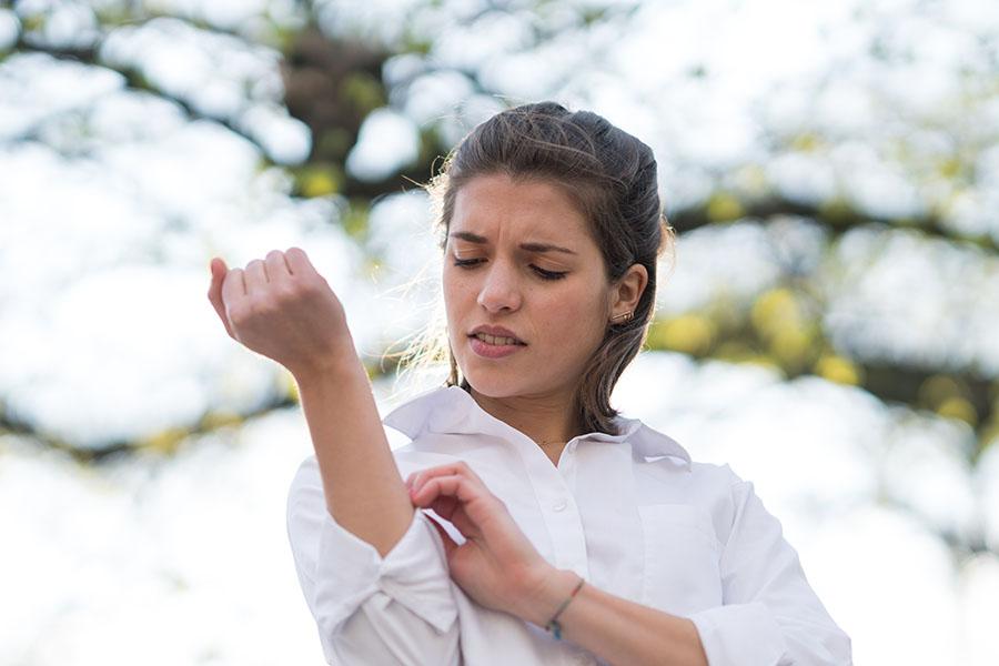
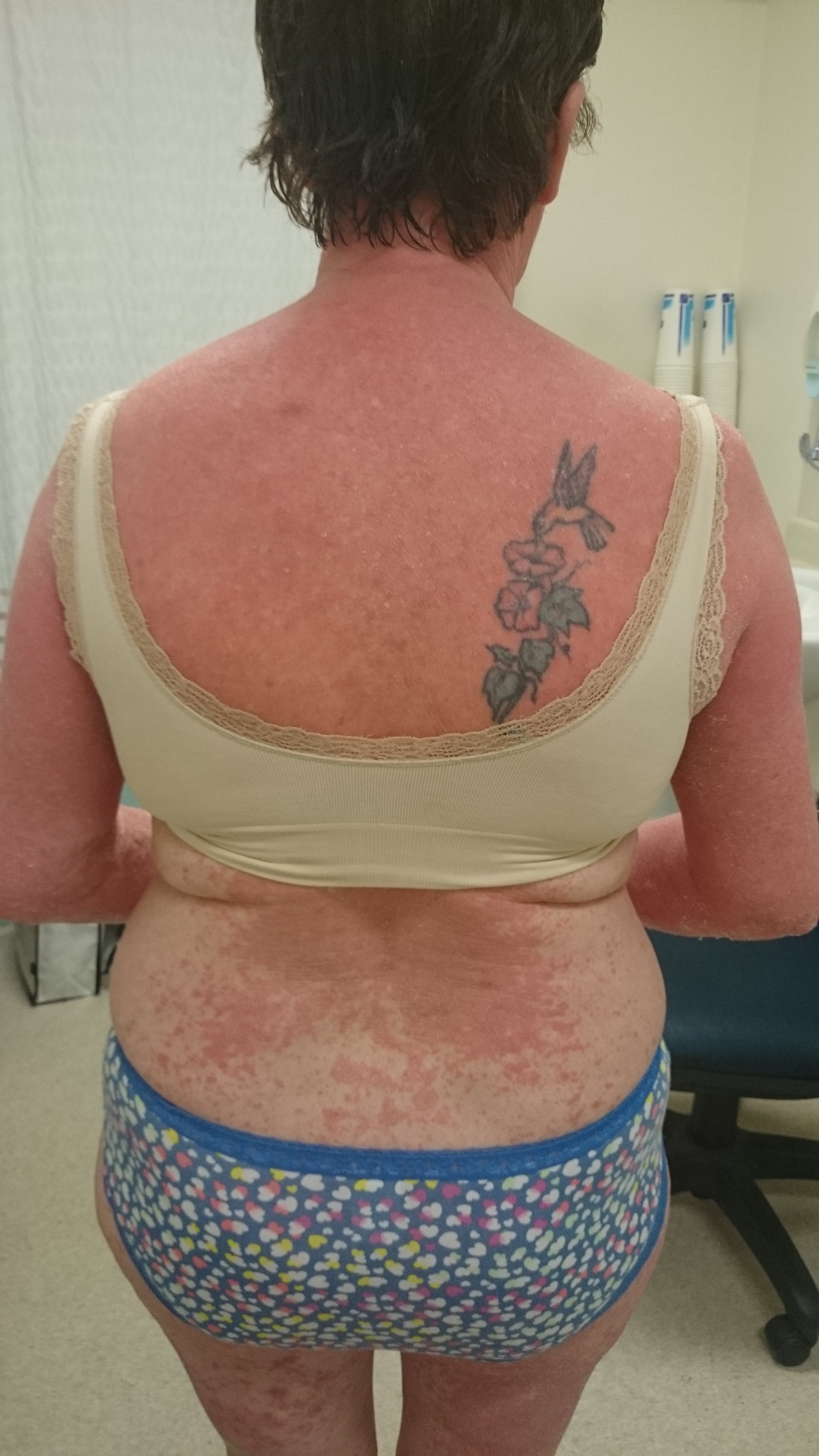
Erythema is a term used in medicine to describe redness of the skin, which occurs as a result of increased blood flow in the superficial capillaries. This redness can be caused by various factors such as inflammation, infection, trauma, or exposure to heat, cold, or ultraviolet radiation. In some cases, erythema may also be accompanied by other symptoms such as swelling, warmth, pain, or itching. It is a common finding in many medical conditions and can vary in severity from mild to severe.
Sunscreening agents, also known as sunscreens or sunblocks, are substances that protect the skin from the harmful effects of ultraviolet (UV) radiation from the sun. They work by absorbing, reflecting, or scattering UV radiation, preventing it from reaching the skin and causing damage such as sunburn, premature aging, and skin cancer.
Sunscreening agents can be chemical or physical. Chemical sunscreens contain organic compounds that absorb UV radiation and convert it into heat, which is then released from the skin. Examples of chemical sunscreens include oxybenzone, avobenzone, octinoxate, and homosalate.
Physical sunscreens, on the other hand, contain inorganic compounds that reflect or scatter UV radiation away from the skin. The most common physical sunscreen agents are zinc oxide and titanium dioxide.
Sunscreening agents are usually formulated into creams, lotions, gels, sprays, or sticks and are applied to the skin before sun exposure. They should be reapplied every two hours or after swimming, sweating, or toweling off to ensure continued protection. It is recommended to use a broad-spectrum sunscreen with an SPF (Sun Protection Factor) of at least 30, which blocks both UVA and UVB radiation.
Ultraviolet microscopy (UV microscopy) is a type of microscopy that uses ultraviolet light to visualize specimens. In this technique, ultraviolet radiation is used as the illumination source, and a special objective lens and filter are used to detect the resulting fluorescence emitted by the specimen.
The sample is usually stained with a fluorescent dye that absorbs the ultraviolet light and re-emits it at a longer wavelength, which can then be detected by the microscope's detector system. This technique allows for the visualization of structures or components within the specimen that may not be visible using traditional brightfield microscopy.
UV microscopy is commonly used in biological research to study the structure and function of cells, tissues, and proteins. It can also be used in forensic science to analyze evidence such as fingerprints, fibers, and other trace materials. However, it's important to note that UV radiation can be harmful to living tissue, so special precautions must be taken when using this technique.
I'm sorry for any confusion, but "sunlight" is not a term with a specific medical definition. However, sunlight does have various implications in the field of medicine. It is the light that comes from the sun, which is a star at the center of our solar system. Sunlight is essential for the production of vitamin D in humans, and it can also have effects on mood and sleep patterns due to its influence on circadian rhythms.
In a medical context, sunlight is often discussed in relation to its potential health benefits and risks. For instance, moderate sun exposure can help increase vitamin D levels, which are important for bone health, immune function, and other bodily processes. However, excessive sun exposure can lead to harmful effects, such as sunburn, premature skin aging, and an increased risk of skin cancer.
It's essential to balance the benefits and risks of sunlight exposure by practicing safe sun habits, such as wearing protective clothing, using a broad-spectrum sunscreen with an SPF of at least 30, seeking shade during peak sunlight hours, and avoiding intentional tanning.
A dose-response relationship in radiation refers to the correlation between the amount of radiation exposure (dose) and the biological response or adverse health effects observed in exposed individuals. As the level of radiation dose increases, the severity and frequency of the adverse health effects also tend to increase. This relationship is crucial in understanding the risks associated with various levels of radiation exposure and helps inform radiation protection standards and guidelines.
The effects of ionizing radiation can be categorized into two types: deterministic and stochastic. Deterministic effects have a threshold dose below which no effect is observed, and above this threshold, the severity of the effect increases with higher doses. Examples include radiation-induced cataracts or radiation dermatitis. Stochastic effects, on the other hand, do not have a clear threshold and are based on probability; as the dose increases, so does the likelihood of the adverse health effect occurring, such as an increased risk of cancer.
Understanding the dose-response relationship in radiation exposure is essential for setting limits on occupational and public exposure to ionizing radiation, optimizing radiation protection practices, and developing effective medical countermeasures in case of radiation emergencies.
 Ultraviolet light therapy
 atopic dermatitis ultraviolet light therapy Archives - Eczema Free
 Ecosvet home therapy device for ultraviolet blood irradiation (UVBI) : buy Ecosvet home therapy device for ultraviolet blood...
 Efficacy of targeted narrowband ultraviolet B therapy in vitiligo - Daavlin
  Heterologous Type I Collagen as an Add-on Therapy to Narrowband Ultraviolet B for the Treatment of Vitiligo: A Pilot Study |...
 psoralen and ultraviolet A (PUVA) therapy - Siteman Cancer Center
 Alopecia areata: MedlinePlus Medical Encyclopedia
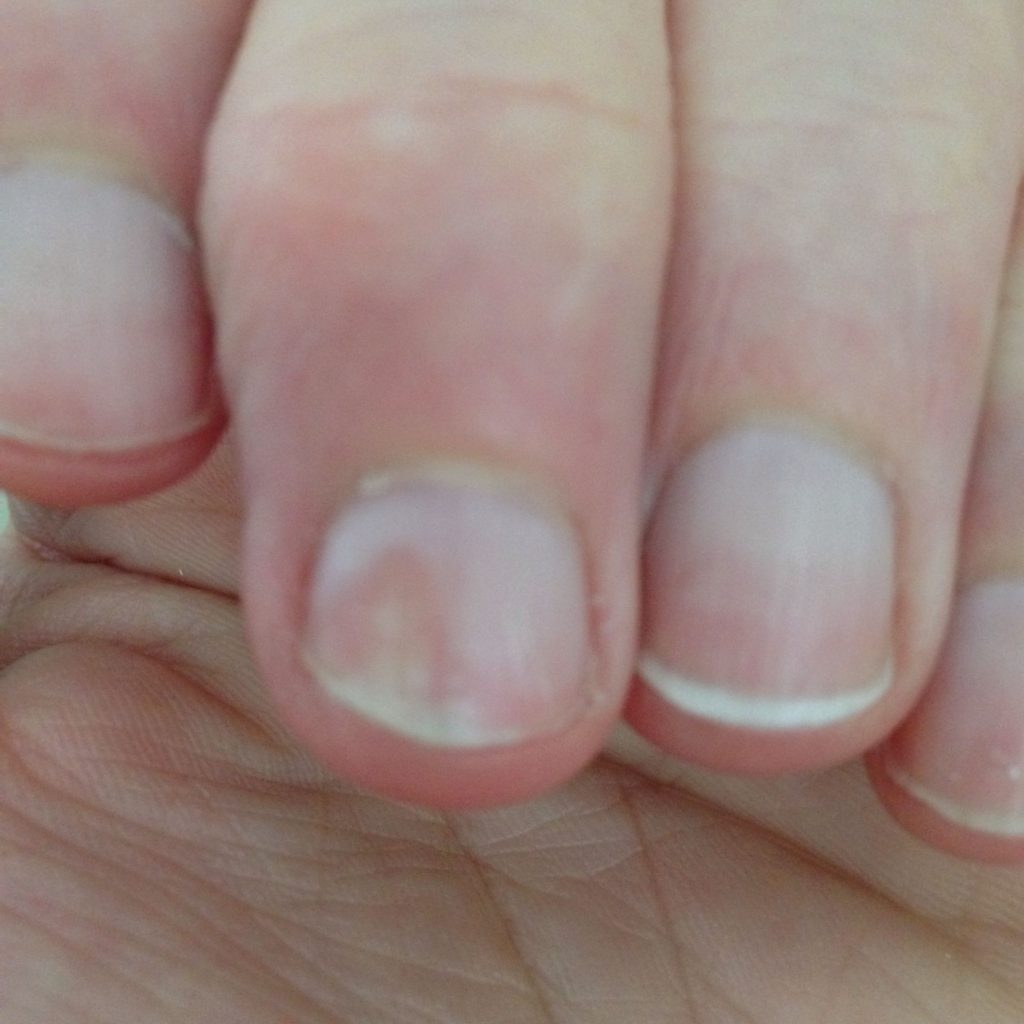
 Melanonychia: Background, Pathophysiology, Etiology
 Ultraviolet and ionizing radiation enhance the growth of BCCs and trichoblastomas in patched heterozygous knockout mice |...
 Treatment of Pityriasis Rosea
 Study uncovers role of ultraviolet radiation in development of rare leukemia in the skin
 How Vitiligo Progresses: Your FAQs
 UV Phototherapy Lamp: The world's most advanced Ultra Violet therapy l
 National Ambulatory Medical Care Survey, 1997
 Ultra-High Performance Flexible Ultraviolet Sensors for Use in Wearables - Tech Briefs
 Who Is Not Suitable for Ultraviolet Light Therapy Machine Treatment? - APK Technology Co., Ltd.
 Thyroiditis - Glossary Definition
 Ultraviolette Launches International-spec F77 for European Markets at EICMA 2023; F99 Factory Racing Platform makes Global...
 Psoriatic arthritis rash: Pictures, symptoms, and treatment
 3 ways to boost your vitamin D levels - WATE 6 On Your Side
 A Strategic Plan for the Elimination of Tuberculosis in
the United States
A Strategic Plan for the Elimination of Tuberculosis in
the United States Liang Deng, MD, PhD - MSK Dermatologist
 Light Therapy | GreenMedInfo | Therapeutic Action | Natural Medicine
 Mice drinking goji berry juice (Lycium barbarum) are protected from UV radiation-induced skin damage via antioxidant pathways