Transplantation, Autologous
Liver Transplantation
Transplantation, Homologous
Bone Marrow Transplantation
Hematopoietic Stem Cell Transplantation
Stem Cell Transplantation
Lung Transplantation
Transplantation Conditioning
Graft Survival
Organ Transplantation
Blood Transfusion, Autologous
Graft Rejection
Cell Transplantation
Islets of Langerhans Transplantation
Tissue Donors
Transplantation Immunology
Transplantation
Treatment Outcome
Transplantation Chimera
Graft vs Host Disease
Immunosuppressive Agents
Peripheral Blood Stem Cell Transplantation
Transplantation, Isogeneic
Transplantation, Heterotopic
Cord Blood Stem Cell Transplantation
Heart-Lung Transplantation
Retrospective Studies
Combined Modality Therapy
Immunosuppression
Transplantation Tolerance
Tissue and Organ Procurement
Histocompatibility Testing
Whole-Body Irradiation
Skin Transplantation
Liver Failure
Fetal Tissue Transplantation
Postoperative Complications
Mesenchymal Stem Cell Transplantation
Hematologic Neoplasms
Follow-Up Studies
Melphalan
Survival Analysis
Tissue Transplantation
Survival Rate
Cyclophosphamide
Remission Induction
Multiple Myeloma
Histocompatibility
Corneal Transplantation
Busulfan
Cyclosporine
T-Lymphocytes
Tacrolimus
Lymphoma, Non-Hodgkin
Antineoplastic Combined Chemotherapy Protocols
Bone Marrow Purging
HLA Antigens
Disease-Free Survival
Hematopoietic Stem Cell Mobilization
Leukemia
Carmustine
Brain Tissue Transplantation
Myeloablative Agonists
Salvage Therapy
Facial Transplantation
Organ Preservation
Antigens, CD34
Donor Selection
Transplants
Hodgkin Disease
Cells, Cultured
Prognosis
Antilymphocyte Serum
Etoposide
Lymphocyte Culture Test, Mixed
Risk Factors
Tissue and Organ Harvesting
Prospective Studies
Leukemia, Myeloid, Acute
Bone Marrow Cells
Kidney Failure, Chronic
Anemia, Aplastic
Granulocyte Colony-Stimulating Factor
Cytotoxicity, Immunologic
Cell Differentiation
Cytomegalovirus Infections
Lymphocyte Activation
Cytarabine
Mice, Inbred C57BL
Bronchiolitis Obliterans
Bone Marrow
Rats, Inbred Lew
Stem Cells
Actuarial Analysis
T-Lymphocytes, Cytotoxic
Thiotepa
Hematopoiesis
Cell Separation
Flow Cytometry
Lymphocyte Transfusion
Hand Transplantation
Liver
Blood Group Incompatibility
Mycophenolic Acid
Patient Selection
Neoplasm Transplantation
Precursor Cell Lymphoblastic Leukemia-Lymphoma
Lymphocyte Depletion
Disease Models, Animal
Graft vs Tumor Effect
End Stage Liver Disease
Infection
Feasibility Studies
Prednisone
Immunotherapy, Adoptive
Myelodysplastic Syndromes
Antigens, Neoplasm
Leukapheresis
Biopsy
Leukemia, Myeloid
Models, Animal
Brain Death
Fatal Outcome
ABO Blood-Group System
Lymphocytes
Hepatic Veno-Occlusive Disease
Dendritic Cells
Chimerism
Blood Transfusion
Liver Failure, Acute
Graft vs Leukemia Effect
Anastomosis, Surgical
Immune Tolerance
Antigens, CD
Liver Cirrhosis
Immunotherapy
Lymphoproliferative Disorders
Immunophenotyping
Leukemia, Myelogenous, Chronic, BCR-ABL Positive
Vidarabine
Primary Graft Dysfunction
Isoantibodies
Multivariate Analysis
Kaplan-Meier Estimate
Mesenchymal Stromal Cells
Delayed Graft Function
Incidence
Fetal Blood
Isoantigens
CD4-Positive T-Lymphocytes
Cryopreservation
Postoperative Care
Mice, SCID
Mice, Inbred BALB C
Biliary Atresia
Interleukin-2
Killer Cells, Natural
Clone Cells
Reoperation
Leukocyte Count
Cell- and Tissue-Based Therapy
Cold Ischemia
Blood Component Removal
Colony-Forming Units Assay
Antibodies, Neoplasm
Tissue Engineering
CD8-Positive T-Lymphocytes
Kidney
Coculture Techniques
Neoplasms
Chronic Disease
T-Lymphocytes, Regulatory
Platelet Transfusion
Siblings
Cancer Vaccines
Graft vs Host Reaction
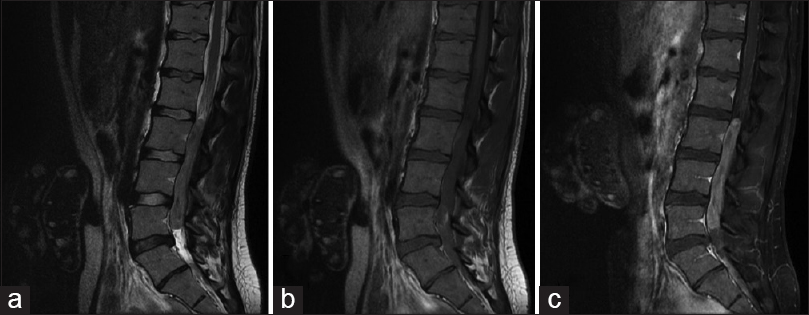
Natural history of papillary lesions of the urinary bladder in schistosomiasis. (1/3859)
Variable epithelial hyperplasia was observed in urinary bladder of nine capuchin monkeys (Cebus apella) when examined at cystotomy 94 to 164 weeks after infection with Schistosoma haematobium. These hosts were followed for 24 to 136 weeks postcystotomy to determine the status of bladder lesions in relation to duration of infection and to ascertain whether lesion samples removed at cystotomy reestablished themselves in autologous and heterologous transfers. There was involution of urothelial hyperplasia in eight of nine animals and no evidence for establishment of transplanted bladder lesions. (+info)Autografting with philadelphia chromosome-negative mobilized hematopoietic progenitor cells in chronic myelogenous leukemia. (2/3859)
Intensive chemotherapy given in early chronic phase of chronic myelogenous leukemia (CML) has resulted in high numbers of circulating Philadelphia (Ph) chromosome-negative hematopoietic progenitor cells (HPC). We have autografted 30 consecutive patients with CML in chronic phase with HPC collected in this way to facilitate restoration of Ph-negative hematopoiesis in bone marrow after high-dose therapy. Hematopoietic recovery to greater than 0.5 x10(9)/L neutrophils and to greater than 25 x 10(9)/L platelets occurred in all patients, a median of 13 (range, 9 to 32) days and 16 (range, 6 to 106) days postautograft, respectively. Regenerating marrow cells were Ph-negative in 16 (53%) patients and greater than 66% Ph-negative in 10 (33%) patients. Twenty-eight patients are alive 6 to 76 months (median, 24 months) after autografting. Three patients have developed blast crisis from which 2 have died. Eight patients are in complete cytogenetic remission at a median of 20 (range, 6 to 44) months with a median ratio BCR-ABL/ABL of 0.002 (range, <0.001 to 0.01). Eight patients are in major cytogenetic remission at a median of 22 (range, 6 to 48) months. No patient died as a consequence of the treatment. All patients had some degree of stomatitis that was severe in 15 (50%) patients. Gastrointestinal and hepatic toxicities were observed in about one fourth of patients. Thus, autografting with Ph-negative mobilized HPC can result in prolonged restoration of Ph-negative hematopoiesis for some patients with CML; moreover, most autograft recipients report normal or near normal activity levels, suggesting that this procedure need not to be associated either with prolonged convalescence or with chronic debility. (+info)The minimum CD34 threshold depends on prior chemotherapy in autologous peripheral blood stem cell recipients. (3/3859)
We analysed 57 patients with non-myeloid malignancies who received a non-purged autologous PBSCT. All had similar mobilisation and conditioning regimens. A high prior chemotherapy score and the number of chemotherapy lines used (P = 0.015 and P = 0.01, respectively) were adverse predictors of CD34 cell yields. Lower CD34 values (P = 0.002) were seen in patients treated with potent stem cell toxins (BCNU, melphalan, CCNU and mustine), designated toxicity factor 4 agents (TF4). All patients infused with grafts containing CD34 cell doses between 1.0 and 2.0 x 10(6)/kg (range 1.25-1.90) engrafted by day 51. The only variable associated with slow platelet recovery was exposure to TF4 (P = 0.007). The majority of patients with CD34 >1.0 x 10(6)/kg achieved rapid and sustained engraftment and the only predictive factor of delayed recovery is prior exposure to stem cell toxins. Potential PBSCT candidates should if possible avoid first line and salvage chemotherapy containing TF4 drugs. We therefore advocate a minimum CD34 threshold of >1.0 x 10(6)/kg in patients without extensive prior chemoradiotherapy, and > or = 2.0 x 10(6)/kg in all other patients. (+info)Infectious complications in 126 patients treated with high-dose chemotherapy and autologous peripheral blood stem cell transplantation. (4/3859)
The effect of an extensive prophylactic antimicrobial regimen was prospectively assessed in 126 patients after high-dose chemotherapy and autologous PBSC. They received ciprofloxacin (500 mg/12 h), acyclovir (200 mg/6 h), and itraconazole (200 mg/12 h) orally until neutrophil recovery. Febrile patients received i.v. imipenem (500 mg/6 h) to which vancomycin and amikacin were added if fever persisted for 2-3 and 5 days, respectively. Amphotericin B lipid complex was further given on day 7 or 8 of fever. Median times for a neutrophil count of >0.5 x 10(9)/l and a platelet count of >20 x 10(9)/l were 9 and 11 days. Severe neutropenia (<0.1 x 10(9)/l) lasted for a median of 5 days in which 72% of febrile episodes and 50% of cases of bacteremia occurred. Gram-positive bacteria were isolated in 30 of 40 episodes of bacteremia, 25 of which were caused by Staphylococcus epidermidis. Clinical foci were the intravascular catheter in 35 cases, respiratory infection in 11, cellulitis in two, anal abscess in one, and neutropenic enterocolitis in one. The high incidence of febrile episodes (94%) and bacteremias (31%) may be due to the lack of efficacy of antimicrobial prophylaxis and the persistence of a 5-day period of severe neutropenia. (+info)Lymphomatoid granulomatosis following autologous stem cell transplantation. (5/3859)
Lymphomatoid granulomatosis (LYG) is a rare angio-destructive lymphoproliferative disorder (LPD) of uncertain etiology, with prominent pulmonary involvement. Recent studies indicate that LYG is an Epstein-Barr virus (EBV)-associated B cell LPD with large numbers of background reactive T lymphocytes (T cell-rich B cell lymphoma). Although the disease frequently, but not exclusively, occurs in various immunodeficiency states, it has not been reported in association with the transient immunosuppression following autologous bone marrow/peripheral stem cell transplantation (ABM/PSCT). We describe a patient who developed lymphomatoid granulomatosis of the lung approximately 2 weeks after high-dose chemotherapy and autologous peripheral stem cell transplantation for multiple myeloma. Although molecular studies showed no evidence of EBV genome in the biopsy material, the serologic profile with high IgM titers was suggestive of primary EBV infection. Complete radiologic remission occurred following reconstitution of the patient's immune response after a 2-week course of ganciclovir treatment. Despite the apparently low frequency of LPD (both LYG and EBV-associated post-transplant lymphoma) in the ABMT setting, we believe that it should be considered in the differential diagnosis of patients whose clinical course following ABMT is complicated by fevers, in the absence of an identifiable infectious process. (+info)Poor outcome of autologous stem cell transplantation for adult T cell leukemia/lymphoma: a case report and review of the literature. (6/3859)
A limited number of patients with adult T cell leukemia/lymphoma (ATL) who received autologous stem cell transplantation (ASCT) have been reported. We report here a case of fatal systemic Candida krusei infection in a female patient with ATL undergoing ASCT. All of the eight patients (including seven patients in the literature) with ATL who received ASCT developed relapse of ATL or death due to ASCT complication, irrespective of subtype or remission state of ATL, source or selection of SCT or conditioning regimen. At present, ASCT appears to provide little benefit for ATL in contrast to that for other types of aggressive non-Hodgkin's lymphoma. (+info)Rapid autologous marrow recovery and eradication of infectious mononucleosis despite severe immunosuppression following second transplantation for aplastic anemia. (7/3859)
A patient with aplastic anemia failed to respond to immunosuppressive therapy and first marrow transplantation (BMT). Recovery of autologous hematopoiesis was rapid following a second stem cell transplant with a non-myeloablative preparatory regimen. The autologous immune response to infectious mononucleosis (IM) 4 weeks post-transplant was normal despite recent and ongoing severe immunosuppression. (+info)Advances in therapy of multiple myeloma: lessons from acute leukemia. (8/3859)
This paper traces the lack of progress, until recently, in the treatment of multiple myeloma (MM) to having ignored the principles that led to cure in acute leukemia more than 2 decades ago. Only in the mid-1980s did investigation begin to consider complete remission (CR) a research objective, representing a necessary first step toward cure. The experience with autologous and allogeneic stem cell-supported high-dose therapy is reviewed, demonstrating, in both historically controlled and randomized studies, the validity of the dose-response concept in MM in terms of increased CR rates as well as extended event-free (EFS) and overall survival (OS). Avoidance of hematopoietic stem cell-damaging agents, especially melphalan, nitrosoureas, and ionizing radiation to marrow-containing sites, assures the ability of peripheral stem cell collection of high quality and quantity, providing rapid engraftment so that mortality is well under 5% following high-dose melphalan (200 mg/m2). This treatment can be applied safely to patients even >70 years of age and in the presence of renal failure. Tandem autotransplants after multiregimen induction have yielded CR rates in the 40% range with median durations of EFS and OS of 43 and 62 months, respectively. Certain chromosomal abnormalities (11 and 13; and translocations) represent the dominant adverse prognosticator for EFS and OS, confirmed in over 500 patients including those with prior therapy. Allogeneic transplants, possible in less than 10% of MM patients, are associated with a 50% mortality during the first year and, unfortunately, late relapses; thus, this approach should be reserved for patients with high-risk disease early in their management. A risk-based treatment algorithm that matches a patient's disease risk with the risk of intervention is presently used, followed by bisphosphonate therapy, not only to delay the onset of MM-related bone disease but also to induce tumor cell apoptosis, indirectly or directly, by down-regulation of cytokines with antiapoptotic activities. Although many patients relapse, this author subscribes to his mentor's motto: "Be Prepared for Success!". (+info)Autologous transplantation is a medical procedure where cells, tissues, or organs are removed from a person, stored and then returned back to the same individual at a later time. This is different from allogeneic transplantation where the tissue or organ is obtained from another donor. The term "autologous" is derived from the Greek words "auto" meaning self and "logos" meaning study.
In autologous transplantation, the patient's own cells or tissues are used to replace or repair damaged or diseased ones. This reduces the risk of rejection and eliminates the need for immunosuppressive drugs, which are required in allogeneic transplants to prevent the body from attacking the foreign tissue.
Examples of autologous transplantation include:
* Autologous bone marrow or stem cell transplantation, where stem cells are removed from the patient's blood or bone marrow, stored and then reinfused back into the same individual after high-dose chemotherapy or radiation therapy to treat cancer.
* Autologous skin grafting, where a piece of skin is taken from one part of the body and transplanted to another area on the same person.
* Autologous chondrocyte implantation, where cartilage cells are harvested from the patient's own knee, cultured in a laboratory and then implanted back into the knee to repair damaged cartilage.
Liver transplantation is a surgical procedure in which a diseased or failing liver is replaced with a healthy one from a deceased donor or, less commonly, a portion of a liver from a living donor. The goal of the procedure is to restore normal liver function and improve the patient's overall health and quality of life.
Liver transplantation may be recommended for individuals with end-stage liver disease, acute liver failure, certain genetic liver disorders, or liver cancers that cannot be treated effectively with other therapies. The procedure involves complex surgery to remove the diseased liver and implant the new one, followed by a period of recovery and close medical monitoring to ensure proper function and minimize the risk of complications.
The success of liver transplantation has improved significantly in recent years due to advances in surgical techniques, immunosuppressive medications, and post-transplant care. However, it remains a major operation with significant risks and challenges, including the need for lifelong immunosuppression to prevent rejection of the new liver, as well as potential complications such as infection, bleeding, and organ failure.
Homologous transplantation is a type of transplant surgery where organs or tissues are transferred between two genetically non-identical individuals of the same species. The term "homologous" refers to the similarity in structure and function of the donated organ or tissue to the recipient's own organ or tissue.
For example, a heart transplant from one human to another is an example of homologous transplantation because both organs are hearts and perform the same function. Similarly, a liver transplant, kidney transplant, lung transplant, and other types of organ transplants between individuals of the same species are also considered homologous transplantations.
Homologous transplantation is in contrast to heterologous or xenogeneic transplantation, where organs or tissues are transferred from one species to another, such as a pig heart transplanted into a human. Homologous transplantation is more commonly performed than heterologous transplantation due to the increased risk of rejection and other complications associated with xenogeneic transplants.
Bone marrow transplantation (BMT) is a medical procedure in which damaged or destroyed bone marrow is replaced with healthy bone marrow from a donor. Bone marrow is the spongy tissue inside bones that produces blood cells. The main types of BMT are autologous, allogeneic, and umbilical cord blood transplantation.
In autologous BMT, the patient's own bone marrow is used for the transplant. This type of BMT is often used in patients with lymphoma or multiple myeloma who have undergone high-dose chemotherapy or radiation therapy to destroy their cancerous bone marrow.
In allogeneic BMT, bone marrow from a genetically matched donor is used for the transplant. This type of BMT is often used in patients with leukemia, lymphoma, or other blood disorders who have failed other treatments.
Umbilical cord blood transplantation involves using stem cells from umbilical cord blood as a source of healthy bone marrow. This type of BMT is often used in children and adults who do not have a matched donor for allogeneic BMT.
The process of BMT typically involves several steps, including harvesting the bone marrow or stem cells from the donor, conditioning the patient's body to receive the new bone marrow or stem cells, transplanting the new bone marrow or stem cells into the patient's body, and monitoring the patient for signs of engraftment and complications.
BMT is a complex and potentially risky procedure that requires careful planning, preparation, and follow-up care. However, it can be a life-saving treatment for many patients with blood disorders or cancer.
Kidney transplantation is a surgical procedure where a healthy kidney from a deceased or living donor is implanted into a patient with end-stage renal disease (ESRD) or permanent kidney failure. The new kidney takes over the functions of filtering waste and excess fluids from the blood, producing urine, and maintaining the body's electrolyte balance.
The transplanted kidney is typically placed in the lower abdomen, with its blood vessels connected to the recipient's iliac artery and vein. The ureter of the new kidney is then attached to the recipient's bladder to ensure proper urine flow. Following the surgery, the patient will require lifelong immunosuppressive therapy to prevent rejection of the transplanted organ by their immune system.
Hematopoietic Stem Cell Transplantation (HSCT) is a medical procedure where hematopoietic stem cells (immature cells that give rise to all blood cell types) are transplanted into a patient. This procedure is often used to treat various malignant and non-malignant disorders affecting the hematopoietic system, such as leukemias, lymphomas, multiple myeloma, aplastic anemia, inherited immune deficiency diseases, and certain genetic metabolic disorders.
The transplantation can be autologous (using the patient's own stem cells), allogeneic (using stem cells from a genetically matched donor, usually a sibling or unrelated volunteer), or syngeneic (using stem cells from an identical twin).
The process involves collecting hematopoietic stem cells, most commonly from the peripheral blood or bone marrow. The collected cells are then infused into the patient after the recipient's own hematopoietic system has been ablated (or destroyed) using high-dose chemotherapy and/or radiation therapy. This allows the donor's stem cells to engraft, reconstitute, and restore the patient's hematopoietic system.
HSCT is a complex and potentially risky procedure with various complications, including graft-versus-host disease, infections, and organ damage. However, it offers the potential for cure or long-term remission in many patients with otherwise fatal diseases.
Heart transplantation is a surgical procedure where a diseased, damaged, or failing heart is removed and replaced with a healthy donor heart. This procedure is usually considered as a last resort for patients with end-stage heart failure or severe coronary artery disease who have not responded to other treatments. The donor heart typically comes from a brain-dead individual whose family has agreed to donate their loved one's organs for transplantation. Heart transplantation is a complex and highly specialized procedure that requires a multidisciplinary team of healthcare professionals, including cardiologists, cardiac surgeons, anesthesiologists, perfusionists, nurses, and other support staff. The success rates for heart transplantation have improved significantly over the past few decades, with many patients experiencing improved quality of life and increased survival rates. However, recipients of heart transplants require lifelong immunosuppressive therapy to prevent rejection of the donor heart, which can increase the risk of infections and other complications.
Stem cell transplantation is a medical procedure where stem cells, which are immature and unspecialized cells with the ability to differentiate into various specialized cell types, are introduced into a patient. The main purpose of this procedure is to restore the function of damaged or destroyed tissues or organs, particularly in conditions that affect the blood and immune systems, such as leukemia, lymphoma, aplastic anemia, and inherited metabolic disorders.
There are two primary types of stem cell transplantation: autologous and allogeneic. In autologous transplantation, the patient's own stem cells are collected, stored, and then reinfused back into their body after high-dose chemotherapy or radiation therapy to destroy the diseased cells. In allogeneic transplantation, stem cells are obtained from a donor (related or unrelated) whose human leukocyte antigen (HLA) type closely matches that of the recipient.
The process involves several steps: first, the patient undergoes conditioning therapy to suppress their immune system and make space for the new stem cells. Then, the harvested stem cells are infused into the patient's bloodstream, where they migrate to the bone marrow and begin to differentiate and produce new blood cells. This procedure requires close monitoring and supportive care to manage potential complications such as infections, graft-versus-host disease, and organ damage.
Lung transplantation is a surgical procedure where one or both diseased lungs are removed and replaced with healthy lungs from a deceased donor. It is typically considered as a treatment option for patients with end-stage lung diseases, such as chronic obstructive pulmonary disease (COPD), cystic fibrosis, idiopathic pulmonary fibrosis, and alpha-1 antitrypsin deficiency, who have exhausted all other medical treatments and continue to suffer from severe respiratory failure.
The procedure involves several steps, including evaluating the patient's eligibility for transplantation, matching the donor's lung size and blood type with the recipient, and performing the surgery under general anesthesia. After the surgery, patients require close monitoring and lifelong immunosuppressive therapy to prevent rejection of the new lungs.
Lung transplantation can significantly improve the quality of life and survival rates for some patients with end-stage lung disease, but it is not without risks, including infection, bleeding, and rejection. Therefore, careful consideration and thorough evaluation are necessary before pursuing this treatment option.
Transplantation conditioning, also known as preparative regimen or immunoablative therapy, refers to the use of various treatments prior to transplantation of cells, tissues or organs. The main goal of transplantation conditioning is to suppress the recipient's immune system, allowing for successful engraftment and minimizing the risk of rejection of the donor tissue.
There are two primary types of transplantation conditioning: myeloablative and non-myeloablative.
1. Myeloablative conditioning is a more intensive regimen that involves the use of high-dose chemotherapy, radiation therapy or both. This approach eliminates not only immune cells but also stem cells in the bone marrow, requiring the recipient to receive a hematopoietic cell transplant (HCT) from the donor to reconstitute their blood and immune system.
2. Non-myeloablative conditioning is a less intensive regimen that primarily targets immune cells while sparing the stem cells in the bone marrow. This approach allows for mixed chimerism, where both recipient and donor immune cells coexist, reducing the risk of severe complications associated with myeloablative conditioning.
The choice between these two types of transplantation conditioning depends on various factors, including the type of transplant, patient's age, overall health, and comorbidities. Both approaches carry risks and benefits, and the decision should be made carefully by a multidisciplinary team of healthcare professionals in consultation with the patient.
Graft survival, in medical terms, refers to the success of a transplanted tissue or organ in continuing to function and integrate with the recipient's body over time. It is the opposite of graft rejection, which occurs when the recipient's immune system recognizes the transplanted tissue as foreign and attacks it, leading to its failure.
Graft survival depends on various factors, including the compatibility between the donor and recipient, the type and location of the graft, the use of immunosuppressive drugs to prevent rejection, and the overall health of the recipient. A successful graft survival implies that the transplanted tissue or organ has been accepted by the recipient's body and is functioning properly, providing the necessary physiological support for the recipient's survival and improved quality of life.
Organ transplantation is a surgical procedure where an organ or tissue from one person (donor) is removed and placed into another person (recipient) whose organ or tissue is not functioning properly or has been damaged beyond repair. The goal of this complex procedure is to replace the non-functioning organ with a healthy one, thereby improving the recipient's quality of life and overall survival.
Organs that can be transplanted include the heart, lungs, liver, kidneys, pancreas, and intestines. Tissues such as corneas, skin, heart valves, and bones can also be transplanted. The donor may be deceased or living, depending on the type of organ and the medical circumstances.
Organ transplantation is a significant and life-changing event for both the recipient and their families. It requires careful evaluation, matching, and coordination between the donor and recipient, as well as rigorous post-transplant care to ensure the success of the procedure and minimize the risk of rejection.
Autologous blood transfusion is a medical procedure in which a patient receives their own blood that has been collected and stored prior to surgery or a medical treatment that may cause significant blood loss. The blood is drawn from the patient, typically in the days or weeks leading up to the scheduled procedure, and then stored until it is needed during or after the surgery.
The primary advantage of autologous blood transfusion is that it eliminates the risk of transfusion reactions, infectious disease transmission, and immunomodulation associated with allogeneic (donor) blood transfusions. However, not all patients are candidates for this type of transfusion due to various factors such as medical conditions, low hemoglobin levels, or insufficient time to collect and store the blood before the procedure.
Autologous blood transfusion can be performed using several methods, including preoperative blood donation, acute normovolemic hemodilution, intraoperative cell salvage, and postoperative blood collection. The choice of method depends on various factors, such as the patient's medical condition, the type and extent of surgery, and the availability of resources.
In summary, autologous blood transfusion is a safe and effective way to reduce the need for allogeneic blood transfusions during or after surgical procedures, but it may not be suitable for all patients.
Graft rejection is an immune response that occurs when transplanted tissue or organ (the graft) is recognized as foreign by the recipient's immune system, leading to the activation of immune cells to attack and destroy the graft. This results in the failure of the transplant and the need for additional medical intervention or another transplant. There are three types of graft rejection: hyperacute, acute, and chronic. Hyperacute rejection occurs immediately or soon after transplantation due to pre-existing antibodies against the graft. Acute rejection typically occurs within weeks to months post-transplant and is characterized by the infiltration of T-cells into the graft. Chronic rejection, which can occur months to years after transplantation, is a slow and progressive process characterized by fibrosis and tissue damage due to ongoing immune responses against the graft.
Cell transplantation is the process of transferring living cells from one part of the body to another or from one individual to another. In medicine, cell transplantation is often used as a treatment for various diseases and conditions, including neurodegenerative disorders, diabetes, and certain types of cancer. The goal of cell transplantation is to replace damaged or dysfunctional cells with healthy ones, thereby restoring normal function to the affected area.
In the context of medical research, cell transplantation may involve the use of stem cells, which are immature cells that have the ability to develop into many different types of specialized cells. Stem cell transplantation has shown promise in the treatment of a variety of conditions, including spinal cord injuries, stroke, and heart disease.
It is important to note that cell transplantation carries certain risks, such as immune rejection and infection. As such, it is typically reserved for cases where other treatments have failed or are unlikely to be effective.
Pancreas transplantation is a surgical procedure that involves implanting a healthy pancreas from a deceased donor into a recipient with diabetes. The primary goal of this procedure is to restore the recipient's insulin production and eliminate the need for insulin injections, thereby improving their quality of life and reducing the risk of long-term complications associated with diabetes.
There are three main types of pancreas transplantation:
1. Simultaneous pancreas-kidney (SPK) transplantation: This is the most common type of pancreas transplant, performed simultaneously with a kidney transplant in patients with diabetes and end-stage renal disease (ESRD). The new pancreas not only restores insulin production but also helps prevent further kidney damage.
2. Pancreas after kidney (PAK) transplantation: In this procedure, a patient receives a kidney transplant first, followed by a pancreas transplant at a later time. This is typically performed in patients who have already undergone a successful kidney transplant and wish to improve their diabetes management.
3. Pancreas transplantation alone (PTA): In rare cases, a pancreas transplant may be performed without a concurrent kidney transplant. This is usually considered for patients with brittle diabetes who experience severe hypoglycemic episodes despite optimal medical management and lifestyle modifications.
The success of pancreas transplantation has significantly improved over the years, thanks to advancements in surgical techniques, immunosuppressive medications, and post-transplant care. However, it is essential to weigh the benefits against the risks, such as potential complications related to surgery, infection, rejection, and long-term use of immunosuppressive drugs. Ultimately, the decision to undergo pancreas transplantation should be made in consultation with a multidisciplinary team of healthcare professionals, considering each patient's unique medical history and personal circumstances.
Islets of Langerhans transplantation is a surgical procedure that involves the transplantation of isolated islets from a deceased donor's pancreas into another person with type 1 diabetes. The islets of Langerhans are clusters of cells within the pancreas that produce hormones, including insulin, which regulates blood sugar levels.
In type 1 diabetes, the body's immune system mistakenly attacks and destroys these insulin-producing cells, leading to high blood sugar levels. Islet transplantation aims to replace the damaged islets with healthy ones from a donor, allowing the recipient's body to produce and regulate its own insulin again.
The procedure involves extracting the islets from the donor pancreas and infusing them into the recipient's liver through a small incision in the abdomen. Once inside the liver, the islets can sense glucose levels in the bloodstream and release insulin as needed to maintain normal blood sugar levels.
Islet transplantation has shown promising results in improving blood sugar control and reducing the risk of severe hypoglycemia (low blood sugar) in people with type 1 diabetes. However, it requires long-term immunosuppressive therapy to prevent rejection of the transplanted islets, which can have side effects and increase the risk of infections.
A tissue donor is an individual who has agreed to allow organs and tissues to be removed from their body after death for the purpose of transplantation to restore the health or save the life of another person. The tissues that can be donated include corneas, heart valves, skin, bone, tendons, ligaments, veins, and cartilage. These tissues can enhance the quality of life for many recipients and are often used in reconstructive surgeries. It is important to note that tissue donation does not interfere with an open casket funeral or other cultural or religious practices related to death and grieving.
Transplantation Immunology is a branch of medicine that deals with the immune responses occurring between a transplanted organ or tissue and the recipient's body. It involves understanding and managing the immune system's reaction to foreign tissue, which can lead to rejection of the transplanted organ. This field also studies the use of immunosuppressive drugs to prevent rejection and the potential risks and side effects associated with their use. The main goal of transplantation immunology is to find ways to promote the acceptance of transplanted tissue while minimizing the risk of infection and other complications.
Transplantation is a medical procedure where an organ or tissue is removed from one person (the donor) and placed into another person (the recipient) for the purpose of replacing the recipient's damaged or failing organ or tissue with a functioning one. The goal of transplantation is to restore normal function, improve quality of life, and extend lifespan in individuals with organ failure or severe tissue damage. Common types of transplants include kidney, liver, heart, lung, pancreas, small intestine, and bone marrow transplantations. The success of a transplant depends on various factors, including the compatibility between the donor and recipient, the health of both individuals, and the effectiveness of immunosuppressive therapy to prevent rejection of the transplanted organ or tissue.
Treatment outcome is a term used to describe the result or effect of medical treatment on a patient's health status. It can be measured in various ways, such as through symptoms improvement, disease remission, reduced disability, improved quality of life, or survival rates. The treatment outcome helps healthcare providers evaluate the effectiveness of a particular treatment plan and make informed decisions about future care. It is also used in clinical research to compare the efficacy of different treatments and improve patient care.
A transplantation chimera is a rare medical condition that occurs after an organ or tissue transplant, where the recipient's body accepts and integrates the donor's cells or tissues to such an extent that the two sets of DNA coexist and function together. This phenomenon can lead to the presence of two different genetic profiles in one individual.
In some cases, this may result in the development of donor-derived cells or organs within the recipient's body, which can express the donor's unique genetic traits. Transplantation chimerism is more commonly observed in bone marrow transplants, where the donor's immune cells can repopulate and establish themselves within the recipient's bone marrow and bloodstream.
It is important to note that while transplantation chimerism can be beneficial for the success of the transplant, it may also pose some risks, such as an increased likelihood of developing graft-versus-host disease (GVHD), where the donor's immune cells attack the recipient's tissues.
Graft-versus-host disease (GVHD) is a condition that can occur after an allogeneic hematopoietic stem cell transplantation (HSCT), where the donated immune cells (graft) recognize the recipient's tissues (host) as foreign and attack them. This results in inflammation and damage to various organs, particularly the skin, gastrointestinal tract, and liver.
Acute GVHD typically occurs within 100 days of transplantation and is characterized by symptoms such as rash, diarrhea, and liver dysfunction. Chronic GVHD, on the other hand, can occur after 100 days or even years post-transplant and may present with a wider range of symptoms, including dry eyes and mouth, skin changes, lung involvement, and issues with mobility and flexibility in joints.
GVHD is a significant complication following allogeneic HSCT and can have a substantial impact on the patient's quality of life and overall prognosis. Preventative measures, such as immunosuppressive therapy, are often taken to reduce the risk of GVHD, but its management remains a challenge in transplant medicine.
Immunosuppressive agents are medications that decrease the activity of the immune system. They are often used to prevent the rejection of transplanted organs and to treat autoimmune diseases, where the immune system mistakenly attacks the body's own tissues. These drugs work by interfering with the immune system's normal responses, which helps to reduce inflammation and damage to tissues. However, because they suppress the immune system, people who take immunosuppressive agents are at increased risk for infections and other complications. Examples of immunosuppressive agents include corticosteroids, azathioprine, cyclophosphamide, mycophenolate mofetil, tacrolimus, and sirolimus.
Peripheral Blood Stem Cell Transplantation (PBSCT) is a medical procedure that involves the transplantation of stem cells, which are immature cells found in the bone marrow that can develop into different types of blood cells. In PBSCT, these stem cells are collected from the peripheral blood instead of directly from the bone marrow.
The process begins with mobilization, where a growth factor medication is given to the donor to stimulate the release of stem cells from the bone marrow into the peripheral blood. After several days, the donor's blood is then removed through a procedure called apheresis, where the stem cells are separated and collected while the remaining blood components are returned to the donor.
The collected stem cells are then infused into the recipient's bloodstream, where they migrate to the bone marrow and begin to repopulate, leading to the production of new blood cells. This procedure is often used as a treatment for various malignant and non-malignant disorders, such as leukemia, lymphoma, multiple myeloma, and aplastic anemia.
PBSCT offers several advantages over traditional bone marrow transplantation, including faster engraftment, lower risk of graft failure, and reduced procedure-related morbidity. However, it also has its own set of challenges, such as the potential for increased incidence of chronic graft-versus-host disease (GVHD) and the need for more stringent HLA matching between donor and recipient.
Isogeneic transplantation is a type of transplant where the donor and recipient are genetically identical, meaning they are identical twins or have the same genetic makeup. In this case, the immune system recognizes the transplanted organ or tissue as its own and does not mount an immune response to reject it. This reduces the need for immunosuppressive drugs, which are typically required in other types of transplantation to prevent rejection.
In medical terms, isogeneic transplantation is defined as the transfer of genetic identical tissues or organs between genetically identical individuals, resulting in minimal risk of rejection and no need for immunosuppressive therapy.
A living donor is a person who voluntarily donates an organ or part of an organ to another person while they are still alive. This can include donations such as a kidney, liver lobe, lung, or portion of the pancreas or intestines. The donor and recipient typically undergo medical evaluation and compatibility testing to ensure the best possible outcome for the transplantation procedure. Living donation is regulated by laws and ethical guidelines to ensure that donors are fully informed and making a voluntary decision.
Heterotopic transplantation is a type of organ or tissue transplant where the graft is placed in a different location from where it normally resides while still maintaining its original site. This is often done to supplement the function of the existing organ rather than replacing it. A common example of heterotopic transplantation is a heart transplant, where the donor's heart is placed in a new location in the recipient's body, while the recipient's own heart remains in place but is typically nonfunctional. This allows for the possibility of returning the function of the recipient's heart if the transplanted organ fails.
In heterotopic kidney transplantation, the donor kidney is placed in a different location, usually in the lower abdomen, while the recipient's own kidneys are left in place. This approach can be beneficial for recipients with poor renal function or other medical conditions that make traditional kidney transplantation too risky.
Heterotopic transplantation is also used in liver transplantation, where a portion of the donor liver is placed in a different location, typically in the recipient's abdomen, while the recipient's own liver remains in place. This approach can be useful for recipients with acute liver failure or other conditions that make traditional liver transplantation too risky.
One advantage of heterotopic transplantation is that it allows for the possibility of returning the function of the recipient's organ if the transplanted organ fails, as well as reducing the risk of rejection and improving overall outcomes for the recipient. However, this approach also has some disadvantages, such as increased complexity of the surgical procedure, potential for complications related to the placement of the graft, and the need for ongoing immunosuppression therapy to prevent rejection.
Cord blood stem cell transplantation is a medical procedure that involves the infusion of stem cells derived from the umbilical cord blood into a patient. These stem cells, specifically hematopoietic stem cells, have the ability to differentiate into various types of blood cells, including red and white blood cells and platelets.
Cord blood stem cell transplantation is often used as a treatment for patients with various malignant and non-malignant disorders, such as leukemia, lymphoma, sickle cell disease, and metabolic disorders. The procedure involves collecting cord blood from the umbilical cord and placenta after the birth of a baby, processing and testing it for compatibility with the recipient's immune system, and then infusing it into the patient through a vein in a process similar to a blood transfusion.
The advantages of using cord blood stem cells include their availability, low risk of transmission of infectious diseases, and reduced risk of graft-versus-host disease compared to other sources of hematopoietic stem cells, such as bone marrow or peripheral blood. However, the number of stem cells in a cord blood unit is generally lower than that found in bone marrow or peripheral blood, which can limit its use in some patients, particularly adults.
Overall, cord blood stem cell transplantation is an important and promising area of regenerative medicine, offering hope for patients with a wide range of disorders.
In the field of medicine, "time factors" refer to the duration of symptoms or time elapsed since the onset of a medical condition, which can have significant implications for diagnosis and treatment. Understanding time factors is crucial in determining the progression of a disease, evaluating the effectiveness of treatments, and making critical decisions regarding patient care.
For example, in stroke management, "time is brain," meaning that rapid intervention within a specific time frame (usually within 4.5 hours) is essential to administering tissue plasminogen activator (tPA), a clot-busting drug that can minimize brain damage and improve patient outcomes. Similarly, in trauma care, the "golden hour" concept emphasizes the importance of providing definitive care within the first 60 minutes after injury to increase survival rates and reduce morbidity.
Time factors also play a role in monitoring the progression of chronic conditions like diabetes or heart disease, where regular follow-ups and assessments help determine appropriate treatment adjustments and prevent complications. In infectious diseases, time factors are crucial for initiating antibiotic therapy and identifying potential outbreaks to control their spread.
Overall, "time factors" encompass the significance of recognizing and acting promptly in various medical scenarios to optimize patient outcomes and provide effective care.
Heart-lung transplantation is a surgical procedure where both the heart and lungs of a patient are replaced with those from a deceased donor. This complex and highly specialized surgery is typically considered as a last resort for patients suffering from end-stage lung or heart-lung diseases, such as cystic fibrosis, pulmonary fibrosis, chronic obstructive pulmonary disease (COPD), or certain forms of congenital heart disease, who have exhausted all other treatment options and face imminent death.
The procedure involves removing the patient's diseased heart and lungs en bloc, followed by implanting the donor's heart and lungs in their place. The surgery requires a skilled multidisciplinary team of cardiothoracic surgeons, anesthesiologists, perfusionists, transplant coordinators, and intensive care specialists.
Following the transplantation, patients require lifelong immunosuppressive therapy to prevent rejection of the transplanted organs. Despite the significant risks associated with this procedure, including infection, bleeding, and rejection, heart-lung transplantation can significantly improve both survival and quality of life for carefully selected patients with advanced heart-lung disease.
Retrospective studies, also known as retrospective research or looking back studies, are a type of observational study that examines data from the past to draw conclusions about possible causal relationships between risk factors and outcomes. In these studies, researchers analyze existing records, medical charts, or previously collected data to test a hypothesis or answer a specific research question.
Retrospective studies can be useful for generating hypotheses and identifying trends, but they have limitations compared to prospective studies, which follow participants forward in time from exposure to outcome. Retrospective studies are subject to biases such as recall bias, selection bias, and information bias, which can affect the validity of the results. Therefore, retrospective studies should be interpreted with caution and used primarily to generate hypotheses for further testing in prospective studies.
Combined modality therapy (CMT) is a medical treatment approach that utilizes more than one method or type of therapy simultaneously or in close succession, with the goal of enhancing the overall effectiveness of the treatment. In the context of cancer care, CMT often refers to the combination of two or more primary treatment modalities, such as surgery, radiation therapy, and systemic therapies (chemotherapy, immunotherapy, targeted therapy, etc.).
The rationale behind using combined modality therapy is that each treatment method can target cancer cells in different ways, potentially increasing the likelihood of eliminating all cancer cells and reducing the risk of recurrence. The specific combination and sequence of treatments will depend on various factors, including the type and stage of cancer, patient's overall health, and individual preferences.
For example, a common CMT approach for locally advanced rectal cancer may involve preoperative (neoadjuvant) chemoradiation therapy, followed by surgery to remove the tumor, and then postoperative (adjuvant) chemotherapy. This combined approach allows for the reduction of the tumor size before surgery, increases the likelihood of complete tumor removal, and targets any remaining microscopic cancer cells with systemic chemotherapy.
It is essential to consult with a multidisciplinary team of healthcare professionals to determine the most appropriate CMT plan for each individual patient, considering both the potential benefits and risks associated with each treatment method.
Recurrence, in a medical context, refers to the return of symptoms or signs of a disease after a period of improvement or remission. It indicates that the condition has not been fully eradicated and may require further treatment. Recurrence is often used to describe situations where a disease such as cancer comes back after initial treatment, but it can also apply to other medical conditions. The likelihood of recurrence varies depending on the type of disease and individual patient factors.
Immunosuppression is a state in which the immune system's ability to mount an immune response is reduced, compromised or inhibited. This can be caused by certain medications (such as those used to prevent rejection of transplanted organs), diseases (like HIV/AIDS), or genetic disorders. As a result, the body becomes more susceptible to infections and cancer development. It's important to note that immunosuppression should not be confused with immunity, which refers to the body's ability to resist and fight off infections and diseases.
Transplantation tolerance, also known as immunological tolerance or transplant tolerance, is a state in which the immune system of a transplant recipient does not mount an immune response against the transplanted organ or tissue. This is an important goal in transplantation medicine to prevent graft rejection and reduce the need for long-term immunosuppressive therapy, which can have significant side effects.
Transplantation tolerance can be achieved through various mechanisms, including the deletion or regulation of donor-reactive T cells, the induction of regulatory T cells (Tregs) that suppress immune responses against the graft, and the modulation of innate immune responses. The development of strategies to induce transplantation tolerance is an active area of research in transplantation medicine.
Tissue and organ procurement is the process of obtaining viable tissues and organs from deceased or living donors for the purpose of transplantation, research, or education. This procedure is performed by trained medical professionals in a sterile environment, adhering to strict medical standards and ethical guidelines. The tissues and organs that can be procured include hearts, lungs, livers, kidneys, pancreases, intestines, corneas, skin, bones, tendons, and heart valves. The process involves a thorough medical evaluation of the donor, as well as consent from the donor or their next of kin. After procurement, the tissues and organs are preserved and transported to recipients in need.
Histocompatibility testing, also known as tissue typing, is a medical procedure that determines the compatibility of tissues between two individuals, usually a potential donor and a recipient for organ or bone marrow transplantation. The test identifies specific antigens, called human leukocyte antigens (HLAs), found on the surface of most cells in the body. These antigens help the immune system distinguish between "self" and "non-self" cells.
The goal of histocompatibility testing is to find a donor whose HLA markers closely match those of the recipient, reducing the risk of rejection of the transplanted organ or tissue. The test involves taking blood samples from both the donor and the recipient and analyzing them for the presence of specific HLA antigens using various laboratory techniques such as molecular typing or serological testing.
A high degree of histocompatibility between the donor and recipient is crucial to ensure the success of the transplantation procedure, minimize complications, and improve long-term outcomes.
Whole-Body Irradiation (WBI) is a medical procedure that involves the exposure of the entire body to a controlled dose of ionizing radiation, typically used in the context of radiation therapy for cancer treatment. The purpose of WBI is to destroy cancer cells or suppress the immune system prior to a bone marrow transplant. It can be delivered using various sources of radiation, such as X-rays, gamma rays, or electrons, and is carefully planned and monitored to minimize harm to healthy tissues while maximizing the therapeutic effect on cancer cells. Potential side effects include nausea, vomiting, fatigue, and an increased risk of infection due to decreased white blood cell counts.
Skin transplantation, also known as skin grafting, is a surgical procedure that involves the removal of healthy skin from one part of the body (donor site) and its transfer to another site (recipient site) that has been damaged or lost due to various reasons such as burns, injuries, infections, or diseases. The transplanted skin can help in healing wounds, restoring functionality, and improving the cosmetic appearance of the affected area. There are different types of skin grafts, including split-thickness grafts, full-thickness grafts, and composite grafts, which vary in the depth and size of the skin removed and transplanted. The success of skin transplantation depends on various factors, including the size and location of the wound, the patient's overall health, and the availability of suitable donor sites.
Liver failure is a serious condition in which the liver is no longer able to perform its normal functions, such as removing toxins and waste products from the blood, producing bile to help digest food, and regulating blood clotting. This can lead to a buildup of toxins in the body, jaundice (yellowing of the skin and eyes), fluid accumulation in the abdomen, and an increased risk of bleeding. Liver failure can be acute (sudden) or chronic (developing over time). Acute liver failure is often caused by medication toxicity, viral hepatitis, or other sudden illnesses. Chronic liver failure is most commonly caused by long-term damage from conditions such as cirrhosis, hepatitis, alcohol abuse, and non-alcoholic fatty liver disease.
It's important to note that Liver Failure is a life threatening condition and need immediate medical attention.
Fetal tissue transplantation is a medical procedure that involves the surgical implantation of tissue from developing fetuses into patients for therapeutic purposes. The tissue used in these procedures typically comes from elective abortions, and can include tissues such as neural cells, liver cells, pancreatic islets, and heart valves.
The rationale behind fetal tissue transplantation is that the developing fetus has a high capacity for cell growth and regeneration, making its tissues an attractive source of cells for transplantation. Additionally, because fetal tissue is often less mature than adult tissue, it may be less likely to trigger an immune response in the recipient, reducing the risk of rejection.
Fetal tissue transplantation has been explored as a potential treatment for a variety of conditions, including Parkinson's disease, diabetes, and heart disease. However, the use of fetal tissue in medical research and therapy remains controversial due to ethical concerns surrounding the sourcing of the tissue.
Postoperative complications refer to any unfavorable condition or event that occurs during the recovery period after a surgical procedure. These complications can vary in severity and may include, but are not limited to:
1. Infection: This can occur at the site of the incision or inside the body, such as pneumonia or urinary tract infection.
2. Bleeding: Excessive bleeding (hemorrhage) can lead to a drop in blood pressure and may require further surgical intervention.
3. Blood clots: These can form in the deep veins of the legs (deep vein thrombosis) and can potentially travel to the lungs (pulmonary embolism).
4. Wound dehiscence: This is when the surgical wound opens up, which can lead to infection and further complications.
5. Pulmonary issues: These include atelectasis (collapsed lung), pneumonia, or respiratory failure.
6. Cardiovascular problems: These include abnormal heart rhythms (arrhythmias), heart attack, or stroke.
7. Renal failure: This can occur due to various reasons such as dehydration, blood loss, or the use of certain medications.
8. Pain management issues: Inadequate pain control can lead to increased stress, anxiety, and decreased mobility.
9. Nausea and vomiting: These can be caused by anesthesia, opioid pain medication, or other factors.
10. Delirium: This is a state of confusion and disorientation that can occur in the elderly or those with certain medical conditions.
Prompt identification and management of these complications are crucial to ensure the best possible outcome for the patient.
Mesenchymal Stem Cell Transplantation (MSCT) is a medical procedure that involves the transplantation of mesenchymal stem cells (MSCs), which are multipotent stromal cells that can differentiate into a variety of cell types, including bone, cartilage, fat, and muscle. These cells can be obtained from various sources, such as bone marrow, adipose tissue, umbilical cord blood, or dental pulp.
In MSCT, MSCs are typically harvested from the patient themselves (autologous transplantation) or from a donor (allogeneic transplantation). The cells are then processed and expanded in a laboratory setting before being injected into the patient's body, usually through an intravenous infusion.
MSCT is being investigated as a potential treatment for a wide range of medical conditions, including degenerative diseases, autoimmune disorders, and tissue injuries. The rationale behind this approach is that MSCs have the ability to migrate to sites of injury or inflammation, where they can help to modulate the immune response, reduce inflammation, and promote tissue repair and regeneration.
However, it's important to note that while MSCT holds promise as a therapeutic option, more research is needed to establish its safety and efficacy for specific medical conditions.
Hematologic neoplasms, also known as hematological malignancies, are a group of diseases characterized by the uncontrolled growth and accumulation of abnormal blood cells or bone marrow cells. These disorders can originate from the myeloid or lymphoid cell lines, which give rise to various types of blood cells, including red blood cells, white blood cells, and platelets.
Hematologic neoplasms can be broadly classified into three categories:
1. Leukemias: These are cancers that primarily affect the bone marrow and blood-forming tissues. They result in an overproduction of abnormal white blood cells, which interfere with the normal functioning of the blood and immune system. There are several types of leukemia, including acute lymphoblastic leukemia (ALL), chronic lymphocytic leukemia (CLL), acute myeloid leukemia (AML), and chronic myeloid leukemia (CML).
2. Lymphomas: These are cancers that develop from the lymphatic system, which is a part of the immune system responsible for fighting infections. Lymphomas can affect lymph nodes, spleen, bone marrow, and other organs. The two main types of lymphoma are Hodgkin lymphoma (HL) and non-Hodgkin lymphoma (NHL).
3. Myelomas: These are cancers that arise from the plasma cells, a type of white blood cell responsible for producing antibodies. Multiple myeloma is the most common type of myeloma, characterized by an excessive proliferation of malignant plasma cells in the bone marrow, leading to the production of abnormal amounts of monoclonal immunoglobulins (M proteins) and bone destruction.
Hematologic neoplasms can have various symptoms, such as fatigue, weakness, frequent infections, easy bruising or bleeding, weight loss, swollen lymph nodes, and bone pain. The diagnosis typically involves a combination of medical history, physical examination, laboratory tests, imaging studies, and sometimes bone marrow biopsy. Treatment options depend on the type and stage of the disease and may include chemotherapy, radiation therapy, targeted therapy, immunotherapy, stem cell transplantation, or a combination of these approaches.
Follow-up studies are a type of longitudinal research that involve repeated observations or measurements of the same variables over a period of time, in order to understand their long-term effects or outcomes. In medical context, follow-up studies are often used to evaluate the safety and efficacy of medical treatments, interventions, or procedures.
In a typical follow-up study, a group of individuals (called a cohort) who have received a particular treatment or intervention are identified and then followed over time through periodic assessments or data collection. The data collected may include information on clinical outcomes, adverse events, changes in symptoms or functional status, and other relevant measures.
The results of follow-up studies can provide important insights into the long-term benefits and risks of medical interventions, as well as help to identify factors that may influence treatment effectiveness or patient outcomes. However, it is important to note that follow-up studies can be subject to various biases and limitations, such as loss to follow-up, recall bias, and changes in clinical practice over time, which must be carefully considered when interpreting the results.
Melphalan is an antineoplastic agent, specifically an alkylating agent. It is used in the treatment of multiple myeloma and other types of cancer. The medical definition of Melphalan is:
A nitrogen mustard derivative that is used as an alkylating agent in the treatment of cancer, particularly multiple myeloma and ovarian cancer. Melphalan works by forming covalent bonds with DNA, resulting in cross-linking of the double helix and inhibition of DNA replication and transcription. This ultimately leads to cell cycle arrest and apoptosis (programmed cell death) in rapidly dividing cells, such as cancer cells.
Melphalan is administered orally or intravenously, and its use is often accompanied by other anticancer therapies, such as radiation therapy or chemotherapy. Common side effects of Melphalan include nausea, vomiting, diarrhea, and bone marrow suppression, which can lead to anemia, neutropenia, and thrombocytopenia. Other potential side effects include hair loss, mucositis, and secondary malignancies.
It is important to note that Melphalan should be used under the close supervision of a healthcare professional, as it can cause serious adverse reactions if not administered correctly.
Survival analysis is a branch of statistics that deals with the analysis of time to event data. It is used to estimate the time it takes for a certain event of interest to occur, such as death, disease recurrence, or treatment failure. The event of interest is called the "failure" event, and survival analysis estimates the probability of not experiencing the failure event until a certain point in time, also known as the "survival" probability.
Survival analysis can provide important information about the effectiveness of treatments, the prognosis of patients, and the identification of risk factors associated with the event of interest. It can handle censored data, which is common in medical research where some participants may drop out or be lost to follow-up before the event of interest occurs.
Survival analysis typically involves estimating the survival function, which describes the probability of surviving beyond a certain time point, as well as hazard functions, which describe the instantaneous rate of failure at a given time point. Other important concepts in survival analysis include median survival times, restricted mean survival times, and various statistical tests to compare survival curves between groups.
Heterologous transplantation is a type of transplantation where an organ or tissue is transferred from one species to another. This is in contrast to allogeneic transplantation, where the donor and recipient are of the same species, or autologous transplantation, where the donor and recipient are the same individual.
In heterologous transplantation, the immune systems of the donor and recipient are significantly different, which can lead to a strong immune response against the transplanted organ or tissue. This is known as a graft-versus-host disease (GVHD), where the immune cells in the transplanted tissue attack the recipient's body.
Heterologous transplantation is not commonly performed in clinical medicine due to the high risk of rejection and GVHD. However, it may be used in research settings to study the biology of transplantation and to develop new therapies for transplant rejection.
Tissue transplantation is a medical procedure where tissues from one part of the body or from another individual's body are removed and implanted in a recipient to replace damaged, diseased, or missing tissues. The tissues may include skin, bone, tendons, ligaments, heart valves, corneas, or even entire organs such as hearts, lungs, livers, and kidneys.
The donor tissue must be compatible with the recipient's body to reduce the risk of rejection, which is the immune system attacking and destroying the transplanted tissue. This often requires matching certain proteins called human leukocyte antigens (HLAs) found on the surface of most cells in the body.
Tissue transplantation can significantly improve a patient's quality of life or, in some cases, save their life. However, it does carry risks such as infection, bleeding, and rejection, which require careful monitoring and management.
Medical survival rate is a statistical measure used to determine the percentage of patients who are still alive for a specific period of time after their diagnosis or treatment for a certain condition or disease. It is often expressed as a five-year survival rate, which refers to the proportion of people who are alive five years after their diagnosis. Survival rates can be affected by many factors, including the stage of the disease at diagnosis, the patient's age and overall health, the effectiveness of treatment, and other health conditions that the patient may have. It is important to note that survival rates are statistical estimates and do not necessarily predict an individual patient's prognosis.
Cyclophosphamide is an alkylating agent, which is a type of chemotherapy medication. It works by interfering with the DNA of cancer cells, preventing them from dividing and growing. This helps to stop the spread of cancer in the body. Cyclophosphamide is used to treat various types of cancer, including lymphoma, leukemia, multiple myeloma, and breast cancer. It can be given orally as a tablet or intravenously as an injection.
Cyclophosphamide can also have immunosuppressive effects, which means it can suppress the activity of the immune system. This makes it useful in treating certain autoimmune diseases, such as rheumatoid arthritis and lupus. However, this immunosuppression can also increase the risk of infections and other side effects.
Like all chemotherapy medications, cyclophosphamide can cause a range of side effects, including nausea, vomiting, hair loss, fatigue, and increased susceptibility to infections. It is important for patients receiving cyclophosphamide to be closely monitored by their healthcare team to manage these side effects and ensure the medication is working effectively.
Remission induction is a treatment approach in medicine, particularly in the field of oncology and hematology. It refers to the initial phase of therapy aimed at reducing or eliminating the signs and symptoms of active disease, such as cancer or autoimmune disorders. The primary goal of remission induction is to achieve a complete response (disappearance of all detectable signs of the disease) or a partial response (a decrease in the measurable extent of the disease). This phase of treatment is often intensive and may involve the use of multiple drugs or therapies, including chemotherapy, immunotherapy, or targeted therapy. After remission induction, patients may receive additional treatments to maintain the remission and prevent relapse, known as consolidation or maintenance therapy.
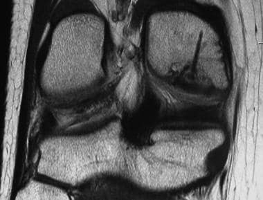
Multiple myeloma is a type of cancer that forms in a type of white blood cell called a plasma cell. Plasma cells help your body fight infection by producing antibodies. In multiple myeloma, cancerous plasma cells accumulate in the bone marrow and crowd out healthy blood cells. Rather than producing useful antibodies, the cancer cells produce abnormal proteins that can cause complications such as kidney damage, bone pain and fractures.
Multiple myeloma is a type of cancer called a plasma cell neoplasm. Plasma cell neoplasms are diseases in which there is an overproduction of a single clone of plasma cells. In multiple myeloma, this results in the crowding out of normal plasma cells, red and white blood cells and platelets, leading to many of the complications associated with the disease.
The abnormal proteins produced by the cancer cells can also cause damage to organs and tissues in the body. These abnormal proteins can be detected in the blood or urine and are often used to monitor the progression of multiple myeloma.
Multiple myeloma is a relatively uncommon cancer, but it is the second most common blood cancer after non-Hodgkin lymphoma. It typically occurs in people over the age of 65, and men are more likely to develop multiple myeloma than women. While there is no cure for multiple myeloma, treatments such as chemotherapy, radiation therapy, and stem cell transplantation can help manage the disease and its symptoms, and improve quality of life.
A waiting list, in the context of healthcare and medicine, refers to a list of patients who are awaiting a particular medical service or procedure, such as surgery, consultation with a specialist, or therapy. These lists are often established when the demand for certain services exceeds the immediate supply of resources, including physician time, hospital beds, or specialized equipment.
Patients on waiting lists are typically ranked based on factors like the severity of their condition, the urgency of their need for treatment, and the date they were placed on the list. The goal is to ensure that those with the most pressing medical needs receive care as soon as possible, while also providing a fair and transparent system for allocating limited resources.
However, it's important to note that extended waiting times can have negative consequences for patients, including worsening of symptoms, decreased quality of life, and potential complications. As such, healthcare systems strive to minimize wait times through various strategies, such as increasing resource allocation, improving efficiency, and implementing alternative service delivery models.
Histocompatibility is the compatibility between tissues or organs from different individuals in terms of their histological (tissue) structure and antigenic properties. The term is most often used in the context of transplantation, where it refers to the degree of match between the human leukocyte antigens (HLAs) and other proteins on the surface of donor and recipient cells.
A high level of histocompatibility reduces the risk of rejection of a transplanted organ or tissue by the recipient's immune system, as their immune cells are less likely to recognize the donated tissue as foreign and mount an attack against it. Conversely, a low level of histocompatibility increases the likelihood of rejection, as the recipient's immune system recognizes the donated tissue as foreign and attacks it.
Histocompatibility testing is therefore an essential part of organ and tissue transplantation, as it helps to identify the best possible match between donor and recipient and reduces the risk of rejection.
Corneal transplantation, also known as keratoplasty, is a surgical procedure in which all or part of a damaged or diseased cornea is replaced with healthy corneal tissue from a deceased donor. The cornea is the clear, dome-shaped surface at the front of the eye that plays an important role in focusing vision. When it becomes cloudy or misshapen due to injury, infection, or inherited conditions, vision can become significantly impaired.
During the procedure, the surgeon carefully removes a circular section of the damaged cornea and replaces it with a similarly sized piece of donor tissue. The new cornea is then stitched into place using very fine sutures that are typically removed several months after surgery.
Corneal transplantation has a high success rate, with more than 90% of procedures resulting in improved vision. However, as with any surgical procedure, there are risks involved, including infection, rejection of the donor tissue, and bleeding. Regular follow-up care is essential to monitor for any signs of complications and ensure proper healing.
Busulfan is a chemotherapy medication used to treat various types of cancer, including chronic myelogenous leukemia (CML) and acute myeloid leukemia (AML). It is an alkylating agent that works by damaging the DNA of cancer cells, which prevents them from dividing and growing.
The medical definition of Busulfan is:
A white crystalline powder used in chemotherapy to treat various types of cancer. Busulfan works by alkylating and cross-linking DNA, which inhibits DNA replication and transcription, leading to cell cycle arrest and apoptosis (programmed cell death) in rapidly dividing cells, including cancer cells. It is administered orally or intravenously and is often used in combination with other chemotherapy agents. Common side effects include nausea, vomiting, diarrhea, and bone marrow suppression, which can lead to anemia, neutropenia, thrombocytopenia, and increased susceptibility to infection. Long-term use of busulfan has been associated with pulmonary fibrosis, infertility, and an increased risk of secondary malignancies.
Cyclosporine is a medication that belongs to a class of drugs called immunosuppressants. It is primarily used to prevent the rejection of transplanted organs, such as kidneys, livers, and hearts. Cyclosporine works by suppressing the activity of the immune system, which helps to reduce the risk of the body attacking the transplanted organ.
In addition to its use in organ transplantation, cyclosporine may also be used to treat certain autoimmune diseases, such as rheumatoid arthritis and psoriasis. It does this by suppressing the overactive immune response that contributes to these conditions.
Cyclosporine is available in capsule, oral solution, and injectable forms. Common side effects of the medication include kidney problems, high blood pressure, tremors, headache, and nausea. Long-term use of cyclosporine can also increase the risk of certain types of cancer and infections.
It is important to note that cyclosporine should only be used under the close supervision of a healthcare provider, as it requires regular monitoring of blood levels and kidney function.
T-lymphocytes, also known as T-cells, are a type of white blood cell that plays a key role in the adaptive immune system's response to infection. They are produced in the bone marrow and mature in the thymus gland. There are several different types of T-cells, including CD4+ helper T-cells, CD8+ cytotoxic T-cells, and regulatory T-cells (Tregs).
CD4+ helper T-cells assist in activating other immune cells, such as B-lymphocytes and macrophages. They also produce cytokines, which are signaling molecules that help coordinate the immune response. CD8+ cytotoxic T-cells directly kill infected cells by releasing toxic substances. Regulatory T-cells help maintain immune tolerance and prevent autoimmune diseases by suppressing the activity of other immune cells.
T-lymphocytes are important in the immune response to viral infections, cancer, and other diseases. Dysfunction or depletion of T-cells can lead to immunodeficiency and increased susceptibility to infections. On the other hand, an overactive T-cell response can contribute to autoimmune diseases and chronic inflammation.
Tacrolimus is an immunosuppressant drug that is primarily used to prevent the rejection of transplanted organs. It works by inhibiting the activity of T-cells, which are a type of white blood cell that plays a central role in the body's immune response. By suppressing the activity of these cells, tacrolimus helps to reduce the risk of an immune response being mounted against the transplanted organ.
Tacrolimus is often used in combination with other immunosuppressive drugs, such as corticosteroids and mycophenolate mofetil, to provide a comprehensive approach to preventing organ rejection. It is available in various forms, including capsules, oral solution, and intravenous injection.
The drug was first approved for use in the United States in 1994 and has since become a widely used immunosuppressant in transplant medicine. Tacrolimus is also being studied as a potential treatment for a variety of other conditions, including autoimmune diseases and cancer.
Non-Hodgkin lymphoma (NHL) is a type of cancer that originates in the lymphatic system, which is part of the immune system. It involves the abnormal growth and proliferation of malignant lymphocytes (a type of white blood cell), leading to the formation of tumors in lymph nodes, spleen, bone marrow, or other organs. NHL can be further classified into various subtypes based on the specific type of lymphocyte involved and its characteristics.
The symptoms of Non-Hodgkin lymphoma may include:
* Painless swelling of lymph nodes in the neck, armpits, or groin
* Persistent fatigue
* Unexplained weight loss
* Fever
* Night sweats
* Itchy skin
The exact cause of Non-Hodgkin lymphoma is not well understood, but it has been associated with certain risk factors such as age (most common in people over 60), exposure to certain chemicals, immune system deficiencies, and infection with viruses like Epstein-Barr virus or HIV.
Treatment for Non-Hodgkin lymphoma depends on the stage and subtype of the disease, as well as the patient's overall health. Treatment options may include chemotherapy, radiation therapy, immunotherapy, targeted therapy, stem cell transplantation, or a combination of these approaches. Regular follow-up care is essential to monitor the progression of the disease and manage any potential long-term side effects of treatment.
Antineoplastic combined chemotherapy protocols refer to a treatment plan for cancer that involves the use of more than one antineoplastic (chemotherapy) drug given in a specific sequence and schedule. The combination of drugs is used because they may work better together to destroy cancer cells compared to using a single agent alone. This approach can also help to reduce the likelihood of cancer cells becoming resistant to the treatment.
The choice of drugs, dose, duration, and frequency are determined by various factors such as the type and stage of cancer, patient's overall health, and potential side effects. Combination chemotherapy protocols can be used in various settings, including as a primary treatment, adjuvant therapy (given after surgery or radiation to kill any remaining cancer cells), neoadjuvant therapy (given before surgery or radiation to shrink the tumor), or palliative care (to alleviate symptoms and prolong survival).
It is important to note that while combined chemotherapy protocols can be effective in treating certain types of cancer, they can also cause significant side effects, including nausea, vomiting, hair loss, fatigue, and an increased risk of infection. Therefore, patients undergoing such treatment should be closely monitored and managed by a healthcare team experienced in administering chemotherapy.
Bone marrow purging is a procedure that involves the removal of cancerous or damaged cells from bone marrow before it is transplanted into a patient. This process is often used in the treatment of blood cancers such as leukemia and lymphoma, as well as other diseases that affect the bone marrow.
The purging process typically involves collecting bone marrow from the patient or a donor, then treating it with chemicals or medications to eliminate any cancerous or damaged cells. The purged bone marrow is then transplanted back into the patient's body, where it can help to produce healthy new blood cells.
There are several methods that can be used for bone marrow purging, including physical separation techniques, chemical treatments, and immunological approaches using antibodies or other immune system components. The choice of method depends on several factors, including the type and stage of the disease being treated, as well as the patient's individual medical history and condition.
It is important to note that bone marrow purging is a complex procedure that carries some risks and potential complications, such as damage to healthy cells, delayed recovery, and increased risk of infection. As with any medical treatment, it should be carefully evaluated and discussed with a healthcare provider to determine whether it is appropriate for a given patient's situation.
A cadaver is a deceased body that is used for medical research or education. In the field of medicine, cadavers are often used in anatomy lessons, surgical training, and other forms of medical research. The use of cadavers allows medical professionals to gain a deeper understanding of the human body and its various systems without causing harm to living subjects. Cadavers may be donated to medical schools or obtained through other means, such as through consent of the deceased or their next of kin. It is important to handle and treat cadavers with respect and dignity, as they were once living individuals who deserve to be treated with care even in death.
HLA (Human Leukocyte Antigen) antigens are a group of proteins found on the surface of cells in our body. They play a crucial role in the immune system's ability to differentiate between "self" and "non-self." HLA antigens are encoded by a group of genes located on chromosome 6, known as the major histocompatibility complex (MHC).
There are three types of HLA antigens: HLA class I, HLA class II, and HLA class III. HLA class I antigens are found on the surface of almost all cells in the body and help the immune system recognize and destroy virus-infected or cancerous cells. They consist of three components: HLA-A, HLA-B, and HLA-C.
HLA class II antigens are primarily found on the surface of immune cells, such as macrophages, B cells, and dendritic cells. They assist in the presentation of foreign particles (like bacteria and viruses) to CD4+ T cells, which then activate other parts of the immune system. HLA class II antigens include HLA-DP, HLA-DQ, and HLA-DR.
HLA class III antigens consist of various molecules involved in immune responses, such as cytokines and complement components. They are not directly related to antigen presentation.
The genetic diversity of HLA antigens is extensive, with thousands of variations or alleles. This diversity allows for a better ability to recognize and respond to a wide range of pathogens. However, this variation can also lead to compatibility issues in organ transplantation, as the recipient's immune system may recognize the donor's HLA antigens as foreign and attack the transplanted organ.
Disease-free survival (DFS) is a term used in medical research and clinical practice, particularly in the field of oncology. It refers to the length of time after primary treatment for a cancer during which no evidence of the disease can be found. This means that the patient shows no signs or symptoms of the cancer, and any imaging studies or other tests do not reveal any tumors or other indications of the disease.
DFS is often used as an important endpoint in clinical trials to evaluate the effectiveness of different treatments for cancer. By measuring the length of time until the cancer recurs or a new cancer develops, researchers can get a better sense of how well a particular treatment is working and whether it is improving patient outcomes.
It's important to note that DFS is not the same as overall survival (OS), which refers to the length of time from primary treatment until death from any cause. While DFS can provide valuable information about the effectiveness of cancer treatments, it does not necessarily reflect the impact of those treatments on patients' overall survival.
Hematopoietic stem cells (HSCs) are immature, self-renewing cells that give rise to all the mature blood and immune cells in the body. They are capable of both producing more hematopoietic stem cells (self-renewal) and differentiating into early progenitor cells that eventually develop into red blood cells, white blood cells, and platelets. HSCs are found in the bone marrow, umbilical cord blood, and peripheral blood. They have the ability to repair damaged tissues and offer significant therapeutic potential for treating various diseases, including hematological disorders, genetic diseases, and cancer.
Hematopoietic Stem Cell Mobilization is the process of mobilizing hematopoietic stem cells (HSCs) from the bone marrow into the peripheral blood. HSCs are immature cells that have the ability to differentiate into all types of blood cells, including red and white blood cells and platelets.
Mobilization is often achieved through the use of medications such as granulocyte-colony stimulating factor (G-CSF) or plerixafor, which stimulate the release of HSCs from the bone marrow into the peripheral blood. This allows for the collection of HSCs from the peripheral blood through a procedure called apheresis.
Mobilized HSCs can be used in stem cell transplantation procedures to reconstitute a patient's hematopoietic system after high-dose chemotherapy or radiation therapy. It is an important process in the field of regenerative medicine and has been used to treat various diseases such as leukemia, lymphoma, and sickle cell disease.
Leukemia is a type of cancer that originates from the bone marrow - the soft, inner part of certain bones where new blood cells are made. It is characterized by an abnormal production of white blood cells, known as leukocytes or blasts. These abnormal cells accumulate in the bone marrow and interfere with the production of normal blood cells, leading to a decrease in red blood cells (anemia), platelets (thrombocytopenia), and healthy white blood cells (leukopenia).
There are several types of leukemia, classified based on the specific type of white blood cell affected and the speed at which the disease progresses:
1. Acute Leukemias - These types of leukemia progress rapidly, with symptoms developing over a few weeks or months. They involve the rapid growth and accumulation of immature, nonfunctional white blood cells (blasts) in the bone marrow and peripheral blood. The two main categories are:
- Acute Lymphoblastic Leukemia (ALL) - Originates from lymphoid progenitor cells, primarily affecting children but can also occur in adults.
- Acute Myeloid Leukemia (AML) - Develops from myeloid progenitor cells and is more common in older adults.
2. Chronic Leukemias - These types of leukemia progress slowly, with symptoms developing over a period of months to years. They involve the production of relatively mature, but still abnormal, white blood cells that can accumulate in large numbers in the bone marrow and peripheral blood. The two main categories are:
- Chronic Lymphocytic Leukemia (CLL) - Affects B-lymphocytes and is more common in older adults.
- Chronic Myeloid Leukemia (CML) - Originates from myeloid progenitor cells, characterized by the presence of a specific genetic abnormality called the Philadelphia chromosome. It can occur at any age but is more common in middle-aged and older adults.
Treatment options for leukemia depend on the type, stage, and individual patient factors. Treatments may include chemotherapy, targeted therapy, immunotherapy, stem cell transplantation, or a combination of these approaches.
Carmustine is a chemotherapy drug used to treat various types of cancer, including brain tumors, multiple myeloma, and Hodgkin's lymphoma. It belongs to a class of drugs called alkylating agents, which work by damaging the DNA in cancer cells, preventing them from dividing and growing.
Carmustine is available as an injectable solution that is administered intravenously (into a vein) or as implantable wafers that are placed directly into the brain during surgery. The drug can cause side effects such as nausea, vomiting, hair loss, and low blood cell counts, among others. It may also increase the risk of certain infections and bleeding complications.
As with all chemotherapy drugs, carmustine can have serious and potentially life-threatening side effects, and it should only be administered under the close supervision of a qualified healthcare professional. Patients receiving carmustine treatment should be closely monitored for signs of toxicity and other adverse reactions.
Brain tissue transplantation is a medical procedure that involves the surgical implantation of healthy brain tissue into a damaged or diseased brain. The goal of this procedure is to replace the non-functioning brain cells with healthy ones, in order to restore lost function or improve neurological symptoms.
The brain tissue used for transplantation can come from various sources, including fetal brain tissue, embryonic stem cells, or autologous cells (the patient's own cells). The most common type of brain tissue transplantation is fetal brain tissue transplantation, where tissue from aborted fetuses is used.
Brain tissue transplantation has been explored as a potential treatment for various neurological conditions, including Parkinson's disease, Huntington's disease, and stroke. However, the procedure remains highly experimental and is not widely available outside of clinical trials. There are also ethical concerns surrounding the use of fetal brain tissue, which has limited its widespread adoption.
It is important to note that while brain tissue transplantation holds promise as a potential treatment for neurological disorders, it is still an area of active research and much more needs to be learned about its safety and efficacy before it becomes a standard treatment option.
I'm sorry for any confusion, but "Myeloablative Agonists" is not a recognized medical term. Myeloablation is a medical process that involves destroying or damaging the bone marrow, often as part of a preparatory regimen before a stem cell transplant. Agonists are substances that bind to receptors and activate them, causing a response in the body. However, combining these two terms doesn't form a recognized medical concept. If you have any questions about myeloablation or agonists individually, I'd be happy to help clarify those concepts!
Liver diseases refer to a wide range of conditions that affect the normal functioning of the liver. The liver is a vital organ responsible for various critical functions such as detoxification, protein synthesis, and production of biochemicals necessary for digestion.
Liver diseases can be categorized into acute and chronic forms. Acute liver disease comes on rapidly and can be caused by factors like viral infections (hepatitis A, B, C, D, E), drug-induced liver injury, or exposure to toxic substances. Chronic liver disease develops slowly over time, often due to long-term exposure to harmful agents or inherent disorders of the liver.
Common examples of liver diseases include hepatitis, cirrhosis (scarring of the liver tissue), fatty liver disease, alcoholic liver disease, autoimmune liver diseases, genetic/hereditary liver disorders (like Wilson's disease and hemochromatosis), and liver cancers. Symptoms may vary widely depending on the type and stage of the disease but could include jaundice, abdominal pain, fatigue, loss of appetite, nausea, and weight loss.
Early diagnosis and treatment are essential to prevent progression and potential complications associated with liver diseases.
Salvage therapy, in the context of medical oncology, refers to the use of treatments that are typically considered less desirable or more aggressive, often due to greater side effects or lower efficacy, when standard treatment options have failed. These therapies are used to attempt to salvage a response or delay disease progression in patients with refractory or relapsed cancers.
In other words, salvage therapy is a last-resort treatment approach for patients who have not responded to first-line or subsequent lines of therapy. It may involve the use of different drug combinations, higher doses of chemotherapy, immunotherapy, targeted therapy, or radiation therapy. The goal of salvage therapy is to extend survival, improve quality of life, or achieve disease stabilization in patients with limited treatment options.
Facial transplantation is a surgical procedure that involves replacing all or part of a patient's face with facial tissue from a deceased donor. The procedure typically includes the skin, muscles, nerves, and bones of the face, and may also include the eyes and eyelids, ears, and tongue. Facial transplantation is performed to significantly improve the appearance and function of a person's face, usually in cases where the patient has suffered severe facial trauma or disfigurement due to burns, cancer, or other medical conditions.
The procedure requires extensive planning, coordination, and expertise from a multidisciplinary team of healthcare professionals, including plastic surgeons, transplant specialists, anesthesiologists, nurses, psychiatrists, and rehabilitation therapists. The surgery itself can take up to 30 hours or more, depending on the extent of the transplant.
Following the procedure, patients must undergo rigorous immunosuppressive therapy to prevent their immune system from rejecting the donor tissue. This involves taking medications that weaken the immune system and make the patient more susceptible to infections and other complications. Despite these risks, facial transplantation has been shown to significantly improve the quality of life for some patients who have undergone the procedure.
Organ preservation is a medical technique used to maintain the viability and functionality of an organ outside the body for a certain period, typically for transplantation purposes. This process involves cooling the organ to slow down its metabolic activity and prevent tissue damage, while using specialized solutions that help preserve the organ's structure and function. Commonly preserved organs include hearts, livers, kidneys, lungs, and pancreases. The goal of organ preservation is to ensure that the transplanted organ remains in optimal condition until it can be successfully implanted into a recipient.
CD34 is a type of antigen that is found on the surface of certain cells in the human body. Specifically, CD34 antigens are present on hematopoietic stem cells, which are immature cells that can develop into different types of blood cells. These stem cells are found in the bone marrow and are responsible for producing red blood cells, white blood cells, and platelets.
CD34 antigens are a type of cell surface marker that is used in medical research and clinical settings to identify and isolate hematopoietic stem cells. They are also used in the development of stem cell therapies and transplantation procedures. CD34 antigens can be detected using various laboratory techniques, such as flow cytometry or immunohistochemistry.
It's important to note that while CD34 is a useful marker for identifying hematopoietic stem cells, it is not exclusive to these cells and can also be found on other cell types, such as endothelial cells that line blood vessels. Therefore, additional markers are often used in combination with CD34 to more specifically identify and isolate hematopoietic stem cells.
Donor selection is the process of evaluating and choosing potential organ, tissue, or stem cell donors based on various medical and non-medical criteria to ensure the safety and efficacy of the transplantation. The goal of donor selection is to identify a compatible donor with minimal risk of rejection and transmission of infectious diseases while also considering ethical and legal considerations.
Medical criteria for donor selection may include:
1. Age: Donors are typically required to be within a certain age range, depending on the type of organ or tissue being donated.
2. Blood type and human leukocyte antigen (HLA) typing: Compatibility between the donor's and recipient's blood types and HLA markers is crucial to reduce the risk of rejection.
3. Medical history: Donors must undergo a thorough medical evaluation, including a review of their medical history, physical examination, and laboratory tests to assess their overall health and identify any potential risks or contraindications for donation.
4. Infectious disease screening: Donors are tested for various infectious diseases, such as HIV, hepatitis B and C, syphilis, and cytomegalovirus (CMV), among others, to ensure they do not transmit infections to the recipient.
5. Tissue typing: For organ transplants, tissue typing is performed to assess the compatibility of the donor's and recipient's major histocompatibility complex (MHC) antigens, which play a significant role in the immune response and rejection risk.
Non-medical criteria for donor selection may include:
1. Consent: Donors must provide informed consent for organ or tissue donation, and their next of kin or legal representative may be involved in the decision-making process for deceased donors.
2. Legal considerations: There are specific laws and regulations governing organ and tissue donation that must be followed, such as age restrictions, geographical proximity between the donor and recipient, and cultural or religious beliefs.
3. Ethical considerations: Donor selection should adhere to ethical principles, such as fairness, respect for autonomy, and non-maleficence, to ensure that the process is transparent, equitable, and free from coercion or exploitation.
A transplant is a medical procedure where an organ or tissue is removed from one person (the donor) and placed into another person (the recipient) for the purpose of replacing the recipient's damaged or failing organ or tissue with a healthy functioning one. The transplanted organ or tissue can come from a deceased donor, a living donor who is genetically related to the recipient, or a living donor who is not genetically related to the recipient.
Transplantation is an important medical intervention for many patients with end-stage organ failure or severe tissue damage, and it can significantly improve their quality of life and longevity. However, transplantation is a complex and risky procedure that requires careful matching of donor and recipient, rigorous evaluation and preparation of the recipient, and close monitoring and management of the transplanted organ or tissue to prevent rejection and other complications.
Hodgkin disease, also known as Hodgkin lymphoma, is a type of cancer that originates in the white blood cells called lymphocytes. It typically affects the lymphatic system, which is a network of vessels and glands spread throughout the body. The disease is characterized by the presence of a specific type of abnormal cell, known as a Reed-Sternberg cell, within the affected lymph nodes.
The symptoms of Hodgkin disease may include painless swelling of the lymph nodes in the neck, armpits, or groin; fever; night sweats; weight loss; and fatigue. The exact cause of Hodgkin disease is unknown, but it is thought to involve a combination of genetic, environmental, and infectious factors.
Hodgkin disease is typically treated with a combination of chemotherapy, radiation therapy, and/or immunotherapy, depending on the stage and extent of the disease. With appropriate treatment, the prognosis for Hodgkin disease is generally very good, with a high cure rate. However, long-term side effects of treatment may include an increased risk of secondary cancers and other health problems.
"Cells, cultured" is a medical term that refers to cells that have been removed from an organism and grown in controlled laboratory conditions outside of the body. This process is called cell culture and it allows scientists to study cells in a more controlled and accessible environment than they would have inside the body. Cultured cells can be derived from a variety of sources, including tissues, organs, or fluids from humans, animals, or cell lines that have been previously established in the laboratory.
Cell culture involves several steps, including isolation of the cells from the tissue, purification and characterization of the cells, and maintenance of the cells in appropriate growth conditions. The cells are typically grown in specialized media that contain nutrients, growth factors, and other components necessary for their survival and proliferation. Cultured cells can be used for a variety of purposes, including basic research, drug development and testing, and production of biological products such as vaccines and gene therapies.
It is important to note that cultured cells may behave differently than they do in the body, and results obtained from cell culture studies may not always translate directly to human physiology or disease. Therefore, it is essential to validate findings from cell culture experiments using additional models and ultimately in clinical trials involving human subjects.
Prognosis is a medical term that refers to the prediction of the likely outcome or course of a disease, including the chances of recovery or recurrence, based on the patient's symptoms, medical history, physical examination, and diagnostic tests. It is an important aspect of clinical decision-making and patient communication, as it helps doctors and patients make informed decisions about treatment options, set realistic expectations, and plan for future care.
Prognosis can be expressed in various ways, such as percentages, categories (e.g., good, fair, poor), or survival rates, depending on the nature of the disease and the available evidence. However, it is important to note that prognosis is not an exact science and may vary depending on individual factors, such as age, overall health status, and response to treatment. Therefore, it should be used as a guide rather than a definitive forecast.
An acute disease is a medical condition that has a rapid onset, develops quickly, and tends to be short in duration. Acute diseases can range from minor illnesses such as a common cold or flu, to more severe conditions such as pneumonia, meningitis, or a heart attack. These types of diseases often have clear symptoms that are easy to identify, and they may require immediate medical attention or treatment.
Acute diseases are typically caused by an external agent or factor, such as a bacterial or viral infection, a toxin, or an injury. They can also be the result of a sudden worsening of an existing chronic condition. In general, acute diseases are distinct from chronic diseases, which are long-term medical conditions that develop slowly over time and may require ongoing management and treatment.
Examples of acute diseases include:
* Acute bronchitis: a sudden inflammation of the airways in the lungs, often caused by a viral infection.
* Appendicitis: an inflammation of the appendix that can cause severe pain and requires surgical removal.
* Gastroenteritis: an inflammation of the stomach and intestines, often caused by a viral or bacterial infection.
* Migraine headaches: intense headaches that can last for hours or days, and are often accompanied by nausea, vomiting, and sensitivity to light and sound.
* Myocardial infarction (heart attack): a sudden blockage of blood flow to the heart muscle, often caused by a buildup of plaque in the coronary arteries.
* Pneumonia: an infection of the lungs that can cause coughing, chest pain, and difficulty breathing.
* Sinusitis: an inflammation of the sinuses, often caused by a viral or bacterial infection.
It's important to note that while some acute diseases may resolve on their own with rest and supportive care, others may require medical intervention or treatment to prevent complications and promote recovery. If you are experiencing symptoms of an acute disease, it is always best to seek medical attention to ensure proper diagnosis and treatment.
Antilymphocyte serum (ALS) is a type of immune serum that contains antibodies against human lymphocytes. It is produced by immunizing animals, such as horses or rabbits, with human lymphocytes to stimulate an immune response and the production of anti-lymphocyte antibodies. The resulting serum is then collected and can be used as a therapeutic agent to suppress the activity of the immune system in certain medical conditions.
ALS is primarily used in the treatment of transplant rejection, particularly in organ transplantation, where it helps to prevent the recipient's immune system from attacking and rejecting the transplanted organ. It can also be used in the management of autoimmune diseases, such as rheumatoid arthritis and lupus, to suppress the overactive immune response that contributes to these conditions.
It is important to note that the use of ALS carries a risk of side effects, including allergic reactions, fever, and decreased white blood cell counts. Close monitoring and appropriate management of these potential adverse events are essential during treatment with ALS.
Etoposide is a chemotherapy medication used to treat various types of cancer, including lung cancer, testicular cancer, and certain types of leukemia. It works by inhibiting the activity of an enzyme called topoisomerase II, which is involved in DNA replication and transcription. By doing so, etoposide can interfere with the growth and multiplication of cancer cells.
Etoposide is often administered intravenously in a hospital or clinic setting, although it may also be given orally in some cases. The medication can cause a range of side effects, including nausea, vomiting, hair loss, and an increased risk of infection. It can also have more serious side effects, such as bone marrow suppression, which can lead to anemia, bleeding, and a weakened immune system.
Like all chemotherapy drugs, etoposide is not without risks and should only be used under the close supervision of a qualified healthcare provider. It is important for patients to discuss the potential benefits and risks of this medication with their doctor before starting treatment.
A Lymphocyte Culture Test, Mixed (LCTM) is not a standardized medical test with a universally accepted definition. However, in some contexts, it may refer to a laboratory procedure where both T-lymphocytes and B-lymphocytes are cultured together from a sample of peripheral blood or other tissues. This test is sometimes used in research or specialized diagnostic settings to evaluate the immune function or to study the interactions between T-cells and B-cells in response to various stimuli, such as antigens or mitogens.
The test typically involves isolating lymphocytes from a sample, adding them to a culture medium along with appropriate stimulants, and then incubating the mixture for a period of time. The resulting responses, such as proliferation, differentiation, or production of cytokines, can be measured and analyzed to gain insights into the immune function or dysfunction.
It's important to note that LCTM is not a routine diagnostic test and its use and interpretation may vary depending on the specific laboratory or research setting.
Medical Definition:
"Risk factors" are any attribute, characteristic or exposure of an individual that increases the likelihood of developing a disease or injury. They can be divided into modifiable and non-modifiable risk factors. Modifiable risk factors are those that can be changed through lifestyle choices or medical treatment, while non-modifiable risk factors are inherent traits such as age, gender, or genetic predisposition. Examples of modifiable risk factors include smoking, alcohol consumption, physical inactivity, and unhealthy diet, while non-modifiable risk factors include age, sex, and family history. It is important to note that having a risk factor does not guarantee that a person will develop the disease, but rather indicates an increased susceptibility.
Tissue and organ harvesting is the surgical removal of healthy tissues or organs from a living or deceased donor for the purpose of transplantation into another person in need of a transplant. This procedure is performed with great care, adhering to strict medical standards and ethical guidelines, to ensure the safety and well-being of both the donor and the recipient.
In the case of living donors, the harvested tissue or organ is typically removed from a site that can be safely spared, such as a kidney, a portion of the liver, or a segment of the lung. The donor must undergo extensive medical evaluation to ensure they are physically and psychologically suitable for the procedure.
For deceased donors, tissue and organ harvesting is performed in a manner that respects their wishes and those of their family, as well as adheres to legal and ethical requirements. Organs and tissues must be recovered promptly after death to maintain their viability for transplantation.
Tissue and organ harvesting is an essential component of the transplant process, allowing individuals with terminal illnesses or severe injuries to receive life-saving or life-enhancing treatments. It is a complex and highly regulated medical practice that requires specialized training, expertise, and coordination among healthcare professionals, donor families, and recipients.
Prospective studies, also known as longitudinal studies, are a type of cohort study in which data is collected forward in time, following a group of individuals who share a common characteristic or exposure over a period of time. The researchers clearly define the study population and exposure of interest at the beginning of the study and follow up with the participants to determine the outcomes that develop over time. This type of study design allows for the investigation of causal relationships between exposures and outcomes, as well as the identification of risk factors and the estimation of disease incidence rates. Prospective studies are particularly useful in epidemiology and medical research when studying diseases with long latency periods or rare outcomes.
Acute myeloid leukemia (AML) is a type of cancer that originates in the bone marrow, the soft inner part of certain bones where new blood cells are made. In AML, the immature cells, called blasts, in the bone marrow fail to mature into normal blood cells. Instead, these blasts accumulate and interfere with the production of normal blood cells, leading to a shortage of red blood cells (anemia), platelets (thrombocytopenia), and normal white blood cells (leukopenia).
AML is called "acute" because it can progress quickly and become severe within days or weeks without treatment. It is a type of myeloid leukemia, which means that it affects the myeloid cells in the bone marrow. Myeloid cells are a type of white blood cell that includes monocytes and granulocytes, which help fight infection and defend the body against foreign invaders.
In AML, the blasts can build up in the bone marrow and spread to other parts of the body, including the blood, lymph nodes, liver, spleen, and brain. This can cause a variety of symptoms, such as fatigue, fever, frequent infections, easy bruising or bleeding, and weight loss.
AML is typically treated with a combination of chemotherapy, radiation therapy, and/or stem cell transplantation. The specific treatment plan will depend on several factors, including the patient's age, overall health, and the type and stage of the leukemia.
Bone marrow cells are the types of cells found within the bone marrow, which is the spongy tissue inside certain bones in the body. The main function of bone marrow is to produce blood cells. There are two types of bone marrow: red and yellow. Red bone marrow is where most blood cell production takes place, while yellow bone marrow serves as a fat storage site.
The three main types of bone marrow cells are:
1. Hematopoietic stem cells (HSCs): These are immature cells that can differentiate into any type of blood cell, including red blood cells, white blood cells, and platelets. They have the ability to self-renew, meaning they can divide and create more hematopoietic stem cells.
2. Red blood cell progenitors: These are immature cells that will develop into mature red blood cells, also known as erythrocytes. Red blood cells carry oxygen from the lungs to the body's tissues and carbon dioxide back to the lungs.
3. Myeloid and lymphoid white blood cell progenitors: These are immature cells that will develop into various types of white blood cells, which play a crucial role in the body's immune system by fighting infections and diseases. Myeloid progenitors give rise to granulocytes (neutrophils, eosinophils, and basophils), monocytes, and megakaryocytes (which eventually become platelets). Lymphoid progenitors differentiate into B cells, T cells, and natural killer (NK) cells.
Bone marrow cells are essential for maintaining a healthy blood cell count and immune system function. Abnormalities in bone marrow cells can lead to various medical conditions, such as anemia, leukopenia, leukocytosis, thrombocytopenia, or thrombocytosis, depending on the specific type of blood cell affected. Additionally, bone marrow cells are often used in transplantation procedures to treat patients with certain types of cancer, such as leukemia and lymphoma, or other hematologic disorders.
Chronic kidney failure, also known as chronic kidney disease (CKD) stage 5 or end-stage renal disease (ESRD), is a permanent loss of kidney function that occurs gradually over a period of months to years. It is defined as a glomerular filtration rate (GFR) of less than 15 ml/min, which means the kidneys are filtering waste and excess fluids at less than 15% of their normal capacity.
CKD can be caused by various underlying conditions such as diabetes, hypertension, glomerulonephritis, polycystic kidney disease, and recurrent kidney infections. Over time, the damage to the kidneys can lead to a buildup of waste products and fluids in the body, which can cause a range of symptoms including fatigue, weakness, shortness of breath, nausea, vomiting, and confusion.
Treatment for chronic kidney failure typically involves managing the underlying condition, making lifestyle changes such as following a healthy diet, and receiving supportive care such as dialysis or a kidney transplant to replace lost kidney function.
Aplastic anemia is a medical condition characterized by pancytopenia (a decrease in all three types of blood cells: red blood cells, white blood cells, and platelets) due to the failure of bone marrow to produce new cells. It is called "aplastic" because the bone marrow becomes hypocellular or "aplastic," meaning it contains few or no blood-forming stem cells.
The condition can be acquired or inherited, with acquired aplastic anemia being more common. Acquired aplastic anemia can result from exposure to toxic chemicals, radiation, drugs, viral infections, or autoimmune disorders. Inherited forms of the disease include Fanconi anemia and dyskeratosis congenita.
Symptoms of aplastic anemia may include fatigue, weakness, shortness of breath, pale skin, easy bruising or bleeding, frequent infections, and fever. Treatment options for aplastic anemia depend on the severity of the condition and its underlying cause. They may include blood transfusions, immunosuppressive therapy, and stem cell transplantation.
Granulocyte Colony-Stimulating Factor (G-CSF) is a type of growth factor that specifically stimulates the production and survival of granulocytes, a type of white blood cell crucial for fighting off infections. G-CSF works by promoting the proliferation and differentiation of hematopoietic stem cells into mature granulocytes, primarily neutrophils, in the bone marrow.
Recombinant forms of G-CSF are used clinically as a medication to boost white blood cell production in patients undergoing chemotherapy or radiation therapy for cancer, those with congenital neutropenia, and those who have had a bone marrow transplant. By increasing the number of circulating neutrophils, G-CSF helps reduce the risk of severe infections during periods of intense immune suppression.
Examples of recombinant G-CSF medications include filgrastim (Neupogen), pegfilgrastim (Neulasta), and lipegfilgrastim (Lonquex).
Immunologic cytotoxicity refers to the damage or destruction of cells that occurs as a result of an immune response. This process involves the activation of immune cells, such as cytotoxic T cells and natural killer (NK) cells, which release toxic substances, such as perforins and granzymes, that can kill target cells.
In addition, antibodies produced by B cells can also contribute to immunologic cytotoxicity by binding to antigens on the surface of target cells and triggering complement-mediated lysis or antibody-dependent cellular cytotoxicity (ADCC) by activating immune effector cells.
Immunologic cytotoxicity plays an important role in the body's defense against viral infections, cancer cells, and other foreign substances. However, it can also contribute to tissue damage and autoimmune diseases if the immune system mistakenly targets healthy cells or tissues.
Cell differentiation is the process by which a less specialized cell, or stem cell, becomes a more specialized cell type with specific functions and structures. This process involves changes in gene expression, which are regulated by various intracellular signaling pathways and transcription factors. Differentiation results in the development of distinct cell types that make up tissues and organs in multicellular organisms. It is a crucial aspect of embryonic development, tissue repair, and maintenance of homeostasis in the body.
Cytomegalovirus (CMV) infections are caused by the human herpesvirus 5 (HHV-5), a type of herpesvirus. The infection can affect people of all ages, but it is more common in individuals with weakened immune systems, such as those with HIV/AIDS or who have undergone organ transplantation.
CMV can be spread through close contact with an infected person's saliva, urine, blood, tears, semen, or breast milk. It can also be spread through sexual contact or by sharing contaminated objects, such as toys, eating utensils, or drinking glasses. Once a person is infected with CMV, the virus remains in their body for life and can reactivate later, causing symptoms to recur.
Most people who are infected with CMV do not experience any symptoms, but some may develop a mononucleosis-like illness, characterized by fever, fatigue, swollen glands, and sore throat. In people with weakened immune systems, CMV infections can cause more severe symptoms, including pneumonia, gastrointestinal disease, retinitis, and encephalitis.
Congenital CMV infection occurs when a pregnant woman passes the virus to her fetus through the placenta. This can lead to serious complications, such as hearing loss, vision loss, developmental delays, and mental disability.
Diagnosis of CMV infections is typically made through blood tests or by detecting the virus in bodily fluids, such as urine or saliva. Treatment depends on the severity of the infection and the patient's overall health. Antiviral medications may be prescribed to help manage symptoms and prevent complications.
Lymphocyte activation is the process by which B-cells and T-cells (types of lymphocytes) become activated to perform effector functions in an immune response. This process involves the recognition of specific antigens presented on the surface of antigen-presenting cells, such as dendritic cells or macrophages.
The activation of B-cells leads to their differentiation into plasma cells that produce antibodies, while the activation of T-cells results in the production of cytotoxic T-cells (CD8+ T-cells) that can directly kill infected cells or helper T-cells (CD4+ T-cells) that assist other immune cells.
Lymphocyte activation involves a series of intracellular signaling events, including the binding of co-stimulatory molecules and the release of cytokines, which ultimately result in the expression of genes involved in cell proliferation, differentiation, and effector functions. The activation process is tightly regulated to prevent excessive or inappropriate immune responses that can lead to autoimmunity or chronic inflammation.
Cytarabine is a chemotherapeutic agent used in the treatment of various types of cancer, including leukemias and lymphomas. Its chemical name is cytosine arabinoside, and it works by interfering with the DNA synthesis of cancer cells, which ultimately leads to their death.
Cytarabine is often used in combination with other chemotherapy drugs and may be administered through various routes, such as intravenous (IV) or subcutaneous injection, or orally. The specific dosage and duration of treatment will depend on the type and stage of cancer being treated, as well as the patient's overall health status.
Like all chemotherapy drugs, cytarabine can cause a range of side effects, including nausea, vomiting, diarrhea, hair loss, and an increased risk of infection. It may also cause more serious side effects, such as damage to the liver, kidneys, or nervous system, and it is important for patients to be closely monitored during treatment to minimize these risks.
It's important to note that medical treatments should only be administered under the supervision of a qualified healthcare professional, and this information should not be used as a substitute for medical advice.
C57BL/6 (C57 Black 6) is an inbred strain of laboratory mouse that is widely used in biomedical research. The term "inbred" refers to a strain of animals where matings have been carried out between siblings or other closely related individuals for many generations, resulting in a population that is highly homozygous at most genetic loci.
The C57BL/6 strain was established in 1920 by crossing a female mouse from the dilute brown (DBA) strain with a male mouse from the black strain. The resulting offspring were then interbred for many generations to create the inbred C57BL/6 strain.
C57BL/6 mice are known for their robust health, longevity, and ease of handling, making them a popular choice for researchers. They have been used in a wide range of biomedical research areas, including studies of cancer, immunology, neuroscience, cardiovascular disease, and metabolism.
One of the most notable features of the C57BL/6 strain is its sensitivity to certain genetic modifications, such as the introduction of mutations that lead to obesity or impaired glucose tolerance. This has made it a valuable tool for studying the genetic basis of complex diseases and traits.
Overall, the C57BL/6 inbred mouse strain is an important model organism in biomedical research, providing a valuable resource for understanding the genetic and molecular mechanisms underlying human health and disease.
Bronchiolitis obliterans is a medical condition characterized by the inflammation and scarring (fibrosis) of the bronchioles, which are the smallest airways in the lungs. This results in the narrowing or complete obstruction of the airways, leading to difficulty breathing and reduced lung function.
The condition is often caused by a respiratory infection, such as adenovirus or mycoplasma pneumonia, but it can also be associated with exposure to certain chemicals, drugs, or radiation therapy. In some cases, the cause may be unknown.
Symptoms of bronchiolitis obliterans include cough, shortness of breath, wheezing, and crackles heard on lung examination. Diagnosis typically involves a combination of medical history, physical exam, imaging studies (such as chest X-ray or CT scan), and pulmonary function tests. In some cases, a biopsy may be necessary to confirm the diagnosis.
Treatment for bronchiolitis obliterans is focused on managing symptoms and preventing further lung damage. This may include bronchodilators to help open up the airways, corticosteroids to reduce inflammation, and oxygen therapy to help with breathing. In severe cases, a lung transplant may be necessary.
Bone marrow is the spongy tissue found inside certain bones in the body, such as the hips, thighs, and vertebrae. It is responsible for producing blood-forming cells, including red blood cells, white blood cells, and platelets. There are two types of bone marrow: red marrow, which is involved in blood cell production, and yellow marrow, which contains fatty tissue.
Red bone marrow contains hematopoietic stem cells, which can differentiate into various types of blood cells. These stem cells continuously divide and mature to produce new blood cells that are released into the circulation. Red blood cells carry oxygen throughout the body, white blood cells help fight infections, and platelets play a crucial role in blood clotting.
Bone marrow also serves as a site for immune cell development and maturation. It contains various types of immune cells, such as lymphocytes, macrophages, and dendritic cells, which help protect the body against infections and diseases.
Abnormalities in bone marrow function can lead to several medical conditions, including anemia, leukopenia, thrombocytopenia, and various types of cancer, such as leukemia and multiple myeloma. Bone marrow aspiration and biopsy are common diagnostic procedures used to evaluate bone marrow health and function.
I'm happy to help! "Rats, Inbred Lew" is a specific strain of laboratory rats that have been inbred for research purposes. The "Lew" part of the name refers to the location where they were first developed, the Lewis Institute in Lake Bluff, Illinois, USA.
Inbreeding is a process of mating closely related individuals over many generations to create a genetically homogeneous population. This results in a high degree of genetic similarity among members of the strain, making them ideal for use as experimental models because any differences observed between individuals are more likely to be due to the experimental manipulation rather than genetic variation.
Inbred Lew rats have been widely used in biomedical research, particularly in studies related to hypertension and cardiovascular disease. They exhibit a number of unique characteristics that make them useful for these types of studies, including their susceptibility to developing high blood pressure when fed a high-salt diet or given certain drugs.
It's important to note that while inbred strains like Lew rats can be very useful tools for researchers, they are not perfect models for human disease. Because they have been bred in a controlled environment and selected for specific traits, they may not respond to experimental manipulations in the same way that humans or other animals would. Therefore, it's important to interpret findings from these studies with caution and consider multiple lines of evidence before drawing any firm conclusions.
The postoperative period is the time following a surgical procedure during which the patient's response to the surgery and anesthesia is monitored, and any complications or adverse effects are managed. This period can vary in length depending on the type of surgery and the individual patient's needs, but it typically includes the immediate recovery phase in the post-anesthesia care unit (PACU) or recovery room, as well as any additional time spent in the hospital for monitoring and management of pain, wound healing, and other aspects of postoperative care.
The goals of postoperative care are to ensure the patient's safety and comfort, promote optimal healing and rehabilitation, and minimize the risk of complications such as infection, bleeding, or other postoperative issues. The specific interventions and treatments provided during this period will depend on a variety of factors, including the type and extent of surgery performed, the patient's overall health and medical history, and any individualized care plans developed in consultation with the patient and their healthcare team.
According to the National Institutes of Health (NIH), stem cells are "initial cells" or "precursor cells" that have the ability to differentiate into many different cell types in the body. They can also divide without limit to replenish other cells for as long as the person or animal is still alive.
There are two main types of stem cells: embryonic stem cells, which come from human embryos, and adult stem cells, which are found in various tissues throughout the body. Embryonic stem cells have the ability to differentiate into all cell types in the body, while adult stem cells have more limited differentiation potential.
Stem cells play an essential role in the development and repair of various tissues and organs in the body. They are currently being studied for their potential use in the treatment of a wide range of diseases and conditions, including cancer, diabetes, heart disease, and neurological disorders. However, more research is needed to fully understand the properties and capabilities of these cells before they can be used safely and effectively in clinical settings.
Actuarial analysis is a process used in the field of actuarial science to evaluate and manage risk, typically for financial or insurance purposes. It involves the use of statistical modeling, mathematical calculations, and data analysis to estimate the probability and potential financial impact of various events or outcomes.
In a medical context, actuarial analysis may be used to assess the risks and costs associated with different health conditions, treatments, or patient populations. For example, an actuary might use data on morbidity rates, mortality rates, and healthcare utilization patterns to estimate the expected costs of providing coverage to a group of patients with a particular medical condition.
Actuarial analysis can help healthcare organizations, insurers, and policymakers make informed decisions about resource allocation, pricing, and risk management. It can also be used to develop predictive models that identify high-risk populations or forecast future trends in healthcare utilization and costs.
Cytotoxic T-lymphocytes, also known as CD8+ T cells, are a type of white blood cell that plays a central role in the cell-mediated immune system. They are responsible for identifying and destroying virus-infected cells and cancer cells. When a cytotoxic T-lymphocyte recognizes a specific antigen presented on the surface of an infected or malignant cell, it becomes activated and releases toxic substances such as perforins and granzymes, which can create pores in the target cell's membrane and induce apoptosis (programmed cell death). This process helps to eliminate the infected or malignant cells and prevent the spread of infection or cancer.
Monoclonal antibodies are a type of antibody that are identical because they are produced by a single clone of cells. They are laboratory-produced molecules that act like human antibodies in the immune system. They can be designed to attach to specific proteins found on the surface of cancer cells, making them useful for targeting and treating cancer. Monoclonal antibodies can also be used as a therapy for other diseases, such as autoimmune disorders and inflammatory conditions.
Monoclonal antibodies are produced by fusing a single type of immune cell, called a B cell, with a tumor cell to create a hybrid cell, or hybridoma. This hybrid cell is then able to replicate indefinitely, producing a large number of identical copies of the original antibody. These antibodies can be further modified and engineered to enhance their ability to bind to specific targets, increase their stability, and improve their effectiveness as therapeutic agents.
Monoclonal antibodies have several mechanisms of action in cancer therapy. They can directly kill cancer cells by binding to them and triggering an immune response. They can also block the signals that promote cancer growth and survival. Additionally, monoclonal antibodies can be used to deliver drugs or radiation directly to cancer cells, increasing the effectiveness of these treatments while minimizing their side effects on healthy tissues.
Monoclonal antibodies have become an important tool in modern medicine, with several approved for use in cancer therapy and other diseases. They are continuing to be studied and developed as a promising approach to treating a wide range of medical conditions.
Thiotepa is an antineoplastic (cancer-fighting) drug. It belongs to a class of medications called alkylating agents, which work by interfering with the DNA of cancer cells, preventing them from dividing and growing. Thiotepa is used in the treatment of various types of cancers, including breast, ovarian, and bladder cancer.
It may be administered intravenously (into a vein), intravesically (into the bladder), or intrathecally (into the spinal cord). The specific dosage and duration of treatment will depend on the type and stage of cancer being treated, as well as the patient's overall health status.
Like all chemotherapy drugs, thiotepa can have significant side effects, including nausea, vomiting, hair loss, and a weakened immune system. It is important for patients to discuss these potential risks with their healthcare provider before starting treatment.
Hematopoiesis is the process of forming and developing blood cells. It occurs in the bone marrow and includes the production of red blood cells (erythropoiesis), white blood cells (leukopoiesis), and platelets (thrombopoiesis). This process is regulated by various growth factors, hormones, and cytokines. Hematopoiesis begins early in fetal development and continues throughout a person's life. Disorders of hematopoiesis can result in conditions such as anemia, leukopenia, leukocytosis, thrombocytopenia, or thrombocytosis.
Cell separation is a process used to separate and isolate specific cell types from a heterogeneous mixture of cells. This can be accomplished through various physical or biological methods, depending on the characteristics of the cells of interest. Some common techniques for cell separation include:
1. Density gradient centrifugation: In this method, a sample containing a mixture of cells is layered onto a density gradient medium and then centrifuged. The cells are separated based on their size, density, and sedimentation rate, with denser cells settling closer to the bottom of the tube and less dense cells remaining near the top.
2. Magnetic-activated cell sorting (MACS): This technique uses magnetic beads coated with antibodies that bind to specific cell surface markers. The labeled cells are then passed through a column placed in a magnetic field, which retains the magnetically labeled cells while allowing unlabeled cells to flow through.
3. Fluorescence-activated cell sorting (FACS): In this method, cells are stained with fluorochrome-conjugated antibodies that recognize specific cell surface or intracellular markers. The stained cells are then passed through a laser beam, which excites the fluorophores and allows for the detection and sorting of individual cells based on their fluorescence profile.
4. Filtration: This simple method relies on the physical size differences between cells to separate them. Cells can be passed through filters with pore sizes that allow smaller cells to pass through while retaining larger cells.
5. Enzymatic digestion: In some cases, cells can be separated by enzymatically dissociating tissues into single-cell suspensions and then using various separation techniques to isolate specific cell types.
These methods are widely used in research and clinical settings for applications such as isolating immune cells, stem cells, or tumor cells from biological samples.
Flow cytometry is a medical and research technique used to measure physical and chemical characteristics of cells or particles, one cell at a time, as they flow in a fluid stream through a beam of light. The properties measured include:
* Cell size (light scatter)
* Cell internal complexity (granularity, also light scatter)
* Presence or absence of specific proteins or other molecules on the cell surface or inside the cell (using fluorescent antibodies or other fluorescent probes)
The technique is widely used in cell counting, cell sorting, protein engineering, biomarker discovery and monitoring disease progression, particularly in hematology, immunology, and cancer research.
A lymphocyte transfusion is not a standard medical practice. However, the term "lymphocyte transfusion" generally refers to the infusion of lymphocytes, a type of white blood cell, from a donor to a recipient. This procedure is rarely performed and primarily used in research or experimental settings, such as in the context of adoptive immunotherapy for cancer treatment.
In adoptive immunotherapy, T lymphocytes (a subtype of lymphocytes) are collected from the patient or a donor, activated, expanded in the laboratory, and then reinfused into the patient to enhance their immune response against cancer cells. This is not a common procedure and should only be performed under the guidance of experienced medical professionals in specialized centers.
It's important to note that lymphocyte transfusions are different from stem cell or bone marrow transplants, which involve the infusion of hematopoietic stem cells to reconstitute the recipient's entire blood and immune system.
Hand transplantation is a surgical procedure that involves the attachment of a donor's hand or hands to a recipient who has lost their hand(s) due to trauma, illness, or congenital conditions. The procedure involves meticulous microvascular and nerve reconstruction to reconnect bones, tendons, nerves, and blood vessels, allowing for the recovery of sensory and motor functions in the transplanted hand. It is an advanced reconstructive option that requires a careful selection of candidates, rigorous postoperative care, and immunosuppressive therapy to prevent rejection of the transplanted organ.
The liver is a large, solid organ located in the upper right portion of the abdomen, beneath the diaphragm and above the stomach. It plays a vital role in several bodily functions, including:
1. Metabolism: The liver helps to metabolize carbohydrates, fats, and proteins from the food we eat into energy and nutrients that our bodies can use.
2. Detoxification: The liver detoxifies harmful substances in the body by breaking them down into less toxic forms or excreting them through bile.
3. Synthesis: The liver synthesizes important proteins, such as albumin and clotting factors, that are necessary for proper bodily function.
4. Storage: The liver stores glucose, vitamins, and minerals that can be released when the body needs them.
5. Bile production: The liver produces bile, a digestive juice that helps to break down fats in the small intestine.
6. Immune function: The liver plays a role in the immune system by filtering out bacteria and other harmful substances from the blood.
Overall, the liver is an essential organ that plays a critical role in maintaining overall health and well-being.
Blood group incompatibility refers to a situation where the blood type of a donor and a recipient are not compatible, leading to an immune response and destruction of the donated red blood cells. This is because the recipient's immune system recognizes the donor's red blood cells as foreign due to the presence of incompatible antigens on their surface.
The most common type of blood group incompatibility occurs between individuals with different ABO blood types, such as when a person with type O blood receives type A, B, or AB blood. This can lead to agglutination and hemolysis of the donated red blood cells, causing potentially life-threatening complications such as hemolytic transfusion reaction.
Another type of blood group incompatibility occurs between Rh-negative mothers and their Rh-positive fetuses. If a mother's immune system is exposed to her fetus's Rh-positive red blood cells during pregnancy or childbirth, she may develop antibodies against them. This can lead to hemolytic disease of the newborn if the mother becomes pregnant with another Rh-positive fetus in the future.
To prevent these complications, it is essential to ensure that donated blood is compatible with the recipient's blood type before transfusion and that appropriate measures are taken during pregnancy and childbirth to prevent sensitization of Rh-negative mothers to Rh-positive red blood cells.
Mycophenolic Acid (MPA) is an immunosuppressive drug that is primarily used to prevent rejection in organ transplantation. It works by inhibiting the enzyme inosine monophosphate dehydrogenase, which is a key enzyme for the de novo synthesis of guanosine nucleotides, an essential component for the proliferation of T and B lymphocytes. By doing this, MPA reduces the activity of the immune system, thereby preventing it from attacking the transplanted organ.
Mycophenolic Acid is available in two forms: as the sodium salt (Mycophenolate Sodium) and as the morpholinoethyl ester (Mycophenolate Mofetil), which is rapidly hydrolyzed to Mycophenolic Acid after oral administration. Common side effects of MPA include gastrointestinal symptoms such as diarrhea, nausea, and vomiting, as well as an increased risk of infections due to its immunosuppressive effects.
Patient selection, in the context of medical treatment or clinical research, refers to the process of identifying and choosing appropriate individuals who are most likely to benefit from a particular medical intervention or who meet specific criteria to participate in a study. This decision is based on various factors such as the patient's diagnosis, stage of disease, overall health status, potential risks, and expected benefits. The goal of patient selection is to ensure that the selected individuals will receive the most effective and safe care possible while also contributing to meaningful research outcomes.
Neoplasm transplantation is not a recognized or established medical procedure in the field of oncology. The term "neoplasm" refers to an abnormal growth of cells, which can be benign or malignant (cancerous). "Transplantation" typically refers to the surgical transfer of living cells, tissues, or organs from one part of the body to another or between individuals.
The concept of neoplasm transplantation may imply the transfer of cancerous cells or tissues from a donor to a recipient, which is not a standard practice due to ethical considerations and the potential harm it could cause to the recipient. In some rare instances, researchers might use laboratory animals to study the transmission and growth of human cancer cells, but this is done for scientific research purposes only and under strict regulatory guidelines.
In summary, there is no medical definition for 'Neoplasm Transplantation' as it does not represent a standard or ethical medical practice.
Precursor Cell Lymphoblastic Leukemia-Lymphoma (previously known as Precursor T-lymphoblastic Leukemia/Lymphoma) is a type of cancer that affects the early stages of T-cell development. It is a subtype of acute lymphoblastic leukemia (ALL), which is characterized by the overproduction of immature white blood cells called lymphoblasts in the bone marrow, blood, and other organs.
In Precursor Cell Lymphoblastic Leukemia-Lymphoma, these abnormal lymphoblasts accumulate primarily in the lymphoid tissues such as the thymus and lymph nodes, leading to the enlargement of these organs. This subtype is more aggressive than other forms of ALL and has a higher risk of spreading to the central nervous system (CNS).
The medical definition of Precursor Cell Lymphoblastic Leukemia-Lymphoma includes:
1. A malignant neoplasm of immature T-cell precursors, also known as lymphoblasts.
2. Characterized by the proliferation and accumulation of these abnormal cells in the bone marrow, blood, and lymphoid tissues such as the thymus and lymph nodes.
3. Often associated with chromosomal abnormalities, genetic mutations, or aberrant gene expression that contribute to its aggressive behavior and poor prognosis.
4. Typically presents with symptoms related to bone marrow failure (anemia, neutropenia, thrombocytopenia), lymphadenopathy (swollen lymph nodes), hepatosplenomegaly (enlarged liver and spleen), and potential CNS involvement.
5. Diagnosed through a combination of clinical evaluation, imaging studies, and laboratory tests, including bone marrow aspiration and biopsy, immunophenotyping, cytogenetic analysis, and molecular genetic testing.
6. Treated with intensive multi-agent chemotherapy regimens, often combined with radiation therapy and/or stem cell transplantation to achieve remission and improve survival outcomes.
Lymphocyte depletion is a medical term that refers to the reduction in the number of lymphocytes (a type of white blood cell) in the body. Lymphocytes play a crucial role in the immune system, as they help to fight off infections and diseases.
Lymphocyte depletion can occur due to various reasons, including certain medical treatments such as chemotherapy or radiation therapy, immune disorders, viral infections, or bone marrow transplantation. This reduction in lymphocytes can make a person more susceptible to infections and diseases, as their immune system is weakened.
There are different types of lymphocytes, including T cells, B cells, and natural killer (NK) cells, and lymphocyte depletion can affect one or all of these types. In some cases, lymphocyte depletion may be temporary and resolve on its own or with treatment. However, in other cases, it may be more prolonged and require medical intervention to manage the associated risks and complications.
Animal disease models are specialized animals, typically rodents such as mice or rats, that have been genetically engineered or exposed to certain conditions to develop symptoms and physiological changes similar to those seen in human diseases. These models are used in medical research to study the pathophysiology of diseases, identify potential therapeutic targets, test drug efficacy and safety, and understand disease mechanisms.
The genetic modifications can include knockout or knock-in mutations, transgenic expression of specific genes, or RNA interference techniques. The animals may also be exposed to environmental factors such as chemicals, radiation, or infectious agents to induce the disease state.
Examples of animal disease models include:
1. Mouse models of cancer: Genetically engineered mice that develop various types of tumors, allowing researchers to study cancer initiation, progression, and metastasis.
2. Alzheimer's disease models: Transgenic mice expressing mutant human genes associated with Alzheimer's disease, which exhibit amyloid plaque formation and cognitive decline.
3. Diabetes models: Obese and diabetic mouse strains like the NOD (non-obese diabetic) or db/db mice, used to study the development of type 1 and type 2 diabetes, respectively.
4. Cardiovascular disease models: Atherosclerosis-prone mice, such as ApoE-deficient or LDLR-deficient mice, that develop plaque buildup in their arteries when fed a high-fat diet.
5. Inflammatory bowel disease models: Mice with genetic mutations affecting intestinal barrier function and immune response, such as IL-10 knockout or SAMP1/YitFc mice, which develop colitis.
Animal disease models are essential tools in preclinical research, but it is important to recognize their limitations. Differences between species can affect the translatability of results from animal studies to human patients. Therefore, researchers must carefully consider the choice of model and interpret findings cautiously when applying them to human diseases.
Lymphoma is a type of cancer that originates from the white blood cells called lymphocytes, which are part of the immune system. These cells are found in various parts of the body such as the lymph nodes, spleen, bone marrow, and other organs. Lymphoma can be classified into two main types: Hodgkin lymphoma (HL) and non-Hodgkin lymphoma (NHL).
HL is characterized by the presence of a specific type of abnormal lymphocyte called Reed-Sternberg cells, while NHL includes a diverse group of lymphomas that lack these cells. The symptoms of lymphoma may include swollen lymph nodes, fever, night sweats, weight loss, and fatigue.
The exact cause of lymphoma is not known, but it is believed to result from genetic mutations in the lymphocytes that lead to uncontrolled cell growth and division. Exposure to certain viruses, chemicals, and radiation may increase the risk of developing lymphoma. Treatment options for lymphoma depend on various factors such as the type and stage of the disease, age, and overall health of the patient. Common treatments include chemotherapy, radiation therapy, immunotherapy, and stem cell transplantation.
The "Graft vs Tumor Effect" is a term used in the field of transplantation medicine, particularly in allogeneic hematopoietic stem cell transplantation (HSCT). It refers to the anti-tumor activity exhibited by donor immune cells (graft) against residual malignant cells (tumor) in the recipient's body.
After HSCT, the donor's immune system is reconstituted in the recipient's body. If the donor and recipient are not identical, there may be differences in their major and minor histocompatibility antigens, which can lead to a graft-versus-host disease (GVHD) where the donor's immune cells attack the recipient's tissues. However, these same donor immune cells can also recognize and target any residual tumor cells in the recipient's body, leading to a graft vs tumor effect.
This effect can contribute to the elimination of residual malignant cells and reduce the risk of relapse, particularly in hematological malignancies such as leukemia and lymphoma. However, it is important to balance this effect with the risk of GVHD, which can cause significant morbidity and mortality. Therefore, strategies such as donor selection, graft manipulation, and immunosuppressive therapy are used to optimize the graft vs tumor effect while minimizing GVHD.
End-stage liver disease (ESLD) is a term used to describe advanced and irreversible liver damage, usually caused by chronic liver conditions such as cirrhosis, hepatitis, or alcoholic liver disease. At this stage, the liver can no longer function properly, leading to a range of serious complications.
The symptoms of ESLD may include:
* Jaundice (yellowing of the skin and eyes)
* Ascites (accumulation of fluid in the abdomen)
* Encephalopathy (confusion, drowsiness, or coma caused by the buildup of toxins in the brain)
* Bleeding from the gastrointestinal tract
* Infections
* Kidney failure
Treatment for ESLD typically focuses on managing symptoms and preventing complications. In some cases, a liver transplant may be necessary to improve survival. However, due to the shortage of available donor livers, many people with ESLD are not eligible for transplantation. The prognosis for individuals with ESLD is generally poor, with a median survival time of less than one year.
Infection is defined medically as the invasion and multiplication of pathogenic microorganisms such as bacteria, viruses, fungi, or parasites within the body, which can lead to tissue damage, illness, and disease. This process often triggers an immune response from the host's body in an attempt to eliminate the infectious agents and restore homeostasis. Infections can be transmitted through various routes, including airborne particles, direct contact with contaminated surfaces or bodily fluids, sexual contact, or vector-borne transmission. The severity of an infection may range from mild and self-limiting to severe and life-threatening, depending on factors such as the type and quantity of pathogen, the host's immune status, and any underlying health conditions.
A feasibility study is a preliminary investigation or analysis conducted to determine the viability of a proposed project, program, or product. In the medical field, feasibility studies are often conducted before implementing new treatments, procedures, equipment, or facilities. These studies help to assess the practicality and effectiveness of the proposed intervention, as well as its potential benefits and risks.
Feasibility studies in healthcare typically involve several steps:
1. Problem identification: Clearly define the problem that the proposed project, program, or product aims to address.
2. Objectives setting: Establish specific, measurable, achievable, relevant, and time-bound (SMART) objectives for the study.
3. Literature review: Conduct a thorough review of existing research and best practices related to the proposed intervention.
4. Methodology development: Design a methodology for data collection and analysis that will help answer the research questions and achieve the study's objectives.
5. Resource assessment: Evaluate the availability and adequacy of resources, including personnel, time, and finances, required to carry out the proposed intervention.
6. Risk assessment: Identify potential risks and challenges associated with the implementation of the proposed intervention and develop strategies to mitigate them.
7. Cost-benefit analysis: Estimate the costs and benefits of the proposed intervention, including direct and indirect costs, as well as short-term and long-term benefits.
8. Stakeholder engagement: Engage relevant stakeholders, such as patients, healthcare providers, administrators, and policymakers, to gather their input and support for the proposed intervention.
9. Decision-making: Based on the findings of the feasibility study, make an informed decision about whether or not to proceed with the proposed project, program, or product.
Feasibility studies are essential in healthcare as they help ensure that resources are allocated efficiently and effectively, and that interventions are evidence-based, safe, and beneficial for patients.
Bone transplantation, also known as bone grafting, is a surgical procedure in which bone or bone-like material is transferred from one part of the body to another or from one person to another. The graft may be composed of cortical (hard outer portion) bone, cancellous (spongy inner portion) bone, or a combination of both. It can be taken from different sites in the same individual (autograft), from another individual of the same species (allograft), or from an animal source (xenograft). The purpose of bone transplantation is to replace missing bone, provide structural support, and stimulate new bone growth. This procedure is commonly used in orthopedic, dental, and maxillofacial surgeries to repair bone defects caused by trauma, tumors, or congenital conditions.
Prednisone is a synthetic glucocorticoid, which is a type of corticosteroid hormone. It is primarily used to reduce inflammation in various conditions such as asthma, allergies, arthritis, and autoimmune disorders. Prednisone works by mimicking the effects of natural hormones produced by the adrenal glands, suppressing the immune system's response and reducing the release of substances that cause inflammation.
It is available in oral tablet form and is typically prescribed to be taken at specific times during the day, depending on the condition being treated. Common side effects of prednisone include increased appetite, weight gain, mood changes, insomnia, and easy bruising. Long-term use or high doses can lead to more serious side effects such as osteoporosis, diabetes, cataracts, and increased susceptibility to infections.
Healthcare providers closely monitor patients taking prednisone for extended periods to minimize the risk of adverse effects. It is essential to follow the prescribed dosage regimen and not discontinue the medication abruptly without medical supervision, as this can lead to withdrawal symptoms or a rebound of the underlying condition.
Adoptive immunotherapy is a type of cancer treatment that involves the removal of immune cells from a patient, followed by their modification and expansion in the laboratory, and then reinfusion back into the patient to help boost their immune system's ability to fight cancer. This approach can be used to enhance the natural ability of T-cells (a type of white blood cell) to recognize and destroy cancer cells.
There are different types of adoptive immunotherapy, including:
1. T-cell transfer therapy: In this approach, T-cells are removed from the patient's tumor or blood, activated and expanded in the laboratory, and then reinfused back into the patient. Some forms of T-cell transfer therapy involve genetically modifying the T-cells to express chimeric antigen receptors (CARs) that recognize specific proteins on the surface of cancer cells.
2. Tumor-infiltrating lymphocyte (TIL) therapy: This type of adoptive immunotherapy involves removing T-cells directly from a patient's tumor, expanding them in the laboratory, and then reinfusing them back into the patient. The expanded T-cells are specifically targeted to recognize and destroy cancer cells.
3. Dendritic cell (DC) vaccine: DCs are specialized immune cells that help activate T-cells. In this approach, DCs are removed from the patient, exposed to tumor antigens in the laboratory, and then reinfused back into the patient to stimulate a stronger immune response against cancer cells.
Adoptive immunotherapy has shown promise in treating certain types of cancer, such as melanoma and leukemia, but more research is needed to determine its safety and efficacy in other types of cancer.
Myelodysplastic syndromes (MDS) are a group of diverse bone marrow disorders characterized by dysplasia (abnormal development or maturation) of one or more types of blood cells or by ineffective hematopoiesis, resulting in cytopenias (lower than normal levels of one or more types of blood cells). MDS can be classified into various subtypes based on the number and type of cytopenias, the degree of dysplasia, the presence of ring sideroblasts, and cytogenetic abnormalities.
The condition primarily affects older adults, with a median age at diagnosis of around 70 years. MDS can evolve into acute myeloid leukemia (AML) in approximately 30-40% of cases. The pathophysiology of MDS involves genetic mutations and chromosomal abnormalities that lead to impaired differentiation and increased apoptosis of hematopoietic stem and progenitor cells, ultimately resulting in cytopenias and an increased risk of developing AML.
The diagnosis of MDS typically requires a bone marrow aspiration and biopsy, along with cytogenetic and molecular analyses to identify specific genetic mutations and chromosomal abnormalities. Treatment options for MDS depend on the subtype, severity of cytopenias, and individual patient factors. These may include supportive care measures, such as transfusions and growth factor therapy, or more aggressive treatments, such as chemotherapy and stem cell transplantation.
Neoplasm antigens, also known as tumor antigens, are substances that are produced by cancer cells (neoplasms) and can stimulate an immune response. These antigens can be proteins, carbohydrates, or other molecules that are either unique to the cancer cells or are overexpressed or mutated versions of normal cellular proteins.
Neoplasm antigens can be classified into two main categories: tumor-specific antigens (TSAs) and tumor-associated antigens (TAAs). TSAs are unique to cancer cells and are not expressed by normal cells, while TAAs are present at low levels in normal cells but are overexpressed or altered in cancer cells.
TSAs can be further divided into viral antigens and mutated antigens. Viral antigens are produced when cancer is caused by a virus, such as human papillomavirus (HPV) in cervical cancer. Mutated antigens are the result of genetic mutations that occur during cancer development and are unique to each patient's tumor.
Neoplasm antigens play an important role in the immune response against cancer. They can be recognized by the immune system, leading to the activation of immune cells such as T cells and natural killer (NK) cells, which can then attack and destroy cancer cells. However, cancer cells often develop mechanisms to evade the immune response, allowing them to continue growing and spreading.
Understanding neoplasm antigens is important for the development of cancer immunotherapies, which aim to enhance the body's natural immune response against cancer. These therapies include checkpoint inhibitors, which block proteins that inhibit T cell activation, and therapeutic vaccines, which stimulate an immune response against specific tumor antigens.
Leukapheresis is a medical procedure that involves the separation and removal of white blood cells (leukocytes) from the blood. It is performed using a specialized machine called an apheresis instrument, which removes the desired component (in this case, leukocytes) and returns the remaining components (red blood cells, platelets, and plasma) back to the donor or patient. This procedure is often used in the treatment of certain blood disorders, such as leukemia and lymphoma, where high white blood cell counts can cause complications. It may also be used to collect stem cells for transplantation purposes. Leukapheresis is generally a safe procedure with minimal side effects, although it may cause temporary discomfort or bruising at the site of needle insertion.
A biopsy is a medical procedure in which a small sample of tissue is taken from the body to be examined under a microscope for the presence of disease. This can help doctors diagnose and monitor various medical conditions, such as cancer, infections, or autoimmune disorders. The type of biopsy performed will depend on the location and nature of the suspected condition. Some common types of biopsies include:
1. Incisional biopsy: In this procedure, a surgeon removes a piece of tissue from an abnormal area using a scalpel or other surgical instrument. This type of biopsy is often used when the lesion is too large to be removed entirely during the initial biopsy.
2. Excisional biopsy: An excisional biopsy involves removing the entire abnormal area, along with a margin of healthy tissue surrounding it. This technique is typically employed for smaller lesions or when cancer is suspected.
3. Needle biopsy: A needle biopsy uses a thin, hollow needle to extract cells or fluid from the body. There are two main types of needle biopsies: fine-needle aspiration (FNA) and core needle biopsy. FNA extracts loose cells, while a core needle biopsy removes a small piece of tissue.
4. Punch biopsy: In a punch biopsy, a round, sharp tool is used to remove a small cylindrical sample of skin tissue. This type of biopsy is often used for evaluating rashes or other skin abnormalities.
5. Shave biopsy: During a shave biopsy, a thin slice of tissue is removed from the surface of the skin using a sharp razor-like instrument. This technique is typically used for superficial lesions or growths on the skin.
After the biopsy sample has been collected, it is sent to a laboratory where a pathologist will examine the tissue under a microscope and provide a diagnosis based on their findings. The results of the biopsy can help guide further treatment decisions and determine the best course of action for managing the patient's condition.
Leukemia, myeloid is a type of cancer that originates in the bone marrow, where blood cells are produced. Myeloid leukemia affects the myeloid cells, which include red blood cells, platelets, and most types of white blood cells. In this condition, the bone marrow produces abnormal myeloid cells that do not mature properly and accumulate in the bone marrow and blood. These abnormal cells hinder the production of normal blood cells, leading to various symptoms such as anemia, fatigue, increased risk of infections, and easy bruising or bleeding.
There are several types of myeloid leukemias, including acute myeloid leukemia (AML) and chronic myeloid leukemia (CML). AML progresses rapidly and requires immediate treatment, while CML tends to progress more slowly. The exact causes of myeloid leukemia are not fully understood, but risk factors include exposure to radiation or certain chemicals, smoking, genetic disorders, and a history of chemotherapy or other cancer treatments.
An animal model in medicine refers to the use of non-human animals in experiments to understand, predict, and test responses and effects of various biological and chemical interactions that may also occur in humans. These models are used when studying complex systems or processes that cannot be easily replicated or studied in human subjects, such as genetic manipulation or exposure to harmful substances. The choice of animal model depends on the specific research question being asked and the similarities between the animal's and human's biological and physiological responses. Examples of commonly used animal models include mice, rats, rabbits, guinea pigs, and non-human primates.
Hematologic diseases, also known as hematological disorders, refer to a group of conditions that affect the production, function, or destruction of blood cells or blood-related components, such as plasma. These diseases can affect erythrocytes (red blood cells), leukocytes (white blood cells), and platelets (thrombocytes), as well as clotting factors and hemoglobin.
Hematologic diseases can be broadly categorized into three main types:
1. Anemia: A condition characterized by a decrease in the total red blood cell count, hemoglobin, or hematocrit, leading to insufficient oxygen transport to tissues and organs. Examples include iron deficiency anemia, sickle cell anemia, and aplastic anemia.
2. Leukemia and other disorders of white blood cells: These conditions involve the abnormal production or function of leukocytes, which can lead to impaired immunity and increased susceptibility to infections. Examples include leukemias (acute lymphoblastic leukemia, chronic myeloid leukemia), lymphomas, and myelodysplastic syndromes.
3. Platelet and clotting disorders: These diseases affect the production or function of platelets and clotting factors, leading to abnormal bleeding or clotting tendencies. Examples include hemophilia, von Willebrand disease, thrombocytopenia, and disseminated intravascular coagulation (DIC).
Hematologic diseases can have various causes, including genetic defects, infections, autoimmune processes, environmental factors, or malignancies. Proper diagnosis and management of these conditions often require the expertise of hematologists, who specialize in diagnosing and treating disorders related to blood and its components.
Vincristine is an antineoplastic agent, specifically a vinca alkaloid. It is derived from the Madagascar periwinkle plant (Catharanthus roseus). Vincristine binds to tubulin, a protein found in microtubules, and inhibits their polymerization, which results in disruption of mitotic spindles leading to cell cycle arrest and apoptosis (programmed cell death). It is used in the treatment of various types of cancer including leukemias, lymphomas, and solid tumors. Common side effects include peripheral neuropathy, constipation, and alopecia.
Brain death is a legal and medical determination that an individual has died because their brain has irreversibly lost all functions necessary for life. It is characterized by the absence of brainstem reflexes, unresponsiveness to stimuli, and the inability to breathe without mechanical support. Brain death is different from a vegetative state or coma, where there may still be some brain activity.
The determination of brain death involves a series of tests and examinations to confirm the absence of brain function. These tests are typically performed by trained medical professionals and may include clinical assessments, imaging studies, and electroencephalograms (EEGs) to confirm the absence of electrical activity in the brain.
Brain death is an important concept in medicine because it allows for the organ donation process to proceed, potentially saving the lives of others. In many jurisdictions, brain death is legally equivalent to cardiopulmonary death, which means that once a person has been declared brain dead, they are considered deceased and their organs can be removed for transplantation.
A fatal outcome is a term used in medical context to describe a situation where a disease, injury, or illness results in the death of an individual. It is the most severe and unfortunate possible outcome of any medical condition, and is often used as a measure of the severity and prognosis of various diseases and injuries. In clinical trials and research, fatal outcome may be used as an endpoint to evaluate the effectiveness and safety of different treatments or interventions.
The ABO blood-group system is a classification system used in blood transfusion medicine to determine the compatibility of donated blood with a recipient's blood. It is based on the presence or absence of two antigens, A and B, on the surface of red blood cells (RBCs), as well as the corresponding antibodies present in the plasma.
There are four main blood types in the ABO system:
1. Type A: These individuals have A antigens on their RBCs and anti-B antibodies in their plasma.
2. Type B: They have B antigens on their RBCs and anti-A antibodies in their plasma.
3. Type AB: They have both A and B antigens on their RBCs but no natural antibodies against either A or B antigens.
4. Type O: They do not have any A or B antigens on their RBCs, but they have both anti-A and anti-B antibodies in their plasma.
Transfusing blood from a donor with incompatible ABO antigens can lead to an immune response, causing the destruction of donated RBCs and potentially life-threatening complications such as acute hemolytic transfusion reaction. Therefore, it is crucial to match the ABO blood type between donors and recipients before performing a blood transfusion.
Lymphocytes are a type of white blood cell that is an essential part of the immune system. They are responsible for recognizing and responding to potentially harmful substances such as viruses, bacteria, and other foreign invaders. There are two main types of lymphocytes: B-lymphocytes (B-cells) and T-lymphocytes (T-cells).
B-lymphocytes produce antibodies, which are proteins that help to neutralize or destroy foreign substances. When a B-cell encounters a foreign substance, it becomes activated and begins to divide and differentiate into plasma cells, which produce and secrete large amounts of antibodies. These antibodies bind to the foreign substance, marking it for destruction by other immune cells.
T-lymphocytes, on the other hand, are involved in cell-mediated immunity. They directly attack and destroy infected cells or cancerous cells. T-cells can also help to regulate the immune response by producing chemical signals that activate or inhibit other immune cells.
Lymphocytes are produced in the bone marrow and mature in either the bone marrow (B-cells) or the thymus gland (T-cells). They circulate throughout the body in the blood and lymphatic system, where they can be found in high concentrations in lymph nodes, the spleen, and other lymphoid organs.
Abnormalities in the number or function of lymphocytes can lead to a variety of immune-related disorders, including immunodeficiency diseases, autoimmune disorders, and cancer.
Hepatic Veno-Occlusive Disease (VOD), also known as Sinusoidal Obstruction Syndrome (SOS), is a medical condition characterized by the obstruction or blockage of the small veins (venules) in the liver. This results in the backup of blood in the liver, leading to swelling and damage to the liver cells.
The obstruction is usually caused by the injury and inflammation of the endothelial cells lining the venules, which can be triggered by various factors such as chemotherapy drugs, radiation therapy, bone marrow transplantation, or exposure to certain toxins. The damage to the liver can lead to symptoms such as fluid accumulation in the abdomen (ascites), enlarged liver, jaundice, and in severe cases, liver failure.
The diagnosis of VOD/SOS is typically made based on a combination of clinical signs, symptoms, and imaging studies, such as ultrasound or CT scan. In some cases, a liver biopsy may be necessary to confirm the diagnosis. Treatment for VOD/SOS is primarily supportive, with the goal of managing symptoms and preventing complications. This may include medications to reduce swelling, improve liver function, and prevent infection. In severe cases, liver transplantation may be considered as a last resort.
Dendritic cells (DCs) are a type of immune cell that play a critical role in the body's defense against infection and cancer. They are named for their dendrite-like projections, which they use to interact with and sample their environment. DCs are responsible for processing antigens (foreign substances that trigger an immune response) and presenting them to T cells, a type of white blood cell that plays a central role in the immune system's response to infection and cancer.
DCs can be found throughout the body, including in the skin, mucous membranes, and lymphoid organs. They are able to recognize and respond to a wide variety of antigens, including those from bacteria, viruses, fungi, and parasites. Once they have processed an antigen, DCs migrate to the lymph nodes, where they present the antigen to T cells. This interaction activates the T cells, which then go on to mount a targeted immune response against the invading pathogen or cancerous cells.
DCs are a diverse group of cells that can be divided into several subsets based on their surface markers and function. Some DCs, such as Langerhans cells and dermal DCs, are found in the skin and mucous membranes, where they serve as sentinels for invading pathogens. Other DCs, such as plasmacytoid DCs and conventional DCs, are found in the lymphoid organs, where they play a role in activating T cells and initiating an immune response.
Overall, dendritic cells are essential for the proper functioning of the immune system, and dysregulation of these cells has been implicated in a variety of diseases, including autoimmune disorders and cancer.
Chimerism is a medical term that refers to the presence of genetically distinct cell populations within an individual. This phenomenon can occur naturally or as a result of a medical procedure such as a stem cell transplant. In natural chimerism, an individual may have cells with different genetic compositions due to events that occurred during embryonic development, such as the fusion of two fertilized eggs (also known as "twinning") or the exchange of cells between twins in utero.
In the context of a stem cell transplant, chimerism can occur when a donor's stem cells engraft and begin to produce new blood cells in the recipient's body. This can result in the presence of both the recipient's own cells and the donor's cells in the recipient's body. The degree of chimerism can vary, with some individuals showing complete chimerism (where all blood cells are derived from the donor) or mixed chimerism (where both the recipient's and donor's cells coexist).
Monitoring chimerism levels is important in stem cell transplantation to assess the success of the procedure and to detect any potential signs of graft rejection or relapse of the original disease.
A blood transfusion is a medical procedure in which blood or its components are transferred from one individual (donor) to another (recipient) through a vein. The donated blood can be fresh whole blood, packed red blood cells, platelets, plasma, or cryoprecipitate, depending on the recipient's needs. Blood transfusions are performed to replace lost blood due to severe bleeding, treat anemia, support patients undergoing major surgeries, or manage various medical conditions such as hemophilia, thalassemia, and leukemia. The donated blood must be carefully cross-matched with the recipient's blood type to minimize the risk of transfusion reactions.
Acute liver failure is a sudden and severe loss of liver function that occurs within a few days or weeks. It can be caused by various factors such as drug-induced liver injury, viral hepatitis, or metabolic disorders. In acute liver failure, the liver cannot perform its vital functions, including protein synthesis, detoxification, and metabolism of carbohydrates, fats, and proteins.
The symptoms of acute liver failure include jaundice (yellowing of the skin and eyes), coagulopathy (bleeding disorders), hepatic encephalopathy (neurological symptoms such as confusion, disorientation, and coma), and elevated levels of liver enzymes in the blood. Acute liver failure is a medical emergency that requires immediate hospitalization and treatment, which may include medications, supportive care, and liver transplantation.
The "Graft versus Leukemia (GvL) Effect" is a term used in the field of hematopoietic stem cell transplantation to describe a desirable outcome where the donor's immune cells (graft) recognize and attack the recipient's leukemia cells (host). This effect occurs when the donor's T-lymphocytes, natural killer cells, and other immune cells become activated against the recipient's malignant cells.
The GvL effect is often observed in patients who have undergone allogeneic hematopoietic stem cell transplantation (allo-HSCT), where the donor and recipient are not genetically identical. The genetic disparity between the donor and recipient creates an environment that allows for the recognition of host leukemia cells as foreign, triggering an immune response against them.
While the GvL effect can be beneficial in eliminating residual leukemia cells, it can also lead to complications such as graft-versus-host disease (GvHD), where the donor's immune cells attack the recipient's healthy tissues. Balancing the GvL effect and minimizing GvHD remains a significant challenge in allo-HSCT.
Cell culture is a technique used in scientific research to grow and maintain cells from plants, animals, or humans in a controlled environment outside of their original organism. This environment typically consists of a sterile container called a cell culture flask or plate, and a nutrient-rich liquid medium that provides the necessary components for the cells' growth and survival, such as amino acids, vitamins, minerals, and hormones.
There are several different types of cell culture techniques used in research, including:
1. Adherent cell culture: In this technique, cells are grown on a flat surface, such as the bottom of a tissue culture dish or flask. The cells attach to the surface and spread out, forming a monolayer that can be observed and manipulated under a microscope.
2. Suspension cell culture: In suspension culture, cells are grown in liquid medium without any attachment to a solid surface. These cells remain suspended in the medium and can be agitated or mixed to ensure even distribution of nutrients.
3. Organoid culture: Organoids are three-dimensional structures that resemble miniature organs and are grown from stem cells or other progenitor cells. They can be used to study organ development, disease processes, and drug responses.
4. Co-culture: In co-culture, two or more different types of cells are grown together in the same culture dish or flask. This technique is used to study cell-cell interactions and communication.
5. Conditioned medium culture: In this technique, cells are grown in a medium that has been conditioned by previous cultures of other cells. The conditioned medium contains factors secreted by the previous cells that can influence the growth and behavior of the new cells.
Cell culture techniques are widely used in biomedical research to study cellular processes, develop drugs, test toxicity, and investigate disease mechanisms. However, it is important to note that cell cultures may not always accurately represent the behavior of cells in a living organism, and results from cell culture experiments should be validated using other methods.
Surgical anastomosis is a medical procedure that involves the connection of two tubular structures, such as blood vessels or intestines, to create a continuous passage. This technique is commonly used in various types of surgeries, including vascular, gastrointestinal, and orthopedic procedures.
During a surgical anastomosis, the ends of the two tubular structures are carefully prepared by removing any damaged or diseased tissue. The ends are then aligned and joined together using sutures, staples, or other devices. The connection must be secure and leak-free to ensure proper function and healing.
The success of a surgical anastomosis depends on several factors, including the patient's overall health, the location and condition of the structures being joined, and the skill and experience of the surgeon. Complications such as infection, bleeding, or leakage can occur, which may require additional medical intervention or surgery.
Proper postoperative care is also essential to ensure the success of a surgical anastomosis. This may include monitoring for signs of complications, administering medications to prevent infection and promote healing, and providing adequate nutrition and hydration.
Immune tolerance, also known as immunological tolerance or specific immune tolerance, is a state of unresponsiveness or non-reactivity of the immune system towards a particular substance (antigen) that has the potential to elicit an immune response. This occurs when the immune system learns to distinguish "self" from "non-self" and does not attack the body's own cells, tissues, and organs.
In the context of transplantation, immune tolerance refers to the absence of a destructive immune response towards the transplanted organ or tissue, allowing for long-term graft survival without the need for immunosuppressive therapy. Immune tolerance can be achieved through various strategies, including hematopoietic stem cell transplantation, costimulation blockade, and regulatory T cell induction.
In summary, immune tolerance is a critical mechanism that prevents the immune system from attacking the body's own structures while maintaining the ability to respond appropriately to foreign pathogens and antigens.
CD (cluster of differentiation) antigens are cell-surface proteins that are expressed on leukocytes (white blood cells) and can be used to identify and distinguish different subsets of these cells. They are important markers in the field of immunology and hematology, and are commonly used to diagnose and monitor various diseases, including cancer, autoimmune disorders, and infectious diseases.
CD antigens are designated by numbers, such as CD4, CD8, CD19, etc., which refer to specific proteins found on the surface of different types of leukocytes. For example, CD4 is a protein found on the surface of helper T cells, while CD8 is found on cytotoxic T cells.
CD antigens can be used as targets for immunotherapy, such as monoclonal antibody therapy, in which antibodies are designed to bind to specific CD antigens and trigger an immune response against cancer cells or infected cells. They can also be used as markers to monitor the effectiveness of treatments and to detect minimal residual disease (MRD) after treatment.
It's important to note that not all CD antigens are exclusive to leukocytes, some can be found on other cell types as well, and their expression can vary depending on the activation state or differentiation stage of the cells.
Liver cirrhosis is a chronic, progressive disease characterized by the replacement of normal liver tissue with scarred (fibrotic) tissue, leading to loss of function. The scarring is caused by long-term damage from various sources such as hepatitis, alcohol abuse, nonalcoholic fatty liver disease, and other causes. As the disease advances, it can lead to complications like portal hypertension, fluid accumulation in the abdomen (ascites), impaired brain function (hepatic encephalopathy), and increased risk of liver cancer. It is generally irreversible, but early detection and treatment of underlying causes may help slow down its progression.
Immunotherapy is a type of medical treatment that uses the body's own immune system to fight against diseases, such as cancer. It involves the use of substances (like vaccines, medications, or immune cells) that stimulate or suppress the immune system to help it recognize and destroy harmful disease-causing cells or agents, like tumor cells.
Immunotherapy can work in several ways:
1. Activating the immune system: Certain immunotherapies boost the body's natural immune responses, helping them recognize and attack cancer cells more effectively.
2. Suppressing immune system inhibitors: Some immunotherapies target and block proteins or molecules that can suppress the immune response, allowing the immune system to work more efficiently against diseases.
3. Replacing or enhancing specific immune cells: Immunotherapy can also involve administering immune cells (like T-cells) that have been genetically engineered or modified to recognize and destroy cancer cells.
Immunotherapies have shown promising results in treating various types of cancer, autoimmune diseases, and allergies. However, they can also cause side effects, as an overactive immune system may attack healthy tissues and organs. Therefore, careful monitoring is necessary during immunotherapy treatment.
Lymphoproliferative disorders (LPDs) are a group of diseases characterized by the excessive proliferation of lymphoid cells, which are crucial components of the immune system. These disorders can arise from both B-cells and T-cells, leading to various clinical manifestations ranging from benign to malignant conditions.
LPDs can be broadly classified into reactive and neoplastic categories:
1. Reactive Lymphoproliferative Disorders: These are typically triggered by infections, autoimmune diseases, or immunodeficiency states. They involve an exaggerated response of the immune system leading to the excessive proliferation of lymphoid cells. Examples include:
* Infectious mononucleosis (IM) caused by Epstein-Barr virus (EBV)
* Lymph node enlargement due to various infections or autoimmune disorders
* Post-transplant lymphoproliferative disorder (PTLD), which occurs in the context of immunosuppression following organ transplantation
2. Neoplastic Lymphoproliferative Disorders: These are malignant conditions characterized by uncontrolled growth and accumulation of abnormal lymphoid cells, leading to the formation of tumors. They can be further classified into Hodgkin lymphoma (HL) and non-Hodgkin lymphoma (NHL). Examples include:
* Hodgkin lymphoma (HL): Classical HL and nodular lymphocyte-predominant HL
* Non-Hodgkin lymphoma (NHL): Various subtypes, such as diffuse large B-cell lymphoma, follicular lymphoma, mantle cell lymphoma, and Burkitt lymphoma
It is important to note that the distinction between reactive and neoplastic LPDs can sometimes be challenging, requiring careful clinical, histopathological, immunophenotypic, and molecular evaluations. Proper diagnosis and classification of LPDs are crucial for determining appropriate treatment strategies and predicting patient outcomes.
Immunophenotyping is a medical laboratory technique used to identify and classify cells, usually in the context of hematologic (blood) disorders and malignancies (cancers), based on their surface or intracellular expression of various proteins and antigens. This technique utilizes specific antibodies tagged with fluorochromes, which bind to the target antigens on the cell surface or within the cells. The labeled cells are then analyzed using flow cytometry, allowing for the detection and quantification of multiple antigenic markers simultaneously.
Immunophenotyping helps in understanding the distribution of different cell types, their subsets, and activation status, which can be crucial in diagnosing various hematological disorders, immunodeficiencies, and distinguishing between different types of leukemias, lymphomas, and other malignancies. Additionally, it can also be used to monitor the progression of diseases, evaluate the effectiveness of treatments, and detect minimal residual disease (MRD) during follow-up care.
Chronic myelogenous leukemia (CML), BCR-ABL positive is a specific subtype of leukemia that originates in the bone marrow and involves the excessive production of mature granulocytes, a type of white blood cell. It is characterized by the presence of the Philadelphia chromosome, which is formed by a genetic translocation between chromosomes 9 and 22, resulting in the formation of the BCR-ABL fusion gene. This gene encodes for an abnormal protein with increased tyrosine kinase activity, leading to uncontrolled cell growth and division. The presence of this genetic abnormality is used to confirm the diagnosis and guide treatment decisions.
Vidarabine is an antiviral medication used to treat herpes simplex infections, particularly severe cases such as herpes encephalitis (inflammation of the brain caused by the herpes simplex virus). It works by interfering with the DNA replication of the virus.
In medical terms, vidarabine is a nucleoside analogue that is phosphorylated intracellularly to the active form, vidarabine triphosphate. This compound inhibits viral DNA polymerase and incorporates into viral DNA, causing termination of viral DNA synthesis.
Vidarabine was previously used as an injectable medication but has largely been replaced by more modern antiviral drugs such as acyclovir due to its greater efficacy and lower toxicity.
Primary graft dysfunction (PGD) is a severe complication that can occur after an organ transplant, such as a lung or heart transplant. It refers to the early functional impairment of the grafted organ that is not due to surgical complications, rejection, or recurrence of the original disease.
In the case of lung transplants, PGD is defined as the evidence of poor oxygenation and stiffness in the lungs within the first 72 hours after the transplant. It is typically caused by inflammation, injury to the blood vessels, or other damage to the lung tissue during the transplant procedure or due to pre-existing conditions in the donor organ.
PGD can lead to serious complications, including respiratory failure, and is associated with increased morbidity and mortality after transplantation. Treatment may include supportive care, such as mechanical ventilation and medications to support lung function, as well as strategies to reduce inflammation and prevent further damage to the grafted organ.
Isoantibodies are antibodies produced by the immune system that recognize and react to antigens (markers) found on the cells or tissues of another individual of the same species. These antigens are typically proteins or carbohydrates present on the surface of red blood cells, but they can also be found on other cell types.
Isoantibodies are formed when an individual is exposed to foreign antigens, usually through blood transfusions, pregnancy, or tissue transplantation. The exposure triggers the immune system to produce specific antibodies against these antigens, which can cause a harmful immune response if the individual receives another transfusion or transplant from the same donor in the future.
There are two main types of isoantibodies:
1. Agglutinins: These are IgM antibodies that cause red blood cells to clump together (agglutinate) when mixed with the corresponding antigen. They develop rapidly after exposure and can cause immediate transfusion reactions or hemolytic disease of the newborn in pregnant women.
2. Hemolysins: These are IgG antibodies that destroy red blood cells by causing their membranes to become more permeable, leading to lysis (bursting) of the cells and release of hemoglobin into the plasma. They take longer to develop but can cause delayed transfusion reactions or hemolytic disease of the newborn in pregnant women.
Isoantibodies are detected through blood tests, such as the crossmatch test, which determines compatibility between a donor's and recipient's blood before transfusions or transplants.
The portal vein is the large venous trunk that carries blood from the gastrointestinal tract, spleen, pancreas, and gallbladder to the liver. It is formed by the union of the superior mesenteric vein (draining the small intestine and a portion of the large intestine) and the splenic vein (draining the spleen and pancreas). The portal vein then divides into right and left branches within the liver, where the blood flows through the sinusoids and gets enriched with oxygen and nutrients before being drained by the hepatic veins into the inferior vena cava. This unique arrangement allows the liver to process and detoxify the absorbed nutrients, remove waste products, and regulate metabolic homeostasis.
Multivariate analysis is a statistical method used to examine the relationship between multiple independent variables and a dependent variable. It allows for the simultaneous examination of the effects of two or more independent variables on an outcome, while controlling for the effects of other variables in the model. This technique can be used to identify patterns, associations, and interactions among multiple variables, and is commonly used in medical research to understand complex health outcomes and disease processes. Examples of multivariate analysis methods include multiple regression, factor analysis, cluster analysis, and discriminant analysis.
Regeneration in a medical context refers to the process of renewal, restoration, and growth that replaces damaged or missing cells, tissues, organs, or even whole limbs in some organisms. This complex biological process involves various cellular and molecular mechanisms, such as cell proliferation, differentiation, and migration, which work together to restore the structural and functional integrity of the affected area.
In human medicine, regeneration has attracted significant interest due to its potential therapeutic applications in treating various conditions, including degenerative diseases, trauma, and congenital disorders. Researchers are actively studying the underlying mechanisms of regeneration in various model organisms to develop novel strategies for promoting tissue repair and regeneration in humans.
Examples of regeneration in human medicine include liver regeneration after partial hepatectomy, where the remaining liver lobes can grow back to their original size within weeks, and skin wound healing, where keratinocytes migrate and proliferate to close the wound and restore the epidermal layer. However, the regenerative capacity of humans is limited compared to some other organisms, such as planarians and axolotls, which can regenerate entire body parts or even their central nervous system.
The Kaplan-Meier estimate is a statistical method used to calculate the survival probability over time in a population. It is commonly used in medical research to analyze time-to-event data, such as the time until a patient experiences a specific event like disease progression or death. The Kaplan-Meier estimate takes into account censored data, which occurs when some individuals are lost to follow-up before experiencing the event of interest.
The method involves constructing a survival curve that shows the proportion of subjects still surviving at different time points. At each time point, the survival probability is calculated as the product of the conditional probabilities of surviving from one time point to the next. The Kaplan-Meier estimate provides an unbiased and consistent estimator of the survival function, even when censoring is present.
In summary, the Kaplan-Meier estimate is a crucial tool in medical research for analyzing time-to-event data and estimating survival probabilities over time while accounting for censored observations.
Mesenchymal Stromal Cells (MSCs) are a type of adult stem cells found in various tissues, including bone marrow, adipose tissue, and umbilical cord blood. They have the ability to differentiate into multiple cell types, such as osteoblasts, chondrocytes, and adipocytes, under specific conditions. MSCs also possess immunomodulatory properties, making them a promising tool in regenerative medicine and therapeutic strategies for various diseases, including autoimmune disorders and tissue injuries. It is important to note that the term "Mesenchymal Stem Cells" has been replaced by "Mesenchymal Stromal Cells" in the scientific community to better reflect their biological characteristics and potential functions.
Delayed graft function (DGF) is a term used in the medical field, particularly in transplant medicine. It refers to a situation where a transplanted organ, most commonly a kidney, fails to function normally immediately after the transplantation procedure. This failure to function occurs within the first week after the transplant and is usually associated with poor urine output and elevated levels of creatinine in the blood.
DGF can be caused by several factors, including pre-existing conditions in the recipient, such as diabetes or hypertension, poor quality of the donor organ, or complications during the surgery. It may also result from the immune system's reaction to the transplanted organ, known as rejection.
In many cases, DGF can be managed with medical interventions, such as administering medications to help reduce inflammation and improve blood flow to the organ. However, in some instances, it may lead to more severe complications, including acute or chronic rejection of the transplanted organ, which could require additional treatments or even another transplant.
It's important to note that not all cases of DGF lead to long-term complications, and many patients with DGF can still go on to have successful transplants with proper management and care.
In epidemiology, the incidence of a disease is defined as the number of new cases of that disease within a specific population over a certain period of time. It is typically expressed as a rate, with the number of new cases in the numerator and the size of the population at risk in the denominator. Incidence provides information about the risk of developing a disease during a given time period and can be used to compare disease rates between different populations or to monitor trends in disease occurrence over time.
Fetal blood refers to the blood circulating in a fetus during pregnancy. It is essential for the growth and development of the fetus, as it carries oxygen and nutrients from the placenta to the developing tissues and organs. Fetal blood also removes waste products, such as carbon dioxide, from the fetal tissues and transports them to the placenta for elimination.
Fetal blood has several unique characteristics that distinguish it from adult blood. For example, fetal hemoglobin (HbF) is the primary type of hemoglobin found in fetal blood, whereas adults primarily have adult hemoglobin (HbA). Fetal hemoglobin has a higher affinity for oxygen than adult hemoglobin, which allows it to more efficiently extract oxygen from the maternal blood in the placenta.
Additionally, fetal blood contains a higher proportion of reticulocytes (immature red blood cells) and nucleated red blood cells compared to adult blood. These differences reflect the high turnover rate of red blood cells in the developing fetus and the need for rapid growth and development.
Examination of fetal blood can provide important information about the health and well-being of the fetus during pregnancy. For example, fetal blood sampling (also known as cordocentesis or percutaneous umbilical blood sampling) can be used to diagnose genetic disorders, infections, and other conditions that may affect fetal development. However, this procedure carries risks, including preterm labor, infection, and fetal loss, and is typically only performed when there is a significant risk of fetal compromise or when other diagnostic tests have been inconclusive.
Isoantigens are antigens that are present on the cells or tissues of one individual of a species, but are absent or different in another individual of the same species. They are also known as "alloantigens." Isoantigens are most commonly found on the surface of red blood cells and other tissues, and they can stimulate an immune response when transplanted into a different individual. This is because the recipient's immune system recognizes the isoantigens as foreign and mounts a defense against them. Isoantigens are important in the field of transplantation medicine, as they must be carefully matched between donor and recipient to reduce the risk of rejection.
Liver neoplasms refer to abnormal growths in the liver that can be benign or malignant. Benign liver neoplasms are non-cancerous tumors that do not spread to other parts of the body, while malignant liver neoplasms are cancerous tumors that can invade and destroy surrounding tissue and spread to other organs.
Liver neoplasms can be primary, meaning they originate in the liver, or secondary, meaning they have metastasized (spread) to the liver from another part of the body. Primary liver neoplasms can be further classified into different types based on their cell of origin and behavior, including hepatocellular carcinoma, cholangiocarcinoma, and hepatic hemangioma.
The diagnosis of liver neoplasms typically involves a combination of imaging studies, such as ultrasound, CT scan, or MRI, and biopsy to confirm the type and stage of the tumor. Treatment options depend on the type and extent of the neoplasm and may include surgery, radiation therapy, chemotherapy, or liver transplantation.
CD4-positive T-lymphocytes, also known as CD4+ T cells or helper T cells, are a type of white blood cell that plays a crucial role in the immune response. They express the CD4 receptor on their surface and help coordinate the immune system's response to infectious agents such as viruses and bacteria.
CD4+ T cells recognize and bind to specific antigens presented by antigen-presenting cells, such as dendritic cells or macrophages. Once activated, they can differentiate into various subsets of effector cells, including Th1, Th2, Th17, and Treg cells, each with distinct functions in the immune response.
CD4+ T cells are particularly important in the immune response to HIV (human immunodeficiency virus), which targets and destroys these cells, leading to a weakened immune system and increased susceptibility to opportunistic infections. The number of CD4+ T cells is often used as a marker of disease progression in HIV infection, with lower counts indicating more advanced disease.
Cryopreservation is a medical procedure that involves the preservation of cells, tissues, or organs by cooling them to very low temperatures, typically below -150°C. This is usually achieved using liquid nitrogen. The low temperature slows down or stops biological activity, including chemical reactions and cellular metabolism, which helps to prevent damage and decay.
The cells, tissues, or organs that are being cryopreserved must be treated with a cryoprotectant solution before cooling to prevent the formation of ice crystals, which can cause significant damage. Once cooled, the samples are stored in specialized containers or tanks until they are needed for use.
Cryopreservation is commonly used in assisted reproductive technologies, such as the preservation of sperm, eggs, and embryos for fertility treatments. It is also used in research, including the storage of cell lines and stem cells, and in clinical settings, such as the preservation of skin grafts and corneas for transplantation.
Postoperative care refers to the comprehensive medical treatment and nursing attention provided to a patient following a surgical procedure. The goal of postoperative care is to facilitate the patient's recovery, prevent complications, manage pain, ensure proper healing of the incision site, and maintain overall health and well-being until the patient can resume their normal activities.
This type of care includes monitoring vital signs, managing pain through medication or other techniques, ensuring adequate hydration and nutrition, helping the patient with breathing exercises to prevent lung complications, encouraging mobility to prevent blood clots, monitoring for signs of infection or other complications, administering prescribed medications, providing wound care, and educating the patient about postoperative care instructions.
The duration of postoperative care can vary depending on the type and complexity of the surgical procedure, as well as the individual patient's needs and overall health status. It may be provided in a hospital setting, an outpatient surgery center, or in the patient's home, depending on the level of care required.
SCID mice is an acronym for Severe Combined Immunodeficiency mice. These are genetically modified mice that lack a functional immune system due to the mutation or knockout of several key genes required for immunity. This makes them ideal for studying the human immune system, infectious diseases, and cancer, as well as testing new therapies and treatments in a controlled environment without the risk of interference from the mouse's own immune system. SCID mice are often used in xenotransplantation studies, where human cells or tissues are transplanted into the mouse to study their behavior and interactions with the human immune system.
BALB/c is an inbred strain of laboratory mouse that is widely used in biomedical research. The strain was developed at the Institute of Cancer Research in London by Henry Baldwin and his colleagues in the 1920s, and it has since become one of the most commonly used inbred strains in the world.
BALB/c mice are characterized by their black coat color, which is determined by a recessive allele at the tyrosinase locus. They are also known for their docile and friendly temperament, making them easy to handle and work with in the laboratory.
One of the key features of BALB/c mice that makes them useful for research is their susceptibility to certain types of tumors and immune responses. For example, they are highly susceptible to developing mammary tumors, which can be induced by chemical carcinogens or viral infection. They also have a strong Th2-biased immune response, which makes them useful models for studying allergic diseases and asthma.
BALB/c mice are also commonly used in studies of genetics, neuroscience, behavior, and infectious diseases. Because they are an inbred strain, they have a uniform genetic background, which makes it easier to control for genetic factors in experiments. Additionally, because they have been bred in the laboratory for many generations, they are highly standardized and reproducible, making them ideal subjects for scientific research.
Biliary atresia is a rare, progressive liver disease in infants and children, characterized by the inflammation, fibrosis, and obstruction of the bile ducts. This results in the impaired flow of bile from the liver to the intestine, leading to cholestasis (accumulation of bile in the liver), jaundice (yellowing of the skin and eyes), and eventually liver cirrhosis and failure if left untreated.
The exact cause of biliary atresia is not known, but it is believed to be a combination of genetic and environmental factors. It can occur as an isolated condition or in association with other congenital anomalies. The diagnosis of biliary atresia is typically made through imaging studies, such as ultrasound and cholangiography, and confirmed by liver biopsy.
The standard treatment for biliary atresia is a surgical procedure called the Kasai portoenterostomy, which aims to restore bile flow from the liver to the intestine. In this procedure, the damaged bile ducts are removed and replaced with a loop of intestine that is connected directly to the liver. The success of the Kasai procedure depends on several factors, including the age at diagnosis and surgery, the extent of liver damage, and the skill and experience of the surgeon.
Despite successful Kasai surgery, many children with biliary atresia will eventually develop cirrhosis and require liver transplantation. The prognosis for children with biliary atresia has improved significantly over the past few decades due to earlier diagnosis, advances in surgical techniques, and better postoperative care. However, it remains a challenging condition that requires close monitoring and multidisciplinary management by pediatric hepatologists, surgeons, and other healthcare professionals.
Interleukin-2 (IL-2) is a type of cytokine, which are signaling molecules that mediate and regulate immunity, inflammation, and hematopoiesis. Specifically, IL-2 is a growth factor for T cells, a type of white blood cell that plays a central role in the immune response. It is primarily produced by CD4+ T cells (also known as T helper cells) and stimulates the proliferation and differentiation of activated T cells, including effector T cells and regulatory T cells. IL-2 also has roles in the activation and function of other immune cells, such as B cells, natural killer cells, and dendritic cells. Dysregulation of IL-2 production or signaling can contribute to various pathological conditions, including autoimmune diseases, chronic infections, and cancer.
Natural Killer (NK) cells are a type of lymphocyte, which are large granular innate immune cells that play a crucial role in the host's defense against viral infections and malignant transformations. They do not require prior sensitization to target and destroy abnormal cells, such as virus-infected cells or tumor cells. NK cells recognize their targets through an array of germline-encoded activating and inhibitory receptors that detect the alterations in the cell surface molecules of potential targets. Upon activation, NK cells release cytotoxic granules containing perforins and granzymes to induce target cell apoptosis, and they also produce a variety of cytokines and chemokines to modulate immune responses. Overall, natural killer cells serve as a critical component of the innate immune system, providing rapid and effective responses against infected or malignant cells.
A clone is a group of cells that are genetically identical to each other because they are derived from a common ancestor cell through processes such as mitosis or asexual reproduction. Therefore, the term "clone cells" refers to a population of cells that are genetic copies of a single parent cell.
In the context of laboratory research, cells can be cloned by isolating a single cell and allowing it to divide in culture, creating a population of genetically identical cells. This is useful for studying the behavior and characteristics of individual cell types, as well as for generating large quantities of cells for use in experiments.
It's important to note that while clone cells are genetically identical, they may still exhibit differences in their phenotype (physical traits) due to epigenetic factors or environmental influences.
A reoperation is a surgical procedure that is performed again on a patient who has already undergone a previous operation for the same or related condition. Reoperations may be required due to various reasons, such as inadequate initial treatment, disease recurrence, infection, or complications from the first surgery. The nature and complexity of a reoperation can vary widely depending on the specific circumstances, but it often carries higher risks and potential complications compared to the original operation.
A leukocyte count, also known as a white blood cell (WBC) count, is a laboratory test that measures the number of leukocytes in a sample of blood. Leukocytes are a vital part of the body's immune system and help fight infection and inflammation. A high or low leukocyte count may indicate an underlying medical condition, such as an infection, inflammation, or a bone marrow disorder. The normal range for a leukocyte count in adults is typically between 4,500 and 11,000 cells per microliter (mcL) of blood. However, the normal range can vary slightly depending on the laboratory and the individual's age and sex.
Cell-and tissue-based therapy is a type of medical treatment that involves the use of living cells or tissues to repair, replace, or regenerate damaged or diseased cells or tissues in the body. This can include the transplantation of stem cells, which are immature cells that have the ability to develop into different types of cells, as well as the use of fully differentiated cells or tissues that have specific functions in the body.
Cell-and tissue-based therapies may be used to treat a wide variety of medical conditions, including degenerative diseases, injuries, and congenital defects. Some examples of cell-and tissue-based therapies include:
* Bone marrow transplantation: This involves the transplantation of blood-forming stem cells from the bone marrow of a healthy donor to a patient in need of new blood cells due to disease or treatment with chemotherapy or radiation.
* Corneal transplantation: This involves the transplantation of healthy corneal tissue from a deceased donor to a patient with damaged or diseased corneas.
* Articular cartilage repair: This involves the use of cells or tissues to repair damaged articular cartilage, which is the smooth, white tissue that covers the ends of bones where they come together to form joints.
Cell-and tissue-based therapies are a rapidly evolving field of medicine, and researchers are continually exploring new ways to use these treatments to improve patient outcomes. However, it is important to note that cell-and tissue-based therapies also carry some risks, including the possibility of rejection or infection, and they should only be performed by qualified medical professionals in appropriate settings.
Cold ischemia is a medical term that refers to the loss of blood flow and subsequent lack of oxygen delivery to an organ or tissue, which is then cooled and stored in a solution at temperatures between 0-4°C (32-39°F) for the purpose of transplantation. The term "cold" indicates the temperature range, while "ischemia" refers to the lack of blood flow and oxygen delivery to the tissue.
During cold ischemia, the metabolic activity of the organ or tissue slows down significantly, which helps to reduce the rate of cellular damage that would otherwise occur due to the absence of oxygen and nutrients. However, even with cold storage, there is still some degree of injury to the organ or tissue, and this can affect its function after transplantation.
The duration of cold ischemia time is an important factor in determining the success of a transplant procedure. Prolonged cold ischemia times are associated with increased risk of poor organ function and rejection, as well as decreased graft survival rates. Therefore, it is essential to minimize the cold ischemia time as much as possible during organ transplantation to ensure optimal outcomes for the recipient.
Blood component removal, also known as blood component therapy or apheresis, is a medical procedure that involves separating and removing specific components of the blood, such as red blood cells, white blood cells, platelets, or plasma, while returning the remaining components back to the donor or patient. This process can be used for therapeutic purposes, such as in the treatment of certain diseases and conditions, or for donation, such as in the collection of blood products for transfusion. The specific method and equipment used to perform blood component removal may vary depending on the intended application and the particular component being removed.
A Colony-Forming Units (CFU) assay is a type of laboratory test used to measure the number of viable, or living, cells in a sample. It is commonly used to enumerate bacteria, yeast, and other microorganisms. The test involves placing a known volume of the sample onto a nutrient-agar plate, which provides a solid growth surface for the cells. The plate is then incubated under conditions that allow the cells to grow and form colonies. Each colony that forms on the plate represents a single viable cell from the original sample. By counting the number of colonies and multiplying by the known volume of the sample, the total number of viable cells in the sample can be calculated. This information is useful in a variety of applications, including monitoring microbial populations, assessing the effectiveness of disinfection procedures, and studying microbial growth and survival.
'Antibodies, Neoplasm' is a medical term that refers to abnormal antibodies produced by neoplastic cells, which are cells that have undergone uncontrolled division and form a tumor or malignancy. These antibodies can be produced in large quantities and may have altered structures or functions compared to normal antibodies.
Neoplastic antibodies can arise from various types of malignancies, including leukemias, lymphomas, and multiple myeloma. In some cases, these abnormal antibodies can interfere with the normal functioning of the immune system and contribute to the progression of the disease.
In addition, neoplastic antibodies can also be used as tumor markers for diagnostic purposes. For example, certain types of monoclonal gammopathy, such as multiple myeloma, are characterized by the overproduction of a single type of immunoglobulin, which can be detected in the blood or urine and used to monitor the disease.
Overall, 'Antibodies, Neoplasm' is a term that encompasses a wide range of abnormal antibodies produced by neoplastic cells, which can have significant implications for both the diagnosis and treatment of malignancies.
Tissue engineering is a branch of biomedical engineering that combines the principles of engineering, materials science, and biological sciences to develop functional substitutes for damaged or diseased tissues and organs. It involves the creation of living, three-dimensional structures that can restore, maintain, or improve tissue function. This is typically accomplished through the use of cells, scaffolds (biodegradable matrices), and biologically active molecules. The goal of tissue engineering is to develop biological substitutes that can ultimately restore normal function and structure in damaged tissues or organs.
CD8-positive T-lymphocytes, also known as CD8+ T cells or cytotoxic T cells, are a type of white blood cell that plays a crucial role in the adaptive immune system. They are named after the CD8 molecule found on their surface, which is a protein involved in cell signaling and recognition.
CD8+ T cells are primarily responsible for identifying and destroying virus-infected cells or cancerous cells. When activated, they release cytotoxic granules that contain enzymes capable of inducing apoptosis (programmed cell death) in the target cells. They also produce cytokines such as interferon-gamma, which can help coordinate the immune response and activate other immune cells.
CD8+ T cells are generated in the thymus gland and are a type of T cell, which is a lymphocyte that matures in the thymus and plays a central role in cell-mediated immunity. They recognize and respond to specific antigens presented on the surface of infected or cancerous cells in conjunction with major histocompatibility complex (MHC) class I molecules.
Overall, CD8+ T cells are an essential component of the immune system's defense against viral infections and cancer.
Hepatectomy is a surgical procedure that involves the removal of part or all of the liver. This procedure can be performed for various reasons, such as removing cancerous or non-cancerous tumors, treating liver trauma, or donating a portion of the liver to another person in need of a transplant (live donor hepatectomy). The extent of the hepatectomy depends on the medical condition and overall health of the patient. It is a complex procedure that requires significant expertise and experience from the surgical team due to the liver's unique anatomy, blood supply, and regenerative capabilities.
A platelet count is a laboratory test that measures the number of platelets, also known as thrombocytes, in a sample of blood. Platelets are small, colorless cell fragments that circulate in the blood and play a crucial role in blood clotting. They help to stop bleeding by sticking together to form a plug at the site of an injured blood vessel.
A normal platelet count ranges from 150,000 to 450,000 platelets per microliter (µL) of blood. A lower than normal platelet count is called thrombocytopenia, while a higher than normal platelet count is known as thrombocytosis.
Abnormal platelet counts can be a sign of various medical conditions, including bleeding disorders, infections, certain medications, and some types of cancer. It is important to consult with a healthcare provider if you have any concerns about your platelet count or if you experience symptoms such as easy bruising, prolonged bleeding, or excessive menstrual flow.
A kidney, in medical terms, is one of two bean-shaped organs located in the lower back region of the body. They are essential for maintaining homeostasis within the body by performing several crucial functions such as:
1. Regulation of water and electrolyte balance: Kidneys help regulate the amount of water and various electrolytes like sodium, potassium, and calcium in the bloodstream to maintain a stable internal environment.
2. Excretion of waste products: They filter waste products from the blood, including urea (a byproduct of protein metabolism), creatinine (a breakdown product of muscle tissue), and other harmful substances that result from normal cellular functions or external sources like medications and toxins.
3. Endocrine function: Kidneys produce several hormones with important roles in the body, such as erythropoietin (stimulates red blood cell production), renin (regulates blood pressure), and calcitriol (activated form of vitamin D that helps regulate calcium homeostasis).
4. pH balance regulation: Kidneys maintain the proper acid-base balance in the body by excreting either hydrogen ions or bicarbonate ions, depending on whether the blood is too acidic or too alkaline.
5. Blood pressure control: The kidneys play a significant role in regulating blood pressure through the renin-angiotensin-aldosterone system (RAAS), which constricts blood vessels and promotes sodium and water retention to increase blood volume and, consequently, blood pressure.
Anatomically, each kidney is approximately 10-12 cm long, 5-7 cm wide, and 3 cm thick, with a weight of about 120-170 grams. They are surrounded by a protective layer of fat and connected to the urinary system through the renal pelvis, ureters, bladder, and urethra.
A chimera, in the context of medicine and biology, is a single organism that is composed of cells with different genetics. This can occur naturally in some situations, such as when fraternal twins do not fully separate in utero and end up sharing some organs or tissues. The term "chimera" can also refer to an organism that contains cells from two different species, which can happen in certain types of genetic research or medical treatments. For example, a patient's cells might be genetically modified in a lab and then introduced into their body to treat a disease; if some of these modified cells mix with the patient's original cells, the result could be a chimera.
It's worth noting that the term "chimera" comes from Greek mythology, where it referred to a fire-breathing monster that was part lion, part goat, and part snake. In modern scientific usage, the term has a specific technical meaning related to genetics and organisms, but it may still evoke images of fantastical creatures for some people.
Coculture techniques refer to a type of experimental setup in which two or more different types of cells or organisms are grown and studied together in a shared culture medium. This method allows researchers to examine the interactions between different cell types or species under controlled conditions, and to study how these interactions may influence various biological processes such as growth, gene expression, metabolism, and signal transduction.
Coculture techniques can be used to investigate a wide range of biological phenomena, including the effects of host-microbe interactions on human health and disease, the impact of different cell types on tissue development and homeostasis, and the role of microbial communities in shaping ecosystems. These techniques can also be used to test the efficacy and safety of new drugs or therapies by examining their effects on cells grown in coculture with other relevant cell types.
There are several different ways to establish cocultures, depending on the specific research question and experimental goals. Some common methods include:
1. Mixed cultures: In this approach, two or more cell types are simply mixed together in a culture dish or flask and allowed to grow and interact freely.
2. Cell-layer cultures: Here, one cell type is grown on a porous membrane or other support structure, while the second cell type is grown on top of it, forming a layered coculture.
3. Conditioned media cultures: In this case, one cell type is grown to confluence and its culture medium is collected and then used to grow a second cell type. This allows the second cell type to be exposed to any factors secreted by the first cell type into the medium.
4. Microfluidic cocultures: These involve growing cells in microfabricated channels or chambers, which allow for precise control over the spatial arrangement and flow of nutrients, waste products, and signaling molecules between different cell types.
Overall, coculture techniques provide a powerful tool for studying complex biological systems and gaining insights into the mechanisms that underlie various physiological and pathological processes.
Neoplasms are abnormal growths of cells or tissues in the body that serve no physiological function. They can be benign (non-cancerous) or malignant (cancerous). Benign neoplasms are typically slow growing and do not spread to other parts of the body, while malignant neoplasms are aggressive, invasive, and can metastasize to distant sites.
Neoplasms occur when there is a dysregulation in the normal process of cell division and differentiation, leading to uncontrolled growth and accumulation of cells. This can result from genetic mutations or other factors such as viral infections, environmental exposures, or hormonal imbalances.
Neoplasms can develop in any organ or tissue of the body and can cause various symptoms depending on their size, location, and type. Treatment options for neoplasms include surgery, radiation therapy, chemotherapy, immunotherapy, and targeted therapy, among others.
A chronic disease is a long-term medical condition that often progresses slowly over a period of years and requires ongoing management and care. These diseases are typically not fully curable, but symptoms can be managed to improve quality of life. Common chronic diseases include heart disease, stroke, cancer, diabetes, arthritis, and COPD (chronic obstructive pulmonary disease). They are often associated with advanced age, although they can also affect children and younger adults. Chronic diseases can have significant impacts on individuals' physical, emotional, and social well-being, as well as on healthcare systems and society at large.
Regulatory T-lymphocytes (Tregs), also known as suppressor T cells, are a subpopulation of T-cells that play a critical role in maintaining immune tolerance and preventing autoimmune diseases. They function to suppress the activation and proliferation of other immune cells, thereby regulating the immune response and preventing it from attacking the body's own tissues.
Tregs constitutively express the surface markers CD4 and CD25, as well as the transcription factor Foxp3, which is essential for their development and function. They can be further divided into subsets based on their expression of other markers, such as CD127 and CD45RA.
Tregs are critical for maintaining self-tolerance by suppressing the activation of self-reactive T cells that have escaped negative selection in the thymus. They also play a role in regulating immune responses to foreign antigens, such as those encountered during infection or cancer, and can contribute to the immunosuppressive microenvironment found in tumors.
Dysregulation of Tregs has been implicated in various autoimmune diseases, including type 1 diabetes, rheumatoid arthritis, and multiple sclerosis, as well as in cancer and infectious diseases. Therefore, understanding the mechanisms that regulate Treg function is an important area of research with potential therapeutic implications.
A platelet transfusion is the process of medically administering platelets, which are small blood cells that help your body form clots to stop bleeding. Platelet transfusions are often given to patients with low platelet counts or dysfunctional platelets due to various reasons such as chemotherapy, bone marrow transplantation, disseminated intravascular coagulation (DIC), and other medical conditions leading to increased consumption or destruction of platelets. This procedure helps to prevent or treat bleeding complications in these patients. It's important to note that platelet transfusions should be given under the supervision of a healthcare professional, taking into account the patient's clinical condition, platelet count, and potential risks associated with transfusion reactions.
I'm sorry for any confusion, but "siblings" is not a medical term. It is a term used in genealogy and sociology to refer to the brothers and sisters that someone has. Sibling relationships can have medical implications, such as when inherited genetic disorders are present in a family, but the term "siblings" itself does not have a specific medical definition.
Cancer vaccines are a type of immunotherapy that stimulate the body's own immune system to recognize and destroy cancer cells. They can be prophylactic (preventive) or therapeutic (treatment) in nature. Prophylactic cancer vaccines, such as the human papillomavirus (HPV) vaccine, are designed to prevent the initial infection that can lead to certain types of cancer. Therapeutic cancer vaccines, on the other hand, are used to treat existing cancer by boosting the immune system's ability to identify and eliminate cancer cells. These vaccines typically contain specific antigens (proteins or sugars) found on the surface of cancer cells, which help the immune system to recognize and target them.
It is important to note that cancer vaccines are different from vaccines used to prevent infectious diseases, such as measles or influenza. While traditional vaccines introduce a weakened or inactivated form of a virus or bacteria to stimulate an immune response, cancer vaccines focus on training the immune system to recognize and attack cancer cells specifically.
There are several types of cancer vaccines under investigation, including:
1. Autologous cancer vaccines: These vaccines use the patient's own tumor cells, which are processed and then reintroduced into the body to stimulate an immune response.
2. Peptide-based cancer vaccines: These vaccines contain specific pieces (peptides) of proteins found on the surface of cancer cells. They are designed to trigger an immune response against cells that express these proteins.
3. Dendritic cell-based cancer vaccines: Dendritic cells are a type of immune cell responsible for presenting antigens to other immune cells, activating them to recognize and destroy infected or cancerous cells. In this approach, dendritic cells are isolated from the patient's blood, exposed to cancer antigens in the lab, and then reintroduced into the body to stimulate an immune response.
4. DNA-based cancer vaccines: These vaccines use pieces of DNA that code for specific cancer antigens. Once inside the body, these DNA fragments are taken up by cells, leading to the production of the corresponding antigen and triggering an immune response.
5. Viral vector-based cancer vaccines: In this approach, a harmless virus is modified to carry genetic material encoding cancer antigens. When introduced into the body, the virus infects cells, causing them to produce the cancer antigen and stimulating an immune response.
While some cancer vaccines have shown promising results in clinical trials, none have yet been approved for widespread use by regulatory authorities such as the US Food and Drug Administration (FDA). Researchers continue to explore and refine various vaccine strategies to improve their efficacy and safety.
A "Graft versus Host Reaction" (GVHR) is a condition that can occur after an organ or bone marrow transplant, where the immune cells in the graft (transplanted tissue) recognize and attack the recipient's (host's) tissues as foreign. This reaction occurs because the donor's immune cells (graft) are able to recognize the host's cells as different from their own due to differences in proteins called human leukocyte antigens (HLAs).
The GVHR can affect various organs, including the skin, liver, gastrointestinal tract, and lungs. Symptoms may include rash, diarrhea, jaundice, and respiratory distress. The severity of the reaction can vary widely, from mild to life-threatening.
To prevent or reduce the risk of GVHR, immunosuppressive drugs are often given to the recipient before and after transplantation to suppress their immune system and prevent it from attacking the graft. Despite these measures, GVHR can still occur in some cases, particularly when there is a significant mismatch between the donor and recipient HLAs.
Azathioprine is an immunosuppressive medication that is used to prevent the rejection of transplanted organs and to treat autoimmune diseases such as rheumatoid arthritis, lupus, and inflammatory bowel disease. It works by suppressing the activity of the immune system, which helps to reduce inflammation and prevent the body from attacking its own tissues.
Azathioprine is a prodrug that is converted into its active form, 6-mercaptopurine, in the body. This medication can have significant side effects, including decreased white blood cell count, increased risk of infection, and liver damage. It may also increase the risk of certain types of cancer, particularly skin cancer and lymphoma.
Healthcare professionals must carefully monitor patients taking azathioprine for these potential side effects. They may need to adjust the dosage or stop the medication altogether if serious side effects occur. Patients should also take steps to reduce their risk of infection and skin cancer, such as practicing good hygiene, avoiding sun exposure, and using sunscreen.
"Unrelated donors" in the context of medicine, specifically in transplantation medicine, refer to individuals who are not genetically related to the recipient and are searched for in national or international registries. They are identified as having a similar human leukocyte antigen (HLA) type to the recipient, making them suitable to donate stem cells for bone marrow transplantation or solid organs such as kidneys, liver, heart, lungs, and pancreas.
The process of finding an unrelated donor is coordinated by transplant centers and registries, such as the National Marrow Donor Program (NMDP) in the United States or World Marrow Donor Association (WMDA) globally. The success of finding a suitable unrelated donor depends on various factors, including the recipient's HLA type, age, ethnicity, and medical urgency.
It is important to note that unrelated donors undergo rigorous screening processes to ensure their health and suitability for donation, as well as to minimize any potential risks to both the donor and the recipient.
Tissue preservation is the process of preventing decomposition or autolysis (self-digestion) of tissues after they have been removed from a living organism. This is typically achieved through the use of fixatives, such as formaldehyde or glutaraldehyde, which stabilize proteins and other cellular structures by creating cross-links between them. Other methods of tissue preservation include freezing, dehydration, and embedding in paraffin or plastic resins. Properly preserved tissues can be stored for long periods of time and used for various research and diagnostic purposes, such as histology, immunohistochemistry, and molecular biology studies.
Mononuclear leukocytes are a type of white blood cells (leukocytes) that have a single, large nucleus. They include lymphocytes (B-cells, T-cells, and natural killer cells), monocytes, and dendritic cells. These cells play important roles in the body's immune system, including defending against infection and disease, and participating in immune responses and surveillance. Mononuclear leukocytes can be found in the bloodstream as well as in tissues throughout the body. They are involved in both innate and adaptive immunity, providing specific and nonspecific defense mechanisms to protect the body from harmful pathogens and other threats.
Immunohistochemistry (IHC) is a technique used in pathology and laboratory medicine to identify specific proteins or antigens in tissue sections. It combines the principles of immunology and histology to detect the presence and location of these target molecules within cells and tissues. This technique utilizes antibodies that are specific to the protein or antigen of interest, which are then tagged with a detection system such as a chromogen or fluorophore. The stained tissue sections can be examined under a microscope, allowing for the visualization and analysis of the distribution and expression patterns of the target molecule in the context of the tissue architecture. Immunohistochemistry is widely used in diagnostic pathology to help identify various diseases, including cancer, infectious diseases, and immune-mediated disorders.
An autograft, also known as an autologous graft, is a type of graft in which tissue is transferred from one part of the body to another in the same individual. In other words, the tissue is taken from the patient themselves and then transplanted to a different site on their own body. This can be done for a variety of reasons, such as to repair damaged or missing tissue due to injury, disease, or surgery.
There are several types of autografts, including:
* Skin grafts: In this type of autograft, healthy skin is taken from one part of the body and transplanted to another part of the body that has been damaged or lost its own skin due to burns, injury, or surgery.
* Bone grafts: In this type of autograft, bone tissue is taken from one part of the body and transplanted to another part of the body to repair a fracture or fusion, or to provide support for dental implants.
* Tendon grafts: In this type of autograft, tendons are taken from one part of the body and transplanted to another part of the body to replace damaged or torn tendons.
* Cartilage grafts: In this type of autograft, cartilage tissue is taken from one part of the body and transplanted to another part of the body to repair damaged or missing cartilage due to injury or disease.
Autografts are generally considered to be the "gold standard" for grafting procedures because they have a lower risk of rejection compared to allografts (grafts from another individual) and xenografts (grafts from an animal). However, there are some risks associated with autografts, including infection, bleeding, and pain at the donor site.
Leukocyte transfusion, also known as white blood cell (WBC) transfusion, involves the intravenous administration of leukocytes (white blood cells) from a donor to a recipient. This procedure is typically used in patients with severe immunodeficiency or those undergoing bone marrow transplantation, where they are unable to produce sufficient white blood cells to fight off infections.
Leukocyte transfusions can help boost the recipient's immune system and provide them with temporary protection against infections. However, this procedure carries some risks, including febrile non-hemolytic transfusion reactions, allergic reactions, transmission of infectious diseases, and the potential for transfusion-associated graft-versus-host disease (TA-GVHD). Therefore, leukocyte transfusions are usually reserved for specific clinical situations where the benefits outweigh the risks.
Genetic therapy, also known as gene therapy, is a medical intervention that involves the use of genetic material, such as DNA or RNA, to treat or prevent diseases. It works by introducing functional genes into cells to replace missing or faulty ones caused by genetic disorders or mutations. The introduced gene is incorporated into the recipient's genome, allowing for the production of a therapeutic protein that can help manage the disease symptoms or even cure the condition.
There are several approaches to genetic therapy, including:
1. Replacing a faulty gene with a healthy one
2. Inactivating or "silencing" a dysfunctional gene causing a disease
3. Introducing a new gene into the body to help fight off a disease, such as cancer
Genetic therapy holds great promise for treating various genetic disorders, including cystic fibrosis, muscular dystrophy, hemophilia, and certain types of cancer. However, it is still an evolving field with many challenges, such as efficient gene delivery, potential immune responses, and ensuring the safety and long-term effectiveness of the therapy.
Severe Combined Immunodeficiency (SCID) is a group of rare genetic disorders characterized by deficient or absent immune responses. It results from mutations in different genes involved in the development and function of T lymphocytes, B lymphocytes, or both, leading to a severe impairment in cell-mediated and humoral immunity.
Infants with SCID are extremely vulnerable to infections, which can be life-threatening. Common symptoms include chronic diarrhea, failure to thrive, recurrent pneumonia, and persistent candidiasis (thrush). If left untreated, it can lead to severe disability or death within the first two years of life. Treatment typically involves bone marrow transplantation or gene therapy to restore immune function.
A "Blood Cell Count" is a medical laboratory test that measures the number of red blood cells (RBCs), white blood cells (WBCs), and platelets in a sample of blood. This test is often used as a part of a routine check-up or to help diagnose various medical conditions, such as anemia, infection, inflammation, and many others.
The RBC count measures the number of oxygen-carrying cells in the blood, while the WBC count measures the number of immune cells that help fight infections. The platelet count measures the number of cells involved in clotting. Abnormal results in any of these counts may indicate an underlying medical condition and further testing may be required for diagnosis and treatment.
Hepatocellular carcinoma (HCC) is the most common type of primary liver cancer in adults. It originates from the hepatocytes, which are the main functional cells of the liver. This type of cancer is often associated with chronic liver diseases such as cirrhosis caused by hepatitis B or C virus infection, alcohol abuse, non-alcoholic fatty liver disease (NAFLD), and aflatoxin exposure.
The symptoms of HCC can vary but may include unexplained weight loss, lack of appetite, abdominal pain or swelling, jaundice, and fatigue. The diagnosis of HCC typically involves imaging tests such as ultrasound, CT scan, or MRI, as well as blood tests to measure alpha-fetoprotein (AFP) levels. Treatment options for Hepatocellular carcinoma depend on the stage and extent of the cancer, as well as the patient's overall health and liver function. Treatment options may include surgery, radiation therapy, chemotherapy, targeted therapy, or liver transplantation.
Antineoplastic agents, alkylating, are a class of chemotherapeutic drugs that work by alkylating (adding alkyl groups) to DNA, which can lead to the death or dysfunction of cancer cells. These agents can form cross-links between strands of DNA, preventing DNA replication and transcription, ultimately leading to cell cycle arrest and apoptosis (programmed cell death). Examples of alkylating agents include cyclophosphamide, melphalan, and cisplatin. While these drugs are designed to target rapidly dividing cancer cells, they can also affect normal cells that divide quickly, such as those in the bone marrow and digestive tract, leading to side effects like anemia, neutropenia, thrombocytopenia, and nausea/vomiting.
The amnion is the innermost fetal membrane in mammals, forming a sac that contains and protects the developing embryo and later the fetus within the uterus. It is one of the extraembryonic membranes that are derived from the outer cell mass of the blastocyst during early embryonic development. The amnion is filled with fluid (amniotic fluid) that allows for the freedom of movement and protection of the developing fetus.
The primary function of the amnion is to provide a protective environment for the growing fetus, allowing for expansion and preventing physical damage from outside forces. Additionally, the amniotic fluid serves as a medium for the exchange of waste products and nutrients between the fetal membranes and the placenta. The amnion also contributes to the formation of the umbilical cord and plays a role in the initiation of labor during childbirth.
B-lymphocytes, also known as B-cells, are a type of white blood cell that plays a key role in the immune system's response to infection. They are responsible for producing antibodies, which are proteins that help to neutralize or destroy pathogens such as bacteria and viruses.
When a B-lymphocyte encounters a pathogen, it becomes activated and begins to divide and differentiate into plasma cells, which produce and secrete large amounts of antibodies specific to the antigens on the surface of the pathogen. These antibodies bind to the pathogen, marking it for destruction by other immune cells such as neutrophils and macrophages.
B-lymphocytes also have a role in presenting antigens to T-lymphocytes, another type of white blood cell involved in the immune response. This helps to stimulate the activation and proliferation of T-lymphocytes, which can then go on to destroy infected cells or help to coordinate the overall immune response.
Overall, B-lymphocytes are an essential part of the adaptive immune system, providing long-lasting immunity to previously encountered pathogens and helping to protect against future infections.
Corneal diseases are a group of disorders that affect the cornea, which is the clear, dome-shaped surface at the front of the eye. The cornea plays an important role in focusing vision, and any damage or disease can cause significant visual impairment or loss. Some common types of corneal diseases include:
1. Keratoconus: A progressive disorder in which the cornea thins and bulges outward into a cone shape, causing distorted vision.
2. Fuchs' dystrophy: A genetic disorder that affects the inner layer of the cornea called the endothelium, leading to swelling, cloudiness, and decreased vision.
3. Dry eye syndrome: A condition in which the eyes do not produce enough tears or the tears evaporate too quickly, causing discomfort, redness, and blurred vision.
4. Corneal ulcers: Open sores on the cornea that can be caused by infection, trauma, or other factors.
5. Herpes simplex keratitis: A viral infection of the cornea that can cause recurrent episodes of inflammation, scarring, and vision loss.
6. Corneal dystrophies: Inherited disorders that affect the structure and clarity of the cornea, leading to visual impairment or blindness.
7. Bullous keratopathy: A condition in which the endothelium fails to pump fluid out of the cornea, causing it to swell and form blisters.
8. Corneal trauma: Injury to the cornea caused by foreign objects, chemicals, or other factors that can lead to scarring, infection, and vision loss.
Treatment for corneal diseases varies depending on the specific condition and severity of the disease. Options may include eyedrops, medications, laser surgery, corneal transplantation, or other treatments.
Opportunistic infections (OIs) are infections that occur more frequently or are more severe in individuals with weakened immune systems, often due to a underlying condition such as HIV/AIDS, cancer, or organ transplantation. These infections are caused by microorganisms that do not normally cause disease in people with healthy immune function, but can take advantage of an opportunity to infect and cause damage when the body's defense mechanisms are compromised. Examples of opportunistic infections include Pneumocystis pneumonia, tuberculosis, candidiasis (thrush), and cytomegalovirus infection. Preventive measures, such as antimicrobial medications and vaccinations, play a crucial role in reducing the risk of opportunistic infections in individuals with weakened immune systems.
Preoperative care refers to the series of procedures, interventions, and preparations that are conducted before a surgical operation. The primary goal of preoperative care is to ensure the patient's well-being, optimize their physical condition, reduce potential risks, and prepare them mentally and emotionally for the upcoming surgery.
Preoperative care typically includes:
1. Preoperative assessment: A thorough evaluation of the patient's overall health status, including medical history, physical examination, laboratory tests, and diagnostic imaging, to identify any potential risk factors or comorbidities that may impact the surgical procedure and postoperative recovery.
2. Informed consent: The process of ensuring the patient understands the nature of the surgery, its purpose, associated risks, benefits, and alternative treatment options. The patient signs a consent form indicating they have been informed and voluntarily agree to undergo the surgery.
3. Preoperative instructions: Guidelines provided to the patient regarding their diet, medication use, and other activities in the days leading up to the surgery. These instructions may include fasting guidelines, discontinuing certain medications, or arranging for transportation after the procedure.
4. Anesthesia consultation: A meeting with the anesthesiologist to discuss the type of anesthesia that will be used during the surgery and address any concerns related to anesthesia risks, side effects, or postoperative pain management.
5. Preparation of the surgical site: Cleaning and shaving the area where the incision will be made, as well as administering appropriate antimicrobial agents to minimize the risk of infection.
6. Medical optimization: Addressing any underlying medical conditions or correcting abnormalities that may negatively impact the surgical outcome. This may involve adjusting medications, treating infections, or managing chronic diseases such as diabetes.
7. Emotional and psychological support: Providing counseling, reassurance, and education to help alleviate anxiety, fear, or emotional distress related to the surgery.
8. Preoperative holding area: The patient is transferred to a designated area near the operating room where they are prepared for surgery by changing into a gown, having intravenous (IV) lines inserted, and receiving monitoring equipment.
By following these preoperative care guidelines, healthcare professionals aim to ensure that patients undergo safe and successful surgical procedures with optimal outcomes.
Cell survival refers to the ability of a cell to continue living and functioning normally, despite being exposed to potentially harmful conditions or treatments. This can include exposure to toxins, radiation, chemotherapeutic drugs, or other stressors that can damage cells or interfere with their normal processes.
In scientific research, measures of cell survival are often used to evaluate the effectiveness of various therapies or treatments. For example, researchers may expose cells to a particular drug or treatment and then measure the percentage of cells that survive to assess its potential therapeutic value. Similarly, in toxicology studies, measures of cell survival can help to determine the safety of various chemicals or substances.
It's important to note that cell survival is not the same as cell proliferation, which refers to the ability of cells to divide and multiply. While some treatments may promote cell survival, they may also inhibit cell proliferation, making them useful for treating diseases such as cancer. Conversely, other treatments may be designed to specifically target and kill cancer cells, even if it means sacrificing some healthy cells in the process.
A cohort study is a type of observational study in which a group of individuals who share a common characteristic or exposure are followed up over time to determine the incidence of a specific outcome or outcomes. The cohort, or group, is defined based on the exposure status (e.g., exposed vs. unexposed) and then monitored prospectively to assess for the development of new health events or conditions.
Cohort studies can be either prospective or retrospective in design. In a prospective cohort study, participants are enrolled and followed forward in time from the beginning of the study. In contrast, in a retrospective cohort study, researchers identify a cohort that has already been assembled through medical records, insurance claims, or other sources and then look back in time to assess exposure status and health outcomes.
Cohort studies are useful for establishing causality between an exposure and an outcome because they allow researchers to observe the temporal relationship between the two. They can also provide information on the incidence of a disease or condition in different populations, which can be used to inform public health policy and interventions. However, cohort studies can be expensive and time-consuming to conduct, and they may be subject to bias if participants are not representative of the population or if there is loss to follow-up.
A biological marker, often referred to as a biomarker, is a measurable indicator that reflects the presence or severity of a disease state, or a response to a therapeutic intervention. Biomarkers can be found in various materials such as blood, tissues, or bodily fluids, and they can take many forms, including molecular, histologic, radiographic, or physiological measurements.
In the context of medical research and clinical practice, biomarkers are used for a variety of purposes, such as:
1. Diagnosis: Biomarkers can help diagnose a disease by indicating the presence or absence of a particular condition. For example, prostate-specific antigen (PSA) is a biomarker used to detect prostate cancer.
2. Monitoring: Biomarkers can be used to monitor the progression or regression of a disease over time. For instance, hemoglobin A1c (HbA1c) levels are monitored in diabetes patients to assess long-term blood glucose control.
3. Predicting: Biomarkers can help predict the likelihood of developing a particular disease or the risk of a negative outcome. For example, the presence of certain genetic mutations can indicate an increased risk for breast cancer.
4. Response to treatment: Biomarkers can be used to evaluate the effectiveness of a specific treatment by measuring changes in the biomarker levels before and after the intervention. This is particularly useful in personalized medicine, where treatments are tailored to individual patients based on their unique biomarker profiles.
It's important to note that for a biomarker to be considered clinically valid and useful, it must undergo rigorous validation through well-designed studies, including demonstrating sensitivity, specificity, reproducibility, and clinical relevance.
Immunologic graft enhancement refers to the manipulation of the immune system to increase the acceptance and survival of a transplanted tissue or organ (graft) in the recipient's body. This is achieved by suppressing the immune response that recognizes and attacks the graft as foreign, thereby reducing the risk of rejection.
Various strategies can be used for immunologic graft enhancement, including:
1. Immunosuppressive therapy: The use of medications to inhibit the activity of the immune system and prevent it from attacking the graft. Commonly used drugs include corticosteroids, calcineurin inhibitors, antiproliferative agents, and monoclonal antibodies.
2. Induction therapy: The administration of high doses of immunosuppressive drugs before or immediately after transplantation to suppress the initial immune response and reduce the risk of early rejection.
3. Tolerance induction: The manipulation of the recipient's immune system to promote tolerance to the graft, allowing for long-term acceptance without the need for ongoing immunosuppression. This can be achieved through various methods, such as costimulatory blockade, regulatory T cell therapy, or mixed chimerism.
4. Desensitization: The reduction of antibodies against the graft in sensitized recipients, who have previously been exposed to foreign antigens and developed an immune response. This can be achieved through various methods, such as plasmapheresis, intravenous immunoglobulin therapy, or protein A immunoabsorption.
It is important to note that while these strategies can enhance graft survival and reduce the risk of rejection, they also increase the risk of infection and malignancy due to the suppression of the immune system. Therefore, careful monitoring and management of the recipient's immune status is essential for successful transplantation outcomes.
Cell proliferation is the process by which cells increase in number, typically through the process of cell division. In the context of biology and medicine, it refers to the reproduction of cells that makes up living tissue, allowing growth, maintenance, and repair. It involves several stages including the transition from a phase of quiescence (G0 phase) to an active phase (G1 phase), DNA replication in the S phase, and mitosis or M phase, where the cell divides into two daughter cells.
Abnormal or uncontrolled cell proliferation is a characteristic feature of many diseases, including cancer, where deregulated cell cycle control leads to excessive and unregulated growth of cells, forming tumors that can invade surrounding tissues and metastasize to distant sites in the body.
An immunocompromised host refers to an individual who has a weakened or impaired immune system, making them more susceptible to infections and decreased ability to fight off pathogens. This condition can be congenital (present at birth) or acquired (developed during one's lifetime).
Acquired immunocompromised states may result from various factors such as medical treatments (e.g., chemotherapy, radiation therapy, immunosuppressive drugs), infections (e.g., HIV/AIDS), chronic diseases (e.g., diabetes, malnutrition, liver disease), or aging.
Immunocompromised hosts are at a higher risk for developing severe and life-threatening infections due to their reduced immune response. Therefore, they require special consideration when it comes to prevention, diagnosis, and treatment of infectious diseases.
A radiation chimera is not a widely used or recognized medical term. However, in the field of genetics and radiation biology, a "chimera" refers to an individual that contains cells with different genetic backgrounds. A radiation chimera, therefore, could refer to an organism that has become a chimera as a result of exposure to radiation, which can cause mutations and changes in the genetic makeup of cells.
Ionizing radiation, such as that used in cancer treatments or nuclear accidents, can cause DNA damage and mutations in cells. If an organism is exposed to radiation and some of its cells undergo mutations while others do not, this could result in a chimera with genetically distinct populations of cells.
However, it's important to note that the term "radiation chimera" is not commonly used in medical literature or clinical settings. If you encounter this term in a different context, I would recommend seeking clarification from the source to ensure a proper understanding.
Antineoplastic agents are a class of drugs used to treat malignant neoplasms or cancer. These agents work by inhibiting the growth and proliferation of cancer cells, either by killing them or preventing their division and replication. Antineoplastic agents can be classified based on their mechanism of action, such as alkylating agents, antimetabolites, topoisomerase inhibitors, mitotic inhibitors, and targeted therapy agents.
Alkylating agents work by adding alkyl groups to DNA, which can cause cross-linking of DNA strands and ultimately lead to cell death. Antimetabolites interfere with the metabolic processes necessary for DNA synthesis and replication, while topoisomerase inhibitors prevent the relaxation of supercoiled DNA during replication. Mitotic inhibitors disrupt the normal functioning of the mitotic spindle, which is essential for cell division. Targeted therapy agents are designed to target specific molecular abnormalities in cancer cells, such as mutated oncogenes or dysregulated signaling pathways.
It's important to note that antineoplastic agents can also affect normal cells and tissues, leading to various side effects such as nausea, vomiting, hair loss, and myelosuppression (suppression of bone marrow function). Therefore, the use of these drugs requires careful monitoring and management of their potential adverse effects.
Risk assessment in the medical context refers to the process of identifying, evaluating, and prioritizing risks to patients, healthcare workers, or the community related to healthcare delivery. It involves determining the likelihood and potential impact of adverse events or hazards, such as infectious diseases, medication errors, or medical devices failures, and implementing measures to mitigate or manage those risks. The goal of risk assessment is to promote safe and high-quality care by identifying areas for improvement and taking action to minimize harm.
"Recovery of function" is a term used in medical rehabilitation to describe the process in which an individual regains the ability to perform activities or tasks that were previously difficult or impossible due to injury, illness, or disability. This can involve both physical and cognitive functions. The goal of recovery of function is to help the person return to their prior level of independence and participation in daily activities, work, and social roles as much as possible.
Recovery of function may be achieved through various interventions such as physical therapy, occupational therapy, speech-language therapy, and other rehabilitation strategies. The specific approach used will depend on the individual's needs and the nature of their impairment. Recovery of function can occur spontaneously as the body heals, or it may require targeted interventions to help facilitate the process.
It is important to note that recovery of function does not always mean a full return to pre-injury or pre-illness levels of ability. Instead, it often refers to the person's ability to adapt and compensate for any remaining impairments, allowing them to achieve their maximum level of functional independence and quality of life.
A residual neoplasm is a term used in pathology and oncology to describe the remaining abnormal tissue or cancer cells after a surgical procedure or treatment aimed at completely removing a tumor. This means that some cancer cells have been left behind and continue to persist in the body. The presence of residual neoplasm can increase the risk of recurrence or progression of the disease, as these remaining cells may continue to grow and divide.
Residual neoplasm is often assessed during follow-up appointments and monitoring, using imaging techniques like CT scans, MRIs, or PET scans, and sometimes through biopsies. The extent of residual neoplasm can influence the choice of further treatment options, such as additional surgery, radiation therapy, chemotherapy, or targeted therapies, to eliminate the remaining cancer cells and reduce the risk of recurrence.
Directed tissue donation is the process by which a person designates a specific individual as the recipient of their donated tissues, such as corneas, heart valves, or skin, after their death. This allows the donor to make a direct and meaningful impact on the life of someone they know or are related to who may be in need of a tissue transplant. It is important to note that the final determination of whether the tissues are suitable for transplantation will be made by medical professionals at the time of donation, taking into account various factors such as the donor's medical history and cause of death. Directed tissue donation can provide comfort and solace to both the donor and their loved ones, knowing that they have been able to help someone in need even after their passing.
The spleen is an organ in the upper left side of the abdomen, next to the stomach and behind the ribs. It plays multiple supporting roles in the body:
1. It fights infection by acting as a filter for the blood. Old red blood cells are recycled in the spleen, and platelets and white blood cells are stored there.
2. The spleen also helps to control the amount of blood in the body by removing excess red blood cells and storing platelets.
3. It has an important role in immune function, producing antibodies and removing microorganisms and damaged red blood cells from the bloodstream.
The spleen can be removed without causing any significant problems, as other organs take over its functions. This is known as a splenectomy and may be necessary if the spleen is damaged or diseased.
Heart-assist devices, also known as mechanical circulatory support devices, are medical equipment designed to help the heart function more efficiently. These devices can be used in patients with advanced heart failure who are not responding to medication or other treatments. They work by taking over some or all of the heart's pumping functions, reducing the workload on the heart and improving blood flow to the rest of the body.
There are several types of heart-assist devices, including:
1. Intra-aortic balloon pumps (IABPs): These devices are inserted into the aorta, the large artery that carries blood from the heart to the rest of the body. The IABP inflates and deflates in time with the heartbeat, helping to improve blood flow to the coronary arteries and reduce the workload on the heart.
2. Ventricular assist devices (VADs): These devices are more invasive than IABPs and are used to support the function of one or both ventricles, the lower chambers of the heart. VADs can be used to support the heart temporarily while a patient recovers from surgery or heart failure, or they can be used as a long-term solution for patients who are not candidates for a heart transplant.
3. Total artificial hearts (TAHs): These devices replace both ventricles and all four valves of the heart. TAHs are used in patients who are not candidates for a heart transplant and have severe biventricular failure, meaning that both ventricles are no longer functioning properly.
Heart-assist devices can be life-saving for some patients with advanced heart failure, but they also carry risks, such as infection, bleeding, and device malfunction. As with any medical treatment, the benefits and risks of using a heart-assist device must be carefully weighed for each individual patient.
Skeletal myoblasts are the precursor cells responsible for the formation and repair of skeletal muscle fibers. They are also known as satellite cells, located in a quiescent state between the basal lamina and sarcolemma of mature muscle fibers. Upon muscle injury or damage, these cells become activated, proliferate, differentiate into myocytes, align with existing muscle fibers, and fuse to form new muscle fibers or repair damaged ones. This process is crucial for postnatal growth, maintenance, and regeneration of skeletal muscles.
I'm sorry for any confusion, but "pilot projects" is not a medical term per se. Instead, it is a general term used in various fields, including healthcare and medicine, to describe a small-scale initiative that is implemented on a temporary basis to evaluate its feasibility, effectiveness, or impact before deciding whether to expand or continue it.
In the context of healthcare, pilot projects might involve testing new treatment protocols, implementing innovative care models, or introducing technology solutions in a limited setting to assess their potential benefits and drawbacks. The results of these projects can help inform decisions about broader implementation and provide valuable insights for improving the quality and efficiency of healthcare services.
Organ preservation solutions are specialized fluids used to maintain the viability and functionality of organs ex vivo (outside the body) during the process of transplantation. These solutions are designed to provide optimal conditions for the organ by preventing tissue damage, reducing metabolic activity, and minimizing ischemic injuries that may occur during the time between organ removal from the donor and implantation into the recipient.
The composition of organ preservation solutions typically includes various ingredients such as:
1. Cryoprotectants: These help prevent ice crystal formation and damage to cell membranes during freezing and thawing processes, especially for organs like the heart and lungs that require deep hypothermia for preservation.
2. Buffers: They maintain physiological pH levels and counteract acidosis caused by anaerobic metabolism in the absence of oxygen supply.
3. Colloids: These substances, such as hydroxyethyl starch or dextran, help preserve oncotic pressure and prevent cellular edema.
4. Electrolytes: Balanced concentrations of ions like sodium, potassium, calcium, magnesium, chloride, and bicarbonate are essential for maintaining physiological osmolarity and membrane potentials.
5. Energy substrates: Glucose, lactate, or other energy-rich compounds can serve as fuel sources to support the metabolic needs of the organ during preservation.
6. Antioxidants: These agents protect against oxidative stress and lipid peroxidation induced by ischemia-reperfusion injuries.
7. Anti-inflammatory agents and immunosuppressants: Some solutions may contain substances that mitigate the inflammatory response and reduce immune activation in the transplanted organ.
Examples of commonly used organ preservation solutions include University of Wisconsin (UW) solution, Histidine-Tryptophan-Ketoglutarate (HTK) solution, Custodiol HTK solution, and Euro-Collins solution. The choice of preservation solution depends on the specific organ being transplanted and the duration of preservation required.
Ischemia is the medical term used to describe a lack of blood flow to a part of the body, often due to blocked or narrowed blood vessels. This can lead to a shortage of oxygen and nutrients in the tissues, which can cause them to become damaged or die. Ischemia can affect many different parts of the body, including the heart, brain, legs, and intestines. Symptoms of ischemia depend on the location and severity of the blockage, but they may include pain, cramping, numbness, weakness, or coldness in the affected area. In severe cases, ischemia can lead to tissue death (gangrene) or organ failure. Treatment for ischemia typically involves addressing the underlying cause of the blocked blood flow, such as through medication, surgery, or lifestyle changes.
"Age factors" refer to the effects, changes, or differences that age can have on various aspects of health, disease, and medical care. These factors can encompass a wide range of issues, including:
1. Physiological changes: As people age, their bodies undergo numerous physical changes that can affect how they respond to medications, illnesses, and medical procedures. For example, older adults may be more sensitive to certain drugs or have weaker immune systems, making them more susceptible to infections.
2. Chronic conditions: Age is a significant risk factor for many chronic diseases, such as heart disease, diabetes, cancer, and arthritis. As a result, age-related medical issues are common and can impact treatment decisions and outcomes.
3. Cognitive decline: Aging can also lead to cognitive changes, including memory loss and decreased decision-making abilities. These changes can affect a person's ability to understand and comply with medical instructions, leading to potential complications in their care.
4. Functional limitations: Older adults may experience physical limitations that impact their mobility, strength, and balance, increasing the risk of falls and other injuries. These limitations can also make it more challenging for them to perform daily activities, such as bathing, dressing, or cooking.
5. Social determinants: Age-related factors, such as social isolation, poverty, and lack of access to transportation, can impact a person's ability to obtain necessary medical care and affect their overall health outcomes.
Understanding age factors is critical for healthcare providers to deliver high-quality, patient-centered care that addresses the unique needs and challenges of older adults. By taking these factors into account, healthcare providers can develop personalized treatment plans that consider a person's age, physical condition, cognitive abilities, and social circumstances.
Cytomegalovirus (CMV) is a type of herpesvirus that can cause infection in humans. It is characterized by the enlargement of infected cells (cytomegaly) and is typically transmitted through close contact with an infected person, such as through saliva, urine, breast milk, or sexual contact.
CMV infection can also be acquired through organ transplantation, blood transfusions, or during pregnancy from mother to fetus. While many people infected with CMV experience no symptoms, it can cause serious complications in individuals with weakened immune systems, such as those undergoing cancer treatment or those who have HIV/AIDS.
In newborns, congenital CMV infection can lead to hearing loss, vision problems, and developmental delays. Pregnant women who become infected with CMV for the first time during pregnancy are at higher risk of transmitting the virus to their unborn child. There is no cure for CMV, but antiviral medications can help manage symptoms and reduce the risk of complications in severe cases.
The hepatic artery is a branch of the celiac trunk or abdominal aorta that supplies oxygenated blood to the liver. It typically divides into two main branches, the right and left hepatic arteries, which further divide into smaller vessels to supply different regions of the liver. The hepatic artery also gives off branches to supply other organs such as the gallbladder, pancreas, and duodenum.
It's worth noting that there is significant variability in the anatomy of the hepatic artery, with some individuals having additional branches or variations in the origin of the vessel. This variability can have implications for surgical procedures involving the liver and surrounding organs.
Cytotoxicity tests, immunologic are a group of laboratory assays used to measure the immune-mediated damage or destruction (cytotoxicity) of cells. These tests are often used in medical research and clinical settings to evaluate the potential toxicity of drugs, biological agents, or environmental factors on specific types of cells.
Immunologic cytotoxicity tests typically involve the use of immune effector cells, such as cytotoxic T lymphocytes (CTLs) or natural killer (NK) cells, which can recognize and kill target cells that express specific antigens on their surface. The tests may also involve the use of antibodies or other immune molecules that can bind to target cells and trigger complement-mediated cytotoxicity.
There are several types of immunologic cytotoxicity tests, including:
1. Cytotoxic T lymphocyte (CTL) assays: These tests measure the ability of CTLs to recognize and kill target cells that express specific antigens. The test involves incubating target cells with CTLs and then measuring the amount of cell death or damage.
2. Natural killer (NK) cell assays: These tests measure the ability of NK cells to recognize and kill target cells that lack self-antigens or express stress-induced antigens. The test involves incubating target cells with NK cells and then measuring the amount of cell death or damage.
3. Antibody-dependent cellular cytotoxicity (ADCC) assays: These tests measure the ability of antibodies to bind to target cells and recruit immune effector cells, such as NK cells or macrophages, to mediate cell lysis. The test involves incubating target cells with antibodies and then measuring the amount of cell death or damage.
4. Complement-dependent cytotoxicity (CDC) assays: These tests measure the ability of complement proteins to bind to target cells and form a membrane attack complex that leads to cell lysis. The test involves incubating target cells with complement proteins and then measuring the amount of cell death or damage.
Immunologic cytotoxicity tests are important tools in immunology, cancer research, and drug development. They can help researchers understand how immune cells recognize and kill infected or damaged cells, as well as how to develop new therapies that enhance or inhibit these processes.
Reperfusion injury is a complex pathophysiological process that occurs when blood flow is restored to previously ischemic tissues, leading to further tissue damage. This phenomenon can occur in various clinical settings such as myocardial infarction (heart attack), stroke, or peripheral artery disease after an intervention aimed at restoring perfusion.
The restoration of blood flow leads to the generation of reactive oxygen species (ROS) and inflammatory mediators, which can cause oxidative stress, cellular damage, and activation of the immune system. This results in a cascade of events that may lead to microvascular dysfunction, capillary leakage, and tissue edema, further exacerbating the injury.
Reperfusion injury is an important consideration in the management of ischemic events, as interventions aimed at restoring blood flow must be carefully balanced with potential harm from reperfusion injury. Strategies to mitigate reperfusion injury include ischemic preconditioning (exposing the tissue to short periods of ischemia before a prolonged ischemic event), ischemic postconditioning (applying brief periods of ischemia and reperfusion after restoring blood flow), remote ischemic preconditioning (ischemia applied to a distant organ or tissue to protect the target organ), and pharmacological interventions that scavenge ROS, reduce inflammation, or improve microvascular function.
Polymerase Chain Reaction (PCR) is a laboratory technique used to amplify specific regions of DNA. It enables the production of thousands to millions of copies of a particular DNA sequence in a rapid and efficient manner, making it an essential tool in various fields such as molecular biology, medical diagnostics, forensic science, and research.
The PCR process involves repeated cycles of heating and cooling to separate the DNA strands, allow primers (short sequences of single-stranded DNA) to attach to the target regions, and extend these primers using an enzyme called Taq polymerase, resulting in the exponential amplification of the desired DNA segment.
In a medical context, PCR is often used for detecting and quantifying specific pathogens (viruses, bacteria, fungi, or parasites) in clinical samples, identifying genetic mutations or polymorphisms associated with diseases, monitoring disease progression, and evaluating treatment effectiveness.
A Severity of Illness Index is a measurement tool used in healthcare to assess the severity of a patient's condition and the risk of mortality or other adverse outcomes. These indices typically take into account various physiological and clinical variables, such as vital signs, laboratory values, and co-morbidities, to generate a score that reflects the patient's overall illness severity.
Examples of Severity of Illness Indices include the Acute Physiology and Chronic Health Evaluation (APACHE) system, the Simplified Acute Physiology Score (SAPS), and the Mortality Probability Model (MPM). These indices are often used in critical care settings to guide clinical decision-making, inform prognosis, and compare outcomes across different patient populations.
It is important to note that while these indices can provide valuable information about a patient's condition, they should not be used as the sole basis for clinical decision-making. Rather, they should be considered in conjunction with other factors, such as the patient's overall clinical presentation, treatment preferences, and goals of care.
Melanoma is defined as a type of cancer that develops from the pigment-containing cells known as melanocytes. It typically occurs in the skin but can rarely occur in other parts of the body, including the eyes and internal organs. Melanoma is characterized by the uncontrolled growth and multiplication of melanocytes, which can form malignant tumors that invade and destroy surrounding tissue.
Melanoma is often caused by exposure to ultraviolet (UV) radiation from the sun or tanning beds, but it can also occur in areas of the body not exposed to the sun. It is more likely to develop in people with fair skin, light hair, and blue or green eyes, but it can affect anyone, regardless of their skin type.
Melanoma can be treated effectively if detected early, but if left untreated, it can spread to other parts of the body and become life-threatening. Treatment options for melanoma include surgery, radiation therapy, chemotherapy, immunotherapy, and targeted therapy, depending on the stage and location of the cancer. Regular skin examinations and self-checks are recommended to detect any changes or abnormalities in moles or other pigmented lesions that may indicate melanoma.
Kidney disease, also known as nephropathy or renal disease, refers to any functional or structural damage to the kidneys that impairs their ability to filter blood, regulate electrolytes, produce hormones, and maintain fluid balance. This damage can result from a wide range of causes, including diabetes, hypertension, glomerulonephritis, polycystic kidney disease, lupus, infections, drugs, toxins, and congenital or inherited disorders.
Depending on the severity and progression of the kidney damage, kidney diseases can be classified into two main categories: acute kidney injury (AKI) and chronic kidney disease (CKD). AKI is a sudden and often reversible loss of kidney function that occurs over hours to days, while CKD is a progressive and irreversible decline in kidney function that develops over months or years.
Symptoms of kidney diseases may include edema, proteinuria, hematuria, hypertension, electrolyte imbalances, metabolic acidosis, anemia, and decreased urine output. Treatment options depend on the underlying cause and severity of the disease and may include medications, dietary modifications, dialysis, or kidney transplantation.
Cyclosporins are a group of cyclic undecapeptides that have immunosuppressive properties. The most well-known and widely used cyclosporin is cyclosporine A, which is commonly used in organ transplantation to prevent rejection. It works by inhibiting the activation of T-cells, a type of white blood cell that plays a central role in the immune response. By suppressing the activity of T-cells, cyclosporine A reduces the risk of an immune response against the transplanted organ.
Cyclosporins are also used in the treatment of autoimmune diseases, such as rheumatoid arthritis and psoriasis, where they help to reduce inflammation and prevent damage to tissues. Like all immunosuppressive drugs, cyclosporins can increase the risk of infection and cancer, so they must be used with caution and under close medical supervision.
Proportional hazards models are a type of statistical analysis used in medical research to investigate the relationship between covariates (predictor variables) and survival times. The most common application of proportional hazards models is in the Cox regression model, which is named after its developer, Sir David Cox.
In a proportional hazards model, the hazard rate or risk of an event occurring at a given time is assumed to be proportional to the hazard rate of a reference group, after adjusting for the covariates. This means that the ratio of the hazard rates between any two individuals remains constant over time, regardless of their survival times.
Mathematically, the hazard function h(t) at time t for an individual with a set of covariates X can be expressed as:
h(t|X) = h0(t) \* exp(β1X1 + β2X2 + ... + βpXp)
where h0(t) is the baseline hazard function, X1, X2, ..., Xp are the covariates, and β1, β2, ..., βp are the regression coefficients that represent the effect of each covariate on the hazard rate.
The assumption of proportionality is crucial in the interpretation of the results from a Cox regression model. If the assumption is violated, then the estimated regression coefficients may be biased and misleading. Therefore, it is important to test for the proportional hazards assumption before interpreting the results of a Cox regression analysis.
Genetic transduction is a process in molecular biology that describes the transfer of genetic material from one bacterium to another by a viral vector called a bacteriophage (or phage). In this process, the phage infects one bacterium and incorporates a portion of the bacterial DNA into its own genetic material. When the phage then infects a second bacterium, it can transfer the incorporated bacterial DNA to the new host. This can result in the horizontal gene transfer (HGT) of traits such as antibiotic resistance or virulence factors between bacteria.
There are two main types of transduction: generalized and specialized. In generalized transduction, any portion of the bacterial genome can be packaged into the phage particle, leading to a random assortment of genetic material being transferred. In specialized transduction, only specific genes near the site where the phage integrates into the bacterial chromosome are consistently transferred.
It's important to note that genetic transduction is not to be confused with transformation or conjugation, which are other mechanisms of HGT in bacteria.
Life tables are statistical tools used in actuarial science, demography, and public health to estimate the mortality rate and survival rates of a population. They provide a data-driven representation of the probability that individuals of a certain age will die before their next birthday (the death rate) or live to a particular age (the survival rate).
Life tables are constructed using data on the number of deaths and the size of the population in specific age groups over a given period. These tables typically include several columns representing different variables, such as:
1. Age group or interval: The age range for which the data is being presented (e.g., 0-1 year, 1-5 years, 5-10 years, etc.).
2. Number of people in the population: The size of the population within each age group.
3. Number of deaths: The number of individuals who died during the study period within each age group.
4. Death rate: The probability that an individual in a given age group will die before their next birthday. It is calculated as the number of deaths divided by the size of the population for that age group.
5. Survival rate: The probability that an individual in a given age group will survive to a specific age or older. It is calculated using the death rates from earlier age groups.
6. Life expectancy: The average number of years a person is expected to live, based on their current age and mortality rates for each subsequent age group.
Life tables are essential in various fields, including insurance, pension planning, social security administration, and healthcare policy development. They help researchers and policymakers understand the health status and demographic trends of populations, allowing them to make informed decisions about resource allocation, program development, and public health interventions.
Interferon-gamma (IFN-γ) is a soluble cytokine that is primarily produced by the activation of natural killer (NK) cells and T lymphocytes, especially CD4+ Th1 cells and CD8+ cytotoxic T cells. It plays a crucial role in the regulation of the immune response against viral and intracellular bacterial infections, as well as tumor cells. IFN-γ has several functions, including activating macrophages to enhance their microbicidal activity, increasing the presentation of major histocompatibility complex (MHC) class I and II molecules on antigen-presenting cells, stimulating the proliferation and differentiation of T cells and NK cells, and inducing the production of other cytokines and chemokines. Additionally, IFN-γ has direct antiproliferative effects on certain types of tumor cells and can enhance the cytotoxic activity of immune cells against infected or malignant cells.
Recombinant proteins are artificially created proteins produced through the use of recombinant DNA technology. This process involves combining DNA molecules from different sources to create a new set of genes that encode for a specific protein. The resulting recombinant protein can then be expressed, purified, and used for various applications in research, medicine, and industry.
Recombinant proteins are widely used in biomedical research to study protein function, structure, and interactions. They are also used in the development of diagnostic tests, vaccines, and therapeutic drugs. For example, recombinant insulin is a common treatment for diabetes, while recombinant human growth hormone is used to treat growth disorders.
The production of recombinant proteins typically involves the use of host cells, such as bacteria, yeast, or mammalian cells, which are engineered to express the desired protein. The host cells are transformed with a plasmid vector containing the gene of interest, along with regulatory elements that control its expression. Once the host cells are cultured and the protein is expressed, it can be purified using various chromatography techniques.
Overall, recombinant proteins have revolutionized many areas of biology and medicine, enabling researchers to study and manipulate proteins in ways that were previously impossible.
"Cell count" is a medical term that refers to the process of determining the number of cells present in a given volume or sample of fluid or tissue. This can be done through various laboratory methods, such as counting individual cells under a microscope using a specialized grid called a hemocytometer, or using automated cell counters that use light scattering and electrical impedance techniques to count and classify different types of cells.
Cell counts are used in a variety of medical contexts, including hematology (the study of blood and blood-forming tissues), microbiology (the study of microscopic organisms), and pathology (the study of diseases and their causes). For example, a complete blood count (CBC) is a routine laboratory test that includes a white blood cell (WBC) count, red blood cell (RBC) count, hemoglobin level, hematocrit value, and platelet count. Abnormal cell counts can indicate the presence of various medical conditions, such as infections, anemia, or leukemia.
Cytokines are a broad and diverse category of small signaling proteins that are secreted by various cells, including immune cells, in response to different stimuli. They play crucial roles in regulating the immune response, inflammation, hematopoiesis, and cellular communication.
Cytokines mediate their effects by binding to specific receptors on the surface of target cells, which triggers intracellular signaling pathways that ultimately result in changes in gene expression, cell behavior, and function. Some key functions of cytokines include:
1. Regulating the activation, differentiation, and proliferation of immune cells such as T cells, B cells, natural killer (NK) cells, and macrophages.
2. Coordinating the inflammatory response by recruiting immune cells to sites of infection or tissue damage and modulating their effector functions.
3. Regulating hematopoiesis, the process of blood cell formation in the bone marrow, by controlling the proliferation, differentiation, and survival of hematopoietic stem and progenitor cells.
4. Modulating the development and function of the nervous system, including neuroinflammation, neuroprotection, and neuroregeneration.
Cytokines can be classified into several categories based on their structure, function, or cellular origin. Some common types of cytokines include interleukins (ILs), interferons (IFNs), tumor necrosis factors (TNFs), chemokines, colony-stimulating factors (CSFs), and transforming growth factors (TGFs). Dysregulation of cytokine production and signaling has been implicated in various pathological conditions, such as autoimmune diseases, chronic inflammation, cancer, and neurodegenerative disorders.
Monoclonal murine-derived antibodies are a type of laboratory-produced antibody that is identical in structure, having been derived from a single clone of cells. These antibodies are created using mouse cells and are therefore composed entirely of mouse immune proteins. They are designed to bind specifically to a particular target protein or antigen, making them useful tools for research, diagnostic testing, and therapeutic applications.
Monoclonal antibodies offer several advantages over polyclonal antibodies (which are derived from multiple clones of cells and can recognize multiple epitopes on an antigen). Monoclonal antibodies have a consistent and uniform structure, making them more reliable for research and diagnostic purposes. They also have higher specificity and affinity for their target antigens, allowing for more sensitive detection and measurement.
However, there are some limitations to using monoclonal murine-derived antibodies in therapeutic applications. Because they are composed entirely of mouse proteins, they can elicit an immune response in humans, leading to the production of human anti-mouse antibodies (HAMA) that can neutralize their effectiveness. To overcome this limitation, researchers have developed chimeric and humanized monoclonal antibodies that incorporate human protein sequences, reducing the risk of an immune response.
Doxorubicin is a type of chemotherapy medication known as an anthracycline. It works by interfering with the DNA in cancer cells, which prevents them from growing and multiplying. Doxorubicin is used to treat a wide variety of cancers, including leukemia, lymphoma, breast cancer, lung cancer, ovarian cancer, and many others. It may be given alone or in combination with other chemotherapy drugs.
Doxorubicin is usually administered through a vein (intravenously) and can cause side effects such as nausea, vomiting, hair loss, mouth sores, and increased risk of infection. It can also cause damage to the heart muscle, which can lead to heart failure in some cases. For this reason, doctors may monitor patients' heart function closely while they are receiving doxorubicin treatment.
It is important for patients to discuss the potential risks and benefits of doxorubicin therapy with their healthcare provider before starting treatment.
Blood grouping, also known as blood typing, is the process of determining a person's ABO and Rh (Rhesus) blood type. The ABO blood group system includes four main blood types: A, B, AB, and O, based on the presence or absence of antigens A and B on the surface of red blood cells. The Rh blood group system is another important classification system that determines whether the Rh factor (a protein also found on the surface of red blood cells) is present or absent.
Knowing a person's blood type is crucial in transfusion medicine to ensure compatibility between donor and recipient blood. If a patient receives an incompatible blood type, it can trigger an immune response leading to serious complications such as hemolysis (destruction of red blood cells), kidney failure, or even death.
Crossmatching is a laboratory test performed before a blood transfusion to determine the compatibility between the donor's and recipient's blood. It involves mixing a small sample of the donor's red blood cells with the recipient's serum (the liquid portion of the blood containing antibodies) and observing for any agglutination (clumping) or hemolysis. If there is no reaction, the blood is considered compatible, and the transfusion can proceed.
In summary, blood grouping and crossmatching are essential tests in transfusion medicine to ensure compatibility between donor and recipient blood and prevent adverse reactions that could harm the patient's health.
Local neoplasm recurrence is the return or regrowth of a tumor in the same location where it was originally removed or treated. This means that cancer cells have survived the initial treatment and started to grow again in the same area. It's essential to monitor and detect any local recurrence as early as possible, as it can affect the prognosis and may require additional treatment.
Histocompatibility antigens, also known as human leukocyte antigens (HLAs), are proteins found on the surface of most cells in the body. They play a critical role in the immune system's ability to differentiate between "self" and "non-self" cells. Histocompatibility antigens are encoded by a group of genes called the major histocompatibility complex (MHC).
There are two main types of histocompatibility antigens: class I and class II. Class I antigens are found on almost all nucleated cells, while class II antigens are primarily expressed on immune cells such as B cells, macrophages, and dendritic cells. These antigens present pieces of proteins (peptides) from both inside and outside the cell to T-cells, a type of white blood cell that plays a central role in the immune response.
When foreign peptides are presented to T-cells by histocompatibility antigens, it triggers an immune response aimed at eliminating the threat. This is why histocompatibility antigens are so important in organ transplantation - if the donor's and recipient's antigens do not match closely enough, the recipient's immune system may recognize the transplanted organ as foreign and attack it.
Understanding the role of histocompatibility antigens has been crucial in developing techniques for matching donors and recipients in organ transplantation, as well as in diagnosing and treating various autoimmune diseases and cancers.
Methylprednisolone is a synthetic glucocorticoid drug, which is a class of hormones that naturally occur in the body and are produced by the adrenal gland. It is often used to treat various medical conditions such as inflammation, allergies, and autoimmune disorders. Methylprednisolone works by reducing the activity of the immune system, which helps to reduce symptoms such as swelling, pain, and redness.
Methylprednisolone is available in several forms, including tablets, oral suspension, and injectable solutions. It may be used for short-term or long-term treatment, depending on the condition being treated. Common side effects of methylprednisolone include increased appetite, weight gain, insomnia, mood changes, and increased susceptibility to infections. Long-term use of methylprednisolone can lead to more serious side effects such as osteoporosis, cataracts, and adrenal suppression.
It is important to note that methylprednisolone should be used under the close supervision of a healthcare provider, as it can cause serious side effects if not used properly. The dosage and duration of treatment will depend on various factors such as the patient's age, weight, medical history, and the condition being treated.
Reconstructive surgical procedures are a type of surgery aimed at restoring the form and function of body parts that are defective or damaged due to various reasons such as congenital abnormalities, trauma, infection, tumors, or disease. These procedures can involve the transfer of tissue from one part of the body to another, manipulation of bones, muscles, and tendons, or use of prosthetic materials to reconstruct the affected area. The goal is to improve both the physical appearance and functionality of the body part, thereby enhancing the patient's quality of life. Examples include breast reconstruction after mastectomy, cleft lip and palate repair, and treatment of severe burns.
A blood bank is a facility that collects, tests, stores, and distributes blood and blood components for transfusion purposes. It is a crucial part of the healthcare system, as it ensures a safe and adequate supply of blood products to meet the needs of patients undergoing various medical procedures or treatments. The term "blood bank" comes from the idea that collected blood is "stored" or "banked" until it is needed for transfusion.
The primary function of a blood bank is to ensure the safety and quality of the blood supply. This involves rigorous screening and testing of donated blood to detect any infectious diseases, such as HIV, hepatitis B and C, syphilis, and West Nile virus. Blood banks also perform compatibility tests between donor and recipient blood types to minimize the risk of transfusion reactions.
Blood banks offer various blood products, including whole blood, red blood cells, platelets, plasma, and cryoprecipitate. These products can be used to treat a wide range of medical conditions, such as anemia, bleeding disorders, cancer, and trauma. In addition, some blood banks may also provide specialized services, such as apheresis (a procedure that separates specific blood components) and therapeutic phlebotomy (the removal of excess blood).
Blood banks operate under strict regulations and guidelines to ensure the safety and quality of their products and services. These regulations are established by national and international organizations, such as the American Association of Blood Banks (AABB), the World Health Organization (WHO), and the U.S. Food and Drug Administration (FDA).
An allograft is a type of transplant in which tissue or an organ is transferred from one individual to another, within the same species. The donor and recipient are genetically different, so the recipient's immune system may recognize the donated tissue or organ as foreign and mount an immune response against it. To minimize the risk of rejection, recipients typically receive immunosuppressive drugs to dampen their immune response.
Allografts can be used in a variety of medical contexts, including reconstructive surgery, orthopedic surgery, and organ transplantation. Examples of allografts include heart valves, tendons, ligaments, corneas, skin, and whole organs such as kidneys, livers, and hearts.
It's worth noting that allografts are distinguished from autografts, which involve transplanting tissue or an organ from one part of the body to another in the same individual, and xenografts, which involve transplanting tissue or organs between different species.
Platelet-Rich Plasma (PRP) is a portion of the plasma fraction of autologous blood that has a platelet concentration above baseline. It is often used in the medical field for its growth factor content, which can help to stimulate healing and tissue regeneration in various types of injuries and degenerative conditions. The preparation process involves drawing a patient's own blood, centrifuging it to separate the platelets and plasma from the red and white blood cells, and then extracting the platelet-rich portion of the plasma. This concentrated solution is then injected back into the site of injury or damage to promote healing.
A phenotype is the physical or biochemical expression of an organism's genes, or the observable traits and characteristics resulting from the interaction of its genetic constitution (genotype) with environmental factors. These characteristics can include appearance, development, behavior, and resistance to disease, among others. Phenotypes can vary widely, even among individuals with identical genotypes, due to differences in environmental influences, gene expression, and genetic interactions.
Diabetes Mellitus, Type 1 is a chronic autoimmune disease characterized by the destruction of insulin-producing beta cells in the pancreas, leading to an absolute deficiency of insulin. This results in an inability to regulate blood glucose levels, causing hyperglycemia (high blood sugar). Type 1 diabetes typically presents in childhood or early adulthood, although it can develop at any age. It is usually managed with regular insulin injections or the use of an insulin pump, along with monitoring of blood glucose levels and adjustments to diet and physical activity. Uncontrolled type 1 diabetes can lead to serious complications such as kidney damage, nerve damage, blindness, and cardiovascular disease.
Disease progression is the worsening or advancement of a medical condition over time. It refers to the natural course of a disease, including its development, the severity of symptoms and complications, and the impact on the patient's overall health and quality of life. Understanding disease progression is important for developing appropriate treatment plans, monitoring response to therapy, and predicting outcomes.
The rate of disease progression can vary widely depending on the type of medical condition, individual patient factors, and the effectiveness of treatment. Some diseases may progress rapidly over a short period of time, while others may progress more slowly over many years. In some cases, disease progression may be slowed or even halted with appropriate medical interventions, while in other cases, the progression may be inevitable and irreversible.
In clinical practice, healthcare providers closely monitor disease progression through regular assessments, imaging studies, and laboratory tests. This information is used to guide treatment decisions and adjust care plans as needed to optimize patient outcomes and improve quality of life.
Follicular lymphoma is a specific type of low-grade or indolent non-Hodgkin lymphoma (NHL). It develops from the B-lymphocytes, a type of white blood cell found in the lymphatic system. This lymphoma is characterized by the presence of abnormal follicles or nodules in the lymph nodes and other organs. The neoplastic cells in this subtype exhibit a distinct growth pattern that resembles normal follicular centers, hence the name "follicular lymphoma."
The majority of cases involve a translocation between chromosomes 14 and 18 [t(14;18)], leading to an overexpression of the BCL-2 gene. This genetic alteration contributes to the cancer cells' resistance to programmed cell death, allowing them to accumulate in the body.
Follicular lymphoma is typically slow-growing and may not cause symptoms for a long time. Common manifestations include painless swelling of lymph nodes, fatigue, weight loss, and night sweats. Treatment options depend on various factors such as the stage of the disease, patient's age, and overall health. Watchful waiting, chemotherapy, immunotherapy, targeted therapy, radiation therapy, or a combination of these approaches may be used to manage follicular lymphoma.
A lymphocyte count is a laboratory test that measures the number of white blood cells called lymphocytes in a sample of blood. Lymphocytes are a vital part of the immune system and help fight off infections and diseases. A normal lymphocyte count ranges from 1,000 to 4,800 cells per microliter (µL) of blood for adults.
An abnormal lymphocyte count can indicate an infection, immune disorder, or blood cancer. A low lymphocyte count is called lymphopenia, while a high lymphocyte count is called lymphocytosis. The cause of an abnormal lymphocyte count should be investigated through further testing and clinical evaluation.
HLA-A antigens are a type of human leukocyte antigen (HLA) found on the surface of cells in our body. They are proteins that play an important role in the immune system by helping the body recognize and distinguish its own cells from foreign substances such as viruses, bacteria, and transplanted organs.
The HLA-A antigens are part of the major histocompatibility complex (MHC) class I molecules, which present peptide fragments from inside the cell to CD8+ T cells, also known as cytotoxic T lymphocytes (CTLs). The CTLs then recognize and destroy any cells that display foreign or abnormal peptides on their HLA-A antigens.
Each person has a unique set of HLA-A antigens, which are inherited from their parents. These antigens can vary widely between individuals, making it important to match HLA types in organ transplantation to reduce the risk of rejection. Additionally, certain HLA-A antigens have been associated with increased susceptibility or resistance to various diseases, including autoimmune disorders and infectious diseases.
Podophyllotoxin is a pharmaceutical agent derived from the podophyllum plant. It is an antimitotic compound that inhibits microtubule assembly, leading to cell cycle arrest and apoptosis. It is primarily used in topical form as a treatment for genital warts, caused by certain types of human papillomavirus (HPV). Podophyllotoxin works by interfering with the growth of the wart cells, eventually causing them to die off.
It's important to note that podophyllotoxin is a potent cytotoxic agent and should only be used under the supervision of a healthcare professional. It should not be taken orally or applied to open wounds, and it should be kept out of reach of children.
Cellular immunity, also known as cell-mediated immunity, is a type of immune response that involves the activation of immune cells, such as T lymphocytes (T cells), to protect the body against infected or damaged cells. This form of immunity is important for fighting off infections caused by viruses and intracellular bacteria, as well as for recognizing and destroying cancer cells.
Cellular immunity involves a complex series of interactions between various immune cells and molecules. When a pathogen infects a cell, the infected cell displays pieces of the pathogen on its surface in a process called antigen presentation. This attracts T cells, which recognize the antigens and become activated. Activated T cells then release cytokines, chemicals that help coordinate the immune response, and can directly attack and kill infected cells or help activate other immune cells to do so.
Cellular immunity is an important component of the adaptive immune system, which is able to learn and remember specific pathogens in order to mount a faster and more effective response upon subsequent exposure. This form of immunity is also critical for the rejection of transplanted organs, as the immune system recognizes the transplanted tissue as foreign and attacks it.
Treatment failure is a term used in medicine to describe the situation when a prescribed treatment or intervention is not achieving the desired therapeutic goals or objectives. This may occur due to various reasons, such as:
1. Development of drug resistance by the pathogen or disease being treated.
2. Inadequate dosage or frequency of the medication.
3. Poor adherence or compliance to the treatment regimen by the patient.
4. The presence of underlying conditions or comorbidities that may affect the efficacy of the treatment.
5. The severity or progression of the disease despite appropriate treatment.
When treatment failure occurs, healthcare providers may need to reassess the patient's condition and modify the treatment plan accordingly, which may include adjusting the dosage, changing the medication, adding new medications, or considering alternative treatments.
Warm ischemia, also known as warm injury or warm hypoxia, refers to the damage that occurs to tissues when there is an inadequate blood supply at body temperature. This can happen during surgical procedures, trauma, or other medical conditions that restrict blood flow to a specific area of the body. The lack of oxygen and nutrients, combined with the buildup of waste products, can cause cells to become damaged or die, leading to tissue dysfunction or failure.
The term "warm" is used to distinguish this type of ischemia from cold ischemia, which occurs when tissues are cooled and blood flow is restricted. Cold ischemia is often used in organ transplantation to preserve the organ until it can be transplanted. Warm ischemia is generally more damaging to tissues than cold ischemia because the metabolic demands of the cells are not being met, leading to a more rapid onset of cellular damage.
The severity and duration of warm ischemia can affect the extent of tissue damage and the likelihood of recovery. In some cases, warm ischemia may be reversible if blood flow is restored quickly enough, but in other cases it may lead to permanent tissue damage or loss of function. Treatment for warm ischemia typically involves restoring blood flow to the affected area as soon as possible, along with supportive care to manage any complications that may arise.
Amyloidosis is a medical condition characterized by the abnormal accumulation of insoluble proteins called amyloid in various tissues and organs throughout the body. These misfolded protein deposits can disrupt the normal function of affected organs, leading to a range of symptoms depending on the location and extent of the amyloid deposition.
There are different types of amyloidosis, classified based on the specific proteins involved:
1. Primary (AL) Amyloidosis: This is the most common form, accounting for around 80% of cases. It results from the overproduction and misfolding of immunoglobulin light chains, typically by clonal plasma cells in the bone marrow. The amyloid deposits can affect various organs, including the heart, kidneys, liver, and nervous system.
2. Secondary (AA) Amyloidosis: This form is associated with chronic inflammatory diseases, such as rheumatoid arthritis, tuberculosis, or familial Mediterranean fever. The amyloid fibrils are composed of serum amyloid A protein (SAA), an acute-phase reactant produced during the inflammatory response. The kidneys are commonly affected in this type of amyloidosis.
3. Hereditary or Familial Amyloidosis: These forms are caused by genetic mutations that result in the production of abnormal proteins prone to misfolding and amyloid formation. Examples include transthyretin (TTR) amyloidosis, fibrinogen amyloidosis, and apolipoprotein AI amyloidosis. These forms can affect various organs, including the heart, nerves, and kidneys.
4. Dialysis-Related Amyloidosis: This form is seen in patients undergoing long-term dialysis for chronic kidney disease. The amyloid fibrils are composed of beta-2 microglobulin, a protein that accumulates due to impaired clearance during dialysis. The joints and bones are commonly affected in this type of amyloidosis.
The diagnosis of amyloidosis typically involves a combination of clinical evaluation, imaging studies, and tissue biopsy with the demonstration of amyloid deposition using special stains (e.g., Congo red). Treatment depends on the specific type and extent of organ involvement and may include supportive care, medications to target the underlying cause (e.g., chemotherapy, immunomodulatory agents), and organ transplantation in some cases.
The hepatic veins are blood vessels that carry oxygen-depleted blood from the liver back to the heart. There are typically three major hepatic veins - right, middle, and left - that originate from the posterior aspect of the liver and drain into the inferior vena cava just below the diaphragm. These veins are responsible for returning the majority of the blood flow from the gastrointestinal tract and spleen to the heart. It's important to note that the hepatic veins do not have valves, which can make them susceptible to a condition called Budd-Chiari syndrome, where blood clots form in the veins and obstruct the flow of blood from the liver.
Tissue scaffolds, also known as bioactive scaffolds or synthetic extracellular matrices, refer to three-dimensional structures that serve as templates for the growth and organization of cells in tissue engineering and regenerative medicine. These scaffolds are designed to mimic the natural extracellular matrix (ECM) found in biological tissues, providing a supportive environment for cell attachment, proliferation, differentiation, and migration.
Tissue scaffolds can be made from various materials, including naturally derived biopolymers (e.g., collagen, alginate, chitosan, hyaluronic acid), synthetic polymers (e.g., polycaprolactone, polylactic acid, poly(lactic-co-glycolic acid)), or a combination of both. The choice of material depends on the specific application and desired properties, such as biocompatibility, biodegradability, mechanical strength, and porosity.
The primary functions of tissue scaffolds include:
1. Cell attachment: Providing surfaces for cells to adhere, spread, and form stable focal adhesions.
2. Mechanical support: Offering a structural framework that maintains the desired shape and mechanical properties of the engineered tissue.
3. Nutrient diffusion: Ensuring adequate transport of nutrients, oxygen, and waste products throughout the scaffold to support cell survival and function.
4. Guided tissue growth: Directing the organization and differentiation of cells through spatial cues and biochemical signals.
5. Biodegradation: Gradually degrading at a rate that matches tissue regeneration, allowing for the replacement of the scaffold with native ECM produced by the cells.
Tissue scaffolds have been used in various applications, such as wound healing, bone and cartilage repair, cardiovascular tissue engineering, and neural tissue regeneration. The design and fabrication of tissue scaffolds are critical aspects of tissue engineering, aiming to create functional substitutes for damaged or diseased tissues and organs.
A registry in the context of medicine is a collection or database of standardized information about individuals who share a certain condition or attribute, such as a disease, treatment, exposure, or demographic group. These registries are used for various purposes, including:
* Monitoring and tracking the natural history of diseases and conditions
* Evaluating the safety and effectiveness of medical treatments and interventions
* Conducting research and generating hypotheses for further study
* Providing information to patients, clinicians, and researchers
* Informing public health policy and decision-making
Registries can be established for a wide range of purposes, including disease-specific registries (such as cancer or diabetes registries), procedure-specific registries (such as joint replacement or cardiac surgery registries), and population-based registries (such as birth defects or cancer registries). Data collected in registries may include demographic information, clinical data, laboratory results, treatment details, and outcomes.
Registries can be maintained by a variety of organizations, including hospitals, clinics, academic medical centers, professional societies, government agencies, and industry. Participation in registries is often voluntary, although some registries may require informed consent from participants. Data collected in registries are typically de-identified to protect the privacy of individuals.
Plasmapheresis is a medical procedure where the liquid portion of the blood (plasma) is separated from the blood cells. The plasma, which may contain harmful substances such as antibodies or toxins, is then removed and replaced with fresh plasma or a plasma substitute. The remaining blood cells are mixed with the new plasma and returned to the body. This process is also known as therapeutic plasma exchange (TPE). It's used to treat various medical conditions including certain autoimmune diseases, poisonings, and neurological disorders.
A genetic vector is a vehicle, often a plasmid or a virus, that is used to introduce foreign DNA into a host cell as part of genetic engineering or gene therapy techniques. The vector contains the desired gene or genes, along with regulatory elements such as promoters and enhancers, which are needed for the expression of the gene in the target cells.
The choice of vector depends on several factors, including the size of the DNA to be inserted, the type of cell to be targeted, and the efficiency of uptake and expression required. Commonly used vectors include plasmids, adenoviruses, retroviruses, and lentiviruses.
Plasmids are small circular DNA molecules that can replicate independently in bacteria. They are often used as cloning vectors to amplify and manipulate DNA fragments. Adenoviruses are double-stranded DNA viruses that infect a wide range of host cells, including human cells. They are commonly used as gene therapy vectors because they can efficiently transfer genes into both dividing and non-dividing cells.
Retroviruses and lentiviruses are RNA viruses that integrate their genetic material into the host cell's genome. This allows for stable expression of the transgene over time. Lentiviruses, a subclass of retroviruses, have the advantage of being able to infect non-dividing cells, making them useful for gene therapy applications in post-mitotic tissues such as neurons and muscle cells.
Overall, genetic vectors play a crucial role in modern molecular biology and medicine, enabling researchers to study gene function, develop new therapies, and modify organisms for various purposes.
Autoimmune diseases are a group of disorders in which the immune system, which normally protects the body from foreign invaders like bacteria and viruses, mistakenly attacks the body's own cells and tissues. This results in inflammation and damage to various organs and tissues in the body.
In autoimmune diseases, the body produces autoantibodies that target its own proteins or cell receptors, leading to their destruction or malfunction. The exact cause of autoimmune diseases is not fully understood, but it is believed that a combination of genetic and environmental factors contribute to their development.
There are over 80 different types of autoimmune diseases, including rheumatoid arthritis, lupus, multiple sclerosis, type 1 diabetes, Hashimoto's thyroiditis, Graves' disease, psoriasis, and inflammatory bowel disease. Symptoms can vary widely depending on the specific autoimmune disease and the organs or tissues affected. Treatment typically involves managing symptoms and suppressing the immune system to prevent further damage.
'Cell lineage' is a term used in biology and medicine to describe the developmental history or relationship of a cell or group of cells to other cells, tracing back to the original progenitor or stem cell. It refers to the series of cell divisions and differentiation events that give rise to specific types of cells in an organism over time.
In simpler terms, cell lineage is like a family tree for cells, showing how they are related to each other through a chain of cell division and specialization events. This concept is important in understanding the development, growth, and maintenance of tissues and organs in living beings.
Creatinine is a waste product that's produced by your muscles and removed from your body by your kidneys. Creatinine is a breakdown product of creatine, a compound found in meat and fish, as well as in the muscles of vertebrates, including humans.
In healthy individuals, the kidneys filter out most of the creatinine and eliminate it through urine. However, when the kidneys are not functioning properly, creatinine levels in the blood can rise. Therefore, measuring the amount of creatinine in the blood or urine is a common way to test how well the kidneys are working. High creatinine levels in the blood may indicate kidney damage or kidney disease.
Renal dialysis is a medical procedure that is used to artificially remove waste products, toxins, and excess fluids from the blood when the kidneys are no longer able to perform these functions effectively. This process is also known as hemodialysis.
During renal dialysis, the patient's blood is circulated through a special machine called a dialyzer or an artificial kidney, which contains a semi-permeable membrane that filters out waste products and excess fluids from the blood. The cleaned blood is then returned to the patient's body.
Renal dialysis is typically recommended for patients with advanced kidney disease or kidney failure, such as those with end-stage renal disease (ESRD). It is a life-sustaining treatment that helps to maintain the balance of fluids and electrolytes in the body, prevent the buildup of waste products and toxins, and control blood pressure.
There are two main types of renal dialysis: hemodialysis and peritoneal dialysis. Hemodialysis is the most common type and involves using a dialyzer to filter the blood outside the body. Peritoneal dialysis, on the other hand, involves placing a catheter in the abdomen and using the lining of the abdomen (peritoneum) as a natural filter to remove waste products and excess fluids from the body.
Overall, renal dialysis is an essential treatment option for patients with kidney failure, helping them to maintain their quality of life and prolong their survival.
Liver function tests (LFTs) are a group of blood tests that are used to assess the functioning and health of the liver. These tests measure the levels of various enzymes, proteins, and waste products that are produced or metabolized by the liver. Some common LFTs include:
1. Alanine aminotransferase (ALT): An enzyme found primarily in the liver, ALT is released into the bloodstream in response to liver cell damage. Elevated levels of ALT may indicate liver injury or disease.
2. Aspartate aminotransferase (AST): Another enzyme found in various tissues, including the liver, heart, and muscles. Like ALT, AST is released into the bloodstream following tissue damage. High AST levels can be a sign of liver damage or other medical conditions.
3. Alkaline phosphatase (ALP): An enzyme found in several organs, including the liver, bile ducts, and bones. Elevated ALP levels may indicate a blockage in the bile ducts, liver disease, or bone disorders.
4. Gamma-glutamyl transferase (GGT): An enzyme found mainly in the liver, pancreas, and biliary system. Increased GGT levels can suggest liver disease, alcohol consumption, or the use of certain medications.
5. Bilirubin: A yellowish pigment produced when hemoglobin from red blood cells is broken down. Bilirubin is processed by the liver and excreted through bile. High bilirubin levels can indicate liver dysfunction, bile duct obstruction, or certain types of anemia.
6. Albumin: A protein produced by the liver that helps maintain fluid balance in the body and transports various substances in the blood. Low albumin levels may suggest liver damage, malnutrition, or kidney disease.
7. Total protein: A measure of all proteins present in the blood, including albumin and other types of proteins produced by the liver. Decreased total protein levels can indicate liver dysfunction or other medical conditions.
These tests are often ordered together as part of a routine health checkup or when evaluating symptoms related to liver function or disease. The results should be interpreted in conjunction with clinical findings, medical history, and other diagnostic tests.
Blood cells are the formed elements in the blood, including red blood cells (erythrocytes), white blood cells (leukocytes), and platelets (thrombocytes). These cells are produced in the bone marrow and play crucial roles in the body's functions. Red blood cells are responsible for carrying oxygen to tissues and carbon dioxide away from them, while white blood cells are part of the immune system and help defend against infection and disease. Platelets are cell fragments that are essential for normal blood clotting.
Dexamethasone is a type of corticosteroid medication, which is a synthetic version of a natural hormone produced by the adrenal glands. It is often used to reduce inflammation and suppress the immune system in a variety of medical conditions, including allergies, asthma, rheumatoid arthritis, and certain skin conditions.
Dexamethasone works by binding to specific receptors in cells, which triggers a range of anti-inflammatory effects. These include reducing the production of chemicals that cause inflammation, suppressing the activity of immune cells, and stabilizing cell membranes.
In addition to its anti-inflammatory effects, dexamethasone can also be used to treat other medical conditions, such as certain types of cancer, brain swelling, and adrenal insufficiency. It is available in a variety of forms, including tablets, liquids, creams, and injectable solutions.
Like all medications, dexamethasone can have side effects, particularly if used for long periods of time or at high doses. These may include mood changes, increased appetite, weight gain, acne, thinning skin, easy bruising, and an increased risk of infections. It is important to follow the instructions of a healthcare provider when taking dexamethasone to minimize the risk of side effects.
Combination drug therapy is a treatment approach that involves the use of multiple medications with different mechanisms of action to achieve better therapeutic outcomes. This approach is often used in the management of complex medical conditions such as cancer, HIV/AIDS, and cardiovascular diseases. The goal of combination drug therapy is to improve efficacy, reduce the risk of drug resistance, decrease the likelihood of adverse effects, and enhance the overall quality of life for patients.
In combining drugs, healthcare providers aim to target various pathways involved in the disease process, which may help to:
1. Increase the effectiveness of treatment by attacking the disease from multiple angles.
2. Decrease the dosage of individual medications, reducing the risk and severity of side effects.
3. Slow down or prevent the development of drug resistance, a common problem in chronic diseases like HIV/AIDS and cancer.
4. Improve patient compliance by simplifying dosing schedules and reducing pill burden.
Examples of combination drug therapy include:
1. Antiretroviral therapy (ART) for HIV treatment, which typically involves three or more drugs from different classes to suppress viral replication and prevent the development of drug resistance.
2. Chemotherapy regimens for cancer treatment, where multiple cytotoxic agents are used to target various stages of the cell cycle and reduce the likelihood of tumor cells developing resistance.
3. Cardiovascular disease management, which may involve combining medications such as angiotensin-converting enzyme (ACE) inhibitors, beta-blockers, diuretics, and statins to control blood pressure, heart rate, fluid balance, and cholesterol levels.
4. Treatment of tuberculosis, which often involves a combination of several antibiotics to target different aspects of the bacterial life cycle and prevent the development of drug-resistant strains.
When prescribing combination drug therapy, healthcare providers must carefully consider factors such as potential drug interactions, dosing schedules, adverse effects, and contraindications to ensure safe and effective treatment. Regular monitoring of patients is essential to assess treatment response, manage side effects, and adjust the treatment plan as needed.
Hepatitis C is a liver infection caused by the hepatitis C virus (HCV). It's primarily spread through contact with contaminated blood, often through sharing needles or other equipment to inject drugs. For some people, hepatitis C is a short-term illness but for most — about 75-85% — it becomes a long-term, chronic infection that can lead to serious health problems like liver damage, liver failure, and even liver cancer. The virus can infect and inflame the liver, causing symptoms like jaundice (yellowing of the skin and eyes), abdominal pain, fatigue, and dark urine. Many people with hepatitis C don't have any symptoms, so they might not know they have the infection until they experience complications. There are effective treatments available for hepatitis C, including antiviral medications that can cure the infection in most people. Regular testing is important to diagnose and treat hepatitis C early, before it causes serious health problems.
"Swine" is a common term used to refer to even-toed ungulates of the family Suidae, including domestic pigs and wild boars. However, in a medical context, "swine" often appears in the phrase "swine flu," which is a strain of influenza virus that typically infects pigs but can also cause illness in humans. The 2009 H1N1 pandemic was caused by a new strain of swine-origin influenza A virus, which was commonly referred to as "swine flu." It's important to note that this virus is not transmitted through eating cooked pork products; it spreads from person to person, mainly through respiratory droplets produced when an infected person coughs or sneezes.
Thalidomide is a pharmaceutical drug that was initially developed and marketed as a sedative and treatment for morning sickness in pregnant women. However, it was later found to cause severe birth defects when given during pregnancy, particularly damage to the limbs, ears, and eyes of the developing fetus. As a result, thalidomide was banned in many countries in the 1960s.
In recent years, thalidomide has been reintroduced as a treatment for certain medical conditions, including multiple myeloma (a type of cancer that affects plasma cells) and leprosy. It is also being studied as a potential treatment for other diseases, such as rheumatoid arthritis and Crohn's disease.
Thalidomide works by suppressing the immune system and inhibiting the formation of new blood vessels (angiogenesis). However, its use is tightly regulated due to its teratogenic effects, meaning it can cause birth defects if taken during pregnancy. Women who are pregnant or planning to become pregnant should not take thalidomide, and healthcare providers must follow strict guidelines when prescribing the drug to ensure that it is used safely and effectively.
Death is the cessation of all biological functions that sustain a living organism. It is characterized by the loss of brainstem reflexes, unresponsiveness, and apnea (no breathing). In medical terms, death can be defined as:
1. Cardiopulmonary Death: The irreversible cessation of circulatory and respiratory functions.
2. Brain Death: The irreversible loss of all brain function, including the brainstem. This is often used as a definition of death when performing organ donation.
It's important to note that the exact definition of death can vary somewhat based on cultural, religious, and legal perspectives.
Transgenic mice are genetically modified rodents that have incorporated foreign DNA (exogenous DNA) into their own genome. This is typically done through the use of recombinant DNA technology, where a specific gene or genetic sequence of interest is isolated and then introduced into the mouse embryo. The resulting transgenic mice can then express the protein encoded by the foreign gene, allowing researchers to study its function in a living organism.
The process of creating transgenic mice usually involves microinjecting the exogenous DNA into the pronucleus of a fertilized egg, which is then implanted into a surrogate mother. The offspring that result from this procedure are screened for the presence of the foreign DNA, and those that carry the desired genetic modification are used to establish a transgenic mouse line.
Transgenic mice have been widely used in biomedical research to model human diseases, study gene function, and test new therapies. They provide a valuable tool for understanding complex biological processes and developing new treatments for a variety of medical conditions.
Physiologic neovascularization is the natural and controlled formation of new blood vessels in the body, which occurs as a part of normal growth and development, as well as in response to tissue repair and wound healing. This process involves the activation of endothelial cells, which line the interior surface of blood vessels, and their migration, proliferation, and tube formation to create new capillaries. Physiologic neovascularization is tightly regulated by a balance of pro-angiogenic and anti-angiogenic factors, ensuring that it occurs only when and where it is needed. It plays crucial roles in various physiological processes, such as embryonic development, tissue regeneration, and wound healing.
T-lymphocyte subsets refer to distinct populations of T-cells, which are a type of white blood cell that plays a central role in cell-mediated immunity. The two main types of T-lymphocytes are CD4+ and CD8+ cells, which are defined by the presence or absence of specific proteins called cluster differentiation (CD) molecules on their surface.
CD4+ T-cells, also known as helper T-cells, play a crucial role in activating other immune cells, such as B-lymphocytes and macrophages, to mount an immune response against pathogens. They also produce cytokines that help regulate the immune response.
CD8+ T-cells, also known as cytotoxic T-cells, directly kill infected cells or tumor cells by releasing toxic substances such as perforins and granzymes.
The balance between these two subsets of T-cells is critical for maintaining immune homeostasis and mounting effective immune responses against pathogens while avoiding excessive inflammation and autoimmunity. Therefore, the measurement of T-lymphocyte subsets is essential in diagnosing and monitoring various immunological disorders, including HIV infection, cancer, and autoimmune diseases.
Immunoglobulins (Igs), also known as antibodies, are glycoprotein molecules produced by the immune system's B cells in response to the presence of foreign substances, such as bacteria, viruses, and toxins. These Y-shaped proteins play a crucial role in identifying and neutralizing pathogens and other antigens, thereby protecting the body against infection and disease.
Immunoglobulins are composed of four polypeptide chains: two identical heavy chains and two identical light chains, held together by disulfide bonds. The variable regions of these chains form the antigen-binding sites, which recognize and bind to specific epitopes on antigens. Based on their heavy chain type, immunoglobulins are classified into five main isotypes or classes: IgA, IgD, IgE, IgG, and IgM. Each class has distinct functions in the immune response, such as providing protection in different body fluids and tissues, mediating hypersensitivity reactions, and aiding in the development of immunological memory.
In medical settings, immunoglobulins can be administered therapeutically to provide passive immunity against certain diseases or to treat immune deficiencies, autoimmune disorders, and other conditions that may benefit from immunomodulation.
Inbred NOD (Nonobese Diabetic) mice are a strain of laboratory mice that are genetically predisposed to develop autoimmune diabetes. This strain was originally developed in Japan and has been widely used as an animal model for studying type 1 diabetes and its complications.
NOD mice typically develop diabetes spontaneously at around 12-14 weeks of age, although the onset and severity of the disease can vary between individual mice. The disease is caused by a breakdown in immune tolerance, leading to an autoimmune attack on the insulin-producing beta cells of the pancreas.
Inbred NOD mice are highly valuable for research purposes because they exhibit many of the same genetic and immunological features as human patients with type 1 diabetes. By studying these mice, researchers can gain insights into the underlying mechanisms of the disease and develop new treatments and therapies.
A blood donor is a person who voluntarily gives their own blood or blood components to be used for the benefit of another person in need. The blood donation process involves collecting the donor's blood, testing it for infectious diseases, and then storing it until it is needed by a patient. There are several types of blood donations, including:
1. Whole blood donation: This is the most common type of blood donation, where a donor gives one unit (about 450-500 milliliters) of whole blood. The blood is then separated into its components (red cells, plasma, and platelets) for transfusion to patients with different needs.
2. Double red cell donation: In this type of donation, the donor's blood is collected using a special machine that separates two units of red cells from the whole blood. The remaining plasma and platelets are returned to the donor during the donation process. This type of donation can be done every 112 days.
3. Platelet donation: A donor's blood is collected using a special machine that separates platelets from the whole blood. The red cells and plasma are then returned to the donor during the donation process. This type of donation can be done every seven days, up to 24 times a year.
4. Plasma donation: A donor's blood is collected using a special machine that separates plasma from the whole blood. The red cells and platelets are then returned to the donor during the donation process. This type of donation can be done every 28 days, up to 13 times a year.
Blood donors must meet certain eligibility criteria, such as being in good health, aged between 18 and 65 (in some countries, the upper age limit may vary), and weighing over 50 kg (110 lbs). Donors are also required to answer medical questionnaires and undergo a mini-physical examination before each donation. The frequency of blood donations varies depending on the type of donation and the donor's health status.
The thymus gland is an essential organ of the immune system, located in the upper chest, behind the sternum and surrounding the heart. It's primarily active until puberty and begins to shrink in size and activity thereafter. The main function of the thymus gland is the production and maturation of T-lymphocytes (T-cells), which are crucial for cell-mediated immunity, helping to protect the body from infection and cancer.
The thymus gland provides a protected environment where immune cells called pre-T cells develop into mature T cells. During this process, they learn to recognize and respond appropriately to foreign substances while remaining tolerant to self-tissues, which is crucial for preventing autoimmune diseases.
Additionally, the thymus gland produces hormones like thymosin that regulate immune cell activities and contribute to the overall immune response.
Antiviral agents are a class of medications that are designed to treat infections caused by viruses. Unlike antibiotics, which target bacteria, antiviral agents interfere with the replication and infection mechanisms of viruses, either by inhibiting their ability to replicate or by modulating the host's immune response to the virus.
Antiviral agents are used to treat a variety of viral infections, including influenza, herpes simplex virus (HSV) infections, human immunodeficiency virus (HIV) infection, hepatitis B and C, and respiratory syncytial virus (RSV) infections.
These medications can be administered orally, intravenously, or topically, depending on the type of viral infection being treated. Some antiviral agents are also used for prophylaxis, or prevention, of certain viral infections.
It is important to note that antiviral agents are not effective against all types of viruses and may have significant side effects. Therefore, it is essential to consult with a healthcare professional before starting any antiviral therapy.
In the context of medicine and healthcare, 'probability' does not have a specific medical definition. However, in general terms, probability is a branch of mathematics that deals with the study of numerical quantities called probabilities, which are assigned to events or sets of events. Probability is a measure of the likelihood that an event will occur. It is usually expressed as a number between 0 and 1, where 0 indicates that the event is impossible and 1 indicates that the event is certain to occur.
In medical research and statistics, probability is often used to quantify the uncertainty associated with statistical estimates or hypotheses. For example, a p-value is a probability that measures the strength of evidence against a hypothesis. A small p-value (typically less than 0.05) suggests that the observed data are unlikely under the assumption of the null hypothesis, and therefore provides evidence in favor of an alternative hypothesis.
Probability theory is also used to model complex systems and processes in medicine, such as disease transmission dynamics or the effectiveness of medical interventions. By quantifying the uncertainty associated with these models, researchers can make more informed decisions about healthcare policies and practices.
CD3 antigens are a group of proteins found on the surface of T-cells, which are a type of white blood cell that plays a central role in the immune response. The CD3 antigens are composed of several different subunits (ε, δ, γ, and α) that associate to form the CD3 complex, which is involved in T-cell activation and signal transduction.
The CD3 complex is associated with the T-cell receptor (TCR), which recognizes and binds to specific antigens presented by antigen-presenting cells. When the TCR binds to an antigen, it triggers a series of intracellular signaling events that lead to T-cell activation and the initiation of an immune response.
CD3 antigens are important targets for immunotherapy in some diseases, such as certain types of cancer. For example, monoclonal antibodies that target CD3 have been developed to activate T-cells and enhance their ability to recognize and destroy tumor cells. However, CD3-targeted therapies can also cause side effects, such as cytokine release syndrome, which can be serious or life-threatening in some cases.
The Predictive Value of Tests, specifically the Positive Predictive Value (PPV) and Negative Predictive Value (NPV), are measures used in diagnostic tests to determine the probability that a positive or negative test result is correct.
Positive Predictive Value (PPV) is the proportion of patients with a positive test result who actually have the disease. It is calculated as the number of true positives divided by the total number of positive results (true positives + false positives). A higher PPV indicates that a positive test result is more likely to be a true positive, and therefore the disease is more likely to be present.
Negative Predictive Value (NPV) is the proportion of patients with a negative test result who do not have the disease. It is calculated as the number of true negatives divided by the total number of negative results (true negatives + false negatives). A higher NPV indicates that a negative test result is more likely to be a true negative, and therefore the disease is less likely to be present.
The predictive value of tests depends on the prevalence of the disease in the population being tested, as well as the sensitivity and specificity of the test. A test with high sensitivity and specificity will generally have higher predictive values than a test with low sensitivity and specificity. However, even a highly sensitive and specific test can have low predictive values if the prevalence of the disease is low in the population being tested.
Cartilage diseases refer to conditions that affect the cartilaginous tissues in the body. Cartilage is a firm, flexible connective tissue found in many areas of the body, including the joints, ribcage, ears, and nose. It provides structure and support, allows for smooth movement between bones, and protects the ends of bones from friction.
There are several types of cartilage diseases, including:
1. Osteoarthritis (OA): This is a degenerative joint disease that occurs when the protective cartilage that cushions the ends of your bones wears down over time. It can cause pain, stiffness, and loss of mobility in the affected joints.
2. Rheumatoid arthritis (RA): This is an autoimmune disorder that causes inflammation in the lining of the joints, leading to cartilage damage and bone erosion.
3. Traumatic arthritis: This occurs when a joint is injured, causing damage to the cartilage and resulting in pain, stiffness, and loss of mobility.
4. Infectious arthritis: This occurs when a joint becomes infected, leading to inflammation and potential damage to the cartilage.
5. Chondromalacia patellae: This is a condition that affects the cartilage on the back of the kneecap, causing pain and stiffness in the knee.
6. Costochondritis: This is an inflammation of the cartilage in the ribcage, causing chest pain and discomfort.
7. Nasal septal deviation: This is a condition where the cartilage that separates the nostrils is crooked or off-center, causing difficulty breathing through the nose.
8. Osteochondritis dissecans (OCD): This is a joint condition that occurs when a piece of cartilage and bone in a joint becomes detached, causing pain and stiffness.
9. Synovial chondromatosis: This is a rare condition where nodules made up of cartilage form in the lining of a joint, causing pain, swelling, and limited mobility.
Treatment for cartilage diseases varies depending on the specific condition and severity, but may include medication, physical therapy, surgery, or a combination of these.
Immunoglobulin G (IgG) is a type of antibody, which is a protective protein produced by the immune system in response to foreign substances like bacteria or viruses. IgG is the most abundant type of antibody in human blood, making up about 75-80% of all antibodies. It is found in all body fluids and plays a crucial role in fighting infections caused by bacteria, viruses, and toxins.
IgG has several important functions:
1. Neutralization: IgG can bind to the surface of bacteria or viruses, preventing them from attaching to and infecting human cells.
2. Opsonization: IgG coats the surface of pathogens, making them more recognizable and easier for immune cells like neutrophils and macrophages to phagocytose (engulf and destroy) them.
3. Complement activation: IgG can activate the complement system, a group of proteins that work together to help eliminate pathogens from the body. Activation of the complement system leads to the formation of the membrane attack complex, which creates holes in the cell membranes of bacteria, leading to their lysis (destruction).
4. Antibody-dependent cellular cytotoxicity (ADCC): IgG can bind to immune cells like natural killer (NK) cells and trigger them to release substances that cause target cells (such as virus-infected or cancerous cells) to undergo apoptosis (programmed cell death).
5. Immune complex formation: IgG can form immune complexes with antigens, which can then be removed from the body through various mechanisms, such as phagocytosis by immune cells or excretion in urine.
IgG is a critical component of adaptive immunity and provides long-lasting protection against reinfection with many pathogens. It has four subclasses (IgG1, IgG2, IgG3, and IgG4) that differ in their structure, function, and distribution in the body.
I believe there may be some confusion in your question. "Rabbits" is a common name used to refer to the Lagomorpha species, particularly members of the family Leporidae. They are small mammals known for their long ears, strong legs, and quick reproduction.
However, if you're referring to "rabbits" in a medical context, there is a term called "rabbit syndrome," which is a rare movement disorder characterized by repetitive, involuntary movements of the fingers, resembling those of a rabbit chewing. It is also known as "finger-chewing chorea." This condition is usually associated with certain medications, particularly antipsychotics, and typically resolves when the medication is stopped or adjusted.
Biliary tract diseases refer to a group of medical conditions that affect the biliary system, which includes the gallbladder, bile ducts, and liver. Bile is a digestive juice produced by the liver, stored in the gallbladder, and released into the small intestine through the bile ducts to help digest fats.
Biliary tract diseases can cause various symptoms such as abdominal pain, jaundice, fever, nausea, vomiting, and changes in stool color. Some of the common biliary tract diseases include:
1. Gallstones: Small, hard deposits that form in the gallbladder or bile ducts made up of cholesterol or bilirubin.
2. Cholecystitis: Inflammation of the gallbladder, often caused by gallstones.
3. Cholangitis: Infection or inflammation of the bile ducts.
4. Biliary dyskinesia: A motility disorder that affects the contraction and relaxation of the muscles in the biliary system.
5. Primary sclerosing cholangitis: A chronic autoimmune disease that causes scarring and narrowing of the bile ducts.
6. Biliary tract cancer: Rare cancers that affect the gallbladder, bile ducts, or liver.
Treatment for biliary tract diseases varies depending on the specific condition and severity but may include medications, surgery, or a combination of both.
"Miniature Swine" is not a medical term per se, but it is commonly used in the field of biomedical research to refer to certain breeds or types of pigs that are smaller in size compared to traditional farm pigs. These miniature swine are often used as animal models for human diseases due to their similarities with humans in terms of anatomy, genetics, and physiology. Examples of commonly used miniature swine include the Yucatan, Sinclair, and Göttingen breeds. It is important to note that while these animals are often called "miniature," they can still weigh between 50-200 pounds depending on the specific breed or age.
A "Drug Administration Schedule" refers to the plan for when and how a medication should be given to a patient. It includes details such as the dose, frequency (how often it should be taken), route (how it should be administered, such as orally, intravenously, etc.), and duration (how long it should be taken) of the medication. This schedule is often created and prescribed by healthcare professionals, such as doctors or pharmacists, to ensure that the medication is taken safely and effectively. It may also include instructions for missed doses or changes in the dosage.
Cell division is the process by which a single eukaryotic cell (a cell with a true nucleus) divides into two identical daughter cells. This complex process involves several stages, including replication of DNA, separation of chromosomes, and division of the cytoplasm. There are two main types of cell division: mitosis and meiosis.
Mitosis is the type of cell division that results in two genetically identical daughter cells. It is a fundamental process for growth, development, and tissue repair in multicellular organisms. The stages of mitosis include prophase, prometaphase, metaphase, anaphase, and telophase, followed by cytokinesis, which divides the cytoplasm.
Meiosis, on the other hand, is a type of cell division that occurs in the gonads (ovaries and testes) during the production of gametes (sex cells). Meiosis results in four genetically unique daughter cells, each with half the number of chromosomes as the parent cell. This process is essential for sexual reproduction and genetic diversity. The stages of meiosis include meiosis I and meiosis II, which are further divided into prophase, prometaphase, metaphase, anaphase, and telophase.
In summary, cell division is the process by which a single cell divides into two daughter cells, either through mitosis or meiosis. This process is critical for growth, development, tissue repair, and sexual reproduction in multicellular organisms.
Operative blood salvage, also known as intraoperative blood recovery or cell salvage, is a medical procedure that involves the collection, washing, and reinfusion of a patient's own blood during surgery. The blood is collected from the surgical site using a suction device and then processed to remove any debris, clots, and free hemoglobin. The resulting red blood cells are then washed and suspended in a sterile solution before being returned to the patient through a transfusion.
This technique is commonly used during surgeries where significant blood loss is expected, such as orthopedic, cardiovascular, and major cancer surgeries. It offers several advantages over allogeneic (donor) blood transfusions, including reduced exposure to potential transfusion reactions, decreased risk of infectious disease transmission, and lower costs. However, it may not be appropriate for all patients or surgical procedures, and its use should be carefully considered based on the individual patient's medical history and condition.
The Islets of Langerhans are clusters of specialized cells within the pancreas, an organ located behind the stomach. These islets are named after Paul Langerhans, who first identified them in 1869. They constitute around 1-2% of the total mass of the pancreas and are distributed throughout its substance.
The Islets of Langerhans contain several types of cells, including:
1. Alpha (α) cells: These produce and release glucagon, a hormone that helps to regulate blood sugar levels by promoting the conversion of glycogen to glucose in the liver when blood sugar levels are low.
2. Beta (β) cells: These produce and release insulin, a hormone that promotes the uptake and utilization of glucose by cells throughout the body, thereby lowering blood sugar levels.
3. Delta (δ) cells: These produce and release somatostatin, a hormone that inhibits the release of both insulin and glucagon and helps regulate their secretion in response to changing blood sugar levels.
4. PP cells (gamma or γ cells): These produce and release pancreatic polypeptide, which plays a role in regulating digestive enzyme secretion and gastrointestinal motility.
Dysfunction of the Islets of Langerhans can lead to various endocrine disorders, such as diabetes mellitus, where insulin-producing beta cells are damaged or destroyed, leading to impaired blood sugar regulation.
Granulocyte-Macrophage Colony-Stimulating Factor (GM-CSF) is a type of cytokine, which is a small signaling protein involved in immune response and hematopoiesis (the formation of blood cells). GM-CSF's specific role is to stimulate the production, proliferation, and activation of granulocytes (a type of white blood cell that fights against infection) and macrophages (large white blood cells that eat foreign substances, bacteria, and dead or dying cells).
In medical terms, GM-CSF is often used in therapeutic settings to boost the production of white blood cells in patients undergoing chemotherapy or radiation treatment for cancer. This can help to reduce the risk of infection during these treatments. It can also be used to promote the growth and differentiation of stem cells in bone marrow transplant procedures.
Renal insufficiency, also known as kidney failure, is a medical condition in which the kidneys are unable to properly filter waste products and excess fluids from the blood. This results in a buildup of these substances in the body, which can cause a variety of symptoms such as weakness, shortness of breath, and fluid retention. Renal insufficiency can be acute, meaning it comes on suddenly, or chronic, meaning it develops over time. It is typically diagnosed through blood tests, urine tests, and imaging studies. Treatment may include medications to control symptoms, dietary changes, and in severe cases, dialysis or a kidney transplant.
Stomatitis is a medical term that refers to inflammation of the mucous membrane of any of the soft tissues in the mouth, including the lips, gums, tongue, palate, and cheek lining. It can cause discomfort, pain, and sores or lesions in the mouth. Stomatitis may result from a variety of causes, such as infection, injury, allergic reaction, or systemic diseases. Treatment depends on the underlying cause and may include medications, mouth rinses, or changes in oral hygiene practices.
Ganciclovir is an antiviral medication used to prevent and treat cytomegalovirus (CMV) infections, particularly in individuals who have undergone organ transplants or have weakened immune systems due to conditions like HIV/AIDS. It works by inhibiting the replication of the virus, thereby reducing its ability to cause damage to the body's cells and tissues.
The medical definition of Ganciclovir is:
A synthetic nucleoside analogue with antiviral activity against herpesviruses, including cytomegalovirus (CMV). Ganciclovir is converted intracellularly to its active form, ganciclovir triphosphate, which inhibits viral DNA polymerase and subsequently prevents viral replication. It is primarily used for the prevention and treatment of CMV infections in immunocompromised patients, such as those who have undergone organ transplants or have HIV/AIDS. Ganciclovir is available in various formulations, including oral capsules, intravenous solution, and ocular implants.
Green Fluorescent Protein (GFP) is not a medical term per se, but a scientific term used in the field of molecular biology. GFP is a protein that exhibits bright green fluorescence when exposed to light, particularly blue or ultraviolet light. It was originally discovered in the jellyfish Aequorea victoria.
In medical and biological research, scientists often use recombinant DNA technology to introduce the gene for GFP into other organisms, including bacteria, plants, and animals, including humans. This allows them to track the expression and localization of specific genes or proteins of interest in living cells, tissues, or even whole organisms.
The ability to visualize specific cellular structures or processes in real-time has proven invaluable for a wide range of research areas, from studying the development and function of organs and organ systems to understanding the mechanisms of diseases and the effects of therapeutic interventions.
A dose-response relationship in the context of drugs refers to the changes in the effects or symptoms that occur as the dose of a drug is increased or decreased. Generally, as the dose of a drug is increased, the severity or intensity of its effects also increases. Conversely, as the dose is decreased, the effects of the drug become less severe or may disappear altogether.
The dose-response relationship is an important concept in pharmacology and toxicology because it helps to establish the safe and effective dosage range for a drug. By understanding how changes in the dose of a drug affect its therapeutic and adverse effects, healthcare providers can optimize treatment plans for their patients while minimizing the risk of harm.
The dose-response relationship is typically depicted as a curve that shows the relationship between the dose of a drug and its effect. The shape of the curve may vary depending on the drug and the specific effect being measured. Some drugs may have a steep dose-response curve, meaning that small changes in the dose can result in large differences in the effect. Other drugs may have a more gradual dose-response curve, where larger changes in the dose are needed to produce significant effects.
In addition to helping establish safe and effective dosages, the dose-response relationship is also used to evaluate the potential therapeutic benefits and risks of new drugs during clinical trials. By systematically testing different doses of a drug in controlled studies, researchers can identify the optimal dosage range for the drug and assess its safety and efficacy.
Monocytes are a type of white blood cell that are part of the immune system. They are large cells with a round or oval shape and a nucleus that is typically indented or horseshoe-shaped. Monocytes are produced in the bone marrow and then circulate in the bloodstream, where they can differentiate into other types of immune cells such as macrophages and dendritic cells.
Monocytes play an important role in the body's defense against infection and tissue damage. They are able to engulf and digest foreign particles, microorganisms, and dead or damaged cells, which helps to clear them from the body. Monocytes also produce cytokines, which are signaling molecules that help to coordinate the immune response.
Elevated levels of monocytes in the bloodstream can be a sign of an ongoing infection, inflammation, or other medical conditions such as cancer or autoimmune disorders.
Sclerosing cholangitis is a chronic progressive disease characterized by inflammation and scarring (fibrosis) of the bile ducts, leading to their narrowing or obstruction. This results in impaired bile flow from the liver to the small intestine, which can cause damage to the liver cells and eventually result in cirrhosis and liver failure.
The condition often affects both the intrahepatic (within the liver) and extrahepatic (outside the liver) bile ducts. The exact cause of sclerosing cholangitis is not known, but it is believed to involve an autoimmune response, genetic predisposition, and environmental factors.
Symptoms of sclerosing cholangitis may include jaundice (yellowing of the skin and eyes), itching, abdominal pain, fatigue, weight loss, dark urine, and light-colored stools. The diagnosis is typically made through imaging tests such as magnetic resonance cholangiopancreatography (MRCP) or endoscopic retrograde cholangiopancreatography (ERCP), which can visualize the bile ducts and detect any abnormalities.
Treatment for sclerosing cholangitis is aimed at managing symptoms, preventing complications, and slowing down the progression of the disease. This may include medications to relieve itching, antibiotics to treat infections, and drugs to reduce inflammation and improve bile flow. In severe cases, a liver transplant may be necessary.
Ifosfamide is an alkylating agent, which is a type of chemotherapy medication. It works by interfering with the DNA of cancer cells, preventing them from dividing and growing. Ifosfamide is used to treat various types of cancers, such as testicular cancer, small cell lung cancer, ovarian cancer, cervical cancer, and certain types of sarcomas.
The medical definition of Ifosfamide is:
Ifosfamide is a synthetic antineoplastic agent, an oxazaphosphorine derivative, with the chemical formula C6H15Cl2N2O2P. It is used in the treatment of various malignancies, including germ cell tumors, sarcomas, lymphomas, and testicular cancer. The drug is administered intravenously and exerts its cytotoxic effects through the alkylation and cross-linking of DNA, leading to the inhibition of DNA replication and transcription. Ifosfamide can cause significant myelosuppression and has been associated with urotoxicity, neurotoxicity, and secondary malignancies. Therefore, it is essential to monitor patients closely during treatment and manage any adverse effects promptly.
HLA-DR antigens are a type of human leukocyte antigen (HLA) class II molecule that plays a crucial role in the immune system. They are found on the surface of antigen-presenting cells, such as dendritic cells, macrophages, and B lymphocytes. HLA-DR molecules present peptide antigens to CD4+ T cells, also known as helper T cells, thereby initiating an immune response.
HLA-DR antigens are highly polymorphic, meaning that there are many different variants of these molecules in the human population. This diversity allows for a wide range of potential peptide antigens to be presented and recognized by the immune system. HLA-DR antigens are encoded by genes located on chromosome 6 in the major histocompatibility complex (MHC) region.
In transplantation, HLA-DR compatibility between donor and recipient is an important factor in determining the success of the transplant. Incompatibility can lead to a heightened immune response against the transplanted organ or tissue, resulting in rejection. Additionally, certain HLA-DR types have been associated with increased susceptibility to autoimmune diseases, such as rheumatoid arthritis and multiple sclerosis.
I believe there might be a misunderstanding in your question. "Dogs" is not a medical term or condition. It is the common name for a domesticated carnivore of the family Canidae, specifically the genus Canis, which includes wolves, foxes, and other extant and extinct species of mammals. Dogs are often kept as pets and companions, and they have been bred in a wide variety of forms and sizes for different purposes, such as hunting, herding, guarding, assisting police and military forces, and providing companionship and emotional support.
If you meant to ask about a specific medical condition or term related to dogs, please provide more context so I can give you an accurate answer.
B-cell lymphoma is a type of cancer that originates from the B-lymphocytes, which are a part of the immune system and play a crucial role in fighting infections. These cells can develop mutations in their DNA, leading to uncontrolled growth and division, resulting in the formation of a tumor.
B-cell lymphomas can be classified into two main categories: Hodgkin's lymphoma and non-Hodgkin's lymphoma. B-cell lymphomas are further divided into subtypes based on their specific characteristics, such as the appearance of the cells under a microscope, the genetic changes present in the cancer cells, and the aggressiveness of the disease.
Some common types of B-cell lymphomas include diffuse large B-cell lymphoma, follicular lymphoma, mantle cell lymphoma, and Burkitt lymphoma. Treatment options for B-cell lymphomas depend on the specific subtype, stage of the disease, and other individual factors. Treatment may include chemotherapy, radiation therapy, immunotherapy, targeted therapy, or stem cell transplantation.
Immunomagnetic separation (IMS) is a medical diagnostic technique that combines the specificity of antibodies with the magnetic properties of nanoparticles to isolate and concentrate target cells or molecules from a sample. This method is widely used in research and clinical laboratories for the detection and analysis of various biological components, including bacteria, viruses, parasites, and tumor cells.
The process involves the use of magnetic beads coated with specific antibodies that bind to the target cells or molecules. Once bound, an external magnetic field is applied to separate the labeled cells or molecules from the unbound components in the sample. The isolated targets can then be washed, concentrated, and further analyzed using various methods such as polymerase chain reaction (PCR), flow cytometry, or microscopy.
IMS offers several advantages over traditional separation techniques, including high specificity, gentle handling of cells, minimal sample manipulation, and the ability to process large volumes of samples. These features make IMS a valuable tool in various fields, such as immunology, microbiology, hematology, oncology, and molecular biology.
The limbus cornea, also known as the corneoscleral junction, is the border between the transparent cornea and the opaque sclera in the eye. It's a circular, narrow region that contains cells called limbal stem cells, which are essential for maintaining the health and clarity of the cornea. These stem cells continuously regenerate and differentiate into corneal epithelial cells, replacing the outermost layer of the cornea. Any damage or disorder in this area can lead to vision impairment or loss.
Liver regeneration is the ability of the liver to restore its original mass and function after injury or surgical resection. This complex process involves the proliferation and differentiation of mature hepatocytes, as well as the activation and transdifferentiation of various types of stem and progenitor cells located in the liver. The mechanisms that regulate liver regeneration include a variety of growth factors, hormones, and cytokines, which act in a coordinated manner to ensure the restoration of normal liver architecture and function. Liver regeneration is essential for the survival of individuals who have undergone partial hepatectomy or who have suffered liver damage due to various causes, such as viral hepatitis, alcohol abuse, or drug-induced liver injury.
Embryonic stem cells are a type of pluripotent stem cell that are derived from the inner cell mass of a blastocyst, which is a very early-stage embryo. These cells have the ability to differentiate into any cell type in the body, making them a promising area of research for regenerative medicine and the study of human development and disease. Embryonic stem cells are typically obtained from surplus embryos created during in vitro fertilization (IVF) procedures, with the consent of the donors. The use of embryonic stem cells is a controversial issue due to ethical concerns surrounding the destruction of human embryos.
Reverse Transcriptase Polymerase Chain Reaction (RT-PCR) is a laboratory technique used in molecular biology to amplify and detect specific DNA sequences. This technique is particularly useful for the detection and quantification of RNA viruses, as well as for the analysis of gene expression.
The process involves two main steps: reverse transcription and polymerase chain reaction (PCR). In the first step, reverse transcriptase enzyme is used to convert RNA into complementary DNA (cDNA) by reading the template provided by the RNA molecule. This cDNA then serves as a template for the PCR amplification step.
In the second step, the PCR reaction uses two primers that flank the target DNA sequence and a thermostable polymerase enzyme to repeatedly copy the targeted cDNA sequence. The reaction mixture is heated and cooled in cycles, allowing the primers to anneal to the template, and the polymerase to extend the new strand. This results in exponential amplification of the target DNA sequence, making it possible to detect even small amounts of RNA or cDNA.
RT-PCR is a sensitive and specific technique that has many applications in medical research and diagnostics, including the detection of viruses such as HIV, hepatitis C virus, and SARS-CoV-2 (the virus that causes COVID-19). It can also be used to study gene expression, identify genetic mutations, and diagnose genetic disorders.
A "second primary neoplasm" is a distinct, new cancer or malignancy that develops in a person who has already had a previous cancer. It is not a recurrence or metastasis of the original tumor, but rather an independent cancer that arises in a different location or organ system. The development of second primary neoplasms can be influenced by various factors such as genetic predisposition, environmental exposures, and previous treatments like chemotherapy or radiation therapy.
It is important to note that the definition of "second primary neoplasm" may vary slightly depending on the specific source or context. In general medical usage, it refers to a new, separate cancer; however, in some research or clinical settings, there might be more precise criteria for defining and diagnosing second primary neoplasms.
Methotrexate is a medication used in the treatment of certain types of cancer and autoimmune diseases. It is an antimetabolite that inhibits the enzyme dihydrofolate reductase, which is necessary for the synthesis of purines and pyrimidines, essential components of DNA and RNA. By blocking this enzyme, methotrexate interferes with cell division and growth, making it effective in treating rapidly dividing cells such as cancer cells.
In addition to its use in cancer treatment, methotrexate is also used to manage autoimmune diseases such as rheumatoid arthritis, psoriasis, and inflammatory bowel disease. In these conditions, methotrexate modulates the immune system and reduces inflammation.
It's important to note that methotrexate can have significant side effects and should be used under the close supervision of a healthcare provider. Regular monitoring of blood counts, liver function, and kidney function is necessary during treatment with methotrexate.
Neutropenia is a condition characterized by an abnormally low concentration (less than 1500 cells/mm3) of neutrophils, a type of white blood cell that plays a crucial role in fighting off bacterial and fungal infections. Neutrophils are essential components of the innate immune system, and their main function is to engulf and destroy microorganisms that can cause harm to the body.
Neutropenia can be classified as mild, moderate, or severe based on the severity of the neutrophil count reduction:
* Mild neutropenia: Neutrophil count between 1000-1500 cells/mm3
* Moderate neutropenia: Neutrophil count between 500-1000 cells/mm3
* Severe neutropenia: Neutrophil count below 500 cells/mm3
Severe neutropenia significantly increases the risk of developing infections, as the body's ability to fight off microorganisms is severely compromised. Common causes of neutropenia include viral infections, certain medications (such as chemotherapy or antibiotics), autoimmune disorders, and congenital conditions affecting bone marrow function. Treatment for neutropenia typically involves addressing the underlying cause, administering granulocyte-colony stimulating factors to boost neutrophil production, and providing appropriate antimicrobial therapy to prevent or treat infections.
Mycoses are a group of diseases caused by fungal infections. These infections can affect various parts of the body, including the skin, nails, hair, lungs, and internal organs. The severity of mycoses can range from superficial, mild infections to systemic, life-threatening conditions, depending on the type of fungus and the immune status of the infected individual. Some common types of mycoses include candidiasis, dermatophytosis, histoplasmosis, coccidioidomycosis, and aspergillosis. Treatment typically involves antifungal medications, which can be topical or systemic, depending on the location and severity of the infection.
Spermatogonia are a type of diploid germ cells found in the seminiferous tubules of the testis. They are the stem cells responsible for sperm production (spermatogenesis) in males. There are two types of spermatogonia: A-dark (Ad) and A-pale (Ap). The Ad spermatogonia function as reserve stem cells, while the Ap spermatogonia serve as the progenitor cells that divide to produce type B spermatogonia. Type B spermatogonia then differentiate into primary spermatocytes, which undergo meiosis to form haploid spermatozoa.
Muromonab-CD3 is a type of immunosuppressant medication that is used in the treatment of acute organ rejection in patients who have received organ transplants. It is a monoclonal antibody that specifically targets and binds to the CD3 receptor found on the surface of T-lymphocytes, which are a type of white blood cell that plays a central role in the immune response.
By binding to the CD3 receptor, Muromonab-CD3 inhibits the activation and proliferation of T-lymphocytes, thereby suppressing the immune system's ability to recognize and attack the transplanted organ. This helps to prevent or reverse the process of acute organ rejection.
Muromonab-CD3 is administered intravenously and is typically given as a series of doses over several days. It may be used in combination with other immunosuppressive drugs to achieve optimal results. As with any medication, Muromonab-CD3 can have side effects, including fever, chills, nausea, and headache. More serious side effects, such as anaphylaxis or severe infections, may also occur, and patients should be closely monitored during treatment.
Viral activation, also known as viral reactivation or virus reactivation, refers to the process in which a latent or dormant virus becomes active and starts to replicate within a host cell. This can occur when the immune system is weakened or compromised, allowing the virus to evade the body's natural defenses and cause disease.
In some cases, viral activation can be triggered by certain environmental factors, such as stress, exposure to UV light, or infection with another virus. Once activated, the virus can cause symptoms similar to those seen during the initial infection, or it may lead to new symptoms depending on the specific virus and the host's immune response.
Examples of viruses that can remain dormant in the body and be reactivated include herpes simplex virus (HSV), varicella-zoster virus (VZV), cytomegalovirus (CMV), and Epstein-Barr virus (EBV). It is important to note that not all viruses can be reactivated, and some may remain dormant in the body indefinitely without causing any harm.
Boronic acids are organic compounds that contain a boron atom bonded to two carbon atoms and a hydroxyl group. The general formula for a boronic acid is RB(OH)2, where R represents a organic group. Boronic acids are important reagents in organic synthesis and have been used in the preparation of pharmaceuticals, agrochemicals, and materials science. They can also form stable complexes with many diols and phenols, which is the basis for their use in the detection and quantification of sugars, as well as in the design of boronic acid-based drugs that target diseases such as cancer and diabetes.
I'm sorry for any confusion, but the term "Europe" is a geographical and political designation, rather than a medical one. It refers to the continent located entirely in the Northern Hemisphere and mostly in the Eastern Hemisphere. It is bordered by the Arctic Ocean to the north, the Atlantic Ocean to the west, and the Mediterranean Sea to the south. Europe is made up of approximately 50 countries, depending on how one defines a "country."
If you have any questions related to medical terminology or health-related topics, I'd be happy to help answer them!
Clinical trials are research studies that involve human participants and are designed to evaluate the safety and efficacy of new medical treatments, drugs, devices, or behavioral interventions. The purpose of clinical trials is to determine whether a new intervention is safe, effective, and beneficial for patients, as well as to compare it with currently available treatments. Clinical trials follow a series of phases, each with specific goals and criteria, before a new intervention can be approved by regulatory authorities for widespread use.
Clinical trials are conducted according to a protocol, which is a detailed plan that outlines the study's objectives, design, methodology, statistical analysis, and ethical considerations. The protocol is developed and reviewed by a team of medical experts, statisticians, and ethicists, and it must be approved by an institutional review board (IRB) before the trial can begin.
Participation in clinical trials is voluntary, and participants must provide informed consent before enrolling in the study. Informed consent involves providing potential participants with detailed information about the study's purpose, procedures, risks, benefits, and alternatives, as well as their rights as research subjects. Participants can withdraw from the study at any time without penalty or loss of benefits to which they are entitled.
Clinical trials are essential for advancing medical knowledge and improving patient care. They help researchers identify new treatments, diagnostic tools, and prevention strategies that can benefit patients and improve public health. However, clinical trials also pose potential risks to participants, including adverse effects from experimental interventions, time commitment, and inconvenience. Therefore, it is important for researchers to carefully design and conduct clinical trials to minimize risks and ensure that the benefits outweigh the risks.
Surgical blood loss is the amount of blood that is lost during a surgical procedure. It can occur through various routes such as incisions, punctures or during the removal of organs or tissues. The amount of blood loss can vary widely depending on the type and complexity of the surgery being performed.
Surgical blood loss can be classified into three categories:
1. Insensible losses: These are small amounts of blood that are lost through the skin, respiratory tract, or gastrointestinal tract during surgery. They are not usually significant enough to cause any clinical effects.
2. Visible losses: These are larger amounts of blood that can be seen and measured directly during surgery. They may require transfusion or other interventions to prevent hypovolemia (low blood volume) and its complications.
3. Hidden losses: These are internal bleeding that cannot be easily seen or measured during surgery. They can occur in the abdominal cavity, retroperitoneal space, or other areas of the body. They may require further exploration or imaging studies to diagnose and manage.
Surgical blood loss can lead to several complications such as hypovolemia, anemia, coagulopathy (disorders of blood clotting), and organ dysfunction. Therefore, it is essential to monitor and manage surgical blood loss effectively to ensure optimal patient outcomes.
Retroviridae is a family of viruses that includes human immunodeficiency virus (HIV) and other viruses that primarily use RNA as their genetic material. The name "retrovirus" comes from the fact that these viruses reverse transcribe their RNA genome into DNA, which then becomes integrated into the host cell's genome. This is a unique characteristic of retroviruses, as most other viruses use DNA as their genetic material.
Retroviruses can cause a variety of diseases in animals and humans, including cancer, neurological disorders, and immunodeficiency syndromes like AIDS. They have a lipid membrane envelope that contains glycoprotein spikes, which allow them to attach to and enter host cells. Once inside the host cell, the viral RNA is reverse transcribed into DNA by the enzyme reverse transcriptase, which is then integrated into the host genome by the enzyme integrase.
Retroviruses can remain dormant in the host genome for extended periods of time, and may be reactivated under certain conditions to produce new viral particles. This ability to integrate into the host genome has also made retroviruses useful tools in molecular biology, where they are used as vectors for gene therapy and other genetic manipulations.
'Rats, Nude' is not a standard medical term or condition. The term 'nude' in the context of laboratory animals like rats usually refers to a strain of rats that are hairless due to a genetic mutation. This can make them useful for studies involving skin disorders, wound healing, and other conditions where fur might interfere with observations or procedures. However, 'Rats, Nude' is not a recognized or established term in medical literature or taxonomy.
Health care rationing refers to the deliberate limitation or restriction of medical services, treatments, or resources provided to patients based on specific criteria or guidelines. These limitations can be influenced by various factors such as cost-effectiveness, scarcity of resources, evidence-based medicine, and clinical appropriateness. The primary goal of health care rationing is to ensure fair distribution and allocation of finite medical resources among a population while maximizing overall health benefits and minimizing harm.
Rationing can occur at different levels within the healthcare system, including individual patient care decisions, insurance coverage policies, and governmental resource allocation. Examples of rationing include prioritizing certain treatments based on their proven effectiveness, restricting access to high-cost procedures with limited clinical benefits, or setting age limits for specific interventions.
It is important to note that health care rationing remains a controversial topic due to ethical concerns about potential disparities in care and the balance between individual patient needs and societal resource constraints.
Sirolimus is a medication that belongs to a class of drugs called immunosuppressants. It is also known as rapamycin. Sirolimus works by inhibiting the mammalian target of rapamycin (mTOR), which is a protein that plays a key role in cell growth and division.
Sirolimus is primarily used to prevent rejection of transplanted organs, such as kidneys, livers, and hearts. It works by suppressing the activity of the immune system, which can help to reduce the risk of the body rejecting the transplanted organ. Sirolimus is often used in combination with other immunosuppressive drugs, such as corticosteroids and calcineurin inhibitors.
Sirolimus is also being studied for its potential therapeutic benefits in a variety of other conditions, including cancer, tuberous sclerosis complex, and lymphangioleiomyomatosis. However, more research is needed to fully understand the safety and efficacy of sirolimus in these contexts.
It's important to note that sirolimus can have significant side effects, including increased risk of infections, mouth sores, high blood pressure, and kidney damage. Therefore, it should only be used under the close supervision of a healthcare provider.
Mitoxantrone is a synthetic antineoplastic anthracenedione drug, which means it is used to treat cancer. Its medical definition can be found in various authoritative sources such as the Merck Manual or Stedman's Medical Dictionary. Here's a brief version of the definition from MedlinePlus, a service of the US National Library of Medicine:
"Mitoxantrone is used to treat certain types of cancer (e.g., breast cancer, leukemia, non-Hodgkin's lymphoma). It works by slowing or stopping the growth of cancer cells. Mitoxantrone belongs to a class of drugs known as antitumor antibiotics."
Please note that this is a simplified definition meant for general information purposes and does not include all the details that might be present in a comprehensive medical definition. Always consult a healthcare professional or refer to authoritative resources for accurate, detailed, and up-to-date information.
I'm sorry for any confusion, but "pyrazines" is not a term commonly used in medical definitions. Pyrazines are actually a class of organic compounds that contain a ring made up of two nitrogen atoms and three carbon atoms. They are found in various foods and contribute to their flavors. If you have any questions about organic chemistry or food science, I'd be happy to try to help!
Wound healing is a complex and dynamic process that occurs after tissue injury, aiming to restore the integrity and functionality of the damaged tissue. It involves a series of overlapping phases: hemostasis, inflammation, proliferation, and remodeling.
1. Hemostasis: This initial phase begins immediately after injury and involves the activation of the coagulation cascade to form a clot, which stabilizes the wound and prevents excessive blood loss.
2. Inflammation: Activated inflammatory cells, such as neutrophils and monocytes/macrophages, infiltrate the wound site to eliminate pathogens, remove debris, and release growth factors that promote healing. This phase typically lasts for 2-5 days post-injury.
3. Proliferation: In this phase, various cell types, including fibroblasts, endothelial cells, and keratinocytes, proliferate and migrate to the wound site to synthesize extracellular matrix (ECM) components, form new blood vessels (angiogenesis), and re-epithelialize the wounded area. This phase can last up to several weeks depending on the size and severity of the wound.
4. Remodeling: The final phase of wound healing involves the maturation and realignment of collagen fibers, leading to the restoration of tensile strength in the healed tissue. This process can continue for months to years after injury, although the tissue may never fully regain its original structure and function.
It is important to note that wound healing can be compromised by several factors, including age, nutrition, comorbidities (e.g., diabetes, vascular disease), and infection, which can result in delayed healing or non-healing chronic wounds.
CD40 ligand (CD40L or CD154) is a type II transmembrane protein and a member of the tumor necrosis factor (TNF) superfamily. It is primarily expressed on activated CD4+ T cells, but can also be found on other immune cells such as activated B cells, macrophages, and dendritic cells.
CD40 ligand binds to its receptor, CD40, which is mainly expressed on the surface of antigen-presenting cells (APCs) such as B cells, dendritic cells, and macrophages. The interaction between CD40L and CD40 plays a crucial role in the activation and regulation of the immune response.
CD40L-CD40 signaling is essential for T cell-dependent B cell activation, antibody production, and class switching. It also contributes to the activation and maturation of dendritic cells, promoting their ability to stimulate T cell responses. Dysregulation of CD40L-CD40 signaling has been implicated in various autoimmune diseases, transplant rejection, and cancer.
Raffinose is a complex carbohydrate, specifically an oligosaccharide, that is composed of three sugars: galactose, fructose, and glucose. It is a non-reducing sugar, which means it does not undergo oxidation reactions like reducing sugars do.
Raffinose is found in various plants, including beans, cabbage, brussels sprouts, broccoli, and whole grains. It is a member of the class of carbohydrates known as alpha-galactosides.
In humans, raffinose cannot be digested because we lack the enzyme alpha-galactosidase, which is necessary to break down the bond between galactose and glucose in raffinose. As a result, it passes through the small intestine intact and enters the large intestine, where it is fermented by gut bacteria. This fermentation process can lead to the production of gases such as methane and hydrogen, which can cause digestive discomfort, bloating, and flatulence in some individuals.
It's worth noting that raffinose has been studied for its potential prebiotic properties, as it can promote the growth of beneficial gut bacteria. However, excessive consumption may lead to digestive issues in sensitive individuals.
Medical Definition of "Herpesvirus 4, Human" (Epstein-Barr Virus)
"Herpesvirus 4, Human," also known as Epstein-Barr virus (EBV), is a member of the Herpesviridae family and is one of the most common human viruses. It is primarily transmitted through saliva and is often referred to as the "kissing disease."
EBV is the causative agent of infectious mononucleosis (IM), also known as glandular fever, which is characterized by symptoms such as fatigue, sore throat, fever, and swollen lymph nodes. The virus can also cause other diseases, including certain types of cancer, such as Burkitt's lymphoma, Hodgkin's lymphoma, and nasopharyngeal carcinoma.
Once a person becomes infected with EBV, the virus remains in the body for the rest of their life, residing in certain white blood cells called B lymphocytes. In most people, the virus remains dormant and does not cause any further symptoms. However, in some individuals, the virus may reactivate, leading to recurrent or persistent symptoms.
EBV infection is diagnosed through various tests, including blood tests that detect antibodies against the virus or direct detection of the virus itself through polymerase chain reaction (PCR) assays. There is no cure for EBV infection, and treatment is generally supportive, focusing on relieving symptoms and managing complications. Prevention measures include practicing good hygiene, avoiding close contact with infected individuals, and not sharing personal items such as toothbrushes or drinking glasses.
A Host vs Graft Reaction, also known as graft-versus-host disease (GVHD), is a condition that can occur after a transplant of immunocompetent tissue (like bone marrow or peripheral blood stem cells) from a donor (graft) to a recipient (host). It occurs when the transplanted immune cells recognize the recipient's tissues as foreign and mount an immune response against them. This reaction can cause inflammation and damage to various organs, including the skin, liver, and gastrointestinal tract.
GVHD can be acute or chronic, depending on the time of onset and the severity of symptoms. Acute GVHD typically occurs within 100 days of transplantation and is characterized by a rash, diarrhea, and liver dysfunction. Chronic GVHD, which can occur after day 100, is often more severe and can affect multiple organs, leading to fibrosis and organ dysfunction.
Preventing and managing GVHD is an important consideration in transplant medicine, as it can significantly impact the success of the transplant and the recipient's quality of life. Strategies for preventing and treating GVHD include immunosuppressive therapy, T-cell depletion of the graft, and careful matching of donor and recipient to minimize histocompatibility differences.
Cell movement, also known as cell motility, refers to the ability of cells to move independently and change their location within tissue or inside the body. This process is essential for various biological functions, including embryonic development, wound healing, immune responses, and cancer metastasis.
There are several types of cell movement, including:
1. **Crawling or mesenchymal migration:** Cells move by extending and retracting protrusions called pseudopodia or filopodia, which contain actin filaments. This type of movement is common in fibroblasts, immune cells, and cancer cells during tissue invasion and metastasis.
2. **Amoeboid migration:** Cells move by changing their shape and squeezing through tight spaces without forming protrusions. This type of movement is often observed in white blood cells (leukocytes) as they migrate through the body to fight infections.
3. **Pseudopodial extension:** Cells extend pseudopodia, which are temporary cytoplasmic projections containing actin filaments. These protrusions help the cell explore its environment and move forward.
4. **Bacterial flagellar motion:** Bacteria use a whip-like structure called a flagellum to propel themselves through their environment. The rotation of the flagellum is driven by a molecular motor in the bacterial cell membrane.
5. **Ciliary and ependymal movement:** Ciliated cells, such as those lining the respiratory tract and fallopian tubes, have hair-like structures called cilia that beat in coordinated waves to move fluids or mucus across the cell surface.
Cell movement is regulated by a complex interplay of signaling pathways, cytoskeletal rearrangements, and adhesion molecules, which enable cells to respond to environmental cues and navigate through tissues.
Immunologic deficiency syndromes refer to a group of disorders characterized by defective functioning of the immune system, leading to increased susceptibility to infections and malignancies. These deficiencies can be primary (genetic or congenital) or secondary (acquired due to environmental factors, medications, or diseases).
Primary immunodeficiency syndromes (PIDS) are caused by inherited genetic mutations that affect the development and function of immune cells, such as T cells, B cells, and phagocytes. Examples include severe combined immunodeficiency (SCID), common variable immunodeficiency (CVID), Wiskott-Aldrich syndrome, and X-linked agammaglobulinemia.
Secondary immunodeficiency syndromes can result from various factors, including:
1. HIV/AIDS: Human Immunodeficiency Virus infection leads to the depletion of CD4+ T cells, causing profound immune dysfunction and increased vulnerability to opportunistic infections and malignancies.
2. Medications: Certain medications, such as chemotherapy, immunosuppressive drugs, and long-term corticosteroid use, can impair immune function and increase infection risk.
3. Malnutrition: Deficiencies in essential nutrients like protein, vitamins, and minerals can weaken the immune system and make individuals more susceptible to infections.
4. Aging: The immune system naturally declines with age, leading to an increased incidence of infections and poorer vaccine responses in older adults.
5. Other medical conditions: Chronic diseases such as diabetes, cancer, and chronic kidney or liver disease can also compromise the immune system and contribute to immunodeficiency syndromes.
Immunologic deficiency syndromes require appropriate diagnosis and management strategies, which may include antimicrobial therapy, immunoglobulin replacement, hematopoietic stem cell transplantation, or targeted treatments for the underlying cause.
A cell line is a culture of cells that are grown in a laboratory for use in research. These cells are usually taken from a single cell or group of cells, and they are able to divide and grow continuously in the lab. Cell lines can come from many different sources, including animals, plants, and humans. They are often used in scientific research to study cellular processes, disease mechanisms, and to test new drugs or treatments. Some common types of human cell lines include HeLa cells (which come from a cancer patient named Henrietta Lacks), HEK293 cells (which come from embryonic kidney cells), and HUVEC cells (which come from umbilical vein endothelial cells). It is important to note that cell lines are not the same as primary cells, which are cells that are taken directly from a living organism and have not been grown in the lab.
Histocompatibility antigens, class I are proteins found on the surface of most cells in the body. They play a critical role in the immune system's ability to differentiate between "self" and "non-self." These antigens are composed of three polypeptides - two heavy chains and one light chain - and are encoded by genes in the major histocompatibility complex (MHC) on chromosome 6 in humans.
Class I MHC molecules present peptide fragments from inside the cell to CD8+ T cells, also known as cytotoxic T cells. This presentation allows the immune system to detect and destroy cells that have been infected by viruses or other intracellular pathogens, or that have become cancerous.
There are three main types of class I MHC molecules in humans: HLA-A, HLA-B, and HLA-C. The term "HLA" stands for human leukocyte antigen, which reflects the original identification of these proteins on white blood cells (leukocytes). The genes encoding these molecules are highly polymorphic, meaning there are many different variants in the population, and matching HLA types is essential for successful organ transplantation to minimize the risk of rejection.
CD45 is a protein that is found on the surface of many types of white blood cells, including T-cells, B-cells, and natural killer (NK) cells. It is also known as leukocyte common antigen because it is present on almost all leukocytes. CD45 is a tyrosine phosphatase that plays a role in regulating the activity of various proteins involved in cell signaling pathways.
As an antigen, CD45 is used as a marker to identify and distinguish different types of white blood cells. It has several isoforms that are generated by alternative splicing of its mRNA, resulting in different molecular weights. The size of the CD45 isoform can be used to distinguish between different subsets of T-cells and B-cells.
CD45 is an important molecule in the immune system, and abnormalities in its expression or function have been implicated in various diseases, including autoimmune disorders and cancer.
Liver circulation, also known as hepatic circulation, refers to the blood flow through the liver. The liver receives blood from two sources: the hepatic artery and the portal vein.
The hepatic artery delivers oxygenated blood from the heart to the liver, accounting for about 25% of the liver's blood supply. The remaining 75% comes from the portal vein, which carries nutrient-rich, deoxygenated blood from the gastrointestinal tract, spleen, pancreas, and gallbladder to the liver.
In the liver, these two sources of blood mix in the sinusoids, small vessels with large spaces between the endothelial cells that line them. This allows for efficient exchange of substances between the blood and the hepatocytes (liver cells). The blood then leaves the liver through the hepatic veins, which merge into the inferior vena cava and return the blood to the heart.
The unique dual blood supply and extensive sinusoidal network in the liver enable it to perform various critical functions, such as detoxification, metabolism, synthesis, storage, and secretion of numerous substances, maintaining body homeostasis.
The myocardium is the middle layer of the heart wall, composed of specialized cardiac muscle cells that are responsible for pumping blood throughout the body. It forms the thickest part of the heart wall and is divided into two sections: the left ventricle, which pumps oxygenated blood to the rest of the body, and the right ventricle, which pumps deoxygenated blood to the lungs.
The myocardium contains several types of cells, including cardiac muscle fibers, connective tissue, nerves, and blood vessels. The muscle fibers are arranged in a highly organized pattern that allows them to contract in a coordinated manner, generating the force necessary to pump blood through the heart and circulatory system.
Damage to the myocardium can occur due to various factors such as ischemia (reduced blood flow), infection, inflammation, or genetic disorders. This damage can lead to several cardiac conditions, including heart failure, arrhythmias, and cardiomyopathy.
Bilirubin is a yellowish pigment that is produced by the liver when it breaks down old red blood cells. It is a normal byproduct of hemoglobin metabolism and is usually conjugated (made water-soluble) in the liver before being excreted through the bile into the digestive system. Elevated levels of bilirubin can cause jaundice, a yellowing of the skin and eyes. Increased bilirubin levels may indicate liver disease or other medical conditions such as gallstones or hemolysis. It is also measured to assess liver function and to help diagnose various liver disorders.
Lung diseases refer to a broad category of disorders that affect the lungs and other structures within the respiratory system. These diseases can impair lung function, leading to symptoms such as coughing, shortness of breath, chest pain, and wheezing. They can be categorized into several types based on the underlying cause and nature of the disease process. Some common examples include:
1. Obstructive lung diseases: These are characterized by narrowing or blockage of the airways, making it difficult to breathe out. Examples include chronic obstructive pulmonary disease (COPD), asthma, bronchiectasis, and cystic fibrosis.
2. Restrictive lung diseases: These involve stiffening or scarring of the lungs, which reduces their ability to expand and take in air. Examples include idiopathic pulmonary fibrosis, sarcoidosis, and asbestosis.
3. Infectious lung diseases: These are caused by bacteria, viruses, fungi, or parasites that infect the lungs. Examples include pneumonia, tuberculosis, and influenza.
4. Vascular lung diseases: These affect the blood vessels in the lungs, impairing oxygen exchange. Examples include pulmonary embolism, pulmonary hypertension, and chronic thromboembolic pulmonary hypertension (CTEPH).
5. Neoplastic lung diseases: These involve abnormal growth of cells within the lungs, leading to cancer. Examples include small cell lung cancer, non-small cell lung cancer, and mesothelioma.
6. Other lung diseases: These include interstitial lung diseases, pleural effusions, and rare disorders such as pulmonary alveolar proteinosis and lymphangioleiomyomatosis (LAM).
It is important to note that this list is not exhaustive, and there are many other conditions that can affect the lungs. Proper diagnosis and treatment of lung diseases require consultation with a healthcare professional, such as a pulmonologist or respiratory therapist.
Minor histocompatibility antigens (miHA) are proteins that exist in cells which can stimulate an immune response, particularly in the context of transplantation. Unlike major histocompatibility complex (MHC) antigens, which are highly polymorphic and well-known to trigger strong immune responses, miHA are generally less variable and may not be as immediately apparent to the immune system.
Minor histocompatibility antigens can arise from differences in genetic sequences that code for proteins outside of the MHC region. These differences can result in the production of altered or unique peptides that can be presented on the surface of cells via MHC molecules, where they may be recognized as foreign by the immune system.
In the context of transplantation, the recipient's immune system may recognize and attack donor tissues expressing these miHA, leading to graft rejection or graft-versus-host disease (GVHD). This is particularly relevant in hematopoietic stem cell transplantation (HSCT), where the transferred stem cells can differentiate into various cell types, including immune cells that may recognize and attack the recipient's tissues.
Understanding miHA and their role in transplant rejection has led to the development of strategies to minimize graft rejection and GVHD, such as T-cell depletion or targeted therapies against specific miHA.
Heart failure is a pathophysiological state in which the heart is unable to pump sufficient blood to meet the metabolic demands of the body or do so only at the expense of elevated filling pressures. It can be caused by various cardiac disorders, including coronary artery disease, hypertension, valvular heart disease, cardiomyopathy, and arrhythmias. Symptoms may include shortness of breath, fatigue, and fluid retention. Heart failure is often classified based on the ejection fraction (EF), which is the percentage of blood that is pumped out of the left ventricle during each contraction. A reduced EF (less than 40%) is indicative of heart failure with reduced ejection fraction (HFrEF), while a preserved EF (greater than or equal to 50%) is indicative of heart failure with preserved ejection fraction (HFpEF). There is also a category of heart failure with mid-range ejection fraction (HFmrEF) for those with an EF between 40-49%.
Chondrocytes are the specialized cells that produce and maintain the extracellular matrix of cartilage tissue. They are responsible for synthesizing and secreting the collagen fibers, proteoglycans, and other components that give cartilage its unique properties, such as elasticity, resiliency, and resistance to compression. Chondrocytes are located within lacunae, or small cavities, in the cartilage matrix, and they receive nutrients and oxygen through diffusion from the surrounding tissue fluid. They are capable of adapting to changes in mechanical stress by modulating the production and organization of the extracellular matrix, which allows cartilage to withstand various loads and maintain its structural integrity. Chondrocytes play a crucial role in the development, maintenance, and repair of cartilaginous tissues throughout the body, including articular cartilage, costal cartilage, and growth plate cartilage.
A nuclear family, in medical and social sciences, refers to a family structure consisting of two married parents and their biological or adopted children living together in one household. It's the basic unit of a traditional family structure, typically comprising of a father (male parent), a mother (female parent) and their direct offspring. However, it's important to note that there are many different types of families and none is considered universally superior or normative. The concept of a nuclear family has evolved over time and varies across cultures and societies.
Prednisolone is a synthetic glucocorticoid drug, which is a class of steroid hormones. It is commonly used in the treatment of various inflammatory and autoimmune conditions due to its potent anti-inflammatory and immunosuppressive effects. Prednisolone works by binding to specific receptors in cells, leading to changes in gene expression that reduce the production of substances involved in inflammation, such as cytokines and prostaglandins.
Prednisolone is available in various forms, including tablets, syrups, and injectable solutions. It can be used to treat a wide range of medical conditions, including asthma, rheumatoid arthritis, inflammatory bowel disease, allergies, skin conditions, and certain types of cancer.
Like other steroid medications, prednisolone can have significant side effects if used in high doses or for long periods of time. These may include weight gain, mood changes, increased risk of infections, osteoporosis, diabetes, and adrenal suppression. As a result, the use of prednisolone should be closely monitored by a healthcare professional to ensure that its benefits outweigh its risks.
Thrombocytopenia is a medical condition characterized by an abnormally low platelet count (thrombocytes) in the blood. Platelets are small cell fragments that play a crucial role in blood clotting, helping to stop bleeding when a blood vessel is damaged. A healthy adult typically has a platelet count between 150,000 and 450,000 platelets per microliter of blood. Thrombocytopenia is usually diagnosed when the platelet count falls below 150,000 platelets/µL.
Thrombocytopenia can be classified into three main categories based on its underlying cause:
1. Immune thrombocytopenia (ITP): An autoimmune disorder where the immune system mistakenly attacks and destroys its own platelets, leading to a decreased platelet count. ITP can be further divided into primary or secondary forms, depending on whether it occurs alone or as a result of another medical condition or medication.
2. Decreased production: Thrombocytopenia can occur when there is insufficient production of platelets in the bone marrow due to various causes, such as viral infections, chemotherapy, radiation therapy, leukemia, aplastic anemia, or vitamin B12 or folate deficiency.
3. Increased destruction or consumption: Thrombocytopenia can also result from increased platelet destruction or consumption due to conditions like disseminated intravascular coagulation (DIC), thrombotic thrombocytopenic purpura (TTP), hemolytic uremic syndrome (HUS), or severe bacterial infections.
Symptoms of thrombocytopenia may include easy bruising, prolonged bleeding from cuts, spontaneous nosebleeds, bleeding gums, blood in urine or stools, and skin rashes like petechiae (small red or purple spots) or purpura (larger patches). The severity of symptoms can vary depending on the degree of thrombocytopenia and the presence of any underlying conditions. Treatment for thrombocytopenia depends on the cause and may include medications, transfusions, or addressing the underlying condition.
I'm sorry for any confusion, but "Oxyquinoline" does not have a specific medical definition as it is not a widely recognized medical term or a medication used in human healthcare. Oxyquinoline is an organic compound that contains a quinoline ring substituted with an alcohol group (hydroxyl) at position 8. It has been used in the past as a disinfectant and antiseptic, but it's not common in modern medical practice.
If you have any questions about medical terminology or concepts, please provide more context so I can offer a more accurate and helpful response.
Bone marrow diseases, also known as hematologic disorders, are conditions that affect the production and function of blood cells in the bone marrow. The bone marrow is the spongy tissue inside bones where all blood cells are produced. There are various types of bone marrow diseases, including:
1. Leukemia: A cancer of the blood-forming tissues, including the bone marrow. Leukemia causes the body to produce large numbers of abnormal white blood cells, which can crowd out healthy blood cells and impair their function.
2. Lymphoma: A cancer that starts in the lymphatic system, which is part of the immune system. Lymphoma can affect the bone marrow and cause an overproduction of abnormal white blood cells.
3. Multiple myeloma: A cancer of the plasma cells, a type of white blood cell found in the bone marrow. Multiple myeloma causes an overproduction of abnormal plasma cells, which can lead to bone pain, fractures, and other complications.
4. Aplastic anemia: A condition in which the bone marrow does not produce enough new blood cells. This can lead to symptoms such as fatigue, weakness, and an increased risk of infection.
5. Myelodysplastic syndromes (MDS): A group of disorders in which the bone marrow does not produce enough healthy blood cells. MDS can lead to anemia, infections, and bleeding.
6. Myeloproliferative neoplasms (MPNs): A group of disorders in which the bone marrow produces too many abnormal white or red blood cells, or platelets. MPNs can lead to symptoms such as fatigue, itching, and an increased risk of blood clots.
Treatment for bone marrow diseases depends on the specific condition and its severity. Treatment options may include chemotherapy, radiation therapy, stem cell transplantation, or targeted therapies that target specific genetic mutations.
I'm sorry for any confusion, but "United States" is a geopolitical entity, specifically the name of the country consisting of 50 states, and it is not a medical term or concept. Therefore, I can't provide a medical definition for it. If you have any questions related to health, medicine, or biology, I would be happy to try to help answer those!
Adult stem cells, also known as somatic stem cells, are undifferentiated cells found in specialized tissues or organs throughout the body of a developed organism. Unlike embryonic stem cells, which are derived from blastocysts and have the ability to differentiate into any cell type in the body (pluripotency), adult stem cells are typically more limited in their differentiation potential, meaning they can only give rise to specific types of cells within the tissue or organ where they reside.
Adult stem cells serve to maintain and repair tissues by replenishing dying or damaged cells. They can divide and self-renew over time, producing one daughter cell that remains a stem cell and another that differentiates into a mature, functional cell type. The most well-known adult stem cells are hematopoietic stem cells, which give rise to all types of blood cells, and mesenchymal stem cells, which can differentiate into various connective tissue cells such as bone, cartilage, fat, and muscle.
The potential therapeutic use of adult stem cells has been explored in various medical fields, including regenerative medicine and cancer therapy. However, their limited differentiation capacity and the challenges associated with isolating and expanding them in culture have hindered their widespread application. Recent advances in stem cell research, such as the development of techniques to reprogram adult cells into induced pluripotent stem cells (iPSCs), have opened new avenues for studying and harnessing the therapeutic potential of these cells.
Neuroblastoma is defined as a type of cancer that develops from immature nerve cells found in the fetal or early postnatal period, called neuroblasts. It typically occurs in infants and young children, with around 90% of cases diagnosed before age five. The tumors often originate in the adrenal glands but can also arise in the neck, chest, abdomen, or spine. Neuroblastoma is characterized by its ability to spread (metastasize) to other parts of the body, including bones, bone marrow, lymph nodes, and skin. The severity and prognosis of neuroblastoma can vary widely, depending on factors such as the patient's age at diagnosis, stage of the disease, and specific genetic features of the tumor.
Tumor-infiltrating lymphocytes (TILs) are a type of immune cell that have migrated from the bloodstream into a tumor. They are primarily composed of T cells, B cells, and natural killer (NK) cells. TILs can be found in various types of solid tumors, and their presence and composition have been shown to correlate with patient prognosis and response to certain therapies.
TILs play a crucial role in the immune response against cancer, as they are able to recognize and kill cancer cells. They can also release cytokines and chemokines that attract other immune cells to the tumor site, enhancing the anti-tumor immune response. However, tumors can develop mechanisms to evade or suppress the immune response, including the suppression of TILs.
TILs have emerged as a promising target for cancer immunotherapy, with adoptive cell transfer (ACT) being one of the most widely studied approaches. In ACT, TILs are isolated from a patient's tumor, expanded in the laboratory, and then reinfused back into the patient to enhance their anti-tumor immune response. This approach has shown promising results in clinical trials for several types of cancer, including melanoma and cervical cancer.
Familial amyloid neuropathies are a group of inherited disorders characterized by the accumulation of abnormal deposits of amyloid proteins in various tissues and organs of the body. These abnormal deposits can cause damage to nerves, leading to a peripheral neuropathy that affects sensation, movement, and organ function.
There are several types of familial amyloid neuropathies, each caused by different genetic mutations. The most common type is known as transthyretin-related hereditary amyloidosis (TTR-HA), which is caused by mutations in the TTR gene. Other types include apolipoprotein A1-related hereditary amyloidosis (APOA1-HA) and gelsolin-related amyloidosis (AGel-HA).
Symptoms of familial amyloid neuropathies can vary depending on the type and severity of the disorder. Common symptoms include:
* Numbness, tingling, or pain in the hands and feet
* Weakness or loss of muscle strength in the legs and arms
* Autonomic nervous system dysfunction, leading to problems with digestion, heart rate, blood pressure, and temperature regulation
* Carpal tunnel syndrome
* Eye abnormalities, such as vitreous opacities or retinal deposits
* Kidney disease
Familial amyloid neuropathies are typically inherited in an autosomal dominant manner, meaning that a child has a 50% chance of inheriting the mutated gene from an affected parent. Diagnosis is usually made through genetic testing and confirmation of the presence of amyloid deposits in tissue samples.
Treatment for familial amyloid neuropathies typically involves managing symptoms and slowing the progression of the disease. This may include medications to control pain, physical therapy to maintain muscle strength and mobility, and devices such as braces or wheelchairs to assist with mobility. In some cases, liver transplantation may be recommended to remove the source of the mutated transthyretin protein.
Bile ducts are tubular structures that carry bile from the liver to the gallbladder for storage or directly to the small intestine to aid in digestion. There are two types of bile ducts: intrahepatic and extrahepatic. Intrahepatic bile ducts are located within the liver and drain bile from liver cells, while extrahepatic bile ducts are outside the liver and include the common hepatic duct, cystic duct, and common bile duct. These ducts can become obstructed or inflamed, leading to various medical conditions such as cholestasis, cholecystitis, and gallstones.
Gene transfer techniques, also known as gene therapy, refer to medical procedures where genetic material is introduced into an individual's cells or tissues to treat or prevent diseases. This can be achieved through various methods:
1. **Viral Vectors**: The most common method uses modified viruses, such as adenoviruses, retroviruses, or lentiviruses, to carry the therapeutic gene into the target cells. The virus infects the cell and inserts the new gene into the cell's DNA.
2. **Non-Viral Vectors**: These include methods like electroporation (using electric fields to create pores in the cell membrane), gene guns (shooting gold particles coated with DNA into cells), or liposomes (tiny fatty bubbles that can enclose DNA).
3. **Direct Injection**: In some cases, the therapeutic gene can be directly injected into a specific tissue or organ.
The goal of gene transfer techniques is to supplement or replace a faulty gene with a healthy one, thereby correcting the genetic disorder. However, these techniques are still largely experimental and have their own set of challenges, including potential immune responses, issues with accurate targeting, and risks of mutations or cancer development.
Mantle cell lymphoma (MCL) is a type of non-Hodgkin lymphoma (NHL), which is a cancer of the lymphatic system. Specifically, MCL arises from abnormal B-lymphocytes (a type of white blood cell) that typically reside in the "mantle zone" of the lymph node. The malignant cells in MCL tend to have a characteristic genetic abnormality where the cyclin D1 gene is translocated to the immunoglobulin heavy chain gene locus, resulting in overexpression of cyclin D1 protein. This leads to uncontrolled cell division and proliferation.
Mantle cell lymphoma often presents with advanced-stage disease, involving multiple lymph nodes, bone marrow, and sometimes extranodal sites such as the gastrointestinal tract. Symptoms may include swollen lymph nodes, fatigue, weight loss, night sweats, and abdominal pain or discomfort.
Treatment for MCL typically involves a combination of chemotherapy, immunotherapy, and sometimes targeted therapy or stem cell transplantation. However, the prognosis for MCL is generally less favorable compared to other types of NHL, with a median overall survival of around 5-7 years.
A "knockout" mouse is a genetically engineered mouse in which one or more genes have been deleted or "knocked out" using molecular biology techniques. This allows researchers to study the function of specific genes and their role in various biological processes, as well as potential associations with human diseases. The mice are generated by introducing targeted DNA modifications into embryonic stem cells, which are then used to create a live animal. Knockout mice have been widely used in biomedical research to investigate gene function, disease mechanisms, and potential therapeutic targets.
Chronic lymphocytic leukemia (CLL) is a type of cancer that starts from cells that become certain white blood cells (called lymphocytes) in the bone marrow. The cancer (leukemia) cells start in the bone marrow but then go into the blood.
In CLL, the leukemia cells often build up slowly. Many people don't have any symptoms for at least a few years. But over time, the cells can spread to other parts of the body, including the lymph nodes, liver, and spleen.
The "B-cell" part of the name refers to the fact that the cancer starts in a type of white blood cell called a B lymphocyte or B cell. The "chronic" part means that this leukemia usually progresses more slowly than other types of leukemia.
It's important to note that chronic lymphocytic leukemia is different from chronic myelogenous leukemia (CML). Although both are cancers of the white blood cells, they start in different types of white blood cells and progress differently.
An erythrocyte transfusion, also known as a red blood cell (RBC) transfusion, is the process of transferring compatible red blood cells from a donor to a recipient. This procedure is typically performed to increase the recipient's oxygen-carrying capacity, usually in situations where there is significant blood loss, anemia, or impaired red blood cell production.
During the transfusion, the donor's red blood cells are collected, typed, and tested for compatibility with the recipient's blood to minimize the risk of a transfusion reaction. Once compatible units are identified, they are infused into the recipient's circulation through a sterile intravenous (IV) line. The recipient's body will eventually eliminate the donated red blood cells within 100-120 days as part of its normal turnover process.
Erythrocyte transfusions can be lifesaving in various clinical scenarios, such as trauma, surgery, severe anemia due to chronic diseases, and hematologic disorders. However, they should only be used when necessary, as there are potential risks associated with the procedure, including allergic reactions, transmission of infectious diseases, transfusion-related acute lung injury (TRALI), and iron overload in cases of multiple transfusions.
Leukocytes, also known as white blood cells (WBCs), are a crucial component of the human immune system. They are responsible for protecting the body against infections and foreign substances. Leukocytes are produced in the bone marrow and circulate throughout the body in the bloodstream and lymphatic system.
There are several types of leukocytes, including:
1. Neutrophils - These are the most abundant type of leukocyte and are primarily responsible for fighting bacterial infections. They contain enzymes that can destroy bacteria.
2. Lymphocytes - These are responsible for producing antibodies and destroying virus-infected cells, as well as cancer cells. There are two main types of lymphocytes: B-lymphocytes and T-lymphocytes.
3. Monocytes - These are the largest type of leukocyte and help to break down and remove dead or damaged tissues, as well as microorganisms.
4. Eosinophils - These play a role in fighting parasitic infections and are also involved in allergic reactions and inflammation.
5. Basophils - These release histamine and other chemicals that cause inflammation in response to allergens or irritants.
An abnormal increase or decrease in the number of leukocytes can indicate an underlying medical condition, such as an infection, inflammation, or a blood disorder.
Epstein-Barr virus (EBV) infections, also known as infectious mononucleosis or "mono," is a viral infection that most commonly affects adolescents and young adults. The virus is transmitted through saliva and other bodily fluids, and can cause a variety of symptoms including fever, sore throat, swollen lymph nodes, fatigue, and skin rash.
EBV is a member of the herpesvirus family and establishes lifelong latency in infected individuals. After the initial infection, the virus remains dormant in the body and can reactivate later in life, causing symptoms such as fatigue and swollen lymph nodes. In some cases, EBV infection has been associated with the development of certain types of cancer, such as Burkitt's lymphoma and nasopharyngeal carcinoma.
The diagnosis of EBV infections is typically made based on a combination of clinical symptoms and laboratory tests, such as blood tests that detect the presence of EBV antibodies or viral DNA. Treatment is generally supportive and aimed at alleviating symptoms, as there is no specific antiviral therapy for EBV infections.
Antigen-presenting cells (APCs) are a group of specialized cells in the immune system that play a critical role in initiating and regulating immune responses. They have the ability to engulf, process, and present antigens (molecules derived from pathogens or other foreign substances) on their surface in conjunction with major histocompatibility complex (MHC) molecules. This presentation of antigens allows APCs to activate T cells, which are crucial for adaptive immunity.
There are several types of APCs, including:
1. Dendritic cells (DCs): These are the most potent and professional APCs, found in various tissues throughout the body. DCs can capture antigens from their environment, process them, and migrate to lymphoid organs where they present antigens to T cells.
2. Macrophages: These large phagocytic cells are found in many tissues and play a role in both innate and adaptive immunity. They can engulf and digest pathogens, then present processed antigens on their MHC class II molecules to activate CD4+ T helper cells.
3. B cells: These are primarily responsible for humoral immune responses by producing antibodies against antigens. When activated, B cells can also function as APCs and present antigens on their MHC class II molecules to CD4+ T cells.
The interaction between APCs and T cells is critical for the development of an effective immune response against pathogens or other foreign substances. This process helps ensure that the immune system can recognize and eliminate threats while minimizing damage to healthy tissues.
Intravenous Immunoglobulins (IVIG) are a preparation of antibodies, specifically immunoglobulins, that are derived from the plasma of healthy donors. They are administered intravenously to provide passive immunity and help boost the immune system's response in individuals with weakened or compromised immune systems. IVIG can be used for various medical conditions such as primary immunodeficiency disorders, secondary immunodeficiencies, autoimmune diseases, and some infectious diseases. The administration of IVIG can help prevent infections, reduce the severity and frequency of infections, and manage the symptoms of certain autoimmune disorders. It is important to note that while IVIG provides temporary immunity, it does not replace a person's own immune system.
Regenerative medicine is a branch of medicine that deals with the repair or replacement of damaged or diseased cells, tissues, and organs using various strategies, including the use of stem cells, tissue engineering, gene therapy, and biomaterials. The goal of regenerative medicine is to restore normal function and structure to tissues and organs, thereby improving the patient's quality of life and potentially curing diseases that were previously considered incurable.
Regenerative medicine has shown promise in a variety of clinical applications, such as the treatment of degenerative diseases like osteoarthritis, spinal cord injuries, heart disease, diabetes, and liver failure. It also holds great potential for use in regenerative therapies for wound healing, tissue reconstruction, and cosmetic surgery.
The field of regenerative medicine is rapidly evolving, with new discoveries and advances being made regularly. As our understanding of the underlying biological mechanisms that drive tissue repair and regeneration continues to grow, so too will the potential clinical applications of this exciting and promising field.
Eye burns typically refer to injuries or damage to the eyes caused by exposure to harmful substances, extreme temperatures, or radiation. This can result in a variety of symptoms, including redness, pain, tearing, swelling, and blurred vision.
Chemical eye burns can occur when the eyes come into contact with strong acids, alkalis, or other irritants. These substances can cause damage to the cornea, conjunctiva, and other structures of the eye. The severity of the burn will depend on the type and concentration of the chemical, as well as the length of time it was in contact with the eye.
Thermal eye burns can result from exposure to hot or cold temperatures, such as steam, flames, or extreme cold. These types of burns can cause damage to the surface of the eye and may require medical attention to prevent further complications.
Radiation eye burns can occur after exposure to high levels of ultraviolet (UV) light, such as from welding torches, sun lamps, or tanning beds. Prolonged exposure to these sources can cause damage to the cornea and other structures of the eye, leading to symptoms like pain, redness, and sensitivity to light.
If you experience symptoms of an eye burn, it is important to seek medical attention as soon as possible. Treatment may include flushing the eyes with water or saline solution, administering medication to relieve pain and inflammation, or in severe cases, surgery to repair damaged tissue.
A splenectomy is a surgical procedure in which the spleen is removed from the body. The spleen is an organ located in the upper left quadrant of the abdomen, near the stomach and behind the ribs. It plays several important roles in the body, including fighting certain types of infections, removing old or damaged red blood cells from the circulation, and storing platelets and white blood cells.
There are several reasons why a splenectomy may be necessary, including:
* Trauma to the spleen that cannot be repaired
* Certain types of cancer, such as Hodgkin's lymphoma or non-Hodgkin's lymphoma
* Sickle cell disease, which can cause the spleen to enlarge and become damaged
* A ruptured spleen, which can be life-threatening if not treated promptly
* Certain blood disorders, such as idiopathic thrombocytopenic purpura (ITP) or hemolytic anemia
A splenectomy is typically performed under general anesthesia and may be done using open surgery or laparoscopically. After the spleen is removed, the incision(s) are closed with sutures or staples. Recovery time varies depending on the individual and the type of surgery performed, but most people are able to return to their normal activities within a few weeks.
It's important to note that following a splenectomy, individuals may be at increased risk for certain types of infections, so it's recommended that they receive vaccinations to help protect against these infections. They should also seek medical attention promptly if they develop fever, chills, or other signs of infection.
Fetal stem cells are a type of stem cell that are derived from fetal tissue, which is tissue obtained from an elective abortion or a spontaneous miscarriage. These stem cells have the ability to differentiate into various cell types, including neurons, cardiac muscle cells, and hepatocytes (liver cells). Fetal stem cells are unique in that they have a greater capacity for self-renewal and can generate a larger number of differentiated cells compared to adult stem cells. They also have the potential to be less immunogenic than other types of stem cells, making them a promising candidate for cell-based therapies and regenerative medicine. However, the use of fetal stem cells is a subject of ethical debate due to their source.
Nerve regeneration is the process of regrowth and restoration of functional nerve connections following damage or injury to the nervous system. This complex process involves various cellular and molecular events, such as the activation of support cells called glia, the sprouting of surviving nerve fibers (axons), and the reformation of neural circuits. The goal of nerve regeneration is to enable the restoration of normal sensory, motor, and autonomic functions impaired due to nerve damage or injury.
Immunologic monitoring refers to the regular and systematic surveillance and evaluation of a patient's immune system response, particularly in the context of medical treatment or disease progression. This may involve measuring various immunological parameters such as levels of immune cells, antibodies, cytokines, and other markers of immune function.
The goal of immunologic monitoring is to assess the effectiveness of treatments that modulate the immune system, such as immunotherapy for cancer or immunosuppressive therapy for autoimmune diseases. It can also help detect any adverse effects or complications related to the treatment, such as immune-related toxicities or infections. Additionally, immunologic monitoring may provide insights into the underlying mechanisms of disease and inform personalized treatment strategies.
Lymphocyte subsets refer to distinct populations of white blood cells called lymphocytes, which are crucial components of the adaptive immune system. There are two main types of lymphocytes: T cells and B cells, and each type has several subsets based on their surface receptors, functions, and activation status.
1. T cell subsets: These include CD4+ T helper cells (Th cells), CD8+ cytotoxic T cells (Tc cells), regulatory T cells (Tregs), and memory T cells. Th cells are further divided into Th1, Th2, Th17, and Tfh cells based on their cytokine production profiles and functions.
* CD4+ T helper cells (Th cells) play a central role in orchestrating the immune response by producing various cytokines that activate other immune cells.
* CD8+ cytotoxic T cells (Tc cells) directly kill virus-infected or malignant cells upon recognition of specific antigens presented on their surface.
* Regulatory T cells (Tregs) suppress the activation and proliferation of other immune cells to maintain self-tolerance and prevent autoimmunity.
* Memory T cells are long-lived cells that remain in the body after an initial infection or immunization, providing rapid protection upon subsequent encounters with the same pathogen.
2. B cell subsets: These include naïve B cells, memory B cells, and plasma cells. Upon activation by antigens, B cells differentiate into antibody-secreting plasma cells that produce specific antibodies to neutralize or eliminate pathogens.
* Naïve B cells are resting cells that have not yet encountered their specific antigen.
* Memory B cells are long-lived cells generated after initial antigen exposure, which can quickly differentiate into antibody-secreting plasma cells upon re-exposure to the same antigen.
* Plasma cells are terminally differentiated B cells that secrete large amounts of specific antibodies.
Analyzing lymphocyte subsets is essential for understanding immune system function and dysfunction, as well as monitoring the effectiveness of immunotherapies and vaccinations.
A lentivirus is a type of slow-acting retrovirus that can cause chronic diseases and cancers. The term "lentivirus" comes from the Latin word "lentus," which means slow. Lentiviruses are characterized by their ability to establish a persistent infection, during which they continuously produce new viral particles.
Lentiviruses have a complex genome that includes several accessory genes, in addition to the typical gag, pol, and env genes found in all retroviruses. These accessory genes play important roles in regulating the virus's replication cycle and evading the host's immune response.
One of the most well-known lentiviruses is the human immunodeficiency virus (HIV), which causes AIDS. Other examples include the feline immunodeficiency virus (FIV) and the simian immunodeficiency virus (SIV). Lentiviruses have also been used as vectors for gene therapy, as they can efficiently introduce new genes into both dividing and non-dividing cells.
Quality of Life (QOL) is a broad, multidimensional concept that usually includes an individual's physical health, psychological state, level of independence, social relationships, personal beliefs, and their relationship to salient features of their environment. It reflects the impact of disease and treatment on a patient's overall well-being and ability to function in daily life.
The World Health Organization (WHO) defines QOL as "an individual's perception of their position in life in the context of the culture and value systems in which they live and in relation to their goals, expectations, standards and concerns." It is a subjective concept, meaning it can vary greatly from person to person.
In healthcare, QOL is often used as an outcome measure in clinical trials and other research studies to assess the impact of interventions or treatments on overall patient well-being.
Resource allocation in a medical context refers to the process of distributing and managing healthcare resources, such as budget, staff, equipment, and supplies, in an efficient and equitable manner to meet the health needs of a population. This involves prioritizing the use of resources to maximize benefits, improve patient outcomes, and ensure fair access to healthcare services. It is a critical aspect of healthcare planning and management, particularly in situations where resources are limited or there are competing demands for them.
The inferior vena cava (IVC) is the largest vein in the human body that carries deoxygenated blood from the lower extremities, pelvis, and abdomen to the right atrium of the heart. It is formed by the union of the left and right common iliac veins at the level of the fifth lumbar vertebra. The inferior vena cava is a retroperitoneal structure, meaning it lies behind the peritoneum, the lining that covers the abdominal cavity. It ascends through the posterior abdominal wall and passes through the central tendon of the diaphragm to enter the thoracic cavity.
The inferior vena cava is composed of three parts:
1. The infrarenal portion, which lies below the renal veins
2. The renal portion, which receives blood from the renal veins
3. The suprahepatic portion, which lies above the liver and receives blood from the hepatic veins before draining into the right atrium of the heart.
The inferior vena cava plays a crucial role in maintaining venous return to the heart and contributing to cardiovascular function.
Inbred strains of mice are defined as lines of mice that have been brother-sister mated for at least 20 consecutive generations. This results in a high degree of homozygosity, where the mice of an inbred strain are genetically identical to one another, with the exception of spontaneous mutations.
Inbred strains of mice are widely used in biomedical research due to their genetic uniformity and stability, which makes them useful for studying the genetic basis of various traits, diseases, and biological processes. They also provide a consistent and reproducible experimental system, as compared to outbred or genetically heterogeneous populations.
Some commonly used inbred strains of mice include C57BL/6J, BALB/cByJ, DBA/2J, and 129SvEv. Each strain has its own unique genetic background and phenotypic characteristics, which can influence the results of experiments. Therefore, it is important to choose the appropriate inbred strain for a given research question.
A newborn infant is a baby who is within the first 28 days of life. This period is also referred to as the neonatal period. Newborns require specialized care and attention due to their immature bodily systems and increased vulnerability to various health issues. They are closely monitored for signs of well-being, growth, and development during this critical time.
HLA-A2 antigen is a type of human leukocyte antigen (HLA) class I molecule, which is found on the surface of cells in our body. HLA molecules are responsible for presenting pieces of proteins (peptides) from inside the cell to the immune system's T-cells, helping them distinguish between "self" and "non-self" proteins.
HLA-A2 is one of the most common HLA class I antigens in the Caucasian population, with an estimated frequency of around 50%. It presents a variety of peptides to T-cells, including those derived from viruses and tumor cells. The presentation of these peptides can trigger an immune response, leading to the destruction of infected or malignant cells.
It is important to note that HLA typing is crucial in organ transplantation, as a mismatch between donor and recipient HLA antigens can lead to rejection of the transplanted organ. Additionally, HLA-A2 has been associated with certain autoimmune diseases and cancer types, making it an area of interest for researchers studying these conditions.
The Major Histocompatibility Complex (MHC) is a group of cell surface proteins in vertebrates that play a central role in the adaptive immune system. They are responsible for presenting peptide antigens to T-cells, which helps the immune system distinguish between self and non-self. The MHC is divided into two classes:
1. MHC Class I: These proteins present endogenous (intracellular) peptides to CD8+ T-cells (cytotoxic T-cells). The MHC class I molecule consists of a heavy chain and a light chain, together with an antigenic peptide.
2. MHC Class II: These proteins present exogenous (extracellular) peptides to CD4+ T-cells (helper T-cells). The MHC class II molecule is composed of two heavy chains and two light chains, together with an antigenic peptide.
MHC genes are highly polymorphic, meaning there are many different alleles within a population. This diversity allows for better recognition and presentation of various pathogens, leading to a more robust immune response. The term "histocompatibility" refers to the compatibility between donor and recipient MHC molecules in tissue transplantation. Incompatible MHC molecules can lead to rejection of the transplanted tissue due to an activated immune response against the foreign MHC antigens.
An epitope is a specific region on the surface of an antigen (a molecule that can trigger an immune response) that is recognized by an antibody, B-cell receptor, or T-cell receptor. It is also commonly referred to as an antigenic determinant. Epitopes are typically composed of linear amino acid sequences or conformational structures made up of discontinuous amino acids in the antigen. They play a crucial role in the immune system's ability to differentiate between self and non-self molecules, leading to the targeted destruction of foreign substances like viruses and bacteria. Understanding epitopes is essential for developing vaccines, diagnostic tests, and immunotherapies.
Gene expression is the process by which the information encoded in a gene is used to synthesize a functional gene product, such as a protein or RNA molecule. This process involves several steps: transcription, RNA processing, and translation. During transcription, the genetic information in DNA is copied into a complementary RNA molecule, known as messenger RNA (mRNA). The mRNA then undergoes RNA processing, which includes adding a cap and tail to the mRNA and splicing out non-coding regions called introns. The resulting mature mRNA is then translated into a protein on ribosomes in the cytoplasm through the process of translation.
The regulation of gene expression is a complex and highly controlled process that allows cells to respond to changes in their environment, such as growth factors, hormones, and stress signals. This regulation can occur at various stages of gene expression, including transcriptional activation or repression, RNA processing, mRNA stability, and translation. Dysregulation of gene expression has been implicated in many diseases, including cancer, genetic disorders, and neurological conditions.
Sprague-Dawley rats are a strain of albino laboratory rats that are widely used in scientific research. They were first developed by researchers H.H. Sprague and R.C. Dawley in the early 20th century, and have since become one of the most commonly used rat strains in biomedical research due to their relatively large size, ease of handling, and consistent genetic background.
Sprague-Dawley rats are outbred, which means that they are genetically diverse and do not suffer from the same limitations as inbred strains, which can have reduced fertility and increased susceptibility to certain diseases. They are also characterized by their docile nature and low levels of aggression, making them easier to handle and study than some other rat strains.
These rats are used in a wide variety of research areas, including toxicology, pharmacology, nutrition, cancer, and behavioral studies. Because they are genetically diverse, Sprague-Dawley rats can be used to model a range of human diseases and conditions, making them an important tool in the development of new drugs and therapies.
Calcineurin is a calcium-calmodulin-activated serine/threonine protein phosphatase that plays a crucial role in signal transduction pathways involved in immune response and neuronal development. It consists of two subunits: the catalytic A subunit (calcineurin A) and the regulatory B subunit (calcineurin B). Calcineurin is responsible for dephosphorylating various substrates, including transcription factors, which leads to changes in their activity and ultimately affects gene expression. In the immune system, calcineurin plays a critical role in T-cell activation by dephosphorylating the nuclear factor of activated T-cells (NFAT), allowing it to translocate into the nucleus and induce the expression of cytokines and other genes involved in the immune response. Inhibitors of calcineurin, such as cyclosporine A and tacrolimus, are commonly used as immunosuppressive drugs to prevent organ rejection after transplantation.
'C3H' is the name of an inbred strain of laboratory mice that was developed at the Jackson Laboratory in Bar Harbor, Maine. The mice are characterized by their uniform genetic background and have been widely used in biomedical research for many decades.
The C3H strain is particularly notable for its susceptibility to certain types of cancer, including mammary tumors and lymphomas. It also has a high incidence of age-related macular degeneration and other eye diseases. The strain is often used in studies of immunology, genetics, and carcinogenesis.
Like all inbred strains, the C3H mice are the result of many generations of brother-sister matings, which leads to a high degree of genetic uniformity within the strain. This makes them useful for studying the effects of specific genes or environmental factors on disease susceptibility and other traits. However, it also means that they may not always be representative of the genetic diversity found in outbred populations, including humans.
An Enzyme-Linked Immunosorbent Assay (ELISA) is a type of analytical biochemistry assay used to detect and quantify the presence of a substance, typically a protein or peptide, in a liquid sample. It takes its name from the enzyme-linked antibodies used in the assay.
In an ELISA, the sample is added to a well containing a surface that has been treated to capture the target substance. If the target substance is present in the sample, it will bind to the surface. Next, an enzyme-linked antibody specific to the target substance is added. This antibody will bind to the captured target substance if it is present. After washing away any unbound material, a substrate for the enzyme is added. If the enzyme is present due to its linkage to the antibody, it will catalyze a reaction that produces a detectable signal, such as a color change or fluorescence. The intensity of this signal is proportional to the amount of target substance present in the sample, allowing for quantification.
ELISAs are widely used in research and clinical settings to detect and measure various substances, including hormones, viruses, and bacteria. They offer high sensitivity, specificity, and reproducibility, making them a reliable choice for many applications.
X-ray computed tomography (CT or CAT scan) is a medical imaging method that uses computer-processed combinations of many X-ray images taken from different angles to produce cross-sectional (tomographic) images (virtual "slices") of the body. These cross-sectional images can then be used to display detailed internal views of organs, bones, and soft tissues in the body.
The term "computed tomography" is used instead of "CT scan" or "CAT scan" because the machines take a series of X-ray measurements from different angles around the body and then use a computer to process these data to create detailed images of internal structures within the body.
CT scanning is a noninvasive, painless medical test that helps physicians diagnose and treat medical conditions. CT imaging provides detailed information about many types of tissue including lung, bone, soft tissue and blood vessels. CT examinations can be performed on every part of the body for a variety of reasons including diagnosis, surgical planning, and monitoring of therapeutic responses.
In computed tomography (CT), an X-ray source and detector rotate around the patient, measuring the X-ray attenuation at many different angles. A computer uses this data to construct a cross-sectional image by the process of reconstruction. This technique is called "tomography". The term "computed" refers to the use of a computer to reconstruct the images.
CT has become an important tool in medical imaging and diagnosis, allowing radiologists and other physicians to view detailed internal images of the body. It can help identify many different medical conditions including cancer, heart disease, lung nodules, liver tumors, and internal injuries from trauma. CT is also commonly used for guiding biopsies and other minimally invasive procedures.
In summary, X-ray computed tomography (CT or CAT scan) is a medical imaging technique that uses computer-processed combinations of many X-ray images taken from different angles to produce cross-sectional images of the body. It provides detailed internal views of organs, bones, and soft tissues in the body, allowing physicians to diagnose and treat medical conditions.
A case-control study is an observational research design used to identify risk factors or causes of a disease or health outcome. In this type of study, individuals with the disease or condition (cases) are compared with similar individuals who do not have the disease or condition (controls). The exposure history or other characteristics of interest are then compared between the two groups to determine if there is an association between the exposure and the disease.
Case-control studies are often used when it is not feasible or ethical to conduct a randomized controlled trial, as they can provide valuable insights into potential causes of diseases or health outcomes in a relatively short period of time and at a lower cost than other study designs. However, because case-control studies rely on retrospective data collection, they are subject to biases such as recall bias and selection bias, which can affect the validity of the results. Therefore, it is important to carefully design and conduct case-control studies to minimize these potential sources of bias.
Hepatocytes are the predominant type of cells in the liver, accounting for about 80% of its cytoplasmic mass. They play a key role in protein synthesis, protein storage, transformation of carbohydrates, synthesis of cholesterol, bile salts and phospholipids, detoxification, modification, and excretion of exogenous and endogenous substances, initiation of formation and secretion of bile, and enzyme production. Hepatocytes are essential for the maintenance of homeostasis in the body.
A Tissue Bank is a specialized facility that collects, stores, and distributes human tissues for medical research, transplantation, or therapeutic purposes. These tissues can include organs, bones, skin, heart valves, tendons, and other bodily tissues that can be used for various medical applications.
Tissue banks follow strict regulations and guidelines to ensure the safety and quality of the tissues they handle. They implement rigorous screening and testing procedures to minimize the risk of disease transmission and maintain the integrity of the tissues. The tissues are stored under specific conditions, such as temperature and humidity, to preserve their function and viability until they are needed for use.
Tissue banks play a critical role in advancing medical research and improving patient outcomes by providing researchers and clinicians with access to high-quality human tissues for study and transplantation.
Indium is not a medical term, but it is a chemical element with the symbol In and atomic number 49. It is a soft, silvery-white, post-transition metal that is rarely found in its pure form in nature. It is primarily used in the production of electronics, such as flat panel displays, and in nuclear medicine as a radiation source for medical imaging.
In nuclear medicine, indium-111 is used in the labeling of white blood cells to diagnose and locate abscesses, inflammation, and infection. The indium-111 labeled white blood cells are injected into the patient's body, and then a gamma camera is used to track their movement and identify areas of infection or inflammation.
Therefore, while indium itself is not a medical term, it does have important medical applications in diagnostic imaging.
Hepatitis B is a viral infection that attacks the liver and can cause both acute and chronic disease. The virus is transmitted through contact with infected blood, semen, and other bodily fluids. It can also be passed from an infected mother to her baby at birth.
Acute hepatitis B infection lasts for a few weeks to several months and often causes no symptoms. However, some people may experience mild to severe flu-like symptoms, yellowing of the skin and eyes (jaundice), dark urine, and fatigue. Most adults with acute hepatitis B recover completely and develop lifelong immunity to the virus.
Chronic hepatitis B infection can lead to serious liver damage, including cirrhosis and liver cancer. People with chronic hepatitis B may experience long-term symptoms such as fatigue, joint pain, and depression. They are also at risk for developing liver failure and liver cancer.
Prevention measures include vaccination, safe sex practices, avoiding sharing needles or other drug injection equipment, and covering wounds and skin rashes. There is no specific treatment for acute hepatitis B, but chronic hepatitis B can be treated with antiviral medications to slow the progression of liver damage.
In the context of healthcare, "safety" refers to the freedom from harm or injury that is intentionally designed into a process, system, or environment. It involves the prevention of adverse events or injuries, as well as the reduction of risk and the mitigation of harm when accidents do occur. Safety in healthcare aims to protect patients, healthcare workers, and other stakeholders from potential harm associated with medical care, treatments, or procedures. This is achieved through evidence-based practices, guidelines, protocols, training, and continuous quality improvement efforts.
Apoptosis is a programmed and controlled cell death process that occurs in multicellular organisms. It is a natural process that helps maintain tissue homeostasis by eliminating damaged, infected, or unwanted cells. During apoptosis, the cell undergoes a series of morphological changes, including cell shrinkage, chromatin condensation, and fragmentation into membrane-bound vesicles called apoptotic bodies. These bodies are then recognized and engulfed by neighboring cells or phagocytic cells, preventing an inflammatory response. Apoptosis is regulated by a complex network of intracellular signaling pathways that involve proteins such as caspases, Bcl-2 family members, and inhibitors of apoptosis (IAPs).
In medical terms, the skin is the largest organ of the human body. It consists of two main layers: the epidermis (outer layer) and dermis (inner layer), as well as accessory structures like hair follicles, sweat glands, and oil glands. The skin plays a crucial role in protecting us from external factors such as bacteria, viruses, and environmental hazards, while also regulating body temperature and enabling the sense of touch.
Idarubicin is an anthracycline antibiotic used in the treatment of various types of cancer, including leukemia and lymphoma. It works by interfering with the DNA of cancer cells, which prevents them from dividing and growing. Idarubicin is often administered intravenously in a hospital or clinic setting. Common side effects include nausea, vomiting, hair loss, and an increased risk of infection due to lowered white blood cell counts. It can also cause damage to the heart muscle, so regular monitoring of cardiac function is necessary during treatment.
Articular cartilage is the smooth, white tissue that covers the ends of bones where they come together to form joints. It provides a cushion between bones and allows for smooth movement by reducing friction. Articular cartilage also absorbs shock and distributes loads evenly across the joint, protecting the bones from damage. It is avascular, meaning it does not have its own blood supply, and relies on the surrounding synovial fluid for nutrients. Over time, articular cartilage can wear down or become damaged due to injury or disease, leading to conditions such as osteoarthritis.
Antibodies are proteins produced by the immune system in response to the presence of a foreign substance, such as a bacterium or virus. They are capable of identifying and binding to specific antigens (foreign substances) on the surface of these invaders, marking them for destruction by other immune cells. Antibodies are also known as immunoglobulins and come in several different types, including IgA, IgD, IgE, IgG, and IgM, each with a unique function in the immune response. They are composed of four polypeptide chains, two heavy chains and two light chains, that are held together by disulfide bonds. The variable regions of the heavy and light chains form the antigen-binding site, which is specific to a particular antigen.
Osteochondritis dissecans (OCD) is a joint condition that occurs when a piece of cartilage or bone in the joint separates from its underlying bone due to a lack of blood supply. This condition most commonly affects the knee, but it can also occur in other joints such as the elbow, ankle, and wrist.
In OCD, the affected area of cartilage and bone may form a loose body that can move around within the joint, causing pain, swelling, and limited mobility. In some cases, the loose body may eventually heal on its own, but in other cases, surgical intervention may be necessary to remove or repair the damaged tissue.
OCD is more common in children and adolescents, particularly those who participate in sports that involve repetitive joint trauma. Treatment for OCD typically involves a combination of rest, physical therapy, and possibly surgery, depending on the severity of the condition.
Adoptive transfer is a medical procedure in which immune cells are transferred from a donor to a recipient with the aim of providing immunity or treating a disease, such as cancer. This technique is often used in the field of immunotherapy and involves isolating specific immune cells (like T-cells) from the donor, expanding their numbers in the laboratory, and then infusing them into the patient. The transferred cells are expected to recognize and attack the target cells, such as malignant or infected cells, leading to a therapeutic effect. This process requires careful matching of donor and recipient to minimize the risk of rejection and graft-versus-host disease.
'Tumor cells, cultured' refers to the process of removing cancerous cells from a tumor and growing them in controlled laboratory conditions. This is typically done by isolating the tumor cells from a patient's tissue sample, then placing them in a nutrient-rich environment that promotes their growth and multiplication.
The resulting cultured tumor cells can be used for various research purposes, including the study of cancer biology, drug development, and toxicity testing. They provide a valuable tool for researchers to better understand the behavior and characteristics of cancer cells outside of the human body, which can lead to the development of more effective cancer treatments.
It is important to note that cultured tumor cells may not always behave exactly the same way as they do in the human body, so findings from cell culture studies must be validated through further research, such as animal models or clinical trials.
T-cell peripheral lymphoma is a type of cancer that affects the T-cells, which are a type of white blood cell that plays a crucial role in the body's immune system. This type of lymphoma is called "peripheral" because it typically develops in T-cells that have matured and are found in various tissues and organs outside of the bone marrow, such as the lymph nodes, spleen, skin, and digestive tract.
Peripheral T-cell lymphomas (PTCL) are relatively rare and can be aggressive, with a tendency to spread quickly throughout the body. They can arise from different types of T-cells, leading to various subtypes of PTCL that may have different clinical features, treatment options, and prognoses.
Some common subtypes of peripheral T-cell lymphoma include:
1. PTCL, not otherwise specified (NOS): This is the most common subtype, accounting for about 25-30% of all PTCL cases. It includes cases that do not fit into any specific category or have features of more than one subtype.
2. Anaplastic large cell lymphoma (ALCL): ALCL can be further divided into two groups: systemic ALCL and cutaneous ALCL. Systemic ALCL is a more aggressive form, while cutaneous ALCL tends to be less aggressive and primarily affects the skin.
3. Angioimmunoblastic T-cell lymphoma (AITL): AITL is an aggressive subtype that often involves the lymph nodes and can affect other organs such as the spleen, liver, and bone marrow. It frequently presents with B symptoms (fever, night sweats, and weight loss) and abnormal blood tests.
4. Enteropathy-associated T-cell lymphoma (EATL): EATL is a rare but aggressive subtype that primarily affects the intestines, particularly in individuals with a history of celiac disease or gluten sensitivity.
5. Adult T-cell leukemia/lymphoma (ATLL): ATLL is caused by the human T-cell leukemia virus type 1 (HTLV-1) and primarily affects adults from regions where HTLV-1 is endemic, such as Japan, the Caribbean, and parts of Africa.
Treatment for PTCL depends on the specific subtype, stage, and individual patient factors. Common treatment options include chemotherapy, targeted therapy, immunotherapy, radiation therapy, stem cell transplantation, or a combination of these approaches. Clinical trials are also available for eligible patients to test new therapies and combinations.
Messenger RNA (mRNA) is a type of RNA (ribonucleic acid) that carries genetic information copied from DNA in the form of a series of three-base code "words," each of which specifies a particular amino acid. This information is used by the cell's machinery to construct proteins, a process known as translation. After being transcribed from DNA, mRNA travels out of the nucleus to the ribosomes in the cytoplasm where protein synthesis occurs. Once the protein has been synthesized, the mRNA may be degraded and recycled. Post-transcriptional modifications can also occur to mRNA, such as alternative splicing and addition of a 5' cap and a poly(A) tail, which can affect its stability, localization, and translation efficiency.
Large B-cell lymphoma, diffuse is a type of cancer that starts in cells called B-lymphocytes, which are part of the body's immune system. "Large B-cell" refers to the size and appearance of the abnormal cells when viewed under a microscope. "Diffuse" means that the abnormal cells are spread throughout the lymph node or tissue where the cancer has started, rather than being clustered in one area.
This type of lymphoma is typically aggressive, which means it grows and spreads quickly. It can occur almost anywhere in the body, but most commonly affects the lymph nodes, spleen, and bone marrow. Symptoms may include swollen lymph nodes, fever, night sweats, weight loss, and fatigue.
Treatment for large B-cell lymphoma, diffuse typically involves chemotherapy, radiation therapy, or a combination of both. In some cases, stem cell transplantation or targeted therapy may also be recommended. The prognosis varies depending on several factors, including the stage and location of the cancer, as well as the patient's age and overall health.
Granulocytes are a type of white blood cell that plays a crucial role in the body's immune system. They are called granulocytes because they contain small granules in their cytoplasm, which are filled with various enzymes and proteins that help them fight off infections and destroy foreign substances.
There are three types of granulocytes: neutrophils, eosinophils, and basophils. Neutrophils are the most abundant type and are primarily responsible for fighting bacterial infections. Eosinophils play a role in defending against parasitic infections and regulating immune responses. Basophils are involved in inflammatory reactions and allergic responses.
Granulocytes are produced in the bone marrow and released into the bloodstream, where they circulate and patrol for any signs of infection or foreign substances. When they encounter a threat, they quickly move to the site of infection or injury and release their granules to destroy the invading organisms or substances.
Abnormal levels of granulocytes in the blood can indicate an underlying medical condition, such as an infection, inflammation, or a bone marrow disorder.
Antibody formation, also known as humoral immune response, is the process by which the immune system produces proteins called antibodies in response to the presence of a foreign substance (antigen) in the body. This process involves several steps:
1. Recognition: The antigen is recognized and bound by a type of white blood cell called a B lymphocyte or B cell, which then becomes activated.
2. Differentiation: The activated B cell undergoes differentiation to become a plasma cell, which is a type of cell that produces and secretes large amounts of antibodies.
3. Antibody production: The plasma cells produce and release antibodies, which are proteins made up of four polypeptide chains (two heavy chains and two light chains) arranged in a Y-shape. Each antibody has two binding sites that can recognize and bind to specific regions on the antigen called epitopes.
4. Neutralization or elimination: The antibodies bind to the antigens, neutralizing them or marking them for destruction by other immune cells. This helps to prevent the spread of infection and protect the body from harmful substances.
Antibody formation is an important part of the adaptive immune response, which allows the body to specifically recognize and respond to a wide variety of pathogens and foreign substances.
The "cause of death" is a medical determination of the disease, injury, or event that directly results in a person's death. This information is typically documented on a death certificate and may be used for public health surveillance, research, and legal purposes. The cause of death is usually determined by a physician based on their clinical judgment and any available medical evidence, such as laboratory test results, autopsy findings, or eyewitness accounts. In some cases, the cause of death may be uncertain or unknown, and the death may be classified as "natural," "accidental," "homicide," or "suicide" based on the available information.
Carboplatin is a chemotherapeutic agent used to treat various types of cancers, including ovarian, lung, and head and neck cancer. It is a platinum-containing compound that works by forming crosslinks in DNA, which leads to the death of rapidly dividing cells, such as cancer cells. Carboplatin is often used in combination with other chemotherapy drugs and is administered intravenously.
The medical definition of Carboplatin is:
"A platinum-containing antineoplastic agent that forms crosslinks with DNA, inducing cell cycle arrest and apoptosis. It is used to treat a variety of cancers, including ovarian, lung, and head and neck cancer."
Lymphokine-activated killer (LAK) cells are a type of immune cell that has been activated to kill certain types of cells, including cancer cells and virus-infected cells. They are called "lymphokine-activated" because they are activated through the action of lymphokines, which are proteins secreted by other immune cells. LAK cells are a type of natural killer (NK) cell, which are a type of white blood cell that plays a role in the body's defense against viruses and cancer.
LAK cells are generated in the laboratory by incubating peripheral blood mononuclear cells (PBMCs), which include lymphocytes and monocytes, with high concentrations of interleukin-2 (IL-2) for several days. This process activates and expands the population of NK cells, resulting in the formation of LAK cells. These activated cells are then able to recognize and kill a wide range of tumor cells and virus-infected cells, regardless of whether they express specific antigens or not.
LAK cell therapy is an experimental form of cancer treatment that involves infusing patients with large numbers of LAK cells in order to enhance their immune response against cancer. While some studies have shown promising results, more research is needed to determine the safety and effectiveness of this approach.
Nonparametric statistics is a branch of statistics that does not rely on assumptions about the distribution of variables in the population from which the sample is drawn. In contrast to parametric methods, nonparametric techniques make fewer assumptions about the data and are therefore more flexible in their application. Nonparametric tests are often used when the data do not meet the assumptions required for parametric tests, such as normality or equal variances.
Nonparametric statistical methods include tests such as the Wilcoxon rank-sum test (also known as the Mann-Whitney U test) for comparing two independent groups, the Wilcoxon signed-rank test for comparing two related groups, and the Kruskal-Wallis test for comparing more than two independent groups. These tests use the ranks of the data rather than the actual values to make comparisons, which allows them to be used with ordinal or continuous data that do not meet the assumptions of parametric tests.
Overall, nonparametric statistics provide a useful set of tools for analyzing data in situations where the assumptions of parametric methods are not met, and can help researchers draw valid conclusions from their data even when the data are not normally distributed or have other characteristics that violate the assumptions of parametric tests.
HLA-B antigens are human leukocyte antigen (HLA) proteins found on the surface of cells that play an important role in the body's immune system. They are part of the major histocompatibility complex (MHC) class I molecules, which present pieces of proteins from inside the cell to T-cells, a type of white blood cell involved in immune responses.
HLA-B antigens are highly polymorphic, meaning that there are many different variations or alleles of this gene in the human population. This genetic diversity allows for a wide range of potential HLA-B proteins to be expressed, which can help recognize and respond to a variety of foreign substances, such as viruses and cancer cells.
The HLA-B antigens are inherited from both parents, and an individual may express one or two different HLA-B antigens depending on their genetic makeup. The specific combination of HLA-B antigens that a person expresses can have implications for their susceptibility to certain diseases, as well as their compatibility with organ transplants.
Aspergillosis is a medical condition that is caused by the infection of the Aspergillus fungi. This fungus is commonly found in decaying organic matter, such as leaf litter and compost piles, and can also be found in some indoor environments like air conditioning systems and old buildings with water damage.
There are several types of aspergillosis, including:
1. Allergic bronchopulmonary aspergillosis (ABPA): This type of aspergillosis occurs when a person's immune system overreacts to the Aspergillus fungi, causing inflammation in the airways and lungs. ABPA is often seen in people with asthma or cystic fibrosis.
2. Invasive aspergillosis: This is a serious and potentially life-threatening condition that occurs when the Aspergillus fungi invade the bloodstream and spread to other organs, such as the brain, heart, or kidneys. Invasive aspergillosis typically affects people with weakened immune systems, such as those undergoing chemotherapy or organ transplantation.
3. Aspergilloma: Also known as a "fungus ball," an aspergilloma is a growth of the Aspergillus fungi that forms in a preexisting lung cavity, such as one caused by previous lung disease or injury. While an aspergilloma itself is not typically harmful, it can cause symptoms like coughing up blood or chest pain if it grows too large or becomes infected.
Symptoms of aspergillosis can vary depending on the type and severity of the infection. Treatment may include antifungal medications, surgery to remove the fungal growth, or management of underlying conditions that increase the risk of infection.
Analysis of Variance (ANOVA) is a statistical technique used to compare the means of two or more groups and determine whether there are any significant differences between them. It is a way to analyze the variance in a dataset to determine whether the variability between groups is greater than the variability within groups, which can indicate that the groups are significantly different from one another.
ANOVA is based on the concept of partitioning the total variance in a dataset into two components: variance due to differences between group means (also known as "between-group variance") and variance due to differences within each group (also known as "within-group variance"). By comparing these two sources of variance, ANOVA can help researchers determine whether any observed differences between groups are statistically significant, or whether they could have occurred by chance.
ANOVA is a widely used technique in many areas of research, including biology, psychology, engineering, and business. It is often used to compare the means of two or more experimental groups, such as a treatment group and a control group, to determine whether the treatment had a significant effect. ANOVA can also be used to compare the means of different populations or subgroups within a population, to identify any differences that may exist between them.
Kidney function tests (KFTs) are a group of diagnostic tests that evaluate how well your kidneys are functioning by measuring the levels of various substances in the blood and urine. The tests typically assess the glomerular filtration rate (GFR), which is an indicator of how efficiently the kidneys filter waste from the blood, as well as the levels of electrolytes, waste products, and proteins in the body.
Some common KFTs include:
1. Serum creatinine: A waste product that's produced by normal muscle breakdown and is excreted by the kidneys. Elevated levels may indicate reduced kidney function.
2. Blood urea nitrogen (BUN): Another waste product that's produced when protein is broken down and excreted by the kidneys. Increased BUN levels can suggest impaired kidney function.
3. Estimated glomerular filtration rate (eGFR): A calculation based on serum creatinine, age, sex, and race that estimates the GFR and provides a more precise assessment of kidney function than creatinine alone.
4. Urinalysis: An examination of a urine sample to detect abnormalities such as protein, blood, or bacteria that may indicate kidney disease.
5. Electrolyte levels: Measurement of sodium, potassium, chloride, and bicarbonate in the blood to ensure they're properly balanced, which is essential for normal kidney function.
KFTs are often ordered as part of a routine check-up or when kidney disease is suspected based on symptoms or other diagnostic tests. Regular monitoring of kidney function can help detect and manage kidney disease early, potentially preventing or slowing down its progression.
An injection is a medical procedure in which a medication, vaccine, or other substance is introduced into the body using a needle and syringe. The substance can be delivered into various parts of the body, including into a vein (intravenous), muscle (intramuscular), under the skin (subcutaneous), or into the spinal canal (intrathecal or spinal).
Injections are commonly used to administer medications that cannot be taken orally, have poor oral bioavailability, need to reach the site of action quickly, or require direct delivery to a specific organ or tissue. They can also be used for diagnostic purposes, such as drawing blood samples (venipuncture) or injecting contrast agents for imaging studies.
Proper technique and sterile conditions are essential when administering injections to prevent infection, pain, and other complications. The choice of injection site depends on the type and volume of the substance being administered, as well as the patient's age, health status, and personal preferences.
Hepatic encephalopathy (HE) is a neuropsychiatric syndrome associated with liver dysfunction and/or portosystemic shunting. It results from the accumulation of toxic substances, such as ammonia and inflammatory mediators, which are normally metabolized by the liver. HE can present with a wide range of symptoms, including changes in sleep-wake cycle, altered mental status, confusion, disorientation, asterixis (flapping tremor), and in severe cases, coma. The diagnosis is based on clinical evaluation, neuropsychological testing, and exclusion of other causes of cognitive impairment. Treatment typically involves addressing the underlying liver dysfunction, reducing ammonia production through dietary modifications and medications, and preventing further episodes with lactulose or rifaximin therapy.
Macrophages are a type of white blood cell that are an essential part of the immune system. They are large, specialized cells that engulf and destroy foreign substances, such as bacteria, viruses, parasites, and fungi, as well as damaged or dead cells. Macrophages are found throughout the body, including in the bloodstream, lymph nodes, spleen, liver, lungs, and connective tissues. They play a critical role in inflammation, immune response, and tissue repair and remodeling.
Macrophages originate from monocytes, which are a type of white blood cell produced in the bone marrow. When monocytes enter the tissues, they differentiate into macrophages, which have a larger size and more specialized functions than monocytes. Macrophages can change their shape and move through tissues to reach sites of infection or injury. They also produce cytokines, chemokines, and other signaling molecules that help coordinate the immune response and recruit other immune cells to the site of infection or injury.
Macrophages have a variety of surface receptors that allow them to recognize and respond to different types of foreign substances and signals from other cells. They can engulf and digest foreign particles, bacteria, and viruses through a process called phagocytosis. Macrophages also play a role in presenting antigens to T cells, which are another type of immune cell that helps coordinate the immune response.
Overall, macrophages are crucial for maintaining tissue homeostasis, defending against infection, and promoting wound healing and tissue repair. Dysregulation of macrophage function has been implicated in a variety of diseases, including cancer, autoimmune disorders, and chronic inflammatory conditions.
A hepatic portoenterostomy, also known as Kasai procedure, is a surgical operation performed on infants with extrahepatic biliary atresia. This condition is characterized by the absence or abnormal formation of the bile ducts that carry bile from the liver to the small intestine, leading to obstruction and damage to the liver.
During a hepatic portoenterostomy, the surgeon creates an anastomosis (connection) between the portal vein, which brings blood to the liver, and a loop of intestine. This connection allows bile to flow directly from the liver into the intestine, bypassing the blocked or absent bile ducts. The goal of the procedure is to restore bile flow and prevent further damage to the liver.
The success of the procedure varies, but it can help improve the child's quality of life and delay or prevent the need for a liver transplant in some cases. However, many children with biliary atresia will eventually require a liver transplant as the disease progresses.
F344 is a strain code used to designate an outbred stock of rats that has been inbreeded for over 100 generations. The F344 rats, also known as Fischer 344 rats, were originally developed at the National Institutes of Health (NIH) and are now widely used in biomedical research due to their consistent and reliable genetic background.
Inbred strains, like the F344, are created by mating genetically identical individuals (siblings or parents and offspring) for many generations until a state of complete homozygosity is reached, meaning that all members of the strain have identical genomes. This genetic uniformity makes inbred strains ideal for use in studies where consistent and reproducible results are important.
F344 rats are known for their longevity, with a median lifespan of around 27-31 months, making them useful for aging research. They also have a relatively low incidence of spontaneous tumors compared to other rat strains. However, they may be more susceptible to certain types of cancer and other diseases due to their inbred status.
It's important to note that while F344 rats are often used as a standard laboratory rat strain, there can still be some genetic variation between individual animals within the same strain, particularly if they come from different suppliers or breeding colonies. Therefore, it's always important to consider the source and history of any animal model when designing experiments and interpreting results.
Antigen presentation is the process by which certain cells in the immune system, known as antigen presenting cells (APCs), display foreign or abnormal proteins (antigens) on their surface to other immune cells, such as T-cells. This process allows the immune system to recognize and mount a response against harmful pathogens, infected or damaged cells.
There are two main types of antigen presentation: major histocompatibility complex (MHC) class I and MHC class II presentation.
1. MHC class I presentation: APCs, such as dendritic cells, macrophages, and B-cells, process and load antigens onto MHC class I molecules, which are expressed on the surface of almost all nucleated cells in the body. The MHC class I-antigen complex is then recognized by CD8+ T-cells (cytotoxic T-cells), leading to the destruction of infected or damaged cells.
2. MHC class II presentation: APCs, particularly dendritic cells and B-cells, process and load antigens onto MHC class II molecules, which are mainly expressed on the surface of professional APCs. The MHC class II-antigen complex is then recognized by CD4+ T-cells (helper T-cells), leading to the activation of other immune cells, such as B-cells and macrophages, to eliminate the pathogen or damaged cells.
In summary, antigen presentation is a crucial step in the adaptive immune response, allowing for the recognition and elimination of foreign or abnormal substances that could potentially harm the body.
Histocompatibility antigens Class II are a group of cell surface proteins that play a crucial role in the immune system's response to foreign substances. They are expressed on the surface of various cells, including immune cells such as B lymphocytes, macrophages, dendritic cells, and activated T lymphocytes.
Class II histocompatibility antigens are encoded by the major histocompatibility complex (MHC) class II genes, which are located on chromosome 6 in humans. These antigens are composed of two non-covalently associated polypeptide chains, an alpha (α) and a beta (β) chain, which form a heterodimer. There are three main types of Class II histocompatibility antigens, known as HLA-DP, HLA-DQ, and HLA-DR.
Class II histocompatibility antigens present peptide antigens to CD4+ T helper cells, which then activate other immune cells, such as B cells and macrophages, to mount an immune response against the presented antigen. Because of their role in initiating an immune response, Class II histocompatibility antigens are important in transplantation medicine, where mismatches between donor and recipient can lead to rejection of the transplanted organ or tissue.
I'm having a bit of difficulty understanding the specific term you're asking about, as "Rats, Inbred ACI" doesn't seem to be a standardized nomenclature in the field of medical or biological sciences. However, I can provide some information that might help you understand this term better.
"Inbred" refers to a strain of animals that have been produced by mating individuals that are closely related over many generations. This results in a population that is highly homozygous at most genetic loci, meaning that the same allele (version of a gene) is present on both copies of the chromosome.
"ACI" is an abbreviation for August Copenhagen Irish, which is a strain of laboratory rats that were developed in the 1920s by crossing several different rat stocks. The ACI rat strain is known for its low incidence of spontaneous tumors and other diseases, making it a popular choice for biomedical research.
Therefore, "Inbred ACI" likely refers to a specific strain of laboratory rats that are genetically identical to each other due to inbreeding, and which belong to the ACI rat strain. However, I would recommend consulting the original source or contacting an expert in the field to confirm this interpretation.
Dilated cardiomyopathy (DCM) is a type of cardiomyopathy characterized by the enlargement and weakened contraction of the heart's main pumping chamber (the left ventricle). This enlargement and weakness can lead to symptoms such as shortness of breath, fatigue, and fluid retention. DCM can be caused by various factors including genetics, viral infections, alcohol and drug abuse, and other medical conditions like high blood pressure and diabetes. It is important to note that this condition can lead to heart failure if left untreated.
I couldn't find a medical definition specifically for "Diabetes Mellitus, Experimental." However, I can provide you with information about experimental diabetes research and its relevance to understanding the disease.
Experimental diabetes models are used in biomedical research to study the pathophysiology of diabetes mellitus and to test potential therapies or treatments. These models can be broadly categorized into two types: chemically-induced and genetically modified.
1. Chemically-induced diabetes models: These involve administering chemicals, such as alloxan or streptozotocin, to animals (commonly mice or rats) to destroy their pancreatic β-cells, which produce insulin. This results in hyperglycemia and symptoms similar to those seen in type 1 diabetes in humans.
2. Genetically modified diabetes models: These involve altering the genes of animals (commonly mice) to create a diabetes phenotype. Examples include non-obese diabetic (NOD) mice, which develop an autoimmune form of diabetes similar to human type 1 diabetes, and various strains of obese mice with insulin resistance, such as ob/ob or db/db mice, which model aspects of type 2 diabetes.
These experimental models help researchers better understand the mechanisms behind diabetes development and progression, identify new therapeutic targets, and test potential treatments before moving on to human clinical trials. However, it's essential to recognize that these models may not fully replicate all aspects of human diabetes, so findings from animal studies should be interpreted with caution.
Molecular sequence data refers to the specific arrangement of molecules, most commonly nucleotides in DNA or RNA, or amino acids in proteins, that make up a biological macromolecule. This data is generated through laboratory techniques such as sequencing, and provides information about the exact order of the constituent molecules. This data is crucial in various fields of biology, including genetics, evolution, and molecular biology, allowing for comparisons between different organisms, identification of genetic variations, and studies of gene function and regulation.
A lung is a pair of spongy, elastic organs in the chest that work together to enable breathing. They are responsible for taking in oxygen and expelling carbon dioxide through the process of respiration. The left lung has two lobes, while the right lung has three lobes. The lungs are protected by the ribcage and are covered by a double-layered membrane called the pleura. The trachea divides into two bronchi, which further divide into smaller bronchioles, leading to millions of tiny air sacs called alveoli, where the exchange of gases occurs.
Human Herpesvirus 6 (HHV-6) is a species of the Roseolovirus genus in the Herpesviridae family. It is a double-stranded DNA virus and is one of the human herpesviruses, which are a group of viruses that includes eight different types that can infect humans.
There are two variants of HHV-6, known as HHV-6A and HHV-6B. Both variants are closely related but have distinct biological properties and clinical manifestations. HHV-6B is the cause of exanthem subitum (also known as roseola infantum or sixth disease), a common childhood illness characterized by fever and rash, while HHV-6A has been associated with various diseases in immunocompromised individuals, such as encephalitis, pneumonitis, and bone marrow suppression.
HHV-6 is highly prevalent in the human population, with most people getting infected during early childhood. After the initial infection, the virus remains latent in the body for the rest of a person's life, and it can reactivate under certain conditions, such as immune suppression or stress. Reactivation of HHV-6 has been associated with various diseases, including encephalitis, seizures, and fatigue.
It is important to note that while HHV-6 infection is common, most people do not develop any symptoms or long-term complications. However, in some cases, the virus can cause significant illness, especially in immunocompromised individuals.
Breast neoplasms refer to abnormal growths in the breast tissue that can be benign or malignant. Benign breast neoplasms are non-cancerous tumors or growths, while malignant breast neoplasms are cancerous tumors that can invade surrounding tissues and spread to other parts of the body.
Breast neoplasms can arise from different types of cells in the breast, including milk ducts, milk sacs (lobules), or connective tissue. The most common type of breast cancer is ductal carcinoma, which starts in the milk ducts and can spread to other parts of the breast and nearby structures.
Breast neoplasms are usually detected through screening methods such as mammography, ultrasound, or MRI, or through self-examination or clinical examination. Treatment options for breast neoplasms depend on several factors, including the type and stage of the tumor, the patient's age and overall health, and personal preferences. Treatment may include surgery, radiation therapy, chemotherapy, hormone therapy, or targeted therapy.
Autoantibodies are defined as antibodies that are produced by the immune system and target the body's own cells, tissues, or organs. These antibodies mistakenly identify certain proteins or molecules in the body as foreign invaders and attack them, leading to an autoimmune response. Autoantibodies can be found in various autoimmune diseases such as rheumatoid arthritis, lupus, and thyroiditis. The presence of autoantibodies can also be used as a diagnostic marker for certain conditions.
Neural stem cells (NSCs) are a type of undifferentiated cells found in the central nervous system, including the brain and spinal cord. They have the ability to self-renew and generate the main types of cells found in the nervous system, such as neurons, astrocytes, and oligodendrocytes. NSCs are capable of dividing symmetrically to increase their own population or asymmetrically to produce one stem cell and one differentiated cell. They play a crucial role in the development and maintenance of the nervous system, and have the potential to be used in regenerative medicine and therapies for neurological disorders and injuries.
Neutrophils are a type of white blood cell that are part of the immune system's response to infection. They are produced in the bone marrow and released into the bloodstream where they circulate and are able to move quickly to sites of infection or inflammation in the body. Neutrophils are capable of engulfing and destroying bacteria, viruses, and other foreign substances through a process called phagocytosis. They are also involved in the release of inflammatory mediators, which can contribute to tissue damage in some cases. Neutrophils are characterized by the presence of granules in their cytoplasm, which contain enzymes and other proteins that help them carry out their immune functions.
Endothelial cells are the type of cells that line the inner surface of blood vessels, lymphatic vessels, and heart chambers. They play a crucial role in maintaining vascular homeostasis by controlling vasomotor tone, coagulation, platelet activation, and inflammation. Endothelial cells also regulate the transport of molecules between the blood and surrounding tissues, and contribute to the maintenance of the structural integrity of the vasculature. They are flat, elongated cells with a unique morphology that allows them to form a continuous, nonthrombogenic lining inside the vessels. Endothelial cells can be isolated from various tissues and cultured in vitro for research purposes.
Thrombosis is the formation of a blood clot (thrombus) inside a blood vessel, obstructing the flow of blood through the circulatory system. When a clot forms in an artery, it can cut off the supply of oxygen and nutrients to the tissues served by that artery, leading to damage or tissue death. If a thrombus forms in the heart, it can cause a heart attack. If a thrombus breaks off and travels through the bloodstream, it can lodge in a smaller vessel, causing blockage and potentially leading to damage in the organ that the vessel supplies. This is known as an embolism.
Thrombosis can occur due to various factors such as injury to the blood vessel wall, abnormalities in blood flow, or changes in the composition of the blood. Certain medical conditions, medications, and lifestyle factors can increase the risk of thrombosis. Treatment typically involves anticoagulant or thrombolytic therapy to dissolve or prevent further growth of the clot, as well as addressing any underlying causes.
The immune system is a complex network of cells, tissues, and organs that work together to defend the body against harmful invaders. It recognizes and responds to threats such as bacteria, viruses, parasites, fungi, and damaged or abnormal cells, including cancer cells. The immune system has two main components: the innate immune system, which provides a general defense against all types of threats, and the adaptive immune system, which mounts specific responses to particular threats.
The innate immune system includes physical barriers like the skin and mucous membranes, chemical barriers such as stomach acid and enzymes in tears and saliva, and cellular defenses like phagocytes (white blood cells that engulf and destroy invaders) and natural killer cells (which recognize and destroy virus-infected or cancerous cells).
The adaptive immune system is more specific and takes longer to develop a response but has the advantage of "remembering" previous encounters with specific threats. This allows it to mount a faster and stronger response upon subsequent exposures, providing immunity to certain diseases. The adaptive immune system includes T cells (which help coordinate the immune response) and B cells (which produce antibodies that neutralize or destroy invaders).
Overall, the immune system is essential for maintaining health and preventing disease. Dysfunction of the immune system can lead to a variety of disorders, including autoimmune diseases, immunodeficiencies, and allergies.
The Chi-square distribution is a continuous probability distribution that is often used in statistical hypothesis testing. It is the distribution of a sum of squares of k independent standard normal random variables. The resulting quantity follows a chi-square distribution with k degrees of freedom, denoted as χ²(k).
The probability density function (pdf) of the Chi-square distribution with k degrees of freedom is given by:
f(x; k) = (1/ (2^(k/2) * Γ(k/2))) \* x^((k/2)-1) \* e^(-x/2), for x > 0 and 0, otherwise.
Where Γ(k/2) is the gamma function evaluated at k/2. The mean and variance of a Chi-square distribution with k degrees of freedom are k and 2k, respectively.
The Chi-square distribution has various applications in statistical inference, including testing goodness-of-fit, homogeneity of variances, and independence in contingency tables.
In a medical context, "survivors" typically refers to individuals who have lived through or recovered from a serious illness, injury, or life-threatening event. This may include people who have survived cancer, heart disease, trauma, or other conditions that posed a significant risk to their health and well-being. The term is often used to describe the resilience and strength of these individuals, as well as to highlight the importance of ongoing support and care for those who have faced serious medical challenges. It's important to note that the definition may vary depending on the context in which it's used.
Bile duct diseases refer to a group of medical conditions that affect the bile ducts, which are tiny tubes that carry bile from the liver to the gallbladder and small intestine. Bile is a digestive juice produced by the liver that helps break down fats in food.
There are several types of bile duct diseases, including:
1. Choledocholithiasis: This occurs when stones form in the common bile duct, causing blockage and leading to symptoms such as abdominal pain, jaundice, and fever.
2. Cholangitis: This is an infection of the bile ducts that can cause inflammation, pain, and fever. It can occur due to obstruction of the bile ducts or as a complication of other medical procedures.
3. Primary Biliary Cirrhosis (PBC): This is a chronic autoimmune disease that affects the bile ducts in the liver, causing inflammation and scarring that can lead to cirrhosis and liver failure.
4. Primary Sclerosing Cholangitis (PSC): This is another autoimmune disease that causes inflammation and scarring of the bile ducts, leading to liver damage and potential liver failure.
5. Bile Duct Cancer: Also known as cholangiocarcinoma, this is a rare form of cancer that affects the bile ducts and can cause jaundice, abdominal pain, and weight loss.
6. Benign Strictures: These are narrowing of the bile ducts that can occur due to injury, inflammation, or surgery, leading to blockage and potential infection.
Symptoms of bile duct diseases may include jaundice, abdominal pain, fever, itching, dark urine, and light-colored stools. Treatment depends on the specific condition and may involve medication, surgery, or other medical interventions.
Stromal cells, also known as stromal/stroma cells, are a type of cell found in various tissues and organs throughout the body. They are often referred to as the "connective tissue" or "supporting framework" of an organ because they play a crucial role in maintaining the structure and function of the tissue. Stromal cells include fibroblasts, adipocytes (fat cells), and various types of progenitor/stem cells. They produce and maintain the extracellular matrix, which is the non-cellular component of tissues that provides structural support and biochemical cues for other cells. Stromal cells also interact with immune cells and participate in the regulation of the immune response. In some contexts, "stromal cells" can also refer to cells found in the microenvironment of tumors, which can influence cancer growth and progression.
The complement system is a group of proteins found in the blood and on the surface of cells that when activated, work together to help eliminate pathogens such as bacteria, viruses, and fungi from the body. The proteins are normally inactive in the bloodstream. When they encounter an invading microorganism or foreign substance, a series of reactions take place leading to the activation of the complement system. Activation results in the production of effector molecules that can punch holes in the cell membranes of pathogens, recruit and activate immune cells, and help remove debris and dead cells from the body.
There are three main pathways that can lead to complement activation: the classical pathway, the lectin pathway, and the alternative pathway. Each pathway involves a series of proteins that work together in a cascade-like manner to amplify the response and generate effector molecules. The three main effector molecules produced by the complement system are C3b, C4b, and C5b. These molecules can bind to the surface of pathogens, marking them for destruction by other immune cells.
Complement proteins also play a role in the regulation of the immune response. They help to prevent excessive activation of the complement system, which could damage host tissues. Dysregulation of the complement system has been implicated in a number of diseases, including autoimmune disorders and inflammatory conditions.
In summary, Complement System Proteins are a group of proteins that play a crucial role in the immune response by helping to eliminate pathogens and regulate the immune response. They can be activated through three different pathways, leading to the production of effector molecules that mark pathogens for destruction. Dysregulation of the complement system has been linked to various diseases.
In the context of medicine, risk is the probability or likelihood of an adverse health effect or the occurrence of a negative event related to treatment or exposure to certain hazards. It is usually expressed as a ratio or percentage and can be influenced by various factors such as age, gender, lifestyle, genetics, and environmental conditions. Risk assessment involves identifying, quantifying, and prioritizing risks to make informed decisions about prevention, mitigation, or treatment strategies.
CD20 is not a medical definition of an antigen, but rather it is a cell surface marker that helps identify a specific type of white blood cell called B-lymphocytes or B-cells. These cells are part of the adaptive immune system and play a crucial role in producing antibodies to fight off infections.
CD20 is a protein found on the surface of mature B-cells, and it is used as a target for monoclonal antibody therapies in the treatment of certain types of cancer and autoimmune diseases. Rituximab is an example of a monoclonal antibody that targets CD20 and is used to treat conditions such as non-Hodgkin lymphoma, chronic lymphocytic leukemia, and rheumatoid arthritis.
While CD20 itself is not an antigen, it can be recognized by the immune system as a foreign substance when a monoclonal antibody such as rituximab binds to it. This binding can trigger an immune response, leading to the destruction of the B-cells that express CD20 on their surface.
Chemical burns are a type of tissue injury that results from exposure to strong acids, bases, or other corrosive chemicals. These substances can cause damage by reacting chemically with the skin or other tissues, leading to destruction of cells and potentially serious harm. The severity of a chemical burn depends on several factors, including the type and concentration of the chemical, the duration of exposure, and the amount of body surface area affected.
Chemical burns can occur through direct contact with the skin or eyes, inhalation of toxic fumes, or ingestion of harmful substances. Symptoms may include redness, pain, blistering, swelling, and irritation at the site of contact. In severe cases, chemical burns can lead to scarring, disability, or even death.
Immediate medical attention is required for chemical burns, as they can continue to cause damage until the source of the injury is removed, and appropriate first aid measures are taken. Treatment typically involves thorough cleaning and irrigation of the affected area, followed by administration of pain medication and other supportive care as needed. In some cases, skin grafting or other surgical interventions may be required to promote healing and minimize scarring.
Medical Definition:
Magnetic Resonance Imaging (MRI) is a non-invasive diagnostic imaging technique that uses a strong magnetic field and radio waves to create detailed cross-sectional or three-dimensional images of the internal structures of the body. The patient lies within a large, cylindrical magnet, and the scanner detects changes in the direction of the magnetic field caused by protons in the body. These changes are then converted into detailed images that help medical professionals to diagnose and monitor various medical conditions, such as tumors, injuries, or diseases affecting the brain, spinal cord, heart, blood vessels, joints, and other internal organs. MRI does not use radiation like computed tomography (CT) scans.
Bacterial infections are caused by the invasion and multiplication of bacteria in or on tissues of the body. These infections can range from mild, like a common cold, to severe, such as pneumonia, meningitis, or sepsis. The symptoms of a bacterial infection depend on the type of bacteria invading the body and the area of the body that is affected.
Bacteria are single-celled microorganisms that can live in many different environments, including in the human body. While some bacteria are beneficial to humans and help with digestion or protect against harmful pathogens, others can cause illness and disease. When bacteria invade the body, they can release toxins and other harmful substances that damage tissues and trigger an immune response.
Bacterial infections can be treated with antibiotics, which work by killing or inhibiting the growth of bacteria. However, it is important to note that misuse or overuse of antibiotics can lead to antibiotic resistance, making treatment more difficult. It is also essential to complete the full course of antibiotics as prescribed, even if symptoms improve, to ensure that all bacteria are eliminated and reduce the risk of recurrence or development of antibiotic resistance.
Immunologic factors refer to the elements of the immune system that contribute to the body's defense against foreign substances, infectious agents, and cancerous cells. These factors include various types of white blood cells (such as lymphocytes, neutrophils, monocytes, and eosinophils), antibodies, complement proteins, cytokines, and other molecules involved in the immune response.
Immunologic factors can be categorized into two main types: innate immunity and adaptive immunity. Innate immunity is the non-specific defense mechanism that provides immediate protection against pathogens through physical barriers (e.g., skin, mucous membranes), chemical barriers (e.g., stomach acid, enzymes), and inflammatory responses. Adaptive immunity, on the other hand, is a specific defense mechanism that develops over time as the immune system learns to recognize and respond to particular pathogens or antigens.
Abnormalities in immunologic factors can lead to various medical conditions, such as autoimmune disorders, immunodeficiency diseases, and allergies. Therefore, understanding immunologic factors is crucial for diagnosing and treating these conditions.
Tissue adhesives, also known as surgical glues or tissue sealants, are medical devices used to approximate and hold together tissues or wounds in place of traditional sutures or staples. They work by creating a bond between the tissue surfaces, helping to promote healing and reduce the risk of infection. Tissue adhesives can be synthetic or biologically derived and are often used in various surgical procedures, including ophthalmic, dermatological, and pediatric surgeries. Some common types of tissue adhesives include cyanoacrylate-based glues, fibrin sealants, and collagen-based sealants.
Cell tracking is a technique used in medical research and clinical applications to monitor the movement, behavior, and fate of cells over time. This process typically involves labeling cells with a marker such as a dye, fluorescent protein, or magnetic nanoparticle, which allows researchers to observe and analyze the cells using various imaging techniques.
The labeled cells can be tracked individually or in groups, enabling the study of cell-cell interactions, migration patterns, proliferation rates, and other biological processes. Cell tracking has numerous applications in fields such as regenerative medicine, cancer research, developmental biology, and drug discovery.
There are different methods for cell tracking, including:
1. Intravital microscopy: This technique involves surgically implanting a microscope into a living organism to directly observe cells in their native environment over time.
2. Two-photon microscopy: Using laser pulses to excite fluorescent markers, this method allows for deep tissue imaging with minimal photodamage.
3. Magnetic resonance imaging (MRI): By labeling cells with magnetic nanoparticles, researchers can use MRI to non-invasively track cell movement and distribution within an organism.
4. Positron emission tomography (PET) and computed tomography (CT) scans: Radioactive tracers can be used to label cells for tracking via PET or CT imaging techniques.
5. Image analysis software: Specialized software can be used to analyze images captured through various imaging techniques, enabling researchers to track cell movement and behavior over time.
Overall, cell tracking is an essential tool in medical research, providing valuable insights into the dynamics of cellular processes and contributing to advancements in diagnostic and therapeutic strategies.
Membrane glycoproteins are proteins that contain oligosaccharide chains (glycans) covalently attached to their polypeptide backbone. They are integral components of biological membranes, spanning the lipid bilayer and playing crucial roles in various cellular processes.
The glycosylation of these proteins occurs in the endoplasmic reticulum (ER) and Golgi apparatus during protein folding and trafficking. The attached glycans can vary in structure, length, and composition, which contributes to the diversity of membrane glycoproteins.
Membrane glycoproteins can be classified into two main types based on their orientation within the lipid bilayer:
1. Type I (N-linked): These glycoproteins have a single transmembrane domain and an extracellular N-terminus, where the oligosaccharides are predominantly attached via asparagine residues (Asn-X-Ser/Thr sequon).
2. Type II (C-linked): These glycoproteins possess two transmembrane domains and an intracellular C-terminus, with the oligosaccharides linked to tryptophan residues via a mannose moiety.
Membrane glycoproteins are involved in various cellular functions, such as:
* Cell adhesion and recognition
* Receptor-mediated signal transduction
* Enzymatic catalysis
* Transport of molecules across membranes
* Cell-cell communication
* Immunological responses
Some examples of membrane glycoproteins include cell surface receptors (e.g., growth factor receptors, cytokine receptors), adhesion molecules (e.g., integrins, cadherins), and transporters (e.g., ion channels, ABC transporters).
The adrenal cortex hormones are a group of steroid hormones produced and released by the outer portion (cortex) of the adrenal glands, which are located on top of each kidney. These hormones play crucial roles in regulating various physiological processes, including:
1. Glucose metabolism: Cortisol helps control blood sugar levels by increasing glucose production in the liver and reducing its uptake in peripheral tissues.
2. Protein and fat metabolism: Cortisol promotes protein breakdown and fatty acid mobilization, providing essential building blocks for energy production during stressful situations.
3. Immune response regulation: Cortisol suppresses immune function to prevent overactivation and potential damage to the body during stress.
4. Cardiovascular function: Aldosterone regulates electrolyte balance and blood pressure by promoting sodium reabsorption and potassium excretion in the kidneys.
5. Sex hormone production: The adrenal cortex produces small amounts of sex hormones, such as androgens and estrogens, which contribute to sexual development and function.
6. Growth and development: Cortisol plays a role in normal growth and development by influencing the activity of growth-promoting hormones like insulin-like growth factor 1 (IGF-1).
The main adrenal cortex hormones include:
1. Glucocorticoids: Cortisol is the primary glucocorticoid, responsible for regulating metabolism and stress response.
2. Mineralocorticoids: Aldosterone is the primary mineralocorticoid, involved in electrolyte balance and blood pressure regulation.
3. Androgens: Dehydroepiandrosterone (DHEA) and its sulfate derivative (DHEAS) are the most abundant adrenal androgens, contributing to sexual development and function.
4. Estrogens: Small amounts of estrogens are produced by the adrenal cortex, mainly in women.
Disorders related to impaired adrenal cortex hormone production or regulation can lead to various clinical manifestations, such as Addison's disease (adrenal insufficiency), Cushing's syndrome (hypercortisolism), and congenital adrenal hyperplasia (CAH).
Multipotent stem cells are a type of stem cell that have the ability to differentiate into multiple cell types, but are more limited than pluripotent stem cells. These stem cells are found in various tissues and organs throughout the body, including bone marrow, adipose tissue, and dental pulp. They can give rise to a number of different cell types within their own germ layer (endoderm, mesoderm, or ectoderm), but cannot cross germ layer boundaries. For example, multipotent stem cells found in bone marrow can differentiate into various blood cells such as red and white blood cells, but they cannot differentiate into nerve cells or liver cells. These stem cells play important roles in tissue repair and regeneration, and have potential therapeutic applications in regenerative medicine.
Cardiomyopathies are a group of diseases that affect the heart muscle, leading to mechanical and/or electrical dysfunction. The American Heart Association (AHA) defines cardiomyopathies as "a heterogeneous group of diseases of the myocardium associated with mechanical and/or electrical dysfunction that usually (but not always) exhibit inappropriate ventricular hypertrophy or dilatation and frequently lead to heart failure."
There are several types of cardiomyopathies, including:
1. Dilated cardiomyopathy (DCM): This is the most common type of cardiomyopathy, characterized by an enlarged left ventricle and impaired systolic function, leading to heart failure.
2. Hypertrophic cardiomyopathy (HCM): In this type, there is abnormal thickening of the heart muscle, particularly in the septum between the two ventricles, which can obstruct blood flow and increase the risk of arrhythmias.
3. Restrictive cardiomyopathy (RCM): This is a rare form of cardiomyopathy characterized by stiffness of the heart muscle, impaired relaxation, and diastolic dysfunction, leading to reduced filling of the ventricles and heart failure.
4. Arrhythmogenic right ventricular cardiomyopathy (ARVC): In this type, there is replacement of the normal heart muscle with fatty or fibrous tissue, primarily affecting the right ventricle, which can lead to arrhythmias and sudden cardiac death.
5. Unclassified cardiomyopathies: These are conditions that do not fit into any of the above categories but still significantly affect the heart muscle and function.
Cardiomyopathies can be caused by genetic factors, acquired conditions (e.g., infections, toxins, or autoimmune disorders), or a combination of both. The diagnosis typically involves a comprehensive evaluation, including medical history, physical examination, electrocardiogram (ECG), echocardiography, cardiac magnetic resonance imaging (MRI), and sometimes genetic testing. Treatment depends on the type and severity of the condition but may include medications, lifestyle modifications, implantable devices, or even heart transplantation in severe cases.
Surface antigens are molecules found on the surface of cells that can be recognized by the immune system as being foreign or different from the host's own cells. Antigens are typically proteins or polysaccharides that are capable of stimulating an immune response, leading to the production of antibodies and activation of immune cells such as T-cells.
Surface antigens are important in the context of infectious diseases because they allow the immune system to identify and target infected cells for destruction. For example, viruses and bacteria often display surface antigens that are distinct from those found on host cells, allowing the immune system to recognize and attack them. In some cases, these surface antigens can also be used as targets for vaccines or other immunotherapies.
In addition to their role in infectious diseases, surface antigens are also important in the context of cancer. Tumor cells often display abnormal surface antigens that differ from those found on normal cells, allowing the immune system to potentially recognize and attack them. However, tumors can also develop mechanisms to evade the immune system, making it difficult to mount an effective response.
Overall, understanding the properties and behavior of surface antigens is crucial for developing effective immunotherapies and vaccines against infectious diseases and cancer.
Erythrocytes, also known as red blood cells (RBCs), are the most common type of blood cell in circulating blood in mammals. They are responsible for transporting oxygen from the lungs to the body's tissues and carbon dioxide from the tissues to the lungs.
Erythrocytes are formed in the bone marrow and have a biconcave shape, which allows them to fold and bend easily as they pass through narrow blood vessels. They do not have a nucleus or mitochondria, which makes them more flexible but also limits their ability to reproduce or repair themselves.
In humans, erythrocytes are typically disc-shaped and measure about 7 micrometers in diameter. They contain the protein hemoglobin, which binds to oxygen and gives blood its red color. The lifespan of an erythrocyte is approximately 120 days, after which it is broken down in the liver and spleen.
Abnormalities in erythrocyte count or function can lead to various medical conditions, such as anemia, polycythemia, and sickle cell disease.
Antigens are substances (usually proteins) on the surface of cells, viruses, fungi, or bacteria that the immune system recognizes as foreign and mounts a response against.
Differentiation in the context of T-lymphocytes refers to the process by which immature T-cells mature and develop into different types of T-cells with specific functions, such as CD4+ helper T-cells or CD8+ cytotoxic T-cells.
T-lymphocytes, also known as T-cells, are a type of white blood cell that plays a central role in cell-mediated immunity. They are produced in the bone marrow and mature in the thymus gland. Once mature, they circulate throughout the body in search of foreign antigens to attack and destroy.
Therefore, 'Antigens, Differentiation, T-Lymphocyte' refers to the process by which T-lymphocytes mature and develop the ability to recognize and respond to specific foreign antigens.
Hepatorenal syndrome (HRS) is a serious complication that primarily affects people with advanced liver disease, particularly those with cirrhosis. It's characterized by functional renal failure in the absence of structural kidney damage. This means that the kidneys stop working properly, but if they were to be removed and examined, there would be no obvious physical reason for their failure.
The medical definition of hepatorenal syndrome includes specific diagnostic criteria:
1. Presence of liver cirrhosis or fulminant hepatic failure.
2. Evidence of impaired liver function, such as ascites (accumulation of fluid in the abdomen) and elevated levels of bilirubin in the blood.
3. Functional renal failure, defined as a serum creatinine level greater than 1.5 mg/dL or a doubling of the baseline creatinine to a level above 1.5 mg/dL in patients with previously normal renal function.
4. Absence of structural kidney damage, confirmed by a normal urinalysis (no protein or red blood cells in the urine), a high urine sodium concentration (greater than 10 mEq/L), and a low fractional excretion of sodium (less than 1%).
5. No alternative explanation for renal failure, such as sepsis, hypovolemia, or use of nephrotoxic medications.
Hepatorenal syndrome is further divided into two types:
- Type 1 HRS: This form is characterized by a rapid and severe decline in kidney function, with a doubling of the serum creatinine to a level greater than 2.5 mg/dL within two weeks. Type 1 HRS has a poor prognosis, with a median survival time of about two weeks if left untreated.
- Type 2 HRS: This form is characterized by a more gradual and modest decline in kidney function, with a serum creatinine level persistently above 1.5 mg/dL. Type 2 HRS has a better prognosis than type 1, but it still significantly worsens the overall survival of patients with liver cirrhosis.
Hepatorenal syndrome is a serious complication of liver cirrhosis and other forms of advanced liver disease. It requires prompt recognition and treatment to improve outcomes and prevent further deterioration of kidney function.
Myocardial infarction (MI), also known as a heart attack, is a medical condition characterized by the death of a segment of heart muscle (myocardium) due to the interruption of its blood supply. This interruption is most commonly caused by the blockage of a coronary artery by a blood clot formed on the top of an atherosclerotic plaque, which is a buildup of cholesterol and other substances in the inner lining of the artery.
The lack of oxygen and nutrients supply to the heart muscle tissue results in damage or death of the cardiac cells, causing the affected area to become necrotic. The extent and severity of the MI depend on the size of the affected area, the duration of the occlusion, and the presence of collateral circulation.
Symptoms of a myocardial infarction may include chest pain or discomfort, shortness of breath, nausea, lightheadedness, and sweating. Immediate medical attention is necessary to restore blood flow to the affected area and prevent further damage to the heart muscle. Treatment options for MI include medications, such as thrombolytics, antiplatelet agents, and pain relievers, as well as procedures such as percutaneous coronary intervention (PCI) or coronary artery bypass grafting (CABG).
"Papio" is a term used in the field of primatology, specifically for a genus of Old World monkeys known as baboons. It's not typically used in human or medical contexts. Baboons are large monkeys with robust bodies and distinctive dog-like faces. They are native to various parts of Africa and are known for their complex social structures and behaviors.
Penetrating keratoplasty (PK) is a type of corneal transplant surgery where the entire thickness of the host's damaged or diseased cornea is removed and replaced with a similar full-thickness portion of a healthy donor's cornea. The procedure aims to restore visual function, alleviate pain, and improve the structural integrity of the eye. It is typically performed for conditions such as severe keratoconus, corneal scarring, or corneal ulcers that cannot be treated with other, less invasive methods. Following the surgery, patients may require extended recovery time and rigorous postoperative care to minimize the risk of complications and ensure optimal visual outcomes.
A fibrin tissue adhesive is a type of surgical glue that is used to approximate and secure together cut or wounded tissues in the body during surgical procedures. It is made from fibrin, a protein involved in blood clotting, and is often combined with other substances like thrombin and calcium chloride to promote clot formation and enhance adhesion.
Fibrin tissue adhesives work by mimicking the body's natural clotting process. When applied to the wound site, the fibrinogen component of the adhesive is converted into fibrin by the thrombin component, creating a stable fibrin clot that holds the edges of the wound together. This helps to promote healing and reduce the risk of complications such as bleeding or infection.
Fibrin tissue adhesives are commonly used in various surgical procedures, including dermatologic, ophthalmic, orthopedic, and neurologic surgeries. They offer several advantages over traditional suturing methods, such as reduced operation time, less trauma to the tissues, and improved cosmetic outcomes. However, they may not be suitable for all types of wounds or surgical sites, and their use should be determined by a qualified healthcare professional based on individual patient needs and circumstances.
A syndrome, in medical terms, is a set of symptoms that collectively indicate or characterize a disease, disorder, or underlying pathological process. It's essentially a collection of signs and/or symptoms that frequently occur together and can suggest a particular cause or condition, even though the exact physiological mechanisms might not be fully understood.
For example, Down syndrome is characterized by specific physical features, cognitive delays, and other developmental issues resulting from an extra copy of chromosome 21. Similarly, metabolic syndromes like diabetes mellitus type 2 involve a group of risk factors such as obesity, high blood pressure, high blood sugar, and abnormal cholesterol or triglyceride levels that collectively increase the risk of heart disease, stroke, and diabetes.
It's important to note that a syndrome is not a specific diagnosis; rather, it's a pattern of symptoms that can help guide further diagnostic evaluation and management.
A blood component transfusion is the process of transferring a specific component of donated blood into a recipient's bloodstream. Blood components include red blood cells, plasma, platelets, and cryoprecipitate (a fraction of plasma that contains clotting factors). These components can be separated from whole blood and stored separately to allow for targeted transfusions based on the individual needs of the patient.
For example, a patient who is anemic may only require a red blood cell transfusion, while a patient with severe bleeding may need both red blood cells and plasma to replace lost volume and clotting factors. Platelet transfusions are often used for patients with low platelet counts or platelet dysfunction, and cryoprecipitate is used for patients with factor VIII or fibrinogen deficiencies.
Blood component transfusions must be performed under strict medical supervision to ensure compatibility between the donor and recipient blood types and to monitor for any adverse reactions. Proper handling, storage, and administration of blood components are also critical to ensure their safety and efficacy.
The mouth mucosa refers to the mucous membrane that lines the inside of the mouth, also known as the oral mucosa. It covers the tongue, gums, inner cheeks, palate, and floor of the mouth. This moist tissue is made up of epithelial cells, connective tissue, blood vessels, and nerve endings. Its functions include protecting the underlying tissues from physical trauma, chemical irritation, and microbial infections; aiding in food digestion by producing enzymes; and providing sensory information about taste, temperature, and texture.
Autoantigens are substances that are typically found in an individual's own body, but can stimulate an immune response because they are recognized as foreign by the body's own immune system. In autoimmune diseases, the immune system mistakenly attacks and damages healthy tissues and organs because it recognizes some of their components as autoantigens. These autoantigens can be proteins, DNA, or other molecules that are normally present in the body but have become altered or exposed due to various factors such as infection, genetics, or environmental triggers. The immune system then produces antibodies and activates immune cells to attack these autoantigens, leading to tissue damage and inflammation.
Hepatopulmonary syndrome (HPS) is a pulmonary vascular disorder characterized by the abnormal dilatation of the blood vessels in the lungs and intrapulmonary shunting, leading to hypoxemia (low levels of oxygen in the blood). This condition primarily affects individuals with liver diseases, particularly those with cirrhosis.
HPS is defined by the following triad of symptoms:
1. Liver dysfunction or portal hypertension
2. Intrapulmonary vascular dilatations
3. Hypoxemia (PaO2 ≤ 80 mmHg or alveolar-arterial oxygen gradient ≥ 15 mmHg in room air)
The pathophysiology of HPS involves the production and release of vasoactive substances from the liver, which cause dilation of the pulmonary vessels. This results in ventilation-perfusion mismatch and right-to-left shunting, leading to hypoxemia. Clinical manifestations include shortness of breath, platypnea (worsening dyspnea while in the upright position), orthodeoxia (decrease in oxygen saturation when changing from supine to upright position), digital clubbing, and cyanosis.
Diagnosis is confirmed through contrast-enhanced echocardiography or macroaggregated albumin lung scan, which demonstrates intrapulmonary shunting. Treatment of HPS primarily focuses on managing the underlying liver disease and improving hypoxemia with supplemental oxygen or other supportive measures. In some cases, liver transplantation may be considered as a definitive treatment option for both the liver disease and HPS.
Acute Myelomonocytic Leukemia (AML-M4) is a subtype of acute myeloid leukemia, which is a type of cancer that affects the blood and bone marrow. In AML-M4, there is an overproduction of immature white blood cells called myeloblasts and monoblasts, which accumulate in the bone marrow and interfere with normal blood cell production.
These abnormal cells can also spread to other parts of the body, such as the skin, lymph nodes, and organs. Symptoms of AML-M4 may include fatigue, fever, frequent infections, easy bruising or bleeding, and shortness of breath. Treatment typically involves chemotherapy, radiation therapy, and/or stem cell transplantation.
It is important to note that a diagnosis of acute myelomonocytic leukemia should be made by a qualified healthcare professional based on a thorough medical evaluation, including a review of the patient's medical history, physical examination, and diagnostic test results.
Glomerular filtration rate (GFR) is a test used to check how well the kidneys are working. Specifically, it estimates how much blood passes through the glomeruli each minute. The glomeruli are the tiny fibers in the kidneys that filter waste from the blood. A lower GFR number means that the kidneys aren't working properly and may indicate kidney disease.
The GFR is typically calculated using a formula that takes into account the patient's serum creatinine level, age, sex, and race. The most commonly used formula is the CKD-EPI (Chronic Kidney Disease Epidemiology Collaboration) equation. A normal GFR is usually above 90 mL/min/1.73m2, but this can vary depending on the individual's age and other factors.
Biliary cirrhosis is a specific type of liver cirrhosis that results from chronic inflammation and scarring of the bile ducts, leading to impaired bile flow, liver damage, and fibrosis. It can be further classified into primary biliary cholangitis (PBC) and secondary biliary cirrhosis. PBC is an autoimmune disease, while secondary biliary cirrhosis is often associated with chronic gallstones, biliary tract obstruction, or recurrent pyogenic cholangitis. Symptoms may include fatigue, itching, jaundice, and abdominal discomfort. Diagnosis typically involves blood tests, imaging studies, and sometimes liver biopsy. Treatment focuses on managing symptoms, slowing disease progression, and preventing complications.
Tissue survival, in the context of medical and surgical sciences, refers to the ability of tissues to maintain their structural and functional integrity after being subjected to various stressors such as injury, surgery, ischemia (restriction in blood supply), or disease. The maintenance of tissue survival is crucial for ensuring proper healing, reducing the risk of complications, and preserving organ function.
Factors that contribute to tissue survival include adequate blood flow, sufficient oxygen and nutrient supply, removal of waste products, maintenance of a healthy cellular environment (pH, temperature, etc.), and minimal exposure to harmful substances or damaging agents. In some cases, therapeutic interventions such as hypothermia, pharmacological treatments, or tissue engineering strategies may be employed to enhance tissue survival in challenging clinical scenarios.
"Drug evaluation" is a medical term that refers to the systematic process of assessing the pharmacological, therapeutic, and safety profile of a drug or medication. This process typically involves several stages, including preclinical testing in the laboratory, clinical trials in human subjects, and post-marketing surveillance.
The goal of drug evaluation is to determine the efficacy, safety, and optimal dosage range of a drug, as well as any potential interactions with other medications or medical conditions. The evaluation process also includes an assessment of the drug's pharmacokinetics, or how it is absorbed, distributed, metabolized, and eliminated by the body.
The findings from drug evaluations are used to inform regulatory decisions about whether a drug should be approved for use in clinical practice, as well as to provide guidance to healthcare providers about how to use the drug safely and effectively.
Intravenous (IV) infusion is a medical procedure in which liquids, such as medications, nutrients, or fluids, are delivered directly into a patient's vein through a needle or a catheter. This route of administration allows for rapid absorption and distribution of the infused substance throughout the body. IV infusions can be used for various purposes, including resuscitation, hydration, nutrition support, medication delivery, and blood product transfusion. The rate and volume of the infusion are carefully controlled to ensure patient safety and efficacy of treatment.
Pancytopenia is a medical condition characterized by a reduction in the number of all three types of blood cells in the peripheral blood: red blood cells (anemia), white blood cells (leukopenia), and platelets (thrombocytopenia). This condition can be caused by various underlying diseases, including bone marrow disorders, viral infections, exposure to toxic substances or radiation, vitamin deficiencies, and certain medications. Symptoms of pancytopenia may include fatigue, weakness, increased susceptibility to infections, and easy bruising or bleeding.
"Rats, Inbred BN" are a strain of laboratory rats (Rattus norvegicus) that have been inbred for many generations to maintain a high level of genetic consistency and uniformity within the strain. The "BN" designation refers to the place where they were first developed, Bratislava, Czechoslovakia (now Slovakia).
These rats are often used in biomedical research because their genetic homogeneity makes them useful for studying the effects of specific genes or environmental factors on health and disease. They have been widely used as a model organism to study various physiological and pathophysiological processes, including hypertension, kidney function, immunology, and neuroscience.
Inbred BN rats are known for their low renin-angiotensin system activity, which makes them a useful model for studying hypertension and related disorders. They also have a unique sensitivity to dietary protein, making them a valuable tool for studying the relationship between diet and kidney function.
Overall, Inbred BN rats are an important tool in biomedical research, providing researchers with a consistent and well-characterized model organism for studying various aspects of human health and disease.
Antigens are substances (usually proteins) on the surface of cells, viruses, fungi, or bacteria that can be recognized by the immune system and provoke an immune response. In the context of differentiation, antigens refer to specific markers that identify the developmental stage or lineage of a cell.
Differentiation antigens are proteins or carbohydrates expressed on the surface of cells during various stages of differentiation, which can be used to distinguish between cells at different maturation stages or of different cell types. These antigens play an essential role in the immune system's ability to recognize and respond to abnormal or infected cells while sparing healthy cells.
Examples of differentiation antigens include:
1. CD (cluster of differentiation) molecules: A group of membrane proteins used to identify and define various cell types, such as T cells, B cells, natural killer cells, monocytes, and granulocytes.
2. Lineage-specific antigens: Antigens that are specific to certain cell lineages, such as CD3 for T cells or CD19 for B cells.
3. Maturation markers: Antigens that indicate the maturation stage of a cell, like CD34 and CD38 on hematopoietic stem cells.
Understanding differentiation antigens is crucial in immunology, cancer research, transplantation medicine, and vaccine development.
CD8 antigens are a type of protein found on the surface of certain immune cells called cytotoxic T lymphocytes or cytotoxic T cells. These cells play a critical role in the adaptive immune response, which is the specific and targeted response of the immune system to foreign substances (antigens) that invade the body.
CD8 antigens help cytotoxic T cells recognize and respond to infected or abnormal cells, such as those that have been infected by a virus or have become cancerous. When a cytotoxic T cell encounters a cell displaying a specific antigen bound to a CD8 molecule, it becomes activated and releases toxic substances that can kill the target cell.
CD8 antigens are also known as cluster of differentiation 8 antigens or CD8 receptors. They belong to a larger family of proteins called major histocompatibility complex class I (MHC class I) molecules, which present antigens to T cells and play a crucial role in the immune system's ability to distinguish between self and non-self.
Autoimmunity is a medical condition in which the body's immune system mistakenly attacks and destroys healthy tissues within the body. In normal function, the immune system recognizes and fights off foreign substances such as bacteria, viruses, and toxins. However, when autoimmunity occurs, the immune system identifies self-molecules or tissues as foreign and produces an immune response against them.
This misguided response can lead to chronic inflammation, tissue damage, and impaired organ function. Autoimmune diseases can affect various parts of the body, including the joints, skin, glands, muscles, and blood vessels. Some common examples of autoimmune diseases are rheumatoid arthritis, lupus, multiple sclerosis, type 1 diabetes, Hashimoto's thyroiditis, and Graves' disease.
The exact cause of autoimmunity is not fully understood, but it is believed to involve a combination of genetic, environmental, and lifestyle factors that trigger an abnormal immune response in susceptible individuals. Treatment for autoimmune diseases typically involves managing symptoms, reducing inflammation, and suppressing the immune system's overactive response using medications such as corticosteroids, immunosuppressants, and biologics.
Immunoconjugates are biomolecules created by the conjugation (coupling) of an antibody or antibody fragment with a cytotoxic agent, such as a drug, radionuclide, or toxin. This coupling is designed to direct the cytotoxic agent specifically to target cells, usually cancer cells, against which the antibody is directed, thereby increasing the effectiveness and reducing the side effects of the therapy.
The antibody part of the immunoconjugate recognizes and binds to specific antigens (proteins or other molecules) on the surface of the target cells, while the cytotoxic agent part enters the cell and disrupts its function, leading to cell death. The linker between the two parts is designed to be stable in circulation but can release the cytotoxic agent once inside the target cell.
Immunoconjugates are a promising area of research in targeted cancer therapy, as they offer the potential for more precise and less toxic treatments compared to traditional chemotherapy. However, their development and use also pose challenges, such as ensuring that the immunoconjugate binds specifically to the target cells and not to normal cells, optimizing the dose and schedule of treatment, and minimizing the risk of resistance to the therapy.
Pathological constriction refers to an abnormal narrowing or tightening of a body passage or organ, which can interfere with the normal flow of blood, air, or other substances through the area. This constriction can occur due to various reasons such as inflammation, scarring, or abnormal growths, and can affect different parts of the body, including blood vessels, airways, intestines, and ureters. Pathological constriction can lead to a range of symptoms and complications depending on its location and severity, and may require medical intervention to correct.
Thromboangiitis obliterans, also known as Buerger's disease, is a rare inflammatory disease that affects the small and medium-sized arteries and veins, most commonly in the legs and feet but sometimes in the arms and hands. The condition is characterized by the formation of blood clots (thrombi) and inflammation in the affected blood vessels, leading to their obstruction and damage.
The exact cause of thromboangiitis obliterans is not known, but it is strongly associated with tobacco use, particularly smoking. The condition primarily affects young men, although women can also develop the disease. The symptoms include pain and cramping in the affected limbs, especially during exercise, skin discoloration, ulcers, and in severe cases, gangrene.
The diagnosis of thromboangiitis obliterans is based on a combination of clinical presentation, medical history, laboratory tests, and imaging studies. There is no cure for the disease, but quitting smoking and other tobacco products can help slow its progression and reduce the risk of complications. Treatment typically involves medications to manage symptoms, improve blood flow, and prevent further clotting. In severe cases, surgery may be necessary to remove damaged tissue or bypass blocked blood vessels.
"Nude mice" is a term used in the field of laboratory research to describe a strain of mice that have been genetically engineered to lack a functional immune system. Specifically, nude mice lack a thymus gland and have a mutation in the FOXN1 gene, which results in a failure to develop a mature T-cell population. This means that they are unable to mount an effective immune response against foreign substances or organisms.
The name "nude" refers to the fact that these mice also have a lack of functional hair follicles, resulting in a hairless or partially hairless phenotype. This feature is actually a secondary consequence of the same genetic mutation that causes their immune deficiency.
Nude mice are commonly used in research because their weakened immune system makes them an ideal host for transplanted tumors, tissues, and cells from other species, including humans. This allows researchers to study the behavior of these foreign substances in a living organism without the complication of an immune response. However, it's important to note that because nude mice lack a functional immune system, they must be kept in sterile conditions and are more susceptible to infection than normal mice.
1. Receptors: In the context of physiology and medicine, receptors are specialized proteins found on the surface of cells or inside cells that detect and respond to specific molecules, known as ligands. These interactions can trigger a range of responses within the cell, such as starting a signaling pathway or changing the cell's behavior. There are various types of receptors, including ion channels, G protein-coupled receptors, and enzyme-linked receptors.
2. Antigen: An antigen is any substance (usually a protein) that can be recognized by the immune system, specifically by antibodies or T-cells, as foreign and potentially harmful. Antigens can be derived from various sources, such as bacteria, viruses, fungi, parasites, or even non-living substances like pollen, chemicals, or toxins. An antigen typically contains epitopes, which are the specific regions that antibodies or T-cell receptors recognize and bind to.
3. T-Cell: Also known as T lymphocytes, T-cells are a type of white blood cell that plays a crucial role in cell-mediated immunity, a part of the adaptive immune system. They are produced in the bone marrow and mature in the thymus gland. There are several types of T-cells, including CD4+ helper T-cells, CD8+ cytotoxic T-cells, and regulatory T-cells (Tregs). T-cells recognize antigens presented to them by antigen-presenting cells (APCs) via their surface receptors called the T-cell receptor (TCR). Once activated, T-cells can proliferate and differentiate into various effector cells that help eliminate infected or damaged cells.
Serum, in the context of clinical and medical laboratory science, refers to the fluid that is obtained after blood coagulation. It is the yellowish, straw-colored liquid fraction of whole blood that remains after the clotting factors have been removed. Serum contains various proteins, electrolytes, hormones, antibodies, antigens, and other substances, which can be analyzed to help diagnose and monitor a wide range of medical conditions. It is commonly used for various clinical tests such as chemistry panels, immunological assays, drug screening, and infectious disease testing.
Rosette formation is a term used in pathology and histology, which refers to the circular arrangement of cells or structures around a central point, creating a pattern that resembles a rose flower. This phenomenon can be observed in various tissues and diseases. For example, in the context of cancer, rosette formation may be seen in certain types of tumors, such as medulloblastomas or retinoblastomas, where cancer cells cluster around blood vessels or form distinctive arrangements that are characteristic of these malignancies. In some cases, rosette formation can provide valuable clues for the diagnosis and classification of neoplasms. However, it is essential to consider other histological features and clinical context when interpreting rosette formation in diagnostic pathology.
Maintenance chemotherapy is a type of cancer treatment that is given to help prevent the return of cancer cells after the primary tumor has been removed or reduced in size. It usually involves the use of lower doses of chemotherapy drugs over a longer period of time, with the aim of maintaining remission and improving overall survival.
The goal of maintenance chemotherapy is to kill any remaining cancer cells that may have survived initial treatment, reduce the risk of recurrence, and prolong the duration of response. This type of therapy is often used in conjunction with other treatments, such as surgery or radiation therapy, and may be given to patients who have responded well to initial chemotherapy but are at high risk of relapse.
It's important to note that maintenance chemotherapy can have side effects, just like any other form of cancer treatment. These side effects can vary depending on the specific drugs used, the dosage, and the duration of treatment. Patients should discuss the potential benefits and risks of maintenance chemotherapy with their healthcare provider to determine whether it is an appropriate treatment option for them.
KIR (Killer-cell Immunoglobulin-like Receptors) are a group of receptors found on the surface of natural killer (NK) cells and some T-cells. These receptors play a crucial role in the regulation of the immune system's response to virally infected or cancerous cells.
KIR receptors can be further classified into two main groups: inhibitory receptors and activating receptors. Inhibitory KIR receptors recognize major histocompatibility complex (MHC) class I molecules on the surface of healthy cells, transmitting an inhibitory signal that prevents NK cells from attacking these cells. Activating KIR receptors, on the other hand, recognize viral or stress-induced ligands and transmit an activating signal, leading to the destruction of infected or abnormal cells.
The interaction between KIR receptors and their ligands is critical for maintaining immune tolerance and preventing autoimmune diseases. Variations in KIR genes and their MHC class I ligands can influence susceptibility to various diseases, including viral infections, cancer, and pregnancy-related complications.
Budd-Chiari syndrome is a rare condition characterized by the obstruction of the hepatic veins, which are the blood vessels that carry blood from the liver to the heart. This obstruction can be caused by blood clots, tumors, or other abnormalities, and it can lead to a backflow of blood in the liver, resulting in various symptoms such as abdominal pain, swelling, and liver enlargement. In severe cases, Budd-Chiari syndrome can cause liver failure and other complications if left untreated. The diagnosis of this condition typically involves imaging tests such as ultrasound, CT scan, or MRI, and treatment may include anticoagulation therapy, thrombolytic therapy, or surgical intervention to remove the obstruction.
Blood preservation refers to the process of keeping blood viable and functional outside of the body for transfusion purposes. This is typically achieved through the addition of various chemical additives, such as anticoagulants and nutrients, to a storage solution in which the blood is contained. The preserved blood is then refrigerated or frozen until it is needed for transfusion.
The goal of blood preservation is to maintain the structural integrity and functional capacity of the red blood cells, white blood cells, and platelets, as well as the coagulation factors, in order to ensure that the transfused blood is safe and effective. Different storage conditions and additives are used for the preservation of different components of blood, depending on their specific requirements.
It's important to note that while blood preservation extends the shelf life of donated blood, it does not last indefinitely. The length of time that blood can be stored depends on several factors, including the type of blood component and the storage conditions. Regular testing is performed to ensure that the preserved blood remains safe and effective for transfusion.
Pregnancy is a physiological state or condition where a fertilized egg (zygote) successfully implants and grows in the uterus of a woman, leading to the development of an embryo and finally a fetus. This process typically spans approximately 40 weeks, divided into three trimesters, and culminates in childbirth. Throughout this period, numerous hormonal and physical changes occur to support the growing offspring, including uterine enlargement, breast development, and various maternal adaptations to ensure the fetus's optimal growth and well-being.
Lymphatic irradiation is a medical procedure that involves the use of radiation therapy to target and treat the lymphatic system. This type of treatment is often used in cancer care, specifically in cases where cancer has spread to the lymph nodes. The goal of lymphatic irradiation is to destroy any remaining cancer cells in the lymphatic system and reduce the risk of cancer recurrence.
The procedure typically involves the use of a linear accelerator, which directs high-energy X-rays or electrons at the affected area. The radiation oncologist will determine the appropriate dose and duration of treatment based on the location and extent of the cancer, as well as the patient's overall health and medical history.
It is important to note that lymphatic irradiation can have side effects, including fatigue, skin changes, and swelling in the affected area. Patients may also experience longer-term side effects, such as lymphedema, which is a chronic swelling of the limbs due to damage to the lymphatic system.
Overall, lymphatic irradiation is an important tool in cancer care and can help improve outcomes for patients with cancer that has spread to the lymphatic system. However, it should be administered by trained medical professionals and accompanied by appropriate supportive care to manage side effects and optimize patient outcomes.
The Philadelphia chromosome is a specific genetic alteration in certain types of leukemia and lymphoma, including chronic myelogenous leukemia (CML) and acute lymphoblastic leukemia (ALL). It is the result of a translocation between chromosomes 9 and 22, which forms an abnormal fusion gene called BCR-ABL. This gene produces an abnormal protein that leads to unregulated cell growth and division, causing cancer. The Philadelphia chromosome was first discovered in Philadelphia, USA, hence the name.
Biocompatible materials are non-toxic and non-reacting substances that can be used in medical devices, tissue engineering, and drug delivery systems without causing harm or adverse reactions to living tissues or organs. These materials are designed to mimic the properties of natural tissues and are able to integrate with biological systems without being rejected by the body's immune system.
Biocompatible materials can be made from a variety of substances, including metals, ceramics, polymers, and composites. The specific properties of these materials, such as their mechanical strength, flexibility, and biodegradability, are carefully selected to meet the requirements of their intended medical application.
Examples of biocompatible materials include titanium used in dental implants and joint replacements, polyethylene used in artificial hips, and hydrogels used in contact lenses and drug delivery systems. The use of biocompatible materials has revolutionized modern medicine by enabling the development of advanced medical technologies that can improve patient outcomes and quality of life.
Blood platelets, also known as thrombocytes, are small, colorless cell fragments in our blood that play an essential role in normal blood clotting. They are formed in the bone marrow from large cells called megakaryocytes and circulate in the blood in an inactive state until they are needed to help stop bleeding. When a blood vessel is damaged, platelets become activated and change shape, releasing chemicals that attract more platelets to the site of injury. These activated platelets then stick together to form a plug, or clot, that seals the wound and prevents further blood loss. In addition to their role in clotting, platelets also help to promote healing by releasing growth factors that stimulate the growth of new tissue.
The Fluorescent Antibody Technique (FAT) is a type of immunofluorescence assay used in laboratory medicine and pathology for the detection and localization of specific antigens or antibodies in tissues, cells, or microorganisms. In this technique, a fluorescein-labeled antibody is used to selectively bind to the target antigen or antibody, forming an immune complex. When excited by light of a specific wavelength, the fluorescein label emits light at a longer wavelength, typically visualized as green fluorescence under a fluorescence microscope.
The FAT is widely used in diagnostic microbiology for the identification and characterization of various bacteria, viruses, fungi, and parasites. It has also been applied in the diagnosis of autoimmune diseases and certain cancers by detecting specific antibodies or antigens in patient samples. The main advantage of FAT is its high sensitivity and specificity, allowing for accurate detection and differentiation of various pathogens and disease markers. However, it requires specialized equipment and trained personnel to perform and interpret the results.
Spinal cord injuries (SCI) refer to damage to the spinal cord that results in a loss of function, such as mobility or feeling. This injury can be caused by direct trauma to the spine or by indirect damage resulting from disease or degeneration of surrounding bones, tissues, or blood vessels. The location and severity of the injury on the spinal cord will determine which parts of the body are affected and to what extent.
The effects of SCI can range from mild sensory changes to severe paralysis, including loss of motor function, autonomic dysfunction, and possible changes in sensation, strength, and reflexes below the level of injury. These injuries are typically classified as complete or incomplete, depending on whether there is any remaining function below the level of injury.
Immediate medical attention is crucial for spinal cord injuries to prevent further damage and improve the chances of recovery. Treatment usually involves immobilization of the spine, medications to reduce swelling and pressure, surgery to stabilize the spine, and rehabilitation to help regain lost function. Despite advances in treatment, SCI can have a significant impact on a person's quality of life and ability to perform daily activities.
A hindlimb, also known as a posterior limb, is one of the pair of extremities that are located distally to the trunk in tetrapods (four-legged vertebrates) and include mammals, birds, reptiles, and amphibians. In humans and other primates, hindlimbs are equivalent to the lower limbs, which consist of the thigh, leg, foot, and toes.
The primary function of hindlimbs is locomotion, allowing animals to move from one place to another. However, they also play a role in other activities such as balance, support, and communication. In humans, the hindlimbs are responsible for weight-bearing, standing, walking, running, and jumping.
In medical terminology, the term "hindlimb" is not commonly used to describe human anatomy. Instead, healthcare professionals use terms like lower limbs or lower extremities to refer to the same region of the body. However, in comparative anatomy and veterinary medicine, the term hindlimb is still widely used to describe the corresponding structures in non-human animals.
Induced Pluripotent Stem Cells (iPSCs) are a type of pluripotent stem cells that are generated from somatic cells, such as skin or blood cells, through the introduction of specific genes encoding transcription factors. These reprogrammed cells exhibit similar characteristics to embryonic stem cells, including the ability to differentiate into any cell type of the three germ layers (endoderm, mesoderm, and ectoderm). The discovery and development of iPSCs have opened up new possibilities in regenerative medicine, drug testing and development, and disease modeling, while avoiding ethical concerns associated with embryonic stem cells.
I'm happy to help! However, I believe there may be a slight error in your question. "WF" is not a recognized strain identifier for inbred rats used in scientific research. Instead, "WI" or "Wistar-Imamichi" is sometimes used to refer to an inbred strain of rat developed in Japan.
Assuming you meant to ask about "Rats, Inbred WI," here's a definition:
Inbred WI rats are a strain of laboratory rats that have been selectively bred for research purposes. "Inbred" means that the rats have been brother-sister mated for at least 20 generations, resulting in a high degree of genetic uniformity within the strain. This uniformity makes it easier to control variables and repeat experiments.
WI rats were developed in Japan by crossing outbred Wistar rats with an inbred strain called F344. They have since been maintained as an independent inbred strain.
These rats are often used in biomedical research due to their well-characterized genetic background and consistent phenotypic traits, such as their size, behavior, and susceptibility to certain diseases. However, like all animal models, they have limitations and may not always accurately reflect human physiology or disease processes.
Refractory anemia with excess blasts is a type of blood disorder that is characterized by the presence of increased numbers of immature blood cells, or "blasts," in the bone marrow and peripheral blood. This condition is considered a subtype of myelodysplastic syndrome (MDS), which is a group of disorders caused by abnormalities in the production of blood cells in the bone marrow.
In refractory anemia with excess blasts, the bone marrow fails to produce sufficient numbers of healthy red blood cells, white blood cells, and platelets. This results in anemia (low red blood cell count), neutropenia (low white blood cell count), and thrombocytopenia (low platelet count). Additionally, there is an increased number of blasts in the bone marrow and peripheral blood, which can indicate the development of acute myeloid leukemia (AML), a more aggressive form of blood cancer.
Refractory anemia with excess blasts is considered "refractory" because it does not respond well to treatment, including chemotherapy and stem cell transplantation. The prognosis for this condition varies depending on the severity of the disease and other individual factors, but it is generally poor, with many patients progressing to AML within a few years.
C-peptide is a byproduct that is produced when the hormone insulin is generated in the body. Insulin is a hormone that helps regulate blood sugar levels, and it is produced in the pancreas by specialized cells called beta cells. When these cells produce insulin, they also generate C-peptide as a part of the same process.
C-peptide is often used as a marker to measure the body's insulin production. By measuring C-peptide levels in the blood, healthcare providers can get an idea of how much insulin the body is producing on its own. This can be helpful in diagnosing and monitoring conditions such as diabetes, which is characterized by impaired insulin production or function.
It's worth noting that C-peptide is not typically used as a treatment for any medical conditions. Instead, it is primarily used as a diagnostic tool to help healthcare providers better understand their patients' health status and make informed treatment decisions.
An epitope is a specific region on an antigen (a substance that triggers an immune response) that is recognized and bound by an antibody or a T-cell receptor. In the case of T-lymphocytes, which are a type of white blood cell that plays a central role in cell-mediated immunity, epitopes are typically presented on the surface of infected cells in association with major histocompatibility complex (MHC) molecules.
T-lymphocytes recognize and respond to epitopes through their T-cell receptors (TCRs), which are membrane-bound proteins that can bind to specific epitopes presented on the surface of infected cells. There are two main types of T-lymphocytes: CD4+ T-cells, also known as helper T-cells, and CD8+ T-cells, also known as cytotoxic T-cells.
CD4+ T-cells recognize epitopes presented in the context of MHC class II molecules, which are typically expressed on the surface of professional antigen-presenting cells such as dendritic cells, macrophages, and B-cells. CD4+ T-cells help to coordinate the immune response by producing cytokines that activate other immune cells.
CD8+ T-cells recognize epitopes presented in the context of MHC class I molecules, which are expressed on the surface of almost all nucleated cells. CD8+ T-cells are able to directly kill infected cells by releasing cytotoxic granules that contain enzymes that can induce apoptosis (programmed cell death) in the target cell.
In summary, epitopes are specific regions on antigens that are recognized and bound by T-lymphocytes through their T-cell receptors. CD4+ T-cells recognize epitopes presented in the context of MHC class II molecules, while CD8+ T-cells recognize epitopes presented in the context of MHC class I molecules.
Fever, also known as pyrexia or febrile response, is a common medical sign characterized by an elevation in core body temperature above the normal range of 36.5-37.5°C (97.7-99.5°F) due to a dysregulation of the body's thermoregulatory system. It is often a response to an infection, inflammation, or other underlying medical conditions, and it serves as a part of the immune system's effort to combat the invading pathogens or to repair damaged tissues.
Fevers can be classified based on their magnitude:
* Low-grade fever: 37.5-38°C (99.5-100.4°F)
* Moderate fever: 38-39°C (100.4-102.2°F)
* High-grade or severe fever: above 39°C (102.2°F)
It is important to note that a single elevated temperature reading does not necessarily indicate the presence of a fever, as body temperature can fluctuate throughout the day and can be influenced by various factors such as physical activity, environmental conditions, and the menstrual cycle in females. The diagnosis of fever typically requires the confirmation of an elevated core body temperature on at least two occasions or a consistently high temperature over a period of time.
While fevers are generally considered beneficial in fighting off infections and promoting recovery, extremely high temperatures or prolonged febrile states may necessitate medical intervention to prevent potential complications such as dehydration, seizures, or damage to vital organs.
Cytogenetics is a branch of genetics that deals with the study of chromosomes and their structure, function, and abnormalities. It involves the examination of chromosome number and structure in the cells of an organism, usually through microscopic analysis of chromosomes prepared from cell cultures or tissue samples. Cytogenetic techniques can be used to identify chromosomal abnormalities associated with genetic disorders, cancer, and other diseases.
The process of cytogenetics typically involves staining the chromosomes to make them visible under a microscope, and then analyzing their number, size, shape, and banding pattern. Chromosomal abnormalities such as deletions, duplications, inversions, translocations, and aneuploidy (abnormal number of chromosomes) can be detected through cytogenetic analysis.
Cytogenetics is an important tool in medical genetics and has many clinical applications, including prenatal diagnosis, cancer diagnosis and monitoring, and identification of genetic disorders. Advances in molecular cytogenetic techniques, such as fluorescence in situ hybridization (FISH) and comparative genomic hybridization (CGH), have improved the resolution and accuracy of chromosome analysis and expanded its clinical applications.
"Wistar rats" are a strain of albino rats that are widely used in laboratory research. They were developed at the Wistar Institute in Philadelphia, USA, and were first introduced in 1906. Wistar rats are outbred, which means that they are genetically diverse and do not have a fixed set of genetic characteristics like inbred strains.
Wistar rats are commonly used as animal models in biomedical research because of their size, ease of handling, and relatively low cost. They are used in a wide range of research areas, including toxicology, pharmacology, nutrition, cancer, cardiovascular disease, and behavioral studies. Wistar rats are also used in safety testing of drugs, medical devices, and other products.
Wistar rats are typically larger than many other rat strains, with males weighing between 500-700 grams and females weighing between 250-350 grams. They have a lifespan of approximately 2-3 years. Wistar rats are also known for their docile and friendly nature, making them easy to handle and work with in the laboratory setting.
Interleukin-10 (IL-10) is an anti-inflammatory cytokine that plays a crucial role in the modulation of immune responses. It is produced by various cell types, including T cells, macrophages, and dendritic cells. IL-10 inhibits the production of pro-inflammatory cytokines, such as TNF-α, IL-1, IL-6, IL-8, and IL-12, and downregulates the expression of costimulatory molecules on antigen-presenting cells. This results in the suppression of T cell activation and effector functions, which ultimately helps to limit tissue damage during inflammation and promote tissue repair. Dysregulation of IL-10 has been implicated in various pathological conditions, including chronic infections, autoimmune diseases, and cancer.
Sensitivity and specificity are statistical measures used to describe the performance of a diagnostic test or screening tool in identifying true positive and true negative results.
* Sensitivity refers to the proportion of people who have a particular condition (true positives) who are correctly identified by the test. It is also known as the "true positive rate" or "recall." A highly sensitive test will identify most or all of the people with the condition, but may also produce more false positives.
* Specificity refers to the proportion of people who do not have a particular condition (true negatives) who are correctly identified by the test. It is also known as the "true negative rate." A highly specific test will identify most or all of the people without the condition, but may also produce more false negatives.
In medical testing, both sensitivity and specificity are important considerations when evaluating a diagnostic test. High sensitivity is desirable for screening tests that aim to identify as many cases of a condition as possible, while high specificity is desirable for confirmatory tests that aim to rule out the condition in people who do not have it.
It's worth noting that sensitivity and specificity are often influenced by factors such as the prevalence of the condition in the population being tested, the threshold used to define a positive result, and the reliability and validity of the test itself. Therefore, it's important to consider these factors when interpreting the results of a diagnostic test.
Viremia is a medical term that refers to the presence of viruses in the bloodstream. It occurs when a virus successfully infects a host and replicates within the body's cells, releasing new viral particles into the blood. This condition can lead to various clinical manifestations depending on the specific virus involved and the immune response of the infected individual. Some viral infections result in asymptomatic viremia, while others can cause severe illness or even life-threatening conditions. The detection of viremia is crucial for diagnosing certain viral infections and monitoring disease progression or treatment effectiveness.
Guided Tissue Regeneration (GTR) is a surgical procedure used in periodontics and implant dentistry that aims to regenerate lost periodontal tissues, such as the alveolar bone, cementum, and periodontal ligament, which have been destroyed due to periodontal disease or trauma. The goal of GTR is to restore the architectural and functional relationship between the teeth and their supporting structures.
The procedure involves placing a barrier membrane between the tooth root and the surrounding soft tissues, creating a protected space that allows the periodontal tissues to regenerate. The membrane acts as a physical barrier, preventing the rapid growth of epithelial cells and fibroblasts from the soft tissue into the defect area, while allowing the slower-growing cells derived from the periodontal ligament and bone to repopulate the space.
There are two main types of membranes used in GTR: resorbable and non-resorbable. Resorbable membranes are made of materials that degrade over time, eliminating the need for a second surgical procedure to remove them. Non-resorbable membranes, on the other hand, must be removed after a period of healing.
GTR has been shown to be effective in treating intrabony defects, furcation involvements, and ridge augmentations, among other applications. However, the success of GTR depends on various factors, including the patient's overall health, the size and location of the defect, and the surgeon's skill and experience.
Radioimmunotherapy (RIT) is a medical treatment that combines the specificity of antibodies and the therapeutic effects of radiation to target and destroy cancer cells. It involves the use of radioactive isotopes, which are attached to monoclonal antibodies, that recognize and bind to antigens expressed on the surface of cancer cells. Once bound, the radioactivity emitted from the isotope irradiates the cancer cells, causing damage to their DNA and leading to cell death. This targeted approach helps minimize radiation exposure to healthy tissues and reduces side effects compared to conventional radiotherapy techniques. RIT has been used in the treatment of various hematological malignancies, such as non-Hodgkin lymphoma, and is being investigated for solid tumors as well.
Left ventricular function refers to the ability of the left ventricle (the heart's lower-left chamber) to contract and relax, thereby filling with and ejecting blood. The left ventricle is responsible for pumping oxygenated blood to the rest of the body. Its function is evaluated by measuring several parameters, including:
1. Ejection fraction (EF): This is the percentage of blood that is pumped out of the left ventricle with each heartbeat. A normal ejection fraction ranges from 55% to 70%.
2. Stroke volume (SV): The amount of blood pumped by the left ventricle in one contraction. A typical SV is about 70 mL/beat.
3. Cardiac output (CO): The total volume of blood that the left ventricle pumps per minute, calculated as the product of stroke volume and heart rate. Normal CO ranges from 4 to 8 L/minute.
Assessment of left ventricular function is crucial in diagnosing and monitoring various cardiovascular conditions such as heart failure, coronary artery disease, valvular heart diseases, and cardiomyopathies.
Active immunotherapy, also known as active immunization or vaccination, is a type of medical treatment that stimulates the immune system to develop an adaptive response against specific antigens, thereby providing protection against future exposures to those antigens. This is typically achieved through the administration of vaccines, which contain either weakened or inactivated pathogens, or components of pathogens (such as proteins or sugars), along with adjuvants that enhance the immune response. The goal of active immunotherapy is to induce long-term immunity by generating memory T and B cells, which can quickly recognize and respond to subsequent infections or reinfections with the targeted pathogen.
In contrast to passive immunotherapy, where preformed antibodies or immune cells are directly administered to a patient for immediate but temporary protection, active immunotherapy relies on the recipient's own immune system to mount a specific and durable response against the antigen of interest. This approach has been instrumental in preventing and controlling various infectious diseases, such as measles, mumps, rubella, polio, hepatitis B, and influenza, among others. Additionally, active immunotherapy is being explored as a potential strategy for treating cancer and other chronic diseases by targeting disease-specific antigens or modulating the immune system to enhance its ability to recognize and eliminate abnormal cells.
The small intestine is the portion of the gastrointestinal tract that extends from the pylorus of the stomach to the beginning of the large intestine (cecum). It plays a crucial role in the digestion and absorption of nutrients from food. The small intestine is divided into three parts: the duodenum, jejunum, and ileum.
1. Duodenum: This is the shortest and widest part of the small intestine, approximately 10 inches long. It receives chyme (partially digested food) from the stomach and begins the process of further digestion with the help of various enzymes and bile from the liver and pancreas.
2. Jejunum: The jejunum is the middle section, which measures about 8 feet in length. It has a large surface area due to the presence of circular folds (plicae circulares), finger-like projections called villi, and microvilli on the surface of the absorptive cells (enterocytes). These structures increase the intestinal surface area for efficient absorption of nutrients, electrolytes, and water.
3. Ileum: The ileum is the longest and final section of the small intestine, spanning about 12 feet. It continues the absorption process, mainly of vitamin B12, bile salts, and any remaining nutrients. At the end of the ileum, there is a valve called the ileocecal valve that prevents backflow of contents from the large intestine into the small intestine.
The primary function of the small intestine is to absorb the majority of nutrients, electrolytes, and water from ingested food. The mucosal lining of the small intestine contains numerous goblet cells that secrete mucus, which protects the epithelial surface and facilitates the movement of chyme through peristalsis. Additionally, the small intestine hosts a diverse community of microbiota, which contributes to various physiological functions, including digestion, immunity, and protection against pathogens.
Fibrosis is a pathological process characterized by the excessive accumulation and/or altered deposition of extracellular matrix components, particularly collagen, in various tissues and organs. This results in the formation of fibrous scar tissue that can impair organ function and structure. Fibrosis can occur as a result of chronic inflammation, tissue injury, or abnormal repair mechanisms, and it is a common feature of many diseases, including liver cirrhosis, lung fibrosis, heart failure, and kidney disease.
In medical terms, fibrosis is defined as:
"The process of producing scar tissue (consisting of collagen) in response to injury or chronic inflammation in normal connective tissue. This can lead to the thickening and stiffening of affected tissues and organs, impairing their function."
Portal hypertension is a medical condition characterized by an increased pressure in the portal vein, which is the large blood vessel that carries blood from the intestines, spleen, and pancreas to the liver. Normal portal venous pressure is approximately 5-10 mmHg. Portal hypertension is defined as a portal venous pressure greater than 10 mmHg.
The most common cause of portal hypertension is cirrhosis of the liver, which leads to scarring and narrowing of the small blood vessels in the liver, resulting in increased resistance to blood flow. Other causes include blood clots in the portal vein, inflammation of the liver or bile ducts, and invasive tumors that block the flow of blood through the liver.
Portal hypertension can lead to a number of complications, including the development of abnormal blood vessels (varices) in the esophagus, stomach, and intestines, which are prone to bleeding. Ascites, or the accumulation of fluid in the abdominal cavity, is another common complication of portal hypertension. Other potential complications include encephalopathy, which is a condition characterized by confusion, disorientation, and other neurological symptoms, and an increased risk of bacterial infections.
Treatment of portal hypertension depends on the underlying cause and the severity of the condition. Medications to reduce pressure in the portal vein, such as beta blockers or nitrates, may be used. Endoscopic procedures to band or inject varices can help prevent bleeding. In severe cases, surgery or liver transplantation may be necessary.
"Random allocation," also known as "random assignment" or "randomization," is a process used in clinical trials and other research studies to distribute participants into different intervention groups (such as experimental group vs. control group) in a way that minimizes selection bias and ensures the groups are comparable at the start of the study.
In random allocation, each participant has an equal chance of being assigned to any group, and the assignment is typically made using a computer-generated randomization schedule or other objective methods. This process helps to ensure that any differences between the groups are due to the intervention being tested rather than pre-existing differences in the participants' characteristics.
Viral pneumonia is a type of pneumonia caused by viral infection. It primarily affects the upper and lower respiratory tract, leading to inflammation of the alveoli (air sacs) in the lungs. This results in symptoms such as cough, difficulty breathing, fever, fatigue, and chest pain. Common viruses that can cause pneumonia include influenza virus, respiratory syncytial virus (RSV), and adenovirus. Viral pneumonia is often milder than bacterial pneumonia but can still be serious, especially in young children, older adults, and people with weakened immune systems. Treatment typically involves supportive care, such as rest, hydration, and fever reduction, while the body fights off the virus. In some cases, antiviral medications may be used to help manage symptoms and prevent complications.
A lymphocele is a localized collection or sac filled with lymph fluid, which usually forms as a result of surgical dissection or injury to the lymphatic vessels. The accumulation of lymph fluid occurs due to the disruption of normal lymphatic drainage in the affected area.
Lymphoceles are most commonly found following surgeries involving the lymph nodes, such as pelvic, groin, or abdominal procedures. They can also occur after radiotherapy treatments that damage the lymphatic vessels. In some cases, lymphoceles may develop spontaneously due to underlying medical conditions affecting the lymphatic system.
While lymphoceles are generally not harmful on their own, they can cause complications such as infection, delayed wound healing, or impaired limb function if they become large enough to put pressure on surrounding tissues and organs. Treatment options for lymphoceles include compression garments, percutaneous drainage, sclerosis (the injection of a substance that causes the sac to stick together), or surgical removal in severe cases.
HLA-A1 antigen is a type of human leukocyte antigen (HLA) class I molecule that plays an important role in the immune system. The HLAs are proteins found on the surface of cells that help the immune system distinguish between the body's own cells and foreign substances, such as viruses and bacteria.
The HLA-A1 antigen is one of several different types of HLA-A molecules, and it is determined by a specific set of genes located on chromosome 6. The HLA-A1 antigen is expressed on the surface of some cells in the human body and can be detected through laboratory testing.
The HLA-A1 antigen is associated with certain diseases or conditions, such as an increased risk of developing certain types of cancer or autoimmune disorders. It is also used as a marker for tissue typing in organ transplantation to help match donors and recipients and reduce the risk of rejection.
It's important to note that the presence or absence of HLA-A1 antigen alone does not determine whether someone will develop a particular disease or experience a successful organ transplant. Other genetic and environmental factors also play a role in these outcomes.
Hemorrhage is defined in the medical context as an excessive loss of blood from the circulatory system, which can occur due to various reasons such as injury, surgery, or underlying health conditions that affect blood clotting or the integrity of blood vessels. The bleeding may be internal, external, visible, or concealed, and it can vary in severity from minor to life-threatening, depending on the location and extent of the bleeding. Hemorrhage is a serious medical emergency that requires immediate attention and treatment to prevent further blood loss, organ damage, and potential death.
Alcoholic Liver Cirrhosis is a medical condition characterized by irreversible scarring (fibrosis) and damage to the liver caused by excessive consumption of alcohol over an extended period. The liver's normal structure and function are progressively impaired as healthy liver tissue is replaced by scarred tissue, leading to the formation of nodules (regenerative noduli).
The condition typically develops after years of heavy drinking, with a higher risk for those who consume more than 60 grams of pure alcohol daily. The damage caused by alcoholic liver cirrhosis can be life-threatening and may result in complications such as:
1. Ascites (accumulation of fluid in the abdomen)
2. Encephalopathy (neurological dysfunction due to liver failure)
3. Esophageal varices (dilated veins in the esophagus that can rupture and bleed)
4. Hepatorenal syndrome (kidney failure caused by liver disease)
5. Increased susceptibility to infections
6. Liver cancer (hepatocellular carcinoma)
7. Portal hypertension (increased blood pressure in the portal vein that supplies blood to the liver)
Abstaining from alcohol and managing underlying medical conditions are crucial for slowing down or halting disease progression. Treatment may involve medications, dietary changes, and supportive care to address complications. In severe cases, a liver transplant might be necessary.
Accelerated Phase Leukemia, Myeloid is a stage in the progression of certain myeloid malignancies such as Chronic Myelogenous Leukemia (CML) or Myelodysplastic Syndromes (MDS). During this phase, there is an increase in the number of immature blood cells (blasts) in the bone marrow and/or blood compared to the chronic phase. However, it has not yet reached the level of blast proliferation seen in the blast crisis phase.
The accelerated phase is characterized by various laboratory and clinical features, including:
- A significant increase in the percentage of blasts (10-19%) in the peripheral blood or bone marrow
- An increase in the white blood cell count, typically over 50 x 10^9/L
- The presence of new cytogenetic abnormalities or an increasing number of existing chromosomal changes
- A decrease in platelet count and/or hemoglobin levels
- Increasing symptoms related to bone marrow failure, such as fatigue, infection, and bleeding
The accelerated phase often precedes the blast crisis phase, which is associated with a worse prognosis. Early detection and intervention in the accelerated phase may help improve treatment outcomes and delay progression to blast crisis.
The Interleukin-2 Receptor alpha Subunit (IL-2Rα), also known as CD25, is a protein that is expressed on the surface of certain immune cells, such as activated T-cells and B-cells. It is a subunit of the interleukin-2 receptor, which plays a crucial role in the activation and regulation of the immune response. The IL-2Rα binds to interleukin-2 (IL-2) with high affinity, forming a complex that initiates intracellular signaling pathways involved in T-cell proliferation, differentiation, and survival. IL-2Rα is also a target for immunosuppressive therapies used to prevent rejection of transplanted organs and to treat autoimmune diseases.
Autoimmune hepatitis is a chronic (long-term) disease in which the body's immune system mistakenly attacks the liver, leading to inflammation and damage. This results in decreased liver function over time if not treated. The exact cause of autoimmune hepatitis is unknown, but it is believed to be associated with genetic factors and exposure to certain environmental triggers, such as viral infections or medications.
There are two main types of autoimmune hepatitis:
1. Type 1 (classic) autoimmune hepatitis: This form can affect both adults and children, and it is more common in women than men. People with this type may also have other autoimmune disorders, such as rheumatoid arthritis, thyroid disease, or ulcerative colitis.
2. Type 2 autoimmune hepatitis: This form primarily affects children and young women. It is less common than type 1 and tends to be more severe. People with this type may also have other autoimmune disorders, such as celiac disease or chronic candidiasis.
Symptoms of autoimmune hepatitis can vary widely, from mild to severe. They may include fatigue, loss of appetite, nausea, vomiting, abdominal pain, joint pain, jaundice (yellowing of the skin and eyes), dark urine, and light-colored stools.
Diagnosis typically involves blood tests, imaging studies, and sometimes a liver biopsy to assess the extent of damage. Treatment usually includes medications that suppress the immune system, such as corticosteroids and immunosuppressants, which can help reduce inflammation and slow or stop liver damage. In some cases, lifestyle changes and supportive care may also be necessary.
Viral diseases are illnesses caused by the infection and replication of viruses in host organisms. These infectious agents are obligate parasites, meaning they rely on the cells of other living organisms to survive and reproduce. Viruses can infect various types of hosts, including animals, plants, and microorganisms, causing a wide range of diseases with varying symptoms and severity.
Once a virus enters a host cell, it takes over the cell's machinery to produce new viral particles, often leading to cell damage or death. The immune system recognizes the viral components as foreign and mounts an immune response to eliminate the infection. This response can result in inflammation, fever, and other symptoms associated with viral diseases.
Examples of well-known viral diseases include:
1. Influenza (flu) - caused by influenza A, B, or C viruses
2. Common cold - usually caused by rhinoviruses or coronaviruses
3. HIV/AIDS - caused by human immunodeficiency virus (HIV)
4. Measles - caused by measles morbillivirus
5. Hepatitis B and C - caused by hepatitis B virus (HBV) and hepatitis C virus (HCV), respectively
6. Herpes simplex - caused by herpes simplex virus type 1 (HSV-1) or type 2 (HSV-2)
7. Chickenpox and shingles - both caused by varicella-zoster virus (VZV)
8. Rabies - caused by rabies lyssavirus
9. Ebola - caused by ebolaviruses
10. COVID-19 - caused by severe acute respiratory syndrome coronavirus 2 (SARS-CoV-2)
Prevention and treatment strategies for viral diseases may include vaccination, antiviral medications, and supportive care to manage symptoms while the immune system fights off the infection.
Immunoglobulin M (IgM) is a type of antibody that is primarily found in the blood and lymph fluid. It is the first antibody to be produced in response to an initial exposure to an antigen, making it an important part of the body's primary immune response. IgM antibodies are large molecules that are composed of five basic units, giving them a pentameric structure. They are primarily found on the surface of B cells as membrane-bound immunoglobulins (mlgM), where they function as receptors for antigens. Once an mlgM receptor binds to an antigen, it triggers the activation and differentiation of the B cell into a plasma cell that produces and secretes large amounts of soluble IgM antibodies.
IgM antibodies are particularly effective at agglutination (clumping) and complement activation, which makes them important in the early stages of an immune response to help clear pathogens from the bloodstream. However, they are not as stable or long-lived as other types of antibodies, such as IgG, and their levels tend to decline after the initial immune response has occurred.
In summary, Immunoglobulin M (IgM) is a type of antibody that plays a crucial role in the primary immune response to antigens by agglutination and complement activation. It is primarily found in the blood and lymph fluid, and it is produced by B cells after they are activated by an antigen.
Insulin is a hormone produced by the beta cells of the pancreatic islets, primarily in response to elevated levels of glucose in the circulating blood. It plays a crucial role in regulating blood glucose levels and facilitating the uptake and utilization of glucose by peripheral tissues, such as muscle and adipose tissue, for energy production and storage. Insulin also inhibits glucose production in the liver and promotes the storage of excess glucose as glycogen or triglycerides.
Deficiency in insulin secretion or action leads to impaired glucose regulation and can result in conditions such as diabetes mellitus, characterized by chronic hyperglycemia and associated complications. Exogenous insulin is used as a replacement therapy in individuals with diabetes to help manage their blood glucose levels and prevent long-term complications.
CD4 antigens, also known as CD4 proteins or CD4 molecules, are a type of cell surface receptor found on certain immune cells, including T-helper cells and monocytes. They play a critical role in the immune response by binding to class II major histocompatibility complex (MHC) molecules on the surface of antigen-presenting cells and helping to activate T-cells. CD4 antigens are also the primary target of the human immunodeficiency virus (HIV), which causes AIDS, leading to the destruction of CD4-positive T-cells and a weakened immune system.
Transfection is a term used in molecular biology that refers to the process of deliberately introducing foreign genetic material (DNA, RNA or artificial gene constructs) into cells. This is typically done using chemical or physical methods, such as lipofection or electroporation. Transfection is widely used in research and medical settings for various purposes, including studying gene function, producing proteins, developing gene therapies, and creating genetically modified organisms. It's important to note that transfection is different from transduction, which is the process of introducing genetic material into cells using viruses as vectors.
Tumor Necrosis Factor-alpha (TNF-α) is a cytokine, a type of small signaling protein involved in immune response and inflammation. It is primarily produced by activated macrophages, although other cell types such as T-cells, natural killer cells, and mast cells can also produce it.
TNF-α plays a crucial role in the body's defense against infection and tissue injury by mediating inflammatory responses, activating immune cells, and inducing apoptosis (programmed cell death) in certain types of cells. It does this by binding to its receptors, TNFR1 and TNFR2, which are found on the surface of many cell types.
In addition to its role in the immune response, TNF-α has been implicated in the pathogenesis of several diseases, including autoimmune disorders such as rheumatoid arthritis, inflammatory bowel disease, and psoriasis, as well as cancer, where it can promote tumor growth and metastasis.
Therapeutic agents that target TNF-α, such as infliximab, adalimumab, and etanercept, have been developed to treat these conditions. However, these drugs can also increase the risk of infections and other side effects, so their use must be carefully monitored.
BK virus, also known as BK polyomavirus, is a type of virus that belongs to the Polyomaviridae family. It is named after the initials of a patient in whom the virus was first isolated. The BK virus is a common infection in humans and is typically acquired during childhood. After the initial infection, the virus remains dormant in the body, often found in the urinary tract and kidneys.
In immunocompetent individuals, the virus usually does not cause any significant problems. However, in people with weakened immune systems, such as those who have undergone organ transplantation or have HIV/AIDS, BK virus can lead to severe complications. One of the most common manifestations of BK virus infection in immunocompromised individuals is hemorrhagic cystitis, a condition characterized by inflammation and bleeding in the bladder. In transplant recipients, BK virus can also cause nephropathy, leading to kidney damage or even failure.
There is no specific treatment for BK virus infection, but antiviral medications may be used to help control the virus's replication in some cases. Maintaining a strong immune system and monitoring viral load through regular testing are essential strategies for managing BK virus infections in immunocompromised individuals.
Cytokine-Induced Killer (CIK) cells are a heterogeneous population of immune effector cells, primarily consisting of CD3+CD56+ T lymphocytes, generated through the ex vivo expansion of peripheral blood mononuclear cells in the presence of interferon-gamma, interleukin-2, and anti-CD3 antibody. These cells exhibit non-MHC-restricted cytotoxicity against various tumor cell types and have been investigated as a potential adoptive immunotherapy for cancer.
Phytohemagglutinins (PHA) are a type of lectin, specifically a mitogen, found in certain plants such as red kidney beans, white kidney beans, and butter beans. They have the ability to agglutinate erythrocytes (red blood cells) and stimulate the proliferation of lymphocytes (a type of white blood cell). PHA is often used in medical research and diagnostics as a means to study immune system function, particularly the activation and proliferation of T-cells. It's also used in some immunological assays. However, it should be noted that ingesting large amounts of raw or undercooked beans containing high levels of PHA can cause adverse gastrointestinal symptoms due to their ability to interact with the cells lining the digestive tract.
Cell communication, also known as cell signaling, is the process by which cells exchange and transmit signals between each other and their environment. This complex system allows cells to coordinate their functions and maintain tissue homeostasis. Cell communication can occur through various mechanisms including:
1. Autocrine signaling: When a cell releases a signal that binds to receptors on the same cell, leading to changes in its behavior or function.
2. Paracrine signaling: When a cell releases a signal that binds to receptors on nearby cells, influencing their behavior or function.
3. Endocrine signaling: When a cell releases a hormone into the bloodstream, which then travels to distant target cells and binds to specific receptors, triggering a response.
4. Synaptic signaling: In neurons, communication occurs through the release of neurotransmitters that cross the synapse and bind to receptors on the postsynaptic cell, transmitting electrical or chemical signals.
5. Contact-dependent signaling: When cells physically interact with each other, allowing for the direct exchange of signals and information.
Cell communication is essential for various physiological processes such as growth, development, differentiation, metabolism, immune response, and tissue repair. Dysregulation in cell communication can contribute to diseases, including cancer, diabetes, and neurological disorders.
A surgical flap is a specialized type of surgical procedure where a section of living tissue (including skin, fat, muscle, and/or blood vessels) is lifted from its original site and moved to another location, while still maintaining a blood supply through its attached pedicle. This technique allows the surgeon to cover and reconstruct defects or wounds that cannot be closed easily with simple suturing or stapling.
Surgical flaps can be classified based on their vascularity, type of tissue involved, or method of transfer. The choice of using a specific type of surgical flap depends on the location and size of the defect, the patient's overall health, and the surgeon's expertise. Some common types of surgical flaps include:
1. Random-pattern flaps: These flaps are based on random blood vessels within the tissue and are typically used for smaller defects in areas with good vascularity, such as the face or scalp.
2. Axial pattern flaps: These flaps are designed based on a known major blood vessel and its branches, allowing them to cover larger defects or reach distant sites. Examples include the radial forearm flap and the anterolateral thigh flap.
3. Local flaps: These flaps involve tissue adjacent to the wound and can be further classified into advancement, rotation, transposition, and interpolation flaps based on their movement and orientation.
4. Distant flaps: These flaps are harvested from a distant site and then transferred to the defect after being tunneled beneath the skin or through a separate incision. Examples include the groin flap and the latissimus dorsi flap.
5. Free flaps: In these flaps, the tissue is completely detached from its original blood supply and then reattached at the new site using microvascular surgical techniques. This allows for greater flexibility in terms of reach and placement but requires specialized expertise and equipment.
Surgical flaps play a crucial role in reconstructive surgery, helping to restore form and function after trauma, tumor removal, or other conditions that result in tissue loss.
The cornea is the clear, dome-shaped surface at the front of the eye. It plays a crucial role in focusing vision. The cornea protects the eye from harmful particles and microorganisms, and it also serves as a barrier against UV light. Its transparency allows light to pass through and get focused onto the retina. The cornea does not contain blood vessels, so it relies on tears and the fluid inside the eye (aqueous humor) for nutrition and oxygen. Any damage or disease that affects its clarity and shape can significantly impact vision and potentially lead to blindness if left untreated.
Premedication is the administration of medication before a medical procedure or surgery to prevent or manage pain, reduce anxiety, minimize side effects of anesthesia, or treat existing medical conditions. The goal of premedication is to improve the safety and outcomes of the medical procedure by preparing the patient's body in advance. Common examples of premedication include administering antibiotics before surgery to prevent infection, giving sedatives to help patients relax before a procedure, or providing medication to control acid reflux during surgery.
Extracorporeal Membrane Oxygenation (ECMO) is a medical procedure that uses a machine to take over the function of the lungs and sometimes also the heart, by pumping and oxygenating the patient's blood outside of their body. This technique is used when a patient's lungs or heart are unable to provide adequate gas exchange or circulation, despite other forms of treatment.
During ECMO, blood is removed from the body through a large catheter or cannula, passed through a membrane oxygenator that adds oxygen and removes carbon dioxide, and then returned to the body through another catheter. This process helps to rest and heal the lungs and/or heart while maintaining adequate oxygenation and circulation to the rest of the body.
ECMO is typically used as a last resort in patients with severe respiratory or cardiac failure who have not responded to other treatments, such as mechanical ventilation or medication. It can be a life-saving procedure, but it also carries risks, including bleeding, infection, and damage to blood vessels or organs.
Monoclonal antibodies are laboratory-produced proteins that mimic the immune system's ability to fight off harmful antigens such as viruses and cancer cells. They are created by fusing a single B cell (the type of white blood cell responsible for producing antibodies) with a tumor cell, resulting in a hybrid cell called a hybridoma. This hybridoma can then be cloned to produce a large number of identical cells, all producing the same antibody, hence "monoclonal."
Humanized monoclonal antibodies are a type of monoclonal antibody that have been genetically engineered to include human components. This is done to reduce the risk of an adverse immune response in patients receiving the treatment. In this process, the variable region of the mouse monoclonal antibody, which contains the antigen-binding site, is grafted onto a human constant region. The resulting humanized monoclonal antibody retains the ability to bind to the target antigen while minimizing the immunogenicity associated with murine (mouse) antibodies.
In summary, "antibodies, monoclonal, humanized" refers to a type of laboratory-produced protein that mimics the immune system's ability to fight off harmful antigens, but with reduced immunogenicity due to the inclusion of human components in their structure.
Immunity, in medical terms, refers to the body's ability to resist or fight against harmful foreign substances or organisms such as bacteria, viruses, parasites, and fungi. This resistance is achieved through various mechanisms, including the production of antibodies, the activation of immune cells like T-cells and B-cells, and the release of cytokines and other chemical messengers that help coordinate the immune response.
There are two main types of immunity: innate immunity and adaptive immunity. Innate immunity is the body's first line of defense against infection and involves nonspecific mechanisms such as physical barriers (e.g., skin and mucous membranes), chemical barriers (e.g., stomach acid and enzymes), and inflammatory responses. Adaptive immunity, on the other hand, is specific to particular pathogens and involves the activation of T-cells and B-cells, which recognize and remember specific antigens (foreign substances that trigger an immune response). This allows the body to mount a more rapid and effective response to subsequent exposures to the same pathogen.
Immunity can be acquired through natural means, such as when a person recovers from an infection and develops immunity to that particular pathogen, or artificially, through vaccination. Vaccines contain weakened or inactivated forms of a pathogen or its components, which stimulate the immune system to produce a response without causing the disease. This response provides protection against future infections with that same pathogen.
A base sequence in the context of molecular biology refers to the specific order of nucleotides in a DNA or RNA molecule. In DNA, these nucleotides are adenine (A), guanine (G), cytosine (C), and thymine (T). In RNA, uracil (U) takes the place of thymine. The base sequence contains genetic information that is transcribed into RNA and ultimately translated into proteins. It is the exact order of these bases that determines the genetic code and thus the function of the DNA or RNA molecule.
Interferon-alpha (IFN-α) is a type I interferon, which is a group of signaling proteins made and released by host cells in response to the presence of viruses, parasites, and tumor cells. It plays a crucial role in the immune response against viral infections. IFN-α has antiviral, immunomodulatory, and anti-proliferative effects.
IFN-α is produced naturally by various cell types, including leukocytes (white blood cells), fibroblasts, and epithelial cells, in response to viral or bacterial stimulation. It binds to specific receptors on the surface of nearby cells, triggering a signaling cascade that leads to the activation of genes involved in the antiviral response. This results in the production of proteins that inhibit viral replication and promote the presentation of viral antigens to the immune system, enhancing its ability to recognize and eliminate infected cells.
In addition to its role in the immune response, IFN-α has been used as a therapeutic agent for various medical conditions, including certain types of cancer, chronic hepatitis B and C, and multiple sclerosis. However, its use is often limited by side effects such as flu-like symptoms, depression, and neuropsychiatric disorders.
"CBA" is an abbreviation for a specific strain of inbred mice that were developed at the Cancer Research Institute in London. The "Inbred CBA" mice are genetically identical individuals within the same strain, due to many generations of brother-sister matings. This results in a homozygous population, making them valuable tools for research because they reduce variability and increase reproducibility in experimental outcomes.
The CBA strain is known for its susceptibility to certain diseases, such as autoimmune disorders and cancer, which makes it a popular choice for researchers studying those conditions. Additionally, the CBA strain has been widely used in studies related to transplantation immunology, infectious diseases, and genetic research.
It's important to note that while "Inbred CBA" mice are a well-established and useful tool in biomedical research, they represent only one of many inbred strains available for scientific investigation. Each strain has its own unique characteristics and advantages, depending on the specific research question being asked.
Hepatitis is a medical condition characterized by inflammation of the liver, often resulting in damage to liver cells. It can be caused by various factors, including viral infections (such as Hepatitis A, B, C, D, and E), alcohol abuse, toxins, medications, and autoimmune disorders. Symptoms may include jaundice, fatigue, abdominal pain, loss of appetite, nausea, vomiting, and dark urine. The severity of the disease can range from mild illness to severe, life-threatening conditions, such as liver failure or cirrhosis.
Immunomodulation is the process of modifying or regulating the immune system's response. It can involve either stimulating or suppressing various components of the immune system, such as white blood cells, antibodies, or cytokines. This can be achieved through various means, including medications (such as immunosuppressive drugs used in organ transplantation), vaccines, and other therapies.
The goal of immunomodulation is to restore balance to an overactive or underactive immune system, depending on the specific medical condition being treated. It can help to prevent or treat diseases that result from abnormal immune responses, such as autoimmune disorders, allergies, and infections.
Bone regeneration is the biological process of new bone formation that occurs after an injury or removal of a portion of bone. This complex process involves several stages, including inflammation, migration and proliferation of cells, matrix deposition, and mineralization, leading to the restoration of the bone's structure and function.
The main cells involved in bone regeneration are osteoblasts, which produce new bone matrix, and osteoclasts, which resorb damaged or old bone tissue. The process is tightly regulated by various growth factors, hormones, and signaling molecules that promote the recruitment, differentiation, and activity of these cells.
Bone regeneration can occur naturally in response to injury or surgical intervention, such as fracture repair or dental implant placement. However, in some cases, bone regeneration may be impaired due to factors such as age, disease, or trauma, leading to delayed healing or non-union of the bone. In these situations, various strategies and techniques, including the use of bone grafts, scaffolds, and growth factors, can be employed to enhance and support the bone regeneration process.
Forkhead transcription factors (FOX) are a family of proteins that play crucial roles in the regulation of gene expression through the process of binding to specific DNA sequences, thereby controlling various biological processes such as cell growth, differentiation, and apoptosis. These proteins are characterized by a conserved DNA-binding domain, known as the forkhead box or FOX domain, which adopts a winged helix structure that recognizes and binds to the consensus sequence 5'-(G/A)(T/C)AA(C/A)A-3'.
The FOX family is further divided into subfamilies based on the structure of their DNA-binding domains, with each subfamily having distinct functions. For example, FOXP proteins are involved in brain development and function, while FOXO proteins play a key role in regulating cellular responses to stress and metabolism. Dysregulation of forkhead transcription factors has been implicated in various diseases, including cancer, diabetes, and neurodegenerative disorders.
"Length of Stay" (LOS) is a term commonly used in healthcare to refer to the amount of time a patient spends receiving care in a hospital, clinic, or other healthcare facility. It is typically measured in hours, days, or weeks and can be used as a metric for various purposes such as resource planning, quality assessment, and reimbursement. The length of stay can vary depending on the type of illness or injury, the severity of the condition, the patient's response to treatment, and other factors. It is an important consideration in healthcare management and can have significant implications for both patients and providers.
Roseolovirus infections are typically caused by human herpesvirus 6 (HHV-6) and human herpesvirus 7 (HHV-7). The most common manifestation of roseolovirus infection is exanthem subitum, also known as roseola infantum or sixth disease, which primarily affects children aged 6 months to 2 years.
The infection usually begins with a fever that can last for up to a week, followed by the appearance of a rash once the fever subsides. The rash is typically pinkish-red, maculopapular (consisting of both flat and raised lesions), and appears on the trunk, spreading to the face, neck, and extremities. It usually lasts for 1-2 days.
In addition to exanthem subitum, roseolovirus infections can also cause a variety of other clinical manifestations, including febrile seizures, hepatitis, pneumonitis, myocarditis, and encephalitis. HHV-6 and HHV-7 have also been associated with several chronic diseases, such as chronic fatigue syndrome, multiple sclerosis, and certain malignancies.
Transmission of roseolovirus occurs through saliva and other bodily fluids, and primary infection is usually acquired during childhood. Once infected, the virus remains latent in the body and can reactivate later in life, although reactivation rarely causes symptoms.
The corneal epithelium is the outermost layer of the cornea, which is the clear, dome-shaped surface at the front of the eye. It is a stratified squamous epithelium, consisting of several layers of flat, scale-like cells that are tightly packed together. The corneal epithelium serves as a barrier to protect the eye from microorganisms, dust, and other foreign particles. It also provides a smooth surface for the refraction of light, contributes to the maintenance of corneal transparency, and plays a role in the eye's sensitivity to touch and pain. The corneal epithelium is constantly being renewed through the process of cell division and shedding, with new cells produced by stem cells located at the limbus, the border between the cornea and the conjunctiva.
Mucopolysaccharidosis I (MPS I) is a rare genetic disorder caused by the deficiency of an enzyme called alpha-L-iduronidase. This enzyme is responsible for breaking down complex sugars called glycosaminoglycans (GAGs), also known as mucopolysaccharides, in the body.
When the enzyme is deficient, GAGs accumulate in various tissues and organs, leading to a range of symptoms that can affect different parts of the body, including the skeletal system, heart, respiratory system, eyes, and central nervous system. There are three subtypes of MPS I: Hurler syndrome (the most severe form), Hurler-Scheie syndrome (an intermediate form), and Scheie syndrome (the least severe form).
The symptoms and severity of MPS I can vary widely depending on the specific subtype, with Hurler syndrome typically causing more significant health problems and a shorter life expectancy than the other two forms. Treatment options for MPS I include enzyme replacement therapy, bone marrow transplantation, and various supportive therapies to manage symptoms and improve quality of life.
Cholestasis is a medical condition characterized by the interruption or reduction of bile flow from the liver to the small intestine. Bile is a digestive fluid produced by the liver that helps in the breakdown and absorption of fats. When the flow of bile is blocked or reduced, it can lead to an accumulation of bile components, such as bilirubin, in the blood, which can cause jaundice, itching, and other symptoms.
Cholestasis can be caused by various factors, including liver diseases (such as hepatitis, cirrhosis, or cancer), gallstones, alcohol abuse, certain medications, pregnancy, and genetic disorders. Depending on the underlying cause, cholestasis may be acute or chronic, and it can range from mild to severe in its symptoms and consequences. Treatment for cholestasis typically involves addressing the underlying cause and managing the symptoms with supportive care.
Hemodynamics is the study of how blood flows through the cardiovascular system, including the heart and the vascular network. It examines various factors that affect blood flow, such as blood volume, viscosity, vessel length and diameter, and pressure differences between different parts of the circulatory system. Hemodynamics also considers the impact of various physiological and pathological conditions on these variables, and how they in turn influence the function of vital organs and systems in the body. It is a critical area of study in fields such as cardiology, anesthesiology, and critical care medicine.
Lymph nodes are small, bean-shaped organs that are part of the immune system. They are found throughout the body, especially in the neck, armpits, groin, and abdomen. Lymph nodes filter lymph fluid, which carries waste and unwanted substances such as bacteria, viruses, and cancer cells. They contain white blood cells called lymphocytes that help fight infections and diseases by attacking and destroying the harmful substances found in the lymph fluid. When an infection or disease is present, lymph nodes may swell due to the increased number of immune cells and fluid accumulation as they work to fight off the invaders.
Neoplasm staging is a systematic process used in medicine to describe the extent of spread of a cancer, including the size and location of the original (primary) tumor and whether it has metastasized (spread) to other parts of the body. The most widely accepted system for this purpose is the TNM classification system developed by the American Joint Committee on Cancer (AJCC) and the Union for International Cancer Control (UICC).
In this system, T stands for tumor, and it describes the size and extent of the primary tumor. N stands for nodes, and it indicates whether the cancer has spread to nearby lymph nodes. M stands for metastasis, and it shows whether the cancer has spread to distant parts of the body.
Each letter is followed by a number that provides more details about the extent of the disease. For example, a T1N0M0 cancer means that the primary tumor is small and has not spread to nearby lymph nodes or distant sites. The higher the numbers, the more advanced the cancer.
Staging helps doctors determine the most appropriate treatment for each patient and estimate the patient's prognosis. It is an essential tool for communication among members of the healthcare team and for comparing outcomes of treatments in clinical trials.
Heart disease is a broad term for a class of diseases that involve the heart or blood vessels. It's often used to refer to conditions that include:
1. Coronary artery disease (CAD): This is the most common type of heart disease. It occurs when the arteries that supply blood to the heart become hardened and narrowed due to the buildup of cholesterol and other substances, which can lead to chest pain (angina), shortness of breath, or a heart attack.
2. Heart failure: This condition occurs when the heart is unable to pump blood efficiently to meet the body's needs. It can be caused by various conditions, including coronary artery disease, high blood pressure, and cardiomyopathy.
3. Arrhythmias: These are abnormal heart rhythms, which can be too fast, too slow, or irregular. They can lead to symptoms such as palpitations, dizziness, and fainting.
4. Valvular heart disease: This involves damage to one or more of the heart's four valves, which control blood flow through the heart. Damage can be caused by various conditions, including infection, rheumatic fever, and aging.
5. Cardiomyopathy: This is a disease of the heart muscle that makes it harder for the heart to pump blood efficiently. It can be caused by various factors, including genetics, viral infections, and drug abuse.
6. Pericardial disease: This involves inflammation or other problems with the sac surrounding the heart (pericardium). It can cause chest pain and other symptoms.
7. Congenital heart defects: These are heart conditions that are present at birth, such as a hole in the heart or abnormal blood vessels. They can range from mild to severe and may require medical intervention.
8. Heart infections: The heart can become infected by bacteria, viruses, or parasites, leading to various symptoms and complications.
It's important to note that many factors can contribute to the development of heart disease, including genetics, lifestyle choices, and certain medical conditions. Regular check-ups and a healthy lifestyle can help reduce the risk of developing heart disease.
T-lymphocytes, also known as T-cells, are a type of white blood cell that plays a key role in the immune response. They help to protect the body from infection and disease by identifying and attacking foreign substances such as viruses and bacteria.
Helper-inducer T-lymphocytes, also known as CD4+ T-cells or Th0 cells, are a specific subset of T-lymphocytes that help to coordinate the immune response. They do this by activating other immune cells, such as B-lymphocytes (which produce antibodies) and cytotoxic T-lymphocytes (which directly attack infected cells). Helper-inducer T-lymphocytes also release cytokines, which are signaling molecules that help to regulate the immune response.
Helper-inducer T-lymphocytes can differentiate into different subsets of T-cells, depending on the type of cytokines they are exposed to. For example, they can differentiate into Th1 cells, which produce cytokines that help to activate cytotoxic T-lymphocytes and macrophages; or Th2 cells, which produce cytokines that help to activate B-lymphocytes and eosinophils.
It is important to note that helper-inducer T-lymphocytes play a crucial role in the immune response, and dysfunction of these cells can lead to immunodeficiency or autoimmune disorders.
Gonadal disorders refer to conditions that affect the function or structure of the gonads, which are the primary reproductive organs. In females, the gonads are the ovaries, and in males, they are the testes. These disorders can result in issues related to sexual development, reproduction, and hormone production.
Examples of gonadal disorders include:
1. Ovarian dysfunction: This includes conditions such as polycystic ovary syndrome (PCOS), premature ovarian failure, and ovarian insufficiency, which can affect menstruation, fertility, and hormone levels.
2. Testicular disorders: These include conditions such as undescended testes, Klinefelter syndrome, and varicocele, which can impact sperm production, male secondary sexual characteristics, and hormone levels.
3. Gonadal dysgenesis: This is a condition where the gonads do not develop properly during fetal development, leading to ambiguous genitalia or sex chromosome abnormalities.
4. Cancer of the gonads: Both ovarian and testicular cancers can affect gonadal function and require prompt medical attention.
5. Gonadal injury or trauma: Injuries to the gonads can impact their function, leading to fertility issues or hormonal imbalances.
Treatment for gonadal disorders depends on the specific condition and its severity. It may involve medications, surgery, hormone replacement therapy, or assisted reproductive technologies.
Anti-idiotypic antibodies are a type of immune protein that recognizes and binds to the unique identifying region (idiotype) of another antibody. These antibodies are produced by the immune system as part of a regulatory feedback mechanism, where they can modulate or inhibit the activity of the original antibody. They have been studied for their potential use in immunotherapy and vaccine development.
Interleukin-2 (IL-2) receptors are a type of cell surface receptor that bind to and interact with the cytokine interleukin-2. IL-2 is a protein that plays an important role in the immune system, particularly in the activation and proliferation of T cells, a type of white blood cell that helps protect the body from infection and disease.
IL-2 receptors are composed of three subunits: alpha (CD25), beta (CD122), and gamma (CD132). These subunits can combine to form different types of IL-2 receptors, each with different functions. The high-affinity IL-2 receptor is made up of all three subunits and is found on the surface of activated T cells. This type of receptor has a strong binding affinity for IL-2 and plays a crucial role in T cell activation and proliferation.
The intermediate-affinity IL-2 receptor, which consists of the beta and gamma subunits, is found on the surface of resting T cells and natural killer (NK) cells. This type of receptor has a lower binding affinity for IL-2 and plays a role in activating and proliferating these cells.
IL-2 receptors are important targets for immunotherapy, as they play a key role in the regulation of the immune response. Drugs that target IL-2 receptors, such as aldesleukin (Proleukin), have been used to treat certain types of cancer and autoimmune diseases.
Kidney neoplasms refer to abnormal growths or tumors in the kidney tissues that can be benign (non-cancerous) or malignant (cancerous). These growths can originate from various types of kidney cells, including the renal tubules, glomeruli, and the renal pelvis.
Malignant kidney neoplasms are also known as kidney cancers, with renal cell carcinoma being the most common type. Benign kidney neoplasms include renal adenomas, oncocytomas, and angiomyolipomas. While benign neoplasms are generally not life-threatening, they can still cause problems if they grow large enough to compromise kidney function or if they undergo malignant transformation.
Early detection and appropriate management of kidney neoplasms are crucial for improving patient outcomes and overall prognosis. Regular medical check-ups, imaging studies, and urinalysis can help in the early identification of these growths, allowing for timely intervention and treatment.
Leukemia, lymphoid is a type of cancer that affects the lymphoid cells, which are a vital part of the body's immune system. It is characterized by the uncontrolled production of abnormal white blood cells (leukocytes or WBCs) in the bone marrow, specifically the lymphocytes. These abnormal lymphocytes accumulate and interfere with the production of normal blood cells, leading to a decrease in red blood cells (anemia), platelets (thrombocytopenia), and healthy white blood cells (leukopenia).
There are two main types of lymphoid leukemia: acute lymphoblastic leukemia (ALL) and chronic lymphocytic leukemia (CLL). Acute lymphoblastic leukemia progresses rapidly, while chronic lymphocytic leukemia has a slower onset and progression.
Symptoms of lymphoid leukemia may include fatigue, frequent infections, easy bruising or bleeding, weight loss, swollen lymph nodes, and bone pain. Treatment options depend on the type, stage, and individual patient factors but often involve chemotherapy, radiation therapy, targeted therapy, immunotherapy, or stem cell transplantation.
T-cell lymphoma is a type of cancer that affects the T-cells, which are a specific type of white blood cell responsible for immune function. These lymphomas develop from mature T-cells and can be classified into various subtypes based on their clinical and pathological features.
T-cell lymphomas can arise in many different organs, including the lymph nodes, skin, and other soft tissues. They often present with symptoms such as enlarged lymph nodes, fever, night sweats, and weight loss. The diagnosis of T-cell lymphoma typically involves a biopsy of the affected tissue, followed by immunophenotyping and genetic analysis to determine the specific subtype.
Treatment for T-cell lymphomas may include chemotherapy, radiation therapy, immunotherapy, or stem cell transplantation, depending on the stage and aggressiveness of the disease. The prognosis for T-cell lymphoma varies widely depending on the subtype and individual patient factors.
A portal system in medicine refers to a venous system in which veins from various tissues or organs (known as tributaries) drain into a common large vessel (known as the portal vein), which then carries the blood to a specific organ for filtration and processing before it is returned to the systemic circulation. The most well-known example of a portal system is the hepatic portal system, where veins from the gastrointestinal tract, spleen, pancreas, and stomach merge into the portal vein and then transport blood to the liver for detoxification and nutrient processing. Other examples include the hypophyseal portal system, which connects the hypothalamus to the anterior pituitary gland, and the renal portal system found in some animals.
K562 cells are a type of human cancer cell that are commonly used in scientific research. They are derived from a patient with chronic myelogenous leukemia (CML), a type of cancer that affects the blood and bone marrow.
K562 cells are often used as a model system to study various biological processes, including cell signaling, gene expression, differentiation, and apoptosis (programmed cell death). They are also commonly used in drug discovery and development, as they can be used to test the effectiveness of potential new therapies against cancer.
K562 cells have several characteristics that make them useful for research purposes. They are easy to grow and maintain in culture, and they can be manipulated genetically to express or knock down specific genes. Additionally, K562 cells are capable of differentiating into various cell types, such as red blood cells and megakaryocytes, which allows researchers to study the mechanisms of cell differentiation.
It's important to note that while K562 cells are a valuable tool for research, they do not fully recapitulate the complexity of human CML or other cancers. Therefore, findings from studies using K562 cells should be validated in more complex model systems or in clinical trials before they can be translated into treatments for patients.
The intraoperative period is the phase of surgical treatment that refers to the time during which the surgery is being performed. It begins when the anesthesia is administered and the patient is prepared for the operation, and it ends when the surgery is completed, the anesthesia is discontinued, and the patient is transferred to the recovery room or intensive care unit (ICU).
During the intraoperative period, the surgical team, including surgeons, anesthesiologists, nurses, and other healthcare professionals, work together to carry out the surgical procedure safely and effectively. The anesthesiologist monitors the patient's vital signs, such as heart rate, blood pressure, oxygen saturation, and body temperature, throughout the surgery to ensure that the patient remains stable and does not experience any complications.
The surgeon performs the operation, using various surgical techniques and instruments to achieve the desired outcome. The surgical team also takes measures to prevent infection, control bleeding, and manage pain during and after the surgery.
Overall, the intraoperative period is a critical phase of surgical treatment that requires close collaboration and communication among members of the healthcare team to ensure the best possible outcomes for the patient.
Immunologic memory, also known as adaptive immunity, refers to the ability of the immune system to recognize and mount a more rapid and effective response upon subsequent exposure to a pathogen or antigen that it has encountered before. This is a key feature of the vertebrate immune system and allows for long-term protection against infectious diseases.
Immunologic memory is mediated by specialized cells called memory T cells and B cells, which are produced during the initial response to an infection or immunization. These cells persist in the body after the pathogen has been cleared and can quickly respond to future encounters with the same or similar antigens. This rapid response leads to a more effective and efficient elimination of the pathogen, resulting in fewer symptoms and reduced severity of disease.
Immunologic memory is the basis for vaccines, which work by exposing the immune system to a harmless form of a pathogen or its components, inducing an initial response and generating memory cells that provide long-term protection against future infections.
Primary hyperoxaluria is a rare genetic disorder characterized by overproduction of oxalate in the body due to mutations in specific enzymes involved in oxalate metabolism. There are three types of primary hyperoxaluria (PH1, PH2, and PH3), with PH1 being the most common and severe form.
In primary hyperoxaluria type 1 (PH1), there is a deficiency or dysfunction in the enzyme alanine-glyoxylate aminotransferase (AGT), which leads to an accumulation of glyoxylate. Glyoxylate is then converted to oxalate, resulting in increased oxalate production. Oxalate is a compound that naturally occurs in the body but is primarily excreted through the kidneys. When there is an overproduction of oxalate, it can lead to the formation of calcium oxalate crystals in various tissues, including the kidneys. This can cause recurrent kidney stones, nephrocalcinosis (calcium deposits in the kidneys), and eventually chronic kidney disease or end-stage renal failure.
Primary hyperoxaluria type 2 (PH2) is caused by a deficiency or dysfunction in the enzyme glyoxylate reductase/hydroxypyruvate reductase (GRHPR), leading to an accumulation of glyoxylate, which is subsequently converted to oxalate. PH2 has a milder clinical presentation compared to PH1.
Primary hyperoxaluria type 3 (PH3) is a rare form caused by mutations in the gene HOGA1, which encodes for 4-hydroxy-2-oxoglutarate aldolase. This enzyme deficiency results in an increase in glyoxylate and, subsequently, oxalate production.
Early diagnosis and management of primary hyperoxaluria are crucial to prevent or slow down the progression of kidney damage. Treatment options include increased fluid intake, medications to reduce stone formation (such as potassium citrate), and in some cases, liver-kidney transplantation.
Immunization is defined medically as the process where an individual is made immune or resistant to an infectious disease, typically through the administration of a vaccine. The vaccine stimulates the body's own immune system to recognize and fight off the specific disease-causing organism, thereby preventing or reducing the severity of future infections with that organism.
Immunization can be achieved actively, where the person is given a vaccine to trigger an immune response, or passively, where antibodies are transferred to the person through immunoglobulin therapy. Immunizations are an important part of preventive healthcare and have been successful in controlling and eliminating many infectious diseases worldwide.
'Gene expression regulation' refers to the processes that control whether, when, and where a particular gene is expressed, meaning the production of a specific protein or functional RNA encoded by that gene. This complex mechanism can be influenced by various factors such as transcription factors, chromatin remodeling, DNA methylation, non-coding RNAs, and post-transcriptional modifications, among others. Proper regulation of gene expression is crucial for normal cellular function, development, and maintaining homeostasis in living organisms. Dysregulation of gene expression can lead to various diseases, including cancer and genetic disorders.
Neoplasm metastasis is the spread of cancer cells from the primary site (where the original or primary tumor formed) to other places in the body. This happens when cancer cells break away from the original (primary) tumor and enter the bloodstream or lymphatic system. The cancer cells can then travel to other parts of the body and form new tumors, called secondary tumors or metastases.
Metastasis is a key feature of malignant neoplasms (cancers), and it is one of the main ways that cancer can cause harm in the body. The metastatic tumors may continue to grow and may cause damage to the organs and tissues where they are located. They can also release additional cancer cells into the bloodstream or lymphatic system, leading to further spread of the cancer.
The metastatic tumors are named based on the location where they are found, as well as the type of primary cancer. For example, if a patient has a primary lung cancer that has metastasized to the liver, the metastatic tumor would be called a liver metastasis from lung cancer.
It is important to note that the presence of metastases can significantly affect a person's prognosis and treatment options. In general, metastatic cancer is more difficult to treat than cancer that has not spread beyond its original site. However, there are many factors that can influence a person's prognosis and response to treatment, so it is important for each individual to discuss their specific situation with their healthcare team.
Adenoviridae is a family of viruses that includes many species that can cause various types of illnesses in humans and animals. These viruses are non-enveloped, meaning they do not have a lipid membrane, and have an icosahedral symmetry with a diameter of approximately 70-90 nanometers.
The genome of Adenoviridae is composed of double-stranded DNA, which contains linear chromosomes ranging from 26 to 45 kilobases in length. The family is divided into five genera: Mastadenovirus, Aviadenovirus, Atadenovirus, Siadenovirus, and Ichtadenovirus.
Human adenoviruses are classified under the genus Mastadenovirus and can cause a wide range of illnesses, including respiratory infections, conjunctivitis, gastroenteritis, and upper respiratory tract infections. Some serotypes have also been associated with more severe diseases such as hemorrhagic cystitis, hepatitis, and meningoencephalitis.
Adenoviruses are highly contagious and can be transmitted through respiratory droplets, fecal-oral route, or by contact with contaminated surfaces. They can also be spread through contaminated water sources. Infections caused by adenoviruses are usually self-limiting, but severe cases may require hospitalization and supportive care.
'DBA' is an abbreviation for 'Database of Genotypes and Phenotypes,' but in the context of "Inbred DBA mice," it refers to a specific strain of laboratory mice that have been inbred for many generations. The DBA strain is one of the oldest inbred strains, and it was established in 1909 by C.C. Little at the Bussey Institute of Harvard University.
The "Inbred DBA" mice are genetically identical mice that have been produced by brother-sister matings for more than 20 generations. This extensive inbreeding results in a homozygous population, where all members of the strain have the same genetic makeup. The DBA strain is further divided into several sub-strains, including DBA/1, DBA/2, and DBA/J, among others.
DBA mice are known for their black coat color, which can fade to gray with age, and they exhibit a range of phenotypic traits that make them useful for research purposes. For example, DBA mice have a high incidence of retinal degeneration, making them a valuable model for studying eye diseases. They also show differences in behavior, immune response, and susceptibility to various diseases compared to other inbred strains.
In summary, "Inbred DBA" mice are a specific strain of laboratory mice that have been inbred for many generations, resulting in a genetically identical population with distinct phenotypic traits. They are widely used in biomedical research to study various diseases and biological processes.
Herpesviridae infections refer to diseases caused by the Herpesviridae family of double-stranded DNA viruses, which include herpes simplex virus type 1 (HSV-1), herpes simplex virus type 2 (HSV-2), varicella-zoster virus (VZV), cytomegalovirus (CMV), human herpesvirus 6 (HHV-6), human herpesvirus 7 (HHV-7), and human herpesvirus 8 (HHV-8). These viruses can cause a variety of clinical manifestations, ranging from mild skin lesions to severe systemic diseases.
After the initial infection, these viruses typically become latent in various tissues and may reactivate later in life, causing recurrent symptoms. The clinical presentation of Herpesviridae infections depends on the specific virus and the immune status of the host. Common manifestations include oral or genital ulcers (HSV-1 and HSV-2), chickenpox and shingles (VZV), mononucleosis (CMV), roseola (HHV-6), and Kaposi's sarcoma (HHV-8).
Preventive measures include avoiding close contact with infected individuals during the active phase of the infection, practicing safe sex, and avoiding sharing personal items that may come into contact with infectious lesions. Antiviral medications are available to treat Herpesviridae infections and reduce the severity and duration of symptoms.
Bioartificial organs are hybrid structures that combine living cells, tissues, or biological materials with non-living synthetic materials to replicate the functions of a natural organ. These constructs are designed to mimic the complex architecture and functionality of native organs, providing a viable alternative to traditional organ transplantation.
The bioartificial organ typically consists of three main components:
1. Scaffold: A porous, biocompatible synthetic material that provides structural support and a framework for cell attachment, growth, and organization. The scaffold can be made from various materials such as polymers, ceramics, or composites, and its design considers factors like mechanical strength, degradation rate, and biocompatibility.
2. Cells: Living cells are seeded onto the scaffold, where they proliferate, differentiate, and synthesize extracellular matrix (ECM) proteins to form functional tissue. The choice of cell type depends on the specific organ being replicated; for example, hepatocytes for a liver or cardiomyocytes for a heart.
3. Vascularization: To ensure adequate nutrient and waste exchange, bioartificial organs require an efficient vascular network. This can be achieved through various methods such as co-culturing endothelial cells with the primary cell type, using bioprinting techniques to create patterned vasculature, or incorporating microfluidic channels within the scaffold.
The development of bioartificial organs holds great promise for addressing the current shortage of donor organs and providing personalized treatment options for patients with organ failure. However, several challenges must be overcome before these constructs can be widely adopted in clinical settings, including optimizing vascularization, maintaining long-term functionality, and ensuring biocompatibility and safety.
Viral load refers to the amount or quantity of virus (like HIV, Hepatitis C, SARS-CoV-2) present in an individual's blood or bodily fluids. It is often expressed as the number of virus copies per milliliter of blood or fluid. Monitoring viral load is important in managing and treating certain viral infections, as a higher viral load may indicate increased infectivity, disease progression, or response to treatment.
Karyotyping is a medical laboratory test used to study the chromosomes in a cell. It involves obtaining a sample of cells from a patient, usually from blood or bone marrow, and then staining the chromosomes so they can be easily seen under a microscope. The chromosomes are then arranged in pairs based on their size, shape, and other features to create a karyotype. This visual representation allows for the identification and analysis of any chromosomal abnormalities, such as extra or missing chromosomes, or structural changes like translocations or inversions. These abnormalities can provide important information about genetic disorders, diseases, and developmental problems.
Plasma cell leukemia (PCL) is a rare and aggressive type of cancer that involves the uncontrolled multiplication of malignant plasma cells in the bone marrow, blood, and sometimes in other organs. Plasma cells are a type of white blood cell that produces antibodies to help fight infections. In PCL, the malignant plasma cells produce abnormal antibodies called M-proteins or paraproteins, which can accumulate in various tissues and cause damage.
PCL is typically classified into two types: primary and secondary. Primary PCL is a distinct clinical entity that presents with more than 20% plasma cells in the bone marrow and/or blood. Secondary PCL is a complication of multiple myeloma, a more common type of plasma cell cancer, and occurs when the malignant plasma cells spread from the bone marrow to the blood.
The symptoms of PCL are similar to those of other types of leukemia and may include fatigue, weakness, weight loss, frequent infections, easy bruising or bleeding, and bone pain. Diagnosis of PCL typically involves a combination of clinical evaluation, laboratory tests, imaging studies, and bone marrow aspiration and biopsy. Treatment options for PCL may include chemotherapy, stem cell transplantation, radiation therapy, and targeted therapies. The prognosis for patients with PCL is generally poor, with a median survival time of less than one year.
Reperfusion, in medical terms, refers to the restoration of blood flow to tissues or organs that have been deprived of adequate oxygen supply, usually as a result of ischemia (lack of blood flow). This process is often initiated through therapeutic interventions such as thrombolysis (breaking up blood clots), angioplasty (opening narrowed or blocked blood vessels using a balloon or stent), or surgical procedures.
Reperfusion aims to salvage the affected tissues and prevent further damage; however, it can also lead to reperfusion injury. This injury occurs when the return of oxygen-rich blood to previously ischemic tissues results in the overproduction of free radicals and inflammatory mediators, which can cause additional cellular damage and organ dysfunction.
Managing reperfusion injury involves using various strategies such as antioxidants, anti-inflammatory agents, and other protective treatments to minimize its negative impact on the recovering tissues or organs.
A bioprosthesis is a type of medical implant that is made from biological materials, such as heart valves or tendons taken from animals (xenografts) or humans (allografts). These materials are processed and sterilized to be used in surgical procedures to replace damaged or diseased tissues in the body.
Bioprosthetic implants are often used in cardiac surgery, such as heart valve replacement, because they are less likely to cause an immune response than synthetic materials. However, they may have a limited lifespan due to calcification and degeneration of the biological tissue over time. Therefore, bioprosthetic implants may need to be replaced after several years.
Bioprostheses can also be used in other types of surgical procedures, such as ligament or tendon repair, where natural tissue is needed to restore function and mobility. These prostheses are designed to mimic the properties of native tissues and provide a more physiological solution than synthetic materials.
Antibody specificity refers to the ability of an antibody to bind to a specific epitope or antigenic determinant on an antigen. Each antibody has a unique structure that allows it to recognize and bind to a specific region of an antigen, typically a small portion of the antigen's surface made up of amino acids or sugar residues. This highly specific binding is mediated by the variable regions of the antibody's heavy and light chains, which form a pocket that recognizes and binds to the epitope.
The specificity of an antibody is determined by its unique complementarity-determining regions (CDRs), which are loops of amino acids located in the variable domains of both the heavy and light chains. The CDRs form a binding site that recognizes and interacts with the epitope on the antigen. The precise fit between the antibody's binding site and the epitope is critical for specificity, as even small changes in the structure of either can prevent binding.
Antibody specificity is important in immune responses because it allows the immune system to distinguish between self and non-self antigens. This helps to prevent autoimmune reactions where the immune system attacks the body's own cells and tissues. Antibody specificity also plays a crucial role in diagnostic tests, such as ELISA assays, where antibodies are used to detect the presence of specific antigens in biological samples.
CD19 is a type of protein found on the surface of B cells, which are a type of white blood cell that plays a key role in the body's immune response. CD19 is a marker that helps identify and distinguish B cells from other types of cells in the body. It is also a target for immunotherapy in certain diseases, such as B-cell malignancies.
An antigen is any substance that can stimulate an immune response, particularly the production of antibodies. In the context of CD19, antigens refer to substances that can bind to CD19 and trigger a response from the immune system. This can include proteins, carbohydrates, or other molecules found on the surface of bacteria, viruses, or cancer cells.
Therefore, 'antigens, CD19' refers to any substances that can bind to the CD19 protein on B cells and trigger an immune response. These antigens may be used in the development of immunotherapies for the treatment of B-cell malignancies or other diseases.
I must clarify that "Jehovah's Witnesses" is not a medical term or condition. It is a religious group with specific beliefs and practices, one of which is the refusal of blood transfusions, even in life-threatening situations, due to their interpretation of biblical passages. This can have significant implications for their healthcare and medical decision-making. However, it does not constitute a medical definition.
Seminiferous tubules are the long, convoluted tubes within the testicles that are responsible for producing sperm in males. They are lined with specialized epithelial cells called Sertoli cells, which provide structural support and nourishment to developing sperm cells. The seminiferous tubules also contain germ cells, which divide and differentiate into spermatozoa (sperm) through the process of spermatogenesis.
The seminiferous tubules are surrounded by a thin layer of smooth muscle called the tunica albuginea, which helps to maintain the structure and integrity of the testicle. The tubules are connected to the rete testis, a network of channels that transport sperm to the epididymis for further maturation and storage before ejaculation.
Damage or dysfunction of the seminiferous tubules can lead to male infertility, as well as other reproductive health issues.
Viral DNA refers to the genetic material present in viruses that consist of DNA as their core component. Deoxyribonucleic acid (DNA) is one of the two types of nucleic acids that are responsible for storing and transmitting genetic information in living organisms. Viruses are infectious agents much smaller than bacteria that can only replicate inside the cells of other organisms, called hosts.
Viral DNA can be double-stranded (dsDNA) or single-stranded (ssDNA), depending on the type of virus. Double-stranded DNA viruses have a genome made up of two complementary strands of DNA, while single-stranded DNA viruses contain only one strand of DNA.
Examples of dsDNA viruses include Adenoviruses, Herpesviruses, and Poxviruses, while ssDNA viruses include Parvoviruses and Circoviruses. Viral DNA plays a crucial role in the replication cycle of the virus, encoding for various proteins necessary for its multiplication and survival within the host cell.
Absorbable implants are medical devices that are designed to be placed inside the body during a surgical procedure, where they provide support, stabilization, or other functions, and then gradually break down and are absorbed by the body over time. These implants are typically made from materials such as polymers, proteins, or ceramics that have been engineered to degrade at a controlled rate, allowing them to be resorbed and eliminated from the body without the need for a second surgical procedure to remove them.
Absorbable implants are often used in orthopedic, dental, and plastic surgery applications, where they can help promote healing and support tissue regeneration. For example, absorbable screws or pins may be used to stabilize fractured bones during the healing process, after which they will gradually dissolve and be absorbed by the body. Similarly, absorbable membranes may be used in dental surgery to help guide the growth of new bone and gum tissue around an implant, and then be resorbed over time.
It's important to note that while absorbable implants offer several advantages over non-absorbable materials, such as reduced risk of infection and improved patient comfort, they may also have some limitations. For example, the mechanical properties of absorbable materials may not be as strong as those of non-absorbable materials, which could affect their performance in certain applications. Additionally, the degradation products of absorbable implants may cause local inflammation or other adverse reactions in some patients. As with any medical device, the use of absorbable implants should be carefully considered and discussed with a qualified healthcare professional.
Presumed consent, in the context of medical and transplantation law, refers to a policy or practice where it is assumed that an individual gives consent for organ donation after death, unless they have explicitly opted out or expressed their objection prior to their death. This means that if there is no clear evidence of the deceased person's wishes regarding organ donation, it is presumed that they would have wanted to donate their organs to help save lives. Presumed consent systems aim to increase the number of available organs for transplantation and reduce the need for potential recipients to wait on transplant lists. However, such policies can be controversial, as they rely on assumptions about a deceased person's wishes, which may not always align with their true intentions or beliefs.
Beta-thalassemia is a genetic blood disorder that affects the production of hemoglobin, a protein in red blood cells that carries oxygen throughout the body. Specifically, beta-thalassemia is caused by mutations in the beta-globin gene, which leads to reduced or absent production of the beta-globin component of hemoglobin.
There are two main types of beta-thalassemia:
1. Beta-thalassemia major (also known as Cooley's anemia): This is a severe form of the disorder that typically becomes apparent in early childhood. It is characterized by a significant reduction or absence of beta-globin production, leading to anemia, enlarged spleen and liver, jaundice, and growth retardation.
2. Beta-thalassemia intermedia: This is a milder form of the disorder that may not become apparent until later in childhood or even adulthood. It is characterized by a variable reduction in beta-globin production, leading to mild to moderate anemia and other symptoms that can range from nonexistent to severe.
Treatment for beta-thalassemia depends on the severity of the disorder and may include blood transfusions, iron chelation therapy, and/or bone marrow transplantation. In some cases, genetic counseling and prenatal diagnosis may also be recommended for families with a history of the disorder.
Ascites is an abnormal accumulation of fluid in the peritoneal cavity, which is the space between the lining of the abdominal wall and the organs within it. This buildup of fluid can cause the belly to swell and become distended. Ascites can be caused by various medical conditions, including liver cirrhosis, cancer, heart failure, and kidney disease. The accumulation of fluid in the peritoneal cavity can lead to complications such as infection, reduced mobility, and difficulty breathing. Treatment for ascites depends on the underlying cause and may include diuretics, paracentesis (a procedure to remove excess fluid from the abdomen), or treatment of the underlying medical condition.
An antigen is a substance (usually a protein) that is recognized as foreign by the immune system and stimulates an immune response, leading to the production of antibodies or activation of T-cells. Antigens can be derived from various sources, including bacteria, viruses, fungi, parasites, and tumor cells. They can also come from non-living substances such as pollen, dust mites, or chemicals.
Antigens contain epitopes, which are specific regions on the antigen molecule that are recognized by the immune system. The immune system's response to an antigen depends on several factors, including the type of antigen, its size, and its location in the body.
In general, antigens can be classified into two main categories:
1. T-dependent antigens: These require the help of T-cells to stimulate an immune response. They are typically larger, more complex molecules that contain multiple epitopes capable of binding to both MHC class II molecules on antigen-presenting cells and T-cell receptors on CD4+ T-cells.
2. T-independent antigens: These do not require the help of T-cells to stimulate an immune response. They are usually smaller, simpler molecules that contain repetitive epitopes capable of cross-linking B-cell receptors and activating them directly.
Understanding antigens and their properties is crucial for developing vaccines, diagnostic tests, and immunotherapies.
'Sus scrofa' is the scientific name for the wild boar, a species of suid that is native to much of Eurasia and North Africa. It is not a medical term or concept. If you have any questions related to medical terminology or health-related topics, I would be happy to help with those instead!
Necrosis is the premature death of cells or tissues due to damage or injury, such as from infection, trauma, infarction (lack of blood supply), or toxic substances. It's a pathological process that results in the uncontrolled and passive degradation of cellular components, ultimately leading to the release of intracellular contents into the extracellular space. This can cause local inflammation and may lead to further tissue damage if not treated promptly.
There are different types of necrosis, including coagulative, liquefactive, caseous, fat, fibrinoid, and gangrenous necrosis, each with distinct histological features depending on the underlying cause and the affected tissues or organs.
I'm sorry for any confusion, but "Italy" is not a medical term or concept. It is a country located in Southern Europe. If you have any questions related to medical terminology or health-related topics, I would be happy to try and help answer those for you.
Pulmonary hypertension is a medical condition characterized by increased blood pressure in the pulmonary arteries, which are the blood vessels that carry blood from the right side of the heart to the lungs. This results in higher than normal pressures in the pulmonary circulation and can lead to various symptoms and complications.
Pulmonary hypertension is typically defined as a mean pulmonary artery pressure (mPAP) greater than or equal to 25 mmHg at rest, as measured by right heart catheterization. The World Health Organization (WHO) classifies pulmonary hypertension into five groups based on the underlying cause:
1. Pulmonary arterial hypertension (PAH): This group includes idiopathic PAH, heritable PAH, drug-induced PAH, and associated PAH due to conditions such as connective tissue diseases, HIV infection, portal hypertension, congenital heart disease, and schistosomiasis.
2. Pulmonary hypertension due to left heart disease: This group includes conditions that cause elevated left atrial pressure, such as left ventricular systolic or diastolic dysfunction, valvular heart disease, and congenital cardiovascular shunts.
3. Pulmonary hypertension due to lung diseases and/or hypoxia: This group includes chronic obstructive pulmonary disease (COPD), interstitial lung disease, sleep-disordered breathing, alveolar hypoventilation disorders, and high altitude exposure.
4. Chronic thromboembolic pulmonary hypertension (CTEPH): This group includes persistent obstruction of the pulmonary arteries due to organized thrombi or emboli.
5. Pulmonary hypertension with unclear and/or multifactorial mechanisms: This group includes hematologic disorders, systemic disorders, metabolic disorders, and other conditions that can cause pulmonary hypertension but do not fit into the previous groups.
Symptoms of pulmonary hypertension may include shortness of breath, fatigue, chest pain, lightheadedness, and syncope (fainting). Diagnosis typically involves a combination of medical history, physical examination, imaging studies, and invasive testing such as right heart catheterization. Treatment depends on the underlying cause but may include medications, oxygen therapy, pulmonary rehabilitation, and, in some cases, surgical intervention.
"Time" is not a medical term or concept. It is a fundamental concept in physics that refers to the ongoing sequence of events taking place. While there are medical terms that include the word "time," such as "reaction time" or "pregnancy due date," these refer to specific measurements or periods within a medical context, rather than the concept of time itself.
Amyloid neuropathies are a group of peripheral nerve disorders caused by the abnormal accumulation of amyloid proteins in the nerves. Amyloid is a protein that can be produced in various diseases and can deposit in different organs, including nerves. When this occurs in the nerves, it can lead to damage and dysfunction, resulting in symptoms such as numbness, tingling, pain, and weakness in the affected limbs.
There are several types of amyloid neuropathies, with the two most common being:
1. Transthyretin (TTR)-related hereditary amyloidosis: This is an inherited disorder caused by mutations in the TTR gene, which leads to the production of abnormal TTR protein that can form amyloid deposits in various organs, including nerves.
2. Immunoglobulin light chain (AL) amyloidosis: This is a disorder in which abnormal plasma cells produce excessive amounts of immunoglobulin light chains, which can form amyloid deposits in various organs, including nerves.
The diagnosis of amyloid neuropathies typically involves a combination of clinical evaluation, nerve conduction studies, and tissue biopsy to confirm the presence of amyloid deposits. Treatment options depend on the underlying cause of the disorder and may include medications, chemotherapy, stem cell transplantation, or supportive care to manage symptoms.
Bleomycin is a type of chemotherapeutic agent used to treat various types of cancer, including squamous cell carcinoma, testicular cancer, and lymphomas. It works by causing DNA damage in rapidly dividing cells, which can inhibit the growth and proliferation of cancer cells.
Bleomycin is an antibiotic derived from Streptomyces verticillus and is often administered intravenously or intramuscularly. While it can be effective in treating certain types of cancer, it can also have serious side effects, including lung toxicity, which can lead to pulmonary fibrosis and respiratory failure. Therefore, bleomycin should only be used under the close supervision of a healthcare professional who is experienced in administering chemotherapy drugs.
Peptides are short chains of amino acid residues linked by covalent bonds, known as peptide bonds. They are formed when two or more amino acids are joined together through a condensation reaction, which results in the elimination of a water molecule and the formation of an amide bond between the carboxyl group of one amino acid and the amino group of another.
Peptides can vary in length from two to about fifty amino acids, and they are often classified based on their size. For example, dipeptides contain two amino acids, tripeptides contain three, and so on. Oligopeptides typically contain up to ten amino acids, while polypeptides can contain dozens or even hundreds of amino acids.
Peptides play many important roles in the body, including serving as hormones, neurotransmitters, enzymes, and antibiotics. They are also used in medical research and therapeutic applications, such as drug delivery and tissue engineering.
The ilium is the largest and broadest of the three parts that make up the hip bone or coxal bone. It is the uppermost portion of the pelvis and forms the side of the waist. The ilium has a curved, fan-like shape and articulates with the sacrum at the back to form the sacroiliac joint. The large, concave surface on the top of the ilium is called the iliac crest, which can be felt as a prominent ridge extending from the front of the hip to the lower back. This region is significant in orthopedics and physical examinations for its use in assessing various medical conditions and performing certain maneuvers during the physical examination.
Polyomavirus infections refer to the infectious diseases caused by polyomaviruses, a type of small, non-enveloped DNA viruses that are capable of infecting humans and animals. There are several different types of polyomaviruses that can cause infection, including JC virus (JCV), BK virus (BKV), KI virus (KIV), WU virus (WUV), and Merkel cell polyomavirus (MCPyV).
Infection with these viruses typically occurs during childhood and is usually asymptomatic or associated with mild respiratory illness. However, in immunocompromised individuals, such as those with HIV/AIDS or organ transplant recipients, polyomavirus infections can lead to more serious complications, including nephropathy (BKV), progressive multifocal leukoencephalopathy (JCV), and Merkel cell carcinoma (MCPyV).
Diagnosis of polyomavirus infections typically involves the detection of viral DNA or antigens in clinical samples, such as blood, urine, or tissue biopsies. Treatment is generally supportive and aimed at managing symptoms, although antiviral therapy may be used in some cases. Prevention strategies include good hygiene practices and avoiding close contact with individuals who are known to be infected.
An antigen is any substance that can stimulate an immune response, particularly the production of antibodies. Viral antigens are antigens that are found on or produced by viruses. They can be proteins, glycoproteins, or carbohydrates present on the surface or inside the viral particle.
Viral antigens play a crucial role in the immune system's recognition and response to viral infections. When a virus infects a host cell, it may display its antigens on the surface of the infected cell. This allows the immune system to recognize and target the infected cells for destruction, thereby limiting the spread of the virus.
Viral antigens are also important targets for vaccines. Vaccines typically work by introducing a harmless form of a viral antigen to the body, which then stimulates the production of antibodies and memory T-cells that can recognize and respond quickly and effectively to future infections with the actual virus.
It's worth noting that different types of viruses have different antigens, and these antigens can vary between strains of the same virus. This is why there are often different vaccines available for different viral diseases, and why flu vaccines need to be updated every year to account for changes in the circulating influenza virus strains.
Induction chemotherapy is a type of cancer treatment that involves the use of cytotoxic drugs to reduce the size of tumors prior to administering other forms of therapy, such as radiation therapy or surgery. The goal of induction chemotherapy is to eliminate as many cancer cells as possible and shrink the tumor to improve the chances of a successful outcome with subsequent treatments.
This approach is often used in the treatment of certain types of cancer, including lymphoma, leukemia, and testicular cancer, among others. The specific drugs used and the duration of treatment may vary depending on the type and stage of cancer being treated.
It's important to note that induction chemotherapy is a complex medical procedure that should be administered under the close supervision of an experienced oncologist. Patients undergoing this treatment may experience side effects, such as nausea, vomiting, fatigue, and hair loss, among others. However, these side effects can often be managed with supportive care and medications.
Aspartate aminotransferases (ASTs) are a group of enzymes found in various tissues throughout the body, including the heart, liver, and muscles. They play a crucial role in the metabolic process of transferring amino groups between different molecules.
In medical terms, AST is often used as a blood test to measure the level of this enzyme in the serum. Elevated levels of AST can indicate damage or injury to tissues that contain this enzyme, such as the liver or heart. For example, liver disease, including hepatitis and cirrhosis, can cause elevated AST levels due to damage to liver cells. Similarly, heart attacks can also result in increased AST levels due to damage to heart muscle tissue.
It is important to note that an AST test alone cannot diagnose a specific medical condition, but it can provide valuable information when used in conjunction with other diagnostic tests and clinical evaluation.
Erythropoietin (EPO) is a hormone that is primarily produced by the kidneys and plays a crucial role in the production of red blood cells in the body. It works by stimulating the bone marrow to produce more red blood cells, which are essential for carrying oxygen to various tissues and organs.
EPO is a glycoprotein that is released into the bloodstream in response to low oxygen levels in the body. When the kidneys detect low oxygen levels, they release EPO, which then travels to the bone marrow and binds to specific receptors on immature red blood cells called erythroblasts. This binding triggers a series of events that promote the maturation and proliferation of erythroblasts, leading to an increase in the production of red blood cells.
In addition to its role in regulating red blood cell production, EPO has also been shown to have neuroprotective effects and may play a role in modulating the immune system. Abnormal levels of EPO have been associated with various medical conditions, including anemia, kidney disease, and certain types of cancer.
EPO is also used as a therapeutic agent for the treatment of anemia caused by chronic kidney disease, chemotherapy, or other conditions that affect red blood cell production. Recombinant human EPO (rhEPO) is a synthetic form of the hormone that is produced using genetic engineering techniques and is commonly used in clinical practice to treat anemia. However, misuse of rhEPO for performance enhancement in sports has been a subject of concern due to its potential to enhance oxygen-carrying capacity and improve endurance.
Genotype, in genetics, refers to the complete heritable genetic makeup of an individual organism, including all of its genes. It is the set of instructions contained in an organism's DNA for the development and function of that organism. The genotype is the basis for an individual's inherited traits, and it can be contrasted with an individual's phenotype, which refers to the observable physical or biochemical characteristics of an organism that result from the expression of its genes in combination with environmental influences.
It is important to note that an individual's genotype is not necessarily identical to their genetic sequence. Some genes have multiple forms called alleles, and an individual may inherit different alleles for a given gene from each parent. The combination of alleles that an individual inherits for a particular gene is known as their genotype for that gene.
Understanding an individual's genotype can provide important information about their susceptibility to certain diseases, their response to drugs and other treatments, and their risk of passing on inherited genetic disorders to their offspring.
The Interleukin Receptor Common Gamma Subunit (IL-2RG or γc) is a protein that forms part of several interleukin receptors, including those for IL-2, IL-4, IL-7, IL-9, IL-15, and IL-21. It is a critical component of the immune system, as it helps to transmit signals from these cytokines into the cell, thereby playing a role in the activation, proliferation, and survival of various immune cells, such as T cells and natural killer (NK) cells.
Mutations in the gene that encodes IL-2RG can lead to a group of disorders known as severe combined immunodeficiencies (SCIDs), which are characterized by profound defects in both cellular and humoral immune responses. One such disorder is X-linked SCID, which primarily affects boys and is caused by mutations in the IL-2RG gene located on the X chromosome. Patients with X-linked SCID lack functional T cells and NK cells, making them highly susceptible to infections and requiring early treatment, often involving bone marrow transplantation.
Drainage, in medical terms, refers to the removal of excess fluid or accumulated collections of fluids from various body parts or spaces. This is typically accomplished through the use of medical devices such as catheters, tubes, or drains. The purpose of drainage can be to prevent the buildup of fluids that may cause discomfort, infection, or other complications, or to treat existing collections of fluid such as abscesses, hematomas, or pleural effusions. Drainage may also be used as a diagnostic tool to analyze the type and composition of the fluid being removed.
HIV-1 (Human Immunodeficiency Virus type 1) is a species of the retrovirus genus that causes acquired immunodeficiency syndrome (AIDS). It is primarily transmitted through sexual contact, exposure to infected blood or blood products, and from mother to child during pregnancy, childbirth, or breastfeeding. HIV-1 infects vital cells in the human immune system, such as CD4+ T cells, macrophages, and dendritic cells, leading to a decline in their numbers and weakening of the immune response over time. This results in the individual becoming susceptible to various opportunistic infections and cancers that ultimately cause death if left untreated. HIV-1 is the most prevalent form of HIV worldwide and has been identified as the causative agent of the global AIDS pandemic.
Intraoperative care refers to the medical care and interventions provided to a patient during a surgical procedure. This care is typically administered by a team of healthcare professionals, including anesthesiologists, surgeons, nurses, and other specialists as needed. The goal of intraoperative care is to maintain the patient's physiological stability throughout the surgery, minimize complications, and ensure the best possible outcome.
Intraoperative care may include:
1. Anesthesia management: Administering and monitoring anesthetic drugs to keep the patient unconscious and free from pain during the surgery.
2. Monitoring vital signs: Continuously tracking the patient's heart rate, blood pressure, oxygen saturation, body temperature, and other key physiological parameters to ensure they remain within normal ranges.
3. Fluid and blood product administration: Maintaining adequate intravascular volume and oxygen-carrying capacity through the infusion of fluids and blood products as needed.
4. Intraoperative imaging: Utilizing real-time imaging techniques, such as X-ray, ultrasound, or CT scans, to guide the surgical procedure and ensure accurate placement of implants or other devices.
5. Neuromonitoring: Using electrophysiological methods to monitor the functional integrity of nerves and neural structures during surgery, particularly in procedures involving the brain, spine, or peripheral nerves.
6. Intraoperative medication management: Administering various medications as needed for pain control, infection prophylaxis, or the treatment of medical conditions that may arise during the surgery.
7. Temperature management: Regulating the patient's body temperature to prevent hypothermia or hyperthermia, which can have adverse effects on surgical outcomes and overall patient health.
8. Communication and coordination: Ensuring effective communication among the members of the surgical team to optimize patient care and safety.
Glomerulonephritis is a medical condition that involves inflammation of the glomeruli, which are the tiny blood vessel clusters in the kidneys that filter waste and excess fluids from the blood. This inflammation can impair the kidney's ability to filter blood properly, leading to symptoms such as proteinuria (protein in the urine), hematuria (blood in the urine), edema (swelling), hypertension (high blood pressure), and eventually kidney failure.
Glomerulonephritis can be acute or chronic, and it may occur as a primary kidney disease or secondary to other medical conditions such as infections, autoimmune disorders, or vasculitis. The diagnosis of glomerulonephritis typically involves a combination of medical history, physical examination, urinalysis, blood tests, and imaging studies, with confirmation often requiring a kidney biopsy. Treatment depends on the underlying cause and severity of the disease but may include medications to suppress inflammation, control blood pressure, and manage symptoms.
Inflammation is a complex biological response of tissues to harmful stimuli, such as pathogens, damaged cells, or irritants. It is characterized by the following signs: rubor (redness), tumor (swelling), calor (heat), dolor (pain), and functio laesa (loss of function). The process involves the activation of the immune system, recruitment of white blood cells, and release of inflammatory mediators, which contribute to the elimination of the injurious stimuli and initiation of the healing process. However, uncontrolled or chronic inflammation can also lead to tissue damage and diseases.
The pancreas is a glandular organ located in the abdomen, posterior to the stomach. It has both exocrine and endocrine functions. The exocrine portion of the pancreas consists of acinar cells that produce and secrete digestive enzymes into the duodenum via the pancreatic duct. These enzymes help in the breakdown of proteins, carbohydrates, and fats in food.
The endocrine portion of the pancreas consists of clusters of cells called islets of Langerhans, which include alpha, beta, delta, and F cells. These cells produce and secrete hormones directly into the bloodstream, including insulin, glucagon, somatostatin, and pancreatic polypeptide. Insulin and glucagon are critical regulators of blood sugar levels, with insulin promoting glucose uptake and storage in tissues and glucagon stimulating glycogenolysis and gluconeogenesis to raise blood glucose when it is low.
Cardiomyoplasty is a surgical procedure that involves wrapping skeletal muscle around the heart to help it pump more effectively. In this procedure, the surgeon typically uses the latissimus dorsi muscle, which is a large muscle in the back, and connects it to the heart with a special type of suture called a Dacron mesh.
The skeletal muscle used in cardiomyoplasty can be stimulated to contract using an electrical impulse, which helps to augment the contractions of the heart and improve its overall function. This procedure is typically reserved for patients with severe heart failure who are not candidates for other forms of treatment, such as a heart transplant.
While cardiomyoplasty has shown promise in some studies, it is still considered an experimental procedure and is not widely performed due to the risks involved and the limited number of patients who may benefit from it. Some of the potential complications of this procedure include infection, bleeding, muscle weakness, and damage to the heart or surrounding tissues.
Steroids, also known as corticosteroids, are a type of hormone that the adrenal gland produces in your body. They have many functions, such as controlling the balance of salt and water in your body and helping to reduce inflammation. Steroids can also be synthetically produced and used as medications to treat a variety of conditions, including allergies, asthma, skin conditions, and autoimmune disorders.
Steroid medications are available in various forms, such as oral pills, injections, creams, and inhalers. They work by mimicking the effects of natural hormones produced by your body, reducing inflammation and suppressing the immune system's response to prevent or reduce symptoms. However, long-term use of steroids can have significant side effects, including weight gain, high blood pressure, osteoporosis, and increased risk of infections.
It is important to note that anabolic steroids are a different class of drugs that are sometimes abused for their muscle-building properties. These steroids are synthetic versions of the male hormone testosterone and can have serious health consequences when taken in large doses or without medical supervision.
Fibroblasts are specialized cells that play a critical role in the body's immune response and wound healing process. They are responsible for producing and maintaining the extracellular matrix (ECM), which is the non-cellular component present within all tissues and organs, providing structural support and biochemical signals for surrounding cells.
Fibroblasts produce various ECM proteins such as collagens, elastin, fibronectin, and laminins, forming a complex network of fibers that give tissues their strength and flexibility. They also help in the regulation of tissue homeostasis by controlling the turnover of ECM components through the process of remodeling.
In response to injury or infection, fibroblasts become activated and start to proliferate rapidly, migrating towards the site of damage. Here, they participate in the inflammatory response, releasing cytokines and chemokines that attract immune cells to the area. Additionally, they deposit new ECM components to help repair the damaged tissue and restore its functionality.
Dysregulation of fibroblast activity has been implicated in several pathological conditions, including fibrosis (excessive scarring), cancer (where they can contribute to tumor growth and progression), and autoimmune diseases (such as rheumatoid arthritis).
Plasma cells are a type of white blood cell that are derived from B cells (another type of white blood cell) and are responsible for producing antibodies. Antibodies are proteins that help the body to fight against infections by recognizing and binding to specific antigens, such as bacteria or viruses. Plasma cells are found in the bone marrow, spleen, and lymph nodes, and they play a crucial role in the immune system's response to infection.
Plasma cells are characterized by their large size, eccentric nucleus, and abundant cytoplasm filled with rough endoplasmic reticulum, which is where antibody proteins are synthesized and stored. When activated, plasma cells can produce and secrete large amounts of antibodies into the bloodstream and lymphatic system, where they can help to neutralize or eliminate pathogens.
It's worth noting that while plasma cells play an important role in the immune response, abnormal accumulations of these cells can also be a sign of certain diseases, such as multiple myeloma, a type of cancer that affects plasma cells.
Blood glucose, also known as blood sugar, is the concentration of glucose in the blood. Glucose is a simple sugar that serves as the main source of energy for the body's cells. It is carried to each cell through the bloodstream and is absorbed into the cells with the help of insulin, a hormone produced by the pancreas.
The normal range for blood glucose levels in humans is typically between 70 and 130 milligrams per deciliter (mg/dL) when fasting, and less than 180 mg/dL after meals. Levels that are consistently higher than this may indicate diabetes or other metabolic disorders.
Blood glucose levels can be measured through a variety of methods, including fingerstick blood tests, continuous glucose monitoring systems, and laboratory tests. Regular monitoring of blood glucose levels is important for people with diabetes to help manage their condition and prevent complications.
Delayed hypersensitivity, also known as type IV hypersensitivity, is a type of immune response that takes place several hours to days after exposure to an antigen. It is characterized by the activation of T cells (a type of white blood cell) and the release of various chemical mediators, leading to inflammation and tissue damage. This reaction is typically associated with chronic inflammatory diseases, such as contact dermatitis, granulomatous disorders (e.g. tuberculosis), and certain autoimmune diseases.
The reaction process involves the following steps:
1. Sensitization: The first time an individual is exposed to an antigen, T cells are activated and become sensitized to it. This process can take several days.
2. Memory: Some of the activated T cells differentiate into memory T cells, which remain in the body and are ready to respond quickly if the same antigen is encountered again.
3. Effector phase: Upon subsequent exposure to the antigen, the memory T cells become activated and release cytokines, which recruit other immune cells (e.g. macrophages) to the site of inflammation. These cells cause tissue damage through various mechanisms, such as phagocytosis, degranulation, and the release of reactive oxygen species.
4. Chronic inflammation: The ongoing immune response can lead to chronic inflammation, which may result in tissue destruction and fibrosis (scarring).
Examples of conditions associated with delayed hypersensitivity include:
* Contact dermatitis (e.g. poison ivy, nickel allergy)
* Tuberculosis
* Leprosy
* Sarcoidosis
* Rheumatoid arthritis
* Type 1 diabetes mellitus
* Multiple sclerosis
* Inflammatory bowel disease (e.g. Crohn's disease, ulcerative colitis)
Blood is the fluid that circulates in the body of living organisms, carrying oxygen and nutrients to the cells and removing carbon dioxide and other waste products. It is composed of red and white blood cells suspended in a liquid called plasma. The main function of blood is to transport oxygen from the lungs to the body's tissues and carbon dioxide from the tissues to the lungs. It also transports nutrients, hormones, and other substances to the cells and removes waste products from them. Additionally, blood plays a crucial role in the body's immune system by helping to fight infection and disease.
Mucositis is a common side effect of cancer treatment, particularly chemotherapy and radiation therapy. It's defined as the inflammation and damage to the mucous membranes that line the digestive tract, from the mouth to the anus. This condition can cause symptoms such as pain, redness, swelling, and ulcers in the mouth, throat, esophagus, stomach, and intestines.
Mucositis can make it difficult for patients to eat, drink, and swallow, which can lead to dehydration, malnutrition, and weight loss. It can also increase the risk of infection, as the damaged mucous membranes provide an entry point for bacteria and other microorganisms.
The severity of mucositis can vary depending on the type and dose of chemotherapy or radiation therapy, as well as individual patient factors such as age, overall health status, and genetic makeup. Mucositis typically occurs within a few days to a week after starting cancer treatment and may persist for several weeks or even months after treatment has ended.
Management of mucositis typically involves a combination of strategies, including pain relief, oral hygiene measures, nutritional support, and infection prevention. In severe cases, hospitalization and intravenous fluids may be necessary to prevent dehydration and manage infection.
Neoplastic stem cells, also known as cancer stem cells (CSCs), are a subpopulation of cells within a tumor that are capable of self-renewal and generating the heterogeneous lineages of cells that comprise the tumor. These cells are believed to be responsible for the initiation, maintenance, and progression of cancer, as well as its recurrence and resistance to therapy.
CSCs share some similarities with normal stem cells, such as their ability to divide asymmetrically and give rise to differentiated progeny. However, they also have distinct characteristics that distinguish them from their normal counterparts, including aberrant gene expression, altered signaling pathways, and increased resistance to apoptosis (programmed cell death).
The existence of CSCs has important implications for cancer diagnosis, treatment, and prevention. Targeting these cells specifically may be necessary to achieve durable remissions and prevent relapse, as they are thought to survive conventional therapies that target the bulk of the tumor. Further research is needed to better understand the biology of CSCs and develop effective strategies for their elimination.
Vascular diseases are medical conditions that affect the circulatory system, specifically the blood vessels (arteries, veins, and capillaries). These diseases can include conditions such as:
1. Atherosclerosis: The buildup of fats, cholesterol, and other substances in and on the walls of the arteries, which can restrict blood flow.
2. Peripheral Artery Disease (PAD): A condition caused by atherosclerosis where there is narrowing or blockage of the peripheral arteries, most commonly in the legs. This can lead to pain, numbness, and cramping.
3. Coronary Artery Disease (CAD): Atherosclerosis of the coronary arteries that supply blood to the heart muscle. This can lead to chest pain, shortness of breath, or a heart attack.
4. Carotid Artery Disease: Atherosclerosis of the carotid arteries in the neck that supply blood to the brain. This can increase the risk of stroke.
5. Cerebrovascular Disease: Conditions that affect blood flow to the brain, including stroke and transient ischemic attack (TIA or "mini-stroke").
6. Aneurysm: A weakened area in the wall of a blood vessel that causes it to bulge outward and potentially rupture.
7. Deep Vein Thrombosis (DVT): A blood clot that forms in the deep veins, usually in the legs, which can cause pain, swelling, and increased risk of pulmonary embolism if the clot travels to the lungs.
8. Varicose Veins: Swollen, twisted, and often painful veins that have filled with an abnormal collection of blood, usually appearing in the legs.
9. Vasculitis: Inflammation of the blood vessels, which can cause damage and narrowing, leading to reduced blood flow.
10. Raynaud's Phenomenon: A condition where the small arteries that supply blood to the skin become narrowed, causing decreased blood flow, typically in response to cold temperatures or stress.
These are just a few examples of vascular conditions that fall under the umbrella term "cerebrovascular disease." Early diagnosis and treatment can significantly improve outcomes for many of these conditions.
Regression analysis is a statistical technique used in medicine, as well as in other fields, to examine the relationship between one or more independent variables (predictors) and a dependent variable (outcome). It allows for the estimation of the average change in the outcome variable associated with a one-unit change in an independent variable, while controlling for the effects of other independent variables. This technique is often used to identify risk factors for diseases or to evaluate the effectiveness of medical interventions. In medical research, regression analysis can be used to adjust for potential confounding variables and to quantify the relationship between exposures and health outcomes. It can also be used in predictive modeling to estimate the probability of a particular outcome based on multiple predictors.
Mesna is a medication used in the prevention and treatment of hemorrhagic cystitis (inflammation and bleeding of the bladder) caused by certain chemotherapy drugs, specifically ifosfamide and cyclophosphamide. Mesna works by neutralizing the toxic metabolites of these chemotherapy agents, which can cause bladder irritation and damage.
Mesna is administered intravenously (into a vein) along with ifosfamide or cyclophosphamide, and it may also be given as a separate infusion after the chemotherapy treatment. The dosage and timing of Mesna administration are determined by the healthcare provider based on the patient's weight, kidney function, and the dose of chemotherapy received.
It is important to note that Mesna does not have any direct anticancer effects and is used solely to manage the side effects of chemotherapy.
CD80 (also known as B7-1) is a cell surface protein that functions as a costimulatory molecule in the immune system. It is primarily expressed on antigen presenting cells such as dendritic cells, macrophages, and B cells. CD80 binds to the CD28 receptor on T cells, providing a critical second signal necessary for T cell activation and proliferation. This interaction plays a crucial role in the initiation of an effective immune response against pathogens and tumors.
CD80 can also interact with another receptor called CTLA-4 (cytotoxic T lymphocyte antigen 4), which is expressed on activated T cells. The binding of CD80 to CTLA-4 delivers a negative signal that helps regulate the immune response and prevent overactivation, contributing to the maintenance of self-tolerance and preventing autoimmunity.
In summary, CD80 is an important antigen involved in the regulation of the adaptive immune response by modulating T cell activation and proliferation through its interactions with CD28 and CTLA-4 receptors.
Biological models, also known as physiological models or organismal models, are simplified representations of biological systems, processes, or mechanisms that are used to understand and explain the underlying principles and relationships. These models can be theoretical (conceptual or mathematical) or physical (such as anatomical models, cell cultures, or animal models). They are widely used in biomedical research to study various phenomena, including disease pathophysiology, drug action, and therapeutic interventions.
Examples of biological models include:
1. Mathematical models: These use mathematical equations and formulas to describe complex biological systems or processes, such as population dynamics, metabolic pathways, or gene regulation networks. They can help predict the behavior of these systems under different conditions and test hypotheses about their underlying mechanisms.
2. Cell cultures: These are collections of cells grown in a controlled environment, typically in a laboratory dish or flask. They can be used to study cellular processes, such as signal transduction, gene expression, or metabolism, and to test the effects of drugs or other treatments on these processes.
3. Animal models: These are living organisms, usually vertebrates like mice, rats, or non-human primates, that are used to study various aspects of human biology and disease. They can provide valuable insights into the pathophysiology of diseases, the mechanisms of drug action, and the safety and efficacy of new therapies.
4. Anatomical models: These are physical representations of biological structures or systems, such as plastic models of organs or tissues, that can be used for educational purposes or to plan surgical procedures. They can also serve as a basis for developing more sophisticated models, such as computer simulations or 3D-printed replicas.
Overall, biological models play a crucial role in advancing our understanding of biology and medicine, helping to identify new targets for therapeutic intervention, develop novel drugs and treatments, and improve human health.
Heterophile antibodies are a type of antibody that can react with antigens from more than one source, rather than being specific to a single antigen. They are produced in response to an initial infection or immunization, but can also cross-react with antigens from unrelated organisms or substances. A common example of heterophile antibodies are those that are produced in response to Epstein-Barr virus (EBV) infection, which can cause infectious mononucleosis. These antibodies, known as Paul-Bunnell antibodies, can agglutinate (clump together) sheep or horse red blood cells, which is the basis for a diagnostic test for EBV infection called the Monospot test. However, it's important to note that not all cases of infectious mononucleosis are caused by EBV, and other infections or conditions can also cause the production of heterophile antibodies, leading to false-positive results.
Spontaneous remission in a medical context refers to the disappearance or significant improvement of symptoms of a disease or condition without any specific treatment being administered. In other words, it's a situation where the disease resolves on its own, without any apparent cause. While spontaneous remission can occur in various conditions, it is relatively rare and not well understood. It's important to note that just because a remission occurs without treatment doesn't mean that medical care should be avoided, as many conditions can worsen or lead to complications if left untreated.
Hepatolenticular degeneration, also known as Wilson's disease, is a rare genetic disorder of copper metabolism. It is characterized by the accumulation of copper in various organs, particularly the liver and brain. This leads to progressive damage and impairment of their functions.
The medical definition of Hepatolenticular degeneration (Wilson's disease) is:
A genetic disorder caused by a mutation in the ATP7B gene, resulting in impaired biliary excretion of copper and its accumulation within hepatocytes. This causes liver damage, which can manifest as acute hepatitis, cirrhosis, or fulminant hepatic failure. Additionally, excess copper is released into the bloodstream and deposited in various tissues, including the basal ganglia of the brain, leading to neurological symptoms such as tremors, rigidity, dysarthria, and behavioral changes. Other features include Kayser-Fleischer rings (copper deposition in the cornea), splenomegaly, and hemolytic anemia. Early diagnosis and treatment with copper-chelating agents can significantly improve outcomes and prevent complications.
Experimental neoplasms refer to abnormal growths or tumors that are induced and studied in a controlled laboratory setting, typically in animals or cell cultures. These studies are conducted to understand the fundamental mechanisms of cancer development, progression, and potential treatment strategies. By manipulating various factors such as genetic mutations, environmental exposures, and pharmacological interventions, researchers can gain valuable insights into the complex processes underlying neoplasm formation and identify novel targets for cancer therapy. It is important to note that experimental neoplasms may not always accurately represent human cancers, and further research is needed to translate these findings into clinically relevant applications.
Polytetrafluoroethylene (PTFE) is not inherently a medical term, but it is a chemical compound with significant uses in the medical field. Medically, PTFE is often referred to by its brand name, Teflon. It is a synthetic fluoropolymer used in various medical applications due to its unique properties such as high resistance to heat, electrical and chemical interaction, and exceptional non-reactivity with body tissues.
PTFE can be found in medical devices like catheters, where it reduces friction, making insertion easier and minimizing trauma. It is also used in orthopedic and dental implants, drug delivery systems, and sutures due to its biocompatibility and non-adhesive nature.
A neoplasm is a tumor or growth that is formed by an abnormal and excessive proliferation of cells, which can be benign or malignant. Neoplasm proteins are therefore any proteins that are expressed or produced in these neoplastic cells. These proteins can play various roles in the development, progression, and maintenance of neoplasms.
Some neoplasm proteins may contribute to the uncontrolled cell growth and division seen in cancer, such as oncogenic proteins that promote cell cycle progression or inhibit apoptosis (programmed cell death). Others may help the neoplastic cells evade the immune system, allowing them to proliferate undetected. Still others may be involved in angiogenesis, the formation of new blood vessels that supply the tumor with nutrients and oxygen.
Neoplasm proteins can also serve as biomarkers for cancer diagnosis, prognosis, or treatment response. For example, the presence or level of certain neoplasm proteins in biological samples such as blood or tissue may indicate the presence of a specific type of cancer, help predict the likelihood of cancer recurrence, or suggest whether a particular therapy will be effective.
Overall, understanding the roles and behaviors of neoplasm proteins can provide valuable insights into the biology of cancer and inform the development of new diagnostic and therapeutic strategies.
Stroke volume is a term used in cardiovascular physiology and medicine. It refers to the amount of blood that is pumped out of the left ventricle of the heart during each contraction (systole). Specifically, it is the difference between the volume of blood in the left ventricle at the end of diastole (when the ventricle is filled with blood) and the volume at the end of systole (when the ventricle has contracted and ejected its contents into the aorta).
Stroke volume is an important measure of heart function, as it reflects the ability of the heart to pump blood effectively to the rest of the body. A low stroke volume may indicate that the heart is not pumping efficiently, while a high stroke volume may suggest that the heart is working too hard. Stroke volume can be affected by various factors, including heart disease, high blood pressure, and physical fitness level.
The formula for calculating stroke volume is:
Stroke Volume = End-Diastolic Volume - End-Systolic Volume
Where end-diastolic volume (EDV) is the volume of blood in the left ventricle at the end of diastole, and end-systolic volume (ESV) is the volume of blood in the left ventricle at the end of systole.
Passive immunization is a type of temporary immunity that is transferred to an individual through the injection of antibodies produced outside of the body, rather than through the active production of antibodies in the body in response to vaccination or infection. This can be done through the administration of preformed antibodies, such as immune globulins, which contain a mixture of antibodies that provide immediate protection against specific diseases.
Passive immunization is often used in situations where individuals have been exposed to a disease and do not have time to develop their own active immune response, or in cases where individuals are unable to produce an adequate immune response due to certain medical conditions. It can also be used as a short-term measure to provide protection until an individual can receive a vaccination that will confer long-term immunity.
Passive immunization provides immediate protection against disease, but the protection is typically short-lived, lasting only a few weeks or months. This is because the transferred antibodies are gradually broken down and eliminated by the body over time. In contrast, active immunization confers long-term immunity through the production of memory cells that can mount a rapid and effective immune response upon re-exposure to the same pathogen in the future.
The intestines, also known as the bowel, are a part of the digestive system that extends from the stomach to the anus. They are responsible for the further breakdown and absorption of nutrients from food, as well as the elimination of waste products. The intestines can be divided into two main sections: the small intestine and the large intestine.
The small intestine is a long, coiled tube that measures about 20 feet in length and is lined with tiny finger-like projections called villi, which increase its surface area and enhance nutrient absorption. The small intestine is where most of the digestion and absorption of nutrients takes place.
The large intestine, also known as the colon, is a wider tube that measures about 5 feet in length and is responsible for absorbing water and electrolytes from digested food, forming stool, and eliminating waste products from the body. The large intestine includes several regions, including the cecum, colon, rectum, and anus.
Together, the intestines play a critical role in maintaining overall health and well-being by ensuring that the body receives the nutrients it needs to function properly.
Nephrectomy is a surgical procedure in which all or part of a kidney is removed. It may be performed due to various reasons such as severe kidney damage, kidney cancer, or living donor transplantation. The type of nephrectomy depends on the reason for the surgery - a simple nephrectomy involves removing only the affected portion of the kidney, while a radical nephrectomy includes removal of the whole kidney along with its surrounding tissues like the adrenal gland and lymph nodes.
In medical terms, the heart is a muscular organ located in the thoracic cavity that functions as a pump to circulate blood throughout the body. It's responsible for delivering oxygen and nutrients to the tissues and removing carbon dioxide and other wastes. The human heart is divided into four chambers: two atria on the top and two ventricles on the bottom. The right side of the heart receives deoxygenated blood from the body and pumps it to the lungs, while the left side receives oxygenated blood from the lungs and pumps it out to the rest of the body. The heart's rhythmic contractions and relaxations are regulated by a complex electrical conduction system.
The testis, also known as the testicle, is a male reproductive organ that is part of the endocrine system. It is located in the scrotum, outside of the abdominal cavity. The main function of the testis is to produce sperm and testosterone, the primary male sex hormone.
The testis is composed of many tiny tubules called seminiferous tubules, where sperm are produced. These tubules are surrounded by a network of blood vessels, nerves, and supportive tissues. The sperm then travel through a series of ducts to the epididymis, where they mature and become capable of fertilization.
Testosterone is produced in the Leydig cells, which are located in the interstitial tissue between the seminiferous tubules. Testosterone plays a crucial role in the development and maintenance of male secondary sexual characteristics, such as facial hair, deep voice, and muscle mass. It also supports sperm production and sexual function.
Abnormalities in testicular function can lead to infertility, hormonal imbalances, and other health problems. Regular self-examinations and medical check-ups are recommended for early detection and treatment of any potential issues.
Bioengineering, also known as biological engineering, is defined as the application of principles and methods from engineering to study, modify, and control biological systems, often with the goal of creating new technologies or improving existing ones. This field combines knowledge and expertise from various disciplines, including biology, chemistry, physics, mathematics, and computer science, to solve complex problems related to health, medicine, agriculture, and the environment.
Bioengineers may work on a wide range of projects, such as developing new medical devices or therapies, designing synthetic biological systems for industrial applications, creating biosensors for environmental monitoring, or engineering tissues and organs for transplantation. They use a variety of tools and techniques, including genetic engineering, biomaterials, computational modeling, and nanotechnology, to design and build novel biological systems that can perform specific functions or solve practical problems.
Bioengineering has the potential to transform many areas of science and technology, with significant implications for human health, sustainability, and innovation. As such, it is an exciting and rapidly growing field that offers many opportunities for interdisciplinary collaboration and discovery.
Heterophile antigens are a type of antigen that can induce an immune response in multiple species, not just the one they originate from. They are called "heterophile" because they exhibit cross-reactivity with antibodies produced against different antigens from other species. A common example of heterophile antigens is the Forssman antigen, which can be found in various animals such as guinea pigs, rabbits, and humans.
Heterophile antibody tests are often used in diagnostic medicine to detect certain infections or autoimmune disorders. One well-known example is the Paul-Bunnell test, which was historically used to diagnose infectious mononucleosis (IM) caused by the Epstein-Barr virus (EBV). The test detects heterophile antibodies produced against EBV antigens that cross-react with sheep red blood cells. However, this test has been largely replaced by more specific and sensitive EBV antibody tests.
It is important to note that heterophile antibody tests can sometimes produce false positive results due to the presence of these cross-reactive antibodies in individuals who have not been infected with the targeted pathogen. Therefore, it is crucial to interpret test results cautiously and consider them alongside clinical symptoms, medical history, and other diagnostic findings.
An amino acid sequence is the specific order of amino acids in a protein or peptide molecule, formed by the linking of the amino group (-NH2) of one amino acid to the carboxyl group (-COOH) of another amino acid through a peptide bond. The sequence is determined by the genetic code and is unique to each type of protein or peptide. It plays a crucial role in determining the three-dimensional structure and function of proteins.
Fetal therapies are medical interventions that are performed on fetuses before they are born to treat or prevent certain serious conditions that could affect their health and development. These therapies can include both surgical and nonsurgical procedures, and they are typically used when it is determined that the potential benefits of treatment outweigh the risks to both the mother and the fetus.
Some examples of fetal therapies include:
* Fetal surgery: This involves operating on the fetus while it is still in the uterus. Fetal surgery may be used to treat conditions such as spina bifida, congenital diaphragmatic hernia, and twin-to-twin transfusion syndrome.
* Intrauterine blood transfusions: This involves transfusing blood into the fetus through a needle that is inserted through the mother's abdomen and uterus. This may be done to treat conditions such as anemia caused by rhesus (Rh) sensitization or other causes.
* Medication therapy: Certain medications can be given to the mother during pregnancy to help treat or prevent fetal conditions. For example, steroids may be given to help mature the lungs of a premature fetus.
It is important to note that fetal therapies are typically only used in cases where the potential benefits of treatment are considered to outweigh the risks. The decision to undergo fetal therapy should be made carefully and with the guidance of medical professionals who have experience with these procedures.
1. Receptors: In the context of physiology and medicine, receptors are specialized proteins found on the surface of cells or inside cells that detect and respond to specific molecules, known as ligands. Receptors play a crucial role in signal transduction, enabling cells to communicate with each other and respond to changes in their environment.
2. Antigen: An antigen is any substance (usually a protein) that can be recognized by the immune system and stimulate an immune response. Antigens can be foreign substances such as bacteria, viruses, or pollen, or they can be components of our own cells, such as tumor antigens in cancer cells. Antigens are typically bound and presented to the immune system by specialized cells called antigen-presenting cells (APCs).
3. T-Cell: T-cells, also known as T lymphocytes, are a type of white blood cell that plays a central role in cell-mediated immunity. T-cells are produced in the bone marrow and mature in the thymus gland. There are two main types of T-cells: CD4+ helper T-cells and CD8+ cytotoxic T-cells. Helper T-cells assist other immune cells, such as B-cells and macrophages, in mounting an immune response, while cytotoxic T-cells directly kill infected or cancerous cells.
4. Alpha-Beta: Alpha-beta is a type of T-cell receptor (TCR) that is found on the surface of most mature T-cells. The alpha-beta TCR is composed of two polypeptide chains, an alpha chain and a beta chain, that are held together by disulfide bonds. The alpha-beta TCR recognizes and binds to specific antigens presented in the context of major histocompatibility complex (MHC) molecules on the surface of APCs. This interaction is critical for initiating an immune response against infected or cancerous cells.
Fertility preservation is a medical procedure or treatment that is aimed at protecting and preserving the reproductive function and potential of an individual, typically before undergoing medical treatments that can potentially compromise their fertility. This may involve the cryopreservation (freezing) and storage of gametes (sperm or eggs), embryos, or reproductive tissues, such as ovarian or testicular tissue, for future use.
Fertility preservation is often recommended for individuals who are facing medical treatments that can have a negative impact on their fertility, such as chemotherapy, radiation therapy, or surgical removal of reproductive organs. It may also be considered for individuals with conditions that can affect their fertility, such as certain genetic disorders or autoimmune diseases.
The goal of fertility preservation is to allow individuals to have biological children in the future, even if their fertility is compromised by medical treatments or conditions. The success of fertility preservation depends on several factors, including the age and health of the individual at the time of preservation, the type and duration of the medical treatment, and the quality of the preserved gametes or tissues.
The conjunctiva is the mucous membrane that lines the inner surface of the eyelids and covers the front part of the eye, also known as the sclera. It helps to keep the eye moist and protected from irritants. The conjunctiva can become inflamed or infected, leading to conditions such as conjunctivitis (pink eye).
The knee joint, also known as the tibiofemoral joint, is the largest and one of the most complex joints in the human body. It is a synovial joint that connects the thighbone (femur) to the shinbone (tibia). The patella (kneecap), which is a sesamoid bone, is located in front of the knee joint and helps in the extension of the leg.
The knee joint is made up of three articulations: the femorotibial joint between the femur and tibia, the femoropatellar joint between the femur and patella, and the tibiofibular joint between the tibia and fibula. These articulations are surrounded by a fibrous capsule that encloses the synovial membrane, which secretes synovial fluid to lubricate the joint.
The knee joint is stabilized by several ligaments, including the medial and lateral collateral ligaments, which provide stability to the sides of the joint, and the anterior and posterior cruciate ligaments, which prevent excessive forward and backward movement of the tibia relative to the femur. The menisci, which are C-shaped fibrocartilaginous structures located between the femoral condyles and tibial plateaus, also help to stabilize the joint by absorbing shock and distributing weight evenly across the articular surfaces.
The knee joint allows for flexion, extension, and a small amount of rotation, making it essential for activities such as walking, running, jumping, and sitting.
Proteinuria is a medical term that refers to the presence of excess proteins, particularly albumin, in the urine. Under normal circumstances, only small amounts of proteins should be found in the urine because the majority of proteins are too large to pass through the glomeruli, which are the filtering units of the kidneys.
However, when the glomeruli become damaged or diseased, they may allow larger molecules such as proteins to leak into the urine. Persistent proteinuria is often a sign of kidney disease and can indicate damage to the glomeruli. It is usually detected through a routine urinalysis and may be confirmed with further testing.
The severity of proteinuria can vary, and it can be a symptom of various underlying conditions such as diabetes, hypertension, glomerulonephritis, and other kidney diseases. Treatment for proteinuria depends on the underlying cause and may include medications to control blood pressure, manage diabetes, or reduce protein loss in the urine.
Cisplatin is a chemotherapeutic agent used to treat various types of cancers, including testicular, ovarian, bladder, head and neck, lung, and cervical cancers. It is an inorganic platinum compound that contains a central platinum atom surrounded by two chloride atoms and two ammonia molecules in a cis configuration.
Cisplatin works by forming crosslinks between DNA strands, which disrupts the structure of DNA and prevents cancer cells from replicating. This ultimately leads to cell death and slows down or stops the growth of tumors. However, cisplatin can also cause damage to normal cells, leading to side effects such as nausea, vomiting, hearing loss, and kidney damage. Therefore, it is essential to monitor patients closely during treatment and manage any adverse effects promptly.
Recombinant fusion proteins are artificially created biomolecules that combine the functional domains or properties of two or more different proteins into a single protein entity. They are generated through recombinant DNA technology, where the genes encoding the desired protein domains are linked together and expressed as a single, chimeric gene in a host organism, such as bacteria, yeast, or mammalian cells.
The resulting fusion protein retains the functional properties of its individual constituent proteins, allowing for novel applications in research, diagnostics, and therapeutics. For instance, recombinant fusion proteins can be designed to enhance protein stability, solubility, or immunogenicity, making them valuable tools for studying protein-protein interactions, developing targeted therapies, or generating vaccines against infectious diseases or cancer.
Examples of recombinant fusion proteins include:
1. Etaglunatide (ABT-523): A soluble Fc fusion protein that combines the heavy chain fragment crystallizable region (Fc) of an immunoglobulin with the extracellular domain of the human interleukin-6 receptor (IL-6R). This fusion protein functions as a decoy receptor, neutralizing IL-6 and its downstream signaling pathways in rheumatoid arthritis.
2. Etanercept (Enbrel): A soluble TNF receptor p75 Fc fusion protein that binds to tumor necrosis factor-alpha (TNF-α) and inhibits its proinflammatory activity, making it a valuable therapeutic option for treating autoimmune diseases like rheumatoid arthritis, ankylosing spondylitis, and psoriasis.
3. Abatacept (Orencia): A fusion protein consisting of the extracellular domain of cytotoxic T-lymphocyte antigen 4 (CTLA-4) linked to the Fc region of an immunoglobulin, which downregulates T-cell activation and proliferation in autoimmune diseases like rheumatoid arthritis.
4. Belimumab (Benlysta): A monoclonal antibody that targets B-lymphocyte stimulator (BLyS) protein, preventing its interaction with the B-cell surface receptor and inhibiting B-cell activation in systemic lupus erythematosus (SLE).
5. Romiplostim (Nplate): A fusion protein consisting of a thrombopoietin receptor agonist peptide linked to an immunoglobulin Fc region, which stimulates platelet production in patients with chronic immune thrombocytopenia (ITP).
6. Darbepoetin alfa (Aranesp): A hyperglycosylated erythropoiesis-stimulating protein that functions as a longer-acting form of recombinant human erythropoietin, used to treat anemia in patients with chronic kidney disease or cancer.
7. Palivizumab (Synagis): A monoclonal antibody directed against the F protein of respiratory syncytial virus (RSV), which prevents RSV infection and is administered prophylactically to high-risk infants during the RSV season.
8. Ranibizumab (Lucentis): A recombinant humanized monoclonal antibody fragment that binds and inhibits vascular endothelial growth factor A (VEGF-A), used in the treatment of age-related macular degeneration, diabetic retinopathy, and other ocular disorders.
9. Cetuximab (Erbitux): A chimeric monoclonal antibody that binds to epidermal growth factor receptor (EGFR), used in the treatment of colorectal cancer and head and neck squamous cell carcinoma.
10. Adalimumab (Humira): A fully humanized monoclonal antibody that targets tumor necrosis factor-alpha (TNF-α), used in the treatment of various inflammatory diseases, including rheumatoid arthritis, psoriasis, and Crohn's disease.
11. Bevacizumab (Avastin): A recombinant humanized monoclonal antibody that binds to VEGF-A, used in the treatment of various cancers, including colorectal, lung, breast, and kidney cancer.
12. Trastuzumab (Herceptin): A humanized monoclonal antibody that targets HER2/neu receptor, used in the treatment of breast cancer.
13. Rituximab (Rituxan): A chimeric monoclonal antibody that binds to CD20 antigen on B cells, used in the treatment of non-Hodgkin's lymphoma and rheumatoid arthritis.
14. Palivizumab (Synagis): A humanized monoclonal antibody that binds to the F protein of respiratory syncytial virus, used in the prevention of respiratory syncytial virus infection in high-risk infants.
15. Infliximab (Remicade): A chimeric monoclonal antibody that targets TNF-α, used in the treatment of various inflammatory diseases, including Crohn's disease, ulcerative colitis, rheumatoid arthritis, and ankylosing spondylitis.
16. Natalizumab (Tysabri): A humanized monoclonal antibody that binds to α4β1 integrin, used in the treatment of multiple sclerosis and Crohn's disease.
17. Adalimumab (Humira): A fully human monoclonal antibody that targets TNF-α, used in the treatment of various inflammatory diseases, including rheumatoid arthritis, psoriatic arthritis, ankylosing spondylitis, Crohn's disease, and ulcerative colitis.
18. Golimumab (Simponi): A fully human monoclonal antibody that targets TNF-α, used in the treatment of rheumatoid arthritis, psoriatic arthritis, ankylosing spondylitis, and ulcerative colitis.
19. Certolizumab pegol (Cimzia): A PEGylated Fab' fragment of a humanized monoclonal antibody that targets TNF-α, used in the treatment of rheumatoid arthritis, psoriatic arthritis, ankylosing spondylitis, and Crohn's disease.
20. Ustekinumab (Stelara): A fully human monoclonal antibody that targets IL-12 and IL-23, used in the treatment of psoriasis, psoriatic arthritis, and Crohn's disease.
21. Secukinumab (Cosentyx): A fully human monoclonal antibody that targets IL-17A, used in the treatment of psoriasis, psoriatic arthritis, and ankylosing spondylitis.
22. Ixekizumab (Taltz): A fully human monoclonal antibody that targets IL-17A, used in the treatment of psoriasis and psoriatic arthritis.
23. Brodalumab (Siliq): A fully human monoclonal antibody that targets IL-17 receptor A, used in the treatment of psoriasis.
24. Sarilumab (Kevzara): A fully human monoclonal antibody that targets the IL-6 receptor, used in the treatment of rheumatoid arthritis.
25. Tocilizumab (Actemra): A humanized monoclonal antibody that targets the IL-6 receptor, used in the treatment of rheumatoid arthritis, systemic juvenile idiopathic arthritis, polyarticular juvenile idiopathic arthritis, giant cell arteritis, and chimeric antigen receptor T-cell-induced cytokine release syndrome.
26. Siltuximab (Sylvant): A chimeric monoclonal antibody that targets IL-6, used in the treatment of multicentric Castleman disease.
27. Satralizumab (Enspryng): A humanized monoclonal antibody that targets IL-6 receptor alpha, used in the treatment of neuromyelitis optica spectrum disorder.
28. Sirukumab (Plivensia): A human monoclonal antibody that targets IL-6, used in the treatment
Non-Langerhans cell histiocytosis (NLCH) is a group of rare disorders characterized by the abnormal proliferation and accumulation of histiocytes, which are immune cells that normally function to help fight infection. Unlike Langerhans cell histiocytosis (LCH), where the histiocytes involved are positive for the marker CD1a and the protein S-100, in NLCH, the histiocytes involved do not express these markers.
NLCH includes several distinct clinicopathological entities, such as juvenile xanthogranuloma, Erdheim-Chester disease, and Rosai-Dorfman disease. These conditions can affect various organs of the body, including the skin, bones, lungs, central nervous system, and others. The clinical manifestations, prognosis, and treatment options vary depending on the specific type of NLCH and the extent of organ involvement.
It is important to note that while some cases of NLCH may be self-limited or respond well to treatment, others can be aggressive and potentially life-threatening. Therefore, prompt and accurate diagnosis and management are crucial for optimizing patient outcomes.
Intestinal diseases refer to a wide range of conditions that affect the function or structure of the small intestine, large intestine (colon), or both. These diseases can cause various symptoms such as abdominal pain, diarrhea, constipation, bloating, nausea, vomiting, and weight loss. They can be caused by infections, inflammation, genetic disorders, or other factors. Some examples of intestinal diseases include inflammatory bowel disease (IBD), irritable bowel syndrome (IBS), celiac disease, Crohn's disease, ulcerative colitis, and intestinal infections. The specific medical definition may vary depending on the context and the specific condition being referred to.
HLA-DR serological subtyping refers to the identification and classification of specific variants or subtypes of the human leukocyte antigen (HLA) class II DR antigens based on their reactivity with a panel of antibodies in serological assays. HLAs are cell surface proteins that play a crucial role in the immune system by presenting peptide antigens to T-cells. The HLA-DR molecules are involved in the presentation of foreign antigens, such as viruses and bacteria, to the immune system.
The serological subtyping of HLA-DR antigens is performed using a panel of antibodies with known specificity for different HLA-DR epitopes. The reactivity of an individual's HLA-DR antigens with these antibodies allows for the determination of their HLA-DR serological subtype. This information can be useful in various clinical settings, such as histocompatibility testing for organ transplantation and the diagnosis of certain autoimmune diseases.
It is important to note that HLA-DR serological subtyping has largely been replaced by molecular typing methods, which provide more precise and detailed information about HLA alleles. Molecular typing involves the direct sequencing or genotyping of HLA genes, allowing for the identification of specific HLA alleles and their associated amino acid sequences.
I apologize, but "sheep" is not a term that has a medical definition. It is a common animal with the scientific name Ovis aries. If you have any medical or health-related questions, I would be happy to try and help answer those for you.
'Staining and labeling' are techniques commonly used in pathology, histology, cytology, and molecular biology to highlight or identify specific components or structures within tissues, cells, or molecules. These methods enable researchers and medical professionals to visualize and analyze the distribution, localization, and interaction of biological entities, contributing to a better understanding of diseases, cellular processes, and potential therapeutic targets.
Medical definitions for 'staining' and 'labeling' are as follows:
1. Staining: A process that involves applying dyes or stains to tissues, cells, or molecules to enhance their contrast and reveal specific structures or components. Stains can be categorized into basic stains (which highlight acidic structures) and acidic stains (which highlight basic structures). Common staining techniques include Hematoxylin and Eosin (H&E), which differentiates cell nuclei from the surrounding cytoplasm and extracellular matrix; special stains, such as PAS (Periodic Acid-Schiff) for carbohydrates or Masson's trichrome for collagen fibers; and immunostains, which use antibodies to target specific proteins.
2. Labeling: A process that involves attaching a detectable marker or tag to a molecule of interest, allowing its identification, quantification, or tracking within a biological system. Labels can be direct, where the marker is directly conjugated to the targeting molecule, or indirect, where an intermediate linker molecule is used to attach the label to the target. Common labeling techniques include fluorescent labels (such as FITC, TRITC, or Alexa Fluor), enzymatic labels (such as horseradish peroxidase or alkaline phosphatase), and radioactive labels (such as ³²P or ¹⁴C). Labeling is often used in conjunction with staining techniques to enhance the specificity and sensitivity of detection.
Together, staining and labeling provide valuable tools for medical research, diagnostics, and therapeutic development, offering insights into cellular and molecular processes that underlie health and disease.
Concanavalin A (Con A) is a type of protein known as a lectin, which is found in the seeds of the plant Canavalia ensiformis, also known as jack bean. It is often used in laboratory settings as a tool to study various biological processes, such as cell division and the immune response, due to its ability to bind specifically to certain sugars on the surface of cells. Con A has been extensively studied for its potential applications in medicine, including as a possible treatment for cancer and viral infections. However, more research is needed before these potential uses can be realized.
I'm sorry for any confusion, but "France" is not a medical term or concept. France is the largest country in Western Europe and the third-largest in Europe after Russia and Ukraine. It has been a major player in world affairs for centuries, with a significant cultural and artistic influence. If you have any questions about medical terms or concepts, I would be happy to help answer those for you.
Cystic fibrosis (CF) is a genetic disorder that primarily affects the lungs and digestive system. It is caused by mutations in the CFTR gene, which regulates the movement of salt and water in and out of cells. When this gene is not functioning properly, thick, sticky mucus builds up in various organs, leading to a range of symptoms.
In the lungs, this mucus can clog the airways, making it difficult to breathe and increasing the risk of lung infections. Over time, lung damage can occur, which may lead to respiratory failure. In the digestive system, the thick mucus can prevent the release of digestive enzymes from the pancreas, impairing nutrient absorption and leading to malnutrition. CF can also affect the reproductive system, liver, and other organs.
Symptoms of cystic fibrosis may include persistent coughing, wheezing, lung infections, difficulty gaining weight, greasy stools, and frequent greasy diarrhea. The severity of the disease can vary significantly among individuals, depending on the specific genetic mutations they have inherited.
Currently, there is no cure for cystic fibrosis, but treatments are available to help manage symptoms and slow the progression of the disease. These may include airway clearance techniques, medications to thin mucus, antibiotics to treat infections, enzyme replacement therapy, and a high-calorie, high-fat diet. Lung transplantation is an option for some individuals with advanced lung disease.
The hematopoietic system is the group of tissues and organs in the body that are responsible for the production and maturation of blood cells. These include:
1. Bone marrow: The spongy tissue inside some bones, like the hips and thighs, where most blood cells are produced.
2. Spleen: An organ located in the upper left part of the abdomen that filters the blood, stores red and white blood cells, and removes waste products.
3. Liver: A large organ in the upper right part of the abdomen that filters blood, detoxifies harmful substances, produces bile to aid in digestion, and stores some nutrients like glucose and iron.
4. Lymph nodes: Small glands found throughout the body, especially in the neck, armpits, and groin, that filter lymph fluid and help fight infection.
5. Thymus: A small organ located in the chest, between the lungs, that helps develop T-cells, a type of white blood cell that fights infection.
The hematopoietic system produces three main types of cells:
1. Red blood cells (erythrocytes): Carry oxygen from the lungs to the body's tissues and carbon dioxide from the tissues to the lungs.
2. White blood cells (leukocytes): Help fight infection and are part of the body's immune system.
3. Platelets (thrombocytes): Small cell fragments that help form blood clots to stop bleeding.
Disorders of the hematopoietic system can lead to conditions such as anemia, leukemia, and lymphoma.
Suture techniques refer to the various methods used by surgeons to sew or stitch together tissues in the body after an injury, trauma, or surgical incision. The main goal of suturing is to approximate and hold the edges of the wound together, allowing for proper healing and minimizing scar formation.
There are several types of suture techniques, including:
1. Simple Interrupted Suture: This is one of the most basic suture techniques where the needle is passed through the tissue at a right angle, creating a loop that is then tightened to approximate the wound edges. Multiple stitches are placed along the length of the incision or wound.
2. Continuous Locking Suture: In this technique, the needle is passed continuously through the tissue in a zigzag pattern, with each stitch locking into the previous one. This creates a continuous line of sutures that provides strong tension and support to the wound edges.
3. Running Suture: Similar to the continuous locking suture, this technique involves passing the needle continuously through the tissue in a straight line. However, instead of locking each stitch, the needle is simply passed through the previous loop before being tightened. This creates a smooth and uninterrupted line of sutures that can be easily removed after healing.
4. Horizontal Mattress Suture: In this technique, two parallel stitches are placed horizontally across the wound edges, creating a "mattress" effect that provides additional support and tension to the wound. This is particularly useful in deep or irregularly shaped wounds.
5. Vertical Mattress Suture: Similar to the horizontal mattress suture, this technique involves placing two parallel stitches vertically across the wound edges. This creates a more pronounced "mattress" effect that can help reduce tension and minimize scarring.
6. Subcuticular Suture: In this technique, the needle is passed just below the surface of the skin, creating a smooth and barely visible line of sutures. This is particularly useful in cosmetic surgery or areas where minimizing scarring is important.
The choice of suture technique depends on various factors such as the location and size of the wound, the type of tissue involved, and the patient's individual needs and preferences. Proper suture placement and tension are crucial for optimal healing and aesthetic outcomes.
A portosystemic shunt is a surgical procedure that creates a connection between the portal vein (the blood vessel that carries blood from the digestive organs to the liver) and another systemic vein (a vein that carries blood away from the liver). This procedure is typically performed in animals, particularly dogs, to treat conditions such as portal hypertension or liver disease.
In a surgical portosystemic shunt, the surgeon creates a connection between the portal vein and a systemic vein, allowing blood from the digestive organs to bypass the liver. This can help to reduce the pressure in the portal vein and improve blood flow to the liver. The specific type of shunt created and the surgical approach used may vary depending on the individual patient's needs and the surgeon's preference.
It is important to note that while a surgical portosystemic shunt can be an effective treatment for certain conditions, it is not without risks and potential complications. As with any surgical procedure, there is always a risk of infection, bleeding, or other complications. Additionally, the creation of a portosystemic shunt can have long-term effects on the liver and overall health of the patient. It is important for pet owners to carefully consider the risks and benefits of this procedure and to discuss any questions or concerns they may have with their veterinarian.
Pulmonary fibrosis is a specific type of lung disease that results from the thickening and scarring of the lung tissues, particularly those in the alveoli (air sacs) and interstitium (the space around the air sacs). This scarring makes it harder for the lungs to properly expand and transfer oxygen into the bloodstream, leading to symptoms such as shortness of breath, coughing, fatigue, and eventually respiratory failure. The exact cause of pulmonary fibrosis can vary, with some cases being idiopathic (without a known cause) or related to environmental factors, medications, medical conditions, or genetic predisposition.
Allopurinol is a medication used to treat chronic gout and certain types of kidney stones. It works by reducing the production of uric acid in the body, which is the substance that can cause these conditions when it builds up in high levels. Allopurinol is a xanthine oxidase inhibitor, meaning it blocks an enzyme called xanthine oxidase from converting purines into uric acid. By doing this, allopurinol helps to lower the levels of uric acid in the body and prevent the formation of new kidney stones or gout attacks.
It is important to note that allopurinol can have side effects, including rash, stomach upset, and liver or kidney problems. It may also interact with other medications, so it is essential to inform your healthcare provider of any other drugs you are taking before starting allopurinol. Your healthcare provider will determine the appropriate dosage and monitoring schedule based on your individual needs and medical history.
A portacaval shunt is a surgical procedure that creates an alternate pathway for blood flow between the portal vein and the inferior vena cava. The portal vein carries blood from the gastrointestinal tract, liver, spleen, and pancreas to the liver. In certain medical conditions, such as severe liver disease or portal hypertension, the blood pressure in the portal vein becomes abnormally high, which can lead to serious complications like variceal bleeding.
In a surgical portacaval shunt procedure, a surgeon creates a connection between the portal vein and the inferior vena cava, allowing a portion of the blood from the portal vein to bypass the liver and flow directly into the systemic circulation. This helps reduce the pressure in the portal vein and prevent complications associated with portal hypertension.
There are different types of portacaval shunts, including:
1. Direct portacaval shunt: In this procedure, the surgeon directly connects the portal vein to the inferior vena cava.
2. Side-to-side portacaval shunt: Here, the surgeon creates an anastomosis (connection) between a side branch of the portal vein and the inferior vena cava.
3. H-type shunt: This involves creating two separate connections between the portal vein and the inferior vena cava, forming an "H" shape.
It is important to note that while portacaval shunts can be effective in managing complications of portal hypertension, they may also have potential risks and side effects, such as worsening liver function, encephalopathy, or heart failure. Therefore, the decision to perform a portacaval shunt should be made carefully, considering the individual patient's medical condition and overall health.
HLA-C antigens are a type of human leukocyte antigen (HLA) found on the surface of cells in the human body. They are part of the major histocompatibility complex (MHC) class I molecules, which play a critical role in the immune system's ability to differentiate between "self" and "non-self" cells.
HLA-C antigens are responsible for presenting peptide fragments from inside the cell to CD8+ T cells, also known as cytotoxic T lymphocytes (CTLs). This presentation allows the CTLs to recognize and destroy infected or damaged cells, helping to prevent the spread of viruses and other pathogens.
Like other HLA antigens, HLA-C antigens are highly polymorphic, meaning that there are many different variations of these molecules in the human population. This diversity allows for a better match between an individual's immune system and the pathogens they encounter, increasing the chances of mounting an effective immune response. However, this same diversity can also make it more challenging to find compatible organ donors for transplantation.
Medical tourism is defined as the practice of traveling to another country to receive medical, dental, or surgical care while also taking advantage of vacation activities in that location. This may be due to lower costs, shorter wait times, or access to treatments not available in one's home country. Medical tourists may seek various forms of healthcare, including elective procedures, complex surgeries, and alternative therapies. It is important for individuals considering medical tourism to thoroughly research the quality and credentials of the healthcare providers and facilities they are considering, as well as understand any potential risks and legal implications associated with receiving care abroad.
Hemoglobin (Hb or Hgb) is the main oxygen-carrying protein in the red blood cells, which are responsible for delivering oxygen throughout the body. It is a complex molecule made up of four globin proteins and four heme groups. Each heme group contains an iron atom that binds to one molecule of oxygen. Hemoglobin plays a crucial role in the transport of oxygen from the lungs to the body's tissues, and also helps to carry carbon dioxide back to the lungs for exhalation.
There are several types of hemoglobin present in the human body, including:
* Hemoglobin A (HbA): This is the most common type of hemoglobin, making up about 95-98% of total hemoglobin in adults. It consists of two alpha and two beta globin chains.
* Hemoglobin A2 (HbA2): This makes up about 1.5-3.5% of total hemoglobin in adults. It consists of two alpha and two delta globin chains.
* Hemoglobin F (HbF): This is the main type of hemoglobin present in fetal life, but it persists at low levels in adults. It consists of two alpha and two gamma globin chains.
* Hemoglobin S (HbS): This is an abnormal form of hemoglobin that can cause sickle cell disease when it occurs in the homozygous state (i.e., both copies of the gene are affected). It results from a single amino acid substitution in the beta globin chain.
* Hemoglobin C (HbC): This is another abnormal form of hemoglobin that can cause mild to moderate hemolytic anemia when it occurs in the homozygous state. It results from a different single amino acid substitution in the beta globin chain than HbS.
Abnormal forms of hemoglobin, such as HbS and HbC, can lead to various clinical disorders, including sickle cell disease, thalassemia, and other hemoglobinopathies.
Cystitis is a medical term that refers to inflammation of the bladder, usually caused by a bacterial infection. The infection can occur when bacteria from the digestive tract or skin enter the urinary tract through the urethra and travel up to the bladder. This condition is more common in women than men due to their shorter urethras, which makes it easier for bacteria to reach the bladder.
Symptoms of cystitis may include a strong, frequent, or urgent need to urinate, pain or burning during urination, cloudy or strong-smelling urine, and discomfort in the lower abdomen or back. In some cases, there may be blood in the urine, fever, chills, or nausea and vomiting.
Cystitis can usually be treated with antibiotics to kill the bacteria causing the infection. Drinking plenty of water to flush out the bacteria and alleviating symptoms with over-the-counter pain medications may also help. Preventive measures include practicing good hygiene, wiping from front to back after using the toilet, urinating after sexual activity, and avoiding using douches or perfumes in the genital area.
Signal transduction is the process by which a cell converts an extracellular signal, such as a hormone or neurotransmitter, into an intracellular response. This involves a series of molecular events that transmit the signal from the cell surface to the interior of the cell, ultimately resulting in changes in gene expression, protein activity, or metabolism.
The process typically begins with the binding of the extracellular signal to a receptor located on the cell membrane. This binding event activates the receptor, which then triggers a cascade of intracellular signaling molecules, such as second messengers, protein kinases, and ion channels. These molecules amplify and propagate the signal, ultimately leading to the activation or inhibition of specific cellular responses.
Signal transduction pathways are highly regulated and can be modulated by various factors, including other signaling molecules, post-translational modifications, and feedback mechanisms. Dysregulation of these pathways has been implicated in a variety of diseases, including cancer, diabetes, and neurological disorders.
A dose-response relationship in immunology refers to the quantitative relationship between the dose or amount of an antigen (a substance that triggers an immune response) and the magnitude or strength of the resulting immune response. Generally, as the dose of an antigen increases, the intensity and/or duration of the immune response also increase, up to a certain point. This relationship helps in determining the optimal dosage for vaccines and immunotherapies, ensuring sufficient immune activation while minimizing potential adverse effects.
Alanine transaminase (ALT) is a type of enzyme found primarily in the cells of the liver and, to a lesser extent, in the cells of other tissues such as the heart, muscles, and kidneys. Its primary function is to catalyze the reversible transfer of an amino group from alanine to another alpha-keto acid, usually pyruvate, to form pyruvate and another amino acid, usually glutamate. This process is known as the transamination reaction.
When liver cells are damaged or destroyed due to various reasons such as hepatitis, alcohol abuse, nonalcoholic fatty liver disease, or drug-induced liver injury, ALT is released into the bloodstream. Therefore, measuring the level of ALT in the blood is a useful diagnostic tool for evaluating liver function and detecting liver damage. Normal ALT levels vary depending on the laboratory, but typically range from 7 to 56 units per liter (U/L) for men and 6 to 45 U/L for women. Elevated ALT levels may indicate liver injury or disease, although other factors such as muscle damage or heart disease can also cause elevations in ALT.
Chromium radioisotopes are unstable isotopes or variants of the chemical element chromium that emit radiation as they decay into more stable forms. These isotopes have an excess of energy and particles, making them unstable and capable of emitting ionizing radiation in the form of gamma rays or subatomic particles such as alpha or beta particles.
Chromium has several radioisotopes, including chromium-50, chromium-51, and chromium-53, among others. Chromium-51 is one of the most commonly used radioisotopes in medical applications, particularly in diagnostic procedures such as red blood cell labeling and imaging studies.
It's important to note that handling and using radioisotopes require proper training and safety measures due to their potential radiation hazards.
Wiskott-Aldrich Syndrome (WAS) is a rare X-linked recessive primary immunodeficiency disorder characterized by the triad of microthrombocytopenia, eczema, and recurrent infections. It is caused by mutations in the WAS gene, which encodes the Wiskott-Aldrich syndrome protein (WASp), a key regulator of actin cytoskeleton reorganization in hematopoietic cells.
The clinical features of WAS include:
1. Microthrombocytopenia: This is characterized by small platelet size and low platelet count, leading to an increased risk of bleeding.
2. Eczema: This is a chronic inflammatory skin disorder that can cause itching, redness, and scaly patches on the skin.
3. Recurrent infections: Patients with WAS are susceptible to bacterial, viral, and fungal infections due to impaired immune function.
Other clinical manifestations of WAS may include autoimmune disorders, lymphoma, and inflammatory bowel disease. The severity of the disease can vary widely among patients, ranging from mild to severe. Treatment options for WAS include hematopoietic stem cell transplantation (HSCT), gene therapy, and supportive care measures such as antibiotics, immunoglobulin replacement therapy, and platelet transfusions.
A blast crisis is a severe and life-threatening complication that can occur in patients with certain types of blood cancer, such as chronic myelogenous leukemia (CML) or acute lymphoblastic leukemia (ALL). It is characterized by the rapid growth and accumulation of immature blood cells, known as blasts, in the bone marrow and peripheral blood.
In a blast crisis, the blasts crowd out normal blood-forming cells in the bone marrow, leading to a significant decrease in the production of healthy red blood cells, white blood cells, and platelets. This can result in symptoms such as anemia, fatigue, infection, easy bruising or bleeding, and an enlarged spleen.
Blast crisis is often treated with aggressive chemotherapy, targeted therapy, or stem cell transplantation to eliminate the abnormal blasts and restore normal blood cell production. The prognosis for patients in blast crisis can be poor, depending on the type of leukemia, the patient's age and overall health, and the response to treatment.
An artificial liver is not a actual organ replacement but a device designed to perform some of the functions of a liver in patients with liver failure. These devices can be divided into two types: bioartificial and non-bioartificial. Non-bioartificial devices, such as hemodialysis machines and molecular adsorbent recirculating system (MARS), use physical and chemical processes to remove toxins from the blood. Bioartificial livers, on the other hand, contain living cells, usually hepatocytes, which can perform more advanced liver functions such as synthesizing proteins and drugs metabolism.
It's important to note that currently there is no FDA approved artificial liver device available for use in clinical practice. However, research and development of these devices are ongoing with the hope that they may provide a bridge to transplantation or recovery for patients with acute liver failure.
Cytapheresis is a medical procedure that involves the separation and removal of specific types of blood cells or particles from a donor or patient's blood, while returning the remaining components back to the circulation. The term "cytapheresis" comes from the Greek words "kytos," meaning cell, and "apherein," meaning to transfer or remove.
There are several types of cytapheresis, including:
1. Erythrocytapheresis (Red Cell Exchange): This procedure is used to reduce the number of red blood cells in patients with severe sickle cell disease or other hemoglobinopathies during a vaso-occlusive crisis or to prevent stroke in children with sickle cell disease.
2. Leukapheresis: It is used to collect large numbers of white blood cells (leukocytes) from donors for the production of immunotherapeutic agents, such as monoclonal antibodies and dendritic cell vaccines. Additionally, it can be employed to reduce the number of white blood cells in patients with leukemia or other hematological disorders.
3. Plateletapheresis: This procedure is used to collect platelets from donors for transfusion purposes or to reduce the number of platelets in patients with thrombocytopenia or thrombocytosis.
4. Lymphapheresis: It is used to collect lymphocytes, mainly T- and B-cells, from donors for immunotherapy or to deplete malignant lymphocytes in patients with certain types of cancer, such as Hodgkin's lymphoma.
5. Lipoproteinapheresis: This procedure is used to lower the levels of low-density lipoproteins (LDL) and lipids in patients with familial hypercholesterolemia or other severe forms of dyslipidemia.
Cytapheresis can be performed using a centrifugation method, where blood is spun in a special machine to separate the components based on their density, or a filtration method, where blood passes through a filter that captures the target cells or particles. The procedure typically takes 1-3 hours and may require the use of anticoagulants to prevent clotting during the process.
Lymphocyte cooperation is a term used in immunology to describe the interaction and communication between different types of lymphocytes, specifically T cells and B cells, to mount an effective immune response against pathogens.
T cells, also known as T lymphocytes, are a type of white blood cell that plays a central role in cell-mediated immunity. They can directly kill infected cells or produce cytokines that regulate the immune response. B cells, on the other hand, are responsible for humoral immunity, producing antibodies that neutralize pathogens or mark them for destruction by other immune cells.
Lymphocyte cooperation occurs when a T cell recognizes an antigen presented to it by an antigen-presenting cell (APC) in the context of major histocompatibility complex (MHC) molecules. Once activated, the T cell can then interact with B cells that have also been activated by recognizing the same antigen. The T cell provides help to the B cell by producing cytokines that stimulate its proliferation and differentiation into antibody-secreting plasma cells.
This cooperation between T and B cells is crucial for an effective immune response, as it allows for the generation of a targeted and specific response against pathogens. Defects in lymphocyte cooperation can lead to immunodeficiency or autoimmune disorders.
Vascular grafting is a surgical procedure where a vascular graft, which can be either a natural or synthetic tube, is used to replace or bypass a damaged or diseased portion of a blood vessel. The goal of this procedure is to restore normal blood flow to the affected area, thereby preventing tissue damage or necrosis due to insufficient oxygen and nutrient supply.
The vascular graft can be sourced from various locations in the body, such as the saphenous vein in the leg, or it can be made of synthetic materials like polytetrafluoroethylene (PTFE) or Dacron. The choice of graft depends on several factors, including the size and location of the damaged vessel, the patient's overall health, and the surgeon's preference.
Vascular grafting is commonly performed to treat conditions such as atherosclerosis, peripheral artery disease, aneurysms, and vasculitis. This procedure carries risks such as bleeding, infection, graft failure, and potential complications related to anesthesia. However, with proper postoperative care and follow-up, vascular grafting can significantly improve the patient's quality of life and overall prognosis.
A fusion protein known as "BCR-ABL" is formed due to a genetic abnormality called the Philadelphia chromosome (derived from a reciprocal translocation between chromosomes 9 and 22). This results in the formation of the oncogenic BCR-ABL tyrosine kinase, which contributes to unregulated cell growth and division, leading to chronic myeloid leukemia (CML) and some types of acute lymphoblastic leukemia (ALL). The BCR-ABL fusion protein has constitutively active tyrosine kinase activity, which results in the activation of various signaling pathways promoting cell proliferation, survival, and inhibition of apoptosis. This genetic alteration is crucial in the development and progression of CML and some types of ALL, making BCR-ABL an important therapeutic target for these malignancies.
A "mutant strain of mice" in a medical context refers to genetically engineered mice that have specific genetic mutations introduced into their DNA. These mutations can be designed to mimic certain human diseases or conditions, allowing researchers to study the underlying biological mechanisms and test potential therapies in a controlled laboratory setting.
Mutant strains of mice are created through various techniques, including embryonic stem cell manipulation, gene editing technologies such as CRISPR-Cas9, and radiation-induced mutagenesis. These methods allow scientists to introduce specific genetic changes into the mouse genome, resulting in mice that exhibit altered physiological or behavioral traits.
These strains of mice are widely used in biomedical research because their short lifespan, small size, and high reproductive rate make them an ideal model organism for studying human diseases. Additionally, the mouse genome has been well-characterized, and many genetic tools and resources are available to researchers working with these animals.
Examples of mutant strains of mice include those that carry mutations in genes associated with cancer, neurodegenerative disorders, metabolic diseases, and immunological conditions. These mice provide valuable insights into the pathophysiology of human diseases and help advance our understanding of potential therapeutic interventions.
Costs refer to the total amount of resources, such as money, time, and labor, that are expended in the provision of a medical service or treatment. Costs can be categorized into direct costs, which include expenses directly related to patient care, such as medication, supplies, and personnel; and indirect costs, which include overhead expenses, such as rent, utilities, and administrative salaries.
Cost analysis is the process of estimating and evaluating the total cost of a medical service or treatment. This involves identifying and quantifying all direct and indirect costs associated with the provision of care, and analyzing how these costs may vary based on factors such as patient volume, resource utilization, and reimbursement rates.
Cost analysis is an important tool for healthcare organizations to understand the financial implications of their operations and make informed decisions about resource allocation, pricing strategies, and quality improvement initiatives. It can also help policymakers and payers evaluate the cost-effectiveness of different treatment options and develop evidence-based guidelines for clinical practice.
An ovary is a part of the female reproductive system in which ova or eggs are produced through the process of oogenesis. They are a pair of solid, almond-shaped structures located one on each side of the uterus within the pelvic cavity. Each ovary measures about 3 to 5 centimeters in length and weighs around 14 grams.
The ovaries have two main functions: endocrine (hormonal) function and reproductive function. They produce and release eggs (ovulation) responsible for potential fertilization and development of an embryo/fetus during pregnancy. Additionally, they are essential in the production of female sex hormones, primarily estrogen and progesterone, which regulate menstrual cycles, sexual development, and reproduction.
During each menstrual cycle, a mature egg is released from one of the ovaries into the fallopian tube, where it may be fertilized by sperm. If not fertilized, the egg, along with the uterine lining, will be shed, leading to menstruation.
Renal replacement therapy (RRT) is a medical treatment that takes over the normal function of the kidneys when they fail. The main objectives of RRT are to remove waste products and excess fluid, correct electrolyte imbalances, and maintain acid-base balance in the body. There are several types of RRT, including hemodialysis, peritoneal dialysis, and kidney transplantation.
Hemodialysis involves circulating the patient's blood through an external filter called a dialyzer, which removes waste products and excess fluid. The cleaned blood is then returned to the patient's body. Hemodialysis can be performed in a hospital or dialysis center, or at home with appropriate training.
Peritoneal dialysis involves instilling a special solution called dialysate into the patient's abdominal cavity, where it remains for a period of time to allow waste products and excess fluid to move from the bloodstream into the dialysate through a membrane in the peritoneum. The used dialysate is then drained out of the body and replaced with fresh dialysate. Peritoneal dialysis can be performed continuously or intermittently, and it can also be done at home.
Kidney transplantation involves surgically implanting a healthy kidney from a donor into the patient's body to replace the failed kidneys. This is usually the most effective form of RRT, but it requires major surgery and long-term immunosuppressive therapy to prevent rejection of the transplanted organ.
Overall, RRT is a life-sustaining treatment for patients with end-stage kidney disease, and it can significantly improve their quality of life and longevity.
HIV (Human Immunodeficiency Virus) infection is a viral illness that progressively attacks and weakens the immune system, making individuals more susceptible to other infections and diseases. The virus primarily infects CD4+ T cells, a type of white blood cell essential for fighting off infections. Over time, as the number of these immune cells declines, the body becomes increasingly vulnerable to opportunistic infections and cancers.
HIV infection has three stages:
1. Acute HIV infection: This is the initial stage that occurs within 2-4 weeks after exposure to the virus. During this period, individuals may experience flu-like symptoms such as fever, fatigue, rash, swollen glands, and muscle aches. The virus replicates rapidly, and the viral load in the body is very high.
2. Chronic HIV infection (Clinical latency): This stage follows the acute infection and can last several years if left untreated. Although individuals may not show any symptoms during this phase, the virus continues to replicate at low levels, and the immune system gradually weakens. The viral load remains relatively stable, but the number of CD4+ T cells declines over time.
3. AIDS (Acquired Immunodeficiency Syndrome): This is the most advanced stage of HIV infection, characterized by a severely damaged immune system and numerous opportunistic infections or cancers. At this stage, the CD4+ T cell count drops below 200 cells/mm3 of blood.
It's important to note that with proper antiretroviral therapy (ART), individuals with HIV infection can effectively manage the virus, maintain a healthy immune system, and significantly reduce the risk of transmission to others. Early diagnosis and treatment are crucial for improving long-term health outcomes and reducing the spread of HIV.
Plateletpheresis is a medical procedure that involves the collection of platelets from a donor's blood through a process called apheresis. In this process, whole blood is withdrawn from the donor, and the platelets are separated from other blood components using a specialized machine. The separated platelets are then collected in a sterile bag, while the remaining blood components (red blood cells, white blood cells, and plasma) are returned to the donor's body.
Plateletpheresis is often used to collect platelets for transfusion purposes, particularly for patients who require large volumes of platelets due to conditions such as leukemia, aplastic anemia, or other forms of cancer. It is also used in the treatment of thrombocytopenia, a condition characterized by abnormally low levels of platelets in the blood.
The procedure typically takes between one to two hours and requires the use of a specialized machine and trained medical staff. Donors may experience mild side effects such as fatigue, bruising, or discomfort at the site where the needle was inserted, but serious complications are rare.
Skin neoplasms refer to abnormal growths or tumors in the skin that can be benign (non-cancerous) or malignant (cancerous). They result from uncontrolled multiplication of skin cells, which can form various types of lesions. These growths may appear as lumps, bumps, sores, patches, or discolored areas on the skin.
Benign skin neoplasms include conditions such as moles, warts, and seborrheic keratoses, while malignant skin neoplasms are primarily classified into melanoma, squamous cell carcinoma, and basal cell carcinoma. These three types of cancerous skin growths are collectively known as non-melanoma skin cancers (NMSCs). Melanoma is the most aggressive and dangerous form of skin cancer, while NMSCs tend to be less invasive but more common.
It's essential to monitor any changes in existing skin lesions or the appearance of new growths and consult a healthcare professional for proper evaluation and treatment if needed.
Myoblasts are types of cells that are responsible for the development and growth of muscle tissue in the body. They are undifferentiated cells, meaning they have not yet developed into their final form or function. Myoblasts fuse together to form myotubes, which then develop into muscle fibers, also known as myofibers. This process is called myogenesis and it plays a crucial role in the growth, repair, and maintenance of skeletal muscle tissue throughout an individual's life.
Myoblasts can be derived from various sources, including embryonic stem cells, induced pluripotent stem cells, or satellite cells, which are adult stem cells found within mature muscle tissue. Satellite cells are typically quiescent but can be activated in response to muscle damage or injury, proliferate and differentiate into myoblasts, and fuse together to repair and replace damaged muscle fibers.
Dysregulation of myogenesis and impaired myoblast function have been implicated in various muscle-related disorders, including muscular dystrophies, sarcopenia, and cachexia. Therefore, understanding the biology of myoblasts and their role in muscle development and regeneration is an important area of research with potential therapeutic implications for muscle-related diseases.
Echocardiography is a medical procedure that uses sound waves to produce detailed images of the heart's structure, function, and motion. It is a non-invasive test that can help diagnose various heart conditions, such as valve problems, heart muscle damage, blood clots, and congenital heart defects.
During an echocardiogram, a transducer (a device that sends and receives sound waves) is placed on the chest or passed through the esophagus to obtain images of the heart. The sound waves produced by the transducer bounce off the heart structures and return to the transducer, which then converts them into electrical signals that are processed to create images of the heart.
There are several types of echocardiograms, including:
* Transthoracic echocardiography (TTE): This is the most common type of echocardiogram and involves placing the transducer on the chest.
* Transesophageal echocardiography (TEE): This type of echocardiogram involves passing a specialized transducer through the esophagus to obtain images of the heart from a closer proximity.
* Stress echocardiography: This type of echocardiogram is performed during exercise or medication-induced stress to assess how the heart functions under stress.
* Doppler echocardiography: This type of echocardiogram uses sound waves to measure blood flow and velocity in the heart and blood vessels.
Echocardiography is a valuable tool for diagnosing and managing various heart conditions, as it provides detailed information about the structure and function of the heart. It is generally safe, non-invasive, and painless, making it a popular choice for doctors and patients alike.
Primary myelofibrosis (PMF) is a rare, chronic bone marrow disorder characterized by the replacement of normal bone marrow tissue with fibrous scar tissue, leading to impaired production of blood cells. This results in cytopenias (anemia, leukopenia, thrombocytopenia), which can cause fatigue, infection susceptibility, and bleeding tendencies. Additionally, PMF is often accompanied by the proliferation of abnormal megakaryocytes (large, atypical bone marrow cells that produce platelets) and extramedullary hematopoiesis (blood cell formation outside the bone marrow, typically in the spleen and liver).
PMF is a type of myeloproliferative neoplasm (MPN), which is a group of clonal stem cell disorders characterized by excessive proliferation of one or more types of blood cells. PMF can present with various symptoms such as fatigue, weight loss, night sweats, abdominal discomfort due to splenomegaly (enlarged spleen), and bone pain. In some cases, PMF may progress to acute myeloid leukemia (AML).
The exact cause of PMF remains unclear; however, genetic mutations are known to play a significant role in its development. The Janus kinase 2 (JAK2), calreticulin (CALR), and MPL genes have been identified as commonly mutated in PMF patients. These genetic alterations contribute to the dysregulated production of blood cells and the activation of signaling pathways that promote fibrosis.
Diagnosis of PMF typically involves a combination of clinical evaluation, complete blood count (CBC), bone marrow aspiration and biopsy, cytogenetic analysis, and molecular testing to identify genetic mutations. Treatment options depend on the individual patient's symptoms, risk stratification, and disease progression. They may include observation, supportive care, medications to manage symptoms and control the disease (such as JAK inhibitors), and stem cell transplantation for eligible patients.
In the context of medicine, plasma refers to the clear, yellowish fluid that is the liquid component of blood. It's composed of water, enzymes, hormones, antibodies, clotting factors, and other proteins. Plasma serves as a transport medium for cells, nutrients, waste products, gases, and other substances throughout the body. Additionally, it plays a crucial role in the immune response and helps regulate various bodily functions.
Plasma can be collected from blood donors and processed into various therapeutic products, such as clotting factors for people with hemophilia or immunoglobulins for patients with immune deficiencies. This process is called plasma fractionation.
Cholangiography is a medical procedure that involves taking X-ray images of the bile ducts (the tubes that carry bile from the liver to the small intestine). This is typically done by injecting a contrast dye into the bile ducts through an endoscope or a catheter that has been inserted into the body.
There are several types of cholangiography, including:
* Endoscopic retrograde cholangiopancreatography (ERCP): This procedure involves inserting an endoscope through the mouth and down the throat into the small intestine. A dye is then injected into the bile ducts through a small tube that is passed through the endoscope.
* Percutaneous transhepatic cholangiography (PTC): This procedure involves inserting a needle through the skin and into the liver to inject the contrast dye directly into the bile ducts.
* Operative cholangiography: This procedure is performed during surgery to examine the bile ducts for any abnormalities or blockages.
Cholangiography can help diagnose a variety of conditions that affect the bile ducts, such as gallstones, tumors, or inflammation. It can also be used to guide treatment decisions, such as whether surgery is necessary to remove a blockage.
Collagen is the most abundant protein in the human body, and it is a major component of connective tissues such as tendons, ligaments, skin, and bones. Collagen provides structure and strength to these tissues and helps them to withstand stretching and tension. It is made up of long chains of amino acids, primarily glycine, proline, and hydroxyproline, which are arranged in a triple helix structure. There are at least 16 different types of collagen found in the body, each with slightly different structures and functions. Collagen is important for maintaining the integrity and health of tissues throughout the body, and it has been studied for its potential therapeutic uses in various medical conditions.
Chronic myeloid leukemia (CML) is a type of cancer that starts in certain blood-forming cells of the bone marrow. In chronic phase CML, the disease progresses slowly and may not cause any symptoms for a period of time. It is characterized by the overproduction of mature and immature white blood cells, called myeloid cells. These cells accumulate in the bone marrow and interfere with the production of normal blood cells, leading to anemia, fatigue, easy bruising, and increased risk of infection. The distinguishing genetic feature of CML is the presence of the Philadelphia chromosome, which is formed by a genetic translocation between chromosomes 9 and 22, resulting in the formation of the BCR-ABL fusion gene. This gene produces an abnormal protein that contributes to the development of leukemia. The chronic phase of CML can last for several years and is typically treated with targeted therapy such as tyrosine kinase inhibitors (TKIs) which target the BCR-ABL protein.
Epithelial cells are types of cells that cover the outer surfaces of the body, line the inner surfaces of organs and glands, and form the lining of blood vessels and body cavities. They provide a protective barrier against the external environment, regulate the movement of materials between the internal and external environments, and are involved in the sense of touch, temperature, and pain. Epithelial cells can be squamous (flat and thin), cuboidal (square-shaped and of equal height), or columnar (tall and narrow) in shape and are classified based on their location and function.
Short Bowel Syndrome (SBS) is a malabsorption disorder that occurs when a significant portion of the small intestine has been removed or is functionally lost due to surgical resection, congenital abnormalities, or other diseases. The condition is characterized by an inability to absorb sufficient nutrients, water, and electrolytes from food, leading to diarrhea, malnutrition, dehydration, and weight loss.
The small intestine plays a crucial role in digestion and absorption of nutrients, and when more than 50% of its length is affected, the body's ability to absorb essential nutrients becomes compromised. The severity of SBS depends on the extent of the remaining small intestine, the presence or absence of the ileocecal valve (a sphincter that separates the small and large intestines), and the functionality of the residual intestinal segments.
Symptoms of Short Bowel Syndrome include:
1. Chronic diarrhea
2. Steatorrhea (fatty stools)
3. Dehydration
4. Weight loss
5. Fat-soluble vitamin deficiencies (A, D, E, and K)
6. Electrolyte imbalances
7. Malnutrition
8. Anemia
9. Bacterial overgrowth in the small intestine
10. Osteoporosis due to calcium and vitamin D deficiencies
Treatment for Short Bowel Syndrome typically involves a combination of nutritional support, medication, and sometimes surgical interventions. Nutritional management includes oral or enteral feeding with specially formulated elemental or semi-elemental diets, as well as parenteral nutrition (intravenous feeding) to provide essential nutrients that cannot be absorbed through the gastrointestinal tract. Medications such as antidiarrheals, H2 blockers, proton pump inhibitors, and antibiotics may also be used to manage symptoms and prevent complications. In some cases, intestinal transplantation might be considered for severe SBS patients who do not respond to other treatments.
Burkitt lymphoma is a type of aggressive non-Hodgkin lymphoma (NHL), which is a cancer that originates in the lymphatic system. It is named after Denis Parsons Burkitt, an Irish surgeon who first described this form of cancer in African children in the 1950s.
Burkitt lymphoma is characterized by the rapid growth and spread of abnormal B-lymphocytes (a type of white blood cell), which can affect various organs and tissues, including the lymph nodes, spleen, liver, gastrointestinal tract, and central nervous system.
There are three main types of Burkitt lymphoma: endemic, sporadic, and immunodeficiency-associated. The endemic form is most common in equatorial Africa and is strongly associated with Epstein-Barr virus (EBV) infection. The sporadic form occurs worldwide but is rare, accounting for less than 1% of all NHL cases in the United States. Immunodeficiency-associated Burkitt lymphoma is seen in individuals with weakened immune systems due to HIV/AIDS or immunosuppressive therapy after organ transplantation.
Burkitt lymphoma typically presents as a rapidly growing mass, often involving the jaw, facial bones, or abdominal organs. Symptoms may include swollen lymph nodes, fever, night sweats, weight loss, and fatigue. Diagnosis is made through a biopsy of the affected tissue, followed by immunohistochemical staining and genetic analysis to confirm the presence of characteristic chromosomal translocations involving the MYC oncogene.
Treatment for Burkitt lymphoma typically involves intensive chemotherapy regimens, often combined with targeted therapy or immunotherapy. The prognosis is generally good when treated aggressively and promptly, with a high cure rate in children and young adults. However, the prognosis may be poorer in older patients or those with advanced-stage disease at diagnosis.
Melanoma-specific antigens are proteins or other molecules that are present on melanoma cells but not normally found on healthy cells in the body. These antigens can be recognized by the immune system as foreign and trigger an immune response, making them potential targets for immunotherapy treatments for melanoma.
There are two main types of melanoma-specific antigens: tumor-specific antigens (TSAs) and tumor-associated antigens (TAAs). TSAs are unique to cancer cells and are not found on normal cells, while TAAs are overexpressed or mutated versions of proteins that are also present in normal cells.
Examples of melanoma-specific antigens include Melan-A/MART-1, gp100, and tyrosinase. These antigens have been studied extensively as targets for cancer vaccines, adoptive cell therapy, and other immunotherapy approaches to treat melanoma.
A fetus is the developing offspring in a mammal, from the end of the embryonic period (approximately 8 weeks after fertilization in humans) until birth. In humans, the fetal stage of development starts from the eleventh week of pregnancy and continues until childbirth, which is termed as full-term pregnancy at around 37 to 40 weeks of gestation. During this time, the organ systems become fully developed and the body grows in size. The fetus is surrounded by the amniotic fluid within the amniotic sac and is connected to the placenta via the umbilical cord, through which it receives nutrients and oxygen from the mother. Regular prenatal care is essential during this period to monitor the growth and development of the fetus and ensure a healthy pregnancy and delivery.
Gastrointestinal diseases refer to a group of conditions that affect the gastrointestinal (GI) tract, which includes the organs from the mouth to the anus, responsible for food digestion, absorption, and elimination of waste. These diseases can affect any part of the GI tract, causing various symptoms such as abdominal pain, bloating, diarrhea, constipation, nausea, vomiting, and weight loss.
Common gastrointestinal diseases include:
1. Gastroesophageal reflux disease (GERD) - a condition where stomach acid flows back into the esophagus, causing heartburn and other symptoms.
2. Peptic ulcers - sores that develop in the lining of the stomach or duodenum, often caused by bacterial infection or long-term use of nonsteroidal anti-inflammatory drugs (NSAIDs).
3. Inflammatory bowel disease (IBD) - a group of chronic inflammatory conditions of the intestine, including Crohn's disease and ulcerative colitis.
4. Irritable bowel syndrome (IBS) - a functional gastrointestinal disorder characterized by abdominal pain, bloating, and altered bowel habits.
5. Celiac disease - an autoimmune disorder where the ingestion of gluten leads to damage in the small intestine.
6. Diverticular disease - a condition that affects the colon, causing diverticula (small pouches) to form and potentially become inflamed or infected.
7. Constipation - a common gastrointestinal symptom characterized by infrequent bowel movements, hard stools, and difficulty passing stools.
8. Diarrhea - a common gastrointestinal symptom characterized by loose, watery stools and frequent bowel movements.
9. Food intolerances and allergies - adverse reactions to specific foods or food components that can cause various gastrointestinal symptoms.
10. Gastrointestinal infections - caused by bacteria, viruses, parasites, or fungi that can lead to a range of symptoms, including diarrhea, vomiting, and abdominal pain.
A cell line that is derived from tumor cells and has been adapted to grow in culture. These cell lines are often used in research to study the characteristics of cancer cells, including their growth patterns, genetic changes, and responses to various treatments. They can be established from many different types of tumors, such as carcinomas, sarcomas, and leukemias. Once established, these cell lines can be grown and maintained indefinitely in the laboratory, allowing researchers to conduct experiments and studies that would not be feasible using primary tumor cells. It is important to note that tumor cell lines may not always accurately represent the behavior of the original tumor, as they can undergo genetic changes during their time in culture.
Immunocompetence is the condition of having a properly functioning immune system that can effectively respond to the presence of foreign substances, such as pathogens (like bacteria, viruses, and parasites) and other potentially harmful agents. It involves the ability of the immune system to recognize, attack, and eliminate these foreign substances while also maintaining tolerance to self-tissues and promoting tissue repair.
Immunocompetence is essential for overall health and wellbeing, as it helps protect the body from infections and diseases. Factors that can affect immunocompetence include age, genetics, stress, nutrition, sleep, and certain medical conditions or treatments (like chemotherapy or immunosuppressive drugs) that can weaken the immune system.
Antifungal agents are a type of medication used to treat and prevent fungal infections. These agents work by targeting and disrupting the growth of fungi, which include yeasts, molds, and other types of fungi that can cause illness in humans.
There are several different classes of antifungal agents, including:
1. Azoles: These agents work by inhibiting the synthesis of ergosterol, a key component of fungal cell membranes. Examples of azole antifungals include fluconazole, itraconazole, and voriconazole.
2. Echinocandins: These agents target the fungal cell wall, disrupting its synthesis and leading to fungal cell death. Examples of echinocandins include caspofungin, micafungin, and anidulafungin.
3. Polyenes: These agents bind to ergosterol in the fungal cell membrane, creating pores that lead to fungal cell death. Examples of polyene antifungals include amphotericin B and nystatin.
4. Allylamines: These agents inhibit squalene epoxidase, a key enzyme in ergosterol synthesis. Examples of allylamine antifungals include terbinafine and naftifine.
5. Griseofulvin: This agent disrupts fungal cell division by binding to tubulin, a protein involved in fungal cell mitosis.
Antifungal agents can be administered topically, orally, or intravenously, depending on the severity and location of the infection. It is important to use antifungal agents only as directed by a healthcare professional, as misuse or overuse can lead to resistance and make treatment more difficult.
A peptide fragment is a short chain of amino acids that is derived from a larger peptide or protein through various biological or chemical processes. These fragments can result from the natural breakdown of proteins in the body during regular physiological processes, such as digestion, or they can be produced experimentally in a laboratory setting for research or therapeutic purposes.
Peptide fragments are often used in research to map the structure and function of larger peptides and proteins, as well as to study their interactions with other molecules. In some cases, peptide fragments may also have biological activity of their own and can be developed into drugs or diagnostic tools. For example, certain peptide fragments derived from hormones or neurotransmitters may bind to receptors in the body and mimic or block the effects of the full-length molecule.
Tetanus toxoid is a purified and inactivated form of the tetanus toxin, which is derived from the bacterium Clostridium tetani. It is used as a vaccine to induce active immunity against tetanus, a potentially fatal disease caused by this toxin. The toxoid is produced through a series of chemical treatments that modify the toxic properties of the tetanus toxin while preserving its antigenic qualities. This allows the immune system to recognize and develop protective antibodies against the toxin without causing illness. Tetanus toxoid is often combined with diphtheria and/or pertussis toxoids in vaccines such as DTaP, Tdap, and Td.
The double-blind method is a study design commonly used in research, including clinical trials, to minimize bias and ensure the objectivity of results. In this approach, both the participants and the researchers are unaware of which group the participants are assigned to, whether it be the experimental group or the control group. This means that neither the participants nor the researchers know who is receiving a particular treatment or placebo, thus reducing the potential for bias in the evaluation of outcomes. The assignment of participants to groups is typically done by a third party not involved in the study, and the codes are only revealed after all data have been collected and analyzed.
Thymectomy is a surgical procedure that involves the removal of the thymus gland. The thymus gland is a part of the immune system located in the upper chest, behind the sternum (breastbone), and above the heart. It is responsible for producing white blood cells called T-lymphocytes, which help fight infections.
Thymectomy is often performed as a treatment option for patients with certain medical conditions, such as:
* Myasthenia gravis: an autoimmune disorder that causes muscle weakness and fatigue. In some cases, the thymus gland may contain abnormal cells that contribute to the development of myasthenia gravis. Removing the thymus gland can help improve symptoms in some patients with this condition.
* Thymomas: tumors that develop in the thymus gland. While most thymomas are benign (non-cancerous), some can be malignant (cancerous) and may require surgical removal.
* Myasthenic syndrome: a group of disorders characterized by muscle weakness and fatigue, similar to myasthenia gravis. In some cases, the thymus gland may be abnormal and contribute to the development of these conditions. Removing the thymus gland can help improve symptoms in some patients.
Thymectomy can be performed using various surgical approaches, including open surgery (through a large incision in the chest), video-assisted thoracoscopic surgery (VATS, using small incisions and a camera to guide the procedure), or robotic-assisted surgery (using a robot to perform the procedure through small incisions). The choice of surgical approach depends on several factors, including the size and location of the thymus gland, the patient's overall health, and the surgeon's expertise.
Viral hepatitis in humans refers to inflammation of the liver caused by infection with viruses that primarily target the liver. There are five main types of human viral hepatitis, designated as Hepatitis A, B, C, D, and E virus (HAV, HBV, HCV, HDV, and HEV). These viruses can cause a range of illnesses, from acute self-limiting hepatitis to chronic hepatitis, which can lead to cirrhosis and liver cancer.
1. Hepatitis A virus (HAV) is typically spread through the fecal-oral route, often through contaminated food or water. It usually results in an acute self-limiting infection, but rarely can cause chronic hepatitis in individuals with weakened immune systems.
2. Hepatitis B virus (HBV) is primarily transmitted through contact with infected blood, semen, and other bodily fluids. It can lead to both acute and chronic hepatitis, which may result in cirrhosis and liver cancer if left untreated.
3. Hepatitis C virus (HCV) is predominantly spread through exposure to infected blood, such as through sharing needles or receiving contaminated blood transfusions. Chronic hepatitis C is common, and it can lead to serious liver complications like cirrhosis and liver cancer if not treated.
4. Hepatitis D virus (HDV) is an incomplete virus that requires the presence of HBV for its replication. HDV infection occurs only in individuals already infected with HBV, leading to more severe liver disease compared to HBV monoinfection.
5. Hepatitis E virus (HEV) is primarily transmitted through the fecal-oral route, often through contaminated food or water. It usually results in an acute self-limiting infection but can cause chronic hepatitis in pregnant women and individuals with weakened immune systems.
Prevention measures include vaccination for HAV and HBV, safe sex practices, avoiding sharing needles, and ensuring proper hygiene and sanitation to prevent fecal-oral transmission.
The epigastric arteries are a pair of blood vessels that supply the anterior abdominal wall. The inferior epigastric artery originates from the external iliac artery and ascends toward the umbilicus (navel), passing along the posterior surface of the rectus sheath. It anastomoses (joins) with the superior epigastric artery, which is a branch of the internal thoracic artery. Together, these arteries supply blood to the muscles and skin of the anterior abdominal wall.
An antigen-antibody reaction is a specific immune response that occurs when an antigen (a foreign substance, such as a protein or polysaccharide on the surface of a bacterium or virus) comes into contact with a corresponding antibody (a protective protein produced by the immune system in response to the antigen). The antigen and antibody bind together, forming an antigen-antibody complex. This interaction can neutralize the harmful effects of the antigen, mark it for destruction by other immune cells, or activate complement proteins to help eliminate the antigen from the body. Antigen-antibody reactions are a crucial part of the adaptive immune response and play a key role in the body's defense against infection and disease.
Reference values, also known as reference ranges or reference intervals, are the set of values that are considered normal or typical for a particular population or group of people. These values are often used in laboratory tests to help interpret test results and determine whether a patient's value falls within the expected range.
The process of establishing reference values typically involves measuring a particular biomarker or parameter in a large, healthy population and then calculating the mean and standard deviation of the measurements. Based on these statistics, a range is established that includes a certain percentage of the population (often 95%) and excludes extreme outliers.
It's important to note that reference values can vary depending on factors such as age, sex, race, and other demographic characteristics. Therefore, it's essential to use reference values that are specific to the relevant population when interpreting laboratory test results. Additionally, reference values may change over time due to advances in measurement technology or changes in the population being studied.
Lung neoplasms refer to abnormal growths or tumors in the lung tissue. These tumors can be benign (non-cancerous) or malignant (cancerous). Malignant lung neoplasms are further classified into two main types: small cell lung carcinoma and non-small cell lung carcinoma. Lung neoplasms can cause symptoms such as cough, chest pain, shortness of breath, and weight loss. They are often caused by smoking or exposure to secondhand smoke, but can also occur due to genetic factors, radiation exposure, and other environmental carcinogens. Early detection and treatment of lung neoplasms is crucial for improving outcomes and survival rates.
The endothelium is a thin layer of simple squamous epithelial cells that lines the interior surface of blood vessels, lymphatic vessels, and heart chambers. The vascular endothelium, specifically, refers to the endothelial cells that line the blood vessels. These cells play a crucial role in maintaining vascular homeostasis by regulating vasomotor tone, coagulation, platelet activation, inflammation, and permeability of the vessel wall. They also contribute to the growth and repair of the vascular system and are involved in various pathological processes such as atherosclerosis, hypertension, and diabetes.
Acute kidney injury (AKI), also known as acute renal failure, is a rapid loss of kidney function that occurs over a few hours or days. It is defined as an increase in the serum creatinine level by 0.3 mg/dL within 48 hours or an increase in the creatinine level to more than 1.5 times baseline, which is known or presumed to have occurred within the prior 7 days, or a urine volume of less than 0.5 mL/kg per hour for six hours.
AKI can be caused by a variety of conditions, including decreased blood flow to the kidneys, obstruction of the urinary tract, exposure to toxic substances, and certain medications. Symptoms of AKI may include decreased urine output, fluid retention, electrolyte imbalances, and metabolic acidosis. Treatment typically involves addressing the underlying cause of the injury and providing supportive care, such as dialysis, to help maintain kidney function until the injury resolves.
Hepatoblastoma is a rare type of liver cancer that primarily affects children, particularly those under the age of 3. It originates from the hepatoblasts, which are immature cells in the liver that eventually develop into mature liver cells (hepatocytes).
The tumor typically grows as a single mass in one lobe of the liver, although multiple tumors can also occur. Hepatoblastoma may cause symptoms such as abdominal pain or swelling, loss of appetite, weight loss, and early satiety. In some cases, it might lead to hormonal imbalances due to the production of certain proteins by the tumor.
The exact cause of hepatoblastoma remains unknown, but genetic factors and certain medical conditions like Beckwith-Wiedemann syndrome and familial adenomatous polyposis (FAP) have been associated with an increased risk of developing this type of cancer. Treatment usually involves surgical resection of the tumor, chemotherapy, and sometimes liver transplantation in advanced cases. Regular follow-up care is essential to monitor for potential recurrence.
I'm sorry for any confusion, but "History, 20th Century" is a broad and complex topic that refers to the events, developments, and transformations that occurred throughout the world during the 1900s. It is not a medical term or concept. If you're interested in learning more about this historical period, I would recommend consulting a history textbook, reputable online resources, or speaking with a historian. They can provide detailed information about the political, social, economic, and cultural changes that took place during the 20th century.
Fascia lata is a medical term that refers to the thick, fibrous sheath of connective tissue that envelops and surrounds the thigh muscles (specifically, the quadriceps femoris and hamstrings). It is a type of fascia, which is the soft tissue component of the deep (internal) fascial system.
The fascia lata is continuous with the fascia of the hip and knee joints and plays an important role in providing stability, support, and protection to the muscles and other structures within the thigh. It also helps to facilitate the gliding and movement of muscles and tendons during physical activity.
Injuries or inflammation of the fascia lata can cause pain and discomfort, and may limit mobility and range of motion in the thigh and lower extremity. Conditions such as fascia lata strain, tears, or myofascial pain syndrome may require medical treatment, including physical therapy, medication, or in some cases, surgery.
Tissue culture techniques refer to the methods used to maintain and grow cells, tissues or organs from multicellular organisms in an artificial environment outside of the living body, called an in vitro culture. These techniques are widely used in various fields such as biology, medicine, and agriculture for research, diagnostics, and therapeutic purposes.
The basic components of tissue culture include a sterile growth medium that contains nutrients, growth factors, and other essential components to support the growth of cells or tissues. The growth medium is often supplemented with antibiotics to prevent contamination by microorganisms. The cells or tissues are cultured in specialized containers called culture vessels, which can be plates, flasks, or dishes, depending on the type and scale of the culture.
There are several types of tissue culture techniques, including:
1. Monolayer Culture: In this technique, cells are grown as a single layer on a flat surface, allowing for easy observation and manipulation of individual cells.
2. Organoid Culture: This method involves growing three-dimensional structures that resemble the organization and function of an organ in vivo.
3. Co-culture: In co-culture, two or more cell types are grown together to study their interactions and communication.
4. Explant Culture: In this technique, small pieces of tissue are cultured to maintain the original structure and organization of the cells within the tissue.
5. Primary Culture: This refers to the initial culture of cells directly isolated from a living organism. These cells can be further subcultured to generate immortalized cell lines.
Tissue culture techniques have numerous applications, such as studying cell behavior, drug development and testing, gene therapy, tissue engineering, and regenerative medicine.
Replantation is a surgical procedure in which a body part that has been completely detached or amputated is reattached to the body. This procedure involves careful reattachment of bones, muscles, tendons, nerves, and blood vessels to restore function and sensation to the greatest extent possible. The success of replantation depends on various factors such as the level of injury, the condition of the amputated part, and the patient's overall health.
Cell adhesion refers to the binding of cells to extracellular matrices or to other cells, a process that is fundamental to the development, function, and maintenance of multicellular organisms. Cell adhesion is mediated by various cell surface receptors, such as integrins, cadherins, and immunoglobulin-like cell adhesion molecules (Ig-CAMs), which interact with specific ligands in the extracellular environment. These interactions lead to the formation of specialized junctions, such as tight junctions, adherens junctions, and desmosomes, that help to maintain tissue architecture and regulate various cellular processes, including proliferation, differentiation, migration, and survival. Disruptions in cell adhesion can contribute to a variety of diseases, including cancer, inflammation, and degenerative disorders.
Osteogenesis is the process of bone formation or development. It involves the differentiation and maturation of osteoblasts, which are bone-forming cells that synthesize and deposit the organic matrix of bone tissue, composed mainly of type I collagen. This organic matrix later mineralizes to form the inorganic crystalline component of bone, primarily hydroxyapatite.
There are two primary types of osteogenesis: intramembranous and endochondral. Intramembranous osteogenesis occurs directly within connective tissue, where mesenchymal stem cells differentiate into osteoblasts and form bone tissue without an intervening cartilage template. This process is responsible for the formation of flat bones like the skull and clavicles.
Endochondral osteogenesis, on the other hand, involves the initial development of a cartilaginous model or template, which is later replaced by bone tissue. This process forms long bones, such as those in the limbs, and occurs through several stages involving chondrocyte proliferation, hypertrophy, and calcification, followed by invasion of blood vessels and osteoblasts to replace the cartilage with bone tissue.
Abnormalities in osteogenesis can lead to various skeletal disorders and diseases, such as osteogenesis imperfecta (brittle bone disease), achondroplasia (a form of dwarfism), and cleidocranial dysplasia (a disorder affecting skull and collarbone development).
A transgene is a segment of DNA that has been artificially transferred from one organism to another, typically between different species, to introduce a new trait or characteristic. The term "transgene" specifically refers to the genetic material that has been transferred and has become integrated into the host organism's genome. This technology is often used in genetic engineering and biomedical research, including the development of genetically modified organisms (GMOs) for agricultural purposes or the creation of animal models for studying human diseases.
Transgenes can be created using various techniques, such as molecular cloning, where a desired gene is isolated, manipulated, and then inserted into a vector (a small DNA molecule, such as a plasmid) that can efficiently enter the host organism's cells. Once inside the cell, the transgene can integrate into the host genome, allowing for the expression of the new trait in the resulting transgenic organism.
It is important to note that while transgenes can provide valuable insights and benefits in research and agriculture, their use and release into the environment are subjects of ongoing debate due to concerns about potential ecological impacts and human health risks.
Hepacivirus is a genus of viruses in the family Flaviviridae. The most well-known member of this genus is Hepatitis C virus (HCV), which is a major cause of liver disease worldwide. HCV infection can lead to chronic hepatitis, cirrhosis, and liver cancer.
Hepaciviruses are enveloped viruses with a single-stranded, positive-sense RNA genome. They have a small icosahedral capsid and infect a variety of hosts, including humans, non-human primates, horses, and birds. The virus enters the host cell by binding to specific receptors on the cell surface and is then internalized through endocytosis.
HCV has a high degree of genetic diversity and is classified into seven major genotypes and numerous subtypes based on differences in its RNA sequence. This genetic variability can affect the virus's ability to evade the host immune response, making treatment more challenging.
In addition to HCV, other hepaciviruses have been identified in various animal species, including equine hepacivirus (EHCV), rodent hepacivirus (RHV), and bat hepacivirus (BtHepCV). These viruses are being studied to better understand the biology of hepaciviruses and their potential impact on human health.
Knee injuries refer to damages or harm caused to the structures surrounding or within the knee joint, which may include the bones (femur, tibia, and patella), cartilage (meniscus and articular cartilage), ligaments (ACL, PCL, MCL, and LCL), tendons (patellar and quadriceps), muscles, bursae, and other soft tissues. These injuries can result from various causes, such as trauma, overuse, degeneration, or sports-related activities. Symptoms may include pain, swelling, stiffness, instability, reduced range of motion, and difficulty walking or bearing weight on the affected knee. Common knee injuries include fractures, dislocations, meniscal tears, ligament sprains or ruptures, and tendonitis. Proper diagnosis and treatment are crucial to ensure optimal recovery and prevent long-term complications.
Siglec-3, also known as CD33, is a type of Siglec (Sialic acid-binding immunoglobulin-like lectin) that is primarily expressed on the surface of myeloid cells, including monocytes, macrophages, and some dendritic cell subsets. It is a transmembrane protein with an extracellular domain containing an N-terminal V-set immunoglobulin-like domain, followed by one to three C2-set immunoglobulin-like domains, a transmembrane region, and a cytoplasmic tail. Siglec-3 selectively binds to sialic acid residues on glycoproteins and gangliosides, and its function is thought to regulate immune cell activation and inflammation. It has been implicated in the pathogenesis of several diseases, including cancer, Alzheimer's disease, and HIV infection.
Intravenous injections are a type of medical procedure where medication or fluids are administered directly into a vein using a needle and syringe. This route of administration is also known as an IV injection. The solution injected enters the patient's bloodstream immediately, allowing for rapid absorption and onset of action. Intravenous injections are commonly used to provide quick relief from symptoms, deliver medications that are not easily absorbed by other routes, or administer fluids and electrolytes in cases of dehydration or severe illness. It is important that intravenous injections are performed using aseptic technique to minimize the risk of infection.
Osteochondritis is a joint condition where a piece of cartilage or bone in the joint separates from its attachment due to a lack of blood supply. This can cause pain, stiffness, and potentially restricted movement in the affected joint. It often occurs in weight-bearing joints like the knee or ankle, and is more common in children and adolescents. The separated piece may sometimes float around in the joint space, causing further damage to the cartilage and bone. If left untreated, it can lead to long-term joint problems. Also known as osteochondrosis or osteochondritis dissecans.
Pluripotent stem cells are a type of undifferentiated stem cell that have the ability to differentiate into any cell type of the three germ layers (endoderm, mesoderm, and ectoderm) of a developing embryo. These cells can give rise to all the cell types that make up the human body, with the exception of those that form the extra-embryonic tissues such as the placenta.
Pluripotent stem cells are characterized by their ability to self-renew, which means they can divide and produce more pluripotent stem cells, and differentiate, which means they can give rise to specialized cell types with specific functions. Pluripotent stem cells can be derived from embryos at the blastocyst stage of development or generated in the lab through a process called induced pluripotency, where adult cells are reprogrammed to have the properties of embryonic stem cells.
Pluripotent stem cells hold great promise for regenerative medicine and tissue engineering because they can be used to generate large numbers of specific cell types that can potentially replace or repair damaged or diseased tissues in the body. However, their use is still a subject of ethical debate due to concerns about the source of embryonic stem cells and the potential risks associated with their use in clinical applications.
Organ size refers to the volume or physical measurement of an organ in the body of an individual. It can be described in terms of length, width, and height or by using specialized techniques such as imaging studies (like CT scans or MRIs) to determine the volume. The size of an organ can vary depending on factors such as age, sex, body size, and overall health status. Changes in organ size may indicate various medical conditions, including growths, inflammation, or atrophy.
Nephrology is a branch of medicine that deals with the study and treatment of kidney diseases. A nephrologist is a medical specialist who specializes in the diagnosis, management, and treatment of various kidney-related disorders such as chronic kidney disease (CKD), acute renal failure, glomerulonephritis, hypertension, kidney stones, electrolyte imbalances, and inherited kidney diseases. They also provide care for patients who require dialysis or transplantation due to end-stage renal disease (ESRD). Nephrologists work closely with other healthcare professionals including primary care physicians, surgeons, radiologists, and pathologists to develop individualized treatment plans for their patients.
Immunologic receptors are specialized proteins found on the surface of immune cells that recognize and bind to specific molecules, known as antigens, on the surface of pathogens or infected cells. This binding triggers a series of intracellular signaling events that activate the immune cell and initiate an immune response.
There are several types of immunologic receptors, including:
1. T-cell receptors (TCRs): These receptors are found on the surface of T cells and recognize antigens presented in the context of major histocompatibility complex (MHC) molecules.
2. B-cell receptors (BCRs): These receptors are found on the surface of B cells and recognize free antigens in solution.
3. Pattern recognition receptors (PRRs): These receptors are found inside immune cells and recognize conserved molecular patterns associated with pathogens, such as lipopolysaccharides and flagellin.
4. Fc receptors: These receptors are found on the surface of various immune cells and bind to the constant region of antibodies, mediating effector functions such as phagocytosis and antibody-dependent cellular cytotoxicity (ADCC).
Immunologic receptors play a critical role in the recognition and elimination of pathogens and infected cells, and dysregulation of these receptors can lead to immune disorders and diseases.
Ventricular remodeling is a structural adaptation process of the heart in response to stress or injury, such as myocardial infarction (heart attack) or pressure overload. This process involves changes in size, shape, and function of the ventricles (the lower chambers of the heart).
In ventricular remodeling, the heart muscle may thicken, enlarge, or become more stiff, leading to alterations in the pumping ability of the heart. These changes can ultimately result in cardiac dysfunction, heart failure, and an increased risk of arrhythmias (irregular heart rhythms).
Ventricular remodeling is often classified into two types:
1. Concentric remodeling: This occurs when the ventricular wall thickens (hypertrophy) without a significant increase in chamber size, leading to a decrease in the cavity volume and an increase in the thickness of the ventricular wall.
2. Eccentric remodeling: This involves an increase in both the ventricular chamber size and wall thickness due to the addition of new muscle cells (hyperplasia) or enlargement of existing muscle cells (hypertrophy). As a result, the overall shape of the ventricle becomes more spherical and less elliptical.
Both types of remodeling can negatively impact heart function and contribute to the development of heart failure. Close monitoring and appropriate treatment are essential for managing ventricular remodeling and preventing further complications.
In a medical context, "survival" generally refers to the continuation of life following a serious illness, injury, or dangerous event. It is often used in research and clinical settings to describe the length and quality of life after a specific treatment or diagnosis. For example, survival rate might refer to the percentage of patients who are still alive after a certain period of time following a cancer diagnosis or surgery. Survival can also be used more broadly to describe an individual's ability to adapt and persist in the face of adversity or challenge, whether that's due to medical conditions or other life circumstances.
Microsurgery is a surgical technique that requires the use of an operating microscope and fine instruments to perform precise surgical manipulations. It is commonly used in various fields such as ophthalmology, neurosurgery, orthopedic surgery, and plastic and reconstructive surgery. The magnification provided by the microscope allows surgeons to work on small structures like nerves, blood vessels, and tiny bones. Some of the most common procedures that fall under microsurgery include nerve repair, replantation of amputated parts, and various types of reconstructions such as free tissue transfer for cancer reconstruction or coverage of large wounds.
Ear cartilage, also known as auricular cartilage, refers to the flexible connective tissue that makes up the structural framework of the external ear or pinna. The ear cartilage provides support and shape to the ear, helping to direct sound waves into the ear canal and towards the eardrum.
The ear cartilage is composed of type II collagen fibers and proteoglycans, which give it its flexibility and resiliency. It is covered by a thin layer of skin on both sides and contains no bones. Instead, the ear cartilage is shaped and maintained by the surrounding muscles and connective tissue.
There are three main parts of the ear cartilage: the helix, the antihelix, and the tragus. The helix is the outer rim of the ear, while the antihelix is the curved ridge that runs parallel to the helix. The tragus is the small piece of cartilage that projects from the front of the ear canal.
Ear cartilage can be affected by various conditions, including trauma, infection, and degenerative changes associated with aging. In some cases, surgical procedures may be required to reshape or reconstruct damaged ear cartilage.
DNA primers are short single-stranded DNA molecules that serve as a starting point for DNA synthesis. They are typically used in laboratory techniques such as the polymerase chain reaction (PCR) and DNA sequencing. The primer binds to a complementary sequence on the DNA template through base pairing, providing a free 3'-hydroxyl group for the DNA polymerase enzyme to add nucleotides and synthesize a new strand of DNA. This allows for specific and targeted amplification or analysis of a particular region of interest within a larger DNA molecule.
Intrahepatic bile ducts are the small tubular structures inside the liver that collect bile from the liver cells (hepatocytes). Bile is a digestive fluid produced by the liver that helps in the absorption of fats and fat-soluble vitamins from food. The intrahepatic bile ducts merge to form larger ducts, which eventually exit the liver and join with the cystic duct from the gallbladder to form the common bile duct. The common bile duct then empties into the duodenum, the first part of the small intestine, where bile aids in digestion. Intrahepatic bile ducts can become obstructed or damaged due to various conditions such as gallstones, tumors, or inflammation, leading to complications like jaundice, liver damage, and infection.
Biliary tract surgical procedures refer to a range of operations that involve the biliary system, which includes the liver, gallbladder, and bile ducts. These procedures can be performed for various reasons, including the treatment of gallstones, bile duct injuries, tumors, or other conditions affecting the biliary tract. Here are some examples of biliary tract surgical procedures:
1. Cholecystectomy: This is the surgical removal of the gallbladder, which is often performed to treat symptomatic gallstones or chronic cholecystitis (inflammation of the gallbladder). It can be done as an open procedure or laparoscopically.
2. Bile duct exploration: This procedure involves opening the common bile duct to remove stones, strictures, or tumors. It is often performed during a cholecystectomy if there is suspicion of common bile duct involvement.
3. Hepaticojejunostomy: This operation connects the liver's bile ducts directly to a portion of the small intestine called the jejunum, bypassing a damaged or obstructed segment of the biliary tract. It is often performed for benign or malignant conditions affecting the bile ducts.
4. Roux-en-Y hepaticojejunostomy: This procedure involves creating a Y-shaped limb of jejunum and connecting it to the liver's bile ducts, bypassing the common bile duct and duodenum. It is often performed for complex biliary tract injuries or malignancies.
5. Whipple procedure (pancreaticoduodenectomy): This extensive operation involves removing the head of the pancreas, the duodenum, a portion of the jejunum, the gallbladder, and the common bile duct. It is performed for malignancies involving the pancreas, bile duct, or duodenum.
6. Liver resection: This procedure involves removing a portion of the liver to treat primary liver tumors (hepatocellular carcinoma or cholangiocarcinoma) or metastatic cancer from other organs.
7. Biliary stenting or bypass: These minimally invasive procedures involve placing a stent or creating a bypass to relieve bile duct obstructions caused by tumors, strictures, or stones. They can be performed endoscopically (ERCP) or percutaneously (PTC).
8. Cholecystectomy: This procedure involves removing the gallbladder, often for symptomatic cholelithiasis (gallstones) or cholecystitis (inflammation of the gallbladder). It can be performed laparoscopically or open.
9. Biliary drainage: This procedure involves placing a catheter to drain bile from the liver or bile ducts, often for acute or chronic obstructions caused by tumors, strictures, or stones. It can be performed endoscopically (ERCP) or percutaneously (PTC).
10. Bilioenteric anastomosis: This procedure involves connecting the biliary tract to a portion of the small intestine, often for benign or malignant conditions affecting the bile ducts or pancreas. It can be performed open or laparoscopically.
Daunorubicin is an anthracycline antibiotic used in the treatment of various types of cancer, including leukemia, Hodgkin's lymphoma, and breast cancer. It works by intercalating with DNA and inhibiting topoisomerase II, which results in DNA damage and ultimately cell death.
The drug is administered intravenously and may cause side effects such as nausea, vomiting, hair loss, mouth sores, and damage to the heart muscle (cardiotoxicity) with long-term use. Regular monitoring of cardiac function is recommended during treatment with daunorubicin.
It's important to note that this medication should only be used under the supervision of a qualified healthcare professional, as it can have serious and potentially life-threatening consequences if not used correctly.
Transgenic rats are genetically modified rats that have incorporated foreign DNA (transgene) into their own genome. This is typically done through the use of recombinant DNA techniques in the laboratory. The transgene can come from any species, including other mammals, plants, or even bacteria. Once the transgene is introduced into the rat's embryonic cells, it becomes a permanent part of the rat's genetic makeup and is passed on to its offspring.
Transgenic rats are used in biomedical research as models for studying human diseases, developing new therapies, and testing the safety and efficacy of drugs. They offer several advantages over traditional laboratory rats, including the ability to manipulate specific genes, study gene function and regulation, and investigate the underlying mechanisms of disease.
Some common applications of transgenic rats in research include:
1. Modeling human diseases: Transgenic rats can be engineered to develop symptoms and characteristics of human diseases, such as cancer, diabetes, Alzheimer's, and Parkinson's. This allows researchers to study the disease progression, test new treatments, and evaluate their effectiveness.
2. Gene function and regulation: By introducing specific genes into rats, scientists can investigate their role in various biological processes, such as development, aging, and metabolism. They can also study how genes are regulated and how they interact with each other.
3. Drug development and testing: Transgenic rats can be used to test the safety and efficacy of new drugs before they are tested in humans. By studying the effects of drugs on transgenic rats, researchers can gain insights into their potential benefits and risks.
4. Toxicology studies: Transgenic rats can be used to study the toxicity of chemicals, pollutants, and other substances. This helps ensure that new products and treatments are safe for human use.
In summary, transgenic rats are genetically modified rats that have incorporated foreign DNA into their own genome. They are widely used in biomedical research to model human diseases, study gene function and regulation, develop new therapies, and test the safety and efficacy of drugs.
HIV antibodies are proteins produced by the immune system in response to the presence of HIV (Human Immunodeficiency Virus) in the body. These antibodies are designed to recognize and bind to specific parts of the virus, known as antigens, in order to neutralize or eliminate it.
There are several types of HIV antibodies that can be produced, including:
1. Anti-HIV-1 and anti-HIV-2 antibodies: These are antibodies that specifically target the HIV-1 and HIV-2 viruses, respectively.
2. Antibodies to HIV envelope proteins: These antibodies recognize and bind to the outer envelope of the virus, which is covered in glycoprotein spikes that allow the virus to attach to and enter host cells.
3. Antibodies to HIV core proteins: These antibodies recognize and bind to the interior of the viral particle, where the genetic material of the virus is housed.
The presence of HIV antibodies in the blood can be detected through a variety of tests, including enzyme-linked immunosorbent assay (ELISA) and Western blot. A positive test result for HIV antibodies indicates that an individual has been infected with the virus, although it may take several weeks or months after infection for the antibodies to become detectable.
Hemolytic anemia, autoimmune is a type of anemia characterized by the premature destruction of red blood cells (RBCs) in which the immune system mistakenly attacks and destroys its own RBCs. This occurs when the body produces autoantibodies that bind to the surface of RBCs, leading to their rupture (hemolysis). The symptoms may include fatigue, weakness, shortness of breath, and dark colored urine. The diagnosis is made through blood tests that measure the number and size of RBCs, reticulocyte count, and the presence of autoantibodies. Treatment typically involves suppressing the immune system with medications such as corticosteroids or immunosuppressive drugs, and sometimes removal of the spleen (splenectomy) may be necessary.
Radioisotopes, also known as radioactive isotopes or radionuclides, are variants of chemical elements that have unstable nuclei and emit radiation in the form of alpha particles, beta particles, gamma rays, or conversion electrons. These isotopes are formed when an element's nucleus undergoes natural or artificial radioactive decay.
Radioisotopes can be produced through various processes, including nuclear fission, nuclear fusion, and particle bombardment in a cyclotron or other types of particle accelerators. They have a wide range of applications in medicine, industry, agriculture, research, and energy production. In the medical field, radioisotopes are used for diagnostic imaging, radiation therapy, and in the labeling of molecules for research purposes.
It is important to note that handling and using radioisotopes requires proper training, safety measures, and regulatory compliance due to their ionizing radiation properties, which can pose potential health risks if not handled correctly.
Heterocyclic compounds are organic compounds that contain at least one atom within the ring structure, other than carbon, such as nitrogen, oxygen, sulfur or phosphorus. These compounds make up a large class of naturally occurring and synthetic materials, including many drugs, pigments, vitamins, and antibiotics. The presence of the heteroatom in the ring can have significant effects on the physical and chemical properties of the compound, such as its reactivity, stability, and bonding characteristics. Examples of heterocyclic compounds include pyridine, pyrimidine, and furan.
Up-regulation is a term used in molecular biology and medicine to describe an increase in the expression or activity of a gene, protein, or receptor in response to a stimulus. This can occur through various mechanisms such as increased transcription, translation, or reduced degradation of the molecule. Up-regulation can have important functional consequences, for example, enhancing the sensitivity or response of a cell to a hormone, neurotransmitter, or drug. It is a normal physiological process that can also be induced by disease or pharmacological interventions.
Lamivudine is an antiretroviral medication used in the treatment and management of HIV (Human Immunodeficiency Virus) infection and HBV (Hepatitis B Virus) infection. It is a nucleoside reverse transcriptase inhibitor (NRTI), which means it works by blocking the action of the reverse transcriptase enzyme that the viruses need to multiply. By doing this, Lamivudine helps to reduce the amount of the virus in the body, which in turn helps to slow down or prevent the damage that the virus can cause to the immune system and improve the patient's quality of life.
The medical definition of Lamivudine is: "A synthetic nucleoside analogue with activity against both HIV-1 and HBV. It is used in the treatment of HIV infection and AIDS, as well as chronic hepatitis B."
Neutralizing antibodies are a type of antibody that defends against pathogens such as viruses or bacteria by neutralizing their ability to infect cells. They do this by binding to specific regions on the surface proteins of the pathogen, preventing it from attaching to and entering host cells. This renders the pathogen ineffective and helps to prevent or reduce the severity of infection. Neutralizing antibodies can be produced naturally in response to an infection or vaccination, or they can be generated artificially for therapeutic purposes.
"Forecasting" is not a term that has a specific medical definition. It is a general term used in various fields, including finance, economics, and meteorology, to describe the process of making predictions or estimates about future events or trends based on historical data, trends, and other relevant factors. In healthcare and public health, forecasting may be used to predict the spread of diseases, identify potential shortages of resources such as hospital beds or medical equipment, or plan for future health care needs. However, there is no medical definition for "forecasting" itself.
Carcinoma, renal cell (also known as renal cell carcinoma or RCC) is a type of cancer that originates in the lining of the tubules of the kidney. These tubules are small structures within the kidney that help filter waste and fluids from the blood to form urine.
Renal cell carcinoma is the most common type of kidney cancer in adults, accounting for about 80-85% of all cases. It can affect people of any age, but it is more commonly diagnosed in those over the age of 50.
There are several subtypes of renal cell carcinoma, including clear cell, papillary, chromophobe, and collecting duct carcinomas, among others. Each subtype has a different appearance under the microscope and may have a different prognosis and response to treatment.
Symptoms of renal cell carcinoma can vary but may include blood in the urine, flank pain, a lump or mass in the abdomen, unexplained weight loss, fatigue, and fever. Treatment options for renal cell carcinoma depend on the stage and grade of the cancer, as well as the patient's overall health and preferences. Treatment may include surgery, radiation therapy, chemotherapy, immunotherapy, or targeted therapy.
"Macaca mulatta" is the scientific name for the Rhesus macaque, a species of monkey that is native to South, Central, and Southeast Asia. They are often used in biomedical research due to their genetic similarity to humans.
Bone substitutes are materials that are used to replace missing or damaged bone in the body. They can be made from a variety of materials, including natural bone from other parts of the body or from animals, synthetic materials, or a combination of both. The goal of using bone substitutes is to provide structural support and promote the growth of new bone tissue.
Bone substitutes are often used in dental, orthopedic, and craniofacial surgery to help repair defects caused by trauma, tumors, or congenital abnormalities. They can also be used to augment bone volume in procedures such as spinal fusion or joint replacement.
There are several types of bone substitutes available, including:
1. Autografts: Bone taken from another part of the patient's body, such as the hip or pelvis.
2. Allografts: Bone taken from a deceased donor and processed to remove any cells and infectious materials.
3. Xenografts: Bone from an animal source, typically bovine or porcine, that has been processed to remove any cells and infectious materials.
4. Synthetic bone substitutes: Materials such as calcium phosphate ceramics, bioactive glass, and polymer-based materials that are designed to mimic the properties of natural bone.
The choice of bone substitute material depends on several factors, including the size and location of the defect, the patient's medical history, and the surgeon's preference. It is important to note that while bone substitutes can provide structural support and promote new bone growth, they may not have the same strength or durability as natural bone. Therefore, they may not be suitable for all applications, particularly those that require high load-bearing capacity.
Vascular Endothelial Growth Factor A (VEGFA) is a specific isoform of the vascular endothelial growth factor (VEGF) family. It is a well-characterized signaling protein that plays a crucial role in angiogenesis, the process of new blood vessel formation from pre-existing vessels. VEGFA stimulates the proliferation and migration of endothelial cells, which line the interior surface of blood vessels, thereby contributing to the growth and development of new vasculature. This protein is essential for physiological processes such as embryonic development and wound healing, but it has also been implicated in various pathological conditions, including cancer, age-related macular degeneration, and diabetic retinopathy. The regulation of VEGFA expression and activity is critical to maintaining proper vascular function and homeostasis.
The talus is a bone in the foot that articulates with the tibia and fibula to form the ankle joint, also known as the talocrural joint. It is unique because it doesn't have any muscle attachments and gets its blood supply from surrounding vessels. Its main function is to transfer weight and force during movement from the lower leg to the foot.
Artificial Skin is a synthetic substitute or equivalent that is used to replace, support, or enhance the function of damaged or absent skin. It can be made from various materials such as biopolymers, composites, or biosynthetic materials. The main purpose of artificial skin is to provide a temporary or permanent covering for wounds, burns, or ulcers that cannot be healed with conventional treatments. Additionally, it may serve as a platform for the delivery of medications or as a matrix for the growth of cells and tissues during skin grafting procedures. Artificial skin must possess properties such as biocompatibility, durability, flexibility, and permeability to air and water vapor in order to promote optimal healing and minimize scarring.
Veins are blood vessels that carry deoxygenated blood from the tissues back to the heart. They have a lower pressure than arteries and contain valves to prevent the backflow of blood. Veins have a thin, flexible wall with a larger lumen compared to arteries, allowing them to accommodate more blood volume. The color of veins is often blue or green due to the absorption characteristics of light and the reduced oxygen content in the blood they carry.
Glucocorticoids are a class of steroid hormones that are naturally produced in the adrenal gland, or can be synthetically manufactured. They play an essential role in the metabolism of carbohydrates, proteins, and fats, and have significant anti-inflammatory effects. Glucocorticoids suppress immune responses and inflammation by inhibiting the release of inflammatory mediators from various cells, such as mast cells, eosinophils, and lymphocytes. They are frequently used in medical treatment for a wide range of conditions, including allergies, asthma, rheumatoid arthritis, dermatological disorders, and certain cancers. Prolonged use or high doses of glucocorticoids can lead to several side effects, such as weight gain, mood changes, osteoporosis, and increased susceptibility to infections.
Thy-1, also known as Thy-1 antigen or CD90, is a glycosylphosphatidylinositol (GPI)-anchored protein found on the surface of various cells in the body. It was first discovered as a cell surface antigen on thymocytes, hence the name Thy-1.
Thy-1 is a member of the immunoglobulin superfamily and is widely expressed in different tissues, including the brain, where it is found on the surface of neurons and glial cells. In the immune system, Thy-1 is expressed on the surface of T lymphocytes, natural killer (NK) cells, and some subsets of dendritic cells.
The function of Thy-1 is not fully understood, but it has been implicated in various biological processes, including cell adhesion, signal transduction, and regulation of immune responses. Thy-1 has also been shown to play a role in the development and maintenance of the nervous system, as well as in the pathogenesis of certain neurological disorders.
As an antigen, Thy-1 can be recognized by specific antibodies, which can be used in various research and clinical applications, such as immunohistochemistry, flow cytometry, and cell sorting.
Immunoenzyme techniques are a group of laboratory methods used in immunology and clinical chemistry that combine the specificity of antibody-antigen reactions with the sensitivity and amplification capabilities of enzyme reactions. These techniques are primarily used for the detection, quantitation, or identification of various analytes (such as proteins, hormones, drugs, viruses, or bacteria) in biological samples.
In immunoenzyme techniques, an enzyme is linked to an antibody or antigen, creating a conjugate. This conjugate then interacts with the target analyte in the sample, forming an immune complex. The presence and amount of this immune complex can be visualized or measured by detecting the enzymatic activity associated with it.
There are several types of immunoenzyme techniques, including:
1. Enzyme-linked Immunosorbent Assay (ELISA): A widely used method for detecting and quantifying various analytes in a sample. In ELISA, an enzyme is attached to either the capture antibody or the detection antibody. After the immune complex formation, a substrate is added that reacts with the enzyme, producing a colored product that can be measured spectrophotometrically.
2. Immunoblotting (Western blot): A method used for detecting specific proteins in a complex mixture, such as a protein extract from cells or tissues. In this technique, proteins are separated by gel electrophoresis and transferred to a membrane, where they are probed with an enzyme-conjugated antibody directed against the target protein.
3. Immunohistochemistry (IHC): A method used for detecting specific antigens in tissue sections or cells. In IHC, an enzyme-conjugated primary or secondary antibody is applied to the sample, and the presence of the antigen is visualized using a chromogenic substrate that produces a colored product at the site of the antigen-antibody interaction.
4. Immunofluorescence (IF): A method used for detecting specific antigens in cells or tissues by employing fluorophore-conjugated antibodies. The presence of the antigen is visualized using a fluorescence microscope.
5. Enzyme-linked immunosorbent assay (ELISA): A method used for detecting and quantifying specific antigens or antibodies in liquid samples, such as serum or culture supernatants. In ELISA, an enzyme-conjugated detection antibody is added after the immune complex formation, and a substrate is added that reacts with the enzyme to produce a colored product that can be measured spectrophotometrically.
These techniques are widely used in research and diagnostic laboratories for various applications, including protein characterization, disease diagnosis, and monitoring treatment responses.
A mutation is a permanent change in the DNA sequence of an organism's genome. Mutations can occur spontaneously or be caused by environmental factors such as exposure to radiation, chemicals, or viruses. They may have various effects on the organism, ranging from benign to harmful, depending on where they occur and whether they alter the function of essential proteins. In some cases, mutations can increase an individual's susceptibility to certain diseases or disorders, while in others, they may confer a survival advantage. Mutations are the driving force behind evolution, as they introduce new genetic variability into populations, which can then be acted upon by natural selection.
Cost-benefit analysis (CBA) is a systematic process used to compare the costs and benefits of different options to determine which one provides the greatest net benefit. In a medical context, CBA can be used to evaluate the value of medical interventions, treatments, or policies by estimating and monetizing all the relevant costs and benefits associated with each option.
The costs included in a CBA may include direct costs such as the cost of the intervention or treatment itself, as well as indirect costs such as lost productivity or time away from work. Benefits may include improved health outcomes, reduced morbidity or mortality, and increased quality of life.
Once all the relevant costs and benefits have been identified and quantified, they are typically expressed in monetary terms to allow for a direct comparison. The option with the highest net benefit (i.e., the difference between total benefits and total costs) is considered the most cost-effective.
It's important to note that CBA has some limitations and can be subject to various biases and assumptions, so it should be used in conjunction with other evaluation methods to ensure a comprehensive understanding of the value of medical interventions or policies.
Cross reactions, in the context of medical diagnostics and immunology, refer to a situation where an antibody or a immune response directed against one antigen also reacts with a different antigen due to similarities in their molecular structure. This can occur in allergy testing, where a person who is allergic to a particular substance may have a positive test result for a different but related substance because of cross-reactivity between them. For example, some individuals who are allergic to birch pollen may also have symptoms when eating certain fruits, such as apples, due to cross-reactive proteins present in both.
Interleukin-6 (IL-6) is a cytokine, a type of protein that plays a crucial role in communication between cells, especially in the immune system. It is produced by various cells including T-cells, B-cells, fibroblasts, and endothelial cells in response to infection, injury, or inflammation.
IL-6 has diverse effects on different cell types. In the immune system, it stimulates the growth and differentiation of B-cells into plasma cells that produce antibodies. It also promotes the activation and survival of T-cells. Moreover, IL-6 plays a role in fever induction by acting on the hypothalamus to raise body temperature during an immune response.
In addition to its functions in the immune system, IL-6 has been implicated in various physiological processes such as hematopoiesis (the formation of blood cells), bone metabolism, and neural development. However, abnormal levels of IL-6 have also been associated with several diseases, including autoimmune disorders, chronic inflammation, and cancer.
Nuclear transfer techniques are scientific procedures that involve the transfer of the nucleus of a cell, containing its genetic material, from one cell to another. The most well-known type of nuclear transfer is somatic cell nuclear transfer (SCNT), which is used in therapeutic cloning and reproductive cloning.
In SCNT, the nucleus of a somatic cell (a body cell, not an egg or sperm cell) is transferred into an enucleated egg cell (an egg cell from which the nucleus has been removed). The egg cell with the new nucleus is then stimulated to divide and grow, creating an embryo that is genetically identical to the donor of the somatic cell.
Nuclear transfer techniques have various potential applications in medicine, including the creation of patient-specific stem cells for use in regenerative medicine, drug development and testing, and the study of genetic diseases. However, these procedures are also associated with ethical concerns, particularly in relation to reproductive cloning and the creation of human embryos for research purposes.
Alcoholic liver disease (ALD) is a term that encompasses a spectrum of liver disorders caused by excessive alcohol consumption. The three main stages of ALD are:
1. Fatty Liver: This is the earliest stage of ALD, characterized by the accumulation of fat droplets within liver cells (hepatocytes). It's often reversible with abstinence from alcohol.
2. Alcoholic Hepatitis: This is a more severe form of ALD, characterized by inflammation and damage to the liver cells. It can range from mild to severe, and severe cases can lead to liver failure. Symptoms may include jaundice, abdominal pain, and fever.
3. Cirrhosis: This is the most advanced stage of ALD, characterized by widespread scarring (fibrosis) and nodular transformation of the liver. It's irreversible and can lead to complications such as liver failure, portal hypertension, and increased risk of liver cancer.
The development and progression of ALD are influenced by various factors, including the amount and duration of alcohol consumption, genetic predisposition, nutritional status, and co-existing viral hepatitis or other liver diseases. Abstaining from alcohol is the most effective way to prevent and manage ALD.
Spermatogenesis is the process by which sperm cells, or spermatozoa, are produced in male organisms. It occurs in the seminiferous tubules of the testes and involves several stages:
1. Spermatocytogenesis: This is the initial stage where diploid spermatogonial stem cells divide mitotically to produce more spermatogonia, some of which will differentiate into primary spermatocytes.
2. Meiosis: The primary spermatocytes undergo meiotic division to form haploid secondary spermatocytes, which then divide again to form haploid spermatids. This process results in the reduction of chromosome number from 46 (diploid) to 23 (haploid).
3. Spermiogenesis: The spermatids differentiate into spermatozoa, undergoing morphological changes such as the formation of a head and tail. During this stage, most of the cytoplasm is discarded, resulting in highly compacted and streamlined sperm cells.
4. Spermation: The final stage where mature sperm are released from the seminiferous tubules into the epididymis for further maturation and storage.
The entire process takes approximately 72-74 days in humans, with continuous production throughout adulthood.
Hydroxyquinolines are a group of synthetic antimicrobial agents that contain a hydroxyl group (-OH) attached to a quinoline ring. They have been used in the treatment of various bacterial, fungal, and parasitic infections. Some common examples of hydroxyquinolines include chloroquine, hydroxychloroquine, and quinacrine. These agents work by inhibiting the growth and multiplication of microorganisms, although their exact mechanisms of action may vary. Chloroquine and hydroxychloroquine, for example, are known to interfere with the replication of the malaria parasite within red blood cells, while quinacrine has been used to treat certain types of protozoal infections.
It is important to note that the use of hydroxyquinolines is associated with a number of potential side effects and risks, including gastrointestinal disturbances, visual disturbances, and cardiac toxicity. As such, they should only be used under the close supervision of a healthcare professional.
A blood vessel prosthesis is a medical device that is used as a substitute for a damaged or diseased natural blood vessel. It is typically made of synthetic materials such as polyester, Dacron, or ePTFE (expanded polytetrafluoroethylene) and is designed to mimic the function of a native blood vessel by allowing the flow of blood through it.
Blood vessel prostheses are used in various surgical procedures, including coronary artery bypass grafting, peripheral arterial reconstruction, and the creation of arteriovenous fistulas for dialysis access. The choice of material and size of the prosthesis depends on several factors, such as the location and diameter of the vessel being replaced, the patient's age and overall health status, and the surgeon's preference.
It is important to note that while blood vessel prostheses can be effective in restoring blood flow, they may also carry risks such as infection, thrombosis (blood clot formation), and graft failure over time. Therefore, careful patient selection, surgical technique, and postoperative management are crucial for the success of these procedures.
A matched-pair analysis is a type of statistical analysis used in epidemiological or clinical research to reduce or control confounding and increase the power of a study. In this approach, pairs of subjects are created who are similar to each other with respect to certain covariates or potential confounders, such as age, sex, race, or disease severity. One member of the pair is then exposed to the factor of interest (e.g., a treatment or risk factor), while the other member is not. By comparing outcomes between the exposed and non-exposed members of each pair, researchers can better isolate the effects of the exposure from the influence of confounding variables.
This technique is particularly useful in observational studies where random assignment to exposure groups is not possible or ethical. However, it's important to note that matching on too many variables or selecting inappropriate matching criteria can actually reduce the generalizability and power of the study. Therefore, careful consideration should be given when designing a matched-pair analysis.
Insulin-secreting cells, also known as beta cells, are a type of cell found in the pancreas. They are responsible for producing and releasing insulin, a hormone that regulates blood glucose levels by allowing cells in the body to take in glucose from the bloodstream. Insulin-secreting cells are clustered together in the pancreatic islets, along with other types of cells that produce other hormones such as glucagon and somatostatin. In people with diabetes, these cells may not function properly, leading to an impaired ability to regulate blood sugar levels.
A randomized controlled trial (RCT) is a type of clinical study in which participants are randomly assigned to receive either the experimental intervention or the control condition, which may be a standard of care, placebo, or no treatment. The goal of an RCT is to minimize bias and ensure that the results are due to the intervention being tested rather than other factors. This design allows for a comparison between the two groups to determine if there is a significant difference in outcomes. RCTs are often considered the gold standard for evaluating the safety and efficacy of medical interventions, as they provide a high level of evidence for causal relationships between the intervention and health outcomes.
Perfusion, in medical terms, refers to the process of circulating blood through the body's organs and tissues to deliver oxygen and nutrients and remove waste products. It is a measure of the delivery of adequate blood flow to specific areas or tissues in the body. Perfusion can be assessed using various methods, including imaging techniques like computed tomography (CT) scans, magnetic resonance imaging (MRI), and perfusion scintigraphy.
Perfusion is critical for maintaining proper organ function and overall health. When perfusion is impaired or inadequate, it can lead to tissue hypoxia, acidosis, and cell death, which can result in organ dysfunction or failure. Conditions that can affect perfusion include cardiovascular disease, shock, trauma, and certain surgical procedures.
The endothelium of the cornea is the thin, innermost layer of cells that lines the inner surface of the cornea, which is the clear, dome-shaped structure at the front of the eye. This single layer of specialized cells is essential for maintaining the transparency and proper hydration of the cornea, allowing light to pass through it and focus on the retina.
The endothelial cells are hexagonal in shape and have tight junctions between them, creating a semi-permeable barrier that controls the movement of water and solutes between the corneal stroma (the middle layer of the cornea) and the anterior chamber (the space between the cornea and the iris). The endothelial cells actively pump excess fluid out of the cornea, maintaining a delicate balance of hydration that is critical for corneal clarity.
Damage to or dysfunction of the corneal endothelium can result in corneal edema (swelling), cloudiness, and loss of vision. Factors contributing to endothelial damage include aging, eye trauma, intraocular surgery, and certain diseases such as Fuchs' dystrophy and glaucoma.
Cardiac myocytes are the muscle cells that make up the heart muscle, also known as the myocardium. These specialized cells are responsible for contracting and relaxing in a coordinated manner to pump blood throughout the body. They differ from skeletal muscle cells in several ways, including their ability to generate their own electrical impulses, which allows the heart to function as an independent rhythmical pump. Cardiac myocytes contain sarcomeres, the contractile units of the muscle, and are connected to each other by intercalated discs that help coordinate contraction and ensure the synchronous beating of the heart.
Longitudinal studies are a type of research design where data is collected from the same subjects repeatedly over a period of time, often years or even decades. These studies are used to establish patterns of changes and events over time, and can help researchers identify causal relationships between variables. They are particularly useful in fields such as epidemiology, psychology, and sociology, where the focus is on understanding developmental trends and the long-term effects of various factors on health and behavior.
In medical research, longitudinal studies can be used to track the progression of diseases over time, identify risk factors for certain conditions, and evaluate the effectiveness of treatments or interventions. For example, a longitudinal study might follow a group of individuals over several decades to assess their exposure to certain environmental factors and their subsequent development of chronic diseases such as cancer or heart disease. By comparing data collected at multiple time points, researchers can identify trends and correlations that may not be apparent in shorter-term studies.
Longitudinal studies have several advantages over other research designs, including their ability to establish temporal relationships between variables, track changes over time, and reduce the impact of confounding factors. However, they also have some limitations, such as the potential for attrition (loss of participants over time), which can introduce bias and affect the validity of the results. Additionally, longitudinal studies can be expensive and time-consuming to conduct, requiring significant resources and a long-term commitment from both researchers and study participants.
Hematology is a branch of medicine that deals with the study of blood, its physiology, and pathophysiology. It involves the diagnosis, treatment, and prevention of diseases related to the blood and blood-forming organs such as the bone marrow, spleen, and lymphatic system. This includes disorders of red and white blood cells, platelets, hemoglobin, blood vessels, and coagulation (blood clotting). Some common hematological diseases include anemia, leukemia, lymphoma, sickle cell disease, and bleeding disorders like hemophilia.
"Evaluation studies" is a broad term that refers to the systematic assessment or examination of a program, project, policy, intervention, or product. The goal of an evaluation study is to determine its merits, worth, and value by measuring its effects, efficiency, and impact. There are different types of evaluation studies, including formative evaluations (conducted during the development or implementation of a program to provide feedback for improvement), summative evaluations (conducted at the end of a program to determine its overall effectiveness), process evaluations (focusing on how a program is implemented and delivered), outcome evaluations (assessing the short-term and intermediate effects of a program), and impact evaluations (measuring the long-term and broad consequences of a program).
In medical contexts, evaluation studies are often used to assess the safety, efficacy, and cost-effectiveness of new treatments, interventions, or technologies. These studies can help healthcare providers make informed decisions about patient care, guide policymakers in developing evidence-based policies, and promote accountability and transparency in healthcare systems. Examples of evaluation studies in medicine include randomized controlled trials (RCTs) that compare the outcomes of a new treatment to those of a standard or placebo treatment, observational studies that examine the real-world effectiveness and safety of interventions, and economic evaluations that assess the costs and benefits of different healthcare options.
HLA-DP antigens are a type of human leukocyte antigen (HLA) class II molecule that plays a crucial role in the immune system. The HLAs are proteins found on the surface of cells that help the immune system distinguish between the body's own cells and foreign substances, such as viruses and bacteria.
The HLA-DP antigens are composed of two polypeptide chains, alpha and beta, which are encoded by genes located on chromosome 6 in the human genome. These antigens are expressed on the surface of various cells, including B lymphocytes, dendritic cells, and macrophages.
HLA-DP antigens present peptides to CD4+ T cells, which then become activated and help coordinate the immune response. The HLA-DP antigens have a wide range of peptide specificity, meaning they can bind and present a diverse array of peptides to the immune system.
Variation in HLA genes is common, and differences in these genes can affect an individual's susceptibility or resistance to various diseases, including autoimmune disorders, infectious diseases, and cancer. Therefore, understanding the role of HLA-DP antigens in the immune response is important for developing new therapies and treatments for a variety of medical conditions.
The omentum, in anatomical terms, refers to a large apron-like fold of abdominal fatty tissue that hangs down from the stomach and loops over the intestines. It is divided into two portions: the greater omentum, which is larger and hangs down further, and the lesser omentum, which is smaller and connects the stomach to the liver.
The omentum has several functions in the body, including providing protection and cushioning for the abdominal organs, assisting with the immune response by containing a large number of immune cells, and helping to repair damaged tissues. It can also serve as a source of nutrients and energy for the body during times of starvation or other stressors.
In medical contexts, the omentum may be surgically mobilized and used to wrap around injured or inflamed tissues in order to promote healing and reduce the risk of infection. This technique is known as an "omentopexy" or "omentoplasty."
In the context of medicine and pharmacology, "kinetics" refers to the study of how a drug moves throughout the body, including its absorption, distribution, metabolism, and excretion (often abbreviated as ADME). This field is called "pharmacokinetics."
1. Absorption: This is the process of a drug moving from its site of administration into the bloodstream. Factors such as the route of administration (e.g., oral, intravenous, etc.), formulation, and individual physiological differences can affect absorption.
2. Distribution: Once a drug is in the bloodstream, it gets distributed throughout the body to various tissues and organs. This process is influenced by factors like blood flow, protein binding, and lipid solubility of the drug.
3. Metabolism: Drugs are often chemically modified in the body, typically in the liver, through processes known as metabolism. These changes can lead to the formation of active or inactive metabolites, which may then be further distributed, excreted, or undergo additional metabolic transformations.
4. Excretion: This is the process by which drugs and their metabolites are eliminated from the body, primarily through the kidneys (urine) and the liver (bile).
Understanding the kinetics of a drug is crucial for determining its optimal dosing regimen, potential interactions with other medications or foods, and any necessary adjustments for special populations like pediatric or geriatric patients, or those with impaired renal or hepatic function.
"Medicine in Art" is not a medical term per se, but rather a term used to describe the intersection and representation of medical themes, practices, or symbols in various art forms. It can include but is not limited to:
1. The depiction of medical scenes, practitioners, or patients in paintings, sculptures, or photographs.
2. The use of medical imagery such as X-rays, MRIs, or anatomical drawings in mixed media works.
3. The exploration of medical issues, diseases, or treatments in conceptual art.
4. The creation of art by artists with medical conditions, which can provide insight into their experiences.
5. The use of art therapy as a healing modality in medical settings.
This term is often used in the context of art history, visual culture, and medical humanities to analyze and understand the complex relationships between art, medicine, and society.
Extracorporeal circulation (ECC) is a term used in medicine to describe the process of temporarily taking over the functions of the heart and lungs by using a machine. This allows the surgeon to perform certain types of surgery, such as open-heart surgery, on a still and bloodless operating field.
During ECC, the patient's blood is circulated outside the body through a pump and oxygenator. The pump helps to maintain blood flow and pressure, while the oxygenator adds oxygen to the blood and removes carbon dioxide. This allows the surgeon to stop the heart and arrest its motion, making it easier to perform delicate procedures on the heart and surrounding structures.
Extracorporeal circulation is a complex and high-risk procedure that requires careful monitoring and management by a team of healthcare professionals. It carries risks such as bleeding, infection, and injury to blood vessels or organs. However, when performed correctly, it can be a life-saving measure for patients undergoing certain types of surgery.
"Newborn animals" refers to the very young offspring of animals that have recently been born. In medical terminology, newborns are often referred to as "neonates," and they are classified as such from birth until about 28 days of age. During this time period, newborn animals are particularly vulnerable and require close monitoring and care to ensure their survival and healthy development.
The specific needs of newborn animals can vary widely depending on the species, but generally, they require warmth, nutrition, hydration, and protection from harm. In many cases, newborns are unable to regulate their own body temperature or feed themselves, so they rely heavily on their mothers for care and support.
In medical settings, newborn animals may be examined and treated by veterinarians to ensure that they are healthy and receiving the care they need. This can include providing medical interventions such as feeding tubes, antibiotics, or other treatments as needed to address any health issues that arise. Overall, the care and support of newborn animals is an important aspect of animal medicine and conservation efforts.
Interleukin-4 (IL-4) is a type of cytokine, which is a cell signaling molecule that mediates communication between cells in the immune system. Specifically, IL-4 is produced by activated T cells and mast cells, among other cells, and plays an important role in the differentiation and activation of immune cells called Th2 cells.
Th2 cells are involved in the immune response to parasites, as well as in allergic reactions. IL-4 also promotes the growth and survival of B cells, which produce antibodies, and helps to regulate the production of certain types of antibodies. In addition, IL-4 has anti-inflammatory effects and can help to downregulate the immune response in some contexts.
Defects in IL-4 signaling have been implicated in a number of diseases, including asthma, allergies, and certain types of cancer.
Thrombotic microangiopathies (TMAs) are a group of disorders characterized by the formation of blood clots in small blood vessels, causing damage to the end organs. This process leads to a constellation of clinical symptoms including thrombocytopenia (low platelet count), microangiopathic hemolytic anemia (breakdown of red blood cells leading to anemia), and organ dysfunction such as renal failure, neurological impairment, or cardiac involvement.
TMAs can be primary or secondary. Primary TMAs are caused by genetic mutations affecting the complement system, coagulation cascade, or other regulatory proteins involved in vascular homeostasis. Examples of primary TMAs include atypical hemolytic uremic syndrome (aHUS), thrombotic thrombocytopenic purpura (TTP), and complement-mediated TMA.
Secondary TMAs are caused by various underlying conditions or exposures, such as infections, autoimmune diseases, malignancies, drugs, pregnancy-related complications, or other systemic disorders. The pathogenesis of secondary TMAs is often multifactorial and may involve endothelial injury, complement activation, and platelet aggregation.
The diagnosis of TMAs requires a combination of clinical, laboratory, and sometimes histopathological findings. Treatment depends on the underlying cause and may include supportive care, plasma exchange, immunosuppressive therapy, or targeted therapies such as complement inhibitors.
Diabetes Mellitus is a chronic metabolic disorder characterized by elevated levels of glucose in the blood (hyperglycemia) due to absolute or relative deficiency in insulin secretion and/or insulin action. There are two main types: Type 1 diabetes, which results from the autoimmune destruction of pancreatic beta cells leading to insulin deficiency, and Type 2 diabetes, which is associated with insulin resistance and relative insulin deficiency.
Type 1 diabetes typically presents in childhood or young adulthood, while Type 2 diabetes tends to occur later in life, often in association with obesity and physical inactivity. Both types of diabetes can lead to long-term complications such as damage to the eyes, kidneys, nerves, and cardiovascular system if left untreated or not well controlled.
The diagnosis of diabetes is usually made based on fasting plasma glucose levels, oral glucose tolerance tests, or hemoglobin A1c (HbA1c) levels. Treatment typically involves lifestyle modifications such as diet and exercise, along with medications to lower blood glucose levels and manage associated conditions.
"Macaca fascicularis" is the scientific name for the crab-eating macaque, also known as the long-tailed macaque. It's a species of monkey that is native to Southeast Asia. They are called "crab-eating" macaques because they are known to eat crabs and other crustaceans. These monkeys are omnivorous and their diet also includes fruits, seeds, insects, and occasionally smaller vertebrates.
Crab-eating macaques are highly adaptable and can be found in a wide range of habitats, including forests, grasslands, and wetlands. They are also known to live in close proximity to human settlements and are often considered pests due to their tendency to raid crops and steal food from humans.
These monkeys are social animals and live in large groups called troops. They have a complex social structure with a clear hierarchy and dominant males. Crab-eating macaques are also known for their intelligence and problem-solving abilities.
In medical research, crab-eating macaques are often used as animal models due to their close genetic relationship to humans. They are used in studies related to infectious diseases, neuroscience, and reproductive biology, among others.
Innate immunity, also known as non-specific immunity or natural immunity, is the inherent defense mechanism that provides immediate protection against potentially harmful pathogens (like bacteria, viruses, fungi, and parasites) without the need for prior exposure. This type of immunity is present from birth and does not adapt to specific threats over time.
Innate immune responses involve various mechanisms such as:
1. Physical barriers: Skin and mucous membranes prevent pathogens from entering the body.
2. Chemical barriers: Enzymes, stomach acid, and lysozyme in tears, saliva, and sweat help to destroy or inhibit the growth of microorganisms.
3. Cellular responses: Phagocytic cells (neutrophils, monocytes, macrophages) recognize and engulf foreign particles and pathogens, while natural killer (NK) cells target and eliminate virus-infected or cancerous cells.
4. Inflammatory response: When an infection occurs, the innate immune system triggers inflammation to increase blood flow, recruit immune cells, and remove damaged tissue.
5. Complement system: A group of proteins that work together to recognize and destroy pathogens directly or enhance phagocytosis by coating them with complement components (opsonization).
Innate immunity plays a crucial role in initiating the adaptive immune response, which is specific to particular pathogens and provides long-term protection through memory cells. Both innate and adaptive immunity work together to maintain overall immune homeostasis and protect the body from infections and diseases.
Medical ethics is a branch of ethics that deals with moral issues in medical care, research, and practice. It provides a framework for addressing questions related to patient autonomy, informed consent, confidentiality, distributive justice, beneficentia (doing good), and non-maleficence (not doing harm). Medical ethics also involves the application of ethical principles such as respect for persons, beneficence, non-maleficence, and justice to specific medical cases and situations. It is a crucial component of medical education and practice, helping healthcare professionals make informed decisions that promote patient well-being while respecting their rights and dignity.
Neurons, also known as nerve cells or neurocytes, are specialized cells that constitute the basic unit of the nervous system. They are responsible for receiving, processing, and transmitting information and signals within the body. Neurons have three main parts: the dendrites, the cell body (soma), and the axon. The dendrites receive signals from other neurons or sensory receptors, while the axon transmits these signals to other neurons, muscles, or glands. The junction between two neurons is called a synapse, where neurotransmitters are released to transmit the signal across the gap (synaptic cleft) to the next neuron. Neurons vary in size, shape, and structure depending on their function and location within the nervous system.
The biliary tract is a system of ducts that transport bile from the liver to the gallbladder and then to the small intestine. Bile is a digestive fluid produced by the liver that helps in the breakdown and absorption of fats in the small intestine. The main components of the biliary tract are:
1. Intrahepatic bile ducts: These are the smaller branches of bile ducts located within the liver that collect bile from the liver cells or hepatocytes.
2. Gallbladder: A small pear-shaped organ located beneath the liver, which stores and concentrates bile received from the intrahepatic bile ducts. The gallbladder releases bile into the small intestine when food is ingested, particularly fats, to aid digestion.
3. Common hepatic duct: This is a duct that forms by the union of the right and left hepatic ducts, which carry bile from the right and left lobes of the liver, respectively.
4. Cystic duct: A short duct that connects the gallbladder to the common hepatic duct, forming the beginning of the common bile duct.
5. Common bile duct: This is a larger duct formed by the union of the common hepatic duct and the cystic duct. It carries bile from the liver and gallbladder into the small intestine.
6. Pancreatic duct: A separate duct that originates from the pancreas, a gland located near the liver and stomach. The pancreatic duct joins the common bile duct just before they both enter the duodenum, the first part of the small intestine.
7. Ampulla of Vater: This is the dilated portion where the common bile duct and the pancreatic duct join together and empty their contents into the duodenum through a shared opening called the papilla of Vater.
Disorders related to the biliary tract include gallstones, cholecystitis (inflammation of the gallbladder), bile duct stones, bile duct strictures or obstructions, and primary sclerosing cholangitis, among others.
I'm sorry for any confusion, but "Germany" is not a medical term or concept. It is the name of a country in central Europe. If you have any questions related to medical topics, I would be happy to try and help answer those for you!
Leukemia, B-cell is a type of cancer that affects the blood and bone marrow, characterized by an overproduction of abnormal B-lymphocytes, a type of white blood cell. These abnormal cells accumulate in the bone marrow and interfere with the production of normal blood cells, leading to anemia, infection, and bleeding.
B-cells are a type of lymphocyte that plays a crucial role in the immune system by producing antibodies to help fight off infections. In B-cell leukemia, the cancerous B-cells do not mature properly and accumulate in the bone marrow, leading to a decrease in the number of healthy white blood cells, red blood cells, and platelets.
There are several types of B-cell leukemia, including acute lymphoblastic leukemia (ALL) and chronic lymphocytic leukemia (CLL). ALL is more common in children and young adults, while CLL is more common in older adults. Treatment options for B-cell leukemia depend on the type and stage of the disease and may include chemotherapy, radiation therapy, stem cell transplantation, or targeted therapies.
Hematocrit is a medical term that refers to the percentage of total blood volume that is made up of red blood cells. It is typically measured as part of a complete blood count (CBC) test. A high hematocrit may indicate conditions such as dehydration, polycythemia, or living at high altitudes, while a low hematocrit may be a sign of anemia, bleeding, or overhydration. It is important to note that hematocrit values can vary depending on factors such as age, gender, and pregnancy status.
Complement C3 is a protein that plays a central role in the complement system, which is a part of the immune system that helps to clear pathogens and damaged cells from the body. Complement C3 can be activated through three different pathways: the classical pathway, the lectin pathway, and the alternative pathway. Once activated, it breaks down into two fragments, C3a and C3b.
C3a is an anaphylatoxin that helps to recruit immune cells to the site of infection or injury, while C3b plays a role in opsonization, which is the process of coating pathogens or damaged cells with proteins to make them more recognizable to the immune system. Additionally, C3b can also activate the membrane attack complex (MAC), which forms a pore in the membrane of target cells leading to their lysis or destruction.
In summary, Complement C3 is an important protein in the complement system that helps to identify and eliminate pathogens and damaged cells from the body through various mechanisms.
Chondromalacia patellae is a medical condition that refers to the softening and degeneration of the articular cartilage on the undersurface of the patella, or kneecap. This cartilage, which provides a smooth, lubricated surface for joint movement, can become damaged due to various reasons such as overuse, misalignment of the patella, or direct trauma. The resulting damage can cause pain and inflammation in the knee, particularly during activities that involve bending or straightening the leg. In some cases, chondromalacia patellae may also lead to the formation of bone spurs or osteophytes, which can further exacerbate the symptoms and limit joint mobility. Treatment for chondromalacia patellae typically involves a combination of rest, physical therapy, and pain management strategies, such as anti-inflammatory medications or corticosteroid injections. In severe cases, surgery may be required to repair or replace the damaged cartilage.
A Transjugular Intrahepatic Portosystemic Shunt (TIPS) is a medical procedure that creates an alternative pathway for blood flow from the portal vein to the hepatic vein within the liver. This shunt is composed of a stent, which is a small metal tube that is inserted into the liver using a long needle that is passed through a vein in the neck (jugular vein).
TIPS is typically used to treat complications of portal hypertension, such as variceal bleeding, ascites, and hepatic hydrothorax. By creating a shunt that bypasses the liver, TIPS reduces the pressure in the portal vein, which can help to alleviate these symptoms. However, because the shunt allows blood to bypass the liver, it can also impair liver function and lead to other complications, such as hepatic encephalopathy.
It is important to note that TIPS is a complex procedure that should only be performed by experienced interventional radiologists in a hospital setting with appropriate medical backup and monitoring capabilities.
Drug monitoring, also known as therapeutic drug monitoring (TDM), is a medical practice that involves testing blood or other bodily fluids to determine the concentration of a particular medication. This information is used to ensure that the patient is receiving an appropriate dosage and to help guide adjustments in medication therapy. It can be especially important for medications with a narrow therapeutic index, meaning that there is a small range between the effective dose and a toxic dose.
The goal of drug monitoring is to optimize medication effectiveness while minimizing potential side effects. This may involve measuring the concentration of a drug at various times after dosing to determine how quickly it is being metabolized or eliminated from the body, as well as to assess compliance with the prescribed treatment regimen.
Drug monitoring can be performed using a variety of methods, including immunoassays, chromatography, and mass spectrometry. The specific method used will depend on the drug being monitored and the level of sensitivity required. Results from drug monitoring tests are typically interpreted in conjunction with other clinical information, such as the patient's age, weight, renal function, liver function, and overall health status.
Urticaria, also known as hives, is an allergic reaction that appears on the skin. It is characterized by the rapid appearance of swollen, pale red bumps or plaques (wheals) on the skin, which are often accompanied by itching, stinging, or burning sensations. These wheals can vary in size and shape, and they may change location and appear in different places over a period of hours or days. Urticaria is usually caused by an allergic reaction to food, medication, or other substances, but it can also be triggered by physical factors such as heat, cold, pressure, or exercise. The condition is generally harmless, but severe cases of urticaria may indicate a more serious underlying medical issue and should be evaluated by a healthcare professional.
Luminescent proteins are a type of protein that emit light through a chemical reaction, rather than by absorbing and re-emitting light like fluorescent proteins. This process is called bioluminescence. The light emitted by luminescent proteins is often used in scientific research as a way to visualize and track biological processes within cells and organisms.
One of the most well-known luminescent proteins is Green Fluorescent Protein (GFP), which was originally isolated from jellyfish. However, GFP is actually a fluorescent protein, not a luminescent one. A true example of a luminescent protein is the enzyme luciferase, which is found in fireflies and other bioluminescent organisms. When luciferase reacts with its substrate, luciferin, it produces light through a process called oxidation.
Luminescent proteins have many applications in research, including as reporters for gene expression, as markers for protein-protein interactions, and as tools for studying the dynamics of cellular processes. They are also used in medical imaging and diagnostics, as well as in the development of new therapies.
Logistic models, specifically logistic regression models, are a type of statistical analysis used in medical and epidemiological research to identify the relationship between the risk of a certain health outcome or disease (dependent variable) and one or more independent variables, such as demographic factors, exposure variables, or other clinical measurements.
In contrast to linear regression models, logistic regression models are used when the dependent variable is binary or dichotomous in nature, meaning it can only take on two values, such as "disease present" or "disease absent." The model uses a logistic function to estimate the probability of the outcome based on the independent variables.
Logistic regression models are useful for identifying risk factors and estimating the strength of associations between exposures and health outcomes, adjusting for potential confounders, and predicting the probability of an outcome given certain values of the independent variables. They can also be used to develop clinical prediction rules or scores that can aid in decision-making and patient care.
Cytogenetic analysis is a laboratory technique used to identify and study the structure and function of chromosomes, which are the structures in the cell that contain genetic material. This type of analysis involves examining the number, size, shape, and banding pattern of chromosomes in cells, typically during metaphase when they are at their most condensed state.
There are several methods used for cytogenetic analysis, including karyotyping, fluorescence in situ hybridization (FISH), and comparative genomic hybridization (CGH). Karyotyping involves staining the chromosomes with a dye to visualize their banding patterns and then arranging them in pairs based on their size and shape. FISH uses fluorescent probes to label specific DNA sequences, allowing for the detection of genetic abnormalities such as deletions, duplications, or translocations. CGH compares the DNA content of two samples to identify differences in copy number, which can be used to detect chromosomal imbalances.
Cytogenetic analysis is an important tool in medical genetics and is used for a variety of purposes, including prenatal diagnosis, cancer diagnosis and monitoring, and the identification of genetic disorders.
Antibody-Dependent Cell Cytotoxicity (ADCC) is a type of immune response in which the effector cells of the immune system, such as natural killer (NK) cells, cytotoxic T-cells or macrophages, recognize and destroy virus-infected or cancer cells that are coated with antibodies.
In this process, an antibody produced by B-cells binds specifically to an antigen on the surface of a target cell. The other end of the antibody then interacts with Fc receptors found on the surface of effector cells. This interaction triggers the effector cells to release cytotoxic substances, such as perforins and granzymes, which create pores in the target cell membrane and induce apoptosis (programmed cell death).
ADCC plays an important role in the immune defense against viral infections and cancer. It is also a mechanism of action for some monoclonal antibody therapies used in cancer treatment.
The pericardium is the double-walled sac that surrounds the heart. It has an outer fibrous layer and an inner serous layer, which further divides into two parts: the parietal layer lining the fibrous pericardium and the visceral layer (epicardium) closely adhering to the heart surface.
The space between these two layers is filled with a small amount of lubricating serous fluid, allowing for smooth movement of the heart within the pericardial cavity. The pericardium provides protection, support, and helps maintain the heart's normal position within the chest while reducing friction during heart contractions.
POEMS syndrome is a rare and complex disorder that affects multiple parts of the body. The name POEMS is an acronym that stands for the following symptoms: Polyneuropathy, Organomegaly, Endocrinopathy, Monoclonal gammopathy, and Skin changes.
Here's a brief definition of each component of the syndrome:
* Polyneuropathy: This refers to damage to the peripheral nerves that can cause symptoms such as numbness, tingling, pain, and weakness in the arms and legs.
* Organomegaly: This means enlargement of organs, such as the liver, spleen, or lymph nodes.
* Endocrinopathy: This refers to abnormalities in hormone-producing glands, which can lead to symptoms such as diabetes, low testosterone levels, and thyroid dysfunction.
* Monoclonal gammopathy: This is an abnormal production of a single type of immunoglobulin (a protein produced by the immune system) in the bone marrow.
* Skin changes: These can include skin thickening, darkening, or redness, as well as skin lesions.
POEMS syndrome is typically caused by an underlying plasma cell disorder, such as multiple myeloma or a related condition called Waldenstrom macroglobulinemia. Treatment for POEMS syndrome usually involves addressing the underlying plasma cell disorder, as well as managing specific symptoms of the syndrome.
Brain neoplasms, also known as brain tumors, are abnormal growths of cells within the brain. These growths can be benign (non-cancerous) or malignant (cancerous). Benign brain tumors typically grow slowly and do not spread to other parts of the body. However, they can still cause serious problems if they press on sensitive areas of the brain. Malignant brain tumors, on the other hand, are cancerous and can grow quickly, invading surrounding brain tissue and spreading to other parts of the brain or spinal cord.
Brain neoplasms can arise from various types of cells within the brain, including glial cells (which provide support and insulation for nerve cells), neurons (nerve cells that transmit signals in the brain), and meninges (the membranes that cover the brain and spinal cord). They can also result from the spread of cancer cells from other parts of the body, known as metastatic brain tumors.
Symptoms of brain neoplasms may vary depending on their size, location, and growth rate. Common symptoms include headaches, seizures, weakness or paralysis in the limbs, difficulty with balance and coordination, changes in speech or vision, confusion, memory loss, and changes in behavior or personality.
Treatment for brain neoplasms depends on several factors, including the type, size, location, and grade of the tumor, as well as the patient's age and overall health. Treatment options may include surgery, radiation therapy, chemotherapy, targeted therapy, or a combination of these approaches. Regular follow-up care is essential to monitor for recurrence and manage any long-term effects of treatment.
Choledochostomy is a surgical procedure that involves creating an opening (stoma) into the common bile duct, which carries bile from the liver and gallbladder to the small intestine. This procedure is typically performed to relieve obstructions or blockages in the bile duct, such as those caused by gallstones, tumors, or scar tissue.
During the choledochostomy procedure, a surgeon makes an incision in the abdomen and exposes the common bile duct. The duct is then cut open, and a small tube (catheter) is inserted into the duct to allow bile to drain out of the body. The catheter may be left in place temporarily or permanently, depending on the underlying condition causing the obstruction.
Choledochostomy is typically performed as an open surgical procedure, but it can also be done using minimally invasive techniques such as laparoscopy or robotic-assisted surgery. As with any surgical procedure, choledochostomy carries risks such as bleeding, infection, and damage to surrounding tissues. However, these risks are generally low in the hands of an experienced surgeon.
Myocardial ischemia is a condition in which the blood supply to the heart muscle (myocardium) is reduced or blocked, leading to insufficient oxygen delivery and potential damage to the heart tissue. This reduction in blood flow typically results from the buildup of fatty deposits, called plaques, in the coronary arteries that supply the heart with oxygen-rich blood. The plaques can rupture or become unstable, causing the formation of blood clots that obstruct the artery and limit blood flow.
Myocardial ischemia may manifest as chest pain (angina pectoris), shortness of breath, fatigue, or irregular heartbeats (arrhythmias). In severe cases, it can lead to myocardial infarction (heart attack) if the oxygen supply is significantly reduced or cut off completely, causing permanent damage or death of the heart muscle. Early diagnosis and treatment of myocardial ischemia are crucial for preventing further complications and improving patient outcomes.
Kanamycin Kinase is not a widely recognized medical term, but it is a concept from the field of microbiology. It refers to an enzyme produced by certain bacteria that catalyzes the phosphorylation of kanamycin, an aminoglycoside antibiotic. The phosphorylation of kanamycin inactivates its antibacterial activity, making it less effective against those bacteria that produce this kinase. This is one mechanism by which some bacteria develop resistance to antibiotics.
Pyrimidines are heterocyclic aromatic organic compounds similar to benzene and pyridine, containing two nitrogen atoms at positions 1 and 3 of the six-member ring. They are one of the two types of nucleobases found in nucleic acids, the other being purines. The pyrimidine bases include cytosine (C) and thymine (T) in DNA, and uracil (U) in RNA, which pair with guanine (G) and adenine (A), respectively, through hydrogen bonding to form the double helix structure of nucleic acids. Pyrimidines are also found in many other biomolecules and have various roles in cellular metabolism and genetic regulation.
A "cell line, transformed" is a type of cell culture that has undergone a stable genetic alteration, which confers the ability to grow indefinitely in vitro, outside of the organism from which it was derived. These cells have typically been immortalized through exposure to chemical or viral carcinogens, or by introducing specific oncogenes that disrupt normal cell growth regulation pathways.
Transformed cell lines are widely used in scientific research because they offer a consistent and renewable source of biological material for experimentation. They can be used to study various aspects of cell biology, including signal transduction, gene expression, drug discovery, and toxicity testing. However, it is important to note that transformed cells may not always behave identically to their normal counterparts, and results obtained using these cells should be validated in more physiologically relevant systems when possible.
Adipose tissue, also known as fatty tissue, is a type of connective tissue that is composed mainly of adipocytes (fat cells). It is found throughout the body, but is particularly abundant in the abdominal cavity, beneath the skin, and around organs such as the heart and kidneys.
Adipose tissue serves several important functions in the body. One of its primary roles is to store energy in the form of fat, which can be mobilized and used as an energy source during periods of fasting or exercise. Adipose tissue also provides insulation and cushioning for the body, and produces hormones that help regulate metabolism, appetite, and reproductive function.
There are two main types of adipose tissue: white adipose tissue (WAT) and brown adipose tissue (BAT). WAT is the more common form and is responsible for storing energy as fat. BAT, on the other hand, contains a higher number of mitochondria and is involved in heat production and energy expenditure.
Excessive accumulation of adipose tissue can lead to obesity, which is associated with an increased risk of various health problems such as diabetes, heart disease, and certain types of cancer.
Reproducibility of results in a medical context refers to the ability to obtain consistent and comparable findings when a particular experiment or study is repeated, either by the same researcher or by different researchers, following the same experimental protocol. It is an essential principle in scientific research that helps to ensure the validity and reliability of research findings.
In medical research, reproducibility of results is crucial for establishing the effectiveness and safety of new treatments, interventions, or diagnostic tools. It involves conducting well-designed studies with adequate sample sizes, appropriate statistical analyses, and transparent reporting of methods and findings to allow other researchers to replicate the study and confirm or refute the results.
The lack of reproducibility in medical research has become a significant concern in recent years, as several high-profile studies have failed to produce consistent findings when replicated by other researchers. This has led to increased scrutiny of research practices and a call for greater transparency, rigor, and standardization in the conduct and reporting of medical research.
Thrombopoietin (TPO) is a glycoprotein hormone that plays a crucial role in the regulation of platelet production, also known as thrombopoiesis. It is primarily produced by the liver and to some extent by megakaryocytes, which are the cells responsible for producing platelets.
TPO binds to its receptor, c-Mpl, on the surface of megakaryocytes and their precursor cells, stimulating their proliferation, differentiation, and maturation into platelets. By regulating the number of platelets in circulation, TPO helps maintain hemostasis, the process that prevents excessive bleeding after injury.
In addition to its role in thrombopoiesis, TPO has been shown to have potential effects on other cell types, including hematopoietic stem cells and certain immune cells. However, its primary function remains the regulation of platelet production.
Beta-2 microglobulin (β2M) is a small protein that is a component of the major histocompatibility complex class I molecule, which plays a crucial role in the immune system. It is found on the surface of almost all nucleated cells in the body and is involved in presenting intracellular peptides to T-cells for immune surveillance.
β2M is produced at a relatively constant rate by cells throughout the body and is freely filtered by the glomeruli in the kidneys. Under normal circumstances, most of the filtrated β2M is reabsorbed and catabolized in the proximal tubules of the nephrons. However, when the glomerular filtration rate (GFR) is decreased, as in chronic kidney disease (CKD), the reabsorption capacity of the proximal tubules becomes overwhelmed, leading to increased levels of β2M in the blood and its subsequent appearance in the urine.
Elevated serum and urinary β2M levels have been associated with various clinical conditions, such as CKD, multiple myeloma, autoimmune disorders, and certain infectious diseases. Measuring β2M concentrations can provide valuable information for diagnostic, prognostic, and monitoring purposes in these contexts.
Compassionate use trials, also known as expanded access or compassionate use programs, refer to the use of an experimental medical treatment outside of a clinical trial, typically for patients who have a serious or life-threatening condition and have exhausted all other available treatment options. These programs are designed to provide access to investigational therapies that have not yet been approved by regulatory authorities but have shown promising results in earlier stages of clinical development.
In compassionate use trials, patients receive the experimental therapy under a specific treatment protocol that outlines the dosage, administration, and monitoring requirements. The primary goal of these trials is to provide potential therapeutic benefits to patients who have no other alternatives, while also gathering additional safety and efficacy data on the investigational treatment.
Regulatory agencies such as the U.S. Food and Drug Administration (FDA) and the European Medicines Agency (EMA) have guidelines and procedures in place for compassionate use trials to ensure that patients receive appropriate care, and that the safety and well-being of participants are protected throughout the process.
Mechlorethamine is an antineoplastic agent, which means it is used to treat cancer. It is a type of alkylating agent, which is a class of drugs that work by interfering with the DNA of cancer cells, preventing them from dividing and growing. Mechlorethamine is used in the treatment of Hodgkin's lymphoma and non-Hodgkin's lymphoma, as well as some other types of cancer. It can be administered intravenously or topically (as a cream) to treat skin lesions caused by certain types of cancer.
Mechlorethamine is a potent drug that can have significant side effects, including nausea, vomiting, hair loss, and an increased risk of infection due to suppression of the immune system. It can also cause damage to the heart, lungs, and reproductive system with long-term use. As with all chemotherapy drugs, mechlorethamine should be administered under the close supervision of a healthcare professional.
Perioperative care is a multidisciplinary approach to the management of patients before, during, and after surgery with the goal of optimizing outcomes and minimizing complications. It encompasses various aspects such as preoperative evaluation and preparation, intraoperative monitoring and management, and postoperative recovery and rehabilitation. The perioperative period begins when a decision is made to pursue surgical intervention and ends when the patient has fully recovered from the procedure. This care is typically provided by a team of healthcare professionals including anesthesiologists, surgeons, nurses, physical therapists, and other specialists as needed.
A pancreatectomy is a surgical procedure in which all or part of the pancreas is removed. There are several types of pancreatectomies, including:
* **Total pancreatectomy:** Removal of the entire pancreas, as well as the spleen and nearby lymph nodes. This type of pancreatectomy is usually done for patients with cancer that has spread throughout the pancreas or for those who have had multiple surgeries to remove pancreatic tumors.
* **Distal pancreatectomy:** Removal of the body and tail of the pancreas, as well as nearby lymph nodes. This type of pancreatectomy is often done for patients with tumors in the body or tail of the pancreas.
* **Partial (or segmental) pancreatectomy:** Removal of a portion of the head or body of the pancreas, as well as nearby lymph nodes. This type of pancreatectomy is often done for patients with tumors in the head or body of the pancreas that can be removed without removing the entire organ.
* **Pylorus-preserving pancreaticoduodenectomy (PPPD):** A type of surgery used to treat tumors in the head of the pancreas, as well as other conditions such as chronic pancreatitis. In this procedure, the head of the pancreas, duodenum, gallbladder, and bile duct are removed, but the stomach and lower portion of the esophagus (pylorus) are left in place.
After a pancreatectomy, patients may experience problems with digestion and blood sugar regulation, as the pancreas plays an important role in these functions. Patients may need to take enzyme supplements to help with digestion and may require insulin therapy to manage their blood sugar levels.
CD28 is a co-stimulatory molecule that plays an important role in the activation and regulation of T cells, which are key players in the immune response. It is a type of protein found on the surface of T cells and interacts with other proteins called B7-1 (also known as CD80) and B7-2 (also known as CD86) that are expressed on the surface of antigen-presenting cells (APCs).
When a T cell encounters an APC that is presenting an antigen, the T cell receptor (TCR) on the surface of the T cell recognizes and binds to the antigen. However, this interaction alone is not enough to fully activate the T cell. The engagement of CD28 with B7-1 or B7-2 provides a critical co-stimulatory signal that promotes T cell activation, proliferation, and survival.
CD28 is also an important target for immune checkpoint inhibitors, which are drugs used to treat cancer by blocking the inhibitory signals that prevent T cells from attacking tumor cells. By blocking CD28, these drugs can enhance the anti-tumor response of T cells and improve cancer outcomes.
Western blotting is a laboratory technique used in molecular biology to detect and quantify specific proteins in a mixture of many different proteins. This technique is commonly used to confirm the expression of a protein of interest, determine its size, and investigate its post-translational modifications. The name "Western" blotting distinguishes this technique from Southern blotting (for DNA) and Northern blotting (for RNA).
The Western blotting procedure involves several steps:
1. Protein extraction: The sample containing the proteins of interest is first extracted, often by breaking open cells or tissues and using a buffer to extract the proteins.
2. Separation of proteins by electrophoresis: The extracted proteins are then separated based on their size by loading them onto a polyacrylamide gel and running an electric current through the gel (a process called sodium dodecyl sulfate-polyacrylamide gel electrophoresis or SDS-PAGE). This separates the proteins according to their molecular weight, with smaller proteins migrating faster than larger ones.
3. Transfer of proteins to a membrane: After separation, the proteins are transferred from the gel onto a nitrocellulose or polyvinylidene fluoride (PVDF) membrane using an electric current in a process called blotting. This creates a replica of the protein pattern on the gel but now immobilized on the membrane for further analysis.
4. Blocking: The membrane is then blocked with a blocking agent, such as non-fat dry milk or bovine serum albumin (BSA), to prevent non-specific binding of antibodies in subsequent steps.
5. Primary antibody incubation: A primary antibody that specifically recognizes the protein of interest is added and allowed to bind to its target protein on the membrane. This step may be performed at room temperature or 4°C overnight, depending on the antibody's properties.
6. Washing: The membrane is washed with a buffer to remove unbound primary antibodies.
7. Secondary antibody incubation: A secondary antibody that recognizes the primary antibody (often coupled to an enzyme or fluorophore) is added and allowed to bind to the primary antibody. This step may involve using a horseradish peroxidase (HRP)-conjugated or alkaline phosphatase (AP)-conjugated secondary antibody, depending on the detection method used later.
8. Washing: The membrane is washed again to remove unbound secondary antibodies.
9. Detection: A detection reagent is added to visualize the protein of interest by detecting the signal generated from the enzyme-conjugated or fluorophore-conjugated secondary antibody. This can be done using chemiluminescent, colorimetric, or fluorescent methods.
10. Analysis: The resulting image is analyzed to determine the presence and quantity of the protein of interest in the sample.
Western blotting is a powerful technique for identifying and quantifying specific proteins within complex mixtures. It can be used to study protein expression, post-translational modifications, protein-protein interactions, and more. However, it requires careful optimization and validation to ensure accurate and reproducible results.
Paraproteins, also known as M-proteins or monoclonal proteins, are immunoglobulins (antibodies) that are produced in abnormal amounts by a single clone of plasma cells. These proteins are typically produced in response to a stimulus such as an infection, but when they are produced in excessive and/or unusual forms, it can indicate the presence of a clonal disorder, such as multiple myeloma, Waldenstrom macroglobulinemia, or other related conditions.
Paraproteins can be detected in the blood or urine and are often used as a marker for disease progression and response to treatment. They can also cause various symptoms and complications, depending on their size, concentration, and location. These may include damage to organs such as the kidneys, nerves, and bones.
In situ hybridization, fluorescence (FISH) is a type of molecular cytogenetic technique used to detect and localize the presence or absence of specific DNA sequences on chromosomes through the use of fluorescent probes. This technique allows for the direct visualization of genetic material at a cellular level, making it possible to identify chromosomal abnormalities such as deletions, duplications, translocations, and other rearrangements.
The process involves denaturing the DNA in the sample to separate the double-stranded molecules into single strands, then adding fluorescently labeled probes that are complementary to the target DNA sequence. The probe hybridizes to the complementary sequence in the sample, and the location of the probe is detected by fluorescence microscopy.
FISH has a wide range of applications in both clinical and research settings, including prenatal diagnosis, cancer diagnosis and monitoring, and the study of gene expression and regulation. It is a powerful tool for identifying genetic abnormalities and understanding their role in human disease.
Plasma exchange, also known as plasmapheresis, is a medical procedure where the liquid portion of the blood (plasma) is separated from the blood cells. The plasma, which may contain harmful substances such as antibodies, clotting factors, or toxins, is then removed and replaced with fresh plasma or a plasma substitute. This process helps to remove the harmful substances from the blood and allows the body to replenish its own plasma with normal components. Plasma exchange is used in the treatment of various medical conditions including autoimmune diseases, poisonings, and certain types of kidney diseases.
Vascular patency is a term used in medicine to describe the state of a blood vessel (such as an artery or vein) being open, unobstructed, and allowing for the normal flow of blood. It is an important concept in the treatment and management of various cardiovascular conditions, such as peripheral artery disease, coronary artery disease, and deep vein thrombosis.
Maintaining vascular patency can help prevent serious complications like tissue damage, organ dysfunction, or even death. This may involve medical interventions such as administering blood-thinning medications to prevent clots, performing procedures to remove blockages, or using devices like stents to keep vessels open. Regular monitoring of vascular patency is also crucial for evaluating the effectiveness of treatments and adjusting care plans accordingly.
Nephritis is a medical term that refers to inflammation of the kidneys, specifically affecting the glomeruli - the tiny filtering units inside the kidneys. The condition can cause damage to the glomeruli, leading to impaired kidney function and the leakage of protein and blood into the urine.
Nephritis can result from a variety of causes, including infections, autoimmune disorders, and exposure to certain medications or toxins. Depending on the severity and underlying cause, nephritis may be treated with medications, dietary modifications, or other therapies aimed at reducing inflammation and preserving kidney function. In severe cases, hospitalization and more intensive treatments may be necessary.
I'm sorry for any confusion, but "Iran" is not a medical term or concept. It is the name of a country located in southwest Asia. If you have any questions about medical terminology or health-related topics, I would be happy to try and help answer them!
H-2 antigens are a group of cell surface proteins found in mice that play a critical role in the immune system. They are similar to the human leukocyte antigen (HLA) complex in humans and are involved in the presentation of peptide antigens to T cells, which is a crucial step in the adaptive immune response.
The H-2 antigens are encoded by a cluster of genes located on chromosome 17 in mice. They are highly polymorphic, meaning that there are many different variations of these proteins circulating in the population. This genetic diversity allows for a wide range of potential peptide antigens to be presented to T cells, thereby enhancing the ability of the immune system to recognize and respond to a variety of pathogens.
The H-2 antigens are divided into two classes based on their function and structure. Class I H-2 antigens are found on almost all nucleated cells and consist of a heavy chain, a light chain, and a peptide fragment. They present endogenous peptides, such as those derived from viruses that infect the cell, to CD8+ T cells.
Class II H-2 antigens, on the other hand, are found primarily on professional antigen-presenting cells, such as dendritic cells and macrophages. They consist of an alpha chain and a beta chain and present exogenous peptides, such as those derived from bacteria that have been engulfed by the cell, to CD4+ T cells.
Overall, H-2 antigens are essential components of the mouse immune system, allowing for the recognition and elimination of pathogens and infected cells.
Erythrocyte count, also known as red blood cell (RBC) count, is a laboratory test that measures the number of red blood cells in a sample of blood. Red blood cells are important because they carry oxygen from the lungs to the rest of the body. A low erythrocyte count may indicate anemia, while a high count may be a sign of certain medical conditions such as polycythemia. The normal range for erythrocyte count varies depending on a person's age, sex, and other factors.
Medical societies are professional organizations composed of physicians, surgeons, and other healthcare professionals who share a common purpose of promoting medical research, education, and patient care. These societies can focus on specific medical specialties, such as the American Society of Clinical Oncology (ASCO) for cancer specialists or the American College of Surgeons (ACS) for surgeons. They may also address broader issues related to healthcare policy, advocacy, and ethics. Medical societies often provide resources for continuing medical education, publish scientific journals, establish clinical practice guidelines, and offer networking opportunities for members.
Fanconi anemia is a rare, inherited disorder that affects the body's ability to produce healthy blood cells. It is characterized by bone marrow failure, congenital abnormalities, and an increased risk of developing certain types of cancer. The condition is caused by mutations in genes responsible for repairing damaged DNA, leading to chromosomal instability and cell death.
The classic form of Fanconi anemia (type A) is typically diagnosed in childhood and is associated with various physical abnormalities such as short stature, skin pigmentation changes, thumb and radial ray anomalies, kidney and genitourinary malformations, and developmental delays. Other types of Fanconi anemia (B-G) may have different clinical presentations but share the common feature of bone marrow failure and cancer predisposition.
Bone marrow failure in Fanconi anemia results in decreased production of all three types of blood cells: red blood cells, white blood cells, and platelets. This can lead to anemia (low red blood cell count), neutropenia (low white blood cell count), and thrombocytopenia (low platelet count). These conditions increase the risk of infections, fatigue, and bleeding.
Individuals with Fanconi anemia have a significantly higher risk of developing various types of cancer, particularly acute myeloid leukemia (AML) and solid tumors such as squamous cell carcinomas of the head, neck, esophagus, and anogenital region.
Treatment for Fanconi anemia typically involves managing symptoms related to bone marrow failure, such as transfusions, growth factors, and antibiotics. Hematopoietic stem cell transplantation (HSCT) is the only curative treatment option for bone marrow failure but carries risks of its own, including graft-versus-host disease and transplant-related mortality. Regular cancer surveillance is essential due to the increased risk of malignancies in these patients.
Skeletal muscle, also known as striated or voluntary muscle, is a type of muscle that is attached to bones by tendons or aponeuroses and functions to produce movements and support the posture of the body. It is composed of long, multinucleated fibers that are arranged in parallel bundles and are characterized by alternating light and dark bands, giving them a striped appearance under a microscope. Skeletal muscle is under voluntary control, meaning that it is consciously activated through signals from the nervous system. It is responsible for activities such as walking, running, jumping, and lifting objects.
Antibodies, viral are proteins produced by the immune system in response to an infection with a virus. These antibodies are capable of recognizing and binding to specific antigens on the surface of the virus, which helps to neutralize or destroy the virus and prevent its replication. Once produced, these antibodies can provide immunity against future infections with the same virus.
Viral antibodies are typically composed of four polypeptide chains - two heavy chains and two light chains - that are held together by disulfide bonds. The binding site for the antigen is located at the tip of the Y-shaped structure, formed by the variable regions of the heavy and light chains.
There are five classes of antibodies in humans: IgA, IgD, IgE, IgG, and IgM. Each class has a different function and is distributed differently throughout the body. For example, IgG is the most common type of antibody found in the bloodstream and provides long-term immunity against viruses, while IgA is found primarily in mucous membranes and helps to protect against respiratory and gastrointestinal infections.
In addition to their role in the immune response, viral antibodies can also be used as diagnostic tools to detect the presence of a specific virus in a patient's blood or other bodily fluids.
Lomustine is a medical term for a specific antineoplastic agent, which is a type of medication used to treat cancer. It's a nitrosourea compound that is classified as an alkylating agent, meaning it works by preventing the reproduction of cancer cells. Lomustine is used in the treatment of various types of cancer, including brain tumors, Hodgkin's lymphoma, and non-Hodgkin's lymphoma. It's usually administered orally in the form of a capsule. As with any medication, it can have side effects, which can include nausea, vomiting, and lowered blood cell counts.
I'm sorry for any confusion, but there seems to be a slight misunderstanding in your question. Monocytes and activated killer cells are two different types of immune cells.
Monocytes are a type of white blood cell that are part of the innate immune system. They play a crucial role in the nonspecific defense against infection and help initiate the adaptive immune response. Monocytes can differentiate into macrophages or dendritic cells under certain conditions, which then participate in the phagocytosis and presentation of antigens to T-cells.
Activated killer cells, also known as cytotoxic T-lymphocytes (CTLs) or CD8+ T-cells, are a type of white blood cell that are part of the adaptive immune system. They are responsible for identifying and eliminating infected or abnormal cells, such as cancer cells, by releasing cytotoxic substances like perforins and granzymes, which induce apoptosis (programmed cell death) in the target cells. Activated killer cells become activated when they recognize specific antigens presented on the surface of infected or abnormal cells via major histocompatibility complex class I molecules.
Therefore, there is no medical definition for 'Activated Killer Monocytes' as it combines two distinct types of immune cells with different functions and characteristics.
Inborn errors of metabolism (IEM) refer to a group of genetic disorders caused by defects in enzymes or transporters that play a role in the body's metabolic processes. These disorders result in the accumulation or deficiency of specific chemicals within the body, which can lead to various clinical manifestations, such as developmental delay, intellectual disability, seizures, organ damage, and in some cases, death.
Examples of IEM include phenylketonuria (PKU), maple syrup urine disease (MSUD), galactosemia, and glycogen storage diseases, among many others. These disorders are typically inherited in an autosomal recessive manner, meaning that an affected individual has two copies of the mutated gene, one from each parent.
Early diagnosis and management of IEM are crucial to prevent or minimize complications and improve outcomes. Treatment options may include dietary modifications, supplementation with missing enzymes or cofactors, medication, and in some cases, stem cell transplantation or gene therapy.
Anemia is a medical condition characterized by a lower than normal number of red blood cells or lower than normal levels of hemoglobin in the blood. Hemoglobin is an important protein in red blood cells that carries oxygen from the lungs to the rest of the body. Anemia can cause fatigue, weakness, shortness of breath, and a pale complexion because the body's tissues are not getting enough oxygen.
Anemia can be caused by various factors, including nutritional deficiencies (such as iron, vitamin B12, or folate deficiency), blood loss, chronic diseases (such as kidney disease or rheumatoid arthritis), inherited genetic disorders (such as sickle cell anemia or thalassemia), and certain medications.
There are different types of anemia, classified based on the underlying cause, size and shape of red blood cells, and the level of hemoglobin in the blood. Treatment for anemia depends on the underlying cause and may include dietary changes, supplements, medication, or blood transfusions.
"Sex factors" is a term used in medicine and epidemiology to refer to the differences in disease incidence, prevalence, or response to treatment that are observed between males and females. These differences can be attributed to biological differences such as genetics, hormones, and anatomy, as well as social and cultural factors related to gender.
For example, some conditions such as autoimmune diseases, depression, and osteoporosis are more common in women, while others such as cardiovascular disease and certain types of cancer are more prevalent in men. Additionally, sex differences have been observed in the effectiveness and side effects of various medications and treatments.
It is important to consider sex factors in medical research and clinical practice to ensure that patients receive appropriate and effective care.
The CD4-CD8 ratio is a measurement of the relative numbers of two types of immune cells, CD4+ T cells (also known as helper T cells) and CD8+ T cells (also known as cytotoxic T cells), in the blood. The CD4-CD8 ratio is commonly used as a marker of immune function and health.
CD4+ T cells play an important role in the immune response by helping to coordinate the activity of other immune cells, producing chemical signals that activate them, and producing antibodies. CD8+ T cells are responsible for directly killing infected cells and tumor cells.
A normal CD4-CD8 ratio is typically between 1.0 and 3.0. A lower ratio may indicate an impaired immune system, such as in cases of HIV infection or other immunodeficiency disorders. A higher ratio may be seen in some viral infections, autoimmune diseases, or cancer. It's important to note that the CD4-CD8 ratio should be interpreted in conjunction with other laboratory and clinical findings for a more accurate assessment of immune function.
Primary Ovarian Insufficiency (POI), also known as Premature Ovarian Failure, is a condition characterized by the cessation of ovarian function before the age of 40. This results in decreased estrogen production and loss of fertility. It is often associated with menstrual irregularities or amenorrhea (absence of menstruation). The exact cause can vary, including genetic factors, autoimmune diseases, toxins, and iatrogenic causes such as chemotherapy or radiation therapy.
Vindesine is a type of chemotherapy medication known as a vinca alkaloid. It is derived from the Madagascar periwinkle plant and works by interfering with the formation of microtubules, which are necessary for cell division. This causes the cancer cells to stop growing and dividing, ultimately leading to their death.
Vindesine is used to treat several types of cancer, including lung cancer, Kaposi's sarcoma, and certain types of leukemia. It may be given alone or in combination with other chemotherapy drugs. The medication is typically administered intravenously (through an IV) in a healthcare setting.
Like all chemotherapy drugs, vindesine can cause side effects, including nausea, vomiting, hair loss, and increased risk of infection. It may also cause peripheral neuropathy, which is damage to the nerves that can result in numbness, tingling, or pain in the hands and feet. Vindesine can also affect blood cell production, leading to anemia, bleeding, or bruising.
It's important for patients receiving vindesine to be closely monitored by their healthcare team to manage any side effects and adjust the dosage as needed.
Membrane proteins are a type of protein that are embedded in the lipid bilayer of biological membranes, such as the plasma membrane of cells or the inner membrane of mitochondria. These proteins play crucial roles in various cellular processes, including:
1. Cell-cell recognition and signaling
2. Transport of molecules across the membrane (selective permeability)
3. Enzymatic reactions at the membrane surface
4. Energy transduction and conversion
5. Mechanosensation and signal transduction
Membrane proteins can be classified into two main categories: integral membrane proteins, which are permanently associated with the lipid bilayer, and peripheral membrane proteins, which are temporarily or loosely attached to the membrane surface. Integral membrane proteins can further be divided into three subcategories based on their topology:
1. Transmembrane proteins, which span the entire width of the lipid bilayer with one or more alpha-helices or beta-barrels.
2. Lipid-anchored proteins, which are covalently attached to lipids in the membrane via a glycosylphosphatidylinositol (GPI) anchor or other lipid modifications.
3. Monotopic proteins, which are partially embedded in the membrane and have one or more domains exposed to either side of the bilayer.
Membrane proteins are essential for maintaining cellular homeostasis and are targets for various therapeutic interventions, including drug development and gene therapy. However, their structural complexity and hydrophobicity make them challenging to study using traditional biochemical methods, requiring specialized techniques such as X-ray crystallography, nuclear magnetic resonance (NMR) spectroscopy, and single-particle cryo-electron microscopy (cryo-EM).
Oral administration is a route of giving medications or other substances by mouth. This can be in the form of tablets, capsules, liquids, pastes, or other forms that can be swallowed. Once ingested, the substance is absorbed through the gastrointestinal tract and enters the bloodstream to reach its intended target site in the body. Oral administration is a common and convenient route of medication delivery, but it may not be appropriate for all substances or in certain situations, such as when rapid onset of action is required or when the patient has difficulty swallowing.
"Intralesional injection" is a medical term that refers to the administration of a medication directly into a lesion or skin abnormality, such as a tumor, cyst, or blister. This technique is used to deliver the medication directly to the site of action, allowing for higher local concentrations and potentially reducing systemic side effects. Common examples include the injection of corticosteroids into inflamed tissues to reduce swelling and pain, or the injection of chemotherapeutic agents directly into tumors to shrink them.
Arthroscopy is a minimally invasive surgical procedure where an orthopedic surgeon uses an arthroscope (a thin tube with a light and camera on the end) to diagnose and treat problems inside a joint. The surgeon makes a small incision, inserts the arthroscope into the joint, and then uses the attached camera to view the inside of the joint on a monitor. They can then insert other small instruments through additional incisions to repair or remove damaged tissue.
Arthroscopy is most commonly used for joints such as the knee, shoulder, hip, ankle, and wrist. It offers several advantages over traditional open surgery, including smaller incisions, less pain and bleeding, faster recovery time, and reduced risk of infection. The procedure can be used to diagnose and treat a wide range of conditions, including torn ligaments or cartilage, inflamed synovial tissue, loose bone or cartilage fragments, and joint damage caused by arthritis.
"Venae Cavae" is a term that refers to the two large veins in the human body that return deoxygenated blood from the systemic circulation to the right atrium of the heart.
The "Superior Vena Cava" receives blood from the upper half of the body, including the head, neck, upper limbs, and chest, while the "Inferior Vena Cava" collects blood from the lower half of the body, including the abdomen and lower limbs.
Together, these veins play a crucial role in the circulatory system by ensuring that oxygen-depleted blood is efficiently returned to the heart for reoxygenation in the lungs.
I'm sorry for any confusion, but "Spain" is not a medical term. It is a country located in southwestern Europe. If you have any questions about medical terms or topics, I would be happy to help answer them.
Diabetic nephropathy is a kidney disease that occurs as a complication of diabetes. It is also known as diabetic kidney disease (DKD). This condition affects the ability of the kidneys to filter waste and excess fluids from the blood, leading to their accumulation in the body.
Diabetic nephropathy is caused by damage to the small blood vessels in the kidneys, which can occur over time due to high levels of glucose in the blood. This damage can lead to scarring and thickening of the kidney's filtering membranes, reducing their ability to function properly.
Symptoms of diabetic nephropathy may include proteinuria (the presence of protein in the urine), edema (swelling in the legs, ankles, or feet due to fluid retention), and hypertension (high blood pressure). Over time, if left untreated, diabetic nephropathy can progress to end-stage kidney disease, which requires dialysis or a kidney transplant.
Preventing or delaying the onset of diabetic nephropathy involves maintaining good control of blood sugar levels, keeping blood pressure under control, and making lifestyle changes such as quitting smoking, eating a healthy diet, and getting regular exercise. Regular monitoring of kidney function through urine tests and blood tests is also important for early detection and treatment of this condition.
I'm not aware of a specific medical definition for "Continental Population Groups." However, in the context of genetics and population health, continental population groups often refer to the major population divisions based on genetic ancestry and geographical origin. These groups typically include:
1. African: Individuals with recent ancestry primarily from Africa, particularly sub-Saharan Africa.
2. European: Individuals with recent ancestry primarily from Europe.
3. Asian: Individuals with recent ancestry primarily from Asia, including East Asia, South Asia, and Central Asia.
4. Native American: Individuals with recent ancestry primarily from the indigenous populations of North, Central, and South America.
5. Oceanian: Individuals with recent ancestry primarily from Australia, New Guinea, and neighboring islands in the Pacific region.
It is important to note that these categories are not exhaustive or mutually exclusive, as human migration and admixture have led to a complex web of genetic ancestries. Furthermore, using continental population labels can oversimplify the rich diversity within each group and may perpetuate harmful stereotypes or misunderstandings about racial and ethnic identities.
Interleukin-7 (IL-7) is a small signaling protein that is involved in the development and function of immune cells, particularly T cells and B cells. It is produced by stromal cells found in the bone marrow, thymus, and lymphoid organs. IL-7 binds to its receptor, IL-7R, which is expressed on the surface of immature T cells and B cells, as well as some mature immune cells.
IL-7 plays a critical role in the survival, proliferation, and differentiation of T cells and B cells during their development in the thymus and bone marrow, respectively. It also helps to maintain the homeostasis of these cell populations in peripheral tissues by promoting their survival and preventing apoptosis.
In addition to its role in immune cell development and homeostasis, IL-7 has been shown to have potential therapeutic applications in the treatment of various diseases, including cancer, infectious diseases, and autoimmune disorders. However, further research is needed to fully understand its mechanisms of action and potential side effects before it can be widely used in clinical settings.
Fluorescence microscopy is a type of microscopy that uses fluorescent dyes or proteins to highlight and visualize specific components within a sample. In this technique, the sample is illuminated with high-energy light, typically ultraviolet (UV) or blue light, which excites the fluorescent molecules causing them to emit lower-energy, longer-wavelength light, usually visible light in the form of various colors. This emitted light is then collected by the microscope and detected to produce an image.
Fluorescence microscopy has several advantages over traditional brightfield microscopy, including the ability to visualize specific structures or molecules within a complex sample, increased sensitivity, and the potential for quantitative analysis. It is widely used in various fields of biology and medicine, such as cell biology, neuroscience, and pathology, to study the structure, function, and interactions of cells and proteins.
There are several types of fluorescence microscopy techniques, including widefield fluorescence microscopy, confocal microscopy, two-photon microscopy, and total internal reflection fluorescence (TIRF) microscopy, each with its own strengths and limitations. These techniques can provide valuable insights into the behavior of cells and proteins in health and disease.
Chronic Hepatitis C is a liver infection caused by the hepatitis C virus (HCV) that lasts for more than six months. This long-term infection can lead to scarring of the liver (cirrhosis), which can cause serious health problems, such as liver failure or liver cancer, in some individuals. The infection is usually asymptomatic until complications arise, but it can be detected through blood tests that identify antibodies to the virus or viral RNA. Chronic hepatitis C is typically managed with antiviral therapy, which can help clear the virus from the body and reduce the risk of liver damage.
Immunologic adjuvants are substances that are added to a vaccine to enhance the body's immune response to the antigens contained in the vaccine. They work by stimulating the immune system and promoting the production of antibodies and activating immune cells, such as T-cells and macrophages, which help to provide a stronger and more sustained immune response to the vaccine.
Immunologic adjuvants can be derived from various sources, including bacteria, viruses, and chemicals. Some common examples include aluminum salts (alum), oil-in-water emulsions (such as MF59), and bacterial components (such as lipopolysaccharide or LPS).
The use of immunologic adjuvants in vaccines can help to improve the efficacy of the vaccine, particularly for vaccines that contain weak or poorly immunogenic antigens. They can also help to reduce the amount of antigen needed in a vaccine, which can be beneficial for vaccines that are difficult or expensive to produce.
It's important to note that while adjuvants can enhance the immune response to a vaccine, they can also increase the risk of adverse reactions, such as inflammation and pain at the injection site. Therefore, the use of immunologic adjuvants must be carefully balanced against their potential benefits and risks.
Agranulocytosis is a medical condition characterized by an abnormally low concentration of granulocytes (a type of white blood cells) in the peripheral blood. Granulocytes, which include neutrophils, eosinophils, and basophils, play a crucial role in the body's defense against infections. A significant reduction in their numbers can make an individual highly susceptible to various bacterial and fungal infections.
The condition is typically defined as having fewer than 150 granulocytes per microliter of blood or less than 1% of the total white blood cell count. Symptoms of agranulocytosis may include fever, fatigue, sore throat, mouth ulcers, and susceptibility to infections. The condition can be caused by various factors, including certain medications, medical treatments (such as chemotherapy or radiation therapy), autoimmune disorders, and congenital conditions. Immediate medical attention is required for individuals diagnosed with agranulocytosis to prevent and treat potential infections and restore the normal granulocyte count.
Ewing sarcoma is a type of cancer that originates in bones or the soft tissues surrounding them, such as muscles and tendons. It primarily affects children and adolescents, although it can occur in adults as well. The disease is characterized by small, round tumor cells that typically grow quickly and are prone to metastasize (spread) to other parts of the body, most commonly the lungs, bones, and bone marrow.
Ewing sarcoma is caused by a genetic abnormality, specifically a chromosomal translocation that results in the fusion of two genes, EWSR1 and FLI1. This gene fusion leads to the formation of an abnormal protein that disrupts normal cell growth and division processes, ultimately resulting in cancer.
Symptoms of Ewing sarcoma can vary depending on the location and size of the tumor but may include pain or swelling in the affected area, fever, fatigue, and weight loss. Diagnosis typically involves imaging studies such as X-rays, CT scans, or MRI scans to locate the tumor, followed by a biopsy to confirm the presence of cancer cells. Treatment may involve surgery, radiation therapy, chemotherapy, or a combination of these approaches, depending on the stage and location of the disease.
An artificial heart is a mechanical device designed to replace the function of one or both ventricles of the natural human heart. It can be used as a temporary or permanent solution for patients with end-stage heart failure who are not candidates for heart transplantation. There are different types of artificial hearts, such as total artificial hearts and ventricular assist devices (VADs), which can help to pump blood throughout the body. These devices are typically composed of titanium and polyurethane materials and are powered by external electrical systems. They are designed to mimic the natural heart's action, helping to maintain adequate blood flow and oxygenation to vital organs.
I'm sorry for any confusion, but "Sarcoma, Experimental" is not a recognized medical term or definition. Sarcomas are a type of cancer that develop in the body's connective tissues, such as bones, muscles, tendons, cartilage, and fat. There are many different types of sarcomas, classified based on the specific type of tissue they originate from.
Experimental, on the other hand, refers to something that is being tested or tried out for the first time, typically as part of a scientific experiment or clinical trial. In the context of cancer treatment, an experimental therapy might refer to a new drug, procedure, or device that is still being studied in clinical trials to determine its safety and effectiveness.
Therefore, "Sarcoma, Experimental" could potentially refer to a clinical trial or research study involving a new treatment for sarcoma, but it would not be a medical definition in and of itself. If you have any specific questions about sarcomas or experimental treatments, I would recommend consulting with a healthcare professional or medical researcher for more accurate information.
Subcutaneous injection is a route of administration where a medication or vaccine is delivered into the subcutaneous tissue, which lies between the skin and the muscle. This layer contains small blood vessels, nerves, and connective tissues that help to absorb the medication slowly and steadily over a period of time. Subcutaneous injections are typically administered using a short needle, at an angle of 45-90 degrees, and the dose is injected slowly to minimize discomfort and ensure proper absorption. Common sites for subcutaneous injections include the abdomen, thigh, or upper arm. Examples of medications that may be given via subcutaneous injection include insulin, heparin, and some vaccines.
Immunotoxins are biomolecules that combine the specificity of an antibody with the toxicity of a toxin. They are created by chemically linking a monoclonal antibody (that recognizes and binds to a specific cell surface antigen) to a protein toxin (that inhibits protein synthesis in cells). The immunotoxin selectively binds to the target cell, gets internalized, and releases the toxin into the cytosol, leading to cell death. Immunotoxins have been explored as potential therapeutic agents for targeted cancer therapy and treatment of other diseases.
Stem Cell Factor (SCF), also known as Kit Ligand or Steel Factor, is a growth factor that plays a crucial role in the regulation of hematopoiesis, which is the process of producing various blood cells. It is a glycoprotein that binds to the c-Kit receptor found on hematopoietic stem cells and progenitor cells, promoting their survival, proliferation, and differentiation into mature blood cells.
SCF is involved in the development and function of several types of blood cells, including red blood cells, white blood cells, and platelets. It also plays a role in the maintenance and self-renewal of hematopoietic stem cells, which are essential for the continuous production of new blood cells throughout an individual's lifetime.
In addition to its role in hematopoiesis, SCF has been implicated in various other biological processes, such as melanogenesis, gametogenesis, and tissue repair and regeneration. Dysregulation of SCF signaling has been associated with several diseases, including certain types of cancer, bone marrow failure disorders, and autoimmune diseases.
Sickle cell anemia is a genetic disorder that affects the hemoglobin in red blood cells. Hemoglobin is responsible for carrying oxygen throughout the body. In sickle cell anemia, the hemoglobin is abnormal and causes the red blood cells to take on a sickle shape, rather than the normal disc shape. These sickled cells are stiff and sticky, and they can block blood vessels, causing tissue damage and pain. They also die more quickly than normal red blood cells, leading to anemia.
People with sickle cell anemia often experience fatigue, chronic pain, and jaundice. They may also have a higher risk of infections and complications such as stroke, acute chest syndrome, and priapism. The disease is inherited from both parents, who must both be carriers of the sickle cell gene. It primarily affects people of African descent, but it can also affect people from other ethnic backgrounds.
There is no cure for sickle cell anemia, but treatments such as blood transfusions, medications to manage pain and prevent complications, and bone marrow transplantation can help improve quality of life for affected individuals. Regular medical care and monitoring are essential for managing the disease effectively.
Graft occlusion in the context of vascular surgery refers to the complete or partial blockage of a blood vessel that has been surgically replaced or repaired with a graft. The graft can be made from either synthetic materials or autologous tissue (taken from another part of the patient's body).
Graft occlusion can occur due to various reasons, including:
1. Thrombosis: Formation of a blood clot within the graft, which can obstruct blood flow.
2. Intimal hyperplasia: Overgrowth of the inner lining (intima) of the graft or the adjacent native vessel, causing narrowing of the lumen and reducing blood flow.
3. Atherosclerosis: Deposition of cholesterol and other substances in the walls of the graft, leading to hardening and narrowing of the vessel.
4. Infection: Bacterial or fungal infection of the graft can cause inflammation, weakening, and ultimately occlusion of the graft.
5. Mechanical factors: Kinking, twisting, or compression of the graft can lead to obstruction of blood flow.
Graft occlusion is a significant complication following vascular surgery, as it can result in reduced perfusion to downstream tissues and organs, leading to ischemia (lack of oxygen supply) and potential tissue damage or loss.
Left ventricular dysfunction (LVD) is a condition characterized by the impaired ability of the left ventricle of the heart to pump blood efficiently during contraction. The left ventricle is one of the four chambers of the heart and is responsible for pumping oxygenated blood to the rest of the body.
LVD can be caused by various underlying conditions, such as coronary artery disease, cardiomyopathy, valvular heart disease, or hypertension. These conditions can lead to structural changes in the left ventricle, including remodeling, hypertrophy, and dilation, which ultimately impair its contractile function.
The severity of LVD is often assessed by measuring the ejection fraction (EF), which is the percentage of blood that is pumped out of the left ventricle during each contraction. A normal EF ranges from 55% to 70%, while an EF below 40% is indicative of LVD.
LVD can lead to various symptoms, such as shortness of breath, fatigue, fluid retention, and decreased exercise tolerance. It can also increase the risk of complications, such as heart failure, arrhythmias, and cardiac arrest. Treatment for LVD typically involves managing the underlying cause, along with medications to improve contractility, reduce fluid buildup, and control heart rate. In severe cases, devices such as implantable cardioverter-defibrillators (ICDs) or left ventricular assist devices (LVADs) may be required.
Myeloid cells are a type of immune cell that originate from the bone marrow. They develop from hematopoietic stem cells, which can differentiate into various types of blood cells. Myeloid cells include monocytes, macrophages, granulocytes (such as neutrophils, eosinophils, and basophils), dendritic cells, and mast cells. These cells play important roles in the immune system, such as defending against pathogens, modulating inflammation, and participating in tissue repair and remodeling.
Myeloid cell development is a tightly regulated process that involves several stages of differentiation, including the commitment to the myeloid lineage, proliferation, and maturation into specific subtypes. Dysregulation of myeloid cell development or function can contribute to various diseases, such as infections, cancer, and autoimmune disorders.
Nephrotic syndrome is a group of symptoms that indicate kidney damage, specifically damage to the glomeruli—the tiny blood vessel clusters in the kidneys that filter waste and excess fluids from the blood. The main features of nephrotic syndrome are:
1. Proteinuria (excess protein in urine): Large amounts of a protein called albumin leak into the urine due to damaged glomeruli, which can't properly filter proteins. This leads to low levels of albumin in the blood, causing fluid buildup and swelling.
2. Hypoalbuminemia (low blood albumin levels): As albumin leaks into the urine, the concentration of albumin in the blood decreases, leading to hypoalbuminemia. This can cause edema (swelling), particularly in the legs, ankles, and feet.
3. Edema (fluid retention and swelling): With low levels of albumin in the blood, fluids move into the surrounding tissues, causing swelling or puffiness. The swelling is most noticeable around the eyes, face, hands, feet, and abdomen.
4. Hyperlipidemia (high lipid/cholesterol levels): The kidneys play a role in regulating lipid metabolism. Damage to the glomeruli can lead to increased lipid production and high cholesterol levels in the blood.
Nephrotic syndrome can result from various underlying kidney diseases, such as minimal change disease, membranous nephropathy, or focal segmental glomerulosclerosis. Treatment depends on the underlying cause and may include medications to control inflammation, manage high blood pressure, and reduce proteinuria. In some cases, dietary modifications and lifestyle changes are also recommended.
CTLA-4 (Cytotoxic T-Lymphocyte Associated Protein 4) antigen is a type of protein found on the surface of activated T cells, which are a type of white blood cell in the immune system. CTLA-4 plays an important role in regulating the immune response by functioning as a negative regulator of T cell activation.
CTLA-4 binds to CD80 and CD86 molecules on the surface of antigen-presenting cells, which are cells that display foreign antigens to T cells and activate them. By binding to these molecules, CTLA-4 inhibits T cell activation and helps prevent an overactive immune response.
CTLA-4 is a target for cancer immunotherapy because blocking its function can enhance the anti-tumor immune response. Certain drugs called checkpoint inhibitors work by blocking CTLA-4, allowing T cells to remain active and attack tumor cells more effectively.
Herpes zoster, also known as shingles, is a viral infection that causes a painful rash. It's caused by the varicella-zoster virus, which also causes chickenpox. After you recover from chickenpox, the virus lies dormant in your nerve cells and can reactivate later in life as herpes zoster.
The hallmark symptom of herpes zoster is a unilateral, vesicular rash that occurs in a dermatomal distribution, which means it follows the path of a specific nerve. The rash usually affects one side of the body and can wrap around either the left or right side of your torso.
Before the rash appears, you may experience symptoms such as pain, tingling, or itching in the area where the rash will develop. Other possible symptoms include fever, headache, fatigue, and muscle weakness. The rash typically scabs over and heals within two to four weeks, but some people may continue to experience pain in the affected area for months or even years after the rash has healed. This is known as postherpetic neuralgia (PHN).
Herpes zoster is most common in older adults and people with weakened immune systems, although anyone who has had chickenpox can develop the condition. It's important to seek medical attention if you suspect you have herpes zoster, as early treatment with antiviral medications can help reduce the severity and duration of the rash and lower your risk of developing complications such as PHN.
Amsacrine is a chemotherapeutic agent, which means it is a medication used to treat cancer. It is classified as an antineoplastic drug, and more specifically, as an intercalating agent and a topoisomerase II inhibitor. Amsacrine works by intercalating, or inserting itself, into the DNA of cancer cells, which prevents the DNA from replicating and ultimately leads to the death of the cancer cell. It is primarily used in the treatment of acute myeloid leukemia (AML) and other hematologic malignancies.
The chemical name for Amsacrine is 5-[3-amino-1-(3-aminopropyl)-2-hydroxybut-1-yloxy]-8-chloro-1,4-naphthoquinone. It has a molecular formula of C16H17ClNO5 and a molecular weight of 359.8 g/mol.
Amsacrine is typically administered intravenously, and its use is usually reserved for patients who have not responded to other forms of chemotherapy. It may be used in combination with other anticancer drugs as part of a treatment regimen. As with any chemotherapeutic agent, Amsacrine can have significant side effects, including nausea, vomiting, and hair loss. It can also cause damage to the heart and other organs, so it is important for patients to be closely monitored during treatment.
It's worth noting that while Amsacrine can be an effective treatment for some types of cancer, it is not a cure-all, and its use must be carefully considered in the context of each individual patient's medical history and current health status.
A teratoma is a type of germ cell tumor, which is a broad category of tumors that originate from the reproductive cells. A teratoma contains developed tissues from all three embryonic germ layers: ectoderm, mesoderm, and endoderm. This means that a teratoma can contain various types of tissue such as hair, teeth, bone, and even more complex organs like eyes, thyroid, or neural tissue.
Teratomas are usually benign (non-cancerous), but they can sometimes be malignant (cancerous) and can spread to other parts of the body. They can occur anywhere in the body, but they're most commonly found in the ovaries and testicles. When found in these areas, they are typically removed surgically.
Teratomas can also occur in other locations such as the sacrum, coccyx (tailbone), mediastinum (the area between the lungs), and pineal gland (a small gland in the brain). These types of teratomas can be more complex to treat due to their location and potential to cause damage to nearby structures.
In medical terms, "gels" are semi-solid colloidal systems in which a solid phase is dispersed in a liquid medium. They have a viscous consistency and can be described as a cross between a solid and a liquid. The solid particles, called the gel network, absorb and swell with the liquid component, creating a system that has properties of both solids and liquids.
Gels are widely used in medical applications such as wound dressings, drug delivery systems, and tissue engineering due to their unique properties. They can provide a moist environment for wounds to heal, control the release of drugs over time, and mimic the mechanical properties of natural tissues.
Caroli disease is a rare genetic disorder that affects the liver and bile ducts. It is characterized by abnormal dilations or sac-like structures in the intrahepatic bile ducts, which are the ducts that carry bile from the liver to the gallbladder and small intestine. These dilations can lead to recurrent cholangitis (inflammation of the bile ducts), stone formation, and liver damage.
Caroli disease is usually diagnosed in childhood or early adulthood, and it can be associated with other congenital anomalies such as polycystic kidney disease. The exact cause of Caroli disease is not fully understood, but it is believed to be inherited in an autosomal recessive manner, meaning that an individual must inherit two copies of the abnormal gene, one from each parent, to develop the condition.
Treatment for Caroli disease may include antibiotics to manage cholangitis, endoscopic procedures to remove stones or dilate strictures, and surgery to bypass or remove affected bile ducts. In severe cases, liver transplantation may be necessary. Regular monitoring of liver function and surveillance for complications are essential in the management of this condition.
The brain is the central organ of the nervous system, responsible for receiving and processing sensory information, regulating vital functions, and controlling behavior, movement, and cognition. It is divided into several distinct regions, each with specific functions:
1. Cerebrum: The largest part of the brain, responsible for higher cognitive functions such as thinking, learning, memory, language, and perception. It is divided into two hemispheres, each controlling the opposite side of the body.
2. Cerebellum: Located at the back of the brain, it is responsible for coordinating muscle movements, maintaining balance, and fine-tuning motor skills.
3. Brainstem: Connects the cerebrum and cerebellum to the spinal cord, controlling vital functions such as breathing, heart rate, and blood pressure. It also serves as a relay center for sensory information and motor commands between the brain and the rest of the body.
4. Diencephalon: A region that includes the thalamus (a major sensory relay station) and hypothalamus (regulates hormones, temperature, hunger, thirst, and sleep).
5. Limbic system: A group of structures involved in emotional processing, memory formation, and motivation, including the hippocampus, amygdala, and cingulate gyrus.
The brain is composed of billions of interconnected neurons that communicate through electrical and chemical signals. It is protected by the skull and surrounded by three layers of membranes called meninges, as well as cerebrospinal fluid that provides cushioning and nutrients.
The periosteum is a highly vascularized and innervated tissue that surrounds the outer surface of bones, except at the articular surfaces. It consists of two layers: an outer fibrous layer containing blood vessels, nerves, and fibroblasts; and an inner cellular layer called the cambium or osteogenic layer, which contains progenitor cells capable of bone formation and repair.
The periosteum plays a crucial role in bone growth, remodeling, and healing by providing a source of osteoprogenitor cells and blood supply. It also contributes to the sensation of pain in response to injury or inflammation of the bone. Additionally, the periosteum can respond to mechanical stress by activating bone formation, making it an essential component in orthopedic treatments such as distraction osteogenesis.
Heart function tests are a group of diagnostic exams that are used to evaluate the structure and functioning of the heart. These tests help doctors assess the pumping efficiency of the heart, the flow of blood through the heart, the presence of any heart damage, and the overall effectiveness of the heart in delivering oxygenated blood to the rest of the body.
Some common heart function tests include:
1. Echocardiogram (Echo): This test uses sound waves to create detailed images of the heart's structure and functioning. It can help detect any damage to the heart muscle, valves, or sac surrounding the heart.
2. Nuclear Stress Test: This test involves injecting a small amount of radioactive substance into the patient's bloodstream and taking images of the heart while it is at rest and during exercise. The test helps evaluate blood flow to the heart and detect any areas of reduced blood flow, which could indicate coronary artery disease.
3. Cardiac Magnetic Resonance Imaging (MRI): This test uses magnetic fields and radio waves to create detailed images of the heart's structure and function. It can help detect any damage to the heart muscle, valves, or other structures of the heart.
4. Electrocardiogram (ECG): This test measures the electrical activity of the heart and helps detect any abnormalities in the heart's rhythm or conduction system.
5. Exercise Stress Test: This test involves walking on a treadmill or riding a stationary bike while being monitored for changes in heart rate, blood pressure, and ECG readings. It helps evaluate exercise capacity and detect any signs of coronary artery disease.
6. Cardiac Catheterization: This is an invasive procedure that involves inserting a catheter into the heart to measure pressures and take samples of blood from different parts of the heart. It can help diagnose various heart conditions, including heart valve problems, congenital heart defects, and coronary artery disease.
Overall, heart function tests play an essential role in diagnosing and managing various heart conditions, helping doctors provide appropriate treatment and improve patient outcomes.
Radiotherapy, also known as radiation therapy, is a medical treatment that uses ionizing radiation to kill cancer cells, shrink tumors, and prevent the growth and spread of cancer. The radiation can be delivered externally using machines or internally via radioactive substances placed in or near the tumor. Radiotherapy works by damaging the DNA of cancer cells, which prevents them from dividing and growing. Normal cells are also affected by radiation, but they have a greater ability to repair themselves compared to cancer cells. The goal of radiotherapy is to destroy as many cancer cells as possible while minimizing damage to healthy tissue.
Fibrin is defined as a protein that is formed from fibrinogen during the clotting of blood. It plays an essential role in the formation of blood clots, also known as a clotting or coagulation cascade. When an injury occurs and bleeding starts, fibrin threads form a net-like structure that entraps platelets and red blood cells to create a stable clot, preventing further loss of blood.
The process of forming fibrin from fibrinogen is initiated by thrombin, another protein involved in the coagulation cascade. Thrombin cleaves fibrinogen into fibrin monomers, which then polymerize to form long strands of fibrin. These strands cross-link with each other through a process catalyzed by factor XIIIa, forming a stable clot that protects the wound and promotes healing.
It is important to note that abnormalities in fibrin formation or breakdown can lead to bleeding disorders or thrombotic conditions, respectively. Proper regulation of fibrin production and degradation is crucial for maintaining healthy hemostasis and preventing excessive clotting or bleeding.
Corneal opacity refers to a condition in which the cornea, the clear front part of the eye, becomes cloudy or opaque. This can occur due to various reasons such as injury, infection, degenerative changes, or inherited disorders. As a result, light is not properly refracted and vision becomes blurred or distorted. In some cases, corneal opacity can lead to complete loss of vision in the affected eye. Treatment options depend on the underlying cause and may include medication, corneal transplantation, or other surgical procedures.
Genetically modified animals (GMAs) are those whose genetic makeup has been altered using biotechnological techniques. This is typically done by introducing one or more genes from another species into the animal's genome, resulting in a new trait or characteristic that does not naturally occur in that species. The introduced gene is often referred to as a transgene.
The process of creating GMAs involves several steps:
1. Isolation: The desired gene is isolated from the DNA of another organism.
2. Transfer: The isolated gene is transferred into the target animal's cells, usually using a vector such as a virus or bacterium.
3. Integration: The transgene integrates into the animal's chromosome, becoming a permanent part of its genetic makeup.
4. Selection: The modified cells are allowed to multiply, and those that contain the transgene are selected for further growth and development.
5. Breeding: The genetically modified individuals are bred to produce offspring that carry the desired trait.
GMAs have various applications in research, agriculture, and medicine. In research, they can serve as models for studying human diseases or testing new therapies. In agriculture, GMAs can be developed to exhibit enhanced growth rates, improved disease resistance, or increased nutritional value. In medicine, GMAs may be used to produce pharmaceuticals or other therapeutic agents within their bodies.
Examples of genetically modified animals include mice with added genes for specific proteins that make them useful models for studying human diseases, goats that produce a human protein in their milk to treat hemophilia, and pigs with enhanced resistance to certain viruses that could potentially be used as organ donors for humans.
It is important to note that the use of genetically modified animals raises ethical concerns related to animal welfare, environmental impact, and potential risks to human health. These issues must be carefully considered and addressed when developing and implementing GMA technologies.
Restrictive cardiomyopathy (RCM) is a type of heart muscle disorder characterized by impaired relaxation and filling of the lower chambers of the heart (the ventricles), leading to reduced pump function. This is caused by stiffening or rigidity of the heart muscle, often due to fibrosis or scarring. The stiffness prevents the ventricles from filling properly with blood during the diastolic phase, which can result in symptoms such as shortness of breath, fatigue, and fluid retention.
RCM is a less common form of cardiomyopathy compared to dilated or hypertrophic cardiomyopathies. It can be idiopathic (no known cause) or secondary to other conditions like amyloidosis, sarcoidosis, or storage diseases. Diagnosis typically involves a combination of medical history, physical examination, echocardiography, and sometimes cardiac MRI or biopsy. Treatment is focused on managing symptoms and addressing underlying causes when possible.
I'm sorry for any confusion, but "Subrenal Capsule Assay" is not a widely recognized or established term in medicine or physiology. It appears that this term may be specific to certain research or experimental contexts.
In general, a capsule assay is a type of experimental setup where cells or tissues are encapsulated within a semi-permeable membrane, allowing for the study of cellular behavior and interactions with the external environment while being protected from immune system attack.
The term "subrenal" suggests that it may have something to do with the kidney, specifically below the renal capsule, which is the outermost layer of the kidney. However, without more context or information about the specific research or experimental procedure, it's difficult to provide a precise medical definition for this term.
If you could provide more context or details about where you encountered this term, I may be able to give a more accurate and helpful explanation.
The jugular veins are a pair of large, superficial veins that carry blood from the head and neck to the heart. They are located in the neck and are easily visible when looking at the side of a person's neck. The external jugular vein runs along the surface of the muscles in the neck, while the internal jugular vein runs within the carotid sheath along with the carotid artery and the vagus nerve.
The jugular veins are important in clinical examinations because they can provide information about a person's cardiovascular function and intracranial pressure. For example, distention of the jugular veins may indicate heart failure or increased intracranial pressure, while decreased venous pulsations may suggest a low blood pressure or shock.
It is important to note that medical conditions such as deep vein thrombosis (DVT) can also affect the jugular veins and can lead to serious complications if not treated promptly.
A mammalian embryo is the developing offspring of a mammal, from the time of implantation of the fertilized egg (blastocyst) in the uterus until the end of the eighth week of gestation. During this period, the embryo undergoes rapid cell division and organ differentiation to form a complex structure with all the major organs and systems in place. This stage is followed by fetal development, which continues until birth. The study of mammalian embryos is important for understanding human development, evolution, and reproductive biology.
Electron microscopy (EM) is a type of microscopy that uses a beam of electrons to create an image of the sample being examined, resulting in much higher magnification and resolution than light microscopy. There are several types of electron microscopy, including transmission electron microscopy (TEM), scanning electron microscopy (SEM), and reflection electron microscopy (REM).
In TEM, a beam of electrons is transmitted through a thin slice of the sample, and the electrons that pass through the sample are focused to form an image. This technique can provide detailed information about the internal structure of cells, viruses, and other biological specimens, as well as the composition and structure of materials at the atomic level.
In SEM, a beam of electrons is scanned across the surface of the sample, and the electrons that are scattered back from the surface are detected to create an image. This technique can provide information about the topography and composition of surfaces, as well as the structure of materials at the microscopic level.
REM is a variation of SEM in which the beam of electrons is reflected off the surface of the sample, rather than scattered back from it. This technique can provide information about the surface chemistry and composition of materials.
Electron microscopy has a wide range of applications in biology, medicine, and materials science, including the study of cellular structure and function, disease diagnosis, and the development of new materials and technologies.
Alcoholic hepatitis is a medical condition characterized by inflammation and damage to the liver caused by excessive alcohol consumption. It is a type of hepatitis that specifically results from alcohol abuse, rather than from viral infections or other causes. The condition can vary in severity, and long-term heavy drinking increases the risk of developing alcoholic hepatitis.
The inflammation in alcoholic hepatitis can lead to symptoms such as jaundice (yellowing of the skin and eyes), abdominal pain, nausea, vomiting, loss of appetite, and fever. In severe cases, it can cause liver failure, which may be life-threatening. Treatment typically involves alcohol abstinence, supportive care, and medications to manage symptoms and prevent further liver damage. In some cases, hospitalization and more intensive treatments may be necessary.
Transforming Growth Factor-beta (TGF-β) is a type of cytokine, which is a cell signaling protein involved in the regulation of various cellular processes, including cell growth, differentiation, and apoptosis (programmed cell death). TGF-β plays a critical role in embryonic development, tissue homeostasis, and wound healing. It also has been implicated in several pathological conditions such as fibrosis, cancer, and autoimmune diseases.
TGF-β exists in multiple isoforms (TGF-β1, TGF-β2, and TGF-β3) that are produced by many different cell types, including immune cells, epithelial cells, and fibroblasts. The protein is synthesized as a precursor molecule, which is cleaved to release the active TGF-β peptide. Once activated, TGF-β binds to its receptors on the cell surface, leading to the activation of intracellular signaling pathways that regulate gene expression and cell behavior.
In summary, Transforming Growth Factor-beta (TGF-β) is a multifunctional cytokine involved in various cellular processes, including cell growth, differentiation, apoptosis, embryonic development, tissue homeostasis, and wound healing. It has been implicated in several pathological conditions such as fibrosis, cancer, and autoimmune diseases.
MART-1, also known as Melanoma Antigen Recognized by T-Cells 1 or Melan-A, is a protein that is primarily found in melanocytes, which are the pigment-producing cells located in the skin, eyes, and hair follicles. It is a member of the family of antigens called melanoma differentiation antigens (MDAs) that are specifically expressed in melanocytes and melanomas. MART-1 is considered a tumor-specific antigen because it is overexpressed in melanoma cells compared to normal cells, making it an attractive target for immunotherapy.
MART-1 is presented on the surface of melanoma cells in complex with major histocompatibility complex (MHC) class I molecules, where it can be recognized by cytotoxic T lymphocytes (CTLs). This recognition triggers an immune response that can lead to the destruction of melanoma cells. MART-1 has been widely used as a target in various immunotherapy approaches, including cancer vaccines and adoptive cell transfer therapies, with the goal of enhancing the body's own immune system to recognize and eliminate melanoma cells.
"Freezing" is a term used in the medical field to describe a phenomenon that can occur in certain neurological conditions, most notably in Parkinson's disease. It refers to a sudden and temporary inability to move or initiate movement, often triggered by environmental factors such as narrow spaces, turning, or approaching a destination. This can increase the risk of falls and make daily activities challenging for affected individuals.
Freezing is also known as "freezing of gait" (FOG) when it specifically affects a person's ability to walk. During FOG episodes, the person may feel like their feet are glued to the ground, making it difficult to take steps forward. This can be very distressing and debilitating for those affected.
It is important to note that "freezing" has different meanings in different medical contexts, such as in the field of orthopedics, where it may refer to a loss of joint motion due to stiffness or inflammation. Always consult with a healthcare professional for accurate information tailored to your specific situation.
Catheterization is a medical procedure in which a catheter (a flexible tube) is inserted into the body to treat various medical conditions or for diagnostic purposes. The specific definition can vary depending on the area of medicine and the particular procedure being discussed. Here are some common types of catheterization:
1. Urinary catheterization: This involves inserting a catheter through the urethra into the bladder to drain urine. It is often performed to manage urinary retention, monitor urine output in critically ill patients, or assist with surgical procedures.
2. Cardiac catheterization: A procedure where a catheter is inserted into a blood vessel, usually in the groin or arm, and guided to the heart. This allows for various diagnostic tests and treatments, such as measuring pressures within the heart chambers, assessing blood flow, or performing angioplasty and stenting of narrowed coronary arteries.
3. Central venous catheterization: A catheter is inserted into a large vein, typically in the neck, chest, or groin, to administer medications, fluids, or nutrition, or to monitor central venous pressure.
4. Peritoneal dialysis catheterization: A catheter is placed into the abdominal cavity for individuals undergoing peritoneal dialysis, a type of kidney replacement therapy.
5. Neurological catheterization: In some cases, a catheter may be inserted into the cerebrospinal fluid space (lumbar puncture) or the brain's ventricular system (ventriculostomy) to diagnose or treat various neurological conditions.
These are just a few examples of catheterization procedures in medicine. The specific definition and purpose will depend on the medical context and the particular organ or body system involved.
Uremia is not a disease itself, but rather it's a condition that results from the buildup of waste products in the blood due to kidney failure. The term "uremia" comes from the word "urea," which is one of the waste products that accumulate when the kidneys are not functioning properly.
In uremia, the kidneys are unable to effectively filter waste and excess fluids from the blood, leading to a variety of symptoms such as nausea, vomiting, fatigue, itching, mental confusion, and ultimately, if left untreated, can lead to coma and death. It is a serious condition that requires immediate medical attention, often involving dialysis or a kidney transplant to manage the underlying kidney dysfunction.
A germinoma is a type of tumor that develops in the brain or the spine, primarily in the pituitary gland or pineal gland. It is a rare form of primary central nervous system (CNS) cancer and is classified as a type of germ cell tumor. These tumors arise from cells that normally develop into sperm or eggs, which can migrate to unusual locations during embryonic development.
Germinomas are highly sensitive to radiation therapy and chemotherapy, making them generally treatable and curable with appropriate medical intervention. Symptoms of a germinoma may include headaches, nausea, vomiting, visual disturbances, hormonal imbalances, and neurological deficits, depending on the location and size of the tumor. Diagnosis typically involves imaging studies like MRI or CT scans, followed by a biopsy to confirm the presence of malignant cells.
Mammaplasty is a surgical procedure performed on the breast tissue. It involves various techniques to alter the size, shape, or position of the breasts. This can include breast augmentation using implants or fat transfer, breast reduction, or mastopexy (breast lift). The specific goal of the mammaplasty will depend on the individual patient's needs and desires.
Breast augmentation is performed to increase the size of the breasts, while breast reduction decreases the size of overly large breasts. Mastopexy or breast lift surgery raises sagging breasts by removing excess skin and tightening the surrounding tissue. These procedures can be done individually or in combination, depending on the patient's goals.
It is essential to consult a board-certified plastic surgeon who can provide detailed information about the different mammaplasty techniques and help determine which approach is best suited for an individual's needs and expectations.
X-linked combined immunodeficiency diseases (XL-CID) are a group of primary immunodeficiency disorders that are characterized by impaired cellular and humoral immune responses. These disorders are caused by mutations in genes located on the X chromosome, which affect the development and function of T cells, B cells, and/or natural killer (NK) cells.
The most common form of XL-CID is X-linked agammaglobulinemia (XLA), also known as Bruton's agammaglobulinemia, which is caused by mutations in the BTK gene. This gene provides instructions for making a protein called Bruton's tyrosine kinase, which is essential for the development and function of B cells. Without functional BTK protein, B cells fail to mature and produce antibodies, leading to recurrent bacterial infections.
Another form of XL-CID is X-linked hyper-IgM syndrome (XHIGM), which is caused by mutations in the CD40LG gene. This gene provides instructions for making a protein called CD40 ligand, which is important for the activation of B cells and the production of antibodies. In XHIGM, B cells are unable to activate and produce antibodies, leading to recurrent bacterial infections.
Other forms of XL-CID include Wiskott-Aldrich syndrome (WAS), X-linked lymphoproliferative syndrome (XLP), and DOCK8 deficiency, among others. These disorders are characterized by varying degrees of T cell, B cell, and/or NK cell dysfunction, leading to recurrent infections, autoimmunity, and/or malignancy.
Treatment for XL-CID typically involves replacement therapy with immunoglobulins, antibiotics to prevent infections, and/or hematopoietic stem cell transplantation (HSCT) to restore immune function.
Gene expression profiling is a laboratory technique used to measure the activity (expression) of thousands of genes at once. This technique allows researchers and clinicians to identify which genes are turned on or off in a particular cell, tissue, or organism under specific conditions, such as during health, disease, development, or in response to various treatments.
The process typically involves isolating RNA from the cells or tissues of interest, converting it into complementary DNA (cDNA), and then using microarray or high-throughput sequencing technologies to determine which genes are expressed and at what levels. The resulting data can be used to identify patterns of gene expression that are associated with specific biological states or processes, providing valuable insights into the underlying molecular mechanisms of diseases and potential targets for therapeutic intervention.
In recent years, gene expression profiling has become an essential tool in various fields, including cancer research, drug discovery, and personalized medicine, where it is used to identify biomarkers of disease, predict patient outcomes, and guide treatment decisions.
Scanning electron microscopy (SEM) is a type of electron microscopy that uses a focused beam of electrons to scan the surface of a sample and produce a high-resolution image. In SEM, a beam of electrons is scanned across the surface of a specimen, and secondary electrons are emitted from the sample due to interactions between the electrons and the atoms in the sample. These secondary electrons are then detected by a detector and used to create an image of the sample's surface topography. SEM can provide detailed images of the surface of a wide range of materials, including metals, polymers, ceramics, and biological samples. It is commonly used in materials science, biology, and electronics for the examination and analysis of surfaces at the micro- and nanoscale.
Systemic Lupus Erythematosus (SLE) is a complex autoimmune disease that can affect almost any organ or system in the body. In SLE, the immune system produces an exaggerated response, leading to the production of autoantibodies that attack the body's own cells and tissues, causing inflammation and damage. The symptoms and severity of SLE can vary widely from person to person, but common features include fatigue, joint pain, skin rashes (particularly a "butterfly" rash across the nose and cheeks), fever, hair loss, and sensitivity to sunlight.
Systemic lupus erythematosus can also affect the kidneys, heart, lungs, brain, blood vessels, and other organs, leading to a wide range of symptoms such as kidney dysfunction, chest pain, shortness of breath, seizures, and anemia. The exact cause of SLE is not fully understood, but it is believed to involve a combination of genetic, environmental, and hormonal factors. Treatment typically involves medications to suppress the immune system and manage symptoms, and may require long-term management by a team of healthcare professionals.
Lymphopenia is a term used in medicine to describe an abnormally low count of lymphocytes, which are a type of white blood cell that plays a crucial role in the body's immune system. Lymphocytes help fight off infections and diseases by producing antibodies and attacking infected cells.
A normal lymphocyte count ranges from 1,000 to 4,800 cells per microliter (cells/μL) of blood in adults. A lymphocyte count lower than 1,000 cells/μL is generally considered lymphopenia.
Several factors can cause lymphopenia, including viral infections, certain medications, autoimmune disorders, and cancer. It's important to note that a low lymphocyte count alone may not indicate a specific medical condition, and further testing may be necessary to determine the underlying cause. If left untreated, lymphopenia can increase the risk of infections and other complications.
Hemolytic-Uremic Syndrome (HUS) is a serious condition that affects the blood and kidneys. It is characterized by three major features: the breakdown of red blood cells (hemolysis), the abnormal clotting of small blood vessels (microthrombosis), and acute kidney failure.
The breakdown of red blood cells leads to the release of hemoglobin into the bloodstream, which can cause anemia. The microthrombi can obstruct the flow of blood in the kidneys' filtering system (glomeruli), leading to damaged kidney function and potentially acute kidney failure.
HUS is often caused by a bacterial infection, most commonly Escherichia coli (E. coli) that produces Shiga toxins. This form of HUS is known as STEC-HUS or Stx-HUS. Other causes include infections with other bacteria, viruses, medications, pregnancy complications, and certain medical conditions such as autoimmune diseases.
Symptoms of HUS may include fever, fatigue, decreased urine output, blood in the stool, swelling in the face, hands, or feet, and irritability or confusion. Treatment typically involves supportive care, including dialysis for kidney failure, transfusions to replace lost red blood cells, and managing high blood pressure. In severe cases, a kidney transplant may be necessary.
Systemic Scleroderma, also known as Systemic Sclerosis (SSc), is a rare, chronic autoimmune disease that involves the abnormal growth and accumulation of collagen in various connective tissues, blood vessels, and organs throughout the body. This excessive collagen production leads to fibrosis or scarring, which can cause thickening, hardening, and tightening of the skin and damage to internal organs such as the heart, lungs, kidneys, and gastrointestinal tract.
Systemic Scleroderma is characterized by two main features: small blood vessel abnormalities (Raynaud's phenomenon) and fibrosis. The disease can be further classified into two subsets based on the extent of skin involvement: limited cutaneous systemic sclerosis (lcSSc) and diffuse cutaneous systemic sclerosis (dcSSc).
Limited cutaneous systemic sclerosis affects the skin distally, typically involving fingers, hands, forearms, feet, lower legs, and face. It is often associated with Raynaud's phenomenon, calcinosis, telangiectasias, and pulmonary arterial hypertension.
Diffuse cutaneous systemic sclerosis involves more extensive skin thickening and fibrosis that spreads proximally to affect the trunk, upper arms, thighs, and face. It is commonly associated with internal organ involvement, such as interstitial lung disease, heart disease, and kidney problems.
The exact cause of Systemic Scleroderma remains unknown; however, it is believed that genetic, environmental, and immunological factors contribute to its development. There is currently no cure for Systemic Scleroderma, but various treatments can help manage symptoms, slow disease progression, and improve quality of life.
Humoral immunity is a type of immune response in which the body produces proteins called antibodies that circulate in bodily fluids such as blood and help to protect against infection. This form of immunity involves the interaction between antigens (foreign substances that trigger an immune response) and soluble factors, including antibodies, complement proteins, and cytokines.
When a pathogen enters the body, it is recognized as foreign by the immune system, which triggers the production of specific antibodies to bind to and neutralize or destroy the pathogen. These antibodies are produced by B cells, a type of white blood cell that is part of the adaptive immune system.
Humoral immunity provides protection against extracellular pathogens, such as bacteria and viruses, that exist outside of host cells. It is an important component of the body's defense mechanisms and plays a critical role in preventing and fighting off infections.
Decision support techniques are methods used to help individuals or groups make informed and effective decisions in a medical context. These techniques can involve various approaches, such as:
1. **Clinical Decision Support Systems (CDSS):** Computerized systems that provide clinicians with patient-specific information and evidence-based recommendations to assist in decision-making. CDSS can be integrated into electronic health records (EHRs) or standalone applications.
2. **Evidence-Based Medicine (EBM):** A systematic approach to clinical decision-making that involves the integration of best available research evidence, clinician expertise, and patient values and preferences. EBM emphasizes the importance of using high-quality scientific studies to inform medical decisions.
3. **Diagnostic Reasoning:** The process of formulating a diagnosis based on history, physical examination, and diagnostic tests. Diagnostic reasoning techniques may include pattern recognition, hypothetico-deductive reasoning, or a combination of both.
4. **Predictive Modeling:** The use of statistical models to predict patient outcomes based on historical data and clinical variables. These models can help clinicians identify high-risk patients and inform treatment decisions.
5. **Cost-Effectiveness Analysis (CEA):** An economic evaluation technique that compares the costs and benefits of different medical interventions to determine which option provides the most value for money. CEA can assist decision-makers in allocating resources efficiently.
6. **Multicriteria Decision Analysis (MCDA):** A structured approach to decision-making that involves identifying, evaluating, and comparing multiple criteria or objectives. MCDA can help clinicians and patients make complex decisions by accounting for various factors, such as efficacy, safety, cost, and patient preferences.
7. **Shared Decision-Making (SDM):** A collaborative approach to decision-making that involves the clinician and patient working together to choose the best course of action based on the available evidence, clinical expertise, and patient values and preferences. SDM aims to empower patients to participate actively in their care.
These techniques can be used individually or in combination to support medical decision-making and improve patient outcomes.
Antigens are substances (usually proteins) on the surface of cells, or viruses, bacteria, and other microorganisms, that can stimulate an immune response.
Differentiation in the context of myelomonocytic cells refers to the process by which these cells mature and develop into specific types of immune cells, such as monocytes, macrophages, and neutrophils.
Myelomonocytic cells are a type of white blood cell that originate from stem cells in the bone marrow. They give rise to two main types of immune cells: monocytes and granulocytes (which include neutrophils, eosinophils, and basophils).
Therefore, 'Antigens, Differentiation, Myelomonocytic' refers to the study or examination of how antigens affect the differentiation process of myelomonocytic cells into specific types of immune cells. This is an important area of research in immunology and hematology as it relates to understanding how the body responds to infections, inflammation, and cancer.
The Karnofsky Performance Status (KPS) is a clinical tool used by healthcare professionals to assess the functional impairment and overall health of a patient with a chronic illness or malignancy. It was originally developed in 1948 by Dr. David A. Karnofsky and Dr. Joseph H. Burchenal to evaluate the ability of cancer patients to undergo specific treatments.
The KPS scale ranges from 0 to 100, with increments of 10, and it is based on the patient's ability to perform daily activities independently and their need for assistance or medical intervention. The following is a brief overview of the KPS scale:
* 100: Normal; no complaints; no evidence of disease
* 90: Able to carry on normal activity; minor symptoms of disease
* 80: Normal activity with effort; some symptoms of disease
* 70: Cares for self; unable to carry on normal activity or do active work
* 60: Requires occasional assistance but can take care of most needs
* 50: Requires considerable assistance and frequent medical care
* 40: Disabled; requires special care and assistance
* 30: Severely disabled; hospitalization is indicated although death not imminent
* 20: Very sick; hospitalization necessary; active supportive treatment required
* 10: Moribund; fatal processes progressing rapidly
* 0: Dead
The KPS score helps healthcare professionals determine the appropriate treatment plan, prognosis, and potential for recovery in patients with various medical conditions. It is widely used in oncology, palliative care, and clinical trials to assess the overall health status of patients and their ability to tolerate specific therapies.
HLA-A24 antigen is a type of human leukocyte antigen (HLA) found on the surface of cells. The HLAs are a group of proteins that play an important role in the body's immune system. They help the immune system distinguish between the body's own cells and foreign substances, such as viruses and bacteria.
The HLA-A24 antigen is one of many different types of HLAs that can be present on the surface of a person's cells. It is located on chromosome 6 and is encoded by the HLA-A gene. The HLA-A24 antigen is found in approximately 15-20% of the Asian population, and is less common in other populations.
The HLA-A24 antigen is involved in presenting pieces of proteins (peptides) to T-cells, a type of white blood cell that plays a central role in the body's immune response. The presentation of these peptides helps the T-cells recognize and respond to foreign substances, such as viruses and cancer cells.
Certain diseases have been associated with the presence of the HLA-A24 antigen, including some types of autoimmune disorders and certain cancers. However, having the HLA-A24 antigen does not necessarily mean that a person will develop these conditions. It is important to note that many other factors, such as genetic and environmental factors, also contribute to the development of these diseases.
A kidney glomerulus is a functional unit in the nephron of the kidney. It is a tuft of capillaries enclosed within a structure called Bowman's capsule, which filters waste and excess fluids from the blood. The glomerulus receives blood from an afferent arteriole and drains into an efferent arteriole.
The process of filtration in the glomerulus is called ultrafiltration, where the pressure within the glomerular capillaries drives plasma fluid and small molecules (such as ions, glucose, amino acids, and waste products) through the filtration membrane into the Bowman's space. Larger molecules, like proteins and blood cells, are retained in the blood due to their larger size. The filtrate then continues down the nephron for further processing, eventually forming urine.
Hepatitis B Surface Antigens (HBsAg) are proteins found on the surface of the Hepatitis B virus. They are present in the blood of individuals infected with the Hepatitis B virus and are used as a marker for the presence of a current Hepatitis B infection. The detection of HBsAg in the blood indicates that an individual is infectious and can transmit the virus to others. It is typically used in diagnostic tests to detect and diagnose Hepatitis B infections, monitor treatment response, and assess the risk of transmission.
Focal segmental glomerulosclerosis (FSGS) is a pattern of kidney injury that involves scarring or sclerosis in some (segmental) areas of some (focal) glomeruli. Glomeruli are the tiny blood vessel clusters within the kidneys that filter waste and excess fluids from the blood.
In FSGS, the scarring occurs due to damage to the glomerular basement membrane, which can be caused by various factors such as genetic mutations, viral infections, or immune system disorders. The damage leads to the accumulation of extracellular matrix proteins and the formation of scar tissue, impairing the kidney's ability to filter blood effectively.
FSGS is characterized by proteinuria (protein in the urine), hematuria (blood in the urine), hypertension (high blood pressure), and declining kidney function, which can lead to end-stage renal disease if left untreated. The focal and segmental nature of the scarring means that not all glomeruli are affected, and only some areas of each affected glomerulus are damaged, making FSGS a highly variable condition with different clinical presentations and outcomes.
The Y chromosome is one of the two sex-determining chromosomes in humans and many other animals, along with the X chromosome. The Y chromosome contains the genetic information that helps to determine an individual's sex as male. It is significantly smaller than the X chromosome and contains fewer genes.
The Y chromosome is present in males, who inherit it from their father. Females, on the other hand, have two X chromosomes, one inherited from each parent. The Y chromosome includes a gene called SRY (sex-determining region Y), which initiates the development of male sexual characteristics during embryonic development.
It is worth noting that the Y chromosome has a relatively high rate of genetic mutation and degeneration compared to other chromosomes, leading to concerns about its long-term viability in human evolution. However, current evidence suggests that the Y chromosome has been stable for at least the past 25 million years.
The myelin sheath is a multilayered, fatty substance that surrounds and insulates many nerve fibers in the nervous system. It is essential for the rapid transmission of electrical signals, or nerve impulses, along these nerve fibers, allowing for efficient communication between different parts of the body. The myelin sheath is produced by specialized cells called oligodendrocytes in the central nervous system (CNS) and Schwann cells in the peripheral nervous system (PNS). Damage to the myelin sheath, as seen in conditions like multiple sclerosis, can significantly impair nerve function and result in various neurological symptoms.
A haplotype is a group of genes or DNA sequences that are inherited together from a single parent. It refers to a combination of alleles (variant forms of a gene) that are located on the same chromosome and are usually transmitted as a unit. Haplotypes can be useful in tracing genetic ancestry, understanding the genetic basis of diseases, and developing personalized medical treatments.
In population genetics, haplotypes are often used to study patterns of genetic variation within and between populations. By comparing haplotype frequencies across populations, researchers can infer historical events such as migrations, population expansions, and bottlenecks. Additionally, haplotypes can provide information about the evolutionary history of genes and genomic regions.
In clinical genetics, haplotypes can be used to identify genetic risk factors for diseases or to predict an individual's response to certain medications. For example, specific haplotypes in the HLA gene region have been associated with increased susceptibility to certain autoimmune diseases, while other haplotypes in the CYP450 gene family can affect how individuals metabolize drugs.
Overall, haplotypes provide a powerful tool for understanding the genetic basis of complex traits and diseases, as well as for developing personalized medical treatments based on an individual's genetic makeup.
Immune system diseases, also known as immunological disorders or autoimmune diseases, refer to a group of conditions in which the immune system mistakenly attacks and damages healthy tissues in the body. The immune system is designed to protect the body from harmful substances such as viruses, bacteria, and toxins. However, in immune system diseases, the immune system fails to distinguish between these harmful substances and the body's own cells, leading to an overactive or misdirected response.
There are several types of immune system diseases, including:
1. Allergies: An abnormal immune response to harmless substances such as pollen, dust mites, or certain foods.
2. Autoimmune disorders: A group of conditions in which the immune system attacks healthy tissues, such as rheumatoid arthritis, lupus, and multiple sclerosis.
3. Immunodeficiency disorders: Conditions that weaken the immune system, making it harder for the body to fight off infections, such as HIV/AIDS or primary immunodeficiency diseases.
4. Autoinflammatory disorders: A group of conditions characterized by recurrent episodes of inflammation due to abnormal activation of the immune system, such as familial Mediterranean fever and cryopyrin-associated periodic syndromes.
5. Transplant rejection: A response in which the immune system attacks and rejects transplanted organs or tissues.
Immune system diseases can cause a wide range of symptoms, depending on the specific condition and the severity of the disease. Treatment may involve medications to suppress the immune system, as well as other therapies to manage symptoms and prevent complications.
The term "Immune Adherence Reaction" is not widely used in modern immunology or medicine. It appears to be an outdated concept that refers to the attachment of immune complexes (consisting of antigens, antibodies, and complement components) to Fc receptors on phagocytic cells, such as neutrophils and monocytes. This interaction facilitates the clearance of immune complexes from circulation and helps to prevent tissue damage caused by their deposition.
However, it is important to note that this term is not commonly used in current scientific literature or clinical settings. Instead, the processes it describes are typically discussed within the broader context of immune complex-mediated inflammation, complement activation, and phagocytosis.
An antigen-antibody complex is a type of immune complex that forms when an antibody binds to a specific antigen. An antigen is any substance that triggers an immune response, while an antibody is a protein produced by the immune system to neutralize or destroy foreign substances like antigens.
When an antibody binds to an antigen, it forms a complex that can be either soluble or insoluble. Soluble complexes are formed when the antigen is small and can move freely through the bloodstream. Insoluble complexes, on the other hand, are formed when the antigen is too large to move freely, such as when it is part of a bacterium or virus.
The formation of antigen-antibody complexes plays an important role in the immune response. Once formed, these complexes can be recognized and cleared by other components of the immune system, such as phagocytes, which help to prevent further damage to the body. However, in some cases, the formation of large numbers of antigen-antibody complexes can lead to inflammation and tissue damage, contributing to the development of certain autoimmune diseases.
Dental pulp is the soft tissue located in the center of a tooth, surrounded by the dentin. It contains nerves, blood vessels, and connective tissue, and plays a vital role in the development and health of the tooth. The dental pulp helps to form dentin during tooth development and continues to provide nourishment to the tooth throughout its life. It also serves as a sensory organ, allowing the tooth to detect hot and cold temperatures and transmit pain signals to the brain. Injury or infection of the dental pulp can lead to serious dental problems, such as tooth decay or abscesses, and may require root canal treatment to remove the damaged tissue and save the tooth.
Central nervous system (CNS) diseases refer to medical conditions that primarily affect the brain and spinal cord. The CNS is responsible for controlling various functions in the body, including movement, sensation, cognition, and behavior. Therefore, diseases of the CNS can have significant impacts on a person's quality of life and overall health.
There are many different types of CNS diseases, including:
1. Infectious diseases: These are caused by viruses, bacteria, fungi, or parasites that infect the brain or spinal cord. Examples include meningitis, encephalitis, and polio.
2. Neurodegenerative diseases: These are characterized by progressive loss of nerve cells in the brain or spinal cord. Examples include Alzheimer's disease, Parkinson's disease, and Huntington's disease.
3. Structural diseases: These involve damage to the physical structure of the brain or spinal cord, such as from trauma, tumors, or stroke.
4. Functional diseases: These affect the function of the nervous system without obvious structural damage, such as multiple sclerosis and epilepsy.
5. Genetic disorders: Some CNS diseases are caused by genetic mutations, such as spinal muscular atrophy and Friedreich's ataxia.
Symptoms of CNS diseases can vary widely depending on the specific condition and the area of the brain or spinal cord that is affected. They may include muscle weakness, paralysis, seizures, loss of sensation, difficulty with coordination and balance, confusion, memory loss, changes in behavior or mood, and pain. Treatment for CNS diseases depends on the specific condition and may involve medications, surgery, rehabilitation therapy, or a combination of these approaches.
A factual database in the medical context is a collection of organized and structured data that contains verified and accurate information related to medicine, healthcare, or health sciences. These databases serve as reliable resources for various stakeholders, including healthcare professionals, researchers, students, and patients, to access evidence-based information for making informed decisions and enhancing knowledge.
Examples of factual medical databases include:
1. PubMed: A comprehensive database of biomedical literature maintained by the US National Library of Medicine (NLM). It contains citations and abstracts from life sciences journals, books, and conference proceedings.
2. MEDLINE: A subset of PubMed, MEDLINE focuses on high-quality, peer-reviewed articles related to biomedicine and health. It is the primary component of the NLM's database and serves as a critical resource for healthcare professionals and researchers worldwide.
3. Cochrane Library: A collection of systematic reviews and meta-analyses focused on evidence-based medicine. The library aims to provide unbiased, high-quality information to support clinical decision-making and improve patient outcomes.
4. OVID: A platform that offers access to various medical and healthcare databases, including MEDLINE, Embase, and PsycINFO. It facilitates the search and retrieval of relevant literature for researchers, clinicians, and students.
5. ClinicalTrials.gov: A registry and results database of publicly and privately supported clinical studies conducted around the world. The platform aims to increase transparency and accessibility of clinical trial data for healthcare professionals, researchers, and patients.
6. UpToDate: An evidence-based, physician-authored clinical decision support resource that provides information on diagnosis, treatment, and prevention of medical conditions. It serves as a point-of-care tool for healthcare professionals to make informed decisions and improve patient care.
7. TRIP Database: A search engine designed to facilitate evidence-based medicine by providing quick access to high-quality resources, including systematic reviews, clinical guidelines, and practice recommendations.
8. National Guideline Clearinghouse (NGC): A database of evidence-based clinical practice guidelines and related documents developed through a rigorous review process. The NGC aims to provide clinicians, healthcare providers, and policymakers with reliable guidance for patient care.
9. DrugBank: A comprehensive, freely accessible online database containing detailed information about drugs, their mechanisms, interactions, and targets. It serves as a valuable resource for researchers, healthcare professionals, and students in the field of pharmacology and drug discovery.
10. Genetic Testing Registry (GTR): A database that provides centralized information about genetic tests, test developers, laboratories offering tests, and clinical validity and utility of genetic tests. It serves as a resource for healthcare professionals, researchers, and patients to make informed decisions regarding genetic testing.
A questionnaire in the medical context is a standardized, systematic, and structured tool used to gather information from individuals regarding their symptoms, medical history, lifestyle, or other health-related factors. It typically consists of a series of written questions that can be either self-administered or administered by an interviewer. Questionnaires are widely used in various areas of healthcare, including clinical research, epidemiological studies, patient care, and health services evaluation to collect data that can inform diagnosis, treatment planning, and population health management. They provide a consistent and organized method for obtaining information from large groups or individual patients, helping to ensure accurate and comprehensive data collection while minimizing bias and variability in the information gathered.
Prostheses: Artificial substitutes or replacements for missing body parts, such as limbs, eyes, or teeth. They are designed to restore the function, appearance, or mobility of the lost part. Prosthetic devices can be categorized into several types, including:
1. External prostheses: Devices that are attached to the outside of the body, like artificial arms, legs, hands, and feet. These may be further classified into:
a. Cosmetic or aesthetic prostheses: Primarily designed to improve the appearance of the affected area.
b. Functional prostheses: Designed to help restore the functionality and mobility of the lost limb.
2. Internal prostheses: Implanted artificial parts that replace missing internal organs, bones, or tissues, such as heart valves, hip joints, or intraocular lenses.
Implants: Medical devices or substances that are intentionally placed inside the body to replace or support a missing or damaged biological structure, deliver medication, monitor physiological functions, or enhance bodily functions. Examples of implants include:
1. Orthopedic implants: Devices used to replace or reinforce damaged bones, joints, or cartilage, such as knee or hip replacements.
2. Cardiovascular implants: Devices that help support or regulate heart function, like pacemakers, defibrillators, and artificial heart valves.
3. Dental implants: Artificial tooth roots that are placed into the jawbone to support dental prostheses, such as crowns, bridges, or dentures.
4. Neurological implants: Devices used to stimulate nerves, brain structures, or spinal cord tissues to treat various neurological conditions, like deep brain stimulators for Parkinson's disease or cochlear implants for hearing loss.
5. Ophthalmic implants: Artificial lenses that are placed inside the eye to replace a damaged or removed natural lens, such as intraocular lenses used in cataract surgery.
CD86 is a type of protein found on the surface of certain immune cells called antigen-presenting cells (APCs), such as dendritic cells, macrophages, and B cells. These proteins are known as co-stimulatory molecules and play an important role in activating T cells, a type of white blood cell that is crucial for adaptive immunity.
When APCs encounter a pathogen or foreign substance, they engulf it, break it down into smaller peptides, and display these peptides on their surface in conjunction with another protein called the major histocompatibility complex (MHC) class II molecule. This presentation of antigenic peptides to T cells is not sufficient to activate them fully. Instead, APCs must also provide a co-stimulatory signal through interactions between co-stimulatory molecules like CD86 and receptors on the surface of T cells, such as CD28.
CD86 binds to its receptor CD28 on T cells, providing a critical second signal that promotes T cell activation, proliferation, and differentiation into effector cells. This interaction is essential for the development of an effective immune response against pathogens or foreign substances. In addition to its role in activating T cells, CD86 also helps regulate immune tolerance by contributing to the suppression of self-reactive T cells that could otherwise attack the body's own tissues and cause autoimmune diseases.
Overall, CD86 is an important player in the regulation of the immune response, helping to ensure that T cells are activated appropriately in response to pathogens or foreign substances while also contributing to the maintenance of self-tolerance.
Intrahepatic cholestasis is a medical condition characterized by the interruption or reduction of bile flow within the liver. Bile is a digestive fluid produced by the liver that helps in the absorption of fats and fat-soluble vitamins. Intrahepatic cholestasis occurs when there is a problem with the transport of bile components inside the liver cells (hepatocytes). This can lead to an accumulation of bile acids, bilirubin, and other substances in the liver, which can cause damage to liver cells and result in symptoms such as jaundice, itching, and dark urine.
Intrahepatic cholestasis can be caused by various factors, including medications, alcohol abuse, hepatitis viruses, autoimmune disorders, genetic defects, and cancer. Depending on the underlying cause, intrahepatic cholestasis can be acute or chronic, and it can range from mild to severe. Treatment typically involves addressing the underlying cause of the condition, as well as providing supportive care to manage symptoms and prevent complications.
Acyclovir is an antiviral medication used for the treatment of infections caused by herpes simplex viruses (HSV) including genital herpes, cold sores, and shingles (varicella-zoster virus). It works by interfering with the replication of the virus's DNA, thereby preventing the virus from multiplying further. Acyclovir is available in various forms such as oral tablets, capsules, creams, and intravenous solutions.
The medical definition of 'Acyclovir' is:
Acyclovir (brand name Zovirax) is a synthetic nucleoside analogue that functions as an antiviral agent, specifically against herpes simplex viruses (HSV) types 1 and 2, varicella-zoster virus (VZV), and Epstein-Barr virus (EBV). Acyclovir is converted to its active form, acyclovir triphosphate, by viral thymidine kinase. This activated form then inhibits viral DNA polymerase, preventing further replication of the virus's DNA.
Acyclovir has a relatively low toxicity profile and is generally well-tolerated, although side effects such as nausea, vomiting, diarrhea, and headache can occur. In rare cases, more serious side effects such as kidney damage, seizures, or neurological problems may occur. It is important to take acyclovir exactly as directed by a healthcare provider and to report any unusual symptoms promptly.
T-cell antigen receptor (TCR) specificity refers to the ability of a T-cell's antigen receptor to recognize and bind to a specific antigenic peptide presented in the context of a major histocompatibility complex (MHC) molecule on the surface of an antigen-presenting cell. The TCR is a protein complex found on the surface of T-cells, which plays a critical role in adaptive immunity by identifying and responding to infected or cancerous cells.
The specificity of the TCR is determined by the complementarity-determining regions (CDRs) within its variable domains. These CDRs form a binding site that recognizes and interacts with a specific epitope, typically an 8-12 amino acid long peptide, presented in the groove of an MHC molecule. The TCR-antigen interaction is highly specific, allowing T-cells to distinguish between self and non-self antigens and initiate an appropriate immune response.
In summary, T-cell antigen receptor specificity refers to the unique ability of a T-cell's antigen receptor to recognize and bind to a specific antigenic peptide presented in the context of an MHC molecule, which is critical for the initiation and regulation of adaptive immune responses.
Interleukin-12 (IL-12) is a naturally occurring protein that is primarily produced by activated macrophages and dendritic cells, which are types of immune cells. It plays a crucial role in the regulation of the immune response, particularly in the development of cell-mediated immunity.
IL-12 is composed of two subunits, p35 and p40, which combine to form a heterodimer. This cytokine stimulates the differentiation and activation of naive T cells into Th1 cells, which are important for fighting intracellular pathogens such as viruses and bacteria. IL-12 also enhances the cytotoxic activity of natural killer (NK) cells and CD8+ T cells, which can directly kill infected or malignant cells.
In addition to its role in the immune response, IL-12 has been implicated in the pathogenesis of several autoimmune diseases, including multiple sclerosis, rheumatoid arthritis, and psoriasis. As a result, therapeutic strategies targeting IL-12 or its signaling pathways have been explored as potential treatments for these conditions.
A pterygium is a benign, triangular-shaped growth of the conjunctiva (the clear, thin tissue that covers the white part of the eye) that extends onto the cornea (the clear front "window" of the eye). It typically forms on the side of the eye closest to the nose and can sometimes grow large enough to interfere with vision.
Pterygium is believed to be caused by a combination of environmental factors, such as prolonged exposure to sunlight, wind, and dust, and genetic predisposition. Chronic inflammation and dry eye syndrome may also contribute to its development.
While pterygium is not cancerous, it can cause discomfort, redness, and irritation. In some cases, surgery may be recommended to remove the growth, especially if it affects vision or becomes cosmetically bothersome. However, recurrence of pterygium after surgery is relatively common.
An Enzyme-Linked Immunospot Assay (ELISPOT) is a sensitive and specific assay used to detect and quantify the number of cells secreting a particular cytokine in response to an antigenic stimulus. It combines the principles of enzyme-linked immunosorbent assay (ELISA) and immunospot assays.
In this assay, peripheral blood mononuclear cells (PBMCs) or other cell populations are isolated from a sample and added to a culture plate that has been precoated with an antibody specific to the cytokine of interest. The cells are then stimulated with an antigen, mitogen, or other activating agents. If any of the cells secrete the cytokine of interest, it will bind to the capture antibody on the plate. After a washing step, a detection antibody specific to the same cytokine is added and allowed to bind to the captured cytokine. This antibody is conjugated with an enzyme that catalyzes a colorimetric reaction when a substrate is added. The resulting spots can be visualized under a microscope, counted, and correlated with the number of cells secreting the cytokine in the original sample.
ELISPOT assays are widely used to study various aspects of cell-mediated immunity, such as T-cell responses against viral infections or cancer cells, vaccine efficacy, and autoimmune diseases. They offer several advantages over other methods for cytokine detection, including high sensitivity, the ability to detect individual cytokine-secreting cells, and the capacity to analyze multiple cytokines simultaneously. However, they also have some limitations, such as the requirement for specialized equipment and reagents, potential variability in spot size and morphology, and the possibility of false positives due to non-specific binding or contamination.
Complement C4b is a protein fragment that is formed during the activation of the complement system, which is a part of the immune system. The complement system helps to eliminate pathogens and damaged cells from the body by tagging them for destruction and attracting immune cells to the site of infection or injury.
C4b is generated when the C4 protein is cleaved into two smaller fragments, C4a and C4b, during the activation of the classical or lectin pathways of the complement system. C4b then binds covalently to the surface of the target cell or pathogen, forming a complex with other complement proteins that can create a membrane attack complex (MAC) and cause cell lysis.
C4b can also act as an opsonin, coating the surface of the target cell or pathogen and making it easier for immune cells to recognize and phagocytose them. Additionally, C4b can activate the alternative pathway of the complement system, leading to further amplification of the complement response.
Prednimustine is not a standalone medication, but rather a combination of two active ingredients: prednisone, a corticosteroid, and chlorambucil, an alkylating agent used in chemotherapy. Prednimustine is a sterile lyophilized powder that is reconstituted with sterile water for injection before use. It is primarily used in the treatment of various types of cancer, including Hodgkin's lymphoma and non-Hodgkin's lymphoma.
The combination of prednisone and chlorambucil in Prednimustine provides a synergistic effect in suppressing the immune system and damaging the DNA of cancer cells, which can lead to cell death and reduced tumor growth. The corticosteroid component, prednisone, helps to reduce inflammation and suppress the immune response, while chlorambucil directly damages the DNA of cancer cells, leading to their destruction.
It is important to note that Prednimustine should only be administered under the supervision of a qualified healthcare professional, as it can have serious side effects, including bone marrow suppression, gastrointestinal disturbances, and increased risk of infections.
Paraproteinemias refer to the presence of abnormal levels of paraproteins in the blood. Paraproteins are immunoglobulins (antibodies) produced by plasma cells, which are a type of white blood cell found in the bone marrow. In healthy individuals, paraproteins play a role in the immune system's response to infection and disease. However, in certain conditions, such as multiple myeloma, monoclonal gammopathy of undetermined significance (MGUS), and Waldenstrom macroglobulinemia, plasma cells produce excessive amounts of a single type of paraprotein, leading to its accumulation in the blood.
Paraproteinemias can cause various symptoms depending on the level of paraproteins present and their impact on organs and tissues. These symptoms may include fatigue, weakness, numbness or tingling in the extremities, bone pain, recurrent infections, and kidney problems. In some cases, paraproteinemias may not cause any symptoms and may only be detected during routine blood tests.
It is important to note that while paraproteinemias are often associated with plasma cell disorders, they can also occur in other conditions such as chronic inflammation or autoimmune diseases. Therefore, further testing and evaluation are necessary to determine the underlying cause of paraproteinemia and develop an appropriate treatment plan.
Complement activation is the process by which the complement system, a part of the immune system, is activated to help eliminate pathogens and damaged cells from the body. The complement system consists of a group of proteins that work together to recognize and destroy foreign substances.
Activation of the complement system can occur through three different pathways: the classical pathway, the lectin pathway, and the alternative pathway. Each pathway involves a series of proteolytic reactions that ultimately result in the formation of the membrane attack complex (MAC), which creates a pore in the membrane of the target cell, leading to its lysis and removal.
The classical pathway is typically activated by the binding of antibodies to antigens on the surface of a pathogen or damaged cell. The lectin pathway is activated by the recognition of specific carbohydrate structures on the surface of microorganisms. The alternative pathway can be spontaneously activated and serves as an amplification loop for both the classical and lectin pathways.
Complement activation plays a crucial role in the immune response, but uncontrolled or excessive activation can also lead to tissue damage and inflammation. Dysregulation of complement activation has been implicated in various diseases, including autoimmune disorders, inflammatory conditions, and neurodegenerative diseases.
Facial injuries refer to any damage or trauma caused to the face, which may include the bones of the skull that form the face, teeth, salivary glands, muscles, nerves, and skin. Facial injuries can range from minor cuts and bruises to severe fractures and disfigurement. They can be caused by a variety of factors such as accidents, falls, sports-related injuries, physical assaults, or animal attacks.
Facial injuries can affect one or more areas of the face, including the forehead, eyes, nose, cheeks, ears, mouth, and jaw. Common types of facial injuries include lacerations (cuts), contusions (bruises), abrasions (scrapes), fractures (broken bones), and burns.
Facial injuries can have significant psychological and emotional impacts on individuals, in addition to physical effects. Treatment for facial injuries may involve simple first aid, suturing of wounds, splinting or wiring of broken bones, reconstructive surgery, or other medical interventions. It is essential to seek prompt medical attention for any facial injury to ensure proper healing and minimize the risk of complications.
Translocation, genetic, refers to a type of chromosomal abnormality in which a segment of a chromosome is transferred from one chromosome to another, resulting in an altered genome. This can occur between two non-homologous chromosomes (non-reciprocal translocation) or between two homologous chromosomes (reciprocal translocation). Genetic translocations can lead to various clinical consequences, depending on the genes involved and the location of the translocation. Some translocations may result in no apparent effects, while others can cause developmental abnormalities, cancer, or other genetic disorders. In some cases, translocations can also increase the risk of having offspring with genetic conditions.
Cholangiocarcinoma is a type of cancer that arises from the cells that line the bile ducts, which are small tubes that carry digestive enzymes from the liver to the small intestine. It can occur in different parts of the bile duct system, including the bile ducts inside the liver (intrahepatic), the bile ducts outside the liver (extrahepatic), and the area where the bile ducts join the pancreas and small intestine (ampulla of Vater).
Cholangiocarcinoma is a relatively rare cancer, but its incidence has been increasing in recent years. It can be difficult to diagnose because its symptoms are often nonspecific and similar to those of other conditions, such as gallstones or pancreatitis. Treatment options depend on the location and stage of the cancer, and may include surgery, radiation therapy, chemotherapy, or a combination of these approaches.
Angiography is a medical procedure in which an x-ray image is taken to visualize the internal structure of blood vessels, arteries, or veins. This is done by injecting a radiopaque contrast agent (dye) into the blood vessel using a thin, flexible catheter. The dye makes the blood vessels visible on an x-ray image, allowing doctors to diagnose and treat various medical conditions such as blockages, narrowing, or malformations of the blood vessels.
There are several types of angiography, including:
* Cardiac angiography (also called coronary angiography) - used to examine the blood vessels of the heart
* Cerebral angiography - used to examine the blood vessels of the brain
* Peripheral angiography - used to examine the blood vessels in the limbs or other parts of the body.
Angiography is typically performed by a radiologist, cardiologist, or vascular surgeon in a hospital setting. It can help diagnose conditions such as coronary artery disease, aneurysms, and peripheral arterial disease, among others.
The iliac arteries are major branches of the abdominal aorta, the large artery that carries oxygen-rich blood from the heart to the rest of the body. The iliac arteries divide into two branches, the common iliac arteries, which further bifurcate into the internal and external iliac arteries.
The internal iliac artery supplies blood to the lower abdomen, pelvis, and the reproductive organs, while the external iliac artery provides blood to the lower extremities, including the legs and feet. Together, the iliac arteries play a crucial role in circulating blood throughout the body, ensuring that all tissues and organs receive the oxygen and nutrients they need to function properly.
Vitiligo is a medical condition characterized by the loss of pigmentation in patches of skin, resulting in irregular white depigmented areas. It's caused by the destruction of melanocytes, the cells responsible for producing melanin, which gives our skin its color. The exact cause of vitiligo is not fully understood, but it's thought to be an autoimmune disorder where the immune system mistakenly attacks and destroys melanocytes. It can affect people of any age, gender, or ethnicity, although it may be more noticeable in people with darker skin tones. The progression of vitiligo is unpredictable and can vary from person to person. Treatment options include topical creams, light therapy, oral medications, and surgical procedures, but the effectiveness of these treatments varies depending on the individual case.
"T-lymphocyte gene rearrangement" refers to the process that occurs during the development of T-cells (a type of white blood cell) in which the genes that code for their antigen receptors are rearranged to create a unique receptor that can recognize and bind to specific foreign molecules, such as viruses or tumor cells.
The T-cell receptor (TCR) is made up of two chains, alpha and beta, which are composed of variable and constant regions. During gene rearrangement, the variable region genes are rearranged through a process called V(D)J recombination, in which specific segments of DNA are cut and joined together to form a unique combination that encodes for a diverse range of antigen receptors.
This allows T-cells to recognize and respond to a wide variety of foreign molecules, contributing to the adaptive immune response. However, this process can also lead to errors and the generation of T-cells with self-reactive receptors, which can contribute to autoimmune diseases if not properly regulated.
Phagocytosis is the process by which certain cells in the body, known as phagocytes, engulf and destroy foreign particles, bacteria, or dead cells. This mechanism plays a crucial role in the immune system's response to infection and inflammation. Phagocytes, such as neutrophils, monocytes, and macrophages, have receptors on their surface that recognize and bind to specific molecules (known as antigens) on the target particles or microorganisms.
Once attached, the phagocyte extends pseudopodia (cell extensions) around the particle, forming a vesicle called a phagosome that completely encloses it. The phagosome then fuses with a lysosome, an intracellular organelle containing digestive enzymes and other chemicals. This fusion results in the formation of a phagolysosome, where the engulfed particle is broken down by the action of these enzymes, neutralizing its harmful effects and allowing for the removal of cellular debris or pathogens.
Phagocytosis not only serves as a crucial defense mechanism against infections but also contributes to tissue homeostasis by removing dead cells and debris.
Multiple Sclerosis (MS) is a chronic autoimmune disease that affects the central nervous system (CNS), which includes the brain, spinal cord, and optic nerves. In MS, the immune system mistakenly attacks the protective covering of nerve fibers, called myelin, leading to damage and scarring (sclerosis). This results in disrupted communication between the brain and the rest of the body, causing a variety of neurological symptoms that can vary widely from person to person.
The term "multiple" refers to the numerous areas of scarring that occur throughout the CNS in this condition. The progression, severity, and specific symptoms of MS are unpredictable and may include vision problems, muscle weakness, numbness or tingling, difficulty with balance and coordination, cognitive impairment, and mood changes. There is currently no cure for MS, but various treatments can help manage symptoms, modify the course of the disease, and improve quality of life for those affected.
A "reporter gene" is a type of gene that is linked to a gene of interest in order to make the expression or activity of that gene detectable. The reporter gene encodes for a protein that can be easily measured and serves as an indicator of the presence and activity of the gene of interest. Commonly used reporter genes include those that encode for fluorescent proteins, enzymes that catalyze colorimetric reactions, or proteins that bind to specific molecules.
In the context of genetics and genomics research, a reporter gene is often used in studies involving gene expression, regulation, and function. By introducing the reporter gene into an organism or cell, researchers can monitor the activity of the gene of interest in real-time or after various experimental treatments. The information obtained from these studies can help elucidate the role of specific genes in biological processes and diseases, providing valuable insights for basic research and therapeutic development.
Stevens-Johnson Syndrome (SJS) is a rare, serious and potentially life-threatening skin reaction that usually occurs as a reaction to medication but can also be caused by an infection. SJS is characterized by the detachment of the epidermis (top layer of the skin) from the dermis (the layer underneath). It primarily affects the mucous membranes, such as those lining the eyes, mouth, throat, and genitals, causing painful raw areas that are prone to infection.
SJS is considered a severe form of erythema multiforme (EM), another skin condition, but it's much more serious and can be fatal. The symptoms of SJS include flu-like symptoms such as fever, sore throat, and fatigue, followed by a red or purplish rash that spreads and blisters, eventually leading to the detachment of the top layer of skin.
The exact cause of Stevens-Johnson Syndrome is not always known, but it's often triggered by medications such as antibiotics, anti-convulsants, nonsteroidal anti-inflammatory drugs (NSAIDs), and antiretroviral drugs. Infections caused by herpes simplex virus or Mycoplasma pneumoniae can also trigger SJS.
Treatment for Stevens-Johnson Syndrome typically involves hospitalization, supportive care, wound care, and medication to manage pain and prevent infection. Discontinuing the offending medication is crucial in managing this condition. In severe cases, patients may require treatment in a burn unit or intensive care unit.
HLA-D antigens, also known as HLA class II antigens, are a group of proteins found on the surface of cells that play an important role in the immune system. "HLA" stands for Human Leukocyte Antigen, which is a part of the major histocompatibility complex (MHC) in humans.
HLA-D antigens are primarily expressed by immune cells such as B lymphocytes, macrophages, and dendritic cells, but they can also be found on other cell types under certain conditions. These antigens help the immune system distinguish between "self" and "non-self" by presenting pieces of proteins (peptides) from both inside and outside the cell to T lymphocytes, a type of white blood cell that is crucial for mounting an immune response.
HLA-D antigens are divided into three subtypes: HLA-DP, HLA-DQ, and HLA-DR. Each subtype has a specific function in presenting peptides to T lymphocytes. The genes that encode HLA-D antigens are highly polymorphic, meaning there are many different variations of these genes in the population. This genetic diversity allows for a better match between an individual's immune system and the wide variety of pathogens they may encounter.
Abnormalities in HLA-D antigens have been associated with several autoimmune diseases, such as rheumatoid arthritis, type 1 diabetes, and multiple sclerosis. Additionally, certain variations in HLA-D genes can influence the severity of infectious diseases, such as HIV/AIDS and hepatitis C.
Clinical protocols, also known as clinical practice guidelines or care paths, are systematically developed statements that assist healthcare professionals and patients in making decisions about the appropriate healthcare for specific clinical circumstances. They are based on a thorough evaluation of the available scientific evidence and consist of a set of recommendations that are designed to optimize patient outcomes, improve the quality of care, and reduce unnecessary variations in practice. Clinical protocols may cover a wide range of topics, including diagnosis, treatment, follow-up, and disease prevention, and are developed by professional organizations, government agencies, and other groups with expertise in the relevant field.
CD40 is a type of protein known as a tumor necrosis factor receptor that is found on the surface of various cells in the body, including B cells, dendritic cells, and activated T cells. It plays an important role in the immune system by interacting with another protein called CD154 (also known as CD40 ligand) to activate immune responses.
CD40 antigens are molecules that can stimulate an immune response when introduced into the body because they are recognized as foreign substances by the immune system. They may be used in vaccines or other immunotherapies to induce an immune response against specific targets, such as cancer cells or infectious agents.
CD40 antigens can also be found on some types of tumor cells, and activating CD40 with CD154 has been shown to enhance the anti-tumor immune response in preclinical models. Therefore, CD40 agonists are being investigated as potential cancer therapies.
In summary, CD40 antigens are proteins that can stimulate an immune response and are involved in activating immune cells. They have potential applications in vaccines, immunotherapies, and cancer treatments.
Myeloma proteins, also known as monoclonal immunoglobulins or M-proteins, are entire or abnormal immunoglobulin (antibody) molecules produced by a single clone of plasma cells, which are malignant in the case of multiple myeloma and some related disorders. These proteins accumulate in the blood and/or urine and can cause damage to various organs and tissues.
In multiple myeloma, the excessive proliferation of these plasma cells leads to the overproduction of a single type of immunoglobulin or its fragments, which can be detected and quantified in serum and/or urine electrophoresis. The most common types of myeloma proteins are IgG and IgA, followed by light chains (Bence Jones proteins) and, less frequently, IgD and IgM.
The presence and levels of myeloma proteins are important diagnostic markers for multiple myeloma and related disorders, such as monoclonal gammopathy of undetermined significance (MGUS) and Waldenström macroglobulinemia. Regular monitoring of these proteins helps assess the disease's activity, response to treatment, and potential complications like kidney dysfunction or amyloidosis.
Propylene glycol is not a medical term, but rather a chemical compound. Medically, it is classified as a humectant, which means it helps retain moisture. It is used in various pharmaceutical and cosmetic products as a solvent, preservative, and moisturizer. In medical settings, it can be found in topical creams, oral and injectable medications, and intravenous (IV) fluids.
The chemical definition of propylene glycol is:
Propylene glycol (IUPAC name: propan-1,2-diol) is a synthetic organic compound with the formula CH3CH(OH)CH2OH. It is a viscous, colorless, and nearly odorless liquid that is miscible with water, acetone, and chloroform. Propylene glycol is used as an antifreeze when mixed with water, as a solvent in the production of polymers, and as a moisturizer in various pharmaceutical and cosmetic products. It has a sweet taste and is considered generally recognized as safe (GRAS) by the U.S. Food and Drug Administration (FDA) for use as a food additive.
A needle biopsy is a medical procedure in which a thin, hollow needle is used to remove a small sample of tissue from a suspicious or abnormal area of the body. The tissue sample is then examined under a microscope to check for cancer cells or other abnormalities. Needle biopsies are often used to diagnose lumps or masses that can be felt through the skin, but they can also be guided by imaging techniques such as ultrasound, CT scan, or MRI to reach areas that cannot be felt. There are several types of needle biopsy procedures, including fine-needle aspiration (FNA) and core needle biopsy. FNA uses a thin needle and gentle suction to remove fluid and cells from the area, while core needle biopsy uses a larger needle to remove a small piece of tissue. The type of needle biopsy used depends on the location and size of the abnormal area, as well as the reason for the procedure.
Prevalence, in medical terms, refers to the total number of people in a given population who have a particular disease or condition at a specific point in time, or over a specified period. It is typically expressed as a percentage or a ratio of the number of cases to the size of the population. Prevalence differs from incidence, which measures the number of new cases that develop during a certain period.
Radiation injuries refer to the damages that occur to living tissues as a result of exposure to ionizing radiation. These injuries can be acute, occurring soon after exposure to high levels of radiation, or chronic, developing over a longer period after exposure to lower levels of radiation. The severity and type of injury depend on the dose and duration of exposure, as well as the specific tissues affected.
Acute radiation syndrome (ARS), also known as radiation sickness, is the most severe form of acute radiation injury. It can cause symptoms such as nausea, vomiting, diarrhea, fatigue, fever, and skin burns. In more severe cases, it can lead to neurological damage, hemorrhage, infection, and death.
Chronic radiation injuries, on the other hand, may not appear until months or even years after exposure. They can cause a range of symptoms, including fatigue, weakness, skin changes, cataracts, reduced fertility, and an increased risk of cancer.
Radiation injuries can be treated with supportive care, such as fluids and electrolytes replacement, antibiotics, wound care, and blood transfusions. In some cases, surgery may be necessary to remove damaged tissue or control bleeding. Prevention is the best approach to radiation injuries, which includes limiting exposure through proper protective measures and monitoring radiation levels in the environment.
HLA-DP beta-chains are proteins that are encoded by the HLA-DPB1 gene in humans. HLA, or Human Leukocyte Antigens, are a group of proteins found on the surface of cells that play an important role in the body's immune system. They help the body recognize and distinguish between its own cells and foreign substances such as viruses and bacteria.
HLA-DP beta-chains are one part of the HLA-DP complex, which is a type of MHC class II molecule. MHC class II molecules present pieces of proteins from outside the cell to T-cells, a type of white blood cell that plays a central role in the immune response. The HLA-DP complex is composed of an alpha and beta chain, and the beta-chain is encoded by the HLA-DPB1 gene.
Variations in the HLA-DPB1 gene can affect an individual's susceptibility to certain diseases, including autoimmune disorders and infectious diseases. Additionally, HLA-DP beta-chains can be used as markers for tissue typing in organ transplantation to help match donors and recipients and reduce the risk of rejection.
Capillary leak syndrome (CLS) is a rare, but serious condition characterized by the abnormal leakage of plasma from the bloodstream into surrounding tissues. This occurs due to increased permeability of the capillary walls, which are the smallest blood vessels in the body that connect arterioles and venules, allowing for the exchange of nutrients, waste products, and gases between the blood and the tissues.
In CLS, the leakage of plasma leads to a rapid loss of intravascular volume, resulting in hypotension (low blood pressure), hemoconcentration (increased concentration of red blood cells due to reduced plasma volume), and edema (swelling) in various parts of the body. The fluid shift from the bloodstream to the tissues can also cause organ dysfunction and failure if not promptly treated.
The exact causes of capillary leak syndrome are not fully understood, but it can be associated with certain medical conditions, such as infections, autoimmune disorders, medications, or cancer. In some cases, CLS may occur without an identifiable underlying cause, known as idiopathic capillary leak syndrome.
Treatment for capillary leak syndrome typically involves supportive care to maintain blood pressure, replace lost fluids and electrolytes, and manage any organ dysfunction. Medications such as corticosteroids, immunoglobulins, or vasopressors may be used depending on the severity of the condition and the presence of underlying causes. In severe cases, extracorporeal membrane oxygenation (ECMO) or other intensive care interventions might be necessary to support organ function and ensure adequate blood flow.
A bone marrow examination is a medical procedure in which a sample of bone marrow, the spongy tissue inside bones where blood cells are produced, is removed and examined. This test is used to diagnose or monitor various conditions affecting blood cell production, such as infections, leukemia, anemia, and other disorders of the bone marrow.
The sample is typically taken from the hipbone (iliac crest) or breastbone (sternum) using a special needle. The procedure may be done under local anesthesia or with sedation to minimize discomfort. Once the sample is obtained, it is examined under a microscope for the presence of abnormal cells, changes in cell size and shape, and other characteristics that can help diagnose specific conditions. Various stains, cultures, and other tests may also be performed on the sample to provide additional information.
Bone marrow examination is an important diagnostic tool in hematology and oncology, as it allows for a detailed assessment of blood cell production and can help guide treatment decisions for patients with various blood disorders.
Ricin is defined as a highly toxic protein that is derived from the seeds of the castor oil plant (Ricinus communis). It can be produced as a white, powdery substance or a mistable aerosol. Ricin works by getting inside cells and preventing them from making the proteins they need. Without protein, cells die. Eventually, this can cause organ failure and death.
It is not easily inhaled or absorbed through the skin, but if ingested or injected, it can be lethal in very small amounts. There is no antidote for ricin poisoning - treatment consists of supportive care. Ricin has been used as a bioterrorism agent in the past and continues to be a concern due to its relative ease of production and potential high toxicity.
Polyethylene glycols (PEGs) are a family of synthetic, water-soluble polymers with a wide range of molecular weights. They are commonly used in the medical field as excipients in pharmaceutical formulations due to their ability to improve drug solubility, stability, and bioavailability. PEGs can also be used as laxatives to treat constipation or as bowel cleansing agents prior to colonoscopy examinations. Additionally, some PEG-conjugated drugs have been developed for use in targeted cancer therapies.
In a medical context, PEGs are often referred to by their average molecular weight, such as PEG 300, PEG 400, PEG 1500, and so on. Higher molecular weight PEGs tend to be more viscous and have longer-lasting effects in the body.
It's worth noting that while PEGs are generally considered safe for use in medical applications, some people may experience allergic reactions or hypersensitivity to these compounds. Prolonged exposure to high molecular weight PEGs has also been linked to potential adverse effects, such as decreased fertility and developmental toxicity in animal studies. However, more research is needed to fully understand the long-term safety of PEGs in humans.
"Surgically-created structures" is not a standard medical term, but I can provide a general explanation of surgical procedures that create or modify anatomical structures.
Surgical procedures may involve creating new structures or modifying existing ones to achieve specific therapeutic goals. These modifications can be temporary or permanent and are often designed to improve organ function, restore physiological processes, or correct congenital abnormalities. Here are some examples of surgically-created structures:
1. Anastomosis: The connection of two hollow organs (e.g., intestines, blood vessels) or the reconnection of severed tubular structures after resection (removal) of damaged or diseased segments. Common types include end-to-end, side-to-side, and end-to-side anastomoses.
2. Stoma: An artificial opening created between a hollow organ (e.g., intestine, bladder) and the body surface to allow for waste elimination or drainage. Examples include colostomy, ileostomy, and urostomy.
3. Fistula: An abnormal connection or passageway between two organs, vessels, or the skin and an organ. Surgical creation of a fistula can be intentional (e.g., to divert intestinal contents in the management of complex wounds) or unintentional (e.g., as a complication).
4. Shunts: Artificial channels created to redirect fluid flow between body compartments, cavities, or vessels. Examples include peritoneal dialysis catheters, ventriculoperitoneal shunts for hydrocephalus management, and portosystemic shunts in the treatment of portal hypertension.
5. Flaps: A surgical technique used to relocate tissue from one part of the body to another while maintaining its blood supply. Flaps can be created using skin, muscle, fascia, or bone and are used for various purposes, such as wound closure, soft tissue reconstruction, or coverage of vital structures.
6. Grafts: Transplantation of tissue from one site to another or from a donor to a recipient. Common types include autografts (from the same individual), allografts (from another individual of the same species), and xenografts (from a different species). Examples include skin grafts, heart valve replacements, and corneal transplants.
7. Implants: Artificial devices or materials placed within the body to replace or augment function, support structures, or deliver medication. Examples include pacemakers, cochlear implants, orthopedic prostheses, and drug-eluting stents.
8. Stomas: Surgically created openings on the body surface that allow for the passage of bodily fluids or waste. Common examples include colostomies, ileostomies, and gastrostomies.
The preoperative period is the time period before a surgical procedure during which various preparations are made to ensure the best possible outcome for the surgery. This includes evaluating the patient's overall health status, identifying and managing any underlying medical conditions that could increase the risk of complications, obtaining informed consent from the patient, and providing preoperative instructions regarding medications, food and drink intake, and other aspects of preparation for the surgery.
The specific activities that occur during the preoperative period may vary depending on the type and complexity of the surgical procedure, as well as the individual needs and medical history of the patient. However, some common elements of the preoperative period include:
* A thorough medical history and physical examination to assess the patient's overall health status and identify any potential risk factors for complications
* Diagnostic tests such as blood tests, imaging studies, or electrocardiograms (ECGs) to provide additional information about the patient's health status
* Consultation with anesthesia providers to determine the appropriate type and dosage of anesthesia for the procedure
* Preoperative teaching to help the patient understand what to expect before, during, and after the surgery
* Management of any underlying medical conditions such as diabetes, heart disease, or lung disease to reduce the risk of complications
* Administration of medications such as antibiotics or anti-coagulants to prevent infection or bleeding
* Fasting instructions to ensure that the stomach is empty during the surgery and reduce the risk of aspiration (inhalation of stomach contents into the lungs)
Overall, the preoperative period is a critical time for ensuring the safety and success of surgical procedures. By taking a thorough and systematic approach to preparing patients for surgery, healthcare providers can help to minimize the risks of complications and ensure the best possible outcomes for their patients.
Experimental implants refer to medical devices that are not yet approved by regulatory authorities for general use in medical practice. These are typically being tested in clinical trials to evaluate their safety and efficacy. The purpose of experimental implants is to determine whether they can be used as a viable treatment option for various medical conditions. They may include, but are not limited to, devices such as artificial joints, heart valves, or spinal cord stimulators that are still in the developmental or testing stage. Participation in clinical trials involving experimental implants is voluntary and usually requires informed consent from the patient.
Chemokine (C-X-C motif) ligand 12 (CXCL12), also known as stromal cell-derived factor 1 (SDF-1), is a small signaling protein belonging to the chemokine family. Chemokines are a group of cytokines, or signaling molecules, that play important roles in immune responses and inflammation by recruiting and activating various immune cells.
CXCL12 is produced by several types of cells, including stromal cells, endothelial cells, and certain immune cells. It exerts its effects by binding to a specific receptor called C-X-C chemokine receptor type 4 (CXCR4), which is found on the surface of various cell types, including immune cells, stem cells, and some cancer cells.
The CXCL12-CXCR4 axis plays crucial roles in various physiological processes, such as embryonic development, tissue homeostasis, hematopoiesis (the formation of blood cells), and neurogenesis (the formation of neurons). Additionally, this signaling pathway has been implicated in several pathological conditions, including cancer metastasis, inflammatory diseases, and HIV infection.
In summary, Chemokine CXCL12 is a small signaling protein that binds to the CXCR4 receptor and plays essential roles in various physiological processes and pathological conditions.
Hyperplasia is a medical term that refers to an abnormal increase in the number of cells in an organ or tissue, leading to an enlargement of the affected area. It's a response to various stimuli such as hormones, chronic irritation, or inflammation. Hyperplasia can be physiological, like the growth of breast tissue during pregnancy, or pathological, like in the case of benign or malignant tumors. The process is generally reversible if the stimulus is removed. It's important to note that hyperplasia itself is not cancerous, but some forms of hyperplasia can increase the risk of developing cancer over time.
Deoxyribonucleic acid (DNA) is the genetic material present in the cells of organisms where it is responsible for the storage and transmission of hereditary information. DNA is a long molecule that consists of two strands coiled together to form a double helix. Each strand is made up of a series of four nucleotide bases - adenine (A), guanine (G), cytosine (C), and thymine (T) - that are linked together by phosphate and sugar groups. The sequence of these bases along the length of the molecule encodes genetic information, with A always pairing with T and C always pairing with G. This base-pairing allows for the replication and transcription of DNA, which are essential processes in the functioning and reproduction of all living organisms.
Vascular surgical procedures are operations that are performed to treat conditions and diseases related to the vascular system, which includes the arteries, veins, and capillaries. These procedures can be invasive or minimally invasive and are often used to treat conditions such as peripheral artery disease, carotid artery stenosis, aortic aneurysms, and venous insufficiency.
Some examples of vascular surgical procedures include:
* Endarterectomy: a procedure to remove plaque buildup from the inside of an artery
* Bypass surgery: creating a new path for blood to flow around a blocked or narrowed artery
* Angioplasty and stenting: using a balloon to open a narrowed artery and placing a stent to keep it open
* Aneurysm repair: surgically repairing an aneurysm, a weakened area in the wall of an artery that has bulged out and filled with blood
* Embolectomy: removing a blood clot from a blood vessel
* Thrombectomy: removing a blood clot from a vein
These procedures are typically performed by vascular surgeons, who are trained in the diagnosis and treatment of vascular diseases.
Debridement is a medical procedure that involves the removal of dead, damaged, or infected tissue to improve the healing process or prevent further infection. This can be done through various methods such as surgical debridement (removal of tissue using scalpel or scissors), mechanical debridement (use of wound irrigation or high-pressure water jet), autolytic debridement (using the body's own enzymes to break down and reabsorb dead tissue), and enzymatic debridement (application of topical enzymes to dissolve necrotic tissue). The goal of debridement is to promote healthy tissue growth, reduce the risk of infection, and improve overall wound healing.
Real-Time Polymerase Chain Reaction (RT-PCR) is a laboratory technique used in molecular biology to amplify and detect specific DNA sequences in real-time. It is a sensitive and specific method that allows for the quantification of target nucleic acids, such as DNA or RNA, through the use of fluorescent reporter molecules.
The RT-PCR process involves several steps: first, the template DNA is denatured to separate the double-stranded DNA into single strands. Then, primers (short sequences of DNA) specific to the target sequence are added and allowed to anneal to the template DNA. Next, a heat-stable enzyme called Taq polymerase adds nucleotides to the annealed primers, extending them along the template DNA until a new double-stranded DNA molecule is formed.
During each amplification cycle, fluorescent reporter molecules are added that bind specifically to the newly synthesized DNA. As more and more copies of the target sequence are generated, the amount of fluorescence increases in proportion to the number of copies present. This allows for real-time monitoring of the PCR reaction and quantification of the target nucleic acid.
RT-PCR is commonly used in medical diagnostics, research, and forensics to detect and quantify specific DNA or RNA sequences. It has been widely used in the diagnosis of infectious diseases, genetic disorders, and cancer, as well as in the identification of microbial pathogens and the detection of gene expression.
CD55, also known as Decay-accelerating factor (DAF), is a protein that acts as an inhibitor of the complement system, which is a part of the immune system. It prevents the formation of the membrane attack complex (MAC) on host cells and tissues, thereby protecting them from damage caused by the complement activation. CD55 is found on the surface of many types of cells in the body, including red blood cells, white blood cells, and cells lining the blood vessels.
As an antigen, CD55 is a molecule that can be recognized by the immune system and stimulate an immune response. However, unlike some other antigens, CD55 does not typically elicit a strong immune response because it is a self-antigen, meaning it is normally present in the body and should not be targeted by the immune system.
In certain medical conditions, such as autoimmune disorders or transplant rejection, the immune system may mistakenly attack cells expressing CD55. In these cases, measuring the levels of CD55 antigens can provide valuable diagnostic information and help guide treatment decisions.
Conjunctival diseases refer to a group of medical conditions that affect the conjunctiva, which is the thin, clear mucous membrane that covers the inner surface of the eyelids and the white part of the eye (known as the sclera). The conjunctiva helps to keep the eye moist and protected from irritants.
Conjunctival diseases can cause a range of symptoms, including redness, itching, burning, discharge, grittiness, and pain. Some common conjunctival diseases include:
1. Conjunctivitis (pink eye): This is an inflammation or infection of the conjunctiva that can be caused by viruses, bacteria, or allergies. Symptoms may include redness, itching, discharge, and watery eyes.
2. Pinguecula: This is a yellowish, raised bump that forms on the conjunctiva, usually near the corner of the eye. It is caused by an overgrowth of connective tissue and may be related to sun exposure or dry eye.
3. Pterygium: This is a fleshy growth that extends from the conjunctiva onto the cornea (the clear front part of the eye). It can cause redness, irritation, and vision problems if it grows large enough to cover the pupil.
4. Allergic conjunctivitis: This is an inflammation of the conjunctiva caused by an allergic reaction to substances such as pollen, dust mites, or pet dander. Symptoms may include redness, itching, watery eyes, and swelling.
5. Chemical conjunctivitis: This is an irritation or inflammation of the conjunctiva caused by exposure to chemicals such as chlorine, smoke, or fumes. Symptoms may include redness, burning, and tearing.
6. Giant papillary conjunctivitis (GPC): This is a type of allergic reaction that occurs in response to the presence of a foreign body in the eye, such as a contact lens. Symptoms may include itching, mucus discharge, and a gritty feeling in the eye.
Treatment for conjunctival diseases depends on the underlying cause. In some cases, over-the-counter medications or home remedies may be sufficient to relieve symptoms. However, more severe cases may require prescription medication or medical intervention. It is important to consult with a healthcare provider if you experience persistent or worsening symptoms of conjunctival disease.
Paroxysmal nocturnal hemoglobinuria (PNH) is a rare, acquired disorder of the blood characterized by the destruction of red blood cells (hemolysis), which can cause symptoms such as fatigue, dark colored urine (especially in the morning), chest pain, shortness of breath, and an increased risk of blood clots. The hemoglobin from the lysed red blood cells appears in the urine, hence the term "hemoglobinuria."
The paroxysmal nature of the disorder refers to the sudden and recurring episodes of hemolysis that can occur at any time, although they may be more frequent at night. The condition is caused by mutations in a gene called PIG-A, which leads to the production of defective red blood cell membranes that are sensitive to destruction by complement, a component of the immune system.
PNH is a serious and potentially life-threatening condition that can lead to complications such as kidney damage, pulmonary hypertension, and thrombosis. Treatment typically involves supportive care, such as blood transfusions, and medications to manage symptoms and prevent complications. In some cases, stem cell transplantation may be considered as a curative treatment option.
Piperazines are a class of heterocyclic organic compounds that contain a seven-membered ring with two nitrogen atoms at positions 1 and 4. They have the molecular formula N-NRR' where R and R' can be alkyl or aryl groups. Piperazines have a wide range of uses in pharmaceuticals, agrochemicals, and as building blocks in organic synthesis.
In a medical context, piperazines are used in the manufacture of various drugs, including some antipsychotics, antidepressants, antihistamines, and anti-worm medications. For example, the antipsychotic drug trifluoperazine and the antidepressant drug nefazodone both contain a piperazine ring in their chemical structure.
However, it's important to note that some piperazines are also used as recreational drugs due to their stimulant and euphoric effects. These include compounds such as BZP (benzylpiperazine) and TFMPP (trifluoromethylphenylpiperazine), which have been linked to serious health risks, including addiction, seizures, and death. Therefore, the use of these substances should be avoided.
The retina is the innermost, light-sensitive layer of tissue in the eye of many vertebrates and some cephalopods. It receives light that has been focused by the cornea and lens, converts it into neural signals, and sends these to the brain via the optic nerve. The retina contains several types of photoreceptor cells including rods (which handle vision in low light) and cones (which are active in bright light and are capable of color vision).
In medical terms, any pathological changes or diseases affecting the retinal structure and function can lead to visual impairment or blindness. Examples include age-related macular degeneration, diabetic retinopathy, retinal detachment, and retinitis pigmentosa among others.
Microcirculation is the circulation of blood in the smallest blood vessels, including arterioles, venules, and capillaries. It's responsible for the delivery of oxygen and nutrients to the tissues and the removal of waste products. The microcirculation plays a crucial role in maintaining tissue homeostasis and is regulated by various physiological mechanisms such as autonomic nervous system activity, local metabolic factors, and hormones.
Impairment of microcirculation can lead to tissue hypoxia, inflammation, and organ dysfunction, which are common features in several diseases, including diabetes, hypertension, sepsis, and ischemia-reperfusion injury. Therefore, understanding the structure and function of the microcirculation is essential for developing new therapeutic strategies to treat these conditions.
Benzamides are a class of organic compounds that consist of a benzene ring (a aromatic hydrocarbon) attached to an amide functional group. The amide group can be bound to various substituents, leading to a variety of benzamide derivatives with different biological activities.
In a medical context, some benzamides have been developed as drugs for the treatment of various conditions. For example, danzol (a benzamide derivative) is used as a hormonal therapy for endometriosis and breast cancer. Additionally, other benzamides such as sulpiride and amisulpride are used as antipsychotic medications for the treatment of schizophrenia and related disorders.
It's important to note that while some benzamides have therapeutic uses, others may be toxic or have adverse effects, so they should only be used under the supervision of a medical professional.
Interstitial lung diseases (ILDs) are a group of disorders characterized by inflammation and scarring (fibrosis) in the interstitium, the tissue and space around the air sacs (alveoli) of the lungs. The interstitium is where the blood vessels that deliver oxygen to the lungs are located. ILDs can be caused by a variety of factors, including environmental exposures, medications, connective tissue diseases, and autoimmune disorders.
The scarring and inflammation in ILDs can make it difficult for the lungs to expand and contract normally, leading to symptoms such as shortness of breath, cough, and fatigue. The scarring can also make it harder for oxygen to move from the air sacs into the bloodstream.
There are many different types of ILDs, including:
* Idiopathic pulmonary fibrosis (IPF): a type of ILD that is caused by unknown factors and tends to progress rapidly
* Hypersensitivity pneumonitis: an ILD that is caused by an allergic reaction to inhaled substances, such as mold or bird droppings
* Connective tissue diseases: ILDs can be a complication of conditions such as rheumatoid arthritis and scleroderma
* Sarcoidosis: an inflammatory disorder that can affect multiple organs, including the lungs
* Asbestosis: an ILD caused by exposure to asbestos fibers
Treatment for ILDs depends on the specific type of disease and its underlying cause. Some treatments may include corticosteroids, immunosuppressive medications, and oxygen therapy. In some cases, a lung transplant may be necessary.
Spinal fusion is a surgical procedure where two or more vertebrae in the spine are fused together to create a solid bone. The purpose of this procedure is to restrict movement between the fused vertebrae, which can help reduce pain and stabilize the spine. This is typically done using bone grafts or bone graft substitutes, along with hardware such as rods, screws, or cages to hold the vertebrae in place while they heal together. The procedure may be recommended for various spinal conditions, including degenerative disc disease, spinal stenosis, spondylolisthesis, scoliosis, or fractures.
Cell transdifferentiation is a process in which one type of differentiated cell transforms into another type of differentiated cell, without going through the stage of pluripotent stem cells. This process involves a series of genetic and epigenetic changes that result in the activation of new genetic programs and repression of old ones, leading to the acquisition of a new cell identity.
Transdifferentiation is a rare event in nature, but it has been induced in the laboratory through various methods such as gene transfer, chemical treatment, or nuclear transplantation. This process has potential applications in regenerative medicine, tissue engineering, and disease modeling. However, it also raises ethical concerns related to the generation of chimeric organisms and the possibility of uncontrolled cell growth.
Neuroglia, also known as glial cells or simply glia, are non-neuronal cells that provide support and protection for neurons in the nervous system. They maintain homeostasis, form myelin sheaths around nerve fibers, and provide structural support. They also play a role in the immune response of the central nervous system. Some types of neuroglia include astrocytes, oligodendrocytes, microglia, and ependymal cells.
Central venous catheterization is a medical procedure in which a flexible tube called a catheter is inserted into a large vein in the body, usually in the neck (internal jugular vein), chest (subclavian vein), or groin (femoral vein). The catheter is threaded through the vein until it reaches a central location, such as the superior vena cava or the right atrium of the heart.
Central venous catheterization may be performed for several reasons, including:
1. To administer medications, fluids, or nutritional support directly into the bloodstream.
2. To monitor central venous pressure (CVP), which can help assess a patient's volume status and cardiac function.
3. To draw blood samples for laboratory tests.
4. To deliver chemotherapy drugs or other medications that may be harmful to peripheral veins.
5. To provide access for hemodialysis or other long-term therapies.
The procedure requires careful attention to sterile technique to minimize the risk of infection, and it is usually performed under local anesthesia with sedation or general anesthesia. Complications of central venous catheterization may include bleeding, infection, pneumothorax (collapsed lung), arterial puncture, and catheter-related bloodstream infections (CRBSI).
"Inbred strains of rats" are genetically identical rodents that have been produced through many generations of brother-sister mating. This results in a high degree of homozygosity, where the genes at any particular locus in the genome are identical in all members of the strain.
Inbred strains of rats are widely used in biomedical research because they provide a consistent and reproducible genetic background for studying various biological phenomena, including the effects of drugs, environmental factors, and genetic mutations on health and disease. Additionally, inbred strains can be used to create genetically modified models of human diseases by introducing specific mutations into their genomes.
Some commonly used inbred strains of rats include the Wistar Kyoto (WKY), Sprague-Dawley (SD), and Fischer 344 (F344) rat strains. Each strain has its own unique genetic characteristics, making them suitable for different types of research.
A skin ulcer is a defined as a loss of continuity or disruption of the skin surface, often accompanied by inflammation and/or infection. These lesions can result from various causes including pressure, venous or arterial insufficiency, diabetes, and chronic dermatological conditions. Skin ulcers are typically characterized by their appearance, depth, location, and underlying cause. Common types of skin ulcers include pressure ulcers (also known as bedsores), venous leg ulcers, arterial ulcers, and diabetic foot ulcers. Proper evaluation, wound care, management of underlying conditions, and prevention strategies are crucial in the treatment of skin ulcers to promote healing and prevent complications.
Hematopoietic cell growth factors are a group of glycoproteins that stimulate the proliferation, differentiation, and survival of hematopoietic cells, which are the precursor cells that give rise to all blood cells. These growth factors include colony-stimulating factors (CSFs) such as granulocyte-colony stimulating factor (G-CSF), granulocyte-macrophage colony-stimulating factor (GM-CSF), and macrophage colony-stimulating factor (M-CSF), as well as erythropoietin (EPO) and thrombopoietin (TPO).
G-CSF primarily stimulates the production of neutrophils, a type of white blood cell that plays a crucial role in the immune response to bacterial infections. GM-CSF stimulates the production of both granulocytes and monocytes/macrophages, while M-CSF specifically stimulates the production of monocytes/macrophages. EPO stimulates the production of red blood cells, while TPO stimulates the production of platelets.
Hematopoietic cell growth factors are used clinically to treat a variety of conditions associated with impaired hematopoiesis, such as chemotherapy-induced neutropenia, aplastic anemia, and congenital disorders of hematopoiesis. They can also be used to mobilize hematopoietic stem cells from the bone marrow into the peripheral blood for collection and transplantation.
Bile duct neoplasms, also known as cholangiocarcinomas, refer to a group of malignancies that arise from the bile ducts. These are the tubes that carry bile from the liver to the gallbladder and small intestine. Bile duct neoplasms can be further classified based on their location as intrahepatic (within the liver), perihilar (at the junction of the left and right hepatic ducts), or distal (in the common bile duct).
These tumors are relatively rare, but their incidence has been increasing in recent years. They can cause a variety of symptoms, including jaundice, abdominal pain, weight loss, and fever. The diagnosis of bile duct neoplasms typically involves imaging studies such as CT or MRI scans, as well as blood tests to assess liver function. In some cases, a biopsy may be necessary to confirm the diagnosis.
Treatment options for bile duct neoplasms depend on several factors, including the location and stage of the tumor, as well as the patient's overall health. Surgical resection is the preferred treatment for early-stage tumors, while chemotherapy and radiation therapy may be used in more advanced cases. For patients who are not candidates for surgery, palliative treatments such as stenting or bypass procedures may be recommended to relieve symptoms and improve quality of life.
Lysosome-Associated Membrane Protein 1 (LAMP-1) is a type I transmembrane protein that is heavily glycosylated and primarily localized to the limiting membrane of lysosomes. It is one of the most abundant proteins in the lysosomal membrane, making up approximately 50% of its total protein mass. LAMP-1 plays a crucial role in maintaining the integrity and stability of the lysosomal membrane by preventing lysosomal enzyme leakage into the cytosol. It also participates in various cellular processes, including autophagy, cell death, and antigen presentation.
LAMP-1 is often used as a marker for late endosomes and lysosomes due to its specific localization in these organelles. The protein contains several structural features that are important for its function, such as a large luminal domain with multiple glycosylation sites, a transmembrane domain, and a short cytoplasmic tail. The cytoplasmic tail interacts with various proteins involved in intracellular trafficking, membrane fusion, and cytoskeletal organization, which contributes to the proper functioning of lysosomes and other related organelles.
Adult T-cell Leukemia/Lymphoma (ATLL) is a rare and aggressive type of cancer that affects the circulating white blood cells called T-lymphocytes or T-cells. It is caused by the human T-cell leukemia virus type 1 (HTLV-1), which infects CD4+ T-cells and leads to their malignant transformation. The disease can present as either acute or chronic leukemia, or as lymphoma, depending on the clinical features and laboratory findings.
The acute form of ATLL is characterized by the rapid proliferation of abnormal T-cells in the blood, bone marrow, and other organs. Patients with acute ATLL typically have a poor prognosis, with a median survival of only a few months. Symptoms may include skin rashes, lymphadenopathy (swollen lymph nodes), hepatosplenomegaly (enlarged liver and spleen), and hypercalcemia (high levels of calcium in the blood).
The chronic form of ATLL is less aggressive than the acute form, but it can still lead to serious complications. Chronic ATLL is characterized by the accumulation of abnormal T-cells in the blood and lymph nodes, as well as skin lesions and hypercalcemia. The median survival for patients with chronic ATLL is around two years.
ATLL can also present as a lymphoma, which is characterized by the proliferation of abnormal T-cells in the lymph nodes, spleen, and other organs. Lymphoma may occur in isolation or in combination with leukemic features.
The diagnosis of ATLL is based on clinical findings, laboratory tests, and the detection of HTLV-1 antibodies or proviral DNA in the blood or tissue samples. Treatment options for ATLL include chemotherapy, antiretroviral therapy, immunotherapy, and stem cell transplantation. The choice of treatment depends on several factors, including the patient's age, overall health, and the stage and type of ATLL.
Perforin is a protein that plays a crucial role in the immune system's response to virally infected or cancerous cells. It is primarily produced and released by cytotoxic T-cells and natural killer (NK) cells, two types of white blood cells involved in defending the body against infection and disease.
Perforin functions by creating pores or holes in the membrane of target cells, leading to their lysis or destruction. This process allows for the release of cellular contents and the exposure of intracellular antigens, which can then be processed and presented to other immune cells, thereby enhancing the immune response against the pathogen or abnormal cells.
In summary, perforin is a vital component of the immune system's cytotoxic activity, contributing to the elimination of infected or malignant cells and maintaining overall health and homeostasis in the body.
Aging is a complex, progressive and inevitable process of bodily changes over time, characterized by the accumulation of cellular damage and degenerative changes that eventually lead to increased vulnerability to disease and death. It involves various biological, genetic, environmental, and lifestyle factors that contribute to the decline in physical and mental functions. The medical field studies aging through the discipline of gerontology, which aims to understand the underlying mechanisms of aging and develop interventions to promote healthy aging and extend the human healthspan.
I'm assuming you are asking for information about "Ly" antigens in the context of human immune system and immunology.
Ly (Lymphocyte) antigens are a group of cell surface markers found on human leukocytes, including T cells, NK cells, and some B cells. These antigens were originally identified through serological analysis and were historically used to distinguish different subsets of lymphocytes based on their surface phenotype.
The "Ly" nomenclature has been largely replaced by the CD (Cluster of Differentiation) system, which is a more standardized and internationally recognized classification system for cell surface markers. However, some Ly antigens are still commonly referred to by their historical names, such as:
* Ly-1 or CD5: A marker found on mature T cells, including both CD4+ and CD8+ subsets.
* Ly-2 or CD8: A marker found on cytotoxic T cells, which are a subset of CD8+ T cells that can directly kill infected or damaged cells.
* Ly-3 or CD56: A marker found on natural killer (NK) cells, which are a type of immune cell that can recognize and destroy virus-infected or cancerous cells without the need for prior activation.
It's worth noting that while these antigens were originally identified through serological analysis, they are now more commonly detected using flow cytometry, which allows for the simultaneous measurement of multiple surface markers on individual cells. This has greatly expanded our ability to identify and characterize different subsets of immune cells and has led to a better understanding of their roles in health and disease.
Neoplasms, germ cell and embryonal are types of tumors that originate from the abnormal growth of cells. Here's a brief medical definition for each:
1. Neoplasms: Neoplasms refer to abnormal tissue growths or masses, which can be benign (non-cancerous) or malignant (cancerous). They result from uncontrolled cell division and may invade surrounding tissues or spread to other parts of the body through a process called metastasis.
2. Germ Cell Tumors: These are rare tumors that develop from the germ cells, which give rise to sperm and eggs in the reproductive organs (ovaries and testes). They can be benign or malignant and may occur in both children and adults. Germ cell tumors can also arise outside of the reproductive organs, a condition known as extragonadal germ cell tumors.
3. Embryonal Tumors: These are a type of malignant neoplasm that primarily affects infants and young children. They develop from embryonic cells, which are immature cells present during fetal development. Embryonal tumors can occur in various organs, including the brain (medulloblastomas), nervous system (primitive neuroectodermal tumors or PNETs), and other areas like the kidneys and liver.
It is essential to note that these conditions require professional medical evaluation and treatment by healthcare professionals with expertise in oncology and related fields.
Hepatitis B virus (HBV) is a DNA virus that belongs to the Hepadnaviridae family and causes the infectious disease known as hepatitis B. This virus primarily targets the liver, where it can lead to inflammation and damage of the liver tissue. The infection can range from acute to chronic, with chronic hepatitis B increasing the risk of developing serious liver complications such as cirrhosis and liver cancer.
The Hepatitis B virus has a complex life cycle, involving both nuclear and cytoplasmic phases. It enters hepatocytes (liver cells) via binding to specific receptors and is taken up by endocytosis. The viral DNA is released into the nucleus, where it is converted into a covalently closed circular DNA (cccDNA) form, which serves as the template for viral transcription.
HBV transcribes several RNAs, including pregenomic RNA (pgRNA), which is used as a template for reverse transcription during virion assembly. The pgRNA is encapsidated into core particles along with the viral polymerase and undergoes reverse transcription to generate new viral DNA. This process occurs within the cytoplasm of the hepatocyte, resulting in the formation of immature virions containing partially double-stranded DNA.
These immature virions are then enveloped by host cell membranes containing HBV envelope proteins (known as surface antigens) to form mature virions that can be secreted from the hepatocyte and infect other cells. The virus can also integrate into the host genome, which may contribute to the development of hepatocellular carcinoma in chronic cases.
Hepatitis B is primarily transmitted through exposure to infected blood or bodily fluids containing the virus, such as through sexual contact, sharing needles, or from mother to child during childbirth. Prevention strategies include vaccination, safe sex practices, and avoiding needle-sharing behaviors. Treatment for hepatitis B typically involves antiviral medications that can help suppress viral replication and reduce the risk of liver damage.
Culture media is a substance that is used to support the growth of microorganisms or cells in an artificial environment, such as a petri dish or test tube. It typically contains nutrients and other factors that are necessary for the growth and survival of the organisms being cultured. There are many different types of culture media, each with its own specific formulation and intended use. Some common examples include blood agar, which is used to culture bacteria; Sabouraud dextrose agar, which is used to culture fungi; and Eagle's minimum essential medium, which is used to culture animal cells.
CD56 is a type of antigen that is found on the surface of certain cells in the human body. It is also known as neural cell adhesion molecule 1 (NCAM-1) and is a member of the immunoglobulin superfamily. CD56 antigens are primarily expressed on natural killer (NK) cells, a type of immune cell that plays a role in the body's defense against viruses and cancer.
CD56 antigens help NK cells recognize and bind to other cells in the body, such as infected or abnormal cells. This binding can trigger the NK cells to release chemicals that can kill the target cells. CD56 antigens also play a role in the development and function of NK cells, including their ability to communicate with other immune cells and coordinate an effective response to threats.
In addition to NK cells, CD56 antigens are also found on some subsets of T cells, another type of immune cell. In these cells, CD56 antigens help regulate the activation and function of the T cells.
Abnormalities in the expression of CD56 antigens have been associated with various diseases, including certain types of cancer and autoimmune disorders.