Transferrin
Receptors, Transferrin
Iron
Transferrins
Ferritins
Iron Radioisotopes
Transferrin-Binding Proteins
Endocytosis
Lactoferrin
Hemochromatosis
Endosomes
Transferrin-Binding Protein B
Apoproteins
Transferrin-Binding Protein A
Gallium
Anemia, Iron-Deficiency
Bacterial Transferrin Receptor Complex
Iron-Regulatory Proteins
Iron Chelating Agents
Iron Overload
Receptors, Cell Surface
Reticulocytes
Hepcidins
Coated Pits, Cell-Membrane
Deferoxamine
Anemia, Hypochromic
Hemoglobins
Clathrin
Biological Transport
Iron Regulatory Protein 1
Siderophores
Cell Membrane
Cell Compartmentation
Ceruloplasmin
Gallium Radioisotopes
Serum Albumin
Membrane Proteins
Molecular Sequence Data
Protein Binding
rab4 GTP-Binding Proteins
Iron, Dietary
rab GTP-Binding Proteins
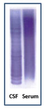
Nephelometry and Turbidimetry
Erythrocyte Indices
Clathrin-Coated Vesicles
Antimicrobial Cationic Peptides
Iron Isotopes
Amino Acid Sequence
Sertoli Cells
Blood Proteins
Erythropoiesis
Isoelectric Focusing
Histocompatibility Antigens Class I
Asialoglycoproteins
The effect of chelating agents on iron mobilization in Chang cell cultures. (1/3281)
The investigation of chelating agents with potential therapeutic value in patients with transfusional iron overload has been facilitated by the use of Chang cell cultures. These cells have been incubated with [59Fe]transferrin for 22 hr, following which most of the intracellular radioiron is found in the cytosol, distributed between a ferritin and a nonferritin form. Iron release from the cells depends on transferrin saturation in the medium, but when transferrin is 100% saturated, which normally does not allow iron release, desferrioxamine, 2,3-dihydroxybenzoic acid, rhodotorulic acid, cholythydroxamic acid, and tropolone all promote the mobilization of ferritin iron and its release from cells. They are effective to an approximately equal degree. The incubation of [59Fe]transferrin with tropolone in vitro at a molar ratio of 1:500 results in the transfer of most of the labeled iron to the chelator, reflecting the exceptionally high binding constant of this compound. How far these phenomena relate to therapeutic potentially remains to be seen. (+info)Studies of the binding of different iron donors to human serum transferrin and isolation of iron-binding fragments from the N- and C-terminal regions of the protein. (2/3281)
1. Trypsin digestion of human serum transferrin partially saturated with iron(III)-nitrilotriacetate at pH 5.5 or pH 8.5 produces a carbohydrate-containing iron-binding fragment of mol.wt. 43000. 2. When iron(III) citrate, FeCl3, iron (III) ascorabate and (NH4)2SO4,FeSO4 are used as iron donors to saturate the protein partially, at pH8.5, proteolytic digestion yields a fragment of mol.wt. 36000 that lacks carbohydrate. 3. The two fragments differ in their antigenic structures, amino acid compositions and peptide 'maps'. 4. The fragment with mol.wt. 36000 was assigned to the N-terminal region of the protein and the other to the C-terminal region. 5. The distribution of iron in human serum transferrin partially saturated with various iron donors was examined by electrophoresis in urea/polyacrylamide gels and the two possible monoferric forms were unequivocally identified. 6. The site designated A on human serum transferrin [Harris (1977) Biochemistry 16, 560--564] was assigned to the C-terminal region of the protein and the B site to the N-terminal region. 7. The distribution of iron on transferrin in human plasma was determined. (+info)The staphylococcal transferrin-binding protein is a cell wall glyceraldehyde-3-phosphate dehydrogenase. (3/3281)
Staphylococcus aureus and Staphylococcus epidermidis possess a 42-kDa cell wall transferrin-binding protein (Tpn) which is involved in the acquisition of transferrin-bound iron. To characterize this protein further, cell wall fractions were subjected to two-dimensional sodium dodecyl sulfate (SDS)-polyacrylamide gel electrophoresis blotted, and the N-terminus of Tpn was sequenced. Comparison of the first 20 amino acid residues of Tpn with the protein databases revealed a high degree of homology to the glycolytic enzyme glyceraldehyde-3-phosphate dehydrogenase (GAPDH). Analysis of staphylococcal cell wall fractions for GAPDH activity confirmed the presence of a functional enzyme which, like Tpn, is regulated by the availability of iron in the growth medium. To determine whether Tpn is responsible for this GAPDH activity, it was affinity purified with NAD+ agarose. Both S. epidermidis and S. aureus Tpn catalyzed the conversion of glyceraldehyde-3-phosphate to 1,3-diphosphoglycerate. In contrast, Staphylococcus saprophyticus, which lacks a Tpn, has no cell wall-associated GAPDH activity. Native polyacrylamide gel electrophoresis of the affinity-purified Tpn revealed that it was present in the cell wall as a tetramer, consistent with the structures of all known cytoplasmic GAPDHs. Furthermore, the affinity-purified Tpn retained its ability to bind human transferrin both in its native tetrameric and SDS-denatured monomeric forms. Apart from interacting with human transferrin, Tpn, in common with the group A streptococcal cell wall GAPDH, binds human plasmin. Tpn-bound plasmin is enzymatically active and therefore may contribute to the ability of staphylococci to penetrate tissues during infections. These studies demonstrate that the staphylococcal transferrin receptor protein, Tpn, is a multifunctional cell wall GAPDH. (+info)Chemical and immunochemical measurement of total iron-binding capacity compared. (4/3281)
Radiometric, colorimetric, and two immunochemical methods for measuring total iron-binding capacity are compared. We evaluated the procedures on the basis of precision, applicability to a pediatric population, and accuracy as assessed by analytical recovery of purified transferrin. The immunoephelometric assay for transferrin provides significant advantages over the other methods examined. (+info)Effects of spinal cord injury on spermatogenesis and the expression of messenger ribonucleic acid for Sertoli cell proteins in rat Sertoli cell-enriched testes. (5/3281)
The study was an examination of the effects of spinal cord injury (SCI) on spermatogenesis and Sertoli cell functions in adult rats with Sertoli cell-enriched (SCE) testes. The effects of SCI on the seminiferous epithelium were characterized by abnormalities in the remaining spermatogenic cells during the first month after SCI. Three days after SCI, serum testosterone levels were 80% lower, while serum FSH and LH levels were 25% and 50% higher, respectively, than those of sham control SCE rats. At this time, the levels of mRNA for androgen receptor (AR), FSH receptor (FSH-R), and androgen-binding protein (ABP) were normal whereas those for transferrin (Trf) had decreased by 40%. Thereafter, serum testosterone levels increased, but they remained lower than those of the sham control rats 28 days after SCI; and serum FSH and LH levels returned to normal. The levels of mRNA for AR, ABP, and Trf exhibited a biphasic increase 7 days after SCI and remained elevated 28 days after SCI. FSH-R mRNA levels were also elevated 90 days after SCI. Unexpectedly, active spermatogenesis, including qualitatively complete spermatogenesis, persisted in > 40% of the tubules 90 days after SCI. These results suggest that the stem cells and/or undifferentiated spermatogonia in SCE testes are less susceptible to the deleterious effects of SCI than the normal testes and that they were able to proliferate and differentiate after SCI. The presence of elevated levels of mRNA for Sertoli cell FSH-R and AR, as well as of that for the Sertoli cell proteins, in the SCE testes during the chronic stage of SCI suggests a modification of Sertoli cell physiology. Such changes in Sertoli cell functions may provide a beneficial environment for the proliferation of the stem cells and differentiation of postmeiotic cells, thus resulting in the persistence of spermatogenesis in these testes. (+info)Coronary heart disease and iron status: meta-analyses of prospective studies. (6/3281)
BACKGROUND: Studies of iron status and coronary heart disease (CHD) have yielded conflicting results. In a systematic review ("meta-analysis"), we quantitatively assessed epidemiological associations reported in prospective studies. METHODS AND RESULTS: Studies were identified by computer-assisted searches of the published literature, scanning of relevant reference lists, hand searching of relevant journals, and discussions with relevant authors. The following was abstracted: size and type of cohort, mean age, mean duration of follow-up, assay methods, degree of adjustment for confounders, and relationship of CHD risk to the baseline assay results. Twelve studies were identified, involving a total of 7800 CHD cases, with several reporting on >1 marker of iron status. For serum ferritin, with 570 CHD cases in 5 studies, comparison of individuals with baseline values >/=200 versus <200 microg/L yielded a combined risk ratio of 1.0 (95% CI, 0.8 to 1.3). For transferrin saturation, with 6194 CHD cases in 5 studies, comparison of individuals in the top third with those in the bottom third of the baseline measurements yielded a combined risk ratio of 0.9 (95% CI, 0.7 to 1.1). Comparisons of individuals in top and bottom thirds of baseline measurements also yielded nonsignificant risk ratios in combined analyses of studies involving total iron-binding capacity (combined risk ratio, 1.0; 95% CI, 0.7 to 1.5), serum iron (0.8; 95% CI, 0.7 to 1.0), and total dietary iron (0.8; 95% CI, 0.7 to 1.1). CONCLUSIONS: Published prospective studies do not provide good evidence to support the existence of strong epidemiological associations between iron status and CHD. (+info)The iron transport protein NRAMP2 is an integral membrane glycoprotein that colocalizes with transferrin in recycling endosomes. (7/3281)
The natural resistance associated macrophage protein (Nramp) gene family is composed of two members in mammals, Nramp1 and Nramp2. Nramp1 is expressed primarily in macrophages and mutations at this locus cause susceptibility to infectious diseases. Nramp2 has a much broader range of tissue expression and mutations at Nramp2 result in iron deficiency, indicating a role for Nramp2 in iron metabolism. To get further insight into the function and mechanism of action of Nramp proteins, we have generated isoform specific anti-Nramp1 and anti-Nramp2 antisera. Immunoblotting experiments indicate that Nramp2 is present in a number of cell types, including hemopoietic precursors, and is coexpressed with Nramp1 in primary macrophages and macrophage cell lines. Nramp2 is expressed as a 90-100-kD integral membrane protein extensively modified by glycosylation (>40% of molecular mass). Subcellular localization studies by immunofluorescence and confocal microscopy indicate distinct and nonoverlapping localization for Nramp1 and Nramp2. Nramp1 is expressed in the lysosomal compartment, whereas Nramp2 is not detectable in the lysosomes but is expressed primarily in recycling endosomes and also, to a lower extent, at the plasma membrane, colocalizing with transferrin. These findings suggest that Nramp2 plays a key role in the metabolism of transferrin-bound iron by transporting free Fe2+ across the endosomal membrane and into the cytoplasm. (+info)Acute haemodynamic and proteinuric effects of prednisolone in patients with a nephrotic syndrome. (8/3281)
BACKGROUND: Administration of prednisolone causes an abrupt rise in proteinuria in patients with a nephrotic syndrome. METHODS: To clarify the mechanisms responsible for this increase in proteinuria we have performed a placebo controlled study in 26 patients with a nephrotic syndrome. Systemic and renal haemodynamics and urinary protein excretion were measured after prednisolone and after placebo. RESULTS: After i.v. administration of 125-150 mg prednisolone total proteinuria increased from 6.66+/-4.42 to 9.37+/-6.07 mg/min (P<0.001). By analysing the excretion of proteins with different charge and weight (albumin, transferrin, IgG, IgG4 and beta2-microglobulin) it became apparent that the increase of proteinuria was the result of a change in size selectivity rather than a change in glomerular charge selectivity or tubular protein reabsorption. Glomerular filtration rate rose from 83+/-34 ml to 95+/-43 ml/min (P<0.001) after 5 h, whereas effective renal plasma flow and endogenous creatinine clearance remained unchanged. As a result filtration fraction was increased, compatible with an increased glomerular pressure, which probably contributes to the size selectivity changes. Since corticosteroids affect both the renin-angiotensin system and renal prostaglandins, we have evaluated the effects of prednisolone on proteinuria after pretreatment with 3 months of the angiotensin-converting enzyme inhibitor lisinopril or after 2 weeks of the prostaglandin synthesis inhibitor indomethacin. Neither drug had any effect on prednisolone-induced increases of proteinuria. CONCLUSIONS: Prednisolone increases proteinuria by changing the size selective barrier of the glomerular capillary. Neither the renin-angiotensin axis nor prostaglandins seem to be involved in these effects of prednisolone on proteinuria. (+info)Transferrin is a glycoprotein that plays a crucial role in the transport and homeostasis of iron in the body. It's produced mainly in the liver and has the ability to bind two ferric (Fe3+) ions in its N-lobe and C-lobe, thus creating transferrin saturation.
This protein is essential for delivering iron to cells while preventing the harmful effects of free iron, which can catalyze the formation of reactive oxygen species through Fenton reactions. Transferrin interacts with specific transferrin receptors on the surface of cells, particularly in erythroid precursors and brain endothelial cells, to facilitate iron uptake via receptor-mediated endocytosis.
In addition to its role in iron transport, transferrin also has antimicrobial properties due to its ability to sequester free iron, making it less available for bacterial growth and survival. Transferrin levels can be used as a clinical marker of iron status, with decreased levels indicating iron deficiency anemia and increased levels potentially signaling inflammation or liver disease.
Transferrin receptors are membrane-bound proteins found on the surface of many cell types, including red and white blood cells, as well as various tissues such as the liver, brain, and placenta. These receptors play a crucial role in iron homeostasis by regulating the uptake of transferrin, an iron-binding protein, into the cells.
Transferrin binds to two ferric ions (Fe3+) in the bloodstream, forming a complex known as holo-transferrin. This complex then interacts with the transferrin receptors on the cell surface, leading to endocytosis of the transferrin-receptor complex into the cell. Once inside the cell, the acidic environment within the endosome causes the release of iron ions from the transferrin molecule, which can then be transported into the cytoplasm for use in various metabolic processes.
After releasing the iron, the apo-transferrin (iron-free transferrin) is recycled back to the cell surface and released back into the bloodstream, where it can bind to more ferric ions and repeat the cycle. This process helps maintain appropriate iron levels within the body and ensures that cells have access to the iron they need for essential functions such as DNA synthesis, energy production, and oxygen transport.
In summary, transferrin receptors are membrane-bound proteins responsible for recognizing and facilitating the uptake of transferrin-bound iron into cells, playing a critical role in maintaining iron homeostasis within the body.
In the context of medicine, iron is an essential micromineral and key component of various proteins and enzymes. It plays a crucial role in oxygen transport, DNA synthesis, and energy production within the body. Iron exists in two main forms: heme and non-heme. Heme iron is derived from hemoglobin and myoglobin in animal products, while non-heme iron comes from plant sources and supplements.
The recommended daily allowance (RDA) for iron varies depending on age, sex, and life stage:
* For men aged 19-50 years, the RDA is 8 mg/day
* For women aged 19-50 years, the RDA is 18 mg/day
* During pregnancy, the RDA increases to 27 mg/day
* During lactation, the RDA for breastfeeding mothers is 9 mg/day
Iron deficiency can lead to anemia, characterized by fatigue, weakness, and shortness of breath. Excessive iron intake may result in iron overload, causing damage to organs such as the liver and heart. Balanced iron levels are essential for maintaining optimal health.
Transferrins are a type of protein found in the plasma component of blood that bind and transport iron ions (Fe3+) from digestion of food or recycling of red blood cells to the cells where they are needed for various metabolic processes, such as the production of hemoglobin. They play a crucial role in maintaining iron homeostasis in the body by preventing the accumulation of free iron, which can be toxic and contribute to the development of oxidative stress and diseases. Transferrins have a high affinity for iron and are capable of binding two ferric ions per molecule. The transferrin-iron complex is then recognized and internalized by specific transferrin receptors on the surface of cells, where the iron is released and utilized.
Ferritin is a protein in iron-metabolizing cells that stores iron in a water-soluble form. It is found inside the cells (intracellular) and is released into the bloodstream when the cells break down or die. Measuring the level of ferritin in the blood can help determine the amount of iron stored in the body. High levels of ferritin may indicate hemochromatosis, inflammation, liver disease, or other conditions. Low levels of ferritin may indicate anemia, iron deficiency, or other conditions.
"Iron radioisotopes" refer to specific forms of the element iron that have unstable nuclei and emit radiation. These isotopes are often used in medical imaging and treatment procedures due to their ability to be detected by specialized equipment. Common iron radioisotopes include Iron-52, Iron-55, Iron-59, and Iron-60. They can be used as tracers to study the distribution, metabolism, or excretion of iron in the body, or for targeted radiation therapy in conditions such as cancer.
Conalbumin is a protein found in egg whites, also known as ovotransferrin. It is one of the three major proteins in egg white along with ovalbumin and ovomucoid. Conalbumin belongs to the transferrin family of proteins, which are responsible for binding and transporting iron in the body.
Conalbumin can bind to iron and sequester it, preventing the growth of certain bacteria that require iron for their survival. This property makes conalbumin an important component of the egg's defense system against bacterial infection. When conalbumin binds to iron, it undergoes a conformational change that prevents the growth of bacteria such as Salmonella and Shigella.
In addition to its antimicrobial properties, conalbumin has been studied for its potential role in nutrition, immunology, and cancer research. It is also used as a marker protein in biochemical and molecular biology techniques.
Transferrin-binding proteins (TBPS) are a group of bacterial surface receptors that bind to transferrin, a glycoprotein involved in iron transport in mammals. These proteins are produced by certain pathogenic bacteria as a means to acquire iron from the host environment, which is essential for their growth and survival.
Transferrin sequesters iron in the bloodstream, making it unavailable to many invading microorganisms. However, some bacteria have evolved TBPS that can bind to transferrin and strip it of its iron, allowing them to use this vital nutrient for their own metabolic needs. The interaction between TBPS and transferrin is an important aspect of bacterial virulence and has been studied as a potential target for developing new antimicrobial therapies.
Endocytosis is the process by which cells absorb substances from their external environment by engulfing them in membrane-bound structures, resulting in the formation of intracellular vesicles. This mechanism allows cells to take up large molecules, such as proteins and lipids, as well as small particles, like bacteria and viruses. There are two main types of endocytosis: phagocytosis (cell eating) and pinocytosis (cell drinking). Phagocytosis involves the engulfment of solid particles, while pinocytosis deals with the uptake of fluids and dissolved substances. Other specialized forms of endocytosis include receptor-mediated endocytosis and caveolae-mediated endocytosis, which allow for the specific internalization of molecules through the interaction with cell surface receptors.
Lactoferrin is a glycoprotein that belongs to the transferrin family. It is an iron-binding protein found in various exocrine secretions such as milk, tears, and saliva, as well as in neutrophils, which are a type of white blood cell involved in immune response. Lactoferrin plays a role in iron homeostasis, antimicrobial activity, and anti-inflammatory responses. It has the ability to bind free iron, which can help prevent bacterial growth by depriving them of an essential nutrient. Additionally, lactoferrin has been shown to have direct antimicrobial effects against various bacteria, viruses, and fungi. Its role in the immune system also includes modulating the activity of immune cells and regulating inflammation.
Hemochromatosis is a medical condition characterized by excessive absorption and accumulation of iron in the body, resulting in damage to various organs. It's often referred to as "iron overload" disorder. There are two main types: primary (hereditary) and secondary (acquired). Primary hemochromatosis is caused by genetic mutations that lead to increased intestinal iron absorption, while secondary hemochromatosis can be the result of various conditions such as multiple blood transfusions, chronic liver disease, or certain types of anemia.
In both cases, the excess iron gets stored in body tissues, particularly in the liver, heart, and pancreas, which can cause organ damage and lead to complications like cirrhosis, liver failure, diabetes, heart problems, and skin discoloration. Early diagnosis and treatment through regular phlebotomy (blood removal) or chelation therapy can help manage the condition and prevent severe complications.
Iron-binding proteins, also known as transferrins, are a type of protein responsible for the transport and storage of iron in the body. They play a crucial role in maintaining iron homeostasis by binding free iron ions and preventing them from participating in harmful chemical reactions that can produce reactive oxygen species (ROS) and cause cellular damage.
Transferrin is the primary iron-binding protein found in blood plasma, while lactoferrin is found in various exocrine secretions such as milk, tears, and saliva. Both transferrin and lactoferrin have a similar structure, consisting of two lobes that can bind one ferric ion (Fe3+) each. When iron is bound to these proteins, they are called holo-transferrin or holo-lactoferrin; when they are unbound, they are referred to as apo-transferrin or apo-lactoferrin.
Iron-binding proteins have a high affinity for iron and can regulate the amount of free iron available in the body. They help prevent iron overload, which can lead to oxidative stress and cellular damage, as well as iron deficiency, which can result in anemia and other health problems.
In summary, iron-binding proteins are essential for maintaining iron homeostasis by transporting and storing iron ions, preventing them from causing harm to the body's cells.
Endosomes are membrane-bound compartments within eukaryotic cells that play a critical role in intracellular trafficking and sorting of various cargoes, including proteins and lipids. They are formed by the invagination of the plasma membrane during endocytosis, resulting in the internalization of extracellular material and cell surface receptors.
Endosomes can be classified into early endosomes, late endosomes, and recycling endosomes based on their morphology, molecular markers, and functional properties. Early endosomes are the initial sorting stations for internalized cargoes, where they undergo sorting and processing before being directed to their final destinations. Late endosomes are more acidic compartments that mature from early endosomes and are responsible for the transport of cargoes to lysosomes for degradation.
Recycling endosomes, on the other hand, are involved in the recycling of internalized cargoes back to the plasma membrane or to other cellular compartments. Endosomal sorting and trafficking are regulated by a complex network of molecular interactions involving various proteins, lipids, and intracellular signaling pathways.
Defects in endosomal function have been implicated in various human diseases, including neurodegenerative disorders, developmental abnormalities, and cancer. Therefore, understanding the mechanisms underlying endosomal trafficking and sorting is of great importance for developing therapeutic strategies to treat these conditions.
Transferrin-binding protein B (TbpB) is not a medical term itself, but it is a bacterial protein involved in the process of iron acquisition by certain bacteria. Therefore, I will provide you with a biological definition:
Transferrin-binding Protein B (TbpB) is a bacterial surface protein primarily found in pathogenic Neisseria species, such as Neisseria gonorrhoeae and Neisseria meningitidis. TbpB plays a crucial role in the iron acquisition process by binding to human transferrin, a glycoprotein that transports iron in the bloodstream.
TbpB, along with Transferrin-binding Protein A (TbpA), facilitates the uptake of iron from transferrin, which is essential for bacterial growth and survival within the host. The interaction between TbpB and transferrin allows the bacteria to evade the host's immune system and establish an infection. Understanding the function of TbpB has implications in developing novel therapeutic strategies against Neisseria infections.
Apoproteins are the protein components of lipoprotein complexes, which are responsible for transporting fat molecules, such as cholesterol and triglycerides, throughout the body. Apoproteins play a crucial role in the metabolism of lipids by acting as recognition signals that allow lipoproteins to interact with specific receptors on cell surfaces.
There are several different types of apoproteins, each with distinct functions. For example, apolipoprotein A-1 (apoA-1) is the major protein component of high-density lipoproteins (HDL), which are responsible for transporting excess cholesterol from tissues to the liver for excretion. Apolipoprotein B (apoB) is a large apoprotein found in low-density lipoproteins (LDL), very low-density lipoproteins (VLDL), and lipoprotein(a). ApoB plays a critical role in the assembly and secretion of VLDL from the liver, and it also mediates the uptake of LDL by cells.
Abnormalities in apoprotein levels or function can contribute to the development of various diseases, including cardiovascular disease, diabetes, and Alzheimer's disease. Therefore, measuring apoprotein levels in the blood can provide valuable information for diagnosing and monitoring these conditions.
Ferric compounds are inorganic compounds that contain the iron(III) cation, Fe3+. Iron(III) is a transition metal and can form stable compounds with various anions. Ferric compounds are often colored due to the d-d transitions of the iron ion. Examples of ferric compounds include ferric chloride (FeCl3), ferric sulfate (Fe2(SO4)3), and ferric oxide (Fe2O3). Ferric compounds have a variety of uses, including as catalysts, in dye production, and in medical applications.
Transferrin-binding protein A (TbpA) is not a medical term itself, but it is a bacterial protein involved in the process of iron acquisition by certain bacteria. Therefore, I will provide a biological definition:
Transferrin-binding Protein A (TbpA) is a bacterial outer membrane protein primarily found in Neisseria species (e.g., Neisseria gonorrhoeae and Neisseria meningitidis). TbpA, along with Transferrin-binding Protein B (TbpB), plays a crucial role in the pathogenesis of these bacteria by facilitating the acquisition of iron from human transferrin, an essential host protein that stores and transports iron. By binding to human transferrin, TbpA and TbpB assist in the transport of iron across the bacterial outer membrane, promoting bacterial growth and survival within the human host.
Gallium is not a medical term, but it's a chemical element with the symbol Ga and atomic number 31. It is a soft, silvery-blue metal that melts at a temperature just above room temperature. In medicine, gallium compounds such as gallium nitrate and gallium citrate are used as radiopharmaceuticals for diagnostic purposes in nuclear medicine imaging studies, particularly in the detection of inflammation, infection, and some types of cancer.
For example, Gallium-67 is a radioactive isotope that can be injected into the body to produce images of various diseases such as abscesses, osteomyelitis (bone infection), and tumors using a gamma camera. The way gallium distributes in the body can provide valuable information about the presence and extent of disease.
Therefore, while gallium is not a medical term itself, it has important medical applications as a diagnostic tool in nuclear medicine.
Iron-deficiency anemia is a condition characterized by a decrease in the total amount of hemoglobin or red blood cells in the blood, caused by insufficient iron levels in the body. Hemoglobin is a protein in red blood cells that carries oxygen from the lungs to the rest of the body. When iron levels are low, the body cannot produce enough hemoglobin, leading to the production of smaller and fewer red blood cells, known as microcytic hypochromic anemia.
Iron is essential for the production of hemoglobin, and a deficiency in iron can result from inadequate dietary intake, chronic blood loss, or impaired absorption. In addition to fatigue and weakness, symptoms of iron-deficiency anemia may include shortness of breath, headaches, dizziness, pale skin, and brittle nails. Treatment typically involves iron supplementation and addressing the underlying cause of the iron deficiency.
I'm not able to find a specific medical definition for the term "Bacterial Transferrin Receptor Complex" as it is not a widely recognized or established term in the field of medicine. However, I can provide some background information that might help you understand the possible meaning behind this term.
Transferrin receptors are proteins found on the surface of many types of cells, including human cells. They play an essential role in iron metabolism by binding to transferrin, a protein that carries iron through the bloodstream. This interaction allows cells to take up and utilize iron for various biological processes.
In some bacteria, similar receptors have been identified that can bind to transferrin or other host proteins to acquire essential nutrients like iron. These bacterial transferrin receptor-like structures might be involved in the pathogenesis of certain bacterial infections by helping the bacteria obtain necessary resources for growth and survival within the human body.
However, it is important to note that the term "Bacterial Transferrin Receptor Complex" may not refer to a specific, well-defined entity in medical research. If you are looking for information on a particular study or context related to this term, I would recommend consulting the original source or seeking further clarification from the author(s) of the source where you encountered this term.
Iron-regulatory proteins (IRPs) are specialized RNA-binding proteins that play a crucial role in the post-transcriptional regulation of iron homeostasis in mammalian cells. They are named as such because they regulate the expression of genes involved in iron metabolism, primarily by binding to specific cis-acting elements known as iron-responsive elements (IREs) located within the untranslated regions (UTRs) of target mRNAs.
There are two main IRPs: IRP1 and IRP2. Both proteins contain an N-terminal RNA-binding domain that recognizes and binds to IREs, as well as a C-terminal region involved in protein-protein interactions and other regulatory functions. Under conditions of iron deficiency or oxidative stress, IRPs become activated and bind to IREs, leading to changes in mRNA stability, translation, or both.
IRP1 can exist in two distinct conformational states: an active RNA-binding form (when iron levels are low) and an inactive aconitase form (when iron levels are sufficient). In contrast, IRP2 is primarily regulated by protein degradation, with its stability being modulated by the presence or absence of iron.
By binding to IREs within mRNAs encoding proteins involved in iron uptake, storage, and utilization, IRPs help maintain cellular iron homeostasis through a variety of mechanisms, including:
1. Promoting translation of transferrin receptor 1 (TfR1) mRNA to increase iron import when iron levels are low.
2. Inhibiting translation of ferritin heavy chain and light chain mRNAs to reduce iron storage when iron levels are low.
3. Stabilizing the mRNA encoding divalent metal transporter 1 (DMT1) to enhance iron uptake under conditions of iron deficiency.
4. Promoting degradation of transferrin receptor 2 (TfR2) and ferroportin mRNAs to limit iron import and export, respectively, when iron levels are high.
Overall, the regulation of iron metabolism by IRPs is crucial for maintaining proper cellular function and preventing the accumulation of toxic free radicals generated by iron-catalyzed reactions.
Iron chelating agents are medications that bind to iron in the body, forming a stable complex that can then be excreted from the body. These agents are primarily used to treat iron overload, a condition that can occur due to frequent blood transfusions or certain genetic disorders such as hemochromatosis. By reducing the amount of iron in the body, these medications can help prevent or reduce damage to organs such as the heart and liver. Examples of iron chelating agents include deferoxamine, deferasirox, and deferiprone.
Iron overload is a condition characterized by an excessive accumulation of iron in the body's tissues and organs, particularly in the liver, heart, and pancreas. This occurs when the body absorbs more iron than it can use or eliminate, leading to iron levels that are higher than normal.
Iron overload can result from various factors, including hereditary hemochromatosis, a genetic disorder that affects how the body absorbs iron from food; frequent blood transfusions, which can cause iron buildup in people with certain chronic diseases such as sickle cell anemia or thalassemia; and excessive consumption of iron supplements or iron-rich foods.
Symptoms of iron overload may include fatigue, joint pain, abdominal discomfort, irregular heartbeat, and liver dysfunction. If left untreated, it can lead to serious complications such as cirrhosis, liver failure, diabetes, heart problems, and even certain types of cancer. Treatment typically involves regular phlebotomy (removal of blood) to reduce iron levels in the body, along with dietary modifications and monitoring by a healthcare professional.
Cell surface receptors, also known as membrane receptors, are proteins located on the cell membrane that bind to specific molecules outside the cell, known as ligands. These receptors play a crucial role in signal transduction, which is the process of converting an extracellular signal into an intracellular response.
Cell surface receptors can be classified into several categories based on their structure and mechanism of action, including:
1. Ion channel receptors: These receptors contain a pore that opens to allow ions to flow across the cell membrane when they bind to their ligands. This ion flux can directly activate or inhibit various cellular processes.
2. G protein-coupled receptors (GPCRs): These receptors consist of seven transmembrane domains and are associated with heterotrimeric G proteins that modulate intracellular signaling pathways upon ligand binding.
3. Enzyme-linked receptors: These receptors possess an intrinsic enzymatic activity or are linked to an enzyme, which becomes activated when the receptor binds to its ligand. This activation can lead to the initiation of various signaling cascades within the cell.
4. Receptor tyrosine kinases (RTKs): These receptors contain intracellular tyrosine kinase domains that become activated upon ligand binding, leading to the phosphorylation and activation of downstream signaling molecules.
5. Integrins: These receptors are transmembrane proteins that mediate cell-cell or cell-matrix interactions by binding to extracellular matrix proteins or counter-receptors on adjacent cells. They play essential roles in cell adhesion, migration, and survival.
Cell surface receptors are involved in various physiological processes, including neurotransmission, hormone signaling, immune response, and cell growth and differentiation. Dysregulation of these receptors can contribute to the development of numerous diseases, such as cancer, diabetes, and neurological disorders.
Reticulocytes are immature red blood cells that still contain remnants of organelles, such as ribosomes and mitochondria, which are typically found in developing cells. These organelles are involved in the process of protein synthesis and energy production, respectively. Reticulocytes are released from the bone marrow into the bloodstream, where they continue to mature into fully developed red blood cells called erythrocytes.
Reticulocytes can be identified under a microscope by their staining characteristics, which reveal a network of fine filaments or granules known as the reticular apparatus. This apparatus is composed of residual ribosomal RNA and other proteins that have not yet been completely eliminated during the maturation process.
The percentage of reticulocytes in the blood can be used as a measure of bone marrow function and erythropoiesis, or red blood cell production. An increased reticulocyte count may indicate an appropriate response to blood loss, hemolysis, or other conditions that cause anemia, while a decreased count may suggest impaired bone marrow function or a deficiency in erythropoietin, the hormone responsible for stimulating red blood cell production.
Hepcidin is a peptide hormone primarily produced in the liver that plays a crucial role in regulating iron homeostasis within the body. It acts by inhibiting the absorption of dietary iron in the intestines and the release of iron from storage sites, such as macrophages, into the bloodstream. By reducing the amount of iron available for use, hepcidin helps prevent excessive iron accumulation in tissues, which can be harmful and contribute to the development of various diseases, including iron overload disorders and certain types of anemia. The production of hepcidin is regulated by several factors, including iron levels, inflammation, and erythropoiesis (the production of red blood cells).
Coated pits are specialized regions on the cell membrane that are involved in the process of endocytosis. They are called "coated" pits because they are covered or coated with a layer of proteins and clathrin molecules, which form a lattice-like structure that helps to shape and invaginate the membrane inward, forming a vesicle.
Coated pits play an important role in regulating cellular uptake of various substances, such as nutrients, hormones, and receptors. Once the coated pit has pinched off from the cell membrane, it becomes a coated vesicle, which can then fuse with other intracellular compartments to deliver its contents.
The formation of coated pits is a highly regulated process that involves the recruitment of specific proteins and adaptors to the site of endocytosis. Defects in this process have been implicated in various diseases, including neurodevelopmental disorders and cancer.
Deferoxamine is a medication used to treat iron overload, which can occur due to various reasons such as frequent blood transfusions or excessive iron intake. It works by binding to excess iron in the body and promoting its excretion through urine. This helps to prevent damage to organs such as the heart and liver that can be caused by high levels of iron.
Deferoxamine is an injectable medication that is typically administered intravenously or subcutaneously, depending on the specific regimen prescribed by a healthcare professional. It may also be used in combination with other medications to manage iron overload more effectively.
It's important to note that deferoxamine should only be used under the guidance of a medical professional, as improper use or dosing can lead to serious side effects or complications.
Hypochromic anemia is a type of anemia characterized by the presence of red blood cells that have lower than normal levels of hemoglobin and appear paler in color than normal. Hemoglobin is a protein in red blood cells that carries oxygen from the lungs to the rest of the body. In hypochromic anemia, there may be a decrease in the production or increased destruction of red blood cells, leading to a reduced number of red blood cells and insufficient oxygen supply to the tissues.
Hypochromic anemia can result from various underlying medical conditions, including iron deficiency, thalassemia, chronic inflammation, lead poisoning, and certain infections or chronic diseases. Treatment for hypochromic anemia depends on the underlying cause and may include iron supplements, dietary changes, medications, or blood transfusions.
Hemoglobin (Hb or Hgb) is the main oxygen-carrying protein in the red blood cells, which are responsible for delivering oxygen throughout the body. It is a complex molecule made up of four globin proteins and four heme groups. Each heme group contains an iron atom that binds to one molecule of oxygen. Hemoglobin plays a crucial role in the transport of oxygen from the lungs to the body's tissues, and also helps to carry carbon dioxide back to the lungs for exhalation.
There are several types of hemoglobin present in the human body, including:
* Hemoglobin A (HbA): This is the most common type of hemoglobin, making up about 95-98% of total hemoglobin in adults. It consists of two alpha and two beta globin chains.
* Hemoglobin A2 (HbA2): This makes up about 1.5-3.5% of total hemoglobin in adults. It consists of two alpha and two delta globin chains.
* Hemoglobin F (HbF): This is the main type of hemoglobin present in fetal life, but it persists at low levels in adults. It consists of two alpha and two gamma globin chains.
* Hemoglobin S (HbS): This is an abnormal form of hemoglobin that can cause sickle cell disease when it occurs in the homozygous state (i.e., both copies of the gene are affected). It results from a single amino acid substitution in the beta globin chain.
* Hemoglobin C (HbC): This is another abnormal form of hemoglobin that can cause mild to moderate hemolytic anemia when it occurs in the homozygous state. It results from a different single amino acid substitution in the beta globin chain than HbS.
Abnormal forms of hemoglobin, such as HbS and HbC, can lead to various clinical disorders, including sickle cell disease, thalassemia, and other hemoglobinopathies.
In the context of medicine and pharmacology, "kinetics" refers to the study of how a drug moves throughout the body, including its absorption, distribution, metabolism, and excretion (often abbreviated as ADME). This field is called "pharmacokinetics."
1. Absorption: This is the process of a drug moving from its site of administration into the bloodstream. Factors such as the route of administration (e.g., oral, intravenous, etc.), formulation, and individual physiological differences can affect absorption.
2. Distribution: Once a drug is in the bloodstream, it gets distributed throughout the body to various tissues and organs. This process is influenced by factors like blood flow, protein binding, and lipid solubility of the drug.
3. Metabolism: Drugs are often chemically modified in the body, typically in the liver, through processes known as metabolism. These changes can lead to the formation of active or inactive metabolites, which may then be further distributed, excreted, or undergo additional metabolic transformations.
4. Excretion: This is the process by which drugs and their metabolites are eliminated from the body, primarily through the kidneys (urine) and the liver (bile).
Understanding the kinetics of a drug is crucial for determining its optimal dosing regimen, potential interactions with other medications or foods, and any necessary adjustments for special populations like pediatric or geriatric patients, or those with impaired renal or hepatic function.
Clathrin is a type of protein that plays a crucial role in the formation of coated vesicles within cells. These vesicles are responsible for transporting materials between different cellular compartments, such as from the plasma membrane to the endoplasmic reticulum or Golgi apparatus. Clathrin molecules form a lattice-like structure that curves around the vesicle, providing stability and shape to the coated vesicle. This process is known as clathrin-mediated endocytosis.
The formation of clathrin-coated vesicles begins with the recruitment of clathrin proteins to specific sites on the membrane, where they assemble into a polygonal lattice structure. As more clathrin molecules join the assembly, the lattice curves and eventually pinches off from the membrane, forming a closed vesicle. The clathrin coat then disassembles, releasing the vesicle to continue with its intracellular transport mission.
Disruptions in clathrin-mediated endocytosis can lead to various cellular dysfunctions and diseases, including neurodegenerative disorders and certain types of cancer.
A cell line is a culture of cells that are grown in a laboratory for use in research. These cells are usually taken from a single cell or group of cells, and they are able to divide and grow continuously in the lab. Cell lines can come from many different sources, including animals, plants, and humans. They are often used in scientific research to study cellular processes, disease mechanisms, and to test new drugs or treatments. Some common types of human cell lines include HeLa cells (which come from a cancer patient named Henrietta Lacks), HEK293 cells (which come from embryonic kidney cells), and HUVEC cells (which come from umbilical vein endothelial cells). It is important to note that cell lines are not the same as primary cells, which are cells that are taken directly from a living organism and have not been grown in the lab.
Biological transport refers to the movement of molecules, ions, or solutes across biological membranes or through cells in living organisms. This process is essential for maintaining homeostasis, regulating cellular functions, and enabling communication between cells. There are two main types of biological transport: passive transport and active transport.
Passive transport does not require the input of energy and includes:
1. Diffusion: The random movement of molecules from an area of high concentration to an area of low concentration until equilibrium is reached.
2. Osmosis: The diffusion of solvent molecules (usually water) across a semi-permeable membrane from an area of lower solute concentration to an area of higher solute concentration.
3. Facilitated diffusion: The assisted passage of polar or charged substances through protein channels or carriers in the cell membrane, which increases the rate of diffusion without consuming energy.
Active transport requires the input of energy (in the form of ATP) and includes:
1. Primary active transport: The direct use of ATP to move molecules against their concentration gradient, often driven by specific transport proteins called pumps.
2. Secondary active transport: The coupling of the movement of one substance down its electrochemical gradient with the uphill transport of another substance, mediated by a shared transport protein. This process is also known as co-transport or counter-transport.
Lactoglobulins, specifically referring to β-lactoglobulin, are a type of protein found in the whey fraction of milk from ruminant animals such as cows and sheep. They are one of the major proteins in bovine milk, making up about 10% of the total protein content.
β-lactoglobulin is a small, stable protein that is resistant to heat and acid denaturation. It has an important role in the nutrition of young mammals as it can bind to fat molecules and help with their absorption. In addition, β-lactoglobulin has been studied for its potential health benefits, including its antioxidant and anti-inflammatory properties.
However, some people may have allergies to β-lactoglobulin, which can cause symptoms such as hives, swelling, and difficulty breathing. In these cases, it is important to avoid foods that contain this protein.
Iron Regulatory Protein 1 (IRP1) is a protein that plays a crucial role in the post-transcriptional regulation of iron homeostasis in cells. It is involved in the detection of cellular iron levels and responds by modulating the translation and stability of messenger RNAs (mRNAs) that encode proteins essential for iron metabolism.
IRP1 can bind to specific sequences called Iron Responsive Elements (IREs) present in the untranslated regions of mRNAs. When cellular iron levels are low, IRP1 binds to IREs and inhibits the translation of mRNAs encoding proteins responsible for iron uptake and storage, while stabilizing mRNAs that encode proteins involved in iron mobilization. Conversely, when iron levels are high, IRP1 dissociates from IREs, allowing for the normal translation of these mRNAs and maintaining iron homeostasis within the cell.
It is important to note that IRP1 has dual functions: it can act as an Iron Regulatory Protein (IRP) when iron levels are low, and as a cytosolic aconitase (an enzyme in the citric acid cycle) when iron levels are sufficient. This ability to switch between these two roles is facilitated by the presence of a [4Fe-4S] cluster, which is sensitive to cellular iron levels. When iron is abundant, the [4Fe-4S] cluster assembles, converting IRP1 into its cytosolic aconitase form; when iron is scarce, the cluster disassembles, enabling IRP1 to bind IREs and regulate iron metabolism-related gene expression.
Siderophores are low-molecular-weight organic compounds that are secreted by microorganisms, such as bacteria and fungi, to chelate and solubilize iron from their environment. They are able to bind ferric iron (Fe3+) with very high affinity and form a siderophore-iron complex, which can then be taken up by the microorganism through specific transport systems. This allows them to acquire iron even in environments where it is present at very low concentrations or in forms that are not readily available for uptake. Siderophores play an important role in the survival and virulence of many pathogenic microorganisms, as they help them to obtain the iron they need to grow and multiply.
A cell membrane, also known as the plasma membrane, is a thin semi-permeable phospholipid bilayer that surrounds all cells in animals, plants, and microorganisms. It functions as a barrier to control the movement of substances in and out of the cell, allowing necessary molecules such as nutrients, oxygen, and signaling molecules to enter while keeping out harmful substances and waste products. The cell membrane is composed mainly of phospholipids, which have hydrophilic (water-loving) heads and hydrophobic (water-fearing) tails. This unique structure allows the membrane to be flexible and fluid, yet selectively permeable. Additionally, various proteins are embedded in the membrane that serve as channels, pumps, receptors, and enzymes, contributing to the cell's overall functionality and communication with its environment.
Cell compartmentation, also known as intracellular compartmentalization, refers to the organization of cells into distinct functional and spatial domains. This is achieved through the separation of cellular components and biochemical reactions into membrane-bound organelles or compartments. Each compartment has its unique chemical composition and environment, allowing for specific biochemical reactions to occur efficiently and effectively without interfering with other processes in the cell.
Some examples of membrane-bound organelles include the nucleus, mitochondria, chloroplasts, endoplasmic reticulum, Golgi apparatus, lysosomes, peroxisomes, and vacuoles. These organelles have specific functions, such as energy production (mitochondria), protein synthesis and folding (endoplasmic reticulum and Golgi apparatus), waste management (lysosomes), and lipid metabolism (peroxisomes).
Cell compartmentation is essential for maintaining cellular homeostasis, regulating metabolic pathways, protecting the cell from potentially harmful substances, and enabling complex biochemical reactions to occur in a controlled manner. Dysfunction of cell compartmentation can lead to various diseases, including neurodegenerative disorders, cancer, and metabolic disorders.
Ceruloplasmin is a protein found in blood plasma that binds and transports copper ions. It plays a crucial role in copper metabolism, including the oxidation of ferrous iron to ferric iron, which is necessary for the incorporation of iron into transferrin, another protein responsible for transporting iron throughout the body. Ceruloplasmin also acts as an antioxidant by scavenging free radicals and has been implicated in neurodegenerative disorders like Alzheimer's disease and Wilson's disease, a genetic disorder characterized by abnormal copper accumulation in various organs.
Gallium radioisotopes refer to specific types of gallium atoms that have unstable nuclei and emit radiation as they decay towards a more stable state. These isotopes are commonly used in medical imaging, such as in gallium scans, to help diagnose conditions like inflammation, infection, or cancer.
Gallium-67 (^67^Ga) is one of the most commonly used radioisotopes for medical purposes. It has a half-life of about 3.26 days and decays by emitting gamma rays. When administered to a patient, gallium-67 binds to transferrin, a protein that carries iron in the blood, and is taken up by cells with increased metabolic activity, such as cancer cells or immune cells responding to infection or inflammation. The distribution of gallium-67 in the body can then be visualized using a gamma camera, providing valuable diagnostic information.
Serum albumin is the most abundant protein in human blood plasma, synthesized by the liver. It plays a crucial role in maintaining the oncotic pressure or colloid osmotic pressure of blood, which helps to regulate the fluid balance between the intravascular and extravascular spaces.
Serum albumin has a molecular weight of around 66 kDa and is composed of a single polypeptide chain. It contains several binding sites for various endogenous and exogenous substances, such as bilirubin, fatty acids, hormones, and drugs, facilitating their transport throughout the body. Additionally, albumin possesses antioxidant properties, protecting against oxidative damage.
Albumin levels in the blood are often used as a clinical indicator of liver function, nutritional status, and overall health. Low serum albumin levels may suggest liver disease, malnutrition, inflammation, or kidney dysfunction.
Heptanoates are chemical compounds that contain the functional group of heptanoic acid. Heptanoic acid, also known as n-caproic acid, is a type of carboxylic acid with a 7-carbon chain and the molecular formula C7H15COOH.
Heptanoates are commonly used in the production of various chemicals, including flavors, fragrances, and pharmaceuticals. In medicine, heptanoates may be used as esters in the formulation of drugs to improve their solubility, absorption, and stability. For example, some injectable forms of medications may use heptanoate salts or esters to enhance their delivery into the body.
It's important to note that specific medical definitions for "heptanoates" may vary depending on the context and application.
Membrane proteins are a type of protein that are embedded in the lipid bilayer of biological membranes, such as the plasma membrane of cells or the inner membrane of mitochondria. These proteins play crucial roles in various cellular processes, including:
1. Cell-cell recognition and signaling
2. Transport of molecules across the membrane (selective permeability)
3. Enzymatic reactions at the membrane surface
4. Energy transduction and conversion
5. Mechanosensation and signal transduction
Membrane proteins can be classified into two main categories: integral membrane proteins, which are permanently associated with the lipid bilayer, and peripheral membrane proteins, which are temporarily or loosely attached to the membrane surface. Integral membrane proteins can further be divided into three subcategories based on their topology:
1. Transmembrane proteins, which span the entire width of the lipid bilayer with one or more alpha-helices or beta-barrels.
2. Lipid-anchored proteins, which are covalently attached to lipids in the membrane via a glycosylphosphatidylinositol (GPI) anchor or other lipid modifications.
3. Monotopic proteins, which are partially embedded in the membrane and have one or more domains exposed to either side of the bilayer.
Membrane proteins are essential for maintaining cellular homeostasis and are targets for various therapeutic interventions, including drug development and gene therapy. However, their structural complexity and hydrophobicity make them challenging to study using traditional biochemical methods, requiring specialized techniques such as X-ray crystallography, nuclear magnetic resonance (NMR) spectroscopy, and single-particle cryo-electron microscopy (cryo-EM).
Molecular sequence data refers to the specific arrangement of molecules, most commonly nucleotides in DNA or RNA, or amino acids in proteins, that make up a biological macromolecule. This data is generated through laboratory techniques such as sequencing, and provides information about the exact order of the constituent molecules. This data is crucial in various fields of biology, including genetics, evolution, and molecular biology, allowing for comparisons between different organisms, identification of genetic variations, and studies of gene function and regulation.
Protein binding, in the context of medical and biological sciences, refers to the interaction between a protein and another molecule (known as the ligand) that results in a stable complex. This process is often reversible and can be influenced by various factors such as pH, temperature, and concentration of the involved molecules.
In clinical chemistry, protein binding is particularly important when it comes to drugs, as many of them bind to proteins (especially albumin) in the bloodstream. The degree of protein binding can affect a drug's distribution, metabolism, and excretion, which in turn influence its therapeutic effectiveness and potential side effects.
Protein-bound drugs may be less available for interaction with their target tissues, as only the unbound or "free" fraction of the drug is active. Therefore, understanding protein binding can help optimize dosing regimens and minimize adverse reactions.
RAB4 GTP-binding proteins are a subfamily of RAB proteins, which are small guanosine triphosphatases (GTPases) that play crucial roles in regulating intracellular vesicle trafficking. Specifically, RAB4 GTP-binding proteins are involved in the early stages of endocytic recycling, a process by which internalized membrane receptors and cargo are transported back to the plasma membrane for reuse.
RAB4 proteins exist in two distinct conformational states: an active, GTP-bound state and an inactive, GDP-bound state. In the active state, RAB4 proteins interact with various effector molecules to facilitate vesicle transport and fusion events. Upon hydrolysis of GTP to GDP, RAB4 proteins switch to their inactive state, which leads to dissociation from effector molecules and subsequent recycling of the RAB4 protein back to the donor membrane compartment.
There are two isoforms of RAB4 proteins, RAB4A and RAB4B, which share a high degree of sequence similarity but have distinct cellular localization patterns and functions. Dysregulation of RAB4 GTP-binding proteins has been implicated in various human diseases, including cancer and neurodegenerative disorders.
Dietary iron is a vital nutrient that plays a crucial role in the production of hemoglobin, a protein in red blood cells responsible for carrying oxygen throughout the body. It is also essential for various other bodily functions, including energy production and immune function.
There are two forms of dietary iron: heme and non-heme. Heme iron is found in animal products such as meat, poultry, and fish, while non-heme iron is found in plant-based foods such as beans, lentils, tofu, spinach, and fortified cereals.
The recommended daily intake of dietary iron varies depending on age, sex, and other factors. For example, adult men typically require 8 milligrams (mg) per day, while adult women need 18 mg per day. Pregnant women may require up to 27 mg per day, while breastfeeding women need around 9-10 mg per day.
It is important to note that the absorption of non-heme iron from plant-based foods can be enhanced by consuming them with vitamin C-rich foods or drinks, such as citrus fruits, strawberries, and bell peppers. On the other hand, certain substances such as tannins (found in tea and coffee) and phytates (found in whole grains and legumes) can inhibit the absorption of non-heme iron.
Rab GTP-binding proteins, also known as Rab GTPases or simply Rabs, are a large family of small GTP-binding proteins that play a crucial role in regulating intracellular vesicle trafficking. They function as molecular switches that cycle between an active GTP-bound state and an inactive GDP-bound state.
In the active state, Rab proteins interact with various effector molecules to mediate specific membrane trafficking events such as vesicle budding, transport, tethering, and fusion. Each Rab protein is thought to have a unique function and localize to specific intracellular compartments or membranes, where they regulate the transport of vesicles and organelles within the cell.
Rab proteins are involved in several important cellular processes, including endocytosis, exocytosis, Golgi apparatus function, autophagy, and intracellular signaling. Dysregulation of Rab GTP-binding proteins has been implicated in various human diseases, such as cancer, neurodegenerative disorders, and infectious diseases.
Nephelometry and turbidimetry are methods used in clinical laboratories to measure the amount of particles, such as proteins or cells, present in a liquid sample. The main difference between these two techniques lies in how they detect and quantify the particles.
1. Nephelometry: This is a laboratory method that measures the amount of light scattered by suspended particles in a liquid medium at a 90-degree angle to the path of the incident light. When light passes through a sample containing particles, some of the light is absorbed, while some is scattered in various directions. In nephelometry, a light beam is shone into the sample, and a detector measures the intensity of the scattered light at a right angle to the light source. The more particles present in the sample, the higher the intensity of scattered light, which correlates with the concentration of particles in the sample. Nephelometry is often used to measure the levels of immunoglobulins, complement components, and other proteins in serum or plasma.
2. Turbidimetry: This is another laboratory method that measures the amount of light blocked or absorbed by suspended particles in a liquid medium. In turbidimetry, a light beam is shone through the sample, and the intensity of the transmitted light is measured. The more particles present in the sample, the more light is absorbed or scattered, resulting in lower transmitted light intensity. Turbidimetric measurements are typically reported as percent transmittance, which is the ratio of the intensity of transmitted light to that of the incident light expressed as a percentage. Turbidimetry can be used to measure various substances, such as proteins, cells, and crystals, in body fluids like urine, serum, or plasma.
In summary, nephelometry measures the amount of scattered light at a 90-degree angle, while turbidimetry quantifies the reduction in transmitted light intensity due to particle presence. Both methods are useful for determining the concentration of particles in liquid samples and are commonly used in clinical laboratories for diagnostic purposes.
Erythrocyte indices are a set of calculated values that provide information about the size and hemoglobin content of red blood cells (erythrocytes). These indices are commonly used in the complete blood count (CBC) test to help diagnose various types of anemia and other conditions affecting the red blood cells.
The three main erythrocyte indices are:
1. Mean Corpuscular Volume (MCV): This is the average volume of a single red blood cell, measured in femtoliters (fL). MCV helps to differentiate between microcytic, normocytic, and macrocytic anemia. Microcytic anemia is characterized by low MCV values (100 fL).
2. Mean Corpuscular Hemoglobin (MCH): This is the average amount of hemoglobin present in a single red blood cell, measured in picograms (pg). MCH helps to assess the oxygen-carrying capacity of red blood cells. Low MCH values may indicate hypochromic anemia, where the red blood cells have reduced hemoglobin content.
3. Mean Corpuscular Hemoglobin Concentration (MCHC): This is the average concentration of hemoglobin in a single red blood cell, measured as a percentage. MCHC reflects the hemoglobin concentration relative to the size of the red blood cells. Low MCHC values may indicate hypochromic anemia, while high MCHC values could suggest spherocytosis or other conditions affecting red blood cell shape and integrity.
These erythrocyte indices are calculated based on the red blood cell count, hemoglobin concentration, and hematocrit results obtained from a CBC test. They provide valuable information for healthcare professionals to diagnose and manage various hematological conditions.
Clathrin-coated vesicles are small, membrane-bound structures that play a crucial role in intracellular transport within eukaryotic cells. They are formed by the coating of the plasma membrane or the membranes of other organelles with a lattice-like structure made up of clathrin proteins.
The formation of clathrin-coated vesicles is initiated when adaptor proteins recognize and bind to specific signals on the cytoplasmic side of the membrane. These adaptor proteins then recruit clathrin molecules, which assemble into a cage-like structure that deforms the membrane into a spherical shape. The vesicle then pinches off from the membrane, enclosed in its clathrin coat.
Once formed, clathrin-coated vesicles can transport proteins and other molecules between different cellular compartments, such as from the plasma membrane to endosomes or from the Golgi apparatus to the endoplasmic reticulum. The clathrin coat is subsequently disassembled, allowing the vesicle to fuse with its target membrane and release its contents.
Defects in clathrin-coated vesicle function have been implicated in a variety of human diseases, including neurodegenerative disorders and certain forms of cancer.
Antimicrobial cationic peptides (ACPs) are a group of small, naturally occurring peptides that possess broad-spectrum antimicrobial activity against various microorganisms, including bacteria, fungi, viruses, and parasites. They are called "cationic" because they contain positively charged amino acid residues (such as lysine and arginine), which allow them to interact with and disrupt the negatively charged membranes of microbial cells.
ACPs are produced by a wide range of organisms, including humans, animals, and plants, as part of their innate immune response to infection. They play an important role in protecting the host from invading pathogens by directly killing them or inhibiting their growth.
The antimicrobial activity of ACPs is thought to be mediated by their ability to disrupt the membranes of microbial cells, leading to leakage of cellular contents and death. Some ACPs may also have intracellular targets, such as DNA or protein synthesis, that contribute to their antimicrobial activity.
ACPs are being studied for their potential use as therapeutic agents to treat infectious diseases, particularly those caused by drug-resistant bacteria. However, their clinical application is still in the early stages of development due to concerns about their potential toxicity to host cells and the emergence of resistance mechanisms in microbial pathogens.
I must clarify that "Iron Isotopes" is not a medical term, but rather a scientific concept from the field of physics and chemistry. However, I can certainly provide a general explanation of isotopes and then focus on iron isotopes specifically.
An isotope is a variant of a chemical element that has the same number of protons (and thus the same atomic number) but a different number of neutrons within its nucleus. This results in variations of the atomic mass of isotopes of the same element. Some isotopes are stable, while others are unstable and will decay over time into other elements or isotopes, a process called radioactive decay.
Iron (Fe) has four naturally occurring stable isotopes: Fe-54, Fe-56, Fe-57, and Fe-58. These iron isotopes have different numbers of neutrons in their nuclei, resulting in slightly different atomic masses. The most abundant iron isotope is Fe-56, which contains 26 protons and 30 neutrons in its nucleus.
In the context of human health, iron is an essential nutrient that plays a crucial role in various biological processes, such as oxygen transport and energy production. However, the concept of iron isotopes does not have a direct medical relevance, but it can be useful in scientific research related to fields like geochemistry, environmental science, or nuclear physics.
An amino acid sequence is the specific order of amino acids in a protein or peptide molecule, formed by the linking of the amino group (-NH2) of one amino acid to the carboxyl group (-COOH) of another amino acid through a peptide bond. The sequence is determined by the genetic code and is unique to each type of protein or peptide. It plays a crucial role in determining the three-dimensional structure and function of proteins.
Sertoli cells, also known as sustentacular cells or nurse cells, are specialized cells in the seminiferous tubules of the testis in mammals. They play a crucial role in supporting and nurturing the development of sperm cells (spermatogenesis). Sertoli cells create a microenvironment within the seminiferous tubules that facilitates the differentiation, maturation, and survival of germ cells.
These cells have several essential functions:
1. Blood-testis barrier formation: Sertoli cells form tight junctions with each other, creating a physical barrier called the blood-testis barrier, which separates the seminiferous tubules into basal and adluminal compartments. This barrier protects the developing sperm cells from the immune system and provides an isolated environment for their maturation.
2. Nutrition and support: Sertoli cells provide essential nutrients and growth factors to germ cells, ensuring their proper development and survival. They also engulf and digest residual bodies, which are byproducts of spermatid differentiation.
3. Phagocytosis: Sertoli cells have phagocytic properties, allowing them to remove debris and dead cells within the seminiferous tubules.
4. Hormone metabolism: Sertoli cells express receptors for various hormones, such as follicle-stimulating hormone (FSH), testosterone, and estradiol. They play a role in regulating hormonal signaling within the testis by metabolizing these hormones or producing inhibins, which modulate FSH secretion from the pituitary gland.
5. Regulation of spermatogenesis: Sertoli cells produce and secrete various proteins and growth factors that influence germ cell development and proliferation. They also control the release of mature sperm cells into the epididymis through a process called spermiation.
Blood proteins, also known as serum proteins, are a group of complex molecules present in the blood that are essential for various physiological functions. These proteins include albumin, globulins (alpha, beta, and gamma), and fibrinogen. They play crucial roles in maintaining oncotic pressure, transporting hormones, enzymes, vitamins, and minerals, providing immune defense, and contributing to blood clotting.
Albumin is the most abundant protein in the blood, accounting for about 60% of the total protein mass. It functions as a transporter of various substances, such as hormones, fatty acids, and drugs, and helps maintain oncotic pressure, which is essential for fluid balance between the blood vessels and surrounding tissues.
Globulins are divided into three main categories: alpha, beta, and gamma globulins. Alpha and beta globulins consist of transport proteins like lipoproteins, hormone-binding proteins, and enzymes. Gamma globulins, also known as immunoglobulins or antibodies, are essential for the immune system's defense against pathogens.
Fibrinogen is a protein involved in blood clotting. When an injury occurs, fibrinogen is converted into fibrin, which forms a mesh to trap platelets and form a clot, preventing excessive bleeding.
Abnormal levels of these proteins can indicate various medical conditions, such as liver or kidney disease, malnutrition, infections, inflammation, or autoimmune disorders. Blood protein levels are typically measured through laboratory tests like serum protein electrophoresis (SPE) and immunoelectrophoresis (IEP).
Erythropoiesis is the process of forming and developing red blood cells (erythrocytes) in the body. It occurs in the bone marrow and is regulated by the hormone erythropoietin (EPO), which is produced by the kidneys. Erythropoiesis involves the differentiation and maturation of immature red blood cell precursors called erythroblasts into mature red blood cells, which are responsible for carrying oxygen to the body's tissues. Disorders that affect erythropoiesis can lead to anemia or other blood-related conditions.
Anemia is a medical condition characterized by a lower than normal number of red blood cells or lower than normal levels of hemoglobin in the blood. Hemoglobin is an important protein in red blood cells that carries oxygen from the lungs to the rest of the body. Anemia can cause fatigue, weakness, shortness of breath, and a pale complexion because the body's tissues are not getting enough oxygen.
Anemia can be caused by various factors, including nutritional deficiencies (such as iron, vitamin B12, or folate deficiency), blood loss, chronic diseases (such as kidney disease or rheumatoid arthritis), inherited genetic disorders (such as sickle cell anemia or thalassemia), and certain medications.
There are different types of anemia, classified based on the underlying cause, size and shape of red blood cells, and the level of hemoglobin in the blood. Treatment for anemia depends on the underlying cause and may include dietary changes, supplements, medication, or blood transfusions.
Isoelectric focusing (IEF) is a technique used in electrophoresis, which is a method for separating proteins or other molecules based on their electrical charges. In IEF, a mixture of ampholytes (molecules that can carry both positive and negative charges) is used to create a pH gradient within a gel matrix. When an electric field is applied, the proteins or molecules migrate through the gel until they reach the point in the gradient where their net charge is zero, known as their isoelectric point (pI). At this point, they focus into a sharp band and stop moving, resulting in a highly resolved separation of the different components based on their pI. This technique is widely used in protein research for applications such as protein identification, characterization, and purification.
Histocompatibility antigens, class I are proteins found on the surface of most cells in the body. They play a critical role in the immune system's ability to differentiate between "self" and "non-self." These antigens are composed of three polypeptides - two heavy chains and one light chain - and are encoded by genes in the major histocompatibility complex (MHC) on chromosome 6 in humans.
Class I MHC molecules present peptide fragments from inside the cell to CD8+ T cells, also known as cytotoxic T cells. This presentation allows the immune system to detect and destroy cells that have been infected by viruses or other intracellular pathogens, or that have become cancerous.
There are three main types of class I MHC molecules in humans: HLA-A, HLA-B, and HLA-C. The term "HLA" stands for human leukocyte antigen, which reflects the original identification of these proteins on white blood cells (leukocytes). The genes encoding these molecules are highly polymorphic, meaning there are many different variants in the population, and matching HLA types is essential for successful organ transplantation to minimize the risk of rejection.
Asialoglycoproteins are glycoproteins that have lost their terminal sialic acid residues. In the body, these molecules are typically recognized and removed from circulation by hepatic lectins, such as the Ashwell-Morrell receptor, found on liver cells. This process is a part of the normal turnover and clearance of glycoproteins in the body.
The liver is a large, solid organ located in the upper right portion of the abdomen, beneath the diaphragm and above the stomach. It plays a vital role in several bodily functions, including:
1. Metabolism: The liver helps to metabolize carbohydrates, fats, and proteins from the food we eat into energy and nutrients that our bodies can use.
2. Detoxification: The liver detoxifies harmful substances in the body by breaking them down into less toxic forms or excreting them through bile.
3. Synthesis: The liver synthesizes important proteins, such as albumin and clotting factors, that are necessary for proper bodily function.
4. Storage: The liver stores glucose, vitamins, and minerals that can be released when the body needs them.
5. Bile production: The liver produces bile, a digestive juice that helps to break down fats in the small intestine.
6. Immune function: The liver plays a role in the immune system by filtering out bacteria and other harmful substances from the blood.
Overall, the liver is an essential organ that plays a critical role in maintaining overall health and well-being.
 Transferrin
Transferrin saturation
Transferrin receptor
Transferrin receptor 2
Transferrin receptor 1
Soluble transferrin receptor
Beta-2 transferrin
Diferric-transferrin reductase
Carbohydrate deficient transferrin
Atransferrinemia
Hemochromatosis type 4
Cerebrospinal fluid leak
IGFBP3
Iron-responsive element-binding protein
Clement Finch
Human serum albumin
Melanotransferrin
Beta thalassemia
Indera Paul Singh
Composition of the human body
King brown snake
Red blood cell
Hereditary haemochromatosis
Ruthenium anti-cancer drugs
Ligand-targeted liposome
GABARAP
S. Samar Hasnain
Ulcerative colitis
Iron
Iron-binding proteins
Transferrin - Wikipedia
Transferrin | Encyclopedia.com
NHANES 2007-2008: Transferrin Receptor Data Documentation, Codebook, and Frequencies
 Soluble Transferrin Receptor (sTfR), plasma or serum
 Total mortality by elevated transferrin saturation in patients with diabetes
 RCSB PDB - 1SUV: Structure of Human Transferrin Receptor-Transferrin Complex
Transferrin-bearing polypropylenimine dendrimer for targeted gene delivery to the brain
RCSB PDB - 1BP5: HUMAN SERUM TRANSFERRIN, RECOMBINANT N-TERMINAL LOBE, APO FORM
 SCOP 1.55: Family c.94.1.2: Transferrin
 Insulin-Transferrin-Selenium (ITS -G) (100X)
 Transferrin Human, CHO Recombinant | Siderophilin | ProSpec
 Effects of consuming fish on iron bioavailability in women with the G277S transferrin gene mutation | GEN-IRON-FISH | Project |...
 Transferrin Receptor 2 (TFR2) (NM 003227) Human Untagged Clone - SC310106 | OriGene
Soluble Transferrin Receptor-1 Levels in Mice Do Not Affect Iron Absorption | Acta Haematologica | Karger Publishers
Receptors, Transferrin | Profiles RNS
 RePub, Erasmus University Repository:
Transferrin microheterogeneity as a probe in normal and disease states
RePub, Erasmus University Repository:
Transferrin microheterogeneity as a probe in normal and disease states
![Transferrin Isoforms serum [ICNApedia]](data:image/png;base64,iVBORw0KGgoAAAANSUhEUgAAABAAAAAQCAMAAAAoLQ9TAAAAk1BMVEX/////+/8ANHvO2+fv8/ects7n7/cAMHvn6/etx94ASYwAPITe4+9jjrX39/f3+/8QVZSUss6Urs4AQYQARZSMqsalvtZrkrVzmr0AOHtzlr1Kfa0ANITe5++1x94QTYwASZQpVZSlts5rhq1rmsZrgq1jirUAIGs5ca05bZxCcaVahrW9z+chWZQxXZTW5/dSgq01Uf+WAAAAoUlEQVQYlUXP2RKDIAwF0AgCgvu+oFZtq61a7f9/Xe0MYp4yZ24mCcBRhmcaYHo+nEWCuLVuQagh6hCncZJqyATLKLN1wsntHkMhXUeB2xHAQ8uTSsH9ARFiyBaFgmcI41SWSMP4MvzqPcvuHHHrnliUkjpXMEiJkIjnJTv3YsCCsVXfBXhvrM/Ej4fOy1aEjY1cgPdFcDCbK0HT74bB+vc/294JlzXPHlUAAAAASUVORK5CYII=) Transferrin Isoforms serum [ICNApedia]
Transferrin Isoforms serum [ICNApedia]
 Reactome | Transferrin endocytosis and recycling
 Submit a review for Human TfR (Transferrin R) Alexa Fluor® 647-conjugated Antibody (FAB2474R-100UG): R&D Systems: R&D Systems
Native Rat Transferrin Tf-392R - Creative BioMart
 Transferrin receptor binding peptide can ferry cargo across the blood brain barrier - Washington Research Foundation
 Transferrin, Human Plasma - rPeptide
 New cut-off values for ferritin and soluble transferrin receptor for the assessment of iron deficiency in children in a high...
 Transferrin variation and evolution of Alaskan reindeer and caribou, Rangifer tarandus L.
| Rangifer
Transferrin variation and evolution of Alaskan reindeer and caribou, Rangifer tarandus L.
| Rangifer
 Phorbol ester treatment increases the exocytic rate of the transferrin receptor recycling pathway independent of serine-24...
Transferrin receptor containing the SDYQRL motif of TGN38 causes a reorganization of the recycling compartment but is not...
 Synthesis and Characterization of Insulin−Transferrin Conjugates
 Global Transferrin Market Growth 2021-2026 - Market Research Insights
 Transferrin
Soluble6
- The method for measurement of soluble transferrin receptor (sTfR) is immuno-turbidimetry using Roche kits on the Hitachi 912 clinical analyzer. (cdc.gov)
- Soluble transferrin receptor-1 (sTfR1) concentrations are increased in the plasma under two conditions that are associated with increased iron absorption, i.e. iron deficiency and increased erythropoiesis. (karger.com)
- In areas with a limited infection pressure, soluble transferrin receptor (sTfR) has been shown to be a promising new tool for the diagnosis of deficiency of iron stores. (bmj.com)
- High levels of transferrin may be a sign of iron deficiency anemia, and doctors can use a soluble transferrin receptor (sTfR) test to check for these levels. (medicalnewstoday.com)
- Limiting dilutions of soluble H5 trimers complexed with horseradish peroxidase−conjugated antibodies were used in a fetuin- or transferrin-binding assay. (cdc.gov)
- Plasma soluble interleukin-2 and transferrin receptor in schizophrenia and major depression. (bvsalud.org)
Receptors8
- Two main transferrin receptors found in humans denoted as transferrin receptor 1 (TfR1) and transferrin receptor 2 (TfR2). (wikipedia.org)
- On the basis that iron can effectively reach the brain by using transferrin receptors for crossing the blood-brain barrier, we propose to investigate if a transferrin-bearing generation 3-polypropylenimine dendrimer would allow the transport of plasmid DNA to the brain after intravenous administration. (nih.gov)
- In vitro, the conjugation of transferrin to the polypropylenimine dendrimer increased the DNA uptake by bEnd.3 murine brain endothelioma cells overexpressing transferrin receptors, by about 1.4-fold and 2.3-fold compared to that observed with the non-targeted dendriplex and naked DNA. (nih.gov)
- In vitro and in vivo experiments will be conducted to investigate how recombinant transferrin interacts with cellular receptors, regulates iron uptake, and influences cellular proliferation. (prospecbio.com)
- Receptors, Transferrin" is a descriptor in the National Library of Medicine's controlled vocabulary thesaurus, MeSH (Medical Subject Headings) . (umassmed.edu)
- This graph shows the total number of publications written about "Receptors, Transferrin" by people in this website by year, and whether "Receptors, Transferrin" was a major or minor topic of these publications. (umassmed.edu)
- Below are the most recent publications written about "Receptors, Transferrin" by people in Profiles. (umassmed.edu)
- M catarrhalis also expresses specific proteins for iron uptake that act as receptors for transferrin and lactoferrin. (medscape.com)
Ferritin7
- transferrin A plasma protein (a β- globulin ) that can bind to iron and transport it to the liver for storage (see ferritin ) or to the cells of bone marrow , where it is used for the formation of haemoglobin. (encyclopedia.com)
- These are widely used in clinics and include ferritin, serum iron, serum transferrin, total iron binding capacity (TIBC) and mean cell volume (MCV). (bmj.com)
- Iron tests (including ferritin, iron, transferrin, and IBC) are ideally drawn early in the morning, after a 12-hour fast, when serum iron values are highest. (medscape.com)
- Iron tests frequently performed together to diagnose iron deficiency or overload include serum iron, ferritin, IBC, and transferrin levels. (medscape.com)
- The IBC value and transferrin saturation are best interpreted in the context of additional iron studies (typically, serum iron, ferritin, transferrin) and in the clinical context of the patient's baseline health state. (medscape.com)
- Serum ferritin and transferrin saturation levels in ß° and ß(+) thalassemia patients. (bvsalud.org)
- Ferritin levels and transferrin saturation were similar in beta-thalassemics with and without mutations in the HFE gene . (bvsalud.org)
Proteins2
- Some invertebrates have proteins that act like transferrin found in the hemolymph. (wikipedia.org)
- Transferrins (TFs) belong to a family of iron-binding proteins that play a crucial role in regulation of the iron levels. (lu.se)
Glycoprotein6
- Human transferrin is encoded by the TF gene and produced as a 76 kDa glycoprotein. (wikipedia.org)
- transferrin ( siderophilin ) (trans- fer -in) n. a glycoprotein, found in the blood plasma, that acts as a carrier for iron in the bloodstream. (encyclopedia.com)
- Human recombinant transferrin, a glycoprotein responsible for iron transport in the body, has gained increasing attention in the fields of biomedicine and health sciences. (prospecbio.com)
- Transferrin is a glycoprotein with an approximate molecular weight of 76.5 kDa. (creative-biolabs.com)
- Transferrin is a glycoprotein with a molecular weight of 79570 daltons. (biorexdiagnostics.com)
- Human transferrin is encoded by the TF gene and is an iron-binding blood plasma glycoprotein that controls the level of free iron in biological fluids. (rockland.com)
Protein13
- When a transferrin protein loaded with iron encounters a transferrin receptor on the surface of a cell, e.g., erythroid precursors in the bone marrow, it binds to it and is transported into the cell in a vesicle by receptor-mediated endocytosis. (wikipedia.org)
- An increased plasma transferrin level is often seen in patients with iron deficiency anemia, during pregnancy, and with the use of oral contraceptives, reflecting an increase in transferrin protein expression. (wikipedia.org)
- Mass spectrometry study of a transferrin-based protein drug reveals the key role of protein aggregation for successful oral delivery. (umassmed.edu)
- Transferrin is a glycosylated metal-carrying serum protein. (rpeptide.com)
- Transferrin, the transport protein of iron, has been implicated as playing a role in AD and PD by regulating the iron concentrations in the brain. (rpeptide.com)
- In Chinese hamster ovary (CHO) fibroblast cells the protein kinase C activating phorbol ester, phorbol myristate acetate (PMA), stimulates an increase in cell surface transferrin receptor (TR) expression by increasing the exocytic rate of the recycling pathway. (rupress.org)
- Transferrin is a visceral protein which serves as the transport protein for iron in the plasma. (rxkinetics.com)
- Transferrin has an intermediate half-life, 8 to 10 days, and therefore changes in nutrient intake or requirements, especially protein, are reflected in the transferrin level within a week. (rxkinetics.com)
- Iron gets into the cells by binding to a protein called transferrin. (medicalnewstoday.com)
- Transferrin levels increase during pregnancy and oestrogen administration and are also associated with a range of conditions including anaemia, iron deficiency, inflammation or malignancy, liver disease, malnutrition and protein loss. (randox.com)
- Transferrin is the iron transport protein in serum. (biorexdiagnostics.com)
- [ 4 ] This IBC value is therefore an indirect measure of protein transferrin, which binds iron in serum. (medscape.com)
- Background: Because iron and cadmium share common transport mechanisms, iron-processing protein variants such as HFE C282Y, HFE H63D, and Transferrin P570S may influence cadmium metabolism. (cdc.gov)
TIBC3
- Refer to the text below the calculator for more information about the interpretation of transferrin saturation and TIBC. (mdapp.co)
- The percentage saturation of transferrin with iron is calculated by dividing the serum iron concentration by the total iron binding capacity (TIBC) and multiplying by 100. (mdapp.co)
- The total iron binding capacity (TIBC) of serum is a measure of total serum transferrin (the three types: apotransferrin, monotransferrin and diferric transferrin) concentration. (mdapp.co)
Conjugate1
- Mass spectrometry-guided optimization and characterization of a biologically active transferrin-lysozyme model drug conjugate. (umassmed.edu)
Serum iron2
- Transferrin saturation calculation requires the serum iron and IBC values. (medscape.com)
- We studied Yemeni patients aged 1-30 years with homozygous SCD to determine their iron status using a set of 4 criteria (low serum iron, low transferrin saturation, high total iron binding capacity and low mean corpuscular volume for age). (who.int)
Binds iron2
- Transferrin is aglycoprotein that binds iron very tightly but reversibly. (creativebiomart.net)
- Desferrioxamine, widely used for the treatment of iron overload in Cooley's anaemia, binds iron so tightly that it should quantitatively remove iron from transferrin. (elsevierpure.com)
Role of transferrin2
- The main role of transferrin is to deliver iron from absorption centers in the duodenum and white blood cell macrophages to all tissues. (wikipedia.org)
- The primary objective of this research is to elucidate the essential role of transferrin in iron homeostasis and its significance for human health. (prospecbio.com)
Uptake2
- The receptor with its ligand bound transferrin is then transported through the endocytic cycle back to the cell surface, ready for another round of iron uptake. (wikipedia.org)
- Transferrin gold conjugates are suitable for use in cell uptake studies and can be detected using light microscopy (requires silver enhancement), darkfield microscopy, and electron microscopy. (cytodiagnostics-us.com)
Produced in CHO cells1
- Recombinant Human Transferrin produced in CHO cells is a glycosylated, polypeptide chain containing having a molecular mass of 76 kDa. (prospecbio.com)
Hemochromatosis2
- Medical professionals may check serum transferrin level in iron deficiency and in iron overload disorders such as hemochromatosis. (wikipedia.org)
- Transferrin saturation, measured as a percentage, helps evaluate iron deficiency, anemia or on the contrary, iron overload (in hemosiderosis, iron poisoning or hemochromatosis). (mdapp.co)
Endocytosis3
- Iron, insoluble as free Fe(3+) and toxic as free Fe(2+), is distributed through the body as Fe(3+) bound to transferrin (Tf) for delivery to cells by endocytosis of its complex with transferrin receptor (TfR). (rcsb.org)
- Serum transferrin binds ferric ions in the bloodstream and transports them to cells, where they are released in a process involving receptor-mediated endocytosis. (rcsb.org)
- Endocytosis of iron loaded transferrin/receptor complex leads, after acidification of the endosome, to the separation of iron and its diffusion out of the vesicle. (reactome.org)
Ligand3
- It enables ligand bonding to the transferrin, as each monomer can bind to one or two atoms of iron. (wikipedia.org)
- They specifically bind iron-bearing transferrin, are endocytosed with its ligand and then returned to the cell surface where transferrin without its iron is released. (umassmed.edu)
- The advantages of using TR as a reporter molecule include the ability to monitor trafficking, in both biochemical and microscopy experiments, using the natural ligand transferrin. (rupress.org)
Saturation value2
- A transferrin saturation value of 30 percent means that 30% of iron-binding sites of transferrin are being occupied by iron. (mdapp.co)
- A normal transferrin saturation value is found in patients with normal iron levels. (medscape.com)
Gene5
- The gene coding for transferrin in humans is located in chromosome band 3q21. (wikipedia.org)
- In vivo, the intravenous injection of transferrin-bearing dendriplex more than doubled the gene expression in the brain compared to the unmodified dendriplex, while decreasing the non-specific gene expression in the lung. (nih.gov)
- These results suggest that transferrin-bearing polypropylenimine dendrimer is a highly promising gene delivery system to the brain. (nih.gov)
- 2.To comparatively study the effect of sous vide fish on non-haem iron absorption from vegetables in a group of women with either the G277S/G277S or G277S/G277G genotype (preferably homozygous) versus a control group that do not present the indicated mutation in the transferrin gene but the wild type G277G/G277G. (europa.eu)
- Transferrin saturation also was significantly higher in men , but only in those without HFE gene mutations . (bvsalud.org)
Reversibly1
- Transferrin glycoproteins bind iron tightly, but reversibly. (wikipedia.org)
Concentrations2
- Methods: In a longitudinal cohort of men age 51-97, the Normative Aging Study (NAS), we evaluated toenail cadmium concentrations and missense single nucleotide polymorphisms (SNPs) in the HFE and Transferrin genes. (cdc.gov)
- Results: HFE and Transferrin genotypes were not associated with toenail cadmium concentrations in the main effect analyses, but there were significant interactions between HFE H63D and hemoglobin (pinteraction=0.021), as well as HFE H63D and vitamin C intake (pinteraction=0.048). (cdc.gov)
Total iron bin2
Prealbumin1
- Like albumin and prealbumin, transferrin is also synthesized by the liver. (rxkinetics.com)
Receptor complex1
- Transferrin receptor complex. (wikipedia.org)
ELISA1
- Component of the Bovine Transferrin ELISA Kit (F120), 12 ml. (cygnustechnologies.com)
Genes1
- Drosophila melanogaster has three transferrin genes and is highly divergent from all other model clades, Ciona intestinalis one, Danio rerio has three highly divergent from each other, as do Takifugu rubripes and Xenopus tropicalis and Gallus gallus, while Monodelphis domestica has two divergent orthologs, and Mus musculus has two relatively close and one more distant ortholog. (wikipedia.org)
Vitro3
- For the quantitative in vitro determination of Transferrin in serum. (randox.com)
- Studies conducted in vivo and in vitro, however, have failed to demonstrate significant depletion of transferrin‐bound iron by a stoichiometric excess of desferrioxamine. (elsevierpure.com)
- FOB + Transferrin Rapid Test Kit is designed for the in vitro qualitative detection and differentiation of fecal occult blood (FOB) and transferrin in human fecal samples. (vitrosens.com)
TfR11
- These changes were associated with lower placental transferrin receptor 1 (TfR1) in syncytiotrophoblasts in both early- and late-onset PE. (lu.se)
Ferric2
- Human Transferrin has homologous C and N-terminal domains, each of which binds one ion of ferric iron. (prospecbio.com)
- However, low molecular weight chelating agents, capable of forming ternary complexes with transferrin and ferric iron, can promote a rapid transfer of iron from transferrin to desferrioxamine. (elsevierpure.com)
Conjugation1
- Human transferrin fluorescein conjugation has been tested in dot blot and is designed for immunofluorescence microscopy, fluorescence based plate assays (FLISA), fluorescent western blotting, multiplex analysis, including multicolor imaging, utilizing various commercial platforms. (rockland.com)
Glycoproteins2
- Transferrins are glycoproteins found in vertebrates which bind and consequently mediate the transport of iron (Fe) through blood plasma. (wikipedia.org)
- Transferrins are glycoproteins that are often found in biological fluids of vertebrates. (wikipedia.org)
Molecule2
- One transferrin molecule can bind to two iron ions. (encyclopedia.com)
- To help define the steps in iron release, we have determined the three-dimensional structure of the iron-free (apo) form of the recombinant N-lobe half-molecule of human serum transferrin (ApoTfN) by X-ray crystallography. (rcsb.org)
Apotransferrin1
- When not bound to iron, transferrin is known as "apotransferrin" (see also apoprotein). (wikipedia.org)
Plasma1
- Plasma levels of Transferrin are regulated by the availability of iron and increase when plasma iron is low. (randox.com)
Synthesis1
- The liver is the main site of transferrin synthesis but other tissues and organs, including the brain, also produce transferrin. (wikipedia.org)
Bovine1
- Gibco™ Insulin-Transferrin-Selenium (ITS-G) is used as a basal medium supplement in order to reduce the amount of fetal bovine serum (FBS) needed to culture cells. (thermofisher.com)
Polypeptide chain1
- In humans, transferrin consists of a polypeptide chain containing 679 amino acids and two carbohydrate chains. (wikipedia.org)
Clinical5
- The second objective is to assess the clinical relevance of human recombinant transferrin in medical interventions. (prospecbio.com)
- Clinical trials and studies involving individuals with iron-related disorders, such as iron-deficiency anemia, will be conducted to evaluate the efficacy and safety of recombinant transferrin supplementation. (prospecbio.com)
- These investigations may provide insights into the use of recombinant transferrin as a therapeutic agent in various clinical settings. (prospecbio.com)
- By delving into the diverse functions of human recombinant transferrin, this research aims to expand our understanding of its physiological roles and clinical applications. (prospecbio.com)
- Transferrin glycosylation patterns may normalize so repeat testing is warranted in patients with significant clinical suspicion [1] . (icnapedia.org)
Ions2
- Transferrins are not limited to only binding to iron but also to different metal ions. (wikipedia.org)
- The pH of the vesicle is reduced by hydrogen ion pumps (H+ ATPases) to about 5.5, causing transferrin to release its iron ions. (wikipedia.org)
Assay1
- Assay by immunoelectrophoresis resulted in a single precipitin arc against anti-Fluorescein, anti-Human Transferrin and anti-Human Serum. (rockland.com)
Absorption2
- Decreased transferrin saturation may be caused by an iron-poor diet, impaired iron absorption, pregnancy and lactation or chronic blood loss. (mdapp.co)
- Increased transferrin saturation may be caused by an iron-rich diet, iron supplements, increased iron absorption or repeated blood transfusions. (mdapp.co)
Reagents1
- Biorex Diagnostics manufactures Transferrin reagents designed for Hitachi 917, Olympus AU 600/400/2700 and CA 180/270/400/2700 Furuno/Daytona system reagents. (biorexdiagnostics.com)
Recombinant3
- This study aims to provide a comprehensive exploration of human recombinant transferrin, shedding light on its various functions and potential applications in health and biomedicine. (prospecbio.com)
- The third objective is to explore the broader implications of human recombinant transferrin in biomedicine and research. (prospecbio.com)
- Understanding the multifaceted properties of recombinant transferrin may open new avenues for innovative approaches in various medical specialties and scientific research. (prospecbio.com)
Pathway2
- To create a system for studying this pathway, we replaced the native transferrin receptor (TR) internalization motif (YTRF) with the SDYQRL TGN-targeting motif. (rupress.org)
- Phorbol ester treatment increases the exocytic rate of the transferrin receptor recycling pathway independent of serine-24 phosphorylation. (rupress.org)
Molecular weight2
- Transferrin has a molecular weight of around 80 kDa and contains two specific high-affinity Fe(III) binding sites. (wikipedia.org)
- Although iron boundto transferrin is less than 0.1% (4 mg) of the total body iron, it is themost important iron pool, with the highest rate of turnover (25 mg/24 h).Transferrin has a molecular weight of around 80 kDa and contains 2 specifichigh-affinity Fe(III) binding sites. (creativebiomart.net)
Iron-bound1
- Although iron bound to transferrin is less than 0.1% (4 mg) of total body iron, it forms the most vital iron pool with the highest rate of turnover (25 mg/24 h). (wikipedia.org)
Human3
- Human transferrin is the most important iron pool in mammals. (rockland.com)
- The affinity of Human transferrin for Fe(III) is extremely high but decreases progressively with decreasing pH below neutrality. (rockland.com)
- The current work focused on stability-structure studies of human serum transferrin using Nano-Differential Scanning Fluorimetry (NanoDSF), Isothermal Chemical Denaturation (ICD), light scattering techniques, and Small Angle X-ray Scattering (SAXS). (lu.se)
C282Y1
- Increased transferrin saturation and HFE genotype C282Y/C282Y predict total mortality in patients with late-onset type 1 diabetes, and increased transferrin saturation after diagnosis is an independent risk factor. (nih.gov)
Cells2
- Having enough transferrin in the body is important to ensure iron can get into the cells. (medicalnewstoday.com)
- Transferrin delivers iron to cells to maintain proper cell division. (lifelinecelltech.com)
![Anti-Laminin [Polyclonal] | BioGenex](https://biogenex.com/wp-content/uploads/2022/07/career.png)