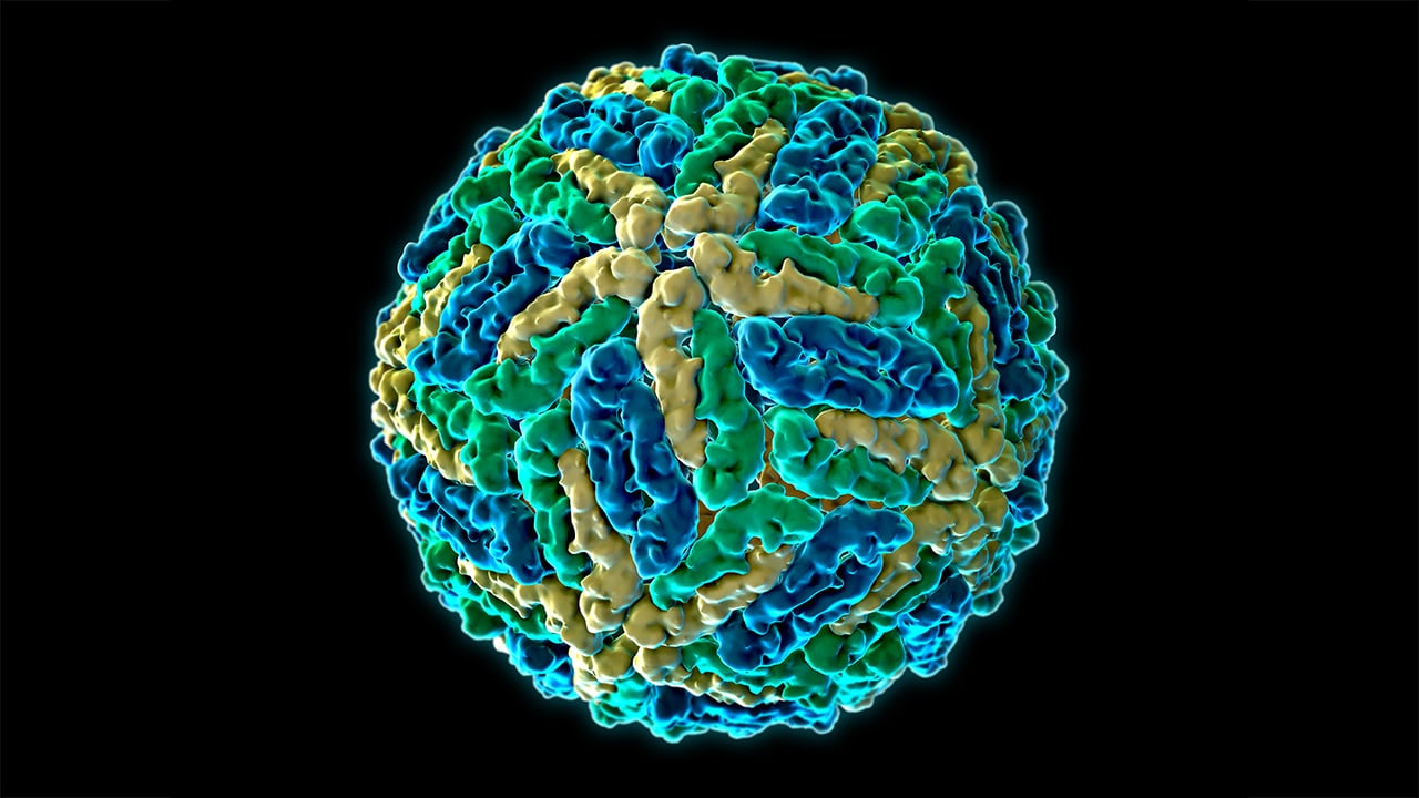
Toxocara canis
Toxocariasis
Larva Migrans, Visceral
Ascariasis
Ehrlichia canis
Eye Infections, Parasitic
Antigens, Helminth
Central Nervous System Helminthiasis
Dog Diseases
Dogs
Helminthiasis, Animal
Parasitic Diseases, Animal
Larva
Ascaris suum
Dirofilaria immitis
Seroepidemiologic Studies
Intestinal Diseases, Parasitic
Albendazole
Anthelmintics
Zoonoses
Soil
Encyclopedias as Topic
Intestine, Small
Reproductive Physiological Phenomena
Glomerulonephritis
Streptococcus pyogenes
Rheumatic Fever
Glomerulonephritis, Membranoproliferative
Aetiological study of the presumed ocular histoplasmosis syndrome in the Netherlands. (1/103)
AIM: To investigate whether presumed ocular histoplasmosis syndrome in the Netherlands is caused by Histoplasma capsulatum and whether other risk factors might play a role in the pathogenesis of this syndrome. METHODS: 23 patients were clinically diagnosed as having presumed ocular histoplasmosis syndrome based on the following criteria: peripapillary atrophy, punched out lesions, a macular disciform lesion or scar in one eye without vitritis. As controls, 66 sex and age matched healthy volunteers were used. Serum samples from both patients and controls were tested for the presence of antibodies against H capsulatum, Toxoplasma gondii, Toxocara canis et cati, Ascaris sp, and for the presence of antigens of Cryptococcus neoformans. Serum samples were also tested for the presence of autoantibodies against retinal or choroidal proteins. To investigate other risk factors, patients and controls were asked to fill in a health and travel related questionnaire. Ten patients with ocular toxoplasmosis were used as a disease control group. RESULTS: None of the patients with presumed ocular histoplasmosis syndrome or controls had circulating antibodies directed against H capsulatum. No risk factors could be identified and no indications for autoimmunity and no evidence for the role of the other infectious agents could be demonstrated. CONCLUSIONS: In a Dutch group of patients fulfilling the criteria of a disease currently named presumed ocular histoplasmosis syndrome, no risk factors or relation with the fungus H capsulatum could be detected. (+info)Identification of abundantly expressed novel and conserved genes from the infective larval stage of Toxocara canis by an expressed sequence tag strategy. (2/103)
Larvae of Toxocara canis, a nematode parasite of dogs, infect humans, causing visceral and ocular larva migrans. In noncanid hosts, larvae neither grow nor differentiate but endure in a state of arrested development. Reasoning that parasite protein production is orientated to immune evasion, we undertook a random sequencing project from a larval cDNA library to characterize the most highly expressed transcripts. In all, 266 clones were sequenced, most from both 3' and 5' ends, and similarity searches against GenBank protein and dbEST nucleotide databases were conducted. Cluster analyses showed that 128 distinct gene products had been found, all but 3 of which represented newly identified genes. Ninety-five genes were represented by a single clone, but seven transcripts were present at high frequencies, each composing >2% of all clones sequenced. These high-abundance transcripts include a mucin and a C-type lectin, which are both major excretory-secretory antigens released by parasites. Four highly expressed novel gene transcripts, termed ant (abundant novel transcript) genes, were found. Together, these four genes comprised 18% of all cDNA clones isolated, but no similar sequences occur in the Caenorhabditis elegans genome. While the coding regions of the four genes are dissimilar, their 3' untranslated tracts have significant homology in nucleotide sequence. The discovery of these abundant, parasite-specific genes of newly identified lectins and mucins, as well as a range of conserved and novel proteins, provides defined candidates for future analysis of the molecular basis of immune evasion by T. canis. (+info)A novel C-type lectin secreted by a tissue-dwelling parasitic nematode. (3/103)
Many parasitic nematodes live for surprisingly long periods in the tissues of their hosts, implying sophisticated mechanisms for evading the host immune system. The nematode Toxocara canis survives for years in mammalian tissues, and when cultivated in vitro, secretes antigens such as TES-32. From the peptide sequence, we cloned TES-32 cDNA, which encodes a 219 amino-acid protein that has a domain characteristic of host calcium-dependent (C-type) lectins, a family of proteins associated with immune defence. Homology modelling predicted that TES-32 bears remarkable structural similarity to mammalian immune-system lectins. Native TES-32 acted as a functional lectin in affinity chromatography. Unusually, it bound both mannose- and galactose-type monosaccharides, a pattern precluded in mammalian lectins by a constraining loop adjacent to the carbohydrate-binding site. In TES-32, this loop appeared to be less obtrusive, permitting a broader range of ligand binding. The similarity of TES-32 to host immune cell receptors suggests a hitherto unsuspected strategy for parasite immune evasion. (+info)Frequency of human toxocariasis in Jos, Plateau State, Nigeria. (4/103)
The enzyme-linked immunosorbent assay (Elisa) was used to examine sera of 104 children and adults in Jos, Plateau State, Nigeria for anti-toxocaral antibodies, out of which 31 (29.8%) were reactive. The seropositive rates were 30.4% for adults, 29.6% for children, 34% for females and 25.9% for males. However, the differences were not significant by age and sex. A highly significant association (p < 0.001) was observed between seropositivity and geography but none between seropositivity and dog ownership (p > 0.05). (+info)Development of a highly specific recombinant Toxocara canis second-stage larva excretory-secretory antigen for immunodiagnosis of human toxocariasis. (5/103)
The specificity of the recombinant Toxocara canis antigen developed for the immunodiagnosis of human toxocariasis was compared with that of the excretory-secretory antigen from T. canis second-stage larvae (TES) by enzyme-linked immunosorbent assay. A total of 153 human serum samples from patients infected with 20 different helminths, including 11 cases of toxocariasis, were examined. No false-negative reactions were observed for the toxocariasis cases. When the TES was used at concentrations of 0.5 and 0.125 microg/ml, cross-reactions were observed in 79 (55.6%) and 61 (43.0%) of 142 cases, respectively. In contrast, when the recombinant antigen was tested at a concentration of 0.5 microg/ml, cross-reactions were observed in 19 (13.4%) of 142 cases. At a concentration of 0.125 microg/ml, however, the cross-reaction rate decreased sharply to only 2.1%, corresponding to 3 of 142 cases. The cross-reactions occurred with one case each of gnathostomiasis, paragonimiasis with Paragonimus miyazakii, and spirometriasis, in which high antibody titers were detected. In addition, the recombinant antigen showed negative reactions with serum samples from patients infected with Ascaris and hookworms, which are the most common parasites in the world. These findings are also supported by experiments with animals infected with Ascaris and hookworm. From these results, the recombinant antigen is highly specific for toxocariasis and may provide more reliable diagnostic results than other methods. (+info)A case of presumed ocular toxocariasis in a 28-year old woman. (6/103)
This is a case of presumed ocular toxocariasis in a 28-year old woman complaining of a sudden onset of nasal side field defect of the right eye. The patient had been suffering from uveitis for ten months. Fundoscopic examination of the right eye showed a rhegmatogenous retinal detachment. Furthermore, a retinochoroidal granulomatous lesion was observed nearby the tear site. Scleral buckling, cryotherapy, and gas injection(SF6, pure gas, 0.7 cc) were conducted. Mebendazole was prescribed for one month at 25 mg/kg per body weight daily. Even though the interventions resulted in the recovery of the field defect, anti-Toxocara IgG and IgE titer levels did not decrease when checked three months after the treatment ended. This is the first confirmed serological ocular toxocariasis case in Korea. Uveitis may be a clinical presentation prior to retinal detachment of a person with toxocariasis. (+info)Human toxocarosis. Its seroprevalence in the city of La Plata. (7/103)
Toxocara canis is very common in dogs throughout the world. It is the primary cause of visceral larva migrans (VLM) in humans. Soil contaminated with T. canis embryonated eggs is the main source of infection of man. Our objective was to describe Toxocara seroprevalence in humans in the city of La Plata associated with some determinants such asage, presence or absence of clinical manifestations and risk factors. Blood samples were collected at random from 156 patients of different sex and age, with and without clinical symptoms compatible with the disease. The diagnostic technique ELISA test was performed with the Bordier Affinity Products Commercial Kit, using excretory-secretory Toxocara antigen with a sensitivity higher than 90%. The values were positive in 39% of the studied population. In the analysis according to age, the younger group presented significant differences with respect to the older one (Chi-square p<0.05). Positive patients presented clinical symptoms compatible with the disease (84%), and 41% presented some risk factor. The level of positivity obtained indicates a certain risk of being infects mainly in patients younger than 15 years old. The authors agree that an early identification and treatment of VLM may save a life. (+info)A family of secreted mucins from the parasitic nematode Toxocara canis bears diverse mucin domains but shares similar flanking six-cysteine repeat motifs. (8/103)
Infective larvae of the parasitic nematode Toxocara canis secrete a family of mucin-like glycoproteins, which are implicated in parasite immune evasion. Analysis of T. canis expressed sequence tags identified a family of four mRNAs encoding distinct apomucins (Tc-muc-1-4), one of which had been previously identified in the TES-120 family of glycoproteins secreted by this parasite. The protein products of all four cDNAs contain signal peptides, a repetitive serine/threonine-rich tract, and varying numbers of 36-amino acid six-cysteine (SXC) domains. SXC domains are found in many nematode proteins and show similarity to cnidarian (sea anemone) toxins. Antibodies to the SXC domains of Tc-MUC-1 and Tc-MUC-3 recognize differently migrating members of TES-120. TES-120 proteins separated by chromatographic methods showed distinct amino acid composition, mass, and sequence information by both Edman degradation and matrix-assisted laser desorption ionization/time of flight mass spectrometry on peptide fragments. Tc-MUC-1, -2, and -3 were shown to be secreted mucins with real masses of 39.7, 47.8, and 45.0 kDa in contrast to their predicted peptide masses of 15.7, 16.2, and 26.0 kDa, respectively. The presence of SXC domains in all mucin products supports the suggestion that the SXC motif is required for mucin assembly or export. Homology modeling indicates that the six-cysteine domains of the T. canis mucins adopt a similar fold to the sea anemone potassium channel-blocking toxin BgK, forming three disulfide bonds within each subunit. (+info)"Toxocara canis" is a species of roundworm that primarily infects canids, such as dogs and foxes. The adult worms live in the intestines of the host animal, where they lay eggs that are passed in the feces. These eggs can then mature and become infective to other animals, including humans, if they ingest them.
In humans, infection with "Toxocara canis" can cause a range of symptoms known as toxocariasis, which can include fever, coughing, wheezing, rash, and abdominal pain. In severe cases, the larvae of the worm can migrate to various organs in the body, including the eyes, leading to potentially serious complications.
Preventive measures for "Toxocara canis" infection include good hygiene practices, such as washing hands after handling pets or coming into contact with soil that may contain infected feces, and regular deworming of pets.
Toxocara is a type of parasitic roundworm that belongs to the genus Toxocara. The two most common species that infect humans are Toxocara canis and Toxocara cati, which are primarily found in dogs and cats, respectively.
Humans can become infected with Toxocara through accidental ingestion of contaminated soil or sand that contains the eggs of the parasite. This can occur when people come into contact with infected animal feces and then touch their mouths without properly washing their hands. Children are particularly at risk of infection due to their frequent hand-to-mouth behaviors and tendency to play in environments where the eggs may be present.
In humans, Toxocara infection can cause a range of symptoms known as toxocariasis. The most common form is visceral larva migrans (VLM), which occurs when the parasite's larvae migrate through various organs in the body, causing inflammation and damage. Symptoms of VLM may include fever, fatigue, coughing, wheezing, abdominal pain, and liver enlargement.
Another form of toxocariasis is ocular larva migrans (OLM), which occurs when the parasite's larvae migrate to the eye, causing inflammation and potentially leading to vision loss. Symptoms of OLM may include eye pain, redness, blurred vision, and light sensitivity.
Preventive measures for Toxocara infection include washing hands thoroughly after handling animals or coming into contact with soil, covering sandboxes when not in use, and cooking meat thoroughly before eating. Treatment for toxocariasis typically involves anti-parasitic medications such as albendazole or mebendazole, which can help kill the parasite's larvae and reduce symptoms.
Toxocariasis is a parasitic infection caused by the roundworms Toxocara canis or Toxocara cati, which are found in the intestines of dogs and cats, respectively. Humans become infected through the accidental ingestion of infective eggs from contaminated soil, water, or food. The larvae hatch in the small intestine and migrate to various tissues, including the liver, lungs, eyes, and central nervous system, where they can cause inflammation and damage.
The severity of the infection depends on the number of larvae that have infected the body and the organs involved. Most infections are asymptomatic or mild, causing symptoms such as fever, cough, rash, or abdominal discomfort. However, in severe cases, toxocariasis can lead to serious complications, including blindness (ocular larva migrans) or neurological damage (visceral larva migrans).
Preventive measures include good hygiene practices, such as washing hands after handling soil or pets, and avoiding contact with dog or cat feces. Regular deworming of pets can also help reduce the risk of transmission.
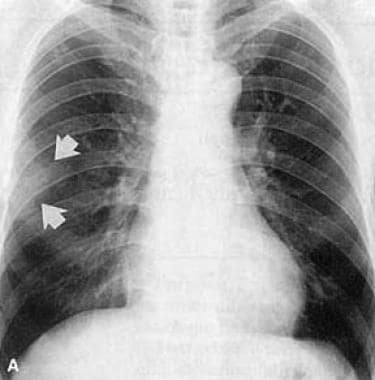
Visceral Larva Migrans is a parasitic infection caused by the migration of the larval stage of certain nematode roundworms, such as Toxocara spp. (most commonly Toxocara canis or Toxocara cati), through the tissues of the host. The larvae are ingested, usually through the consumption of contaminated soil, water, or undercooked meat, and then penetrate the intestinal wall, entering the bloodstream and migrating to various organs, including the liver, lungs, central nervous system, and eyes. This condition is more commonly seen in children due to their higher likelihood of engaging in pica (the consumption of soil or other non-food items) and having close contact with pets that may carry these parasites. Symptoms can vary widely depending on the organs involved but often include fever, coughing, wheezing, abdominal pain, and skin rashes. In severe cases, it can lead to potentially life-threatening complications such as blindness or neurological damage. Diagnosis typically involves a combination of clinical presentation, imaging studies, and laboratory tests, such as serology or stool examination for parasite eggs. Treatment usually consists of anthelmintic medications to eliminate the parasites and supportive care to manage symptoms.
Antibodies are proteins produced by the immune system in response to the presence of a foreign substance, known as an antigen. They are capable of recognizing and binding to specific antigens, neutralizing or marking them for destruction by other immune cells.
Helminths are parasitic worms that can infect humans and animals. They include roundworms, tapeworms, and flukes, among others. Helminth infections can cause a range of symptoms, depending on the type of worm and the location of the infection.
Antibodies to helminths are produced by the immune system in response to an infection with one of these parasitic worms. These antibodies can be detected in the blood and serve as evidence of a current or past infection. They may also play a role in protecting against future infections with the same type of worm.
There are several different classes of antibodies, including IgA, IgD, IgE, IgG, and IgM. Antibodies to helminths are typically of the IgE class, which are associated with allergic reactions and the defense against parasites. IgE antibodies can bind to mast cells and basophils, triggering the release of histamine and other inflammatory mediators that help to protect against the worm.
In addition to IgE, other classes of antibodies may also be produced in response to a helminth infection. For example, IgG antibodies may be produced later in the course of the infection and can provide long-term immunity to reinfection. IgA antibodies may also be produced and can help to prevent the attachment and entry of the worm into the body.
Overall, the production of antibodies to helminths is an important part of the immune response to these parasitic worms. However, in some cases, the presence of these antibodies may also be associated with allergic reactions or other immunological disorders.
Ascariasis is a medical condition caused by infection with the parasitic roundworm Ascaris lumbricoides. This type of worm infection, also known as intestinal ascariasis, occurs when people ingest contaminated soil, food, or water that contains Ascaris eggs. Once inside the body, these eggs hatch into larvae, which then migrate through the tissues and eventually reach the small intestine, where they mature into adult worms.
The adult worms can grow to be several inches long and live in the small intestine, where they feed on partially digested food. Female worms can produce thousands of eggs per day, which are then passed out of the body in feces. If these eggs hatch and infect other people, the cycle of infection continues.
Symptoms of ascariasis can vary depending on the severity of the infection. Mild infections may not cause any symptoms, while more severe infections can lead to abdominal pain, nausea, vomiting, diarrhea, and weight loss. In some cases, the worms can cause intestinal blockages or migrate to other parts of the body, leading to potentially serious complications.
Treatment for ascariasis typically involves medication to kill the adult worms and prevent them from producing more eggs. Preventive measures include good hygiene practices, such as washing hands thoroughly after using the bathroom and before eating, and avoiding contact with contaminated soil or water.
'Ehrlichia canis' is a gram-negative, intracellular bacterium that belongs to the family Anaplasmataceae. It is the etiological agent of canine monocytic ehrlichiosis (CME), which is a tick-borne disease in dogs. The bacteria are transmitted to dogs through the bite of infected brown dog ticks (Rhipicephalus sanguineus).
The infection can cause a variety of clinical signs, including fever, lethargy, anorexia, lymphadenopathy, thrombocytopenia, and hemorrhages. In severe cases, the infection may lead to serious complications such as disseminated intravascular coagulation (DIC), neurological disorders, and even death.
Diagnosis of CME is typically made through detection of Ehrlichia canis antibodies in the dog's serum or by PCR-based methods to detect the bacterial DNA. Treatment usually involves the use of antibiotics such as doxycycline, which has been shown to be effective against Ehrlichia canis.
It is important to note that 'Ehrlichia canis' can also infect humans, causing a similar disease known as human monocytic ehrlichiosis (HME). However, this is rare and usually occurs in individuals who are immunocompromised or have been exposed to infected dogs or ticks.
Parasitic eye infections are conditions characterized by the invasion and infestation of the eye or its surrounding structures by parasites. These can be protozoans, helminths, or ectoparasites. Examples of such infections include Acanthamoeba keratitis, which is caused by a free-living amoeba found in water and soil; Toxoplasmosis, which is caused by the protozoan Toxoplasma gondii; Loiasis, which is caused by the parasitic filarial worm Loa loa; and Demodicosis, which is caused by the mite Demodex folliculorum. Symptoms can vary depending on the type of parasite but often include redness, pain, discharge, and vision changes. Treatment typically involves antiparasitic medications and sometimes surgery to remove the parasites or damaged tissue. Prevention measures include good hygiene practices and avoiding contact with contaminated water or soil.
Helminth antigens refer to the proteins or other molecules found on the surface or within helminth parasites that can stimulate an immune response in a host organism. Helminths are large, multicellular parasitic worms that can infect various tissues and organs in humans and animals, causing diseases such as schistosomiasis, lymphatic filariasis, and soil-transmitted helminthiases.
Helminth antigens can be recognized by the host's immune system as foreign invaders, leading to the activation of various immune cells and the production of antibodies. However, many helminths have evolved mechanisms to evade or suppress the host's immune response, allowing them to establish long-term infections.
Studying helminth antigens is important for understanding the immunology of helminth infections and developing new strategies for diagnosis, treatment, and prevention. Some researchers have also explored the potential therapeutic use of helminth antigens or whole helminths as a way to modulate the immune system and treat autoimmune diseases or allergies. However, more research is needed to determine the safety and efficacy of these approaches.
Central nervous system helminthiasis is a medical condition that refers to the invasion and infection of the central nervous system (CNS), specifically the brain and spinal cord, by parasitic worms, also known as helminths. This rare but serious condition can occur when helminth larvae or eggs accidentally migrate from their usual location in the body to the CNS through the bloodstream or cerebrospinal fluid.
The most common types of helminths that can cause CNS helminthiasis include:
1. Neurocysticercosis: This is caused by the larval stage of the tapeworm Taenia solium, which typically infects the muscles and brain. However, when the larvae invade the CNS, they can form cysts that cause inflammation, swelling, and damage to brain tissue.
2. Echinococcosis: This is caused by the larval stage of the tapeworm Echinococcus granulosus or Echinococcus multilocularis. The larvae can form hydatid cysts in various organs, including the brain, leading to neurological symptoms.
3. Gnathostomiasis: This is caused by the larval stage of the nematode Gnathostoma spinigerum or Gnathostoma hispidum. The larvae can migrate to various organs, including the CNS, causing inflammation and damage to brain tissue.
4. Angiostrongyliasis: This is caused by the nematode Angiostrongylus cantonensis, which typically infects rats but can accidentally infect humans through contaminated food or water. The larvae can migrate to the CNS and cause eosinophilic meningitis, an inflammation of the membranes surrounding the brain and spinal cord.
Symptoms of CNS helminthiasis depend on the type of parasite involved, the location and extent of the infection, and the host's immune response. They can range from mild to severe and may include headache, seizures, weakness, numbness, vision changes, confusion, and cognitive impairment. Diagnosis is usually based on clinical presentation, imaging studies, and laboratory tests, such as serology or CSF analysis. Treatment depends on the type of parasite involved and may include antiparasitic drugs, corticosteroids, and supportive care. Prevention measures include avoiding contaminated food and water, practicing good hygiene, and using insect repellents to prevent mosquito-borne infections.
There is no medical definition for "dog diseases" as it is too broad a term. However, dogs can suffer from various health conditions and illnesses that are specific to their species or similar to those found in humans. Some common categories of dog diseases include:
1. Infectious Diseases: These are caused by viruses, bacteria, fungi, or parasites. Examples include distemper, parvovirus, kennel cough, Lyme disease, and heartworms.
2. Hereditary/Genetic Disorders: Some dogs may inherit certain genetic disorders from their parents. Examples include hip dysplasia, elbow dysplasia, progressive retinal atrophy (PRA), and degenerative myelopathy.
3. Age-Related Diseases: As dogs age, they become more susceptible to various health issues. Common age-related diseases in dogs include arthritis, dental disease, cancer, and cognitive dysfunction syndrome (CDS).
4. Nutritional Disorders: Malnutrition or improper feeding can lead to various health problems in dogs. Examples include obesity, malnutrition, and vitamin deficiencies.
5. Environmental Diseases: These are caused by exposure to environmental factors such as toxins, allergens, or extreme temperatures. Examples include heatstroke, frostbite, and toxicities from ingesting harmful substances.
6. Neurological Disorders: Dogs can suffer from various neurological conditions that affect their nervous system. Examples include epilepsy, intervertebral disc disease (IVDD), and vestibular disease.
7. Behavioral Disorders: Some dogs may develop behavioral issues due to various factors such as anxiety, fear, or aggression. Examples include separation anxiety, noise phobias, and resource guarding.
It's important to note that regular veterinary care, proper nutrition, exercise, and preventative measures can help reduce the risk of many dog diseases.
Eosinophilia is a medical condition characterized by an abnormally high concentration of eosinophils in the circulating blood. Eosinophils are a type of white blood cell that play an important role in the immune system, particularly in fighting off parasitic infections and regulating allergic reactions. However, when their numbers become excessively high, they can contribute to tissue damage and inflammation.
Eosinophilia is typically defined as a count of more than 500 eosinophils per microliter of blood. Mild eosinophilia (up to 1,500 cells/μL) may not cause any symptoms and may be discovered during routine blood tests. However, higher levels of eosinophilia can lead to various symptoms such as coughing, wheezing, skin rashes, and organ damage, depending on the underlying cause.
The causes of eosinophilia are varied and can include allergic reactions, parasitic infections, autoimmune disorders, certain medications, and some types of cancer. Accurate diagnosis and treatment of eosinophilia require identification and management of the underlying cause.
I believe there might be a misunderstanding in your question. "Dogs" is not a medical term or condition. It is the common name for a domesticated carnivore of the family Canidae, specifically the genus Canis, which includes wolves, foxes, and other extant and extinct species of mammals. Dogs are often kept as pets and companions, and they have been bred in a wide variety of forms and sizes for different purposes, such as hunting, herding, guarding, assisting police and military forces, and providing companionship and emotional support.
If you meant to ask about a specific medical condition or term related to dogs, please provide more context so I can give you an accurate answer.
Helminthiasis, in general, refers to the infection or infestation of humans and animals by helminths, which are parasitic worms. When referring to "Animal Helminthiasis," it specifically pertains to the condition where animals, including domestic pets and livestock, are infected by various helminth species. These parasitic worms can reside in different organs of the animal's body, leading to a wide range of clinical signs depending on the worm species and the location of the infestation.
Animal Helminthiasis can be caused by different types of helminths:
1. Nematodes (roundworms): These include species like Ascaris suum in pigs, Toxocara cati and Toxascaris leonina in cats, and Toxocara canis in dogs. They can cause gastrointestinal issues such as diarrhea, vomiting, and weight loss.
2. Cestodes (tapeworms): Examples include Taenia saginata in cattle, Echinococcus granulosus in sheep and goats, and Dipylidium caninum in dogs and cats. Tapeworm infestations may lead to gastrointestinal symptoms like diarrhea or constipation and may also cause vitamin deficiencies due to the worm's ability to absorb nutrients from the host animal's digestive system.
3. Trematodes (flukes): These include liver flukes such as Fasciola hepatica in sheep, goats, and cattle, and schistosomes that can affect various animals, including birds and mammals. Liver fluke infestations may cause liver damage, leading to symptoms like weight loss, decreased appetite, and jaundice. Schistosome infestations can lead to issues in multiple organs depending on the species involved.
Preventing and controlling Helminthiasis in animals is crucial for maintaining animal health and welfare, as well as ensuring food safety for humans who consume products from these animals. Regular deworming programs, good hygiene practices, proper pasture management, and monitoring for clinical signs are essential components of a comprehensive parasite control strategy.
A "Parasite Egg Count" is a laboratory measurement used to estimate the number of parasitic eggs present in a fecal sample. It is commonly used in veterinary and human medicine to diagnose and monitor parasitic infections, such as those caused by roundworms, hookworms, tapeworms, and other intestinal helminths (parasitic worms).
The most common method for measuring parasite egg counts is the McMaster technique. This involves mixing a known volume of feces with a flotation solution, which causes the eggs to float to the top of the mixture. A small sample of this mixture is then placed on a special counting chamber and examined under a microscope. The number of eggs present in the sample is then multiplied by a dilution factor to estimate the total number of eggs per gram (EPG) of feces.
Parasite egg counts can provide valuable information about the severity of an infection, as well as the effectiveness of treatment. However, it is important to note that not all parasitic infections produce visible eggs in the feces, and some parasites may only shed eggs intermittently. Therefore, a negative egg count does not always rule out the presence of a parasitic infection.
Parasitic diseases, animal, refer to conditions in animals that are caused by parasites, which are organisms that live on or inside a host and derive benefits from the host at its expense. Parasites can be classified into different groups such as protozoa, helminths (worms), and arthropods (e.g., ticks, fleas).
Parasitic diseases in animals can cause a wide range of clinical signs depending on the type of parasite, the animal species affected, and the location and extent of infection. Some common examples of parasitic diseases in animals include:
* Heartworm disease in dogs and cats caused by Dirofilaria immitis
* Coccidiosis in various animals caused by different species of Eimeria
* Toxoplasmosis in cats and other animals caused by Toxoplasma gondii
* Giardiasis in many animal species caused by Giardia spp.
* Lungworm disease in dogs and cats caused by Angiostrongylus vasorum or Aelurostrongylus abstrusus
* Tapeworm infection in dogs, cats, and other animals caused by different species of Taenia or Dipylidium caninum
Prevention and control of parasitic diseases in animals typically involve a combination of strategies such as regular veterinary care, appropriate use of medications, environmental management, and good hygiene practices.
A larva is a distinct stage in the life cycle of various insects, mites, and other arthropods during which they undergo significant metamorphosis before becoming adults. In a medical context, larvae are known for their role in certain parasitic infections. Specifically, some helminth (parasitic worm) species use larval forms to infect human hosts. These invasions may lead to conditions such as cutaneous larva migrans, visceral larva migrans, or gnathostomiasis, depending on the specific parasite involved and the location of the infection within the body.
The larval stage is characterized by its markedly different morphology and behavior compared to the adult form. Larvae often have a distinct appearance, featuring unsegmented bodies, simple sense organs, and undeveloped digestive systems. They are typically adapted for a specific mode of life, such as free-living or parasitic existence, and rely on external sources of nutrition for their development.
In the context of helminth infections, larvae may be transmitted to humans through various routes, including ingestion of contaminated food or water, direct skin contact with infective stages, or transmission via an intermediate host (such as a vector). Once inside the human body, these parasitic larvae can cause tissue damage and provoke immune responses, leading to the clinical manifestations of disease.
It is essential to distinguish between the medical definition of 'larva' and its broader usage in biology and zoology. In those fields, 'larva' refers to any juvenile form that undergoes metamorphosis before reaching adulthood, regardless of whether it is parasitic or not.
'Ascaris suum' is a species of roundworm that primarily infects pigs, although it can also rarely infect humans. It is a type of parasitic nematode that lives in the intestines of its host and obtains nutrients from ingested food. The adult female worm can grow up to 40 cm in length and produces thousands of eggs every day. These eggs are passed in the feces of infected animals and can survive in the environment for years, making them a significant source of infection for other pigs or humans who come into contact with them.
In pigs, 'Ascaris suum' infection can cause a range of symptoms, including diarrhea, vomiting, and stunted growth. In severe cases, it can lead to intestinal blockages or pneumonia. Humans who become infected with 'Ascaris suum' typically experience milder symptoms, such as abdominal pain, coughing, and wheezing. However, in rare cases, the infection can cause more serious complications, particularly if the worms migrate to other parts of the body.
Preventing 'Ascaris suum' infection involves good hygiene practices, such as washing hands thoroughly after handling animals or coming into contact with soil that may contain infected feces. It is also important to properly cook pork before eating it and to avoid consuming raw or undercooked meat. In areas where 'Ascaris suum' is common, deworming programs for pigs can help reduce the risk of infection for both animals and humans.
"Dirofilaria immitis" is a species of parasitic roundworm that can infect dogs, cats, and other animals, including humans. It is the causative agent of heartworm disease in these animals. The adult worms typically reside in the pulmonary arteries and hearts of infected animals, where they can cause serious damage to the cardiovascular system.
The life cycle of Dirofilaria immitis involves mosquitoes as intermediate hosts. Infected animals produce microfilariae, which are taken up by mosquitoes during blood meals. These larvae then develop into infective stages within the mosquito and can be transmitted to other animals through the mosquito's bite.
In dogs, heartworm disease is often asymptomatic in the early stages but can progress to cause coughing, exercise intolerance, heart failure, and even death if left untreated. In cats, heartworm disease is more difficult to diagnose and often causes respiratory symptoms such as coughing and wheezing.
Preventive measures, such as regular administration of heartworm preventatives, are essential for protecting animals from this parasitic infection.
Seroepidemiologic studies are a type of epidemiological study that measures the presence and levels of antibodies in a population's blood serum to investigate the prevalence, distribution, and transmission of infectious diseases. These studies help to identify patterns of infection and immunity within a population, which can inform public health policies and interventions.
Seroepidemiologic studies typically involve collecting blood samples from a representative sample of individuals in a population and testing them for the presence of antibodies against specific pathogens. The results are then analyzed to estimate the prevalence of infection and immunity within the population, as well as any factors associated with increased or decreased risk of infection.
These studies can provide valuable insights into the spread of infectious diseases, including emerging and re-emerging infections, and help to monitor the effectiveness of vaccination programs. Additionally, seroepidemiologic studies can also be used to investigate the transmission dynamics of infectious agents, such as identifying sources of infection or tracking the spread of antibiotic resistance.
Parasitic intestinal diseases are disorders caused by microscopic parasites that invade the gastrointestinal tract, specifically the small intestine. These parasites include protozoa (single-celled organisms) and helminths (parasitic worms). The most common protozoan parasites that cause intestinal disease are Giardia lamblia, Cryptosporidium parvum, and Entamoeba histolytica. Common helminthic parasites include roundworms (Ascaris lumbricoides), tapeworms (Taenia saginata and Taenia solium), hookworms (Ancylostoma duodenale and Necator americanus), and pinworms (Enterobius vermicularis).
Parasitic intestinal diseases can cause a variety of symptoms, including diarrhea, abdominal pain, bloating, nausea, vomiting, fatigue, and weight loss. The severity and duration of the symptoms depend on the type of parasite, the number of organisms present, and the immune status of the host.
Transmission of these parasites can occur through various routes, including contaminated food and water, person-to-person contact, and contact with contaminated soil or feces. Preventive measures include practicing good hygiene, washing hands thoroughly after using the toilet and before handling food, cooking food thoroughly, and avoiding consumption of raw or undercooked meat, poultry, or seafood.
Treatment of parasitic intestinal diseases typically involves the use of antiparasitic medications that target the specific parasite causing the infection. In some cases, supportive care such as fluid replacement and symptom management may also be necessary.
Albendazole is an antiparasitic medication used to treat a variety of parasitic infections, including neurocysticercosis (a tapeworm infection that affects the brain), hydatid disease (a parasitic infection that can affect various organs), and other types of worm infestations such as pinworm, roundworm, hookworm, and whipworm infections.
Albendazole works by inhibiting the polymerization of beta-tubulin, a protein found in the microtubules of parasitic cells, which disrupts the parasite's ability to maintain its shape and move. This leads to the death of the parasite and elimination of the infection.
Albendazole is available in oral form and is typically taken two to three times a day with meals for several days or weeks, depending on the type and severity of the infection being treated. Common side effects of albendazole include nausea, vomiting, diarrhea, abdominal pain, and headache. Rare but serious side effects may include liver damage, bone marrow suppression, and neurological problems.
It is important to note that albendazole should only be used under the supervision of a healthcare provider, as it can have serious side effects and interactions with other medications. Additionally, it is not effective against all types of parasitic infections, so proper diagnosis is essential before starting treatment.
Anthelmintics are a type of medication used to treat infections caused by parasitic worms, also known as helminths. These medications work by either stunting the growth of the worms, paralyzing them, or killing them outright, allowing the body to expel the worms through normal bodily functions. Anthelmintics are commonly used to treat infections caused by roundworms, tapeworms, flukeworms, and hookworms. Examples of anthelmintic drugs include albendazole, mebendazole, praziquantel, and ivermectin.
Zoonoses are infectious diseases that can be transmitted from animals to humans. They are caused by pathogens such as viruses, bacteria, parasites, or fungi that naturally infect non-human animals and can sometimes infect and cause disease in humans through various transmission routes like direct contact with infected animals, consumption of contaminated food or water, or vectors like insects. Some well-known zoonotic diseases include rabies, Lyme disease, salmonellosis, and COVID-19 (which is believed to have originated from bats). Public health officials work to prevent and control zoonoses through various measures such as surveillance, education, vaccination, and management of animal populations.
Helminth DNA refers to the genetic material found in parasitic worms that belong to the phylum Platyhelminthes (flatworms) and Nematoda (roundworms). These parasites can infect various organs and tissues of humans and animals, causing a range of diseases.
Helminths have complex life cycles involving multiple developmental stages and hosts. The study of their DNA has provided valuable insights into their evolutionary history, genetic diversity, and mechanisms of pathogenesis. It has also facilitated the development of molecular diagnostic tools for identifying and monitoring helminth infections.
Understanding the genetic makeup of these parasites is crucial for developing effective control strategies, including drug discovery, vaccine development, and disease management.
I'm sorry for any confusion, but "soil" is not a term that has a medical definition. Soil is defined as the top layer of earth in which plants grow, a mixture of organic material, clay, sand, and silt. If you have any questions related to medicine or health, I would be happy to try to help answer them for you.
Helminth proteins refer to the proteins that are produced and expressed by helminths, which are parasitic worms that cause diseases in humans and animals. These proteins can be found on the surface or inside the helminths and play various roles in their biology, such as in development, reproduction, and immune evasion. Some helminth proteins have been identified as potential targets for vaccines or drug development, as blocking their function may help to control or eliminate helminth infections. Examples of helminth proteins that have been studied include the antigen Bm86 from the cattle tick Boophilus microplus, and the tetraspanin protein Sm22.6 from the blood fluke Schistosoma mansoni.
An encyclopedia is a comprehensive reference work containing articles on various topics, usually arranged in alphabetical order. In the context of medicine, a medical encyclopedia is a collection of articles that provide information about a wide range of medical topics, including diseases and conditions, treatments, tests, procedures, and anatomy and physiology. Medical encyclopedias may be published in print or electronic formats and are often used as a starting point for researching medical topics. They can provide reliable and accurate information on medical subjects, making them useful resources for healthcare professionals, students, and patients alike. Some well-known examples of medical encyclopedias include the Merck Manual and the Stedman's Medical Dictionary.
The small intestine is the portion of the gastrointestinal tract that extends from the pylorus of the stomach to the beginning of the large intestine (cecum). It plays a crucial role in the digestion and absorption of nutrients from food. The small intestine is divided into three parts: the duodenum, jejunum, and ileum.
1. Duodenum: This is the shortest and widest part of the small intestine, approximately 10 inches long. It receives chyme (partially digested food) from the stomach and begins the process of further digestion with the help of various enzymes and bile from the liver and pancreas.
2. Jejunum: The jejunum is the middle section, which measures about 8 feet in length. It has a large surface area due to the presence of circular folds (plicae circulares), finger-like projections called villi, and microvilli on the surface of the absorptive cells (enterocytes). These structures increase the intestinal surface area for efficient absorption of nutrients, electrolytes, and water.
3. Ileum: The ileum is the longest and final section of the small intestine, spanning about 12 feet. It continues the absorption process, mainly of vitamin B12, bile salts, and any remaining nutrients. At the end of the ileum, there is a valve called the ileocecal valve that prevents backflow of contents from the large intestine into the small intestine.
The primary function of the small intestine is to absorb the majority of nutrients, electrolytes, and water from ingested food. The mucosal lining of the small intestine contains numerous goblet cells that secrete mucus, which protects the epithelial surface and facilitates the movement of chyme through peristalsis. Additionally, the small intestine hosts a diverse community of microbiota, which contributes to various physiological functions, including digestion, immunity, and protection against pathogens.
Reproductive physiological phenomena refer to the various functional processes and changes that occur in the reproductive system, enabling the production, development, and reproduction of offspring in living organisms. These phenomena encompass a wide range of events, including:
1. Hormonal regulation: The release and circulation of hormones that control and coordinate reproductive functions, such as follicle-stimulating hormone (FSH), luteinizing hormone (LH), estrogen, progesterone, testosterone, and inhibin.
2. Ovarian and testicular function: The development and maturation of ova (eggs) in females and sperm in males, including folliculogenesis, ovulation, spermatogenesis, and the maintenance of secondary sexual characteristics.
3. Menstrual cycle: The series of events that occur in the female reproductive system over a 28-day period, consisting of the follicular phase, ovulation, and luteal phase, resulting in the shedding of the uterine lining if fertilization does not occur.
4. Fertilization: The process by which a sperm penetrates and fuses with an egg to form a zygote, initiating embryonic development.
5. Implantation: The attachment and embedding of the developing blastocyst (early-stage embryo) into the uterine lining, leading to pregnancy.
6. Pregnancy: The physiological state of carrying a developing offspring within the female reproductive system, characterized by hormonal changes, growth and development of the fetus, and preparation for childbirth.
7. Lactation: The production and secretion of milk from the mammary glands to provide nutrition for newborn offspring.
8. Menopause: The permanent cessation of menstrual cycles and reproductive function in females, typically occurring in the fourth or fifth decade of life, characterized by a decline in hormone production and various physical and emotional symptoms.
These reproductive physiological phenomena are complex and highly regulated processes that ensure the continuation of species and the maintenance of genetic diversity.
Glomerulonephritis is a medical condition that involves inflammation of the glomeruli, which are the tiny blood vessel clusters in the kidneys that filter waste and excess fluids from the blood. This inflammation can impair the kidney's ability to filter blood properly, leading to symptoms such as proteinuria (protein in the urine), hematuria (blood in the urine), edema (swelling), hypertension (high blood pressure), and eventually kidney failure.
Glomerulonephritis can be acute or chronic, and it may occur as a primary kidney disease or secondary to other medical conditions such as infections, autoimmune disorders, or vasculitis. The diagnosis of glomerulonephritis typically involves a combination of medical history, physical examination, urinalysis, blood tests, and imaging studies, with confirmation often requiring a kidney biopsy. Treatment depends on the underlying cause and severity of the disease but may include medications to suppress inflammation, control blood pressure, and manage symptoms.
Pharyngitis is the medical term for inflammation of the pharynx, which is the back portion of the throat. This condition is often characterized by symptoms such as sore throat, difficulty swallowing, and scratchiness in the throat. Pharyngitis can be caused by a variety of factors, including viral infections (such as the common cold), bacterial infections (such as strep throat), and irritants (such as smoke or chemical fumes). Treatment for pharyngitis depends on the underlying cause of the condition, but may include medications to relieve symptoms or antibiotics to treat a bacterial infection.
Streptococcal infections are a type of infection caused by group A Streptococcus bacteria (Streptococcus pyogenes). These bacteria can cause a variety of illnesses, ranging from mild skin infections to serious and potentially life-threatening conditions such as sepsis, pneumonia, and necrotizing fasciitis (flesh-eating disease).
Some common types of streptococcal infections include:
* Streptococcal pharyngitis (strep throat) - an infection of the throat and tonsils that can cause sore throat, fever, and swollen lymph nodes.
* Impetigo - a highly contagious skin infection that causes sores or blisters on the skin.
* Cellulitis - a bacterial infection of the deeper layers of the skin and underlying tissue that can cause redness, swelling, pain, and warmth in the affected area.
* Scarlet fever - a streptococcal infection that causes a bright red rash on the body, high fever, and sore throat.
* Necrotizing fasciitis - a rare but serious bacterial infection that can cause tissue death and destruction of the muscles and fascia (the tissue that covers the muscles).
Treatment for streptococcal infections typically involves antibiotics to kill the bacteria causing the infection. It is important to seek medical attention if you suspect a streptococcal infection, as prompt treatment can help prevent serious complications.
Streptococcus pyogenes is a Gram-positive, beta-hemolytic streptococcus bacterium that causes various suppurative (pus-forming) and nonsuppurative infections in humans. It is also known as group A Streptococcus (GAS) due to its ability to produce the M protein, which confers type-specific antigenicity and allows for serological classification into more than 200 distinct Lancefield groups.
S. pyogenes is responsible for a wide range of clinical manifestations, including pharyngitis (strep throat), impetigo, cellulitis, erysipelas, scarlet fever, rheumatic fever, and acute poststreptococcal glomerulonephritis. In rare cases, it can lead to invasive diseases such as necrotizing fasciitis (flesh-eating disease) and streptococcal toxic shock syndrome (STSS).
The bacterium is typically transmitted through respiratory droplets or direct contact with infected skin lesions. Effective prevention strategies include good hygiene practices, such as frequent handwashing and avoiding sharing personal items, as well as prompt recognition and treatment of infections to prevent spread.
Rheumatic fever is a systemic inflammatory disease that may occur following an untreated Group A streptococcal infection, such as strep throat. It primarily affects children between the ages of 5 and 15, but it can occur at any age. The condition is characterized by inflammation in various parts of the body, including the heart (carditis), joints (arthritis), skin (erythema marginatum, subcutaneous nodules), and brain (Sydenham's chorea).
The onset of rheumatic fever usually occurs 2-4 weeks after a streptococcal infection. The exact cause of the immune system's overreaction leading to rheumatic fever is not fully understood, but it involves molecular mimicry between streptococcal antigens and host tissues.
The Jones Criteria are used to diagnose rheumatic fever, which include:
1. Evidence of a preceding streptococcal infection (e.g., positive throat culture or rapid strep test, elevated or rising anti-streptolysin O titer)
2. Carditis (heart inflammation), including new murmurs or changes in existing murmurs, electrocardiogram abnormalities, or evidence of heart failure
3. Polyarthritis (inflammation of multiple joints) – typically large joints like the knees and ankles, migratory, and may be associated with warmth, swelling, and pain
4. Erythema marginatum (a skin rash characterized by pink or red, irregularly shaped macules or rings that blanch in the center and spread outward)
5. Subcutaneous nodules (firm, round, mobile lumps under the skin, usually over bony prominences)
6. Sydenham's chorea (involuntary, rapid, irregular movements, often affecting the face, hands, and feet)
Treatment of rheumatic fever typically involves antibiotics to eliminate any residual streptococcal infection, anti-inflammatory medications like corticosteroids or nonsteroidal anti-inflammatory drugs (NSAIDs) to manage symptoms and prevent long-term heart complications, and secondary prophylaxis with regular antibiotic administration to prevent recurrent streptococcal infections.
Membranoproliferative Glomerulonephritis (MPGN) is a type of glomerulonephritis, which is a group of kidney disorders characterized by inflammation and damage to the glomeruli, the tiny blood vessels in the kidneys responsible for filtering waste and excess fluids from the blood.
MPGN is specifically characterized by thickening of the glomerular basement membrane and proliferation (increased number) of cells in the mesangium, a region within the glomerulus. This condition can be primary or secondary to other diseases such as infections, autoimmune disorders, or monoclonal gammopathies.
MPGN is typically classified into three types based on the pattern of injury seen on electron microscopy: Type I, Type II (Dense Deposit Disease), and Type III. Each type has distinct clinical features, laboratory findings, and treatment approaches. Symptoms of MPGN may include hematuria (blood in urine), proteinuria (protein in urine), hypertension (high blood pressure), edema (swelling), and eventually progress to chronic kidney disease or end-stage renal disease if left untreated.
Penicillin V, also known as Penicillin V Potassium, is an antibiotic medication used to treat various bacterial infections. It belongs to the class of medications called penicillins, which work by interfering with the bacteria's ability to form a protective covering (cell wall), causing the bacteria to become more susceptible to destruction by the body's immune system.
Penicillin V is specifically used to treat infections of the respiratory tract, skin, and ear. It is also used to prevent recurrent rheumatic fever and chorea (Sydenham's chorea), a neurological disorder associated with rheumatic fever.
The medication is available as oral tablets or liquid solutions and is typically taken by mouth every 6 to 12 hours, depending on the severity and type of infection being treated. As with any antibiotic, it is important to take Penicillin V exactly as directed by a healthcare professional and for the full duration of treatment, even if symptoms improve before all doses have been taken.
Penicillin V is generally well-tolerated, but like other penicillins, it can cause allergic reactions in some people. It may also interact with certain medications, so it is important to inform a healthcare provider of any other medications being taken before starting Penicillin V therapy.
 Toxocara canis
Pooper-scooper
Toxocaridae
Fading puppy syndrome
List of dog diseases
Nematode infection in dogs
Toxocariasis
Dog
Dhole
Deworming
Visceral larva migrans
Diffuse unilateral subacute neuroretinitis
Ancylostoma caninum
Retinal vasculitis
Myelitis
Albendazole
Parasites and pathogens of wolves
Red fox
Jingmenvirus
Zoonosis
Flaviviridae
Ocular larva migrans
Nicarbazin
Nitroscanate
N. H. Ashton
Spiruria
Toxascaris leonina
Baylisascaris procyonis
Veterinary parasitology
VLM
Cati14
- Visceral Larva migrans (see Toxocariasis: Toxocara canis and Toxocara cati). (ivami.com)
- Toxocara canis and Toxocara cati ). (ivami.com)
- On the basis of their findings, the authors reported Toxocara cati ova in 7 (6.3%) soil samples after examination by light microscopy. (who.int)
- However the methodology was not clearly described to understand how T. cati were differentiated from other Toxocara species, which is difficult using light microscopy. (who.int)
- Several studies have indicated that light microscopy is unable to differentiate Toxocara eggs isolated from soil specimens by size [2-4] and light microscopy is not helpful in the differentiation of T. canis and T. cati eggs [5]. (who.int)
- Furthermore, the differentiation of T. canis and T. cati eggs on the basis of morphological features is difficult and inconclusive. (who.int)
- 5] concluded that 89% of T. canis and 67% of T. cati eggs could be differentiated morphologically by considering the shape and size of the pits and their surrounding albuminous elevation using simple light microscopy. (who.int)
- Although as indicated above, egg size alone is not helpful in such studies and not a good criterion for the differentiation of T. canis and T. cati eggs, the authors do not appear to have measured this to use in conjunction with other criteria. (who.int)
- so, it is possible to find both T. cati and T. canis eggs from soil in this region. (who.int)
- Differentiation of Toxocara canis and Toxocara cati eggs by light and scanning electron microscopy. (who.int)
- Fogt-Wyrwas R, Jarosz W, Mizgajska-Wiktor H. Utilizing a polymerase chain reaction method for the detection of Toxocara canis and T. cati eggs in soil. (who.int)
- Prevalence and intensity of infestation with Toxocara cati in stray cats in Shiraz, Iran. (who.int)
- The eggs of Toxocara canis, T. cati , and other animal ascarid helminths mature in soil and infect dogs, cats, and other animals. (msdmanuals.com)
- Durvet WormEze Feline Liquid for use in food or drinking water for the removal of large roundworms (ascarids) (Toxocara canis, Toxocara cati, Toxascaris leonina). (southernagriculture.com)
Infection7
- An enzyme-linked immunosorbent assay (ELISA) was used to determine the seroprevalence of Toxocara canis infection in different socio-economic groups of the tropical population of Venezuela. (nih.gov)
- Increases in cytotoxic CD8 cells in the acute phase and helper CD4 cells in late phase infection suggest a mixed immune response elicited by T. canis . (vin.com)
- There is not any document evidence of Toxocara infection in the west Iran. (ac.ir)
- Due to high prevalence of Toxocara infection in the dogs, as well as high prevalence of A. Lumbricoides in this region and the same route of human infection to these Ascarids, there is string hazards of visceral larva migrans syndrome in this area. (ac.ir)
- This issue is discussed using as an example infection of mice with Toxocara canis, the common roundworm of dogs. (nih.gov)
- A positive result indicates an infection with Toxocara sp. (cdc.gov)
- We determined the seroprevalence of Toxocara canis infection in 544 children under 10 years randomly selected from urban and rural areas of Hamadan. (who.int)
Toxocariasis3
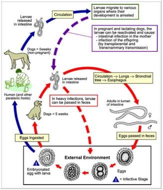
- The disease (toxocariasis) caused by migrating T. canis larvae results in two syndromes: visceral larva migrans and ocular larva migrans. (wikipedia.org)
- The lack of definitive independent diagnostic criteria for toxocariasis required the establishment of operational upper limits of normality for Toxocara ELISA values, based upon their log-normalized distribution in a presumptive "non-toxocariasis" sub-population. (nih.gov)
- 109. In Toxocara and Toxocariasis: Clinical Epidemiological and Molecular Perspectives. (cdc.gov)
Visceral3
- Experimental Visceral Larva Migrans in Chicken With Toxocara Canis" by AYŞEN GARGILI, ERKUT TÜZER et al. (tubitak.gov.tr)
- Visceral larva migrans, Toxocara canis, chicken. (tubitak.gov.tr)
- For visceral larva migrans (VLM), enzyme immunoassay (EIA) for antibodies against Toxocara is recommended to confirm the diagnosis. (msdmanuals.com)
Eggs13
- Humans are infected, like other paratenic hosts, by ingestion of embryonated T. canis eggs. (wikipedia.org)
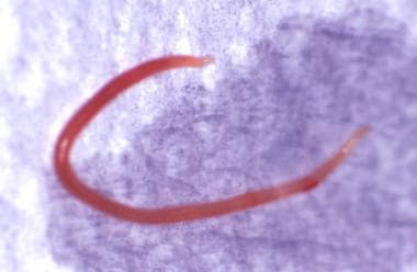
- The eggs are brownish and almost spherical.T. canis eggs have oval or spherical shapes with granulated surfaces, are thick-walled, and measure from 72 to 85 μm. (wikipedia.org)
- Three of them were orally infected with 1500 T. canis eggs and the other three received just saline. (vin.com)
- Each experimental chick recevied 5000 embriyonated T. canis eggs orally. (tubitak.gov.tr)
- Prevalence of Toxocara species eggs in the sandpits of public parks in Hyogo prefecture, Japan. (who.int)
- Scanning electron microscopy of the eggs of Ascaris lumbricoides, A. suum, Toxocara canis and T. mystax. (who.int)
- Scanning electron microscopy of Toxocara canis eggs. (who.int)
- Dog manure can contain the eggs of Toxocara canis (the common large roundworm), which can also infect humans. (thriftyfun.com)
- Toxocara eggs can remain viable in the soil for up to 10 years depending on environmental conditions. (thriftyfun.com)
- Because no information is known on the effects hot composting has on Toxocara eggs, it also unsafe to add dog manure to compost heaps intended for food crops. (thriftyfun.com)
- Toxocara eggs are passed in the host's feces, where they can be detected if a fecal sample is tested. (vin.com)
- Toxocara eggs are famous for weathering harsh environmental conditions. (vin.com)
- Toxocarosis is a zoonotic disease caused by the ingestion of infective eggs of Toxocara spp. (scielo.org.ar)
Roundworm3
- Toxocara canis (T. canis, also known as dog roundworm) is a worldwide-distributed helminth parasite that primarily infects dogs and other canids, but can also infect other animals including humans. (wikipedia.org)
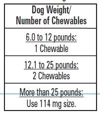
- Oxibendazole treats Roundworm species (Toxocara canis, Toxascaris leonina) and Hookworm species (Ancylostoma spp. (jedds.com)
- T. canis is the most common roundworm of the domestic dog, and it is not able to infect cats. (vin.com)
Larvae2
- The aim of this study was to determine the distribution of Toxocara canis larvae in some organs of chicks and to clarify if the larvae found in brain will be able to lead to the behavioral disorder or not. (tubitak.gov.tr)
- T. canis completes its life cycle in dogs, but when it infects aberrant mammalian hosts (e.g., humans or mice), larvae migrate through various organ systems including the brain, where they can remain viable and mobile for extended periods of time. (nih.gov)
Experimentally infected1
- The aim of this work was to analyze the number and distribution of T (CD3, CD4 and CD8) and B (IgA) cells in lungs of Toxocara canis experimentally infected BALB/c mice. (vin.com)
Caninum and Uncinaria stenocephala1
- For the removal of large roundworms, (Toxocara canis and Toxascaris leonina) and hookworms, (Ancylostoma caninum and Uncinaria stenocephala) in dogs and puppies. (entirelypets.com)
Roundworms2
- Durvet Triple Wormer® is for the treatment and control of roundworms (Toxocara canis, Toxascaris leonina), hookworms (Ancylostoma caninum, Ancylostoma braziliense, Uncinaria stenocephala) and tapeworms (Dipylidium caninum, Taenia pisiformis) in dogs and puppies. (entirelypets.com)
- and for the treatment and control of adult roundworms (Toxocara canis, Toxascaris leonina) , adult hookworm (Ancylostoma caninum) , adult whipworm (Trichuris vulpis) , and adult tapeworms (Taenia pisiformis, Echinococcus multilocularis, Echinococcus granulosus and Dipylidium caninum) infections in dogs and puppies. (drugs.com)
Trichuris1
- Trichuris vulpis, Toxocara canis and Toxoscaris leonine were not as common with a prevalence of 2.5%, 7.5% and 12.5% respectively. (slu.se)
Larva Migrans1
- Toxocara larva migrans now. (cdc.gov)
Antibodies3
- Current literature suggests that two-thirds of all dogs have been infected with this parasite, and 7% of all humans have antibodies to T. canis. (nih.gov)
- were tested for antibodies to Toxocara in an indirect enzyme immunoassay (EIA) with an excretory/secretory antigen of Toxocara canis. (cdc.gov)
- Description of Toxocara antibodies were measured with an in-house EIA developed at Laboratory the Centers for Disease Control and Prevention (CDC). (cdc.gov)
Ascarid1
- Toxacara canis is an ascarid nematodes of the small intestine of dogs and free-ranging canids. (usask.ca)
Toxoplasma1
- In addition, mice immunized with Toxoplasma gondii tachyzoites, or Toxocara canis egg antigens, that were subsequently challenged with fibrosarcoma cells, showed a reduction in solid tumor growth compared with mice that were not immunized, yet were challenged with the same cells [ 7 ]. (hindawi.com)
Antigen2
- utilizes an excretory/secretory antigen of Toxocara canis, absorbed to 96-well Immulon II HB Flat Bottom plates. (cdc.gov)
- Using the ES/L 3 purified antigen, it can be considered that the reactive sera, with compatible symptoms correspond to patients who are or were parasitized with Toxocara canis. (scielo.org.ar)
Puppies1
- Liquid Wormer 2X is used to prevent reinfestation of Toxocara canis in puppies and adult dogs and in lactating bitches after whelping. (entirelypets.com)
Dirofilaria2
- The sampled population had a prevalence of 12% for Anaplasma spp, 22% for Ehrlichia canis, 4% for Dirofilaria immitis and 1% for Leishmania spp. (slu.se)
- I den provtagna populationen var prevalensen 12% för Anaplasma spp, 22% för Ehrlichia canis, 4% för Dirofilaria immitis och 1% för Leishmania spp. (slu.se)
Taenia2
- was discovered in 113 (40.1%) samples, Toxocara canis in 82 (29.1%), Dipylidium caninum in 76 (27.0%), Giardia intestinalis in 45 (16.0%), Taenia spp. (jidc.org)
- Toxocara canis (5.4 ± 3.1%) and Taenia spp. (qld.gov.au)
Humans2
Feces2
Mice infected2
Specimens1
- In total, 228 specimens were analyzed for anti-Toxocara canis IgG at. (koreamed.org)
Adult1
- The adult T. canis has a round body with spiky cranial and caudal parts, covered by yellow cuticula. (wikipedia.org)
Dogs2
Parasite1
- These results indicate that T. canis is yet another parasite that is widely distributed in economically underprivileged tropical populations. (nih.gov)
Animals1
- Thirty one out of 60 animals (51.6%) were infected to Toxocara. (ac.ir)
Tropical1
- Specificity of Toxocara ELISA in tropical populations. (nih.gov)
Remain1
- Titers to Toxocara may remain elevated for several years. (cdc.gov)
Date1
- To date several studies have been carried out to differentiate Toxocara spp. (who.int)
Positive1
- Only 1.8% of urban subjects of medium-high socio-economic level were considered to be clearly positive in Toxocara ELISA, compared to 20.0% of urban slum dwellers, 25.6% rural subsistence farmers and 34.9% Amazon Indians. (nih.gov)
Life1
- Toxocara Canis has one of the most amazing life cycles in the animal kingdom. (vin.com)