Tibial Arteries
Popliteal Artery
Arterial Occlusive Diseases
Ischemia
Limb Salvage
Lower Extremity
Peripheral Vascular Diseases
Aneurysm
Pulmonary Artery
Peripheral Arterial Disease
Angioplasty, Balloon
Carotid Arteries
Ultrasonography, Doppler, Duplex
Mesenteric Arteries
Foot
Basilar Artery
Constriction, Pathologic
Life Tables
Blood Vessel Prosthesis Implantation
Iliac Artery
Vertebral Artery
Coronary Artery Bypass
Treatment Outcome
Radial Artery
Mammary Arteries
Reoperation
Carotid Artery, Internal
Blood Vessel Prosthesis
Subclavian Artery
Follow-Up Studies
Carotid Artery Diseases
Splenic Artery
Brachial Artery
Hepatic Artery
Carotid Artery, Common
Celiac Artery
Ophthalmic Artery
Mesenteric Artery, Superior
Retrospective Studies
Prospective Studies
Umbilical Arteries
Middle Cerebral Artery
Renal Artery Obstruction
Thoracic Arteries
Isolated femoropopliteal bypass graft for limb salvage after failed tibial reconstruction: a viable alternative to amputation. (1/199)
PURPOSE: Femoropopliteal bypass grafting procedures performed to isolated popliteal arteries after failure of a previous tibial reconstruction were studied. The results were compared with those of a study of primary isolated femoropopliteal bypass grafts (IFPBs). METHODS: IFPBs were only constructed if the uninvolved or patent popliteal segment measured at least 7 cm in length and had at least one major collateral supplying the calf. When IFPB was performed for ischemic lesions, these lesions were usually limited to the digits or small portions of the foot. Forty-seven polytetrafluoroethylene grafts and three autogenous reversed saphenous vein grafts were used. RESULTS: Ankle brachial pressure index (ABI) increased after bypass grafting by a mean of 0.46. Three-year primary life table patency and limb-salvage rates for primary IFPBs were 73% and 86%, respectively. All eight IFPBs performed after failed tibial bypass grafts remained patent for 2 to 44 months, with patients having viable, healed feet. CONCLUSION: In the presence of a suitable popliteal artery and limited tissue necrosis, IFPB can have acceptable patency and limb-salvage rates, even when a polytetrafluoroethylene graft is used. Secondary IFPB can be used to achieve limb salvage after failed tibial bypass grafting. (+info)Angiographic runoff score as a predictor of outcome following femorocrural bypass surgery. (2/199)
OBJECTIVE: to evaluate the efficacy of the revised ad hoc scoring system in predicting the outcome of femorocrural bypass surgery. DESIGN: retrospective study. MATERIALS AND METHODS: seventy-seven infrainguinal bypass procedures to the crural arteries were performed in 69 patients with critical leg ischaemia. Preoperative angiographic findings were graded according to the revised ad hoc scoring system and other preoperative angiographic measures. RESULTS: the revised ad hoc scores were valuable in predicting the outcome of these grafts. The status of the outflow artery throughout its length had a great impact on the long-term outcome in terms of secondary patency, leg salvage, patients alive with legs, and survival rates. In situ autogenous saphenous grafts achieved the best immediate and long-term results. CONCLUSIONS: the revised ad hoc angiographic scoring method is useful in predicting the outcome of patients undergoing femorocrural arterial reconstruction. Patients with an outflow artery completely open throughout its length had excellent long-term results. (+info)PTFE-vein composite grafts for critical limb ischaemia: a valuable alternative to all-autogenous infrageniculate reconstructions. (3/199)
OBJECTIVES: to analyse the long-term results of primary composite bypass grafts comparing them to PTFE and vein grafts. DESIGN: a retrospective observational study. MATERIALS AND METHODS: between 1980 and 1996, 568 primary infrageniculate bypass procedures were performed; a saphenous-vein graft was used in 428 procedures, a PTFE graft in 44 and a composite PTFE-saphenous-vein graft in 96. Thirty-six composite grafts were below the knee and the remaining 60 extended more distally. Twenty-one bypass grafts from the latter group were sequential. Mean follow-up was 45.6 months. Five-year primary and secondary patency and limb salvage rates were compared by life-table analysis. RESULTS: cumulative 5-year primary patency for composite grafts was 58% and for saphenous-vein grafts 74%, while secondary patency rate was 75% and 82%, respectively (p <0.05). The 5-year limb salvage rate was 80% for composite grafts and 88% for saphenous-vein grafts (p >0.05). The primary and secondary patency and limb salvage rate for PTFE grafts was 24%, 31% and 40%, respectively. CONCLUSION: Composite grafts of PTFE and saphenous vein are significantly superior to PTFE graft alone and should be used in patients who lack sufficient length of saphenous vein. (+info)Lack of association between Helicobacter pylori infection and extracardiac atherosclerosis in dyspeptic elderly subjects. (4/199)
BACKGROUND: There are conflicting data on the association between Helicobacter pylori (HP) infection and cardiovascular diseases. AIM: To determine if there is an association between gastric HP infection and atherosclerosis of cerebral or peripheral arteries in elderly subjects. METHODS: 90 dyspeptic elderly subjects had upper gastro-intestinal endoscopy and the gastroduodenal pathology was identified. HP infection was confirmed by gastric histology and the rapid urease test. Vascular ultrasonography of extracranial cerebral arteries and leg arteries was performed to evaluate (i) the presence of an atherosclerotic lesion, (ii) the total length of all plaques documented and (iii) the number of arteries with atherosclerotic lesions. Statistical analysis was by the chi2 test, Yates's corrected chi2 test, the Mann-Whitney test and logistic regression. RESULTS: 59 subjects were HP-positive. These had a higher prevalence of peptic ulcer disease (P = 0.01) and higher serum levels of IgG anti-HP antibodies (P = 0.0001), but no significant differences in the number of atherosclerotic lesions, the total length of the plaques or the number of arteries with lesions. No significant association of HP positivity was found with diabetes mellitus, hypertension, cigarette smoking or coronary heart disease, nor with serum concentrations of HDL-cholesterol, fibrinogen, triglycerides or glucose. CONCLUSIONS: Elderly dyspeptic subjects with gastric HP infection had significantly more peptic ulcer disease but no more atherosclerotic lesions than those who were HP-negative. Atherosclerosis was not associated with HP infection. In this cross-sectional study of elderly patients with dyspepsia, no association between HP infection and extracardiac atherosclerosis was found. (+info)Axial flow fields in cuffed end-to-side anastomoses: effect of angle and disease progression. (5/199)
OBJECTIVE: to visualise the axial flow fields in standard and cuffed end-to-side anastomoses (ESA). DESIGN: in vitro experiments using a flow rig, custom-built glass models and frame-by-frame video analysis of flow patterns in standard and cuffed ESA. SUBJECTS: glass models of standard or cuffed (1 cm or 2 cm high) ESA of angles 15, 30, 45 and 60 degrees. RESULTS: the cross-sectional area of standard ESA is much smaller than that of ESA between graft and cuff. The size of the vortex in the anastomotic zone of both standard and cuffed ESA increased with increasing ESA angle and cuff height, but did not change with flow rate. The presence of the vortex maintains a zone of flow separation and low shear at the heel of standard and graft/cuff anastomoses. CONCLUSIONS: the observations explain the clinical findings of intimal hyperplasia (IH) at the heel of PTFE/cuff anastomoses. The improved patency rates of cuffed ESA may be due not to decreased IH, but to an increased ability of the cuff to accommodate IH before causing a significant stenosis. (+info)Vascular complications of osteotomies in limb reconstruction. (6/199)
Osteotomies are commonly carried out in orthopaedic surgery, particularly in limb reconstruction. Complications are uncommon provided that sufficient care is taken and a sound technique used. We describe three cases of formation of false aneurysm after osteotomy, with acute, delayed and asymptomatic onset. The diagnosis was supported by ultrasound investigation, and confirmed by angiography. Embolisation with coils was a successful method of treatment. We recommend a safe method of osteotomy with good bone exposure and adequate soft-tissue protection. (+info)Contribution of genetic and environmental influences to ankle-brachial blood pressure index in the NHLBI Twin Study. National Heart, Lung, and Blood Institute. (7/199)
The ankle-brachial index (ABI) is widely used in the clinical diagnosis of peripheral arterial disease. The contributions of genetic and environmental influences to normal and abnormal ABI values are unknown. In this study, the authors used available data on 94 monozygotic pairs and 90 dizygotic pairs of elderly, White, male twins examined in 1995-1997 to investigate the contributions of genetic and environmental influences to normative ABI values. Within-twin-pair correlations for normative ABI values were statistically significant, and the correlation in monozygotic twin pairs was significantly greater than that in dizygotic pairs. Structural equation modeling of the variance-covariance matrices of monozygotic and dizygotic twins indicated that 48% of the observed variability in ABI values could be attributed to additive genetic effects. In contrast, concordance rates for low ABI values (ABI< or =0.9) for both monozygotic and dizygotic twins were significantly greater than would be expected by chance alone, but within-pair monozygotic similarity was not significantly greater than dizygotic similarity. A matched-cotwin analysis in 21 pairs that were discordant for low ABI values found that twins with low ABI values were physically less active and more likely to be persistent smokers than their normal-control brothers. These findings reinforce the role of individual health practices (e.g., physical activity, smoking) in the manifestation of peripheral arterial disease among subjects matched for age, genetics, and early shared environment. (+info)Long-term results of arterial allograft below-knee bypass grafts for limb salvage: a retrospective multicenter study. (8/199)
PURPOSE: Arterial allografts (AAs) have been recently reconsidered in the treatment of critical limb ischemia when vein material is absent, because of the disappointing results with artificial grafts. The aim of this study was to report the results observed in three centers where AAs were used for infrainguinal reconstruction in limb-threatening ischemia. METHODS: Between 1991 and 1997, 165 AA bypass procedures were performed in 148 patients (male, 90) with a mean age of 70 years (range, 20-93 years). Indications for operation were rest pain in 54 cases and tissue loss in 111 cases. Mean resting ankle pressure was 53 mm Hg in 96 patients who did not have diabetes and mean transcutaneous pressure of oxygen was 10 mm Hg in 52 patients who did have diabetes. In 123 cases (75%), there was at least one previous revascularization on the same limb. AAs were obtained from cadaveric donors. The distal anastomosis was to the below-knee popliteal artery in 34 cases, to a tibial artery in 114 cases, and to a pedal artery in 17 cases. RESULTS: At 30 days, the mortality rate was 3.4%; the primary patency rate was 83.3%; the secondary patency rate was 90%; and the limb salvage rate was 98%. During follow-up (mean, 31 months), 65 grafts failed primarily. Causes of primary failure were thought to be progression of the distal disease in 15 cases, myointimal hyperplasia in 16 cases, graft degradation in 10 cases (four dilations, three stenoses, two ruptures, and one dissection), miscellaneous in eight cases, and not known in 16 cases. Primary patency rates at 1, 3, and 5 years were, respectively, 48.7% +/- 4%, 34.9% +/- 6%, and 16.1% +/- 7%. Secondary patency rates at 1, 3, and 5 years were, respectively, 59. 8% +/- 4%, 42.1% +/- 5%, and 25.9% +/- 8%. Limb salvage rates at 1, 3, and 5 years were, respectively, 83.8% +/- 3%, 76.4% +/- 5%, and 74.2 % +/- 8%. CONCLUSION: AA leads to an acceptable limb salvage rate but poor patency rates. A randomized trial that will compare AAs and polytetrafluoroethylene should be undertaken. (+info)The tibial arteries are three major arteries that supply blood to the lower leg and foot. They are branches of the popliteal artery, which is a continuation of the femoral artery. The three tibial arteries are:
1. Anterior tibial artery: This artery runs down the front of the leg and supplies blood to the muscles in the anterior compartment of the leg, as well as to the foot. It becomes the dorsalis pedis artery as it approaches the ankle.
2. Posterior tibial artery: This artery runs down the back of the leg and supplies blood to the muscles in the posterior compartment of the leg. It then branches into the fibular (peroneal) artery and the medial and lateral plantar arteries, which supply blood to the foot.
3. Fibular (peroneal) artery: This artery runs down the outside of the leg and supplies blood to the muscles in the lateral compartment of the leg. It also provides branches that anastomose with the anterior and posterior tibial arteries, forming a network of vessels that helps ensure adequate blood flow to the foot.
Together, these arteries play a critical role in providing oxygenated blood and nutrients to the lower leg and foot, helping to maintain their health and function.
Arteries are blood vessels that carry oxygenated blood away from the heart to the rest of the body. They have thick, muscular walls that can withstand the high pressure of blood being pumped out of the heart. Arteries branch off into smaller vessels called arterioles, which further divide into a vast network of tiny capillaries where the exchange of oxygen, nutrients, and waste occurs between the blood and the body's cells. After passing through the capillary network, deoxygenated blood collects in venules, then merges into veins, which return the blood back to the heart.
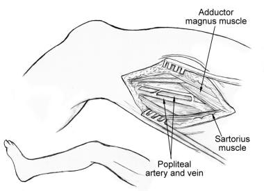
The popliteal artery is the continuation of the femoral artery that passes through the popliteal fossa, which is the area behind the knee. It is the major blood vessel that supplies oxygenated blood to the lower leg and foot. The popliteal artery divides into the anterior tibial artery and the tibioperoneal trunk at the lower border of the popliteus muscle. Any damage or blockage to this artery can result in serious health complications, including reduced blood flow to the leg and foot, which may lead to pain, cramping, numbness, or even tissue death (gangrene) if left untreated.
The femoral artery is the major blood vessel that supplies oxygenated blood to the lower extremity of the human body. It is a continuation of the external iliac artery and becomes the popliteal artery as it passes through the adductor hiatus in the adductor magnus muscle of the thigh.
The femoral artery is located in the femoral triangle, which is bound by the sartorius muscle anteriorly, the adductor longus muscle medially, and the biceps femoris muscle posteriorly. It can be easily palpated in the groin region, making it a common site for taking blood samples, measuring blood pressure, and performing surgical procedures such as femoral artery catheterization and bypass grafting.
The femoral artery gives off several branches that supply blood to the lower limb, including the deep femoral artery, the superficial femoral artery, and the profunda femoris artery. These branches provide blood to the muscles, bones, skin, and other tissues of the leg, ankle, and foot.
Arterial occlusive diseases are medical conditions characterized by the blockage or narrowing of the arteries, which can lead to a reduction in blood flow to various parts of the body. This reduction in blood flow can cause tissue damage and may result in serious complications such as tissue death (gangrene), organ dysfunction, or even death.
The most common cause of arterial occlusive diseases is atherosclerosis, which is the buildup of plaque made up of fat, cholesterol, calcium, and other substances in the inner lining of the artery walls. Over time, this plaque can harden and narrow the arteries, restricting blood flow. Other causes of arterial occlusive diseases include blood clots, emboli (tiny particles that travel through the bloodstream and lodge in smaller vessels), inflammation, trauma, and certain inherited conditions.
Symptoms of arterial occlusive diseases depend on the location and severity of the blockage. Common symptoms include:
* Pain, cramping, or fatigue in the affected limb, often triggered by exercise and relieved by rest (claudication)
* Numbness, tingling, or weakness in the affected limb
* Coldness or discoloration of the skin in the affected area
* Slow-healing sores or wounds on the toes, feet, or legs
* Erectile dysfunction in men
Treatment for arterial occlusive diseases may include lifestyle changes such as quitting smoking, exercising regularly, and eating a healthy diet. Medications to lower cholesterol, control blood pressure, prevent blood clots, or manage pain may also be prescribed. In severe cases, surgical procedures such as angioplasty, stenting, or bypass surgery may be necessary to restore blood flow.
In medical terms, the leg refers to the lower portion of the human body that extends from the knee down to the foot. It includes the thigh (femur), lower leg (tibia and fibula), foot, and ankle. The leg is primarily responsible for supporting the body's weight and enabling movements such as standing, walking, running, and jumping.
The leg contains several important structures, including bones, muscles, tendons, ligaments, blood vessels, nerves, and joints. These structures work together to provide stability, support, and mobility to the lower extremity. Common medical conditions that can affect the leg include fractures, sprains, strains, infections, peripheral artery disease, and neurological disorders.
Ischemia is the medical term used to describe a lack of blood flow to a part of the body, often due to blocked or narrowed blood vessels. This can lead to a shortage of oxygen and nutrients in the tissues, which can cause them to become damaged or die. Ischemia can affect many different parts of the body, including the heart, brain, legs, and intestines. Symptoms of ischemia depend on the location and severity of the blockage, but they may include pain, cramping, numbness, weakness, or coldness in the affected area. In severe cases, ischemia can lead to tissue death (gangrene) or organ failure. Treatment for ischemia typically involves addressing the underlying cause of the blocked blood flow, such as through medication, surgery, or lifestyle changes.
Vascular patency is a term used in medicine to describe the state of a blood vessel (such as an artery or vein) being open, unobstructed, and allowing for the normal flow of blood. It is an important concept in the treatment and management of various cardiovascular conditions, such as peripheral artery disease, coronary artery disease, and deep vein thrombosis.
Maintaining vascular patency can help prevent serious complications like tissue damage, organ dysfunction, or even death. This may involve medical interventions such as administering blood-thinning medications to prevent clots, performing procedures to remove blockages, or using devices like stents to keep vessels open. Regular monitoring of vascular patency is also crucial for evaluating the effectiveness of treatments and adjusting care plans accordingly.
Limb salvage is a medical term used to describe the surgical procedures and treatments aimed at preserving and restoring the functionality of a severely injured or diseased limb, rather than amputating it. The goal of limb salvage is to improve the patient's quality of life by maintaining their mobility, independence, and overall well-being.
Limb salvage may involve various surgical techniques such as vascular reconstruction, bone realignment, muscle flap coverage, and external fixation. These procedures aim to restore blood flow, stabilize bones, cover exposed tissues, and prevent infection. Additionally, adjuvant therapies like hyperbaric oxygen treatment, physical therapy, and pain management may be employed to support the healing process and improve functional outcomes.
Limb salvage is typically considered when a limb is threatened by conditions such as severe trauma, tumors, infections, or peripheral arterial disease. The decision to pursue limb salvage over amputation depends on factors like the patient's overall health, age, and personal preferences, as well as the extent of the injury or disease, potential for recovery, and likelihood of successful rehabilitation.
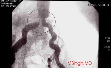
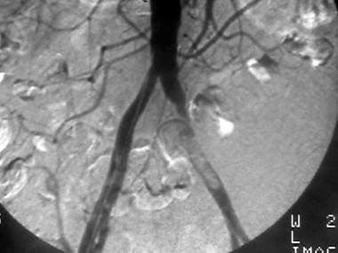
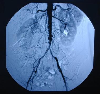
Angiography is a medical procedure in which an x-ray image is taken to visualize the internal structure of blood vessels, arteries, or veins. This is done by injecting a radiopaque contrast agent (dye) into the blood vessel using a thin, flexible catheter. The dye makes the blood vessels visible on an x-ray image, allowing doctors to diagnose and treat various medical conditions such as blockages, narrowing, or malformations of the blood vessels.
There are several types of angiography, including:
* Cardiac angiography (also called coronary angiography) - used to examine the blood vessels of the heart
* Cerebral angiography - used to examine the blood vessels of the brain
* Peripheral angiography - used to examine the blood vessels in the limbs or other parts of the body.
Angiography is typically performed by a radiologist, cardiologist, or vascular surgeon in a hospital setting. It can help diagnose conditions such as coronary artery disease, aneurysms, and peripheral arterial disease, among others.
Amputation is defined as the surgical removal of all or part of a limb or extremity such as an arm, leg, foot, hand, toe, or finger. This procedure is typically performed to remove damaged or dead tissue due to various reasons like severe injury, infection, tumors, or chronic conditions that impair circulation, such as diabetes or peripheral arterial disease. The goal of amputation is to alleviate pain, prevent further complications, and improve the patient's quality of life. Following the surgery, patients may require rehabilitation and prosthetic devices to help them adapt to their new physical condition.
The term "lower extremity" is used in the medical field to refer to the portion of the human body that includes the structures below the hip joint. This includes the thigh, lower leg, ankle, and foot. The lower extremities are responsible for weight-bearing and locomotion, allowing individuals to stand, walk, run, and jump. They contain many important structures such as bones, muscles, tendons, ligaments, nerves, and blood vessels.
Peripheral Vascular Diseases (PVD) refer to a group of medical conditions that affect the blood vessels outside of the heart and brain. These diseases are characterized by a narrowing or blockage of the peripheral arteries, which can lead to reduced blood flow to the limbs, particularly the legs.
The primary cause of PVD is atherosclerosis, a buildup of fats, cholesterol, and other substances in and on the walls of the arteries, forming plaques that restrict blood flow. Other risk factors include smoking, diabetes, hypertension, high cholesterol levels, and a family history of vascular disease.
Symptoms of PVD can vary depending on the severity of the condition but may include leg pain or cramping during exercise (claudication), numbness or tingling in the legs, coldness or discoloration of the feet, sores or wounds that heal slowly or not at all, and in severe cases, gangrene.
PVD can increase the risk of heart attack and stroke, so it is essential to diagnose and treat the condition as early as possible. Treatment options include lifestyle changes such as quitting smoking, exercising regularly, and maintaining a healthy diet, medications to control symptoms and reduce the risk of complications, and surgical procedures such as angioplasty or bypass surgery to restore blood flow.
An aneurysm is a localized, balloon-like bulge in the wall of a blood vessel. It occurs when the pressure inside the vessel causes a weakened area to swell and become enlarged. Aneurysms can develop in any blood vessel, but they are most common in arteries at the base of the brain (cerebral aneurysm) and the main artery carrying blood from the heart to the rest of the body (aortic aneurysm).
Aneurysms can be classified as saccular or fusiform, depending on their shape. A saccular aneurysm is a round or oval bulge that projects from the side of a blood vessel, while a fusiform aneurysm is a dilated segment of a blood vessel that is uniform in width and involves all three layers of the arterial wall.
The size and location of an aneurysm can affect its risk of rupture. Generally, larger aneurysms are more likely to rupture than smaller ones. Aneurysms located in areas with high blood pressure or where the vessel branches are also at higher risk of rupture.
Ruptured aneurysms can cause life-threatening bleeding and require immediate medical attention. Symptoms of a ruptured aneurysm may include sudden severe headache, neck stiffness, nausea, vomiting, blurred vision, or loss of consciousness. Unruptured aneurysms may not cause any symptoms and are often discovered during routine imaging tests for other conditions.
Treatment options for aneurysms depend on their size, location, and risk of rupture. Small, unruptured aneurysms may be monitored with regular imaging tests to check for growth or changes. Larger or symptomatic aneurysms may require surgical intervention, such as clipping or coiling, to prevent rupture and reduce the risk of complications.
The pulmonary artery is a large blood vessel that carries deoxygenated blood from the right ventricle of the heart to the lungs for oxygenation. It divides into two main branches, the right and left pulmonary arteries, which further divide into smaller vessels called arterioles, and then into a vast network of capillaries in the lungs where gas exchange occurs. The thin walls of these capillaries allow oxygen to diffuse into the blood and carbon dioxide to diffuse out, making the blood oxygen-rich before it is pumped back to the left side of the heart through the pulmonary veins. This process is crucial for maintaining proper oxygenation of the body's tissues and organs.
Peripheral Arterial Disease (PAD) is a medical condition characterized by the narrowing or blockage of arteries that supply blood to the extremities, most commonly the legs. This results in reduced blood flow, leading to symptoms such as leg pain, cramping, numbness, or weakness during physical activity, and in severe cases, tissue damage or gangrene. PAD is often indicative of widespread atherosclerosis, which is the hardening and narrowing of arteries due to the buildup of fatty deposits called plaques. It's important to note that early detection and management can help prevent serious complications.
Angioplasty, balloon refers to a medical procedure used to widen narrowed or obstructed blood vessels, particularly the coronary arteries that supply blood to the heart muscle. This procedure is typically performed using a catheter-based technique, where a thin, flexible tube called a catheter is inserted into an artery, usually through the groin or wrist, and guided to the site of the narrowing or obstruction in the coronary artery.
Once the catheter reaches the affected area, a small balloon attached to the tip of the catheter is inflated, which compresses the plaque against the artery wall and stretches the artery, thereby restoring blood flow. The balloon is then deflated and removed, along with the catheter.
Balloon angioplasty is often combined with the placement of a stent, a small metal mesh tube that helps to keep the artery open and prevent it from narrowing again. This procedure is known as percutaneous coronary intervention (PCI) or coronary angioplasty and stenting.
Overall, balloon angioplasty is a relatively safe and effective treatment for coronary artery disease, although complications such as bleeding, infection, or re-narrowing of the artery can occur in some cases.
Veins are blood vessels that carry deoxygenated blood from the tissues back to the heart. They have a lower pressure than arteries and contain valves to prevent the backflow of blood. Veins have a thin, flexible wall with a larger lumen compared to arteries, allowing them to accommodate more blood volume. The color of veins is often blue or green due to the absorption characteristics of light and the reduced oxygen content in the blood they carry.
The carotid arteries are a pair of vital blood vessels in the human body that supply oxygenated blood to the head and neck. Each person has two common carotid arteries, one on each side of the neck, which branch off from the aorta, the largest artery in the body.
The right common carotid artery originates from the brachiocephalic trunk, while the left common carotid artery arises directly from the aortic arch. As they ascend through the neck, they split into two main branches: the internal and external carotid arteries.
The internal carotid artery supplies oxygenated blood to the brain, eyes, and other structures within the skull, while the external carotid artery provides blood to the face, scalp, and various regions of the neck.
Maintaining healthy carotid arteries is crucial for overall cardiovascular health and preventing serious conditions like stroke, which can occur when the arteries become narrowed or blocked due to the buildup of plaque or fatty deposits (atherosclerosis). Regular check-ups with healthcare professionals may include monitoring carotid artery health through ultrasound or other imaging techniques.
The saphenous vein is a term used in anatomical description to refer to the great or small saphenous veins, which are superficial veins located in the lower extremities of the human body.
The great saphenous vein (GSV) is the longest vein in the body and originates from the medial aspect of the foot, ascending along the medial side of the leg and thigh, and drains into the femoral vein at the saphenofemoral junction, located in the upper third of the thigh.
The small saphenous vein (SSV) is a shorter vein that originates from the lateral aspect of the foot, ascends along the posterior calf, and drains into the popliteal vein at the saphenopopliteal junction, located in the popliteal fossa.
These veins are often used as conduits for coronary artery bypass grafting (CABG) surgery due to their consistent anatomy and length.
Cerebral arteries refer to the blood vessels that supply oxygenated blood to the brain. These arteries branch off from the internal carotid arteries and the vertebral arteries, which combine to form the basilar artery. The major cerebral arteries include:
1. Anterior cerebral artery (ACA): This artery supplies blood to the frontal lobes of the brain, including the motor and sensory cortices responsible for movement and sensation in the lower limbs.
2. Middle cerebral artery (MCA): The MCA is the largest of the cerebral arteries and supplies blood to the lateral surface of the brain, including the temporal, parietal, and frontal lobes. It is responsible for providing blood to areas involved in motor function, sensory perception, speech, memory, and vision.
3. Posterior cerebral artery (PCA): The PCA supplies blood to the occipital lobe, which is responsible for visual processing, as well as parts of the temporal and parietal lobes.
4. Anterior communicating artery (ACoA) and posterior communicating arteries (PComAs): These are small arteries that connect the major cerebral arteries, forming an important circulatory network called the Circle of Willis. The ACoA connects the two ACAs, while the PComAs connect the ICA with the PCA and the basilar artery.
These cerebral arteries play a crucial role in maintaining proper brain function by delivering oxygenated blood to various regions of the brain. Any damage or obstruction to these arteries can lead to serious neurological conditions, such as strokes or transient ischemic attacks (TIAs).
The renal artery is a pair of blood vessels that originate from the abdominal aorta and supply oxygenated blood to each kidney. These arteries branch into several smaller vessels that provide blood to the various parts of the kidneys, including the renal cortex and medulla. The renal arteries also carry nutrients and other essential components needed for the normal functioning of the kidneys. Any damage or blockage to the renal artery can lead to serious consequences, such as reduced kidney function or even kidney failure.
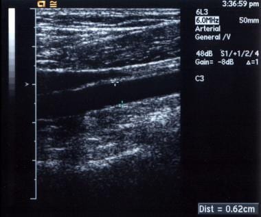
Ultrasonography, Doppler, and Duplex are diagnostic medical techniques that use sound waves to create images of internal body structures and assess their function. Here are the definitions for each:
1. Ultrasonography: Also known as ultrasound, this is a non-invasive imaging technique that uses high-frequency sound waves to produce images of internal organs and tissues. A small handheld device called a transducer is placed on the skin surface, which emits and receives sound waves. The returning echoes are then processed to create real-time visual images of the internal structures.
2. Doppler: This is a type of ultrasound that measures the velocity and direction of blood flow in the body by analyzing the frequency shift of the reflected sound waves. It can be used to assess blood flow in various parts of the body, such as the heart, arteries, and veins.
3. Duplex: Duplex ultrasonography is a combination of both gray-scale ultrasound and Doppler ultrasound. It provides detailed images of internal structures, as well as information about blood flow velocity and direction. This technique is often used to evaluate conditions such as deep vein thrombosis, carotid artery stenosis, and peripheral arterial disease.
In summary, ultrasonography is a diagnostic imaging technique that uses sound waves to create images of internal structures, Doppler is a type of ultrasound that measures blood flow velocity and direction, and duplex is a combination of both techniques that provides detailed images and information about blood flow.
Vascular surgical procedures are operations that are performed to treat conditions and diseases related to the vascular system, which includes the arteries, veins, and capillaries. These procedures can be invasive or minimally invasive and are often used to treat conditions such as peripheral artery disease, carotid artery stenosis, aortic aneurysms, and venous insufficiency.
Some examples of vascular surgical procedures include:
* Endarterectomy: a procedure to remove plaque buildup from the inside of an artery
* Bypass surgery: creating a new path for blood to flow around a blocked or narrowed artery
* Angioplasty and stenting: using a balloon to open a narrowed artery and placing a stent to keep it open
* Aneurysm repair: surgically repairing an aneurysm, a weakened area in the wall of an artery that has bulged out and filled with blood
* Embolectomy: removing a blood clot from a blood vessel
* Thrombectomy: removing a blood clot from a vein
These procedures are typically performed by vascular surgeons, who are trained in the diagnosis and treatment of vascular diseases.
The mesenteric arteries are the arteries that supply oxygenated blood to the intestines. There are three main mesenteric arteries: the superior mesenteric artery, which supplies blood to the small intestine (duodenum to two-thirds of the transverse colon) and large intestine (cecum, ascending colon, and the first part of the transverse colon); the inferior mesenteric artery, which supplies blood to the distal third of the transverse colon, descending colon, sigmoid colon, and rectum; and the middle colic artery, which is a branch of the superior mesenteric artery that supplies blood to the transverse colon. These arteries are important in maintaining adequate blood flow to the intestines to support digestion and absorption of nutrients.
In medical terms, the foot is the part of the lower limb that is distal to the leg and below the ankle, extending from the tarsus to the toes. It is primarily responsible for supporting body weight and facilitating movement through push-off during walking or running. The foot is a complex structure made up of 26 bones, 33 joints, and numerous muscles, tendons, ligaments, and nerves that work together to provide stability, balance, and flexibility. It can be divided into three main parts: the hindfoot, which contains the talus and calcaneus (heel) bones; the midfoot, which includes the navicular, cuboid, and cuneiform bones; and the forefoot, which consists of the metatarsals and phalanges that form the toes.
The basilar artery is a major blood vessel that supplies oxygenated blood to the brainstem and cerebellum. It is formed by the union of two vertebral arteries at the lower part of the brainstem, near the junction of the medulla oblongata and pons.
The basilar artery runs upward through the center of the brainstem and divides into two posterior cerebral arteries at the upper part of the brainstem, near the midbrain. The basilar artery gives off several branches that supply blood to various parts of the brainstem, including the pons, medulla oblongata, and midbrain, as well as to the cerebellum.
The basilar artery is an important part of the circle of Willis, a network of arteries at the base of the brain that ensures continuous blood flow to the brain even if one of the arteries becomes blocked or narrowed.
Pathological constriction refers to an abnormal narrowing or tightening of a body passage or organ, which can interfere with the normal flow of blood, air, or other substances through the area. This constriction can occur due to various reasons such as inflammation, scarring, or abnormal growths, and can affect different parts of the body, including blood vessels, airways, intestines, and ureters. Pathological constriction can lead to a range of symptoms and complications depending on its location and severity, and may require medical intervention to correct.
Life tables are statistical tools used in actuarial science, demography, and public health to estimate the mortality rate and survival rates of a population. They provide a data-driven representation of the probability that individuals of a certain age will die before their next birthday (the death rate) or live to a particular age (the survival rate).
Life tables are constructed using data on the number of deaths and the size of the population in specific age groups over a given period. These tables typically include several columns representing different variables, such as:
1. Age group or interval: The age range for which the data is being presented (e.g., 0-1 year, 1-5 years, 5-10 years, etc.).
2. Number of people in the population: The size of the population within each age group.
3. Number of deaths: The number of individuals who died during the study period within each age group.
4. Death rate: The probability that an individual in a given age group will die before their next birthday. It is calculated as the number of deaths divided by the size of the population for that age group.
5. Survival rate: The probability that an individual in a given age group will survive to a specific age or older. It is calculated using the death rates from earlier age groups.
6. Life expectancy: The average number of years a person is expected to live, based on their current age and mortality rates for each subsequent age group.
Life tables are essential in various fields, including insurance, pension planning, social security administration, and healthcare policy development. They help researchers and policymakers understand the health status and demographic trends of populations, allowing them to make informed decisions about resource allocation, program development, and public health interventions.
The ankle, also known as the talocrural region, is the joint between the leg and the foot. It is a synovial hinge joint that allows for dorsiflexion and plantarflexion movements. The ankle is composed of three bones: the tibia and fibula of the lower leg, and the talus of the foot. The bottom portion of the tibia and fibula, called the malleoli, form a mortise that surrounds and articulates with the talus.
The ankle joint is strengthened by several ligaments, including the medial (deltoid) ligament and lateral ligament complex. The ankle also contains important nerves and blood vessels that provide sensation and circulation to the foot.
Damage to the ankle joint, such as sprains or fractures, can result in pain, swelling, and difficulty walking. Proper care and rehabilitation are essential for maintaining the health and function of the ankle joint.
Blood vessel prosthesis implantation is a surgical procedure in which an artificial blood vessel, also known as a vascular graft or prosthetic graft, is inserted into the body to replace a damaged or diseased native blood vessel. The prosthetic graft can be made from various materials such as Dacron (polyester), PTFE (polytetrafluoroethylene), or bovine/human tissue.
The implantation of a blood vessel prosthesis is typically performed to treat conditions that cause narrowing or blockage of the blood vessels, such as atherosclerosis, aneurysms, or traumatic injuries. The procedure may be used to bypass blocked arteries in the legs (peripheral artery disease), heart (coronary artery bypass surgery), or neck (carotid endarterectomy). It can also be used to replace damaged veins for hemodialysis access in patients with kidney failure.
The success of blood vessel prosthesis implantation depends on various factors, including the patient's overall health, the location and extent of the vascular disease, and the type of graft material used. Possible complications include infection, bleeding, graft thrombosis (clotting), and graft failure, which may require further surgical intervention or endovascular treatments.
The iliac arteries are major branches of the abdominal aorta, the large artery that carries oxygen-rich blood from the heart to the rest of the body. The iliac arteries divide into two branches, the common iliac arteries, which further bifurcate into the internal and external iliac arteries.
The internal iliac artery supplies blood to the lower abdomen, pelvis, and the reproductive organs, while the external iliac artery provides blood to the lower extremities, including the legs and feet. Together, the iliac arteries play a crucial role in circulating blood throughout the body, ensuring that all tissues and organs receive the oxygen and nutrients they need to function properly.
The vertebral artery is a major blood vessel that supplies oxygenated blood to the brain and upper spinal cord. It arises from the subclavian artery, then ascends through the transverse processes of several cervical vertebrae before entering the skull through the foramen magnum. Inside the skull, it joins with the opposite vertebral artery to form the basilar artery, which supplies blood to the brainstem and cerebellum. The vertebral artery also gives off several important branches that supply blood to various regions of the brainstem and upper spinal cord.
Coronary artery bypass surgery, also known as coronary artery bypass grafting (CABG), is a surgical procedure used to improve blood flow to the heart in patients with severe coronary artery disease. This condition occurs when the coronary arteries, which supply oxygen-rich blood to the heart muscle, become narrowed or blocked due to the buildup of fatty deposits, called plaques.
During CABG surgery, a healthy blood vessel from another part of the body is grafted, or attached, to the coronary artery, creating a new pathway for oxygen-rich blood to flow around the blocked or narrowed portion of the artery and reach the heart muscle. This bypass helps to restore normal blood flow and reduce the risk of angina (chest pain), shortness of breath, and other symptoms associated with coronary artery disease.
There are different types of CABG surgery, including traditional on-pump CABG, off-pump CABG, and minimally invasive CABG. The choice of procedure depends on various factors, such as the patient's overall health, the number and location of blocked arteries, and the presence of other medical conditions.
It is important to note that while CABG surgery can significantly improve symptoms and quality of life in patients with severe coronary artery disease, it does not cure the underlying condition. Lifestyle modifications, such as regular exercise, a healthy diet, smoking cessation, and medication therapy, are essential for long-term management and prevention of further progression of the disease.
Treatment outcome is a term used to describe the result or effect of medical treatment on a patient's health status. It can be measured in various ways, such as through symptoms improvement, disease remission, reduced disability, improved quality of life, or survival rates. The treatment outcome helps healthcare providers evaluate the effectiveness of a particular treatment plan and make informed decisions about future care. It is also used in clinical research to compare the efficacy of different treatments and improve patient care.
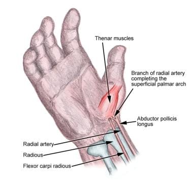
The radial artery is a key blood vessel in the human body, specifically a part of the peripheral arterial system. Originating from the brachial artery in the upper arm, the radial artery travels down the arm and crosses over the wrist, where it can be palpated easily. It then continues into the hand, dividing into several branches to supply blood to the hand's tissues and digits.
The radial artery is often used for taking pulse readings due to its easy accessibility at the wrist. Additionally, in medical procedures such as coronary angiography or bypass surgery, the radial artery can be utilized as a site for catheter insertion. This allows healthcare professionals to examine the heart's blood vessels and assess cardiovascular health.
The mammary arteries are a set of blood vessels that supply oxygenated blood to the mammary glands, which are the structures in female breasts responsible for milk production during lactation. The largest mammary artery, also known as the internal thoracic or internal mammary artery, originates from the subclavian artery and descends along the inner side of the chest wall. It then branches into several smaller arteries that supply blood to the breast tissue. These include the anterior and posterior intercostal arteries, lateral thoracic artery, and pectoral branches. The mammary arteries are crucial in maintaining the health and function of the breast tissue, and any damage or blockage to these vessels can lead to various breast-related conditions or diseases.
A reoperation is a surgical procedure that is performed again on a patient who has already undergone a previous operation for the same or related condition. Reoperations may be required due to various reasons, such as inadequate initial treatment, disease recurrence, infection, or complications from the first surgery. The nature and complexity of a reoperation can vary widely depending on the specific circumstances, but it often carries higher risks and potential complications compared to the original operation.
The internal carotid artery is a major blood vessel that supplies oxygenated blood to the brain. It originates from the common carotid artery and passes through the neck, entering the skull via the carotid canal in the temporal bone. Once inside the skull, it branches into several smaller vessels that supply different parts of the brain with blood.
The internal carotid artery is divided into several segments: cervical, petrous, cavernous, clinoid, and supraclinoid. Each segment has distinct clinical significance in terms of potential injury or disease. The most common conditions affecting the internal carotid artery include atherosclerosis, which can lead to stroke or transient ischemic attack (TIA), and dissection, which can cause severe headache, neck pain, and neurological symptoms.
It's important to note that any blockage or damage to the internal carotid artery can have serious consequences, as it can significantly reduce blood flow to the brain and lead to permanent neurological damage or even death. Therefore, regular check-ups and screening tests are recommended for individuals at high risk of developing vascular diseases.
A blood vessel prosthesis is a medical device that is used as a substitute for a damaged or diseased natural blood vessel. It is typically made of synthetic materials such as polyester, Dacron, or ePTFE (expanded polytetrafluoroethylene) and is designed to mimic the function of a native blood vessel by allowing the flow of blood through it.
Blood vessel prostheses are used in various surgical procedures, including coronary artery bypass grafting, peripheral arterial reconstruction, and the creation of arteriovenous fistulas for dialysis access. The choice of material and size of the prosthesis depends on several factors, such as the location and diameter of the vessel being replaced, the patient's age and overall health status, and the surgeon's preference.
It is important to note that while blood vessel prostheses can be effective in restoring blood flow, they may also carry risks such as infection, thrombosis (blood clot formation), and graft failure over time. Therefore, careful patient selection, surgical technique, and postoperative management are crucial for the success of these procedures.
The subclavian artery is a major blood vessel that supplies the upper limb and important structures in the neck and head. It arises from the brachiocephalic trunk (in the case of the right subclavian artery) or directly from the aortic arch (in the case of the left subclavian artery).
The subclavian artery has several branches, including:
1. The vertebral artery, which supplies blood to the brainstem and cerebellum.
2. The internal thoracic artery (also known as the mammary artery), which supplies blood to the chest wall, breast, and anterior mediastinum.
3. The thyrocervical trunk, which gives rise to several branches that supply the neck, including the inferior thyroid artery, the suprascapular artery, and the transverse cervical artery.
4. The costocervical trunk, which supplies blood to the neck and upper back, including the posterior chest wall and the lower neck muscles.
The subclavian artery is a critical vessel in maintaining adequate blood flow to the upper limb, and any blockage or damage to this vessel can lead to significant morbidity, including arm pain, numbness, weakness, or even loss of function.
Follow-up studies are a type of longitudinal research that involve repeated observations or measurements of the same variables over a period of time, in order to understand their long-term effects or outcomes. In medical context, follow-up studies are often used to evaluate the safety and efficacy of medical treatments, interventions, or procedures.
In a typical follow-up study, a group of individuals (called a cohort) who have received a particular treatment or intervention are identified and then followed over time through periodic assessments or data collection. The data collected may include information on clinical outcomes, adverse events, changes in symptoms or functional status, and other relevant measures.
The results of follow-up studies can provide important insights into the long-term benefits and risks of medical interventions, as well as help to identify factors that may influence treatment effectiveness or patient outcomes. However, it is important to note that follow-up studies can be subject to various biases and limitations, such as loss to follow-up, recall bias, and changes in clinical practice over time, which must be carefully considered when interpreting the results.
Carotid artery diseases refer to conditions that affect the carotid arteries, which are the major blood vessels that supply oxygen-rich blood to the head and neck. The most common type of carotid artery disease is atherosclerosis, which occurs when fatty deposits called plaques build up in the inner lining of the arteries.
These plaques can cause the arteries to narrow or become blocked, reducing blood flow to the brain and increasing the risk of stroke. Other carotid artery diseases include carotid artery dissection, which occurs when there is a tear in the inner lining of the artery, and fibromuscular dysplasia, which is a condition that affects the muscle and tissue in the walls of the artery.
Symptoms of carotid artery disease may include neck pain or pulsations, transient ischemic attacks (TIAs) or "mini-strokes," and strokes. Treatment options for carotid artery disease depend on the severity and type of the condition but may include lifestyle changes, medications, endarterectomy (a surgical procedure to remove plaque from the artery), or angioplasty and stenting (procedures to open blocked arteries using a balloon and stent).
The splenic artery is the largest branch of the celiac trunk, which arises from the abdominal aorta. It supplies blood to the spleen and several other organs in the upper left part of the abdomen. The splenic artery divides into several branches that ultimately form a network of capillaries within the spleen. These capillaries converge to form the main venous outflow, the splenic vein, which drains into the hepatic portal vein.
The splenic artery is a vital structure in the human body, and any damage or blockage can lead to serious complications, including splenic infarction (reduced blood flow to the spleen) or splenic rupture (a surgical emergency that can be life-threatening).
The brachial artery is a major blood vessel in the upper arm. It supplies oxygenated blood to the muscles and tissues of the arm, forearm, and hand. The brachial artery originates from the axillary artery at the level of the shoulder joint and runs down the medial (inner) aspect of the arm, passing through the cubital fossa (the depression on the anterior side of the elbow) where it can be palpated during a routine blood pressure measurement. At the lower end of the forearm, the brachial artery bifurcates into the radial and ulnar arteries, which further divide into smaller vessels to supply the hand and fingers.
The hepatic artery is a branch of the celiac trunk or abdominal aorta that supplies oxygenated blood to the liver. It typically divides into two main branches, the right and left hepatic arteries, which further divide into smaller vessels to supply different regions of the liver. The hepatic artery also gives off branches to supply other organs such as the gallbladder, pancreas, and duodenum.
It's worth noting that there is significant variability in the anatomy of the hepatic artery, with some individuals having additional branches or variations in the origin of the vessel. This variability can have implications for surgical procedures involving the liver and surrounding organs.
The common carotid artery is a major blood vessel in the neck that supplies oxygenated blood to the head and neck. It originates from the brachiocephalic trunk or the aortic arch and divides into the internal and external carotid arteries at the level of the upper border of the thyroid cartilage. The common carotid artery is an important structure in the circulatory system, and any damage or blockage to it can have serious consequences, including stroke.
In the field of medicine, "time factors" refer to the duration of symptoms or time elapsed since the onset of a medical condition, which can have significant implications for diagnosis and treatment. Understanding time factors is crucial in determining the progression of a disease, evaluating the effectiveness of treatments, and making critical decisions regarding patient care.
For example, in stroke management, "time is brain," meaning that rapid intervention within a specific time frame (usually within 4.5 hours) is essential to administering tissue plasminogen activator (tPA), a clot-busting drug that can minimize brain damage and improve patient outcomes. Similarly, in trauma care, the "golden hour" concept emphasizes the importance of providing definitive care within the first 60 minutes after injury to increase survival rates and reduce morbidity.
Time factors also play a role in monitoring the progression of chronic conditions like diabetes or heart disease, where regular follow-ups and assessments help determine appropriate treatment adjustments and prevent complications. In infectious diseases, time factors are crucial for initiating antibiotic therapy and identifying potential outbreaks to control their spread.
Overall, "time factors" encompass the significance of recognizing and acting promptly in various medical scenarios to optimize patient outcomes and provide effective care.
Coronary vessels refer to the network of blood vessels that supply oxygenated blood and nutrients to the heart muscle, also known as the myocardium. The two main coronary arteries are the left main coronary artery and the right coronary artery.
The left main coronary artery branches off into the left anterior descending artery (LAD) and the left circumflex artery (LCx). The LAD supplies blood to the front of the heart, while the LCx supplies blood to the side and back of the heart.
The right coronary artery supplies blood to the right lower part of the heart, including the right atrium and ventricle, as well as the back of the heart.
Coronary vessel disease (CVD) occurs when these vessels become narrowed or blocked due to the buildup of plaque, leading to reduced blood flow to the heart muscle. This can result in chest pain, shortness of breath, or a heart attack.
The celiac artery, also known as the anterior abdominal aortic trunk, is a major artery that originates from the abdominal aorta and supplies oxygenated blood to the foregut, which includes the stomach, liver, spleen, pancreas, and upper part of the duodenum. It branches into three main branches: the left gastric artery, the splenic artery, and the common hepatic artery. The celiac artery plays a crucial role in providing blood to these vital organs, and any disruption or damage to it can lead to serious health consequences.
The ophthalmic artery is the first branch of the internal carotid artery, which supplies blood to the eye and its adnexa. It divides into several branches that provide oxygenated blood to various structures within the eye, including the retina, optic nerve, choroid, iris, ciliary body, and cornea. Any blockage or damage to the ophthalmic artery can lead to serious vision problems or even blindness.
The superior mesenteric artery (SMA) is a major artery that supplies oxygenated blood to the intestines, specifically the lower part of the duodenum, jejunum, ileum, cecum, ascending colon, and the first and second parts of the transverse colon. It originates from the abdominal aorta, located just inferior to the pancreas, and passes behind the neck of the pancreas before dividing into several branches to supply the intestines. The SMA is an essential vessel in the digestive system, providing blood flow for nutrient absorption and overall gut function.
Retrospective studies, also known as retrospective research or looking back studies, are a type of observational study that examines data from the past to draw conclusions about possible causal relationships between risk factors and outcomes. In these studies, researchers analyze existing records, medical charts, or previously collected data to test a hypothesis or answer a specific research question.
Retrospective studies can be useful for generating hypotheses and identifying trends, but they have limitations compared to prospective studies, which follow participants forward in time from exposure to outcome. Retrospective studies are subject to biases such as recall bias, selection bias, and information bias, which can affect the validity of the results. Therefore, retrospective studies should be interpreted with caution and used primarily to generate hypotheses for further testing in prospective studies.
Prospective studies, also known as longitudinal studies, are a type of cohort study in which data is collected forward in time, following a group of individuals who share a common characteristic or exposure over a period of time. The researchers clearly define the study population and exposure of interest at the beginning of the study and follow up with the participants to determine the outcomes that develop over time. This type of study design allows for the investigation of causal relationships between exposures and outcomes, as well as the identification of risk factors and the estimation of disease incidence rates. Prospective studies are particularly useful in epidemiology and medical research when studying diseases with long latency periods or rare outcomes.
The umbilical arteries are a pair of vessels that develop within the umbilical cord during fetal development. They carry oxygenated and nutrient-rich blood from the mother to the developing fetus through the placenta. These arteries arise from the internal iliac arteries in the fetus and pass through the umbilical cord to connect with the two umbilical veins within the placenta. After birth, the umbilical arteries become ligaments (the medial umbilical ligaments) that run along the inner abdominal wall.
The Middle Cerebral Artery (MCA) is one of the main blood vessels that supplies oxygenated blood to the brain. It arises from the internal carotid artery and divides into several branches, which supply the lateral surface of the cerebral hemisphere, including the frontal, parietal, and temporal lobes.
The MCA is responsible for providing blood flow to critical areas of the brain, such as the primary motor and sensory cortices, Broca's area (associated with speech production), Wernicke's area (associated with language comprehension), and the visual association cortex.
Damage to the MCA or its branches can result in a variety of neurological deficits, depending on the specific location and extent of the injury. These may include weakness or paralysis on one side of the body, sensory loss, language impairment, and visual field cuts.
Renal artery obstruction is a medical condition that refers to the blockage or restriction of blood flow in the renal artery, which is the main vessel that supplies oxygenated and nutrient-rich blood to the kidneys. This obstruction can be caused by various factors, such as blood clots, atherosclerosis (the buildup of fats, cholesterol, and other substances in and on the artery walls), emboli (tiny particles or air bubbles that travel through the bloodstream and lodge in smaller vessels), or compressive masses like tumors.
The obstruction can lead to reduced kidney function, hypertension, and even kidney failure in severe cases. Symptoms may include high blood pressure, proteinuria (the presence of protein in the urine), hematuria (blood in the urine), and a decrease in kidney function as measured by serum creatinine levels. Diagnosis typically involves imaging studies like Doppler ultrasound, CT angiography, or magnetic resonance angiography to visualize the renal artery and assess the extent of the obstruction. Treatment options may include medications to control blood pressure and reduce kidney damage, as well as invasive procedures like angioplasty and stenting or surgical intervention to remove the obstruction and restore normal blood flow to the kidneys.
The Thoracic Arteries are branches of the aorta that supply oxygenated blood to the thoracic region of the body. The pair of arteries originate from the descending aorta and divide into several smaller branches, including intercostal arteries that supply blood to the muscles between the ribs, and posterior intercostal arteries that supply blood to the back and chest wall. Other branches of the thoracic arteries include the superior phrenic arteries, which supply blood to the diaphragm, and the bronchial arteries, which supply blood to the lungs. These arteries play a crucial role in maintaining the health and function of the chest and respiratory system.
Temporal arteries are the paired set of arteries that run along the temples on either side of the head. They are branches of the external carotid artery and play a crucial role in supplying oxygenated blood to the scalp and surrounding muscles. One of the most common conditions associated with temporal arteries is Temporal Arteritis (also known as Giant Cell Arteritis), which is an inflammation of these arteries that can lead to serious complications like vision loss if not promptly diagnosed and treated.
 Tibial arteries - Wikipedia
 Anterior tibial artery
 Recurrent tibial arteries | Radiology Reference Article | Radiopaedia.org
 Retrograde recanalization of the anterior tibial artery following surgical vessel exposure: a combined approach for single...
 What part of the body does the tibial artery supply? - Onteenstoday.com
 6d. The Anterior Tibial Artery - Collection at Bartleby.com
 Anterior Tibial Artery - Mid Calf Illustration by Cooley Studios | Medical Illustration & Animation
 Posterior tibial artery - Trauma International
 Anterior tibial artery | Anatomia Collection: anatomical plates 1522-1867
 Hail |
GTEx sQTL Artery Tibial all snp gene associations
Hail |
GTEx sQTL Artery Tibial all snp gene associations Angioplasty and stent placement - peripheral arteries: MedlinePlus Medical Encyclopedia
 Posterior tibial artery aneurysm: a case report with review of literature | BMC Surgery | Peer Review
 Table of Contents | Case Reports in Vascular Medicine | Hindawi
 February 2011 - Volume 127 - Issue 2 : Plastic and Reconstructive Surgery
 ENC TF Binding ENCODE 3 TFBS Track Settings
  Peripheral Thrombolysis: Practice Essentials, Choice of Agent and Mechanism of Action, Acute and Chronic Ischemia
 What to Know About Your Heart Rate and Pulse
 Erowid.org: Erowid Reference 6237 : Ergotism related to concurrent administration of ergotamine tartrate and indinavir :...
 P2RY8 (purinergic receptor P2Y, G-protein coupled, 8)
 Bassett Collection - Lane Medical Library - Stanford University School of Medicine