Tibia
Fibula
Pseudarthrosis
Bone and Bones
Fracture Healing
Epiphyses
Bone Density
Weight-Bearing
Ilizarov Technique
Bone Lengthening
Periosteum
External Fixators
Fracture Fixation, Intramedullary
Biomechanical Phenomena
Bone Development
Bone Diseases, Developmental
Bony Callus
Osteomyelitis
Leg Length Inequality
Bone Nails
Fracture Fixation, Internal
Fractures, Malunited
Osteogenesis, Distraction
Lead
Home Health Aides
Adamantinoma
Osseointegration
Bone Plates
Grasshoppers
Fracture Fixation
Bone Remodeling
Tomography, X-Ray Computed
Stress, Mechanical
Spectrometry, X-Ray Emission
Fractures, Stress
Bone Regeneration
Lead Poisoning, Nervous System, Adult
Ankle Joint
Fibrous Dysplasia of Bone
Range of Motion, Articular
Osteochondrosis
Bone Diseases, Metabolic
Orthopedic Procedures
Curettage
Anterior Cruciate Ligament
Reconstructive Surgical Procedures
Osteoarthritis, Knee
Osteocytes
Absorptiometry, Photon
Level of amputation following failed arterial reconstruction compared to primary amputation--a meta-analysis. (1/2843)
OBJECTIVES: To determine if the level of amputation after failed vascular reconstruction was comparable to the level of amputation after primary amputation. DESIGN AND METHODS: Medline literature search (1975-1996), meta-analysis. RESULTS: The odds ratio of transtibial to transfemoral (TT/TF) amputations was 927/657 = 1.41 (95% confidence limits: 1.278-1.561) in postrevascularisation amputation (PRVA) and 1590/1162 = 1.37 (95% confidence limits: 1.269-1.477) in primary amputation (PA) (p = 0.65). The pooled data show that the number of conversions from transtibial (TT) to transfemoral (TF) amputations due to amputation stump complications were 85/369 (23%) in PRVA against 93/752 (12.4%) in PA (p < 0.01). CONCLUSIONS: We could not detect any difference in TT/TF ratio between PRVA and PA. However, the risk of conversion i.e. reamputation to a higher level is higher after PRVA compared to PA. The chance of having a successful transtibial amputation is approximately 58% for postrevascularisation amputation as well as for primary amputations. An aggressive approach towards vascular reconstruction seems justified. (+info)Active signaling of leg loading and unloading in the cockroach. (2/2843)
The ability to detect changes in load is important for effective use of a leg in posture and locomotion. While a number of limb receptors have been shown to encode increases in load, few afferents have been demonstrated to signal leg unloading, which occurs cyclically during walking and is indicative of slipping or perturbations. We applied mechanical forces to the cockroach leg at controlled rates and recorded activities of the tibial group of campaniform sensilla, mechanoreceptors that encode forces through the strains they produce in the exoskeleton. Discrete responses were elicited from the group to decreasing as well as increasing levels of leg loading. Discharges of individual afferents depended on the direction of force application, and unit responses were correlated morphologically with the orientation of the receptor's cuticular cap. No units responded bidirectionally. Although discharges to decreasing levels of load were phasic, we found that these bursts could effectively encode the rate of force decreases. These discharges may be important in indicating leg unloading in the step cycle during walking and could rapidly signal force decreases during perturbations or loss of ground support. (+info)Chondrodiatasis in a patient with spondyloepimetaphyseal dysplasia using the Ilizarov technique: successful correction of an angular deformity with ensuing ossification of a large metaphyseal lesion. A case report. (3/2843)
Distraction through the physis (chondrodiatasis) is a controversial technique with unpredictable results. However, it has been used in the past for the lengthening and correction of angular deformities of long bones. We report the case of an 11-year-old patient with spondyloepimetaphyseal dysplasia (SEMD) who presented with a severe recurvatum deformity of the left proximal tibia secondary to collapse of the tibial plateau into a large metaphyseal cystic lesion. Using the chondrodiatasis technique with a percutaneously applied Ilizarov circular frame, we were able to correct this deformity. Surprisingly, healing and ossification of the metaphyseal lesion was simultaneously observed at the end of the treatment, a finding which, to the best of our knowledge, has not been previously reported. (+info)The aetiology of congenital angulation of tubular bones with constriction of the medullary canal, and its relationship to congenital pseudarthrosis. (4/2843)
It is suggested that there is a group of cases of congenital angulation of tubular bones in which the lesion is a defect of ossification of the primary cartilaginous anlage and in which neurofibromatosis is not implicated. It appears that in this group the prognosis with regard to the resolution of deformity and the prevention of pseudarthrosis with conservative treatment or relatively simple surgical procedures is better than that in the neurofibromatous type. (+info)The clinical manifestations and pathomechanics of contracture of the extensor mechanism of the knee. (5/2843)
Experience with thirty-eight Asian children and adolescents who presented with either stiffness of the knee, genu recurvatum, habitual dislocation of the patella or congenital lateral dislocation of the patella showed that all those disorders were manifestations of contracture of the extensor mechanism, which fell into two groups according to the components involved. In Group I the main components affected were in the midline of the limb, namely rectus femoris and vastus intermedius; these patients presented with varying degrees of stiffness of the knee, or worse, with genu recurvatum. In Group II the main components involved were lateral to the midline of the limb, namely vastus lateralis and the ilio-tibial band; these patients presented with habitual dislocation of the patella, or worse, congenital lateral dislocation of the patella. In both groups untreated patients developed secondary adaptive changes such as subluxation of the tibia or marked genu valgum which made operative procedures more formidable and less effective. Release of the contracture should therefore be performed as early as possible. (+info)Limb salvage surgery in bone tumour with modular endoprosthesis. (6/2843)
Thirty-three patients with bone tumours were treated by resection of the growth and reconstruction with a Kotz modular endoprosthesis. The average follow-up was for 50 months, ranging from 14 to 79 months. At the last review, 12 patients (36%) had died due to the tumour and 9 others (27%) had metastases. All 4 patients with proximal tibial reconstruction had poor functional results, due to an extension lag or to knee stiffness. Four of the six tumours of the proximal femur were complicated by local recurrence or dislocation of the hip, and had poor or fair functional results. Of the patients with distal femoral reconstruction, 17 out of 22 had excellent or good functional results. Reconstruction with a modular prosthesis after resection of a tumour gives excellent or good functional results in more than three-fourths of the cases of distal femur reconstruction, but it should be used with caution in the proximal tibia and proximal femur. (+info)Spontaneous or traumatic premature closure of the tibial tubercle. (7/2843)
A premature closure of the physis of the tibial tubercle in a young man has given rise to a shortening of the tibia, a patella alta and a reversed tibial slope of 20 degrees with clinical genu recurvatum. After a proximal open wedge tibial osteotomy all three postural deformities could be restored. The etiology of this complex deformity is discussed. (+info)Characterization of bone marrow laminins and identification of alpha5-containing laminins as adhesive proteins for multipotent hematopoietic FDCP-Mix cells. (8/2843)
Laminins are extracellular matrix glycoproteins that influence the phenotype and functions of many types of cells. Laminins are heterotrimers composed of alpha, beta, and gamma polypeptides. So far five alpha, three beta, and two gamma polypeptide chains, and 11 variants of laminins have been proposed. Laminins interact in vitro with mature blood cells and malignant hematopoietic cells. Most studies have been performed with laminin-1 (alpha1beta1gamma1), and its expression in bone marrow is unclear. Employing an antiserum reacting with most laminin isoforms, we found laminins widely expressed in mouse bone marrow. However, no laminin alpha1 chain but rather laminin alpha2, alpha4, and alpha5 polypeptides were found in bone marrow. Our data suggest presence of laminin-2 (alpha2beta1gamma1), laminin-8 (alpha4beta1gamma1), and laminin-10 (alpha5beta1gamma1) in bone marrow. Northern blot analysis showed expression of laminin alpha1, alpha2, alpha4, and alpha5 chains in long-term bone marrow cultures, indicating upregulation of laminin alpha1 chain expression in vitro. Laminins containing alpha5 chain, in contrast to laminin-1, were strongly adhesive for multipotent hematopoietic FDCP-mix cells. Integrin alpha6 and beta1 chains mediated this adhesion, as shown by antibody perturbation experiments. Our findings indicate that laminins other than laminin-1 are functional in adhesive interactions in bone marrow. (+info)The tibia, also known as the shin bone, is the larger of the two bones in the lower leg and part of the knee joint. It supports most of the body's weight and is a major insertion point for muscles that flex the foot and bend the leg. The tibia articulates with the femur at the knee joint and with the fibula and talus bone at the ankle joint. Injuries to the tibia, such as fractures, are common in sports and other activities that put stress on the lower leg.
The fibula is a slender bone located in the lower leg of humans and other vertebrates. It runs parallel to the larger and more robust tibia, and together they are known as the bones of the leg or the anterior tibial segment. The fibula is the lateral bone in the leg, positioned on the outside of the tibia.
In humans, the fibula extends from the knee joint proximally to the ankle joint distally. Its proximal end, called the head of the fibula, articulates with the lateral condyle of the tibia and forms part of the inferior aspect of the knee joint. The narrowed portion below the head is known as the neck of the fibula.
The shaft of the fibula, also called the body of the fibula, is a long, thin structure that descends from the neck and serves primarily for muscle attachment rather than weight-bearing functions. The distal end of the fibula widens to form the lateral malleolus, which is an important bony landmark in the ankle region. The lateral malleolus articulates with the talus bone of the foot and forms part of the ankle joint.
The primary functions of the fibula include providing attachment sites for muscles that act on the lower leg, ankle, and foot, as well as contributing to the stability of the ankle joint through its articulation with the talus bone. Fractures of the fibula can occur due to various injuries, such as twisting or rotational forces applied to the ankle or direct trauma to the lateral aspect of the lower leg.
The femur is the medical term for the thigh bone, which is the longest and strongest bone in the human body. It connects the hip bone to the knee joint and plays a crucial role in supporting the weight of the body and allowing movement during activities such as walking, running, and jumping. The femur is composed of a rounded head, a long shaft, and two condyles at the lower end that articulate with the tibia and patella to form the knee joint.
Pseudarthrosis is a medical term that refers to a false joint or a nonunion of bones, meaning that the broken bone ends do not heal properly and continue to move at the fracture site. This condition can cause pain, instability, and deformity in the affected limb. It may require additional treatment such as surgery to promote bone healing and stabilization.
"Bone" is the hard, dense connective tissue that makes up the skeleton of vertebrate animals. It provides support and protection for the body's internal organs, and serves as a attachment site for muscles, tendons, and ligaments. Bone is composed of cells called osteoblasts and osteoclasts, which are responsible for bone formation and resorption, respectively, and an extracellular matrix made up of collagen fibers and mineral crystals.
Bones can be classified into two main types: compact bone and spongy bone. Compact bone is dense and hard, and makes up the outer layer of all bones and the shafts of long bones. Spongy bone is less dense and contains large spaces, and makes up the ends of long bones and the interior of flat and irregular bones.
The human body has 206 bones in total. They can be further classified into five categories based on their shape: long bones, short bones, flat bones, irregular bones, and sesamoid bones.
The diaphysis refers to the shaft or middle portion of a long bone in the body. It is the part that is typically cylindrical in shape and contains the medullary cavity, which is filled with yellow marrow. The diaphysis is primarily composed of compact bone tissue, which provides strength and support for weight-bearing and movement.
In contrast to the diaphysis, the ends of long bones are called epiphyses, and they are covered with articular cartilage and contain spongy bone tissue filled with red marrow, which is responsible for producing blood cells. The area where the diaphysis meets the epiphysis is known as the metaphysis, and it contains growth plates that are responsible for the longitudinal growth of bones during development.
Fracture healing is the natural process by which a broken bone repairs itself. When a fracture occurs, the body responds by initiating a series of biological and cellular events aimed at restoring the structural integrity of the bone. This process involves the formation of a hematoma (a collection of blood) around the fracture site, followed by the activation of inflammatory cells that help to clean up debris and prepare the area for repair.
Over time, specialized cells called osteoblasts begin to lay down new bone matrix, or osteoid, along the edges of the broken bone ends. This osteoid eventually hardens into new bone tissue, forming a bridge between the fracture fragments. As this process continues, the callus (a mass of newly formed bone and connective tissue) gradually becomes stronger and more compact, eventually remodeling itself into a solid, unbroken bone.
The entire process of fracture healing can take several weeks to several months, depending on factors such as the severity of the injury, the patient's age and overall health, and the location of the fracture. In some cases, medical intervention may be necessary to help promote healing or ensure proper alignment of the bone fragments. This may include the use of casts, braces, or surgical implants such as plates, screws, or rods.
The epiphyses are the rounded ends of long bones in the body, which articulate with other bones to form joints. They are separated from the main shaft of the bone (diaphysis) by a growth plate called the physis or epiphyseal plate. The epiphyses are made up of spongy bone and covered with articular cartilage, which allows for smooth movement between bones. During growth, the epiphyseal plates produce new bone cells that cause the bone to lengthen until they eventually fuse during adulthood, at which point growth stops.
Bone density refers to the amount of bone mineral content (usually measured in grams) in a given volume of bone (usually measured in cubic centimeters). It is often used as an indicator of bone strength and fracture risk. Bone density is typically measured using dual-energy X-ray absorptiometry (DXA) scans, which provide a T-score that compares the patient's bone density to that of a young adult reference population. A T-score of -1 or above is considered normal, while a T-score between -1 and -2.5 indicates osteopenia (low bone mass), and a T-score below -2.5 indicates osteoporosis (porous bones). Regular exercise, adequate calcium and vitamin D intake, and medication (if necessary) can help maintain or improve bone density and prevent fractures.
The knee joint, also known as the tibiofemoral joint, is the largest and one of the most complex joints in the human body. It is a synovial joint that connects the thighbone (femur) to the shinbone (tibia). The patella (kneecap), which is a sesamoid bone, is located in front of the knee joint and helps in the extension of the leg.
The knee joint is made up of three articulations: the femorotibial joint between the femur and tibia, the femoropatellar joint between the femur and patella, and the tibiofibular joint between the tibia and fibula. These articulations are surrounded by a fibrous capsule that encloses the synovial membrane, which secretes synovial fluid to lubricate the joint.
The knee joint is stabilized by several ligaments, including the medial and lateral collateral ligaments, which provide stability to the sides of the joint, and the anterior and posterior cruciate ligaments, which prevent excessive forward and backward movement of the tibia relative to the femur. The menisci, which are C-shaped fibrocartilaginous structures located between the femoral condyles and tibial plateaus, also help to stabilize the joint by absorbing shock and distributing weight evenly across the articular surfaces.
The knee joint allows for flexion, extension, and a small amount of rotation, making it essential for activities such as walking, running, jumping, and sitting.
"Weight-bearing" is a term used in the medical field to describe the ability of a body part or limb to support the weight or pressure exerted upon it, typically while standing, walking, or performing other physical activities. In a clinical setting, healthcare professionals often use the term "weight-bearing exercise" to refer to physical activities that involve supporting one's own body weight, such as walking, jogging, or climbing stairs. These exercises can help improve bone density, muscle strength, and overall physical function, particularly in individuals with conditions affecting the bones, joints, or muscles.
In addition, "weight-bearing" is also used to describe the positioning of a body part during medical imaging studies, such as X-rays or MRIs. For example, a weight-bearing X-ray of the foot or ankle involves taking an image while the patient stands on the affected limb, allowing healthcare providers to assess any alignment or stability issues that may not be apparent in a non-weight-bearing position.
The Ilizarov technique is a surgical method used for limb lengthening and reconstruction. It involves the use of an external fixation device, which consists of rings connected by adjustable rods and wires that are attached to the bone. This apparatus allows for gradual distraction (slow, steady stretching) of the bone, allowing new bone tissue to grow in the gap created by the distraction. The Ilizarov technique can be used to treat various conditions such as limb length discrepancies, bone deformities, and nonunions (failed healing of a fracture). It is named after its developer, Gavriil Abramovich Ilizarov, a Soviet orthopedic surgeon.
Bone lengthening is a surgical procedure that involves cutting and then gradually stretching the bone apart, allowing new bone to grow in its place. This process is also known as distraction osteogenesis. The goal of bone lengthening is to increase the length of a bone, either to improve function or to correct a deformity.
The procedure typically involves making an incision in the skin over the bone and using specialized tools to cut through the bone. Once the bone is cut, a device called an external fixator is attached to the bone on either side of the cut. The external fixator is then gradually adjusted over time to slowly stretch the bone apart, creating a gap between the two ends of the bone. As the bone is stretched, new bone tissue begins to grow in the space between the two ends, eventually filling in the gap and lengthening the bone.
Bone lengthening can be used to treat a variety of conditions, including limb length discrepancies, congenital deformities, and injuries that result in bone loss. It is typically performed by an orthopedic surgeon and may require several months of follow-up care to ensure proper healing and growth of the new bone tissue.
The periosteum is a highly vascularized and innervated tissue that surrounds the outer surface of bones, except at the articular surfaces. It consists of two layers: an outer fibrous layer containing blood vessels, nerves, and fibroblasts; and an inner cellular layer called the cambium or osteogenic layer, which contains progenitor cells capable of bone formation and repair.
The periosteum plays a crucial role in bone growth, remodeling, and healing by providing a source of osteoprogenitor cells and blood supply. It also contributes to the sensation of pain in response to injury or inflammation of the bone. Additionally, the periosteum can respond to mechanical stress by activating bone formation, making it an essential component in orthopedic treatments such as distraction osteogenesis.
An external fixator is a type of orthopedic device used in the treatment of severe fractures or deformities of bones. It consists of an external frame that is attached to the bone with pins or wires that pass through the skin and into the bone. This provides stability to the injured area while allowing for alignment and adjustment of the bone during the healing process.
External fixators are typically used in cases where traditional casting or internal fixation methods are not feasible, such as when there is extensive soft tissue damage, infection, or when a limb needs to be gradually stretched or shortened. They can also be used in reconstructive surgery for bone defects or deformities.
The external frame of the fixator is made up of bars and clamps that are adjustable, allowing for precise positioning and alignment of the bones. The pins or wires that attach to the bone are carefully inserted through small incisions in the skin, and are held in place by the clamps on the frame.
External fixators can be used for a period of several weeks to several months, depending on the severity of the injury and the individual's healing process. During this time, the patient may require regular adjustments and monitoring by an orthopedic surgeon or other medical professional. Once the bone has healed sufficiently, the external fixator can be removed in a follow-up procedure.
Intramedullary fracture fixation is a surgical technique used to stabilize and align bone fractures. In this procedure, a metal rod or nail is inserted into the marrow cavity (intramedullary canal) of the affected bone, spanning the length of the fracture. The rod is then secured to the bone using screws or other fixation devices on either side of the fracture. This provides stability and helps maintain proper alignment during the healing process.
The benefits of intramedullary fixation include:
1. Load sharing: The intramedullary rod shares some of the load bearing capacity with the bone, which can help reduce stress on the healing bone.
2. Minimal soft tissue dissection: Since the implant is inserted through the medullary canal, there is less disruption to the surrounding muscles, tendons, and ligaments compared to other fixation methods.
3. Biomechanical stability: Intramedullary fixation provides rotational and bending stiffness, which helps maintain proper alignment of the fracture fragments during healing.
4. Early mobilization: Patients with intramedullary fixation can often begin weight bearing and rehabilitation exercises earlier than those with other types of fixation, leading to faster recovery times.
Common indications for intramedullary fracture fixation include long bone fractures in the femur, tibia, humerus, and fibula, as well as certain pelvic and spinal fractures. However, the choice of fixation method depends on various factors such as patient age, fracture pattern, location, and associated injuries.
Biomechanics is the application of mechanical laws to living structures and systems, particularly in the field of medicine and healthcare. A biomechanical phenomenon refers to a observable event or occurrence that involves the interaction of biological tissues or systems with mechanical forces. These phenomena can be studied at various levels, from the molecular and cellular level to the tissue, organ, and whole-body level.
Examples of biomechanical phenomena include:
1. The way that bones and muscles work together to produce movement (known as joint kinematics).
2. The mechanical behavior of biological tissues such as bone, cartilage, tendons, and ligaments under various loads and stresses.
3. The response of cells and tissues to mechanical stimuli, such as the way that bone tissue adapts to changes in loading conditions (known as Wolff's law).
4. The biomechanics of injury and disease processes, such as the mechanisms of joint injury or the development of osteoarthritis.
5. The use of mechanical devices and interventions to treat medical conditions, such as orthopedic implants or assistive devices for mobility impairments.
Understanding biomechanical phenomena is essential for developing effective treatments and prevention strategies for a wide range of medical conditions, from musculoskeletal injuries to neurological disorders.
Bone development, also known as ossification, is the process by which bone tissue is formed and grows. This complex process involves several different types of cells, including osteoblasts, which produce new bone matrix, and osteoclasts, which break down and resorb existing bone tissue.
There are two main types of bone development: intramembranous and endochondral ossification. Intramembranous ossification occurs when bone tissue forms directly from connective tissue, while endochondral ossification involves the formation of a cartilage model that is later replaced by bone.
During fetal development, most bones develop through endochondral ossification, starting as a cartilage template that is gradually replaced by bone tissue. However, some bones, such as those in the skull and clavicles, develop through intramembranous ossification.
Bone development continues after birth, with new bone tissue being laid down and existing tissue being remodeled throughout life. This ongoing process helps to maintain the strength and integrity of the skeleton, allowing it to adapt to changing mechanical forces and repair any damage that may occur.
Osteotomy is a surgical procedure in which a bone is cut to shorten, lengthen, or change its alignment. It is often performed to correct deformities or to realign bones that have been damaged by trauma or disease. The bone may be cut straight across (transverse osteotomy) or at an angle (oblique osteotomy). After the bone is cut, it can be realigned and held in place with pins, plates, or screws until it heals. This procedure is commonly performed on bones in the leg, such as the femur or tibia, but can also be done on other bones in the body.
Developmental bone diseases are a group of medical conditions that affect the growth and development of bones. These diseases are present at birth or develop during childhood and adolescence, when bones are growing rapidly. They can result from genetic mutations, hormonal imbalances, or environmental factors such as poor nutrition.
Some examples of developmental bone diseases include:
1. Osteogenesis imperfecta (OI): Also known as brittle bone disease, OI is a genetic disorder that affects the body's production of collagen, a protein necessary for healthy bones. People with OI have fragile bones that break easily and may also experience other symptoms such as blue sclerae (whites of the eyes), hearing loss, and joint laxity.
2. Achondroplasia: This is the most common form of dwarfism, caused by a genetic mutation that affects bone growth. People with achondroplasia have short limbs and a large head relative to their body size.
3. Rickets: A condition caused by vitamin D deficiency or an inability to absorb or use vitamin D properly. This leads to weak, soft bones that can bow or bend easily, particularly in children.
4. Fibrous dysplasia: A rare bone disorder where normal bone is replaced with fibrous tissue, leading to weakened bones and deformities.
5. Scoliosis: An abnormal curvature of the spine that can develop during childhood or adolescence. While not strictly a developmental bone disease, scoliosis can be caused by various underlying conditions such as cerebral palsy, muscular dystrophy, or spina bifida.
Treatment for developmental bone diseases varies depending on the specific condition and its severity. Treatment may include medication, physical therapy, bracing, or surgery to correct deformities and improve function. Regular follow-up with a healthcare provider is essential to monitor growth, manage symptoms, and prevent complications.
Bony callus is a medical term that refers to the specialized tissue that forms in response to a bone fracture. It is a crucial part of the natural healing process, as it helps to stabilize and protect the broken bone while it mends.
When a bone is fractured, the body responds by initiating an inflammatory response, which triggers the production of various cells and signaling molecules that promote healing. As part of this process, specialized cells called osteoblasts begin to produce new bone tissue at the site of the fracture. This tissue is initially soft and pliable, allowing it to bridge the gap between the broken ends of the bone.
Over time, this soft callus gradually hardens and calcifies, forming a bony callus that helps to stabilize the fracture and provide additional support as the bone heals. The bony callus is typically composed of a mixture of woven bone (which is less organized than normal bone) and more structured lamellar bone (which is similar in structure to normal bone).
As the bone continues to heal, the bony callus may be gradually remodeled and reshaped by osteoclasts, which are specialized cells that break down and remove excess or unwanted bone tissue. This process helps to restore the bone's original shape and strength, allowing it to function normally again.
It is worth noting that excessive bony callus formation can sometimes lead to complications, such as stiffness, pain, or decreased range of motion in the affected limb. In some cases, surgical intervention may be necessary to remove or reduce the size of the bony callus and promote proper healing.
Osteomyelitis is a medical condition characterized by an infection that involves the bone or the bone marrow. It can occur as a result of a variety of factors, including bacterial or fungal infections that spread to the bone from another part of the body, or direct infection of the bone through trauma or surgery.
The symptoms of osteomyelitis may include pain and tenderness in the affected area, fever, chills, fatigue, and difficulty moving the affected limb. In some cases, there may also be redness, swelling, and drainage from the infected area. The diagnosis of osteomyelitis typically involves imaging tests such as X-rays, CT scans, or MRI scans, as well as blood tests and cultures to identify the underlying cause of the infection.
Treatment for osteomyelitis usually involves a combination of antibiotics or antifungal medications to eliminate the infection, as well as pain management and possibly surgical debridement to remove infected tissue. In severe cases, hospitalization may be necessary to monitor and manage the condition.
'Leg length inequality' (LLIS) is a condition where there is a discrepancy in the lengths of an individual's lower extremities, specifically the bones of the thigh (femur) and/or the leg (tibia/fibula). This discrepancy can be congenital or acquired due to various causes such as fractures, infections, or surgical procedures. The inequality can lead to functional scoliosis, lower back pain, and other musculoskeletal issues. It is typically diagnosed through physical examination and imaging studies like X-rays, and may be treated with various methods including orthotics, shoe lifts, or in some cases, surgical intervention.
I believe you are referring to "bone pins" or "bone nails" rather than "bone nails." These terms are used in the medical field to describe surgical implants made of metal or biocompatible materials that are used to stabilize and hold together fractured bones during the healing process. They can also be used in spinal fusion surgery to provide stability and promote bone growth between vertebrae.
Bone pins or nails typically have a threaded or smooth shaft, with a small diameter that allows them to be inserted into the medullary canal of long bones such as the femur or tibia. They may also have a head or eyelet on one end that allows for attachment to external fixation devices or other surgical instruments.
The use of bone pins and nails has revolutionized orthopedic surgery, allowing for faster healing times, improved stability, and better functional outcomes for patients with fractures or spinal deformities.
Fracture fixation, internal, is a surgical procedure where a fractured bone is fixed using metal devices such as plates, screws, or rods that are implanted inside the body. This technique helps to maintain the alignment and stability of the broken bone while it heals. The implants may be temporarily or permanently left inside the body, depending on the nature and severity of the fracture. Internal fixation allows for early mobilization and rehabilitation, which can result in a faster recovery and improved functional outcome.
Malunited fractures refer to a type of fracture where the bones do not heal in their proper alignment or position. This can occur due to various reasons such as inadequate reduction of the fracture fragments during initial treatment, improper casting or immobilization, or failure of the patient to follow proper immobilization instructions. Malunited fractures can result in deformity, limited range of motion, and decreased functionality of the affected limb. Additional treatments such as surgery may be required to correct the malunion and restore normal function.
Bone transplantation, also known as bone grafting, is a surgical procedure in which bone or bone-like material is transferred from one part of the body to another or from one person to another. The graft may be composed of cortical (hard outer portion) bone, cancellous (spongy inner portion) bone, or a combination of both. It can be taken from different sites in the same individual (autograft), from another individual of the same species (allograft), or from an animal source (xenograft). The purpose of bone transplantation is to replace missing bone, provide structural support, and stimulate new bone growth. This procedure is commonly used in orthopedic, dental, and maxillofacial surgeries to repair bone defects caused by trauma, tumors, or congenital conditions.
Osteogenesis, distraction refers to a surgical procedure and controlled rehabilitation process used in orthopedic surgery, oral and maxillofacial surgery, and neurosurgery to lengthen bones or correct bone deformities. The term "osteogenesis" means bone formation, while "distraction" refers to the gradual separation of bone segments.
In this procedure, a surgeon first cuts the bone (osteotomy) and then applies an external or internal distraction device that slowly moves apart the cut ends of the bone. Over time, new bone forms in the gap between the separated bone segments through a process called distraction osteogenesis. This results in increased bone length or correction of deformities.
Distraction osteogenesis is often used to treat various conditions such as limb length discrepancies, craniofacial deformities, and spinal deformities. The procedure requires careful planning, precise surgical technique, and close postoperative management to ensure optimal outcomes.
In the context of medicine, "lead" most commonly refers to lead exposure or lead poisoning. Lead is a heavy metal that can be harmful to the human body, even at low levels. It can enter the body through contaminated air, water, food, or soil, and it can also be absorbed through the skin.
Lead poisoning occurs when lead builds up in the body over time, causing damage to the brain, nervous system, red blood cells, and kidneys. Symptoms of lead poisoning may include abdominal pain, constipation, fatigue, headache, irritability, memory problems, and in severe cases, seizures, coma, or even death.
Lead exposure is particularly dangerous for children, as their developing bodies are more sensitive to the harmful effects of lead. Even low levels of lead exposure can cause learning disabilities, behavioral problems, and developmental delays in children. Therefore, it's important to minimize lead exposure and seek medical attention if lead poisoning is suspected.
A growth plate, also known as an epiphyseal plate or physis, is a layer of cartilaginous tissue found near the ends of long bones in children and adolescents. This region is responsible for the longitudinal growth of bones during development. The growth plate contains actively dividing cells that differentiate into chondrocytes, which produce and deposit new matrix, leading to bone elongation. Once growth is complete, usually in late adolescence or early adulthood, the growth plates ossify (harden) and are replaced by solid bone, transforming into the epiphyseal line.
Bone neoplasms are abnormal growths or tumors that develop in the bone. They can be benign (non-cancerous) or malignant (cancerous). Benign bone neoplasms do not spread to other parts of the body and are rarely a threat to life, although they may cause problems if they grow large enough to press on surrounding tissues or cause fractures. Malignant bone neoplasms, on the other hand, can invade and destroy nearby tissue and may spread (metastasize) to other parts of the body.
There are many different types of bone neoplasms, including:
1. Osteochondroma - a benign tumor that develops from cartilage and bone
2. Enchondroma - a benign tumor that forms in the cartilage that lines the inside of the bones
3. Chondrosarcoma - a malignant tumor that develops from cartilage
4. Osteosarcoma - a malignant tumor that develops from bone cells
5. Ewing sarcoma - a malignant tumor that develops in the bones or soft tissues around the bones
6. Giant cell tumor of bone - a benign or occasionally malignant tumor that develops from bone tissue
7. Fibrosarcoma - a malignant tumor that develops from fibrous tissue in the bone
The symptoms of bone neoplasms vary depending on the type, size, and location of the tumor. They may include pain, swelling, stiffness, fractures, or limited mobility. Treatment options depend on the type and stage of the tumor but may include surgery, radiation therapy, chemotherapy, or a combination of these treatments.
Home Health Aides (HHAs) are healthcare professionals who provide basic medical and personal care services to patients in their homes. They work under the supervision of licensed healthcare professionals, such as registered nurses or therapists, and assist with tasks that may include:
* Basic healthcare procedures, such as monitoring vital signs, administering medications, and providing wound care
* Personal care activities, such as bathing, dressing, grooming, and toileting
* Light housekeeping duties, including laundry, meal preparation, and shopping for groceries
* Providing companionship and emotional support to patients and their families
HHAs are trained to provide a range of services that help patients maintain their independence and quality of life while recovering from illness or injury, or managing chronic conditions. They may work for home health agencies, hospices, or other healthcare organizations, or they may be self-employed. In order to become a Home Health Aide, individuals typically need to complete a state-approved training program and pass a certification exam.
Adamantinoma is a rare, slow-growing malignant (cancerous) tumor that typically develops in the tibia or fibula bones of the lower leg. It primarily affects young adults and can be difficult to diagnose due to its rarity and nonspecific symptoms.
The name "adamantinoma" comes from its microscopic appearance, which resembles that of a type of skin cancer called adamantinoma of the skin or adamantoblastoma. However, they are not related.
Adamantinomas are characterized by the presence of epithelial cells (cells that line the outer surface of the body and internal organs) within the bone tissue. These tumors tend to be locally aggressive, meaning they can invade surrounding tissues and bones. In some cases, adamantinomas may metastasize (spread) to other parts of the body, such as the lungs or lymph nodes.
Treatment for adamantinoma usually involves surgical removal of the tumor, along with a portion of the affected bone. In some cases, reconstruction or limb-sparing surgery may be necessary to maintain function and appearance. Radiation therapy and chemotherapy are not typically effective against adamantinomas, but they might be used in specific situations or as part of clinical trials.
Regular follow-up appointments with a healthcare provider are essential for monitoring the patient's condition and detecting any potential recurrence or metastasis early on.
Osseointegration is a direct structural and functional connection between living bone and the surface of an implant. It's a process where the bone grows in and around the implant, which is typically made of titanium or another biocompatible material. This process provides a solid foundation for dental prosthetics, such as crowns, bridges, or dentures, or for orthopedic devices like artificial limbs. The success of osseointegration depends on various factors, including the patient's overall health, the quality and quantity of available bone, and the surgical technique used for implant placement.
Osteogenesis is the process of bone formation or development. It involves the differentiation and maturation of osteoblasts, which are bone-forming cells that synthesize and deposit the organic matrix of bone tissue, composed mainly of type I collagen. This organic matrix later mineralizes to form the inorganic crystalline component of bone, primarily hydroxyapatite.
There are two primary types of osteogenesis: intramembranous and endochondral. Intramembranous osteogenesis occurs directly within connective tissue, where mesenchymal stem cells differentiate into osteoblasts and form bone tissue without an intervening cartilage template. This process is responsible for the formation of flat bones like the skull and clavicles.
Endochondral osteogenesis, on the other hand, involves the initial development of a cartilaginous model or template, which is later replaced by bone tissue. This process forms long bones, such as those in the limbs, and occurs through several stages involving chondrocyte proliferation, hypertrophy, and calcification, followed by invasion of blood vessels and osteoblasts to replace the cartilage with bone tissue.
Abnormalities in osteogenesis can lead to various skeletal disorders and diseases, such as osteogenesis imperfecta (brittle bone disease), achondroplasia (a form of dwarfism), and cleidocranial dysplasia (a disorder affecting skull and collarbone development).
A closed fracture, also known as a simple fracture, is a type of bone break where the skin remains intact and there is no open wound. The bone may be broken in such a way that it does not pierce the skin, but still requires medical attention for proper diagnosis, treatment, and healing. Closed fractures can range from hairline cracks to complete breaks and can occur due to various reasons, including trauma, overuse, or weakened bones. It is important to seek immediate medical care if a closed fracture is suspected, as improper healing can lead to long-term complications such as decreased mobility, chronic pain, or deformity.
Bone plates are medical devices used in orthopedic surgery to stabilize and hold together fractured or broken bones during the healing process. They are typically made of surgical-grade stainless steel, titanium, or other biocompatible materials. The plate is shaped to fit the contour of the bone and is held in place with screws that are inserted through the plate and into the bone on either side of the fracture. This provides stability and alignment to the broken bones, allowing them to heal properly. Bone plates can be used to treat a variety of fractures, including those that are complex or unstable. After healing is complete, the bone plate may be left in place or removed, depending on the individual's needs and the surgeon's recommendation.
I'm sorry for any confusion, but "grasshoppers" is not a term used in medical definitions. Grasshoppers are a type of insect that belongs to the order Orthoptera and suborder Caelifera. They are known for their long hind legs which they use for jumping, and some species can jump over 20 times their own body length. If you have any questions about medical terminology or topics, I'd be happy to help with those instead!
Fracture fixation is a surgical procedure in orthopedic trauma surgery where a fractured bone is stabilized using various devices and techniques to promote proper healing and alignment. The goal of fracture fixation is to maintain the broken bone ends in correct anatomical position and length, allowing for adequate stability during the healing process.
There are two main types of fracture fixation:
1. Internal fixation: In this method, metal implants like plates, screws, or intramedullary rods are inserted directly into the bone to hold the fragments in place. These implants can be either removed or left in the body once healing is complete, depending on the type and location of the fracture.
2. External fixation: This technique involves placing pins or screws through the skin and into the bone above and below the fracture site. These pins are then connected to an external frame that maintains alignment and stability. External fixators are typically used when there is significant soft tissue damage, infection, or when internal fixation is not possible due to the complexity of the fracture.
The choice between internal and external fixation depends on various factors such as the type and location of the fracture, patient's age and overall health, surgeon's preference, and potential complications. Both methods aim to provide a stable environment for bone healing while minimizing the risk of malunion, nonunion, or deformity.
Bone remodeling is the normal and continuous process by which bone tissue is removed from the skeleton (a process called resorption) and new bone tissue is formed (a process called formation). This ongoing cycle allows bones to repair microdamage, adjust their size and shape in response to mechanical stress, and maintain mineral homeostasis. The cells responsible for bone resorption are osteoclasts, while the cells responsible for bone formation are osteoblasts. These two cell types work together to maintain the structural integrity and health of bones throughout an individual's life.
During bone remodeling, the process can be divided into several stages:
1. Activation: The initiation of bone remodeling is triggered by various factors such as microdamage, hormonal changes, or mechanical stress. This leads to the recruitment and activation of osteoclast precursor cells.
2. Resorption: Osteoclasts attach to the bone surface and create a sealed compartment called a resorption lacuna. They then secrete acid and enzymes that dissolve and digest the mineralized matrix, creating pits or cavities on the bone surface. This process helps remove old or damaged bone tissue and releases calcium and phosphate ions into the bloodstream.
3. Reversal: After resorption is complete, the osteoclasts undergo apoptosis (programmed cell death), and mononuclear cells called reversal cells appear on the resorbed surface. These cells prepare the bone surface for the next stage by cleaning up debris and releasing signals that attract osteoblast precursors.
4. Formation: Osteoblasts, derived from mesenchymal stem cells, migrate to the resorbed surface and begin producing a new organic matrix called osteoid. As the osteoid mineralizes, it forms a hard, calcified structure that gradually replaces the resorbed bone tissue. The osteoblasts may become embedded within this newly formed bone as they differentiate into osteocytes, which are mature bone cells responsible for maintaining bone homeostasis and responding to mechanical stress.
5. Mineralization: Over time, the newly formed bone continues to mineralize, becoming stronger and more dense. This process helps maintain the structural integrity of the skeleton and ensures adequate calcium storage.
Throughout this continuous cycle of bone remodeling, hormones, growth factors, and mechanical stress play crucial roles in regulating the balance between resorption and formation. Disruptions to this delicate equilibrium can lead to various bone diseases, such as osteoporosis, where excessive resorption results in weakened bones and increased fracture risk.
A bone fracture is a medical condition in which there is a partial or complete break in the continuity of a bone due to external or internal forces. Fractures can occur in any bone in the body and can vary in severity from a small crack to a shattered bone. The symptoms of a bone fracture typically include pain, swelling, bruising, deformity, and difficulty moving the affected limb. Treatment for a bone fracture may involve immobilization with a cast or splint, surgery to realign and stabilize the bone, or medication to manage pain and prevent infection. The specific treatment approach will depend on the location, type, and severity of the fracture.
X-ray computed tomography (CT or CAT scan) is a medical imaging method that uses computer-processed combinations of many X-ray images taken from different angles to produce cross-sectional (tomographic) images (virtual "slices") of the body. These cross-sectional images can then be used to display detailed internal views of organs, bones, and soft tissues in the body.
The term "computed tomography" is used instead of "CT scan" or "CAT scan" because the machines take a series of X-ray measurements from different angles around the body and then use a computer to process these data to create detailed images of internal structures within the body.
CT scanning is a noninvasive, painless medical test that helps physicians diagnose and treat medical conditions. CT imaging provides detailed information about many types of tissue including lung, bone, soft tissue and blood vessels. CT examinations can be performed on every part of the body for a variety of reasons including diagnosis, surgical planning, and monitoring of therapeutic responses.
In computed tomography (CT), an X-ray source and detector rotate around the patient, measuring the X-ray attenuation at many different angles. A computer uses this data to construct a cross-sectional image by the process of reconstruction. This technique is called "tomography". The term "computed" refers to the use of a computer to reconstruct the images.
CT has become an important tool in medical imaging and diagnosis, allowing radiologists and other physicians to view detailed internal images of the body. It can help identify many different medical conditions including cancer, heart disease, lung nodules, liver tumors, and internal injuries from trauma. CT is also commonly used for guiding biopsies and other minimally invasive procedures.
In summary, X-ray computed tomography (CT or CAT scan) is a medical imaging technique that uses computer-processed combinations of many X-ray images taken from different angles to produce cross-sectional images of the body. It provides detailed internal views of organs, bones, and soft tissues in the body, allowing physicians to diagnose and treat medical conditions.
A cadaver is a deceased body that is used for medical research or education. In the field of medicine, cadavers are often used in anatomy lessons, surgical training, and other forms of medical research. The use of cadavers allows medical professionals to gain a deeper understanding of the human body and its various systems without causing harm to living subjects. Cadavers may be donated to medical schools or obtained through other means, such as through consent of the deceased or their next of kin. It is important to handle and treat cadavers with respect and dignity, as they were once living individuals who deserve to be treated with care even in death.
Mechanical stress, in the context of physiology and medicine, refers to any type of force that is applied to body tissues or organs, which can cause deformation or displacement of those structures. Mechanical stress can be either external, such as forces exerted on the body during physical activity or trauma, or internal, such as the pressure changes that occur within blood vessels or other hollow organs.
Mechanical stress can have a variety of effects on the body, depending on the type, duration, and magnitude of the force applied. For example, prolonged exposure to mechanical stress can lead to tissue damage, inflammation, and chronic pain. Additionally, abnormal or excessive mechanical stress can contribute to the development of various musculoskeletal disorders, such as tendinitis, osteoarthritis, and herniated discs.
In order to mitigate the negative effects of mechanical stress, the body has a number of adaptive responses that help to distribute forces more evenly across tissues and maintain structural integrity. These responses include changes in muscle tone, joint positioning, and connective tissue stiffness, as well as the remodeling of bone and other tissues over time. However, when these adaptive mechanisms are overwhelmed or impaired, mechanical stress can become a significant factor in the development of various pathological conditions.
X-ray emission spectrometry is a technique used to analyze the elements present in a sample by measuring the characteristic X-rays that are emitted when the sample is bombarded with high-energy X-rays or charged particles. The sample is excited to emit X-rays, which have specific energies (wavelengths) that correspond to the energy levels of the electrons in the atoms of the elements present in the sample. These X-ray emissions are then detected and analyzed using a spectrometer, which separates and measures the intensity of the different X-ray energies. The resulting spectrum provides information about the identity and quantity of the elements present in the sample. This technique is widely used in materials analysis, particularly for the identification and quantification of heavy metals and other elements in a variety of samples, including geological, biological, and industrial materials.
Stress fractures are defined as small cracks or severe bruising in bones that occur from repetitive stress or overuse. They most commonly occur in weight-bearing bones, such as the legs and feet, but can also occur in the arms, hips, and back. Stress fractures differ from regular fractures because they typically do not result from a single, traumatic event. Instead, they are caused by repeated stress on the bone that results in microscopic damage over time. Athletes, military personnel, and individuals who engage in high-impact activities or have weak bones (osteoporosis) are at increased risk of developing stress fractures. Symptoms may include pain, swelling, tenderness, and difficulty walking or bearing weight on the affected bone.
A comminuted fracture is a type of bone break where the bone is shattered into three or more pieces. This type of fracture typically occurs after high-energy trauma, such as a car accident or a fall from a great height. Commminuted fractures can also occur in bones that are weakened by conditions like osteoporosis or cancer. Because of the severity and complexity of comminuted fractures, they often require extensive treatment, which may include surgery to realign and stabilize the bone fragments using metal screws, plates, or rods.
Bone regeneration is the biological process of new bone formation that occurs after an injury or removal of a portion of bone. This complex process involves several stages, including inflammation, migration and proliferation of cells, matrix deposition, and mineralization, leading to the restoration of the bone's structure and function.
The main cells involved in bone regeneration are osteoblasts, which produce new bone matrix, and osteoclasts, which resorb damaged or old bone tissue. The process is tightly regulated by various growth factors, hormones, and signaling molecules that promote the recruitment, differentiation, and activity of these cells.
Bone regeneration can occur naturally in response to injury or surgical intervention, such as fracture repair or dental implant placement. However, in some cases, bone regeneration may be impaired due to factors such as age, disease, or trauma, leading to delayed healing or non-union of the bone. In these situations, various strategies and techniques, including the use of bone grafts, scaffolds, and growth factors, can be employed to enhance and support the bone regeneration process.
Lead poisoning in adults refers to the harmful effects that occur due to elevated levels of lead in the body, particularly affecting the nervous system. Lead is a potent neurotoxin with no known safe level of exposure. In adults, chronic exposure to lead can result in a range of neurological symptoms, including cognitive impairment, memory loss, headaches, sleep disturbances, and peripheral neuropathy (nerve damage causing weakness, numbness, or pain in the hands and feet).
The primary source of lead poisoning in adults is occupational exposure, such as through mining, smelting, construction, manufacturing, or recycling activities. Lead can also enter the body through ingestion or inhalation of lead-contaminated dust, soil, water, or food. Adults with certain risk factors, including living in older homes with lead-based paint, engaging in hobbies that involve lead (e.g., stained glass making, pottery), or consuming traditional medicines or imported foods containing lead, are also at increased risk.
Diagnosis of lead poisoning in adults typically involves blood tests to measure the level of lead in the blood. Levels above 5 micrograms per deciliter (µg/dL) are considered elevated and may require medical intervention. Treatment for lead poisoning includes removing the source of exposure, providing supportive care, and, in some cases, administering chelation therapy to remove lead from the body. Prevention is key in reducing the risk of lead poisoning, including implementing safety measures at work, maintaining a healthy diet, and avoiding sources of lead exposure.
The ankle joint, also known as the talocrural joint, is the articulation between the bones of the lower leg (tibia and fibula) and the talus bone in the foot. It is a synovial hinge joint that allows for dorsiflexion and plantarflexion movements, which are essential for walking, running, and jumping. The ankle joint is reinforced by strong ligaments on both sides to provide stability during these movements.
Fibrous Dysplasia of Bone is a rare, benign bone disorder that is characterized by the replacement of normal bone tissue with fibrous (scar-like) and immature bone tissue. This results in weakened bones that are prone to fractures, deformities, and pain. The condition can affect any bone in the body but most commonly involves the long bones of the legs, arms, and skull. It can occur as an isolated finding or as part of a genetic disorder called McCune-Albright syndrome. The exact cause of fibrous dysplasia is not fully understood, but it is believed to result from a genetic mutation that occurs during early bone development. There is no cure for fibrous dysplasia, and treatment typically focuses on managing symptoms and preventing complications.
The humerus is the long bone in the upper arm that extends from the shoulder joint (glenohumeral joint) to the elbow joint. It articulates with the glenoid cavity of the scapula to form the shoulder joint and with the radius and ulna bones at the elbow joint. The proximal end of the humerus has a rounded head that provides for movement in multiple planes, making it one of the most mobile joints in the body. The greater and lesser tubercles are bony prominences on the humeral head that serve as attachment sites for muscles that move the shoulder and arm. The narrow shaft of the humerus provides stability and strength for weight-bearing activities, while the distal end forms two articulations: one with the ulna (trochlea) and one with the radius (capitulum). Together, these structures allow for a wide range of motion in the shoulder and elbow joints.
Bone resorption is the process by which bone tissue is broken down and absorbed into the body. It is a normal part of bone remodeling, in which old or damaged bone tissue is removed and new tissue is formed. However, excessive bone resorption can lead to conditions such as osteoporosis, in which bones become weak and fragile due to a loss of density. This process is carried out by cells called osteoclasts, which break down the bone tissue and release minerals such as calcium into the bloodstream.
Articular Range of Motion (AROM) is a term used in physiotherapy and orthopedics to describe the amount of movement available in a joint, measured in degrees of a circle. It refers to the range through which synovial joints can actively move without causing pain or injury. AROM is assessed by measuring the degree of motion achieved by active muscle contraction, as opposed to passive range of motion (PROM), where the movement is generated by an external force.
Assessment of AROM is important in evaluating a patient's functional ability and progress, planning treatment interventions, and determining return to normal activities or sports participation. It is also used to identify any restrictions in joint mobility that may be due to injury, disease, or surgery, and to monitor the effectiveness of rehabilitation programs.
Osteochondrosis is a group of orthopedic disorders that primarily affect the epiphyseal growth plates (the areas of growing tissue at the ends of long bones) and adjacent articular (joint) cartilage in children and adolescents. These disorders are characterized by abnormal development, degeneration, or fragmentation of the affected bone and/or cartilage, which can lead to pain, stiffness, and, in some cases, restricted mobility.
The term "osteochondrosis" is often used interchangeably with "osteochondritis dissecans," but they are not identical conditions. Osteochondrosis refers to the general category of disorders, while osteochondritis dissecans is a specific type of osteochondrosis that primarily affects the subchondral bone (the layer of bone directly beneath the articular cartilage) and results in the formation of loose fragments or "joint mice."
Examples of osteochondrosis include:
1. Legg-Calvé-Perthes disease, which affects the hip joint
2. Köhler's disease, which affects the navicular bone in the foot
3. Panner's disease, which affects the elbow joint
4. Scheuermann's disease, which affects the vertebral bodies in the spine
5. Freiberg's infarction, which affects the metatarsal heads in the foot
The exact cause of osteochondrosis remains unclear, but it is believed to involve a combination of genetic, biomechanical, and environmental factors that contribute to the abnormal growth and development of the affected bone and cartilage. Treatment typically involves rest, physical therapy, bracing or casting, and, in some cases, surgery to remove loose fragments or promote healing.
Metabolic bone diseases are a group of conditions that affect the bones and are caused by disorders in the body's metabolism. These disorders can result in changes to the bone structure, density, and strength, leading to an increased risk of fractures and other complications. Some common examples of metabolic bone diseases include:
1. Osteoporosis: a condition characterized by weak and brittle bones that are more likely to break, often as a result of age-related bone loss or hormonal changes.
2. Paget's disease of bone: a chronic disorder that causes abnormal bone growth and deformities, leading to fragile and enlarged bones.
3. Osteomalacia: a condition caused by a lack of vitamin D or problems with the body's ability to absorb it, resulting in weak and soft bones.
4. Hyperparathyroidism: a hormonal disorder that causes too much parathyroid hormone to be produced, leading to bone loss and other complications.
5. Hypoparathyroidism: a hormonal disorder that results in low levels of parathyroid hormone, causing weak and brittle bones.
6. Renal osteodystrophy: a group of bone disorders that occur as a result of chronic kidney disease, including osteomalacia, osteoporosis, and high turnover bone disease.
Treatment for metabolic bone diseases may include medications to improve bone density and strength, dietary changes, exercise, and lifestyle modifications. In some cases, surgery may be necessary to correct bone deformities or fractures.
Orthopedic procedures are surgical or nonsurgical methods used to treat musculoskeletal conditions, including injuries, deformities, or diseases of the bones, joints, muscles, ligaments, and tendons. These procedures can range from simple splinting or casting to complex surgeries such as joint replacements, spinal fusions, or osteotomies (cutting and repositioning bones). The primary goal of orthopedic procedures is to restore function, reduce pain, and improve the quality of life for patients.
Curettage is a medical procedure that involves scraping or removing tissue from the lining of an organ or body cavity, typically performed using a curette, which is a long, thin surgical instrument with a looped or sharp end. In gynecology, curettage is often used to remove tissue from the uterus during a procedure called dilation and curettage (D&C) to diagnose or treat abnormal uterine bleeding, or to remove residual placental or fetal tissue following a miscarriage or abortion. Curettage may also be used in other medical specialties to remove damaged or diseased tissue from areas such as the nose, throat, or skin.
The Anterior Cruciate Ligament (ACL) is a major stabilizing ligament in the knee. It is one of the four strong bands of tissue that connect the bones of the knee joint together. The ACL runs diagonally through the middle of the knee and helps to control the back and forth motion of the knee, as well as provide stability to the knee joint. Injuries to the ACL often occur during sports or physical activities that involve sudden stops, changes in direction, or awkward landings.
Reconstructive surgical procedures are a type of surgery aimed at restoring the form and function of body parts that are defective or damaged due to various reasons such as congenital abnormalities, trauma, infection, tumors, or disease. These procedures can involve the transfer of tissue from one part of the body to another, manipulation of bones, muscles, and tendons, or use of prosthetic materials to reconstruct the affected area. The goal is to improve both the physical appearance and functionality of the body part, thereby enhancing the patient's quality of life. Examples include breast reconstruction after mastectomy, cleft lip and palate repair, and treatment of severe burns.
Arthroplasty, replacement, knee is a surgical procedure where the damaged or diseased joint surface of the knee is removed and replaced with an artificial joint or prosthesis. The procedure involves resurfacing the worn-out ends of the femur (thigh bone) and tibia (shin bone) with metal components, and the back of the kneecap with a plastic button. This surgery is usually performed to relieve pain and restore function in patients with severe knee osteoarthritis, rheumatoid arthritis, or traumatic injuries that have damaged the joint beyond repair. The goal of knee replacement surgery is to improve mobility, reduce pain, and enhance the quality of life for the patient.
Osteoarthritis (OA) of the knee is a degenerative joint disease that affects the articular cartilage and subchondral bone in the knee joint. It is characterized by the breakdown and eventual loss of the smooth, cushioning cartilage that covers the ends of bones and allows for easy movement within joints. As the cartilage wears away, the bones rub against each other, causing pain, stiffness, and limited mobility. Osteoarthritis of the knee can also lead to the formation of bone spurs (osteophytes) and cysts in the joint. This condition is most commonly found in older adults, but it can also occur in younger people as a result of injury or overuse. Risk factors include obesity, family history, previous joint injuries, and repetitive stress on the knee joint. Treatment options typically include pain management, physical therapy, and in some cases, surgery.
Osteocytes are the most abundant cell type in mature bone tissue. They are star-shaped cells that are located inside the mineralized matrix of bones, with their processes extending into small spaces called lacunae and canaliculi. Osteocytes are derived from osteoblasts, which are bone-forming cells that become trapped within the matrix they produce.
Osteocytes play a crucial role in maintaining bone homeostasis by regulating bone remodeling, sensing mechanical stress, and modulating mineralization. They communicate with each other and with osteoblasts and osteoclasts (bone-resorbing cells) through a network of interconnected processes and via the release of signaling molecules. Osteocytes can also respond to changes in their environment, such as hormonal signals or mechanical loading, by altering their gene expression and releasing factors that regulate bone metabolism.
Dysfunction of osteocytes has been implicated in various bone diseases, including osteoporosis, osteogenesis imperfecta, and Paget's disease of bone.
Photon Absorptiometry is a medical technique used to measure the absorption of photons (light particles) by tissues or materials. In clinical practice, it is often used as a non-invasive method for measuring bone mineral density (BMD). This technique uses a low-energy X-ray beam or gamma ray to penetrate the tissue and then measures the amount of radiation absorbed by the bone. The amount of absorption is related to the density and thickness of the bone, allowing for an assessment of BMD. It can be used to diagnose osteoporosis and monitor treatment response in patients with bone diseases. There are two types of photon absorptiometry: single-photon absorptiometry (SPA) and dual-photon absorptiometry (DPA). SPA uses one energy level, while DPA uses two different energy levels to measure BMD, providing more precise measurements.
Ankle injuries refer to damages or traumas that occur in the ankle joint and its surrounding structures, including bones, ligaments, tendons, and muscles. The ankle joint is a complex structure composed of three bones: the tibia (shinbone), fibula (lower leg bone), and talus (a bone in the foot). These bones are held together by various strong ligaments that provide stability and enable proper movement.
There are several types of ankle injuries, with the most common being sprains, strains, and fractures:
1. Ankle Sprain: A sprain occurs when the ligaments surrounding the ankle joint get stretched or torn due to sudden twisting, rolling, or forced movements. The severity of a sprain can range from mild (grade 1) to severe (grade 3), with partial or complete tearing of the ligament(s).
2. Ankle Strain: A strain is an injury to the muscles or tendons surrounding the ankle joint, often caused by overuse, excessive force, or awkward positioning. This results in pain, swelling, and difficulty moving the ankle.
3. Ankle Fracture: A fracture occurs when one or more bones in the ankle joint break due to high-impact trauma, such as a fall, sports injury, or vehicle accident. Fractures can vary in severity, from small cracks to complete breaks that may require surgery and immobilization for proper healing.
Symptoms of ankle injuries typically include pain, swelling, bruising, tenderness, and difficulty walking or bearing weight on the affected ankle. Immediate medical attention is necessary for severe injuries, such as fractures, dislocations, or significant ligament tears, to ensure appropriate diagnosis and treatment. Treatment options may include rest, ice, compression, elevation (RICE), immobilization with a brace or cast, physical therapy, medication, or surgery, depending on the type and severity of the injury.
Bone diseases is a broad term that refers to various medical conditions that affect the bones. These conditions can be categorized into several groups, including:
1. Developmental and congenital bone diseases: These are conditions that affect bone growth and development before or at birth. Examples include osteogenesis imperfecta (brittle bone disease), achondroplasia (dwarfism), and cleidocranial dysostosis.
2. Metabolic bone diseases: These are conditions that affect the body's ability to maintain healthy bones. They are often caused by hormonal imbalances, vitamin deficiencies, or problems with mineral metabolism. Examples include osteoporosis, osteomalacia, and Paget's disease of bone.
3. Inflammatory bone diseases: These are conditions that cause inflammation in the bones. They can be caused by infections, autoimmune disorders, or other medical conditions. Examples include osteomyelitis, rheumatoid arthritis, and ankylosing spondylitis.
4. Degenerative bone diseases: These are conditions that cause the bones to break down over time. They can be caused by aging, injury, or disease. Examples include osteoarthritis, avascular necrosis, and diffuse idiopathic skeletal hyperostosis (DISH).
5. Tumors and cancers of the bone: These are conditions that involve abnormal growths in the bones. They can be benign or malignant. Examples include osteosarcoma, chondrosarcoma, and Ewing sarcoma.
6. Fractures and injuries: While not strictly a "disease," fractures and injuries are common conditions that affect the bones. They can result from trauma, overuse, or weakened bones. Examples include stress fractures, compound fractures, and dislocations.
Overall, bone diseases can cause a wide range of symptoms, including pain, stiffness, deformity, and decreased mobility. Treatment for these conditions varies depending on the specific diagnosis but may include medication, surgery, physical therapy, or lifestyle changes.
 Tibia
Tibia Tibia: MedlinePlus Medical Encyclopedia
 Open Tibia Fractures: Practice Essentials, Etiology, Epidemiology
 Definition of TIB/Rendezvous | PCMag
 Tibia | KNEEguru
 Malunion of the Tibia : Wheeless' Textbook of Orthopaedics
 J11d.2 - TIB-183 | ATCC
 MOECK : TIBIA - Online
 Austin Pets Alive! | Roxanne's Tibia Fracture Repair Surgery - PAUSED…
 WineHQ - Tibia 8.x
 tibia | OTLand
 LCP Medial Proximal Tibia Plate 3.5 and 4.5
 Prof Dr Perween Hasan new TIB trustee board chairperson | Daily Star
 Bug #1298362 "Fail explicitly if partitions with size over 2 TiB..." : Bugs : ironic-lib
 GDC Vault - Inside Tibia - The Technical Infrastructure of an MMORPG
 Happy Holidays from TIB! - The Impulsive Buy
 Solved: Re: Acronis Image (*.tib) Converted to VM? - VMware Technology Network VMTN
 Tibia - Free Multiplayer Online Role Playing Game - Library
 The Effect of Axial Load in the Tibia on the Response of the 90° Flexed Knee to Blunt Impacts with a Deformable Interface
 Unreal Software - File: RPG-Tibia :)
 Effect of intrawound vancomycin powder in operatively treated high-risk tibia fractures: a randomized clinical trial
 TIB 060622 078 xw.jpg | Peter & Faith Menzel
 Roche Acquires TIB Molbiol Group - ChemistryViews
 Tibia | Radiology Reference Article | Radiopaedia.org
 Tumor-Induced Osteomalacia: Increased Level of FGF-23 in a Patient with a Phosphaturic Mesenchymal Tumor at the Tibia...
 Human African-American Female Left Tibia - Bone Clones, Inc. - Osteological Reproductions


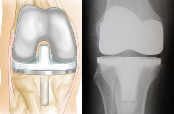


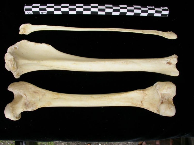


![Trucos para dormir rápido 🏰 | [Actualizado 2020] Casas Increíbles](https://casasincreibles.com/wp-content/uploads/2020/09/trucos-para-dormir-rapido.jpg)