Thyrotropin
Receptors, Thyrotropin
Thyroid Gland
Thyrotropin-Releasing Hormone
Thyroid Hormones
Thyroxine
Hypothyroidism
Hormones
Triiodothyronine
Hyperthyroidism
Follicle Stimulating Hormone
Thyrotropin Alfa
Luteinizing Hormone
Immunoglobulins, Thyroid-Stimulating
Graves Disease
Congenital Hypothyroidism
Thyrotropin, beta Subunit
Parathyroid Hormone
Thyroglobulin
Gonadotropin-Releasing Hormone
Radioimmunoassay
Gonadal Steroid Hormones
Pituitary Hormones, Anterior
Receptors, Thyroid Hormone
Pituitary Gland
Adrenocorticotropic Hormone
Human Growth Hormone
Prolactin
Pituitary Hormones
Triiodothyronine, Reverse
Iodide Peroxidase
Pituitary Gland, Anterior
Iodine
Cyclic AMP
Glycoprotein Hormones, alpha Subunit
Receptors, Thyrotropin-Releasing Hormone
Hormone Replacement Therapy
Goiter
Growth Hormone-Releasing Hormone
Propylthiouracil
Exophthalmos
Pituitary Neoplasms
Juvenile Hormones
Immunoassay
Thyrotoxicosis
Reagent Kits, Diagnostic
Antithyroid Agents
Thyroxine-Binding Proteins
Graves Ophthalmopathy
Cattle
Iodine Radioisotopes
Estradiol
Chorionic Gonadotropin
Harderian Gland
Corticotropin-Releasing Hormone
Methimazole
Pituitary Hormone-Releasing Hormones
Hypopituitarism
RNA, Messenger
Thyroiditis, Autoimmune
Hypothalamic Hormones
Carbimazole
Molecular Sequence Data
Peptide Hormones
Receptors, Cell Surface
Autoantibodies
Myxedema
Testosterone
Transcription Factor Pit-1
Anti-Mullerian Hormone
Gonadal Hormones
Hypophysectomy
Hydrocortisone
Thyroid Hormone Receptors beta
Adenylate Cyclase
Pyrrolidonecarboxylic Acid
Goiter, Nodular
Euthyroid Sick Syndromes
Thyroid Hormone Resistance Syndrome
Potassium Iodide
Progesterone
Thyroiditis
Gastrointestinal Hormones
Amino Acid Sequence
Hypothalamus
Pregnancy
Cells, Cultured
Bucladesine
Growth Hormone
Reference Values
Cell Membrane
Thyroid Hormone Receptors alpha
Base Sequence
Insect Hormones
Hormone Antagonists
Hypothalamo-Hypophyseal System
Invertebrate Hormones
Pituitary Hormones, Posterior
Gene Expression Regulation
Paper
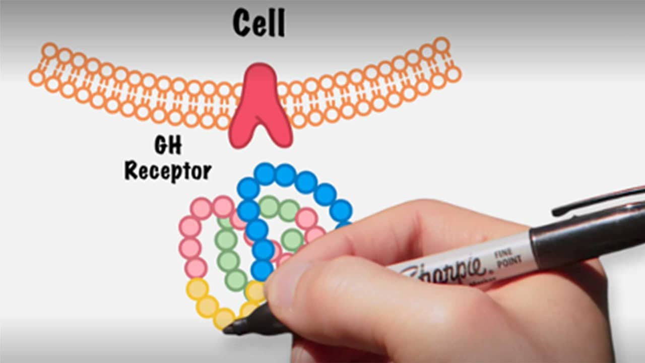
Receptors, Somatotropin
Somatostatin
Estrogens
CHO Cells
Receptors, FSH
Rats, Inbred Strains
Sodium Iodide
Insulin-Like Growth Factor I
Melanocyte-Stimulating Hormones
Dogs
Follicle Stimulating Hormone, beta Subunit
Signal Transduction
Cricetinae
Microchemistry
Thyroiditis, Subacute
Insulin
Receptor, Parathyroid Hormone, Type 1
Swine
Placental Hormones
Pancreatic Hormones
Dose-Response Relationship, Drug
Transfection
Pituitary Diseases
Ablation Techniques
Acromegaly
Methylthiouracil
Ovary
Receptors, Parathyroid Hormone
DNA
Perchlorates
Binding Sites
Colforsin
Long-Acting Thyroid Stimulator
Stimulation, Chemical
Immunoradiometric Assay
Peptide Fragments
Receptors, Gonadotropin
Fluoroimmunoassay
Luminescent Measurements
Receptors, LH
Binding, Competitive
Receptors, LHRH
Mutation
Central autonomic activation by intracisternal TRH analogue excites gastric splanchnic afferent neurons. (1/1011)
Intracisternal (ic) injection of thyrotropin-releasing hormone (TRH) or its stable analogue RX 77368 influences gastric function via stimulation of vagal muscarinic pathways. In rats, the increase in gastric mucosal blood flow evoked by a low ic dose of RX 77368 occurs via release of calcitonin gene-related peptide from capsaicin-sensitive afferent neurons, most probably of spinal origin. In this study, the effect of low ic doses of RX 77368 on afferent impulse activity in splanchnic single fibers was investigated. The cisterna magna of overnight-fasted, urethan-anesthetized Sprague-Dawley rats was acutely cannulated, and fine splanchnic nerve twigs containing at least one fiber responsive to mechanical probing of the stomach were isolated at a site immediately distal to the left suprarenal ganglion. Unit mechanoreceptive fields were encountered in all portions of the stomach, both superficially and in deeper layers. Splanchnic afferent unit impulse activity was recorded continuously during basal conditions and in response to consecutive ic injections of saline and RX 77368 (15-30 min later; 1.5 or 3 ng). Basal discharge rates ranged from 0 to 154 impulses/min (median = 10.2 impulses/min). A majority of splanchnic single units with ongoing activity increased their mean discharge rate by >/=20% after ic injection of RX 77368 at either 1.5 ng (6/10 units; median increase 63%) or 3 ng (19/24 units; median increase 175%). Five units lacking impulse activity in the 5-min before ic RX 77368 (3 ng) were also excited, with the onset of discharge occurring within 1.0-5.0 min postinjection. In units excited by ic RX 77368, peak discharge occurred 15.6 +/- 1.3 min after injection and was followed by a decline to stable activity levels +info)Secretagogue-induced exocytosis recruits G protein-gated K+ channels to plasma membrane in endocrine cells. (2/1011)
Stimulation-regulated fusion of vesicles to the plasma membrane is an essential step for hormone secretion but may also serve for the recruitment of functional proteins to the plasma membrane. While studying the distribution of G protein-gated K+ (KG) channels in the anterior pituitary lobe, we found KG channel subunits Kir3.1 and Kir3.4 localized on the membranes of intracellular dense core vesicles that contained thyrotropin. Stimulation of these thyrotroph cells with thyrotropin-releasing hormone provoked fusion of vesicles to the plasma membrane, increased expression of Kir3.1 and Kir3.4 subunits in the plasma membrane, and markedly enhanced KG currents stimulated by dopamine and somatostatin. These data indicate a novel mechanism for the rapid insertion of functional ion channels into the plasma membrane, which could form a new type of negative feedback control loop for hormone secretion in the endocrine system. (+info)High concentration of thyrotropin-releasing hormone in pancreatic islets. (3/1011)
The concentration of thyrotropin-releasing hormone (TRH, thyroliberin) in rat islets of Langerhans is 30-fold higher than in whole rat pancreas, indicating that the islets are the main source of pancreatic TRH. The TRH extracted from islets is indistinguishable from synthetic TRH in its immunological and biological properties and in its inactivation by human serum. The physiologic function of islet TRH is unknown. However, because TRH is antagonistic to somatostatin in other systems, and somatostatin also is concentrated in islets in high concentrations, it is possible that islet TRH may serve a similar antagonistic function in the regulation of islet cell secretory activity. (+info)Somatostatin inhibits release of thyrotropin releasing factor from organ cultures of rat hypothalamus. (4/1011)
Somatostatin in concentrations of 10(-6) to 10(-8) M inhibited basal release of thyrotropin releasing factor in organ culture of rat hypothalamus. Norepinephrine in doses of 10(-4)--10(-6) M induced release of thyrotropin releasing factor which increased progressively with time and was temperature and dose dependent. This enhanced thyrotropin-releasing-factor release was inhibited by somatostatin at 10(-6)--10(-8) M. (+info)Purification of rat prolactin releasing factor by gel filtration. (5/1011)
Pregnant rat hypothalamic fragments were extracted with 30 mM Tris-HCl buffer at pH 7.8, subjected to enzymatic digestion, and applied to gel filtration on Sephadex G-25 for purification of the prolactin releasing factor. Effect of the eluted fractions on the release of prolactin were tested by the determination of serum and pituitary prolactin after the injection in lactating rats. Prolactin was estimated by radioimmunoassay. One fraction (tube number 61--73) eluted later than synthetic ACTH enhanced release of prolactin 30 min after injection, but other fractions had no effect on the release of prolactin. Prolactin releasing factor would be quantitatively predominant over prolactin inhibiting factor in pregnant rat. (+info)Paradoxical GH response to TRH during status epilepticus in man. (6/1011)
Information on GH in relation to epilepsy is sparse, and to our knowledge there is no information on GH levels during status epilepticus in man. We studied GH in serum in six patients during status epilepticus, and in a control group of six seizure-free patients with epilepsy, before and after injection of TRH. The baseline GH values before TRH administration were within the normal range in all patients. After injection of TRH all patients with status epilepticus showed a paradoxical peak-shaped increase of GH to at least twice their baseline levels within 45 min after the injection (median basal GH value 1.5 mU/l and median peak GH value 6. 5 mU/l, mean increase 330%). No uniform reaction to TRH was observed in the control group (median basal GH value 2.7 U/l and median of the highest value within 45 min 5.2mU/l). A paradoxical peak reaction of GH to TRH was significantly more frequent in the status epilepticus group compared with the control group (P=0.008, Fisher exact probability test). TRH is not considered a GH-releasing hormone in humans during normal conditions, but a paradoxical response of GH to TRH, similar to that observed during status epilepticus, has been reported in various other pathological conditions, such as acromegaly, liver cirrhosis, mental depression and hypothyroidism. Our results of GH release after TRH administration in patients with status epilepticus suggest an altered regulation of GH as a result of the long-standing epileptic activity. (+info)Evaluation of hypothalamic-pituitary-adrenal axis in amenorrhoeic women with insulin-dependent diabetes. (7/1011)
Diabetes is associated with a higher incidence of secondary hypogonadotrophic amenorrhoea. In amenorrhoeic women with insulin-dependent diabetes a derangement in hypothalamic-pituitary-ovary axis has been proposed. No data exist on hypothalamic-pituitary-adrenal function in these women. Gonadotrophin releasing hormone (GnRH), corticotrophin releasing hormone (CRH), metoclopramide and thyroid releasing hormone (TRH) tests were performed in 15 diabetic women, eight amenorrhoeic (AD) and seven eumenorrhoeic (ED). Frequent blood samples were taken during 24 h to evaluate cortisol plasma concentrations. There were no differences between the groups in body mass index, duration of diabetes, insulin dose and metabolic control. The AD women had lower plasma concentrations of luteinizing hormone (LH), follicle stimulating hormone (FSH), prolactin, oestradiol, androstenedione and 17-hydroxyprogesterone (17-OHP) than the ED women. The responses of pituitary gonadotrophins to GnRH, and of thyroid stimulating hormone (TSH) to TRH, were similar in both groups. The AD women had a lower prolactin response to TRH and metoclopramide, and lower ACTH and cortisol responses to CRH, than the ED women. Mean cortisol concentrations > 24 h were higher in the amenorrhoeic group. Significant differences in cortisol concentrations from 2400 to 1000 h were found between the two groups. Insulin-dependent diabetes may involve mild chronic hypercortisolism which may affect metabolic control. Stress-induced activation of the hypothalamic-pituitary-adrenal axis would increase hypothalamic secretion of CRH. This would lead directly and perhaps also indirectly by increasing dopaminergic tonus to inhibition of GnRH secretion and hence hypogonadotrophic amenorrhoea. Amenorrhoea associated with metabolically controlled insulin-dependent diabetes is a form of functional hypothalamic amenorrhoea that requires pharmacological and psychological management. (+info)Intracisternal TRH analog increases gastrin release and corpus histidine decarboxylase activity in rats. (8/1011)
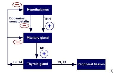
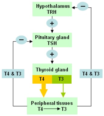
Thyrotropin-releasing hormone (TRH) acts in brain stem nuclei to induce vagally mediated stimulation of gastric secretion. The effects of intracisternal injection of the TRH analog RX-77368 on plasma gastrin levels and corpus histidine decarboxylase (HDC) activity were studied in 48-h fasted conscious rats. RX-77368 (25-100 ng) increased plasma gastrin levels by threefold at 30 min, which remained significantly higher than control at 2 and 4 h postinjection. Corpus HDC activity began to increase at 2 h and reached a peak at 4 h postinjection with a 21-fold maximum response observed at 50 ng. Morphological changes in the appearance of corpus HDC-immunoreactive cells correlated well with HDC activity. Pretreatment with gastrin monoclonal antibody completely prevented RX-77368 stimulatory effects on HDC activity. Atropine significantly attenuated gastrin increase at 30 min by 26%. These results indicated that in conscious fasted rats, TRH analog acts in the brain to increase corpus HDC activity in the enterochromaffin-like cells, which involves gastrin release stimulated by central TRH analog. (+info)Thyrotropin, also known as thyroid-stimulating hormone (TSH), is a hormone secreted by the anterior pituitary gland. Its primary function is to regulate the production and release of thyroxine (T4) and triiodothyronine (T3) hormones from the thyroid gland. Thyrotropin binds to receptors on the surface of thyroid follicular cells, stimulating the uptake of iodide and the synthesis and release of T4 and T3. The secretion of thyrotropin is controlled by the hypothalamic-pituitary-thyroid axis: thyrotropin-releasing hormone (TRH) from the hypothalamus stimulates the release of thyrotropin, while T3 and T4 inhibit its release through a negative feedback mechanism.
Thyrotropin receptors (TSHRs) are a type of G protein-coupled receptor found on the surface of cells in the thyroid gland. They bind to thyroid-stimulating hormone (TSH), which is produced and released by the pituitary gland. When TSH binds to the TSHR, it activates a series of intracellular signaling pathways that stimulate the production and release of thyroid hormones, triiodothyronine (T3) and thyroxine (T4). These hormones are important for regulating metabolism, growth, and development in the body. Mutations in the TSHR gene can lead to various thyroid disorders, such as hyperthyroidism or hypothyroidism.
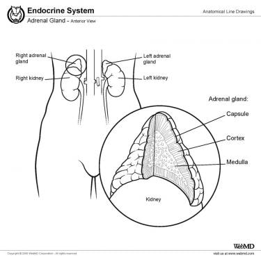
The thyroid gland is a major endocrine gland located in the neck, anterior to the trachea and extends from the lower third of the Adams apple to the suprasternal notch. It has two lateral lobes, connected by an isthmus, and sometimes a pyramidal lobe. This gland plays a crucial role in the metabolism, growth, and development of the human body through the production of thyroid hormones (triiodothyronine/T3 and thyroxine/T4) and calcitonin. The thyroid hormones regulate body temperature, heart rate, and the production of protein, while calcitonin helps in controlling calcium levels in the blood. The function of the thyroid gland is controlled by the hypothalamus and pituitary gland through the thyroid-stimulating hormone (TSH).
Thyrotropin-Releasing Hormone (TRH) is a tripeptide hormone that is produced and released by the hypothalamus in the brain. Its main function is to regulate the release of thyroid-stimulating hormone (TSH) from the anterior pituitary gland. TRH acts on the pituitary gland to stimulate the synthesis and secretion of TSH, which then stimulates the thyroid gland to produce and release thyroid hormones (triiodothyronine (T3) and thyroxine (T4)) into the bloodstream.
TRH is a tripeptide amino acid sequence with the structure of pGlu-His-Pro-NH2, and it is synthesized as a larger precursor molecule called preprothyrotropin-releasing hormone (preproTRH) in the hypothalamus. PreproTRH undergoes post-translational processing to produce TRH, which is then stored in secretory vesicles and released into the hypophyseal portal system, where it travels to the anterior pituitary gland and binds to TRH receptors on thyrotroph cells.
In addition to its role in regulating TSH release, TRH has been shown to have other physiological functions, including modulation of feeding behavior, body temperature, and neurotransmitter release. Dysregulation of the TRH-TSH axis can lead to various thyroid disorders, such as hypothyroidism or hyperthyroidism.
Thyroid hormones are hormones produced and released by the thyroid gland, a small endocrine gland located in the neck that helps regulate metabolism, growth, and development in the human body. The two main thyroid hormones are triiodothyronine (T3) and thyroxine (T4), which contain iodine atoms. These hormones play a crucial role in various bodily functions, including heart rate, body temperature, digestion, and brain development. They help regulate the rate at which your body uses energy, affects how sensitive your body is to other hormones, and plays a vital role in the development and differentiation of all cells of the human body. Thyroid hormone levels are regulated by the hypothalamus and pituitary gland through a feedback mechanism that helps maintain proper balance.
Thyroxine (T4) is a type of hormone produced and released by the thyroid gland, a small butterfly-shaped endocrine gland located in the front of your neck. It is one of two major hormones produced by the thyroid gland, with the other being triiodothyronine (T3).
Thyroxine plays a crucial role in regulating various metabolic processes in the body, including growth, development, and energy expenditure. Specifically, T4 helps to control the rate at which your body burns calories for energy, regulates protein, fat, and carbohydrate metabolism, and influences the body's sensitivity to other hormones.
T4 is produced by combining iodine and tyrosine, an amino acid found in many foods. Once produced, T4 circulates in the bloodstream and gets converted into its active form, T3, in various tissues throughout the body. Thyroxine has a longer half-life than T3, which means it remains active in the body for a more extended period.
Abnormal levels of thyroxine can lead to various medical conditions, such as hypothyroidism (underactive thyroid) or hyperthyroidism (overactive thyroid). These conditions can cause a range of symptoms, including weight gain or loss, fatigue, mood changes, and changes in heart rate and blood pressure.
Hypothyroidism is a medical condition where the thyroid gland, which is a small butterfly-shaped gland located in the front of your neck, does not produce enough thyroid hormones. This results in a slowing down of the body's metabolic processes, leading to various symptoms such as fatigue, weight gain, constipation, cold intolerance, dry skin, hair loss, muscle weakness, and depression.
The two main thyroid hormones produced by the thyroid gland are triiodothyronine (T3) and thyroxine (T4). These hormones play crucial roles in regulating various bodily functions, including heart rate, body temperature, and energy levels. In hypothyroidism, the production of these hormones is insufficient, leading to a range of symptoms that can affect multiple organ systems.
Hypothyroidism can be caused by several factors, including autoimmune disorders (such as Hashimoto's thyroiditis), surgical removal of the thyroid gland, radiation therapy for neck cancer, certain medications, and congenital defects. Hypothyroidism is typically diagnosed through blood tests that measure levels of TSH (thyroid-stimulating hormone), T3, and T4. Treatment usually involves taking synthetic thyroid hormones to replace the missing hormones and alleviate symptoms.
Hormones are defined as chemical messengers that are produced by endocrine glands or specialized cells and are transported through the bloodstream to tissues and organs, where they elicit specific responses. They play crucial roles in regulating various physiological processes such as growth, development, metabolism, reproduction, and mood. Examples of hormones include insulin, estrogen, testosterone, adrenaline, and thyroxine.
Triiodothyronine (T3) is a thyroid hormone, specifically the active form of thyroid hormone, that plays a critical role in the regulation of metabolism, growth, and development in the human body. It is produced by the thyroid gland through the iodination and coupling of the amino acid tyrosine with three atoms of iodine. T3 is more potent than its precursor, thyroxine (T4), which has four iodine atoms, as T3 binds more strongly to thyroid hormone receptors and accelerates metabolic processes at the cellular level.
In circulation, about 80% of T3 is bound to plasma proteins, while the remaining 20% is unbound or free, allowing it to enter cells and exert its biological effects. The primary functions of T3 include increasing the rate of metabolic reactions, promoting protein synthesis, enhancing sensitivity to catecholamines (e.g., adrenaline), and supporting normal brain development during fetal growth and early infancy. Imbalances in T3 levels can lead to various medical conditions, such as hypothyroidism or hyperthyroidism, which may require clinical intervention and management.
Hyperthyroidism is a medical condition characterized by an excessive production and release of thyroid hormones from the thyroid gland, leading to an increased metabolic rate in various body systems. The thyroid gland, located in the front of the neck, produces two main thyroid hormones: triiodothyronine (T3) and thyroxine (T4). These hormones play crucial roles in regulating many bodily functions, including heart rate, digestion, energy levels, and mood.
In hyperthyroidism, the elevated levels of T3 and T4 can cause a wide range of symptoms, such as rapid heartbeat, weight loss, heat intolerance, increased appetite, tremors, anxiety, and sleep disturbances. Some common causes of hyperthyroidism include Graves' disease, toxic adenoma, Plummer's disease (toxic multinodular goiter), and thyroiditis. Proper diagnosis and treatment are essential to manage the symptoms and prevent potential complications associated with this condition.
Follicle-Stimulating Hormone (FSH) is a glycoprotein hormone secreted and released by the anterior pituitary gland. In females, it promotes the growth and development of ovarian follicles in the ovary, which ultimately leads to the maturation and release of an egg (ovulation). In males, FSH stimulates the testes to produce sperm. It works in conjunction with luteinizing hormone (LH) to regulate reproductive processes. The secretion of FSH is controlled by the hypothalamic-pituitary-gonadal axis and its release is influenced by the levels of gonadotropin-releasing hormone (GnRH), estrogen, inhibin, and androgens.
Thyrotropin alfa is a recombinant form of human thyroid-stimulating hormone (TSH) used as a diagnostic aid in the investigation of reduced thyroid function (hypothyroidism). It is not to be confused with thyrotropin, which is the endogenous TSH produced by the pituitary gland. Thyrotropin alfa is used in tests to evaluate thyroid gland function and to help diagnose the cause of low thyroid hormone levels.
The medication is administered via subcutaneous injection, stimulating the thyroid gland to produce and release thyroid hormones T3 and T4. Blood samples are then taken at various intervals following the injection, allowing the measurement of T3 and T4 levels over time. This information can help differentiate between primary hypothyroidism (a problem with the thyroid gland itself) and secondary hypothyroidism (a problem with the pituitary gland or hypothalamus that affects TSH production).
It is important to note that thyrotropin alfa should only be used under the supervision of a healthcare professional, as inappropriate use can lead to hyperthyroidism and other related complications.
Luteinizing Hormone (LH) is a glycoprotein hormone, which is primarily produced and released by the anterior pituitary gland. In women, a surge of LH triggers ovulation, the release of an egg from the ovaries during the menstrual cycle. During pregnancy, LH stimulates the corpus luteum to produce progesterone. In men, LH stimulates the testes to produce testosterone. It plays a crucial role in sexual development, reproduction, and maintaining the reproductive system.
Immunoglobulins, Thyroid-Stimulating (TSI), are autoantibodies that bind to the thyroid-stimulating hormone receptor (TSHR) on the surface of thyroid cells. These antibodies mimic the action of TSH and stimulate the growth and function of the thyroid gland, leading to excessive production of thyroid hormones. This results in a condition known as Graves' disease, which is characterized by hyperthyroidism, goiter, and sometimes ophthalmopathy (eye problems). The presence and titer of TSIs are used in the diagnosis of Graves' disease.
Thyroid function tests (TFTs) are a group of blood tests that assess the functioning of the thyroid gland, which is a small butterfly-shaped gland located in the front of the neck. The thyroid gland produces hormones that regulate metabolism, growth, and development in the body.
TFTs typically include the following tests:
1. Thyroid-stimulating hormone (TSH) test: This test measures the level of TSH, a hormone produced by the pituitary gland that regulates the production of thyroid hormones. High levels of TSH may indicate an underactive thyroid gland (hypothyroidism), while low levels may indicate an overactive thyroid gland (hyperthyroidism).
2. Thyroxine (T4) test: This test measures the level of T4, a hormone produced by the thyroid gland. High levels of T4 may indicate hyperthyroidism, while low levels may indicate hypothyroidism.
3. Triiodothyronine (T3) test: This test measures the level of T3, another hormone produced by the thyroid gland. High levels of T3 may indicate hyperthyroidism, while low levels may indicate hypothyroidism.
4. Thyroid peroxidase antibody (TPOAb) test: This test measures the level of TPOAb, an antibody that attacks the thyroid gland and can cause hypothyroidism.
5. Thyroglobulin (Tg) test: This test measures the level of Tg, a protein produced by the thyroid gland. It is used to monitor the treatment of thyroid cancer.
These tests help diagnose and manage various thyroid disorders, including hypothyroidism, hyperthyroidism, thyroiditis, and thyroid cancer.
Graves' disease is defined as an autoimmune disorder that leads to overactivity of the thyroid gland (hyperthyroidism). It results when the immune system produces antibodies that stimulate the thyroid gland, causing it to produce too much thyroid hormone. This can result in a variety of symptoms such as rapid heartbeat, weight loss, heat intolerance, and bulging eyes (Graves' ophthalmopathy). The exact cause of Graves' disease is unknown, but it is more common in women and people with a family history of the disorder. Treatment may include medications to control hyperthyroidism, radioactive iodine therapy to destroy thyroid tissue, or surgery to remove the thyroid gland.
Congenital hypothyroidism is a medical condition characterized by the partial or complete absence of thyroid hormone production in the baby's body at birth. The thyroid gland, which is located in the front of the neck, produces hormones that are essential for normal growth and development of the brain and body.
Congenital hypothyroidism can occur due to various reasons such as the absence or abnormal development of the thyroid gland, or a defect in the production or regulation of thyroid hormones. In some cases, it may be caused by genetic mutations that affect the development or function of the thyroid gland.
If left untreated, congenital hypothyroidism can lead to mental and physical retardation, growth problems, and other health issues. Therefore, it is important to diagnose and treat this condition as early as possible, usually within the first few weeks of life. Treatment typically involves replacing the missing thyroid hormones with synthetic medications, which are safe and effective when administered under a doctor's supervision.
Thyrotropin, also known as thyroid-stimulating hormone (TSH), is a hormone produced and released by the anterior pituitary gland. It plays a crucial role in regulating the function of the thyroid gland by stimulating the production and release of thyroid hormones, triiodothyronine (T3) and thyroxine (T4).
The TSH molecule is composed of two subunits: alpha and beta. The alpha subunit is common to several pituitary hormones, including TSH, follicle-stimulating hormone (FSH), luteinizing hormone (LH), and human chorionic gonadotropin (hCG). In contrast, the beta subunit is unique to each hormone, determining its specific biological activity.
Therefore, 'Thyrotropin, beta Subunit' refers to the distinct portion of the TSH molecule that confers its thyroid-stimulating properties and allows it to be identified and measured separately from other pituitary hormones sharing the common alpha subunit. Beta-subunit assays are sometimes used in clinical settings to evaluate thyroid function, as they can provide information about TSH levels independent of the common alpha subunit.
Parathyroid hormone (PTH) is a polypeptide hormone that plays a crucial role in the regulation of calcium and phosphate levels in the body. It is produced and secreted by the parathyroid glands, which are four small endocrine glands located on the back surface of the thyroid gland.
The primary function of PTH is to maintain normal calcium levels in the blood by increasing calcium absorption from the gut, mobilizing calcium from bones, and decreasing calcium excretion by the kidneys. PTH also increases phosphate excretion by the kidneys, which helps to lower serum phosphate levels.
In addition to its role in calcium and phosphate homeostasis, PTH has been shown to have anabolic effects on bone tissue, stimulating bone formation and preventing bone loss. However, chronic elevations in PTH levels can lead to excessive bone resorption and osteoporosis.
Overall, Parathyroid Hormone is a critical hormone that helps maintain mineral homeostasis and supports healthy bone metabolism.
Thyroglobulin is a protein produced and used by the thyroid gland in the production of thyroid hormones, primarily thyroxine (T4) and triiodothyronine (T3). It is composed of two subunits, an alpha and a beta or gamma unit, which bind iodine atoms necessary for the synthesis of the thyroid hormones. Thyroglobulin is exclusively produced by the follicular cells of the thyroid gland.
In clinical practice, measuring thyroglobulin levels in the blood can be useful as a tumor marker for monitoring treatment and detecting recurrence of thyroid cancer, particularly in patients with differentiated thyroid cancer (papillary or follicular) who have had their thyroid gland removed. However, it is important to note that thyroglobulin is not specific to thyroid tissue and can be produced by some non-thyroidal cells under certain conditions, which may lead to false positive results in some cases.
Gonadotropin-Releasing Hormone (GnRH), also known as Luteinizing Hormone-Releasing Hormone (LHRH), is a hormonal peptide consisting of 10 amino acids. It is produced and released by the hypothalamus, an area in the brain that links the nervous system to the endocrine system via the pituitary gland.
GnRH plays a crucial role in regulating reproduction and sexual development through its control of two gonadotropins: follicle-stimulating hormone (FSH) and luteinizing hormone (LH). These gonadotropins, in turn, stimulate the gonads (ovaries or testes) to produce sex steroids and eggs or sperm.
GnRH acts on the anterior pituitary gland by binding to its specific receptors, leading to the release of FSH and LH. The hypothalamic-pituitary-gonadal axis is under negative feedback control, meaning that when sex steroid levels are high, they inhibit the release of GnRH, which subsequently decreases FSH and LH secretion.
GnRH agonists and antagonists have clinical applications in various medical conditions, such as infertility treatments, precocious puberty, endometriosis, uterine fibroids, prostate cancer, and hormone-responsive breast cancer.
Radioimmunoassay (RIA) is a highly sensitive analytical technique used in clinical and research laboratories to measure concentrations of various substances, such as hormones, vitamins, drugs, or tumor markers, in biological samples like blood, urine, or tissues. The method relies on the specific interaction between an antibody and its corresponding antigen, combined with the use of radioisotopes to quantify the amount of bound antigen.
In a typical RIA procedure, a known quantity of a radiolabeled antigen (also called tracer) is added to a sample containing an unknown concentration of the same unlabeled antigen. The mixture is then incubated with a specific antibody that binds to the antigen. During the incubation period, the antibody forms complexes with both the radiolabeled and unlabeled antigens.
After the incubation, the unbound (free) radiolabeled antigen is separated from the antibody-antigen complexes, usually through a precipitation or separation step involving centrifugation, filtration, or chromatography. The amount of radioactivity in the pellet (containing the antibody-antigen complexes) is then measured using a gamma counter or other suitable radiation detection device.
The concentration of the unlabeled antigen in the sample can be determined by comparing the ratio of bound to free radiolabeled antigen in the sample to a standard curve generated from known concentrations of unlabeled antigen and their corresponding bound/free ratios. The higher the concentration of unlabeled antigen in the sample, the lower the amount of radiolabeled antigen that will bind to the antibody, resulting in a lower bound/free ratio.
Radioimmunoassays offer high sensitivity, specificity, and accuracy, making them valuable tools for detecting and quantifying low levels of various substances in biological samples. However, due to concerns about radiation safety and waste disposal, alternative non-isotopic immunoassay techniques like enzyme-linked immunosorbent assays (ELISAs) have become more popular in recent years.
Gonadal steroid hormones, also known as gonadal sex steroids, are hormones that are produced and released by the gonads (i.e., ovaries in women and testes in men). These hormones play a critical role in the development and maintenance of secondary sexual characteristics, reproductive function, and overall health.
The three main classes of gonadal steroid hormones are:
1. Androgens: These are male sex hormones that are primarily produced by the testes but also produced in smaller amounts by the ovaries and adrenal glands. The most well-known androgen is testosterone, which plays a key role in the development of male secondary sexual characteristics such as facial hair, deepening of the voice, and increased muscle mass.
2. Estrogens: These are female sex hormones that are primarily produced by the ovaries but also produced in smaller amounts by the adrenal glands. The most well-known estrogen is estradiol, which plays a key role in the development of female secondary sexual characteristics such as breast development and the menstrual cycle.
3. Progestogens: These are hormones that are produced by the ovaries during the second half of the menstrual cycle and play a key role in preparing the uterus for pregnancy. The most well-known progestogen is progesterone, which also plays a role in maintaining pregnancy and regulating the menstrual cycle.
Gonadal steroid hormones can have significant effects on various physiological processes, including bone density, cognitive function, mood, and sexual behavior. Disorders of gonadal steroid hormone production or action can lead to a range of health problems, including infertility, osteoporosis, and sexual dysfunction.
Anterior pituitary hormones are a group of six major hormones that are produced and released by the anterior portion (lobe) of the pituitary gland, a small endocrine gland located at the base of the brain. These hormones play crucial roles in regulating various bodily functions and activities. The six main anterior pituitary hormones are:
1. Growth Hormone (GH): Also known as somatotropin, GH is essential for normal growth and development in children and adolescents. It helps regulate body composition, metabolism, and bone density in adults.
2. Prolactin (PRL): A hormone that stimulates milk production in females after childbirth and is also involved in various reproductive and immune functions in both sexes.
3. Follicle-Stimulating Hormone (FSH): FSH regulates the development, growth, and maturation of follicles in the ovaries (in females) and sperm production in the testes (in males).
4. Luteinizing Hormone (LH): LH plays a key role in triggering ovulation in females and stimulating testosterone production in males.
5. Thyroid-Stimulating Hormone (TSH): TSH regulates the function of the thyroid gland, which is responsible for producing and releasing thyroid hormones that control metabolism and growth.
6. Adrenocorticotropic Hormone (ACTH): ACTH stimulates the adrenal glands to produce cortisol, a steroid hormone involved in stress response, metabolism, and immune function.
These anterior pituitary hormones are regulated by the hypothalamus, which is located above the pituitary gland. The hypothalamus releases releasing and inhibiting factors that control the synthesis and secretion of anterior pituitary hormones, creating a complex feedback system to maintain homeostasis in the body.
Thyroid hormone receptors (THRs) are nuclear receptor proteins that bind to thyroid hormones, triiodothyronine (T3) and thyroxine (T4), and regulate gene transcription in target cells. These receptors play a crucial role in the development, growth, and metabolism of an organism by mediating the actions of thyroid hormones. THRs are encoded by genes THRA and THRB, which give rise to two major isoforms: TRα1 and TRβ1. Additionally, alternative splicing results in other isoforms with distinct tissue distributions and functions. THRs function as heterodimers with retinoid X receptors (RXRs) and bind to thyroid hormone response elements (TREs) in the regulatory regions of target genes. The binding of T3 or T4 to THRs triggers a conformational change, which leads to recruitment of coactivators or corepressors, ultimately resulting in activation or repression of gene transcription.
The pituitary gland is a small, endocrine gland located at the base of the brain, in the sella turcica of the sphenoid bone. It is often called the "master gland" because it controls other glands and makes the hormones that trigger many body functions. The pituitary gland measures about 0.5 cm in height and 1 cm in width, and it weighs approximately 0.5 grams.
The pituitary gland is divided into two main parts: the anterior lobe (adenohypophysis) and the posterior lobe (neurohypophysis). The anterior lobe is further divided into three zones: the pars distalis, pars intermedia, and pars tuberalis. Each part of the pituitary gland has distinct functions and produces different hormones.
The anterior pituitary gland produces and releases several important hormones, including:
* Growth hormone (GH), which regulates growth and development in children and helps maintain muscle mass and bone strength in adults.
* Thyroid-stimulating hormone (TSH), which controls the production of thyroid hormones by the thyroid gland.
* Adrenocorticotropic hormone (ACTH), which stimulates the adrenal glands to produce cortisol and other steroid hormones.
* Follicle-stimulating hormone (FSH) and luteinizing hormone (LH), which regulate reproductive function in both males and females.
* Prolactin, which stimulates milk production in pregnant and lactating women.
The posterior pituitary gland stores and releases two hormones that are produced by the hypothalamus:
* Antidiuretic hormone (ADH), which helps regulate water balance in the body by controlling urine production.
* Oxytocin, which stimulates uterine contractions during childbirth and milk release during breastfeeding.
Overall, the pituitary gland plays a critical role in maintaining homeostasis and regulating various bodily functions, including growth, development, metabolism, and reproductive function.
Thyroid diseases are a group of conditions that affect the function and structure of the thyroid gland, a small butterfly-shaped endocrine gland located in the base of the neck. The thyroid gland produces hormones that regulate many vital functions in the body, including metabolism, growth, and development.
Thyroid diseases can be classified into two main categories: hypothyroidism and hyperthyroidism. Hypothyroidism occurs when the thyroid gland does not produce enough hormones, leading to symptoms such as fatigue, weight gain, cold intolerance, constipation, and depression. Hyperthyroidism, on the other hand, occurs when the thyroid gland produces too much hormone, resulting in symptoms such as weight loss, heat intolerance, rapid heart rate, tremors, and anxiety.
Other common thyroid diseases include:
1. Goiter: an enlargement of the thyroid gland that can be caused by iodine deficiency or autoimmune disorders.
2. Thyroid nodules: abnormal growths on the thyroid gland that can be benign or malignant.
3. Thyroid cancer: a malignant tumor of the thyroid gland that requires medical treatment.
4. Hashimoto's disease: an autoimmune disorder that causes chronic inflammation of the thyroid gland, leading to hypothyroidism.
5. Graves' disease: an autoimmune disorder that causes hyperthyroidism and can also lead to eye problems and skin changes.
Thyroid diseases are diagnosed through a combination of physical examination, medical history, blood tests, and imaging studies such as ultrasound or CT scan. Treatment options depend on the specific type and severity of the disease and may include medication, surgery, or radioactive iodine therapy.
Adrenocorticotropic Hormone (ACTH) is a hormone produced and released by the anterior pituitary gland, a small endocrine gland located at the base of the brain. ACTH plays a crucial role in the regulation of the body's stress response and has significant effects on various physiological processes.
The primary function of ACTH is to stimulate the adrenal glands, which are triangular-shaped glands situated on top of the kidneys. The adrenal glands consist of two parts: the outer cortex and the inner medulla. ACTH specifically targets the adrenal cortex, where it binds to specific receptors and initiates a series of biochemical reactions leading to the production and release of steroid hormones, primarily cortisol (a glucocorticoid) and aldosterone (a mineralocorticoid).
Cortisol is involved in various metabolic processes, such as regulating blood sugar levels, modulating the immune response, and helping the body respond to stress. Aldosterone plays a vital role in maintaining electrolyte and fluid balance by promoting sodium reabsorption and potassium excretion in the kidneys.
ACTH release is controlled by the hypothalamus, another part of the brain, which produces corticotropin-releasing hormone (CRH). CRH stimulates the anterior pituitary gland to secrete ACTH, which in turn triggers cortisol production in the adrenal glands. This complex feedback system helps maintain homeostasis and ensures that appropriate amounts of cortisol are released in response to various physiological and psychological stressors.
Disorders related to ACTH can lead to hormonal imbalances, resulting in conditions such as Cushing's syndrome (excessive cortisol production) or Addison's disease (insufficient cortisol production). Proper diagnosis and management of these disorders typically involve assessing the function of the hypothalamic-pituitary-adrenal axis and addressing any underlying issues affecting ACTH secretion.
Human Growth Hormone (HGH), also known as somatotropin, is a peptide hormone produced in the pituitary gland. It plays a crucial role in human development and growth by stimulating the production of another hormone called insulin-like growth factor 1 (IGF-1). IGF-1 promotes the growth and reproduction of cells throughout the body, particularly in bones and other tissues. HGH also helps regulate body composition, body fluids, muscle and bone growth, sugar and fat metabolism, and possibly heart function. It is essential for human development and continues to have important effects throughout life. The secretion of HGH decreases with age, which is thought to contribute to the aging process.
Prolactin is a hormone produced by the pituitary gland, a small gland located at the base of the brain. Its primary function is to stimulate milk production in women after childbirth, a process known as lactation. However, prolactin also plays other roles in the body, including regulating immune responses, metabolism, and behavior. In men, prolactin helps maintain the sexual glands and contributes to paternal behaviors.
Prolactin levels are usually low in both men and non-pregnant women but increase significantly during pregnancy and after childbirth. Various factors can affect prolactin levels, including stress, sleep, exercise, and certain medications. High prolactin levels can lead to medical conditions such as amenorrhea (absence of menstruation), galactorrhea (spontaneous milk production not related to childbirth), infertility, and reduced sexual desire in both men and women.
Pituitary hormones are chemical messengers produced and released by the pituitary gland, a small endocrine gland located at the base of the brain. The pituitary gland is often referred to as the "master gland" because it controls several other endocrine glands and regulates various bodily functions.
There are two main types of pituitary hormones: anterior pituitary hormones and posterior pituitary hormones, which are produced in different parts of the pituitary gland and have distinct functions.
Anterior pituitary hormones include:
1. Growth hormone (GH): regulates growth and metabolism.
2. Thyroid-stimulating hormone (TSH): stimulates the thyroid gland to produce thyroid hormones.
3. Adrenocorticotropic hormone (ACTH): stimulates the adrenal glands to produce cortisol and other steroid hormones.
4. Follicle-stimulating hormone (FSH) and luteinizing hormone (LH): regulate reproductive function in both males and females.
5. Prolactin: stimulates milk production in lactating women.
6. Melanocyte-stimulating hormone (MSH): regulates skin pigmentation and appetite.
Posterior pituitary hormones include:
1. Oxytocin: stimulates uterine contractions during childbirth and milk ejection during lactation.
2. Vasopressin (antidiuretic hormone, ADH): regulates water balance in the body by controlling urine production in the kidneys.
Overall, pituitary hormones play crucial roles in regulating growth, development, metabolism, reproductive function, and various other bodily functions. Abnormalities in pituitary hormone levels can lead to a range of medical conditions, such as dwarfism, acromegaly, Cushing's disease, infertility, and diabetes insipidus.
Reverse Triiodothyronine (rT3) is a thyroid hormone that is chemically identical to triiodothyronine (T3), but has a reverse configuration at one end of the molecule. It is produced in smaller quantities compared to T3 and its function is not well understood. In some cases, increased levels of rT3 have been associated with decreased thyroid hormone action, such as in non-thyroidal illnesses or during calorie restriction. However, the clinical significance of rT3 levels remains a topic of ongoing research and debate.
Iodide peroxidase, also known as iodide:hydrogen peroxide oxidoreductase, is an enzyme that belongs to the family of oxidoreductases. Specifically, it is a peroxidase that uses iodide as its physiological reducing substrate. This enzyme catalyzes the oxidation of iodide by hydrogen peroxide to produce iodine, which plays a crucial role in thyroid hormone biosynthesis.
The systematic name for this enzyme is iodide:hydrogen-peroxide oxidoreductase (iodinating). It is most commonly found in the thyroid gland, where it helps to produce and regulate thyroid hormones by facilitating the iodination of tyrosine residues on thyroglobulin, a protein produced by the thyroid gland.
Iodide peroxidase requires a heme cofactor for its enzymatic activity, which is responsible for the oxidation-reduction reactions it catalyzes. The enzyme's ability to iodinate tyrosine residues on thyroglobulin is essential for the production of triiodothyronine (T3) and thyroxine (T4), two critical hormones that regulate metabolism, growth, and development in mammals.
The anterior pituitary, also known as the adenohypophysis, is the front portion of the pituitary gland. It is responsible for producing and secreting several important hormones that regulate various bodily functions. These hormones include:
* Growth hormone (GH), which stimulates growth and cell reproduction in bones and other tissues.
* Thyroid-stimulating hormone (TSH), which regulates the production of thyroid hormones by the thyroid gland.
* Adrenocorticotropic hormone (ACTH), which stimulates the adrenal glands to produce cortisol and other steroid hormones.
* Follicle-stimulating hormone (FSH) and luteinizing hormone (LH), which regulate reproductive function in both males and females by controlling the development and release of eggs or sperm.
* Prolactin, which stimulates milk production in pregnant and nursing women.
* Melanocyte-stimulating hormone (MSH), which regulates skin pigmentation and appetite.
The anterior pituitary gland is controlled by the hypothalamus, a small region of the brain located just above it. The hypothalamus produces releasing and inhibiting hormones that regulate the secretion of hormones from the anterior pituitary. These hormones are released into a network of blood vessels called the portal system, which carries them directly to the anterior pituitary gland.
Damage or disease of the anterior pituitary can lead to hormonal imbalances and various medical conditions, such as growth disorders, thyroid dysfunction, adrenal insufficiency, reproductive problems, and diabetes insipidus.
Iodine is an essential trace element that is necessary for the production of thyroid hormones in the body. These hormones play crucial roles in various bodily functions, including growth and development, metabolism, and brain development during pregnancy and infancy. Iodine can be found in various foods such as seaweed, dairy products, and iodized salt. In a medical context, iodine is also used as an antiseptic to disinfect surfaces, wounds, and skin infections due to its ability to kill bacteria, viruses, and fungi.
Cyclic adenosine monophosphate (cAMP) is a key secondary messenger in many biological processes, including the regulation of metabolism, gene expression, and cellular excitability. It is synthesized from adenosine triphosphate (ATP) by the enzyme adenylyl cyclase and is degraded by the enzyme phosphodiesterase.
In the body, cAMP plays a crucial role in mediating the effects of hormones and neurotransmitters on target cells. For example, when a hormone binds to its receptor on the surface of a cell, it can activate a G protein, which in turn activates adenylyl cyclase to produce cAMP. The increased levels of cAMP then activate various effector proteins, such as protein kinases, which go on to regulate various cellular processes.
Overall, the regulation of cAMP levels is critical for maintaining proper cellular function and homeostasis, and abnormalities in cAMP signaling have been implicated in a variety of diseases, including cancer, diabetes, and neurological disorders.
Glycoprotein hormones are a group of hormones that share a similar structure and are made up of four subunits: two identical alpha subunits and two distinct beta subunits. The alpha subunit is common to all glycoprotein hormones, including thyroid-stimulating hormone (TSH), follicle-stimulating hormone (FSH), luteinizing hormone (LH), and human chorionic gonadotropin (hCG).
The alpha subunit of glycoprotein hormones is a 92 amino acid polypeptide chain that contains several disulfide bonds, which help to stabilize its structure. It is heavily glycosylated, meaning that it contains many carbohydrate groups attached to the protein backbone. The alpha subunit plays an important role in the biological activity of the hormone by interacting with a specific receptor on the target cell surface.
The alpha subunit contains several regions that are important for its function, including a signal peptide, a variable region, and a conserved region. The signal peptide is a short sequence of amino acids at the N-terminus of the protein that directs it to the endoplasmic reticulum for processing and secretion. The variable region contains several amino acid residues that differ between different glycoprotein hormones, while the conserved region contains amino acids that are identical or very similar in all glycoprotein hormones.
Together with the beta subunit, the alpha subunit forms the functional hormone molecule. The beta subunit determines the specificity of the hormone for its target cells and regulates its biological activity.
Thyrotropin-releasing hormone (TRH) receptors are a type of G protein-coupled receptor found in the pituitary gland and other tissues throughout the body. TRH is a tripeptide hormone that plays a crucial role in regulating the release of thyroid-stimulating hormone (TSH) from the anterior pituitary gland.
TRH receptors are activated when TRH binds to them, which triggers a signaling cascade that ultimately leads to an increase in intracellular calcium and the release of TSH. In addition to regulating TSH secretion, TRH receptors have been found to play a role in various physiological processes, including feeding behavior, energy metabolism, and neuroprotection.
Abnormalities in TRH receptor function have been implicated in several endocrine disorders, such as thyroid dysfunction and obesity. Therefore, understanding the structure and function of TRH receptors is essential for developing new therapeutic strategies to treat these conditions.
Iodides are chemical compounds that contain iodine in the form of an iodide ion (I-). Iodide ions are negatively charged ions that consist of one iodine atom and an extra electron. Iodides are commonly found in dietary supplements and medications, and they are often used to treat or prevent iodine deficiency. They can also be used as expectorants to help thin and loosen mucus in the respiratory tract. Examples of iodides include potassium iodide (KI) and sodium iodide (NaI).
Hormone Replacement Therapy (HRT) is a medical treatment that involves the use of hormones to replace or supplement those that the body is no longer producing or no longer producing in sufficient quantities. It is most commonly used to help manage symptoms associated with menopause and conditions related to hormonal imbalances.
In women, HRT typically involves the use of estrogen and/or progesterone to alleviate hot flashes, night sweats, vaginal dryness, and mood changes that can occur during menopause. In some cases, testosterone may also be prescribed to help improve energy levels, sex drive, and overall sense of well-being.
In men, HRT is often used to treat low testosterone levels (hypogonadism) and related symptoms such as fatigue, decreased muscle mass, and reduced sex drive.
It's important to note that while HRT can be effective in managing certain symptoms, it also carries potential risks, including an increased risk of blood clots, stroke, breast cancer (in women), and cardiovascular disease. Therefore, the decision to undergo HRT should be made carefully and discussed thoroughly with a healthcare provider.
Goiter is a medical term that refers to an enlarged thyroid gland. The thyroid gland is a small, butterfly-shaped gland located in the front of your neck below the larynx or voice box. It produces hormones that regulate your body's metabolism, growth, and development.
Goiter can vary in size and may be visible as a swelling at the base of the neck. It can be caused by several factors, including iodine deficiency, autoimmune disorders, thyroid cancer, pregnancy, or the use of certain medications. Depending on the underlying cause and the severity of the goiter, treatment options may include medication, surgery, or radioactive iodine therapy.
Growth Hormone-Releasing Hormone (GHRH) is a hormone that is produced and released by the hypothalamus, a small gland located in the brain. Its primary function is to stimulate the anterior pituitary gland to release growth hormone (GH) into the bloodstream. GH plays a crucial role in growth and development, particularly during childhood and adolescence, by promoting the growth of bones and muscles.
GHRH is a 44-amino acid peptide that binds to specific receptors on the surface of pituitary cells, triggering a series of intracellular signals that ultimately lead to the release of GH. The production and release of GHRH are regulated by various factors, including sleep, stress, exercise, and nutrition.
Abnormalities in the production or function of GHRH can lead to growth disorders, such as dwarfism or gigantism, as well as other hormonal imbalances. Therefore, understanding the role of GHRH in regulating GH release is essential for diagnosing and treating these conditions.
Propylthiouracil is a medication that is primarily used to treat hyperthyroidism, a condition characterized by an overactive thyroid gland that produces too much thyroid hormone. The medication works by inhibiting the production of thyroid hormones in the body. It belongs to a class of drugs called antithyroid agents or thionamides.
In medical terms, propylthiouracil is defined as an antithyroid medication used to manage hyperthyroidism due to Graves' disease or toxic adenoma. It acts by inhibiting the synthesis of thyroid hormones, triiodothyronine (T3) and thyroxine (T4), in the thyroid gland. Propylthiouracil also reduces the peripheral conversion of T4 to T3. The medication is available as a tablet for oral administration and is typically prescribed at a starting dose of 100-150 mg three times daily, with adjustments made based on the patient's response and thyroid function tests.
It's important to note that propylthiouracil should be used under the close supervision of a healthcare provider due to potential side effects and risks associated with its use. Regular monitoring of thyroid function tests is necessary during treatment, and patients should promptly report any signs or symptoms of adverse reactions to their healthcare provider.
Exophthalmos is a medical condition that refers to the abnormal protrusion or bulging of one or both eyes beyond the normal orbit (eye socket). This condition is also known as proptosis. Exophthalmos can be caused by various factors, including thyroid eye disease (Graves' ophthalmopathy), tumors, inflammation, trauma, or congenital abnormalities. It can lead to various symptoms such as double vision, eye discomfort, redness, and difficulty closing the eyes. Treatment of exophthalmos depends on the underlying cause and may include medications, surgery, or radiation therapy.
Pituitary neoplasms refer to abnormal growths or tumors in the pituitary gland, a small endocrine gland located at the base of the brain. These neoplasms can be benign (non-cancerous) or malignant (cancerous), with most being benign. They can vary in size and may cause various symptoms depending on their location, size, and hormonal activity.
Pituitary neoplasms can produce and secrete excess hormones, leading to a variety of endocrine disorders such as Cushing's disease (caused by excessive ACTH production), acromegaly (caused by excessive GH production), or prolactinoma (caused by excessive PRL production). They can also cause local compression symptoms due to their size, leading to headaches, vision problems, and cranial nerve palsies.
The exact causes of pituitary neoplasms are not fully understood, but genetic factors, radiation exposure, and certain inherited conditions may increase the risk of developing these tumors. Treatment options for pituitary neoplasms include surgical removal, radiation therapy, and medical management with drugs that can help control hormonal imbalances.
Juvenile hormones (JHs) are a class of sesquiterpenoid compounds that play a crucial role in the regulation of insect development, reproduction, and other physiological processes. They are primarily produced by the corpora allata, a pair of endocrine glands located in the head of insects.
JHs are essential for maintaining the larval or nymphal stage of insects, preventing the expression of adult characteristics during molting. As the concentration of JH decreases in the hemolymph (insect blood), a molt to the next developmental stage occurs, and if the insect has reached its final instar, it will metamorphose into an adult.
In addition to their role in development, JHs also influence various aspects of insect reproductive physiology, such as vitellogenesis (yolk protein synthesis), oocyte maturation, and spermatogenesis. Furthermore, JHs have been implicated in regulating diapause (a period of suspended development during unfavorable environmental conditions) and caste determination in social insects like bees and ants.
Overall, juvenile hormones are vital regulators of growth, development, and reproduction in insects, making them attractive targets for the development of novel pest management strategies.
An immunoassay is a biochemical test that measures the presence or concentration of a specific protein, antibody, or antigen in a sample using the principles of antibody-antigen reactions. It is commonly used in clinical laboratories to diagnose and monitor various medical conditions such as infections, hormonal disorders, allergies, and cancer.
Immunoassays typically involve the use of labeled reagents, such as enzymes, radioisotopes, or fluorescent dyes, that bind specifically to the target molecule. The amount of label detected is proportional to the concentration of the target molecule in the sample, allowing for quantitative analysis.
There are several types of immunoassays, including enzyme-linked immunosorbent assay (ELISA), radioimmunoassay (RIA), fluorescence immunoassay (FIA), and chemiluminescent immunoassay (CLIA). Each type has its own advantages and limitations, depending on the sensitivity, specificity, and throughput required for a particular application.
Thyrotoxicosis is a medical condition that results from an excess of thyroid hormones in the body, leading to an overactive metabolic state. It can be caused by various factors such as Graves' disease, toxic adenoma, Plummer's disease, or excessive intake of thyroid hormone medication. Symptoms may include rapid heart rate, weight loss, heat intolerance, tremors, and increased sweating, among others. Thyrotoxicosis is not a diagnosis itself but a manifestation of various underlying thyroid disorders. Proper diagnosis and management are crucial to prevent complications and improve quality of life.
Reagent kits, diagnostic are prepackaged sets of chemical reagents and other components designed for performing specific diagnostic tests or assays. These kits are often used in clinical laboratories to detect and measure the presence or absence of various biomarkers, such as proteins, antibodies, antigens, nucleic acids, or small molecules, in biological samples like blood, urine, or tissues.
Diagnostic reagent kits typically contain detailed instructions for their use, along with the necessary reagents, controls, and sometimes specialized equipment or supplies. They are designed to simplify the testing process, reduce human error, and increase standardization, ensuring accurate and reliable results. Examples of diagnostic reagent kits include those used for pregnancy tests, infectious disease screening, drug testing, genetic testing, and cancer biomarker detection.
Antithyroid agents are a class of medications that are used to treat hyperthyroidism, a condition in which the thyroid gland produces too much thyroid hormone. These medications work by inhibiting the production of thyroid hormones in the thyroid gland. There are several types of antithyroid agents available, including:
1. Propylthiouracil (PTU): This medication works by blocking the enzyme that is needed to produce thyroid hormones. It also reduces the conversion of thyroxine (T4) to triiodothyronine (T3), another thyroid hormone, in peripheral tissues.
2. Methimazole: This medication works similarly to propylthiouracil by blocking the enzyme that is needed to produce thyroid hormones. However, it does not affect the conversion of T4 to T3 in peripheral tissues.
3. Carbimazole: This medication is converted to methimazole in the body and works similarly to block the production of thyroid hormones.
Antithyroid agents are usually taken orally, and their effects on thyroid hormone production begin within a few hours after ingestion. However, it may take several weeks for patients to notice an improvement in their symptoms. These medications can have side effects, including rash, hives, and joint pain. In rare cases, they can cause liver damage or agranulocytosis, a condition in which the body does not produce enough white blood cells.
It is important to note that antithyroid agents do not cure hyperthyroidism; they only treat the symptoms by reducing thyroid hormone production. Therefore, patients may need to take these medications for several months or even years, depending on their individual circumstances. In some cases, surgery or radioactive iodine therapy may be recommended as alternative treatments for hyperthyroidism.
Thyroxine-binding proteins (TBPs) are specialized transport proteins in the blood that bind and carry thyroid hormones, primarily Thyroxine (T4), but also Triiodothyronine (T3) to a lesser extent. The majority of T4 and T3 in the blood are bound to these proteins, while only a small fraction (0.03% of T4 and 0.3% of T3) remains unbound or free, which is the biologically active form that can enter cells and tissues to exert its physiological effects.
There are three main types of thyroxine-binding proteins:
1. Thyroxine-binding globulin (TBG): This is the major thyroid hormone transport protein, synthesized in the liver and accounting for approximately 70-80% of T4 and T3 binding. TBG has a high affinity but low capacity for thyroid hormones.
2. Transthyretin (TTR), also known as prealbumin: This protein accounts for around 10-20% of T4 and T3 binding. It has a lower affinity but higher capacity for thyroid hormones compared to TBG.
3. Albumin: This is the most abundant protein in the blood and binds approximately 15-20% of T4 and a smaller fraction of T3. Although albumin has a low affinity for thyroid hormones, its high concentration allows it to contribute significantly to their transport.
The binding of thyroid hormones to these proteins helps maintain stable levels in the blood and ensures a steady supply to tissues. Additionally, TBPs protect thyroid hormones from degradation and rapid clearance by the kidneys, thereby extending their half-life in the circulation.
Graves' ophthalmopathy, also known as Graves' eye disease or thyroid eye disease, is an autoimmune condition that affects the eyes. It often occurs in individuals with Graves' disease, an autoimmune disorder that causes hyperthyroidism (overactive thyroid gland). However, it can also occur in people without Graves' disease.
In Graves' ophthalmopathy, the immune system attacks the tissue behind the eyes, causing inflammation and enlargement of the muscles, fatty tissue, and connective tissue within the orbit (eye socket). This leads to symptoms such as:
1. Protrusion or bulging of the eyes (exophthalmos)
2. Redness and swelling of the eyelids
3. Double vision (diplopia) due to restricted eye movement
4. Pain and discomfort, especially when looking up, down, or sideways
5. Light sensitivity (photophobia)
6. Tearing and dryness in the eyes
7. Vision loss in severe cases
The treatment for Graves' ophthalmopathy depends on the severity of the symptoms and may include medications to manage inflammation, eye drops or ointments for dryness, prisms to correct double vision, or surgery for severe cases.
"Cattle" is a term used in the agricultural and veterinary fields to refer to domesticated animals of the genus *Bos*, primarily *Bos taurus* (European cattle) and *Bos indicus* (Zebu). These animals are often raised for meat, milk, leather, and labor. They are also known as bovines or cows (for females), bulls (intact males), and steers/bullocks (castrated males). However, in a strict medical definition, "cattle" does not apply to humans or other animals.
Thyroidectomy is a surgical procedure where all or part of the thyroid gland is removed. The thyroid gland is a butterfly-shaped endocrine gland located in the neck, responsible for producing hormones that regulate metabolism, growth, and development.
There are different types of thyroidectomy procedures, including:
1. Total thyroidectomy: Removal of the entire thyroid gland.
2. Partial (or subtotal) thyroidectomy: Removal of a portion of the thyroid gland.
3. Hemithyroidectomy: Removal of one lobe of the thyroid gland, often performed to treat benign solitary nodules or differentiated thyroid cancer.
Thyroidectomy may be recommended for various reasons, such as treating thyroid nodules, goiter, hyperthyroidism (overactive thyroid), or thyroid cancer. Potential risks and complications of the procedure include bleeding, infection, damage to nearby structures like the parathyroid glands and recurrent laryngeal nerve, and hypoparathyroidism or hypothyroidism due to removal of or damage to the parathyroid glands or thyroid gland, respectively. Close postoperative monitoring and management are essential to minimize these risks and ensure optimal patient outcomes.
Iodine radioisotopes are radioactive isotopes of the element iodine, which decays and emits radiation in the form of gamma rays. Some commonly used iodine radioisotopes include I-123, I-125, I-131. These radioisotopes have various medical applications such as in diagnostic imaging, therapy for thyroid disorders, and cancer treatment.
For example, I-131 is commonly used to treat hyperthyroidism and differentiated thyroid cancer due to its ability to destroy thyroid tissue. On the other hand, I-123 is often used in nuclear medicine scans of the thyroid gland because it emits gamma rays that can be detected by a gamma camera, allowing for detailed images of the gland's structure and function.
It is important to note that handling and administering radioisotopes require specialized training and safety precautions due to their radiation-emitting properties.
Estradiol is a type of estrogen, which is a female sex hormone. It is the most potent and dominant form of estrogen in humans. Estradiol plays a crucial role in the development and maintenance of secondary sexual characteristics in women, such as breast development and regulation of the menstrual cycle. It also helps maintain bone density, protect the lining of the uterus, and is involved in cognition and mood regulation.
Estradiol is produced primarily by the ovaries, but it can also be synthesized in smaller amounts by the adrenal glands and fat cells. In men, estradiol is produced from testosterone through a process called aromatization. Abnormal levels of estradiol can contribute to various health issues, such as hormonal imbalances, infertility, osteoporosis, and certain types of cancer.
Chorionic Gonadotropin (hCG) is a hormone that is produced during pregnancy. It is produced by the placenta after implantation of the fertilized egg in the uterus. The main function of hCG is to prevent the disintegration of the corpus luteum, which is a temporary endocrine structure that forms in the ovary after ovulation and produces progesterone during early pregnancy. Progesterone is essential for maintaining the lining of the uterus and supporting the pregnancy.
hCG can be detected in the blood or urine as early as 10 days after conception, and its levels continue to rise throughout the first trimester of pregnancy. In addition to its role in maintaining pregnancy, hCG is also used as a clinical marker for pregnancy and to monitor certain medical conditions such as gestational trophoblastic diseases.
Thyroid neoplasms refer to abnormal growths or tumors in the thyroid gland, which can be benign (non-cancerous) or malignant (cancerous). These growths can vary in size and may cause a noticeable lump or nodule in the neck. Thyroid neoplasms can also affect the function of the thyroid gland, leading to hormonal imbalances and related symptoms. The exact causes of thyroid neoplasms are not fully understood, but risk factors include radiation exposure, family history, and certain genetic conditions. It is important to note that most thyroid nodules are benign, but a proper medical evaluation is necessary to determine the nature of the growth and develop an appropriate treatment plan.
The Harderian gland is a specialized exocrine gland located in many vertebrate species, including birds and mammals. In humans, it is rudimentary and not fully developed. However, in other animals like rodents, lagomorphs (rabbits and hares), and some reptiles, this gland plays a significant role.
The Harderian gland is primarily responsible for producing and secreting lipids, which help to lubricate the eye's surface and the nictitating membrane (third eyelid). This lubrication ensures that the eyes remain moist and protected from dryness and external irritants. Additionally, the secretions of the Harderian gland contain immunoglobulins, which contribute to the animal's immune defense system by providing protection against pathogens.
In some animals, the Harderian gland also has a role in pheromone production and communication. The study and understanding of this gland are particularly important in toxicological research, as it is often used as an indicator of environmental pollutant exposure and their effects on wildlife.
Corticotropin-Releasing Hormone (CRH) is a hormone that is produced and released by the hypothalamus, a small gland located in the brain. CRH plays a critical role in the body's stress response system.
When the body experiences stress, the hypothalamus releases CRH, which then travels to the pituitary gland, another small gland located at the base of the brain. Once there, CRH stimulates the release of adrenocorticotropic hormone (ACTH) from the pituitary gland.
ACTH then travels through the bloodstream to the adrenal glands, which are located on top of the kidneys. ACTH stimulates the adrenal glands to produce and release cortisol, a hormone that helps the body respond to stress by regulating metabolism, immune function, and blood pressure, among other things.
Overall, CRH is an important part of the hypothalamic-pituitary-adrenal (HPA) axis, which regulates many bodily functions related to stress response, mood, and cognition. Dysregulation of the HPA axis and abnormal levels of CRH have been implicated in various psychiatric and medical conditions, including depression, anxiety disorders, post-traumatic stress disorder (PTSD), and Cushing's syndrome.
Methimazole is an anti-thyroid medication that is primarily used to treat hyperthyroidism, a condition in which the thyroid gland produces excessive amounts of thyroid hormones. It works by inhibiting the enzyme thyroperoxidase, which is essential for the production of thyroid hormones. By blocking this enzyme, methimazole reduces the amount of thyroid hormones produced by the thyroid gland, helping to restore normal thyroid function.
Methimazole is available in oral tablet form and is typically taken two to three times a day. Common side effects of methimazole include nausea, vomiting, skin rashes, and joint pain. In rare cases, it can cause more serious side effects such as liver damage or agranulocytosis (a severe decrease in white blood cell count).
It is important to note that methimazole should only be used under the close supervision of a healthcare provider, as regular monitoring of thyroid function and potential side effects is necessary. Additionally, it may take several weeks or months of treatment with methimazole before thyroid function returns to normal.
Pituitary hormone-releasing hormones (PRHs), also known as hypothalamic releasing hormones or hypothalamic hormones, are small neuropeptides produced and released by the hypothalamus - a small region of the brain. These hormones play crucial roles in regulating the secretion and release of various pituitary hormones, which in turn control several essential bodily functions, including growth, development, metabolism, stress response, reproduction, and lactation.
There are several PRHs, each with a specific target pituitary hormone:
1. Thyrotropin-releasing hormone (TRH): Stimulates the release of thyroid-stimulating hormone (TSH) from the anterior pituitary gland, which then promotes the production and release of thyroid hormones.
2. Gonadotropin-releasing hormone (GnRH): Regulates the secretion of follicle-stimulating hormone (FSH) and luteinizing hormone (LH) from the anterior pituitary gland, which are essential for reproductive functions.
3. Corticotropin-releasing hormone (CRH): Stimulates the release of adrenocorticotropic hormone (ACTH) from the anterior pituitary gland, which then promotes the production and release of cortisol and other glucocorticoids from the adrenal glands.
4. Growth hormone-releasing hormone (GHRH): Stimulates the release of growth hormone (GH) from the anterior pituitary gland, which is essential for growth, development, and metabolism regulation.
5. Somatostatin or growth hormone-inhibiting hormone (GHIH): Inhibits the release of GH from the anterior pituitary gland and also suppresses the secretion of thyroid hormones.
6. Prolactin-releasing hormone (PRH) or prolactin-releasing factor (PRF): Stimulates the release of prolactin from the anterior pituitary gland, which is essential for lactation and reproductive functions.
7. Prolactin-inhibiting hormone (PIH) or dopamine: Inhibits the release of prolactin from the anterior pituitary gland.
These releasing hormones and inhibitory hormones work together to maintain a delicate balance in various physiological processes, including growth, development, metabolism, stress response, and reproductive functions. Dysregulation of these hormonal systems can lead to various endocrine disorders and diseases.
Hypopituitarism is a medical condition characterized by deficient secretion of one or more hormones produced by the pituitary gland, a small endocrine gland located at the base of the brain. The pituitary gland controls several other endocrine glands in the body, including the thyroid, adrenals, and sex glands (ovaries and testes).
Hypopituitarism can result from damage to the pituitary gland due to various causes such as tumors, surgery, radiation therapy, trauma, or inflammation. In some cases, hypopituitarism may also be caused by a dysfunction of the hypothalamus, a region in the brain that regulates the pituitary gland's function.
The symptoms and signs of hypopituitarism depend on which hormones are deficient and can include fatigue, weakness, decreased appetite, weight loss, low blood pressure, decreased sex drive, infertility, irregular menstrual periods, intolerance to cold, constipation, thinning hair, dry skin, and depression.
Treatment of hypopituitarism typically involves hormone replacement therapy to restore the deficient hormones' normal levels. The type and dosage of hormones used will depend on which hormones are deficient and may require regular monitoring and adjustments over time.
Messenger RNA (mRNA) is a type of RNA (ribonucleic acid) that carries genetic information copied from DNA in the form of a series of three-base code "words," each of which specifies a particular amino acid. This information is used by the cell's machinery to construct proteins, a process known as translation. After being transcribed from DNA, mRNA travels out of the nucleus to the ribosomes in the cytoplasm where protein synthesis occurs. Once the protein has been synthesized, the mRNA may be degraded and recycled. Post-transcriptional modifications can also occur to mRNA, such as alternative splicing and addition of a 5' cap and a poly(A) tail, which can affect its stability, localization, and translation efficiency.
Autoimmune thyroiditis, also known as Hashimoto's disease, is a chronic inflammation of the thyroid gland caused by an autoimmune response. In this condition, the immune system produces antibodies that attack and damage the thyroid gland, leading to hypothyroidism (underactive thyroid). The thyroid gland may become enlarged (goiter), and symptoms can include fatigue, weight gain, cold intolerance, constipation, dry skin, and depression. Autoimmune thyroiditis is more common in women than men and tends to run in families. It is often associated with other autoimmune disorders such as rheumatoid arthritis, Addison's disease, and type 1 diabetes. The diagnosis is typically made through blood tests that measure levels of thyroid hormones and antibodies. Treatment usually involves thyroid hormone replacement therapy to manage the symptoms of hypothyroidism.
Hypothalamic hormones are a group of hormones that are produced and released by the hypothalamus, a small region at the base of the brain. These hormones play a crucial role in regulating various bodily functions, including temperature, hunger, thirst, sleep, and emotional behavior.
The hypothalamus produces two main types of hormones: releasing hormones and inhibiting hormones. Releasing hormones stimulate the pituitary gland to release its own hormones, while inhibiting hormones prevent the pituitary gland from releasing hormones.
Some examples of hypothalamic hormones include:
* Thyroid-releasing hormone (TRH), which stimulates the release of thyroid-stimulating hormone (TSH) from the pituitary gland.
* Growth hormone-releasing hormone (GHRH) and somatostatin, which regulate the release of growth hormone (GH) from the pituitary gland.
* Gonadotropin-releasing hormone (GnRH), which stimulates the release of follicle-stimulating hormone (FSH) and luteinizing hormone (LH) from the pituitary gland, which in turn regulate reproductive function.
* Corticotropin-releasing hormone (CRH), which stimulates the release of adrenocorticotropic hormone (ACTH) from the pituitary gland, which regulates the stress response.
* Prolactin-inhibiting hormone (PIH) and prolactin-releasing hormone (PRH), which regulate the release of prolactin from the pituitary gland, which is involved in lactation.
Overall, hypothalamic hormones play a critical role in maintaining homeostasis in the body by regulating various physiological processes.
Carbimazole is an antithyroid medication that is primarily used to manage hyperthyroidism, a condition characterized by an overactive thyroid gland that produces excessive amounts of thyroid hormones. The drug works by inhibiting the enzyme responsible for producing these hormones, thereby reducing their levels in the body and alleviating symptoms associated with the disorder.
Hyperthyroidism can manifest as various signs and symptoms, including rapid heartbeat, weight loss, heat intolerance, tremors, anxiety, and sleep disturbances. Common causes of hyperthyroidism include Graves' disease, toxic adenoma, and thyroiditis.
Carbimazole is a prodrug that gets converted to its active metabolite, methimazole, in the liver. Methimazole inhibits the activity of thyroperoxidase, an enzyme involved in the synthesis of thyroid hormones triiodothyronine (T3) and thyroxine (T4). By blocking this enzyme, carbimazole reduces the production of T3 and T4, ultimately helping to control hyperthyroidism.
The medication is typically administered orally in tablet form, with dosages varying depending on individual patient needs and response to treatment. Common side effects of carbimazole include gastrointestinal disturbances such as nausea, vomiting, and diarrhea. Rare but severe adverse reactions may include agranulocytosis (a severe decrease in white blood cells), aplastic anemia (a condition where the bone marrow fails to produce sufficient numbers of blood cells), and hepatotoxicity (liver damage).
Patients taking carbimazole should be closely monitored for signs of adverse reactions, and regular blood tests are necessary to assess thyroid hormone levels and potential side effects. Pregnant women should avoid using carbimazole due to the risk of birth defects in the developing fetus. In such cases, alternative antithyroid medications like propylthiouracil may be prescribed instead.
In summary, carbimazole is an antithyroid medication used primarily for managing hyperthyroidism by inhibiting thyroperoxidase and reducing the production of thyroid hormones T3 and T4. While effective, it carries potential risks and side effects that necessitate close monitoring during treatment.
Molecular sequence data refers to the specific arrangement of molecules, most commonly nucleotides in DNA or RNA, or amino acids in proteins, that make up a biological macromolecule. This data is generated through laboratory techniques such as sequencing, and provides information about the exact order of the constituent molecules. This data is crucial in various fields of biology, including genetics, evolution, and molecular biology, allowing for comparisons between different organisms, identification of genetic variations, and studies of gene function and regulation.
Peptide hormones are a type of hormone consisting of short chains of amino acids known as peptides. They are produced and released by various endocrine glands and play crucial roles in regulating many physiological processes in the body, including growth and development, metabolism, stress response, and reproductive functions.
Peptide hormones exert their effects by binding to specific receptors on the surface of target cells, which triggers a series of intracellular signaling events that ultimately lead to changes in cell behavior or function. Some examples of peptide hormones include insulin, glucagon, growth hormone, prolactin, oxytocin, and vasopressin.
Peptide hormones are synthesized as larger precursor proteins called prohormones, which are cleaved by enzymes to release the active peptide hormone. They are water-soluble and cannot pass through the cell membrane, so they exert their effects through autocrine, paracrine, or endocrine mechanisms. Autocrine signaling occurs when a cell releases a hormone that binds to receptors on the same cell, while paracrine signaling involves the release of a hormone that acts on nearby cells. Endocrine signaling, on the other hand, involves the release of a hormone into the bloodstream, which then travels to distant target cells to exert its effects.
Cell surface receptors, also known as membrane receptors, are proteins located on the cell membrane that bind to specific molecules outside the cell, known as ligands. These receptors play a crucial role in signal transduction, which is the process of converting an extracellular signal into an intracellular response.
Cell surface receptors can be classified into several categories based on their structure and mechanism of action, including:
1. Ion channel receptors: These receptors contain a pore that opens to allow ions to flow across the cell membrane when they bind to their ligands. This ion flux can directly activate or inhibit various cellular processes.
2. G protein-coupled receptors (GPCRs): These receptors consist of seven transmembrane domains and are associated with heterotrimeric G proteins that modulate intracellular signaling pathways upon ligand binding.
3. Enzyme-linked receptors: These receptors possess an intrinsic enzymatic activity or are linked to an enzyme, which becomes activated when the receptor binds to its ligand. This activation can lead to the initiation of various signaling cascades within the cell.
4. Receptor tyrosine kinases (RTKs): These receptors contain intracellular tyrosine kinase domains that become activated upon ligand binding, leading to the phosphorylation and activation of downstream signaling molecules.
5. Integrins: These receptors are transmembrane proteins that mediate cell-cell or cell-matrix interactions by binding to extracellular matrix proteins or counter-receptors on adjacent cells. They play essential roles in cell adhesion, migration, and survival.
Cell surface receptors are involved in various physiological processes, including neurotransmission, hormone signaling, immune response, and cell growth and differentiation. Dysregulation of these receptors can contribute to the development of numerous diseases, such as cancer, diabetes, and neurological disorders.
Autoantibodies are defined as antibodies that are produced by the immune system and target the body's own cells, tissues, or organs. These antibodies mistakenly identify certain proteins or molecules in the body as foreign invaders and attack them, leading to an autoimmune response. Autoantibodies can be found in various autoimmune diseases such as rheumatoid arthritis, lupus, and thyroiditis. The presence of autoantibodies can also be used as a diagnostic marker for certain conditions.
Myxedema is not a term used in modern medicine to describe a specific medical condition. However, historically, it was used to refer to the severe form of hypothyroidism, a condition characterized by an underactive thyroid gland that doesn't produce enough thyroid hormones. In hypothyroidism, various body functions slow down, which can lead to symptoms such as fatigue, weight gain, cold intolerance, constipation, and dry skin.
Myxedema specifically refers to the physical signs of severe hypothyroidism, including swelling (edema) and thickening of the skin, particularly around the face, hands, and feet, as well as a puffy appearance of the face. The term myxedema coma was used to describe a rare but life-threatening complication of long-standing, untreated hypothyroidism, characterized by altered mental status, hypothermia, and other systemic manifestations.
Nowadays, healthcare professionals use more precise medical terminology to describe these conditions, such as hypothyroidism or myxedematous edema, rather than the outdated term myxedema.
Testosterone is a steroid hormone that belongs to androsten class of hormones. It is primarily secreted by the Leydig cells in the testes of males and, to a lesser extent, by the ovaries and adrenal glands in females. Testosterone is the main male sex hormone and anabolic steroid. It plays a key role in the development of masculine characteristics, such as body hair and muscle mass, and contributes to bone density, fat distribution, red cell production, and sex drive. In females, testosterone contributes to sexual desire and bone health. Testosterone is synthesized from cholesterol and its production is regulated by luteinizing hormone (LH) and follicle-stimulating hormone (FSH).
Transcription Factor Pit-1, also known as POU1F1 or pituitary-specific transcription factor 1, is a protein that plays a crucial role in the development and function of the anterior pituitary gland. It is a member of the POU domain family of transcription factors, which are characterized by a conserved DNA-binding domain.
Pit-1 is essential for the differentiation and proliferation of certain types of pituitary cells, including those that produce growth hormone (GH), prolactin (PRL), and thyroid-stimulating hormone (TSH). Pit-1 binds to specific DNA sequences in the promoter regions of these hormone genes, thereby activating their transcription and promoting hormone production.
Mutations in the gene encoding Pit-1 can lead to a variety of pituitary disorders, such as dwarfism due to GH deficiency, delayed puberty, and hypothyroidism due to TSH deficiency. Additionally, some studies have suggested that Pit-1 may also play a role in regulating energy balance and body weight, although the exact mechanisms are not fully understood.
Anti-Mullerian Hormone (AMH) is a glycoprotein hormone that belongs to the transforming growth factor-beta (TGF-β) family. It is primarily produced by the granulosa cells of developing follicles in the ovaries of females. AMH plays an essential role in female reproductive physiology, as it inhibits the recruitment and further development of primordial follicles, thereby regulating the size of the primordial follicle pool and the onset of puberty.
AMH levels are often used as a biomarker for ovarian reserve assessment in women. High AMH levels indicate a larger ovarian reserve, while low levels suggest a decreased reserve, which may be associated with reduced fertility or an earlier onset of menopause. Additionally, measuring AMH levels can help predict the response to ovarian stimulation during assisted reproductive technologies (ART) such as in vitro fertilization (IVF).
Gonadal hormones, also known as sex hormones, are steroid hormones that are primarily produced by the gonads (ovaries in females and testes in males). They play crucial roles in the development and regulation of sexual characteristics and reproductive functions. The three main types of gonadal hormones are:
1. Estrogens - predominantly produced by ovaries, they are essential for female sexual development and reproduction. The most common estrogen is estradiol, which supports the growth and maintenance of secondary sexual characteristics in women, such as breast development and wider hips. Estrogens also play a role in regulating the menstrual cycle and maintaining bone health.
2. Progesterone - primarily produced by ovaries during the menstrual cycle and pregnancy, progesterone prepares the uterus for implantation of a fertilized egg and supports the growth and development of the fetus during pregnancy. It also plays a role in regulating the menstrual cycle.
3. Androgens - produced by both ovaries and testes, but primarily by testes in males. The most common androgen is testosterone, which is essential for male sexual development and reproduction. Testosterone supports the growth and maintenance of secondary sexual characteristics in men, such as facial hair, a deeper voice, and increased muscle mass. It also plays a role in regulating sex drive (libido) and bone health in both males and females.
In summary, gonadal hormones are steroid hormones produced by the gonads that play essential roles in sexual development, reproduction, and maintaining secondary sexual characteristics.
Hypophysectomy is a surgical procedure that involves the removal or partial removal of the pituitary gland, also known as the hypophysis. The pituitary gland is a small endocrine gland located at the base of the brain, just above the nasal cavity, and is responsible for producing and secreting several important hormones that regulate various bodily functions.
Hypophysectomy may be performed for therapeutic or diagnostic purposes. In some cases, it may be used to treat pituitary tumors or other conditions that affect the function of the pituitary gland. It may also be performed as a research procedure in animal models to study the effects of pituitary hormone deficiency on various physiological processes.
The surgical approach for hypophysectomy may vary depending on the specific indication and the patient's individual anatomy. In general, however, the procedure involves making an incision in the skull and exposing the pituitary gland through a small opening in the bone. The gland is then carefully dissected and removed or partially removed as necessary.
Potential complications of hypophysectomy include damage to surrounding structures such as the optic nerves, which can lead to vision loss, and cerebrospinal fluid leaks. Additionally, removal of the pituitary gland can result in hormonal imbalances that may require long-term management with hormone replacement therapy.
Hydrocortisone is a synthetic glucocorticoid, which is a class of steroid hormones. It is identical to the naturally occurring cortisol, a hormone produced by the adrenal gland that helps regulate metabolism and helps your body respond to stress. Hydrocortisone has anti-inflammatory effects and is used to treat various inflammatory conditions such as allergies, skin disorders, and autoimmune diseases. It works by suppressing the immune system's response to reduce swelling, redness, itching, and other symptoms caused by inflammation.
Hydrocortisone is available in different forms, including oral tablets, topical creams, lotions, gels, and ointments, as well as injectable solutions. The specific use and dosage depend on the condition being treated and the individual patient's medical history and current health status.
As with any medication, hydrocortisone can have side effects, especially when used in high doses or for extended periods. Common side effects include increased appetite, weight gain, mood changes, insomnia, and skin thinning. Long-term use of hydrocortisone may also increase the risk of developing osteoporosis, diabetes, cataracts, and other health problems. Therefore, it is essential to follow your healthcare provider's instructions carefully when using this medication.
Thyroid hormone receptors (THRs) are nuclear receptor proteins that bind to thyroid hormones and mediate their effects in target cells. There are two main types of THRs, referred to as THR alpha and THR beta. THR beta is further divided into two subtypes, THR beta1 and THR beta2.
THR beta is a type of nuclear receptor that is primarily expressed in the liver, kidney, and heart, as well as in the central nervous system. It plays an important role in regulating the metabolism of carbohydrates, lipids, and proteins, as well as in the development and function of the heart. THR beta is also involved in the regulation of body weight and energy expenditure.
THR beta1 is the predominant subtype expressed in the liver and is responsible for many of the metabolic effects of thyroid hormones in this organ. THR beta2, on the other hand, is primarily expressed in the heart and plays a role in regulating cardiac function.
Abnormalities in THR beta function can lead to various diseases, including thyroid hormone resistance, a condition in which the body's cells are unable to respond properly to thyroid hormones. This can result in symptoms such as weight gain, fatigue, and cold intolerance.
Adenylate cyclase is an enzyme that catalyzes the conversion of adenosine triphosphate (ATP) to cyclic adenosine monophosphate (cAMP). It plays a crucial role in various cellular processes, including signal transduction and metabolism. Adenylate cyclase is activated by hormones and neurotransmitters that bind to G-protein-coupled receptors on the cell membrane, leading to the production of cAMP, which then acts as a second messenger to regulate various intracellular responses. There are several isoforms of adenylate cyclase, each with distinct regulatory properties and subcellular localization.
Pyrrolidonecarboxylic acid, also known as Proline or Prolinic acid, is an organic compound with the formula N-pyrrolidinecarboxylic acid. It is a cyclic amino acid, which means that its side chain is bonded to the rest of the molecule in a ring structure.
Proline is an important constituent of many proteins and plays a crucial role in maintaining the structural integrity of the protein. It is classified as a non-essential amino acid because it can be synthesized by the human body from other amino acids, such as glutamic acid.
Pyrrolidonecarboxylic acid has a variety of uses in medicine and industry, including as a chiral auxiliary in organic synthesis, a building block for pharmaceuticals, and a component in cosmetics and personal care products. It is also used as a buffering agent and a stabilizer in various medical and industrial applications.
In the context of medicine and pharmacology, "kinetics" refers to the study of how a drug moves throughout the body, including its absorption, distribution, metabolism, and excretion (often abbreviated as ADME). This field is called "pharmacokinetics."
1. Absorption: This is the process of a drug moving from its site of administration into the bloodstream. Factors such as the route of administration (e.g., oral, intravenous, etc.), formulation, and individual physiological differences can affect absorption.
2. Distribution: Once a drug is in the bloodstream, it gets distributed throughout the body to various tissues and organs. This process is influenced by factors like blood flow, protein binding, and lipid solubility of the drug.
3. Metabolism: Drugs are often chemically modified in the body, typically in the liver, through processes known as metabolism. These changes can lead to the formation of active or inactive metabolites, which may then be further distributed, excreted, or undergo additional metabolic transformations.
4. Excretion: This is the process by which drugs and their metabolites are eliminated from the body, primarily through the kidneys (urine) and the liver (bile).
Understanding the kinetics of a drug is crucial for determining its optimal dosing regimen, potential interactions with other medications or foods, and any necessary adjustments for special populations like pediatric or geriatric patients, or those with impaired renal or hepatic function.
A goiter is an abnormal enlargement of the thyroid gland, which is a butterfly-shaped endocrine gland located in the front of the neck. Goiters can be either diffuse (uniformly enlarged) or nodular (lumpy with distinct nodules). Nodular goiter refers to a thyroid gland that has developed one or more discrete lumps or nodules while the remaining tissue is normal or may also be diffusely enlarged.
Nodular goiters can be classified into two types: multinodular goiter and solitary thyroid nodule. Multinodular goiter consists of multiple nodules in the thyroid gland, while a solitary thyroid nodule is an isolated nodule within an otherwise normal or diffusely enlarged thyroid gland.
The majority of nodular goiters are benign and do not cause symptoms. However, some patients may experience signs and symptoms related to compression of nearby structures (such as difficulty swallowing or breathing), hyperthyroidism (overactive thyroid), or hypothyroidism (underactive thyroid). The evaluation of a nodular goiter typically includes a physical examination, imaging studies like ultrasound, and sometimes fine-needle aspiration biopsy to determine the nature of the nodules and assess the risk of malignancy. Treatment options depend on various factors, including the size and number of nodules, the presence of compressive symptoms, and the patient's thyroid function.
Euthyroid sick syndrome, also known as non-thyroidal illness syndrome (NTIS), is a condition characterized by abnormal thyroid function tests that occur in individuals with underlying non-thyroidal systemic illness. Despite the presence of abnormal test results, these individuals do not have evidence of clinical hypothyroidism or hyperthyroidism.
In euthyroid sick syndrome, the levels of triiodothyronine (T3) and thyroxine (T4) hormones may be decreased, while thyroid-stimulating hormone (TSH) levels remain normal or low. This is thought to occur due to alterations in the peripheral metabolism of thyroid hormones, rather than changes in the function of the thyroid gland itself.
The condition is often seen in individuals with severe illness, such as sepsis, cancer, malnutrition, or following major surgery. It is thought to represent an adaptive response to stress and illness, although the exact mechanisms are not fully understood. In most cases, euthyroid sick syndrome resolves on its own once the underlying illness has been treated.
Thyroid Hormone Resistance Syndrome, also known as Refractory Thyroid Disease or Generalized T3 Resistance, is a rare genetic disorder characterized by reduced sensitivity and impaired response of the body's tissues to thyroid hormones, despite having normal or elevated levels of these hormones in the blood. This condition is caused by mutations in the THRB gene, which encodes the thyroid hormone receptor beta.
In this syndrome, the target cells and tissues do not respond properly to thyroid hormones, leading to a wide range of symptoms similar to those seen in hypothyroidism (underactive thyroid), such as fatigue, weight gain, cold intolerance, constipation, dry skin, and depression. However, unlike hypothyroidism, patients with Thyroid Hormone Resistance Syndrome usually have normal or increased levels of thyroid-stimulating hormone (TSH) and free thyroxine (FT4) in their blood.
The diagnosis of Thyroid Hormone Resistance Syndrome is often challenging, as it requires the exclusion of other causes of hypothyroidism and the confirmation of normal or elevated thyroid hormone levels with impaired tissue response. Treatment typically involves careful monitoring and management of symptoms, as the use of additional thyroid hormones may not improve the condition and can even worsen symptoms in some cases.
Potassium iodide is an inorganic, non-radioactive salt of iodine. Medically, it is used as a thyroid blocking agent to prevent the absorption of radioactive iodine in the event of a nuclear accident or radiation exposure. It works by saturating the thyroid gland with stable iodide, which then prevents the uptake of radioactive iodine. This can help reduce the risk of thyroid cancer and other thyroid related issues that may arise from exposure to radioactive materials. Potassium iodide is also used in the treatment of iodine deficiency disorders.
Pituitary function tests are a group of diagnostic exams that evaluate the proper functioning of the pituitary gland, a small endocrine gland located at the base of the brain. The pituitary gland is responsible for producing and releasing several essential hormones that regulate various bodily functions, including growth, metabolism, stress response, reproduction, and lactation.
These tests typically involve measuring the levels of different hormones in the blood, stimulating or suppressing the pituitary gland with specific medications, and assessing the body's response to these challenges. Some common pituitary function tests include:
1. Growth hormone (GH) testing: Measures GH levels in the blood, often after a provocative test using substances like insulin, arginine, clonidine, or glucagon to stimulate GH release.
2. Thyroid-stimulating hormone (TSH) and free thyroxine (FT4) testing: Assesses the function of the thyroid gland by measuring TSH and FT4 levels in response to TRH (thyrotropin-releasing hormone) stimulation.
3. Adrenocorticotropic hormone (ACTH) and cortisol testing: Evaluates the hypothalamic-pituitary-adrenal axis by measuring ACTH and cortisol levels after a CRH (corticotropin-releasing hormone) stimulation test or an insulin tolerance test.
4. Prolactin (PRL) testing: Measures PRL levels in the blood, which can be elevated due to pituitary tumors or other conditions affecting the hypothalamus.
5. Follicle-stimulating hormone (FSH) and luteinizing hormone (LH) testing: Assesses reproductive function by measuring FSH and LH levels, often in conjunction with estradiol or testosterone levels.
6. Gonadotropin-releasing hormone (GnRH) stimulation test: Evaluates gonadal function by measuring FSH and LH levels after GnRH administration.
7. Growth hormone (GH) testing: Measures GH levels in response to various stimuli, such as insulin-like growth factor-1 (IGF-1), glucagon, or arginine.
8. Vasopressin (ADH) testing: Assesses the posterior pituitary function by measuring ADH levels and performing a water deprivation test.
These tests can help diagnose various pituitary disorders, such as hypopituitarism, hyperpituitarism, or pituitary tumors, and guide appropriate treatment strategies.
Progesterone is a steroid hormone that is primarily produced in the ovaries during the menstrual cycle and in pregnancy. It plays an essential role in preparing the uterus for implantation of a fertilized egg and maintaining the early stages of pregnancy. Progesterone works to thicken the lining of the uterus, creating a nurturing environment for the developing embryo.
During the menstrual cycle, progesterone is produced by the corpus luteum, a temporary structure formed in the ovary after an egg has been released from a follicle during ovulation. If pregnancy does not occur, the levels of progesterone will decrease, leading to the shedding of the uterine lining and menstruation.
In addition to its reproductive functions, progesterone also has various other effects on the body, such as helping to regulate the immune system, supporting bone health, and potentially influencing mood and cognition. Progesterone can be administered medically in the form of oral pills, intramuscular injections, or vaginal suppositories for various purposes, including hormone replacement therapy, contraception, and managing certain gynecological conditions.
Thyroiditis is a general term that refers to inflammation of the thyroid gland. It can be caused by various factors such as infections, autoimmune disorders, or medications. Depending on the cause and severity, thyroiditis may lead to overproduction (hyperthyroidism) or underproduction (hypothyroidism) of thyroid hormones, or it can result in a temporary or permanent loss of thyroid function.
There are several types of thyroiditis, including:
1. Hashimoto's thyroiditis - an autoimmune disorder where the body attacks and damages the thyroid gland, leading to hypothyroidism.
2. Subacute granulomatous thyroiditis (De Quervain's thyroiditis) - often follows a viral infection and results in painful inflammation of the thyroid gland, causing hyperthyroidism followed by hypothyroidism.
3. Silent thyroiditis - an autoimmune disorder similar to Hashimoto's thyroiditis but without symptoms like pain or tenderness; it can cause temporary hyperthyroidism and later hypothyroidism.
4. Postpartum thyroiditis - occurs in women after childbirth, causing inflammation of the thyroid gland leading to hyperthyroidism followed by hypothyroidism.
5. Acute suppurative thyroiditis - a rare bacterial infection that causes painful swelling and redness of the thyroid gland, usually requiring antibiotics for treatment.
Symptoms of thyroiditis depend on whether it leads to hyperthyroidism or hypothyroidism. Hyperthyroidism symptoms include rapid heartbeat, weight loss, heat intolerance, anxiety, and tremors. Hypothyroidism symptoms include fatigue, weight gain, cold intolerance, constipation, dry skin, and depression. Treatment varies depending on the type of thyroiditis and its severity.
Gastrointestinal (GI) hormones are a group of hormones that are secreted by cells in the gastrointestinal tract in response to food intake and digestion. They play crucial roles in regulating various physiological processes, including appetite regulation, gastric acid secretion, motility of the gastrointestinal tract, insulin secretion, and pancreatic enzyme release.
Examples of GI hormones include:
* Gastrin: Secreted by G cells in the stomach, gastrin stimulates the release of hydrochloric acid from parietal cells in the stomach lining.
* Ghrelin: Produced by the stomach, ghrelin is often referred to as the "hunger hormone" because it stimulates appetite and food intake.
* Cholecystokinin (CCK): Secreted by I cells in the small intestine, CCK promotes digestion by stimulating the release of pancreatic enzymes and bile from the liver. It also inhibits gastric emptying and reduces appetite.
* Gastric inhibitory peptide (GIP): Produced by K cells in the small intestine, GIP promotes insulin secretion and inhibits glucagon release.
* Secretin: Released by S cells in the small intestine, secretin stimulates the pancreas to produce bicarbonate-rich fluid that neutralizes stomach acid in the duodenum.
* Motilin: Secreted by MO cells in the small intestine, motilin promotes gastrointestinal motility and regulates the migrating motor complex (MMC), which is responsible for cleaning out the small intestine between meals.
These hormones work together to regulate digestion and maintain homeostasis in the body. Dysregulation of GI hormones can contribute to various gastrointestinal disorders, such as gastroparesis, irritable bowel syndrome (IBS), and diabetes.
An amino acid sequence is the specific order of amino acids in a protein or peptide molecule, formed by the linking of the amino group (-NH2) of one amino acid to the carboxyl group (-COOH) of another amino acid through a peptide bond. The sequence is determined by the genetic code and is unique to each type of protein or peptide. It plays a crucial role in determining the three-dimensional structure and function of proteins.
The hypothalamus is a small, vital region of the brain that lies just below the thalamus and forms part of the limbic system. It plays a crucial role in many important functions including:
1. Regulation of body temperature, hunger, thirst, fatigue, sleep, and circadian rhythms.
2. Production and regulation of hormones through its connection with the pituitary gland (the hypophysis). It controls the release of various hormones by producing releasing and inhibiting factors that regulate the anterior pituitary's function.
3. Emotional responses, behavior, and memory formation through its connections with the limbic system structures like the amygdala and hippocampus.
4. Autonomic nervous system regulation, which controls involuntary physiological functions such as heart rate, blood pressure, and digestion.
5. Regulation of the immune system by interacting with the autonomic nervous system.
Damage to the hypothalamus can lead to various disorders like diabetes insipidus, growth hormone deficiency, altered temperature regulation, sleep disturbances, and emotional or behavioral changes.
A cell line is a culture of cells that are grown in a laboratory for use in research. These cells are usually taken from a single cell or group of cells, and they are able to divide and grow continuously in the lab. Cell lines can come from many different sources, including animals, plants, and humans. They are often used in scientific research to study cellular processes, disease mechanisms, and to test new drugs or treatments. Some common types of human cell lines include HeLa cells (which come from a cancer patient named Henrietta Lacks), HEK293 cells (which come from embryonic kidney cells), and HUVEC cells (which come from umbilical vein endothelial cells). It is important to note that cell lines are not the same as primary cells, which are cells that are taken directly from a living organism and have not been grown in the lab.
Pregnancy is a physiological state or condition where a fertilized egg (zygote) successfully implants and grows in the uterus of a woman, leading to the development of an embryo and finally a fetus. This process typically spans approximately 40 weeks, divided into three trimesters, and culminates in childbirth. Throughout this period, numerous hormonal and physical changes occur to support the growing offspring, including uterine enlargement, breast development, and various maternal adaptations to ensure the fetus's optimal growth and well-being.
In the field of medicine, "time factors" refer to the duration of symptoms or time elapsed since the onset of a medical condition, which can have significant implications for diagnosis and treatment. Understanding time factors is crucial in determining the progression of a disease, evaluating the effectiveness of treatments, and making critical decisions regarding patient care.
For example, in stroke management, "time is brain," meaning that rapid intervention within a specific time frame (usually within 4.5 hours) is essential to administering tissue plasminogen activator (tPA), a clot-busting drug that can minimize brain damage and improve patient outcomes. Similarly, in trauma care, the "golden hour" concept emphasizes the importance of providing definitive care within the first 60 minutes after injury to increase survival rates and reduce morbidity.
Time factors also play a role in monitoring the progression of chronic conditions like diabetes or heart disease, where regular follow-ups and assessments help determine appropriate treatment adjustments and prevent complications. In infectious diseases, time factors are crucial for initiating antibiotic therapy and identifying potential outbreaks to control their spread.
Overall, "time factors" encompass the significance of recognizing and acting promptly in various medical scenarios to optimize patient outcomes and provide effective care.
"Cells, cultured" is a medical term that refers to cells that have been removed from an organism and grown in controlled laboratory conditions outside of the body. This process is called cell culture and it allows scientists to study cells in a more controlled and accessible environment than they would have inside the body. Cultured cells can be derived from a variety of sources, including tissues, organs, or fluids from humans, animals, or cell lines that have been previously established in the laboratory.
Cell culture involves several steps, including isolation of the cells from the tissue, purification and characterization of the cells, and maintenance of the cells in appropriate growth conditions. The cells are typically grown in specialized media that contain nutrients, growth factors, and other components necessary for their survival and proliferation. Cultured cells can be used for a variety of purposes, including basic research, drug development and testing, and production of biological products such as vaccines and gene therapies.
It is important to note that cultured cells may behave differently than they do in the body, and results obtained from cell culture studies may not always translate directly to human physiology or disease. Therefore, it is essential to validate findings from cell culture experiments using additional models and ultimately in clinical trials involving human subjects.
Iopanoic acid is a contrast medium, specifically a radiocontrast agent, that is used during imaging examinations such as X-rays and CT scans to help improve the visibility of internal body structures. It works by blocking the absorption of X-rays in the digestive tract, making it possible to visualize the gastrointestinal tract more clearly on imaging studies. Iopanoic acid is typically given orally before the examination.
It's important to note that the use of iopanoic acid and other radiocontrast agents should be carefully weighed against the potential risks, as they can cause allergic reactions, kidney damage, and other complications in some individuals. Therefore, it is usually reserved for situations where the benefits of improved imaging outweigh these potential risks.
I apologize for the confusion, but "Bucladesine" is not a recognized medical term or a medication in current use in medicine. It's possible that there may be some mistake or typo in the spelling. If you have any more context about where you encountered this term, I might be able to provide a more accurate and helpful response.
Growth Hormone (GH), also known as somatotropin, is a peptide hormone secreted by the somatotroph cells in the anterior pituitary gland. It plays a crucial role in regulating growth, cell reproduction, and regeneration by stimulating the production of another hormone called insulin-like growth factor 1 (IGF-1) in the liver and other tissues. GH also has important metabolic functions, such as increasing glucose levels, enhancing protein synthesis, and reducing fat storage. Its secretion is regulated by two hypothalamic hormones: growth hormone-releasing hormone (GHRH), which stimulates its release, and somatostatin (SRIF), which inhibits its release. Abnormal levels of GH can lead to various medical conditions, such as dwarfism or gigantism if there are deficiencies or excesses, respectively.
Reference values, also known as reference ranges or reference intervals, are the set of values that are considered normal or typical for a particular population or group of people. These values are often used in laboratory tests to help interpret test results and determine whether a patient's value falls within the expected range.
The process of establishing reference values typically involves measuring a particular biomarker or parameter in a large, healthy population and then calculating the mean and standard deviation of the measurements. Based on these statistics, a range is established that includes a certain percentage of the population (often 95%) and excludes extreme outliers.
It's important to note that reference values can vary depending on factors such as age, sex, race, and other demographic characteristics. Therefore, it's essential to use reference values that are specific to the relevant population when interpreting laboratory test results. Additionally, reference values may change over time due to advances in measurement technology or changes in the population being studied.
A cell membrane, also known as the plasma membrane, is a thin semi-permeable phospholipid bilayer that surrounds all cells in animals, plants, and microorganisms. It functions as a barrier to control the movement of substances in and out of the cell, allowing necessary molecules such as nutrients, oxygen, and signaling molecules to enter while keeping out harmful substances and waste products. The cell membrane is composed mainly of phospholipids, which have hydrophilic (water-loving) heads and hydrophobic (water-fearing) tails. This unique structure allows the membrane to be flexible and fluid, yet selectively permeable. Additionally, various proteins are embedded in the membrane that serve as channels, pumps, receptors, and enzymes, contributing to the cell's overall functionality and communication with its environment.
Thyroid hormone receptors (THRs) are nuclear receptor proteins that bind to thyroid hormones and mediate their effects in the body. There are two main types of THRs, referred to as THRα and THRβ.
THRα is a subtype of thyroid hormone receptor that is primarily expressed in tissues such as the heart, skeletal muscle, and brown adipose tissue. It plays an important role in regulating metabolism, growth, and development in these tissues. THRα has two subtypes, THRα1 and THRα2, which have different functions and are expressed in different tissues.
THRα1 is the predominant form of THRα and is found in many tissues, including the heart, skeletal muscle, and brown adipose tissue. It regulates genes involved in metabolism, growth, and development, and has been shown to play a role in regulating heart rate and contractility.
THRα2, on the other hand, is primarily expressed in the brain and pituitary gland, where it regulates the production of thyroid-stimulating hormone (TSH). THRα2 is unable to bind to thyroid hormones, but can form heterodimers with THRα1 or THRβ1, which allows it to modulate their activity.
Overall, THRα plays an important role in regulating various physiological processes in the body, and dysregulation of THRα function has been implicated in a number of diseases, including heart disease, muscle wasting, and neurological disorders.
A base sequence in the context of molecular biology refers to the specific order of nucleotides in a DNA or RNA molecule. In DNA, these nucleotides are adenine (A), guanine (G), cytosine (C), and thymine (T). In RNA, uracil (U) takes the place of thymine. The base sequence contains genetic information that is transcribed into RNA and ultimately translated into proteins. It is the exact order of these bases that determines the genetic code and thus the function of the DNA or RNA molecule.
In medical terms, the orbit refers to the bony cavity or socket in the skull that contains and protects the eye (eyeball) and its associated structures, including muscles, nerves, blood vessels, fat, and the lacrimal gland. The orbit is made up of several bones: the frontal bone, sphenoid bone, zygomatic bone, maxilla bone, and palatine bone. These bones form a pyramid-like shape that provides protection for the eye while also allowing for a range of movements.
Insect hormones are chemical messengers that regulate various physiological and behavioral processes in insects. They are produced and released by endocrine glands and organs, such as the corpora allata, prothoracic glands, and neurosecretory cells located in the brain. Insect hormones play crucial roles in the regulation of growth and development, reproduction, diapause (a state of dormancy), metamorphosis, molting, and other vital functions. Some well-known insect hormones include juvenile hormone (JH), ecdysteroids (such as 20-hydroxyecdysone), and neuropeptides like the brain hormone and adipokinetic hormone. These hormones act through specific receptors, often transmembrane proteins, to elicit intracellular signaling cascades that ultimately lead to changes in gene expression, cell behavior, or organ function. Understanding insect hormones is essential for developing novel strategies for pest management and control, as well as for advancing our knowledge of insect biology and evolution.
Hormone antagonists are substances or drugs that block the action of hormones by binding to their receptors without activating them, thereby preventing the hormones from exerting their effects. They can be classified into two types: receptor antagonists and enzyme inhibitors. Receptor antagonists bind directly to hormone receptors and prevent the hormone from binding, while enzyme inhibitors block the production or breakdown of hormones by inhibiting specific enzymes involved in their metabolism. Hormone antagonists are used in the treatment of various medical conditions, such as cancer, hormonal disorders, and cardiovascular diseases.
The Hypothalamo-Hypophyseal system, also known as the hypothalamic-pituitary system, is a crucial part of the endocrine system that regulates many bodily functions. It consists of two main components: the hypothalamus and the pituitary gland.
The hypothalamus is a region in the brain that receives information from various parts of the body and integrates them to regulate vital functions such as body temperature, hunger, thirst, sleep, and emotional behavior. It also produces and releases neurohormones that control the secretion of hormones from the pituitary gland.
The pituitary gland is a small gland located at the base of the brain, just below the hypothalamus. It consists of two parts: the anterior pituitary (also called adenohypophysis) and the posterior pituitary (also called neurohypophysis). The anterior pituitary produces and releases several hormones that regulate various bodily functions such as growth, metabolism, reproduction, and stress response. The posterior pituitary stores and releases hormones produced by the hypothalamus, including antidiuretic hormone (ADH) and oxytocin.
The hypothalamo-hypophyseal system works together to maintain homeostasis in the body by regulating various physiological processes through hormonal signaling. Dysfunction of this system can lead to several endocrine disorders, such as diabetes insipidus, pituitary tumors, and hypothalamic-pituitary axis disorders.
Invertebrate hormones refer to the chemical messengers that regulate various physiological processes in invertebrate animals, which include insects, mollusks, worms, and other animals without a backbone. These hormones are produced by specialized endocrine cells or glands and released into the bloodstream to target organs, where they elicit specific responses that help control growth, development, reproduction, metabolism, and behavior.
Examples of invertebrate hormones include:
1. Ecdysteroids: These are steroid hormones found in arthropods such as insects and crustaceans. They regulate molting (ecdysis) and metamorphosis by stimulating the growth and differentiation of new cuticle layers.
2. Juvenile hormone (JH): This is a sesquiterpenoid hormone produced by the corpora allata glands in insects. JH plays a crucial role in maintaining the juvenile stage, regulating reproduction, and controlling diapause (a period of suspended development during unfavorable conditions).
3. Neuropeptides: These are short chains of amino acids that act as hormones or neurotransmitters in invertebrates. They regulate various functions such as feeding behavior, growth, reproduction, and circadian rhythms. Examples include the neuropeptide F (NPF), which controls food intake and energy balance, and the insulin-like peptides (ILPs) that modulate metabolism and growth.
4. Molluscan cardioactive peptides: These are neuropeptides found in mollusks that regulate heart function by controlling heart rate and contractility. An example is FMRFamide, which has been identified in various mollusk species and influences several physiological processes, including feeding behavior, muscle contraction, and reproduction.
5. Vertebrate-like hormones: Some invertebrates produce hormones that are structurally and functionally similar to those found in vertebrates. For example, some annelids (segmented worms) and cephalopods (squid and octopus) have insulin-like peptides that regulate metabolism and growth, while certain echinoderms (starfish and sea urchins) produce steroid hormones that control reproduction.
In summary, invertebrates utilize various types of hormones to regulate their physiological functions, including neuropeptides, cardioactive peptides, insulin-like peptides, and vertebrate-like hormones. These hormones play crucial roles in controlling growth, development, reproduction, feeding behavior, and other essential processes that maintain homeostasis and ensure survival. Understanding the mechanisms of hormone action in invertebrates can provide valuable insights into the evolution of hormonal systems and their functions across different animal taxa.
Pituitary hormones refer to the chemical messengers produced and released by the pituitary gland, which is a small endocrine gland located at the base of the brain. The pituitary gland is divided into two main parts: the anterior lobe (also known as the adenohypophysis) and the posterior lobe (also known as the neurohypophysis).
Posterior pituitary hormones are those that are produced by the hypothalamus, a region of the brain located above the pituitary gland, and stored in the posterior pituitary before being released. There are two main posterior pituitary hormones:
1. Oxytocin: This hormone plays a role in social bonding, sexual reproduction, and childbirth. During childbirth, oxytocin stimulates uterine contractions to help facilitate delivery of the baby. After delivery, oxytocin continues to be released to stimulate milk production and letdown during breastfeeding.
2. Vasopressin (also known as antidiuretic hormone or ADH): This hormone helps regulate water balance in the body by controlling the amount of urine that is produced by the kidneys. When vasopressin is released, it causes the kidneys to retain water and increase blood volume, which can help to maintain blood pressure.
Together, these posterior pituitary hormones play important roles in regulating various physiological processes in the body.
'Gene expression regulation' refers to the processes that control whether, when, and where a particular gene is expressed, meaning the production of a specific protein or functional RNA encoded by that gene. This complex mechanism can be influenced by various factors such as transcription factors, chromatin remodeling, DNA methylation, non-coding RNAs, and post-transcriptional modifications, among others. Proper regulation of gene expression is crucial for normal cellular function, development, and maintaining homeostasis in living organisms. Dysregulation of gene expression can lead to various diseases, including cancer and genetic disorders.
An adenoma is a benign (noncancerous) tumor that develops from glandular epithelial cells. These types of cells are responsible for producing and releasing fluids, such as hormones or digestive enzymes, into the surrounding tissues. Adenomas can occur in various organs and glands throughout the body, including the thyroid, pituitary, adrenal, and digestive systems.
Depending on their location, adenomas may cause different symptoms or remain asymptomatic. Some common examples of adenomas include:
1. Colorectal adenoma (also known as a polyp): These growths occur in the lining of the colon or rectum and can develop into colorectal cancer if left untreated. Regular screenings, such as colonoscopies, are essential for early detection and removal of these polyps.
2. Thyroid adenoma: This type of adenoma affects the thyroid gland and may result in an overproduction or underproduction of hormones, leading to conditions like hyperthyroidism (overactive thyroid) or hypothyroidism (underactive thyroid).
3. Pituitary adenoma: These growths occur in the pituitary gland, which is located at the base of the brain and controls various hormonal functions. Depending on their size and location, pituitary adenomas can cause vision problems, headaches, or hormonal imbalances that affect growth, reproduction, and metabolism.
4. Liver adenoma: These rare benign tumors develop in the liver and may not cause any symptoms unless they become large enough to press on surrounding organs or structures. In some cases, liver adenomas can rupture and cause internal bleeding.
5. Adrenal adenoma: These growths occur in the adrenal glands, which are located above the kidneys and produce hormones that regulate stress responses, metabolism, and blood pressure. Most adrenal adenomas are nonfunctioning, meaning they do not secrete excess hormones. However, functioning adrenal adenomas can lead to conditions like Cushing's syndrome or Conn's syndrome, depending on the type of hormone being overproduced.
It is essential to monitor and manage benign tumors like adenomas to prevent potential complications, such as rupture, bleeding, or hormonal imbalances. Treatment options may include surveillance with imaging studies, medication to manage hormonal issues, or surgical removal of the tumor in certain cases.
I'm sorry for any confusion, but "paper" is not a medical term. In general, it refers to a thin material made from pulp derived from wood, rags, or other fibrous substances, which is used for writing, printing, drawing, or packaging. If you have a question about a specific medical concept or condition, I'd be happy to help if I can!
Somatotropin receptors are a type of cell surface receptor that binds to and gets activated by the hormone somatotropin, also known as growth hormone (GH). These receptors are found in many tissues throughout the body, including the liver, muscle, and fat. When somatotropin binds to its receptor, it activates a series of intracellular signaling pathways that regulate various physiological processes such as growth, metabolism, and cell reproduction.
Somatotropin receptors belong to the class I cytokine receptor family and are composed of two subunits, a homodimer of extracellular glycoproteins that bind to the hormone and an intracellular tyrosine kinase domain that activates downstream signaling pathways. Mutations in the somatotropin receptor gene can lead to growth disorders such as dwarfism or gigantism, depending on whether the mutation results in a decrease or increase in receptor activity.
Somatostatin is a hormone that inhibits the release of several hormones and also has a role in slowing down digestion. It is produced by the body in various parts of the body, including the hypothalamus (a part of the brain), the pancreas, and the gastrointestinal tract.
Somatostatin exists in two forms: somatostatin-14 and somatostatin-28, which differ in their length. Somatostatin-14 is the predominant form found in the brain, while somatostatin-28 is the major form found in the gastrointestinal tract.
Somatostatin has a wide range of effects on various physiological processes, including:
* Inhibiting the release of several hormones such as growth hormone, insulin, glucagon, and gastrin
* Slowing down digestion by inhibiting the release of digestive enzymes from the pancreas and reducing blood flow to the gastrointestinal tract
* Regulating neurotransmission in the brain
Somatostatin is used clinically as a diagnostic tool for detecting certain types of tumors that overproduce growth hormone or other hormones, and it is also used as a treatment for some conditions such as acromegaly (a condition characterized by excessive growth hormone production) and gastrointestinal disorders.
Estrogens are a group of steroid hormones that are primarily responsible for the development and regulation of female sexual characteristics and reproductive functions. They are also present in lower levels in males. The main estrogen hormone is estradiol, which plays a key role in promoting the growth and development of the female reproductive system, including the uterus, fallopian tubes, and breasts. Estrogens also help regulate the menstrual cycle, maintain bone density, and have important effects on the cardiovascular system, skin, hair, and cognitive function.
Estrogens are produced primarily by the ovaries in women, but they can also be produced in smaller amounts by the adrenal glands and fat cells. In men, estrogens are produced from the conversion of testosterone, the primary male sex hormone, through a process called aromatization.
Estrogen levels vary throughout a woman's life, with higher levels during reproductive years and lower levels after menopause. Estrogen therapy is sometimes used to treat symptoms of menopause, such as hot flashes and vaginal dryness, or to prevent osteoporosis in postmenopausal women. However, estrogen therapy also carries risks, including an increased risk of certain cancers, blood clots, and stroke, so it is typically recommended only for women who have a high risk of these conditions.
CHO cells, or Chinese Hamster Ovary cells, are a type of immortalized cell line that are commonly used in scientific research and biotechnology. They were originally derived from the ovaries of a female Chinese hamster (Cricetulus griseus) in the 1950s.
CHO cells have several characteristics that make them useful for laboratory experiments. They can grow and divide indefinitely under appropriate conditions, which allows researchers to culture large quantities of them for study. Additionally, CHO cells are capable of expressing high levels of recombinant proteins, making them a popular choice for the production of therapeutic drugs, vaccines, and other biologics.
In particular, CHO cells have become a workhorse in the field of biotherapeutics, with many approved monoclonal antibody-based therapies being produced using these cells. The ability to genetically modify CHO cells through various methods has further expanded their utility in research and industrial applications.
It is important to note that while CHO cells are widely used in scientific research, they may not always accurately represent human cell behavior or respond to drugs and other compounds in the same way as human cells do. Therefore, results obtained using CHO cells should be validated in more relevant systems when possible.
Follicle-stimulating hormone (FSH) receptors are specialized protein structures found on the surface of specific cells in the body. They play a crucial role in the endocrine system, particularly in the regulation of reproduction and development.
FSH receptors are primarily located on the granulosa cells that surround and support the developing eggs (oocytes) within the ovarian follicles in females. In males, these receptors can be found on the Sertoli cells in the seminiferous tubules of the testes.
When FSH, a glycoprotein hormone secreted by the anterior pituitary gland, binds to its specific receptor, it triggers a series of intracellular signaling events that ultimately lead to various physiological responses. In females, FSH receptor activation stimulates follicle growth, estrogen production, and oocyte maturation. In males, FSH receptor signaling supports spermatogenesis, the process of sperm cell development within the testes.
In summary, FSH receptors are essential components in the hormonal regulation of reproduction and development, mediating the actions of follicle-stimulating hormone on target cells in both females and males.
"Inbred strains of rats" are genetically identical rodents that have been produced through many generations of brother-sister mating. This results in a high degree of homozygosity, where the genes at any particular locus in the genome are identical in all members of the strain.
Inbred strains of rats are widely used in biomedical research because they provide a consistent and reproducible genetic background for studying various biological phenomena, including the effects of drugs, environmental factors, and genetic mutations on health and disease. Additionally, inbred strains can be used to create genetically modified models of human diseases by introducing specific mutations into their genomes.
Some commonly used inbred strains of rats include the Wistar Kyoto (WKY), Sprague-Dawley (SD), and Fischer 344 (F344) rat strains. Each strain has its own unique genetic characteristics, making them suitable for different types of research.
Sodium iodide is a chemical compound with the formula NaI. It is a white, crystalline solid that is widely used in medicine, particularly as a radiocontrast agent for imaging procedures such as CT scans and X-rays. Sodium iodide is also used in the treatment of thyroid disorders because it contains iodine, which is an essential nutrient for proper thyroid function.
In medical applications, sodium iodide may be combined with a radioactive isotope such as technetium-99m or iodine-131 to create a radiopharmaceutical that can be used to diagnose or treat various conditions. The radiation emitted by the isotope can be detected by medical imaging equipment, allowing doctors to visualize and assess the function of organs and tissues within the body.
It's important to note that sodium iodide should only be used under the supervision of a qualified healthcare professional, as it may have potential side effects and risks associated with its use.
Insulin-like growth factor I (IGF-I) is a hormone that plays a crucial role in growth and development. It is a small protein with structural and functional similarity to insulin, hence the name "insulin-like." IGF-I is primarily produced in the liver under the regulation of growth hormone (GH).
IGF-I binds to its specific receptor, the IGF-1 receptor, which is widely expressed throughout the body. This binding activates a signaling cascade that promotes cell proliferation, differentiation, and survival. In addition, IGF-I has anabolic effects on various tissues, including muscle, bone, and cartilage, contributing to their growth and maintenance.
IGF-I is essential for normal growth during childhood and adolescence, and it continues to play a role in maintaining tissue homeostasis throughout adulthood. Abnormal levels of IGF-I have been associated with various medical conditions, such as growth disorders, diabetes, and certain types of cancer.
Melanocyte-stimulating hormones (MSH) are a group of peptide hormones that originate from the precursor protein proopiomelanocortin (POMC). They play crucial roles in various physiological processes, including pigmentation, energy balance, and appetite regulation.
There are several types of MSH, but the most well-known ones include α-MSH, β-MSH, and γ-MSH. These hormones bind to melanocortin receptors (MCRs), which are found in various tissues throughout the body. The binding of MSH to MCRs triggers a series of intracellular signaling events that ultimately lead to changes in cell behavior.
In the context of skin physiology, α-MSH and β-MSH bind to melanocortin 1 receptor (MC1R) on melanocytes, which are the cells responsible for producing pigment (melanin). This binding stimulates the production and release of eumelanin, a type of melanin that is brown or black in color. As a result, increased levels of MSH can lead to darkening of the skin, also known as hyperpigmentation.
Apart from their role in pigmentation, MSH hormones have been implicated in several other physiological processes. For instance, α-MSH has been shown to suppress appetite and promote weight loss by binding to melanocortin 4 receptor (MC4R) in the hypothalamus, a region of the brain that regulates energy balance. Additionally, MSH hormones have been implicated in inflammation, immune response, and sexual function.
Overall, melanocyte-stimulating hormones are a diverse group of peptide hormones that play important roles in various physiological processes, including pigmentation, energy balance, and appetite regulation.
I believe there might be a misunderstanding in your question. "Dogs" is not a medical term or condition. It is the common name for a domesticated carnivore of the family Canidae, specifically the genus Canis, which includes wolves, foxes, and other extant and extinct species of mammals. Dogs are often kept as pets and companions, and they have been bred in a wide variety of forms and sizes for different purposes, such as hunting, herding, guarding, assisting police and military forces, and providing companionship and emotional support.
If you meant to ask about a specific medical condition or term related to dogs, please provide more context so I can give you an accurate answer.
Testicular hormones, also known as androgens, are a type of sex hormone primarily produced in the testes of males. The most important and well-known androgen is testosterone, which plays a crucial role in the development of male reproductive system and secondary sexual characteristics. Testosterone is responsible for the growth and maintenance of male sex organs, such as the testes and prostate, and it also promotes the development of secondary sexual characteristics like facial hair, deep voice, and muscle mass.
Testicular hormones are produced and regulated by a feedback system involving the hypothalamus and pituitary gland in the brain. The hypothalamus produces gonadotropin-releasing hormone (GnRH), which stimulates the pituitary gland to release follicle-stimulating hormone (FSH) and luteinizing hormone (LH). LH stimulates the testes to produce testosterone, while FSH works together with testosterone to promote sperm production.
In addition to their role in male sexual development and function, testicular hormones also have important effects on other bodily functions, such as bone density, muscle mass, red blood cell production, mood, and cognitive function.
Follicle-stimulating hormone (FSH) is a glycoprotein hormone produced and released by the anterior pituitary gland. It plays crucial roles in the reproductive system, primarily by promoting the growth and development of follicles in the ovaries or sperm production in the testes.
The FSH molecule consists of two subunits: α (alpha) and β (beta). The α-subunit is common to several glycoprotein hormones, including thyroid-stimulating hormone (TSH), luteinizing hormone (LH), and human chorionic gonadotropin (hCG). In contrast, the β-subunit is unique to each hormone and determines its specific biological activity.
A medical definition of 'Follicle Stimulating Hormone, beta Subunit' refers to the distinct portion of the FSH molecule that is responsible for its particular functions in the body. The β-subunit of FSH enables the hormone to bind to its specific receptors in the gonads and initiate downstream signaling pathways leading to follicular development and spermatogenesis. Any alterations or mutations in the FSH beta subunit can lead to disruptions in reproductive processes, potentially causing infertility or other related disorders.
Signal transduction is the process by which a cell converts an extracellular signal, such as a hormone or neurotransmitter, into an intracellular response. This involves a series of molecular events that transmit the signal from the cell surface to the interior of the cell, ultimately resulting in changes in gene expression, protein activity, or metabolism.
The process typically begins with the binding of the extracellular signal to a receptor located on the cell membrane. This binding event activates the receptor, which then triggers a cascade of intracellular signaling molecules, such as second messengers, protein kinases, and ion channels. These molecules amplify and propagate the signal, ultimately leading to the activation or inhibition of specific cellular responses.
Signal transduction pathways are highly regulated and can be modulated by various factors, including other signaling molecules, post-translational modifications, and feedback mechanisms. Dysregulation of these pathways has been implicated in a variety of diseases, including cancer, diabetes, and neurological disorders.
Cricetinae is a subfamily of rodents that includes hamsters, gerbils, and relatives. These small mammals are characterized by having short limbs, compact bodies, and cheek pouches for storing food. They are native to various parts of the world, particularly in Europe, Asia, and Africa. Some species are popular pets due to their small size, easy care, and friendly nature. In a medical context, understanding the biology and behavior of Cricetinae species can be important for individuals who keep them as pets or for researchers studying their physiology.
I'm sorry for any confusion, but "Microchemistry" is not a commonly used term in medicine. It is, however, a branch of chemistry that deals with the separation, identification, and analysis of chemical substances in minute quantities. This field can be applied in various scientific disciplines, including forensic science, environmental science, and materials science.
In the medical field, you might encounter similar concepts under terms like "microanalysis" or "clinical chemistry," which refer to the identification and measurement of chemical components in body fluids (like blood or urine) for diagnostic purposes. But again, "Microchemistry" is not a standard term used in this context.
Subacute thyroiditis, also known as de Quervain's thyroiditis or granulomatous thyroiditis, is a inflammatory disorder of the thyroid gland. It is characterized by the presence of granulomas, which are collections of immune cells, within the thyroid tissue. The condition often follows an upper respiratory infection and is more common in women than men.
Subacute thyroiditis typically presents with pain and tenderness in the front of the neck, along with systemic symptoms such as fatigue, weakness, and low-grade fever. The disorder can cause hyperthyroidism (overactive thyroid) initially, followed by hypothyroidism (underactive thyroid) as the gland becomes damaged and inflamed. In some cases, the thyroid function may return to normal on its own after several months. Treatment typically involves anti-inflammatory medications to reduce pain and inflammation, and beta blockers to manage symptoms of hyperthyroidism.
Insulin is a hormone produced by the beta cells of the pancreatic islets, primarily in response to elevated levels of glucose in the circulating blood. It plays a crucial role in regulating blood glucose levels and facilitating the uptake and utilization of glucose by peripheral tissues, such as muscle and adipose tissue, for energy production and storage. Insulin also inhibits glucose production in the liver and promotes the storage of excess glucose as glycogen or triglycerides.
Deficiency in insulin secretion or action leads to impaired glucose regulation and can result in conditions such as diabetes mellitus, characterized by chronic hyperglycemia and associated complications. Exogenous insulin is used as a replacement therapy in individuals with diabetes to help manage their blood glucose levels and prevent long-term complications.
Parathyroid Hormone Receptor Type 1 (PTH1R) is a type of G protein-coupled receptor that binds to parathyroid hormone (PTH) and parathyroid hormone-related peptide (PTHrP). It is primarily found in bone and kidney cells.
The activation of PTH1R by PTH or PTHrP leads to a series of intracellular signaling events that regulate calcium homeostasis, bone metabolism, and renal function. In the bone, PTH1R stimulates the release of calcium from bone matrix into the bloodstream, while in the kidney, it increases the reabsorption of calcium in the distal tubule and inhibits phosphate reabsorption.
Mutations in the gene encoding PTH1R can lead to several genetic disorders, such as Blomstrand chondrodysplasia, Jansen metaphyseal chondrodysplasia, and hypoparathyroidism type 1B. These conditions are characterized by abnormalities in bone development, growth, and mineralization.
"Swine" is a common term used to refer to even-toed ungulates of the family Suidae, including domestic pigs and wild boars. However, in a medical context, "swine" often appears in the phrase "swine flu," which is a strain of influenza virus that typically infects pigs but can also cause illness in humans. The 2009 H1N1 pandemic was caused by a new strain of swine-origin influenza A virus, which was commonly referred to as "swine flu." It's important to note that this virus is not transmitted through eating cooked pork products; it spreads from person to person, mainly through respiratory droplets produced when an infected person coughs or sneezes.
Placental hormones are a type of hormones that are produced by the placenta, an organ that develops in the uterus during pregnancy. These hormones play a crucial role in maintaining and supporting a healthy pregnancy. Some of the key placental hormones include:
1. Human Chorionic Gonadotropin (hCG): This hormone is produced after implantation and is detected in the urine or blood to confirm pregnancy. It maintains the corpus luteum, which produces progesterone during early pregnancy.
2. Progesterone: This hormone is critical for preparing the uterus for pregnancy and maintaining the pregnancy. It suppresses maternal immune response to prevent rejection of the developing embryo/fetus.
3. Estrogen: This hormone plays a vital role in the growth and development of the fetal brain, as well as promoting the growth of the uterus and mammary glands during pregnancy.
4. Human Placental Lactogen (hPL): This hormone stimulates maternal metabolism to provide nutrients for the developing fetus and helps prepare the breasts for lactation.
5. Relaxin: This hormone relaxes the pelvic ligaments and softens and widens the cervix in preparation for childbirth.
These hormones work together to support fetal growth, maintain pregnancy, and prepare the mother's body for childbirth and lactation.
Pancreatic hormones are chemical messengers produced and released by the pancreas, a gland located in the abdomen. The two main types of pancreatic hormones are insulin and glucagon, which are released by specialized cells called islets of Langerhans.
Insulin is produced by beta cells and helps regulate blood sugar levels by allowing cells in the body to take in sugar (glucose) from the bloodstream. It also helps the body store excess glucose in the liver for later use.
Glucagon is produced by alpha cells and has the opposite effect of insulin. When blood sugar levels are low, glucagon stimulates the release of stored glucose from the liver to raise blood sugar levels.
Together, insulin and glucagon help maintain balanced blood sugar levels and are essential for the proper functioning of the body's metabolism. Other hormones produced by the pancreas include somatostatin, which regulates the release of insulin and glucagon, and gastrin, which stimulates the production of digestive enzymes in the stomach.
A dose-response relationship in the context of drugs refers to the changes in the effects or symptoms that occur as the dose of a drug is increased or decreased. Generally, as the dose of a drug is increased, the severity or intensity of its effects also increases. Conversely, as the dose is decreased, the effects of the drug become less severe or may disappear altogether.
The dose-response relationship is an important concept in pharmacology and toxicology because it helps to establish the safe and effective dosage range for a drug. By understanding how changes in the dose of a drug affect its therapeutic and adverse effects, healthcare providers can optimize treatment plans for their patients while minimizing the risk of harm.
The dose-response relationship is typically depicted as a curve that shows the relationship between the dose of a drug and its effect. The shape of the curve may vary depending on the drug and the specific effect being measured. Some drugs may have a steep dose-response curve, meaning that small changes in the dose can result in large differences in the effect. Other drugs may have a more gradual dose-response curve, where larger changes in the dose are needed to produce significant effects.
In addition to helping establish safe and effective dosages, the dose-response relationship is also used to evaluate the potential therapeutic benefits and risks of new drugs during clinical trials. By systematically testing different doses of a drug in controlled studies, researchers can identify the optimal dosage range for the drug and assess its safety and efficacy.
Transfection is a term used in molecular biology that refers to the process of deliberately introducing foreign genetic material (DNA, RNA or artificial gene constructs) into cells. This is typically done using chemical or physical methods, such as lipofection or electroporation. Transfection is widely used in research and medical settings for various purposes, including studying gene function, producing proteins, developing gene therapies, and creating genetically modified organisms. It's important to note that transfection is different from transduction, which is the process of introducing genetic material into cells using viruses as vectors.
Pituitary diseases refer to a group of conditions that affect the pituitary gland, a small endocrine gland located at the base of the brain. The pituitary gland is responsible for producing and secreting several important hormones that regulate various bodily functions, including growth and development, metabolism, stress response, and reproduction.
Pituitary diseases can be classified into two main categories:
1. Pituitary tumors: These are abnormal growths in or around the pituitary gland that can affect its function. Pituitary tumors can be benign (non-cancerous) or malignant (cancerous), and they can vary in size. Some pituitary tumors produce excess hormones, leading to a variety of symptoms, while others may not produce any hormones but can still cause problems by compressing nearby structures in the brain.
2. Pituitary gland dysfunction: This refers to conditions that affect the normal function of the pituitary gland without the presence of a tumor. Examples include hypopituitarism, which is a condition characterized by decreased production of one or more pituitary hormones, and Sheehan's syndrome, which occurs when the pituitary gland is damaged due to severe blood loss during childbirth.
Symptoms of pituitary diseases can vary widely depending on the specific condition and the hormones that are affected. Treatment options may include surgery, radiation therapy, medication, or a combination of these approaches.
Recombinant proteins are artificially created proteins produced through the use of recombinant DNA technology. This process involves combining DNA molecules from different sources to create a new set of genes that encode for a specific protein. The resulting recombinant protein can then be expressed, purified, and used for various applications in research, medicine, and industry.
Recombinant proteins are widely used in biomedical research to study protein function, structure, and interactions. They are also used in the development of diagnostic tests, vaccines, and therapeutic drugs. For example, recombinant insulin is a common treatment for diabetes, while recombinant human growth hormone is used to treat growth disorders.
The production of recombinant proteins typically involves the use of host cells, such as bacteria, yeast, or mammalian cells, which are engineered to express the desired protein. The host cells are transformed with a plasmid vector containing the gene of interest, along with regulatory elements that control its expression. Once the host cells are cultured and the protein is expressed, it can be purified using various chromatography techniques.
Overall, recombinant proteins have revolutionized many areas of biology and medicine, enabling researchers to study and manipulate proteins in ways that were previously impossible.
Ovariectomy is a surgical procedure in which one or both ovaries are removed. It is also known as "ovary removal" or "oophorectomy." This procedure is often performed as a treatment for various medical conditions, including ovarian cancer, endometriosis, uterine fibroids, and pelvic pain. Ovariectomy can also be part of a larger surgical procedure called an hysterectomy, in which the uterus is also removed.
In some cases, an ovariectomy may be performed as a preventative measure for individuals at high risk of developing ovarian cancer. This is known as a prophylactic ovariectomy. After an ovariectomy, a person will no longer have menstrual periods and will be unable to become pregnant naturally. Hormone replacement therapy may be recommended in some cases to help manage symptoms associated with the loss of hormones produced by the ovaries.
Ablation techniques are medical procedures that involve the removal or destruction of body tissue or cells. This can be done through various methods, including:
1. Radiofrequency ablation (RFA): This technique uses heat generated by radio waves to destroy targeted tissue. A thin probe is inserted into the body, and the tip of the probe emits high-frequency electrical currents that heat up and destroy the surrounding tissue.
2. Cryoablation: Also known as cryosurgery, this technique uses extreme cold to destroy abnormal tissue. A probe is inserted into the body, and a gas is passed through it to create a ball of ice that freezes and destroys the targeted tissue.
3. Microwave ablation: This technique uses microwaves to heat up and destroy targeted tissue. A probe is inserted into the body, and microwaves are emitted from the tip of the probe to heat up and destroy the surrounding tissue.
4. Laser ablation: This technique uses laser energy to vaporize and destroy targeted tissue. A laser fiber is inserted into the body, and the laser energy is directed at the targeted tissue to destroy it.
5. High-intensity focused ultrasound (HIFU): This technique uses high-frequency sound waves to heat up and destroy targeted tissue. The sound waves are focused on a specific area of the body, and the heat generated by the sound waves destroys the targeted tissue.
Ablation techniques are used in various medical fields, including cardiology, oncology, and neurology, to treat a range of conditions such as arrhythmias, cancer, and chronic pain.
Acromegaly is a rare hormonal disorder that typically occurs in middle-aged adults. It results from the pituitary gland producing too much growth hormone (GH) during adulthood. The excessive production of GH leads to abnormal growth of body tissues, particularly in the hands, feet, and face.
The term "acromegaly" is derived from two Greek words: "akros," meaning extremities, and "megaly," meaning enlargement. In most cases, acromegaly is caused by a benign tumor (adenoma) of the pituitary gland, which results in overproduction of GH.
Common symptoms include enlarged hands and feet, coarse facial features, deepened voice, joint pain, and sweating. If left untreated, acromegaly can lead to serious complications such as diabetes, hypertension, heart disease, and arthritis. Treatment usually involves surgical removal of the tumor, radiation therapy, or medication to control GH production.
Methimazole (brand name Tapazole) is often used instead of methylthiouracil in current clinical practice due to its more favorable side effect profile. However, I will provide the medical definition for methylthiouracil as you requested:
Methylthiouracil is an anti-thyroid medication primarily used to manage hyperthyroidism (overactive thyroid gland). It works by inhibiting the enzyme thyroperoxidase, which is essential for the production of thyroid hormones triiodothyronine (T3) and thyroxine (T4). By blocking this enzyme, methylthiouracil helps reduce the levels of these hormones in the body.
Methylthiouracil has additional immunomodulatory effects that can help suppress the autoimmune response responsible for some forms of hyperthyroidism, such as Graves' disease. It may be used to prepare patients for thyroid surgery or radioactive iodine therapy, or it can be employed as a long-term treatment option in certain cases.
Common side effects of methylthiouracil include nausea, vomiting, skin rashes, and joint pain. Rare but serious side effects may include agranulocytosis (a severe decrease in white blood cells), hepatotoxicity (liver damage), and vasculitis (inflammation of the blood vessels). Due to these potential adverse reactions, methylthiouracil is generally used less frequently than methimazole in current clinical practice.
An ovary is a part of the female reproductive system in which ova or eggs are produced through the process of oogenesis. They are a pair of solid, almond-shaped structures located one on each side of the uterus within the pelvic cavity. Each ovary measures about 3 to 5 centimeters in length and weighs around 14 grams.
The ovaries have two main functions: endocrine (hormonal) function and reproductive function. They produce and release eggs (ovulation) responsible for potential fertilization and development of an embryo/fetus during pregnancy. Additionally, they are essential in the production of female sex hormones, primarily estrogen and progesterone, which regulate menstrual cycles, sexual development, and reproduction.
During each menstrual cycle, a mature egg is released from one of the ovaries into the fallopian tube, where it may be fertilized by sperm. If not fertilized, the egg, along with the uterine lining, will be shed, leading to menstruation.
Parathyroid hormone (PTH) receptors are a type of cell surface receptor that bind to and respond to parathyroid hormone, a hormone secreted by the parathyroid glands. These receptors are found in various tissues throughout the body, including bone, kidney, and intestine.
The PTH receptor is a member of the G protein-coupled receptor (GPCR) family, which consists of seven transmembrane domains. When PTH binds to the receptor, it activates a signaling pathway that leads to increased calcium levels in the blood. In bone, activation of PTH receptors stimulates the release of calcium from bone matrix, while in the kidney, it increases the reabsorption of calcium from the urine and decreases the excretion of phosphate.
In the intestine, PTH receptors play a role in the regulation of vitamin D metabolism, which is important for calcium absorption. Overall, the activation of PTH receptors helps to maintain normal calcium levels in the blood and regulate bone metabolism.
Deoxyribonucleic acid (DNA) is the genetic material present in the cells of organisms where it is responsible for the storage and transmission of hereditary information. DNA is a long molecule that consists of two strands coiled together to form a double helix. Each strand is made up of a series of four nucleotide bases - adenine (A), guanine (G), cytosine (C), and thymine (T) - that are linked together by phosphate and sugar groups. The sequence of these bases along the length of the molecule encodes genetic information, with A always pairing with T and C always pairing with G. This base-pairing allows for the replication and transcription of DNA, which are essential processes in the functioning and reproduction of all living organisms.
Perchlorates are chemical compounds containing the perchlorate ion (ClO4-). Perchloric acid is the parent compound and has the formula HClO4. Perchlorates contain chlorine in its highest oxidation state (+7) and are strong oxidizing agents. They have been used in various industrial and military applications, such as in explosives, rocket propellants, and matches.
In a medical context, perchlorates can be relevant due to their potential health effects. Exposure to high levels of perchlorates can affect the thyroid gland's function because they can compete with iodide ions for uptake by the thyroid gland. Iodide is an essential component of thyroid hormones, and disruption of iodide uptake may lead to hypothyroidism, particularly in individuals who are iodine-deficient. However, it's important to note that the evidence for adverse health effects in humans from environmental exposures to perchlorates is still a subject of ongoing research and debate.
In the context of medical and biological sciences, a "binding site" refers to a specific location on a protein, molecule, or cell where another molecule can attach or bind. This binding interaction can lead to various functional changes in the original protein or molecule. The other molecule that binds to the binding site is often referred to as a ligand, which can be a small molecule, ion, or even another protein.
The binding between a ligand and its target binding site can be specific and selective, meaning that only certain ligands can bind to particular binding sites with high affinity. This specificity plays a crucial role in various biological processes, such as signal transduction, enzyme catalysis, or drug action.
In the case of drug development, understanding the location and properties of binding sites on target proteins is essential for designing drugs that can selectively bind to these sites and modulate protein function. This knowledge can help create more effective and safer therapeutic options for various diseases.
Colforsin is a drug that belongs to a class of medications called phosphodiesterase inhibitors. It works by increasing the levels of a chemical called cyclic AMP (cyclic adenosine monophosphate) in the body, which helps to relax and widen blood vessels.
Colforsin is not approved for use in humans in many countries, including the United States. However, it has been used in research settings to study its potential effects on heart function and other physiological processes. In animals, colforsin has been shown to have positive inotropic (contractility-enhancing) and lusitropic (relaxation-enhancing) effects on the heart, making it a potential therapeutic option for heart failure and other cardiovascular conditions.
It is important to note that while colforsin has shown promise in preclinical studies, more research is needed to establish its safety and efficacy in humans. Therefore, it should only be used under the supervision of a qualified healthcare professional and in the context of a clinical trial or research study.
Hyperthyroxinemia is a condition characterized by an elevated level of thyroxine (T4) in the blood. Thyroxine is a hormone produced by the thyroid gland, and its levels are regulated by another hormone called thyroid-stimulating hormone (TSH). Hyperthyroxinemia can be caused by various factors, including overactive thyroid gland (hyperthyroidism), excessive intake of thyroid hormones, or genetic disorders affecting thyroid hormone metabolism.
It is important to note that hyperthyroxinemia may not always result in symptoms or clinical manifestations of hyperthyroidism, as T4 levels must be converted to the active form of the hormone, triiodothyronine (T3), to exert its effects on various organs and tissues. Therefore, additional tests, such as measuring free T3 and TSH levels, may be necessary to confirm the diagnosis of hyperthyroidism.
A long-acting thyroid stimulator (LATS) is a type of antibody that can stimulate the thyroid gland to produce excess thyroid hormones over an extended period. These antibodies are typically present in individuals with Graves' disease, an autoimmune disorder characterized by hyperthyroidism, goiter, and sometimes eye changes (Graves' ophthalmopathy).
LATS binds to the TSH receptor on thyroid cells, mimicking the action of thyroid-stimulating hormone (TSH) and leading to increased production and release of thyroxine (T4) and triiodothyronine (T3), resulting in hyperthyroidism. The "long-acting" nature of these antibodies distinguishes them from other TSH receptor antibodies, which may have a more transient effect on thyroid function.
A chemical stimulation in a medical context refers to the process of activating or enhancing physiological or psychological responses in the body using chemical substances. These chemicals can interact with receptors on cells to trigger specific reactions, such as neurotransmitters and hormones that transmit signals within the nervous system and endocrine system.
Examples of chemical stimulation include the use of medications, drugs, or supplements that affect mood, alertness, pain perception, or other bodily functions. For instance, caffeine can chemically stimulate the central nervous system to increase alertness and decrease feelings of fatigue. Similarly, certain painkillers can chemically stimulate opioid receptors in the brain to reduce the perception of pain.
It's important to note that while chemical stimulation can have therapeutic benefits, it can also have adverse effects if used improperly or in excessive amounts. Therefore, it's essential to follow proper dosing instructions and consult with a healthcare provider before using any chemical substances for stimulation purposes.
An Immunoradiometric Assay (IRMA) is a type of radioimmunoassay (RIA), which is a technique used in clinical laboratories to measure the concentration of specific analytes, such as hormones, drugs, or vitamins, in biological samples. In an IRMA, the sample containing the unknown amount of the analyte is incubated with a known quantity of a labeled antibody that specifically binds to the analyte.
The labeled antibody is usually radiolabeled with a radioisotope such as iodine-125 (^125^I) or tritium (^3^H). During the incubation, the labeled antibody binds to the analyte in the sample, forming an immune complex. The unbound labeled antibody is then separated from the immune complex by a variety of methods such as precipitation, centrifugation, or chromatography.
The amount of radioactivity in the pellet (immune complex) is measured using a gamma counter (for ^125^I) or liquid scintillation counter (for ^3^H). The amount of radioactivity is directly proportional to the amount of analyte present in the sample. By comparing the radioactivity in the sample to a standard curve prepared with known concentrations of the analyte, the concentration of the analyte in the sample can be determined.
IRMAs are highly sensitive and specific assays that can detect very low levels of analytes in biological samples. However, they require specialized equipment and handling procedures due to the use of radioisotopes.
A peptide fragment is a short chain of amino acids that is derived from a larger peptide or protein through various biological or chemical processes. These fragments can result from the natural breakdown of proteins in the body during regular physiological processes, such as digestion, or they can be produced experimentally in a laboratory setting for research or therapeutic purposes.
Peptide fragments are often used in research to map the structure and function of larger peptides and proteins, as well as to study their interactions with other molecules. In some cases, peptide fragments may also have biological activity of their own and can be developed into drugs or diagnostic tools. For example, certain peptide fragments derived from hormones or neurotransmitters may bind to receptors in the body and mimic or block the effects of the full-length molecule.
Gonadotropin receptors are specialized protein molecules found on the surface of certain cells in the body. They play a crucial role in regulating the functions of the reproductive system.
More specifically, gonadotropin receptors are found on the surface of cells in the gonads (ovaries and testes) and respond to two hormones produced by the pituitary gland: follicle-stimulating hormone (FSH) and luteinizing hormone (LH). These hormones are called gonadotropins because they stimulate the gonads.
When FSH or LH binds to its specific receptor on the target cell, it triggers a series of intracellular signals that ultimately lead to various physiological responses. For example, in the ovaries, FSH binds to its receptor on granulosa cells and stimulates the production of estrogen, which is essential for follicle development and ovulation. LH, on the other hand, binds to its receptor on theca cells and granulosa cells, triggering the final stages of follicle development and ovulation, as well as the production of progesterone, which supports pregnancy.
In the testes, FSH binds to its receptor on Sertoli cells and stimulates spermatogenesis (the production of sperm), while LH binds to its receptor on Leydig cells and stimulates the production of testosterone, which is necessary for male sexual development and function.
Abnormalities in gonadotropin receptors have been linked to various reproductive disorders, such as polycystic ovary syndrome (PCOS) and precocious puberty.
A fluoroimmunoassay (FIA) is a type of biochemical test that uses fluorescence to detect and measure the presence or concentration of a specific component, such as a protein or hormone, in a sample. In a FIA, the sample is mixed with a reagent that contains a fluorescent label, which binds to the target component. When the mixture is exposed to light of a specific wavelength, the labeled component emits light at a different wavelength, allowing it to be detected and measured.
FIAs are often used in clinical laboratories to diagnose and monitor various medical conditions, as they can provide sensitive and accurate measurements of specific components in biological samples. They are also used in research settings to study the interactions between biomolecules and to develop new diagnostic tests.
Luminescent measurements refer to the quantitative assessment of the emission of light from a substance that has been excited, typically through some form of energy input such as electrical energy or radiation. In the context of medical diagnostics and research, luminescent measurements can be used in various applications, including bioluminescence imaging, which is used to study biological processes at the cellular and molecular level.
Bioluminescence occurs when a chemical reaction produces light within a living organism, often through the action of enzymes such as luciferase. By introducing a luciferase gene into cells or organisms, researchers can use bioluminescent measurements to track cellular processes and monitor gene expression in real time.
Luminescent measurements may also be used in medical research to study the properties of materials used in medical devices, such as LEDs or optical fibers, or to develop new diagnostic tools based on light-emitting nanoparticles or other luminescent materials.
In summary, luminescent measurements are a valuable tool in medical research and diagnostics, providing a non-invasive way to study biological processes and develop new technologies for disease detection and treatment.
Luteinizing Hormone (LH) receptors are specialized protein structures found on the surface of certain cells in the body. They play a crucial role in the endocrine system by binding to specific hormones, such as Luteinizing Hormone, and triggering a series of intracellular events that ultimately lead to changes in cell function.
In particular, LH receptors are found on the cells of the ovaries and testes. In females, when LH binds to its receptor in the ovary, it stimulates ovulation and the development of the corpus luteum, which produces progesterone. In males, LH (also known as Interstitial Cell-Stimulating Hormone in this context) binding to its receptor on testicular Leydig cells triggers the production of testosterone.
Therefore, LH receptors are essential for reproductive processes and the maintenance of secondary sexual characteristics.
"Competitive binding" is a term used in pharmacology and biochemistry to describe the behavior of two or more molecules (ligands) competing for the same binding site on a target protein or receptor. In this context, "binding" refers to the physical interaction between a ligand and its target.
When a ligand binds to a receptor, it can alter the receptor's function, either activating or inhibiting it. If multiple ligands compete for the same binding site, they will compete to bind to the receptor. The ability of each ligand to bind to the receptor is influenced by its affinity for the receptor, which is a measure of how strongly and specifically the ligand binds to the receptor.
In competitive binding, if one ligand is present in high concentrations, it can prevent other ligands with lower affinity from binding to the receptor. This is because the higher-affinity ligand will have a greater probability of occupying the binding site and blocking access to the other ligands. The competition between ligands can be described mathematically using equations such as the Langmuir isotherm, which describes the relationship between the concentration of ligand and the fraction of receptors that are occupied by the ligand.
Competitive binding is an important concept in drug development, as it can be used to predict how different drugs will interact with their targets and how they may affect each other's activity. By understanding the competitive binding properties of a drug, researchers can optimize its dosage and delivery to maximize its therapeutic effect while minimizing unwanted side effects.
LHRH (Luteinizing Hormone-Releasing Hormone) receptors are a type of G protein-coupled receptor found on the surface of certain cells in the body, most notably in the anterior pituitary gland. These receptors bind to LHRH, a hormone that is produced and released by the hypothalamus in the brain.
When LHRH binds to its receptor, it triggers a series of intracellular signaling events that ultimately lead to the release of two other hormones from the anterior pituitary gland: luteinizing hormone (LH) and follicle-stimulating hormone (FSH). These hormones play critical roles in regulating reproductive function, including the development and maturation of sex cells (sperm and eggs), the production of sex steroid hormones (such as testosterone and estrogen), and the regulation of the menstrual cycle in females.
Disorders of the LHRH receptor or its signaling pathway can lead to a variety of reproductive disorders, including precocious puberty, delayed puberty, and infertility.
A mutation is a permanent change in the DNA sequence of an organism's genome. Mutations can occur spontaneously or be caused by environmental factors such as exposure to radiation, chemicals, or viruses. They may have various effects on the organism, ranging from benign to harmful, depending on where they occur and whether they alter the function of essential proteins. In some cases, mutations can increase an individual's susceptibility to certain diseases or disorders, while in others, they may confer a survival advantage. Mutations are the driving force behind evolution, as they introduce new genetic variability into populations, which can then be acted upon by natural selection.
I apologize, but "sheep" is not a term that has a medical definition. It is a common animal with the scientific name Ovis aries. If you have any medical or health-related questions, I would be happy to try and help answer those for you.
 Thyrotropin-releasing hormone
Thyrotropin-releasing hormone receptor
Pituitary-specific positive transcription factor 1
Dyschronometria
TRHDE
TRH stimulation test
Parafollicular cell
Antithyroid agent
Pyroglutamyl-peptidase II
Taltirelin
Posterior pituitary
Neuroendocrine cell
Spinal muscular atrophy
Somatostatin receptor 2
Thyroid-stimulating hormone
Corticotropin-releasing hormone
Thyroid
Neurohormone
Corticotropin-releasing hormone binding protein family
Paraventricular nucleus of hypothalamus
Agouti-related peptide
Pre-Bötzinger complex
Euthyroid sick syndrome
Breast pain
Parvocellular neurosecretory cell
Thyroid function tests
Clonazepam
Pituitary pars intermedia dysfunction
Thyrotroph Thyroid Hormone Sensitivity Index
Pulsatile secretion
Receptor15
- Following secretion at the median eminence, TRH travels to the anterior pituitary via the hypophyseal portal system where it binds to the TRH receptor stimulating the release of thyroid-stimulating hormone from thyrotropes and prolactin from lactotropes. (wikipedia.org)
- thyrotropin-releasing hormone receptor thyroid-stimulating hormone hypothalamic-pituitary thyroid axis hypothalamic-pituitary-prolactin axis Urayama A, Yamada S, Kimura R, Zhang J, Watanabe Y (December 2002). (wikipedia.org)
- Thyrotropin-releasing hormone receptor activation in the spinal cord increases blood pressure and sympathetic tone to the vasculature and the adrenals. (aspetjournals.org)
- T3 inhibits transcription of the TSHβ gene via thyroid hormone receptor (TR) [1] and TSHβ manifestation increases in individuals with hypothyroidism. (bioerc-iend.org)
- Thyrotropin-releasing hormone (TRH) receptor number determines the size of the TRH-responsive phosphoinositide pool. (cornell.edu)
- We use an adenovirus vector, AdCMVmTRHR, to express thyrotropin-releasing hormone (TRH) receptors (TRH-Rs) to determine whether the size of the hormone-responsive phosphoinositide pool in mammalian cells is directly related to receptor number. (cornell.edu)
- The thyrotropin-releasing hormone (TRH) analog taltirelin is of high pre-clinical interest given its neuronal-stimulant properties, minimal endocrine activity, tongue muscle activation following microperfusion into the hypoglossal motor nucleus (HMN) or systemic delivery, and high TRH receptor expression at the HMN compared to rest of the brain. (nature.com)
- We use a number of model systems (pituitary cells, cell lines, transgenic animals) to study signal pathways for a hypothalamic peptide hormone, TRH, which acts via a G protein coupled receptor to increase cytoplasmic calcium and protein kinase C activity. (rochester.edu)
- siRNA screen identifies the phosphatase acting on the G protein-coupled thyrotropin-releasing hormone receptor. (rochester.edu)
- Desensitization, trafficking, and resensitization of the pituitary thyrotropin-releasing hormone receptor. (rochester.edu)
- Central adrenergic system may play a stimulating role through alpha2-receptor in the acute hypoxia-modulating TRH release from rat hypothalamus. (nel.edu)
- On one hand, this relation can be concentrated in verifying the changes induced by epileptic condition on neurotransmitter synthesis, released content and receptor activity. (scielo.br)
- This hormone nuclear receptor complex activates gene transcription and synthesis of messenger RNA and cytoplasmic proteins. (nih.gov)
- cAMP- Cyclic Adenosine Monophosphate, TRH - Thyrotropin Releasing Hormone, TRHR - Thyrotropin Releasing Hormone Receptor, TSH - Thyroid Stimulating Hormone. (endotext.org)
- Investigations in two IGSF1 knockout mouse models converged to show that IGSF1 deficiency leads to reduced expression of the receptor for thyrotropin-releasing hormone (TRH) and impaired TRH stimulation of thyrotropin secretion, providing a candidate mechanism for the central hypothyroidism observed in patients. (medscape.com)
Receptors3
- Thyrotropin then binds to TSH receptors on the thyroid gland, setting off a cascade of events within the thyroid gland, leading to the release of the thyroid hormones, primarily thyroxine (T 4 ) and, to a lesser degree, triiodothyronine (T 3 ). (medscape.com)
- Molecular cloning of thyrotropin-releasing hormone receptors (TRHR) was performed in a model teleost fish, medaka (Oryzias latipes). (johnshopkins.edu)
- By binding to α2 receptors in the CNS, clonidine can modulate the release of norepinephrine, resulting in a decrease in sympathetic outflow and ultimately leading to a decrease in heart rate. (proprofs.com)
Hypothalamus8
- Thyrotropin-releasing hormone (TRH) is a hypophysiotropic hormone produced by neurons in the hypothalamus that stimulates the release of thyroid-stimulating hormone (TSH) and prolactin from the anterior pituitary. (wikipedia.org)
- Introduction The hypothalamus-pituitary-thyroid (H-P-T) axis is the central mechanism for thyroid hormone (T3) homeostasis [1] [2]. (bioerc-iend.org)
- Elevated levels of these hormones, in turn, act on the hypothalamus and anterior pituitary gland, decreasing synthesis of TSH. (medscape.com)
- Norepinephrine attenuates hypoxia-inhibited thyrotropin-releasing hormone release in median eminence and paraventricular nucleus of rat hypothalamus. (nel.edu)
- Hypothalamus also generates releasing Hormone s (e.g. (fpnotebook.com)
- Thyrotropin-releasing hormone (TRH) released from the hypothalamus stimulates secretion of thyrotropin-stimulating hormone, TSH, from the anterior pituitary. (nih.gov)
- SCHUILING, G. A. Semicircadian rhythm of dopamine release in the mediobasal hypothalamus in awake rats during pseudopregnancy: evidence that a thyrotropin-releasing hormone analogue stimulates dopamine release and thereby inhibits prolactin secretion. (bvsalud.org)
- The secretion of testosterone is regulated by luteinizing hormone (LH), and is subject to negative feedback via the pituitary and hypothalamus. (cdc.gov)
Thyroxine6
- The treatment goals for hypothyroidism are to reverse clinical progression and correct metabolic derangements, as evidenced by normal blood levels of thyroid-stimulating hormone (TSH) and free thyroxine (T4). (medscape.com)
- TSH, in turn, is the physiologic stimulus for the synthesis and secretion of thyroid hormones, L-thyroxine (T4) and L-triiodothyronine (T3), by the thyroid gland. (nih.gov)
- Distribution - Circulating thyroid hormones are greater than 99% bound to plasma proteins, including thyroxine-binding globulin (TBG), thyroxine-binding prealbumin (TBPA), and albumin (TBA), whose capacities and affinities vary for each hormone. (nih.gov)
- The term thyrotoxicosis refers to the hypermetabolic clinical syndrome resulting from serum elevations in thyroid hormone levels, specifically free thyroxine (T4) and/or triiodothyronine (T3). (medscape.com)
- A decrease in blood thyroid hormone triiodothyronine (T3)/thyroxine (T4) levels results in the inhibition of thyrotropin-releasing hormone and thyrotropin production. (medscape.com)
- Thyroid function tests done during the admission showed elevated serum free thyroxine at 54.0 (normal 12.0-22.0) pmol/L and total triiodothyronine at 3.3 (normal 1.3-2.6) nmol/L, and suppressed thyroid-stimulating hormone (TSH) at less than 0.02 (normal 0.27-4.20) mIU/L. He had never taken amiodarone and had not received iodinated contrast before his presentation. (cmaj.ca)
Hypothalamic4
- Inhibition of hypothalamic TRH neurons is a well-established adaptation to caloric restriction that suppresses pituitary secretion of thyroid hormone, but may also participate in modulation of autonomic function. (fsu.edu)
- Secretion of the TSH LM22A-4 molecule from thyrotroph and the transcriptions of TSHβ and αGSU genes are stimulated by the thyrotropin-releasing hormone (TRH) generated in the hypothalamic paraventricular nucleus. (bioerc-iend.org)
- Deficiency of this hormone has been associated with hypothalamic hypothyroidism. (antibodies-online.com)
- Thyroid hormone synthesis and secretion is regulated by the hypothalamic-pituitary-thyroid axis. (nih.gov)
Stimulates1
- The released thyrotropin stimulates synthesis and release of T3/T4 by the thyroid, which in turn tends to inhibit further thyrotropin release. (medscape.com)
Dopamine2
- The release of thyrotropin, or thyroid-stimulating hormone (TSH), from the anterior pituitary gland is stimulated by low circulating levels of thyroid hormones (negative feedback) and is under the influence of thyrotropin-releasing hormone (TRH), somatostatin, or dopamine. (medscape.com)
- In contrast to other anterior pituitary hormones, prolactin is regulated primarily by suppression by dopamine , and not by negative feedback from peripheral hormones. (msdmanuals.com)
Hypothyroidism2
- Hypothyroidism (Under-active Thyroid): Symptoms, causes and treatmentHypothyroidism is a health condition that occurs when the thyroid gland produces extremely less amounts of thyroid hormone. (powershow.com)
- Hypothyroidism Hypothyroidism is thyroid hormone deficiency. (msdmanuals.com)
Serum7
- The suppressed PRL response to TRH was not explained by changes in serum estradiol or sex hormone-binding globulin. (lu.se)
- When the normal women took T3 (60-120 micrograms for 6 to 8 days), their serum free T3 increased to almost the level of the hyperthyroid patients, but the TRH stimulated PRL release remained close to the control level. (lu.se)
- Intraventricular injection (icv) of NE (4 nmol/L) induced a decrease in TRH levels in the median eminence (ME) and PVN, and an increased serum T3 levels following hypoxia of 7 km exposure for 2 h, compared with icv saline control, indicating TRH release increased. (nel.edu)
- Many drugs and physiologic conditions affect the binding of thyroid hormones to serum proteins (see PRECAUTIONS, Drug Interactions and Drug-Laboratory Test Interactions). (nih.gov)
- Iodine-123 scan in a patient with a palpable nodule in the right neck, a low serum level for thyrotropin, and a slightly elevated serum level of free triiodothyronine. (medscape.com)
- The concentration of TSH in circulation is expressed as milli-international units of biological activity per liter of serum (mIU/L). This way of expressing the concentration originates from the way in which the TSH (and other hormones) was initially evaluated. (medscape.com)
- To address this, concentrations of testosterone, sex hormone binding globulin, androstanediol glucuronide (a metabolite of dihydrotestosterone) and estradiol were measured in stored serum specimens from men selected for the 1/3 subsample where organochlorine pesticide levels were determined. (cdc.gov)
Stimulation2
- A canine and feline pituitary-thyroid function test based on thyrotropin-releasing hormone (TRH) stimulation of endogenous thyrotropin is described. (avmi.net)
- The effects of dieting and weight loss upon the stimulation of thyrotropin (TSH) by thyrotropin-releasing hormone (TRH) and suppression of cortisol secretion by dexamethasone in men and women. (ox.ac.uk)
Secretion of thyroid hormones1
- It] triggers the secretion of thyroid hormones by the thyroid gland. (hogwartsprofessor.com)
Endogenous4
- Thyroid hormone is administered to supplement or replace endogenous production. (medscape.com)
- It is often confused with hyperthyroidism , which is caused by excessive endogenous thyroid hormone production. (medscape.com)
- If there is exposure to extrathyroidal sources of thyroid hormone, which may be either endogenous (struma ovarii, metastatic differentiated thyroid cancer) or exogenous (factitious thyrotoxicosis). (medscape.com)
- Endogenous testosterone released from the sample by ANS (8-anilino-1-naphthalene sulfonic acid) and norgestrel competes with the added testosterone derivative labeled with ruthenium complex for the binding sites on the biotinylated antibody. (cdc.gov)
Analogue1
- IC injection of interleukin-1 beta inhibited vagally stimulated gastric acid secretion induced by IC injection of the stable thyrotropin-releasing hormone analogue RX 77368. (nih.gov)
Synthesis3
- The mechanisms by which thyroid hormones exert their physiologic actions are not completely understood, but it is thought that their principal effects are exerted through control of DNA transcription and protein synthesis. (nih.gov)
- If constitutive activation of thyroid hormone synthesis and secretion occurs, leading to autonomous release of excess thyroid hormone. (medscape.com)
- [ 7 ] This is because, although many patients have thyrotoxicosis caused by hyperthyroidism, other patients may have thyrotoxicosis resulting from inflammation of the thyroid gland, which causes the release of stored thyroid hormone but not accelerated synthesis, or they may have thyrotoxicosis, which is caused by ingestion of exogenous thyroid hormone. (medscape.com)
Responses2
- The effects upon basal hormone levels and neuroendocrine responses of a weight reducing diet allowing 1200 kcal daily were determined in male and female volunteers. (ox.ac.uk)
- We are interested in the broad question of how a hormone triggers a characteristic set of cellular responses. (rochester.edu)
Hyperthyroidism3
- Hyperthyroidism refers to overactivity of the thyroid gland, which leads to excessive release of thyroid hormones and consequently accelerated metabolism in the peripheral tissues. (medscape.com)
- Conditions that Impair Thyroid Function: Hyperthyroidism The over production of thyroid hormones. (powershow.com)
- Hyperthyroidism is a type of thyrotoxicosis in which accelerated thyroid hormone biosynthesis and secretion by the thyroid gland produce thyrotoxicosis. (medscape.com)
Follicle-stimula2
- TSHβ is specific to TSH while αGSU is common to luteinizing hormone follicle-stimulating hormone and chorionic gonadotropin [3]. (bioerc-iend.org)
- It should be noted that the pituitary extracts also contain small amounts of luteinizing hormone (LH) and follicle-stimulating hormone (FSH): 5-5% LH and 2-77% FSH. (medscape.com)
Pituitary hormone1
- This possibility is supported by the fact that Pit1 mutations cause compound pituitary hormone deficiency (CPHD) [11] in which expressions of the TSHβ gene as well as the prolactin (PRL) and growth LM22A-4 hormone (GH) genes are decreased or abolished. (bioerc-iend.org)
Suppression1
- Levothyroxine is also effective in the suppression of pituitary TSH secretion in the treatment or prevention of various types of euthyroid goiters, including thyroid nodules, Hashimoto's thyroiditis, multinodular goiter and, as adjunctive therapy in the management of thyrotropin-dependent well-differentiated thyroid cancer (see INDICATIONS AND USAGE, PRECAUTIONS, DOSAGE AND ADMINISTRATION). (nih.gov)
Deficiency1
- however, for reasons that are unclear, prolactin secreting adenomas have more aggressive growth and cause more severe symptoms of mass effect (eg headaches or visual complaints) and fewer symptoms related to hormone deficiency in men. (msdmanuals.com)
Paraventricular nucleus1
- We have previously found that chronic hypoxia inhibited thyrotropin-releasing hormone (TRH) mRNA expression in rat paraventricular nucleus (PVN). (nel.edu)
Molecular Cloning1
- Molecular Cloning and Expression Analysis of Thyrotropin-Releasing Hormone, and Its Possible Role in Gonadal Differentiation in Rice Field eel Monopterus albus . (bvsalud.org)
Growth Hormone1
- Moreover, IGSF1 mutations are also commonly associated with other clinical phenotypes, including prolactin and growth hormone dysregulation, and macroorchidism. (medscape.com)
Peptide hormone1
- Schally and Guillemin shared the 1977 Nobel Prize in Medicine "for their discoveries concerning the peptide hormone production of the brain. (wikipedia.org)
Prolactin Secretion1
- The prolactin response to thyrotropin-releasing hormone (TRH) gives evidence of the dynamics of prolactin secretion. (portlandpress.com)
Gene1
- This gene encodes a member of the thyrotropin-releasing hormone family. (nih.gov)
Activates1
- Thyrotropin-releasing hormone (TRH) activates not merely the secretion of thyrotropin (TSH) but also the transcription of TSHβ and α-glycoprotein (αGSU) subunit genes. (bioerc-iend.org)
Homeostasis2
- Thyroid hormone homeostasis is illustrated in the image below. (medscape.com)
- Illustration of the negative feedback loop causing homeostasis of thyroid hormone levels. (medscape.com)
Tumors1
- Prolactin is the hormone most frequently produced in excess by pituitary tumors. (msdmanuals.com)
Unbound2
- Thyrotoxicosis refers to the clinical effects of unbound thyroid hormones, whether or not the thyroid gland is the primary source. (medscape.com)
- Only unbound hormone is metabolically active. (nih.gov)
Globulin2
Physiologic conditions1
- Under physiologic conditions, the levels of circulating free thyroid hormones are tightly regulated. (medscape.com)
Amounts2
- Protein-bound thyroid hormones exist in reverse equilibrium with small amounts of free hormone. (nih.gov)
- If thyroid stores of preformed hormone are passively released in excessive amounts because of autoimmune, infectious, chemical, or mechanical insult. (medscape.com)
Sheep1
- News accounts of their work often focused on their "fierce competition" and use of a very large amount of sheep and pig brains to locate the hormone. (wikipedia.org)
Oxytocin2
- Pituitary releases Vasopressin and Oxytocin . (fpnotebook.com)
- Du Vigneaud, V. Hormones of the posterior pituitary gland: oxytocin and vasopressin. (nature.com)
Levothyroxine1
- Thyroid hormones, including Levothyroxine sodium tablets, USP, either alone or with other therapeutic agents, should not be used for the treatment of obesity or for weight loss. (nih.gov)
Proteins1
- The metabolic actions of thyroid hormones include augmentation of cellular respiration and thermogenesis, as well as metabolism of proteins, carbohydrates and lipids. (nih.gov)
Neurons1
- Many of these neurons can co-release neurotransmitters and neuropeptides in a use-dependent manner. (nature.com)
Abstract1
- abstract = "In Xenopus oocytes injected with total rat pituitary GH3 cell RNA, thyrotropin‐releasing hormone (TRH) causes the activation of the inositol lipid transduction pathway and the induction of chloride conductance via calcium‐activated channels (Oron et al. (tau.ac.il)
Regulation1
- This hormone is responsible for the regulation and release of thyroid-stimulating hormone, as well as prolactin. (antibodies-online.com)
Peripheral1
- The physiological actions of thyroid hormones are produced predominantly by T3, the majority of which (approximately 80%) is derived from T4 by deiodination in peripheral tissues. (nih.gov)
Blood2
- A TSH test measures the amount of thyroid stimulating hormone (TSH) in your blood. (medlineplus.gov)
- It prompts the thyroid gland to make and release thyroid hormones into the blood. (medlineplus.gov)
Basal1
- In 10 hyperthyroid women studied in the follicular phase of the menstrual cycle, basal plasma PRL was normal, but PRL release after TRH was significantly suppressed compared with that in 11 control women. (lu.se)
Levels4
- When thyroid hormone levels decrease, TRH and TSH secretion increase. (nih.gov)
- In a classic negative feedback loop, elevated levels of thyroid hormones in the bloodstream then trigger a drop in production of TRH and subsequently TSH. (hogwartsprofessor.com)
- can cause hyperprolactinemia and galactorrhea because increased levels of thyroid-releasing hormone increase secretion of prolactin as well as thyroid-stimulating hormone (TSH). (msdmanuals.com)
- It has been proposed that racial/ethnic variation in prostate cancer incidence may be, in part, due to racial/ethnic variation in sex steroid hormone levels. (cdc.gov)
Doses1
- Thyroid hormone can be started at anticipated full replacement doses in individuals who are young and otherwise healthy (1.6 μg/kg/day). (medscape.com)