Thymine
Thymine DNA Glycosylase
Uracil
Thymine Nucleotides
Pyrimidine Dimers
Deoxyribonuclease (Pyrimidine Dimer)
Bromouracil
Pentoxyl
Osmium Tetroxide
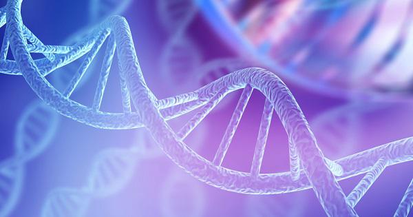
DNA
Thymidine
Endodeoxyribonucleases
Potassium Permanganate
Ultraviolet Rays
Guanine
DNA Repair
Oligodeoxyribonucleotides
5-Methylcytosine
N-Glycosyl Hydrolases
Escherichia coli
Base Sequence
Osmium
Nucleic Acid Conformation
DNA Glycosylases
Base Pairing
DNA Damage
Deoxyribodipyrimidine Photo-Lyase
Oligonucleotides
Base Pair Mismatch
Nucleic Acid Denaturation
Nucleosides
Deoxyribose
Uracil-DNA Glycosidase
Pyrimidine Nucleosides
Molecular Sequence Data
Tritium
Radiation Effects
Photochemistry
Substrate Specificity
Mutation
Rhodotorula
DNA-Directed DNA Polymerase
Specific binding of high-mobility-group I (HMGI) protein and histone H1 to the upstream AT-rich region of the murine beta interferon promoter: HMGI protein acts as a potential antirepressor of the promoter. (1/1371)
The high-mobility-group I (HMGI) protein is a nonhistone component of active chromatin. In this work, we demonstrate that HMGI protein specifically binds to the AT-rich region of the murine beta interferon (IFN-beta) promoter localized upstream of the murine virus-responsive element (VRE). Contrary to what has been described for the human promoter, HMGI protein did not specifically bind to the VRE of the murine IFN-beta promoter. Stably transfected promoters carrying mutations on this HMGI binding site displayed delayed virus-induced kinetics of transcription. When integrated into chromatin, the mutated promoter remained repressed and never reached normal transcriptional activity. Such a phenomenon was not observed with transiently transfected promoters upon which chromatin was only partially reconstituted. Using UV footprinting, we show that the upstream AT-rich sequences of the murine IFN-beta promoter constitute a preferential binding region for histone H1. Transfection with a plasmid carrying scaffold attachment regions as well as incubation with distamycin led to the derepression of the IFN-beta promoter stably integrated into chromatin. In vitro, HMGI protein was able to displace histone H1 from the upstream AT-rich region of the wild-type promoter but not from the promoter carrying mutations on the upstream high-affinity HMGI binding site. Our results suggest that the binding of histone H1 to the upstream AT-rich region of the promoter might be partly responsible for the constitutive repression of the promoter. The displacement by HMGI protein of histone H1 could help to convert the IFN-beta promoter from a repressed to an active state. (+info)The SKN-1 amino-terminal arm is a DNA specificity segment. (2/1371)
The Caenorhabditis elegans SKN-1 protein binds DNA through a basic region like those of bZIP proteins and through a flexible amino-terminal arm segment similar to those with which numerous helix-turn-helix proteins bind to bases in the minor groove. A recent X-ray crystallographic structure suggests that the SKN-1 amino-terminal arm provides only nonspecific DNA binding. In this study, however, we demonstrate that this segment mediates recognition of an AT-rich element that is part of the preferred SKN-1 binding site and thereby significantly increases the sequence specificity with which SKN-1 binds DNA. Mutagenesis experiments show that multiple amino acid residues within the arm are involved in binding. These residues provide binding affinity through distinct but partially redundant interactions and enhance specificity by discriminating against alternate sites. The AT-rich element minor groove is important for binding of the arm, which appears to affect DNA conformation in this region. This conformational effect does not seem to involve DNA bending, however, because the arm does not appear to affect a modest DNA bend that is induced by SKN-1. The data illustrate an example of how a small, flexible protein segment can make an important contribution to DNA binding specificity through multiple interactions and mechanisms. (+info)The catalytic mechanism of a pyrimidine dimer-specific glycosylase (pdg)/abasic lyase, Chlorella virus-pdg. (3/1371)
The repair of UV light-induced cyclobutane pyrimidine dimers can proceed via the base excision repair pathway, in which the initial step is catalyzed by DNA glycosylase/abasic (AP) lyases. The prototypical enzyme studied for this pathway is endonuclease V from the bacteriophage T4 (T4 bacteriophage pyrimidine dimer glycosylase (T4-pdg)). The first homologue for T4-pdg has been found in a strain of Chlorella virus (strain Paramecium bursaria Chlorella virus-1), which contains a gene that predicts an amino acid sequence homology of 41% with T4-pdg. Because both the structure and critical catalytic residues are known for T4-pdg, homology modeling of the Chlorella virus pyrimidine dimer glycosylase (cv-pdg) predicted that a conserved glutamic acid residue (Glu-23) would be important for catalysis at pyrimidine dimers and abasic sites. Site-directed mutations were constructed at Glu-23 to assess the necessity of a negatively charged residue at that position (Gln-23) and the importance of the length of the negatively charged side chain (Asp-23). E23Q lost glycosylase activity completely but retained low levels of AP lyase activity. In contrast, E23D retained near wild type glycosylase and AP lyase activities on cis-syn dimers but completely lost its activity on the trans-syn II dimer, which is very efficiently cleaved by the wild type cv-pdg. As has been shown for other glyscosylases, the wild type cv-pdg catalyzes the cleavage at dimers or AP sites via formation of an imino intermediate, as evidenced by the ability of the enzyme to be covalently trapped on substrate DNA when the reactions are carried out in the presence of a strong reducing agent; in contrast, E23D was very poorly trapped on cis-syn dimers but was readily trapped on DNA containing AP sites. It is proposed that Glu-23 protonates the sugar ring, so that the imino intermediate can be formed. (+info)Identification and characterisation of the Drosophila melanogaster O6-alkylguanine-DNA alkyltransferase cDNA. (4/1371)
The protein O 6-alkylguanine-DNA alkyltransferase(alkyltransferase) is involved in the repair of O 6-alkylguanine and O 4-alkylthymine in DNA and plays an important role in most organisms in attenuating the cytotoxic and mutagenic effects of certain classes of alkylating agents. A genomic clone encompassing the Drosophila melanogaster alkyltransferase gene ( DmAGT ) was identified on the basis of sequence homology with corresponding genes in Saccharomyces cerevisiae and man. The DmAGT gene is located at position 84A on the third chromosome. The nucleotide sequence of DmAGT cDNA revealed an open reading frame encoding 194 amino acids. The MNNG-hypersensitive phenotype of alkyltransferase-deficient bacteria was rescued by expression of the DmAGT cDNA. Furthermore, alkyltransferase activity was identified in crude extracts of Escherichia coli harbouring DmAGT cDNA and this activity was inhibited by preincubation of the extract with an oligonucleotide containing a single O6-methylguanine lesion. Similar to E.coli Ogt and yeast alkyltransferase but in contrast to the human alkyltransferase, the Drosophila alkyltransferase is resistant to inactivation by O 6-benzylguanine. In an E.coli lac Z reversion assay, expression of DmAGT efficiently suppressed MNNG-induced G:C-->A:T as well as A:T-->G:C transition mutations in vivo. These results demonstrate the presence of an alkyltransferase specific for the repair of O 6-methylguanine and O 4-methylthymine in Drosophila. (+info)Reactivity of potassium permanganate and tetraethylammonium chloride with mismatched bases and a simple mutation detection protocol. (5/1371)
Many mutation detection techniques rely upon recognition of mismatched base pairs in DNA hetero-duplexes. Potassium permanganate in combination with tetraethylammonium chloride (TEAC) is capable of chemically modifying mismatched thymidine residues. The DNA strand can then be cleaved at that point by treatment with piperidine. The reactivity of potassium permanganate (KMnO4) in TEAC toward mismatches was investigated in 29 different mutations, representing 58 mismatched base pairs and 116 mismatched bases. All mismatched thymidine residues were modified by KMnO4/TEAC with the majority of these showing strong reactivity. KMnO4/TEAC was also able to modify many mismatched guanosine and cytidine residues, as well as matched guanosine, cytidine and thymidine residues adjacent to, or nearby, mismatched base pairs. Previous techniques using osmium tetroxide (OsO4) to modify mismatched thymidine residues have been limited by the apparent lack of reactivity of a third of all T/G mismatches. KMnO4/TEAC showed no such phenomenon. In this series, all 29 mutations were detected by KMnO4/TEAC treatment. The latest development of the Single Tube Chemical Cleavage of Mismatch Method detects both thymidine and cytidine mismatches by KMnO4/TEAC and hydroxylamine (NH2OH) in a single tube without a clean-up step in between the two reactions. This technique saves time and material without disrupting the sensitivity and efficiency of either reaction. (+info)A new promoter polymorphism in the gene of lipopolysaccharide receptor CD14 is associated with expired myocardial infarction in patients with low atherosclerotic risk profile. (6/1371)
Recent findings suggest that inflammation plays a role in atherosclerosis and its acute complications. Cellular response in infections with Gram-negative bacteria is mediated by bacterial lipopolysaccharide (LPS), which activates monocytes to expression of cytokines, growth factors, and procoagulatory factors via LPS receptor CD14. Endothelial cells and smooth muscle cells are stimulated by a complex of LPS and soluble CD14. In this study, LPS receptor CD14 was analyzed to find genetic variants and check them for an association with coronary artery disease or myocardial infarction (MI). When screening the CD14 gene by single-strand conformation polymorphism analysis, a promoter polymorphism was detected and confirmed as a T-to-C exchange at position -159. We determined the genotypes of 2228 men who had undergone coronary angiography for diagnostic purposes. Within the total study group there was no significant association of either genotype with MI or coronary artery disease. However, in a subgroup with low coronary risk (normotensive nonsmokers), a relative risk for MI in probands homozygous for the T allele could be evaluated (OR, 1.6; 95% CI, 1.0 to 2.4; P<0.05). The association was even stronger in low-risk patients older than 62 years (OR, 3.8; 95% CI, 1.6 to 9.0; P<0.01). In conclusion, we describe a new CD14 promoter polymorphism that is associated with MI, especially in older patients with a low atherosclerotic risk profile. (+info)The Cys4 zinc finger of bacteriophage T7 primase in sequence-specific single-stranded DNA recognition. (7/1371)
Bacteriophage T7 DNA primase recognizes 5'-GTC-3' in single-stranded DNA. The primase contains a single Cys4 zinc-binding motif that is essential for recognition. Biochemical and mutagenic analyses suggest that the Cys4 motif contacts cytosine of 5'-GTC-3' and may also contribute to thymine recognition. Residues His33 and Asp31 are critical for these interactions. Biochemical analysis also reveals that T7 primase selectively binds CTP in the absence of DNA. We propose that bound CTP selects the remaining base G, of 5'-GTC-3', by base pairing. Our deduced mechanism for recognition of ssDNA by Cys4 motifs bears little resemblance to the recognition of trinucleotides of double-stranded DNA by Cys2His2 zinc fingers. (+info)The Saccharomyces cerevisiae homologues of endonuclease III from Escherichia coli, Ntg1 and Ntg2, are both required for efficient repair of spontaneous and induced oxidative DNA damage in yeast. (8/1371)
Endonuclease III from Escherichia coli is the prototype of a ubiquitous DNA repair enzyme essential for the removal of oxidized pyrimidine base damage. The yeast genome project has revealed the presence of two genes in Saccharomyces cerevisiae, NTG1 and NTG2, encoding proteins with similarity to endonuclease III. Both contain the highly conserved helix-hairpin-helix motif, whereas only one (Ntg2) harbors the characteristic iron-sulfur cluster of the endonuclease III family. We have characterized these gene functions by mutant and enzyme analysis as well as by gene expression and intracellular localization studies. Targeted gene disruption of NTG1 and NTG2 produced mutants with greatly increased spontaneous and hydrogen peroxide-induced mutation frequency relative to the wild type, and the mutation response was further increased in the double mutant. Both enzymes were found to remove thymine glycol and 2, 6-diamino-4-hydroxy-5-N-methylformamidopyrimidine (faPy) residues from DNA with high efficiency. However, on UV-irradiated DNA, saturating concentrations of Ntg2 removed only half of the cytosine photoproducts released by Ntg1. Conversely, 5-hydroxycytosine was removed efficiently only by Ntg2. The enzymes appear to have different reaction modes, as judged from much higher affinity of Ntg2 for damaged DNA and more efficient borhydride trapping of Ntg1 to abasic sites in DNA despite limited DNA binding. Northern blot and promoter fusion analysis showed that NTG1 is inducible by cell exposure to DNA-damaging agents, whereas NTG2 is constitutively expressed. Ntg2 appears to be a nuclear enzyme, whereas Ntg1 was sorted both to the nucleus and to the mitochondria. We conclude that functions of both NTG1 and NTG2 are important for removal of oxidative DNA damage in yeast. (+info)Thymine is a pyrimidine nucleobase that is one of the four nucleobases in the nucleic acid double helix of DNA (the other three being adenine, guanine, and cytosine). It is denoted by the letter T in DNA notation and pairs with adenine via two hydrogen bonds. Thymine is not typically found in RNA, where uracil takes its place pairing with adenine. The structure of thymine consists of a six-membered ring (pyrimidine) fused to a five-membered ring containing two nitrogen atoms and a ketone group.
Thymine DNA Glycosylase (TDG) is an enzyme that plays a crucial role in the process of base excision repair (BER), which is a mechanism for correcting damaged or mismatched bases in DNA. Specifically, TDG is responsible for removing thymine bases that have been improperly incorporated into DNA opposite to guanine, forming a so-called "mismatch" or "lesion." This type of lesion can arise due to errors during DNA replication or from the mutagenic effects of environmental agents such as chemicals and radiation.
TDG recognizes and binds to the thymine-guanine mismatch, then catalyzes the removal of the thymine base by cleaving the N-glycosidic bond that links it to the deoxyribose sugar in the DNA backbone. This creates an abasic site, which is subsequently processed by other enzymes involved in BER to restore the original DNA sequence.
In addition to its role in DNA repair, TDG has been implicated in various cellular processes such as transcriptional regulation and epigenetic modification, due to its ability to interact with other proteins and regulatory elements in the genome. Dysregulation of TDG function has been linked to several human diseases, including cancer and neurological disorders.
Uracil is not a medical term, but it is a biological molecule. Medically or biologically, uracil can be defined as one of the four nucleobases in the nucleic acid of RNA (ribonucleic acid) that is linked to a ribose sugar by an N-glycosidic bond. It forms base pairs with adenine in double-stranded RNA and DNA. Uracil is a pyrimidine derivative, similar to thymine found in DNA, but it lacks the methyl group (-CH3) that thymine has at the 5 position of its ring.
Thymine nucleotides are biochemical components that play a crucial role in the structure and function of DNA (deoxyribonucleic acid), which is the genetic material present in living organisms. A thymine nucleotide consists of three parts: a sugar molecule called deoxyribose, a phosphate group, and a nitrogenous base called thymine.
Thymine is one of the four nucleobases in DNA, along with adenine, guanine, and cytosine. It specifically pairs with adenine through hydrogen bonding, forming a base pair that is essential for maintaining the structure and stability of the double helix. Thymine nucleotides are linked together by phosphodiester bonds between the sugar molecules of adjacent nucleotides, creating a long, linear polymer known as a DNA strand.
In summary, thymine nucleotides are building blocks of DNA that consist of deoxyribose, a phosphate group, and the nitrogenous base thymine, which pairs with adenine in the double helix structure.
Pyrimidine dimers are a type of DNA lesion that form when two adjacent pyrimidine bases on the same strand of DNA become covalently linked, usually as a result of exposure to ultraviolet (UV) light. The most common type of pyrimidine dimer is the cyclobutane pyrimidine dimer (CPD), which forms when two thymine bases are linked together in a cyclobutane ring structure.
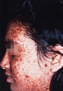
Pyrimidine dimers can distort the DNA helix and interfere with normal replication and transcription processes, leading to mutations and potentially cancer. The formation of pyrimidine dimers is a major mechanism by which UV radiation causes skin damage and increases the risk of skin cancer.
The body has several mechanisms for repairing pyrimidine dimers, including nucleotide excision repair (NER) and base excision repair (BER). However, if these repair mechanisms are impaired or overwhelmed, pyrimidine dimers can persist and contribute to the development of cancer.
Bromouracil is a chemical compound that is used in the synthesis of DNA. It is a brominated derivative of uracil, which is one of the nucleobases found in RNA. Bromouracil can be incorporated into DNA during replication in place of thymine, another nucleobase. This can lead to mutations in the DNA because bromouracil behaves differently from thymine in certain chemical reactions.
Bromouracil is not typically found in living organisms and is not considered to be a normal part of the genetic material. It may be used in research settings to study the mechanisms of DNA replication and mutation. In clinical medicine, bromouracil has been used in the treatment of psoriasis, a skin condition characterized by red, scaly patches. However, its use in this context is not common.
It is important to note that bromouracil can have toxic effects and should be handled with care. It can cause irritation to the skin and eyes, and prolonged exposure may lead to more serious health problems. If you have any questions about bromouracil or its use, it is best to speak with a healthcare professional or a qualified scientist.
I'm sorry for any confusion, but "Pentoxyl" doesn't seem to be a recognized term in medical terminology or pharmacology. It's possible that there might be a spelling mistake or it could be a brand name of a drug that is not widely known.
If you meant "Pentoxifylline," however, I can provide a definition. Pentoxifylline is a medication that belongs to a class of drugs known as methylxanthines. It works by improving the flow of blood in the body, particularly in the hands and feet, by decreasing the thickness (viscosity) of the blood. This medication is used to treat conditions such as intermittent claudication (pain in the legs due to poor blood flow) and certain types of Raynaud's phenomenon.
Please make sure that you have the correct spelling when looking for medical information, as it's crucial to have accurate details when researching health-related topics.
Osmium tetroxide is not a medical term per se, but it is a chemical compound with the formula OsO4. It is used in some medical and scientific applications due to its properties as a strong oxidizing agent and its ability to form complexes with organic compounds.
In histology, osmium tetroxide is sometimes used as a fixative for electron microscopy because it reacts with unsaturated lipids and proteins in biological tissue, creating an electron-dense deposit that can be visualized under the microscope. It is also used to stain fatty acids and other lipids in biological samples.
However, osmium tetroxide is highly toxic and volatile, and it can cause damage to the eyes, skin, and respiratory system if not handled with appropriate precautions. Therefore, its use in medical and scientific settings is typically limited to specialized applications where its unique properties are required.
Cytosine is one of the four nucleobases in the nucleic acid molecules DNA and RNA, along with adenine, guanine, and thymine (in DNA) or uracil (in RNA). The single-letter abbreviation for cytosine is "C."
Cytosine base pairs specifically with guanine through hydrogen bonding, forming a base pair. In DNA, the double helix consists of two complementary strands of nucleotides held together by these base pairs, such that the sequence of one strand determines the sequence of the other. This property is critical for DNA replication and transcription, processes that are essential for life.
Cytosine residues in DNA can undergo spontaneous deamination to form uracil, which can lead to mutations if not corrected by repair mechanisms. In RNA, cytosine can be methylated at the 5-carbon position to form 5-methylcytosine, a modification that plays a role in regulating gene expression and other cellular processes.
Deoxyribonucleic acid (DNA) is the genetic material present in the cells of organisms where it is responsible for the storage and transmission of hereditary information. DNA is a long molecule that consists of two strands coiled together to form a double helix. Each strand is made up of a series of four nucleotide bases - adenine (A), guanine (G), cytosine (C), and thymine (T) - that are linked together by phosphate and sugar groups. The sequence of these bases along the length of the molecule encodes genetic information, with A always pairing with T and C always pairing with G. This base-pairing allows for the replication and transcription of DNA, which are essential processes in the functioning and reproduction of all living organisms.
Thymidine is a pyrimidine nucleoside that consists of a thymine base linked to a deoxyribose sugar by a β-N1-glycosidic bond. It plays a crucial role in DNA replication and repair processes as one of the four nucleosides in DNA, along with adenosine, guanosine, and cytidine. Thymidine is also used in research and clinical settings for various purposes, such as studying DNA synthesis or as a component of antiviral and anticancer therapies.
Endodeoxyribonucleases are a type of enzyme that cleave, or cut, phosphodiester bonds within the backbone of DNA molecules. These enzymes are also known as restriction endonucleases or simply restriction enzymes. They are called "restriction" enzymes because they were first discovered in bacteria, where they function to protect the organism from foreign DNA by cleaving and destroying invading viral DNA.
Endodeoxyribonucleases recognize specific sequences of nucleotides within the DNA molecule, known as recognition sites or restriction sites, and cut the phosphodiester bonds at specific locations within these sites. The cuts made by endodeoxyribonucleases can be either "sticky" or "blunt," depending on whether the enzyme leaves single-stranded overhangs or creates blunt ends at the site of cleavage, respectively.
Endodeoxyribonucleases are widely used in molecular biology research for various applications, including DNA cloning, genome mapping, and genetic engineering. They allow researchers to cut DNA molecules at specific sites, creating defined fragments that can be manipulated and recombined in a variety of ways.
Potassium permanganate is not a medical term, but it is a chemical compound with the formula KMnO4. It's a dark purple crystalline solid that is soluble in water and has strong oxidizing properties. In a medical context, potassium permanganate is occasionally used as a topical antiseptic and disinfectant, particularly for treating minor wounds, burns, and ulcers. It's also used to treat certain skin conditions such as eczema and psoriasis. However, its use is limited due to the potential for skin irritation and staining of the skin and clothing. It should always be used under medical supervision and with caution.
According to the medical definition, ultraviolet (UV) rays are invisible radiations that fall in the range of the electromagnetic spectrum between 100-400 nanometers. UV rays are further divided into three categories: UVA (320-400 nm), UVB (280-320 nm), and UVC (100-280 nm).
UV rays have various sources, including the sun and artificial sources like tanning beds. Prolonged exposure to UV rays can cause damage to the skin, leading to premature aging, eye damage, and an increased risk of skin cancer. UVA rays penetrate deeper into the skin and are associated with skin aging, while UVB rays primarily affect the outer layer of the skin and are linked to sunburns and skin cancer. UVC rays are the most harmful but fortunately, they are absorbed by the Earth's atmosphere and do not reach the surface.
Healthcare professionals recommend limiting exposure to UV rays, wearing protective clothing, using broad-spectrum sunscreen with an SPF of at least 30, and avoiding tanning beds to reduce the risk of UV-related health problems.
Guanine is not a medical term per se, but it is a biological molecule that plays a crucial role in the body. Guanine is one of the four nucleobases found in the nucleic acids DNA and RNA, along with adenine, cytosine, and thymine (in DNA) or uracil (in RNA). Specifically, guanine pairs with cytosine via hydrogen bonds to form a base pair.
Guanine is a purine derivative, which means it has a double-ring structure. It is formed through the synthesis of simpler molecules in the body and is an essential component of genetic material. Guanine's chemical formula is C5H5N5O.
While guanine itself is not a medical term, abnormalities or mutations in genes that contain guanine nucleotides can lead to various medical conditions, including genetic disorders and cancer.
DNA repair is the process by which cells identify and correct damage to the DNA molecules that encode their genome. DNA can be damaged by a variety of internal and external factors, such as radiation, chemicals, and metabolic byproducts. If left unrepaired, this damage can lead to mutations, which may in turn lead to cancer and other diseases.
There are several different mechanisms for repairing DNA damage, including:
1. Base excision repair (BER): This process repairs damage to a single base in the DNA molecule. An enzyme called a glycosylase removes the damaged base, leaving a gap that is then filled in by other enzymes.
2. Nucleotide excision repair (NER): This process repairs more severe damage, such as bulky adducts or crosslinks between the two strands of the DNA molecule. An enzyme cuts out a section of the damaged DNA, and the gap is then filled in by other enzymes.
3. Mismatch repair (MMR): This process repairs errors that occur during DNA replication, such as mismatched bases or small insertions or deletions. Specialized enzymes recognize the error and remove a section of the newly synthesized strand, which is then replaced by new nucleotides.
4. Double-strand break repair (DSBR): This process repairs breaks in both strands of the DNA molecule. There are two main pathways for DSBR: non-homologous end joining (NHEJ) and homologous recombination (HR). NHEJ directly rejoins the broken ends, while HR uses a template from a sister chromatid to repair the break.
Overall, DNA repair is a crucial process that helps maintain genome stability and prevent the development of diseases caused by genetic mutations.
Adenine is a purine nucleotide base that is a fundamental component of DNA and RNA, the genetic material of living organisms. In DNA, adenine pairs with thymine via double hydrogen bonds, while in RNA, it pairs with uracil. Adenine is essential for the structure and function of nucleic acids, as well as for energy transfer reactions in cells through its role in the formation of adenosine triphosphate (ATP), the primary energy currency of the cell.
Oligodeoxyribonucleotides (ODNs) are relatively short, synthetic single-stranded DNA molecules. They typically contain 15 to 30 nucleotides, but can range from 2 to several hundred nucleotides in length. ODNs are often used as tools in molecular biology research for various applications such as:
1. Nucleic acid detection and quantification (e.g., real-time PCR)
2. Gene regulation (antisense, RNA interference)
3. Gene editing (CRISPR-Cas systems)
4. Vaccine development
5. Diagnostic purposes
Due to their specificity and affinity towards complementary DNA or RNA sequences, ODNs can be designed to target a particular gene or sequence of interest. This makes them valuable tools in understanding gene function, regulation, and interaction with other molecules within the cell.
5-Methylcytosine (5mC) is a chemical modification of the nucleotide base cytosine in DNA, where a methyl group (-CH3) is added to the 5th carbon atom of the cytosine ring. This modification is catalyzed by DNA methyltransferase enzymes and plays an essential role in epigenetic regulation of gene expression, genomic imprinting, X-chromosome inactivation, and suppression of transposable elements in eukaryotic cells. Abnormal DNA methylation patterns have been associated with various diseases, including cancer.
N-Glycosyl hydrolases (or N-glycanases) are a class of enzymes that catalyze the hydrolysis of the glycosidic bond between an N-glycosyl group and an aglycon, which is typically another part of a larger molecule such as a protein or lipid. N-Glycosyl groups refer to carbohydrate moieties attached to an nitrogen atom, usually in the side chain of an amino acid such as asparagine (Asn) in proteins.
N-Glycosyl hydrolases play important roles in various biological processes, including the degradation and processing of glycoproteins, the modification of glycolipids, and the breakdown of complex carbohydrates. These enzymes are widely distributed in nature and have been found in many organisms, from bacteria to humans.
The classification and nomenclature of N-Glycosyl hydrolases are based on the type of glycosidic bond they cleave and the stereochemistry of the reaction they catalyze. They are grouped into different families in the Carbohydrate-Active enZymes (CAZy) database, which provides a comprehensive resource for the study of carbohydrate-active enzymes.
It is worth noting that N-Glycosyl hydrolases can have both beneficial and detrimental effects on human health. For example, they are involved in the normal turnover and degradation of glycoproteins in the body, but they can also contribute to the pathogenesis of certain diseases, such as lysosomal storage disorders, where mutations in N-Glycosyl hydrolases lead to the accumulation of undigested glycoconjugates and cellular damage.
'Escherichia coli' (E. coli) is a type of gram-negative, facultatively anaerobic, rod-shaped bacterium that commonly inhabits the intestinal tract of humans and warm-blooded animals. It is a member of the family Enterobacteriaceae and one of the most well-studied prokaryotic model organisms in molecular biology.
While most E. coli strains are harmless and even beneficial to their hosts, some serotypes can cause various forms of gastrointestinal and extraintestinal illnesses in humans and animals. These pathogenic strains possess virulence factors that enable them to colonize and damage host tissues, leading to diseases such as diarrhea, urinary tract infections, pneumonia, and sepsis.
E. coli is a versatile organism with remarkable genetic diversity, which allows it to adapt to various environmental niches. It can be found in water, soil, food, and various man-made environments, making it an essential indicator of fecal contamination and a common cause of foodborne illnesses. The study of E. coli has contributed significantly to our understanding of fundamental biological processes, including DNA replication, gene regulation, and protein synthesis.
A base sequence in the context of molecular biology refers to the specific order of nucleotides in a DNA or RNA molecule. In DNA, these nucleotides are adenine (A), guanine (G), cytosine (C), and thymine (T). In RNA, uracil (U) takes the place of thymine. The base sequence contains genetic information that is transcribed into RNA and ultimately translated into proteins. It is the exact order of these bases that determines the genetic code and thus the function of the DNA or RNA molecule.
I'm sorry for the confusion, but "Osmium" is not a medical term. It is actually a chemical element with symbol "Os" and atomic number 76. Osmium is a hard, brittle, blue-gray or blue-black transition metal in the platinum group that is found as a minor constituent in some mineral deposits such as placer gold.
It's primarily used in the manufacture of fountain pen tips, electrical contacts, and other specialized applications. Osmium tetroxide, a volatile, pale yellow, water-soluble compound formed by the reaction of osmium with oxygen or chlorine, is used as a fixative and stain in electron microscopy, histology, and in mortuary science for the stabilization and staining of tissues. However, exposure to osmium tetroxide can cause respiratory irritation and damage, so it's handled with great care.
Nucleic acid conformation refers to the three-dimensional structure that nucleic acids (DNA and RNA) adopt as a result of the bonding patterns between the atoms within the molecule. The primary structure of nucleic acids is determined by the sequence of nucleotides, while the conformation is influenced by factors such as the sugar-phosphate backbone, base stacking, and hydrogen bonding.
Two common conformations of DNA are the B-form and the A-form. The B-form is a right-handed helix with a diameter of about 20 Å and a pitch of 34 Å, while the A-form has a smaller diameter (about 18 Å) and a shorter pitch (about 25 Å). RNA typically adopts an A-form conformation.
The conformation of nucleic acids can have significant implications for their function, as it can affect their ability to interact with other molecules such as proteins or drugs. Understanding the conformational properties of nucleic acids is therefore an important area of research in molecular biology and medicine.
DNA glycosylases are a group of enzymes that play a crucial role in the maintenance of genetic material. They are responsible for initiating the base excision repair (BER) pathway, which is one of the major DNA repair mechanisms in cells.
The function of DNA glycosylases is to remove damaged or mismatched bases from DNA molecules. These enzymes recognize and bind to specific types of damaged or incorrect bases, and then cleave the N-glycosidic bond between the base and the deoxyribose sugar in the DNA backbone. This results in the formation of an apurinic/apyrimidinic (AP) site, which is subsequently processed by other enzymes in the BER pathway.
There are several different types of DNA glycosylases that recognize and remove specific types of damaged or incorrect bases. For example, some DNA glycosylases specialize in removing oxidized bases, while others are responsible for removing mismatched bases or those that have been alkylated or methylated.
Overall, the proper functioning of DNA glycosylases is essential for maintaining genomic stability and preventing the accumulation of mutations that can lead to diseases such as cancer.
Base pairing is a specific type of chemical bonding that occurs between complementary base pairs in the nucleic acid molecules DNA and RNA. In DNA, these bases are adenine (A), thymine (T), guanine (G), and cytosine (C). Adenine always pairs with thymine via two hydrogen bonds, while guanine always pairs with cytosine via three hydrogen bonds. This precise base pairing is crucial for the stability of the double helix structure of DNA and for the accurate replication and transcription of genetic information. In RNA, uracil (U) takes the place of thymine and pairs with adenine.
DNA damage refers to any alteration in the structure or composition of deoxyribonucleic acid (DNA), which is the genetic material present in cells. DNA damage can result from various internal and external factors, including environmental exposures such as ultraviolet radiation, tobacco smoke, and certain chemicals, as well as normal cellular processes such as replication and oxidative metabolism.
Examples of DNA damage include base modifications, base deletions or insertions, single-strand breaks, double-strand breaks, and crosslinks between the two strands of the DNA helix. These types of damage can lead to mutations, genomic instability, and chromosomal aberrations, which can contribute to the development of diseases such as cancer, neurodegenerative disorders, and aging-related conditions.
The body has several mechanisms for repairing DNA damage, including base excision repair, nucleotide excision repair, mismatch repair, and double-strand break repair. However, if the damage is too extensive or the repair mechanisms are impaired, the cell may undergo apoptosis (programmed cell death) to prevent the propagation of potentially harmful mutations.
Deoxyribodipyrimidine photo-lyase is an enzyme involved in the repair of DNA damage, specifically the repair of cyclobutane pyrimidine dimers (CPDs) that are formed when DNA is exposed to ultraviolet (UV) light. CPDs can distort the structure of DNA and interfere with replication and transcription, so it's important for cells to have mechanisms to repair this damage.
Deoxyribodipyrimidine photo-lyase works by cleaving the bond between two adjacent pyrimidines in the DNA strand that form the CPD, releasing one of the pyrimidines and allowing the remaining portion of the strand to be repaired. This enzyme is also known as photolyase or DNA repair photolyase.
It's worth noting that there are different types of photolyases that can repair different kinds of DNA damage, but deoxyribodipyrimidine photo-lyase specifically repairs CPDs caused by UV light.
Bacterial DNA refers to the genetic material found in bacteria. It is composed of a double-stranded helix containing four nucleotide bases - adenine (A), thymine (T), guanine (G), and cytosine (C) - that are linked together by phosphodiester bonds. The sequence of these bases in the DNA molecule carries the genetic information necessary for the growth, development, and reproduction of bacteria.
Bacterial DNA is circular in most bacterial species, although some have linear chromosomes. In addition to the main chromosome, many bacteria also contain small circular pieces of DNA called plasmids that can carry additional genes and provide resistance to antibiotics or other environmental stressors.
Unlike eukaryotic cells, which have their DNA enclosed within a nucleus, bacterial DNA is present in the cytoplasm of the cell, where it is in direct contact with the cell's metabolic machinery. This allows for rapid gene expression and regulation in response to changing environmental conditions.
Oligonucleotides are short sequences of nucleotides, the building blocks of DNA and RNA. They typically contain fewer than 100 nucleotides, and can be synthesized chemically to have specific sequences. Oligonucleotides are used in a variety of applications in molecular biology, including as probes for detecting specific DNA or RNA sequences, as inhibitors of gene expression, and as components of diagnostic tests and therapies. They can also be used in the study of protein-nucleic acid interactions and in the development of new drugs.
A base pair mismatch is a type of mutation that occurs during the replication or repair of DNA, where two incompatible nucleotides pair up instead of the usual complementary bases (adenine-thymine or cytosine-guanine). This can result in the substitution of one base pair for another and may lead to changes in the genetic code, potentially causing errors in protein synthesis and possibly contributing to genetic disorders or diseases, including cancer.
Nucleic acid denaturation is the process of separating the two strands of a double-stranded DNA molecule, or unwinding the helical structure of an RNA molecule, by disrupting the hydrogen bonds that hold the strands together. This process is typically caused by exposure to high temperatures, changes in pH, or the presence of chemicals called denaturants.
Denaturation can also cause changes in the shape and function of nucleic acids. For example, it can disrupt the secondary and tertiary structures of RNA molecules, which can affect their ability to bind to other molecules and carry out their functions within the cell.
In molecular biology, nucleic acid denaturation is often used as a tool for studying the structure and function of nucleic acids. For example, it can be used to separate the two strands of a DNA molecule for sequencing or amplification, or to study the interactions between nucleic acids and other molecules.
It's important to note that denaturation is a reversible process, and under the right conditions, the double-stranded structure of DNA can be restored through a process called renaturation or annealing.
A nucleoside is a biochemical molecule that consists of a pentose sugar (a type of simple sugar with five carbon atoms) covalently linked to a nitrogenous base. The nitrogenous base can be one of several types, including adenine, guanine, cytosine, thymine, or uracil. Nucleosides are important components of nucleic acids, such as DNA and RNA, which are the genetic materials found in cells. They play a crucial role in various biological processes, including cell division, protein synthesis, and gene expression.
DNA replication is the biological process by which DNA makes an identical copy of itself during cell division. It is a fundamental mechanism that allows genetic information to be passed down from one generation of cells to the next. During DNA replication, each strand of the double helix serves as a template for the synthesis of a new complementary strand. This results in the creation of two identical DNA molecules. The enzymes responsible for DNA replication include helicase, which unwinds the double helix, and polymerase, which adds nucleotides to the growing strands.
Deoxyribose is a type of sugar that makes up the structural backbone of DNA (deoxyribonucleic acid), one of the two main types of nucleic acids in cells. The chemical formula for deoxyribose is C5H10O4, and it has a five-carbon ring structure with four hydroxyl (-OH) groups and one hydrogen atom attached to the carbons.
The key difference between deoxyribose and ribose, which makes up the structural backbone of RNA (ribonucleic acid), is that deoxyribose lacks a hydroxyl group on the second carbon atom in its ring structure. This small difference has significant implications for the structure and function of DNA compared to RNA.
Deoxyribose plays an essential role in the replication, transcription, and repair of genetic material in cells. It forms the sugar-phosphate backbone of DNA by linking with phosphate groups through ester bonds between the 3' carbon atom of one deoxyribose molecule and the 5' carbon atom of another, creating a long, twisted ladder-like structure known as a double helix. The nitrogenous bases adenine, thymine, guanine, and cytosine attach to the 1' carbon atom of each deoxyribose molecule in the DNA strand, forming pairs that are complementary to each other (adenine with thymine and guanine with cytosine).
Overall, deoxyribose is a crucial component of DNA, enabling the storage and transmission of genetic information from one generation to the next.
Uracil-DNA glycosylase (UDG) is an enzyme that plays a crucial role in the maintenance of genomic stability by removing uracil residues from DNA. These enzymes are essential because uracil can arise in DNA through the deamination of cytosine or through the misincorporation of dUMP during DNA replication. If left unrepaired, uracil can pair with adenine, leading to C:G to T:A transitions during subsequent rounds of replication.
UDGs initiate the base excision repair (BER) pathway by cleaving the N-glycosidic bond between the uracil base and the deoxyribose sugar, releasing the uracil base and creating an abasic site. The resulting apurinic/apyrimidinic (AP) site is then processed further by AP endonucleases, DNA polymerases, and ligases to complete the repair process.
There are several subtypes of UDGs that differ in their substrate specificity, cellular localization, and regulation. For example, some UDGs specifically remove uracil from single-stranded or double-stranded DNA, while others have broader substrate specificity and can also remove other damaged bases. Understanding the function and regulation of these enzymes is important for understanding the mechanisms that maintain genomic stability and prevent mutations.
Deamination is a biochemical process that refers to the removal of an amino group (-NH2) from a molecule, especially from an amino acid. This process typically results in the formation of a new functional group and the release of ammonia (NH3). Deamination plays a crucial role in the metabolism of amino acids, as it helps to convert them into forms that can be excreted or used for energy production. In some cases, deamination can also lead to the formation of toxic byproducts, which must be efficiently eliminated from the body to prevent harm.
Pyrimidine nucleosides are organic compounds that consist of a pyrimidine base (a heterocyclic aromatic ring containing two nitrogen atoms and four carbon atoms) linked to a sugar molecule, specifically ribose or deoxyribose, via a β-glycosidic bond. The pyrimidine bases found in nucleosides can be cytosine (C), thymine (T), or uracil (U). When the sugar component is ribose, it is called a pyrimidine nucleoside, and when it is linked to deoxyribose, it is referred to as a deoxy-pyrimidine nucleoside. These molecules play crucial roles in various biological processes, particularly in the structure and function of nucleic acids such as DNA and RNA.
Molecular sequence data refers to the specific arrangement of molecules, most commonly nucleotides in DNA or RNA, or amino acids in proteins, that make up a biological macromolecule. This data is generated through laboratory techniques such as sequencing, and provides information about the exact order of the constituent molecules. This data is crucial in various fields of biology, including genetics, evolution, and molecular biology, allowing for comparisons between different organisms, identification of genetic variations, and studies of gene function and regulation.
Base composition in genetics refers to the relative proportion of the four nucleotide bases (adenine, thymine, guanine, and cytosine) in a DNA or RNA molecule. In DNA, adenine pairs with thymine, and guanine pairs with cytosine, so the base composition is often expressed in terms of the ratio of adenine + thymine (A-T) to guanine + cytosine (G-C). This ratio can vary between species and even between different regions of the same genome. The base composition can provide important clues about the function, evolution, and structure of genetic material.
Deoxyribonucleosides are chemical compounds that constitute the basic building blocks of DNA, one of the two nucleic acids found in cells. They consist of a sugar molecule called deoxyribose, a nitrogenous base (either adenine, guanine, cytosine, or thymine), and a phosphate group.
The nitrogenous base is attached to the 1' carbon atom of the deoxyribose sugar, forming a glycosidic bond. The phosphate group is linked to the 5' carbon atom of the deoxyribose sugar through an ester linkage, creating a phosphodiester bond with another deoxyribonucleoside.
When multiple deoxyribonucleosides are joined together through their phosphate groups, they form a polynucleotide chain, which is the backbone of DNA. The sequence of nitrogenous bases along this chain encodes genetic information that determines the characteristics and functions of living organisms.
Deoxyribonucleosides play a crucial role in various biological processes, including DNA replication, repair, and transcription. They are also used as therapeutic agents for the treatment of certain genetic disorders and cancer.
Tritium is not a medical term, but it is a term used in the field of nuclear physics and chemistry. Tritium (symbol: T or 3H) is a radioactive isotope of hydrogen with two neutrons and one proton in its nucleus. It is also known as heavy hydrogen or superheavy hydrogen.
Tritium has a half-life of about 12.3 years, which means that it decays by emitting a low-energy beta particle (an electron) to become helium-3. Due to its radioactive nature and relatively short half-life, tritium is used in various applications, including nuclear weapons, fusion reactors, luminous paints, and medical research.
In the context of medicine, tritium may be used as a radioactive tracer in some scientific studies or medical research, but it is not a term commonly used to describe a medical condition or treatment.
Radiation effects refer to the damages that occur in living tissues when exposed to ionizing radiation. These effects can be categorized into two types: deterministic and stochastic. Deterministic effects have a threshold dose below which the effect does not occur, and above which the severity of the effect increases with the dose. Examples include radiation-induced erythema, epilation, and organ damage. Stochastic effects, on the other hand, do not have a threshold dose, and the probability of the effect occurring increases with the dose. Examples include genetic mutations and cancer induction. The severity of the effect is not related to the dose in this case.
I'm sorry for any confusion, but "photochemistry" is not a term typically used in medical definitions. Photochemistry is a branch of chemistry that deals with the chemical effects of light. It involves the absorption of light by a substance, which can lead to the promotion of an electron to a higher energy state, and subsequently result in various chemical reactions.
In a medical context, photochemical processes might be discussed in relation to certain therapies or diagnostic techniques, such as photodynamic therapy for cancer treatment, where a photosensitizing agent is used that reacts with light to produce singlet oxygen or other reactive species to destroy nearby cells. However, it's not a term used to define a specific medical condition or concept in the same way that one might define "inflammation" or "metabolism."
Substrate specificity in the context of medical biochemistry and enzymology refers to the ability of an enzyme to selectively bind and catalyze a chemical reaction with a particular substrate (or a group of similar substrates) while discriminating against other molecules that are not substrates. This specificity arises from the three-dimensional structure of the enzyme, which has evolved to match the shape, charge distribution, and functional groups of its physiological substrate(s).
Substrate specificity is a fundamental property of enzymes that enables them to carry out highly selective chemical transformations in the complex cellular environment. The active site of an enzyme, where the catalysis takes place, has a unique conformation that complements the shape and charge distribution of its substrate(s). This ensures efficient recognition, binding, and conversion of the substrate into the desired product while minimizing unwanted side reactions with other molecules.
Substrate specificity can be categorized as:
1. Absolute specificity: An enzyme that can only act on a single substrate or a very narrow group of structurally related substrates, showing no activity towards any other molecule.
2. Group specificity: An enzyme that prefers to act on a particular functional group or class of compounds but can still accommodate minor structural variations within the substrate.
3. Broad or promiscuous specificity: An enzyme that can act on a wide range of structurally diverse substrates, albeit with varying catalytic efficiencies.
Understanding substrate specificity is crucial for elucidating enzymatic mechanisms, designing drugs that target specific enzymes or pathways, and developing biotechnological applications that rely on the controlled manipulation of enzyme activities.
A mutation is a permanent change in the DNA sequence of an organism's genome. Mutations can occur spontaneously or be caused by environmental factors such as exposure to radiation, chemicals, or viruses. They may have various effects on the organism, ranging from benign to harmful, depending on where they occur and whether they alter the function of essential proteins. In some cases, mutations can increase an individual's susceptibility to certain diseases or disorders, while in others, they may confer a survival advantage. Mutations are the driving force behind evolution, as they introduce new genetic variability into populations, which can then be acted upon by natural selection.
In the context of medicine and pharmacology, "kinetics" refers to the study of how a drug moves throughout the body, including its absorption, distribution, metabolism, and excretion (often abbreviated as ADME). This field is called "pharmacokinetics."
1. Absorption: This is the process of a drug moving from its site of administration into the bloodstream. Factors such as the route of administration (e.g., oral, intravenous, etc.), formulation, and individual physiological differences can affect absorption.
2. Distribution: Once a drug is in the bloodstream, it gets distributed throughout the body to various tissues and organs. This process is influenced by factors like blood flow, protein binding, and lipid solubility of the drug.
3. Metabolism: Drugs are often chemically modified in the body, typically in the liver, through processes known as metabolism. These changes can lead to the formation of active or inactive metabolites, which may then be further distributed, excreted, or undergo additional metabolic transformations.
4. Excretion: This is the process by which drugs and their metabolites are eliminated from the body, primarily through the kidneys (urine) and the liver (bile).
Understanding the kinetics of a drug is crucial for determining its optimal dosing regimen, potential interactions with other medications or foods, and any necessary adjustments for special populations like pediatric or geriatric patients, or those with impaired renal or hepatic function.
A pentose is a monosaccharide (simple sugar) that contains five carbon atoms. The name "pentose" comes from the Greek word "pente," meaning five, and "ose," meaning sugar. Pentoses play important roles in various biological processes, such as serving as building blocks for nucleic acids (DNA and RNA) and other biomolecules.
Some common pentoses include:
1. D-Ribose - A naturally occurring pentose found in ribonucleic acid (RNA), certain coenzymes, and energy-carrying molecules like adenosine triphosphate (ATP).
2. D-Deoxyribose - A pentose that lacks a hydroxyl (-OH) group on the 2' carbon atom, making it a key component of deoxyribonucleic acid (DNA).
3. Xylose - A naturally occurring pentose found in various plants and woody materials; it is used as a sweetener and food additive.
4. Arabinose - Another plant-derived pentose, arabinose can be found in various fruits, vegetables, and grains. It has potential applications in the production of biofuels and other bioproducts.
5. Lyxose - A less common pentose that can be found in some polysaccharides and glycoproteins.
Pentoses are typically less sweet than hexoses (six-carbon sugars) like glucose or fructose, but they still contribute to the overall sweetness of many foods and beverages.
Rhodotorula is a genus of unicellular, budding yeasts that are commonly found in the environment, particularly in damp and nutrient-rich places such as soil, water, and vegetation. They are characterized by their ability to produce carotenoid pigments, which give them a distinctive pinkish-red color.
While Rhodotorula species are not typically associated with human disease, they can occasionally cause infections in people with weakened immune systems or underlying medical conditions. These infections can occur in various parts of the body, including the respiratory tract, urinary tract, and skin.
Rhodotorula infections are usually treated with antifungal medications, such as fluconazole or amphotericin B. Preventing exposure to sources of Rhodotorula, such as contaminated medical equipment or water supplies, can also help reduce the risk of infection.
DNA-directed DNA polymerase is a type of enzyme that synthesizes new strands of DNA by adding nucleotides to an existing DNA template in a 5' to 3' direction. These enzymes are essential for DNA replication, repair, and recombination. They require a single-stranded DNA template, a primer with a free 3' hydroxyl group, and the four deoxyribonucleoside triphosphates (dNTPs) as substrates to carry out the polymerization reaction.
DNA polymerases also have proofreading activity, which allows them to correct errors that occur during DNA replication by removing mismatched nucleotides and replacing them with the correct ones. This helps ensure the fidelity of the genetic information passed from one generation to the next.
There are several different types of DNA polymerases, each with specific functions and characteristics. For example, DNA polymerase I is involved in both DNA replication and repair, while DNA polymerase III is the primary enzyme responsible for DNA replication in bacteria. In eukaryotic cells, DNA polymerase alpha, beta, gamma, delta, and epsilon have distinct roles in DNA replication, repair, and maintenance.
Deoxycytidine monophosphate (dCMP) is a nucleotide that is a building block of DNA. It consists of the sugar deoxyribose, the base cytosine, and one phosphate group. Nucleotides like dCMP are linked together through the phosphate groups to form long chains of DNA. In this way, dCMP plays an essential role in the structure and function of DNA, including the storage and transmission of genetic information.
No data available that match "thymine"
 Thymine - Wikipedia
Thymine dioxygenase - Wikipedia
 thymine (CHEBI:17821)
 THE ELECTRONIC STRUCTURE OF THYMINE. - Nokia Bell Labs
 Thymine - YourGenome
 1FJB: NMR Study of an 11-Mer DNA Duplex Containing 7,8-Dihydro-8-Oxoadenine (AOXO) Opposite Thymine
 RCSB PDB - 1NK8: A BACILLUS DNA POLYMERASE I PRODUCT COMPLEX BOUND TO A GUANINE-THYMINE MISMATCH AFTER A SINGLE ROUND OF PRIMER...
 Human Infections with Thymine-Requiring Bacteria | Microbiology Society
 Contributions of the Thymine Methyl Group to the Specific Recognition of Poly- and Mononucleotides: An Analysis of the Relative...
 A Gadolinium(III) Complex Based on the Thymine Nucleobase With Properties Suitable for Magnetic Resonance Imaging Applications...
thymine - Dictionary of botany
 A new Synthesis of 1-(2,3-Dideoxy-β-D-Glycero-Pent-2-Enofuranosyl)- Thymine. A Highly Potent and Selective Anti-Hiv Agent
 Reactome | thymine or uracil + 2-deoxy-D-ribose 1-phosphate <=> thymidine or deoxyuridine + orthophosphate...
What happens if adenine pairs with thymine? - Tipseri
 BIOB11H3 Lecture Notes - Winter 2016, Lecture 14 - Proofreading, Polynucleotide, Thymine
 Engineering DNA Backbone Interactions Results in TALE Scaffolds with Enhanced 5-Methylcytosine Selectivity | Scientific Reports
 Cleavage of Thymine Dimers Sensitized by Quinones. Chemically Induced Dynamic Nuclear Polarization in Radical Ions<...
Translesion synthesis past guanine(C8)-thymine(N3) intrastrand cross-links catalyzed by selected A- and Y-family polymerases<...
Polbase - Structure of polymerase: THYMINE-THYMINE MISMATCH AT THE POLYMERASE ACTIVE SITE
 Neisseria gonorrhoeae with Reduced Susceptibility to Azithromycin --- San Diego County, California, 2009
The crystal and molecular structure of an osmium bispyridine adduct of thymine - CAMS Oxford Institute
 Synthesis of two PNA thymine monomers based on L-alanine and glycine | Lyanov | Fine Chemical Technologies
China Thymine CAS:65-71-4 manufacturer supplier producer and factory-NINGBO INNO PHARMCHEM CO.,LTD.
SFB749 - Thymine Dimerization in DNA Model Systems: Cyclobutane Photolesion Is Predominantly Formed via the Singlet Channel
 Dihydropyrimidine dehydrogenase deficiency: MedlinePlus Genetics
 How DNA Evidence Works | HowStuffWorks
 Oculoleptomeningeal amyloidosis associated with a new transthyretin variant Ser64
 Glen Report 16.21 - Thymine dimers - DNA lesions induced by sunlight cis-syn thymine dimer phosphoramidite now available
 Registration Dossier - ECHA
Uracil14
- In RNA, thymine is replaced by the nucleobase uracil. (wikipedia.org)
- As its alternate name (5-methyluracil) suggests, thymine may be derived by methylation of uracil at the 5th carbon. (wikipedia.org)
- In RNA, thymine is replaced with uracil in most cases. (wikipedia.org)
- 5-FU can be a metabolic analog of thymine (in DNA synthesis) or uracil (in RNA synthesis). (wikipedia.org)
- In March 2015, NASA scientists reported that, for the first time, complex DNA and RNA organic compounds of life, including uracil, cytosine and thymine, have been formed in the laboratory under outer space conditions, using starting chemicals, such as pyrimidine, found in meteorites. (wikipedia.org)
- We report a molecular dynamics perturbation thermodynamics (MD/PT) analysis of the relative free energy of solvation of thymine and uracil, both as the free bases and in the context of double-stranded DNA. (caltech.edu)
- Cytosolic thymidine phosphorylase (TYMP) catalyzes the reversible reactions of thymine or uracil with 2-deoxy-D-ribose 1-phosphate to form thymidine or deoxyuridine and orthiophosphate. (reactome.org)
- In RNA, uracil replaces thymine, therefore adenine always pairs with uracil in RNA. (tipseri.com)
- DNA uses thymine instead of uracil because thymine has greater resistance to photochemical mutations, making the genetic message more stable. (tipseri.com)
- This gene provides instructions for making an enzyme called dihydropyrimidine dehydrogenase, which is involved in the breakdown of molecules called uracil and thymine. (medlineplus.gov)
- Dihydropyrimidine dehydrogenase deficiency interferes with the breakdown of uracil and thymine, and results in excess quantities of these molecules in the blood, urine, and the fluid that surrounds the brain and spinal cord ( cerebrospinal fluid ). (medlineplus.gov)
- It is unclear how the excess uracil and thymine are related to the specific signs and symptoms of dihydropyrimidine dehydrogenase deficiency. (medlineplus.gov)
- Can Cytosine, Uracil, and Thymine Be Formed from HC3N and H2NCO+ in Interstellar Space? (aanda.org)
- Lastly, RNA has a uracil nitrogen base instead of thymine allowing the base-pair complementary rule: Adenine binds to Uracil, and Cytosine binds to Guanine. (cdc.gov)
Nucleotides4
- UV radiation induces cross-linking (dimerization) between thymine nucleotides. (medscape.com)
- After exposure to UV light, normal cultured cells identify and excise the UV-induced thymine dimers and insert undamaged nucleotides after DNA synthesis and ligation. (medscape.com)
- The four types of nitrogen bases found in nucleotides are: adenine, thymine, guanine, and cytosine. (cdc.gov)
- With the development of sequencing techniques, it is now possible to study the structure of the genome in detail, defined by the order of the nitrogenous bases (nucleotides) adenine, guanine, cytosine and thymine in the DNA. (lu.se)
Bases11
- Thymine bases are frequently oxidized to hydantoins over time after the death of an organism. (wikipedia.org)
- Thymine is linked to other bases of the same strand through a sugar-phosphodiester backbone, the nucleotide of thymine being called thymidine (thymine + deoxyribose sugar + phosphate). (botanydictionary.org)
- Base pairs The two strands are held together by hydrogen bonds between the bases, with adenine forming a base pair with thymine and cytosine forming a base pair with guanine. (tipseri.com)
- Pyrimidine dimer: pyrimidine dimers are molecular lesions formed from thymine or cytosine bases in. (oneclass.com)
- The dimers formed in the most significant quantity are the cis-syn cyclobutane dimer of two thymine bases (1) and the corresponding 6-4 photoproduct (3). (glenresearch.com)
- Adenine (A) is one of the four nucleotide bases in DNA, with the other three being cytosine (C), guanine (G) and thymine (T). Within a double-stranded DNA molecule, adenine bases on one strand pair with thymine bases on the opposite strand. (genome.gov)
- Of the nucleosides from deoxyribonucleic acids, all that was known with any certainty [in the 1940s] was that they were 2-deoxy--D-ribosides of the bases adenine, guanine, thymine and cytosine and it was assumed that they were structurally analogous to the ribonucleosides. (todayinsci.com)
- The bases pair up as follows: adenine to thymine and guanine to cytosine. (answersingenesis.org)
- The four bases that make up DNA-adenine, cytosine, guanine, and thymine-always pair up in the same way. (thebulletin.org)
- Oxidorreductasa que interviene en la degradación de bases pirimidínicas. (bvsalud.org)
- The nitrogen bases protruding from the existing strand bind to bases of the strand being synthesized according to the base pairing rules: Adenine binds to Thymine, and Cytosine binds to Guanine. (cdc.gov)
Thymidine1
- Thymine combined with deoxyribose creates the nucleoside deoxythymidine, which is synonymous with the term thymidine. (wikipedia.org)
Adenine to thymine1
- It has been found experimentally that the ratio of the amounts of adenine to thymine, and the ratio of guanine to cytosine, are always very close to unity for deoxyribose nucleic acid. (todayinsci.com)
Nucleotide1
- In this video I discuss DNA Complementary Base Pairing in nucleotide: Adenine, Thymine, Guanine, Cytosine. (tipseri.com)
Dimers4
- One of the common mutations of DNA involves two adjacent thymines or cytosine, which, in presence of ultraviolet light, may form thymine dimers, causing "kinks" in the DNA molecule that inhibit normal function. (wikipedia.org)
- Cleavage of Thymine Dimers Sensitized by Quinones. (researchwithrutgers.com)
- UV-induced formation of cylcobutane pyrimidine dimers (CPD) in all thymine DNA models have been studied by femtosecond IR spectroscopy. (sfb749.de)
- Cell complementation analysis of cultured cells from patients with xeroderma pigmentosum demonstrated that xeroderma pigmentosum was genetically heterogeneous for the ability to repair UV-induced thymine dimers. (medscape.com)
Nucleobase2
- Thymine is also known as 5-methyluracil, a pyrimidine nucleobase. (wikipedia.org)
- We have synthesized the gadolinium(III) complex of formula [Gd(thy)2(H2O)6](ClO4)3·2H2O (1) [thy = 5-methyl-1H-pyrimidine-2,4-dione or thymine], which is the first reported compound based on gadolinium and thymine nucleobase. (preprints.org)
Cytosine and guanine1
- Adenine and thymine always bond together as a pair, and cytosine and guanine bond together as a pair. (howstuffworks.com)
Binds1
- In DNA, thymine (T) binds to adenine (A) via two hydrogen bonds, thereby stabilizing the nucleic acid structures. (wikipedia.org)
Escherichia2
- In the bacterium Escherichia coli, thymine deficiency was also found to be mutagenic and cause AT to GC transitions. (wikipedia.org)
- Thymidilate synthetase in thymine-requiring Escherichia coli infected by T2 and T5 bacteriophages. (microbiologyresearch.org)
Synthesis3
- A new Synthesis of 1-(2,3-Dideoxy-β-D-Glycero-Pent-2-Enofuranosyl)- Thymine. (slu.se)
- Effective preparative synthesis of two thymine containing monomers of polyamide DNA-mimics based on L-alanine and glycine is presented. (finechem-mirea.ru)
- Lyanov M.A., Kirillova Yu.G., Prokhorov D.I., Lyutik A.I., Esipova O.V., Shvets V.I. Synthesis of two PNA thymine monomers based on L-alanine and glycine. (finechem-mirea.ru)
Nucleic acid2
- Thymine (/ˈθaɪmɪn/) (symbol T or Thy) is one of the four nucleobases in the nucleic acid of DNA that are represented by the letters G-C-A-T. The others are adenine, guanine, and cytosine. (wikipedia.org)
- Ueber das Thymin, ein Spaltungsproduct der Nucleïnsäure" [On thymine, a cleavage product of nucleic acid]. (wikipedia.org)
Mutations2
- The mutations caused by thymine deficiency appear to occur only at AT base pair sites in DNA and are often AT to GC transition mutations. (wikipedia.org)
- Secondary mutations to a low thymine requirement may now be occurring more rapidly, thereby allowing more mutant organisms to survive. (microbiologyresearch.org)
Paired with cytosine2
- I wasn't aware of Chargaff's rules when he said them, but the effect on me was quite electric because I realized immediately that if you had this sort of scheme that John Griffith was proposing, of adenine being paired with thymine, and guanine being paired with cytosine, then you should get Chargaff's rules. (todayinsci.com)
- In the steps, adenine is paired with thymine and guanine is paired with cytosine. (msdmanuals.com)
Complementary2
- In DNA it pairs specifically with adenine in the complementary strand, so that the thymine:adenine base ratio is 1:1. (botanydictionary.org)
- Complementary Base Pairing You can see, cytosine can form three hydrogen bonds with guanine, and adenine can form two hydrogen bonds with thymine. (tipseri.com)
Molecule1
- The possibility of Adenine=40% and Thymine=60% is only in single-stranded DNA molecule. (tipseri.com)
Hydrogen2
- Thymine likely formed within some meteorite parent bodies, but may not have persisted within these bodies due to an oxidation reaction with hydrogen peroxide. (wikipedia.org)
- This is why A cannot bond with G and C cannot bond with T. The only pairs that can form hydrogen bonds in that space are adenine with thymine and cytosine with guanine. (tipseri.com)
COMPOUND1
- The urine of six patients infected with thy − mutants contained levels of a thymine-like compound sufficient to support their growth. (microbiologyresearch.org)
Mutation1
- During growth of bacteriophage T4, an imbalance of thymine availability, either a deficiency or an excess of thymine, causes increased mutation. (wikipedia.org)
Transition2
- Direct DNA sequencing identified a thymine-to-cytosine transition at the second base of codon 64, which resulted in a replacement of serine for phenylalanine. (nih.gov)
- Only one guanine to thymine transition was observed. (spaceref.com)
Substances1
- The data showed a slow increase in related substances, including thymine, though all attributes were within the agreed specifications at both storage conditions. (who.int)
Catalyzes1
- In enzymology, a thymine dioxygenase (EC 1.14.11.6) is an enzyme that catalyzes the chemical reaction thymine + 2-oxoglutarate + O2 ⇌ {\displaystyle \rightleftharpoons } 5-hydroxymethyluracil + succinate + CO2 The 3 substrates of this enzyme are thymine, 2-oxoglutarate, and O2, whereas its 3 products are 5-hydroxymethyluracil, succinate, and CO2. (wikipedia.org)
Strand1
- 2. Sequence-selective recognition of DNA by strand displacement with a thymine-substituted polyamide / P. E Nielsen, M. Egholm, R. H. Berg, О. (finechem-mirea.ru)
Pair1
- Why does adenine always pair with thymine? (tipseri.com)
Pairs2
- What happens if adenine pairs with thymine? (tipseri.com)
- In DNA, adenine always pairs with thymine and cytosine always pairs with guanine. (tipseri.com)
Rapidly1
- Outside the nucleus, thymine is rapidly destroyed. (tipseri.com)
Breakdown1
- This might be the result either of the breakdown of pus cells or of thymine production by living bacteria that persist in stones or scar tissue, a suggestion supported by the observation of mutant growth "in satellitism" in vitro. (microbiologyresearch.org)
Sequence1
- It has been clear to us for some time that researchers into DNA damage and repair would value the ability to produce oligonucleotides containing cis-syn thymine dimer at specific locations within the sequence. (glenresearch.com)
Bond1
- Why does cytosine only bond with guanine and thymine only bond with adenine? (khanacademy.org)
Obtain2
- Thymine has not been found in meteorites, which suggests the first strands of DNA had to look elsewhere to obtain this building block. (wikipedia.org)
- Unfortunately, the chemical processes required to produce cis-syn thymine dimer phosphoramidite are very tortuous 4,5 and it was only recently that we were able to obtain this phosphoramidite in sufficient quantity to offer it for sale. (glenresearch.com)