Forced Expiratory Volume
Vital Capacity
Respiratory Function Tests
Pulmonary Disease, Chronic Obstructive
Asthma
Bronchodilator Agents
Lung Diseases, Obstructive
Lung
Maximal Midexpiratory Flow Rate
Forced Expiratory Flow Rates
Peak Expiratory Flow Rate
Pulmonary Medicine
Airway Resistance
Respiration Disorders
Respiratory Tract Diseases
Bronchial Provocation Tests
Bronchospirometry
Maximal Expiratory Flow Rate
Lung Volume Measurements
Severity of Illness Index
Plethysmography, Whole Body
Respiratory Therapy
Pulmonary Diffusing Capacity
Breathing Exercises
Respiratory Sounds
Cross-Sectional Studies
Occupational Exposure
Albuterol
Methacholine Chloride
Cough
Administration, Inhalation
Primary Health Care
Pulmonary Ventilation
Total Lung Capacity
Residual Volume
Pulmonary Emphysema
Maximal Expiratory Flow-Volume Curves
Pulmonary Atelectasis
Respiratory Physiological Phenomena
Questionnaires
Bronchial Hyperreactivity
Reference Values
Respiratory Mechanics
Bronchitis
Reproducibility of Results
Skin Tests
Cystic Fibrosis
Education, Nursing, Continuing
Practice Guidelines as Topic
Bronchitis, Chronic
Air Pollutants, Occupational
Early Diagnosis
Prevalence
Sputum
Physician's Practice Patterns
Respiratory Therapy Department, Hospital
Inspiratory Capacity
Deinstitutionalization
September 11 Terrorist Attacks
Rhinitis, Allergic, Perennial
Tasmania
Predictive Value of Tests
Ipratropium
Bronchial Diseases
Maximal Voluntary Ventilation
Exercise Test
Adrenal Cortex Hormones
Welding
Intermittent Positive-Pressure Breathing
Functional Residual Capacity
Double-blind intervention trial on modulation of ozone effects on pulmonary function by antioxidant supplements. (1/2225)
The aim of this study was to investigate whether the acute effects of ozone on lung function could be modulated by antioxidant vitamin supplementation in a placebo-controlled study. Lung function was measured in Dutch bicyclists (n = 38) before and after each training session on a number of occasions (n = 380) during the summer of 1996. The vitamin group (n = 20) received 100 mg of vitamin E and 500 mg of vitamin C daily for 15 weeks. The average ozone concentration during exercise was 77 microg/m3 (range, 14-186 microg/m3). After exclusion of subjects with insufficient compliance from the analysis, a difference in ozone exposure of 100 microg/m3 decreased forced expiratory volume in 1 second (FEV1) 95 ml (95% confidence interval (CI) -265 to -53) in the placebo group and 1 ml (95% CI -94 to 132) in the vitamin group; for forced vital capacity, the change was -125 ml (95% CI -384 to -36) in the placebo group and -42 ml (95% CI -130 to 35) in the vitamin group. The differences in ozone effect on lung function between the groups were statistically significant. The results suggest that supplementation with the antioxidant vitamins C and E confers partial protection against the acute effects of ozone on FEV1 and forced vital capacity in cyclists. (+info)Decline in FEV1 related to smoking status in individuals with severe alpha1-antitrypsin deficiency (PiZZ). (2/2225)
Severe alpha1-antitrypsin (AAT) deficiency predisposes to emphysema development. Highly variable rates of decline in lung function are reported in PiZZ individuals. The annual decline in forced expiratory volume in one second (FEV1; delta FEV1) was analysed in relation to smoking status in a cohort of 608 adult PiZZ individuals included in the Swedish national AAT deficiency register. Delta FEV1 was analysed in 211 never-smokers, in 351 exsmokers, and in 46 current smokers after performing at least two spirometries during a follow-up time of 1 yr or longer (median 5.5 yrs, range 1-31). The adjusted mean delta FEV1 in never-smokers was 47 mL x yr(-1) (95% confidence interval (CI) 41-53 mL x yr(-1)), 41 mL x yr(-1) (95% CI 36-48 mL x yr(-1)) in exsmokers, and 70 mL x yr(-1) (95% CI 58-82 mL x yr(-1)) in current smokers. A dose-response relationship was found between cigarette consumption and delta FEV1 in current smokers and exsmokers. In never-smokers, a greater delta FEV1 was found after 50 yrs of age than before. No sex differences were found in delta FEV1. In conclusion, among PiZZ individuals, the change in forced expiratory volume in one second is essentially the same in never-smokers and exsmokers. Smoking is associated with a dose-dependent increase in the change in forced expiratory volume in one second. (+info)Exhaled and nasal NO levels in allergic rhinitis: relation to sensitization, pollen season and bronchial hyperresponsiveness. (3/2225)
Exhaled nitric oxide is a potential marker of lower airway inflammation. Allergic rhinitis is associated with asthma and bronchial hyperresponsiveness. To determine whether or not nasal and exhaled NO concentrations are increased in allergic rhinitis and to assess the relation between hyperresponsiveness and exhaled NO, 46 rhinitic and 12 control subjects, all nonasthmatic nonsmokers without upper respiratory tract infection, were randomly selected from a large-scale epidemiological survey in Central Norway. All were investigated with flow-volume spirometry, methacholine provocation test, allergy testing and measurement of nasal and exhaled NO concentration in the nonpollen season. Eighteen rhinitic subjects completed an identical follow-up investigation during the following pollen season. Exhaled NO was significantly elevated in allergic rhinitis in the nonpollen season, especially in perennially sensitized subjects, as compared with controls (p=0.01), and increased further in the pollen season (p=0.04), mainly due to a two-fold increase in those with seasonal sensitization. Nasal NO was not significantly different from controls in the nonpollen season and did not increase significantly in the pollen season. Exhaled NO was increased in hyperresponsive subjects, and decreased significantly after methacholine-induced bronchoconstriction, suggesting that NO production occurs in the peripheral airways. In allergic rhinitis, an increase in exhaled nitric oxide on allergen exposure, particularly in hyperresponsive subjects, may be suggestive of airway inflammation and an increased risk for developing asthma. (+info)Acute saline infusion reduces alveolar-capillary membrane conductance and increases airflow obstruction in patients with left ventricular dysfunction. (4/2225)
BACKGROUND: Impaired alveolar-capillary membrane conductance is the major cause for the reduction in pulmonary diffusing capacity for carbon monoxide (DLCO) in heart failure. Whether this reduction is fixed, reflecting pulmonary microvascular damage, or is variable is unknown. The aim of this study was to assess whether DLCO and its subdivisions, alveolar-capillary membrane conductance (DM) and pulmonary capillary blood volume (Vc), were sensitive to changes in intravascular volume. In addition, we examined the effects of volume loading on airflow rates. METHODS AND RESULTS: Ten patients with left ventricular dysfunction (LVD) and 8 healthy volunteers were studied. DM and Vc were determined by the Roughton and Forster method. The forced expiratory volume in 1 second (FEV1), vital capacity, and peak expiratory flow rates (PEFR) were also recorded. In patients with LVD, infusion of 10 mL. kg-1 body wt of 0.9% saline acutely reduced DM (12.0+/-3.3 versus 10.4+/-3.5 mmol. min-1. kPa-1, P<0.005), FEV1 (2.3+/-0.4 versus 2.1+/-0.4 L, P<0.0005), and PEFR (446+/-55 versus 414+/-56 L. min-1, P<0.005). All pulmonary function tests had returned to baseline values 24 hours later. In normal subjects, saline infusion had no measurable effect on lung function. CONCLUSIONS: Acute intravascular volume expansion impairs alveolar-capillary membrane function and increases airflow obstruction in patients with LVD but not in normal subjects. Thus, the abnormalities of pulmonary diffusion in heart failure, which were believed to be fixed, also have a variable component that could be amenable to therapeutic intervention. (+info)Spirometric reference equations for older adults. (5/2225)
The objective of this study was to develop spirometric reference equations for healthy, never-smoking, older adults. It was designed as a cross-sectional observational study consisting of 1510 Seventh Day Adventists, ages 43-79 years enrolled in a study of health effects of air pollutants. Individuals were excluded from the reference group (n = 565) for a history of current respiratory illness, smoking, or chronic respiratory disease, and for a number of 'non-respiratory' conditions which were observed in these data to be related to lower values of FEV1. Gender-specific reference equations were developed for the entire reference group and for a subset above 65 years of age (n = 312). Controlling for height and age, lung function was found to be positively related to the difference between armspan and height, and in males was found to be quadratically related to age. The predicted values for this population generally fell within the range of those of other population groups containing large numbers of adults over the age of 65 years. Individuals with lung function below the 5th percentile in this sample, however, could not be reliably identified by using the lower limits of normal predictions commonly used in North America and Europe. (+info)The role of domestic factors and day-care attendance on lung function of primary school children. (6/2225)
The results of studies examining the relationship of domestic factors to lung function are contradictory. We therefore examined the independent effects of maternal smoking during pregnancy, exposure to environmental tobacco smoke (ETS), the presence of a cat, type of heating and cooking used in the home and day-care attendance on lung function after controlling for socioeconomic status (SES). Nine hundred and eighty-nine children from 18 Montreal schools were studied between April 1990 and November 1992. Information on the child's health and exposure to domestic factors was collected by questionnaire. Spirometry was performed at school. The data were analysed by multiple linear regression with percent predicted FEV1, FVC, and FEV1/FVC as dependent variables. In the overall sample (both sexes combined), cat in the home (regression coefficient, beta = -1.15, 95% confidence interval, CI: -2.26-(-)0.05) and electric baseboard units (beta = -1.26, 95% CI: -2.39-(-)0.13) were independently associated with a lower FEV1/FVC, while day-care attendance (beta = -2.05, 95% CI: -3.71-(-)0.40) significantly reduced FEV1. Household ETS was significantly associated with increasing level of FVC (beta = 2.86, 95% CI: +0.55 to +5.17). In boys but not girls, household ETS (beta = -2.13, 95% CI: -4.07-(-)0.19) and the presence of a cat (beta = -2.19, 95% CI: -3.94-(-)0.45) were associated with lower FEV1/FVC. By contrast, day-care attendance was associated with lower FEV1 (beta = -2.92, 95% CI: -5.27-(-)0.56) and FEV1/FVC (beta = -1.53, 95% CI: -2.73-(-)0.33) in girls only. In conclusion, the results provide evidence that domestic factors and day-care attendance primarily affected airway caliber and gender differences were apparent in the effects of these factors. (+info)Time course of respiratory decompensation in chronic obstructive pulmonary disease: a prospective, double-blind study of peak flow changes prior to emergency department visits. (7/2225)
The aim of this study was to look at changes in peak expiratory flow rates (PEFR) prior to emergency department visits for decompensated chronic obstructive pulmonary disease (COPD). It was designed as a prospective, double-blind study at the Albuquerque Veterans Affairs Medical Center. Twelve patients with an irreversible component of airflow obstruction on pulmonary function tests were assessed. At entry, all subjects were instructed in the use of a mini-Wright peak flow meter with electronic data storage. They then entered a 6-month monitoring phase in which they recorded PEFR twice daily, before and after bronchodilators. The meter displays were disabled so that the patients and their physicians were blinded to all values. Medical care was provided in the customary manner. Patients were considered to have respiratory decompensation if they required treatment for airflow obstruction in the Emergency Department (ED) and no other causes of dyspnea could be identified. Simple linear regression was used to model changes in PEFR over time. The 12 subjects had 22 episodes of respiratory decompensation during 1741 patient-days of observation. Two episodes could not be analysed because of missing values. Ten episodes in seven subjects were characterized by a significant linear decline in at least one peak flow parameter prior to presentation. The mean rates of change for the four daily parameters varied from 0.22% to 0.27% predicted per day (or 1.19 to 1.44 1 min-1 day-1). The average decrement in these parameters ranged from 30.0 to 33.8 1 min-1 (or 18.6%-25.9% of their baseline values). No temporal trends were found for the 10 episodes occurring in the other five subjects. We concluded that respiratory decompensation is characterized by a gradual decline in PEFR in about half of cases. Future studies should be done to elucidate the mechanisms of respiratory distress in the other cases. (+info)Plasma levels of enalaprilat in chronic therapy of heart failure: relationship to adverse events. (8/2225)
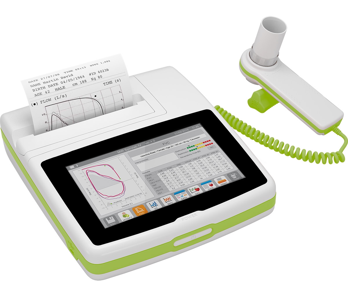
Angiotensin-converting enzyme (ACE) inhibitors are established as first-line therapy in chronic heart failure (CHF). However, little is known about the dosage-plasma-level relationship of ACE inhibitors in CHF and its relation to drug-induced adverse effects. We investigated 45 patients (age 55 +/- 10 years) with stable CHF who presented with a maintenance dosage of enalapril of either 5 mg b.i.d. (E10, n = 16), 10 mg b.i.d. (E20, n = 18), or 20 mg b.i.d. (E40, n = 11). This dosage was changed three times to treat all patients with lower, higher, and, finally, the initial dosage for 4 weeks each. Patients were examined clinically, by questionnaire, and by spiroergometry. In addition, neurohormones (atrial and brain natriuretic peptide and norepinephrine), enalaprilat trough levels, and serum potassium and creatinine were measured. Enalaprilat trough levels differed significantly between the three groups at study entry but also varied markedly within each group. In addition to the dose of enalapril, serum creatinine, severity of CHF, basal metabolic rate, and body weight significantly influenced enalaprilat trough levels (R2 =.84, p <.001). Within-patient comparisons revealed that serum creatinine (107 +/- 26 versus 102 +/- 20 micromol/liter) and potassium (3.8 +/- 0.4 versus 3.7 +/- 0. 3mmol/liter) were higher, cough was more common (scored on a scale of 0-8: 1.7 +/- 2.1 versus 1.4 +/- 1.8), and blood pressure was lower (systolic, 112 +/- 14 versus 117 +/- 13 mm Hg; diastolic, 66 +/- 9 versus 69 +/- 11 mm Hg) on the highest than on the lowest enalaprilat trough level (all p <.05). Highly variable enalaprilat trough levels and the fact that adverse effects were more common on high enalaprilat trough levels provide a rationale for individually adjusting ACE-inhibitor dose in case of adverse effects. (+info)Spirometry is a common type of pulmonary function test (PFT) that measures how well your lungs work. This is done by measuring how much air you can exhale from your lungs after taking a deep breath, and how quickly you can exhale it. The results are compared to normal values for your age, height, sex, and ethnicity.
Spirometry is used to diagnose and monitor certain lung conditions, such as asthma, chronic obstructive pulmonary disease (COPD), and other respiratory diseases that cause narrowing of the airways. It can also be used to assess the effectiveness of treatment for these conditions. The test is non-invasive, safe, and easy to perform.
Forced Expiratory Volume (FEV) is a medical term used to describe the volume of air that can be forcefully exhaled from the lungs in one second. It is often measured during pulmonary function testing to assess lung function and diagnose conditions such as chronic obstructive pulmonary disease (COPD) or asthma.
FEV is typically expressed as a percentage of the Forced Vital Capacity (FVC), which is the total volume of air that can be exhaled from the lungs after taking a deep breath in. The ratio of FEV to FVC is used to determine whether there is obstruction in the airways, with a lower ratio indicating more severe obstruction.
There are different types of FEV measurements, including FEV1 (the volume of air exhaled in one second), FEV25-75 (the average volume of air exhaled during the middle 50% of the FVC maneuver), and FEV0.5 (the volume of air exhaled in half a second). These measurements can provide additional information about lung function and help guide treatment decisions.
Vital capacity (VC) is a term used in pulmonary function tests to describe the maximum volume of air that can be exhaled after taking a deep breath. It is the sum of inspiratory reserve volume, tidal volume, and expiratory reserve volume. In other words, it's the total amount of air you can forcibly exhale after inhaling as deeply as possible. Vital capacity is an important measurement in assessing lung function and can be reduced in conditions such as chronic obstructive pulmonary disease (COPD), asthma, and other respiratory disorders.
Respiratory Function Tests (RFTs) are a group of medical tests that measure how well your lungs take in and exhale air, and how well they transfer oxygen and carbon dioxide into and out of your blood. They can help diagnose certain lung disorders, measure the severity of lung disease, and monitor response to treatment.
RFTs include several types of tests, such as:
1. Spirometry: This test measures how much air you can exhale and how quickly you can do it. It's often used to diagnose and monitor conditions like asthma, chronic obstructive pulmonary disease (COPD), and other lung diseases.
2. Lung volume testing: This test measures the total amount of air in your lungs. It can help diagnose restrictive lung diseases, such as pulmonary fibrosis or sarcoidosis.
3. Diffusion capacity testing: This test measures how well oxygen moves from your lungs into your bloodstream. It's often used to diagnose and monitor conditions like pulmonary fibrosis, interstitial lung disease, and other lung diseases that affect the ability of the lungs to transfer oxygen to the blood.
4. Bronchoprovocation testing: This test involves inhaling a substance that can cause your airways to narrow, such as methacholine or histamine. It's often used to diagnose and monitor asthma.
5. Exercise stress testing: This test measures how well your lungs and heart work together during exercise. It's often used to diagnose lung or heart disease.
Overall, Respiratory Function Tests are an important tool for diagnosing and managing a wide range of lung conditions.
Chronic obstructive pulmonary disease (COPD) is a progressive lung disease characterized by the persistent obstruction of airflow in and out of the lungs. This obstruction is usually caused by two primary conditions: chronic bronchitis and emphysema. Chronic bronchitis involves inflammation and narrowing of the airways, leading to excessive mucus production and coughing. Emphysema is a condition where the alveoli (air sacs) in the lungs are damaged, resulting in decreased gas exchange and shortness of breath.
The main symptoms of COPD include progressive shortness of breath, chronic cough, chest tightness, wheezing, and excessive mucus production. The disease is often associated with exposure to harmful particles or gases, such as cigarette smoke, air pollution, or occupational dusts and chemicals. While there is no cure for COPD, treatments can help alleviate symptoms, improve quality of life, and slow the progression of the disease. These treatments may include bronchodilators, corticosteroids, combination inhalers, pulmonary rehabilitation, and, in severe cases, oxygen therapy or lung transplantation.
Asthma is a chronic respiratory disease characterized by inflammation and narrowing of the airways, leading to symptoms such as wheezing, coughing, shortness of breath, and chest tightness. The airway obstruction in asthma is usually reversible, either spontaneously or with treatment.
The underlying cause of asthma involves a combination of genetic and environmental factors that result in hypersensitivity of the airways to certain triggers, such as allergens, irritants, viruses, exercise, and emotional stress. When these triggers are encountered, the airways constrict due to smooth muscle spasm, swell due to inflammation, and produce excess mucus, leading to the characteristic symptoms of asthma.
Asthma is typically managed with a combination of medications that include bronchodilators to relax the airway muscles, corticosteroids to reduce inflammation, and leukotriene modifiers or mast cell stabilizers to prevent allergic reactions. Avoiding triggers and monitoring symptoms are also important components of asthma management.
There are several types of asthma, including allergic asthma, non-allergic asthma, exercise-induced asthma, occupational asthma, and nocturnal asthma, each with its own set of triggers and treatment approaches. Proper diagnosis and management of asthma can help prevent exacerbations, improve quality of life, and reduce the risk of long-term complications.
Bronchodilators are medications that relax and widen the airways (bronchioles) in the lungs, making it easier to breathe. They work by relaxing the smooth muscle around the airways, which allows them to dilate or open up. This results in improved airflow and reduced symptoms of bronchoconstriction, such as wheezing, coughing, and shortness of breath.
Bronchodilators can be classified into two main types: short-acting and long-acting. Short-acting bronchodilators are used for quick relief of symptoms and last for 4 to 6 hours, while long-acting bronchodilators are used for maintenance therapy and provide symptom relief for 12 hours or more.
Examples of bronchodilator agents include:
* Short-acting beta-agonists (SABAs) such as albuterol, levalbuterol, and pirbuterol
* Long-acting beta-agonists (LABAs) such as salmeterol, formoterol, and indacaterol
* Anticholinergics such as ipratropium, tiotropium, and aclidinium
* Combination bronchodilators that contain both a LABA and an anticholinergic, such as umeclidinium/vilanterol and glycopyrrolate/formoterol.
Airway obstruction is a medical condition that occurs when the normal flow of air into and out of the lungs is partially or completely blocked. This blockage can be caused by a variety of factors, including swelling of the tissues in the airway, the presence of foreign objects or substances, or abnormal growths such as tumors.
When the airway becomes obstructed, it can make it difficult for a person to breathe normally. They may experience symptoms such as shortness of breath, wheezing, coughing, and chest tightness. In severe cases, airway obstruction can lead to respiratory failure and other life-threatening complications.
There are several types of airway obstruction, including:
1. Upper airway obstruction: This occurs when the blockage is located in the upper part of the airway, such as the nose, throat, or voice box.
2. Lower airway obstruction: This occurs when the blockage is located in the lower part of the airway, such as the trachea or bronchi.
3. Partial airway obstruction: This occurs when the airway is partially blocked, allowing some air to flow in and out of the lungs.
4. Complete airway obstruction: This occurs when the airway is completely blocked, preventing any air from flowing into or out of the lungs.
Treatment for airway obstruction depends on the underlying cause of the condition. In some cases, removing the obstruction may be as simple as clearing the airway of foreign objects or mucus. In other cases, more invasive treatments such as surgery may be necessary.
Obstructive lung disease is a category of respiratory diseases characterized by airflow limitation that causes difficulty in completely emptying the alveoli (tiny air sacs) of the lungs during exhaling. This results in the trapping of stale air and prevents fresh air from entering the alveoli, leading to various symptoms such as coughing, wheezing, shortness of breath, and decreased exercise tolerance.
The most common obstructive lung diseases include:
1. Chronic Obstructive Pulmonary Disease (COPD): A progressive disease that includes chronic bronchitis and emphysema, often caused by smoking or exposure to harmful pollutants.
2. Asthma: A chronic inflammatory disorder of the airways characterized by variable airflow obstruction, bronchial hyperresponsiveness, and an underlying inflammation. Symptoms can be triggered by various factors such as allergens, irritants, or physical activity.
3. Bronchiectasis: A condition in which the airways become abnormally widened, scarred, and thickened due to chronic inflammation or infection, leading to mucus buildup and impaired clearance.
4. Cystic Fibrosis: An inherited genetic disorder that affects the exocrine glands, resulting in thick and sticky mucus production in various organs, including the lungs. This can lead to chronic lung infections, inflammation, and airway obstruction.
5. Alpha-1 Antitrypsin Deficiency: A genetic condition characterized by low levels of alpha-1 antitrypsin protein, which leads to uncontrolled protease enzyme activity that damages the lung tissue, causing emphysema-like symptoms.
Treatment for obstructive lung diseases typically involves bronchodilators (to relax and widen the airways), corticosteroids (to reduce inflammation), and lifestyle modifications such as smoking cessation and pulmonary rehabilitation programs. In severe cases, oxygen therapy or even lung transplantation may be considered.
Lung diseases refer to a broad category of disorders that affect the lungs and other structures within the respiratory system. These diseases can impair lung function, leading to symptoms such as coughing, shortness of breath, chest pain, and wheezing. They can be categorized into several types based on the underlying cause and nature of the disease process. Some common examples include:
1. Obstructive lung diseases: These are characterized by narrowing or blockage of the airways, making it difficult to breathe out. Examples include chronic obstructive pulmonary disease (COPD), asthma, bronchiectasis, and cystic fibrosis.
2. Restrictive lung diseases: These involve stiffening or scarring of the lungs, which reduces their ability to expand and take in air. Examples include idiopathic pulmonary fibrosis, sarcoidosis, and asbestosis.
3. Infectious lung diseases: These are caused by bacteria, viruses, fungi, or parasites that infect the lungs. Examples include pneumonia, tuberculosis, and influenza.
4. Vascular lung diseases: These affect the blood vessels in the lungs, impairing oxygen exchange. Examples include pulmonary embolism, pulmonary hypertension, and chronic thromboembolic pulmonary hypertension (CTEPH).
5. Neoplastic lung diseases: These involve abnormal growth of cells within the lungs, leading to cancer. Examples include small cell lung cancer, non-small cell lung cancer, and mesothelioma.
6. Other lung diseases: These include interstitial lung diseases, pleural effusions, and rare disorders such as pulmonary alveolar proteinosis and lymphangioleiomyomatosis (LAM).
It is important to note that this list is not exhaustive, and there are many other conditions that can affect the lungs. Proper diagnosis and treatment of lung diseases require consultation with a healthcare professional, such as a pulmonologist or respiratory therapist.
A lung is a pair of spongy, elastic organs in the chest that work together to enable breathing. They are responsible for taking in oxygen and expelling carbon dioxide through the process of respiration. The left lung has two lobes, while the right lung has three lobes. The lungs are protected by the ribcage and are covered by a double-layered membrane called the pleura. The trachea divides into two bronchi, which further divide into smaller bronchioles, leading to millions of tiny air sacs called alveoli, where the exchange of gases occurs.
The Maximal Mid-Expiratory Flow Rate (MMEFR), also known as Maximum Expiratory Flow at 50% of the FVC (FEF50%), is a measure of pulmonary function that reflects the rate of airflow during the middle portion of a forced expiratory maneuver. It is calculated as the maximum flow rate achieved during the expiration of air from the lungs, starting at 50% of the Forced Vital Capacity (FVC) and ending at the residual volume.
MMEFR is expressed in liters per second (L/s) or seconds (s). A decreased MMEFR may indicate obstruction in the smaller airways, such as bronchitis or asthma, while a normal value suggests that the small airways are functioning properly. However, it's important to note that MMEFR is just one of several measures used to assess pulmonary function and should be interpreted in conjunction with other test results and clinical findings.
Forced expiratory flow rates (FEFR) are measures of how quickly and efficiently air can be exhaled from the lungs during a forced breath maneuver. These measurements are often used in pulmonary function testing to help diagnose and monitor obstructive lung diseases such as asthma or chronic obstructive pulmonary disease (COPD).
FEFR is typically measured during a forced expiratory maneuver, where the person takes a deep breath in and then exhales as forcefully and quickly as possible into a mouthpiece connected to a spirometer. The spirometer measures the volume and flow rate of the exhaled air over time.
There are several different FEFR measurements that can be reported, including:
* Forced Expiratory Flow (FEF) 25-75%: This is the average flow rate during the middle half of the forced expiratory maneuver.
* Peak Expiratory Flow Rate (PEFR): This is the maximum flow rate achieved during the first second of the forced expiratory maneuver.
* Forced Expiratory Volume in 1 Second (FEV1): This is the volume of air exhaled in the first second of the forced expiratory maneuver.
Abnormal FEFR values can indicate obstruction in the small airways of the lungs, which can make it difficult to breathe out fully and quickly. The specific pattern of abnormalities in FEFR measurements can help doctors differentiate between different types of obstructive lung diseases.
Dyspnea is defined as difficulty or discomfort in breathing, often described as shortness of breath. It can range from mild to severe, and may occur during rest, exercise, or at any time. Dyspnea can be caused by various medical conditions, including heart and lung diseases, anemia, and neuromuscular disorders. It is important to seek medical attention if experiencing dyspnea, as it can be a sign of a serious underlying condition.
Peak Expiratory Flow Rate (PEFR) is a measurement of how quickly a person can exhale air from their lungs. It is often used as a quick test to assess breathing difficulties in people with respiratory conditions such as asthma or chronic obstructive pulmonary disease (COPD). PEFR is measured in liters per minute (L/min) and the highest value obtained during a forceful exhalation is recorded as the peak expiratory flow rate. Regular monitoring of PEFR can help to assess the severity of an asthma attack or the effectiveness of treatment.
Pulmonary medicine is a medical specialty that deals with the diagnosis, treatment, and prevention of diseases and conditions affecting the respiratory system, including the lungs, trachea, bronchi, bronchioles, and alveoli. Pulmonologists are specialists who treat a wide range of respiratory disorders such as chronic obstructive pulmonary disease (COPD), asthma, bronchitis, pneumonia, lung cancer, sleep-disordered breathing, tuberculosis, and interstitial lung diseases. They use various diagnostic techniques including chest X-rays, CT scans, pulmonary function tests, bronchoscopy, and sleep studies to evaluate and manage respiratory disorders. Pulmonologists also provide care for patients who require long-term mechanical ventilation or oxygen therapy.
Oscillometry is a non-invasive method to measure various mechanical properties of the respiratory system, including lung volumes and airway resistance. It involves applying small pressure oscillations to the airways and measuring the resulting flow or volume changes. The technique can be used to assess lung function in patients with obstructive or restrictive lung diseases, as well as in healthy individuals. Oscillometry is often performed during tidal breathing, making it a comfortable method for both children and adults who may have difficulty performing traditional spirometry maneuvers.
Airway resistance is a measure of the opposition to airflow during breathing, which is caused by the friction between the air and the walls of the respiratory tract. It is an important parameter in respiratory physiology because it can affect the work of breathing and gas exchange.
Airway resistance is usually expressed in units of cm H2O/L/s or Pa·s/m, and it can be measured during spontaneous breathing or during forced expiratory maneuvers, such as those used in pulmonary function testing. Increased airway resistance can result from a variety of conditions, including asthma, chronic obstructive pulmonary disease (COPD), bronchitis, and bronchiectasis. Decreased airway resistance can be seen in conditions such as emphysema or after a successful bronchodilator treatment.
Respiratory disorders are a group of conditions that affect the respiratory system, including the nose, throat (pharynx), windpipe (trachea), bronchi, lungs, and diaphragm. These disorders can make it difficult for a person to breathe normally and may cause symptoms such as coughing, wheezing, shortness of breath, and chest pain.
There are many different types of respiratory disorders, including:
1. Asthma: A chronic inflammatory disease that causes the airways to become narrow and swollen, leading to difficulty breathing.
2. Chronic obstructive pulmonary disease (COPD): A group of lung diseases, including emphysema and chronic bronchitis, that make it hard to breathe.
3. Pneumonia: An infection of the lungs that can cause coughing, chest pain, and difficulty breathing.
4. Lung cancer: A type of cancer that forms in the tissues of the lungs and can cause symptoms such as coughing, chest pain, and shortness of breath.
5. Tuberculosis (TB): A bacterial infection that mainly affects the lungs but can also affect other parts of the body.
6. Sleep apnea: A disorder that causes a person to stop breathing for short periods during sleep.
7. Interstitial lung disease: A group of disorders that cause scarring of the lung tissue, leading to difficulty breathing.
8. Pulmonary fibrosis: A type of interstitial lung disease that causes scarring of the lung tissue and makes it hard to breathe.
9. Pleural effusion: An abnormal accumulation of fluid in the space between the lungs and chest wall.
10. Lung transplantation: A surgical procedure to replace a diseased or failing lung with a healthy one from a donor.
Respiratory disorders can be caused by a variety of factors, including genetics, exposure to environmental pollutants, smoking, and infections. Treatment for respiratory disorders may include medications, oxygen therapy, breathing exercises, and lifestyle changes. In some cases, surgery may be necessary to treat the disorder.
Respiratory tract diseases refer to a broad range of medical conditions that affect the respiratory system, which includes the nose, throat (pharynx), windpipe (trachea), bronchi, bronchioles, and lungs. These diseases can be categorized into upper and lower respiratory tract infections based on the location of the infection.
Upper respiratory tract infections affect the nose, sinuses, pharynx, and larynx, and include conditions such as the common cold, flu, sinusitis, and laryngitis. Symptoms often include nasal congestion, sore throat, cough, and fever.
Lower respiratory tract infections affect the trachea, bronchi, bronchioles, and lungs, and can be more severe. They include conditions such as pneumonia, bronchitis, and tuberculosis. Symptoms may include cough, chest congestion, shortness of breath, and fever.
Respiratory tract diseases can also be caused by allergies, irritants, or genetic factors. Treatment varies depending on the specific condition and severity but may include medications, breathing treatments, or surgery in severe cases.
Bronchial provocation tests are a group of medical tests used to assess the airway responsiveness of the lungs by challenging them with increasing doses of a specific stimulus, such as methacholine or histamine, which can cause bronchoconstriction (narrowing of the airways) in susceptible individuals. These tests are often performed to diagnose and monitor asthma and other respiratory conditions that may be associated with heightened airway responsiveness.
The most common type of bronchial provocation test is the methacholine challenge test, which involves inhaling increasing concentrations of methacholine aerosol via a nebulizer. The dose response is measured by monitoring lung function (usually through spirometry) before and after each exposure. A positive test is indicated when there is a significant decrease in forced expiratory volume in one second (FEV1) or other measures of airflow, which suggests bronchial hyperresponsiveness.
Other types of bronchial provocation tests include histamine challenges, exercise challenges, and mannitol challenges. These tests have specific indications, contraindications, and protocols that should be followed to ensure accurate results and patient safety. Bronchial provocation tests are typically conducted in a controlled clinical setting under the supervision of trained healthcare professionals.
I'm sorry for any confusion, but "Bronchospirometry" is not a widely recognized or established medical term or procedure in the field of pulmonology (lung medicine) or respiratory function testing. It seems that this term may be a combination of 'broncho-,' referring to the bronchi or airways within the lungs, and '-spirometry,' which relates to measuring lung volumes and flows.
However, I cannot find any specific medical definition, research studies, or literature that directly use or describe "Bronchospirometry." It is essential to rely on established and well-defined medical terminology to ensure accurate communication between healthcare professionals and researchers.
If you have more context or information about where you encountered this term, I would be happy to help further clarify the situation.
Smoking is not a medical condition, but it's a significant health risk behavior. Here is the definition from a public health perspective:
Smoking is the act of inhaling and exhaling the smoke of burning tobacco that is commonly consumed through cigarettes, pipes, and cigars. The smoke contains over 7,000 chemicals, including nicotine, tar, carbon monoxide, and numerous toxic and carcinogenic substances. These toxins contribute to a wide range of diseases and health conditions, such as lung cancer, heart disease, stroke, chronic obstructive pulmonary disease (COPD), and various other cancers, as well as adverse reproductive outcomes and negative impacts on the developing fetus during pregnancy. Smoking is highly addictive due to the nicotine content, which makes quitting smoking a significant challenge for many individuals.
Maximal Expiratory Flow Rate (MEFR) is a measure of how quickly a person can exhale air from their lungs. It is often used in pulmonary function testing to assess the degree of airflow obstruction in conditions such as chronic obstructive pulmonary disease (COPD) or asthma.
The MEFR is typically measured by having the person take a deep breath and then exhale as forcefully and quickly as possible into a device that measures the volume and flow of air. The MEFR is calculated as the maximum flow rate achieved during the exhalation maneuver, usually expressed in liters per second (L/s) or seconds (L/sec).
MEFR can be measured at different lung volumes, such as at functional residual capacity (FRC) or at total lung capacity (TLC), to provide additional information about the severity and location of airflow obstruction. However, MEFR is not as commonly used in clinical practice as other measures of pulmonary function, such as forced expiratory volume in one second (FEV1) or forced vital capacity (FVC).
Lung volume measurements are clinical tests that determine the amount of air inhaled, exhaled, and present in the lungs at different times during the breathing cycle. These measurements include:
1. Tidal Volume (TV): The amount of air inhaled or exhaled during normal breathing, usually around 500 mL in resting adults.
2. Inspiratory Reserve Volume (IRV): The additional air that can be inhaled after a normal inspiration, approximately 3,000 mL in adults.
3. Expiratory Reserve Volume (ERV): The extra air that can be exhaled after a normal expiration, about 1,000-1,200 mL in adults.
4. Residual Volume (RV): The air remaining in the lungs after a maximal exhalation, approximately 1,100-1,500 mL in adults.
5. Total Lung Capacity (TLC): The total amount of air the lungs can hold at full inflation, calculated as TV + IRV + ERV + RV, around 6,000 mL in adults.
6. Functional Residual Capacity (FRC): The volume of air remaining in the lungs after a normal expiration, equal to ERV + RV, about 2,100-2,700 mL in adults.
7. Inspiratory Capacity (IC): The maximum amount of air that can be inhaled after a normal expiration, equal to TV + IRV, around 3,500 mL in adults.
8. Vital Capacity (VC): The total volume of air that can be exhaled after a maximal inspiration, calculated as IC + ERV, approximately 4,200-5,600 mL in adults.
These measurements help assess lung function and identify various respiratory disorders such as chronic obstructive pulmonary disease (COPD), asthma, and restrictive lung diseases.
A Severity of Illness Index is a measurement tool used in healthcare to assess the severity of a patient's condition and the risk of mortality or other adverse outcomes. These indices typically take into account various physiological and clinical variables, such as vital signs, laboratory values, and co-morbidities, to generate a score that reflects the patient's overall illness severity.
Examples of Severity of Illness Indices include the Acute Physiology and Chronic Health Evaluation (APACHE) system, the Simplified Acute Physiology Score (SAPS), and the Mortality Probability Model (MPM). These indices are often used in critical care settings to guide clinical decision-making, inform prognosis, and compare outcomes across different patient populations.
It is important to note that while these indices can provide valuable information about a patient's condition, they should not be used as the sole basis for clinical decision-making. Rather, they should be considered in conjunction with other factors, such as the patient's overall clinical presentation, treatment preferences, and goals of care.
Exhalation is the act of breathing out or exhaling, which is the reverse process of inhalation. During exhalation, the diaphragm relaxes and moves upwards, while the chest muscles also relax, causing the chest cavity to decrease in size. This decrease in size puts pressure on the lungs, causing them to deflate and expel air.
Exhalation is a passive process that occurs naturally after inhalation, but it can also be actively controlled during activities such as speaking, singing, or playing a wind instrument. In medical terms, exhalation may also be referred to as expiration.
Whole-body plethysmography is a non-invasive medical technique used to measure changes in the volume of air in the lungs and chest during breathing. It is often utilized in the diagnosis and assessment of various respiratory disorders such as chronic obstructive pulmonary disease (COPD), asthma, and restrictive lung diseases.
During whole-body plethysmography, the patient enters a sealed, clear chamber, usually in a standing or sitting position. The patient is instructed to breathe normally while the machine measures changes in pressure within the chamber as the chest and abdomen move during respiration. These measurements are then used to calculate lung volume, airflow, and other respiratory parameters.
This technique provides valuable information about the functional status of the lungs and can help healthcare providers make informed decisions regarding diagnosis, treatment planning, and disease monitoring.
Respiratory therapy is a healthcare profession that specializes in the diagnosis, treatment, and management of respiratory disorders and diseases. Respiratory therapists (RTs) work under the direction of physicians to provide care for patients with conditions such as chronic obstructive pulmonary disease (COPD), asthma, cystic fibrosis, sleep apnea, and neuromuscular diseases that affect breathing.
RTs use a variety of techniques and treatments to help patients breathe more easily, including oxygen therapy, aerosol medication delivery, chest physiotherapy, mechanical ventilation, and patient education. They also perform diagnostic tests such as pulmonary function studies to assess lung function and help diagnose respiratory conditions.
RTs work in a variety of healthcare settings, including hospitals, clinics, long-term care facilities, and home health agencies. They may provide care for patients of all ages, from premature infants to the elderly. The overall goal of respiratory therapy is to help patients achieve and maintain optimal lung function and quality of life.
Pulmonary diffusing capacity, also known as pulmonary diffusion capacity, is a measure of the ability of the lungs to transfer gas from the alveoli to the bloodstream. It is often used to assess the severity of lung diseases such as chronic obstructive pulmonary disease (COPD) and pulmonary fibrosis.
The most common measurement of pulmonary diffusing capacity is the diffusing capacity for carbon monoxide (DLCO), which reflects the transfer of carbon monoxide from the alveoli to the red blood cells in the capillaries. The DLCO is measured during a spirometry test, which involves breathing in a small amount of carbon monoxide and then measuring how much of it is exhaled.
A reduced DLCO may indicate a problem with the lung's ability to transfer oxygen to the blood, which can be caused by a variety of factors including damage to the alveoli or capillaries, thickening of the alveolar membrane, or a decrease in the surface area available for gas exchange.
It is important to note that other factors such as hemoglobin concentration, carboxyhemoglobin level, and lung volume can also affect the DLCO value, so these should be taken into account when interpreting the results of a diffusing capacity test.
Breathing exercises are a series of deliberate breathing techniques that aim to improve respiratory function, reduce stress and anxiety, and promote relaxation. These exercises can involve various methods such as deep, slow, or rhythmic breathing, often combined with other practices like pursed-lips breathing, diaphragmatic breathing, or alternate nostril breathing. By focusing on the breath and controlling its pace and depth, individuals can experience numerous health benefits, including improved lung capacity, reduced heart rate, increased oxygenation of the blood, and a greater sense of calm and well-being. Breathing exercises are often used as a complementary therapy in various medical and holistic practices, such as yoga, meditation, and stress management programs.
Respiratory sounds are the noises produced by the airflow through the respiratory tract during breathing. These sounds can provide valuable information about the health and function of the lungs and airways. They are typically categorized into two main types: normal breath sounds and adventitious (or abnormal) breath sounds.
Normal breath sounds include:
1. Vesicular breath sounds: These are soft, low-pitched sounds heard over most of the lung fields during quiet breathing. They are produced by the movement of air through the alveoli and smaller bronchioles.
2. Bronchovesicular breath sounds: These are medium-pitched, hollow sounds heard over the mainstem bronchi and near the upper sternal border during both inspiration and expiration. They are a combination of vesicular and bronchial breath sounds.
Abnormal or adventitious breath sounds include:
1. Crackles (or rales): These are discontinuous, non-musical sounds that resemble the crackling of paper or bubbling in a fluid-filled container. They can be heard during inspiration and are caused by the sudden opening of collapsed airways or the movement of fluid within the airways.
2. Wheezes: These are continuous, musical sounds resembling a whistle. They are produced by the narrowing or obstruction of the airways, causing turbulent airflow.
3. Rhonchi: These are low-pitched, rumbling, continuous sounds that can be heard during both inspiration and expiration. They are caused by the vibration of secretions or fluids in the larger airways.
4. Stridor: This is a high-pitched, inspiratory sound that resembles a harsh crowing or barking noise. It is usually indicative of upper airway narrowing or obstruction.
The character, location, and duration of respiratory sounds can help healthcare professionals diagnose various respiratory conditions, such as pneumonia, chronic obstructive pulmonary disease (COPD), asthma, and bronchitis.
A cross-sectional study is a type of observational research design that examines the relationship between variables at one point in time. It provides a snapshot or a "cross-section" of the population at a particular moment, allowing researchers to estimate the prevalence of a disease or condition and identify potential risk factors or associations.
In a cross-sectional study, data is collected from a sample of participants at a single time point, and the variables of interest are measured simultaneously. This design can be used to investigate the association between exposure and outcome, but it cannot establish causality because it does not follow changes over time.
Cross-sectional studies can be conducted using various data collection methods, such as surveys, interviews, or medical examinations. They are often used in epidemiology to estimate the prevalence of a disease or condition in a population and to identify potential risk factors that may contribute to its development. However, because cross-sectional studies only provide a snapshot of the population at one point in time, they cannot account for changes over time or determine whether exposure preceded the outcome.
Therefore, while cross-sectional studies can be useful for generating hypotheses and identifying potential associations between variables, further research using other study designs, such as cohort or case-control studies, is necessary to establish causality and confirm any findings.
Occupational exposure refers to the contact of an individual with potentially harmful chemical, physical, or biological agents as a result of their job or occupation. This can include exposure to hazardous substances such as chemicals, heavy metals, or dusts; physical agents such as noise, radiation, or ergonomic stressors; and biological agents such as viruses, bacteria, or fungi.
Occupational exposure can occur through various routes, including inhalation, skin contact, ingestion, or injection. Prolonged or repeated exposure to these hazards can increase the risk of developing acute or chronic health conditions, such as respiratory diseases, skin disorders, neurological damage, or cancer.
Employers have a legal and ethical responsibility to minimize occupational exposures through the implementation of appropriate control measures, including engineering controls, administrative controls, personal protective equipment, and training programs. Regular monitoring and surveillance of workers' health can also help identify and prevent potential health hazards in the workplace.
Albuterol is a medication that is used to treat bronchospasm, or narrowing of the airways in the lungs, in conditions such as asthma and chronic obstructive pulmonary disease (COPD). It is a short-acting beta-2 agonist, which means it works by relaxing the muscles around the airways, making it easier to breathe. Albuterol is available in several forms, including an inhaler, nebulizer solution, and syrup, and it is typically used as needed to relieve symptoms of bronchospasm. It may also be used before exercise to prevent bronchospasm caused by physical activity.
The medical definition of Albuterol is: "A short-acting beta-2 adrenergic agonist used to treat bronchospasm in conditions such as asthma and COPD. It works by relaxing the muscles around the airways, making it easier to breathe."
Methacholine chloride is a medication that is used as a diagnostic tool to help identify and assess the severity of asthma or other respiratory conditions that cause airway hyperresponsiveness. It is a synthetic derivative of acetylcholine, which is a neurotransmitter that causes smooth muscle contraction in the body.
When methacholine chloride is inhaled, it stimulates the muscarinic receptors in the airways, causing them to constrict or narrow. This response is measured and used to determine the degree of airway hyperresponsiveness, which can help diagnose asthma and assess its severity.
The methacholine challenge test involves inhaling progressively higher doses of methacholine chloride until a significant decrease in lung function is observed or until a maximum dose is reached. The test results are then used to guide treatment decisions and monitor the effectiveness of therapy. It's important to note that this test should be conducted under the supervision of a healthcare professional, as it carries some risks, including bronchoconstriction and respiratory distress.
A cough is a reflex action that helps to clear the airways of irritants, foreign particles, or excess mucus or phlegm. It is characterized by a sudden, forceful expulsion of air from the lungs through the mouth and nose. A cough can be acute (short-term) or chronic (long-term), and it can be accompanied by other symptoms such as chest pain, shortness of breath, or fever. Coughing can be caused by various factors, including respiratory infections, allergies, asthma, environmental pollutants, gastroesophageal reflux disease (GERD), and chronic lung diseases such as chronic obstructive pulmonary disease (COPD) and bronchitis. In some cases, a cough may be a symptom of a more serious underlying condition, such as heart failure or lung cancer.
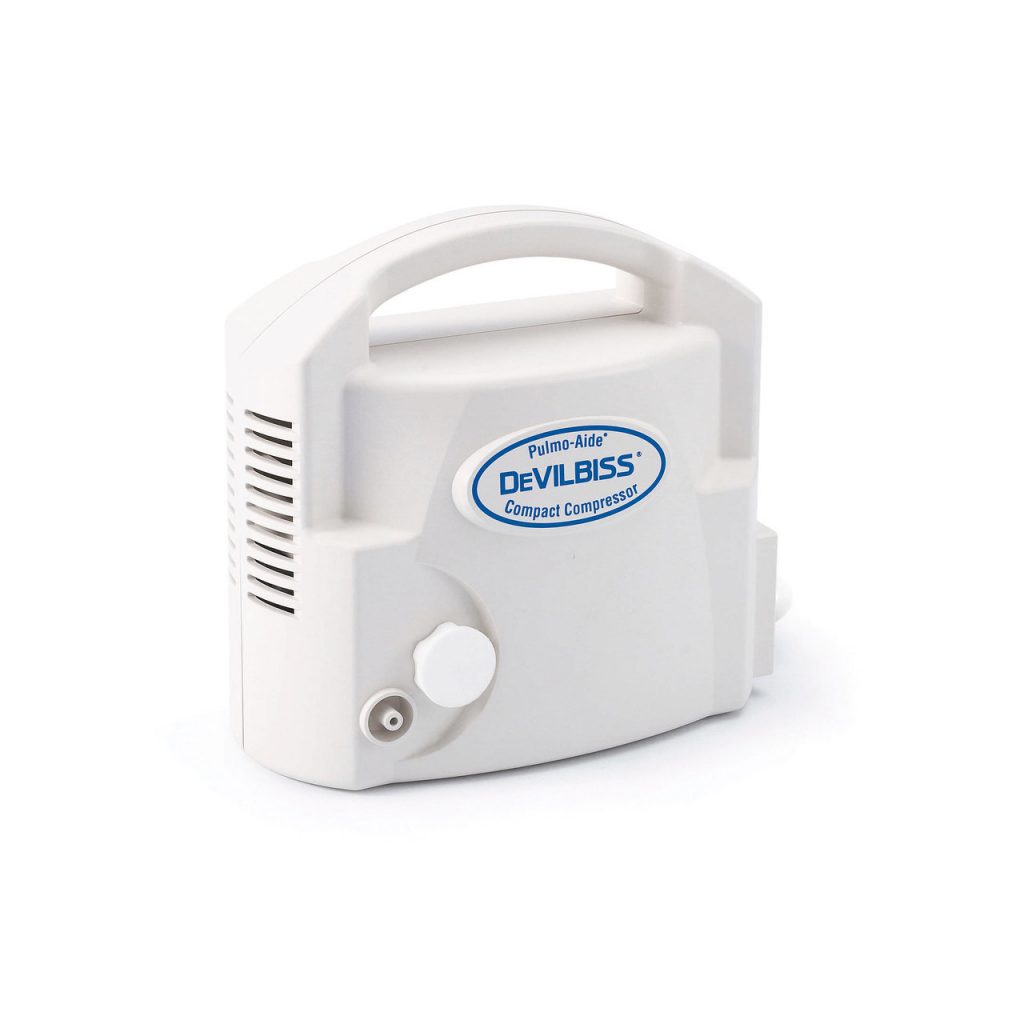
"Inhalation administration" is a medical term that refers to the method of delivering medications or therapeutic agents directly into the lungs by inhaling them through the airways. This route of administration is commonly used for treating respiratory conditions such as asthma, COPD (chronic obstructive pulmonary disease), and cystic fibrosis.
Inhalation administration can be achieved using various devices, including metered-dose inhalers (MDIs), dry powder inhalers (DPIs), nebulizers, and soft-mist inhalers. Each device has its unique mechanism of delivering the medication into the lungs, but they all aim to provide a high concentration of the drug directly to the site of action while minimizing systemic exposure and side effects.
The advantages of inhalation administration include rapid onset of action, increased local drug concentration, reduced systemic side effects, and improved patient compliance due to the ease of use and non-invasive nature of the delivery method. However, proper technique and device usage are crucial for effective therapy, as incorrect usage may result in suboptimal drug deposition and therapeutic outcomes.
Occupational diseases are health conditions or illnesses that occur as a result of exposure to hazards in the workplace. These hazards can include physical, chemical, and biological agents, as well as ergonomic factors and work-related psychosocial stressors. Examples of occupational diseases include respiratory illnesses caused by inhaling dust or fumes, hearing loss due to excessive noise exposure, and musculoskeletal disorders caused by repetitive movements or poor ergonomics. The development of an occupational disease is typically related to the nature of the work being performed and the conditions in which it is carried out. It's important to note that these diseases can be prevented or minimized through proper risk assessment, implementation of control measures, and adherence to safety regulations.
Primary health care is defined by the World Health Organization (WHO) as:
"Essential health care that is based on practical, scientifically sound and socially acceptable methods and technology made universally accessible to individuals and families in the community through their full participation and at a cost that the community and country can afford. It forms an integral part both of the country's health system, of which it is the central function and main focus, and of the overall social and economic development of the community. It is the first level of contact of individuals, the family and community with the national health system bringing health care as close as possible to where people live and work, and constitutes the first element of a continuing health care process."
Primary health care includes a range of services such as preventive care, health promotion, curative care, rehabilitation, and palliative care. It is typically provided by a team of health professionals including doctors, nurses, midwives, pharmacists, and other community health workers. The goal of primary health care is to provide comprehensive, continuous, and coordinated care to individuals and families in a way that is accessible, affordable, and culturally sensitive.
In medical terms, "dust" is not defined as a specific medical condition or disease. However, generally speaking, dust refers to small particles of solid matter that can be found in the air and can come from various sources, such as soil, pollen, hair, textiles, paper, or plastic.
Exposure to certain types of dust, such as those containing allergens, chemicals, or harmful pathogens, can cause a range of health problems, including respiratory issues like asthma, allergies, and lung diseases. Prolonged exposure to certain types of dust, such as silica or asbestos, can even lead to serious conditions like silicosis or mesothelioma.
Therefore, it is important for individuals who work in environments with high levels of dust to take appropriate precautions, such as wearing masks and respirators, to minimize their exposure and reduce the risk of health problems.
A breath test is a medical or forensic procedure used to analyze a sample of exhaled breath in order to detect and measure the presence of various substances, most commonly alcohol. The test is typically conducted using a device called a breathalyzer, which measures the amount of alcohol in the breath and converts it into a reading of blood alcohol concentration (BAC).
In addition to alcohol, breath tests can also be used to detect other substances such as drugs or volatile organic compounds (VOCs) that may indicate certain medical conditions. However, these types of breath tests are less common and may not be as reliable or accurate as other diagnostic tests.
Breath testing is commonly used by law enforcement officers to determine whether a driver is impaired by alcohol and to establish probable cause for arrest. It is also used in some healthcare settings to monitor patients who are being treated for alcohol abuse or dependence.
Pulmonary ventilation, also known as pulmonary respiration or simply ventilation, is the process of moving air into and out of the lungs to facilitate gas exchange. It involves two main phases: inhalation (or inspiration) and exhalation (or expiration). During inhalation, the diaphragm and external intercostal muscles contract, causing the chest volume to increase and the pressure inside the chest to decrease, which then draws air into the lungs. Conversely, during exhalation, these muscles relax, causing the chest volume to decrease and the pressure inside the chest to increase, which pushes air out of the lungs. This process ensures that oxygen-rich air from the atmosphere enters the alveoli (air sacs in the lungs), where it can diffuse into the bloodstream, while carbon dioxide-rich air from the bloodstream in the capillaries surrounding the alveoli is expelled out of the body.
Total Lung Capacity (TLC) is the maximum volume of air that can be contained within the lungs at the end of a maximal inspiration. It includes all of the following lung volumes: tidal volume, inspiratory reserve volume, expiratory reserve volume, and residual volume. TLC can be measured directly using gas dilution techniques or indirectly by adding residual volume to vital capacity. Factors that affect TLC include age, sex, height, and lung health status.
Residual Volume (RV) is the amount of air that remains in the lungs after a forced exhale, also known as the "expiratory reserve volume." It is the lowest lung volume that can be reached during a forced exhalation and cannot be completely emptied due to the presence of alveoli that are too small or too far from the airways. This volume is important for maintaining the structural integrity of the lungs and preventing their collapse. Any additional air that enters the lungs after this point will increase the total lung capacity. The normal residual volume for an average adult human is typically around 1 to 1.5 liters.
Pulmonary emphysema is a chronic respiratory disease characterized by abnormal, permanent enlargement of the airspaces distal to the terminal bronchioles, accompanied by destruction of their walls and without obvious fibrosis. This results in loss of elastic recoil, which leads to trappling of air within the lungs and difficulty exhaling. It is often caused by cigarette smoking or long-term exposure to harmful pollutants. The disease is part of a group of conditions known as chronic obstructive pulmonary disease (COPD), which also includes chronic bronchitis.
Maximal expiratory flow-volume (MEFV) curves are a graphical representation of the maximum volume of air that can be exhaled during a forced breath, measured at different lung volumes. It is a pulmonary function test used to assess obstructive lung diseases such as asthma or chronic obstructive pulmonary disease (COPD).
The MEFV curve is created by having the patient take a deep breath in and then exhale as forcefully and quickly as possible into a spirometer, which measures the volume and flow of air. The test is repeated multiple times to ensure accurate results.
The MEFV curve provides information on the degree of obstruction in the airways, the location of the obstruction (central or peripheral), and the severity of the disease. It can also be used to monitor the effectiveness of treatment and disease progression over time.
Pulmonary atelectasis is a medical condition characterized by the collapse or closure of the alveoli (tiny air sacs) in the lungs, leading to reduced or absent gas exchange in the affected area. This results in decreased lung volume and can cause hypoxemia (low oxygen levels in the blood). Atelectasis can be caused by various factors such as obstruction of the airways, surfactant deficiency, pneumothorax, or compression from outside the lung. It can also occur after surgical procedures, particularly when the patient is lying in one position for a long time. Symptoms may include shortness of breath, cough, and chest discomfort, but sometimes it may not cause any symptoms, especially if only a small area of the lung is affected. Treatment depends on the underlying cause and may include bronchodilators, chest physiotherapy, or even surgery in severe cases.
Respiratory physiological phenomena refer to the various mechanical, chemical, and biological processes and functions that occur in the respiratory system during breathing and gas exchange. These phenomena include:
1. Ventilation: The movement of air into and out of the lungs, which is achieved through the contraction and relaxation of the diaphragm and intercostal muscles.
2. Gas Exchange: The diffusion of oxygen (O2) from the alveoli into the bloodstream and carbon dioxide (CO2) from the bloodstream into the alveoli.
3. Respiratory Mechanics: The physical properties and forces that affect the movement of air in and out of the lungs, such as lung compliance, airway resistance, and chest wall elasticity.
4. Control of Breathing: The regulation of ventilation by the central nervous system through the integration of sensory information from chemoreceptors and mechanoreceptors in the respiratory system.
5. Acid-Base Balance: The maintenance of a stable pH level in the blood through the regulation of CO2 elimination and bicarbonate balance by the respiratory and renal systems.
6. Oxygen Transport: The binding of O2 to hemoglobin in the red blood cells and its delivery to the tissues for metabolic processes.
7. Defense Mechanisms: The various protective mechanisms that prevent the entry and colonization of pathogens and foreign particles into the respiratory system, such as mucociliary clearance, cough reflex, and immune responses.
A questionnaire in the medical context is a standardized, systematic, and structured tool used to gather information from individuals regarding their symptoms, medical history, lifestyle, or other health-related factors. It typically consists of a series of written questions that can be either self-administered or administered by an interviewer. Questionnaires are widely used in various areas of healthcare, including clinical research, epidemiological studies, patient care, and health services evaluation to collect data that can inform diagnosis, treatment planning, and population health management. They provide a consistent and organized method for obtaining information from large groups or individual patients, helping to ensure accurate and comprehensive data collection while minimizing bias and variability in the information gathered.
Bronchial hyperresponsiveness (BHR) or bronchial hyperreactivity (BH) is a medical term that refers to the increased sensitivity and exaggerated response of the airways to various stimuli. In people with BHR, the airways narrow (constrict) more than usual in response to certain triggers such as allergens, cold air, exercise, or irritants like smoke or fumes. This narrowing can cause symptoms such as wheezing, coughing, chest tightness, and shortness of breath.
BHR is often associated with asthma and other respiratory conditions, including chronic obstructive pulmonary disease (COPD) and bronchiectasis. It is typically diagnosed through a series of tests that measure the degree of airway narrowing in response to various stimuli. These tests may include spirometry, methacholine challenge test, or histamine challenge test.
BHR can be managed with medications such as bronchodilators and anti-inflammatory drugs, which help to relax the muscles around the airways and reduce inflammation. It is also important to avoid triggers that can exacerbate symptoms and make BHR worse.
Bronchoconstrictor agents are substances that cause narrowing or constriction of the bronchioles, the small airways in the lungs. This can lead to symptoms such as wheezing, coughing, and shortness of breath. Bronchoconstrictor agents include certain medications (such as some beta-blockers and prostaglandin F2alpha), environmental pollutants (such as tobacco smoke and air pollution particles), and allergens (such as dust mites and pollen).
In contrast to bronchodilator agents, which are medications that widen the airways and improve breathing, bronchoconstrictor agents can make it more difficult for a person to breathe. People with respiratory conditions such as asthma or chronic obstructive pulmonary disease (COPD) may be particularly sensitive to bronchoconstrictor agents and may experience severe symptoms when exposed to them.
Plethysmography is a non-invasive medical technique used to measure changes in volume or blood flow within an organ or body part, typically in the lungs or extremities. There are several types of plethysmography, including:
1. **Whole Body Plethysmography (WBP):** This type of plethysmography is used to assess lung function and volumes by measuring changes in pressure within a sealed chamber that contains the patient's entire body except for their head. The patient breathes normally while wearing a nose clip, allowing technicians to analyze respiratory patterns, airflow, and lung volume changes.
2. **Segmental or Local Plethysmography:** This technique measures volume or blood flow changes in specific body parts, such as the limbs or digits. It can help diagnose and monitor conditions affecting peripheral circulation, like deep vein thrombosis, arterial occlusive disease, or Raynaud's phenomenon.
3. **Impedance Plethysmography (IPG):** This non-invasive method uses electrical impedance to estimate changes in blood volume within an organ or body part. By applying a small electrical current and measuring the opposition to flow (impedance), technicians can determine variations in blood volume, which can help diagnose conditions like deep vein thrombosis or heart failure.
4. **Optical Plethysmography:** This technique uses light to measure changes in blood volume, typically in the skin or mucous membranes. By shining a light on the area and analyzing the reflected or transmitted light, technicians can detect variations in blood volume related to cardiac output, respiration, or other physiological factors.
Overall, plethysmography is an essential tool for diagnosing and monitoring various medical conditions affecting circulation, respiratory function, and organ volumes.
Reference values, also known as reference ranges or reference intervals, are the set of values that are considered normal or typical for a particular population or group of people. These values are often used in laboratory tests to help interpret test results and determine whether a patient's value falls within the expected range.
The process of establishing reference values typically involves measuring a particular biomarker or parameter in a large, healthy population and then calculating the mean and standard deviation of the measurements. Based on these statistics, a range is established that includes a certain percentage of the population (often 95%) and excludes extreme outliers.
It's important to note that reference values can vary depending on factors such as age, sex, race, and other demographic characteristics. Therefore, it's essential to use reference values that are specific to the relevant population when interpreting laboratory test results. Additionally, reference values may change over time due to advances in measurement technology or changes in the population being studied.
Occupational asthma is a type of asthma that is caused or worsened by exposure to specific agents in the workplace. These agents, known as occupational sensitizers, can cause an immune response that leads to airway inflammation and narrowing, resulting in classic asthma symptoms such as wheezing, shortness of breath, coughing, and chest tightness.
Occupational asthma can develop in individuals who have no prior history of asthma, or it can worsen pre-existing asthma. The onset of symptoms may be immediate (within hours) or delayed (up to several days) after exposure to the sensitizer. Common occupational sensitizers include isocyanates (found in certain paints and spray foam insulation), flour and grain dust, wood dust, animal dander, and various chemicals used in manufacturing processes.
Prevention of occupational asthma involves minimizing or eliminating exposure to known sensitizers through proper engineering controls, personal protective equipment, and workplace practices. If occupational asthma is suspected, individuals should consult with a healthcare professional for appropriate diagnosis and management strategies.
Respiratory mechanics refers to the biomechanical properties and processes that involve the movement of air through the respiratory system during breathing. It encompasses the mechanical behavior of the lungs, chest wall, and the muscles of respiration, including the diaphragm and intercostal muscles.
Respiratory mechanics includes several key components:
1. **Compliance**: The ability of the lungs and chest wall to expand and recoil during breathing. High compliance means that the structures can easily expand and recoil, while low compliance indicates greater resistance to expansion and recoil.
2. **Resistance**: The opposition to airflow within the respiratory system, primarily due to the friction between the air and the airway walls. Airway resistance is influenced by factors such as airway diameter, length, and the viscosity of the air.
3. **Lung volumes and capacities**: These are the amounts of air present in the lungs during different phases of the breathing cycle. They include tidal volume (the amount of air inspired or expired during normal breathing), inspiratory reserve volume (additional air that can be inspired beyond the tidal volume), expiratory reserve volume (additional air that can be exhaled beyond the tidal volume), and residual volume (the air remaining in the lungs after a forced maximum exhalation).
4. **Work of breathing**: The energy required to overcome the resistance and elastic forces during breathing. This work is primarily performed by the respiratory muscles, which contract to generate negative intrathoracic pressure and expand the chest wall, allowing air to flow into the lungs.
5. **Pressure-volume relationships**: These describe how changes in lung volume are associated with changes in pressure within the respiratory system. Important pressure components include alveolar pressure (the pressure inside the alveoli), pleural pressure (the pressure between the lungs and the chest wall), and transpulmonary pressure (the difference between alveolar and pleural pressures).
Understanding respiratory mechanics is crucial for diagnosing and managing various respiratory disorders, such as chronic obstructive pulmonary disease (COPD), asthma, and restrictive lung diseases.
Respiratory muscles are a group of muscles involved in the process of breathing. They include the diaphragm, intercostal muscles (located between the ribs), scalene muscles (located in the neck), and abdominal muscles. These muscles work together to allow the chest cavity to expand or contract, which draws air into or pushes it out of the lungs. The diaphragm is the primary muscle responsible for breathing, contracting to increase the volume of the chest cavity and draw air into the lungs during inhalation. The intercostal muscles help to further expand the ribcage, while the abdominal muscles assist in exhaling by compressing the abdomen and pushing up on the diaphragm.
Bronchitis is a medical condition characterized by inflammation of the bronchi, which are the large airways that lead to the lungs. This inflammation can cause a variety of symptoms, including coughing, wheezing, chest tightness, and shortness of breath. Bronchitis can be either acute or chronic.
Acute bronchitis is usually caused by a viral infection, such as a cold or the flu, and typically lasts for a few days to a week. Symptoms may include a productive cough (coughing up mucus or phlegm), chest discomfort, and fatigue. Acute bronchitis often resolves on its own without specific medical treatment, although rest, hydration, and over-the-counter medications to manage symptoms may be helpful.
Chronic bronchitis, on the other hand, is a long-term condition that is characterized by a persistent cough with mucus production that lasts for at least three months out of the year for two consecutive years. Chronic bronchitis is typically caused by exposure to irritants such as cigarette smoke, air pollution, or occupational dusts and chemicals. It is often associated with chronic obstructive pulmonary disease (COPD), which includes both chronic bronchitis and emphysema.
Treatment for chronic bronchitis may include medications to help open the airways, such as bronchodilators and corticosteroids, as well as lifestyle changes such as smoking cessation and avoiding irritants. In severe cases, oxygen therapy or lung transplantation may be necessary.
Reproducibility of results in a medical context refers to the ability to obtain consistent and comparable findings when a particular experiment or study is repeated, either by the same researcher or by different researchers, following the same experimental protocol. It is an essential principle in scientific research that helps to ensure the validity and reliability of research findings.
In medical research, reproducibility of results is crucial for establishing the effectiveness and safety of new treatments, interventions, or diagnostic tools. It involves conducting well-designed studies with adequate sample sizes, appropriate statistical analyses, and transparent reporting of methods and findings to allow other researchers to replicate the study and confirm or refute the results.
The lack of reproducibility in medical research has become a significant concern in recent years, as several high-profile studies have failed to produce consistent findings when replicated by other researchers. This has led to increased scrutiny of research practices and a call for greater transparency, rigor, and standardization in the conduct and reporting of medical research.
Skin tests are medical diagnostic procedures that involve the application of a small amount of a substance to the skin, usually through a scratch, prick, or injection, to determine if the body has an allergic reaction to it. The most common type of skin test is the patch test, which involves applying a patch containing a small amount of the suspected allergen to the skin and observing the area for signs of a reaction, such as redness, swelling, or itching, over a period of several days. Another type of skin test is the intradermal test, in which a small amount of the substance is injected just beneath the surface of the skin. Skin tests are used to help diagnose allergies, including those to pollen, mold, pets, and foods, as well as to identify sensitivities to medications, chemicals, and other substances.
Cystic fibrosis (CF) is a genetic disorder that primarily affects the lungs and digestive system. It is caused by mutations in the CFTR gene, which regulates the movement of salt and water in and out of cells. When this gene is not functioning properly, thick, sticky mucus builds up in various organs, leading to a range of symptoms.
In the lungs, this mucus can clog the airways, making it difficult to breathe and increasing the risk of lung infections. Over time, lung damage can occur, which may lead to respiratory failure. In the digestive system, the thick mucus can prevent the release of digestive enzymes from the pancreas, impairing nutrient absorption and leading to malnutrition. CF can also affect the reproductive system, liver, and other organs.
Symptoms of cystic fibrosis may include persistent coughing, wheezing, lung infections, difficulty gaining weight, greasy stools, and frequent greasy diarrhea. The severity of the disease can vary significantly among individuals, depending on the specific genetic mutations they have inherited.
Currently, there is no cure for cystic fibrosis, but treatments are available to help manage symptoms and slow the progression of the disease. These may include airway clearance techniques, medications to thin mucus, antibiotics to treat infections, enzyme replacement therapy, and a high-calorie, high-fat diet. Lung transplantation is an option for some individuals with advanced lung disease.
Continuing nursing education (CNE) refers to the lifelong learning process that enables nurses to expand their knowledge and skills, update their practice, and improve patient outcomes. CNE programs are designed to meet the needs of practicing nurses and can include a variety of formats such as conferences, seminars, workshops, online courses, and self-study modules. These activities provide nurses with the opportunity to learn about new developments in nursing practice, research, and technology, and to earn continuing education credits or contact hours that are required for licensure renewal and professional certification. The goal of CNE is to promote excellence in nursing practice and to ensure that nurses have the knowledge and skills necessary to provide safe, high-quality care to their patients.
Scopolamine derivatives are a class of compounds that are chemically related to scopolamine, a natural alkaloid found in certain plants such as nightshade. These derivatives share similar structural and pharmacological properties with scopolamine, which is a muscarinic antagonist. They block the action of acetylcholine, a neurotransmitter, at muscarinic receptors in the nervous system.
Scopolamine derivatives are commonly used in medical settings as anticholinergics, which have various therapeutic applications. They can be used to treat conditions such as motion sickness, nausea and vomiting, Parkinson's disease, and certain types of nerve agent poisoning. Some examples of scopolamine derivatives include hyoscine, pirenzepine, and telenzepine.
It is important to note that scopolamine derivatives can have significant side effects, including dry mouth, blurred vision, dizziness, and cognitive impairment. Therefore, they should be used with caution and under the close supervision of a healthcare provider.
Anti-asthmatic agents are a class of medications used to prevent or alleviate the symptoms of asthma, such as wheezing, shortness of breath, and coughing. These medications work by reducing inflammation, relaxing muscles in the airways, and preventing allergic reactions that can trigger an asthma attack.
There are several types of anti-asthmatic agents, including:
1. Bronchodilators: These medications relax the muscles around the airways, making it easier to breathe. They can be short-acting or long-acting, depending on how long they work.
2. Inhaled corticosteroids: These medications reduce inflammation in the airways and help prevent asthma symptoms from occurring.
3. Leukotriene modifiers: These medications block the action of leukotrienes, chemicals that contribute to inflammation and narrowing of the airways.
4. Combination therapies: Some anti-asthmatic agents combine different types of medications, such as a bronchodilator and an inhaled corticosteroid, into one inhaler.
5. Biologics: These are newer types of anti-asthmatic agents that target specific molecules involved in the inflammatory response in asthma. They are usually given by injection.
It's important to note that different people with asthma may require different medications or combinations of medications to manage their symptoms effectively. Therefore, it is essential to work closely with a healthcare provider to determine the best treatment plan for each individual.
Practice guidelines, also known as clinical practice guidelines, are systematically developed statements that aim to assist healthcare professionals and patients in making informed decisions about appropriate health care for specific clinical circumstances. They are based on a thorough evaluation of the available scientific evidence, consensus of expert opinion, and consideration of patient preferences. Practice guidelines can cover a wide range of topics, including diagnosis, management, prevention, and treatment options for various medical conditions. They are intended to improve the quality and consistency of care, reduce unnecessary variations in practice, and promote evidence-based medicine. However, they should not replace clinical judgment or individualized patient care.
Chronic bronchitis is a long-term inflammation of the airways (bronchi) in the lungs. It is characterized by a persistent cough that produces excessive mucus or sputum. The cough and mucus production must be present for at least three months in two consecutive years to meet the diagnostic criteria for chronic bronchitis.
The inflammation of the airways can lead to narrowing, obstructing the flow of air into and out of the lungs, resulting in shortness of breath and wheezing. Chronic bronchitis is often associated with exposure to irritants such as tobacco smoke, dust, or chemical fumes over an extended period.
It is a significant component of chronic obstructive pulmonary disease (COPD), which also includes emphysema. While there is no cure for chronic bronchitis, treatments can help alleviate symptoms and slow the progression of the disease. These may include bronchodilators, corticosteroids, and pulmonary rehabilitation. Quitting smoking is crucial in managing this condition.
Auscultation is a medical procedure in which a healthcare professional uses a stethoscope to listen to the internal sounds of the body, such as heart, lung, or abdominal sounds. These sounds can provide important clues about a person's health and help diagnose various medical conditions, such as heart valve problems, lung infections, or digestive issues.
During auscultation, the healthcare professional places the stethoscope on different parts of the body and listens for any abnormal sounds, such as murmurs, rubs, or wheezes. They may also ask the person to perform certain movements, such as breathing deeply or coughing, to help identify any changes in the sounds.
Auscultation is a simple, non-invasive procedure that can provide valuable information about a person's health. It is an essential part of a physical examination and is routinely performed by healthcare professionals during regular checkups and hospital visits.
Occupational air pollutants refer to harmful substances present in the air in workplaces or occupational settings. These pollutants can include dusts, gases, fumes, vapors, or mists that are produced by industrial processes, chemical reactions, or other sources. Examples of occupational air pollutants include:
1. Respirable crystalline silica: A common mineral found in sand, stone, and concrete that can cause lung disease and cancer when inhaled in high concentrations.
2. Asbestos: A naturally occurring mineral fiber that was widely used in construction materials and industrial applications until the 1970s. Exposure to asbestos fibers can cause lung diseases such as asbestosis, lung cancer, and mesothelioma.
3. Welding fumes: Fumes generated during welding processes can contain harmful metals such as manganese, chromium, and nickel that can cause neurological damage and respiratory problems.
4. Isocyanates: Chemicals used in the production of foam insulation, spray-on coatings, and other industrial applications that can cause asthma and other respiratory symptoms.
5. Coal dust: Fine particles generated during coal mining, transportation, and handling that can cause lung disease and other health problems.
6. Diesel exhaust: Emissions from diesel engines that contain harmful particulates and gases that can cause respiratory and cardiovascular problems.
Occupational air pollutants are regulated by various government agencies, including the Occupational Safety and Health Administration (OSHA) in the United States, to protect workers from exposure and minimize health risks.
Early diagnosis refers to the identification and detection of a medical condition or disease in its initial stages, before the appearance of significant symptoms or complications. This is typically accomplished through various screening methods, such as medical history reviews, physical examinations, laboratory tests, and imaging studies. Early diagnosis can allow for more effective treatment interventions, potentially improving outcomes and quality of life for patients, while also reducing the overall burden on healthcare systems.
Prevalence, in medical terms, refers to the total number of people in a given population who have a particular disease or condition at a specific point in time, or over a specified period. It is typically expressed as a percentage or a ratio of the number of cases to the size of the population. Prevalence differs from incidence, which measures the number of new cases that develop during a certain period.
Sputum is defined as a mixture of saliva and phlegm that is expelled from the respiratory tract during coughing, sneezing or deep breathing. It can be clear, mucoid, or purulent (containing pus) depending on the underlying cause of the respiratory issue. Examination of sputum can help diagnose various respiratory conditions such as infections, inflammation, or other lung diseases.
Physician's practice patterns refer to the individual habits and preferences of healthcare providers when it comes to making clinical decisions and managing patient care. These patterns can encompass various aspects, such as:
1. Diagnostic testing: The types and frequency of diagnostic tests ordered for patients with similar conditions.
2. Treatment modalities: The choice of treatment options, including medications, procedures, or referrals to specialists.
3. Patient communication: The way physicians communicate with their patients, including the amount and type of information shared, as well as the level of patient involvement in decision-making.
4. Follow-up care: The frequency and duration of follow-up appointments, as well as the monitoring of treatment effectiveness and potential side effects.
5. Resource utilization: The use of healthcare resources, such as hospitalizations, imaging studies, or specialist consultations, and the associated costs.
Physician practice patterns can be influenced by various factors, including medical training, clinical experience, personal beliefs, guidelines, and local availability of resources. Understanding these patterns is essential for evaluating the quality of care, identifying potential variations in care, and implementing strategies to improve patient outcomes and reduce healthcare costs.
The Respiratory Therapy Department in a hospital is a specialized area that provides diagnostic and therapeutic services to patients with respiratory disorders or conditions that affect their breathing. The department is typically staffed by licensed respiratory therapists who work under the direction of pulmonologists or other medical professionals.
Respiratory therapists use various techniques and equipment to assess, treat, and manage patients' respiratory needs. This may include administering oxygen therapy, aerosol treatments, chest physiotherapy, mechanical ventilation, and other life support measures. They also provide education and counseling to patients and their families on topics such as breathing exercises, medication management, and lifestyle modifications to improve respiratory health.
The Respiratory Therapy Department may be responsible for providing services in various hospital settings, including intensive care units, emergency departments, operating rooms, and pulmonary function labs. They work closely with other healthcare professionals, such as physicians, nurses, and respiratory technologists, to provide comprehensive care to patients with respiratory conditions.
Inspiratory Capacity (IC) is the maximum volume of air that can be breathed in after a normal expiration. It is the sum of the tidal volume (the amount of air displaced between normal inspiration and expiration during quiet breathing) and the inspiratory reserve volume (the additional amount of air that can be inspired over and above the tidal volume). IC is an important parameter used in pulmonary function testing to assess lung volumes and capacities in patients with respiratory disorders.
Deinstitutionalization is a social policy aimed at transitioning individuals with mental illness or developmental disabilities out of long-term institutional care and reintegrating them into community-based settings. This process typically involves the closure of large institutions, such as psychiatric hospitals and state-run developmental centers, and the development of community-based services, such as group homes, supported housing, and case management.
The goal of deinstitutionalization is to provide individuals with disabilities more autonomy, dignity, and quality of life while also promoting their inclusion in society. However, it has been a controversial policy, with some critics arguing that insufficient community-based services have led to homelessness, incarceration, and other negative outcomes for some individuals who were deinstitutionalized.
Deinstitutionalization became a significant social movement in many developed countries during the mid-to-late 20th century, driven by changing attitudes towards disability, human rights advocacy, and evidence of the harmful effects of institutionalization. However, its implementation has varied widely across different regions and populations, with varying degrees of success.
I'm sorry for any confusion, but "September 11 Terrorist Attacks" is a historical event rather than a medical condition or term. Therefore, it doesn't have a specific medical definition. However, I can provide some context:
The September 11 Terrorist Attacks, often referred to as 9/11, were a series of coordinated terrorist attacks that occurred in the United States on September 11, 2001. These attacks were carried out by the Islamic extremist group al-Qaeda. The attacks involved the hijacking of four commercial airplanes. Two of these planes were flown into the Twin Towers of the World Trade Center complex in New York City, causing both towers to collapse within hours. Another plane hit the Pentagon building in Arlington, Virginia, and the fourth crashed in a field in Pennsylvania after passengers attempted to overcome the hijackers.
The attacks resulted in thousands of deaths and injuries, making it one of the deadliest terrorist attacks in world history. The physical and psychological health effects were significant and far-reaching, affecting not only the survivors and first responders but also the nation as a whole. These effects included acute trauma reactions, post-traumatic stress disorder (PTSD), depression, anxiety, and various physical health issues.
While "September 11 Terrorist Attacks" is not a medical term, it is associated with numerous mental and physical health issues that have been studied and documented in the medical and psychological literature.
Diagnostic errors refer to inaccurate or delayed diagnoses of a patient's medical condition, which can lead to improper or unnecessary treatment and potentially serious harm to the patient. These errors can occur due to various factors such as lack of clinical knowledge, failure to consider all possible diagnoses, inadequate communication between healthcare providers and patients, and problems with testing or interpretation of test results. Diagnostic errors are a significant cause of preventable harm in medical care and have been identified as a priority area for quality improvement efforts.
Allergic rhinitis, perennial type, is a medical condition characterized by inflammation of the nasal passages caused by an allergic response to environmental allergens that are present throughout the year. Unlike seasonal allergic rhinitis, which is triggered by specific pollens or molds during certain times of the year, perennial allergic rhinitis is a persistent condition that occurs year-round.
Common allergens responsible for perennial allergic rhinitis include dust mites, cockroaches, pet dander, and indoor mold spores. Symptoms may include sneezing, runny or stuffy nose, itchy eyes, ears, throat, or roof of the mouth. Treatment options typically involve avoiding exposure to the offending allergens, if possible, as well as medications such as antihistamines, nasal corticosteroids, and leukotriene receptor antagonists to manage symptoms. Immunotherapy (allergy shots) may also be recommended for long-term management in some cases.
I'm sorry for any confusion, but "Tasmania" is not a medical term. It's actually the name of an island state of Australia. It's located south of the mainland Australian continent, separated by the Bass Strait. It's known for its unique wildlife, including the Tasmanian devil, and diverse landscapes ranging from mountains and forests to beaches and coastline. If you have any medical terms you would like defined, I'd be happy to help!
The Predictive Value of Tests, specifically the Positive Predictive Value (PPV) and Negative Predictive Value (NPV), are measures used in diagnostic tests to determine the probability that a positive or negative test result is correct.
Positive Predictive Value (PPV) is the proportion of patients with a positive test result who actually have the disease. It is calculated as the number of true positives divided by the total number of positive results (true positives + false positives). A higher PPV indicates that a positive test result is more likely to be a true positive, and therefore the disease is more likely to be present.
Negative Predictive Value (NPV) is the proportion of patients with a negative test result who do not have the disease. It is calculated as the number of true negatives divided by the total number of negative results (true negatives + false negatives). A higher NPV indicates that a negative test result is more likely to be a true negative, and therefore the disease is less likely to be present.
The predictive value of tests depends on the prevalence of the disease in the population being tested, as well as the sensitivity and specificity of the test. A test with high sensitivity and specificity will generally have higher predictive values than a test with low sensitivity and specificity. However, even a highly sensitive and specific test can have low predictive values if the prevalence of the disease is low in the population being tested.
Ipratropium is an anticholinergic bronchodilator medication that is often used to treat respiratory conditions such as chronic obstructive pulmonary disease (COPD) and asthma. It works by blocking the action of acetylcholine, a chemical messenger in the body that causes muscles around the airways to tighten and narrow. By preventing this effect, ipratropium helps to relax the muscles around the airways, making it easier to breathe.
Ipratropium is available in several forms, including an aerosol spray, nebulizer solution, and dry powder inhaler. It is typically used in combination with other respiratory medications, such as beta-agonists or corticosteroids, to provide more effective relief of symptoms. Common side effects of ipratropium include dry mouth, throat irritation, and headache.
Bronchial diseases refer to medical conditions that affect the bronchi, which are the large airways that lead into the lungs. These diseases can cause inflammation, narrowing, or obstruction of the bronchi, leading to symptoms such as coughing, wheezing, chest tightness, and difficulty breathing.
Some common bronchial diseases include:
1. Asthma: A chronic inflammatory disease of the airways that causes recurring episodes of wheezing, breathlessness, chest tightness, and coughing.
2. Chronic Bronchitis: A long-term inflammation of the bronchi that leads to a persistent cough and excessive mucus production.
3. Bronchiectasis: A condition in which the bronchi become damaged and widened, leading to chronic infection and inflammation.
4. Bronchitis: An inflammation of the bronchi that can cause coughing, wheezing, and chest tightness.
5. Emphysema: A lung condition that causes shortness of breath due to damage to the air sacs in the lungs. While not strictly a bronchial disease, it is often associated with chronic bronchitis and COPD (Chronic Obstructive Pulmonary Disease).
Treatment for bronchial diseases may include medications such as bronchodilators, corticosteroids, or antibiotics, as well as lifestyle changes such as quitting smoking and avoiding irritants. In severe cases, oxygen therapy or surgery may be necessary.
Maximal Voluntary Ventilation (MVV) is a measure of the maximum amount of air that can be voluntarily breathed in and out of the lungs in one minute. It is often used as a clinical assessment to evaluate respiratory function and lung capacity. The test involves breathing as deeply and quickly as possible for a period of time, usually 12-15 breaths, and the total volume of air exhaled during that time is measured. This value is then extrapolated to one minute to determine the MVV. It is typically expressed in liters per minute (L/min).
MVV provides information about a person's overall respiratory muscle strength and endurance, as well as their ability to ventilate their lungs effectively. Reduced MVV values may indicate restrictive or obstructive lung diseases, such as COPD or pulmonary fibrosis, or neuromuscular disorders that affect the respiratory muscles. However, MVV should be interpreted in conjunction with other clinical data and tests to make a definitive diagnosis.
An exercise test, also known as a stress test or an exercise stress test, is a medical procedure used to evaluate the heart's function and response to physical exertion. It typically involves walking on a treadmill or pedaling a stationary bike while being monitored for changes in heart rate, blood pressure, electrocardiogram (ECG), and sometimes other variables such as oxygen consumption or gas exchange.
During the test, the patient's symptoms, such as chest pain or shortness of breath, are also closely monitored. The exercise test can help diagnose coronary artery disease, assess the severity of heart-related symptoms, and evaluate the effectiveness of treatments for heart conditions. It may also be used to determine a person's safe level of physical activity and fitness.
There are different types of exercise tests, including treadmill stress testing, stationary bike stress testing, nuclear stress testing, and stress echocardiography. The specific type of test used depends on the patient's medical history, symptoms, and overall health status.
Bronchiectasis is a medical condition characterized by permanent, abnormal widening and thickening of the walls of the bronchi (the airways leading to the lungs). This can lead to recurrent respiratory infections, coughing, and the production of large amounts of sputum. The damage to the airways is usually irreversible and can be caused by various factors such as bacterial or viral infections, genetic disorders, immune deficiencies, or exposure to environmental pollutants. In some cases, the cause may remain unknown. Treatment typically includes chest physiotherapy, bronchodilators, antibiotics, and sometimes surgery.
The adrenal cortex hormones are a group of steroid hormones produced and released by the outer portion (cortex) of the adrenal glands, which are located on top of each kidney. These hormones play crucial roles in regulating various physiological processes, including:
1. Glucose metabolism: Cortisol helps control blood sugar levels by increasing glucose production in the liver and reducing its uptake in peripheral tissues.
2. Protein and fat metabolism: Cortisol promotes protein breakdown and fatty acid mobilization, providing essential building blocks for energy production during stressful situations.
3. Immune response regulation: Cortisol suppresses immune function to prevent overactivation and potential damage to the body during stress.
4. Cardiovascular function: Aldosterone regulates electrolyte balance and blood pressure by promoting sodium reabsorption and potassium excretion in the kidneys.
5. Sex hormone production: The adrenal cortex produces small amounts of sex hormones, such as androgens and estrogens, which contribute to sexual development and function.
6. Growth and development: Cortisol plays a role in normal growth and development by influencing the activity of growth-promoting hormones like insulin-like growth factor 1 (IGF-1).
The main adrenal cortex hormones include:
1. Glucocorticoids: Cortisol is the primary glucocorticoid, responsible for regulating metabolism and stress response.
2. Mineralocorticoids: Aldosterone is the primary mineralocorticoid, involved in electrolyte balance and blood pressure regulation.
3. Androgens: Dehydroepiandrosterone (DHEA) and its sulfate derivative (DHEAS) are the most abundant adrenal androgens, contributing to sexual development and function.
4. Estrogens: Small amounts of estrogens are produced by the adrenal cortex, mainly in women.
Disorders related to impaired adrenal cortex hormone production or regulation can lead to various clinical manifestations, such as Addison's disease (adrenal insufficiency), Cushing's syndrome (hypercortisolism), and congenital adrenal hyperplasia (CAH).
I must apologize, but "welding" is not a term that is typically used in medical definitions. Welding is a process that is commonly used in manufacturing and construction to join two pieces of metal together by melting them and adding a filler material to form a pool of molten metal (the weld puddle) that cools to become a strong joint.
If you have any questions related to medical terminology or health-related topics, I would be happy to help answer them for you.
Intermittent Positive-Pressure Breathing (IPPB) is a type of ventilatory support that involves the intermittent delivery of positive pressure to the airways and alveoli during inspiration, while allowing for expiration to occur passively. This technique is often used in medical settings to assist patients with respiratory insufficiency or failure, such as those with chronic obstructive pulmonary disease (COPD), neuromuscular disorders, or following surgery.
During IPPB, the patient breathes in through a mouthpiece or mask that is connected to a ventilator or breathing machine. The machine delivers positive pressure to the airways, which helps to inflate the lungs and improve oxygenation. The pressure can be adjusted to meet the needs of each individual patient, and the frequency and duration of breaths can also be controlled by the healthcare provider.
IPPB is typically used on a short-term basis, as a means of providing respiratory support while a patient's underlying condition improves. It may be used in conjunction with other therapies, such as bronchodilators or corticosteroids, to help improve lung function and reduce symptoms. While IPPB can be an effective tool for managing respiratory insufficiency, it is not without risks, and careful monitoring is required to ensure that it is used safely and effectively.
Emphysema is a chronic respiratory disease characterized by abnormal, permanent enlargement of the airspaces called alveoli in the lungs, accompanied by destruction of their walls. This results in loss of elasticity and decreased gas exchange efficiency, causing shortness of breath and coughing. It is often caused by smoking or exposure to harmful pollutants. The damage to the lungs is irreversible, but quitting smoking and using medications can help alleviate symptoms and slow disease progression.
Functional Residual Capacity (FRC) is the volume of air that remains in the lungs after normal expiration during quiet breathing. It represents the sum of the residual volume (RV) and the expiratory reserve volume (ERV). The FRC is approximately 2.5-3.5 liters in a healthy adult. This volume of air serves to keep the alveoli open and maintain oxygenation during periods of quiet breathing, as well as providing a reservoir for additional ventilation during increased activity or exercise.
Prospective studies, also known as longitudinal studies, are a type of cohort study in which data is collected forward in time, following a group of individuals who share a common characteristic or exposure over a period of time. The researchers clearly define the study population and exposure of interest at the beginning of the study and follow up with the participants to determine the outcomes that develop over time. This type of study design allows for the investigation of causal relationships between exposures and outcomes, as well as the identification of risk factors and the estimation of disease incidence rates. Prospective studies are particularly useful in epidemiology and medical research when studying diseases with long latency periods or rare outcomes.
 Spirometry
Spirometer
Race and health
Mohammed Fazle Rabbee
Cardiothoracic surgery
Lundy Braun
Bronchial challenge test
Incentive spirometer
Airway obstruction
Pulmonary function testing
Asthma
Workplace health surveillance
Exercise-induced bronchoconstriction
Nuss procedure
Subglottic stenosis
Pleurisy
Granulomatous-lymphocytic interstitial lung disease
Chronic cough
Respiratory therapist
Artificial butter flavoring
Gary R. Epler
Restrictive lung disease
Side stitch
Arm span
Vocal cord dysfunction
Post bronchodilator test
Rib fracture
Obstructive lung disease
Deep inspiration breath-hold
Functional residual capacity
Exhalation
Bronchiectasis
Chronic obstructive pulmonary disease
Cough
Asthma-COPD overlap
Spirometry - Wikipedia
 Guidance on Spirometry | CDC
 Spirometry (Video) (for Teens) - Children's Health Network
 Underuse of spirometry in primary care
 spirometry portfolio - 7674 Words | Education Index
 Spirometry Data (1971-75)
 Spirometry Results
 Spirometry Testing at UBC Hospital | Vancouver Coastal Health
 Early COPD Diagnosis in Family Medicine Practice: How to Implement Spirometry?
 Standardisation of spirometry | European Respiratory Society
Validity of spirometry performed online | European Respiratory Society
 Spirometry - wikidoc
Comparison between deep breathing exercises and incentive spirometry after CABG surgery
Spirometry test and follow up questions?
 Incentive Spirometry | Difference Between
 Spirometry Outside the Hospital | Archivos de Bronconeumología
 Spirometry, Respiratory & COPD Equipment | Williams Medical
 Spirometry reference equations for central European populations from school age to old age.
 Spirometry Testing Form Mobile App - iPhone, iPad, Android
 A Study of the Test Results of Performing an Exhaled Nitric Oxide Before versus After Spirometry - Mayo Clinic
Spirometry Testing | Allergy and Asthma
![Get valid spirometry results EVERY time [Chinese]](data:image/png;base64,iVBORw0KGgoAAAANSUhEUgAAAA4AAAAQCAYAAAAmlE46AAACsklEQVQokW2TW0jTARjFf9v+Npt5WbnUdFNMW8lSs6gwK8IwQgnppaggiIpu9lCRdLGHrnR/qV4KHyq6kEREWRmY0U1HZbnZZaWVs0hn03Sbzr/29VCDsg4c+DhwvgOH78PbHaCjy09QHaA3qBJUBxARkgoPIyKICA2fPIdDc4hafmOYouOHCMMUXTgQH9KX77vmzUwu2HKkwu7gT3i7A4gIK/deQ0S4W9t0EObKsctPHOfvOFyYS4Ss7QJFUlZe0xpKVEILTMYIAHaXV28laQKblp2yoVMwzrRSlJvGOYE9K44nensC305snD9K4+/rx6APS33U2Lrt5oM3k05eqptcODOdt+4unO/bGFBVimdncPXGMwjXQ/0tPnfeP6DUOdxMz07OybMtXInBBtYxuNt6mDUxib6+fqZlmOlXVeKSTbTdfsaL5lvV7Z6e7do5U1IZrugqxi1ezqodxeTPSGN0jIE0SyzjLSamZSYCQkenn+++c1XX773KB3616nJ/K3NVN3K6oo4lBVkkxkUxxhRFXnYSCcYRdHzvJfBgV1XlI9e8UCdaZ7Nnv9VSuDt1hpXJEy3YnW6K8qxEGsKYlZOKr1flyoFFt+/Uvp/n6fLzxdNDty+IRkQ4evGJ98j5h8YtS3OJMugZn2LCaolF0WmIjgwv1Wm1hxgCLcDz+o/er5VPuf+iBYM+jJT4GEaPjGBktIEfg3iGmgAUIObCibtjc9cUEPAHcTS1Y46LxhwfDUBN/YdyqyVWpw4Mnhlq7JJA+QiNcbUPYwS29HgeO1tIMEWiV7ScvGKnZNHUzM7uvr8SNSICQP6Gs9Lw7gsdVQ4W71xIRLhCY1MbtfYmxHkwq73T3/CX88+Lf+n6upaE9YKtVEjfLOYFR8Xj9a0b+hkiwj+CiBhFBPvr1rL/GUL8CUoraK4cxlmyAAAAAElFTkSuQmCC) Get valid spirometry results EVERY time [Chinese]
Get valid spirometry results EVERY time [Chinese]
 Uncertain role of spirometry in managing childhood asthma in the UK 2019 | Archives of Disease in Childhood
 Revista de Pediatría de Atención Primaria - Nurse experience in performing spirometry in a pediatric team
 Simple forced oscillatory technique and spirometry in assessment of bronchial responsiveness in non-asthmatic and asthmatic...
 Spirometry | CUH
Spirometry in Cherry Run - Grand Rapids, MI
High-quality spirometry2
- If high-quality spirometry could be performed online, from the pulmonary function laboratory in hospitals, most of the technical problems could be solved. (ersjournals.com)
- Discover high-quality spirometry, respiratory, and COPD equipment at Williams Medical. (wms.co.uk)
COPD18
- Spirometry is helpful in assessing breathing patterns that identify conditions such as asthma, pulmonary fibrosis, cystic fibrosis, and COPD. (wikipedia.org)
- Llame al 866.731.COPD (2673) y presione 9 para hablar en español con un paciente o cuidador. (copdfoundation.org)
- Spirometry testing is a simple breathing test used to diagnose and monitor COPD (chronic obstructive pulmonary disease) and other breathing problems. (vch.ca)
- Early COPD Diagnosis in Family Medicine Practice: How to Implement Spirometry? (hindawi.com)
- Patients had a spirometry to confirm the presence of airflow obstruction and COPD diagnosis. (hindawi.com)
- Spirometry, a simple test, is recommended in guidelines to make the diagnosis of COPD. (hindawi.com)
- It is evident that spirometry is not routinely used to diagnose COPD in primary care [ 14 , 15 ]. (hindawi.com)
- We carried out a fieldwork evaluative project in different family medicine practices to assess the implementation of spirometry testing combined with a simple clinical assessment tool for the early diagnosis of COPD "target testing" in patients not known and not already treated for a chronic respiratory disease. (hindawi.com)
- Specifically, spirometry is essential to the diagnosis and management of very common diseases, such as asthma and chronic obstructive pulmonary disease (COPD) 1 . (ersjournals.com)
- In this context, the performance of spirometry in primary care has been encouraged for improving the diagnosis and characterisation of COPD and asthma patients 6 - 10 . (ersjournals.com)
- Spirometry is an important tool used for generating pneumotachograph to assessing conditions such as asthma , pulmonary fibrosis , and COPD . (wikidoc.org)
- Spirometry is completed in your doctor's office and may be used if you have asthma , COPD , emphysema or other medical conditions that affect breathing. (cardiology-doctors.com)
- In obstructive lung diseases such as chronic obstructive pulmonary disease (COPD), the characteristic changes in spirometry are a reduction in the FEV(1) with respect to the vital capacity (FEV(1)/VC ratio). (duke.edu)
- Spirometry in primary care: is it good enough to face demands like World COPD Day? (ers-education.org)
- Effect of spirometry on COPD management in primary care: where are the studies that we really need? (ers-education.org)
- A doctor or trained medical technician may use spirometry to examine a person who has chronic cough and mucus production, or a history of risk factors for COPD. (sunwaymedical.com)
- Follow us on LinkedIn and Twitter to learn how we're de veloping management and prevention solutions for spirometry and COPD. (electronrx.com)
- Spirometry testing plays an important role in the diagnosis and management of Chronic Obstructive Pulmonary Disease (COPD) and asthma [1-3].In addition, a restrictive spirometry pattern can prompt additional testing (e.g., lung volumes, diffusing capacity) to diagnose restrictive abnormalities like interstitial lung disease .The clinical impact of Spirometry Measuring Technology Resources ,Rapid testing for screening and epidemiology. (joannamioduchowska.pl)
Spirometer7
- The spirometry test is performed using a device called a spirometer, which comes in several different varieties. (wikipedia.org)
- Spirometry involves a patient blowing into a mouthpiece that is connected to a spirometer, which is a cylinder with markings, or a digital device that will measure the airflow. (floridamedicalclinic.com)
- Of the 81 tests, 16% were baseline tests, 32.1% were spirometry with bronchodilator responsiveness testing (BRT), and 51.9% spirometry with exercise challenge and BRT All tests were performed with a Sibelmed Datospir Touch 511-B00-MU1 spirometer. (pap.es)
- Minimal recommendations for spirometry systems: The spirometer equipment must be able to accommodate a volume of atleast 8 liters, accumulating continuously for minimum 15 seconds at airflow rate between 0-14L/s. (saansfoundationindia.com)
- Another factor that can affect the accuracy of spirometry is the quality and condition of the spirometer itself [2]. (electronrx.com)
- In a spirometry test, while you are sitting, you breathe into a mouthpiece that is connected to an instrument called a spirometer. (medlineplus.gov)
- Spirometry uses a computer (that is, a spirometer) to measure lung function. (msdmanuals.com)
Abnormal spirometry3
- The latter had a lower percentage of off-range patients and high agreement to determine abnormal spirometry in the off-range group. (ersjournals.com)
- RESULTS: At the second examination, 24.1% of individuals had abnormal spirometry findings. (cdc.gov)
- Two prevalent cases of abnormal spirometry were observed among participants with no history of respiratory disease: one participant had borderline obstruction (88% predicted FEV1, and 112% predicted FVC) and another had mild restriction (75% predicted FVC and 87% predicted FEV1). (cdc.gov)
Alternative to conventional spirometry2
- Spirometry performed online from a hospital can be an adequate alternative to conventional spirometry for primary care centres. (ersjournals.com)
- Home-based spirometry, particularly, those with on-screen encouragement and/or remote guidance by a technician, could be an alternative to conventional spirometry for management and telemonitoring of chronic respiratory diseases. (archbronconeumol.org)
Recommendations for spirometry systems1
- see Minimal recommendations for spirometry systems section). (ersjournals.com)
Asthma7
- Another limitation is that persons with intermittent or mild asthma can present normal spirometry values between acute exacerbation, reducing spirometry's effectiveness as a diagnostic tool in these circumstances. (wikipedia.org)
- Asthma guidelines recommend that spirometry should be used for monitoring the condition in children. (bmj.com)
- 1,2 Still, these signs and symptoms are not specific to asthma, so clinical practice guidelines recommend performance of objective diagnostic tests, such as forced spirometry. (pap.es)
- The factors associated with a greater likelihood of receiving visit/spirometry prescriptions were local health unit of residence, age and high use of asthma drugs. (bmj.com)
- Studies evaluating asthma management documented an underuse of spirometry and under/overuse of drug therapy. (bmj.com)
- At Allergy, Asthma & Sinus Associates all of our clinical faculties offer spirometry services. (allergysfl.com)
- All 7 had normal spirometry but, three were taking asthma medication. (cdc.gov)
Experience in performing2
- Marsiñach Sánchez S, Santafé Méndez MC, Gascón Casaredi I. Nurse experience in performing spirometry in a pediatric team. (pap.es)
- the aim of the study was to describe our experience in performing spirometry in the paediatric population and encourage other primary care centres to offer this test to their patients. (pap.es)
Lungs6
- Spirometry generates pneumotachographs, which are charts that plot the volume and flow of air coming in and out of the lungs from one inhalation and one exhalation. (wikipedia.org)
- Spirometry consists of a breathing technique from completely full lungs emptying all the way down. (cuh.nhs.uk)
- Spirometry is a lung test that's used to measure how well your lungs are working. (cardiology-doctors.com)
- Spirometry is an accurate, affordable and easy way to measure the health and function of your lungs. (cardiology-doctors.com)
- 2014-6-6 · OSHA-NIOSH Info Sheet: Maximize Your Spirometry Screening and Surveillance Resources Español (Spanish) March 2011 DHHS (NIOSH) Publication Number 2011-133 Spirometry is a common type of pulmonary function test (PFT) that measures how well a person can move air in and out of their lungs.Guidance on Spirometry CDC,2019-10-7 · In March 2011, CDC/NIOSH and OSHA published the following guidance documents regarding spirometry. (joannamioduchowska.pl)
- Spirometry is a common type of pulmonary function test (PFT) that measures how well a person can move air in and out of their lungs.Screening Spirometry: Dispelling Myths to Optimize Use,2007-2-7 · Screening spirometry is a reimbursable procedure. (joannamioduchowska.pl)
Changes in spirometry1
- We assessed prevalence of spirometric abnormalities and changes in spirometry between baseline and first follow-up evaluation in participants in the WTC Worker and Volunteer Medical Monitoring Program. (cdc.gov)
Performance of spirometry1
- Healthcare Assistants to attend 8.45-12.30 for Performance of Spirometry only. (northkenttraininghub.nhs.uk)
Standardisation of Spirometry2
- Regardless of differences in testing procedure providers are recommended to follow the ATS/ERS Standardisation of Spirometry. (wikipedia.org)
- The Snowbird workshop held in 1979 resulted in the first American Thoracic Society (ATS) statement on the standardisation of spirometry 1 . (ersjournals.com)
Quality spirometry2
- The RESPTREC ® Spirometry Refresher course is intended for those who conduct spirometry and is a review of how to conduct quality spirometry in light of the Standardization of Spirometry 2019 Update - An Official American Thoracic Society and European Respiratory Society Technical Statement. (resptrec.org)
- Participants will be able to conduct quality spirometry meeting current international guidelines. (resptrec.org)
Incentive spirometry3
- To compare the effects of deep breathing exercises (DBE) and the flow-oriented incentive spirometry (IS) in patients undergone coronary artery bypass grafting (CABG) through the following variables: forced vital capacity - FVC, forced expiratory volume in 1 second - FEV(1), maximal respiratory pressures and oxygen saturation. (nih.gov)
- There were not observed significant differences in maximal respiratory pressures, spirometric variables and oxygen saturation in patients undergone deep breathing exercises and flow-oriented incentive spirometry after coronary artery bypass grafting. (nih.gov)
- Want more amazing articles related to Incentive Spirometry? (differencebetween.net)
Obstruction4
- Spirometry is most important to confirm the presence of airflow obstruction and to assess severity. (hindawi.com)
- Measurements from spirometry alone can be useful in patients whose main physiologic impairment is airway obstruction. (saansfoundationindia.com)
- Correlation of Impulse Oscillometry with Spirometry in Deployed Military Personnel with Airway Obstruction. (bvsalud.org)
- Only 32 (8.9%) had evidence of obstruction by both spirometry and IOS, whereas 210 (57.3%) had neither. (bvsalud.org)
Type of pulmonary1
- Spirometry testing (a type of pulmonary or lung function test) may identify breathing problems early, which may prevent more serious problems. (cdc.gov)
Bronchodilator2
- 3,6,7 There are additional methods used to assess lung function in children, such as spirometry with bronchodilator responsiveness testing and spirometry with bronchial challenge tests. (pap.es)
- PFT consisted of full PFTs, forced inspiratory/expiratory pressures , post- spirometry bronchodilator testing, IOS, exhaled nitric oxide , and methacholine challenge testing. (bvsalud.org)
Assessment5
- However, this approach has limitations, because the routine use of spirometry in primary care is infrequent 11 and technical quality is poor 12 - 14 , according to several reports of quality assessment 15 . (ersjournals.com)
- Home-based spirometry transmits the lung function data from patients' home to a remote monitoring center (generally on a daily basis from previously trained patients) for a cautious evaluation by a respiratory function technician and interpretation by an expert physician, upholding the quality of the lung function assessment. (archbronconeumol.org)
- Severity: Severity assessment of both restrictive and obstructive defects on spirometry should be based on FEV! (saansfoundationindia.com)
- Longitudinal assessment of spirometry in the World Trade Center medical monitoring program. (cdc.gov)
- Comment in Longitudinal assessment of spirometry in World Trade Center responders. (cdc.gov)
Patients9
- Despite spirometry being strongly recommended in patients with respiratory symptoms by the ACP, ACCP, ATS, ERS, and GOLD, many physicians rely on clinical examination only. (hindawi.com)
- Conventional spirometry had a higher percentage of patients with spirometric quality criteria although the quality criteria difference was only 5.9%, when both procedures were the first to start. (ersjournals.com)
- Spirometry with and without bronchodilators is used to evaluate all patients with ongoing respiratory illnesses. (wikidoc.org)
- a total of 81 spirometry tests were performed in 67 patients aged 5 to 14 years between January 2019 and February 2020. (pap.es)
- it is possible to perform forced spirometry in paediatric patients with high-quality results at the primary care level if the necessary material resources and qualified personnel are available. (pap.es)
- By taking steps to ensure that the test is performed properly, using high-quality equipment, and conducting regular quality assurance checks, it is possible to improve the accuracy of spirometry and empower patients to help them adhere to therapeutic interventions. (electronrx.com)
- Some patients may be evaluated using a pre and post spirometry testing where we assess a patient's lung function and then give them a breathing treatment. (allergysfl.com)
- Une étude rétrospective a été menée à Riyad (Arabie saoudite) pour connaître l'impact de la rééducation pulmonaire d'une part sur les paramètres respiratoires d'un groupe de patients en consultation externe pour des affections pulmonaires chroniques autres que la bronchopneumopathie chronique obstructive (BPCO) et d'autre part sur leur utilisation des soins de santé. (who.int)
- 4 ings highlight the critical need for spirometry services to identify lung abnormalities in patients with chronic res- piratory symptoms. (who.int)
Severity2
- Forced spirometry is a basic tool to explore the function of the respiratory system, confirm normality, detect and classify potential respiratory patterns (restrictive or obstructive), and indicate the degree of severity of the disease. (ersjournals.com)
- Forced spirometry is used to confirm the diagnosis, rule out other diseases, assess severity, assess the response to treatment and monitor the course of disease. (pap.es)
Test26
- citation needed] Spirometry can also be part of a bronchial challenge test, used to determine bronchial hyperresponsiveness to either rigorous exercise, inhalation of cold/dry air, or with a pharmaceutical agent such as methacholine or histamine. (wikipedia.org)
- Spirometry is a common pulmonary function test. (vch.ca)
- Spirometry is a physiological test that measures how an individual inhales or exhales volumes of air as a function of time. (ersjournals.com)
- Spirometry is invaluable as a screening test of general respiratory health in the same way that blood pressure provides important information about general cardiovascular health. (ersjournals.com)
- Spirometry test and follow up questions? (copdfoundation.org)
- I was hoping someone might be able to comment on the results of my recent spirometry test, and suggest any questions or further tests I should request from my GP, for when I have the follow up consultation. (copdfoundation.org)
- Then on Thursday, I finally had my spirometry test appointment. (copdfoundation.org)
- Your healthcare provider may order a spirometry test If you suffer from breathing problems, if you are wheezing, or if you have a serious cough. (floridamedicalclinic.com)
- Why am I having a spirometry test? (cuh.nhs.uk)
- Spirometry is a painless test that measures how well you can breathe in and out and how quickly you can do it. (cardiology-doctors.com)
- The goal of a spirometry test is to measure how well (and how deeply) you can breathe in and out. (cardiology-doctors.com)
- Schedule your spirometry test today by calling (616) 226-9637. (cardiology-doctors.com)
- Get your spirometry test today. (cardiology-doctors.com)
- Spirometry should thus be considered a medical test and not simply a vital sign that can be performed by minimally trained personnel. (duke.edu)
- A spirometry (also known as a pulmonary function test or lung function test) is a common and effective diagnostic test to assess lung function. (sunwaymedical.com)
- Spirometry is a painless, noninvasive and effective test. (sunwaymedical.com)
- Spirometry is a commonly used diagnostic test that measures the amount of air a person can inhale and exhale and the speed at which they can do so [1]. (electronrx.com)
- While spirometry is generally considered to be a reliable and accurate test, there are some factors that can affect its accuracy and lead to incorrect results. (electronrx.com)
- One of the main factors that can affect the accuracy of spirometry is the technique used by the patient during the test [2]. (electronrx.com)
- A detailed resource spirometry manual is provided with all the what, why, when, where and how of doing the test. (palmerassociates.com)
- Breathe a sigh of relief: we have all you need for your spirometry test. (tecnocarta.com)
- Spirometry is also known as a pulmonary function test, and it is a way to measure how well your respiratory system is working. (respiratoryteacher.com)
- When would a spirometry test be useful? (respiratoryteacher.com)
- 2020-8-20 · Consideration #3: Screen the patient before the spirometry test. (joannamioduchowska.pl)
- Spirometry is a non-invasive pulmonary function test (PFT) of a person's respiratory system. (ctc-ab.se)
- Spirometry is used to test lung function. (msdmanuals.com)
Interpretation1
- Spirometry reference values are important for the interpretation of spirometry results. (uni-muenchen.de)
FEV11
- Meets ISO 26782:2009 spirometry standard for FEV1. (joannamioduchowska.pl)
Obstructive2
20192
- Participants will demonstrate understanding of the revisions in the 2019 ATS/ERS technical standards for spirometry relating to equipment, quality control, patient instruction and conducting and evaluating spirometry manoeuvres. (resptrec.org)
- RESPTREC ® gratefully acknowledges Dr. Brian Graham, PhD, lead author of the 2019 ATS/ERS Spirometry Standards, for his work in creating this course. (resptrec.org)
Baseline1
- Bronchodilators can be administered after obtaining baseline spirometry . (wikidoc.org)
Restrictive2
- Diseases such as bronchiolitis obliterans with organizing pneumonia, interstitial lung disease,or neuromuscular limitation, present with restrictive patterns on spirometry. (wikidoc.org)
- Other tests used for restrictive lung patterns along with spirometry are helium lung volumes and diffusing capacity of carbon monoxide. (wikidoc.org)
Clinical2
- We developed spirometry reference equations for a central European population between 8 and 90 years of age that can be implemented in a wide range of clinical settings. (uni-muenchen.de)
- Underutilisation of spirometry monitoring was observed, suggesting that a gap exists between clinical practice and guideline recommendations. (bmj.com)
Outcomes1
- Spirometry data is pivotal to assessing primary or secondary outcomes in most respiratory trials. (ctc-ab.se)
Technician1
- Using a teleconference link, the technician guided the patient through the spirometry. (ersjournals.com)
Patient1
- citation needed] Due to the need for patient cooperation and an ability to understand and follow instructions, spirometry can typically only be done in cooperative children when they at least 5 years old or adults without physical or mental impairment preventing effective diagnostic results. (wikipedia.org)
Procedure1
- Spirometry is a low-risk procedure. (floridamedicalclinic.com)
Results2
- twenty-six spirometry tests included a BRT, with negative results of the BRT in 15 and positive results in 11. (pap.es)
- It is important that healthcare providers take these factors into account when interpreting the results of spirometry tests. (electronrx.com)
PFTs2
- Spirometry (meaning the measuring of breath) is the most common of the pulmonary function tests (PFTs). (wikipedia.org)
- Spirometry (meaning the measuring of breath ) is the most common of the Pulmonary Function Tests (PFTs), measuring lung function, specifically the measurement of the amount (volume) and/or speed (flow) of air that can be inhaled and exhaled. (wikidoc.org)
Screening1
- 2021-2-2 · The Resource Maximize your spirometry screening and surveillance resources, (electronic resource)Maximize your spirometry screening and surveillance,2021-9-27 · University of Missouri Libraries. (joannamioduchowska.pl)
Expiratory1
- In this document, the most important aspects of spirometry are the forced vital capacity (FVC), which is the volume delivered during an expiration made as forcefully and completely as possible starting from full inspiration, and the forced expiratory volume (FEV) in one second, which is the volume delivered in the first second of an FVC manoeuvre. (ersjournals.com)
Guideline1
- Conclusions Despite international guideline recommendations, spirometry monitoring is still underused in asthmatic children, even in subjects who initiated pharmacological treatment and therefore need an airway function evaluation. (bmj.com)
Diagnosis and management1
- Spirometry is essential for the diagnosis and management of common respiratory diseases. (ersjournals.com)
Exhale1
- By measuring how much air you exhale, and how quickly you exhale, spirometry can evaluate a broad range of lung diseases. (medlineplus.gov)
Assess2
- The allergy and immunology specialists at Florida Medical Clinic are prepared to assess your needs for spirometry testing at our convenient locations. (floridamedicalclinic.com)
- Our office spirometry testing allows for our physicians to assess a patient's lung function, diagnose certain lung conditions, monitor pulmonary function, and assess for improvement of symptoms. (allergysfl.com)
Clinicians2
- However, on its own, spirometry does not lead clinicians directly to an aetiological diagnosis. (ersjournals.com)
- Suitable for all clinicians performing and interpreting spirometry including GPs, Advanced Nurse Practitioners and Practice Nurses. (northkenttraininghub.nhs.uk)
Examinations2
- Information for this component will be obtained from spirometry examinations which will be performed on all examinees over the age of eight years, and data collection from interviews and physical examinations. (cdc.gov)
- METHODS: Prebronchodilator and postbronchodilator spirometry, demographics, occupational history, smoking status, and respiratory symptoms and exposure onset were obtained at both examinations (about 3 years apart). (cdc.gov)
Function3
- Spirometry testing measures a patient's lung function. (floridamedicalclinic.com)
- Additionally, the accuracy of spirometry can be affected by the presence of other medical conditions such as being overweight or having reduced lung function due to other medical conditions [3,4]. (electronrx.com)
- Lung function was measured by spirometry. (who.int)
Implementation1
- An evaluation of the process was done to better understand facilitating/limiting factors to the implementation of a primary care based spirometry program. (hindawi.com)
Clinic2
Primary4
- Even in developed countries, the number of spirometry tests performed in primary care only increased marginally [ 12 , 13 ]. (hindawi.com)
- The primary signal measured in spirometry may be volume or flow. (ersjournals.com)
- Accordingly, various approaches have been proposed to extend the routine application of spirometry in primary care. (ersjournals.com)
- What determines the quality of spirometry in primary care? (ers-education.org)
Questionnaire1
- We collected questionnaire and spirometry data during the crew's preseason training session in May and for 10 days during a wildfire in Alaska in July 2004. (cdc.gov)