Spinal Cord Ischemia
Spinal Cord
Paraplegia
Spinal Cord Injuries
Ischemia
Aortic Aneurysm, Thoracic
Paraparesis
Cerebrospinal Fluid Pressure
Reperfusion Injury
Aorta, Thoracic
Reperfusion
Blood Vessel Prosthesis Implantation
Aortography
Rabbits
Injections, Spinal
Subclavian Artery
Infarction
Spinal Cord Diseases
Paraparesis, Spastic
Monitoring, Intraoperative
Evoked Potentials, Somatosensory
Myocardial Ischemia
Brain Ischemia
Thoracic Arteries
Aortic Aneurysm, Abdominal
Spinal Cord Compression
Spinal Cord Neoplasms
Evoked Potentials, Motor
Ischemic Preconditioning
Aorta, Abdominal
Disease Models, Animal
Neuroprotective Agents
Endovascular Procedures
Neurologic Examination
Hindlimb
Hypothermia, Induced
Blood Vessel Prosthesis
Cerebrospinal Fluid
Postoperative Complications
Rats, Sprague-Dawley
Treatment Outcome
Random Allocation
Spinal Nerve Roots
Angioplasty
Aneurysm, Dissecting
Spinal Nerves
Tomography, X-Ray Computed
Intraoperative Complications
Embolism
Neurons
Iliac Artery
Stents
Spinal Cord Regeneration
In Situ Nick-End Labeling
Aortic Rupture
Anastomosis, Surgical
Spinal Neoplasms
Cervical Vertebrae
Quadriplegia
Immunohistochemistry
Retrospective Studies
Thoracic Vertebrae
Recovery of Function
Spinal Diseases
Umbilical Cord
Posterior Horn Cells
Laminectomy
Prognosis and recovery in ischaemic and traumatic spinal cord injury: clinical and electrophysiological evaluation. (1/183)
OBJECTIVES: To compare prognostic factors and functional recovery between paraplegic patients with either ischaemic (28 patients) or traumatic (39 patients) spinal cord injury (SCI). METHODS: On admission to the spinal injury centre and 6 months later the patients underwent clinical (following the guidelines set down by the American Spinal Injury Association) and electrophysiological (tibial and pudendal somatosensory evoked potentials) examinations in parallel. The degree of ambulatory capacity was assessed after discharge from the rehabilitation programme or at least 6 months after trauma. RESULTS: At the acute stage of either ischaemic or traumatic SCI similar motor and sensory deficits and pathological SSEP recordings were present. Both patient groups recovered to similar degrees with respect to motor, sensory, and ambulatory capacity. The clinical examination in both patient groups was the most sensitive prognostic factor of functional recovery irrespective of the aetiology of the SCI. In the ischaemic patients only the tibial SSEP whereas in the traumatic patients both the pudendal and tibial SSEP were of value in predicting recovery. CONCLUSIONS: Although the two patient groups are pathophysiologically different, the severity and extent of neurological deficits and rate of recovery are quite similar. In both ischaemic and traumatic SCI clinical and electrophysiological examinations are of prognostic value for the functional recovery. (+info)Comparison of transcranial motor evoked potentials and somatosensory evoked potentials during thoracoabdominal aortic aneurysm repair. (2/183)
OBJECTIVE: To compare transcranial motor evoked potentials (tc-MEPs) and somatosensory evoked potentials (SSEPs) as indicators of spinal cord function during thoracoabdominal aortic aneurysm repair. SUMMARY BACKGROUND DATA: Somatosensory evoked potentials reflect conduction in dorsal columns. tc-MEPs represent anterior horn motor neuron function. This is the first study to compare the techniques directly during thoracoabdominal aortic aneurysm repair. METHODS: In 38 patients, thoracoabdominal aortic aneurysm repair (type I, n = 10, type II, n = 14, type III, n = 6, type IV, n = 8) was performed using left heart bypass and segmental artery reimplantation. tc-MEP amplitudes <25% and SSEP amplitudes <50% and/or latencies >110% were considered indicators of cord ischemia. The authors compared the response of both methods to interventions and correlated the responses at the end of surgery to neurologic outcomes. RESULTS: Ischemic tc-MEP changes occurred in 18/38 patients and could be restored by segmental artery reperfusion (n = 12) or by increasing blood pressure (n = 6). Significant SSEP changes accompanied these tc-MEP events in only 5/18 patients, with a delay of 2 to 34 minutes. SSEPs recovered in only two patients. In another 11 patients, SSEP amplitudes fell progressively to <50% of control without parallel tc-MEP changes or association with cross-clamp events or pressure decreases. At the end of the procedure, tc-MEP amplitudes were 84 +/- 46% of control. In contrast, SSEP amplitudes were <50% of control in 15 patients (39%). No paraplegia occurred. CONCLUSION: In all patients, tc-MEP events could be corrected by applying protective strategies. No patient awoke paraplegic. SSEPs showed delayed ischemia detection and a high rate of false-positive results. (+info)Cyclin D1 and Cdk4 protein induction in motor neurons after transient spinal cord ischemia in rabbits. (3/183)
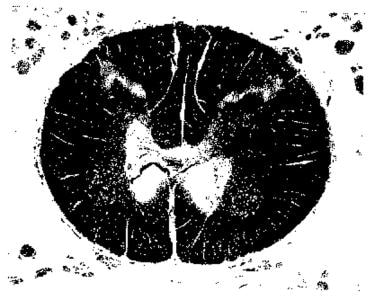
BACKGROUND AND PURPOSE: The mechanism of spinal cord injury has been thought to be related to the vulnerability of spinal motor neuron cells against ischemia. However, the mechanisms of such vulnerability are not fully understood. We hypothesized that spinal motor neurons might be lost by programmed cell death and investigated a possible mechanism of neuronal death by detection of double-strand breaks in genomic DNA and immunohistochemical analysis for cyclin D1 and cyclin-dependent kinases (Cdk) 4. METHODS: We used a rabbit spinal cord ischemia model with a balloon catheter. Spinal cord was removed at 8 hours and 1, 2, and 7 days after 15 minutes of transient ischemia, and histological changes were studied with hematoxylin-eosin staining. In situ terminal deoxynucleotidyl transferase (TdT)-mediated dUTP-biotin nick-end labeling (TUNEL), DNA fragment with gel electrophoresis, Western blot analysis for cyclin D1 and Cdk4, and temporal profiles of cyclin D1 and Cdk4 immunoreactivity were investigated. RESULTS: Most motor neurons were preserved until 2 days but were selectively lost at 7 days of reperfusion. Immunocytochemistry showed positive TUNEL selectively at 2 days of reperfusion in spinal motor neuron nuclei. Typical ladders of oligonucleosomal DNA fragments were detected at 2 days of reperfusion. Immunoreactivity of cyclin D1 and Cdk4 proteins was induced selectively at 8 hours in motor neuron nuclei, which eventually died. CONCLUSIONS: These results indicate that induction of cyclin D1 and Cdk4 may be implicated in programmed cell death change after transient spinal cord ischemia in rabbits. (+info)Spinal cord ischemia. Development of a model in the mouse. (4/183)
BACKGROUND AND PURPOSE: Spinal cord ischemia with resulting paraplegia is a devastating complication of thoracoabdominal aortic surgery. Experimental models of spinal cord ischemia have been developed in primate, dog, pig, rabbit, and rat with variable reproducibility, but none has been developed in mouse. Because genetically engineered mice have become important to examine the impact of specific genes in ischemic pathophysiology, we sought to develop a reproducible mouse model of spinal cord ischemia. METHODS: C57BL/6NCrlBR mice were subjected to cross-clamping of the aortic arch, left subclavian artery, and internal mammary artery for 9 minutes (group A; n=8) or 11 minutes (group B; n=29) followed by reperfusion for 24 or 48 hours. Mean distal arterial blood pressure (left femoral artery) and lumbar (L1) spinal cord blood flow (laser-Doppler flowmetry) were measured for the duration of the procedure. The arterial blood supply of the spinal cord was visualized by intravascular perfusion of carbon black ink. We evaluated motor function in the hind limbs at 0, 1, 3, 6, and 24 hours after reperfusion using a rating scale of 0 (normal function) to 6 (total absence of movement). Spinal cord histopathology was evaluated after 24 and 48 hours of reperfusion by Luxol fast blue-hematoxylin and eosin. RESULTS: The vascular anatomy of the mouse and human spinal cord appeared similar in that blood was supplied by 1 anterior and 2 posterior spinal arteries and heterosegmental radicular arteries. During combined occlusion of aortic arch and left subclavian artery, mean distal arterial blood pressure dropped to 10+/-5 mm Hg, and spinal cord blood flow at the L1 level decreased to 27+/-7% of baseline. All animals recovered from anesthesia with acute paraplegia. Animals in the 9-minute group (group A) showed steady recovery of hind limb function over the ensuing 24 hours, whereas the majority (80%) in the 11-minute group (group B) remained paralyzed with maximum deficit throughout the postoperative period. Mortality was 0% and 21% in groups A and B, respectively. Maximal ischemic damage was observed at the lower thoracic and higher lumbar spinal levels in both groups. In group A (9 minutes), tissue damage was mild, affecting predominantly dorsal horns and intermediate gray matter, whereas ventral horns were minimally involved. All mice in group B (11 minutes) showed extensive gray matter lesions particularly involving dorsal horns and intermediate areas; in ventral horns, >50% of motor neurons died. White matter lesions were present in the most severely damaged cords only. CONCLUSIONS: Spinal cord ischemia caused by aortic arch plus left subclavian artery cross-clamping provides a mouse model useful for the study of spinal cord injury and of potential relevance to the complications following thoracoabdominal aortic surgery in humans. (+info)Reversal of twice-delayed neurologic deficits with cerebrospinal fluid drainage after thoracoabdominal aneurysm repair: a case report and plea for a national database collection. (5/183)
Delayed neurologic deficits are an uncommon yet devastating complication of thoracoabdominal aortic aneurysm repair. The mechanisms involved in the development of delayed spinal cord ischemia remain ill defined. We report a case of complete reversal of delayed neurologic deficits with postoperative cerebrospinal fluid (CSF) drainage. After a thoracoabdominal aneurysm extent I repair, the patient experienced delayed paraplegia at 6 hours and again at 34 hours after the operation, with elevated CSF pressure (>10 mm Hg) on both occasions. Prompt CSF decompression completely reversed the neurologic deficits within hours after onset. The findings in this case further support the role of CSF drainage in spinal cord protection for patients who undergo thoracoabdominal aneurysm repair and make a plea for a national database collection. (+info)Surgical repair of aneurysms involving the suprarenal, visceral, and lower thoracic aortic segments: early results and late outcome. (6/183)
OBJECTIVE: The purpose of this study is to review our experience with surgical repair of lower thoracoabdominal and suprarenal aortic aneurysms to determine early and late survival rates and identify factors influencing morbidity and survival among these patients. MATERIALS: From 1989 through 1998, 165 consecutive patients underwent repair of 108 thoracoabdominal (55 group III and 53 group IV) and 57 suprarenal aneurysms. The study group consisted of 109 men and 56 women with a mean age of 70 years (median, 70 years; range, 29-89 years). Mean aneurysm diameter was 6.9 cm (median, 6.5 cm; range, 4-12 cm). There were 125 aneurysms (76%) repaired electively; 40 repairs (24%) were nonelective. The cause of 12 aneurysms (7%) was chronic aortic dissection; the remaining 153 (93%) were degenerative aneurysms. RESULTS: The early postoperative (30-day) mortality rates were 7% (9/125) for elective and 23% (9/40) for nonelective operations (P =.016). For both elective and urgent procedures, early mortality was 1.8% (1/57) for suprarenal aneurysm repair, 11% (6/53) for group IV thoracoabdominal aneurysms, and 20% (11/55) for group III thoracoabdominal aneurysms (P =.013, suprarenal vs group III). Spinal cord ischemia occurred after 6% (10/165) of aneurysm repairs (4% paraplegia, 2% paraparesis). None of the 57 suprarenal aneurysm repairs were complicated by spinal cord ischemia, whereas it occurred in 2% (1/53) of group IV thoracoabdominal aneurysms and 16% (9/55) of group III thoracoabdominal aneurysms (P =.001, suprarenal vs group III; P =. 016, group IV vs group III). Three (25%) of the 12 patients with dissection developed spinal cord ischemia; this compared with seven (5%) of 153 patients with degenerative aneurysms (P =.027). The cumulative 3-year survival rate for the entire series was 71% (95% CI, 64%-79%), and 5-year survival was 50% (95% CI, 40%-60%). CONCLUSIONS: Aneurysms involving the suprarenal, visceral, and lower thoracic aorta may be repaired with acceptable perioperative mortality and late survival rates. The risk of spinal cord ischemia is increased for patients with aortic dissection and may be stratified according to the proximal extent of the aneurysm. (+info)Spinal cord infarction and tetraplegia--rare complications of meningococcal meningitis. (7/183)
A previously healthy 25-yr-old female developed flaccid areflexic tetraplegia, with intact cranial nerve function, 36 h after the diagnosis of bacterial meningitis. Polymerase chain reaction studies of cerebrospinal fluid and blood were positive for Neisseria meningitidis, serogroup B. Magnetic resonance of the cervicothoracic spine revealed increased signal intensity and expansion in the lower medulla, upper cervical cord and cerebellar tonsils. Neurosurgical consultation recommended hyperventilation, dexamethasone and regular mannitol therapy rather than decompressive intervention. The clinical course over the following 12 days was complicated by the development of progressive central nervous and multisystem organ failure with disseminated intravascular coagulopathy. Autopsy revealed cerebral oedema with cystic infarction extending from the medulla to the upper cervical cord and cerebellar tonsils. Flaccid areflexic tetraplegia with spinal cord infarction has not been reported following bacterial infection in an adult. The clinical implications would suggest complete central nervous system evaluation of patients recovering from meningococcal meningitis, since spinal cord lesions, although uncommon, do occur. In those very rare situations where a patient develops significant peripheral neurological deficits, urgent magnetic resonance imaging is warranted, to rule out an infective focus or an underlying anatomical anomaly. (+info)Epidural cooling for spinal cord protection during thoracoabdominal aneurysm repair: A five-year experience. (8/183)
PURPOSE: We developed and applied a method for providing regional spinal cord hypothermia with epidural cooling (EC) during thoracoabdominal aneurysm (TAA) repair. Preliminary results indicated significant reduction in spinal cord ischemic complications (SCI), compared with historical controls, and a 5-year experience with EC was reviewed. METHODS: From July 1993 to September 1998, 170 patients with thoracic aneurysms (n = 14; 8.2%) or TAAs (types I and II, n = 83 [49%]; type III, n = 66 [39%]; type IV, n = 7 [4.1%]) were treated with EC. An earlier aneurysm resection was noted in 44% of patients, an emergent operation was noted in 20% of patients, and an aortic dissection was noted in 16% of patients. The EC was successful (mean cerebrospinal fluid [CSF] temperature at cross-clamp, 26.4 +/- 3 degrees C) in 97% of cases, with all 170 patients included in an intention-to-treat analysis. The operation was performed with a clamp/sew technique (98% patients) and selective (T(9) to L(1) region) reimplantation of intercostal vessels. Clinical and EC variables were examined for association with operative mortality and SCI by means of the Fischer exact test, and those variables with a P value less than.1 were included in multivariate logistic regression analysis. RESULTS: The operative mortality rate was 9.5% and was weakly associated (P =.07) with SCI; postoperative cardiac complications (odds ratio [OR], 35. 3; 95% CI, 5.3 to 233; P <.001) and renal failure (OR, 32.2; 95% CI, 6.6 to 157; P <.001) were the only independent predictors of postoperative death. SCI of any severity occurred in 7% of cases (type I/II, 10 of 83 [12%]; all other types, 2 of 87 [2.3%]), versus a predicted (Acher model) incidence of 18.5% for this cohort (P =. 003). Half the deficits were minor, with good functional recovery, and devastating paraplegia occurred in three patients (2.0%). Independent correlates of SCI included types I and II TAA (OR, 8.0; 95% CI, 1.4 to 46.3; P =.021), nonelective operation (OR, 8.3, 95% CI, 1.8 to 37.7; P =.006), oversewn T(9) to L(2) intercostal vessels (OR, 6.1; 95% CI, 1.3 to 28.8; P =.023), and postoperative renal failure (OR, 23.6; 95% CI, 4.4 to 126; P <.001). These same clinical variables of nonelective operations (OR, 7.7; 95% CI, 1.4 to 41.4; P =.017), oversewn T(9) to L(2) intercostal arteries (OR, 9.7; 95% CI, 1.5 to 61.2; P =.016), and postoperative renal failure (OR, 20.8; 95% CI, 3.0 to 142.1; P =.002) were independent predictors of SCI in the subgroup analysis of high-risk patients, ie, patients with type I/II TAA. CONCLUSION: EC has been effective in reducing immediate, devastating, total paraplegia after TAA repair. A strategy that combines the neuroprotective effect of regional cord hypothermia, avoiding the sacrifice of potential spinal cord blood supply, and postoperative adjuncts (eg, avoidance of hypotension, CSF drainage) appears necessary to minimize SCI after TAA repair. (+info)Spinal cord ischemia refers to a reduction or interruption of blood flow to the spinal cord, leading to insufficient oxygen and nutrient supply. This condition can cause damage to the spinal cord tissue, potentially resulting in neurological deficits, such as muscle weakness, sensory loss, or autonomic dysfunction. Spinal cord ischemia may be caused by various factors, including atherosclerosis, embolism, spinal artery stenosis, or complications during surgery. The severity and extent of the neurological impairment depend on the duration and location of the ischemic event in the spinal cord.
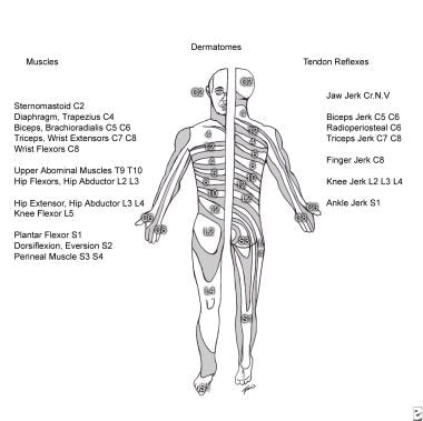
The spinal cord is a major part of the nervous system, extending from the brainstem and continuing down to the lower back. It is a slender, tubular bundle of nerve fibers (axons) and support cells (glial cells) that carries signals between the brain and the rest of the body. The spinal cord primarily serves as a conduit for motor information, which travels from the brain to the muscles, and sensory information, which travels from the body to the brain. It also contains neurons that can independently process and respond to information within the spinal cord without direct input from the brain.
The spinal cord is protected by the bony vertebral column (spine) and is divided into 31 segments: 8 cervical, 12 thoracic, 5 lumbar, 5 sacral, and 1 coccygeal. Each segment corresponds to a specific region of the body and gives rise to pairs of spinal nerves that exit through the intervertebral foramina at each level.
The spinal cord is responsible for several vital functions, including:
1. Reflexes: Simple reflex actions, such as the withdrawal reflex when touching a hot surface, are mediated by the spinal cord without involving the brain.
2. Muscle control: The spinal cord carries motor signals from the brain to the muscles, enabling voluntary movement and muscle tone regulation.
3. Sensory perception: The spinal cord transmits sensory information, such as touch, temperature, pain, and vibration, from the body to the brain for processing and awareness.
4. Autonomic functions: The sympathetic and parasympathetic divisions of the autonomic nervous system originate in the thoracolumbar and sacral regions of the spinal cord, respectively, controlling involuntary physiological responses like heart rate, blood pressure, digestion, and respiration.
Damage to the spinal cord can result in various degrees of paralysis or loss of sensation below the level of injury, depending on the severity and location of the damage.
Paraplegia is a medical condition characterized by partial or complete loss of motor function and sensation in the lower extremities, typically affecting both legs. This results from damage to the spinal cord, often due to trauma such as accidents, falls, or gunshot wounds, or from diseases like spina bifida, polio, or tumors. The specific area and extent of the injury on the spinal cord determine the severity and location of paralysis. Individuals with paraplegia may require assistive devices for mobility, such as wheelchairs, and may face various health challenges, including pressure sores, urinary tract infections, and chronic pain.
Spinal cord injuries (SCI) refer to damage to the spinal cord that results in a loss of function, such as mobility or feeling. This injury can be caused by direct trauma to the spine or by indirect damage resulting from disease or degeneration of surrounding bones, tissues, or blood vessels. The location and severity of the injury on the spinal cord will determine which parts of the body are affected and to what extent.
The effects of SCI can range from mild sensory changes to severe paralysis, including loss of motor function, autonomic dysfunction, and possible changes in sensation, strength, and reflexes below the level of injury. These injuries are typically classified as complete or incomplete, depending on whether there is any remaining function below the level of injury.
Immediate medical attention is crucial for spinal cord injuries to prevent further damage and improve the chances of recovery. Treatment usually involves immobilization of the spine, medications to reduce swelling and pressure, surgery to stabilize the spine, and rehabilitation to help regain lost function. Despite advances in treatment, SCI can have a significant impact on a person's quality of life and ability to perform daily activities.
Ischemia is the medical term used to describe a lack of blood flow to a part of the body, often due to blocked or narrowed blood vessels. This can lead to a shortage of oxygen and nutrients in the tissues, which can cause them to become damaged or die. Ischemia can affect many different parts of the body, including the heart, brain, legs, and intestines. Symptoms of ischemia depend on the location and severity of the blockage, but they may include pain, cramping, numbness, weakness, or coldness in the affected area. In severe cases, ischemia can lead to tissue death (gangrene) or organ failure. Treatment for ischemia typically involves addressing the underlying cause of the blocked blood flow, such as through medication, surgery, or lifestyle changes.
A thoracic aortic aneurysm is a localized dilatation or bulging of the thoracic aorta, which is the part of the aorta that runs through the chest cavity. The aorta is the largest artery in the body, and it carries oxygenated blood from the heart to the rest of the body.
Thoracic aortic aneurysms can occur anywhere along the thoracic aorta, but they are most commonly found in the aortic arch or the descending thoracic aorta. These aneurysms can vary in size, and they are considered significant when they are 50% larger than the expected normal diameter of the aorta.
The exact cause of thoracic aortic aneurysms is not fully understood, but several factors can contribute to their development, including:
* Atherosclerosis (hardening and narrowing of the arteries)
* High blood pressure
* Genetic disorders such as Marfan syndrome or Ehlers-Danlos syndrome
* Infections or inflammation of the aorta
* Trauma to the chest
Thoracic aortic aneurysms can be asymptomatic and found incidentally on imaging studies, or they may present with symptoms such as chest pain, cough, difficulty swallowing, or hoarseness. If left untreated, thoracic aortic aneurysms can lead to serious complications, including aortic dissection (tearing of the inner layer of the aorta) or rupture, which can be life-threatening.
Treatment options for thoracic aortic aneurysms include medical management with blood pressure control and cholesterol-lowering medications, as well as surgical repair or endovascular stenting, depending on the size, location, and growth rate of the aneurysm. Regular follow-up imaging is necessary to monitor the size and progression of the aneurysm over time.
Paraparesis is a medical term that refers to a mild to moderate form of paralysis affecting the lower limbs, specifically the legs. It is characterized by partial loss of strength and mobility, which may result in difficulty walking or maintaining balance. Paraparesis can be caused by various conditions such as spinal cord injuries, multiple sclerosis, spina bifida, or other neurological disorders affecting the spinal cord.
The term "para" means "two," and "paresis" comes from the Greek word "paresis," which means "loosening" or "relaxation." Therefore, paraparesis implies weakness or partial paralysis in two lower extremities. It is important to note that while paraparesis can impact a person's ability to walk and perform daily activities, it does not necessarily lead to complete loss of movement or sensation in the affected limbs.
Proper diagnosis and management of the underlying cause are crucial for improving symptoms and preventing further progression of paraparesis. Treatment options may include physical therapy, medications, assistive devices, or surgical interventions depending on the specific condition causing the paraparesis.
Cerebrospinal Fluid Pressure (CSFP) is the pressure exerted by the cerebrospinal fluid (CSF), a clear, colorless fluid that surrounds and protects the brain and spinal cord. CSF acts as a cushion for the brain, allowing it to float within the skull and protecting it from trauma.
The normal range of CSFP is typically between 6 and 18 cm of water (cm H2O) when measured in the lateral decubitus position (lying on one's side). Elevated CSFP can be a sign of various medical conditions, such as hydrocephalus, meningitis, or brain tumors. Conversely, low CSFP may indicate dehydration or other underlying health issues.
It is important to monitor and maintain normal CSFP levels, as abnormal pressure can lead to serious neurological complications, including damage to the optic nerve, cognitive impairment, and even death in severe cases. Regular monitoring of CSFP may be necessary for individuals with conditions that affect CSF production or absorption.
In medical terms, constriction refers to the narrowing or tightening of a body part or passageway. This can occur due to various reasons such as spasms of muscles, inflammation, or abnormal growths. It can lead to symptoms like difficulty in breathing, swallowing, or blood flow, depending on where it occurs. For example, constriction of the airways in asthma, constriction of blood vessels in hypertension, or constriction of the esophagus in certain digestive disorders.
Reperfusion injury is a complex pathophysiological process that occurs when blood flow is restored to previously ischemic tissues, leading to further tissue damage. This phenomenon can occur in various clinical settings such as myocardial infarction (heart attack), stroke, or peripheral artery disease after an intervention aimed at restoring perfusion.
The restoration of blood flow leads to the generation of reactive oxygen species (ROS) and inflammatory mediators, which can cause oxidative stress, cellular damage, and activation of the immune system. This results in a cascade of events that may lead to microvascular dysfunction, capillary leakage, and tissue edema, further exacerbating the injury.
Reperfusion injury is an important consideration in the management of ischemic events, as interventions aimed at restoring blood flow must be carefully balanced with potential harm from reperfusion injury. Strategies to mitigate reperfusion injury include ischemic preconditioning (exposing the tissue to short periods of ischemia before a prolonged ischemic event), ischemic postconditioning (applying brief periods of ischemia and reperfusion after restoring blood flow), remote ischemic preconditioning (ischemia applied to a distant organ or tissue to protect the target organ), and pharmacological interventions that scavenge ROS, reduce inflammation, or improve microvascular function.
The thoracic aorta is the segment of the largest artery in the human body (the aorta) that runs through the chest region (thorax). The thoracic aorta begins at the aortic arch, where it branches off from the ascending aorta, and extends down to the diaphragm, where it becomes the abdominal aorta.
The thoracic aorta is divided into three parts: the ascending aorta, the aortic arch, and the descending aorta. The ascending aorta rises from the left ventricle of the heart and is about 2 inches (5 centimeters) long. The aortic arch curves backward and to the left, giving rise to the brachiocephalic trunk, the left common carotid artery, and the left subclavian artery. The descending thoracic aorta runs downward through the chest, passing through the diaphragm to become the abdominal aorta.
The thoracic aorta supplies oxygenated blood to the upper body, including the head, neck, arms, and chest. It plays a critical role in maintaining blood flow and pressure throughout the body.
Reperfusion, in medical terms, refers to the restoration of blood flow to tissues or organs that have been deprived of adequate oxygen supply, usually as a result of ischemia (lack of blood flow). This process is often initiated through therapeutic interventions such as thrombolysis (breaking up blood clots), angioplasty (opening narrowed or blocked blood vessels using a balloon or stent), or surgical procedures.
Reperfusion aims to salvage the affected tissues and prevent further damage; however, it can also lead to reperfusion injury. This injury occurs when the return of oxygen-rich blood to previously ischemic tissues results in the overproduction of free radicals and inflammatory mediators, which can cause additional cellular damage and organ dysfunction.
Managing reperfusion injury involves using various strategies such as antioxidants, anti-inflammatory agents, and other protective treatments to minimize its negative impact on the recovering tissues or organs.
Blood vessel prosthesis implantation is a surgical procedure in which an artificial blood vessel, also known as a vascular graft or prosthetic graft, is inserted into the body to replace a damaged or diseased native blood vessel. The prosthetic graft can be made from various materials such as Dacron (polyester), PTFE (polytetrafluoroethylene), or bovine/human tissue.
The implantation of a blood vessel prosthesis is typically performed to treat conditions that cause narrowing or blockage of the blood vessels, such as atherosclerosis, aneurysms, or traumatic injuries. The procedure may be used to bypass blocked arteries in the legs (peripheral artery disease), heart (coronary artery bypass surgery), or neck (carotid endarterectomy). It can also be used to replace damaged veins for hemodialysis access in patients with kidney failure.
The success of blood vessel prosthesis implantation depends on various factors, including the patient's overall health, the location and extent of the vascular disease, and the type of graft material used. Possible complications include infection, bleeding, graft thrombosis (clotting), and graft failure, which may require further surgical intervention or endovascular treatments.
Aortography is a medical procedure that involves taking X-ray images of the aorta, which is the largest blood vessel in the body. The procedure is usually performed to diagnose or assess various conditions related to the aorta, such as aneurysms, dissections, or blockages.
To perform an aortography, a contrast dye is injected into the aorta through a catheter that is inserted into an artery, typically in the leg or arm. The contrast dye makes the aorta visible on X-ray images, allowing doctors to see its structure and any abnormalities that may be present.
The procedure is usually performed in a hospital or outpatient setting and may require sedation or anesthesia. While aortography can provide valuable diagnostic information, it also carries some risks, such as allergic reactions to the contrast dye, damage to blood vessels, or infection. Therefore, it is typically reserved for situations where other diagnostic tests have been inconclusive or where more invasive treatment may be required.
I believe there may be some confusion in your question. "Rabbits" is a common name used to refer to the Lagomorpha species, particularly members of the family Leporidae. They are small mammals known for their long ears, strong legs, and quick reproduction.
However, if you're referring to "rabbits" in a medical context, there is a term called "rabbit syndrome," which is a rare movement disorder characterized by repetitive, involuntary movements of the fingers, resembling those of a rabbit chewing. It is also known as "finger-chewing chorea." This condition is usually associated with certain medications, particularly antipsychotics, and typically resolves when the medication is stopped or adjusted.
Spinal injections, also known as epidural injections or intrathecal injections, are medical procedures involving the injection of medications directly into the spinal canal. The medication is usually delivered into the space surrounding the spinal cord (the epidural space) or into the cerebrospinal fluid that surrounds and protects the spinal cord (the subarachnoid space).
The medications used in spinal injections can include local anesthetics, steroids, opioids, or a combination of these. The purpose of spinal injections is to provide diagnostic information, therapeutic relief, or both. They are commonly used to treat various conditions affecting the spine, such as radicular pain (pain that radiates down the arms or legs), disc herniation, spinal stenosis, and degenerative disc disease.
Spinal injections can be administered using different techniques, including fluoroscopy-guided injections, computed tomography (CT) scan-guided injections, or with the help of a nerve stimulator. These techniques ensure accurate placement of the medication and minimize the risk of complications.
It is essential to consult a healthcare professional for specific information regarding spinal injections and their potential benefits and risks.
The subclavian artery is a major blood vessel that supplies the upper limb and important structures in the neck and head. It arises from the brachiocephalic trunk (in the case of the right subclavian artery) or directly from the aortic arch (in the case of the left subclavian artery).
The subclavian artery has several branches, including:
1. The vertebral artery, which supplies blood to the brainstem and cerebellum.
2. The internal thoracic artery (also known as the mammary artery), which supplies blood to the chest wall, breast, and anterior mediastinum.
3. The thyrocervical trunk, which gives rise to several branches that supply the neck, including the inferior thyroid artery, the suprascapular artery, and the transverse cervical artery.
4. The costocervical trunk, which supplies blood to the neck and upper back, including the posterior chest wall and the lower neck muscles.
The subclavian artery is a critical vessel in maintaining adequate blood flow to the upper limb, and any blockage or damage to this vessel can lead to significant morbidity, including arm pain, numbness, weakness, or even loss of function.
Infarction is the term used in medicine to describe the death of tissue (also known as an "area of necrosis") due to the lack of blood supply. This can occur when a blood vessel that supplies oxygen and nutrients to a particular area of the body becomes blocked or obstructed, leading to the deprivation of oxygen and nutrients necessary for the survival of cells in that region.
The blockage in the blood vessel is usually caused by a clot (thrombus) or an embolus, which is a small particle that travels through the bloodstream and lodges in a smaller vessel. The severity and extent of infarction depend on several factors, including the size and location of the affected blood vessel, the duration of the obstruction, and the presence of collateral circulation (alternative blood vessels that can compensate for the blocked one).
Common examples of infarctions include myocardial infarction (heart attack), cerebral infarction (stroke), and pulmonary infarction (lung tissue death due to obstruction in the lung's blood vessels). Infarctions can lead to various symptoms, depending on the affected organ or tissue, and may require medical intervention to manage complications and prevent further damage.
Spinal cord diseases refer to a group of conditions that affect the spinal cord, which is a part of the central nervous system responsible for transmitting messages between the brain and the rest of the body. These diseases can cause damage to the spinal cord, leading to various symptoms such as muscle weakness, numbness, pain, bladder and bowel dysfunction, and difficulty with movement and coordination.
Spinal cord diseases can be congenital or acquired, and they can result from a variety of causes, including infections, injuries, tumors, degenerative conditions, autoimmune disorders, and genetic factors. Some examples of spinal cord diseases include multiple sclerosis, spina bifida, spinal cord injury, herniated discs, spinal stenosis, and motor neuron diseases such as amyotrophic lateral sclerosis (ALS).
The treatment for spinal cord diseases varies depending on the underlying cause and severity of the condition. Treatment options may include medication, physical therapy, surgery, and rehabilitation. In some cases, the damage to the spinal cord may be irreversible, leading to permanent disability or paralysis.
Paraparesis, spastic type, is a medical term used to describe a condition characterized by partial weakness or loss of voluntary movement in the lower extremities (legs). The term "paraparesis" comes from Greek words "para" meaning beside or beyond, and "paresis" meaning loosening or relaxation.
In spastic paraparesis, the muscle tone is increased, causing stiffness and resistance to movement, particularly during quick or forceful movements. This increased muscle tone, also known as spasticity, results from an upper motor neuron lesion in the brain or spinal cord that affects the corticospinal tract, which carries signals from the brain to the muscles.
Spastic paraparesis can be caused by various conditions, including spinal cord injuries, multiple sclerosis, hereditary spastic paraplegia, and stroke, among others. The severity of symptoms may vary widely, ranging from mild weakness to complete paralysis. Treatment options for spastic paraparesis depend on the underlying cause and may include physical therapy, medications, surgery, or a combination of these approaches.
Intraoperative monitoring (IOM) is the practice of using specialized techniques to monitor physiological functions or neural structures in real-time during surgical procedures. The primary goal of IOM is to provide continuous information about the patient's status and the effects of surgery on neurological function, allowing surgeons to make informed decisions and minimize potential risks.
IOM can involve various methods such as:
1. Electrophysiological monitoring: This includes techniques like somatosensory evoked potentials (SSEP), motor evoked potentials (MEP), and electroencephalography (EEG) to assess the integrity of neural pathways and brain function during surgery.
2. Neuromonitoring: Direct electrical stimulation of nerves or spinal cord structures can help identify critical neuroanatomical structures, evaluate their functional status, and guide surgical interventions.
3. Hemodynamic monitoring: Measuring blood pressure, heart rate, cardiac output, and oxygen saturation helps assess the patient's overall physiological status during surgery.
4. Imaging modalities: Intraoperative imaging techniques like ultrasound, computed tomography (CT), or magnetic resonance imaging (MRI) can provide real-time visualization of anatomical structures and surgical progress.
The specific IOM methods employed depend on the type of surgery, patient characteristics, and potential risks involved. Intraoperative monitoring is particularly crucial in procedures where there is a risk of neurological injury, such as spinal cord or brain surgeries, vascular interventions, or tumor resections near critical neural structures.
Somatosensory evoked potentials (SEPs) are electrical signals generated in the brain and spinal cord in response to the stimulation of peripheral nerves. These responses are recorded and measured to assess the functioning of the somatosensory system, which is responsible for processing sensations such as touch, temperature, vibration, and proprioception (the sense of the position and movement of body parts).
SEPs are typically elicited by applying electrical stimuli to peripheral nerves in the arms or legs. The resulting neural responses are then recorded using electrodes placed on the scalp or other locations on the body. These recordings can provide valuable information about the integrity and function of the nervous system, and are often used in clinical settings to diagnose and monitor conditions such as nerve damage, spinal cord injury, multiple sclerosis, and other neurological disorders.
SEPs can be further categorized based on the specific type of stimulus used and the location of the recording electrodes. For example, short-latency SEPs (SLSEPs) are those that occur within the first 50 milliseconds after stimulation, and are typically recorded from the scalp over the primary sensory cortex. These responses reflect the earliest stages of sensory processing and can be used to assess the integrity of the peripheral nerves and the ascending sensory pathways in the spinal cord.
In contrast, long-latency SEPs (LLSEPs) occur after 50 milliseconds and are typically recorded from more posterior regions of the scalp over the parietal cortex. These responses reflect later stages of sensory processing and can be used to assess higher-level cognitive functions such as attention, memory, and perception.
Overall, SEPs provide a valuable tool for clinicians and researchers seeking to understand the functioning of the somatosensory system and diagnose or monitor neurological disorders.
Myocardial ischemia is a condition in which the blood supply to the heart muscle (myocardium) is reduced or blocked, leading to insufficient oxygen delivery and potential damage to the heart tissue. This reduction in blood flow typically results from the buildup of fatty deposits, called plaques, in the coronary arteries that supply the heart with oxygen-rich blood. The plaques can rupture or become unstable, causing the formation of blood clots that obstruct the artery and limit blood flow.
Myocardial ischemia may manifest as chest pain (angina pectoris), shortness of breath, fatigue, or irregular heartbeats (arrhythmias). In severe cases, it can lead to myocardial infarction (heart attack) if the oxygen supply is significantly reduced or cut off completely, causing permanent damage or death of the heart muscle. Early diagnosis and treatment of myocardial ischemia are crucial for preventing further complications and improving patient outcomes.
Brain ischemia is the medical term used to describe a reduction or interruption of blood flow to the brain, leading to a lack of oxygen and glucose delivery to brain tissue. This can result in brain damage or death of brain cells, known as infarction. Brain ischemia can be caused by various conditions such as thrombosis (blood clot formation), embolism (obstruction of a blood vessel by a foreign material), or hypoperfusion (reduced blood flow). The severity and duration of the ischemia determine the extent of brain damage. Symptoms can range from mild, such as transient ischemic attacks (TIAs or "mini-strokes"), to severe, including paralysis, speech difficulties, loss of consciousness, and even death. Immediate medical attention is required for proper diagnosis and treatment to prevent further damage and potential long-term complications.
The Thoracic Arteries are branches of the aorta that supply oxygenated blood to the thoracic region of the body. The pair of arteries originate from the descending aorta and divide into several smaller branches, including intercostal arteries that supply blood to the muscles between the ribs, and posterior intercostal arteries that supply blood to the back and chest wall. Other branches of the thoracic arteries include the superior phrenic arteries, which supply blood to the diaphragm, and the bronchial arteries, which supply blood to the lungs. These arteries play a crucial role in maintaining the health and function of the chest and respiratory system.
An abdominal aortic aneurysm (AAA) is a localized dilatation or bulging of the abdominal aorta, which is the largest artery in the body that supplies oxygenated blood to the trunk and lower extremities. Normally, the diameter of the abdominal aorta measures about 2 centimeters (cm) in adults. However, when the diameter of the aorta exceeds 3 cm, it is considered an aneurysm.
AAA can occur anywhere along the length of the abdominal aorta, but it most commonly occurs below the renal arteries and above the iliac bifurcation. The exact cause of AAA remains unclear, but several risk factors have been identified, including smoking, hypertension, advanced age, male gender, family history, and certain genetic disorders such as Marfan syndrome and Ehlers-Danlos syndrome.
The main concern with AAA is the risk of rupture, which can lead to life-threatening internal bleeding. The larger the aneurysm, the greater the risk of rupture. Symptoms of AAA may include abdominal or back pain, a pulsating mass in the abdomen, or symptoms related to compression of surrounding structures such as the kidneys, ureters, or nerves. However, many AAAs are asymptomatic and are discovered incidentally during imaging studies performed for other reasons.
Diagnosis of AAA typically involves imaging tests such as ultrasound, computed tomography (CT) scan, or magnetic resonance imaging (MRI). Treatment options depend on the size and location of the aneurysm, as well as the patient's overall health status. Small AAAs that are not causing symptoms may be monitored with regular imaging studies to assess for growth. Larger AAAs or those that are growing rapidly may require surgical repair, either through open surgery or endovascular repair using a stent graft.
Spinal cord compression is a medical condition that refers to the narrowing of the spinal canal, which puts pressure on the spinal cord and the nerves that branch out from it. This can occur due to various reasons such as degenerative changes in the spine, herniated discs, bone spurs, tumors, or fractures. The compression can lead to a range of symptoms including pain, numbness, tingling, weakness, or loss of bladder and bowel control. In severe cases, it can cause paralysis. Treatment options depend on the underlying cause and may include physical therapy, medication, surgery, or radiation therapy.
In the field of medicine, "time factors" refer to the duration of symptoms or time elapsed since the onset of a medical condition, which can have significant implications for diagnosis and treatment. Understanding time factors is crucial in determining the progression of a disease, evaluating the effectiveness of treatments, and making critical decisions regarding patient care.
For example, in stroke management, "time is brain," meaning that rapid intervention within a specific time frame (usually within 4.5 hours) is essential to administering tissue plasminogen activator (tPA), a clot-busting drug that can minimize brain damage and improve patient outcomes. Similarly, in trauma care, the "golden hour" concept emphasizes the importance of providing definitive care within the first 60 minutes after injury to increase survival rates and reduce morbidity.
Time factors also play a role in monitoring the progression of chronic conditions like diabetes or heart disease, where regular follow-ups and assessments help determine appropriate treatment adjustments and prevent complications. In infectious diseases, time factors are crucial for initiating antibiotic therapy and identifying potential outbreaks to control their spread.
Overall, "time factors" encompass the significance of recognizing and acting promptly in various medical scenarios to optimize patient outcomes and provide effective care.
Spinal cord neoplasms refer to abnormal growths or tumors within the spinal cord. These can be benign (non-cancerous) or malignant (cancerous). They originate from the cells within the spinal cord itself (primary tumors), or they may spread to the spinal cord from other parts of the body (metastatic tumors). Spinal cord neoplasms can cause various symptoms depending on their location and size, including back pain, neurological deficits, and even paralysis. Treatment options include surgery, radiation therapy, and chemotherapy.
Drainage, in medical terms, refers to the removal of excess fluid or accumulated collections of fluids from various body parts or spaces. This is typically accomplished through the use of medical devices such as catheters, tubes, or drains. The purpose of drainage can be to prevent the buildup of fluids that may cause discomfort, infection, or other complications, or to treat existing collections of fluid such as abscesses, hematomas, or pleural effusions. Drainage may also be used as a diagnostic tool to analyze the type and composition of the fluid being removed.
Motor neurons are specialized nerve cells in the brain and spinal cord that play a crucial role in controlling voluntary muscle movements. They transmit electrical signals from the brain to the muscles, enabling us to perform actions such as walking, talking, and swallowing. There are two types of motor neurons: upper motor neurons, which originate in the brain's motor cortex and travel down to the brainstem and spinal cord; and lower motor neurons, which extend from the brainstem and spinal cord to the muscles. Damage or degeneration of these motor neurons can lead to various neurological disorders, such as amyotrophic lateral sclerosis (ALS) and spinal muscular atrophy (SMA).
Evoked potentials, motor, are a category of tests used in clinical neurophysiology to measure the electrical activity generated by the nervous system in response to a stimulus that specifically activates the motor pathways. These tests can help assess the integrity and function of the motor neurons, which are responsible for controlling voluntary muscle movements.
During a motor evoked potentials test, electrodes are placed on the scalp or directly on the surface of the brain or spinal cord. A stimulus is then applied to the motor cortex or peripheral nerves, causing the muscles to contract. The resulting electrical signals are recorded and analyzed to evaluate the conduction velocity, amplitude, and latency of the motor responses.
Motor evoked potentials tests can be useful in diagnosing various neurological conditions, such as multiple sclerosis, spinal cord injuries, and motor neuron diseases. They can also help monitor the progression of these conditions and assess the effectiveness of treatments.
Ischemic preconditioning is a phenomenon in which brief, non-lethal episodes of ischemia (restriction or interruption of blood supply to an organ or tissue) render the tissue more resistant to subsequent prolonged ischemia and reperfusion injury. This adaptive response involves a complex series of cellular and molecular changes that protect the myocardium, brain, kidney, or other organs from ischemic damage. The underlying mechanisms include the activation of various signaling pathways, such as adenosine, opioid, and kinase pathways, which lead to the production of protective factors and the modulation of cellular responses to ischemia and reperfusion injury. Ischemic preconditioning has been extensively studied in the context of cardiovascular medicine, where it has been shown to reduce infarct size and improve cardiac function after myocardial infarction. However, this protective phenomenon has also been observed in other organs and systems, including the brain, kidney, liver, and skeletal muscle.
The abdominal aorta is the portion of the aorta, which is the largest artery in the body, that runs through the abdomen. It originates from the thoracic aorta at the level of the diaphragm and descends through the abdomen, where it branches off into several smaller arteries that supply blood to the pelvis, legs, and various abdominal organs. The abdominal aorta is typically divided into four segments: the suprarenal, infrarenal, visceral, and parietal portions. Disorders of the abdominal aorta can include aneurysms, atherosclerosis, and dissections, which can have serious consequences if left untreated.
Animal disease models are specialized animals, typically rodents such as mice or rats, that have been genetically engineered or exposed to certain conditions to develop symptoms and physiological changes similar to those seen in human diseases. These models are used in medical research to study the pathophysiology of diseases, identify potential therapeutic targets, test drug efficacy and safety, and understand disease mechanisms.
The genetic modifications can include knockout or knock-in mutations, transgenic expression of specific genes, or RNA interference techniques. The animals may also be exposed to environmental factors such as chemicals, radiation, or infectious agents to induce the disease state.
Examples of animal disease models include:
1. Mouse models of cancer: Genetically engineered mice that develop various types of tumors, allowing researchers to study cancer initiation, progression, and metastasis.
2. Alzheimer's disease models: Transgenic mice expressing mutant human genes associated with Alzheimer's disease, which exhibit amyloid plaque formation and cognitive decline.
3. Diabetes models: Obese and diabetic mouse strains like the NOD (non-obese diabetic) or db/db mice, used to study the development of type 1 and type 2 diabetes, respectively.
4. Cardiovascular disease models: Atherosclerosis-prone mice, such as ApoE-deficient or LDLR-deficient mice, that develop plaque buildup in their arteries when fed a high-fat diet.
5. Inflammatory bowel disease models: Mice with genetic mutations affecting intestinal barrier function and immune response, such as IL-10 knockout or SAMP1/YitFc mice, which develop colitis.
Animal disease models are essential tools in preclinical research, but it is important to recognize their limitations. Differences between species can affect the translatability of results from animal studies to human patients. Therefore, researchers must carefully consider the choice of model and interpret findings cautiously when applying them to human diseases.
Aortic diseases refer to conditions that affect the aorta, which is the largest and main artery in the body. The aorta carries oxygenated blood from the heart to the rest of the body. Aortic diseases can weaken or damage the aorta, leading to various complications. Here are some common aortic diseases with their medical definitions:
1. Aortic aneurysm: A localized dilation or bulging of the aortic wall, which can occur in any part of the aorta but is most commonly found in the abdominal aorta (abdominal aortic aneurysm) or the thoracic aorta (thoracic aortic aneurysm). Aneurysms can increase the risk of rupture, leading to life-threatening bleeding.
2. Aortic dissection: A separation of the layers of the aortic wall due to a tear in the inner lining, allowing blood to flow between the layers and potentially cause the aorta to rupture. This is a medical emergency that requires immediate treatment.
3. Aortic stenosis: A narrowing of the aortic valve opening, which restricts blood flow from the heart to the aorta. This can lead to shortness of breath, chest pain, and other symptoms. Severe aortic stenosis may require surgical or transcatheter intervention to replace or repair the aortic valve.
4. Aortic regurgitation: Also known as aortic insufficiency, this condition occurs when the aortic valve does not close properly, allowing blood to leak back into the heart. This can lead to symptoms such as fatigue, shortness of breath, and palpitations. Treatment may include medication or surgical repair or replacement of the aortic valve.
5. Aortitis: Inflammation of the aorta, which can be caused by various conditions such as infections, autoimmune diseases, or vasculitides. Aortitis can lead to aneurysms, dissections, or stenosis and may require medical treatment with immunosuppressive drugs or surgical intervention.
6. Marfan syndrome: A genetic disorder that affects the connective tissue, including the aorta. People with Marfan syndrome are at risk of developing aortic aneurysms and dissections, and may require close monitoring and prophylactic surgery to prevent complications.
Neuroprotective agents are substances that protect neurons or nerve cells from damage, degeneration, or death caused by various factors such as trauma, inflammation, oxidative stress, or excitotoxicity. These agents work through different mechanisms, including reducing the production of free radicals, inhibiting the release of glutamate (a neurotransmitter that can cause cell damage in high concentrations), promoting the growth and survival of neurons, and preventing apoptosis (programmed cell death). Neuroprotective agents have been studied for their potential to treat various neurological disorders, including stroke, traumatic brain injury, Parkinson's disease, Alzheimer's disease, and multiple sclerosis. However, more research is needed to fully understand their mechanisms of action and to develop effective therapies.
Endovascular procedures are minimally invasive medical treatments that involve accessing and repairing blood vessels or other interior parts of the body through small incisions or punctures. These procedures typically use specialized catheters, wires, and other tools that are inserted into the body through an artery or vein, usually in the leg or arm.
Endovascular procedures can be used to treat a wide range of conditions, including aneurysms, atherosclerosis, peripheral artery disease, carotid artery stenosis, and other vascular disorders. Some common endovascular procedures include angioplasty, stenting, embolization, and thrombectomy.
The benefits of endovascular procedures over traditional open surgery include smaller incisions, reduced trauma to surrounding tissues, faster recovery times, and lower risks of complications such as infection and bleeding. However, endovascular procedures may not be appropriate for all patients or conditions, and careful evaluation and consideration are necessary to determine the best treatment approach.
A neurological examination is a series of tests used to evaluate the functioning of the nervous system, including both the central nervous system (the brain and spinal cord) and peripheral nervous system (the nerves that extend from the brain and spinal cord to the rest of the body). It is typically performed by a healthcare professional such as a neurologist or a primary care physician with specialized training in neurology.
During a neurological examination, the healthcare provider will assess various aspects of neurological function, including:
1. Mental status: This involves evaluating a person's level of consciousness, orientation, memory, and cognitive abilities.
2. Cranial nerves: There are 12 cranial nerves that control functions such as vision, hearing, smell, taste, and movement of the face and neck. The healthcare provider will test each of these nerves to ensure they are functioning properly.
3. Motor function: This involves assessing muscle strength, tone, coordination, and reflexes. The healthcare provider may ask the person to perform certain movements or tasks to evaluate these functions.
4. Sensory function: The healthcare provider will test a person's ability to feel different types of sensations, such as touch, pain, temperature, vibration, and proprioception (the sense of where your body is in space).
5. Coordination and balance: The healthcare provider may assess a person's ability to perform coordinated movements, such as touching their finger to their nose or walking heel-to-toe.
6. Reflexes: The healthcare provider will test various reflexes throughout the body using a reflex hammer.
The results of a neurological examination can help healthcare providers diagnose and monitor conditions that affect the nervous system, such as stroke, multiple sclerosis, Parkinson's disease, or peripheral neuropathy.
A hindlimb, also known as a posterior limb, is one of the pair of extremities that are located distally to the trunk in tetrapods (four-legged vertebrates) and include mammals, birds, reptiles, and amphibians. In humans and other primates, hindlimbs are equivalent to the lower limbs, which consist of the thigh, leg, foot, and toes.
The primary function of hindlimbs is locomotion, allowing animals to move from one place to another. However, they also play a role in other activities such as balance, support, and communication. In humans, the hindlimbs are responsible for weight-bearing, standing, walking, running, and jumping.
In medical terminology, the term "hindlimb" is not commonly used to describe human anatomy. Instead, healthcare professionals use terms like lower limbs or lower extremities to refer to the same region of the body. However, in comparative anatomy and veterinary medicine, the term hindlimb is still widely used to describe the corresponding structures in non-human animals.
Induced hypothermia is a medically controlled lowering of the core body temperature to around 89.6-93.2°F (32-34°C) for therapeutic purposes. It is intentionally induced to reduce the metabolic rate and oxygen demand of organs, thereby offering protection during periods of low blood flow or inadequate oxygenation, such as during cardiac bypass surgery, severe trauma, or after a cardiac arrest. The deliberate induction and maintenance of hypothermia can help minimize tissue damage and improve outcomes in specific clinical scenarios. Once the risk has passed, the body temperature is gradually rewarmed to normal levels under controlled conditions.
A blood vessel prosthesis is a medical device that is used as a substitute for a damaged or diseased natural blood vessel. It is typically made of synthetic materials such as polyester, Dacron, or ePTFE (expanded polytetrafluoroethylene) and is designed to mimic the function of a native blood vessel by allowing the flow of blood through it.
Blood vessel prostheses are used in various surgical procedures, including coronary artery bypass grafting, peripheral arterial reconstruction, and the creation of arteriovenous fistulas for dialysis access. The choice of material and size of the prosthesis depends on several factors, such as the location and diameter of the vessel being replaced, the patient's age and overall health status, and the surgeon's preference.
It is important to note that while blood vessel prostheses can be effective in restoring blood flow, they may also carry risks such as infection, thrombosis (blood clot formation), and graft failure over time. Therefore, careful patient selection, surgical technique, and postoperative management are crucial for the success of these procedures.
Cerebrospinal fluid (CSF) is a clear, colorless fluid that surrounds and protects the brain and spinal cord. It acts as a shock absorber for the central nervous system and provides nutrients to the brain while removing waste products. CSF is produced by specialized cells called ependymal cells in the choroid plexus of the ventricles (fluid-filled spaces) inside the brain. From there, it circulates through the ventricular system and around the outside of the brain and spinal cord before being absorbed back into the bloodstream. CSF analysis is an important diagnostic tool for various neurological conditions, including infections, inflammation, and cancer.
Postoperative complications refer to any unfavorable condition or event that occurs during the recovery period after a surgical procedure. These complications can vary in severity and may include, but are not limited to:
1. Infection: This can occur at the site of the incision or inside the body, such as pneumonia or urinary tract infection.
2. Bleeding: Excessive bleeding (hemorrhage) can lead to a drop in blood pressure and may require further surgical intervention.
3. Blood clots: These can form in the deep veins of the legs (deep vein thrombosis) and can potentially travel to the lungs (pulmonary embolism).
4. Wound dehiscence: This is when the surgical wound opens up, which can lead to infection and further complications.
5. Pulmonary issues: These include atelectasis (collapsed lung), pneumonia, or respiratory failure.
6. Cardiovascular problems: These include abnormal heart rhythms (arrhythmias), heart attack, or stroke.
7. Renal failure: This can occur due to various reasons such as dehydration, blood loss, or the use of certain medications.
8. Pain management issues: Inadequate pain control can lead to increased stress, anxiety, and decreased mobility.
9. Nausea and vomiting: These can be caused by anesthesia, opioid pain medication, or other factors.
10. Delirium: This is a state of confusion and disorientation that can occur in the elderly or those with certain medical conditions.
Prompt identification and management of these complications are crucial to ensure the best possible outcome for the patient.
Sprague-Dawley rats are a strain of albino laboratory rats that are widely used in scientific research. They were first developed by researchers H.H. Sprague and R.C. Dawley in the early 20th century, and have since become one of the most commonly used rat strains in biomedical research due to their relatively large size, ease of handling, and consistent genetic background.
Sprague-Dawley rats are outbred, which means that they are genetically diverse and do not suffer from the same limitations as inbred strains, which can have reduced fertility and increased susceptibility to certain diseases. They are also characterized by their docile nature and low levels of aggression, making them easier to handle and study than some other rat strains.
These rats are used in a wide variety of research areas, including toxicology, pharmacology, nutrition, cancer, and behavioral studies. Because they are genetically diverse, Sprague-Dawley rats can be used to model a range of human diseases and conditions, making them an important tool in the development of new drugs and therapies.
Vascular surgical procedures are operations that are performed to treat conditions and diseases related to the vascular system, which includes the arteries, veins, and capillaries. These procedures can be invasive or minimally invasive and are often used to treat conditions such as peripheral artery disease, carotid artery stenosis, aortic aneurysms, and venous insufficiency.
Some examples of vascular surgical procedures include:
* Endarterectomy: a procedure to remove plaque buildup from the inside of an artery
* Bypass surgery: creating a new path for blood to flow around a blocked or narrowed artery
* Angioplasty and stenting: using a balloon to open a narrowed artery and placing a stent to keep it open
* Aneurysm repair: surgically repairing an aneurysm, a weakened area in the wall of an artery that has bulged out and filled with blood
* Embolectomy: removing a blood clot from a blood vessel
* Thrombectomy: removing a blood clot from a vein
These procedures are typically performed by vascular surgeons, who are trained in the diagnosis and treatment of vascular diseases.
An aortic aneurysm is a medical condition characterized by the abnormal widening or bulging of the wall of the aorta, which is the largest artery in the body. The aorta carries oxygenated blood from the heart to the rest of the body. When the aortic wall weakens, it can stretch and balloon out, forming an aneurysm.
Aortic aneurysms can occur anywhere along the aorta but are most commonly found in the abdominal section (abdominal aortic aneurysm) or the chest area (thoracic aortic aneurysm). The size and location of the aneurysm, as well as the patient's overall health, determine the risk of rupture and associated complications.
Aneurysms often do not cause symptoms until they become large or rupture. Symptoms may include:
* Pain in the chest, back, or abdomen
* Pulsating sensation in the abdomen
* Difficulty breathing
* Hoarseness
* Coughing or vomiting
Risk factors for aortic aneurysms include age, smoking, high blood pressure, family history, and certain genetic conditions. Treatment options depend on the size and location of the aneurysm and may include monitoring, medication, or surgical repair.
Regional blood flow (RBF) refers to the rate at which blood flows through a specific region or organ in the body, typically expressed in milliliters per minute per 100 grams of tissue (ml/min/100g). It is an essential physiological parameter that reflects the delivery of oxygen and nutrients to tissues while removing waste products. RBF can be affected by various factors such as metabolic demands, neural regulation, hormonal influences, and changes in blood pressure or vascular resistance. Measuring RBF is crucial for understanding organ function, diagnosing diseases, and evaluating the effectiveness of treatments.
Thoracotomy is a surgical procedure that involves making an incision on the chest wall to gain access to the thoracic cavity, which contains the lungs, heart, esophagus, trachea, and other vital organs. The incision can be made on the side (lateral thoracotomy), back (posterolateral thoracotomy), or front (median sternotomy) of the chest wall, depending on the specific surgical indication.
Thoracotomy is performed for various indications, including lung biopsy, lung resection, esophagectomy, heart surgery, and mediastinal mass removal. The procedure allows the surgeon to directly visualize and access the organs within the thoracic cavity, perform necessary procedures, and control bleeding if needed.
After the procedure, the incision is typically closed with sutures or staples, and a chest tube may be placed to drain any accumulated fluid or air from the pleural space around the lungs. The patient will require postoperative care and monitoring in a hospital setting until their condition stabilizes.
Treatment outcome is a term used to describe the result or effect of medical treatment on a patient's health status. It can be measured in various ways, such as through symptoms improvement, disease remission, reduced disability, improved quality of life, or survival rates. The treatment outcome helps healthcare providers evaluate the effectiveness of a particular treatment plan and make informed decisions about future care. It is also used in clinical research to compare the efficacy of different treatments and improve patient care.
"Random allocation," also known as "random assignment" or "randomization," is a process used in clinical trials and other research studies to distribute participants into different intervention groups (such as experimental group vs. control group) in a way that minimizes selection bias and ensures the groups are comparable at the start of the study.
In random allocation, each participant has an equal chance of being assigned to any group, and the assignment is typically made using a computer-generated randomization schedule or other objective methods. This process helps to ensure that any differences between the groups are due to the intervention being tested rather than pre-existing differences in the participants' characteristics.
Spinal nerve roots are the initial parts of spinal nerves that emerge from the spinal cord through the intervertebral foramen, which are small openings between each vertebra in the spine. These nerve roots carry motor, sensory, and autonomic fibers to and from specific regions of the body. There are 31 pairs of spinal nerve roots in total, with 8 cervical, 12 thoracic, 5 lumbar, 5 sacral, and 1 coccygeal pair. Each root has a dorsal (posterior) and ventral (anterior) ramus that branch off to form the peripheral nervous system. Irritation or compression of these nerve roots can result in pain, numbness, weakness, or loss of reflexes in the affected area.
Angioplasty is a medical procedure used to open narrowed or blocked blood vessels, often referred to as coronary angioplasty when it involves the heart's blood vessels (coronary arteries). The term "angio" refers to an angiogram, which is a type of X-ray image that reveals the inside of blood vessels.
The procedure typically involves the following steps:
1. A thin, flexible catheter (tube) is inserted into a blood vessel, usually through a small incision in the groin or arm.
2. The catheter is guided to the narrowed or blocked area using real-time X-ray imaging.
3. Once in place, a tiny balloon attached to the tip of the catheter is inflated to widen the blood vessel and compress any plaque buildup against the artery walls.
4. A stent (a small mesh tube) may be inserted to help keep the blood vessel open and prevent it from narrowing again.
5. The balloon is deflated, and the catheter is removed.
Angioplasty helps improve blood flow, reduce symptoms such as chest pain or shortness of breath, and lower the risk of heart attack in patients with blocked arteries. It's important to note that angioplasty is not a permanent solution for coronary artery disease, and lifestyle changes, medications, and follow-up care are necessary to maintain long-term cardiovascular health.
A dissecting aneurysm is a serious and potentially life-threatening condition that occurs when there is a tear in the inner layer of the artery wall, allowing blood to flow between the layers of the artery wall. This can cause the artery to bulge or balloon out, leading to a dissection aneurysm.
Dissecting aneurysms can occur in any artery, but they are most commonly found in the aorta, which is the largest artery in the body. When a dissecting aneurysm occurs in the aorta, it is often referred to as a "dissecting aortic aneurysm."
Dissecting aneurysms can be caused by various factors, including high blood pressure, atherosclerosis (hardening and narrowing of the arteries), genetic disorders that affect the connective tissue, trauma, or illegal drug use (such as cocaine).
Symptoms of a dissecting aneurysm may include sudden severe chest or back pain, which can feel like ripping or tearing, shortness of breath, sweating, lightheadedness, or loss of consciousness. If left untreated, a dissecting aneurysm can lead to serious complications, such as rupture of the artery, stroke, or even death.
Treatment for a dissecting aneurysm typically involves surgery or endovascular repair to prevent further damage and reduce the risk of rupture. The specific treatment approach will depend on various factors, including the location and size of the aneurysm, the patient's overall health, and their medical history.
Spinal nerves are the bundles of nerve fibers that transmit signals between the spinal cord and the rest of the body. There are 31 pairs of spinal nerves in the human body, which can be divided into five regions: 8 cervical, 12 thoracic, 5 lumbar, 5 sacral, and 1 coccygeal. Each spinal nerve carries both sensory information (such as touch, temperature, and pain) from the periphery to the spinal cord, and motor information (such as muscle control) from the spinal cord to the muscles and other structures in the body. Spinal nerves also contain autonomic fibers that regulate involuntary functions such as heart rate, digestion, and blood pressure.
X-ray computed tomography (CT or CAT scan) is a medical imaging method that uses computer-processed combinations of many X-ray images taken from different angles to produce cross-sectional (tomographic) images (virtual "slices") of the body. These cross-sectional images can then be used to display detailed internal views of organs, bones, and soft tissues in the body.
The term "computed tomography" is used instead of "CT scan" or "CAT scan" because the machines take a series of X-ray measurements from different angles around the body and then use a computer to process these data to create detailed images of internal structures within the body.
CT scanning is a noninvasive, painless medical test that helps physicians diagnose and treat medical conditions. CT imaging provides detailed information about many types of tissue including lung, bone, soft tissue and blood vessels. CT examinations can be performed on every part of the body for a variety of reasons including diagnosis, surgical planning, and monitoring of therapeutic responses.
In computed tomography (CT), an X-ray source and detector rotate around the patient, measuring the X-ray attenuation at many different angles. A computer uses this data to construct a cross-sectional image by the process of reconstruction. This technique is called "tomography". The term "computed" refers to the use of a computer to reconstruct the images.
CT has become an important tool in medical imaging and diagnosis, allowing radiologists and other physicians to view detailed internal images of the body. It can help identify many different medical conditions including cancer, heart disease, lung nodules, liver tumors, and internal injuries from trauma. CT is also commonly used for guiding biopsies and other minimally invasive procedures.
In summary, X-ray computed tomography (CT or CAT scan) is a medical imaging technique that uses computer-processed combinations of many X-ray images taken from different angles to produce cross-sectional images of the body. It provides detailed internal views of organs, bones, and soft tissues in the body, allowing physicians to diagnose and treat medical conditions.
Intraoperative complications refer to any unforeseen problems or events that occur during the course of a surgical procedure, once it has begun and before it is completed. These complications can range from minor issues, such as bleeding or an adverse reaction to anesthesia, to major complications that can significantly impact the patient's health and prognosis.
Examples of intraoperative complications include:
1. Bleeding (hemorrhage) - This can occur due to various reasons such as injury to blood vessels or organs during surgery.
2. Infection - Surgical site infections can develop if the surgical area becomes contaminated during the procedure.
3. Anesthesia-related complications - These include adverse reactions to anesthesia, difficulty maintaining the patient's airway, or cardiovascular instability.
4. Organ injury - Accidental damage to surrounding organs can occur during surgery, leading to potential long-term consequences.
5. Equipment failure - Malfunctioning surgical equipment can lead to complications and compromise the safety of the procedure.
6. Allergic reactions - Patients may have allergies to certain medications or materials used during surgery, causing an adverse reaction.
7. Prolonged operative time - Complications may arise if a surgical procedure takes longer than expected, leading to increased risk of infection and other issues.
Intraoperative complications require prompt identification and management by the surgical team to minimize their impact on the patient's health and recovery.
An embolism is a medical condition that occurs when a substance, such as a blood clot or an air bubble, blocks a blood vessel. This can happen in any part of the body, but it is particularly dangerous when it affects the brain (causing a stroke) or the lungs (causing a pulmonary embolism). Embolisms can cause serious harm by preventing oxygen and nutrients from reaching the tissues and organs that need them. They are often the result of underlying medical conditions, such as heart disease or deep vein thrombosis, and may require immediate medical attention to prevent further complications.
Neurons, also known as nerve cells or neurocytes, are specialized cells that constitute the basic unit of the nervous system. They are responsible for receiving, processing, and transmitting information and signals within the body. Neurons have three main parts: the dendrites, the cell body (soma), and the axon. The dendrites receive signals from other neurons or sensory receptors, while the axon transmits these signals to other neurons, muscles, or glands. The junction between two neurons is called a synapse, where neurotransmitters are released to transmit the signal across the gap (synaptic cleft) to the next neuron. Neurons vary in size, shape, and structure depending on their function and location within the nervous system.
The iliac arteries are major branches of the abdominal aorta, the large artery that carries oxygen-rich blood from the heart to the rest of the body. The iliac arteries divide into two branches, the common iliac arteries, which further bifurcate into the internal and external iliac arteries.
The internal iliac artery supplies blood to the lower abdomen, pelvis, and the reproductive organs, while the external iliac artery provides blood to the lower extremities, including the legs and feet. Together, the iliac arteries play a crucial role in circulating blood throughout the body, ensuring that all tissues and organs receive the oxygen and nutrients they need to function properly.
A stent is a small mesh tube that's used to treat narrow or weak arteries. Arteries are blood vessels that carry blood away from your heart to other parts of your body. A stent is placed in an artery as part of a procedure called angioplasty. Angioplasty restores blood flow through narrowed or blocked arteries by inflating a tiny balloon inside the blocked artery to widen it.
The stent is then inserted into the widened artery to keep it open. The stent is usually made of metal, but some are coated with medication that is slowly and continuously released to help prevent the formation of scar tissue in the artery. This can reduce the chance of the artery narrowing again.
Stents are also used in other parts of the body, such as the neck (carotid artery) and kidneys (renal artery), to help maintain blood flow and prevent blockages. They can also be used in the urinary system to treat conditions like ureteropelvic junction obstruction or narrowing of the urethra.
Spinal cord regeneration is the process of regrowth or repair of damaged or severed nerves and neural connections within the spinal cord. This complex process involves various biological mechanisms, including the activation of stem cells, the promotion of axonal growth, and the remodeling of neural circuits. The ultimate goal of spinal cord regeneration research is to develop effective therapies for individuals with spinal cord injuries, enabling them to regain sensory and motor functions and improve their quality of life.
Malondialdehyde (MDA) is a naturally occurring organic compound that is formed as a byproduct of lipid peroxidation, a process in which free radicals or reactive oxygen species react with polyunsaturated fatty acids. MDA is a highly reactive aldehyde that can modify proteins, DNA, and other biomolecules, leading to cellular damage and dysfunction. It is often used as a marker of oxidative stress in biological systems and has been implicated in the development of various diseases, including cancer, cardiovascular disease, and neurodegenerative disorders.
In situ nick-end labeling (ISEL, also known as TUNEL) is a technique used in pathology and molecular biology to detect DNA fragmentation, which is a characteristic of apoptotic cells (cells undergoing programmed cell death). The method involves labeling the 3'-hydroxyl termini of double or single stranded DNA breaks in situ (within tissue sections or individual cells) using modified nucleotides that are coupled to a detectable marker, such as a fluorophore or an enzyme. This technique allows for the direct visualization and quantification of apoptotic cells within complex tissues or cell populations.
Aortic rupture is a medical emergency that refers to the tearing or splitting of the aorta, which is the largest and main artery in the body. The aorta carries oxygenated blood from the heart to the rest of the body. An aortic rupture can lead to life-threatening internal bleeding and requires immediate medical attention.
There are two types of aortic ruptures:
1. Aortic dissection: This occurs when there is a tear in the inner lining of the aorta, allowing blood to flow between the layers of the aortic wall. This can cause the aorta to bulge or split, leading to a rupture.
2. Thoracic aortic aneurysm rupture: An aneurysm is a weakened and bulging area in the aortic wall. When an aneurysm in the thoracic aorta (the part of the aorta that runs through the chest) ruptures, it can cause severe bleeding and other complications.
Risk factors for aortic rupture include high blood pressure, smoking, aging, family history of aortic disease, and certain genetic conditions such as Marfan syndrome or Ehlers-Danlos syndrome. Symptoms of an aortic rupture may include sudden severe chest or back pain, difficulty breathing, weakness, sweating, and loss of consciousness. Treatment typically involves emergency surgery to repair the aorta and control bleeding.
Surgical anastomosis is a medical procedure that involves the connection of two tubular structures, such as blood vessels or intestines, to create a continuous passage. This technique is commonly used in various types of surgeries, including vascular, gastrointestinal, and orthopedic procedures.
During a surgical anastomosis, the ends of the two tubular structures are carefully prepared by removing any damaged or diseased tissue. The ends are then aligned and joined together using sutures, staples, or other devices. The connection must be secure and leak-free to ensure proper function and healing.
The success of a surgical anastomosis depends on several factors, including the patient's overall health, the location and condition of the structures being joined, and the skill and experience of the surgeon. Complications such as infection, bleeding, or leakage can occur, which may require additional medical intervention or surgery.
Proper postoperative care is also essential to ensure the success of a surgical anastomosis. This may include monitoring for signs of complications, administering medications to prevent infection and promote healing, and providing adequate nutrition and hydration.
The spinal canal is the bony, protective channel within the vertebral column that contains and houses the spinal cord. It extends from the foramen magnum at the base of the skull to the sacrum, where the spinal cord ends and forms the cauda equina. The spinal canal is formed by a series of vertebral bodies stacked on top of each other, intervertebral discs in between them, and the laminae and spinous processes that form the posterior elements of the vertebrae. The spinal canal provides protection to the spinal cord from external trauma and contains cerebrospinal fluid (CSF) that circulates around the cord, providing nutrients and cushioning. Any narrowing or compression of the spinal canal, known as spinal stenosis, can cause various neurological symptoms due to pressure on the spinal cord or nerve roots.
Spinal neoplasms refer to abnormal growths or tumors found within the spinal column, which can be benign (non-cancerous) or malignant (cancerous). These tumors can originate in the spine itself, called primary spinal neoplasms, or they can spread to the spine from other parts of the body, known as secondary or metastatic spinal neoplasms. Spinal neoplasms can cause various symptoms, such as back pain, neurological deficits, and even paralysis, depending on their location and size. Early diagnosis and treatment are crucial to prevent or minimize long-term complications and improve the patient's prognosis.
The cervical vertebrae are the seven vertebrae that make up the upper part of the spine, also known as the neck region. They are labeled C1 to C7, with C1 being closest to the skull and C7 connecting to the thoracic vertebrae in the chest region. The cervical vertebrae have unique structures to allow for a wide range of motion in the neck while also protecting the spinal cord and providing attachment points for muscles and ligaments.
Spinal anesthesia is a type of regional anesthesia that involves injecting local anesthetic medication into the cerebrospinal fluid in the subarachnoid space, which is the space surrounding the spinal cord. This procedure is typically performed by introducing a needle into the lower back, between the vertebrae, to reach the subarachnoid space.
Once the local anesthetic is introduced into this space, it spreads to block nerve impulses from the corresponding levels of the spine, resulting in numbness and loss of sensation in specific areas of the body below the injection site. The extent and level of anesthesia depend on the amount and type of medication used, as well as the patient's individual response.
Spinal anesthesia is often used for surgeries involving the lower abdomen, pelvis, or lower extremities, such as cesarean sections, hernia repairs, hip replacements, and knee arthroscopies. It can also be utilized for procedures like epidural steroid injections to manage chronic pain conditions affecting the spine and lower limbs.
While spinal anesthesia provides effective pain relief during and after surgery, it may cause side effects such as low blood pressure, headache, or difficulty urinating. These potential complications should be discussed with the healthcare provider before deciding on this type of anesthesia.
Quadriplegia, also known as tetraplegia, is a medical condition characterized by paralysis affecting all four limbs and the trunk of the body. It results from damage to the cervical spinal cord, typically at levels C1-C8, which controls signals to the muscles in the arms, hands, trunk, legs, and pelvic organs. The extent of quadriplegia can vary widely, ranging from weakness to complete loss of movement and sensation below the level of injury. Other symptoms may include difficulty breathing, bowel and bladder dysfunction, and sexual dysfunction. The severity and prognosis depend on the location and extent of the spinal cord injury.
Prosthesis design is a specialized field in medical device technology that involves creating and developing artificial substitutes to replace a missing body part, such as a limb, tooth, eye, or internal organ. The design process typically includes several stages: assessment of the patient's needs, selection of appropriate materials, creation of a prototype, testing and refinement, and final fabrication and fitting of the prosthesis.
The goal of prosthesis design is to create a device that functions as closely as possible to the natural body part it replaces, while also being comfortable, durable, and aesthetically pleasing for the patient. The design process may involve collaboration between medical professionals, engineers, and designers, and may take into account factors such as the patient's age, lifestyle, occupation, and overall health.
Prosthesis design can be highly complex, particularly for advanced devices such as robotic limbs or implantable organs. These devices often require sophisticated sensors, actuators, and control systems to mimic the natural functions of the body part they replace. As a result, prosthesis design is an active area of research and development in the medical field, with ongoing efforts to improve the functionality, comfort, and affordability of these devices for patients.
Immunohistochemistry (IHC) is a technique used in pathology and laboratory medicine to identify specific proteins or antigens in tissue sections. It combines the principles of immunology and histology to detect the presence and location of these target molecules within cells and tissues. This technique utilizes antibodies that are specific to the protein or antigen of interest, which are then tagged with a detection system such as a chromogen or fluorophore. The stained tissue sections can be examined under a microscope, allowing for the visualization and analysis of the distribution and expression patterns of the target molecule in the context of the tissue architecture. Immunohistochemistry is widely used in diagnostic pathology to help identify various diseases, including cancer, infectious diseases, and immune-mediated disorders.
Retrospective studies, also known as retrospective research or looking back studies, are a type of observational study that examines data from the past to draw conclusions about possible causal relationships between risk factors and outcomes. In these studies, researchers analyze existing records, medical charts, or previously collected data to test a hypothesis or answer a specific research question.
Retrospective studies can be useful for generating hypotheses and identifying trends, but they have limitations compared to prospective studies, which follow participants forward in time from exposure to outcome. Retrospective studies are subject to biases such as recall bias, selection bias, and information bias, which can affect the validity of the results. Therefore, retrospective studies should be interpreted with caution and used primarily to generate hypotheses for further testing in prospective studies.
The thoracic vertebrae are the 12 vertebrae in the thoracic region of the spine, which is the portion between the cervical and lumbar regions. These vertebrae are numbered T1 to T12, with T1 being closest to the skull and T12 connecting to the lumbar region.
The main function of the thoracic vertebrae is to provide stability and support for the chest region, including protection for the vital organs within, such as the heart and lungs. Each thoracic vertebra has costal facets on its sides, which articulate with the heads of the ribs, forming the costovertebral joints. This connection between the spine and the ribcage allows for a range of movements while maintaining stability.
The thoracic vertebrae have a unique structure compared to other regions of the spine. They are characterized by having long, narrow bodies, small bony processes, and prominent spinous processes that point downwards. This particular shape and orientation of the thoracic vertebrae contribute to their role in limiting excessive spinal movement and providing overall trunk stability.
"Recovery of function" is a term used in medical rehabilitation to describe the process in which an individual regains the ability to perform activities or tasks that were previously difficult or impossible due to injury, illness, or disability. This can involve both physical and cognitive functions. The goal of recovery of function is to help the person return to their prior level of independence and participation in daily activities, work, and social roles as much as possible.
Recovery of function may be achieved through various interventions such as physical therapy, occupational therapy, speech-language therapy, and other rehabilitation strategies. The specific approach used will depend on the individual's needs and the nature of their impairment. Recovery of function can occur spontaneously as the body heals, or it may require targeted interventions to help facilitate the process.
It is important to note that recovery of function does not always mean a full return to pre-injury or pre-illness levels of ability. Instead, it often refers to the person's ability to adapt and compensate for any remaining impairments, allowing them to achieve their maximum level of functional independence and quality of life.
Spinal diseases refer to a range of medical conditions that affect the spinal column, which is made up of vertebrae (bones), intervertebral discs, facet joints, nerves, ligaments, and muscles. These diseases can cause pain, discomfort, stiffness, numbness, weakness, or even paralysis, depending on the severity and location of the condition. Here are some examples of spinal diseases:
1. Degenerative disc disease: This is a condition where the intervertebral discs lose their elasticity and height, leading to stiffness, pain, and decreased mobility.
2. Herniated disc: This occurs when the inner material of the intervertebral disc bulges or herniates out through a tear in the outer layer, causing pressure on the spinal nerves and resulting in pain, numbness, tingling, or weakness in the affected area.
3. Spinal stenosis: This is a narrowing of the spinal canal or the neural foramen (the openings where the spinal nerves exit the spinal column), which can cause pressure on the spinal cord or nerves and result in pain, numbness, tingling, or weakness.
4. Scoliosis: This is a curvature of the spine that can occur in children or adults, leading to an abnormal posture, back pain, and decreased lung function.
5. Osteoarthritis: This is a degenerative joint disease that affects the facet joints in the spine, causing pain, stiffness, and decreased mobility.
6. Ankylosing spondylitis: This is a chronic inflammatory disease that affects the spine and sacroiliac joints, leading to pain, stiffness, and fusion of the vertebrae.
7. Spinal tumors: These are abnormal growths that can occur in the spinal column, which can be benign or malignant, causing pain, neurological symptoms, or even paralysis.
8. Infections: Bacterial or viral infections can affect the spine, leading to pain, fever, and other systemic symptoms.
9. Trauma: Fractures, dislocations, or sprains of the spine can occur due to accidents, falls, or sports injuries, causing pain, neurological deficits, or even paralysis.
The umbilical cord is a flexible, tube-like structure that connects the developing fetus to the placenta in the uterus during pregnancy. It arises from the abdomen of the fetus and transports essential nutrients, oxygen, and blood from the mother's circulation to the growing baby. Additionally, it carries waste products, such as carbon dioxide, from the fetus back to the placenta for elimination. The umbilical cord is primarily composed of two arteries (the umbilical arteries) and one vein (the umbilical vein), surrounded by a protective gelatinous substance called Wharton's jelly, and enclosed within a fibrous outer covering known as the umbilical cord coating. Following birth, the umbilical cord is clamped and cut, leaving behind the stump that eventually dries up and falls off, resulting in the baby's belly button.
Posterior horn cells refer to the neurons located in the posterior (or dorsal) horn of the gray matter in the spinal cord. These cells are primarily responsible for receiving and processing sensory information from peripheral nerves, particularly related to touch, pressure, pain, and temperature. The axons of these cells form the ascending tracts that carry this information to the brain for further processing. It's worth noting that damage to posterior horn cells can result in various sensory deficits, such as those seen in certain neurological conditions.
A laminectomy is a surgical procedure that involves the removal of the lamina, which is the back part of the vertebra that covers the spinal canal. This procedure is often performed to relieve pressure on the spinal cord or nerves caused by conditions such as herniated discs, spinal stenosis, or tumors. By removing the lamina, the surgeon can access the affected area and alleviate the compression on the spinal cord or nerves, thereby reducing pain, numbness, or weakness in the back, legs, or arms.
Laminectomy may be performed as a standalone procedure or in combination with other surgical techniques such as discectomy, foraminotomy, or spinal fusion. The specific approach and extent of the surgery will depend on the patient's individual condition and symptoms.
The lumbosacral region is the lower part of the back where the lumbar spine (five vertebrae in the lower back) connects with the sacrum (a triangular bone at the base of the spine). This region is subject to various conditions such as sprains, strains, herniated discs, and degenerative disorders that can cause pain and discomfort. It's also a common site for surgical intervention when non-surgical treatments fail to provide relief.
 Spinal Cord Ischemia | Harvard Catalyst Profiles | Harvard Catalyst
![Article Metrics] Successful reversal of isolated delayed spinal cord ischemia following | VHRM](data:image/png;base64,iVBORw0KGgoAAAANSUhEUgAAABAAAAAQCAMAAAAoLQ9TAAAATlBMVEX////+/v4zha4zha00ha4wg63p8fa/2eWkyNpWmbs/i7LT4+zG3einydy00eDb6O9no8FIkLXw9fh4rciRu9JcnL2cwtZvqMW20uH1+PpZ1C38AAAAj0lEQVQYlVXPyQ6DMAwEUC/jQgIJCUsp//+jjYED+GQ9jeQxjf05KeWJmYmoKyJSSgAsZAfmKvYhmvtoWFqGOZgD86fCVs+EM9FoNIFvATfwBnwd9AKiVbC/IIttL/hB35BElgcwLybpeXaOiLMXw1WMd2grz0eEdQ2Gouhb9RxUFbVG033w4lN3z3Cc7/8BycoFTf53rTgAAAAASUVORK5CYII=) Article Metrics] Successful reversal of isolated delayed spinal cord ischemia following | VHRM
Article Metrics] Successful reversal of isolated delayed spinal cord ischemia following | VHRM
Risk of spinal cord ischemia after endograft repair of thoracic aortic aneurysms<...
 Spinal Cord Infarction: Practice Essentials, Background, Pathophysiology
 Spinal cord injury - Wikipedia
 Effect of N-acetylcysteine and allopurinol combination to protect spinal cord ischemia/reperfusion injury induced by aortic...
A patient presenting with intact sensory modalities in acute spinal cord ischemia syndrome: a case report | Journal of Medical...
Hydrogen sulfide protects spinal cord and induces autophagy via miR-30c in a rat model of spinal cord ischemia-reperfusion...
Spinal Cord Trauma and Related Diseases: Background, Pathophysiology, Epidemiology
 Anatomical Correlation of the Arterial Blood Supply to the Spinal Cord in Human and Experimental Animals: A Review | IntechOpen
 Kefir | GreenMedInfo | Substance | Natural Medicine | Alternative
Plus it
 JCM | Free Full-Text | Therapeutic Alternatives in Diabetic Foot Patients without an Option for Revascularization: A Narrative...
 Neurologic Disorders - Merck Manuals Professional Edition
 Frontiers | Pain modulation by nitric oxide in the spinal cord.
 Michael E. Johnson, M.D., Ph.D. - Mayo Clinic Faculty Profiles - Mayo Clinic Research
Frontiers | Editorial: Perinatal hypoxic-ischemic brain injury: Mechanisms, pathogenesis, and potential therapeutic strategies
 Free Nursing Flashcards about Volume 1
 Posterior Cord Syndrome
 Alexandre Campos Moraes Amato - Google Acad mico
 Neurotree - Cluster #41
Neurogenic Bladder: Overview, Neuroanatomy, Physiology and Pathophysiology
Neurogenic Bladder: Overview, Neuroanatomy, Physiology and Pathophysiology
 Vascular Mechanisms in CNS Trauma | Ebook | Ellibs Ebookstore
 Spinal Cord - EMCrit Project
Anthony Rousou's research topics | Profiles RNS
 Psicothema
 Novel therapies
 Paulin Andréll | University of Gothenburg
Lennart Bergfeldt | University of Gothenburg
INFARCTION24
- Prolonged ischemia may lead to INFARCTION of spinal cord tissue. (harvard.edu)
- A spinal sensory level is classically found, but sensory complaints and findings limited to the distal extremities can also be seen early in the course of spinal infarction. (medscape.com)
- The circulation to the spinal cord has unique features related to the rich anastomotic anatomy of the cord that result in relative rarity of spinal cord infarction in comparison to cerebral infarction, as described in the images below. (medscape.com)
- Spinal cord infarction is not common, but only fragmentary or indirect data are available on incidence or prevalence. (medscape.com)
- A large study showed that only 9 of 3784 autopsies revealed spinal cord infarction, with a rate of occurrence of 0.23% at death. (medscape.com)
- Recent reports that describe patients developing spinal cord infarction in an increasing number of situations and pathologies would influence this because procedures ranging from major surgery to injections for epidural anesthesia, infections and especially meningitis, and medications vary in different countries. (medscape.com)
- The risk to life and its quality from spinal cord infarction is substantial because of the disability, which can be severe, with paraplegia, risk of pulmonary emboli , and risk of infection (eg, bladder, lungs, decubiti). (medscape.com)
- Published series of reports of spinal cord infarction are relatively small, ranging up to 36-44 patients. (medscape.com)
- [ 1 ] They find a mortality rate in the vicinity of 20-25% for patients admitted to hospital with spinal cord infarction. (medscape.com)
- Spinal cord infarction, or death of a macroscopic region of tissue in the spinal cord, is a rare event. (biomedcentral.com)
- Estimates report spinal cord infarction comprising only 1.2% of all strokes and an overall annual incidence of only 12 per 100,000. (biomedcentral.com)
- In this case study, the patient's spinal cord infarction is likely to have resulted from atherosclerotic changes in the spinal cord vasculature because of our patient's vascular risk factors. (biomedcentral.com)
- The hallmarks of acute spinal cord infarction are a sudden, apoplectic onset of severe back or neck pain (50-80% of cases) accompanied by paraparesis or paraplegia [ 4 ]. (biomedcentral.com)
- To this point, there have been no reports of the use of thrombolytic agents such as tissue thromboplastin activator in spinal cord infarction. (medscape.com)
- Cheshire WP, Santos CC, Massey EW, Howard JF Jr. Spinal cord infarction: etiology and outcome. (medscape.com)
- Salvador de la Barrera S, Barca-Buyo A, Montoto-Marques A. Spinal cord infarction: prognosis and recovery in a series of 36 patients. (medscape.com)
- Combarros O, Vadillo A, Gutierrez-Perez R. Cervical spinal cord infarction simulating myocardial infarction. (medscape.com)
- Weber P, Vogel T, Bitterling H, Utzschneider S, von Schulze Pellengahr C, Birkenmaier C. Spinal cord infarction after operative stabilisation of the thoracic spine in a patient with tuberculous spondylodiscitis and sickle cell trait. (medscape.com)
- Joseph G, Santosh C, Marimuthu R. Spinal cord infarction due to a self-inflicted needle stick injury. (medscape.com)
- Spinal cord infarction occurring during insertion of aortic graft. (medscape.com)
- Spinal cord infarction in disease and surgery of the aorta. (medscape.com)
- Lyders EM, Morris PP. A Case of Spinal Cord Infarction Following Lumbar Transforaminal Epidural Steroid Injection: MR Imaging and Angiographic Findings. (medscape.com)
- Zhang J, Huan Y, Qian Y. Multishot diffusion-weighted imaging features in spinal cord infarction. (medscape.com)
- Spinal cord infarction during use of zolmitriptan: a case report. (medscape.com)
Aortic6
- Predictors and outcomes of spinal cord injury following complex branched/fenestrated endovascular aortic repair in the US Aortic Research Consortium. (harvard.edu)
- Preoperative Spinal Drain Placement is Associated with Reduced Risk of Spinal Cord Ischemia in Patients Undergoing Thoracic Endovascular Aortic Repair for Aortic Dissection. (harvard.edu)
- Spinal cord ischemia after endovascular repair of thoracic aortic aneurysms (TAAs) has also been reported. (mssm.edu)
- Ischemia/reperfusion (I/R) injury of the spinal cord is a dynamic process that frequently occurs during a variety of clinical situations such as thoracoabdominal aortic surgery or spinal cord injury [ 1 ]. (biomedcentral.com)
- During aortic aneurysm surgery, cross-clamping can lead to inadequate blood supply to the spinal cord resulting in neurological deficit. (cochrane.org)
- To determine the effect of CSFD during thoracic and thoracoabdominal aortic aneurysm (TAAA) surgery on the risk of developing spinal cord injury. (cochrane.org)
Thoracic5
- Paraplegia occurs when the legs are affected by the spinal cord damage (in thoracic, lumbar, or sacral injuries), and tetraplegia occurs when all four limbs are affected (cervical damage). (wikipedia.org)
- Mid-thoracic levels are most vulnerable to ischemia (for example, as a result of thrombosis) as there is only one radicular artery supplying the anterior spinal artery in this region, and sparse anastomoses. (biomedcentral.com)
- Note that the spinal cord level doesn't line up precisely with the corresponding vertebrae, especially caudal to the thoracic spine. (emcrit.org)
- The spinal cord is located inside the vertebral canal, which is formed by the foramina of 7 cervical, 12 thoracic, 5 lumbar, and 5 sacral vertebrae, which together form the spine. (medscape.com)
- The thoracic cord has 12 segments and provides motor control to the thoracoabdominal musculature. (medscape.com)
Injury49
- A spinal cord injury (SCI) is damage to the spinal cord that causes temporary or permanent changes in its function. (wikipedia.org)
- Symptoms may include loss of muscle function, sensation, or autonomic function in the parts of the body served by the spinal cord below the level of the injury. (wikipedia.org)
- Injury can occur at any level of the spinal cord and can be complete, with a total loss of sensation and muscle function at lower sacral segments, or incomplete, meaning some nervous signals are able to travel past the injured area of the cord up to the Sacral S4-5 spinal cord segments. (wikipedia.org)
- In the United States, about 12,000 people a year survive a spinal cord injury. (wikipedia.org)
- Spinal cord injury can be traumatic or nontraumatic, and can be classified into three types based on cause: mechanical forces, toxic, and ischemic (from lack of blood flow). (wikipedia.org)
- The International Standards for Neurological Classification of Spinal Cord Injury (ISNCSCI), published by the American Spinal Injury Association (ASIA), is widely used to document sensory and motor impairments following SCI. (wikipedia.org)
- In a "complete" spinal injury, all functions below the injured area are lost, whether or not the spinal cord is severed. (wikipedia.org)
- An "incomplete" spinal cord injury involves preservation of motor or sensory function below the level of injury in the spinal cord. (wikipedia.org)
- First described by Spiller in 1909 [ 2 ], thrombosis of the anterior spinal artery is often due to fracture of a cervical vertebra or a cervical hyperextension injury. (biomedcentral.com)
- The present study was undertaken to study the effects of exogenous H2S on ischemia/reperfusion (I/R) injury of spinal cord and the underlying mechanisms. (biomedcentral.com)
- The effects of exogenous H2S on I/R injury were examined by using assessment of hind motor function, spinal cord infarct zone by Triphenyltetrazolium chloride (TTC) staining. (biomedcentral.com)
- Compared to I/R injury groups, H 2 S pretreatment had reduced spinal cord infarct zone, improved hind motor function in rats. (biomedcentral.com)
- In rat model of I/R injury, pretreatment of pre-miR-30c or 3-MA (an inhibitor for autophagy) can abrogated spinal cord protective effect of H2S. (biomedcentral.com)
- However, the protective effect of H2S on I/R injury of spinal cord has not been described. (biomedcentral.com)
- Thus, we attempt to investigated the possible regulation role of H2S on spinal cord I/R injury in vitro or in vivo. (biomedcentral.com)
- Although no evidence for miR-30c in spinal cord I/R injury, it has been proved to be a neural protector by targeting brain derived neurotrophic factoras well as neurotrophin signaling and axon guidance [ 10 ]. (biomedcentral.com)
- Autophagy, a regulated process of degradation and recycling of cellular constituents, can participated in organelle turnover and the bioenergetic management of starvation of spinal cord injury [ 13 ]. (biomedcentral.com)
- Considerable differences exist in terms of clinical complications after traumatic and nontraumatic spinal cord injury (SCI). (medscape.com)
- Trauma to the spinal cord typically leads to a combination of symptoms and signs resulting from immediate and delayed injury. (medscape.com)
- The cord fills the whole spinal canal at the injury level and leads to further secondary ischemia. (medscape.com)
- Ischemia, toxic metabolic compounds, and electrolyte changes cause a secondary injury cascade. (medscape.com)
- Several animal models exist to examine physiological and functional changes after the spinal cord injury with aim to explain knowledge about the spinal cord injury in human. (intechopen.com)
- This chapter compares the arterial spinal cord blood supply of the frequently used species (pig, dog, cat, rabbit and rat) in experimental spinal cord injury and in human. (intechopen.com)
- Spinal cord injury is associated with sustainable disability and results in loss of bladder, respiratory, cardiac, or sexual functions, and in varying degree of paralysis [ 2 ]. (intechopen.com)
- Neonates exposed to hypoxia-ischemia (HI) injury can have poor neurological and behavioral outcomes including cerebral palsy (CP) and incidences of learning deficits and disabilities due to the diffuse nature of the insults. (frontiersin.org)
- Therefore, a sensory level to light touch may be absent unless both of these are involved (e.g., a complete injury that involves all spinal cord tracts). (emcrit.org)
- What treatments from the spinal cord injury literature are effective in patients with DCM? (aofoundation.org)
- Postmortem microscopic examination of each dog's spinal cord at the lumbar intumescence revealed necrosis of the gray matter with relative white matter preservation suggestive of an ischemic injury. (avma.org)
- These findings provide a unique behavioral model to define mechanisms and sequelae of perinatal brain injury from antenatal hypoxia-ischemia. (jneurosci.org)
- Hypoxia-ischemia (H-I) is also associated with subsequent cerebral injury in a disproportionately high percentage of the survivors of premature birth ( Volpe, 2001 ). (jneurosci.org)
- Uterine ischemia in the dams resulted in a distinct neurobehavioral phenotype in the newborn pups, which was characterized by an increase in forelimb tone that was significantly correlated with histological evidence of persistent injury to subcortical motor pathways involving the basal ganglia and anterior thalamus. (jneurosci.org)
- There is usually primary spinal cord injury (SCI) that occurs within minutes of the original insult. (medscape.com)
- To assess the occurrence and risk factors for complications following spinal cord injury during and after inpatient rehabilitation. (medicaljournals.se)
- A total of 212 persons with a spinal cord injury admitted to specialized rehabilitation centres. (medicaljournals.se)
- Multi-level random coefficient analyses revealed that complications were common following spinal cord injury. (medicaljournals.se)
- Complications are common following spinal cord injury. (medicaljournals.se)
- Spinal cord injury (SCI) is often followed by complications, which add to the detrimental effect that loss of motor, sensory and autonomic function have on a person's health, social participation and quality of life (1-4). (medicaljournals.se)
- Draining CSF from the lumbar region may lessen the CSF pressure, improve blood flow to the spinal cord and reduce the risk of ischaemic spinal cord injury. (cochrane.org)
- Cerebrospinal fluid drainage (CSFD) may increase the perfusion pressure to the spinal cord and hence reduce the risk of ischaemic spinal cord injury. (cochrane.org)
- CSFD did not have a significant benefit in preventing ischaemic injury to the spinal cord. (cochrane.org)
- Recent work has indicated that direct-current (DC) fields may promote recovery after acute spinal cord injury. (thejns.org)
- The rats were randomly allocated to one of four groups including 10 rats each: two groups received a 17-gm cord injury and two groups a 53-gm injury. (thejns.org)
- At 8 weeks after injury, motor and somatosensory evoked potentials (MEP's and SSEP's) were recorded, and the axonal tracer horseradish peroxidase (HRP) was introduced into the cord at T-6. (thejns.org)
- There is evidence that posttraumatic ischemia is important in the pathogenesis of acute spinal cord injury (SCI). (thejns.org)
- Thirty rats received a 53-gm clip compression injury on the cord at T-1 and were then randomly and blindly allocated to one of six treatment groups (five rats in each). (thejns.org)
- Traumatic injury to the spinal cord initiates a series of pathological cellular processes that exacerbate tissue damage at and beyond the original site of injury. (researchsquare.com)
- Here, we investigate whether Hv1 mediates microglial/macrophage activation and aggravates secondary damage following spinal cord injury (SCI). (researchsquare.com)
- Hence, therapeutic intervention during the window of secondary injury is key to influence the tissue and behavioral recovery following spinal cord injury (SCI). (researchsquare.com)
- Trauma is the most common cause of spinal cord injury. (medscape.com)
Posterior spinal a4
- Reduced blood flow to the spinal cord which is supplied by the anterior spinal artery and the paired posterior spinal arteries. (harvard.edu)
- Pattern of arterial supply to spinal cord and (left) territories of the anterior and posterior spinal arteries. (medscape.com)
- The posterior spinal arteries are smaller paired arteries lying just medial to the dorsal roots. (medscape.com)
- The spinal circulation comprises two paired posterior spinal arteries running down the dorsum of the cord and a single anterior artery found in the median fissure. (biomedcentral.com)
Cervical3
- Craniocervical junction abnormalities are congenital or acquired abnormalities of the occipital bone, foramen magnum, or first two cervical vertebrae that decrease the space for the lower brain stem and cervical cord. (merckmanuals.com)
- Nerves to the cervical cord are located closest to the middle of the cord, whereas nerves to the sacrum are located farthest towards the edges of the spinal cord. (emcrit.org)
- Lhermitte's sign (an electric-shock sensation elicited by neck flexion that runs down the spine and sometimes into the limbs) suggests involvement of the posterior columns of the cervical spinal cord. (emcrit.org)
Deficits4
- The classic presentation for anterior spinal artery ischemia or infarct is sensory deficits in the following pattern: distal to the lesion, pain and temperature are lost bilaterally (owing to the involvement of anterolateral spinothalamic tracts), but light touch, vibration and position sense are preserved (owing to sparing of dorsal columns). (biomedcentral.com)
- Thus, a central spinal cord lesion may cause neurologic deficits in a descending order. (emcrit.org)
- Preterm Fetal Hypoxia-Ischemia Causes Hypertonia and Motor Deficits in the Neonatal Rabbit: A Model for Human Cerebral Palsy? (jneurosci.org)
- Prenatal hypoxia-ischemia to the developing brain has been strongly implicated in the subsequent development of the hypertonic motor deficits of cerebral palsy (CP) in premature and full-term infants who present with neonatal encephalopathy. (jneurosci.org)
Risk of spinal cord1
- however, the risk of spinal cord ischemia remains. (mssm.edu)
Pathophysiology1
- In this case report, we review acute spinal cord ischemia syndrome and consider the pathophysiology, diagnostic measures and prognostic factors associated with patient recovery. (biomedcentral.com)
Trauma2
- Compression by intervertebral discs or embolization of disc fragments can also cause ischemia (often without a history of trauma). (biomedcentral.com)
- Spinal cord disease can result from diverse pathologic processes including trauma . (medscape.com)
Brain11
- The purpose of the neurologic examination is to establish whether the patient's brain, special senses, spinal cord, peripheral nerves, and muscle and skin receptors are functioning normally. (merckmanuals.com)
- Neuroprotection of brain and spinal cord from peri-anesthetic ischemia. (mayo.edu)
- What happens if there is posterior cord ischemia or brain ischemia? (studystack.com)
- Normal voiding is essentially a spinal reflex modulated by the central nervous system (brain and spinal cord), which coordinates function of the bladder and urethra. (medscape.com)
- The central nervous system is composed of the brain, brain stem, and the spinal cord. (medscape.com)
- The signal transmitted by the brain is routed through 2 intermediate segments (the brainstem and the sacral spinal cord) prior to reaching the bladder. (medscape.com)
- Autopsy findings indicated acute brain and spinal cord damage due to hypoxia/ischemia (lack of oxygen), apparently a result of the cardiac arrest. (cdc.gov)
- The death certificate, completed by the coroner of the deceased fire fighter's county, listed "Hypoxia / Ischemia, Acute [of the brain and spinal cord]" as the immediate cause of death, various autopsy findings as intermediate causes, and "Overexertion from responding to a fire call with Fire Dept" as the underlying cause. (cdc.gov)
- The internal neuromodulation devices market is further segmented into spinal cord stimulation devices, deep brain stimulation devices, and other internal neuromodulation devices (vagus nerve stimulators, sacral nerve stimulators, and gastric electrical stimulators). (seekingalpha.com)
- On the basis of application, the Neurosurgery Instruments Market is segmented into spinal cord stimulation, deep brain stimulation, and neuroendoscopy applications. (seekingalpha.com)
- Myelopathy is a clinical diagnosis with localization of the neurological findings to the spinal cord, rather than the brain or the peripheral nervous system, and then to a particular segment of the spinal cord. (bvsalud.org)
Tissue2
- Research into potential treatments includes stem cell implantation, hypothermia, engineered materials for tissue support, epidural spinal stimulation, and wearable robotic exoskeletons. (wikipedia.org)
- This reduces the supply of blood and oxygen to the spinal cord (ischaemia) and tissue damage can lead to the partial or incomplete paralysis of the lower limbs (paresis) and paraplegia (paralysis of the legs and lower part of the body). (cochrane.org)
Level of the spinal cord1
- Predictably, the syndrome of symptoms varies with the level of the spinal cord in which the lesion occurs. (biomedcentral.com)
Myelopathy2
- Immediately after surgery, all dogs were paraplegic with pelvic limb neurologic dysfunction consistent with myelopathy affecting the L4 through caudal spinal cord segments. (avma.org)
- Myelopathy can be the result of primary intrinsic disorders of the spinal cord or from secondary conditions, which result in extrinsic compression of the spinal cord. (bvsalud.org)
Cardiovascular Diseases1
- The high growth of this segment can be attributed to the high incidence of ischemia and other cardiovascular diseases. (seekingalpha.com)
Hemorrhage2
- To exclude other causes, such as compression or hemorrhage, MRI was repeated and revealed hyperintensity of the spinal cord gray matter on T2-weighted images, which microscopically corresponded with ischemic neurons and neuronal loss. (avma.org)
- Fortunately, such diagnosis is generally obvious: hemorrhage, pneumothorax, and spinal shock are easily diagnosed by the experienced clinician. (medicosecuador.com)
Cerebral3
- Acute spinal cord ischemia syndrome is a rare condition comprising a small fraction of neurovascular accidents, the majority of which occur within the cerebral circulation. (biomedcentral.com)
- Most previous studies have employed models of cerebral hypoxia-ischemia in postnatal animals. (jneurosci.org)
- Various treatments are used to reduce the ischaemic insult to the spinal cord including temporary blood shunts (such as distal atriofemoral bypass and re-connection of intercostal and lumbar vessels), pharmaceutical interventions (to protect the heart and cerebral blood vessels), epidural cooling and CSF drainage. (cochrane.org)
Sacral2
- The lumbar and sacral portions of the cord have 5 segments each. (medscape.com)
- The cauda equina (Latin for horse tail) is the collection of lumbar and sacral spinal nerve roots that travel caudally prior to exiting at their respective intervertebral foramina. (medscape.com)
Acute spinal cord4
- A basic knowledge of the signs and symptoms of acute spinal cord dysfunction are required in order to perform a relevant history and physical examination. (medscape.com)
- Acute spinal cord ischemia syndrome with atypical patterns of sensory deficit is uncommon. (biomedcentral.com)
- Acute spinal cord ischemia syndrome (ASCIS) is predictably due primarily to pathology in the anterior spinal artery, its feeders or its branches. (biomedcentral.com)
- These data strongly indicate that an applied DC field can produce functional neurological and anatomical improvement in rats with acute spinal cord injuries. (thejns.org)
Traumatic1
- Various pathological conditions, including surgical treatments, traumatic injuries, embolism, malformations and tumors, result in severe changes in the arterial blood supply to the spinal cord [ 1 ]. (intechopen.com)
Sensory7
- Simplified representation of course of major sensory pathways in the spinal cord. (medscape.com)
- Muscle weakness (involvement of the corticospinal tract) and sensory loss occur at the spinal cord segmental levels of infarct. (biomedcentral.com)
- Spinal nerves and corresponding sensory dermatomes are shown above. (emcrit.org)
- Evaluating for a sensory level using a pin to detect pain sensation is more sensitive to detect a spinal level originating from a lesion in the spinothalamic tract. (emcrit.org)
- Patients with partial cord involvement may fail to display a sensory level. (emcrit.org)
- Depending on its pathogenesis, spinal cord disease can manifest with variable impairment of motor, sensory, or autonomic function. (medscape.com)
- The spinal nerves consist of the sensory nerve roots, which enter the spinal cord at each level, and the motor roots, which emerge from the cord at each level. (medscape.com)
Anatomy3
- A complete understanding of the anatomy of the arterial blood supply to the spinal cord is critical for the anatomists and clinicians to determinate the advantages and disadvantages of each animal model for next studies. (intechopen.com)
- This review focuses on spinal cord anatomy. (medscape.com)
- Basic clinical descriptions of common patterns of spinal cord involvement are related to essential aspects of spinal cord anatomy. (medscape.com)
Nerves6
- At each level of the spinal column, spinal nerves branch off from either side of the spinal cord and exit between a pair of vertebrae, to innervate a specific part of the body. (wikipedia.org)
- The part of the spinal cord that was damaged corresponds to the spinal nerves at that level and below. (wikipedia.org)
- Autonomic nerves run within the grey matter, near the center of the cord. (emcrit.org)
- The back comprises the spine and spinal nerves, as well as several different muscle groups. (medicalnewstoday.com)
- The spinal cord has five sections of spinal nerves branching off. (medicalnewstoday.com)
- The spinal nerves are named and numbered according to the site of their emergence from the vertebral canal. (medscape.com)
Paraplegia2
- Intranasal administration of polysulfide prevents neurodegeneration in spinal cord and rescues mice from delayed paraplegia after spinal cord ischemia. (harvard.edu)
- As more of the blood supply to the spinal cord is interrupted, the likelihood of paraplegia is increased. (cochrane.org)
Tracts3
- The territory of the anterior spinal artery covers the anterolateral and corticospinal tracts, but not the dorsal columns. (biomedcentral.com)
- What operations are the gracilis and cuneatus tracts of the posterior spinal cord assessed during? (studystack.com)
- Three major tracts run through the spinal cord, as shown below. (emcrit.org)
Diagnosis1
- The first step towards diagnosis and therapy is recognizing that the patient has some sort of spinal cord pathology. (emcrit.org)
Hypoxia2
- Antenatal hypoxia-ischemia at preterm gestation results in hypertonia and abnormalities in motor control. (jneurosci.org)
- In the present study, a new neurobehavioral battery of tests was used to characterize the motor performance of newborn pups after sustained hypoxia-ischemia to the preterm rabbit fetus at 67-70% gestation. (jneurosci.org)
Neurological1
- The second trial of 33 patients reported that a combination of CSF drainage and papaverine in the region of the spinal cord (intrathecally) reduced the rate of postoperative neurological deficit compared to controls. (cochrane.org)
Hypotension3
- Spinal cord protective measures (ie, cerebrospinal fluid drainage, steroids, prevention of hypotension) should be used for patients with the aforementioned risk factors undergoing endovascular TAA repair. (mssm.edu)
- Loss of autoregulation and spinal shock cause systemic hypotension and exacerbate ischemia. (medscape.com)
- Reduction in myocardial contractility results in decreased cardiac output, hypotension, systemic vasoconstriction, and cardiac ischemia. (medscape.com)
Aorta1
- The arterial supply of the spinal cord arises from the aorta and at its cephalad and caudal ends from tributaries of the subclavian and iliac arteries. (medscape.com)
Secondary2
- Dramatic neurologic deterioration following decompressive surgery for intervertebral disk herniation in dogs may be associated with the development of poliomyelomalacia secondary to ischemia. (avma.org)
- Hours to days after the insult, there may be secondary SCI from edema, ischemia, and electrolyte shifts. (medscape.com)
Lumbar2
- Within 24 hours after surgery, repeated MRI in all dogs revealed hyperintensity in the spinal cord gray matter of the lumbar intumescence on T2-weighted images. (avma.org)
- The spinal cord extends from the foramen magnum down to the level of the first and second lumbar vertebrae (at birth, down to second and third lumbar vertebrae). (medscape.com)
Endovascular2
- This investigation reviews the occurrence of spinal cord ischemia after endovascular repair of descending TAAs at the Mount Sinai Medical Center. (mssm.edu)
- In one patient, cord ischemia developed that manifested as early postoperative left leg weakness occurring after concomitant open infrarenal abdominal and endovascular TAA repair. (mssm.edu)
Lesions2
- Occlusive vascular lesions affecting the spinal cord (spinal stroke) are diagnostic challenges. (medscape.com)
- 2. Evidence for DIS in the spinal cord based on =2 T2 lesions in the cord. (who.int)
Diverse pathologic processes1
- Spinal cord disease results from multiple diverse pathologic processes. (medscape.com)
Neurologic2
- The neurologic deficit resolved 12 hours after spinal drainage, steroid bolus, and the maintenance of hemodynamic stability. (mssm.edu)
- For specific nontraumatic neurologic diseases that affect the spinal cord, see Multiple Sclerosis , Amyotrophic Lateral Sclerosis , and other articles listed in Differentials. (medscape.com)
Transverse2
- Transverse section of spinal cord showing location of main pathways. (medscape.com)
- Less common causes of neurogenic shock include spinal anesthesia, transverse myelitis, Guillain-Barré syndrome , and autonomic nervous system toxins. (medscape.com)
Patients5
- Spinal cord ischemia developed in three of the 53 patients (5.7%) postoperatively. (mssm.edu)
- Irreversible cord ischemia and paralysis occurred in both of these patients. (mssm.edu)
- Current knowledge of the long-term outcome in patients with spinal cord ischemia is based on only a few small studies, some of which are discussed here. (biomedcentral.com)
- Today, in well-organized spinal cord centers, 94% of patients survive the initial hospitalization. (medscape.com)
- Background: The healing of foot wounds in patients with diabetes mellitus is frequently complicated by critical limb threatening ischemia (neuro-ischemic diabetic foot syndrome, DFS). (mdpi.com)
Caudal1
- The conus medullaris is the cone-shaped termination of the caudal cord. (medscape.com)
Corticospinal tract1
- Motor weakness may be used to evaluate the level of a spinal lesion that involves the corticospinal tract. (emcrit.org)
Vertebrae1
- The spine is composed of 33 bones called vertebrae, which stack together to form the spinal canal. (medicalnewstoday.com)
Cerebrospinal fluid1
- The cerebrospinal fluid (CSF) pressure increases during clamping further decreasing the perfusion pressure of the spinal cord. (cochrane.org)
Operative1
- A new , non operative way to decompress the spinal cord. (aofoundation.org)
Symptoms2
- The degree and type of present symptoms depend on the affected part of the spinal cord. (intechopen.com)
- Kwaliteit van Leven Klein zijn kan psychosociale problemen veroorzaken en uit eerder onderzoek is gebleken dat deze problemen vaker voorkomen bij kinderen die in verband met hun lengte verwezen worden naar een arts anxiety zone symptoms [url=http://www.hardenfoundation.org/cheap-med/purchase-serpina-online/]buy serpina 60 caps online[/url]. (ehd.org)
Oxygen1
- Shock is a life-threatening condition that results in a mismatch between oxygen supply and demand, leading to end-organ ischemia. (medscape.com)
Segment5
- The spinal cord stimulation devices segment accounted for the largest share of the internal neuromodulation devices market, by type, in 2018. (seekingalpha.com)
- The large share of this segment can be attributed to the rising healthcare expenditure per capita, an increase in aging demographics, and a growing number of people suffering from FBSS, ischemia, and chronic pain. (seekingalpha.com)
- Ischemia segment to register the highest CAGR in the spinal cord stimulation applications market during the forecast period. (seekingalpha.com)
- The ischemia segment is expected to grow at the highest CAGR during the forecast period. (seekingalpha.com)
- The coccyx has only 1 spinal segment. (medscape.com)
Thrombosis1
- If an unusual cause for the spinal thrombosis is suggested, such as vasculitis or infection, one must consider drugs effective in that disorder including steroids and antibiotics, respectively. (medscape.com)
Incidence1
- Conversely, if spinal stroke is approximately 1.2% of strokes, an overall annual incidence of 12 in 100,000 can be estimated. (medscape.com)
Arterial1
- The knowledge of the arterial blood supply to the spinal cord is very important in planning the procedures of the spinal cord treatment as well as in animal experiments. (intechopen.com)
Anesthesia1
- In these 3 dogs, ischemia developed despite probable maintenance of normal spinal cord blood flow and perfusion during anesthesia. (avma.org)
Canal1
- CT is superior to magnetic resonance imaging (MRI) in visualizing fine bone detail in (but not the contents of) the posterior fossa, base of the skull, and spinal canal. (merckmanuals.com)
Patterns1
- This review focuses on the clinical description of common patterns of spinal cord involvement. (medscape.com)
Diseases1
- Thus, diseases affecting the anterior horn of the spinal cord cause flaccid paralysis. (emcrit.org)
Compression2
- In all dogs, MRI revealed spinal cord compression caused by L3-4 disk herniation. (avma.org)
- In the present experiments, the therapeutic value of an applied DC field was studied in 40 rats with clip compression injuries of the cord at C7-T1. (thejns.org)
Injuries1
- In many cases, spinal cord injuries require long-term physical and occupational therapy, especially if it interferes with activities of daily living. (wikipedia.org)