Sphenoid Sinus
Sphenoid Bone
Sphenoid Sinusitis
Paranasal Sinus Diseases
Cerebrospinal Fluid Rhinorrhea
Pneumoencephalography
Sella Turcica
Mucocele
Skull Base
Ethmoid Sinus
Maxillary Sinus
Cavernous Sinus
Frontal Sinus
Paranasal Sinuses
Endoscopy
Ethmoid Bone
Meningocele
Skull Base Neoplasms
Tomography, X-Ray Computed
Otorhinolaryngologic Surgical Procedures
Pituitary Neoplasms
Osteoma
Cranial Sinuses
Neuronavigation
Magnetic Resonance Imaging
Abducens Nerve Diseases
Pituitary Apoplexy
Craniofacial Abnormalities
Carotid Sinus
Cranial Fossa, Middle
Coronary Sinus
Cone-Beam Computed Tomography
Histology, Comparative
Mucocele involving the anterior clinoid process: MR and CT findings. (1/127)
We report two patients with surgically proved mucoceles involving the anterior clinoid process. One patient had a mucocele of an Onodi cell and the other had a mucocele isolated to the anterior clinoid process. The MR signal was increased on both T1- and T2-weighted images in the first patient but was isointense on both sequences in the second patient, a finding that resulted in misdiagnosis. The developmental and anatomic features, as well as the diagnostic pitfalls, are discussed. (+info)Benign expansile lesions of the sphenoid sinus: differentiation from normal asymmetry of the lateral recesses. (2/127)
BACKGROUND AND PURPOSE: There is a wide range of normal variation is sphenoid sinus development, especially in the size of the lateral recesses. The purpose of this study was to determine imaging characteristics that may help differentiate between opacification of a developmentally asymmetric lateral recess and a true expansile lesion of the sphenoid sinus. METHODS: Coronal CT was performed in seven patients with expansile or erosive benign lesions of the sphenoid sinus, and results were compared to a control population of 72 subjects with unopacified sphenoid sinuses. The degree of asymmetry of lateral recess development was assessed with particular attention to the separation of vidian's canal and the foramen rotundum (vidian-rotundum distance). The images were also examined for evidence of: erosion, defined as loss of the normal thin bony margin on at least two contiguous sections; apparent thinning of the sinus wall, defined as a focal apparent decrease in thickness again on at least two contiguous sections; and for vidian's canal or foramen rotundum rim erosion or flattening. RESULTS: Of the seven patients with expansile lesions, vidian's canal margin erosion was present in seven, unequivocal sinus expansion in three, wall erosion in three, wall thinning in three, erosion of the foramen rotundum in two, and flattening in the foramen rotundum in four. Forty-one of the 72 controls had lateral recess formation, 28 of which were asymmetric. The distance between vidian's canal and the foramen rotundum (vidian-rotundum distance) relied on the presence or absence of pneumatization, with a significantly larger distance in the presence of greater wing pneumatization. Examination of 24 controls revealed apparent thinning of the sinus wall, typically at the carotid groove, but no flattening, thinning, or erosion of the vidian canal or of the foramen rotundum. CONCLUSION: Examination of controls and patients with expansile or erosive lesions of the sphenoid sinus revealed side-to-side asymmetry in the development of the sinus and lateral recess, making subtle expansion difficult to assess. Furthermore, variability in the vidian-rotundum distance correlated with degree of pneumatization, and did not necessarily reflect expansion. Thus, in the absence of gross sinus wall erosion, flattening or erosion of the rims of vidian's canal or the foramen rotundum provides the most specific evidence of an expansile or erosive process within the sinus. (+info)Preoperative short-term administration of octreotide for facilitating transsphenoidal removal of invasive growth hormone-secreting macroadenomas. (3/127)
The somatostatin analog octreotide was administered prior to transsphenoidal surgery in three patients with tumors that extended to the suprasellar space and one side of the cavernous sinus. Octreotide, 100 micrograms twice a day, was subcutaneously injected for 2 weeks. Octreotide administration reduced the serum growth hormone (GH) levels in these patients from 82 to 22 ng/ml, from 148 to 12 ng/ml, and from 129 to 9 ng/ml. The tumor size shrank by about 50%, and the suprasellar extension disappeared in two patients. The main tumor was sharply dissected from the normal pituitary gland at surgery. Intracavernous portions were removed using a curette. Postoperatively, GH levels were less than 5 ng/ml in two patients, and 8.5 ng/ml in one patient. Follow-up magnetic resonance imaging revealed a small residual tumor in one side of the cavernous sinus in all patients. Follow-up GH levels were less than 5 ng/ml in one patient, and less than 2 ng/ml in two patients treated with bromocriptine. Preoperative administration of octreotide for 2 weeks reduced tumor volume and allowed near-total surgical resection of invasive macroadenomas without compromising the treatment course. Residual tumor due to intracavernous extension can be managed with bromocriptine or gamma knife radiosurgery. (+info)Primary osteogenic sarcoma involving sella-sphenoid sinus--case report. (4/127)
A 38-year-old male presented with an extremely rare primary osteogenic sarcoma, unassociated with Paget's disease or late effects of radiation, involving the sella and sphenoid sinus region. Complete excision of the tumor was achieved through an extended frontobasal approach. Postoperatively, six cycles of combination chemotherapy (adriamycin, ifosphamide, and cisplatin) followed by a total of 55 Gy local radiotherapy in 33 fractions was given. Primary osteogenic sarcoma should be considered in the differential diagnosis of the central skull base tumors. Osteogenic sarcoma, in general, has a bad prognosis, and should be managed aggressively with multimodality treatment including gross total surgical resection, combination chemotherapy, and radiotherapy. (+info)Cavernous sinus syndrome associated with nonsecretory myeloma. (5/127)
The case of a 53-year-old man who developed cavernous sinus syndrome (CSS) four years after being diagnosed as having nonsecretory myeloma is described. He was admitted with diplopia and dull pain over the right infraorbital and zygomatic region in June 1997. The cause of CSS was the intracranial involvement of myeloma, which was diagnosed by fiberscopic biopsy. The results of endocrinologic evaluation were almost normal. The response to radiotherapy and chemotherapy was mild. CSS caused by nonsecretory myeloma is rare and its prognosis is poor. More aggressive chemotherapy with stem cell support may be indicated. (+info)Age-related expansion and reduction in aeration of the sphenoid sinus: volume assessment by helical CT scanning. (6/127)
BACKGROUND AND PURPOSE: Aeration of the sphenoid sinus expands with the development of the sphenoid bone, but scant detailed volumetric data regarding this process, as it evolves from childhood to old age, exist. Using helical CT scanning, we assessed age-related volumetric changes of the sphenoid sinus. METHODS: We used CT data obtained from 214 patients (age range, 1 to 80 years; 111 male and 103 female subjects) with middle or inner ear disease to assess the extent of sphenoid aeration. We also determined volumes of the sphenoid sinuses on 1.0- or 1.5-mm reformatted images by integrating the sinus air (< or = -900 HU) area. RESULTS: Sphenoid sinus aeration began as a doublet in the anterior boundary of the sphenoid bone by the age of 5 years, with patients more than 6 years old exhibiting varying degrees of aeration. The aeration on both sides continued to expand until the third decade of life. The maximum average volume was 8.2 +/- 0.5 cm3. Thereafter, the volume decreased gradually, with the average volume in the seventh decade of life being 71% of the maximum level. The aeration of the peripheral portions of the sphenoid bone, such as the pterygoid process, anterior clinoid process, and dorsum sella, occurred predominantly after closure of the spheno-occipital suture, and showed a tendency to recede during aging. CONCLUSION: Volumetric assessment of the sphenoid sinus by helical CT scanning revealed age-related expansion and reduction in aeration. (+info)A retrospective analysis of spontaneous sphenoid sinus fistula: MR and CT findings. (7/127)
BACKGROUND AND PURPOSE: The sphenoid sinus is rarely implicated as a site of spontaneous CSF fistula. We undertook this study to evaluate the potential etiopathogenesis of spontaneous CSF fistula involving the sphenoid sinus and to review the imaging findings. METHODS: We retrospectively reviewed the imaging findings of 145 cases of CSF fistula from our departmental archives (August 1995 through August 1998). Fifteen (10%) patients had CSF fistulas involving the sphenoid sinus. Eleven (7%) patients had spontaneous CSF fistulas, whereas in four patients, the CSF fistulas in the sphenoid sinus were related to trauma. Of the 11 patients, nine underwent only plain high-resolution CT and MR cisternography. One patient additionally underwent contrast-enhanced CT cisternography, and one other patient underwent MR cisternography only. For each patient, the CSF fistula site was surgically confirmed. The MR imaging technique included T1-weighted and fast spin-echo T2-weighted 3-mm-thick coronal sequences obtained with the patient in the supine position. The plain high-resolution CT study included 3-mm-thick, and sometimes 1- to 1.5-mm-thick, coronal sections obtained with the patient in the prone position. Similar sections were obtained after injecting nonionic contrast material intrathecally via lumbar puncture for the CT cisternographic study. We evaluated each of the 11 patients for the exact site of CSF leak in the sphenoid sinus. We also determined the presence of pneumatization of lateral recess of the sphenoid sinus, orientation of the lateral wall of the sphenoid sinus, presence of arachnoid pits, presence of brain tissue herniation, and presence of empty sella in each of these patients. RESULTS: The exact sites of the CSF fistulas were documented for all 11 patients by using plain high-resolution CT, MR cisternography, or CT cisternography. In nine (82%) patients, the sites of the CSF fistulas were at the junction of the anterior portion of the lateral wall of the sphenoid sinus and the floor of the middle cranial fossa. In the remaining two (18%) patients, the sites of the CSF fistulas were along the midportion of the lateral wall of the sphenoid sinus. Of these 11 patients, one had bilateral sites of the CSF fistula at the junction of the anterior portion of the lateral wall of the sphenoid sinus with the floor of the middle cranial fossa. In nine (82%) patients, the presence of brain tissue herniation was revealed, and this finding was best shown by MR cisternography. Ten (91%) patients had extensive pneumatization of the lateral recess of the sphenoid sinus, with an equal number having outward concave orientation of the inferior portion of the lateral wall of the sphenoid sinus. In seven (63%) patients, the presence of arachnoid pits, predominantly along the anteromedial aspect of the middle cranial fossa, was shown. In seven (63%) patients, empty sella was shown. For comparison, we reviewed the CT studies of the paranasal sinuses in 100 age-matched control subjects from a normal population. Twenty-three had extensive lateral pneumatization of the sphenoid sinus along with outward concavity of the inferior portion of the lateral wall. None of these 23 patients had arachnoid pits. CONCLUSION: The sphenoid sinus, when implicated as a site of spontaneous CSF leak, yields a multitude of imaging findings. These are extensive pneumatization of the lateral recess of the sphenoid sinus, outward concave orientation of the inferior portion of the lateral wall of the sphenoid sinus, arachnoid pits, and empty sella. Considering the normative data, we speculate that this constellation of findings could play a role in the etiopathogenesis of spontaneous sphenoid sinus fistulas. Our findings also show the efficacy of noninvasive imaging techniques, such as plain high-resolution CT and MR cisternography, in the evaluation of sphenoid sinus CSF leak. Our data also suggest that spontaneous sphenoid sinus CSF leak is not an uncommon occurrenc (+info)A case of optic neuropathy treated by percutaneous trans-coronary angiography. (8/127)
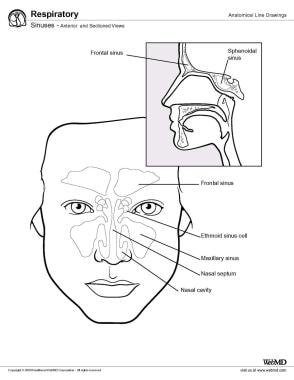
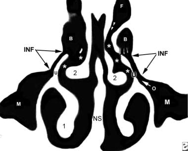
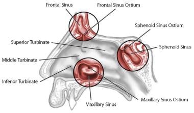
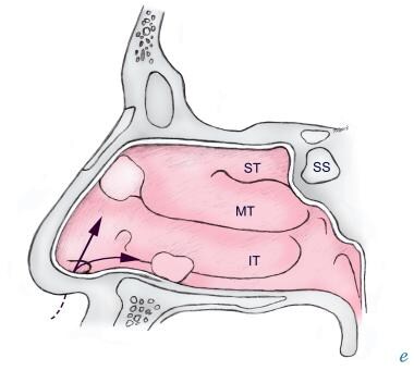
There are many risk factors involved in the development of ischemic optic neuropathy such as diabetes mellitus, hypertension, arteriosclerosis, and vascular incompetence. Therefore, the treatment of ischemic optic neuropathy should not be solely based on proper diagnosis but should also involve a thorough and systemic investigation to identify those multifactorial possibilities, which may contribute to the pathogenesis of the disease. We report upon a patient who developed non-arteritic ischemic optic neuropathy following treatment of a sphenoethmoid mucocele, which lead to recovered vision and a satisfactory improvement of visual field defects, after percutaneous trans-coronary angiography with stent insertion of the coronary arteries. (+info)The sphenoid sinuses are air-filled spaces located within the sphenoid bone, which is one of the bones that make up the skull base. These sinuses are located deep inside the skull, behind the eyes and nasal cavity. They are paired and separated by a thin bony septum, and each one opens into the corresponding nasal cavity through a small opening called the sphenoethmoidal recess. The sphenoid sinuses vary greatly in size and shape between individuals. They develop during childhood and continue to grow until early adulthood. The function of the sphenoid sinuses, like other paranasal sinuses, is not entirely clear, but they may contribute to reducing the weight of the skull, resonating voice during speech, and insulating the brain from trauma.
The sphenoid bone is a complex, irregularly shaped bone located in the middle cranial fossa and forms part of the base of the skull. It articulates with several other bones, including the frontal, parietal, temporal, ethmoid, palatine, and zygomatic bones. The sphenoid bone has two main parts: the body and the wings.
The body of the sphenoid bone is roughly cuboid in shape and contains several important structures, such as the sella turcica, which houses the pituitary gland, and the sphenoid sinuses, which are air-filled cavities within the bone. The greater wings of the sphenoid bone extend laterally from the body and form part of the skull's lateral walls. They contain the superior orbital fissure, through which important nerves and blood vessels pass between the cranial cavity and the orbit of the eye.
The lesser wings of the sphenoid bone are thin, blade-like structures that extend anteriorly from the body and form part of the floor of the anterior cranial fossa. They contain the optic canal, which transmits the optic nerve and ophthalmic artery between the brain and the orbit of the eye.
Overall, the sphenoid bone plays a crucial role in protecting several important structures within the skull, including the pituitary gland, optic nerves, and ophthalmic arteries.
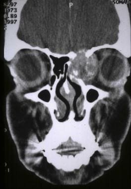
Sphenoid sinusitis is a medical condition characterized by the inflammation or infection of the sphenoid sinuses, which are air-filled cavities located in the sphenoid bone at the center of the skull base, behind the eyes. These sinuses are relatively small and difficult to access, making infections less common than in other sinuses. However, when sphenoid sinusitis does occur, it can cause various symptoms such as headaches, facial pain, nasal congestion, fever, and vision problems. Sphenoid sinusitis may result from bacterial or fungal infections, allergies, or autoimmune disorders. Diagnosis typically involves a combination of clinical evaluation, imaging studies like CT scans, and sometimes endoscopic examination. Treatment options include antibiotics for bacterial infections, antifungal medications for fungal infections, nasal sprays, decongestants, pain relievers, and, in severe or recurrent cases, surgical intervention.
Paranasal sinus diseases refer to a group of medical conditions that affect the paranasal sinuses, which are air-filled cavities located within the skull near the nasal cavity. These sinuses include the maxillary, frontal, ethmoid, and sphenoid sinuses.
Paranasal sinus diseases can be caused by a variety of factors, including viral, bacterial, or fungal infections, allergies, structural abnormalities, or autoimmune disorders. Some common paranasal sinus diseases include:
1. Sinusitis: Inflammation or infection of the sinuses, which can cause symptoms such as nasal congestion, thick nasal discharge, facial pain or pressure, and reduced sense of smell.
2. Nasal polyps: Soft, benign growths that develop in the lining of the nasal passages or sinuses, which can obstruct airflow and cause difficulty breathing through the nose.
3. Sinonasal tumors: Abnormal growths that can be benign or malignant, which can cause symptoms such as nasal congestion, facial pain, and bleeding from the nose.
4. Sinus cysts: Fluid-filled sacs that form in the sinuses, which can cause symptoms similar to those of sinusitis.
5. Fungal sinusitis: Infection of the sinuses with fungi, which can cause symptoms such as nasal congestion, facial pain, and thick, discolored mucus.
Treatment for paranasal sinus diseases depends on the underlying cause and severity of the condition. Treatment options may include medications, such as antibiotics, antihistamines, or corticosteroids, as well as surgical intervention in more severe cases.
Paranasal sinus neoplasms refer to abnormal growths or tumors that develop within the paranasal sinuses, which are air-filled cavities located inside the skull near the nasal cavity. These tumors can be benign (noncancerous) or malignant (cancerous), and they can arise from various types of tissue within the sinuses, such as the lining of the sinuses (mucosa), bone, or other soft tissues.
Paranasal sinus neoplasms can cause a variety of symptoms, including nasal congestion, nosebleeds, facial pain or numbness, and visual disturbances. The diagnosis of these tumors typically involves a combination of imaging studies (such as CT or MRI scans) and biopsy to determine the type and extent of the tumor. Treatment options may include surgery, radiation therapy, chemotherapy, or a combination of these approaches, depending on the specific type and stage of the neoplasm.
Cerebrospinal fluid (CSF) rhinorrhea is a condition where the cerebrospinal fluid, which surrounds and protects the brain and spinal cord, leaks through the nasal cavity. This occurs due to a defect or opening in the skull base or the thin bone that separates the brain from the nasal cavity, known as the cribriform plate.
CSF rhinorrhea can result from trauma, surgery, or spontaneously due to increased pressure in the brain. It is important to diagnose and treat this condition promptly because it increases the risk of meningitis, an infection of the membranes covering the brain and spinal cord. Treatment options include bed rest, hydration, stool softeners, and sometimes surgical repair of the defect.
Pneumoencephalography is a diagnostic procedure that is rarely used today, due to the development of less invasive techniques. It involves the introduction of air or another gas into the ventricular system or subarachnoid space of the brain, followed by X-ray imaging to visualize the structures and any abnormalities within the intracranial cavity.
The primary purpose of this procedure was to diagnose conditions affecting the brain's ventricles, such as hydrocephalus, tumors, or inflammation. The introduction of air into the cranium allowed for better visualization of these structures and any potential abnormalities. However, due to its invasive nature, risks associated with the procedure, and the availability of non-invasive imaging techniques like CT and MRI scans, pneumoencephalography has fallen out of favor in modern medicine.
The Sella Turcica, also known as the Turkish saddle, is a depression or fossa in the sphenoid bone located at the base of the skull. It forms a housing for the pituitary gland, which is a small endocrine gland often referred to as the "master gland" because it controls other glands and makes several essential hormones. The Sella Turcica has a saddle-like shape, with its anterior and posterior clinoids forming the front and back of the saddle, respectively. This region is of significant interest in neuroimaging and clinical settings, as various conditions such as pituitary tumors or other abnormalities may affect the size, shape, and integrity of the Sella Turcica.
A mucocele is a mucus-containing cystic lesion that results from the accumulation of mucin within a damaged minor salivary gland duct or mucous gland. It is typically caused by trauma, injury, or blockage of the duct. Mucocele appears as a round, dome-shaped, fluid-filled swelling, which may be bluish or clear in color. They are most commonly found on the lower lip but can also occur on other areas of the oral cavity. Mucocele is generally painless unless it becomes secondarily infected; however, it can cause discomfort during speaking, chewing, or swallowing, and may affect aesthetics. Treatment usually involves surgical excision of the mucocele to prevent recurrence.
The skull base is the lower part of the skull that forms the floor of the cranial cavity and the roof of the facial skeleton. It is a complex anatomical region composed of several bones, including the frontal, sphenoid, temporal, occipital, and ethmoid bones. The skull base supports the brain and contains openings for blood vessels and nerves that travel between the brain and the face or neck. The skull base can be divided into three regions: the anterior cranial fossa, middle cranial fossa, and posterior cranial fossa, which house different parts of the brain.
The ethmoid sinuses are a pair of air-filled spaces located in the ethmoid bone, which is a part of the skull that forms the upper portion of the nasal cavity and the inner eye socket. These sinuses are divided into anterior and posterior groups and are present in adults, but not at birth. They continue to grow and develop until early adulthood.
The ethmoid sinuses are lined with mucous membrane, which helps to warm, humidify, and filter the air we breathe. They are surrounded by a network of blood vessels and nerves, making them susceptible to inflammation and infection. Inflammation of the ethmoid sinuses can lead to conditions such as sinusitis, which can cause symptoms such as nasal congestion, headache, and facial pain.
The maxillary sinuses, also known as the antrums of Highmore, are the largest of the four pairs of paranasal sinuses located in the maxilla bones. They are air-filled cavities that surround the nasolacrimal duct and are situated superior to the upper teeth and lateral to the nasal cavity. Each maxillary sinus is lined with a mucous membrane, which helps to warm, humidify, and filter the air we breathe. Inflammation or infection of the maxillary sinuses can result in conditions such as sinusitis, leading to symptoms like facial pain, headaches, and nasal congestion.
The cavernous sinus is a venous structure located in the middle cranial fossa, which is a depression in the skull that houses several important nerves and blood vessels. The cavernous sinus is situated on either side of the sphenoid bone, near the base of the skull, and it contains several important structures:
* The internal carotid artery, which supplies oxygenated blood to the brain
* The abducens nerve (cranial nerve VI), which controls lateral movement of the eye
* The oculomotor nerve (cranial nerve III), which controls most of the muscles that move the eye
* The trochlear nerve (cranial nerve IV), which controls one of the muscles that moves the eye
* The ophthalmic and maxillary divisions of the trigeminal nerve (cranial nerve V), which transmit sensory information from the face and head
The cavernous sinus is an important structure because it serves as a conduit for several critical nerves and blood vessels. However, it is also vulnerable to various pathological conditions such as thrombosis (blood clots), infection, tumors, or aneurysms, which can lead to serious neurological deficits or even death.
A frontal sinus is a paired, air-filled paranasal sinus located in the frontal bone of the skull, above the eyes and behind the forehead. It is one of the four pairs of sinuses found in the human head. The frontal sinuses are lined with mucous membrane and are interconnected with the nasal cavity through small openings called ostia. They help to warm, humidify, and filter the air we breathe, and contribute to the resonance of our voice. Variations in size, shape, and asymmetry of frontal sinuses are common among individuals.
Enophthalmos is a medical term that refers to the abnormal positioning of the eyeball within its socket, resulting in a posterior or backward displacement of the eye. This condition can occur due to various reasons such as trauma, surgical procedures, or diseases that affect the orbital tissues, including cancer, inflammation, or infection. Enophthalmos may lead to cosmetic concerns and visual disturbances, depending on its severity. A thorough examination by an ophthalmologist or an oculoplastic surgeon is necessary for accurate diagnosis and management of this condition.
Paranasal sinuses are air-filled cavities in the skull that surround the nasal cavity. There are four pairs of paranasal sinuses, including the maxillary, frontal, ethmoid, and sphenoid sinuses. These sinuses help to warm, humidify, and filter the air we breathe. They also contribute to our voice resonance and provide a slight cushioning effect for the skull. The openings of the paranasal sinuses lead directly into the nasal cavity, allowing mucus produced in the sinuses to drain into the nose. Infections or inflammation of the paranasal sinuses can result in conditions such as sinusitis.
Endoscopy is a medical procedure that involves the use of an endoscope, which is a flexible tube with a light and camera at the end, to examine the interior of a body cavity or organ. The endoscope is inserted through a natural opening in the body, such as the mouth or anus, or through a small incision. The images captured by the camera are transmitted to a monitor, allowing the physician to visualize the internal structures and detect any abnormalities, such as inflammation, ulcers, or tumors. Endoscopy can also be used for diagnostic purposes, such as taking tissue samples for biopsy, or for therapeutic purposes, such as removing polyps or performing minimally invasive surgeries.
Dental radiography is a specific type of imaging that uses radiation to produce detailed images of the teeth, bones, and soft tissues surrounding them. It is a crucial tool in dental diagnostics and treatment planning. There are several types of dental radiographs, including:
1. Intraoral Radiographs: These are taken inside the mouth and provide detailed images of individual teeth or small groups of teeth. They can help detect cavities, assess periodontal health, plan for restorations, and monitor tooth development in children. Common types of intraoral radiographs include bitewing, periapical, and occlusal radiographs.
2. Extraoral Radiographs: These are taken outside the mouth and provide images of larger areas, such as the entire jaw or skull. They can help diagnose issues related to the temporomandibular joint (TMJ), detect impacted teeth, assess bone health, and identify any abnormalities in the facial structure. Common types of extraoral radiographs include panoramic, cephalometric, and sialography radiographs.
3. Cone Beam Computed Tomography (CBCT): This is a specialized type of dental radiography that uses a cone-shaped X-ray beam to create detailed 3D images of the teeth, bones, and soft tissues. It is particularly useful in planning complex treatments such as dental implants, orthodontic treatment, and oral surgery.
Dental radiographs are typically taken using a specialized machine that emits a low dose of radiation. Patients are provided with protective lead aprons to minimize exposure to radiation. The frequency of dental radiographs depends on the patient's individual needs and medical history. Dentists follow strict guidelines to ensure that dental radiography is safe and effective for their patients.
The ethmoid bone is a paired, thin, and lightweight bone that forms part of the skull's anterior cranial fossa and contributes to the formation of the orbit and nasal cavity. It is located between the frontal bone above and the maxilla and palatine bones below. The ethmoid bone has several important features:
1. Cribriform plate: This is the horizontal, sieve-like portion that forms part of the anterior cranial fossa and serves as the roof of the nasal cavity. It contains small openings (foramina) through which olfactory nerves pass.
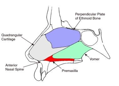
2. Perpendicular plate: The perpendicular plate is a vertical structure that projects downward from the cribriform plate, forming part of the nasal septum and separating the left and right nasal cavities.
3. Superior and middle nasal conchae: These are curved bony projections within the lateral walls of the nasal cavity that help to warm, humidify, and filter incoming air.
4. Lacrimal bone: The ethmoid bone articulates with the lacrimal bone, forming part of the medial wall of the orbit.
5. Frontal process: This is a thin, vertical plate that articulates with the frontal bone above the orbit.
6. Sphenoidal process: The sphenoidal process connects the ethmoid bone to the sphenoid bone posteriorly.
The ethmoid bone plays a crucial role in protecting the brain and providing structural support for the eyes, as well as facilitating respiration by warming, humidifying, and filtering incoming air.
A meningocele is a type of neural tube defect that results in the herniation of the meninges (the protective membranes covering the brain and spinal cord) through a defect in the vertebral column. The meninges protrude as a sac-like structure, which may be covered by skin or a thin layer of tissue. Meningoceles usually do not contain neural tissue, but cerebrospinal fluid is present within the sac. They are typically asymptomatic unless there is compression of surrounding structures or infection. Treatment generally involves surgical repair to prevent potential complications such as meningitis or neurological damage.
Skull base neoplasms refer to abnormal growths or tumors located in the skull base, which is the region where the skull meets the spine and where the brain connects with the blood vessels and nerves that supply the head and neck. These neoplasms can be benign (non-cancerous) or malignant (cancerous), and they can arise from various types of cells in this area, including bone, nerve, glandular, and vascular tissue.
Skull base neoplasms can cause a range of symptoms depending on their size, location, and growth rate. Some common symptoms include headaches, vision changes, hearing loss, facial numbness or weakness, difficulty swallowing, and balance problems. Treatment options for skull base neoplasms may include surgery, radiation therapy, chemotherapy, or a combination of these approaches. The specific treatment plan will depend on the type, size, location, and stage of the tumor, as well as the patient's overall health and medical history.
X-ray computed tomography (CT or CAT scan) is a medical imaging method that uses computer-processed combinations of many X-ray images taken from different angles to produce cross-sectional (tomographic) images (virtual "slices") of the body. These cross-sectional images can then be used to display detailed internal views of organs, bones, and soft tissues in the body.
The term "computed tomography" is used instead of "CT scan" or "CAT scan" because the machines take a series of X-ray measurements from different angles around the body and then use a computer to process these data to create detailed images of internal structures within the body.
CT scanning is a noninvasive, painless medical test that helps physicians diagnose and treat medical conditions. CT imaging provides detailed information about many types of tissue including lung, bone, soft tissue and blood vessels. CT examinations can be performed on every part of the body for a variety of reasons including diagnosis, surgical planning, and monitoring of therapeutic responses.
In computed tomography (CT), an X-ray source and detector rotate around the patient, measuring the X-ray attenuation at many different angles. A computer uses this data to construct a cross-sectional image by the process of reconstruction. This technique is called "tomography". The term "computed" refers to the use of a computer to reconstruct the images.
CT has become an important tool in medical imaging and diagnosis, allowing radiologists and other physicians to view detailed internal images of the body. It can help identify many different medical conditions including cancer, heart disease, lung nodules, liver tumors, and internal injuries from trauma. CT is also commonly used for guiding biopsies and other minimally invasive procedures.
In summary, X-ray computed tomography (CT or CAT scan) is a medical imaging technique that uses computer-processed combinations of many X-ray images taken from different angles to produce cross-sectional images of the body. It provides detailed internal views of organs, bones, and soft tissues in the body, allowing physicians to diagnose and treat medical conditions.
Otorhinolaryngologic surgical procedures are surgeries that are performed on the head and neck region, specifically involving the ear, nose, and throat (ENT) regions. This field is also known as otolaryngology-head and neck surgery. The procedures can range from relatively minor ones, such as removing a small nasal polyp or inserting ear tubes, to more complex surgeries like cochlear implantation, endoscopic sinus surgery, or removal of tumors in the head and neck region. These surgical procedures are typically performed by specialized physicians called otorhinolaryngologists (also known as ENT surgeons) who have completed extensive training in this area.
Pituitary neoplasms refer to abnormal growths or tumors in the pituitary gland, a small endocrine gland located at the base of the brain. These neoplasms can be benign (non-cancerous) or malignant (cancerous), with most being benign. They can vary in size and may cause various symptoms depending on their location, size, and hormonal activity.
Pituitary neoplasms can produce and secrete excess hormones, leading to a variety of endocrine disorders such as Cushing's disease (caused by excessive ACTH production), acromegaly (caused by excessive GH production), or prolactinoma (caused by excessive PRL production). They can also cause local compression symptoms due to their size, leading to headaches, vision problems, and cranial nerve palsies.
The exact causes of pituitary neoplasms are not fully understood, but genetic factors, radiation exposure, and certain inherited conditions may increase the risk of developing these tumors. Treatment options for pituitary neoplasms include surgical removal, radiation therapy, and medical management with drugs that can help control hormonal imbalances.
Osteoma is a benign (noncancerous) tumor that is made up of mature bone tissue. It usually grows slowly over a period of years and is most commonly found in the skull or jaw, although it can occur in other bones of the body as well. Osteomas are typically small, but they can grow to be several centimeters in size. They may cause symptoms if they press on nearby tissues or structures, such as nerves or blood vessels. In some cases, osteomas may not cause any symptoms and may only be discovered during routine imaging studies. Treatment for osteoma is typically not necessary unless it is causing problems or growing rapidly. If treatment is needed, it may involve surgical removal of the tumor.
Cranial sinuses are a part of the venous system in the human head. They are air-filled spaces located within the skull and are named according to their location. The cranial sinuses include:
1. Superior sagittal sinus: It runs along the top of the brain, inside the skull, and drains blood from the scalp and the veins of the brain.
2. Inferior sagittal sinus: It runs along the bottom of the brain and drains into the straight sinus.
3. Straight sinus: It is located at the back of the brain and receives blood from the inferior sagittal sinus and great cerebral vein.
4. Occipital sinuses: They are located at the back of the head and drain blood from the scalp and skull.
5. Cavernous sinuses: They are located on each side of the brain, near the temple, and receive blood from the eye and surrounding areas.
6. Sphenoparietal sinus: It is a small sinus that drains blood from the front part of the brain into the cavernous sinus.
7. Petrosquamosal sinuses: They are located near the ear and drain blood from the scalp and skull.
The cranial sinuses play an essential role in draining blood from the brain and protecting it from injury.
Neuronavigation is a surgical technique that uses imaging technology, such as MRI or CT scans, to create a 3D map of the patient's brain in real-time during surgery. This allows surgeons to accurately locate and navigate to specific areas of the brain with greater precision and less invasiveness, improving surgical outcomes and reducing the risk of complications.
The neuronavigation system typically consists of a computer workstation, tracking systems, and instruments that are equipped with sensors. The system is able to track the position and orientation of these instruments relative to the patient's brain, allowing the surgeon to visualize the location of the instruments on the 3D map in real-time.
Neuronavigation has become an essential tool in many neurosurgical procedures, including tumor resection, functional neurosurgery, and deep brain stimulation. It enables surgeons to perform more complex surgeries with increased safety and efficacy, ultimately improving the quality of care for patients undergoing these procedures.
Medical Definition:
Magnetic Resonance Imaging (MRI) is a non-invasive diagnostic imaging technique that uses a strong magnetic field and radio waves to create detailed cross-sectional or three-dimensional images of the internal structures of the body. The patient lies within a large, cylindrical magnet, and the scanner detects changes in the direction of the magnetic field caused by protons in the body. These changes are then converted into detailed images that help medical professionals to diagnose and monitor various medical conditions, such as tumors, injuries, or diseases affecting the brain, spinal cord, heart, blood vessels, joints, and other internal organs. MRI does not use radiation like computed tomography (CT) scans.
The abducens nerve, also known as the sixth cranial nerve, is responsible for controlling the lateral rectus muscle of the eye, which enables the eye to move outward. Abducens nerve diseases refer to conditions that affect this nerve and can result in various symptoms, primarily affecting eye movement.
Here are some medical definitions related to abducens nerve diseases:
1. Abducens Nerve Palsy: A condition characterized by weakness or paralysis of the abducens nerve, causing difficulty in moving the affected eye outward. This results in double vision (diplopia), especially when gazing towards the side of the weakened nerve. Abducens nerve palsy can be congenital, acquired, or caused by various factors such as trauma, tumors, aneurysms, infections, or diseases like diabetes and multiple sclerosis.
2. Sixth Nerve Palsy: Another term for abducens nerve palsy, referring to the weakness or paralysis of the sixth cranial nerve.
3. Internuclear Ophthalmoplegia (INO): A neurological condition affecting eye movement, often caused by a lesion in the medial longitudinal fasciculus (MLF), a bundle of nerve fibers that connects the abducens nucleus with the oculomotor nucleus. INO results in impaired adduction (inward movement) of the eye on the side of the lesion and nystagmus (involuntary eye movements) of the abducting eye on the opposite side when attempting to look towards the side of the lesion.
4. One-and-a-Half Syndrome: A rare neurological condition characterized by a combination of INO and internuclear ophthalmoplegia with horizontal gaze palsy on the same side, caused by damage to both the abducens nerve and the paramedian pontine reticular formation (PPRF). This results in limited or no ability to move the eyes towards the side of the lesion and impaired adduction of the eye on the opposite side.
5. Brainstem Encephalitis: Inflammation of the brainstem, which can affect the abducens nerve and other cranial nerves, leading to various neurological symptoms such as diplopia (double vision), ataxia (loss of balance and coordination), and facial weakness. Brainstem encephalitis can be caused by infectious agents, autoimmune disorders, or paraneoplastic syndromes.
6. Multiple Sclerosis (MS): An autoimmune disorder characterized by inflammation and demyelination of the central nervous system, including the brainstem and optic nerves. MS can cause various neurological symptoms, such as diplopia, nystagmus, and INO, due to damage to the abducens nerve and other cranial nerves.
7. Wernicke's Encephalopathy: A neurological disorder caused by thiamine (vitamin B1) deficiency, often seen in alcoholics or individuals with malnutrition. Wernicke's encephalopathy can affect the brainstem and cause various symptoms such as diplopia, ataxia, confusion, and oculomotor abnormalities.
8. Pontine Glioma: A rare type of brain tumor that arises from the glial cells in the pons (a part of the brainstem). Pontine gliomas can cause various neurological symptoms such as diplopia, facial weakness, and difficulty swallowing due to their location in the brainstem.
9. Brainstem Cavernous Malformation: A benign vascular lesion that arises from the small blood vessels in the brainstem. Brainstem cavernous malformations can cause various neurological symptoms such as diplopia, ataxia, and facial weakness due to their location in the brainstem.
10. Pituitary Adenoma: A benign tumor that arises from the pituitary gland, located at the base of the brain. Large pituitary adenomas can compress the optic nerves and cause various visual symptoms such as diplopia, visual field defects, and decreased vision.
11. Craniopharyngioma: A benign tumor that arises from the remnants of the Rathke's pouch, a structure that gives rise to the anterior pituitary gland. Craniopharyngiomas can cause various neurological and endocrine symptoms such as diplopia, visual field defects, headaches, and hormonal imbalances due to their location near the optic nerves and pituitary gland.
12. Meningioma: A benign tumor that arises from the meninges, the protective covering of the brain and spinal cord. Meningiomas can cause various neurological symptoms such as diplopia, headaches, and seizures depending on their location in the brain or spinal cord.
13. Chordoma: A rare type of malignant tumor that arises from the remnants of the notochord, a structure that gives rise to the spine during embryonic development. Chordomas can cause various neurological and endocrine symptoms such as diplopia, visual field defects, headaches, and hormonal imbalances due to their location near the brainstem and spinal cord.
14. Metastatic Brain Tumors: Malignant tumors that spread from other parts of the body to the brain. Metastatic brain tumors can cause various neurological symptoms such as diplopia, headaches, seizures, and cognitive impairment depending on their location in the brain.
15. Other Rare Brain Tumors: There are many other rare types of brain tumors that can cause diplopia or other neurological symptoms, including gliomas, ependymomas, pineal region tumors, and others. These tumors require specialized diagnosis and treatment by neuro-oncologists and neurosurgeons with expertise in these rare conditions.
In summary, diplopia can be caused by various brain tumors, including pituitary adenomas, meningiomas, chordomas, metastatic brain tumors, and other rare types of tumors. It is important to seek medical attention promptly if you experience diplopia or other neurological symptoms, as early diagnosis and treatment can improve outcomes and quality of life.
The Sinus of Valsalva are three pouch-like dilations or outpouchings located at the upper part (root) of the aorta, just above the aortic valve. They are named after Antonio Maria Valsalva, an Italian anatomist and physician. These sinuses are divided into three parts:
1. Right Sinus of Valsalva: It is located to the right of the ascending aorta and usually gives rise to the right coronary artery.
2. Left Sinus of Valsalva: It is situated to the left of the ascending aorta and typically gives rise to the left coronary artery.
3. Non-coronary Sinus of Valsalva: This sinus is located in between the right and left coronary sinuses, and it does not give rise to any coronary arteries.
These sinuses play a crucial role during the cardiac cycle, particularly during ventricular contraction (systole). The pressure difference between the aorta and the ventricles causes the aortic valve cusps to be pushed into these sinuses, preventing the backflow of blood from the aorta into the ventricles.
Anatomical variations in the size and shape of the Sinuses of Valsalva can occur, and certain conditions like congenital heart diseases (e.g., aortic valve stenosis or bicuspid aortic valve) may affect their structure and function. Additionally, aneurysms or ruptures of the sinuses can lead to severe complications, such as cardiac tamponade, endocarditis, or stroke.
Pituitary apoplexy is a medical emergency that involves bleeding into the pituitary gland (a small gland at the base of the brain) and/or sudden swelling of the pituitary gland. This can lead to compression of nearby structures, such as the optic nerves and the hypothalamus, causing symptoms like severe headache, visual disturbances, hormonal imbalances, and altered mental status. It is often associated with a pre-existing pituitary tumor (such as a pituitary adenoma), but can also occur in individuals without any known pituitary abnormalities. Immediate medical attention is required to manage this condition, which may include surgical intervention, hormone replacement therapy, and supportive care.
Craniofacial abnormalities refer to a group of birth defects that affect the development of the skull and face. These abnormalities can range from mild to severe and may involve differences in the shape and structure of the head, face, and jaws, as well as issues with the formation of facial features such as the eyes, nose, and mouth.
Craniofacial abnormalities can be caused by genetic factors, environmental influences, or a combination of both. Some common examples of craniofacial abnormalities include cleft lip and palate, craniosynostosis (premature fusion of the skull bones), and hemifacial microsomia (underdevelopment of one side of the face).
Treatment for craniofacial abnormalities may involve a team of healthcare professionals, including plastic surgeons, neurosurgeons, orthodontists, speech therapists, and other specialists. Treatment options may include surgery, bracing, therapy, and other interventions to help improve function and appearance.
The carotid sinus is a small, dilated area located at the bifurcation (or fork) of the common carotid artery into the internal and external carotid arteries. It is a baroreceptor region, which means it contains specialized sensory nerve endings that can detect changes in blood pressure. When the blood pressure increases, the walls of the carotid sinus stretch, activating these nerve endings and sending signals to the brain. The brain then responds by reducing the heart rate and relaxing the blood vessels, which helps to lower the blood pressure back to normal.
The carotid sinus is an important part of the body's autonomic nervous system, which regulates various involuntary functions such as heart rate, blood pressure, and digestion. It plays a crucial role in maintaining cardiovascular homeostasis and preventing excessive increases in blood pressure that could potentially damage vital organs.
The middle cranial fossa is a depression or hollow in the skull that forms the upper and central portion of the cranial cavity. It is located between the anterior cranial fossa (which lies anteriorly) and the posterior cranial fossa (which lies posteriorly). The middle cranial fossa contains several important structures, including the temporal lobes of the brain, the pituitary gland, the optic chiasm, and the cavernous sinuses. It is also where many of the cranial nerves pass through on their way to the brain.
The middle cranial fossa can be further divided into two parts: the anterior and posterior fossae. The anterior fossa contains the optic chiasm and the pituitary gland, while the posterior fossa contains the temporal lobes of the brain and the cavernous sinuses.
The middle cranial fossa is formed by several bones of the skull, including the sphenoid bone, the temporal bone, and the parietal bone. The shape and size of the middle cranial fossa can vary from person to person, and abnormalities in its structure can be associated with various medical conditions, such as pituitary tumors or aneurysms.
Aspergillosis is a medical condition that is caused by the infection of the Aspergillus fungi. This fungus is commonly found in decaying organic matter, such as leaf litter and compost piles, and can also be found in some indoor environments like air conditioning systems and old buildings with water damage.
There are several types of aspergillosis, including:
1. Allergic bronchopulmonary aspergillosis (ABPA): This type of aspergillosis occurs when a person's immune system overreacts to the Aspergillus fungi, causing inflammation in the airways and lungs. ABPA is often seen in people with asthma or cystic fibrosis.
2. Invasive aspergillosis: This is a serious and potentially life-threatening condition that occurs when the Aspergillus fungi invade the bloodstream and spread to other organs, such as the brain, heart, or kidneys. Invasive aspergillosis typically affects people with weakened immune systems, such as those undergoing chemotherapy or organ transplantation.
3. Aspergilloma: Also known as a "fungus ball," an aspergilloma is a growth of the Aspergillus fungi that forms in a preexisting lung cavity, such as one caused by previous lung disease or injury. While an aspergilloma itself is not typically harmful, it can cause symptoms like coughing up blood or chest pain if it grows too large or becomes infected.
Symptoms of aspergillosis can vary depending on the type and severity of the infection. Treatment may include antifungal medications, surgery to remove the fungal growth, or management of underlying conditions that increase the risk of infection.
Skull neoplasms refer to abnormal growths or tumors that develop within the skull. These growths can be benign (non-cancerous) or malignant (cancerous). They can originate from various types of cells, such as bone cells, nerve cells, or soft tissues. Skull neoplasms can cause various symptoms depending on their size and location, including headaches, seizures, vision problems, hearing loss, and neurological deficits. Treatment options include surgery, radiation therapy, and chemotherapy. It is important to note that a neoplasm in the skull can also refer to metastatic cancer, which has spread from another part of the body to the skull.
The coronary sinus is a large vein that receives blood from the heart's muscle tissue. It is located on the posterior side of the heart and is a part of the cardiovascular system. The coronary sinus collects oxygen-depleted blood from the myocardium (the heart muscle) and drains it into the right atrium, where it will then be pumped to the lungs for oxygenation.
The coronary sinus is an essential structure in medical procedures such as cardiac catheterization and electrophysiological studies. It is also a common site for the implantation of pacemakers and other cardiac devices.
"Foreign bodies" refer to any object or substance that is not normally present in a particular location within the body. These can range from relatively harmless items such as splinters or pieces of food in the skin or gastrointestinal tract, to more serious objects like bullets or sharp instruments that can cause significant damage and infection.
Foreign bodies can enter the body through various routes, including ingestion, inhalation, injection, or penetrating trauma. The location of the foreign body will determine the potential for harm and the necessary treatment. Some foreign bodies may pass through the body without causing harm, while others may require medical intervention such as removal or surgical extraction.
It is important to seek medical attention if a foreign body is suspected, as untreated foreign bodies can lead to complications such as infection, inflammation, and tissue damage.
Cone-beam computed tomography (CBCT) is a medical imaging technique that uses a cone-shaped X-ray beam to create detailed, cross-sectional images of the body. In dental and maxillofacial radiology, CBCT is used to produce three-dimensional images of the teeth, jaws, and surrounding bones.
CBCT differs from traditional computed tomography (CT) in that it uses a cone-shaped X-ray beam instead of a fan-shaped beam, which allows for a faster scan time and lower radiation dose. The X-ray beam is rotated around the patient's head, capturing data from multiple angles, which is then reconstructed into a three-dimensional image using specialized software.
CBCT is commonly used in dental implant planning, orthodontic treatment planning, airway analysis, and the diagnosis and management of jaw pathologies such as tumors and fractures. It provides detailed information about the anatomy of the teeth, jaws, and surrounding structures, which can help clinicians make more informed decisions about patient care.
However, it is important to note that CBCT should only be used when necessary, as it still involves exposure to ionizing radiation. The benefits of using CBCT must be weighed against the potential risks associated with radiation exposure.
Comparative histology is a branch of pathology that deals with the comparison of tissue structures and organization across different species. It involves the microscopic examination and study of tissues from various organisms to understand the similarities and differences in their cellular and extracellular components, as well as their functions. By comparing and contrasting tissues from diverse species, researchers can gain insights into evolutionary relationships, adaptations, and the development of diseases across different taxa. This field is particularly important for understanding the fundamental principles of tissue organization and function, and has applications in areas such as veterinary medicine, comparative medical research, and evolutionary biology.
Histology is the study of the microscopic structure of tissues. It involves the examination of tissues at the level of individual cells and their organization into functional units. This field uses various staining techniques to visualize different cellular components, allowing for the identification and analysis of specific cell types, tissue architecture, and pathological changes. Histology is a fundamental discipline in anatomy, physiology, and pathology, providing essential information for understanding normal tissue function and disease processes.
Anatomy is the branch of biology that deals with the study of the structure of organisms and their parts. In medicine, anatomy is the detailed study of the structures of the human body and its organs. It can be divided into several subfields, including:
1. Gross anatomy: Also known as macroscopic anatomy, this is the study of the larger structures of the body, such as the organs and organ systems, using techniques such as dissection and observation.
2. Histology: This is the study of tissues at the microscopic level, including their structure, composition, and function.
3. Embryology: This is the study of the development of the embryo and fetus from conception to birth.
4. Neuroanatomy: This is the study of the structure and organization of the nervous system, including the brain and spinal cord.
5. Comparative anatomy: This is the study of the structures of different species and how they have evolved over time.
Anatomy is a fundamental subject in medical education, as it provides the basis for understanding the function of the human body and the underlying causes of disease.
The nasal cavity is the air-filled space located behind the nose, which is divided into two halves by the nasal septum. It is lined with mucous membrane and is responsible for several functions including respiration, filtration, humidification, and olfaction (smell). The nasal cavity serves as an important part of the upper respiratory tract, extending from the nares (nostrils) to the choanae (posterior openings of the nasal cavity that lead into the pharynx). It contains specialized structures such as turbinate bones, which help to warm, humidify and filter incoming air.
 Sphenoid sinus
Cerebrospinal fluid rhinorrhoea
Oral mucocele
Waters' view
Endoscopic endonasal surgery
Cavernous sinus thrombosis
Posterior ethmoidal nerve
Sphenoid wing meningioma
Ophthalmic nerve
Clivus (anatomy)
Neurosurgery
Dextroscope
Sella turcica
Orbital emphysema
Sinus (anatomy)
Rhinoplasty
Respiratory system of the horse
Jacques Dubois
Facial nerve
Wirginia Maixner
Superior nasal concha
Aerosinusitis
Eric M. Genden
Human nose
Upper respiratory tract infection
Paranasal sinuses
Sinusitis
Hypophysectomy
Aspergilloma
Palatovaginal canal
Sphenoid sinus - Wikipedia
 Management of spontaneous cerebrospinal fluid leaks of the sphenoid sinus: our experience
 PRIME PubMed | Pituitary abscess following expanding sphenoid sinus pyocele: complication of endoscopic endonasal...
 31051 Sphenoid sinus surgery - ClearHealthCosts
 Neurovascular relationships of the sphenoid sinus. A microsurgical study. | Read by QxMD
 Paranasal Sinus Anatomy: Overview, Gross Anatomy, Microscopic Anatomy
CT Scan of the Paranasal Sinuses: History, Basic Concepts, Anatomy
Hemangiomas of the sphenoid sinus - Fingerprint - Mayo Clinic
 Nasal Cartilages Anatomy, Function & Diagram | Body Maps
 Chronic Rhinosinusitis - Ear, Nose & Throat | Medtronic
 Anatomy, Head and Neck, Nasal Cavity - StatPearls - NCBI Bookshelf
Horsey WJ[au] - Search Results - PubMed
 Bassett Collection - Lane Medical Library - Stanford University School of Medicine
ICD-10-PCS - Medical Codes
 Thieme E-Journals - Journal of Neurological Surgery Part B: Skull Base / Abstract
 Vol. 11 No. 1 (2022)
| Anatomy Journal of Africa
Vol. 11 No. 1 (2022)
| Anatomy Journal of Africa
 2020-2021 BCSC Basic and Clinical Science Course™
 Medtronic NuVent EM Sinus Dilation System with Electromagnetic Navigation Announced (VIDEO)
 The AMEDEO Literature Guide
 Parlodel (Bromocriptine Mesylate): Uses, Dosage, Side Effects, Interactions, Warning
 Aspergillosis | Institut Pasteur
 Alexandra Golby, MD - Dana-Farber Cancer Institute | Boston, MA
Flow Cytometry and Squamous Cell Carcinoma of the Maxillary Sinus: A Possible Prognostic Indicator for Multimodality...
Mucormycosis (Zygomycosis) Medication: Antifungal agents, Polyene, Antifungal agents, Azole derivatives
 Related Articles - Annals Singapore
Sinusitis8
- A potential complication of sphenoidal sinusitis is cavernous sinus thrombosis. (wikipedia.org)
- Today, CT is the radiologic examination of choice in evaluating the paranasal sinuses of a patient with sinusitis. (medscape.com)
- [ 2 ] However, for most patients with sinusitis, noncontrast CT of the paranasal sinuses generally suffices. (medscape.com)
- At least one type of sinus infection - sphenoid sinusitis - is linked to an ache behind the eyes. (pursuantmedia.com)
- Although functional endoscopic sinus surgery is the primary approach used today for the surgical treatment of chronic sinusitis, the time-honored external approaches still play a role. (medscape.com)
- Sinusitis Sinusitis is inflammation of the sinuses, most commonly caused by a viral or bacterial infection or by an allergy. (msdmanuals.com)
- Generalmente se presenta en conjunción con otras sinusitis paranasales. (bvsalud.org)
- Isolated sphenoid sinusitis is uncommon. (bvsalud.org)
Ethmoid sinuses4
- The ethmoid sinuses arise in the ethmoid bone, forming several distinct air cells between the eyes. (medscape.com)
- Air containing mucosal lined sinuses surround the nasal cavity, which includes the frontal, paired maxillary, sphenoid, and ethmoid sinuses. (nih.gov)
- Surrounding the nasal cavities are air-containing mucosal lined sinuses, which include the frontal sinuses (superior anterior), ethmoid sinuses (superior), paired maxillary sinuses (lateral), and sphenoid sinuses (posterior). (nih.gov)
- of the sphenoid or ethmoid sinuses. (msdmanuals.com)
Paranasal sinus anatomy2
- The introduction of computed tomography (CT) scanning and its wider use over time have additionally improved the physician's ability to appreciate nuances of paranasal sinus anatomy and accurate disease correlation. (medscape.com)
- Conclusion EEA to the orbit is challenging, in particular for trainees unfamiliar with nasal and paranasal sinus anatomy. (thieme-connect.com)
Tomography1
- Cone beam computed tomography images revealed the absence of the sphenoid, frontal, and maxillary sinus, flattening of the condyles and glenoid fossa, and bilateral hypoplasia of the mandibular condyles. (wjgnet.com)
Internal caroti1
- citation needed] Proximal structures include: the optic canal and optic nerve, internal carotid artery, cavernous sinus, trigeminal nerve, pituitary gland, and the anterior ethmoidal cells. (wikipedia.org)
Tumor6
- citation needed] If a fast-growing tumor erodes the floor of the sphenoidal sinus, the vidian nerve could be in danger. (wikipedia.org)
- citation needed] If the tumor spreads laterally, the cavernous sinus and all its constituent nerves could be in danger. (wikipedia.org)
- The types of tumor tissues in the renal sinus are extensive, including fat, lymphatic, nerve and vascular tissues. (evanewyork.net)
- In this procedure, a neurosurgeon reaches the tumor through the nasal passages and sphenoid sinus. (atheistsforhumanrights.org)
- either a lateral rhinotomy method or a midfacial degloving method can be used for en bloc tumor removal. (medscape.com)
- Only 1 patient harboring a left cavernous sinus meningioma had tumor recurrence and underwent repeat resection. (medscape.com)
Sets of sinuses2
- The ethmoid sinus (one of six sets of sinuses) is part of the paranasal sinus system and is located between the nose and eyes. (healthline.com)
- The nose is surrounded by four sets of sinuses, which are hollow cavities in the head. (fjmc.org)
Endoscopic12
- TY - JOUR T1 - Pituitary abscess following expanding sphenoid sinus pyocele: complication of endoscopic endonasal transsphenoidal surgery. (unboundmedicine.com)
- The use of CT scanning combined with functional endoscopic sinus surgery (FESS) has empowered the modern sinus surgeon to treat patients more effectively, facilitating reduced morbidity and complications. (medscape.com)
- Validation of modular endoscopic medial maxillectomies for inverted papilloma of the maxillary sinus. (amedeo.com)
- For those who do not improve on medical therapy, a surgical procedure called Functional Endoscopic Sinus Surgery may be the answer for final relief. (fjmc.org)
- A complete evaluation including a thorough history, physical examination and an endoscopic evaluation will allow you sinus specialist to make an accurate diagnosis with which to design a customized treatment plan. (fjmc.org)
- In the 1990s, a distinct impulse came from the otorhinolaryngologists, with the use of the endoscope in functional endoscopic sinus surgery, disclosing the pathway to the sella turcica and the endoscopic approach for resection of pituitary tumors either alone or as an adjunct to the microneurosurgery. (scielo.br)
- In the endoscopic technique, a rigid endoscope is used to get into the sphenoid sinus and the sella turcica through both nostrils. (scielo.br)
- Senior et al reported that symptoms improved in 66 of 72 (91.6%) patients following endoscopic sinus surgery, with a mean follow-up time of 7.8 years. (medscape.com)
- A recently developed alternative to functional endoscopic sinus surgery is balloon sinuplasty. (medscape.com)
- Further study and long-term outcomes with this technology will determine its role in endoscopic sinus surgery. (medscape.com)
- Endoscopic sinus surgery is most commonly performed for inflammatory and infectious sinus disease. (medscape.com)
- Typically, endoscopic sinus surgery is reserved for patients with documented rhinosinusitis, based on a thorough history and a complete physical examination, including CT scans if appropriate, and in whom appropriate medical treatment has failed. (medscape.com)
Bone12
- The sphenoid sinus is a paired paranasal sinus occurring within the body of the sphenoid bone. (wikipedia.org)
- Each spehoid sinus is contained within the body of sphenoid bone, being situated just inferior to the sella turcica. (wikipedia.org)
- When exceptionally large, the sphenoid sinuses may extend into the roots of the pterygoid processes or greater wings of sphenoid bone, and may invade the basilar part of the occipital bone. (wikipedia.org)
- Because only thin shelves of bone separate the sphenoidal sinuses from the nasal cavities below and hypophyseal fossa above, the pituitary gland can be surgically approached through the roof of the nasal cavities by first passing through the anterioinferior aspect of the sphenoid bone and into the sinuses, followed by entry through the top of the sphenoid bone into the hypophyseal fossa. (wikipedia.org)
- The maxillary sinus is the largest paranasal sinus and lies inferior to the eyes in the maxillary bone. (medscape.com)
- The frontal sinus is housed in the frontal bone superior to the eyes in the forehead. (medscape.com)
- The sphenoid sinus originates in the sphenoid bone at the center of the head. (medscape.com)
- and (2) the lesser wing of the sphenoid bone. (aao.org)
- Medially, the frontal bone forms the roof of the ethmoid sinus and extends to the cribriform plate. (aao.org)
- The pituitary gland is surrounded by bone (sphenoid bone), and it sits in a pouch called the sella turcica. (cancer.ca)
- This approach requires a wide opening of the sella, the bone that separates the sphenoid sinus from the pituitary. (csrf.net)
- This technique uses balloon catheters to dilate the maxillary, frontal, and sphenoid natural ostia without bone or soft-tissue removal. (medscape.com)
Nose10
- The maxillary sinus is one of the four paranasal sinuses, which are sinuses located near the nose. (healthline.com)
- Together, the sinuses and nose act to filter, heat, vaporize and warm the air brought through the nose into the lungs. (fjmc.org)
- Should surgery be required, tiny telescopes and cameras will allow the sinus surgeon to reestablish the normal draining patterns within the nose, allowing relief of symptoms. (fjmc.org)
- The sphenoid sinus is an air-filled sinus that drains into the nose. (mayfieldclinic.com)
- This chapter provides criteria for assessing permanent impairment from entitled conditions of the nose, sinuses and trachea. (gc.ca)
- Impairment from malignant conditions of the nose, throat and sinuses is rated within Chapter 18 , Malignant Impairment. (gc.ca)
- The most direct way to the pituitary is through the nose and the sphenoid sinus. (csrf.net)
- Cavernous sinus thrombosis is usually caused by the spread of bacteria from infections of the face (including the skin of the nose), orbit, or sinus. (msdmanuals.com)
- The cavernous sinus is not one of the air-filled sinuses around the nose (the nasal sinuses). (msdmanuals.com)
- Infection can spread to the orbit from sources such as the sinuses around the nose. (msdmanuals.com)
Cavernous sinus thro2
- Cavernous sinus thrombosis is a very rare disorder in which a blood clot (thrombosis) forms in the cavernous sinus (a large vein at the base of the skull). (msdmanuals.com)
- Cavernous sinus thrombosis (CST) is usually caused by the spread of bacteria (usually Staphylococcus aureus ) from a facial, dental, or nasal sinus infection. (msdmanuals.com)
Include the frontal1
- Table 10.2 is used to rate impairment of the paranasal sinuses which include the frontal, ethmoid, sphenoid, and maxillary sinuses. (gc.ca)
Trigeminal nerve1
- Innervation of the sphenoid sinus comes from branches of the first and second divisions of the trigeminal nerve. (medscape.com)
Cavities2
- [ 3 ] The frontal sinuses are funnel-shaped structures with their ostia located in the most dependent portion of the cavities. (medscape.com)
- Mucus gets trapped in the sinus cavities and can become infected: hence, sinus symptoms prevail. (fjmc.org)
Tumors3
- It can also be caused by temporal lobe seizures, inflamed sinuses, brain tumors and Parkinson's disease. (evanewyork.net)
- Large tumors can press on the optic nerves and invade the cavernous sinuses, which house the carotid arteries and the nerves involved in eye movement. (mayfieldclinic.com)
- It's my pleasure to be with you today and to discuss with you some advanced cases of tumors within the paranasal sinuses. (brainlab.com)
Carotid arteries1
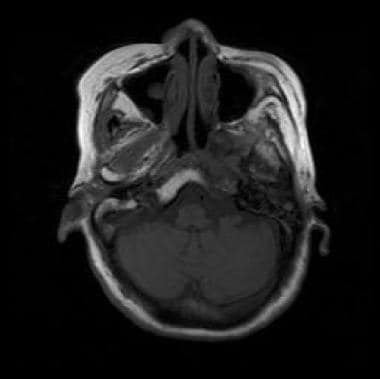
- Both cavernous sinuses are seen indented along their inner aspects by the mass (more on the right), with patent signal void intra-cavernous carotid arteries. (radiopaedia.org)
Mucus3
- Like the nasal cavity, the wall lining of the sinuses also secretes mucus. (nih.gov)
- The sinuses themselves produce mucus to cleanse inhaled air of bacteria, viruses, fungus, pollutants and dirt. (fjmc.org)
- These microbes trigger excessive mucus throughout the sinus and block the nasal cavity. (regressiveantidote.net)
Posterior wall3
- anteriorly, an opening of sphenoidal sinus opens into the roof of the nasal cavity via an aperture on the posterior wall of the sphenoethmoidal recess (occurring just superior the choana). (wikipedia.org)
- The posterior wall of the frontal sinus, which separates the sinus from the anterior cranial fossa, is much thinner than its anterior wall. (medscape.com)
- Anteriorly: The lesion causes scalloping of the posterior wall of the sphenoid sinus. (radiopaedia.org)
Mucoceles1
- granulomatous, inflammatory and infectious processes such as tuberculosis, sarcoidosis, giant cell granuloma, sphenoid sinus mucoceles and others [4]. (bvsalud.org)
Pituitary gland4
- The pituitary gland is bordered on either side by the cavernous sinuses and below by the sphenoid sinus. (mayfieldclinic.com)
- A cross-section of the pituitary gland (green) shows its relationship to the optic chiasm, the sphenoid sinus, and the cavernous sinuses on each side. (mayfieldclinic.com)
- The pituitary gland is related to the optic chiasm above and the sphenoid sinus below. (mayfieldclinic.com)
- The pituitary gland is behind the nasal cavity and lies just above a sinus (sphenoid sinus). (cancer.ca)
Infection6
- In patients with an effective immune system , infection gives rise to chronic headaches and nasal obstruction, and sinus drainage is generally an effective treatment. (pasteur.fr)
- The micro organism that is most often answerable for irritation and sphenoid sinus infection is Streptococcus pneumonia. (regressiveantidote.net)
- The viral infection Haemophilus influenza can even spurn sphenoidal sinus ache. (regressiveantidote.net)
- It could also be because of a sinus infection or injury. (pursuantmedia.com)
- Sinus infection Problems with the sinuses often include feelings of pain in and around the face. (pursuantmedia.com)
- One of the main symptoms of a sinus infection is throbbing pain and pressure around the eyeballs. (pursuantmedia.com)
Turbinate1
- The sphenoid sinus ostium is located on the anterosuperior surface of the sphenoid face, usually medial to the superior turbinate. (medscape.com)
Lateral4
- 500 The sphenoid sinuses vary in size and shape, and, owing to the lateral displacement of the intervening septum of sphenoid sinuses, are rarely symmetrical. (wikipedia.org)
- Closure of spontaneous sphenoid sinus cerebrospinal fluid leaks can be challenging because of the relative inaccessibility of the lateral recess and the presence of intracranial hypertension. (nih.gov)
- The lateral walls are spiral shaped mucosal folds that overlie the turbinates and sinus ducts draining into the ostia. (nih.gov)
- All of these paranasal sinuses, except the sphenoid, communicate with the nasal cavity via ducts that drain through ostia, which empty into spaces located on the lateral wall. (nih.gov)
Skull Base3
- Behind the posteromedial wall of the maxillary sinus lies the pterygopalatine fossa, a small inverted space that houses several important neurovascular structures and communicates with several skull base foramina. (medscape.com)
- Vascular lesions with an intraosseus nidus involving the skull base are uncommon and challenging [Gianoli GJ, Amedee RG Vascular malformation of the sphenoid sinus. (upmc.com)
- 1994)]. We present a pediatric patient, with a life-threatening arteriovenous malformation (AVM) of the sphenoid sinus, clivus, and ventral skull base, who failed routine multimodality management of AVMs. (upmc.com)
Ostia2
- The secretions from these sinuses drain into the nasal cavity via the thin-walled ostia. (nih.gov)
- Reports show persistent patient symptom improvement and sinus ostia patency. (medscape.com)
Surgical7
- An endonasal surgical procedure called a sphenoidotomy may be carried out to enlarge the sphenoid sinus, usually in order to drain it. (wikipedia.org)
- CT has become a useful diagnostic modality in the evaluation of the paranasal sinuses and an integral part of surgical planning. (medscape.com)
- In addition to reviewing the scan to determine the presence of disease, CT scans of the sinuses can also be reviewed to evaluate potential areas of occlusion and variations of the patient's sinus anatomy in the setting of surgical planning. (medscape.com)
- See Dr. Suh, MD, discuss the use of surgical navigation in a sphenoid sinus surgery for a patient with sinus issues that weren't responding to medical therapy. (medtronic.com)
- NuVent™ EM balloon sinus dilation system is a simple, targeted system for balloon sinus surgery with built-in surgical navigation technology. (medtronic.com)
- This allows the physician to adequately determine the extent of sinus disease and prepare for appropriate medical and/or surgical management. (fjmc.org)
- Abstract THE PITUITARY FOSSA, because of its intimate anatomical relationship to the sphenoid sinus, lends itself well to a transsphenoidal surgical approach. (deepdyve.com)
Excision1
- strong>A total of 162 patients underwent pituitary adenoma excision through the sphenoid sinus approach. (uwi.edu)
Inferior1
- The parasellar region encompasses the cavernous sinuses, suprasellar cistern, hypothalamus, and ventral inferior third ventricle. (atheistsforhumanrights.org)
Ethmoidal1
- The sphenoid sinus is supplied by the sphenopalatine artery, except for the planum sphenoidale, which is supplied by the posterior ethmoidal artery. (medscape.com)
Medial1
- The natural ostium of the maxillary sinus is located in the superior portion of the medial wall. (medscape.com)
Underwent1
- Among patients, five underwent the sublabio-septo-sphenoidal approach (abbreviated as "sublabio approach"), seven underwent the sphenoid sinus approach and 150 underwent the nasal septum approach. (uwi.edu)
Septum3
- The pair of sphenoid sinuses are separated in the middle by a septum of sphenoid sinuses. (wikipedia.org)
- The two sphenoid sinuses are separated medially by the septum of sphenoidal sinuses (which is usually asymmetrical). (wikipedia.org)
- The septum of the sphenoidal sinuses may be partially or completely absent. (wikipedia.org)
NuVent1
- Medtronic just launched its new NuVent EM Sinus Dilation System for the company's Fusion ENT Navigation System. (medgadget.com)
Balloon5
- Unlike any other sinus dilation system it features electromagnetic navigation capabilities that help the surgeon place the balloon accurately every time. (medgadget.com)
- Balloon sinus seekers with built-in AxiEM™ tracking. (medgadget.com)
- Simple system for balloon sinus surgery. (medgadget.com)
- Only three parts: balloon sinus seeker, tubing, and inflator. (medgadget.com)
- For balloon sinus surgery in the frontal, maxillary, and sphenoid sinuses. (medgadget.com)
Pharynx1
- Postoperative angiography revealed minimal residual shunting in the pharynx and cavernous sinus. (upmc.com)
Rhinology1
- Rhinology and sinus surgery have undergone a tremendous expansion since the discourses of Messerklinger and Wigand in the late 1970s. (medscape.com)
Roof6
- The anterior maxillary sinus wall houses the infraorbital nerve, which runs through the infraorbital canal along the roof of the sinus and sends branches to the soft tissues of the cheek. (medscape.com)
- The roof of the maxillary sinus is the floor of the orbit. (medscape.com)
- The thickness of the walls of the sphenoid sinus is variable, with the anterosuperior wall and the roof of the sphenoid sinus (the planum sphenoidale) being the thinnest bones. (medscape.com)
- The sphenoid sinus empties into the posterior roof. (nih.gov)
- The sphenoid sinus has been widely opened and a window cut in its bony roof to expose the hypophysis. (stanford.edu)
- The frontal sinus lies within the anterior orbital roof. (aao.org)
Drainage1
- The nasal cavity also functions to facilitate drainage for the secretions from the adjacent paranasal sinuses. (nih.gov)
Inflammatory1
- The investigators recommended using windows specific to the sinuses or bones to reduce the chance of missing inflammatory paranasal sinus lesions. (medscape.com)
Bones2
- The paranasal sinuses are air-filled spaces located within the bones of the skull and facial bones. (medscape.com)
- The paranasal sinuses are air-filled spaces located within the bones of the skull and face. (medscape.com)
Nerves2
- The renal sinus is a cavity within the kidney containing the pelvis and calyces, adipose tissue, kidney vessels, nerves and lymphatic tissues, and is a continuation of the renal hilum. (evanewyork.net)
- The renal sinus is a cavity within the kidney which is occupied by the renal pelvis, renal calyces, blood vessels, nerves and fat. (evanewyork.net)
Surgery3
- Watch the full video of Dr. Jeffrey Suh's case study about sphenoid sinus surgery. (medtronic.com)
- If the sphenoid sinuses are affected, surgery and an antifungal treatment are required. (pasteur.fr)
- Aukema et al found that although 12 weeks of treatment with fluticasone propionate nasal drops reduced the need for sinus surgery in patients with nasal polyposis and chronic rhinosinusitis, 14 of 27 patients still required surgery. (medscape.com)
Extend1
- The sphenoid sinus is variably pneumatized and may extend as far as the foramen magnum in some patients. (medscape.com)
Anterior wall1
- The thinnest portion of the anterior wall is above the canine tooth, called the canine fossa, which is an ideal entry site for addressing various disease processes of the maxillary sinus. (medscape.com)
Fungus1
- The fungus can develop in the sinuses. (pasteur.fr)
Patients2
- Eleven patients with spontaneous, laboratory confirmed, sphenoid sinus cerebrospinal fluid leaks were included. (nih.gov)
- Physicians who are interested in treating patients with sinus disease must be able to read and interpret sinus CT scans. (medscape.com)
Cerebrospinal1
- Long-lasting cerebrospinal fluid fistula sealing in the sphenoid sinus requires stable reconstruction of the defect in three layers or fat obliteration if the anatomy is unfavourable. (nih.gov)