Spermatocytes
Spermatogenesis
Spermatids
Testis
Meiosis
Sertoli Cells
Pachytene Stage
Spermatogonia
Spermatozoa
Seminiferous Tubules
Seminiferous Epithelium
Meiotic Prophase I
Synaptonemal Complex
Prophase
Blood-Testis Barrier
Infertility, Male
Chromosomes
Diptera
Leydig Cells
Metaphase
Sex Chromosomes
Y Chromosome
Fertility
Triethylenemelamine
Salamandridae
Oligospermia
Epididymis
Orthoptera
Sex Chromatin
Immunohistochemistry
In Situ Hybridization
Molecular Sequence Data
RNA, Messenger
Grasshoppers
Oncogene Proteins v-mos
Cryptorchidism
Gene Expression Regulation, Developmental
Cyclin A1
Nuclear Proteins
Anaphase
Testosterone
Rats, Sprague-Dawley
Histones
Ethylene Glycols
Acrosome
Base Sequence
Cell Nucleus
Microscopy, Electron
Drosophila Proteins
Amino Acid Sequence
Drosophila melanogaster
Contraceptive Agents, Male
Gravity, Altered
Haploidy
In Situ Nick-End Labeling
Androgen-Binding Protein
Identification of a nuclear localization signal in activin/inhibin betaA subunit; intranuclear betaA in rat spermatogenic cells. (1/1036)
Activin is a dimeric glycoprotein hormone that was initially characterized by its ability to stimulate pituitary FSH secretion and was subsequently recognized as a growth factor with diverse biological functions in a large variety of tissues. In the testis, activin has been implicated in the auto/paracrine regulation of spermatogenesis through its cognate cell membrane receptors on Sertoli and germ cells. In this study we provide evidence for intranuclear activin/inhibin betaA subunit and show its distribution in the rat seminiferous epithelium. We have shown by transient expression in HeLa cells of beta-galactosidase fusion proteins that the betaA subunit precursor contains a functional nuclear localization signal within the lysine-rich sequence corresponding to amino acids 231-244. In all stages of the rat seminiferous epithelial cycle, an intense immunohistochemical staining of nuclear betaA was demonstrated in intermediate or type B spermatogonia or primary spermatocytes in their initial stages of the first meiotic prophase, as well as in pachytene spermatocytes and elongating spermatids primarily in stages IX-XII. In some pachytene spermatocytes, the pattern of betaA immunoreactivity was consistent with the characteristic distribution of pachytene chromosomes. In the nuclei of round spermatids, betaA immunoreactivity was less intense, and in late spermatids it was localized in the residual cytoplasm, suggesting disposal of betaA before spermatozoal maturation. Immunoblot analysis of a protein extract from isolated testicular nuclei revealed a nuclear betaA species with a molecular mass of approximately 24 kDa, which is more than 1.5 times that of the mature activin betaA subunit present in activin dimers. These results suggest that activin/inhibin betaA may elicit its biological functions through two parallel signal transduction pathways, one involving the dimeric molecule and cell surface receptors and the other an alternately processed betaA sequence acting directly within the nucleus. According to our immunohistochemical data, betaA may play a significant role in the regulation of nuclear functions during meiosis and spermiogenesis. (+info)Role of class B scavenger receptor type I in phagocytosis of apoptotic rat spermatogenic cells by Sertoli cells. (2/1036)
Rat Sertoli cells phagocytose apoptotic spermatogenic cells, which consist mostly of spermatocytes, in primary culture by recognizing phosphatidylserine (PS) exposed on the surface of degenerating spermatogenic cells. We compared the mode of phagocytosis using spermatogenic cells at different stages of spermatogenesis. Spermatogenic cells were separated into several groups based on their ploidy, with purities of 60-90%. When the fractionated spermatogenic cell populations were subjected to a phagocytosis assay, cells with ploidies of 1n, 2n, and 4n were almost equally phagocytosed by Sertoli cells. All the cell populations exposed PS on the cell surface, and phagocytosis of all cell populations was similarly inhibited by the addition of PS-containing liposomes. Class B scavenger receptor type I (SR-BI), a candidate for the PS receptor, was detected in Sertoli cells. Overexpression of the rat SR-BI cDNA increased the PS-mediated phagocytic activity of Sertoli cell-derived cell lines. Moreover, phagocytosis of spermatogenic cells by Sertoli cells was inhibited in the presence of an anti-SR-BI antibody. Finally, the addition of high density lipoprotein, a ligand specific for SR-BI, decreased both phagocytosis of spermatogenic cells and incorporation of PS-containing liposomes by Sertoli cells. In conclusion, SR-BI functions at least partly as a PS receptor, enabling Sertoli cells to recognize and phagocytose apoptotic spermatogenic cells at all stages of differentiation. (+info)Genetic analysis of viable Hsp90 alleles reveals a critical role in Drosophila spermatogenesis. (3/1036)
The Hsp90 chaperone protein maintains the activities of a remarkable variety of signal transducers, but its most critical functions in the context of the whole organism are unknown. Point mutations of Hsp83 (the Drosophila Hsp90 gene) obtained in two different screens are lethal as homozygotes. We report that eight transheterozygous mutant combinations produce viable adults. All exhibit the same developmental defects: sterile males and sterile or weakly fertile females. We also report that scratch, a previously identified male-sterile mutation, is an allele of Hsp82 with a P-element insertion in the intron that reduces expression. Thus, it is a simple reduction in Hsp90 function, rather than possible altered functions in the point mutants, that leads to male sterility. As shown by light and electron microscopy, all stages of spermatogenesis involving microtubule function are affected, from early mitotic divisions to later stages of sperm maturation, individualization, and motility. Aberrant microtubules are prominent in yeast cells carrying mutations in HSP82 (the yeast Hsp90 gene), confirming that Hsp90 function is connected to microtubule dynamics and that this connection is highly conserved. A small fraction of Hsp90 copurifies with taxol-stabilized microtubule proteins in Drosophila embryo extracts, but Hsp90 does not remain associated with microtubules through repeated temperature-induced assembly and disassembly reactions. If the spermatogenesis phenotypes are due to defects in microtubule dynamics, we suggest these are indirect, reflecting a role for Hsp90 in maintaining critical signal transduction pathways and microtubule effectors, rather than a direct role in the assembly and disassembly of microtubules themselves. (+info)Histone ubiquitination and chromatin remodeling in mouse spermatogenesis. (4/1036)
Male infertility in HR6B knockout mice is associated with impairment of spermatogenesis. The HR6B gene is a mammalian, autosomal homolog of the Saccharomyces cerevisiae gene Rad6 encoding a ubiquitin-conjugating enzyme. In addition, X-chromosomal HR6A has been identified, in human and mouse. RAD6 in yeast is required for a variety of cellular functions, including sporulation, DNA repair, and mutagenesis. Since RAD6 and its mammalian homologs can ubiquitinate histones in vitro, we have investigated the pattern of histone ubiquitination in mouse testis. By immunoblot and immunohistochemical analysis of wild-type mouse testis, a high amount of ubiquitinated H2A (uH2A) was detected in pachytene spermatocytes. This signal became undetectable in round spermatids, but then increased again during a relatively short developmental period, in elongating spermatids. No other ubiquitinated histones were observed. In the HR6B knockout mice, we failed to detect an overt defect in the overall pattern of histone ubiquitination. For somatic cell types, it has been shown that histone ubiquitination is associated with destabilization of nucleosomes, in relation to active gene transcription. Unexpectedly, the most intense uH2A signal in pachytene spermatocytes was detected in the sex body, an inactive nuclear structure that contains the heterochromatic X and Y chromosomes. The postmeiotic uH2A immunoexpression in elongating spermatids indicates that nucleosome destabilization induced by histone ubiquitination may play a facilitating role during histone-to-protamine replacement. (+info)Possible carcinogenic effects of X-rays in a transgenerational study with CBA mice. (5/1036)
A lifetime experiment using 4279 CBA/J mice was carried out to investigate whether the pre-conceptual exposure of sperm cells to X-ray radiation or urethane would result in an increased cancer risk in the untreated progeny, and/or increased susceptibility to cancer following exposure to a promoting agent. The study consisted of four main groups, namely a control group (saline), a urethane group (1 mg/g body wt) and two X-ray radiation groups (1 Gy, 2 Gy). At 1, 3 and 9 weeks after treatment, the males of these four parental groups were mated with untreated virgin females. The offspring of each parental group was divided into two subgroups: one received s.c. urethane (0.1 mg/g body wt once) as a promoter, the other saline, at the age of 6 weeks. All animals were evaluated for the occurrence of tumours. K-ras oncogene and p53 tumour suppressor gene mutations were investigated in frozen lung tumour samples. The female offspring of male parents exposed to X-rays 1 week before their mating showed a trend towards a higher tumour incidence of the haematopoietic system than the F1 controls. In addition, a higher percentage of bronchioloalveolar adenocarcinomas in male offspring born to irradiated paternals mated 1 week after X-ray treatment points to a plausible increased sensitivity of post-meiotic germ cell stages towards transgenerational carcinogenic effects. On the other hand, no increased tumour incidence and malignancy were observed in the offspring born to irradiated paternals mated 3 and 9 weeks after X-ray treatment. Paternal urethane treatment 1, 3 and 9 weeks prior to conception did not result in significantly altered incidence or malignancy of tumours of the lung, liver and haematopoietic tissue in the offspring. K-ras mutations increased during tumour progression from bronchioloalveolar hyperplasia to adenoma. Codon 61 K-ras mutations were more frequent in lung tumours of urethane-promoted progeny from irradiated parents than from control parents. P53 mutations were absent from these lung alterations. (+info)Mouse MutS-like protein Msh5 is required for proper chromosome synapsis in male and female meiosis. (6/1036)
Members of the mammalian mismatch repair protein family of MutS and MutL homologs have been implicated in postreplicative mismatch correction and chromosome interactions during meiotic recombination. Here we demonstrate that mice carrying a disruption in MutS homolog Msh5 show a meiotic defect, leading to male and female sterility. Histological and cytological examination of prophase I stages in both sexes revealed an extended zygotene stage, characterized by impaired and aberrant chromosome synapsis, that was followed by apoptotic cell death. Thus, murine Msh5 promotes synapsis of homologous chromosomes in meiotic prophase I. (+info)Distribution of crossing over on mouse synaptonemal complexes using immunofluorescent localization of MLH1 protein. (7/1036)
We have used immunofluorescent localization to examine the distribution of MLH1 (MutL homolog) foci on synaptonemal complexes (SCs) from juvenile male mice. MLH1 is a mismatch repair protein necessary for meiotic recombination in mice, and MLH1 foci have been proposed to mark crossover sites. We present evidence that the number and distribution of MLH1 foci on SCs closely correspond to the number and distribution of chiasmata on diplotene-metaphase I chromosomes. MLH1 foci were typically excluded from SC in centromeric heterochromatin. For SCs with one MLH1 focus, most foci were located near the middle of long SCs, but near the distal end of short SCs. For SCs with two MLH1 foci, the distribution of foci was bimodal regardless of SC length, with most foci located near the proximal and distal ends. The distribution of MLH1 foci indicated interference between foci. We observed a consistent relative distance (percent of SC length in euchromatin) between two foci on SCs of different lengths, suggesting that positive interference between MLH1 foci is a function of relative SC length. The extended length of pachytene SCs, as compared to more condensed diplotene-metaphase I bivalents, makes mapping crossover events and interference distances using MLH1 foci more accurate than using chiasmata. (+info)Architecture of the nuclear periphery of rat pachytene spermatocytes: distribution of nuclear envelope proteins in relation to synaptonemal complex attachment sites. (8/1036)
The nucleus of spermatocytes provides during the first meiotic prophase an interesting model for investigating relationships of the nuclear envelope (NE) with components of the nuclear interior. During the pachytene stage, meiotic chromosomes are synapsed via synaptonemal complexes (SCs) and attached through both ends to the nuclear periphery. This association is dynamic because chromosomes move during the process of synapsis and desynapsis that takes place during meiotic prophase. The NE of spermatocytes possesses some peculiarities (e.g., lower stability than in somatic cells, expression of short meiosis-specific lamin isoforms called C2 and B3) that could be critically involved in this process. For better understanding of the association of chromosomes with the nuclear periphery, in the present study we have investigated the distribution of NE proteins in relation to SC attachment sites. A major outcome was the finding that lamin C2 is distributed in the form of discontinuous domains at the NE of spermatocytes and that SC attachment sites are embedded in these domains. Lamin C2 appears to form part of larger structures as suggested by cell fractionation experiments. According to these results, we propose that the C2-containing domains represent local reinforcements of the NE that are involved in the proper attachment of SCs. (+info)Spermatocytes are a type of cell that is involved in the process of spermatogenesis, which is the formation of sperm in the testes. Specifically, spermatocytes are the cells that undergo meiosis, a special type of cell division that results in the production of four haploid daughter cells, each containing half the number of chromosomes as the parent cell.
There are two types of spermatocytes: primary and secondary. Primary spermatocytes are diploid cells that contain 46 chromosomes (23 pairs). During meiosis I, these cells undergo a process called crossing over, in which genetic material is exchanged between homologous chromosomes. After crossing over, the primary spermatocytes divide into two secondary spermatocytes, each containing 23 chromosomes (but still with 23 pairs).
Secondary spermatocytes then undergo meiosis II, which results in the formation of four haploid spermatids. Each spermatid contains 23 single chromosomes and will eventually develop into a mature sperm cell through a process called spermiogenesis.
It's worth noting that spermatocytes are only found in males, as they are specific to the male reproductive system.
Spermatogenesis is the process by which sperm cells, or spermatozoa, are produced in male organisms. It occurs in the seminiferous tubules of the testes and involves several stages:
1. Spermatocytogenesis: This is the initial stage where diploid spermatogonial stem cells divide mitotically to produce more spermatogonia, some of which will differentiate into primary spermatocytes.
2. Meiosis: The primary spermatocytes undergo meiotic division to form haploid secondary spermatocytes, which then divide again to form haploid spermatids. This process results in the reduction of chromosome number from 46 (diploid) to 23 (haploid).
3. Spermiogenesis: The spermatids differentiate into spermatozoa, undergoing morphological changes such as the formation of a head and tail. During this stage, most of the cytoplasm is discarded, resulting in highly compacted and streamlined sperm cells.
4. Spermation: The final stage where mature sperm are released from the seminiferous tubules into the epididymis for further maturation and storage.
The entire process takes approximately 72-74 days in humans, with continuous production throughout adulthood.
Spermatids are immature sperm cells that are produced during the process of spermatogenesis in the male testes. They are the product of the final stage of meiosis, where a diploid spermatocyte divides into four haploid spermatids. Each spermatid then undergoes a series of changes, including the development of a tail for motility and the condensation of its nucleus to form a head containing the genetic material. Once this process is complete, the spermatids are considered mature spermatozoa and are capable of fertilizing an egg.
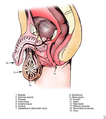
The testis, also known as the testicle, is a male reproductive organ that is part of the endocrine system. It is located in the scrotum, outside of the abdominal cavity. The main function of the testis is to produce sperm and testosterone, the primary male sex hormone.
The testis is composed of many tiny tubules called seminiferous tubules, where sperm are produced. These tubules are surrounded by a network of blood vessels, nerves, and supportive tissues. The sperm then travel through a series of ducts to the epididymis, where they mature and become capable of fertilization.
Testosterone is produced in the Leydig cells, which are located in the interstitial tissue between the seminiferous tubules. Testosterone plays a crucial role in the development and maintenance of male secondary sexual characteristics, such as facial hair, deep voice, and muscle mass. It also supports sperm production and sexual function.
Abnormalities in testicular function can lead to infertility, hormonal imbalances, and other health problems. Regular self-examinations and medical check-ups are recommended for early detection and treatment of any potential issues.
Meiosis is a type of cell division that results in the formation of four daughter cells, each with half the number of chromosomes as the parent cell. It is a key process in sexual reproduction, where it generates gametes or sex cells (sperm and eggs).
The process of meiosis involves one round of DNA replication followed by two successive nuclear divisions, meiosis I and meiosis II. In meiosis I, homologous chromosomes pair, form chiasma and exchange genetic material through crossing over, then separate from each other. In meiosis II, sister chromatids separate, leading to the formation of four haploid cells. This process ensures genetic diversity in offspring by shuffling and recombining genetic information during the formation of gametes.
Sertoli cells, also known as sustentacular cells or nurse cells, are specialized cells in the seminiferous tubules of the testis in mammals. They play a crucial role in supporting and nurturing the development of sperm cells (spermatogenesis). Sertoli cells create a microenvironment within the seminiferous tubules that facilitates the differentiation, maturation, and survival of germ cells.
These cells have several essential functions:
1. Blood-testis barrier formation: Sertoli cells form tight junctions with each other, creating a physical barrier called the blood-testis barrier, which separates the seminiferous tubules into basal and adluminal compartments. This barrier protects the developing sperm cells from the immune system and provides an isolated environment for their maturation.
2. Nutrition and support: Sertoli cells provide essential nutrients and growth factors to germ cells, ensuring their proper development and survival. They also engulf and digest residual bodies, which are byproducts of spermatid differentiation.
3. Phagocytosis: Sertoli cells have phagocytic properties, allowing them to remove debris and dead cells within the seminiferous tubules.
4. Hormone metabolism: Sertoli cells express receptors for various hormones, such as follicle-stimulating hormone (FSH), testosterone, and estradiol. They play a role in regulating hormonal signaling within the testis by metabolizing these hormones or producing inhibins, which modulate FSH secretion from the pituitary gland.
5. Regulation of spermatogenesis: Sertoli cells produce and secrete various proteins and growth factors that influence germ cell development and proliferation. They also control the release of mature sperm cells into the epididymis through a process called spermiation.
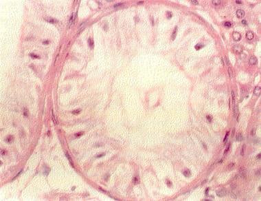
The pachytene stage is a phase in the meiotic division of sex cells (gametes) such as sperm and egg cells, specifically during prophase I. In this stage, homologous chromosomes are fully paired and have formed tetrads, or four-stranded structures called chiasma where genetic recombination occurs between the non-sister chromatids of each homologous chromosome. This is a crucial step in the creation of genetic diversity in the offspring. The pachytene stage is characterized by the presence of a protein matrix called the synaptonemal complex, which holds the homologous chromosomes together and facilitates crossing over.
Spermatogonia are a type of diploid germ cells found in the seminiferous tubules of the testis. They are the stem cells responsible for sperm production (spermatogenesis) in males. There are two types of spermatogonia: A-dark (Ad) and A-pale (Ap). The Ad spermatogonia function as reserve stem cells, while the Ap spermatogonia serve as the progenitor cells that divide to produce type B spermatogonia. Type B spermatogonia then differentiate into primary spermatocytes, which undergo meiosis to form haploid spermatozoa.
Spermatozoa are the male reproductive cells, or gametes, that are produced in the testes. They are microscopic, flagellated (tail-equipped) cells that are highly specialized for fertilization. A spermatozoon consists of a head, neck, and tail. The head contains the genetic material within the nucleus, covered by a cap-like structure called the acrosome which contains enzymes to help the sperm penetrate the female's egg (ovum). The long, thin tail propels the sperm forward through fluid, such as semen, enabling its journey towards the egg for fertilization.
Seminiferous tubules are the long, convoluted tubes within the testicles that are responsible for producing sperm in males. They are lined with specialized epithelial cells called Sertoli cells, which provide structural support and nourishment to developing sperm cells. The seminiferous tubules also contain germ cells, which divide and differentiate into spermatozoa (sperm) through the process of spermatogenesis.
The seminiferous tubules are surrounded by a thin layer of smooth muscle called the tunica albuginea, which helps to maintain the structure and integrity of the testicle. The tubules are connected to the rete testis, a network of channels that transport sperm to the epididymis for further maturation and storage before ejaculation.
Damage or dysfunction of the seminiferous tubules can lead to male infertility, as well as other reproductive health issues.
The seminiferous epithelium is a specialized type of epithelial tissue that lines the seminiferous tubules within the testes. It is composed of various cell types, including germ cells in different stages of development (spermatogonia, primary and secondary spermatocytes, spermatids) and supportive cells called Sertoli cells.
The primary function of the seminiferous epithelium is to support sperm production (spermatogenesis). The Sertoli cells provide structural support and nourishment to the developing germ cells, helping them to differentiate into mature spermatozoa (sperm). This process involves a series of complex cellular events, including mitosis, meiosis, and spermiogenesis.
In addition to its role in sperm production, the seminiferous epithelium also plays a crucial part in maintaining the blood-testis barrier, which separates the testicular environment from the systemic circulation. This barrier helps protect developing germ cells from potential immune attacks and maintains an optimal microenvironment for spermatogenesis.
Meiotic Prophase I is a stage in the meiotic division of cellular reproduction that results in the formation of gametes or sex cells (sperm and egg). It is the first of five stages in Meiosis I, which is a type of cell division that reduces the chromosome number by half.
During Meiotic Prophase I, homologous chromosomes pair and form tetrads (four-stranded structures), which then undergo genetic recombination or crossing over, resulting in new combinations of alleles on the chromatids of each homologous chromosome. This stage can be further divided into several substages: leptonema, zygonema, pachynema, diplonema, and diakinesis. These substages are characterized by distinct changes in chromosome structure and behavior, including the condensation and movement of the chromosomes, as well as the formation and dissolution of the synaptonemal complex, a protein structure that holds the homologous chromosomes together during crossing over.
Overall, Meiotic Prophase I is a critical stage in meiosis that ensures genetic diversity in offspring by shuffling the genetic material between homologous chromosomes and creating new combinations of alleles.
The synaptonemal complex is a protein structure that forms between two homologous chromosomes during meiosis, the type of cell division that leads to the production of gametes (sex cells). The synaptonemal complex consists of two lateral elements, which are associated with each of the homologous chromosomes, and a central element that runs parallel to the length of the complex and connects the two lateral elements.
The synaptonemal complex plays a crucial role in the process of genetic recombination, which occurs during meiosis. Genetic recombination is the exchange of genetic material between two homologous chromosomes that results in new combinations of genes on the chromosomes. This process helps to increase genetic diversity and is essential for the proper segregation of chromosomes during meiosis.
The synaptonemal complex also helps to ensure that the correct number of chromosomes are distributed to each gamete by holding the homologous chromosomes together until they can be properly aligned and separated during meiosis. Mutations in genes involved in the formation and maintenance of the synaptonemal complex can lead to fertility problems, developmental abnormalities, and other genetic disorders.
Prophase is the first phase of mitosis, the process by which eukaryotic cells divide and reproduce. During prophase, the chromosomes condense and become visible. The nuclear envelope breaks down, allowing the spindle fibers to attach to the centromeres of each chromatid in the chromosome. This is a critical step in preparing for the separation of genetic material during cell division. Prophase is also marked by the movement of the centrosomes to opposite poles of the cell, forming the mitotic spindle.
Chromosome pairing, also known as chromosome synapsis, is a process that occurs during meiosis, which is the type of cell division that results in the formation of sex cells or gametes (sperm and eggs).
In humans, each cell contains 23 pairs of chromosomes, for a total of 46 chromosomes. Of these, 22 pairs are called autosomal chromosomes, and they are similar in size and shape between the two copies in a pair. The last pair is called the sex chromosomes (X and Y), which determine the individual's biological sex.
During meiosis, homologous chromosomes (one from each parent) come together and pair up along their lengths in a process called synapsis. This pairing allows for the precise alignment of corresponding genes and genetic regions between the two homologous chromosomes. Once paired, the chromosomes exchange genetic material through a process called crossing over, which increases genetic diversity in the resulting gametes.
After crossing over, the homologous chromosomes separate during meiosis I, followed by the separation of sister chromatids (the two copies of each chromosome) during meiosis II. The end result is four haploid cells, each containing 23 chromosomes, which then develop into sperm or eggs.
Chromosome pairing is a crucial step in the process of sexual reproduction, ensuring that genetic information is accurately passed from one generation to the next while also promoting genetic diversity through recombination and independent assortment of chromosomes.
The Blood-Testis Barrier (BTB) is a unique structural and functional feature of the seminiferous epithelium in the testes, which forms a tight junction between adjacent Sertoli cells in the semi-niferous tubules. This barrier selectively restricts the passage of molecules, including potentially harmful substances and immune cells, from the systemic circulation into the adluminal compartment of the seminiferous epithelium where spermatogenesis occurs. This helps to maintain a immunologically privileged microenvironment that is essential for the survival and maturation of developing sperm cells, preventing an immune response against them. The BTB also regulates the movement of molecules required for spermatogenesis, such as nutrients, hormones, and signaling molecules, from the basal compartment to the adluminal compartment.
Male infertility is a condition characterized by the inability to cause pregnancy in a fertile female. It is typically defined as the failure to achieve a pregnancy after 12 months or more of regular unprotected sexual intercourse.
The causes of male infertility can be varied and include issues with sperm production, such as low sperm count or poor sperm quality, problems with sperm delivery, such as obstructions in the reproductive tract, or hormonal imbalances that affect sperm production. Other factors that may contribute to male infertility include genetic disorders, environmental exposures, lifestyle choices, and certain medical conditions or treatments.
It is important to note that male infertility can often be treated or managed with medical interventions, such as medication, surgery, or assisted reproductive technologies (ART). A healthcare provider can help diagnose the underlying cause of male infertility and recommend appropriate treatment options.
Sperm count, also known as sperm concentration, is the number of sperm present in a given volume of semen. The World Health Organization (WHO) previously defined a normal sperm count as at least 20 million sperm per milliliter of semen. However, more recent studies suggest that fertility may be affected even when sperm counts are slightly lower than this threshold. It's important to note that sperm count is just one factor among many that can influence male fertility. Other factors, such as sperm motility (the ability of sperm to move properly) and morphology (the shape of the sperm), also play crucial roles in successful conception.
Germ cells are the reproductive cells, also known as sex cells, that combine to form offspring in sexual reproduction. In females, germ cells are called ova or egg cells, and in males, they are called spermatozoa or sperm cells. These cells are unique because they carry half the genetic material necessary for creating new life. They are produced through a process called meiosis, which reduces their chromosome number by half, ensuring that when two germ cells combine during fertilization, the normal diploid number of chromosomes is restored.
Chromosomes are thread-like structures that exist in the nucleus of cells, carrying genetic information in the form of genes. They are composed of DNA and proteins, and are typically present in pairs in the nucleus, with one set inherited from each parent. In humans, there are 23 pairs of chromosomes for a total of 46 chromosomes. Chromosomes come in different shapes and forms, including sex chromosomes (X and Y) that determine the biological sex of an individual. Changes or abnormalities in the number or structure of chromosomes can lead to genetic disorders and diseases.
Diptera is an order of insects that includes flies, mosquitoes, and gnats. The name "Diptera" comes from the Greek words "di," meaning two, and "pteron," meaning wing. This refers to the fact that all members of this order have a single pair of functional wings for flying, while the other pair is reduced to small knob-like structures called halteres, which help with balance and maneuverability during flight.
Some common examples of Diptera include houseflies, fruit flies, horseflies, tsetse flies, and midges. Many species in this order are important pollinators, while others can be significant pests or disease vectors. The study of Diptera is called dipterology.
Leydig cells, also known as interstitial cells of Leydig or interstitial cell-stroma, are cells in the testes that produce and release testosterone and other androgens into the bloodstream. They are located in the seminiferous tubules of the testis, near the blood vessels, and are named after Franz Leydig, the German physiologist who discovered them in 1850.
Leydig cells contain cholesterol esters, which serve as precursors for the synthesis of testosterone. They respond to luteinizing hormone (LH) released by the anterior pituitary gland, which stimulates the production and release of testosterone. Testosterone is essential for the development and maintenance of male secondary sexual characteristics, such as facial hair, deep voice, and muscle mass. It also plays a role in sperm production and bone density.
In addition to their endocrine function, Leydig cells have been shown to have non-hormonal functions, including phagocytosis, antigen presentation, and immune regulation. However, these functions are not as well understood as their hormonal roles.
Metaphase is a phase in the cell division process (mitosis or meiosis) where the chromosomes align in the middle of the cell, also known as the metaphase plate or equatorial plane. During this stage, each chromosome consists of two sister chromatids attached to each other by a protein complex called the centromere. The spindle fibers from opposite poles of the cell attach to the centromeres of each chromosome, and through a process called congression, they align the chromosomes in the middle of the cell. This alignment allows for accurate segregation of genetic material during the subsequent anaphase stage.
Sex chromosomes, often denoted as X and Y, are one of the 23 pairs of human chromosomes found in each cell of the body. Normally, females have two X chromosomes (46,XX), and males have one X and one Y chromosome (46,XY). The sex chromosomes play a significant role in determining the sex of an individual. They contain genes that contribute to physical differences between men and women. Any variations or abnormalities in the number or structure of these chromosomes can lead to various genetic disorders and conditions related to sexual development and reproduction.
The Y chromosome is one of the two sex-determining chromosomes in humans and many other animals, along with the X chromosome. The Y chromosome contains the genetic information that helps to determine an individual's sex as male. It is significantly smaller than the X chromosome and contains fewer genes.
The Y chromosome is present in males, who inherit it from their father. Females, on the other hand, have two X chromosomes, one inherited from each parent. The Y chromosome includes a gene called SRY (sex-determining region Y), which initiates the development of male sexual characteristics during embryonic development.
It is worth noting that the Y chromosome has a relatively high rate of genetic mutation and degeneration compared to other chromosomes, leading to concerns about its long-term viability in human evolution. However, current evidence suggests that the Y chromosome has been stable for at least the past 25 million years.
Fertility is the natural ability to conceive or to cause conception of offspring. In humans, it is the capacity of a woman and a man to reproduce through sexual reproduction. For women, fertility usually takes place during their reproductive years, which is from adolescence until menopause. A woman's fertility depends on various factors including her age, overall health, and the health of her reproductive system.
For men, fertility can be affected by a variety of factors such as age, genetics, general health, sexual function, and environmental factors that may affect sperm production or quality. Factors that can negatively impact male fertility include exposure to certain chemicals, radiation, smoking, alcohol consumption, drug use, and sexually transmitted infections (STIs).
Infertility is a common medical condition affecting about 10-15% of couples trying to conceive. Infertility can be primary or secondary. Primary infertility refers to the inability to conceive after one year of unprotected sexual intercourse, while secondary infertility refers to the inability to conceive following a previous pregnancy.
Infertility can be treated with various medical and surgical interventions depending on the underlying cause. These may include medications to stimulate ovulation, intrauterine insemination (IUI), in vitro fertilization (IVF), or surgery to correct anatomical abnormalities.
Triethylenemelamine (TEM) is not typically classified as a medication, but rather as a chemical compound. However, it has been used in the past in some experimental cancer treatments.
Medically, TEM is known as an alkylating agent, which means it works by chemically modifying and damaging the DNA of cells, preventing them from dividing and replicating. This makes it a potential antineoplastic agent, or a chemical used to treat cancer. However, due to its significant side effects and the development of more effective treatments, TEM is no longer commonly used in cancer therapy.
It's important to note that the use of TEM as a medical treatment is highly specialized and not routinely used in modern medicine. Always consult with a healthcare professional or pharmacist for accurate and up-to-date information on medical definitions and treatments.
Salamandridae is not a medical term, but a taxonomic designation in the field of biology. It refers to a family of amphibians commonly known as newts and salamanders. These creatures are characterized by their slender bodies, moist skin, and four legs. Some species have the ability to regenerate lost body parts, including limbs, spinal cord, heart, and more.
If you're looking for a medical term, please provide more context or check if you may have made a typo in your question.
Oligospermia is a medical term used to describe a condition in which the semen contains a lower than normal number of sperm. Generally, a sperm count of less than 15 million sperm per milliliter (ml) of semen is considered to be below the normal range.
Oligospermia can make it more difficult for a couple to conceive naturally and may require medical intervention such as intracytoplasmic sperm injection (ICSI) or in vitro fertilization (IVF). The condition can result from various factors, including hormonal imbalances, genetic abnormalities, varicocele, environmental factors, and certain medications.
It's important to note that oligospermia is not the same as azoospermia, which is a condition where there is no sperm present in the semen at all.
The epididymis is a tightly coiled tube located on the upper and posterior portion of the testicle that serves as the site for sperm maturation and storage. It is an essential component of the male reproductive system. The epididymis can be divided into three parts: the head (where newly produced sperm enter from the testicle), the body, and the tail (where mature sperm exit and are stored). Any abnormalities or inflammation in the epididymis may lead to discomfort, pain, or infertility.
Orthoptera is not a medical term, but rather a taxonomic order in zoology. It includes grasshoppers, crickets, and related insects. These insects are characterized by their long antennae, rear wings that are typically narrower than the front pair, and jumping or leaping locomotion.
While not directly related to medicine, some species of Orthoptera can have medical implications for humans. For example, certain types of ticks (which belong to a different order) can transmit diseases, and chigger mites (also not Orthoptera) can cause itchy skin rashes. However, the order Orthoptera itself does not have specific relevance to medical definitions or human health.
Sex chromatin, also known as the Barr body, is an inactive X chromosome found in the nucleus of female cells. In females, one of the two X chromosomes is randomly inactivated during embryonic development to ensure that the dosage of X-linked genes is equivalent between males (who have one X chromosome) and females (who have two X chromosomes). The inactive X chromosome condenses and forms a compact structure called a sex chromatin body or Barr body, which can be observed during microscopic examination of cell nuclei. This phenomenon is known as X-inactivation and helps to prevent an overexpression of X-linked genes that could lead to developmental abnormalities.
Immunohistochemistry (IHC) is a technique used in pathology and laboratory medicine to identify specific proteins or antigens in tissue sections. It combines the principles of immunology and histology to detect the presence and location of these target molecules within cells and tissues. This technique utilizes antibodies that are specific to the protein or antigen of interest, which are then tagged with a detection system such as a chromogen or fluorophore. The stained tissue sections can be examined under a microscope, allowing for the visualization and analysis of the distribution and expression patterns of the target molecule in the context of the tissue architecture. Immunohistochemistry is widely used in diagnostic pathology to help identify various diseases, including cancer, infectious diseases, and immune-mediated disorders.
In situ hybridization (ISH) is a molecular biology technique used to detect and localize specific nucleic acid sequences, such as DNA or RNA, within cells or tissues. This technique involves the use of a labeled probe that is complementary to the target nucleic acid sequence. The probe can be labeled with various types of markers, including radioisotopes, fluorescent dyes, or enzymes.
During the ISH procedure, the labeled probe is hybridized to the target nucleic acid sequence in situ, meaning that the hybridization occurs within the intact cells or tissues. After washing away unbound probe, the location of the labeled probe can be visualized using various methods depending on the type of label used.
In situ hybridization has a wide range of applications in both research and diagnostic settings, including the detection of gene expression patterns, identification of viral infections, and diagnosis of genetic disorders.
Molecular sequence data refers to the specific arrangement of molecules, most commonly nucleotides in DNA or RNA, or amino acids in proteins, that make up a biological macromolecule. This data is generated through laboratory techniques such as sequencing, and provides information about the exact order of the constituent molecules. This data is crucial in various fields of biology, including genetics, evolution, and molecular biology, allowing for comparisons between different organisms, identification of genetic variations, and studies of gene function and regulation.
Messenger RNA (mRNA) is a type of RNA (ribonucleic acid) that carries genetic information copied from DNA in the form of a series of three-base code "words," each of which specifies a particular amino acid. This information is used by the cell's machinery to construct proteins, a process known as translation. After being transcribed from DNA, mRNA travels out of the nucleus to the ribosomes in the cytoplasm where protein synthesis occurs. Once the protein has been synthesized, the mRNA may be degraded and recycled. Post-transcriptional modifications can also occur to mRNA, such as alternative splicing and addition of a 5' cap and a poly(A) tail, which can affect its stability, localization, and translation efficiency.
I'm sorry for any confusion, but "grasshoppers" is not a term used in medical definitions. Grasshoppers are a type of insect that belongs to the order Orthoptera and suborder Caelifera. They are known for their long hind legs which they use for jumping, and some species can jump over 20 times their own body length. If you have any questions about medical terminology or topics, I'd be happy to help with those instead!
Organ size refers to the volume or physical measurement of an organ in the body of an individual. It can be described in terms of length, width, and height or by using specialized techniques such as imaging studies (like CT scans or MRIs) to determine the volume. The size of an organ can vary depending on factors such as age, sex, body size, and overall health status. Changes in organ size may indicate various medical conditions, including growths, inflammation, or atrophy.
The v-mos oncogene protein is derived from the retrovirus called Moloney murine sarcoma virus (Mo-MSV). This oncogene encodes for a serine/threonine protein kinase, which is involved in cell proliferation and differentiation. When incorporated into the host genome during viral infection, the v-mos oncogene can cause unregulated cell growth and tumor formation, leading to sarcomas in mice. The normal cellular homolog of v-mos is called c-mos, which plays a crucial role in regulating cell division and is tightly controlled in normal cells. However, mutations or aberrant activation of c-mos can also contribute to oncogenic transformation and tumorigenesis.
Cryptorchidism is a medical condition in which one or both of a male infant's testicles fail to descend from the abdomen into the scrotum before birth or within the first year of life. Normally, the testicles descend from the abdomen into the scrotum during fetal development in the second trimester. If the testicles do not descend on their own, medical intervention may be necessary to correct the condition.
Cryptorchidism is a common birth defect, affecting about 3-5% of full-term and 30% of preterm male infants. In most cases, the testicle will descend on its own within the first six months of life. If it does not, treatment may be necessary to prevent complications such as infertility, testicular cancer, and inguinal hernia.
Treatment for cryptorchidism typically involves surgery to bring the testicle down into the scrotum. This procedure is called orchiopexy and is usually performed before the age of 2. In some cases, hormonal therapy may be used as an alternative to surgery. However, this approach has limited success and is generally only recommended in certain situations.
Overall, cryptorchidism is a treatable condition that can help prevent future health problems if addressed early on. Regular check-ups with a pediatrician or healthcare provider can help ensure timely diagnosis and treatment of this condition.
Sexual maturation is the process of physical development during puberty that leads to the ability to reproduce. This process involves the development of primary and secondary sexual characteristics, changes in hormone levels, and the acquisition of reproductive capabilities. In females, this includes the onset of menstruation and the development of breasts and hips. In males, this includes the deepening of the voice, growth of facial hair, and the production of sperm. Achieving sexual maturation is an important milestone in human development and typically occurs during adolescence.
Developmental gene expression regulation refers to the processes that control the activation or repression of specific genes during embryonic and fetal development. These regulatory mechanisms ensure that genes are expressed at the right time, in the right cells, and at appropriate levels to guide proper growth, differentiation, and morphogenesis of an organism.
Developmental gene expression regulation is a complex and dynamic process involving various molecular players, such as transcription factors, chromatin modifiers, non-coding RNAs, and signaling molecules. These regulators can interact with cis-regulatory elements, like enhancers and promoters, to fine-tune the spatiotemporal patterns of gene expression during development.
Dysregulation of developmental gene expression can lead to various congenital disorders and developmental abnormalities. Therefore, understanding the principles and mechanisms governing developmental gene expression regulation is crucial for uncovering the etiology of developmental diseases and devising potential therapeutic strategies.
Cyclin A1 is a type of cyclin protein that regulates the cell cycle, particularly during the S and G2 phases. It forms a complex with and acts as a regulatory subunit of cyclin-dependent kinase 2 (CDK2), helping to control the transition from the G1 phase to the S phase and from the S phase to the G2 phase. Cyclin A1 is expressed in various tissues, including ovary, testis, bone marrow, and lymphoid cells. Overexpression or dysregulation of cyclin A1 has been implicated in several types of cancer, making it a potential target for cancer therapy.
Nuclear proteins are a category of proteins that are primarily found in the nucleus of a eukaryotic cell. They play crucial roles in various nuclear functions, such as DNA replication, transcription, repair, and RNA processing. This group includes structural proteins like lamins, which form the nuclear lamina, and regulatory proteins, such as histones and transcription factors, that are involved in gene expression. Nuclear localization signals (NLS) often help target these proteins to the nucleus by interacting with importin proteins during active transport across the nuclear membrane.
Anaphase is a stage in the cell division process called mitosis, where sister chromatids (the two copies of each chromosome formed during DNA replication) separate at the centromeres and move toward opposite poles of the cell. This separation is facilitated by the attachment of microtubules from the spindle apparatus to the kinetochores, protein structures located on the centromeres of each sister chromatid. Anaphase is followed by telophase, during which the nuclear membrane reforms around each set of separated chromosomes, and cytokinesis, the division of the cytoplasm to form two separate daughter cells.
Testosterone is a steroid hormone that belongs to androsten class of hormones. It is primarily secreted by the Leydig cells in the testes of males and, to a lesser extent, by the ovaries and adrenal glands in females. Testosterone is the main male sex hormone and anabolic steroid. It plays a key role in the development of masculine characteristics, such as body hair and muscle mass, and contributes to bone density, fat distribution, red cell production, and sex drive. In females, testosterone contributes to sexual desire and bone health. Testosterone is synthesized from cholesterol and its production is regulated by luteinizing hormone (LH) and follicle-stimulating hormone (FSH).
Sprague-Dawley rats are a strain of albino laboratory rats that are widely used in scientific research. They were first developed by researchers H.H. Sprague and R.C. Dawley in the early 20th century, and have since become one of the most commonly used rat strains in biomedical research due to their relatively large size, ease of handling, and consistent genetic background.
Sprague-Dawley rats are outbred, which means that they are genetically diverse and do not suffer from the same limitations as inbred strains, which can have reduced fertility and increased susceptibility to certain diseases. They are also characterized by their docile nature and low levels of aggression, making them easier to handle and study than some other rat strains.
These rats are used in a wide variety of research areas, including toxicology, pharmacology, nutrition, cancer, and behavioral studies. Because they are genetically diverse, Sprague-Dawley rats can be used to model a range of human diseases and conditions, making them an important tool in the development of new drugs and therapies.
Histones are highly alkaline proteins found in the chromatin of eukaryotic cells. They are rich in basic amino acid residues, such as arginine and lysine, which give them their positive charge. Histones play a crucial role in packaging DNA into a more compact structure within the nucleus by forming a complex with it called a nucleosome. Each nucleosome contains about 146 base pairs of DNA wrapped around an octamer of eight histone proteins (two each of H2A, H2B, H3, and H4). The N-terminal tails of these histones are subject to various post-translational modifications, such as methylation, acetylation, and phosphorylation, which can influence chromatin structure and gene expression. Histone variants also exist, which can contribute to the regulation of specific genes and other nuclear processes.
Ethylene glycols are a class of synthetic chemical compounds that are commonly used as automotive antifreeze, de-icing agents, and as raw materials in the manufacture of polyester fibers and resins. The two most common types of ethylene glycol are ethylene glycol monoethyl ether (also known as ethylene glycol monomethyl ether or EGME) and diethylene glycol (DEG).
Ethylene glycols are colorless, odorless liquids with a sweet taste. They are highly toxic to humans and animals if ingested, inhaled, or absorbed through the skin. Exposure can cause a range of symptoms, including nausea, vomiting, abdominal pain, dizziness, confusion, seizures, coma, and even death.
In medical terms, ethylene glycols are often referred to as "toxic alcohols" or "antifreeze poisoning" when they cause toxicity in humans. Treatment typically involves supportive care, such as fluid replacement and kidney dialysis, as well as the use of specific antidotes, such as fomepizole or ethanol, to prevent further absorption and metabolism of the toxic alcohol.
The acrosome is a specialized structure located on the anterior part of the sperm head in many species of animals, including humans. It contains enzymes that help the sperm penetrate the outer covering of the egg (zona pellucida) during fertilization. The acrosome reaction is the process by which the acrosome releases its enzymes, allowing the sperm to digest a path through the zona pellucida and reach the egg plasma membrane for fusion and fertilization.
The acrosome is formed during spermatogenesis, the process of sperm production in the testis, from the Golgi apparatus, a cellular organelle involved in protein trafficking and modification. The acrosome contains hydrolytic enzymes such as hyaluronidase, acrosin, and proteases that are activated during the acrosome reaction to facilitate sperm-egg fusion.
Abnormalities in acrosome formation or function can lead to infertility in males.
A base sequence in the context of molecular biology refers to the specific order of nucleotides in a DNA or RNA molecule. In DNA, these nucleotides are adenine (A), guanine (G), cytosine (C), and thymine (T). In RNA, uracil (U) takes the place of thymine. The base sequence contains genetic information that is transcribed into RNA and ultimately translated into proteins. It is the exact order of these bases that determines the genetic code and thus the function of the DNA or RNA molecule.
The cell nucleus is a membrane-bound organelle found in the eukaryotic cells (cells with a true nucleus). It contains most of the cell's genetic material, organized as DNA molecules in complex with proteins, RNA molecules, and histones to form chromosomes.
The primary function of the cell nucleus is to regulate and control the activities of the cell, including growth, metabolism, protein synthesis, and reproduction. It also plays a crucial role in the process of mitosis (cell division) by separating and protecting the genetic material during this process. The nuclear membrane, or nuclear envelope, surrounding the nucleus is composed of two lipid bilayers with numerous pores that allow for the selective transport of molecules between the nucleoplasm (nucleus interior) and the cytoplasm (cell exterior).
The cell nucleus is a vital structure in eukaryotic cells, and its dysfunction can lead to various diseases, including cancer and genetic disorders.
Electron microscopy (EM) is a type of microscopy that uses a beam of electrons to create an image of the sample being examined, resulting in much higher magnification and resolution than light microscopy. There are several types of electron microscopy, including transmission electron microscopy (TEM), scanning electron microscopy (SEM), and reflection electron microscopy (REM).
In TEM, a beam of electrons is transmitted through a thin slice of the sample, and the electrons that pass through the sample are focused to form an image. This technique can provide detailed information about the internal structure of cells, viruses, and other biological specimens, as well as the composition and structure of materials at the atomic level.
In SEM, a beam of electrons is scanned across the surface of the sample, and the electrons that are scattered back from the surface are detected to create an image. This technique can provide information about the topography and composition of surfaces, as well as the structure of materials at the microscopic level.
REM is a variation of SEM in which the beam of electrons is reflected off the surface of the sample, rather than scattered back from it. This technique can provide information about the surface chemistry and composition of materials.
Electron microscopy has a wide range of applications in biology, medicine, and materials science, including the study of cellular structure and function, disease diagnosis, and the development of new materials and technologies.
'Drosophila proteins' refer to the proteins that are expressed in the fruit fly, Drosophila melanogaster. This organism is a widely used model system in genetics, developmental biology, and molecular biology research. The study of Drosophila proteins has contributed significantly to our understanding of various biological processes, including gene regulation, cell signaling, development, and aging.
Some examples of well-studied Drosophila proteins include:
1. HSP70 (Heat Shock Protein 70): A chaperone protein involved in protein folding and protection from stress conditions.
2. TUBULIN: A structural protein that forms microtubules, important for cell division and intracellular transport.
3. ACTIN: A cytoskeletal protein involved in muscle contraction, cell motility, and maintenance of cell shape.
4. BETA-GALACTOSIDASE (LACZ): A reporter protein often used to monitor gene expression patterns in transgenic flies.
5. ENDOGLIN: A protein involved in the development of blood vessels during embryogenesis.
6. P53: A tumor suppressor protein that plays a crucial role in preventing cancer by regulating cell growth and division.
7. JUN-KINASE (JNK): A signaling protein involved in stress response, apoptosis, and developmental processes.
8. DECAPENTAPLEGIC (DPP): A member of the TGF-β (Transforming Growth Factor Beta) superfamily, playing essential roles in embryonic development and tissue homeostasis.
These proteins are often studied using various techniques such as biochemistry, genetics, molecular biology, and structural biology to understand their functions, interactions, and regulation within the cell.
An amino acid sequence is the specific order of amino acids in a protein or peptide molecule, formed by the linking of the amino group (-NH2) of one amino acid to the carboxyl group (-COOH) of another amino acid through a peptide bond. The sequence is determined by the genetic code and is unique to each type of protein or peptide. It plays a crucial role in determining the three-dimensional structure and function of proteins.
'Drosophila melanogaster' is the scientific name for a species of fruit fly that is commonly used as a model organism in various fields of biological research, including genetics, developmental biology, and evolutionary biology. Its small size, short generation time, large number of offspring, and ease of cultivation make it an ideal subject for laboratory studies. The fruit fly's genome has been fully sequenced, and many of its genes have counterparts in the human genome, which facilitates the understanding of genetic mechanisms and their role in human health and disease.
Here is a brief medical definition:
Drosophila melanogaster (droh-suh-fih-luh meh-lon-guh-ster): A species of fruit fly used extensively as a model organism in genetic, developmental, and evolutionary research. Its genome has been sequenced, revealing many genes with human counterparts, making it valuable for understanding genetic mechanisms and their role in human health and disease.
Contraceptive agents for males are substances or methods that are used to prevent pregnancy by reducing the likelihood of fertilization. These can include:
1. Barrier methods: Condoms, diaphragms, and spermicides create a physical barrier that prevents sperm from reaching the egg.
2. Hormonal methods: Testosterone and progestin hormone therapies can decrease sperm production and reduce fertility.
3. Intrauterine devices (IUDs) for men: These are still in the experimental stage, but they involve placing a device in the male reproductive tract to prevent sperm from reaching the female reproductive system.
4. Withdrawal method: This involves the man withdrawing his penis from the vagina before ejaculation, although this is not a highly reliable form of contraception.
5. Fertility awareness methods: These involve tracking the woman's menstrual cycle and avoiding sexual intercourse during her fertile period.
6. Sterilization: Vasectomy is a surgical procedure that blocks or cuts the vas deferens, preventing sperm from leaving the body. It is a permanent form of contraception for men.
It's important to note that no contraceptive method is 100% effective, and individuals should consult with their healthcare provider to determine which option is best for them based on their personal needs, lifestyle, and medical history.
"Altered gravity" is not a medical condition or diagnosis itself, but rather a state that can have various medical implications. It refers to a situation where the force of gravity is different from what humans normally experience on Earth's surface (approximately 9.8 m/s²). This could include conditions such as:
1. Microgravity: This is the condition experienced in outer space, where the force of gravity is significantly reduced. It can have various effects on the human body, including muscle atrophy, bone loss, fluid shifts, and changes in balance and coordination.
2. Hypergravity: This refers to environments where the force of gravity is greater than Earth's normal level. Examples might include high-speed centrifuges or certain types of space travel. Hypergravity can lead to symptoms such as nausea, disorientation, and cardiovascular changes.
Medical research into altered gravity conditions is important for understanding the effects of space travel on the human body, as well as for developing countermeasures to mitigate these effects.
Haploidy is a term used in genetics to describe the condition of having half the normal number of chromosomes in a cell or an organism. In humans, for example, a haploid cell contains 23 chromosomes, whereas a diploid cell has 46 chromosomes.
Haploid cells are typically produced through a process called meiosis, which is a type of cell division that occurs in the reproductive organs of sexually reproducing organisms. During meiosis, a diploid cell undergoes two rounds of division to produce four haploid cells, each containing only one set of chromosomes.
In humans, haploid cells are found in the sperm and egg cells, which fuse together during fertilization to create a diploid zygote with 46 chromosomes. Haploidy is important for maintaining the correct number of chromosomes in future generations and preventing genetic abnormalities that can result from having too many or too few chromosomes.
In situ nick-end labeling (ISEL, also known as TUNEL) is a technique used in pathology and molecular biology to detect DNA fragmentation, which is a characteristic of apoptotic cells (cells undergoing programmed cell death). The method involves labeling the 3'-hydroxyl termini of double or single stranded DNA breaks in situ (within tissue sections or individual cells) using modified nucleotides that are coupled to a detectable marker, such as a fluorophore or an enzyme. This technique allows for the direct visualization and quantification of apoptotic cells within complex tissues or cell populations.
Testicular diseases refer to a range of conditions that affect the testicles, the male reproductive organs located in the scrotum. These diseases can affect either one or both testicles and may cause pain, swelling, or impact fertility. Here are some examples of testicular diseases:
1. Testicular cancer: A malignant tumor that develops in the testicle. It is a relatively rare cancer but is highly treatable if detected early.
2. Testicular torsion: A surgical emergency that occurs when the spermatic cord, which supplies blood to the testicle, becomes twisted, cutting off the blood flow.
3. Epididymitis: An infection or inflammation of the epididymis, a coiled tube that stores and carries sperm from the testicle.
4. Orchitis: An infection or inflammation of the testicle itself. It can occur on its own or as a complication of mumps.
5. Hydrocele: A fluid-filled sac that forms around the testicle, causing swelling.
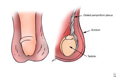
6. Varicocele: Enlarged veins in the scrotum that can cause pain and affect fertility.
7. Inguinal hernia: A condition where a portion of the intestine or fat protrudes through a weakened area in the abdominal wall, often appearing as a bulge in the groin or scrotum.
8. Testicular trauma: Injury to the testicle, which can result from accidents, sports injuries, or other causes.
9. Undescended testicles: A condition where one or both testicles fail to descend from the abdomen into the scrotum before birth.
It is essential for men to perform regular self-examinations to check for any unusual lumps, swelling, or pain in the testicles and seek medical attention if they notice any changes.
Androgen-binding protein (ABP) is a protein that binds specifically to androgens, which are hormones such as testosterone that play a role in male sexual development and masculine characteristics. ABP is produced in the Sertoli cells of the testes and helps to regulate the levels of androgens within the testes by storing them and slowly releasing them over time. This is important for maintaining normal sperm production and male reproductive function.
ABP is also found in other tissues, including the prostate gland, where it may play a role in regulating the growth and development of this tissue. Abnormal levels of ABP have been associated with certain medical conditions, such as prostate cancer and infertility.
Northern blotting is a laboratory technique used in molecular biology to detect and analyze specific RNA molecules (such as mRNA) in a mixture of total RNA extracted from cells or tissues. This technique is called "Northern" blotting because it is analogous to the Southern blotting method, which is used for DNA detection.
The Northern blotting procedure involves several steps:
1. Electrophoresis: The total RNA mixture is first separated based on size by running it through an agarose gel using electrical current. This separates the RNA molecules according to their length, with smaller RNA fragments migrating faster than larger ones.
2. Transfer: After electrophoresis, the RNA bands are denatured (made single-stranded) and transferred from the gel onto a nitrocellulose or nylon membrane using a technique called capillary transfer or vacuum blotting. This step ensures that the order and relative positions of the RNA fragments are preserved on the membrane, similar to how they appear in the gel.
3. Cross-linking: The RNA is then chemically cross-linked to the membrane using UV light or heat treatment, which helps to immobilize the RNA onto the membrane and prevent it from washing off during subsequent steps.
4. Prehybridization: Before adding the labeled probe, the membrane is prehybridized in a solution containing blocking agents (such as salmon sperm DNA or yeast tRNA) to minimize non-specific binding of the probe to the membrane.
5. Hybridization: A labeled nucleic acid probe, specific to the RNA of interest, is added to the prehybridization solution and allowed to hybridize (form base pairs) with its complementary RNA sequence on the membrane. The probe can be either a DNA or an RNA molecule, and it is typically labeled with a radioactive isotope (such as ³²P) or a non-radioactive label (such as digoxigenin).
6. Washing: After hybridization, the membrane is washed to remove unbound probe and reduce background noise. The washing conditions (temperature, salt concentration, and detergent concentration) are optimized based on the stringency required for specific hybridization.
7. Detection: The presence of the labeled probe is then detected using an appropriate method, depending on the type of label used. For radioactive probes, this typically involves exposing the membrane to X-ray film or a phosphorimager screen and analyzing the resulting image. For non-radioactive probes, detection can be performed using colorimetric, chemiluminescent, or fluorescent methods.
8. Data analysis: The intensity of the signal is quantified and compared to controls (such as housekeeping genes) to determine the relative expression level of the RNA of interest. This information can be used for various purposes, such as identifying differentially expressed genes in response to a specific treatment or comparing gene expression levels across different samples or conditions.
 Spermatocyte
Gametogenesis
Gustav Eisen
Sponge
Synizesis (biology)
Spermatogenesis
Isostichopus fuscus
SLC26A8
Germline development
Health effects of Bisphenol A
Viral gametocytic hypertrophy
ZFY
Major sperm protein
Achiasmate Meiosis
Non-random segregation of chromosomes
KCNU1
Spermatogonium
Daniel Djakiew
Gametocyte
Germ cell
Spermatidogenesis
Meiotic recombination checkpoint
Demosponge
Interferon-inducible GTPase 5
DNA repair and recombination protein RAD54-like
Sperm
Spermatogonial stem cell
Annulate lamella
Patricia Louise Dudley
Spindle checkpoint
Spermatocyte - Wikipedia
 Spermatocyte - Everything2.com
 CIL:10729, Nephrotoma, Nephrotoma suturalis, primary spermatocyte. CIL. Dataset
 making sperm (spermatogensis): primary spermatocyte-then ...
 A Wisp3 Cre-knockin Allele Produces Efficient Recombination in Spermatocytes during Early Prophase of Meiosis I
 LOCAL REDUCTION OF SPINDLE FIBER BIREFRINGENCE IN LIVING NEPHROTOMA SUTURALIS (LOEW) SPERMATOCYTES INDUCED BY ULTRAVIOLET...
THE EFFECT OF X-RAYS ON THE FUNCTIONAL STRUCTURES OF THE Y CHROMOSOME IN SPERMATOCYTES OF DROSOPHILA HYDEI | Journal of Cell...
 meiotic prophase I spermatocyte | PLANOSPHERE
CIL:39229, Psophus stridulus, spermatocyte. CIL. Dataset
Babel Library: The Theater of Babel #1781: Her spermatocyte.
Development of normal mice from metaphase I oocytes fertilized with primary spermatocytes<...
 The PRDM9 KRAB domain is required for meiosis and involved in protein interactions | Chromosoma
![Smc5 structural maintenance of chromosomes 5 [Mus musculus (house mouse)] - Gene - NCBI](data:image/png;base64,iVBORw0KGgoAAAANSUhEUgAAABAAAAAQCAYAAAAf8/9hAAAB1ElEQVQ4jaWSPWgTcRjGf/ehuWhobO2JxGJRY3taTTRV2yoqSpW6iIWO4iAoUsRBioNDKUWKLU7i4KA4OfhVREQnETRia03k7IdiS0LaQYKJQg3mLtfc30GySNUDn/V5nx/vy/vAf0pqad3db2xquiBJku93s2Tb2eEHdw1rTcsxol23sObTjN7oIp9KVmaU9kMdTxcLAyiqGtA0bfms+XKQULSdQG2EmnUx0q9ughAA8p/CFW0IN3Sv0vUI5p2zIMpUrd5JeP/Jii//80ZJUlrb9lyV8qn3zI5dB8A4MoBWtcITAKBmZe3eRmPzccYf9uIUsyzx6zQd7fMMAIjFdgxpkuPy4clFANbu6qa6fouybXtznxeAoqoBn0/zz5kvBqVQ5DBasJ5gXaPnDQAWFpwCkiwLZekyAMp2wTPAsqy5d8nEZcIHThPQo7jlIua9854BibdvekqKX8PouARAOn6F+c8pT4Bc7svz6U8f77O1cwDVV439PcPU4yHw8AUhhDPyOn4OfWOMuuZfBZp41INTLACorhC2/Jc2zsxMX8vl8lMcPBUHFL5mnpEZGa748sS42esKYS8WLtl2NjE22s/6fScIhtr48W2S5O0zIFwvp3vST6Z+myCvkaonAAAAAElFTkSuQmCC) Smc5 structural maintenance of chromosomes 5 [Mus musculus (house mouse)] - Gene - NCBI
Smc5 structural maintenance of chromosomes 5 [Mus musculus (house mouse)] - Gene - NCBI
The forming of a chromatoid is first recognized in early spermatocytes as several small particles connected with a...
 High fat diet-induced obesity prolongs critical stages of the spermatogenic cycle in a Ldlr−/−.Leiden mouse model | Scientific...
 Frontiers | Subcellular Evidence for Biogenesis of Autophagosomal Membrane during Spermiogenesis In vivo
Crotonaldehyde | Medical Management Guidelines | Toxic Substance Portal | ATSDR
 Male Infertility: Practice Essentials, Background, Pathophysiology
 Search: protein class:Kinases - The Human Protein Atlas
Single cell type - SERPINB1 - The Human Protein Atlas
 ARCHIVED - Environment and Climate Change Canada - Evaluating Existing Substances - Assessment report for Benzene, 1-methyl-2...
 PRKRA Localizes to Nuage Structures and the Ectoplasmic Specialization and Tubulobulbar Complexes in Rat and Mouse Testis
 Bedaquiline, Pretomanid, and Linezolid (BPaL) | TB |CDC
 A K+-selective CNG channel orchestrates Ca2+ signalling in zebrafish sperm | eLife
JCI - The key role of vitamin A in spermatogenesis
 RX List database - use generic or medication brand name - GlobalRPH
Spermatids9
- Each secondary spermatocyte will form two spermatids after Meiosis II. (wikipedia.org)
- The process of spermatogenesis as the cells progress from spermatogium, to primary spermatocytes, to secondary spermatocytes, to spermatids and to Sperm. (iiab.me)
- PRKRA localized to four types of nuage structures, including aggregates of 60-90 nm particles, irregularly-shaped perinuclear granules, and intermitochondrial cement of pachytene spermatocytes, and chromatoid bodies of round spermatids. (hindawi.com)
- At the completion of meiosis, four haploid gametes, termed round spermatids, result from the division of every spermatocyte. (jci.org)
- Histology evaluation: Abnormal spermiogenesis with accumulation of meiotic division-phase spermatocytes and degenerating round spermatids present in multinucleated cell bodies (see 1). (jax.org)
- Using RT-PCR, 5'- and 3'-RACE approaches we clearly show that PDE10A transcript variants X3 and X5 are expressed in bovine testis as well as in primary spermatocytes and spermatids. (plos.org)
- Spermatids and spermatocytes were about 10 to 20 times more sensitive to germ cell differences than spermatozoa. (cdc.gov)
- means mature spermatozoa or precursor cells such as spermatids and spermatocytes. (lawinsider.com)
- In spermatogenesis, each germinal cell (spermatogonium), located adjacent to the Sertoli cells, undergoes differentiation into 16 primary spermatocytes, each of which generates 4 spermatids. (msdmanuals.com)
Pachytene10
- In spermatocytes, HRR events occur mainly in the pachytene stage of meiosis and the gene conversion type of HRR is predominant, whereas in other stages of spermatogenesis the reciprocal exchange type of HRR is more frequent. (wikipedia.org)
- During mouse spermatogenesis, the mutation frequencies of cells at the different stages, including pachytene spermatocytes, are 5 to 10-fold lower than the mutation frequencies in somatic cells. (wikipedia.org)
- Primary spermatocytes are the male germ cells before meiosis I. To examine whether these 4n diploid cells are genetically competent to fertilize oocytes and support full embryo development, we introduced the nuclei of pachytene/diplotene spermatocytes into oocytes that were arrested in prophase I (germinal vesicle stage), metaphase I, or metaphase II (Met II). (elsevierpure.com)
- Recombination and separation of homologous chromosomes occurs in pachytene spermatocytes during meiosis I and results in the formation of secondary spermatocytes. (jci.org)
- TIF) pgen.1007300.s007.tif (2.0M) GUID:?417E3125-2DDD-44DE-80F5-D79333156D59 S8 Fig: DMC1 foci persist in deleted pachytene and diplotene spermatocytes. (mingsheng88.org)
- Immunofluorescence with SYCP3 (reddish) and DMC1 (green) antibodies in control and cKO spermatocytes at leptotene (A), zygotene (C), early pachytene (E), mid-late pachytene (G) and diplotene (I) phases. (mingsheng88.org)
- Level bars, 10 m.The mean quantity of DMC1 foci per cell in control and cKO leptotene(B), zygotene (D), early pachytene (F), mid-late pachytene (H) and diplotene (J) spermatocytes. (mingsheng88.org)
- TIF) pgen.1007300.s009.tif (1.6M) GUID:?63E1D1CD-556F-43F3-AB2A-6C01716726D8 S10 Fig: The expression of MSCI related genes remain undisturbed in cKO pachytene spermatocytes. (mingsheng88.org)
- The manifestation of and mRNAs in isolated pachytene/diplotene spermatocytes from control and cKO mice was recognized by RT-PCR. (mingsheng88.org)
- The second model postulates that the most likely target cell for transformation is the zygotene-pachytene spermatocyte. (medscape.com)
Meiotic3
- All the experiments reported were on primary spermatocytes which completed the second meiotic division subsequent to the experimentation. (rupress.org)
- Human population of spermatocytes at different meiotic substages in control and cKO mice. (mingsheng88.org)
- Level bars, 10 m.B. The percentage of spermatocytes with defective development of H2AX phosphorylation (with H2AX phosphorylation restricted to SCs only) at indicated meiotic substages. (mingsheng88.org)
Meiosis7
- After meiosis I, two secondary spermatocytes are formed. (wikipedia.org)
- The primary spermatocytes within the adluminal compartment will continue on to Meiosis I and divide into two daughters cells, known as secondary spermatocytes, a process which takes 24 days to complete. (wikipedia.org)
- Spermatocytes regularly overcome double-strand breaks and other DNA damages in the prophase stage of meiosis. (wikipedia.org)
- Movie of meiosis I and II in primary spermatocytes of the crane-fly Nephrotoma suturalis. (cellimagelibrary.org)
- 2013. "A Wisp3 Cre-knockin Allele Produces Efficient Recombination in Spermatocytes during Early Prophase of Meiosis I." PLoS ONE 8 (9): e75116. (harvard.edu)
- The only tissue that had high levels of Cre-mediated recombination was the testis, where recombination in spermatocytes occurred by early prophase of meiosis I. As a consequence, males that were double heterozygous for a Wisp3GFP-Cre and a floxed allele only contributed a recombined allele to their offspring. (harvard.edu)
- Time lapse movie of meiosis II in spermatocytes of the Locust Psophus stridulus showing telophase and the early spermatid stage. (ucsd.edu)
Testis1
- Single outstretched testis, spermatocytes in a double row. (unl.edu)
Spermatogonia2
Secondary4
- There are two types of spermatocytes, primary and secondary spermatocytes. (wikipedia.org)
- Primary and secondary spermatocytes are formed through the process of spermatocytogenesis. (wikipedia.org)
- Secondary spermatocytes are haploid (N) cells that contain half the number of chromosomes. (wikipedia.org)
- Making sperm (spermatogensis): primary spermatocyte-then we are going to get a secondary spermatocytes (2) and these each break into two cells called spermatid- has to do more growth to become a spermatozoan (when the spermatid becomes the spermatozoan: this is called spermiogenesis). (memory.com)
Primary4
- Primary spermatocytes are diploid (2N) cells. (wikipedia.org)
- The formation of primary spermatocytes (a process known as spermatocytogenesis) begins in humans when a male is sexually matured at puberty, around the age of 10 through 14. (wikipedia.org)
- Primary spermatocytes are arrested after DNA synthesis and prior to division. (wikipedia.org)
- Following the germline transmission of these two alleles, trans-heterozygous males (Mash2+/M2, Ins2I2/+) carrying the primary spermatocyte-specific Sycpl-Cre transgene were obtained. (geneimprint.com)
Chromosomes1
- In the third group, the paternal Met II chromosomes were obtained by transferring spermatocyte nuclei into Met II oocytes twice. (elsevierpure.com)
Suturalis1
- Irradiation of the mitotic spindle in living Nephrotoma suturalis (Loew) spermatocytes with an ultraviolet microbeam of controlled dose produced a localized area of reduced birefringence in the spindle fibers. (rupress.org)
Mice3
- Homologous recombinational repair (HRR) of double-strand breaks occurs in mice during sequential stages of spermatogenesis but is most prominent in spermatocytes. (wikipedia.org)
- n, the Quinupristin number of analyzed spermatocytes from 3 mice. (mingsheng88.org)
- n, the number of analyzed spermatocytes from 3 mice. (mingsheng88.org)
Males1
- In all animals, males produce spermatocytes, even hermaphrodites such as C. elegans, which exist as a male or hermaphrodite. (wikipedia.org)
Male1
- Spermatocytes are a type of male gametocyte in animals. (wikipedia.org)
Cells1
- Although spermatocytes that divide mitotically and meiotically are sensitive to radiation and cancer, spermatogonial stem cells are not. (wikipedia.org)
Spermatogonia6
- Nucleus of Sertoli cells and germ cells (spermatogonia type A, pachytene primary spermatocyte and round spermatid) were counted in 20 seminiferous tubules per rat (n = 10 animals [group. (thefreedictionary.com)
- A preliminary account of the spermatogenesis of Batrachoseps attenuatus, polymorphous spermatogonia, auxocytes and spermatocytes. (nih.gov)
- polymorphous spermatogonia, auxocytes, and spermatocytes. (nih.gov)
- Part 1: background to spermatogenesis, spermatogonia, and spermatocytes. (nih.gov)
- Spermatocytes are initially formed from spermatogonia, which undergo mitotic division to produce primary spermatocytes. (proprofs.com)
- Observations were made of damage to the spermatozoa, spermatid, spermatocyte, or spermatogonia stage. (cdc.gov)
Spermatogenesis4
- Homologous recombinational repair (HRR) of double-strand breaks occurs in mice during sequential stages of spermatogenesis but is most prominent in spermatocytes. (wikipedia.org)
- In spermatocytes, HRR events occur mainly in the pachytene stage of meiosis and the gene conversion type of HRR is predominant, whereas in other stages of spermatogenesis the reciprocal exchange type of HRR is more frequent. (wikipedia.org)
- During mouse spermatogenesis, the mutation frequencies of cells at the different stages, including pachytene spermatocytes, are 5 to 10-fold lower than the mutation frequencies in somatic cells. (wikipedia.org)
- Spermatocytogenesis is the first one amongst the spermatogenesis process stages, and seems to be the male form of gametocytogenesis, in which spermatocytes with half the regular complement of genetic material are produced. (vedantu.com)
Pachytene spermatocytes2
- They find that pachytene spermatocytes have a unique chromosome organization that may result from the presence of the synaptonemal complex and transcription-associated proteins. (nih.gov)
- The synapsis of homologous chromosomes and the frequency and distribution of meiotic recombination events were studied in pachytene spermatocytes of captive bred male impalas ( Aepyceros melampus ) polymorphic for der(14;20) by immunofluorescent analysis and fluorescence in situ hybridization. (springer.com)
Detected in the cytoplasm1
- At early stage of 2 weeks, TDRD12 was detected in the cytoplasm of primary spermatocyte (Figure 4). (thefreedictionary.com)
Spermatozoa1
- Soggy-1 is localized to the acrosome in developing spermatocytes and mature spermatozoa (3, 4). (rndsystems.com)
Drosophila1
- To identify proteins that might collaborate with the dTAF5 homolog can to regulate expression of specific target genes in Drosophila spermatocytes, the expression and function of other TAF II homologs in the Drosophila genome were investigated (Hiller, 2004). (sdbonline.org)
Testis-specific1
- Isoform Testis-specific is expressed in spermatocytes and adult testis. (abcam.com)
Elegans1
- In all animals, males produce spermatocytes, even hermaphrodites such as C. elegans, which exist as a male or hermaphrodite. (wikipedia.org)
Divide1
- Although spermatocytes that divide mitotically and meiotically are sensitive to radiation and cancer, spermatogonial stem cells are not. (wikipedia.org)
Transcription1
- Soggy-1 transcription is regulated by the spermatocyte specific factor, RFX2 (6). (rndsystems.com)
Gene1
- Therefore, this short promoter region of CaMII seemed to carry a certain novel machinery for the spermatocyte-specific gene expression. (elsevierpure.com)
Occur1
- Research suggests that epigenetic changes occur within spermatocytes in the same regions that are responsible for several diseases in children of fathers of advanced age. (contemporarypediatrics.com)
Protein2
- Instead, our results suggest that the DNC-1(G33S) protein, which is aggregation prone in vitro , is less stable in spermatocytes than the early embryo, resulting in different phenotypic severity in the two dividing tissues. (nih.gov)
- Soggy-1, as expressed in developing mouse spermatocytes, is a 34 kDa N-glycosylated protein. (rndsystems.com)
Division1
- Transmission electron micrograph of cell division in an insect spermatocyte captured in late anaphase - telophase with cytokinesis in progress. (cellimagelibrary.org)
Target1
- The second model postulates that the most likely target cell for transformation is the zygotene-pachytene spermatocyte. (medscape.com)