Sentinel Lymph Node Biopsy
Lymph Nodes
Lymphatic Metastasis
Lymph Node Excision
Rosaniline Dyes
Technetium Tc 99m Sulfur Colloid
Technetium Compounds
False Negative Reactions
Melanoma
Biopsy
Frozen Sections
Coloring Agents
Neoplasm Staging
Gamma Cameras
Methylene Blue
Carcinoma, Ductal, Breast
Lymphography
Carcinoma, Lobular
Inguinal Canal
Radiopharmaceuticals
Nevus, Epithelioid and Spindle Cell
Biopsy, Needle
Sarcoma, Clear Cell
Neck Dissection
Technetium Tc 99m Aggregated Albumin
Sensitivity and Specificity
Prognosis
Seroma
Neoplasm Micrometastasis
Carcinoma, Intraductal, Noninfiltrating
Predictive Value of Tests
Prospective Studies
Indocyanine Green
Sentinel Surveillance
Lymphatic Irradiation
Colloids
Feasibility Studies
Retrospective Studies
Intraoperative Care
Mastectomy, Segmental
Neoplasm Recurrence, Local
Technetium
Mastectomy, Simple
Tuberculosis, Lymph Node
Immunohistochemistry
Hematoxylin
Neoadjuvant Therapy
Carcinoma, Merkel Cell
Spectroscopy, Near-Infrared
Follow-Up Studies
Carcinoma
Reproducibility of Results
Biopsy, Fine-Needle
Eosine Yellowish-(YS)
Carcinoma, Squamous Cell
Histiocytic Necrotizing Lymphadenitis
Radiotherapy, Adjuvant
Nomograms
Breast
Cytodiagnosis
Chemotherapy, Adjuvant
Treatment Outcome
Keratin-19
Optical Imaging
Keratins
Tumor Markers, Biological
Breast Neoplasms, Male
Preoperative Care
Quantum Dots
Monophenol Monooxygenase
MART-1 Antigen
Ultrasonography, Mammary
Cat-Scratch Disease
Neoplasms, Ductal, Lobular, and Medullary
Hodgkin Disease
Sentinel node biopsy as a practical alternative to axillary lymph node dissection in breast cancer patients: an approach to its validity. (1/921)
BACKGROUND: Sentinel node biopsy (SNB) has been proposed as an alternative to axillary lymph-node dissection (ALND) in breast cancer. Before implementing SNB in our practice, we wished to test its validity by comparing it to the standard ALND, both in our hands and with other reported series. PATIENTS AND METHODS: One hundred thirty-two patients were included prospectively. SNB and immediate ALND were performed. For SNB, a technetium-colloid was used to produce preoperative lymphoscintigraphy and intraoperative gamma-probe search for the SN. Serial sectioning and immunostains were used on the SN. A comprehensive review of the literature was done in order to run a meta-analysis of diagnostic tests using a summary receiver operating characteristic curve (SROC) to calculate the pooled parameters of sensitivity and associated 95% confidence interval (95% CI), including our own data. RESULTS: Our technical success rate was 96%. Local sensitivity was 96%, with a 95% CI from 85%-99%. Seven patients were upstaged by the SNB. A literature search identified 18 studies published from 1996-1999. Estimates of sensitivity ranged from 83%-100%. The pooled data meta-analysis gave a global sensitivity of 91%, with a 95% CI from 89%-93%. The area under the global SROC curve was 0.9967. CONCLUSIONS: The minimally invasive SNB was shown to be a practical alternative to ALND. We propose to use local as well as global sensitivity and associated 95% CI to test the validity of SNB in the clinical setting. Due to limitations of ALND as the golden standard, SNB can in fact be considered a more accurate method for nodal staging. (+info)Sentinel lymph node biopsy is accurate after neoadjuvant chemotherapy for breast cancer. (2/921)
PURPOSE: Sentinel lymph node (SLN) biopsy has proved to be an accurate method for detecting nodal micrometastases in previously untreated patients with early-stage breast cancer. We investigated the accuracy of this technique for patients with more advanced breast cancer after neoadjuvant chemotherapy. PATIENTS AND METHODS: Patients with stage II or III breast cancer who had undergone doxorubicin-based neoadjuvant chemotherapy before breast surgery were eligible. Intraoperative lymphatic mapping was performed with peritumoral injections of blue dye alone or in combination with technetium-labeled sulfur colloid. All patients were offered axillary lymph node dissection. Negative sentinel and axillary nodes were subjected to additional processing with serial step sectioning and immunohistochemical staining with an anticytokeratin antibody to detect micrometastases. RESULTS: Fifty-one patients underwent SLN biopsy after neoadjuvant chemotherapy from 1994 to 1999. The SLN identification rate improved from 64.7% to 94.1%. Twenty-two (51.2%) of the 43 successfully mapped patients had positive SLNs, and in 10 of those 22 patients (45.5%), the SLN was the only positive node. Three patients had false-negative SLN biopsy; that is, the sentinel node was negative, but at least one nonsentinel node contained metastases. Additional processing revealed occult micrometastases in four patients (three in sentinel nodes and one in a nonsentinel node). CONCLUSION: SLN biopsy is accurate after neoadjuvant chemotherapy. The SLN identification improved with experience. False-negative findings occurred at a low rate throughout the series. This technique is a potential way to guide the axillary treatment of patients who are clinically node negative after neoadjuvant chemotherapy. (+info)Factors affecting sentinel node localization during preoperative breast lymphoscintigraphy. (3/921)
Variable success rates for identifying axillary (AX) sentinel nodes in breast cancer patients using preoperative lymphoscintigraphy have been reported. We evaluated the effects of age, weight, breast size, method of biopsy, interval after biopsy, and imaging view on the success of sentinel node identification and on the kinetics of radiopharmaceutical migration. METHODS: Preoperative breast lymphoscintigraphy was performed in consecutive breast cancer patients from February 1998 to December 1998. The ipsilateral shoulder was elevated on a foam wedge and the arm was abducted and elevated overhead. Imaging using this modified oblique view of the axilla (MOVA) started immediately after peritumoral injection of Millipore-filtered 99mTc-sulfur colloid and continued until AX sentinel nodes were identified. Anterior views were obtained after MOVA. AX, internal mammary (IM), and clavicular (CL) basins were monitored in all patients. MOVA was compared with the anterior view for sentinel node identification. Age, weight, breast size, method of biopsy, interval after biopsy, and primary tumor location were evaluated for their effects on sentinel node localization and transit times from injection to arrival at the sentinel nodes. RESULTS: Seventy-six lymphoscintigrams were obtained for 75 patients. AX sentinel nodes were revealed in 75 (99%) cases. IM or CL sentinel nodes were found in 19 (25%) cases and were not related to tumor location; exclusive IM drainage was present in 1 (1%) case. Identification of AX sentinel nodes was equivalent with MOVA and anterior views in 18 (24%) patients, was better with MOVA in 20 (26%) patients, and was accomplished only with MOVA in 38 (50%) patients. Median transit time was 17.5 min (range, 1 min to 18 h) after injection, and larger breast size was associated with increased transit time. No effect of age, weight, biopsy method, interval from biopsy, or tumor location on transit time was found. CONCLUSION: Use of MOVA can improve identification of AX sentinel nodes. Although AX drainage is the predominant pattern, a tumor in any portion of the breast can drain to IM sentinel nodes. Transit time was influenced by breast size. Overall short arrival times with this technique allow sentinel lymph node dissection to be performed on the same day as lymphoscintigraphy. (+info)Axillary staging of breast cancer and the sentinel node. (4/921)
Pathological aspects of axillary nodal staging of breast cancer and in particular sentinel lymph node (SLN) biopsy are reviewed. SLN biopsy seems an almost ideal staging procedure because it has both high accuracy and a low false negative rate. It may also allow a cost effective use of more sensitive methods of metastasis detection. However, the biological relevance of metastases detected only by modern tools remains to be elucidated. This review focuses on standard axillary staging and the histopathological investigation of SLNs, with emphasis on the intraoperative setting. Future trends including ancillary studies, quality control issues, prediction of non-SLN involvement, and suggestions concerning the minimum requirements for the histology of axillary SLNs are also discussed. (+info)Is intra-operative gamma probe detection really necessary for inguinal sentinel lymph node biopsy? (5/921)
CONTEXT: Sentinel node (SN) biopsy has changed the surgical treatment of malignant melanoma. The literature has emphasized the importance of gamma probe detection (GPD) of the SN. OBJECTIVE: Our objective was to evaluate the efficacy of patent blue dye (PBD) and GPD for SN biopsy in different lymphatic basins. DESIGN: Patients with cutaneous malignant melanoma in stages I and II were submitted to biopsy of the SN, identified by PBD and GPD, as part of a research project. SETTING: Patients were seen at Hospital Sao Paulo by a multidisciplinary group (Plastic Surgery Tumor Branch, Nuclear Medicine and Pathology). PATIENTS: 64 patients with localized malignant melanoma were studied. The median age was 46.5 years. The primary tumor was located in the neck, trunk or extremities. INTERVENTIONS: Preoperative lymphoscintigraphy, lymphatic mapping with PBD and intraoperative GPD was performed on all patients. The SN was examined by conventional and immunohistochemical staining. If the SN was not found or contained micrometastases, only complete lymphadenectomy was performed. MAIN MEASUREMENTS: The SN was identified by PBD if it was blue-stained, and by GPD if demonstrated activity five times greater than the adipose tissue of the neighborhood. RESULTS: Seventy lymphatic basins were explored. Lymphoscintigraphy showed ambiguous drainage in 7 patients. GPD identified the SN in 68 basins (97%) and PBD in 53 (76%). PBD and GPD identified SN in 100% of the inguinal basins. For the remaining basins both techniques were complementary. A metastatic SN was found in 10 basins. Three patients with negative SN had recurrence (median follow-up = 11 months). CONCLUSION: Although both GPD and PBD are useful and complementary, PBD alone identified the SN in 100% of the inguinal lymphatic basins. (+info)Multicenter trial of sentinel node biopsy for breast cancer using both technetium sulfur colloid and isosulfan blue dye. (6/921)
OBJECTIVE: To determine the factors associated with false-negative results on sentinel node biopsy and sentinel node localization (identification rate) in patients with breast cancer enrolled in a multicenter trial using a combination technique of isosulfan blue with technetium sulfur colloid (Tc99). SUMMARY BACKGROUND DATA: Sentinel node biopsy is a diagnostic test used to detect breast cancer metastases. To test the reliability of this method, a complete lymph node dissection must be performed to determine the false-negative rate. Single-institution series have reported excellent results, although one multicenter trial reported a false-negative rate as high as 29% using radioisotope alone. A multicenter trial was initiated to test combined use of Tc99 and isosulfan blue. METHODS: Investigators (both private-practice and academic surgeons) were recruited after attending a course on the technique of sentinel node biopsy. No investigator participated in a learning trial before entering patients. Tc99 and isosulfan blue were injected into the peritumoral region. RESULTS: Five hundred twenty-nine patients underwent 535 sentinel node biopsy procedures for an overall identification rate in finding a sentinel node of 87% and a false-negative rate of 13%. The identification rate increased and the false-negative rate decreased to 90% and 4.3%, respectively, after investigators had performed more than 30 cases. Univariate analysis of tumor showed the poorest success rate with older patients and inexperienced surgeons. Multivariate analysis identified both age and experience as independent predictors of failure. However, with older patients, inexperienced surgeons, and patients with five or more metastatic axillary nodes, the false-negative rate was consistently greater. CONCLUSIONS: This multicenter trial, from both private practice and academic institutions, is an excellent indicator of the general utility of sentinel node biopsy. It establishes the factors that play an important role (patient age, surgical experience, tumor location) and those that are irrelevant (prior surgery, tumor size, Tc99 timing). This widens the applicability of the technique and identifies factors that require further investigation. (+info)Methodological questions in sentinel lymph node analysis in breast cancer patients. (7/921)
BACKGROUND: The sentinel lymph node (SLN) procedure has been proposed to women with breast cancer with clinically negative axillary lymph nodes, in order to avoid conventional axillary lymph node dissection and its associated side-effects. Methodological aspects of the validation of the SLN procedure are questioned here. MATERIALS AND METHODS: The results of relevant published studies are reviewed, with emphasis on pathological techniques. The ability of the SLN procedure to diagnose lymph node metastases, the extent to which axillary lymph node dissection contributes to treatment, apart from identification of the stage, and the effect of a modified staging procedure on treatment strategies are analyzed. RESULTS AND CONCLUSION: Both the sensitivity and the negative predictive value of the SLN procedure are overestimated if the probability of missing lymph node metastases is not taken into account, even when a complete axillary dissection is performed as a control. The SLN strategy and its effects on staging and treatment cannot be evaluated by comparison with conventional axillary lymph node dissection in a one-arm study but require carefully designed randomized trials. (+info)Sentinel lymph node biopsy in the management of patients with primary cutaneous melanoma: review of a large single-institutional experience with an emphasis on recurrence. (8/921)
OBJECTIVE: To analyze the authors' experience with sentinel lymph node biopsy (SLNB) and the subsequent incidence and pattern of recurrence in patients with positive and negative nodes. SUMMARY BACKGROUND DATA: Lymphatic mapping with SLNB has become widely accepted in the management of patients with melanoma who are at risk for occult regional lymph node metastases. Because this procedure is relatively new, the pattern of recurrence after SLNB is not yet clear. METHODS: All patients with primary cutaneous melanoma who underwent SLNB from 1991 through 1998 were identified from a prospective single-institution melanoma database. RESULTS: Three hundred fifty-seven consecutive patients with localized primary cutaneous melanoma who underwent SLNB were identified. The sentinel node was identified in 332 patients (93%) and was positive in 56 (17%). Fourteen percent of patients had developed a recurrence at a median follow-up of 24 months. The median time to recurrence was 13 months. The 3-year relapse-free survival rates for patients with positive and negative nodes were 56% and 75%, respectively. SLN status was the most important predictor of disease recurrence. The site of first recurrence in patients with negative and positive nodes was more commonly locoregional than distant. Reexamination of the SLN in 11 patients with negative nodes with initial nodal and in-transit recurrence showed evidence of metastases in 7 (64%). CONCLUSIONS: Patients with positive sentinel nodes have a significantly increased risk for recurrence. The early pattern of first recurrence for patients with negative and positive results is characterized by a preponderance of locoregional sites, similar to that reported in previous series of elective lymph node dissection. These data underscore the need for careful pathologic analysis of the SLN as well as a careful, directed locoregional physical examination in the follow-up of these patients. (+info)A sentinel lymph node biopsy is a surgical procedure used in cancer staging to determine if the cancer has spread beyond the primary tumor to the lymphatic system. This procedure involves identifying and removing the sentinel lymph node(s), which are the first few lymph nodes to which cancer cells are most likely to spread from the primary tumor site.
The sentinel lymph node(s) are identified by injecting a tracer substance (usually a radioactive material and/or a blue dye) near the tumor site. The tracer substance is taken up by the lymphatic vessels and transported to the sentinel lymph node(s), allowing the surgeon to locate and remove them.
The removed sentinel lymph node(s) are then examined under a microscope for the presence of cancer cells. If no cancer cells are found, it is unlikely that the cancer has spread to other lymph nodes or distant sites in the body. However, if cancer cells are present, further lymph node dissection and/or additional treatment may be necessary.
Sentinel lymph node biopsy is commonly used in the staging of melanoma, breast cancer, and some types of head and neck cancer.
Lymph nodes are small, bean-shaped organs that are part of the immune system. They are found throughout the body, especially in the neck, armpits, groin, and abdomen. Lymph nodes filter lymph fluid, which carries waste and unwanted substances such as bacteria, viruses, and cancer cells. They contain white blood cells called lymphocytes that help fight infections and diseases by attacking and destroying the harmful substances found in the lymph fluid. When an infection or disease is present, lymph nodes may swell due to the increased number of immune cells and fluid accumulation as they work to fight off the invaders.
The term "axilla" is used in anatomical context to refer to the armpit region, specifically the space located lateral to the upper part of the chest wall and medial to the upper arm. This area contains a number of important structures such as blood vessels, nerves, and lymph nodes, which play a critical role in the health and functioning of the upper limb. Understanding the anatomy of the axilla is essential for medical professionals performing various procedures, including surgeries and injections, in this region.
Lymphatic metastasis is the spread of cancer cells from a primary tumor to distant lymph nodes through the lymphatic system. It occurs when malignant cells break away from the original tumor, enter the lymphatic vessels, and travel to nearby or remote lymph nodes. Once there, these cancer cells can multiply and form new tumors, leading to further progression of the disease. Lymphatic metastasis is a common way for many types of cancer to spread and can have significant implications for prognosis and treatment strategies.
Lymph node excision is a surgical procedure in which one or more lymph nodes are removed from the body for the purpose of examination. This procedure is often conducted to help diagnose or stage various types of cancer, as malignant cells may spread to the lymphatic system and eventually accumulate within nearby lymph nodes.
During a lymph node excision, an incision is made in the skin overlying the affected lymph node(s). The surgeon carefully dissects the tissue surrounding the lymph node(s) to isolate them from adjacent structures before removing them. In some cases, a sentinel lymph node biopsy may be performed instead, where only the sentinel lymph node (the first lymph node to which cancer cells are likely to spread) is removed and examined.
The excised lymph nodes are then sent to a laboratory for histopathological examination, which involves staining and microscopic evaluation of the tissue to determine whether it contains any malignant cells. The results of this examination can help guide further treatment decisions and provide valuable prognostic information.
Rosaniline dyes are a type of basic dye that were first synthesized in the late 19th century. They are named after rosaniline, which is a primary chemical used in their production. Rosaniline dyes are characterized by their ability to form complexes with metal ions, which can then bind to proteins and other biological molecules. This property makes them useful as histological stains, which are used to highlight specific structures or features within tissues and cells.
Rosaniline dyes include a range of different chemicals, such as methyl violet, crystal violet, and basic fuchsin. These dyes are often used in combination with other staining techniques to provide contrast and enhance the visibility of specific cellular components. For example, they may be used to stain nuclei, cytoplasm, or other structures within cells, allowing researchers and clinicians to visualize and analyze tissue samples more effectively.
It's worth noting that some rosaniline dyes have been found to have potential health hazards, particularly when used in certain forms or concentrations. Therefore, it's important to follow proper safety protocols when handling these chemicals and to use them only under the guidance of trained professionals.
Technetium Tc 99m Sulfur Colloid is a radioactive tracer used in medical imaging procedures, specifically in nuclear medicine. It is composed of tiny particles of sulfur colloid that are labeled with the radioisotope Technetium-99m. This compound is typically injected into the patient's body, where it accumulates in certain organs or tissues, depending on the specific medical test being conducted.
The radioactive emissions from Technetium Tc 99m Sulfur Colloid are then detected by a gamma camera, which produces images that can help doctors diagnose various medical conditions, such as liver disease, inflammation, or tumors. The half-life of Technetium-99m is approximately six hours, which means that its radioactivity decreases rapidly and is eliminated from the body within a few days.
Lymphoscintigraphy is a medical imaging technique that uses radioactive tracers to examine the lymphatic system, specifically the lymph nodes and vessels. In this procedure, a small amount of radioactive material is injected into the area of interest, usually an extremity or the site of a surgical incision. The tracer then travels through the lymphatic channels and accumulates in the regional lymph nodes. A specialized camera called a gamma camera detects the radiation emitted by the tracer and creates images that reveal the function and anatomy of the lymphatic system.
Lymphoscintigraphy is often used to diagnose and assess conditions affecting the lymphatic system, such as lymphedema, cancer metastasis to lymph nodes, or unusual lymphatic flow patterns. It can help identify sentinel lymph nodes (the first node(s) to receive drainage from a tumor) in patients with melanoma and breast cancer, which is crucial for surgical planning and staging purposes.
In summary, lymphoscintigraphy is a non-invasive imaging technique that utilizes radioactive tracers to visualize the lymphatic system's structure and function, providing valuable information for diagnostic and therapeutic decision-making in various clinical scenarios.
Technetium compounds refer to chemical substances that contain the radioactive technetium (Tc) element. Technetium is a naturally rare element and does not have any stable isotopes, making it only exist in trace amounts in the Earth's crust. However, it can be produced artificially in nuclear reactors.
Technetium compounds are widely used in medical imaging as radioactive tracers in diagnostic procedures. The most common technetium compound is Technetium-99m (Tc-99m), which has a half-life of 6 hours and emits gamma rays that can be detected by external cameras. Tc-99m is often bound to various pharmaceuticals, such as methylene diphosphonate (MDP) or human serum albumin (HSA), to target specific organs or tissues in the body.
Technetium compounds are used in a variety of diagnostic procedures, including bone scans, lung perfusion scans, myocardial perfusion imaging, and brain scans. They provide valuable information about organ function, blood flow, and tissue metabolism, helping doctors diagnose various medical conditions such as cancer, heart disease, and bone fractures.
It is important to note that technetium compounds should only be used under the supervision of trained medical professionals due to their radioactive nature. Proper handling, administration, and disposal procedures must be followed to ensure safety and minimize radiation exposure.
A "false negative" reaction in medical testing refers to a situation where a diagnostic test incorrectly indicates the absence of a specific condition or disease, when in fact it is present. This can occur due to various reasons such as issues with the sensitivity of the test, improper sample collection, or specimen handling and storage.
False negative results can have serious consequences, as they may lead to delayed treatment, misdiagnosis, or a false sense of security for the patient. Therefore, it is essential to interpret medical test results in conjunction with other clinical findings, patient history, and physical examination. In some cases, repeating the test or using a different diagnostic method may be necessary to confirm the initial result.
Breast neoplasms refer to abnormal growths in the breast tissue that can be benign or malignant. Benign breast neoplasms are non-cancerous tumors or growths, while malignant breast neoplasms are cancerous tumors that can invade surrounding tissues and spread to other parts of the body.
Breast neoplasms can arise from different types of cells in the breast, including milk ducts, milk sacs (lobules), or connective tissue. The most common type of breast cancer is ductal carcinoma, which starts in the milk ducts and can spread to other parts of the breast and nearby structures.
Breast neoplasms are usually detected through screening methods such as mammography, ultrasound, or MRI, or through self-examination or clinical examination. Treatment options for breast neoplasms depend on several factors, including the type and stage of the tumor, the patient's age and overall health, and personal preferences. Treatment may include surgery, radiation therapy, chemotherapy, hormone therapy, or targeted therapy.
Melanoma is defined as a type of cancer that develops from the pigment-containing cells known as melanocytes. It typically occurs in the skin but can rarely occur in other parts of the body, including the eyes and internal organs. Melanoma is characterized by the uncontrolled growth and multiplication of melanocytes, which can form malignant tumors that invade and destroy surrounding tissue.
Melanoma is often caused by exposure to ultraviolet (UV) radiation from the sun or tanning beds, but it can also occur in areas of the body not exposed to the sun. It is more likely to develop in people with fair skin, light hair, and blue or green eyes, but it can affect anyone, regardless of their skin type.
Melanoma can be treated effectively if detected early, but if left untreated, it can spread to other parts of the body and become life-threatening. Treatment options for melanoma include surgery, radiation therapy, chemotherapy, immunotherapy, and targeted therapy, depending on the stage and location of the cancer. Regular skin examinations and self-checks are recommended to detect any changes or abnormalities in moles or other pigmented lesions that may indicate melanoma.
Skin neoplasms refer to abnormal growths or tumors in the skin that can be benign (non-cancerous) or malignant (cancerous). They result from uncontrolled multiplication of skin cells, which can form various types of lesions. These growths may appear as lumps, bumps, sores, patches, or discolored areas on the skin.
Benign skin neoplasms include conditions such as moles, warts, and seborrheic keratoses, while malignant skin neoplasms are primarily classified into melanoma, squamous cell carcinoma, and basal cell carcinoma. These three types of cancerous skin growths are collectively known as non-melanoma skin cancers (NMSCs). Melanoma is the most aggressive and dangerous form of skin cancer, while NMSCs tend to be less invasive but more common.
It's essential to monitor any changes in existing skin lesions or the appearance of new growths and consult a healthcare professional for proper evaluation and treatment if needed.
A biopsy is a medical procedure in which a small sample of tissue is taken from the body to be examined under a microscope for the presence of disease. This can help doctors diagnose and monitor various medical conditions, such as cancer, infections, or autoimmune disorders. The type of biopsy performed will depend on the location and nature of the suspected condition. Some common types of biopsies include:
1. Incisional biopsy: In this procedure, a surgeon removes a piece of tissue from an abnormal area using a scalpel or other surgical instrument. This type of biopsy is often used when the lesion is too large to be removed entirely during the initial biopsy.
2. Excisional biopsy: An excisional biopsy involves removing the entire abnormal area, along with a margin of healthy tissue surrounding it. This technique is typically employed for smaller lesions or when cancer is suspected.
3. Needle biopsy: A needle biopsy uses a thin, hollow needle to extract cells or fluid from the body. There are two main types of needle biopsies: fine-needle aspiration (FNA) and core needle biopsy. FNA extracts loose cells, while a core needle biopsy removes a small piece of tissue.
4. Punch biopsy: In a punch biopsy, a round, sharp tool is used to remove a small cylindrical sample of skin tissue. This type of biopsy is often used for evaluating rashes or other skin abnormalities.
5. Shave biopsy: During a shave biopsy, a thin slice of tissue is removed from the surface of the skin using a sharp razor-like instrument. This technique is typically used for superficial lesions or growths on the skin.
After the biopsy sample has been collected, it is sent to a laboratory where a pathologist will examine the tissue under a microscope and provide a diagnosis based on their findings. The results of the biopsy can help guide further treatment decisions and determine the best course of action for managing the patient's condition.
"Frozen sections" is a medical term that refers to the process of quickly preparing and examining a small piece of tissue during surgery. This procedure is typically performed by a pathologist in order to provide immediate diagnostic information to the surgeon, who can then make informed decisions about the course of the operation.
To create a frozen section, the surgical team first removes a small sample of tissue from the patient's body. This sample is then quickly frozen, typically using a special machine that can freeze the tissue in just a few seconds. Once the tissue is frozen, it can be cut into thin slices and stained with dyes to help highlight its cellular structures.
The stained slides are then examined under a microscope by a pathologist, who looks for any abnormalities or signs of disease. The results of this examination are typically available within 10-30 minutes, allowing the surgeon to make real-time decisions about whether to remove more tissue, change the surgical approach, or take other actions based on the findings.
Frozen sections are often used in cancer surgery to help ensure that all of the cancerous tissue has been removed, and to guide decisions about whether additional treatments such as radiation therapy or chemotherapy are necessary. They can also be used in other types of surgeries to help diagnose conditions and make treatment decisions during the procedure.
Coloring agents, also known as food dyes or color additives, are substances that are added to foods, medications, and cosmetics to improve their appearance by giving them a specific color. These agents can be made from both synthetic and natural sources. They must be approved by regulatory agencies such as the U.S. Food and Drug Administration (FDA) before they can be used in products intended for human consumption.
Coloring agents are used for various reasons, including:
* To replace color lost during food processing or preparation
* To make foods more visually appealing
* To help consumers easily identify certain types of food
* To indicate the flavor of a product (e.g., fruit-flavored candies)
It's important to note that while coloring agents can enhance the appearance of products, they do not affect their taste or nutritional value. Some people may have allergic reactions to certain coloring agents, so it's essential to check product labels if you have any known allergies. Additionally, excessive consumption of some synthetic coloring agents has been linked to health concerns, so moderation is key.
Neoplasm staging is a systematic process used in medicine to describe the extent of spread of a cancer, including the size and location of the original (primary) tumor and whether it has metastasized (spread) to other parts of the body. The most widely accepted system for this purpose is the TNM classification system developed by the American Joint Committee on Cancer (AJCC) and the Union for International Cancer Control (UICC).
In this system, T stands for tumor, and it describes the size and extent of the primary tumor. N stands for nodes, and it indicates whether the cancer has spread to nearby lymph nodes. M stands for metastasis, and it shows whether the cancer has spread to distant parts of the body.
Each letter is followed by a number that provides more details about the extent of the disease. For example, a T1N0M0 cancer means that the primary tumor is small and has not spread to nearby lymph nodes or distant sites. The higher the numbers, the more advanced the cancer.
Staging helps doctors determine the most appropriate treatment for each patient and estimate the patient's prognosis. It is an essential tool for communication among members of the healthcare team and for comparing outcomes of treatments in clinical trials.
A gamma camera, also known as a scintillation camera, is a device used in nuclear medicine to image gamma-emitting radionuclides in the body. It detects gamma radiation emitted by radioisotopes that have been introduced into the body, usually through injection or ingestion. The camera consists of a large flat crystal (often sodium iodide) that scintillates when struck by gamma rays, producing light flashes that are detected by an array of photomultiplier tubes.
The resulting signals are then processed by a computer to generate images that reflect the distribution and concentration of the radionuclide in the body. Gamma cameras are used in a variety of medical imaging procedures, including bone scans, lung scans, heart scans (such as myocardial perfusion imaging), and brain scans. They can help diagnose conditions such as cancer, heart disease, and neurological disorders.
Methylene Blue is a heterocyclic aromatic organic compound with the molecular formula C16H18ClN3S. It is primarily used as a medication, but can also be used as a dye or as a chemical reagent. As a medication, it is used in the treatment of methemoglobinemia (a condition where an abnormal amount of methemoglobin is present in the blood), as well as in some forms of poisoning and infections. It works by acting as a reducing agent, converting methemoglobin back to hemoglobin, which is the form of the protein that is responsible for carrying oxygen in the blood. Methylene Blue has also been used off-label for other conditions, such as vasculitis and Alzheimer's disease, although its effectiveness for these uses is not well established.
It is important to note that Methylene Blue should be used with caution, as it can cause serious side effects in some people, particularly those with kidney or liver problems, or those who are taking certain medications. It is also important to follow the instructions of a healthcare provider when using this medication, as improper use can lead to toxicity.
Carcinoma, ductal, breast is a type of breast cancer that begins in the milk ducts (the tubes that carry milk from the lobules of the breast to the nipple). It is called "ductal" because it starts in the cells that line the milk ducts. Ductal carcinoma can be further classified as either non-invasive or invasive, based on whether the cancer cells are confined to the ducts or have spread beyond them into the surrounding breast tissue.
Non-invasive ductal carcinoma (also known as intraductal carcinoma or ductal carcinoma in situ) is a condition where abnormal cells have been found in the lining of the milk ducts, but they have not spread outside of the ducts. These cells have the potential to become invasive and spread to other parts of the breast or body if left untreated.
Invasive ductal carcinoma (IDC) is a type of breast cancer that starts in a milk duct and then grows into the surrounding breast tissue. From there, it can spread to other parts of the body through the bloodstream and lymphatic system. IDC is the most common form of breast cancer, accounting for about 80% of all cases.
Symptoms of ductal carcinoma may include a lump or thickening in the breast, changes in the size or shape of the breast, dimpling or puckering of the skin on the breast, nipple discharge (especially if it is clear or bloody), and/or redness or scaling of the nipple or breast skin. However, many cases of ductal carcinoma are detected through mammography before any symptoms develop.
Treatment for ductal carcinoma depends on several factors, including the stage and grade of the cancer, as well as the patient's overall health and personal preferences. Treatment options may include surgery (such as a lumpectomy or mastectomy), radiation therapy, chemotherapy, hormone therapy, and/or targeted therapies.
Lymphography is not a commonly used term in current medical practice. However, historically, it referred to a radiographic imaging technique that involved the injection of a contrast material into the lymphatic system to visualize the lymph nodes and lymph vessels. This procedure was used primarily for diagnostic purposes, particularly in the evaluation of cancerous conditions like lymphoma or melanoma.
The process typically involved injecting a radiopaque substance into the interstitial tissue, which would then be taken up by the lymphatic vessels and transported to the regional lymph nodes. X-ray imaging was used to track the progression of the contrast material, creating detailed images of the lymphatic system.
Due to advancements in medical imaging technology, lymphography has largely been replaced by other non-invasive imaging techniques such as computed tomography (CT), magnetic resonance imaging (MRI), and positron emission tomography (PET) scans. These modern methods provide high-resolution images of the body's internal structures without requiring invasive procedures or the use of contrast materials.
The intraoperative period is the phase of surgical treatment that refers to the time during which the surgery is being performed. It begins when the anesthesia is administered and the patient is prepared for the operation, and it ends when the surgery is completed, the anesthesia is discontinued, and the patient is transferred to the recovery room or intensive care unit (ICU).
During the intraoperative period, the surgical team, including surgeons, anesthesiologists, nurses, and other healthcare professionals, work together to carry out the surgical procedure safely and effectively. The anesthesiologist monitors the patient's vital signs, such as heart rate, blood pressure, oxygen saturation, and body temperature, throughout the surgery to ensure that the patient remains stable and does not experience any complications.
The surgeon performs the operation, using various surgical techniques and instruments to achieve the desired outcome. The surgical team also takes measures to prevent infection, control bleeding, and manage pain during and after the surgery.
Overall, the intraoperative period is a critical phase of surgical treatment that requires close collaboration and communication among members of the healthcare team to ensure the best possible outcomes for the patient.
Carcinoma, lobular is a type of breast cancer that begins in the milk-producing glands (lobules) of the breast. It can be either invasive or non-invasive (in situ). Invasive lobular carcinoma (ILC) occurs when the cancer cells break through the wall of the lobule and invade the surrounding breast tissue, and can potentially spread to other parts of the body. Non-invasive lobular carcinoma (LCIS), on the other hand, refers to the presence of abnormal cells within the lobule that have not invaded nearby breast tissue.
ILC is usually detected as a mass or thickening in the breast, and it may not cause any symptoms or show up on mammograms until it has grown quite large. It tends to grow more slowly than some other types of breast cancer, but it can still be serious and require extensive treatment. LCIS does not typically cause any symptoms and is usually found during a biopsy performed for another reason.
Treatment options for carcinoma, lobular depend on several factors, including the stage of the cancer, the patient's overall health, and their personal preferences. Treatment may include surgery, radiation therapy, chemotherapy, hormone therapy, or targeted therapy. Regular follow-up care is essential to monitor for recurrence or the development of new cancers.
The inguinal canal is a narrow passage in the lower abdominal wall. In males, it allows for the spermatic cord and blood vessels to travel from the abdomen to the scrotum. In females, it provides a pathway for the round ligament of the uterus to pass through. The inguinal canal is located in the groin region, and an inguinal hernia occurs when a portion of the intestine protrudes through this canal.
Lymphatic diseases refer to a group of conditions that affect the lymphatic system, which is an important part of the immune and circulatory systems. The lymphatic system consists of a network of vessels, organs, and tissues that help to transport lymph fluid throughout the body, fight infection, and remove waste products.
Lymphatic diseases can be caused by various factors, including genetics, infections, cancer, and autoimmune disorders. Some common types of lymphatic diseases include:
1. Lymphedema: A condition that causes swelling in the arms or legs due to a blockage or damage in the lymphatic vessels.
2. Lymphoma: A type of cancer that affects the lymphatic system, including Hodgkin's and non-Hodgkin's lymphoma.
3. Infections: Certain bacterial and viral infections can affect the lymphatic system, such as tuberculosis, cat-scratch disease, and HIV/AIDS.
4. Autoimmune disorders: Conditions such as rheumatoid arthritis, lupus, and scleroderma can cause inflammation and damage to the lymphatic system.
5. Congenital abnormalities: Some people are born with abnormalities in their lymphatic system, such as malformations or missing lymph nodes.
Symptoms of lymphatic diseases may vary depending on the specific condition and its severity. Treatment options may include medication, physical therapy, surgery, or radiation therapy. It is important to seek medical attention if you experience symptoms of a lymphatic disease, as early diagnosis and treatment can improve outcomes.
Radiopharmaceuticals are defined as pharmaceutical preparations that contain radioactive isotopes and are used for diagnosis or therapy in nuclear medicine. These compounds are designed to interact specifically with certain biological targets, such as cells, tissues, or organs, and emit radiation that can be detected and measured to provide diagnostic information or used to destroy abnormal cells or tissue in therapeutic applications.
The radioactive isotopes used in radiopharmaceuticals have carefully controlled half-lives, which determine how long they remain radioactive and how long the pharmaceutical preparation remains effective. The choice of radioisotope depends on the intended use of the radiopharmaceutical, as well as factors such as its energy, range of emission, and chemical properties.
Radiopharmaceuticals are used in a wide range of medical applications, including imaging, cancer therapy, and treatment of other diseases and conditions. Examples of radiopharmaceuticals include technetium-99m for imaging the heart, lungs, and bones; iodine-131 for treating thyroid cancer; and samarium-153 for palliative treatment of bone metastases.
The use of radiopharmaceuticals requires specialized training and expertise in nuclear medicine, as well as strict adherence to safety protocols to minimize radiation exposure to patients and healthcare workers.
In medical terms, the "groin" refers to the area where the lower abdomen meets the thigh. It is located on both sides of the body, in front of the upper part of each leg. The groin contains several important structures such as the inguinal canal, which contains blood vessels and nerves, and the femoral artery and vein, which supply blood to and from the lower extremities. Issues in this region, such as pain or swelling, may indicate a variety of medical conditions, including muscle strains, hernias, or infections.
Lymphedema is a chronic condition characterized by swelling in one or more parts of the body, usually an arm or leg, due to the accumulation of lymph fluid. This occurs when the lymphatic system is unable to properly drain the fluid, often as a result of damage or removal of lymph nodes, or because of a genetic abnormality that affects lymphatic vessel development.
The swelling can range from mild to severe and may cause discomfort, tightness, or a feeling of heaviness in the affected limb. In some cases, lymphedema can also lead to skin changes, recurrent infections, and reduced mobility. The condition is currently not curable but can be managed effectively with various treatments such as compression garments, manual lymphatic drainage, exercise, and skincare routines.
An image-guided biopsy is a medical procedure in which imaging technologies, such as ultrasound, CT (computed tomography), MRI (magnetic resonance imaging), or mammography, are used to guide the removal of tissue samples from a suspicious area in the body for further examination and diagnosis. This technique allows healthcare professionals to obtain biopsy specimens precisely and accurately, even from deep-seated or hard-to-reach locations, minimizing injury to surrounding tissues and improving diagnostic confidence. The type of imaging modality used depends on the location, size, and nature of the suspected abnormality.
A nevus is a general term for a benign growth or mole on the skin. There are many different types of nevi, including epithelioid and spindle cell nevi.
Epithelioid cell: A type of cell that is typically found in certain types of nevi, as well as in some malignant tumors such as melanoma. Epithelioid cells are large, round cells with a pale, clear cytoplasm and centrally located nuclei.
Spindle cell: A type of cell that is often found in certain types of nevi, including Spitz nevi and deep penetrating nevi. Spindle cells are elongated, thin cells with cigar-shaped nuclei. They can also be found in some malignant tumors such as melanoma.
Epithelioid and spindle cell nevus: A type of nevus that contains both epithelioid and spindle cells. These nevi are typically benign, but they can sometimes be difficult to distinguish from melanoma, especially if they have atypical features. Therefore, it is important for these types of nevi to be evaluated by a dermatopathologist or a specialist in skin pathology.
A needle biopsy is a medical procedure in which a thin, hollow needle is used to remove a small sample of tissue from a suspicious or abnormal area of the body. The tissue sample is then examined under a microscope to check for cancer cells or other abnormalities. Needle biopsies are often used to diagnose lumps or masses that can be felt through the skin, but they can also be guided by imaging techniques such as ultrasound, CT scan, or MRI to reach areas that cannot be felt. There are several types of needle biopsy procedures, including fine-needle aspiration (FNA) and core needle biopsy. FNA uses a thin needle and gentle suction to remove fluid and cells from the area, while core needle biopsy uses a larger needle to remove a small piece of tissue. The type of needle biopsy used depends on the location and size of the abnormal area, as well as the reason for the procedure.
Sarcoma, clear cell, is a rare type of cancer that arises from certain types of connective tissue in the body. It is called "clear cell" because the cancer cells have a clear appearance when viewed under a microscope due to the presence of lipids or glycogen within the cytoplasm.
Clear cell sarcoma can occur in various parts of the body, but it most commonly affects the soft tissues of the extremities, such as the legs and arms. It is an aggressive cancer that tends to spread to other parts of the body, including the lungs, lymph nodes, and bones.
Clear cell sarcoma typically occurs in young adults, with a median age at diagnosis of around 30 years old. The exact cause of this type of sarcoma is not known, but it has been linked to genetic mutations involving the EWSR1 gene. Treatment for clear cell sarcoma usually involves surgery to remove the tumor, followed by radiation therapy and/or chemotherapy to kill any remaining cancer cells. Despite treatment, the prognosis for patients with clear cell sarcoma is generally poor, with a five-year survival rate of around 50%.
Neck dissection is a surgical procedure that involves the removal of lymph nodes and other tissues from the neck. It is typically performed as part of cancer treatment, particularly in cases of head and neck cancer, to help determine the stage of the cancer, prevent the spread of cancer, or treat existing metastases. There are several types of neck dissections, including radical, modified radical, and selective neck dissection, which vary based on the extent of tissue removal. The specific type of neck dissection performed depends on the location and extent of the cancer.
Technetium Tc 99m Aggregated Albumin is a radiopharmaceutical preparation used in diagnostic imaging. It consists of radioactive technetium-99m (^99m^Tc) chemically bonded to human serum albumin, which has been aggregated to increase its size and alter its clearance from the body.
The resulting compound is injected into the patient's bloodstream, where it accumulates in the reticuloendothelial system (RES), including the liver, spleen, and bone marrow. The radioactive emission of technetium-99m can then be detected by a gamma camera, producing images that reflect the distribution and function of the RES.
This imaging technique is used to diagnose and monitor various conditions, such as liver disease, inflammation, or tumors. It provides valuable information about the patient's health status and helps guide medical decision-making.
Sensitivity and specificity are statistical measures used to describe the performance of a diagnostic test or screening tool in identifying true positive and true negative results.
* Sensitivity refers to the proportion of people who have a particular condition (true positives) who are correctly identified by the test. It is also known as the "true positive rate" or "recall." A highly sensitive test will identify most or all of the people with the condition, but may also produce more false positives.
* Specificity refers to the proportion of people who do not have a particular condition (true negatives) who are correctly identified by the test. It is also known as the "true negative rate." A highly specific test will identify most or all of the people without the condition, but may also produce more false negatives.
In medical testing, both sensitivity and specificity are important considerations when evaluating a diagnostic test. High sensitivity is desirable for screening tests that aim to identify as many cases of a condition as possible, while high specificity is desirable for confirmatory tests that aim to rule out the condition in people who do not have it.
It's worth noting that sensitivity and specificity are often influenced by factors such as the prevalence of the condition in the population being tested, the threshold used to define a positive result, and the reliability and validity of the test itself. Therefore, it's important to consider these factors when interpreting the results of a diagnostic test.
A mastectomy is a surgical procedure where the entire breast tissue along with the nipple and areola is removed. This is usually performed to treat or prevent breast cancer. There are different types of mastectomies, such as simple (total) mastectomy, skin-sparing mastectomy, and nipple-sparing mastectomy. The choice of procedure depends on various factors including the type and stage of cancer, patient's preference, and the recommendation of the surgical team.
Penile neoplasms refer to abnormal growths or tumors in the penis. These can be benign (non-cancerous) or malignant (cancerous). The most common type of penile cancer is squamous cell carcinoma, which begins in the flat cells that line the surface of the penis. Other types of penile cancer include melanoma, basal cell carcinoma, and adenocarcinoma.
Benign penile neoplasms include conditions such as papillomas, condylomas, and peyronie's disease. These growths are usually not life-threatening, but they can cause discomfort, pain, or other symptoms that may require medical treatment.
It is important to note that any unusual changes in the penis, such as lumps, bumps, or sores, should be evaluated by a healthcare professional to determine the underlying cause and appropriate treatment.
Prognosis is a medical term that refers to the prediction of the likely outcome or course of a disease, including the chances of recovery or recurrence, based on the patient's symptoms, medical history, physical examination, and diagnostic tests. It is an important aspect of clinical decision-making and patient communication, as it helps doctors and patients make informed decisions about treatment options, set realistic expectations, and plan for future care.
Prognosis can be expressed in various ways, such as percentages, categories (e.g., good, fair, poor), or survival rates, depending on the nature of the disease and the available evidence. However, it is important to note that prognosis is not an exact science and may vary depending on individual factors, such as age, overall health status, and response to treatment. Therefore, it should be used as a guide rather than a definitive forecast.
A seroma is an accumulation of sterile clear fluid, specifically serous fluid, that forms in a closed surgical space or dead space within the body after trauma, injury, or surgery. It is a common post-surgical complication and can occur following various types of surgeries, including but not limited to breast augmentation, mastectomy, lumpectomy, gynecologic procedures, and orthopedic surgeries.
Seromas form due to the disruption of lymphatic vessels during surgery, which results in the leakage of fluid into the surgical site. The body's natural response is to produce more fluid to fill the space, leading to the formation of a seroma. In some cases, seromas may resolve independently as the body reabsorbs the fluid over time. However, larger or persistent seromas might require medical intervention, such as aspiration (drainage) with a needle or surgical drain placement to facilitate healing and prevent complications like infection or delayed recovery.
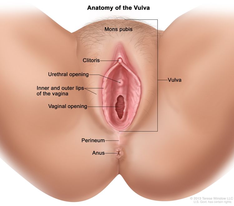
Vulvar neoplasms refer to abnormal growths or tumors in the vulvar region, which is the exterior female genital area including the mons pubis, labia majora, labia minora, clitoris, and the vaginal vestibule. These neoplasms can be benign (non-cancerous) or malignant (cancerous).
Benign vulvar neoplasms may include conditions such as vulvar cysts, fibromas, lipomas, or condylomas (genital warts). They are typically slow-growing and less likely to spread or invade surrounding tissues.
Malignant vulvar neoplasms, on the other hand, are cancers that can invade nearby tissues and potentially metastasize (spread) to distant parts of the body. The most common types of malignant vulvar neoplasms are squamous cell carcinoma, vulvar melanoma, and adenocarcinoma.
Early detection and treatment of vulvar neoplasms are essential for improving prognosis and reducing the risk of complications or recurrence. Regular gynecological examinations, self-examinations, and prompt attention to any unusual symptoms or changes in the vulvar area can help ensure timely diagnosis and management.
A neoplasm micrometastasis is a small focus of cancer cells that has spread (metastasized) from the primary tumor to a distant site and is too small to be detected by standard diagnostic methods, such as imaging studies or clinical examination. It is typically identified through the use of immunohistochemical stains or molecular techniques during microscopic examination of tissue samples.
Micrometastases are often found in patients with early-stage cancer and can indicate a higher risk of recurrence or metastasis. However, not all micrometastases will progress to clinical metastases, and their significance is still an area of ongoing research.
Intraductal carcinoma, noninfiltrating is a medical term used to describe a type of breast cancer that is confined to the milk ducts of the breast. It is also sometimes referred to as ductal carcinoma in situ (DCIS). Noninfiltrating means that the cancer cells have not spread beyond the ducts into the surrounding breast tissue or elsewhere in the body.
In this type of cancer, abnormal cells line the milk ducts and fill the inside of the ducts. These abnormal cells may look like cancer cells under a microscope, but they have not grown through the walls of the ducts into the surrounding breast tissue. However, if left untreated, noninfiltrating intraductal carcinoma can progress to an invasive form of breast cancer where the cancer cells spread beyond the milk ducts and invade the surrounding breast tissue.
It is important to note that while noninfiltrating intraductal carcinoma is considered a precancerous condition, it still requires medical treatment to prevent the development of invasive breast cancer. Treatment options may include surgery, radiation therapy, or hormone therapy, depending on the size and location of the tumor and other individual factors.
Lymph is a colorless, transparent fluid that circulates throughout the lymphatic system, which is a part of the immune and circulatory systems. It consists of white blood cells called lymphocytes, proteins, lipids, glucose, electrolytes, hormones, and waste products. Lymph plays an essential role in maintaining fluid balance, absorbing fats from the digestive tract, and defending the body against infection by transporting immune cells to various tissues and organs. It is collected from tissues through lymph capillaries and flows through increasingly larger lymphatic vessels, ultimately returning to the bloodstream via the subclavian veins in the chest region.
The Predictive Value of Tests, specifically the Positive Predictive Value (PPV) and Negative Predictive Value (NPV), are measures used in diagnostic tests to determine the probability that a positive or negative test result is correct.
Positive Predictive Value (PPV) is the proportion of patients with a positive test result who actually have the disease. It is calculated as the number of true positives divided by the total number of positive results (true positives + false positives). A higher PPV indicates that a positive test result is more likely to be a true positive, and therefore the disease is more likely to be present.
Negative Predictive Value (NPV) is the proportion of patients with a negative test result who do not have the disease. It is calculated as the number of true negatives divided by the total number of negative results (true negatives + false negatives). A higher NPV indicates that a negative test result is more likely to be a true negative, and therefore the disease is less likely to be present.
The predictive value of tests depends on the prevalence of the disease in the population being tested, as well as the sensitivity and specificity of the test. A test with high sensitivity and specificity will generally have higher predictive values than a test with low sensitivity and specificity. However, even a highly sensitive and specific test can have low predictive values if the prevalence of the disease is low in the population being tested.
Prospective studies, also known as longitudinal studies, are a type of cohort study in which data is collected forward in time, following a group of individuals who share a common characteristic or exposure over a period of time. The researchers clearly define the study population and exposure of interest at the beginning of the study and follow up with the participants to determine the outcomes that develop over time. This type of study design allows for the investigation of causal relationships between exposures and outcomes, as well as the identification of risk factors and the estimation of disease incidence rates. Prospective studies are particularly useful in epidemiology and medical research when studying diseases with long latency periods or rare outcomes.
Indocyanine green (ICG) is a sterile, water-soluble, tricarbocyanine dye that is used as a diagnostic agent in medical imaging. It is primarily used in ophthalmology for fluorescein angiography to examine blood flow in the retina and choroid, and in cardiac surgery to assess cardiac output and perfusion. When injected into the body, ICG binds to plasma proteins and fluoresces when exposed to near-infrared light, allowing for visualization of various tissues and structures. It is excreted primarily by the liver and has a half-life of approximately 3-4 minutes in the bloodstream.
Sentinel surveillance is a type of public health surveillance that is used to monitor the occurrence and spread of specific diseases or health events in a defined population. It is called "sentinel" because it relies on a network of carefully selected healthcare providers, hospitals, or laboratories to report cases of the disease or event of interest.
The main goal of sentinel surveillance is to provide timely and accurate information about the incidence and trends of a particular health problem in order to inform public health action. This type of surveillance is often used when it is not feasible or practical to monitor an entire population, such as in the case of rare diseases or emerging infectious diseases.
Sentinel surveillance systems typically require well-defined criteria for case identification and reporting, as well as standardized data collection and analysis methods. They may also involve active monitoring and follow-up of cases to better understand the epidemiology of the disease or event. Overall, sentinel surveillance is an important tool for detecting and responding to public health threats in a timely and effective manner.
Lymphatic irradiation is a medical procedure that involves the use of radiation therapy to target and treat the lymphatic system. This type of treatment is often used in cancer care, specifically in cases where cancer has spread to the lymph nodes. The goal of lymphatic irradiation is to destroy any remaining cancer cells in the lymphatic system and reduce the risk of cancer recurrence.
The procedure typically involves the use of a linear accelerator, which directs high-energy X-rays or electrons at the affected area. The radiation oncologist will determine the appropriate dose and duration of treatment based on the location and extent of the cancer, as well as the patient's overall health and medical history.
It is important to note that lymphatic irradiation can have side effects, including fatigue, skin changes, and swelling in the affected area. Patients may also experience longer-term side effects, such as lymphedema, which is a chronic swelling of the limbs due to damage to the lymphatic system.
Overall, lymphatic irradiation is an important tool in cancer care and can help improve outcomes for patients with cancer that has spread to the lymphatic system. However, it should be administered by trained medical professionals and accompanied by appropriate supportive care to manage side effects and optimize patient outcomes.
Colloids are a type of mixture that contains particles that are intermediate in size between those found in solutions and suspensions. These particles range in size from about 1 to 1000 nanometers in diameter, which is smaller than what can be seen with the naked eye, but larger than the molecules in a solution.
Colloids are created when one substance, called the dispersed phase, is dispersed in another substance, called the continuous phase. The dispersed phase can consist of particles such as proteins, emulsified fats, or finely divided solids, while the continuous phase is usually a liquid, but can also be a gas or a solid.
Colloids are important in many areas of medicine and biology, including drug delivery, diagnostic imaging, and tissue engineering. They are also found in nature, such as in milk, blood, and fog. The properties of colloids can be affected by factors such as pH, temperature, and the presence of other substances, which can influence their stability and behavior.
A feasibility study is a preliminary investigation or analysis conducted to determine the viability of a proposed project, program, or product. In the medical field, feasibility studies are often conducted before implementing new treatments, procedures, equipment, or facilities. These studies help to assess the practicality and effectiveness of the proposed intervention, as well as its potential benefits and risks.
Feasibility studies in healthcare typically involve several steps:
1. Problem identification: Clearly define the problem that the proposed project, program, or product aims to address.
2. Objectives setting: Establish specific, measurable, achievable, relevant, and time-bound (SMART) objectives for the study.
3. Literature review: Conduct a thorough review of existing research and best practices related to the proposed intervention.
4. Methodology development: Design a methodology for data collection and analysis that will help answer the research questions and achieve the study's objectives.
5. Resource assessment: Evaluate the availability and adequacy of resources, including personnel, time, and finances, required to carry out the proposed intervention.
6. Risk assessment: Identify potential risks and challenges associated with the implementation of the proposed intervention and develop strategies to mitigate them.
7. Cost-benefit analysis: Estimate the costs and benefits of the proposed intervention, including direct and indirect costs, as well as short-term and long-term benefits.
8. Stakeholder engagement: Engage relevant stakeholders, such as patients, healthcare providers, administrators, and policymakers, to gather their input and support for the proposed intervention.
9. Decision-making: Based on the findings of the feasibility study, make an informed decision about whether or not to proceed with the proposed project, program, or product.
Feasibility studies are essential in healthcare as they help ensure that resources are allocated efficiently and effectively, and that interventions are evidence-based, safe, and beneficial for patients.
Retrospective studies, also known as retrospective research or looking back studies, are a type of observational study that examines data from the past to draw conclusions about possible causal relationships between risk factors and outcomes. In these studies, researchers analyze existing records, medical charts, or previously collected data to test a hypothesis or answer a specific research question.
Retrospective studies can be useful for generating hypotheses and identifying trends, but they have limitations compared to prospective studies, which follow participants forward in time from exposure to outcome. Retrospective studies are subject to biases such as recall bias, selection bias, and information bias, which can affect the validity of the results. Therefore, retrospective studies should be interpreted with caution and used primarily to generate hypotheses for further testing in prospective studies.
Intraoperative care refers to the medical care and interventions provided to a patient during a surgical procedure. This care is typically administered by a team of healthcare professionals, including anesthesiologists, surgeons, nurses, and other specialists as needed. The goal of intraoperative care is to maintain the patient's physiological stability throughout the surgery, minimize complications, and ensure the best possible outcome.
Intraoperative care may include:
1. Anesthesia management: Administering and monitoring anesthetic drugs to keep the patient unconscious and free from pain during the surgery.
2. Monitoring vital signs: Continuously tracking the patient's heart rate, blood pressure, oxygen saturation, body temperature, and other key physiological parameters to ensure they remain within normal ranges.
3. Fluid and blood product administration: Maintaining adequate intravascular volume and oxygen-carrying capacity through the infusion of fluids and blood products as needed.
4. Intraoperative imaging: Utilizing real-time imaging techniques, such as X-ray, ultrasound, or CT scans, to guide the surgical procedure and ensure accurate placement of implants or other devices.
5. Neuromonitoring: Using electrophysiological methods to monitor the functional integrity of nerves and neural structures during surgery, particularly in procedures involving the brain, spine, or peripheral nerves.
6. Intraoperative medication management: Administering various medications as needed for pain control, infection prophylaxis, or the treatment of medical conditions that may arise during the surgery.
7. Temperature management: Regulating the patient's body temperature to prevent hypothermia or hyperthermia, which can have adverse effects on surgical outcomes and overall patient health.
8. Communication and coordination: Ensuring effective communication among the members of the surgical team to optimize patient care and safety.
A segmental mastectomy, also known as a partial mastectomy, is a surgical procedure that involves the removal of a portion of the breast tissue. This type of mastectomy is typically used to treat breast cancer that is limited to a specific area of the breast. During the procedure, the surgeon removes the cancerous tumor along with some surrounding healthy tissue, as well as the lining of the chest wall below the tumor and the lymph nodes in the underarm area.
In a segmental mastectomy, the goal is to remove the cancer while preserving as much of the breast tissue as possible. This approach can help to achieve a more cosmetic outcome compared to a total or simple mastectomy, which involves removing the entire breast. However, the extent of the surgery will depend on the size and location of the tumor, as well as other factors such as the patient's overall health and personal preferences.
It is important to note that while a segmental mastectomy can be an effective treatment option for breast cancer, it may not be appropriate for all patients or tumors. The decision to undergo this procedure should be made in consultation with a healthcare provider, taking into account the individual patient's medical history, diagnosis, and treatment goals.
Local neoplasm recurrence is the return or regrowth of a tumor in the same location where it was originally removed or treated. This means that cancer cells have survived the initial treatment and started to grow again in the same area. It's essential to monitor and detect any local recurrence as early as possible, as it can affect the prognosis and may require additional treatment.
Technetium is not a medical term itself, but it is a chemical element with the symbol Tc and atomic number 43. However, in the field of nuclear medicine, which is a branch of medicine that uses small amounts of radioactive material to diagnose or treat diseases, Technetium-99m (a radioisotope of technetium) is commonly used for various diagnostic procedures.
Technetium-99m is a metastable nuclear isomer of technetium-99, and it emits gamma rays that can be detected outside the body to create images of internal organs or tissues. It has a short half-life of about 6 hours, which makes it ideal for diagnostic imaging since it decays quickly and reduces the patient's exposure to radiation.
Technetium-99m is used in a variety of medical procedures, such as bone scans, lung scans, heart scans, liver-spleen scans, brain scans, and kidney scans, among others. It can be attached to different pharmaceuticals or molecules that target specific organs or tissues, allowing healthcare professionals to assess their function or identify any abnormalities.
A simple mastectomy, also known as a total mastectomy, is a surgical procedure that involves the removal of the entire breast tissue, including the nipple and areola, but does not include the removal of the lymph nodes or muscles in the chest wall. This type of mastectomy may be recommended for patients with early-stage breast cancer, large tumors, or multiple tumors in one breast, as well as those who have a high risk of developing breast cancer due to genetic factors.
The goal of a simple mastectomy is to remove the cancerous tissue while preserving as much healthy tissue as possible. This procedure may be performed as a preventative measure for individuals at high risk of developing breast cancer, or as a treatment option for those diagnosed with breast cancer. It's important to note that a simple mastectomy does not involve the removal of axillary lymph nodes, which are typically removed in a modified radical mastectomy for patients with breast cancer that has spread to the lymph nodes.
After the procedure, patients may require reconstructive surgery to rebuild the shape and appearance of the breast. It's essential for patients to discuss their options with their healthcare provider to determine the best course of treatment based on their individual needs and circumstances.
Lymphadenitis is a medical term that refers to the inflammation of one or more lymph nodes, which are small, bean-shaped glands that are part of the body's immune system. Lymph nodes contain white blood cells called lymphocytes, which help fight infection and disease.
Lymphadenitis can occur as a result of an infection in the area near the affected lymph node or as a result of a systemic infection that has spread through the bloodstream. The inflammation causes the lymph node to become swollen, tender, and sometimes painful to the touch.
The symptoms of lymphadenitis may include fever, fatigue, and redness or warmth in the area around the affected lymph node. In some cases, the overlying skin may also appear red and inflamed. Lymphadenitis can occur in any part of the body where there are lymph nodes, including the neck, armpits, groin, and abdomen.
The underlying cause of lymphadenitis must be diagnosed and treated promptly to prevent complications such as the spread of infection or the formation of an abscess. Treatment may include antibiotics, pain relievers, and warm compresses to help reduce swelling and discomfort.
Tuberculosis (TB) of the lymph node, also known as scrofula or tuberculous lymphadenitis, is a specific form of extrapulmonary tuberculosis. It involves the infection and inflammation of the lymph nodes (lymph glands) by the Mycobacterium tuberculosis bacterium. The lymph nodes most commonly affected are the cervical (neck) and supraclavicular (above the collarbone) lymph nodes, but other sites can also be involved.
The infection typically spreads to the lymph nodes through the bloodstream or via nearby infected organs, such as the lungs or intestines. The affected lymph nodes may become enlarged, firm, and tender, forming masses called cold abscesses that can suppurate (form pus) and eventually rupture. In some cases, the lymph nodes may calcify, leaving hard, stone-like deposits.
Diagnosis of tuberculous lymphadenitis often involves a combination of clinical evaluation, imaging studies (such as CT or MRI scans), and microbiological or histopathological examination of tissue samples obtained through fine-needle aspiration biopsy or surgical excision. Treatment typically consists of a standard anti-tuberculosis multi-drug regimen, which may include isoniazid, rifampin, ethambutol, and pyrazinamide for at least six months. Surgical intervention might be necessary in cases with complications or treatment failure.
Tin compounds refer to chemical substances that contain tin (Sn) combined with one or more other elements. Tin can form various types of compounds, including oxides, sulfides, halides, and organometallic compounds. These compounds have different properties and uses depending on the other element(s) they are combined with.
For example:
* Tin (IV) oxide (SnO2) is a white powder used as an opacifying agent in glass and ceramics, as well as a component in some types of batteries.
* Tin (II) sulfide (SnS) is a black or brown solid used in the manufacture of some types of semiconductors.
* Tin (IV) chloride (SnCl4) is a colorless liquid used as a catalyst in the production of polyvinyl chloride (PVC) and other plastics.
* Organotin compounds, such as tributyltin (TBT), are used as biocides and antifouling agents in marine paints. However, they have been found to be toxic to aquatic life and are being phased out in many countries.
Immunohistochemistry (IHC) is a technique used in pathology and laboratory medicine to identify specific proteins or antigens in tissue sections. It combines the principles of immunology and histology to detect the presence and location of these target molecules within cells and tissues. This technique utilizes antibodies that are specific to the protein or antigen of interest, which are then tagged with a detection system such as a chromogen or fluorophore. The stained tissue sections can be examined under a microscope, allowing for the visualization and analysis of the distribution and expression patterns of the target molecule in the context of the tissue architecture. Immunohistochemistry is widely used in diagnostic pathology to help identify various diseases, including cancer, infectious diseases, and immune-mediated disorders.
Hematoxylin is not a medical term per se, but it is widely used in the field of histology and pathology, which are subspecialties within medicine. Hematoxylin is a natural dye that is commonly used in histological staining procedures to highlight cell nuclei in tissue samples. It is often combined with eosin, another dye, to create the well-known hematoxylin and eosin (H&E) stain, which is routinely used to examine tissue architecture and diagnose various medical conditions.
In essence, hematoxylin is a histological stain that selectively binds to the acidic components of nuclear chromatin, imparting a blue-purple color to the cell nuclei when visualized under a microscope. This staining technique helps pathologists and researchers identify and analyze various cellular structures and abnormalities within tissue samples.
Neoadjuvant therapy is a treatment regimen that is administered to patients before they undergo definitive or curative surgery for their cancer. The main goal of neoadjuvant therapy is to reduce the size and extent of the tumor, making it easier to remove surgically and increasing the likelihood of complete resection. This type of therapy often involves the use of chemotherapy, radiation therapy, or targeted therapy, and it can help improve treatment outcomes by reducing the risk of recurrence and improving overall survival rates. Neoadjuvant therapy is commonly used in the treatment of various types of cancer, including breast, lung, esophageal, rectal, and bladder cancer.
Merkel cell carcinoma (MCC) is a rare and aggressive type of skin cancer that originates from the uncontrolled growth of Merkel cells, which are specialized nerve cells found in the top layer of the skin (epidermis). These cells are responsible for touch sensation. MCC typically presents as a painless, firm, rapidly growing nodule or mass, often on sun-exposed areas such as the head, neck, and arms of older adults.
The primary risk factors for Merkel cell carcinoma include:
1. Exposure to ultraviolet (UV) radiation from sunlight or tanning beds
2. Advanced age (most commonly occurs in people over 50)
3. A weakened immune system due to conditions like HIV/AIDS, organ transplantation, or long-term use of immunosuppressive medications
4. History of other types of skin cancer, such as melanoma or basal cell carcinoma
5. Fair skin and light eye color
MCC is considered an aggressive cancer because it can spread quickly to nearby lymph nodes and other parts of the body (metastasize). The major prognostic factor for MCC is the presence or absence of lymph node involvement at the time of diagnosis. Early detection and treatment are crucial for improving outcomes.
Standard treatments for Merkel cell carcinoma include surgical excision, radiation therapy, and chemotherapy. Immunotherapy with drugs like avelumab has also shown promising results in treating advanced stages of MCC. Regular follow-up care is essential to monitor for recurrence or metastasis.
Near-infrared spectroscopy (NIRS) is a non-invasive optical technique that uses the near-infrared region of the electromagnetic spectrum (approximately 700-2500 nanometers) to analyze various chemical and physical properties of materials, primarily in the fields of biomedical research and industry. In medicine, NIRS is often used to measure tissue oxygenation, hemodynamics, and metabolism, providing valuable information about organ function and physiology. This technique is based on the principle that different molecules absorb and scatter near-infrared light differently, allowing for the identification and quantification of specific chromophores, such as oxyhemoglobin, deoxyhemoglobin, and cytochrome c oxidase. NIRS can be employed in a variety of clinical settings, including monitoring cerebral or muscle oxygenation during surgery, assessing tissue viability in wound healing, and studying brain function in neuroscience research.
Follow-up studies are a type of longitudinal research that involve repeated observations or measurements of the same variables over a period of time, in order to understand their long-term effects or outcomes. In medical context, follow-up studies are often used to evaluate the safety and efficacy of medical treatments, interventions, or procedures.
In a typical follow-up study, a group of individuals (called a cohort) who have received a particular treatment or intervention are identified and then followed over time through periodic assessments or data collection. The data collected may include information on clinical outcomes, adverse events, changes in symptoms or functional status, and other relevant measures.
The results of follow-up studies can provide important insights into the long-term benefits and risks of medical interventions, as well as help to identify factors that may influence treatment effectiveness or patient outcomes. However, it is important to note that follow-up studies can be subject to various biases and limitations, such as loss to follow-up, recall bias, and changes in clinical practice over time, which must be carefully considered when interpreting the results.
Neoplasm invasiveness is a term used in pathology and oncology to describe the aggressive behavior of cancer cells as they invade surrounding tissues and organs. This process involves the loss of cell-to-cell adhesion, increased motility and migration, and the ability of cancer cells to degrade the extracellular matrix (ECM) through the production of enzymes such as matrix metalloproteinases (MMPs).
Invasive neoplasms are cancers that have spread beyond the original site where they first developed and have infiltrated adjacent tissues or structures. This is in contrast to non-invasive or in situ neoplasms, which are confined to the epithelial layer where they originated and have not yet invaded the underlying basement membrane.
The invasiveness of a neoplasm is an important prognostic factor in cancer diagnosis and treatment, as it can indicate the likelihood of metastasis and the potential effectiveness of various therapies. In general, more invasive cancers are associated with worse outcomes and require more aggressive treatment approaches.
Carcinoma is a type of cancer that develops from epithelial cells, which are the cells that line the inner and outer surfaces of the body. These cells cover organs, glands, and other structures within the body. Carcinomas can occur in various parts of the body, including the skin, lungs, breasts, prostate, colon, and pancreas. They are often characterized by the uncontrolled growth and division of abnormal cells that can invade surrounding tissues and spread to other parts of the body through a process called metastasis. Carcinomas can be further classified based on their appearance under a microscope, such as adenocarcinoma, squamous cell carcinoma, and basal cell carcinoma.
Reproducibility of results in a medical context refers to the ability to obtain consistent and comparable findings when a particular experiment or study is repeated, either by the same researcher or by different researchers, following the same experimental protocol. It is an essential principle in scientific research that helps to ensure the validity and reliability of research findings.
In medical research, reproducibility of results is crucial for establishing the effectiveness and safety of new treatments, interventions, or diagnostic tools. It involves conducting well-designed studies with adequate sample sizes, appropriate statistical analyses, and transparent reporting of methods and findings to allow other researchers to replicate the study and confirm or refute the results.
The lack of reproducibility in medical research has become a significant concern in recent years, as several high-profile studies have failed to produce consistent findings when replicated by other researchers. This has led to increased scrutiny of research practices and a call for greater transparency, rigor, and standardization in the conduct and reporting of medical research.
A fine-needle biopsy (FNB) is a medical procedure in which a thin, hollow needle is used to obtain a sample of cells or tissue from a suspicious or abnormal area in the body, such as a lump or mass. The needle is typically smaller than that used in a core needle biopsy, and it is guided into place using imaging techniques such as ultrasound, CT scan, or MRI.
The sample obtained during an FNB can be used to diagnose various medical conditions, including cancer, infection, or inflammation. The procedure is generally considered safe and well-tolerated, with minimal risks of complications such as bleeding, infection, or discomfort. However, the accuracy of the diagnosis depends on the skill and experience of the healthcare provider performing the biopsy, as well as the adequacy of the sample obtained.
Overall, FNB is a valuable diagnostic tool that can help healthcare providers make informed decisions about treatment options and improve patient outcomes.
Squamous cell carcinoma is a type of skin cancer that begins in the squamous cells, which are flat, thin cells that form the outer layer of the skin (epidermis). It commonly occurs on sun-exposed areas such as the face, ears, lips, and backs of the hands. Squamous cell carcinoma can also develop in other areas of the body including the mouth, lungs, and cervix.
This type of cancer usually develops slowly and may appear as a rough or scaly patch of skin, a red, firm nodule, or a sore or ulcer that doesn't heal. While squamous cell carcinoma is not as aggressive as some other types of cancer, it can metastasize (spread) to other parts of the body if left untreated, making early detection and treatment important.
Risk factors for developing squamous cell carcinoma include prolonged exposure to ultraviolet (UV) radiation from the sun or tanning beds, fair skin, a history of sunburns, a weakened immune system, and older age. Prevention measures include protecting your skin from the sun by wearing protective clothing, using a broad-spectrum sunscreen with an SPF of at least 30, avoiding tanning beds, and getting regular skin examinations.
Histiocytic Necrotizing Lymphadenitis is a condition characterized by the inflammation and necrosis (death of tissue) of lymph nodes, caused by an abnormal proliferation and activation of histiocytes (a type of white blood cell). It is also known as Kikuchi's disease. The exact cause of this condition is unknown, but it is thought to be related to an immune response to viral infections or other antigens.
Histopathologically, it is characterized by the presence of necrotizing granulomatous inflammation with histiocytic predominance and absence of neutrophils. The condition is typically self-limiting, with symptoms resolving within a few months without specific treatment. However, in some cases, it can be associated with systemic symptoms or other autoimmune disorders.
Adjuvant radiotherapy is a type of cancer treatment that uses radiation therapy as an adjunct to a primary surgical procedure. The goal of adjuvant radiotherapy is to eliminate any remaining microscopic cancer cells that may be present in the surrounding tissues after surgery, thereby reducing the risk of local recurrence and improving the chances of cure.
Radiotherapy involves the use of high-energy radiation to destroy cancer cells and shrink tumors. In adjuvant radiotherapy, the radiation is usually delivered to the tumor bed and regional lymph nodes in order to target any potential sites of residual disease. The timing and dosing of adjuvant radiotherapy may vary depending on the type and stage of cancer being treated, as well as other factors such as patient age and overall health status.
Adjuvant radiotherapy is commonly used in the treatment of various types of cancer, including breast, colorectal, lung, head and neck, and gynecologic cancers. Its use has been shown to improve survival rates and reduce the risk of recurrence in many cases, making it an important component of comprehensive cancer care.
In medical terms, the arm refers to the upper limb of the human body, extending from the shoulder to the wrist. It is composed of three major bones: the humerus in the upper arm, and the radius and ulna in the lower arm. The arm contains several joints, including the shoulder joint, elbow joint, and wrist joint, which allow for a wide range of motion. The arm also contains muscles, blood vessels, nerves, and other soft tissues that are essential for normal function.
A nomogram is a graphical representation of a mathematical formula or equation that allows the user to quickly solve a problem by simply drawing a line between different values on the chart. In the field of medicine, nomograms are often used as a tool for predicting patient outcomes, assessing risk, or making diagnostic decisions based on specific clinical data.
For example, a nomogram may be used to estimate the probability of survival in patients with a particular type of cancer, based on factors such as age, tumor size, and stage of disease. The user would locate the appropriate values for each factor on the nomogram, draw a line connecting them, and read off the estimated probability at the intersection point.
Nomograms can be a useful and intuitive way to communicate complex medical information and help clinicians make informed decisions in a timely manner. However, it is important to note that nomograms are only as accurate as the data they are based on, and should always be used in conjunction with clinical judgment and other relevant factors.
The breast is the upper ventral region of the human body in females, which contains the mammary gland. The main function of the breast is to provide nutrition to infants through the production and secretion of milk, a process known as lactation. The breast is composed of fibrous connective tissue, adipose (fatty) tissue, and the mammary gland, which is made up of 15-20 lobes that are arranged in a radial pattern. Each lobe contains many smaller lobules, where milk is produced during lactation. The milk is then transported through a network of ducts to the nipple, where it can be expressed by the infant.
In addition to its role in lactation, the breast also has important endocrine and psychological functions. It contains receptors for hormones such as estrogen and progesterone, which play a key role in sexual development and reproduction. The breast is also a source of sexual pleasure and can be an important symbol of femininity and motherhood.
It's worth noting that males also have breast tissue, although it is usually less developed than in females. Male breast tissue consists mainly of adipose tissue and does not typically contain functional mammary glands. However, some men may develop enlarged breast tissue due to conditions such as gynecomastia, which can be caused by hormonal imbalances or certain medications.
Cytodiagnosis is the rapid, initial evaluation and diagnosis of a disease based on the examination of individual cells obtained from a body fluid or tissue sample. This technique is often used in cytopathology to investigate abnormalities such as lumps, bumps, or growths that may be caused by cancerous or benign conditions.
The process involves collecting cells through various methods like fine-needle aspiration (FNA), body fluids such as urine, sputum, or washings from the respiratory, gastrointestinal, or genitourinary tracts. The collected sample is then spread onto a microscope slide, stained, and examined under a microscope for abnormalities in cell size, shape, structure, and organization.
Cytodiagnosis can provide crucial information to guide further diagnostic procedures and treatment plans. It is often used as an initial screening tool due to its speed, simplicity, and cost-effectiveness compared to traditional histopathological methods that require tissue biopsy and more extensive processing. However, cytodiagnosis may not always be able to distinguish between benign and malignant conditions definitively; therefore, additional tests or follow-up evaluations might be necessary for a conclusive diagnosis.
In medical terms, the "neck" is defined as the portion of the body that extends from the skull/head to the thorax or chest region. It contains 7 cervical vertebrae, muscles, nerves, blood vessels, lymphatic vessels, and glands (such as the thyroid gland). The neck is responsible for supporting the head, allowing its movement in various directions, and housing vital structures that enable functions like respiration and circulation.
The lymphatic system is a complex network of organs, tissues, vessels, and cells that work together to defend the body against infectious diseases and also play a crucial role in the immune system. It is made up of:
1. Lymphoid Organs: These include the spleen, thymus, lymph nodes, tonsils, adenoids, and Peyer's patches (in the intestines). They produce and mature immune cells.
2. Lymphatic Vessels: These are thin tubes that carry clear fluid called lymph towards the heart.
3. Lymph: This is a clear-to-white fluid that contains white blood cells, mainly lymphocytes, which help fight infections.
4. Other tissues and cells: These include bone marrow where immune cells are produced, and lymphocytes (T cells and B cells) which are types of white blood cells that help protect the body from infection and disease.
The primary function of the lymphatic system is to transport lymph throughout the body, collecting waste products, bacteria, viruses, and other foreign substances from the tissues, and filtering them out through the lymph nodes. The lymphatic system also helps in the absorption of fats and fat-soluble vitamins from food in the digestive tract.
Lymphangiogenesis is the formation of new lymphatic vessels from pre-existing ones. It is a complex biological process that involves the growth, differentiation, and remodeling of lymphatic endothelial cells, which line the interior surface of lymphatic vessels. Lymphangiogenesis plays crucial roles in various physiological processes, including tissue drainage, immune surveillance, and lipid absorption. However, it can also contribute to pathological conditions such as cancer metastasis, inflammation, and fibrosis when it is dysregulated.
The process of lymphangiogenesis is regulated by a variety of growth factors, receptors, and signaling molecules, including vascular endothelial growth factor (VEGF)-C, VEGF-D, and their receptor VEGFR-3, as well as other factors such as angiopoietins, integrins, and matrix metalloproteinases. Understanding the mechanisms of lymphangiogenesis has important implications for developing novel therapies for a range of diseases associated with abnormal lymphatic vessel growth and function.
Adjuvant chemotherapy is a medical treatment that is given in addition to the primary therapy, such as surgery or radiation, to increase the chances of a cure or to reduce the risk of recurrence in patients with cancer. It involves the use of chemicals (chemotherapeutic agents) to destroy any remaining cancer cells that may not have been removed by the primary treatment. This type of chemotherapy is typically given after the main treatment has been completed, and its goal is to kill any residual cancer cells that may be present in the body and reduce the risk of the cancer coming back. The specific drugs used and the duration of treatment will depend on the type and stage of cancer being treated.
Treatment outcome is a term used to describe the result or effect of medical treatment on a patient's health status. It can be measured in various ways, such as through symptoms improvement, disease remission, reduced disability, improved quality of life, or survival rates. The treatment outcome helps healthcare providers evaluate the effectiveness of a particular treatment plan and make informed decisions about future care. It is also used in clinical research to compare the efficacy of different treatments and improve patient care.
Keratin-19 is a type I acidic keratin that is primarily expressed in simple epithelia, such as the gastrointestinal tract, respiratory tract, and epidermal appendages (e.g., hair follicles, sweat glands). It plays an essential role in maintaining the structure and integrity of these tissues by forming intermediate filaments that provide mechanical support to cells.
Keratin-19 is often used as a marker for simple epithelial differentiation and has been implicated in various pathological conditions, including cancer progression and metastasis. Mutations in the KRT19 gene, which encodes keratin-19, have been associated with certain genetic disorders, such as epidermolysis bullosa simplex, a blistering skin disorder.
In summary, Keratin-19 is an important structural protein expressed in simple epithelia that plays a crucial role in maintaining tissue integrity and has implications in various pathological conditions.
Optical imaging is a non-invasive medical imaging technique that uses light to capture images of internal structures and processes within the body. This method often involves the use of endoscopes, microscopes, or specialized cameras to visualize targeted areas, such as organs, tissues, or cells. Optical imaging can be used for various diagnostic and therapeutic purposes, including monitoring disease progression, guiding surgical procedures, and studying biological functions at the cellular level. Different optical imaging techniques include reflectance imaging, fluorescence imaging, bioluminescence imaging, and optical coherence tomography (OCT).
In summary, optical imaging is a versatile and non-ionizing medical imaging technique that utilizes light to visualize internal body structures and processes for diagnostic and therapeutic applications.
Keratins are a type of fibrous structural proteins that constitute the main component of the integumentary system, which includes the hair, nails, and skin of vertebrates. They are also found in other tissues such as horns, hooves, feathers, and reptilian scales. Keratins are insoluble proteins that provide strength, rigidity, and protection to these structures.
Keratins are classified into two types: soft keratins (Type I) and hard keratins (Type II). Soft keratins are found in the skin and simple epithelial tissues, while hard keratins are present in structures like hair, nails, horns, and hooves.
Keratin proteins have a complex structure consisting of several domains, including an alpha-helical domain, beta-pleated sheet domain, and a non-repetitive domain. These domains provide keratin with its unique properties, such as resistance to heat, chemicals, and mechanical stress.
In summary, keratins are fibrous structural proteins that play a crucial role in providing strength, rigidity, and protection to various tissues in the body.
Lymphatic vessels are thin-walled, valved structures that collect and transport lymph, a fluid derived from the interstitial fluid surrounding the cells, throughout the lymphatic system. They play a crucial role in immune function and maintaining fluid balance in the body. The primary function of lymphatic vessels is to return excess interstitial fluid, proteins, waste products, and immune cells to the bloodstream via the subclavian veins near the heart.
There are two types of lymphatic vessels:
1. Lymphatic capillaries: These are the smallest lymphatic vessels, found in most body tissues except for the central nervous system (CNS). They have blind ends and are highly permeable to allow the entry of interstitial fluid, proteins, and other large molecules.
2. Larger lymphatic vessels: These include precollecting vessels, collecting vessels, and lymphatic trunks. Precollecting vessels have valves that prevent backflow of lymph and merge to form larger collecting vessels. Collecting vessels contain smooth muscle in their walls, which helps to propel the lymph forward. They also have valves at regular intervals to ensure unidirectional flow towards the heart. Lymphatic trunks are large vessels that collect lymph from various regions of the body and eventually drain into the two main lymphatic ducts: the thoracic duct and the right lymphatic duct.
Overall, lymphatic vessels play a vital role in maintaining fluid balance, immune surveillance, and waste removal in the human body.
Tumor markers are substances that can be found in the body and their presence can indicate the presence of certain types of cancer or other conditions. Biological tumor markers refer to those substances that are produced by cancer cells or by other cells in response to cancer or certain benign (non-cancerous) conditions. These markers can be found in various bodily fluids such as blood, urine, or tissue samples.
Examples of biological tumor markers include:
1. Proteins: Some tumor markers are proteins that are produced by cancer cells or by other cells in response to the presence of cancer. For example, prostate-specific antigen (PSA) is a protein produced by normal prostate cells and in higher amounts by prostate cancer cells.
2. Genetic material: Tumor markers can also include genetic material such as DNA, RNA, or microRNA that are shed by cancer cells into bodily fluids. For example, circulating tumor DNA (ctDNA) is genetic material from cancer cells that can be found in the bloodstream.
3. Metabolites: Tumor markers can also include metabolic products produced by cancer cells or by other cells in response to cancer. For example, lactate dehydrogenase (LDH) is an enzyme that is released into the bloodstream when cancer cells break down glucose for energy.
It's important to note that tumor markers are not specific to cancer and can be elevated in non-cancerous conditions as well. Therefore, they should not be used alone to diagnose cancer but rather as a tool in conjunction with other diagnostic tests and clinical evaluations.
In the field of medicine, "time factors" refer to the duration of symptoms or time elapsed since the onset of a medical condition, which can have significant implications for diagnosis and treatment. Understanding time factors is crucial in determining the progression of a disease, evaluating the effectiveness of treatments, and making critical decisions regarding patient care.
For example, in stroke management, "time is brain," meaning that rapid intervention within a specific time frame (usually within 4.5 hours) is essential to administering tissue plasminogen activator (tPA), a clot-busting drug that can minimize brain damage and improve patient outcomes. Similarly, in trauma care, the "golden hour" concept emphasizes the importance of providing definitive care within the first 60 minutes after injury to increase survival rates and reduce morbidity.
Time factors also play a role in monitoring the progression of chronic conditions like diabetes or heart disease, where regular follow-ups and assessments help determine appropriate treatment adjustments and prevent complications. In infectious diseases, time factors are crucial for initiating antibiotic therapy and identifying potential outbreaks to control their spread.
Overall, "time factors" encompass the significance of recognizing and acting promptly in various medical scenarios to optimize patient outcomes and provide effective care.
A nipple is a small projection or tubular structure located at the center of the areola, which is the darker circle of skin surrounding the nipple on the breast. The primary function of the nipple is to provide a pathway for milk flow from the mammary glands during lactation in females.
The nipple contains smooth muscle fibers that contract and cause the nipple to become erect when stimulated, such as during sexual arousal or cold temperatures. Nipples can come in various shapes, sizes, and colors, and some individuals may have inverted or flat nipples. It is essential to monitor any changes in the appearance or sensation of the nipples, as these could be indicative of underlying medical conditions, such as breast cancer.
Breast neoplasms in males refer to abnormal growths or tumors in the male breast tissue. These neoplasms can be benign (non-cancerous) or malignant (cancerous). While breast cancer is much less common in men than in women, it can still occur and should be taken seriously.
The most common type of breast cancer in men is invasive ductal carcinoma, which starts in the milk ducts and spreads to surrounding tissue. Other types of breast cancer that can occur in men include inflammatory breast cancer, lobular carcinoma, and Paget's disease of the nipple.
Risk factors for developing male breast cancer include age (most cases are diagnosed after age 60), family history of breast cancer, genetic mutations such as BRCA1 or BRCA2, radiation exposure, obesity, liver disease, and testicular conditions such as undescended testicles.
Symptoms of male breast neoplasms may include a painless lump in the breast tissue, skin changes such as dimpling or redness, nipple discharge, or a retracted nipple. If you notice any of these symptoms, it is important to consult with a healthcare professional for further evaluation and treatment.
Preoperative care refers to the series of procedures, interventions, and preparations that are conducted before a surgical operation. The primary goal of preoperative care is to ensure the patient's well-being, optimize their physical condition, reduce potential risks, and prepare them mentally and emotionally for the upcoming surgery.
Preoperative care typically includes:
1. Preoperative assessment: A thorough evaluation of the patient's overall health status, including medical history, physical examination, laboratory tests, and diagnostic imaging, to identify any potential risk factors or comorbidities that may impact the surgical procedure and postoperative recovery.
2. Informed consent: The process of ensuring the patient understands the nature of the surgery, its purpose, associated risks, benefits, and alternative treatment options. The patient signs a consent form indicating they have been informed and voluntarily agree to undergo the surgery.
3. Preoperative instructions: Guidelines provided to the patient regarding their diet, medication use, and other activities in the days leading up to the surgery. These instructions may include fasting guidelines, discontinuing certain medications, or arranging for transportation after the procedure.
4. Anesthesia consultation: A meeting with the anesthesiologist to discuss the type of anesthesia that will be used during the surgery and address any concerns related to anesthesia risks, side effects, or postoperative pain management.
5. Preparation of the surgical site: Cleaning and shaving the area where the incision will be made, as well as administering appropriate antimicrobial agents to minimize the risk of infection.
6. Medical optimization: Addressing any underlying medical conditions or correcting abnormalities that may negatively impact the surgical outcome. This may involve adjusting medications, treating infections, or managing chronic diseases such as diabetes.
7. Emotional and psychological support: Providing counseling, reassurance, and education to help alleviate anxiety, fear, or emotional distress related to the surgery.
8. Preoperative holding area: The patient is transferred to a designated area near the operating room where they are prepared for surgery by changing into a gown, having intravenous (IV) lines inserted, and receiving monitoring equipment.
By following these preoperative care guidelines, healthcare professionals aim to ensure that patients undergo safe and successful surgical procedures with optimal outcomes.
Quantum dots are not a medical term per se, but they are often referred to in the field of medical research and technology. Quantum dots are semiconductor nanocrystals that exhibit unique optical properties, making them useful for various applications in biology and medicine. They can range in size from 1 to 10 nanometers in diameter and can be composed of materials such as cadmium selenide (CdSe), indium arsenide (InAs), or lead sulfide (PbS).
In the medical context, quantum dots have been explored for use in bioimaging, biosensing, and drug delivery. Their small size and tunable optical properties make them ideal for tracking cells, proteins, and other biological molecules in real-time with high sensitivity and specificity. Additionally, quantum dots can be functionalized with various biomolecules, such as antibodies or peptides, to target specific cell types or disease markers.
However, it is important to note that the use of quantum dots in medical applications is still largely in the research stage, and there are concerns about their potential toxicity due to the heavy metals used in their composition. Therefore, further studies are needed to evaluate their safety and efficacy before they can be widely adopted in clinical settings.
Tyrosinase, also known as monophenol monooxygenase, is an enzyme (EC 1.14.18.1) that catalyzes the ortho-hydroxylation of monophenols (like tyrosine) to o-diphenols (like L-DOPA) and the oxidation of o-diphenols to o-quinones. This enzyme plays a crucial role in melanin synthesis, which is responsible for the color of skin, hair, and eyes in humans and animals. Tyrosinase is found in various organisms, including plants, fungi, and animals. In humans, tyrosinase is primarily located in melanocytes, the cells that produce melanin. The enzyme's activity is regulated by several factors, such as pH, temperature, and metal ions like copper, which are essential for its catalytic function.
MART-1, also known as Melanoma Antigen Recognized by T-Cells 1 or Melan-A, is a protein that is primarily found in melanocytes, which are the pigment-producing cells located in the skin, eyes, and hair follicles. It is a member of the family of antigens called melanoma differentiation antigens (MDAs) that are specifically expressed in melanocytes and melanomas. MART-1 is considered a tumor-specific antigen because it is overexpressed in melanoma cells compared to normal cells, making it an attractive target for immunotherapy.
MART-1 is presented on the surface of melanoma cells in complex with major histocompatibility complex (MHC) class I molecules, where it can be recognized by cytotoxic T lymphocytes (CTLs). This recognition triggers an immune response that can lead to the destruction of melanoma cells. MART-1 has been widely used as a target in various immunotherapy approaches, including cancer vaccines and adoptive cell transfer therapies, with the goal of enhancing the body's own immune system to recognize and eliminate melanoma cells.
Mammary ultrasonography, also known as breast ultrasound, is a non-invasive diagnostic imaging technique that uses high-frequency sound waves to produce detailed images of the internal structures of the breast tissue. It is often used in conjunction with mammography to help identify and characterize breast abnormalities, such as lumps, cysts, or tumors, and to guide biopsy procedures.
Ultrasonography is particularly useful for evaluating palpable masses, assessing the integrity of breast implants, and distinguishing between solid and fluid-filled lesions. It is also a valuable tool for monitoring treatment response in patients with known breast cancer. Because it does not use radiation like mammography, mammary ultrasonography is considered safe and can be repeated as often as necessary. However, its effectiveness is highly dependent on the skill and experience of the sonographer performing the examination.
Cat-scratch disease (CSD) is a bacterial infection caused by Bartonella henselae. It is typically transmitted through contact with a cat, especially when the animal scratches or bites a person and then introduces the bacteria into the wound. The incubation period for CSD is usually 7-14 days after exposure.
The most common symptoms of CSD include:
* A small, raised bump (called a papule) that develops at the site of the scratch or bite within a few days of being scratched or bitten by a cat. This bump may be tender and can sometimes form a crust or pustule.
* Swollen lymph nodes (also called lymphadenopathy) near the site of the infection, which usually develop 1-2 weeks after the initial scratch or bite. These swollen lymph nodes are often painful and may be warm to the touch.
* Fatigue, fever, headache, and muscle aches are also common symptoms of CSD.
In most cases, cat-scratch disease is a mild illness that resolves on its own within a few weeks or months. However, in some cases, it can cause more severe complications, such as infection of the heart valves (endocarditis), inflammation of the brain (encephalitis), or damage to the eyes (retinitis).
Treatment for cat-scratch disease typically involves supportive care, such as pain relief and anti-inflammatory medications. Antibiotics may be prescribed in some cases, particularly if the infection is severe or if the patient has a weakened immune system. Preventive measures include washing hands after handling cats, avoiding rough play with cats, and promptly treating cat bites and scratches.
Neoplasms: Neoplasms are abnormal growths of cells or tissues in the body that serve no physiological function. They can be benign (non-cancerous) or malignant (cancerous).
Ductal Neoplasms: Ductal neoplasms refer to abnormal growths that occur in the milk ducts of the breast. The most common type of ductal neoplasm is ductal carcinoma, which can be either non-invasive (also known as ductal carcinoma in situ or DCIS) or invasive (also known as infiltrating ductal carcinoma).
Lobular Neoplasms: Lobular neoplasms refer to abnormal growths that occur in the milk-producing lobules of the breast. The two main types of lobular neoplasms are lobular carcinoma in situ (LCIS) and invasive lobular carcinoma.
Medullary Neoplasms: Medullary neoplasms refer to a type of cancer that can occur in various organs, including the breast, thyroid gland, and salivary glands. In the breast, medullary carcinoma is a rare type of invasive breast cancer that accounts for less than 5% of all breast cancers. Medullary carcinoma typically affects younger women and tends to have a better prognosis compared to other types of invasive breast cancer.
In summary, neoplasms refer to abnormal growths in the body, while ductal, lobular, and medullary neoplasms refer to specific types of abnormal growths that occur in different parts of the breast or other organs.
Hodgkin disease, also known as Hodgkin lymphoma, is a type of cancer that originates in the white blood cells called lymphocytes. It typically affects the lymphatic system, which is a network of vessels and glands spread throughout the body. The disease is characterized by the presence of a specific type of abnormal cell, known as a Reed-Sternberg cell, within the affected lymph nodes.
The symptoms of Hodgkin disease may include painless swelling of the lymph nodes in the neck, armpits, or groin; fever; night sweats; weight loss; and fatigue. The exact cause of Hodgkin disease is unknown, but it is thought to involve a combination of genetic, environmental, and infectious factors.
Hodgkin disease is typically treated with a combination of chemotherapy, radiation therapy, and/or immunotherapy, depending on the stage and extent of the disease. With appropriate treatment, the prognosis for Hodgkin disease is generally very good, with a high cure rate. However, long-term side effects of treatment may include an increased risk of secondary cancers and other health problems.
Disease-free survival (DFS) is a term used in medical research and clinical practice, particularly in the field of oncology. It refers to the length of time after primary treatment for a cancer during which no evidence of the disease can be found. This means that the patient shows no signs or symptoms of the cancer, and any imaging studies or other tests do not reveal any tumors or other indications of the disease.
DFS is often used as an important endpoint in clinical trials to evaluate the effectiveness of different treatments for cancer. By measuring the length of time until the cancer recurs or a new cancer develops, researchers can get a better sense of how well a particular treatment is working and whether it is improving patient outcomes.
It's important to note that DFS is not the same as overall survival (OS), which refers to the length of time from primary treatment until death from any cause. While DFS can provide valuable information about the effectiveness of cancer treatments, it does not necessarily reflect the impact of those treatments on patients' overall survival.