Secretory Rate
Pancreatic Juice
Bicarbonates
Potassium
Sodium
Insulin
Mesopic Vision
Encyclopedias as Topic
Retina
Photoreceptor Cells, Vertebrate
Vision, Ocular
Retinal Cone Photoreceptor Cells
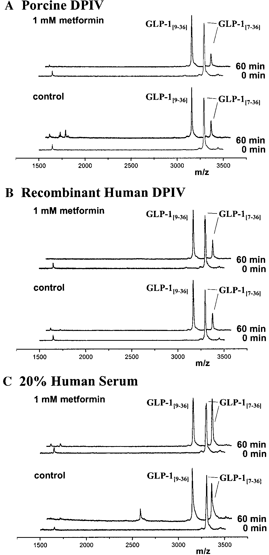
Fluid secretion by the malpighian tubules of the tsetse fly Glossina morsitans: the effects of ouabain, ethacrynic acid and amiloride. (1/1163)
The effects of three inhibitors of sodium transport on the secretion of fluid by the Malpighian tubules of Glossina morsitans have been observed. The cardiac glycoside, ouabain, affects neither the rate of secretion nor the sodium concentration of the fluid secreted when isolated tubules are bathed by solutions containing a range of sodium and potassium concentrations. Secretion is inhibited, however, by ethacrynic acid and amiloride. The results confirm that fluid secretion by the Malpighian tubules of this insect is dependent on the active transport of sodium ions and show that Na+/k+ exchange pumps are not involved in this process. (+info)Activation of stimulus-specific serine esterases (proteases) in the initiation of platelet secretion. I. Demonstration with organophosphorus inhibitors. (2/1163)
The effect of organophosphorus inhibitors of serine esterases (proteases) on secretion from washed rabbit platelets was examined. Five noncytotoxic stimuli were employed: collagen, thrombin, heterologous anti-platelet antibody (in the absence of complement), rabbit C3 bound to zymosan, and platelet activating factor derived from antigen-stimulated, IgE-sensitized rabbit basophils. Diisoprophyl phosphofluoridate, three series of p-nitrophenyl ethyl phosphonates, and a series of cyclohexyl phenylalkylphosphonofluridates were all found to be inhibitory to the platelet secretion. These are irreversible inhibitors of serine proteases but in this system were only inhibitory if added to the platelets concurrently with the stimuli. Pretreatment of either the platelets or the stimuli with the inhibitors followed by washing, was without effect on the subsequent reaction. This suggested the involvement of stimulus-activatable serine proteases in the secretory process. The concept was supported by finding that nonphosphorylating phosphonates or hydrolyzed phosphonates or phosphonofluoridates were without inhibitory action. The effect of a series of phosphonates or phosphonoflouridates in inhibiting each stimulus exhibited a unique activity-structure profile. The demonstration of such unique profiles with four series of inhibitors for each of the five stimuli was interpreted as demonstrating that a specific activatable serine protease was involved in the platelet secretory response to each stimulus. (+info)Pancreatic beta-cell-to-beta-cell interactions are required for integrated responses to nutrient stimuli: enhanced Ca2+ and insulin secretory responses of MIN6 pseudoislets. (3/1163)
The effect of cell-to-cell contact on Ca2+ influx and secretory responses in the beta-cell line MIN6 was studied using MIN6 pseudoislets, which are three-dimensional islet-like cell aggregates that develop when MIN6 cells are cultured for 6-8 days on gelatin. The formation of pseudoislets is dependent on the Ca2+-dependent adhesion molecule E-cadherin (E-CAD), since the process can be inhibited by incubation in the absence of Ca2+ or in the presence of an anti-E-CAD antibody. Glucose and alpha-ketoisocaproic acid (KIC) evoked a Ca2+ influx in only a small fraction of the MIN6 monolayer cells, whereas >80% of cell groups within the pseudoislets responded to both nutrients. In contrast, changes in the intracellular free Ca2+ concentration ([Ca2+]i) were observed in all or most monolayer cells or pseudoislet cell groups in response to physical or pharmacological depolarizing stimuli. No significant increase in insulin release was observed from MIN6 monolayer cells in response to nutrient or nonnutrient insulin secretagogues. Conversely, pseudoislets were found to respond significantly to both nutrients and nonnutrients. These results suggest that close cell-to-cell contact improves the functional responsiveness of MIN6 cells and that pseudoislets may therefore serve as a useful research model in the study of beta-cell function. (+info)Exposure of human islets to cytokines can result in disproportionately elevated proinsulin release. (4/1163)
Infiltration of immunocytes into pancreatic islets precedes loss of beta cells in type 1 diabetes. It is conceivable that local release of cytokines affects the function of beta cells before their apoptosis. This study examines whether the elevated proinsulin levels that have been described in prediabetes can result from exposure of beta cells to cytokines. Human beta-cell preparations were cultured for 48 or 72 hours with or without IL-1beta, TNF-alpha, or IFN-gamma, alone or in combination. None of these conditions were cytotoxic, nor did they reduce insulin biosynthetic activity. Single cytokines did not alter medium or cellular content in insulin or proinsulin. Cytokine combinations, in particular IL-1beta plus IFN-gamma, disproportionately elevated medium proinsulin levels. This effect expresses an altered functional state of the beta cells characterized by preserved proinsulin synthesis, a slower hormone conversion, and an increased ratio of cellular proinsulin over insulin content. The delay in proinsulin conversion can be attributed to lower expression of PC1 and PC2 convertases. It is concluded that disproportionately elevated proinsulin levels in pre-type 1 diabetic patients might result from exposure of their beta cells to cytokines released from infiltrating immunocytes. This hormonal alteration expresses an altered functional state of the beta cells that can occur independently of beta-cell death. (+info)Secretory responses to sympathetic stimulation of the cat's salivary glands in a state of resting secretion. (5/1163)
The secretory effect of sympathetic stimulation on the cat's submaxillary gland was augmented greatly when studied against a background of slow secretion evoked by parasympathetic stimulation at a low frequency and imitating the slow resting secretion normally present in the waking state. The sympathetic secretory threshold was markedly lowered, and even at low frequencies sympathetic stimulation caused a large, well-maintained response. After an alpha-adrenoceptor blocking drug sympathetic stimulation alone lost its secretory effect, but during resting secretion part of the accelerating effect was found to remain; this effect was elicited via beta-adrenoceptors. A marked secretory effect of sympathetic stimulation was also obtained during resting secretion in the parotid gland, where the sympathetic secretory effect is normally very small. (+info)Sexual differentiation of oestradiol-LH positive feedback in a marsupial. (6/1163)
The surge of LH that induces ovulation in mammals showing spontaneous ovulation is precipitated by the positive feedback of increasing oestrogens from the developing follicles in the ovary. In eutherians, exogenous oestrogens can mimic this effect by eliciting an LH surge in females, but not usually in males. The absence of a positive LH response to eutherian males is either due to an acute suppression by the secretory products of the testes during adulthood or the permanent disabling of the system by testosterone during early development. This phenomenon is examined in tammar wallabies, Macropus eugenii. The results show that the oestradiol-LH positive feedback response is sexually dimorphic in this marsupial. A surge in plasma LH occurred between 15 and 28 h after injection of 2.5 micrograms oestradiol benzoate kg-1 in 13 of 16 intact females and 4 of 4 ovariectomized females, but in none of 11 intact males. Five females each implanted with a 100 mg testosterone pellet 3 months earlier failed to produce an LH surge. Four males castrated in adulthood and three adult males castrated before puberty also failed to show an LH surge. However, three males castrated 24-26 days after birth showed an unambiguous LH surge when challenged with oestradiol benzoate during adulthood. Thus, in tammar wallabies, the ability to generate an LH surge to oestradiol is a sexually dimorphic response that is suppressed in the male by the organizational effects of the testes in early life and presumably supplemented by an inhibitory effect of circulating testosterone in adulthood. (+info)Compensation by pulsatile GnRH infusions for incompetence for oestradiol-induced LH surges in long-term ovariectomized gilts and castrated male pigs. (7/1163)
The aim of this study was to investigate incompetence for oestradiol-induced LH surges in long-term ovariectomized gilts and male pigs. Gilts (250 days old; n = 36), which had been ovariectomized 30 (OVX 30) or 100 days (OVX 100) before the start of treatment, were challenged i.m. with oestradiol benzoate and were either given no further treatment, fed methallibure to inhibit endogenous GnRH release or fed methallibure and given i.v. pulses of 100 or 200 ng GnRH agonist at 1 h intervals during the LH surge (48-96 h after oestradiol benzoate). The same treatments were applied to long-term orchidectomized male pigs (ORC, n = 23). In addition, one ORC group was not injected with oestradiol benzoate but was fed methallibure and given pulses of 200 ng GnRH agonist. Oestradiol benzoate alone induced an LH surge in the OVX 30 group only (5/6 gilts), methallibure suppressed (P < 0.05) oestradiol benzoate-induced LH secretion, while pulses of 100 ng GnRH agonist in animals fed methallibure produced LH surges in four of six OVX 30 and four of six OVX 100 gilts. The induced LH surges were similar to those produced by oestradiol benzoate alone in OVX 30 gilts. Pulses of 200 ng GnRH agonist produced LH surges in OVX 30 (6/6) and OVX 100 (6/6) gilts and increased the magnitude of the induced LH surge in OVX 100 gilts (P < 0.05 compared with 100 ng GnRH agonist or OVX 30 control). Pulses of 200 ng GnRH agonist also induced LH surge release in ORC male pigs (5/6), but were unable to increase LH concentrations in a surge-like manner in ORC animals that had not been given oestradiol benzoate, indicating that oestradiol increases pituitary responsiveness to GnRH. These results support the hypothesis that oestradiol must inhibit secretion of LH before an LH surge can occur. It is concluded that incompetence for oestradiol-induced LH surges in long-term ovarian secretion-deprived gilts and in male pigs is due to the failure of oestradiol to promote a sufficient increase in the release of GnRH. (+info)Stimulation-induced factors which affect augmentation and potentiation of trasmitter release at the neuromuscular junction. (8/1163)
1. End-plate potentials (e.p.p.s) were recorded from frog sartorius neuromuscular junctions under conditions of decreased transmitter release to study the effect of repetitive stimulation on augmentation and potentiation of transmitter release. 2. The magnitudes and time constants of decay of augmentation and potentiation were determined both following a primary conditioning train and following an identical secondary conditioning train applied from 30 to 170 sec after the primary conditioning train. 3. The magnitude of augmentation following the secondary conditioning trains was increased over that following the primary conditioning trains even though augmentation, with a time constant of decay of about 7 sec, had decayed to insignificant levels before the onset of the secondary trains. This increase in augmentation was not due to a change in its rate of decay during the secondary trains. 4. The increased magnitude of augmentation can be described as arising from an expression factor which, for conditioning trains of 200 impulses at 20/sec, has an initial magnitude of 1-6 +/- 1-2 (S.D. of observation) (the magnitude of augmentation is increased 2-6 times) and decays approximately exponentially with a time constant of 90 +/- 50 (S.D. of observation) sec. The expression factor thus decays about ten times slower than augmentation. 5. Doubling the number of impulses in the primary conditioning train from 100 to 200 led to a 2-8 +/- 1-0 (S.D. of observation) times increase in the magnitude of the expression factor, estimated by placing a 200 impulse secondary conditioning train 40 sec after the primary conditioning train. 6. The expression factor, while increasing the magnitude of augmentation, had little or no effect on the magnitude of potentiation or on trasmitter release in the absence of augmentation. The expression factor decayed about twice as slowly as potentiation. 7. The time constants characterizing the decay of potentiation were greater following the secondary conditioning trains than following the primary conditioning trains. 8. The increased time constant for the decay of potentiation can be described as arising from a time constant factor which, for conditioning trains of 200 impulses at 20/sec, has an initial magnitude of 1-2 +/- 0-7 (S.D. of observation) (the time constant of potentiation is increased 2-2 times) and decays approximately exponentially with a time constant of 130 +/- 45 (S.D. of observation) sec. The time constant factor decayed about three times slower than potentiation. 9. Doubling the number of impulses in the primary conditioning train from 100 to 200 led to a 1-6 +/- 0-8 (S.D. of observation) times increase in the magnitude of the time constant factor, estimated by placing a 200 impulse secondary conditioning train 40 sec after the primary conditioning train. 10... (+info)Secretory rate refers to the amount or volume of a secretion produced by a gland or an organ over a given period of time. It is a measure of the productivity or activity level of the secreting structure. The secretory rate can be quantified for various bodily fluids, such as saliva, sweat, digestive enzymes, hormones, or milk, depending on the context and the specific gland or organ being studied.
In clinical settings, measuring the secretory rate might involve collecting and analyzing samples over a certain duration to estimate the production rate of the substance in question. This information can be helpful in diagnosing conditions related to impaired secretion, monitoring treatment responses, or understanding the physiological adaptations of the body under different circumstances.
Pancreatic juice is an alkaline fluid secreted by the exocrine component of the pancreas, primarily containing digestive enzymes such as amylase, lipase, and trypsin. These enzymes aid in the breakdown of carbohydrates, fats, and proteins, respectively, in the small intestine during the digestion process. The bicarbonate ions present in pancreatic juice help neutralize the acidic chyme that enters the duodenum from the stomach, creating an optimal environment for enzymatic activity.
Bicarbonates, also known as sodium bicarbonate or baking soda, is a chemical compound with the formula NaHCO3. In the context of medical definitions, bicarbonates refer to the bicarbonate ion (HCO3-), which is an important buffer in the body that helps maintain normal pH levels in blood and other bodily fluids.
The balance of bicarbonate and carbonic acid in the body helps regulate the acidity or alkalinity of the blood, a condition known as pH balance. Bicarbonates are produced by the body and are also found in some foods and drinking water. They work to neutralize excess acid in the body and help maintain the normal pH range of 7.35 to 7.45.
In medical testing, bicarbonate levels may be measured as part of an electrolyte panel or as a component of arterial blood gas (ABG) analysis. Low bicarbonate levels can indicate metabolic acidosis, while high levels can indicate metabolic alkalosis. Both conditions can have serious consequences if not treated promptly and appropriately.
Potassium is a essential mineral and an important electrolyte that is widely distributed in the human body. The majority of potassium in the body (approximately 98%) is found within cells, with the remaining 2% present in blood serum and other bodily fluids. Potassium plays a crucial role in various physiological processes, including:
1. Regulation of fluid balance and maintenance of normal blood pressure through its effects on vascular tone and sodium excretion.
2. Facilitation of nerve impulse transmission and muscle contraction by participating in the generation and propagation of action potentials.
3. Protein synthesis, enzyme activation, and glycogen metabolism.
4. Regulation of acid-base balance through its role in buffering systems.
The normal serum potassium concentration ranges from 3.5 to 5.0 mEq/L (milliequivalents per liter) or mmol/L (millimoles per liter). Potassium levels outside this range can have significant clinical consequences, with both hypokalemia (low potassium levels) and hyperkalemia (high potassium levels) potentially leading to serious complications such as cardiac arrhythmias, muscle weakness, and respiratory failure.
Potassium is primarily obtained through the diet, with rich sources including fruits (e.g., bananas, oranges, and apricots), vegetables (e.g., leafy greens, potatoes, and tomatoes), legumes, nuts, dairy products, and meat. In cases of deficiency or increased needs, potassium supplements may be recommended under the guidance of a healthcare professional.
Sodium is an essential mineral and electrolyte that is necessary for human health. In a medical context, sodium is often discussed in terms of its concentration in the blood, as measured by serum sodium levels. The normal range for serum sodium is typically between 135 and 145 milliequivalents per liter (mEq/L).
Sodium plays a number of important roles in the body, including:
* Regulating fluid balance: Sodium helps to regulate the amount of water in and around your cells, which is important for maintaining normal blood pressure and preventing dehydration.
* Facilitating nerve impulse transmission: Sodium is involved in the generation and transmission of electrical signals in the nervous system, which is necessary for proper muscle function and coordination.
* Assisting with muscle contraction: Sodium helps to regulate muscle contractions by interacting with other minerals such as calcium and potassium.
Low sodium levels (hyponatremia) can cause symptoms such as confusion, seizures, and coma, while high sodium levels (hypernatremia) can lead to symptoms such as weakness, muscle cramps, and seizures. Both conditions require medical treatment to correct.
Insulin is a hormone produced by the beta cells of the pancreatic islets, primarily in response to elevated levels of glucose in the circulating blood. It plays a crucial role in regulating blood glucose levels and facilitating the uptake and utilization of glucose by peripheral tissues, such as muscle and adipose tissue, for energy production and storage. Insulin also inhibits glucose production in the liver and promotes the storage of excess glucose as glycogen or triglycerides.
Deficiency in insulin secretion or action leads to impaired glucose regulation and can result in conditions such as diabetes mellitus, characterized by chronic hyperglycemia and associated complications. Exogenous insulin is used as a replacement therapy in individuals with diabetes to help manage their blood glucose levels and prevent long-term complications.
Mesopic vision is a term used to describe the intermediate level of vision that occurs in conditions of decreased illumination, specifically between 0.02 and 3 candelas per square meter (cd/m²). This range falls between photopic vision, which is vision in bright light (>3 cd/m²), and scotopic vision, which is vision in very low light (
An encyclopedia is a comprehensive reference work containing articles on various topics, usually arranged in alphabetical order. In the context of medicine, a medical encyclopedia is a collection of articles that provide information about a wide range of medical topics, including diseases and conditions, treatments, tests, procedures, and anatomy and physiology. Medical encyclopedias may be published in print or electronic formats and are often used as a starting point for researching medical topics. They can provide reliable and accurate information on medical subjects, making them useful resources for healthcare professionals, students, and patients alike. Some well-known examples of medical encyclopedias include the Merck Manual and the Stedman's Medical Dictionary.
The retina is the innermost, light-sensitive layer of tissue in the eye of many vertebrates and some cephalopods. It receives light that has been focused by the cornea and lens, converts it into neural signals, and sends these to the brain via the optic nerve. The retina contains several types of photoreceptor cells including rods (which handle vision in low light) and cones (which are active in bright light and are capable of color vision).
In medical terms, any pathological changes or diseases affecting the retinal structure and function can lead to visual impairment or blindness. Examples include age-related macular degeneration, diabetic retinopathy, retinal detachment, and retinitis pigmentosa among others.
Photoreceptor cells in vertebrates are specialized types of neurons located in the retina of the eye that are responsible for converting light stimuli into electrical signals. These cells are primarily responsible for the initial process of vision and have two main types: rods and cones.
Rods are more numerous and are responsible for low-light vision or scotopic vision, enabling us to see in dimly lit conditions. They do not contribute to color vision but provide information about the shape and movement of objects.
Cones, on the other hand, are less numerous and are responsible for color vision and high-acuity vision or photopic vision. There are three types of cones, each sensitive to different wavelengths of light: short (S), medium (M), and long (L) wavelengths, which correspond to blue, green, and red, respectively. The combination of signals from these three types of cones allows us to perceive a wide range of colors.
Both rods and cones contain photopigments that consist of a protein called opsin and a light-sensitive chromophore called retinal. When light hits the photopigment, it triggers a series of chemical reactions that ultimately lead to the generation of an electrical signal that is transmitted to the brain via the optic nerve. This process enables us to see and perceive our visual world.
Ocular vision refers to the ability to process and interpret visual information that is received by the eyes. This includes the ability to see clearly and make sense of the shapes, colors, and movements of objects in the environment. The ocular system, which includes the eye and related structures such as the optic nerve and visual cortex of the brain, works together to enable vision.
There are several components of ocular vision, including:
* Visual acuity: the clarity or sharpness of vision
* Field of vision: the extent of the visual world that is visible at any given moment
* Color vision: the ability to distinguish different colors
* Depth perception: the ability to judge the distance of objects in three-dimensional space
* Contrast sensitivity: the ability to distinguish an object from its background based on differences in contrast
Disorders of ocular vision can include refractive errors such as nearsightedness or farsightedness, as well as more serious conditions such as cataracts, glaucoma, and macular degeneration. These conditions can affect one or more aspects of ocular vision and may require medical treatment to prevent further vision loss.
In the context of medical terminology, "light" doesn't have a specific or standardized definition on its own. However, it can be used in various medical terms and phrases. For example, it could refer to:
1. Visible light: The range of electromagnetic radiation that can be detected by the human eye, typically between wavelengths of 400-700 nanometers. This is relevant in fields such as ophthalmology and optometry.
2. Therapeutic use of light: In some therapies, light is used to treat certain conditions. An example is phototherapy, which uses various wavelengths of ultraviolet (UV) or visible light for conditions like newborn jaundice, skin disorders, or seasonal affective disorder.
3. Light anesthesia: A state of reduced consciousness in which the patient remains responsive to verbal commands and physical stimulation. This is different from general anesthesia where the patient is completely unconscious.
4. Pain relief using light: Certain devices like transcutaneous electrical nerve stimulation (TENS) units have a 'light' setting, indicating lower intensity or frequency of electrical impulses used for pain management.
Without more context, it's hard to provide a precise medical definition of 'light'.
Retinal cone photoreceptor cells are specialized neurons located in the retina of the eye, responsible for visual phototransduction and color vision. They are one of the two types of photoreceptors, with the other being rods, which are more sensitive to low light levels. Cones are primarily responsible for high-acuity, color vision during daylight or bright-light conditions.
There are three types of cone cells, each containing different photopigments that absorb light at distinct wavelengths: short (S), medium (M), and long (L) wavelengths, which correspond to blue, green, and red light, respectively. The combination of signals from these three types of cones allows the human visual system to perceive a wide range of colors and discriminate between them. Cones are densely packed in the central region of the retina, known as the fovea, which provides the highest visual acuity.