Sarcoma
Sarcoma, Ewing
Sarcoma, Synovial
Sarcoma, Kaposi
Avian Sarcoma Viruses
Sarcoma, Experimental
Sarcoma 180
Sarcoma Viruses, Murine
Soft Tissue Neoplasms
Sarcoma, Avian
Histiocytic Sarcoma
Sarcoma, Myeloid
Sarcoma, Endometrial Stromal
Sarcoma, Clear Cell
Sarcoma, Yoshida
Kirsten murine sarcoma virus
Moloney murine sarcoma virus
Sarcoma Viruses, Feline
Rous sarcoma virus
Sarcoma, Alveolar Soft Part
Leiomyosarcoma
RNA-Binding Protein EWS
Osteosarcoma
Herpesvirus 8, Human
Alpharetrovirus
Cell Transformation, Viral
Proto-Oncogene Protein c-fli-1
Harvey murine sarcoma virus
Gammaretrovirus
Rhabdomyosarcoma
Histiocytoma, Malignant Fibrous
Liposarcoma
Cell Transformation, Neoplastic
Oncogene Proteins, Fusion
Sarcoma 37
Histiocytoma, Benign Fibrous
Sarcoma, Small Cell
Langerhans Cell Sarcoma
Vascular Neoplasms
Retroperitoneal Neoplasms
Chick Embryo
Ifosfamide
Dendritic Cell Sarcoma, Follicular
Helper Viruses
Muscle Neoplasms
Avian leukosis virus
Hemangiosarcoma
Retroviridae
Fibrosarcoma
Neuroectodermal Tumors, Primitive, Peripheral
Moloney murine leukemia virus
Immunohistochemistry
Chondrosarcoma
Base Sequence
Doxorubicin
Acquired Immunodeficiency Syndrome
RNA-Binding Protein FUS
Dendritic Cell Sarcoma, Interdigitating
Retroviridae Proteins
Oncogenes
Neoplasm Recurrence, Local
Neoplasm Transplantation
Neuroectodermal Tumors
Combined Modality Therapy
Mesna
Hemangioendothelioma
Translocation, Genetic
Fatal Outcome
Defective Viruses
Fibroblasts
Treatment Outcome
Prognosis
Neoplasms, Experimental
Chemotherapy, Cancer, Regional Perfusion
Histiocytic Disorders, Malignant
Neoplasms, Connective Tissue
Neuroectodermal Tumors, Primitive
Epsilonretrovirus
Quail
Gene Products, gag
Femoral Neoplasms
Molecular Sequence Data
Cells, Cultured
Mutation
Retrospective Studies
Mesenchymoma
Tumor Cells, Cultured
Abdominal Neoplasms
Radiotherapy, Adjuvant
Tumor Markers, Biological
Peripheral Nervous System Neoplasms
Epithelioid Cells
RNA-Directed DNA Polymerase
Antigens, Neoplasm
Nucleic Acid Hybridization
Neoplasm Metastasis
Heart Neoplasms
Dioxoles
Tomography, X-Ray Computed
Antineoplastic Agents, Alkylating
Antineoplastic Combined Chemotherapy Protocols
Neoplasm Proteins
Thoracic Neoplasms
Gene Expression Regulation, Neoplastic
Chickens
Survival Analysis
Mice, Nude
Neoplasms, Radiation-Induced
Survival Rate
Rhabdomyosarcoma, Embryonal
Tetrahydroisoquinolines
Antibiotics, Antineoplastic
Giant Lymph Node Hyperplasia
Disease-Free Survival
Neoplasms
L-[1-11C]-tyrosine PET to evaluate response to hyperthermic isolated limb perfusion for locally advanced soft-tissue sarcoma and skin cancer. (1/2226)
PET with L-[1-11C]-tyrosine (TYR) was investigated in patients undergoing hyperthermic isolated limb perfusion (HILP) with recombinant tumor necrosis factor alpha (rTNF-alpha) and melphalan for locally advanced soft-tissue sarcoma and skin cancer of the lower limb. METHODS: Seventeen patients (5 women, 12 men; age range 24-75 y; mean age 52 y) were studied. TYR PET studies were performed before HILP and 2 and 8 wk afterwards. The protein synthesis rates (PSRs) in nanomoles per milliliter per minute were calculated. After final PET studies, tumors were resected and pathologically examined. Patients with pathologically complete responses (pCR) showed no viable tumors after treatment. Those with pathologically partial responses (pPR) showed various amounts of viable tumors in the resected tumor specimens. RESULTS: Six patients (35%) showed a pCR and 11 patients (65%) showed a pPR. All tumors were depicted as hot spots on PET studies before HILP. The PSR in the pCR group at 2 and 8 wk after perfusion had decreased significantly (P < 0.05) in comparison to the PSR before HILP. A significant difference was found in PSR between the pCR and pPR groups at 2 and at 8 wk (P < 0.05). Median PSR in nonviable tumor tissue was 0.62 and ranged from 0.22 to 0.91. With a threshold PSR of 0.91, sensitivity and specificity of TYR PET were 82% and 100%, respectively. The predictive value of a PSR > 0.91 for having viable tumor after HILP was 100%, whereas the predictive value of a PSR < or = 0.91 for having nonviable tumor tissue after HILP was 75%. The 2 patients in the pPR groups with a PSR < 0.91 showed microscopic islets of tumor cells surrounded by extensive necrosis on pathological examination. CONCLUSION: Based on the calculated PSR after HILP, TYR PET gave a good indication of the pathological outcome. Inflammatory tissue after treatment did not interfere with viable tumor on the images, suggesting that it may be worthwhile to pursue TYR PET in other therapy evaluation settings. (+info)Comparison of the Ki-67 score and S-phase fraction as prognostic variables in soft-tissue sarcoma. (2/2226)
Immunohistochemically determined Ki-67 scores and flow cytometrically determined S-phase fractions were successfully evaluated from the primary tumours of 123 patients with soft-tissue sarcoma. All patients had either limb or superficial trunk tumours. Ki-67 score correlated strongly with ploidy, S-phase fraction and grade. Ki-67 did not correlate with the size of the primary tumour. When analysed as a continuous variable, Ki-67 was a stronger predictor of both metastasis-free survival and disease-specific overall survival (P = 0.003 and 0.04 respectively) than was the S-phase fraction (P = 0.06 and 0.07 respectively). We tested the relevance of different cut-point values by dividing the whole material into two parts at every 10% (e.g. 10% of patients vs. the remaining 90%, 20% vs. 80%, etc.). We counted the relative risk and confidence interval at all these cut-off points. Ki-67 had good prognostic discriminating power irrespective of the cut-point value, but S-phase fraction lost its prognostic power at higher cut-point values. In conclusion, we found that Ki-67 is a useful prognostic tool in the treatment of soft-tissue sarcoma patients irrespective of the cut-point value. S-phase fraction can be used at lower cut-point values. (+info)Sarcomatoid renal cell carcinoma: biologic behavior, prognosis, and response to combined surgical resection and immunotherapy. (3/2226)
PURPOSE: Sarcomatoid variants of renal cell carcinoma (RCC) are aggressive tumors that respond poorly to immunotherapy. We report the outcomes of 31 patients with sarcomatoid RCC treated with a combination of surgical resection and immunotherapy. PATIENTS AND METHODS: Patients were identified from the database of the University of California Los Angeles Kidney Cancer Program. We retrospectively reviewed the cases of 31 consecutive patients in whom sarcomatoid RCC was diagnosed between 1990 and 1997. Clinical stage, sites of metastasis, pathologic stage, and type of immunotherapy were abstracted from the medical records. The primary end point analyzed was overall survival, and a multivariate analysis was performed to distinguish any factors conferring an improved survivorship. RESULTS: Twenty-six percent of patients were male and 74% were female, and the median age was 59 years (range, 34 to 73 years). Length of follow-up ranged from 2 to 77 months (mean, 21.4 months). Twenty-eight patients (84%) had known metastases at the time of radical nephrectomy (67% had lung metastases and 40% had bone, 21% had liver, 33% had lymphatic, and 15% had brain metastases). Twenty-five patients (81%) received immunotherapy, including low-dose interleukin (IL)-2-based therapy (five patients), tumor-infiltrating lymphocyte-based therapy plus IL-2 (nine patients), high-dose IL-2-based therapy (nine patients), dendritic cell vaccine-based therapy (one patient), and interferon alpha-based therapy alone (one patient). Two patients (6%) achieved complete responses (median duration, 46+ months) and five patients (15%) achieved partial responses (median duration, 36 months). One- and 2-year overall survival rates were 48% and 37%, respectively. Using a multivariate analysis, age, sex, and percentage of sarcomatoid tumor (< or >50%) did not significantly correlate with survival. Improved survival was found in patients receiving high-dose IL-2 therapy compared with patients treated with surgery alone or any other form of immunotherapy (P = .025). Adjusting for age, sex, and percentage of sarcomatoid tumor, the relative risk of death was 10.4 times higher in patients not receiving high-dose IL-2 therapy. Final pathologic T stage did not correlate significantly with outcome, but node-positive patients had a higher death rate per year of follow-up than did the rest of the population (1.26 v 0.76, Cox regression analysis). CONCLUSION: Surgical resection and high-dose IL-2-based immunotherapy may play a role in the treatment of sarcomatoid RCCs in select patients. (+info)Prognostic implications of various models for calculation of S-phase fraction in 259 patients with soft tissue sarcoma. (4/2226)
The S-phase fraction (SPF) in flow cytometric DNA histograms in soft tissue sarcoma (STS) can be calculated in various ways. The traditional planimetric method of Baisch has been shown to be prognostic, but is hampered by a failure rate of around 40%. We therefore tested other models to see if this rate could be decreased with retained prognostic value. In 259 STS of the locomotor system the SPF was calculated according to Baisch and with commercial parametric MultiCycle software using different corrections for background. Using the Baisch model, 159 histograms could be evaluated for SPF. The 5-year metastasis-free survival rate (MFSR) was 0.94 for the low-risk group (defined with SPF), and 0.53 for the high-risk group. In the low-risk group, four of the seven patients who developed metastasis did so after 5 years Using the MultiCycle software, SPF could be calculated in 253 tumours. Depending on type of background correction used, the 5-year MFSR varied between 0.67 and 0.82 for the low-risk group, and between 0.47 and 0.53 for the high-risk group. The late metastasis pattern in the low-risk group was never seen using the MultiCycle software. We conclude that in paraffin archival material, calculation of SPF according to Baisch is preferable in clinical use due to better separation between low-risk and high-risk groups, and also the possibility to identify patients who metastasize late. (+info)Histochemical investigation into the molecular mechanisms of malignant transformation in a benign glomus tumour. (5/2226)
A glomangiosarcoma arose in a benign glomus tumour. The histological and immunohistochemical characteristics of the tumour were investigated. Apoptotic cells were identified by terminal deoxynucleotidyl transferase (TdT) mediated dUTP-biotin nick end labelling (TUNEL). The proportion of apoptotic cells was found to be low and TUNEL positive nuclei were present in the benign part of the tumour. Bcl-2 protein, an inhibitor of apoptosis, was strongly expressed in the glomangiosarcoma with only weak staining in the benign area. The proliferation index of the glomangiosarcoma was almost 10-fold higher than that of the benign glomus tumour. Numerous nuclei in the glomangiosarcoma were intensely stained for the tumour suppressor protein p53. The results of the this study may contribute to an understanding of the molecular basis of malignant transformation in benign glomus tumours. (+info)Clinical and radiological aspects of idiopathic diabetic muscle infarction. Rational approach to diagnosis and treatment. (6/2226)
The systemic effects of diabetes mellitus are well recognised. The heart, kidney, central and peripheral nervous systems, and the distal parts of the limbs are often the site of end-organ damage resulting from ischaemia. Infarction of large muscle groups in the limb, not associated with gangrene, is uncommon. There have been few reported cases other than radiological descriptions of diabetic muscle infarcts. While previous reports have illustrated some of the clinical and radiological characteristics of this condition, the paucity of published cases makes it difficult to determine the most appropriate methods of diagnosis and treatment. During a five-year period we treated 14 patients with diabetes mellitus, aged from 32 to 59 years, who were referred to a musculoskeletal oncology service for suspected soft-tissue sarcoma, but were subsequently found to have a diabetic muscle infarct. Closed needle biopsy was performed in 13 without complications. In 12 patients, the symptoms resolved without surgical treatment. (+info)GLI gene expression in bone and soft tissue sarcomas of adult patients correlates with tumor grade. (7/2226)
The GLI gene encodes a transcription factor harboring five zinc finger motifs that bind to DNA in a sequence-specific manner. The gene was originally identified because of its amplification in a human glioblastoma, and previous studies have shown it to be amplified in a significant proportion of mesenchymal tumors, such as childhood sarcomas. Here we evaluate GLI gene expression in bone and soft tissue sarcomas of adult patients. Samples from 40 patients (37 sarcomas and 3 benign mesenchymal tumors) and samples of 15 normal mesenchymal tissues were examined for GLI gene amplification and expression by Southern hybridization, reverse transcription-PCR of tissue RNA, and immunohistochemistry, using a new polyclonal GLI antibody developed against an epitope outside of the zinc finger region. In contrast to childhood sarcomas, amplification of the GLI gene was not observed in sarcomas of adult patients. Although GLI gene expression in sarcomas was significantly higher than that in normal mesenchymal tissues (P < 0.0001), the levels were very variable. Attempts to correlate the expression data with different pathophysiological parameters only showed a significant relationship to tumor grade. Based on these data, increased levels of GLI gene expression may be indicative of the aggressiveness of the tumor. (+info)Pulmonary metastases from soft tissue sarcoma: analysis of patterns of diseases and postmetastasis survival. (8/2226)
OBJECTIVE: To report the patterns of disease and postmetastasis survival for patients with pulmonary metastases from soft tissue sarcoma in a large group of patients treated at a single institution. Clinical factors that influence postmetastasis survival are analyzed. SUMMARY BACKGROUND DATA: For patients with soft tissue sarcoma, the lungs are the most common site of metastatic disease. Although pulmonary metastases most commonly arise from primary tumors in the extremities, they may arise from almost any primary site or histology. To date, resection of disease has been the only effective therapy for metastatic sarcoma. METHODS: From July 1982 to February 1997, 3149 adult patients with soft tissue sarcoma were admitted and treated at Memorial Sloan-Kettering Cancer Center. During this interval, 719 patients either developed or presented with lung metastases. Patients were treated with resection of metastatic disease whenever possible. Disease-specific survival was the endpoint of the study. Time to death was modeled using the method of Kaplan and Meier. The association of factors to time-to-event endpoints was analyzed using the log-rank test for univariate analysis and the Cox proportional hazards model for multivariate analysis. RESULTS: The overall median survival from diagnosis of pulmonary metastasis for all patients was 15 months. The 3-year actuarial survival rate was 25%. The ability to resect all metastatic disease completely was the most important prognostic factor for survival. Patients treated with complete resection had a median survival of 33 months and a 3-year actuarial survival rate of 46%. For patients treated with nonoperative therapy, the median survival was 11 months. A disease-free interval of more than 12 months before the development of metastases was also a favorable prognostic factor. Unfavorable factors included the histologic variants of liposarcoma and malignant peripheral nerve tumors and patient age older than 50 years at the time of treatment of metastasis. CONCLUSIONS: Resection of metastatic disease is the single most important factor that determines outcome in these patients. Long-term survival is possible in selected patients, particularly when recurrent pulmonary disease is resected. Surgical excision should remain the treatment of choice for metastases of soft tissue sarcoma to the lung. (+info)Sarcoma is a type of cancer that develops from certain types of connective tissue (such as muscle, fat, fibrous tissue, blood vessels, or nerves) found throughout the body. It can occur in any part of the body, but it most commonly occurs in the arms, legs, chest, and abdomen.
Sarcomas are classified into two main groups: bone sarcomas and soft tissue sarcomas. Bone sarcomas develop in the bones, while soft tissue sarcomas develop in the soft tissues of the body, such as muscles, tendons, ligaments, fat, blood vessels, and nerves.
Sarcomas can be further classified into many subtypes based on their specific characteristics, such as the type of tissue they originate from, their genetic makeup, and their appearance under a microscope. The different subtypes of sarcoma have varying symptoms, prognoses, and treatment options.
Overall, sarcomas are relatively rare cancers, accounting for less than 1% of all cancer diagnoses in the United States each year. However, they can be aggressive and may require intensive treatment, such as surgery, radiation therapy, and chemotherapy.
Ewing sarcoma is a type of cancer that originates in bones or the soft tissues surrounding them, such as muscles and tendons. It primarily affects children and adolescents, although it can occur in adults as well. The disease is characterized by small, round tumor cells that typically grow quickly and are prone to metastasize (spread) to other parts of the body, most commonly the lungs, bones, and bone marrow.
Ewing sarcoma is caused by a genetic abnormality, specifically a chromosomal translocation that results in the fusion of two genes, EWSR1 and FLI1. This gene fusion leads to the formation of an abnormal protein that disrupts normal cell growth and division processes, ultimately resulting in cancer.
Symptoms of Ewing sarcoma can vary depending on the location and size of the tumor but may include pain or swelling in the affected area, fever, fatigue, and weight loss. Diagnosis typically involves imaging studies such as X-rays, CT scans, or MRI scans to locate the tumor, followed by a biopsy to confirm the presence of cancer cells. Treatment may involve surgery, radiation therapy, chemotherapy, or a combination of these approaches, depending on the stage and location of the disease.
Synovial sarcoma is a rare type of cancer that typically develops in the soft tissues surrounding the joints, such as the synovial membrane, which lines the joint capsules. Despite its name, synovial sarcoma does not necessarily arise from the synovium. It is called so due to its resemblance to this tissue under a microscope.
This form of sarcoma primarily affects young adults and can be found in various parts of the body, but it most commonly occurs in the extremities, particularly near the knees. Synovial sarcoma is characterized by specific genetic changes that result in the formation of fusion proteins, which contribute to uncontrolled cell growth and tumor development.
There are two main subtypes of synovial sarcoma: monophasic and biphasic. Monophasic synovial sarcoma is composed of either spindle-shaped (spaghetti-like) cells or epithelioid (roundish) cells, while biphasic synovial sarcoma contains both types of cells. A third subtype, called poorly differentiated synovial sarcoma, has a more aggressive behavior and is composed of small round cells that do not resemble the typical spindle or epithelioid cells.
Treatment for synovial sarcoma usually involves surgical removal of the tumor, often followed by radiation therapy and/or chemotherapy to reduce the risk of recurrence and metastasis. The prognosis varies depending on factors such as the size and location of the tumor, the patient's age, and the presence of metastases at diagnosis.
Kaposi sarcoma (KS) is a type of cancer that causes abnormal growths in the skin, lymph nodes, or other organs. It is caused by the Kaposi sarcoma-associated herpesvirus (KSHV), also known as human herpesvirus 8 (HHV8). There are several forms of KS, including:
1. Classic KS: This form primarily affects older men of Mediterranean, Middle Eastern, or Ashkenazi Jewish descent. It tends to progress slowly and mainly involves the skin.
2. Endemic KS: Found in parts of Africa, this form predominantly affects children and young adults, regardless of their HIV status.
3. Immunosuppression-associated KS: This form is more aggressive and occurs in people with weakened immune systems due to organ transplantation or other causes.
4. Epidemic KS (AIDS-related KS): This is the most common form of KS, seen primarily in people with HIV/AIDS. The widespread use of antiretroviral therapy (ART) has significantly reduced its incidence.
KS lesions can appear as red, purple, or brown spots on the skin and may also affect internal organs such as the lungs, lymph nodes, or gastrointestinal tract. Symptoms vary depending on the location of the lesions but often include fever, fatigue, weight loss, and swelling in the legs or abdomen. Treatment options depend on the extent and severity of the disease and may involve local therapies (e.g., radiation, topical treatments), systemic therapies (e.g., chemotherapy, immunotherapy), or a combination of these approaches.
Avian sarcoma viruses (ASVs) are a group of retroviruses that primarily infect birds and cause various types of tumors, particularly sarcomas. These viruses contain an oncogene, which is a gene that has the ability to transform normal cells into cancerous ones. The oncogene in ASVs is often derived from cellular genes called proto-oncogenes, which are normally involved in regulating cell growth and division.
ASVs can be divided into two main types: non-defective and defective. Non-defective ASVs contain a complete set of viral genes that allow them to replicate independently, while defective ASVs lack some of the necessary viral genes and require assistance from other viruses to replicate.
One well-known example of an avian sarcoma virus is the Rous sarcoma virus (RSV), which was first discovered in chickens by Peyton Rous in 1910. RSV causes a highly malignant form of sarcoma in chickens and has been extensively studied as a model system for cancer research. The oncogene in RSV is called v-src, which is derived from the normal cellular gene c-src.
Avian sarcoma viruses have contributed significantly to our understanding of the molecular mechanisms underlying cancer development and have provided valuable insights into the role of oncogenes in tumorigenesis.
I'm sorry for any confusion, but "Sarcoma, Experimental" is not a recognized medical term or definition. Sarcomas are a type of cancer that develop in the body's connective tissues, such as bones, muscles, tendons, cartilage, and fat. There are many different types of sarcomas, classified based on the specific type of tissue they originate from.
Experimental, on the other hand, refers to something that is being tested or tried out for the first time, typically as part of a scientific experiment or clinical trial. In the context of cancer treatment, an experimental therapy might refer to a new drug, procedure, or device that is still being studied in clinical trials to determine its safety and effectiveness.
Therefore, "Sarcoma, Experimental" could potentially refer to a clinical trial or research study involving a new treatment for sarcoma, but it would not be a medical definition in and of itself. If you have any specific questions about sarcomas or experimental treatments, I would recommend consulting with a healthcare professional or medical researcher for more accurate information.
I'm sorry for any confusion, but "Sarcoma 180" is not a recognized medical term or an official classification of sarcomas in humans. It appears to be a term used primarily in research involving mice. Sarcoma 180 is a transplantable tumor that was first isolated from a mouse and has been used as a model for cancer research, particularly in studies involving immunotherapy and cancer treatment.
In general, sarcomas are cancers that develop from connective tissues such as bones, muscles, tendons, cartilages, nerves, and blood vessels. They can be further classified into various subtypes based on the specific type of tissue they originate from and their genetic characteristics. If you have any concerns about a specific medical condition or term, I would recommend consulting with a healthcare professional for accurate information.
Sarcoma viruses, murine, are a group of RNA viruses that primarily affect mice and other rodents. They are classified as type C retroviruses, which means they contain an envelope, have reverse transcriptase enzyme activity, and replicate through a DNA intermediate.
The murine sarcoma viruses (MSVs) are associated with the development of various types of tumors in mice, particularly fibrosarcomas, which are malignant tumors that originate from fibroblasts, the cells that produce collagen and other fibers in connective tissue.
The MSVs are closely related to the murine leukemia viruses (MLVs), and together they form a complex called the murine leukemia virus-related viruses (MLVRVs). The MLVRVs can undergo recombination events, leading to the generation of new viral variants with altered biological properties.
The MSVs are important tools in cancer research because they can transform normal cells into tumor cells in vitro and in vivo. The study of these viruses has contributed significantly to our understanding of the molecular mechanisms underlying cancer development and progression.
Soft tissue neoplasms refer to abnormal growths or tumors that develop in the soft tissues of the body. Soft tissues include muscles, tendons, ligaments, fascia, nerves, blood vessels, fat, and synovial membranes (the thin layer of cells that line joints and tendons). Neoplasms can be benign (non-cancerous) or malignant (cancerous), and their behavior and potential for spread depend on the specific type of neoplasm.
Benign soft tissue neoplasms are typically slow-growing, well-circumscribed, and rarely spread to other parts of the body. They can often be removed surgically with a low risk of recurrence. Examples of benign soft tissue neoplasms include lipomas (fat tumors), schwannomas (nerve sheath tumors), and hemangiomas (blood vessel tumors).
Malignant soft tissue neoplasms, on the other hand, can grow rapidly, invade surrounding tissues, and may metastasize (spread) to distant parts of the body. They are often more difficult to treat than benign neoplasms and require a multidisciplinary approach, including surgery, radiation therapy, and chemotherapy. Examples of malignant soft tissue neoplasms include sarcomas, such as rhabdomyosarcoma (arising from skeletal muscle), leiomyosarcoma (arising from smooth muscle), and angiosarcoma (arising from blood vessels).
It is important to note that soft tissue neoplasms can occur in any part of the body, and their diagnosis and treatment require a thorough evaluation by a healthcare professional with expertise in this area.
I'm sorry for any confusion, but "Avian Sarcoma" is not a widely recognized medical term. The term "sarcoma" is used in pathology to describe a type of cancer that develops from certain types of connective tissue (such as bone, cartilage, muscle, or blood vessels).
On the other hand, "avian" refers to birds or related to birds. In medical literature, avian sarcomas are sometimes used to describe sarcomas that occur in birds. However, specific types of avian sarcomas would be defined by the type of cell from which they originate (like a fibrosarcoma, osteosarcoma, etc.).
If you're asking about a specific medical condition or context, could you please provide more details? I'm here to help!
Histiocytic sarcoma is a rare type of cancer that originates from histiocytes, which are cells that are part of the immune system and found in various tissues throughout the body. These cells normally function to help fight infection and remove foreign substances. In histiocytic sarcoma, there is an abnormal accumulation and proliferation of these cells, leading to the formation of tumors.
Histiocytic sarcoma can affect people of any age but is more commonly found in adults, with a slight male predominance. It can occur in various parts of the body, such as the lymph nodes, skin, soft tissues, and internal organs like the spleen, liver, and lungs. The exact cause of histiocytic sarcoma remains unknown, but it is not considered to be hereditary.
The symptoms of histiocytic sarcoma depend on the location and extent of the tumor(s). Common signs include swollen lymph nodes, fatigue, fever, weight loss, night sweats, and pain or discomfort in the affected area. Diagnosis typically involves a combination of imaging studies (like CT scans, PET scans, or MRI), biopsies, and laboratory tests to confirm the presence of histiocytic sarcoma and assess its extent.
Treatment for histiocytic sarcoma usually involves a multidisciplinary approach, including surgery, radiation therapy, and chemotherapy. The choice of treatment depends on several factors, such as the location and stage of the disease, the patient's overall health, and their personal preferences. Clinical trials may also be an option for some patients, allowing them to access new and experimental therapies.
Prognosis for histiocytic sarcoma is generally poor, with a five-year survival rate of approximately 15-30%. However, outcomes can vary significantly depending on individual factors, such as the patient's age, the extent of the disease at diagnosis, and the effectiveness of treatment. Continued research is necessary to improve our understanding of this rare cancer and develop more effective therapies for those affected.
A myeloid sarcoma is a rare type of cancer that can develop in various parts of the body. It is also known as a granulocytic sarcoma or chloroma.
Myeloid sarcomas occur when immature white blood cells, called myeloblasts, accumulate and form a tumor in an extramedullary site, which means outside of the bone marrow. These tumors can develop in various organs and tissues, such as the skin, soft tissue, bones, lymph nodes, or gastrointestinal tract.
Myeloid sarcomas are often associated with acute myeloid leukemia (AML), a type of blood cancer that affects the bone marrow's ability to produce healthy blood cells. However, they can also occur in individuals who have previously been treated for AML or other myeloid disorders, or rarely, in those without a known history of these conditions.
The diagnosis of myeloid sarcoma typically involves a biopsy of the affected tissue, followed by microscopic examination and immunohistochemical staining to confirm the presence of myeloblasts and other specific markers. Treatment options for myeloid sarcoma depend on several factors, including the patient's overall health, the extent and location of the disease, and whether it is associated with AML or another myeloid disorder. Treatment may include chemotherapy, radiation therapy, targeted therapy, or stem cell transplantation.
Endometrial stromal sarcoma is a rare type of cancer that arises from the connective tissue cells (stromal cells) of the endometrium, which is the inner lining of the uterus. This type of sarcoma is typically low-grade and slow-growing, but it can still metastasize or spread to other parts of the body.
Endometrial stromal sarcomas are usually diagnosed in postmenopausal women, although they can also occur in younger women. The most common symptom is abnormal vaginal bleeding, especially if it occurs after menopause. Other symptoms may include pelvic pain or a mass that can be felt during a physical examination.
The diagnosis of endometrial stromal sarcoma typically involves a combination of imaging studies, such as ultrasound, MRI, or CT scan, and a biopsy to confirm the presence of cancer cells. Treatment usually involves surgery to remove the uterus and surrounding tissues, followed by hormone therapy, radiation therapy, or chemotherapy, depending on the stage and grade of the tumor. Regular follow-up care is essential to monitor for recurrence and manage any long-term effects of treatment.
Sarcoma, clear cell, is a rare type of cancer that arises from certain types of connective tissue in the body. It is called "clear cell" because the cancer cells have a clear appearance when viewed under a microscope due to the presence of lipids or glycogen within the cytoplasm.
Clear cell sarcoma can occur in various parts of the body, but it most commonly affects the soft tissues of the extremities, such as the legs and arms. It is an aggressive cancer that tends to spread to other parts of the body, including the lungs, lymph nodes, and bones.
Clear cell sarcoma typically occurs in young adults, with a median age at diagnosis of around 30 years old. The exact cause of this type of sarcoma is not known, but it has been linked to genetic mutations involving the EWSR1 gene. Treatment for clear cell sarcoma usually involves surgery to remove the tumor, followed by radiation therapy and/or chemotherapy to kill any remaining cancer cells. Despite treatment, the prognosis for patients with clear cell sarcoma is generally poor, with a five-year survival rate of around 50%.
I'm sorry for the confusion, but "Sarcoma, Yoshida" is not a recognized medical term or a specific type of sarcoma in any major oncology reference or database. It appears that "Yoshida" might be referring to a person who described or studied a particular type of sarcoma. However, I cannot find any relevant information related to this exact term.
Sarcomas are cancers that develop from connective tissues such as bones, muscles, tendons, cartilages, nerves, and blood vessels. They can be categorized into two main groups: bone sarcomas and soft tissue sarcomas. There are many subtypes of sarcoma, each with its unique features, diagnostic criteria, and treatment approaches.
If you have more context or information about "Sarcoma, Yoshida," I would be happy to help you further research the topic. However, based on the available data, it is not possible to provide a medical definition for this term.
The Kirsten murine sarcoma virus (KiMSV) is a type of retrovirus that can cause tumors in mice. It was first discovered in 1968 by Charlotte Kirsten and her colleagues. KiMSV is a complex retrovirus, which means that it contains additional genes beyond the standard gag, pol, and env genes found in simple retroviruses.
In particular, KiMSV contains an oncogene called v-Ki-ras, which encodes a protein that can transform cells and lead to cancer. This oncogene is derived from the host cell's c-Ki-ras gene, which is involved in normal cell signaling pathways. When the viral oncogene is expressed in infected cells, it can cause uncontrolled cell growth and division, leading to the formation of tumors.
KiMSV primarily causes fibrosarcomas, a type of cancer that arises from connective tissue cells called fibroblasts. However, it has also been shown to induce other types of tumors in mice, including leukemias and lymphomas.
While KiMSV is not known to infect humans or cause disease in humans, the study of this virus and its oncogene have provided important insights into the mechanisms of cancer development and progression. The v-Ki-ras oncogene, for example, has been found to be mutated and activated in many human cancers, including lung, colon, and pancreatic cancers.
Moloney murine sarcoma virus (Mo-MSV) is a type of retrovirus, specifically a sarcoma virus that infects mice. It was first discovered and isolated by John Moloney in 1960. Mo-MSV is a horizontally transmitted virus, meaning it is typically spread through the direct transfer of bodily fluids between infected and uninfected hosts.
Mo-MSV is closely related to Moloney leukemia virus (Mo-MLV), and both viruses are often found as co-infections in mice. Mo-MSV is associated with the development of sarcomas, which are malignant tumors that arise from connective tissues such as bone, cartilage, fat, muscle, or fibrous tissue.
The virus contains an RNA genome and integrates its genetic material into the host cell's DNA upon infection. Mo-MSV is capable of transforming cells by introducing oncogenes into the host cell's genome, which can lead to uncontrolled cell growth and ultimately result in cancer formation.
Mo-MSV has been extensively studied as a model system for retroviral infection and tumorigenesis, contributing significantly to our understanding of oncogene function and the molecular mechanisms underlying cancer development.
Sarcoma viruses in cats, also known as feline sarcoma viruses (FeSVs), are a group of retroviruses that can cause tumors and other diseases in felines. There are two main types of FeSVs: the feline leukemia virus (FeLV)-related sarcoma viruses and the independent feline sarcoma viruses.
The FeLV-related sarcoma viruses are formed when a cat is infected with FeLV, and the FeLV genome integrates into the host's DNA in such a way that it becomes rearranged and acquires new oncogenic properties. These rearranged FeLV proviruses can then cause various types of tumors, including fibrosarcomas, lymphosarcomas, and leukemias.
The independent feline sarcoma viruses, on the other hand, are not associated with FeLV infection. They contain their own unique oncogenes that can induce the formation of fibrosarcomas, a type of soft tissue cancer. These viruses are typically transmitted through direct contact with an infected cat or its saliva and can cause rapidly growing tumors at the site of inoculation.
It is important to note that not all cats infected with FeSVs will develop tumors, and other factors such as the cat's age, immune status, and genetic background may also play a role in the development of disease.
Rous sarcoma virus (RSV) is an avian retrovirus that was first discovered by Peyton Rous in 1910. It is the cause of a type of cancer called avian sarcoma, which affects birds, particularly chickens. The virus is transmitted through the spread of infected cells or cell-free filtrates and can induce tumors at the site of infection.
RSV contains an RNA genome that is reverse transcribed into DNA upon entry into the host cell. This DNA then integrates into the host's chromosomal DNA, leading to a persistent infection. The virus encodes several oncogenes, including src, which play a crucial role in the transformation of infected cells and the development of cancer.
RSV has been extensively studied as a model system for retroviral-induced tumorigenesis and has contributed significantly to our understanding of the molecular mechanisms underlying cancer development.
Bone neoplasms are abnormal growths or tumors that develop in the bone. They can be benign (non-cancerous) or malignant (cancerous). Benign bone neoplasms do not spread to other parts of the body and are rarely a threat to life, although they may cause problems if they grow large enough to press on surrounding tissues or cause fractures. Malignant bone neoplasms, on the other hand, can invade and destroy nearby tissue and may spread (metastasize) to other parts of the body.
There are many different types of bone neoplasms, including:
1. Osteochondroma - a benign tumor that develops from cartilage and bone
2. Enchondroma - a benign tumor that forms in the cartilage that lines the inside of the bones
3. Chondrosarcoma - a malignant tumor that develops from cartilage
4. Osteosarcoma - a malignant tumor that develops from bone cells
5. Ewing sarcoma - a malignant tumor that develops in the bones or soft tissues around the bones
6. Giant cell tumor of bone - a benign or occasionally malignant tumor that develops from bone tissue
7. Fibrosarcoma - a malignant tumor that develops from fibrous tissue in the bone
The symptoms of bone neoplasms vary depending on the type, size, and location of the tumor. They may include pain, swelling, stiffness, fractures, or limited mobility. Treatment options depend on the type and stage of the tumor but may include surgery, radiation therapy, chemotherapy, or a combination of these treatments.
Alveolar Soft Part Sarcoma (ASPS) is a rare type of sarcoma, which is a cancer that develops in the body's connective or supportive tissues such as muscles, tendons, ligaments, cartilage, nerves, and blood vessels. ASPS typically arises in deep soft tissues, often in the legs or arms, but can also occur in other parts of the body like the head and neck region.
ASPS is called "alveolar" because the cancer cells sometimes form structures that look like the air sacs (alveoli) found in the lungs. The term "soft part" indicates that this type of sarcoma usually arises in the soft tissues of the body.
Histologically, ASPS is characterized by the presence of distinctive organoid nests or alveolar structures composed of large polygonal cells with eosinophilic cytoplasm and distinct cell borders. The nuclei are round to oval, with finely dispersed chromatin and prominent nucleoli. Immunohistochemically, ASPS cells typically express TFE3, a transcription factor that can be used in the diagnosis of this tumor type.
ASPS tends to grow slowly but can metastasize (spread) to other parts of the body, such as the lungs, brain, and bones. It primarily affects adolescents and young adults, with a slight female predominance. Treatment usually involves surgical resection, radiation therapy, and/or systemic treatment like targeted therapy or chemotherapy. The prognosis for ASPS is variable, depending on factors such as the tumor's size, location, and extent of metastasis at diagnosis.
Leiomyosarcoma is a type of cancer that arises from the smooth muscle cells, which are responsible for the involuntary contractions of various organs and blood vessels. It most commonly occurs in the uterus, soft tissues (such as muscles and fat), and the gastrointestinal tract.
Leiomyosarcomas can vary in their aggressiveness and may spread to other parts of the body (metastasize) through the bloodstream or lymphatic system. The prognosis for leiomyosarcoma depends on several factors, including the location and size of the tumor, the patient's age and overall health, and the extent of metastasis. Treatment typically involves surgical removal of the tumor, along with radiation therapy and/or chemotherapy to help prevent recurrence or spread of the cancer.
Ewing Sarcoma (EWS) RNA-Binding Protein, also known as EWSR1, is a protein that plays a role in gene expression by binding to RNA. It is a member of the FET family of proteins, which also includes FUS and TAF15. These proteins are involved in various cellular processes such as transcription, splicing, and translation.
Mutations in the EWSR1 gene have been associated with several types of cancer, most notably Ewing sarcoma, a rare tumor that typically affects children and adolescents. In Ewing sarcoma, a fusion protein is formed when EWSR1 combines with another protein, most commonly ETS translocation variant 1 (ETV1), FLI1, ERG or FEV. This fusion protein can lead to abnormal gene expression and tumor formation.
EWSR1 has also been found to be involved in other types of cancer such as acute myeloid leukemia, clear cell sarcoma, desmoplastic small round cell tumors and liposarcomas.
It's important to note that while EWSR1 is a RNA-binding protein, it can also bind to DNA in certain contexts, such as when it forms a fusion protein with an ETS transcription factor in Ewing sarcoma.
Osteosarcoma is defined as a type of cancerous tumor that arises from the cells that form bones (osteoblasts). It's the most common primary bone cancer, and it typically develops in the long bones of the body, such as the arms or legs, near the growth plates. Osteosarcoma can metastasize (spread) to other parts of the body, including the lungs, making it a highly malignant form of cancer. Symptoms may include bone pain, swelling, and fractures. Treatment usually involves a combination of surgery, chemotherapy, and/or radiation therapy.
Medical Definition of "Herpesvirus 8, Human" (HHV-8):
Human Herpesvirus 8 (HHV-8), also known as Kaposi's Sarcoma-associated Herpesvirus (KSHV), is a DNA virus from the family of Herpesviridae. It is the causative agent of several malignancies, including Kaposi's sarcoma (KS), primary effusion lymphoma (PEL), and multicentric Castleman's disease (MCD). HHV-8 is primarily transmitted through saliva, sexual contact, or organ transplantation. In immunocompromised individuals, such as those with HIV/AIDS, the risk of HHV-8-associated malignancies significantly increases. The virus establishes latency in infected cells and can periodically reactivate, causing inflammation and potentially leading to the development of cancer.
An alpharetrovirus is a type of retrovirus, which is a group of viruses that integrate their genetic material into the DNA of the host cell. Alpharetroviruses are characterized by their ability to cause persistent infections and are associated with various diseases in animals. One well-known example of an alpharetrovirus is the Rous sarcoma virus (RSV), which was the first retrovirus to be discovered and is known to cause cancer in chickens.
Alpharetroviruses have a complex structure, consisting of an outer envelope that contains glycoprotein spikes, and an inner core that contains the viral RNA genome and associated enzymes. The viral RNA genome contains three main genes: gag, pol, and env, which encode for the structural proteins, enzymes, and envelope proteins of the virus, respectively.
Alpharetroviruses are transmitted through various routes, including horizontal transmission (from host to host) and vertical transmission (from parent to offspring). They can cause a range of diseases, depending on the specific virus and the host species. In addition to RSV, other examples of alpharetroviruses include the avian leukosis virus, which causes tumors and immunosuppression in birds, and the Jaagsiekte sheep retrovirus, which causes a wasting disease in sheep.
It's worth noting that while alpharetroviruses are associated with diseases in animals, there are no known alpharetroviruses that infect humans. However, understanding the biology and behavior of these viruses in animal hosts can provide valuable insights into retroviral replication and pathogenesis, which may have implications for human health.
Cell transformation, viral refers to the process by which a virus causes normal cells to become cancerous or tumorigenic. This occurs when the genetic material of the virus integrates into the DNA of the host cell and alters its regulation, leading to uncontrolled cell growth and division. Some viruses known to cause cell transformation include human papillomavirus (HPV), hepatitis B virus (HBV), and certain types of herpesviruses.
Proto-oncogene protein c-Fli-1 is a transcription factor that belongs to the ETS family and plays crucial roles in hematopoiesis, vascular development, and cell proliferation. The gene encoding this protein, called c-Fli-1, can be mutated or its expression can be dysregulated, leading to the formation of a proto-oncogene. When this happens, the protein can contribute to the development of various types of cancer, such as Ewing's sarcoma and acute myeloid leukemia. In these cases, the protein promotes cell growth and division, inhibits apoptosis (programmed cell death), and increases angiogenesis (the formation of new blood vessels). Overall, c-Fli-1 is an important regulator of normal cellular processes, but when its activity is deregulated, it can contribute to the development of cancer.
Harvey murine sarcoma virus (HMSV) is a type of retrovirus, specifically a sarcoma virus that was first isolated from mice. It is named after J. Harvey, who discovered the virus in 1964. HMSV is closely related to Moloney murine leukemia virus (M-MuLV).
HMSV is a complex retrovirus, which contains several accessory genes that are not required for replication but contribute to viral pathogenesis and oncogenic transformation. The most well-known oncogene carried by HMSV is v-src, which encodes the pp60v-src protein tyrosine kinase. This oncogene was the first cellular oncogene (c-src) to be discovered, and it plays a crucial role in the transformation of cells and the development of sarcomas in infected mice.
HMSV infection typically occurs through the direct introduction of viral particles into susceptible tissues or by the transfer of infected cells. Once inside the host, HMSV integrates its genetic material into the host cell's DNA, leading to the expression of viral genes and the production of new virus particles. The activation of the v-src oncogene results in uncontrolled cell growth and division, ultimately leading to the formation of tumors.
In summary, Harvey murine sarcoma virus is a retrovirus that carries the v-src oncogene, causing uncontrolled cell growth and leading to the development of sarcomas in infected mice.
A gammaretrovirus is a type of retrovirus, which is a virus that contains RNA as its genetic material and uses the reverse transcriptase enzyme to produce DNA from its RNA genome. Gammaretroviruses are enveloped viruses, meaning they have a lipid membrane derived from the host cell. They are also classified as simple retroviruses because their genome only contains the genes gag, pol, and env.
Gammaretroviruses are known to cause diseases in animals, including leukemias and immunodeficiencies. One example of a gammaretrovirus is the feline leukemia virus (FeLV), which can cause a variety of symptoms in cats, including anemia, lymphoma, and immune suppression.
Gammaretroviruses have also been implicated in some human diseases, although they are not thought to be major causes of human disease. For example, the human T-cell leukemia virus type 1 (HTLV-1) is a retrovirus that is closely related to gammaretroviruses and can cause adult T-cell leukemia/lymphoma and tropical spastic paraparesis/ HTLV-associated myelopathy (TSP/HAM).
It's important to note that the classification of retroviruses has evolved over time, and some viruses that were once classified as gammaretroviruses are now considered to be part of other retrovirus genera.
Rhabdomyosarcoma is a type of cancer that develops in the body's soft tissues, specifically in the muscle cells. It is a rare and aggressive form of sarcoma, which is a broader category of cancers that affect the connective tissues such as muscles, tendons, cartilages, bones, blood vessels, and fatty tissues.
Rhabdomyosarcomas can occur in various parts of the body, including the head, neck, arms, legs, trunk, and genitourinary system. They are more common in children than adults, with most cases diagnosed before the age of 18. The exact cause of rhabdomyosarcoma is not known, but genetic factors and exposure to radiation or certain chemicals may increase the risk.
There are several subtypes of rhabdomyosarcoma, including embryonal, alveolar, pleomorphic, and spindle cell/sclerosing. The type and stage of the cancer determine the treatment options, which may include surgery, radiation therapy, chemotherapy, or a combination of these approaches. Early diagnosis and prompt treatment are crucial for improving the prognosis and long-term survival rates.
Malignant fibrous histiocytoma (MFH) is not a specific type of histiocytoma; rather, it is a type of soft tissue sarcoma. Histiocytomas are benign tumors that arise from cells called histiocytes, which are part of the immune system. MFH, on the other hand, is a malignant (cancerous) tumor that can arise in various types of soft tissues, such as muscle, fat, tendons, and ligaments.
MFH was once thought to originate from histiocytes, but more recent research suggests that it may actually arise from undifferentiated mesenchymal cells, which are capable of developing into a variety of different cell types. MFH is the most common type of soft tissue sarcoma in adults over the age of 50 and typically presents as a painless mass in the extremities or retroperitoneum (the area in the back of the abdomen).
The tumor is characterized by the presence of fibroblastic and histiocytic-like cells, which can be quite pleomorphic (varied in shape and size) and may contain numerous mitotic figures (indicating rapid cell division). Treatment typically involves surgical excision, often followed by radiation therapy and/or chemotherapy. The prognosis for MFH depends on several factors, including the tumor's location, size, grade (degree of differentiation), and the patient's age and overall health.
Liposarcoma is a type of soft tissue sarcoma, which is a cancer that develops in the soft tissues of the body, such as fat, muscle, nerves, blood vessels, and fibrous tissues. Specifically, liposarcoma arises from fat cells (adipocytes) or their precursors.
There are several subtypes of liposarcoma, which differ in their appearance under the microscope, genetic features, and clinical behavior. These include well-differentiated, dedifferentiated, myxoid, round cell, and pleomorphic liposarcomas. The most common sites for liposarcoma are the thigh, retroperitoneum (the area behind the abdominal cavity), and the buttock.
Liposarcomas can grow slowly or rapidly, and they may spread to other parts of the body (metastasize) through the bloodstream or lymphatic system. Treatment typically involves surgical removal of the tumor, often followed by radiation therapy and/or chemotherapy. The prognosis for liposarcoma depends on several factors, including the type and grade of the tumor, its size and location, and whether it has spread to other parts of the body.
Neoplastic cell transformation is a process in which a normal cell undergoes genetic alterations that cause it to become cancerous or malignant. This process involves changes in the cell's DNA that result in uncontrolled cell growth and division, loss of contact inhibition, and the ability to invade surrounding tissues and metastasize (spread) to other parts of the body.
Neoplastic transformation can occur as a result of various factors, including genetic mutations, exposure to carcinogens, viral infections, chronic inflammation, and aging. These changes can lead to the activation of oncogenes or the inactivation of tumor suppressor genes, which regulate cell growth and division.
The transformation of normal cells into cancerous cells is a complex and multi-step process that involves multiple genetic and epigenetic alterations. It is characterized by several hallmarks, including sustained proliferative signaling, evasion of growth suppressors, resistance to cell death, enabling replicative immortality, induction of angiogenesis, activation of invasion and metastasis, reprogramming of energy metabolism, and evading immune destruction.
Neoplastic cell transformation is a fundamental concept in cancer biology and is critical for understanding the molecular mechanisms underlying cancer development and progression. It also has important implications for cancer diagnosis, prognosis, and treatment, as identifying the specific genetic alterations that underlie neoplastic transformation can help guide targeted therapies and personalized medicine approaches.
An oncogene protein fusion is a result of a genetic alteration in which parts of two different genes combine to create a hybrid gene that can contribute to the development of cancer. This fusion can lead to the production of an abnormal protein that promotes uncontrolled cell growth and division, ultimately resulting in a malignant tumor. Oncogene protein fusions are often caused by chromosomal rearrangements such as translocations, inversions, or deletions and are commonly found in various types of cancer, including leukemia and sarcoma. These genetic alterations can serve as potential targets for cancer diagnosis and therapy.
I'm sorry for any confusion, but "Sarcoma 37" is not a recognized medical term or classification for a specific type of sarcoma. Sarcomas are a diverse group of cancers that arise from various tissues such as bone, cartilage, fat, muscle, and connective tissue. They are typically classified based on the type of tissue they originate from, such as osteosarcoma (bone), chondrosarcoma (cartilage), liposarcoma (fat), rhabdomyosarcoma (muscle), or synovial sarcoma (synovial tissue).
The number "37" could potentially refer to a specific subtype within one of these classifications, or it might be part of a unique identifier for a particular patient's medical record. However, without more context, I cannot provide an accurate definition for "Sarcoma 37." If you have additional information or if you meant to ask about something else, please let me know and I will do my best to help.
Benign fibrous histiocytoma (BFH) is a common benign tumor of the skin and superficial soft tissues. It primarily affects middle-aged adults and is more prevalent in men than women. The exact cause of BFH is unknown, but it's thought to arise from dermal fibroblasts or histiocytes.
Medical Definition: Benign Fibrous Histiocytoma (BFH) is a benign, slowly growing, solitary cutaneous or subcutaneous nodular tumor predominantly composed of a mixture of fibroblastic and histiocytic-like cells. The tumor typically presents as a well-circumscribed, firm, dome-shaped papule or nodule, ranging in size from a few millimeters to several centimeters. Histologically, BFH is characterized by the proliferation of spindle-shaped fibroblasts and histiocytes arranged in a storiform pattern, along with variable amounts of collagen deposition, multinucleated giant cells, and hemosiderin deposits. The lesion usually has a pushing border with no invasion into the surrounding tissues. BFH generally follows a benign clinical course, with local recurrence being uncommon following complete surgical excision.
Small cell sarcoma is a very rare and aggressive type of cancer that affects the connective tissues in the body, such as muscles, tendons, bones, cartilage, and fat. It is called "small cell" because the cancer cells are small and appear round or oval in shape, with scant cytoplasm and finely granular chromatin.
Small cell sarcoma typically occurs in adults between the ages of 40 and 70, and it can develop in any part of the body. However, it is most commonly found in the extremities, trunk, and retroperitoneum. The exact cause of small cell sarcoma is not known, but it is thought to be associated with genetic mutations that occur during a person's lifetime.
Small cell sarcoma can be difficult to diagnose because it often does not cause any symptoms until it has advanced to an aggressive stage. When symptoms do occur, they may include pain, swelling, or a lump in the affected area. Diagnosis typically involves a biopsy of the tumor tissue, followed by imaging tests such as CT scans, MRI scans, or PET scans to determine the extent of the cancer.
Treatment for small cell sarcoma usually involves surgery to remove the tumor, followed by radiation therapy and/or chemotherapy to kill any remaining cancer cells. However, because small cell sarcoma is so rare and aggressive, treatment options may be limited, and the prognosis is often poor. Clinical trials of new treatments are also an option for some patients.
Langerhans cell sarcoma is a very rare and aggressive type of cancer that affects a specific group of cells called Langerhans cells, which are part of the immune system. These cells are normally found in the skin and mucous membranes, where they help to fight infection. In Langerhans cell sarcoma, these cells become malignant (cancerous) and can multiply and spread to other parts of the body.
Langerhans cell sarcoma is distinct from a more common type of cancer called Langerhans cell histiocytosis, which is not considered a true cancer but rather a disorder of the immune system. The exact cause of Langerhans cell sarcoma is not known, but it is thought to arise from genetic mutations that occur in Langerhans cells.
Symptoms of Langerhans cell sarcoma can vary depending on the location and extent of the cancer. Common symptoms may include skin rashes or lesions, fever, fatigue, weight loss, and swollen lymph nodes. Treatment for Langerhans cell sarcoma typically involves a combination of surgery, chemotherapy, and radiation therapy. However, because this is such a rare and aggressive cancer, treatment options may vary depending on the individual case.
Vascular neoplasms are a type of tumor that develops from cells that line the blood vessels or lymphatic vessels. These tumors can be benign (non-cancerous) or malignant (cancerous). Benign vascular neoplasms, such as hemangiomas and lymphangiomas, are usually harmless and may not require treatment unless they cause symptoms or complications. Malignant vascular neoplasms, on the other hand, are known as angiosarcomas and can be aggressive, spreading to other parts of the body and potentially causing serious health problems.
Angiosarcomas can develop in any part of the body but are most commonly found in the skin, particularly in areas exposed to radiation or chronic lymph edema. They can also occur in the breast, liver, spleen, and heart. Treatment for vascular neoplasms depends on the type, location, size, and stage of the tumor, as well as the patient's overall health. Treatment options may include surgery, radiation therapy, chemotherapy, or a combination of these approaches.
Retroperitoneal neoplasms refer to abnormal growths or tumors that develop in the retroperitoneal space. This is the area located behind the peritoneum, which is the membrane that lines the abdominal cavity and covers the abdominal organs. The retroperitoneal space contains several vital structures such as the kidneys, adrenal glands, pancreas, aorta, and lymphatic vessels.
Retroperitoneal neoplasms can be benign or malignant (cancerous). Malignant retroperitoneal neoplasms are often aggressive and can invade surrounding tissues and organs, leading to various complications. Common types of retroperitoneal neoplasms include lymphomas, sarcomas, and metastatic tumors from other primary sites. Symptoms may vary depending on the size and location of the tumor but can include abdominal or back pain, weight loss, and swelling in the legs. Diagnosis typically involves imaging studies such as CT scans or MRI, followed by a biopsy to determine the type and grade of the tumor. Treatment options may include surgery, radiation therapy, chemotherapy, or a combination of these approaches.
A chick embryo refers to the developing organism that arises from a fertilized chicken egg. It is often used as a model system in biological research, particularly during the stages of development when many of its organs and systems are forming and can be easily observed and manipulated. The study of chick embryos has contributed significantly to our understanding of various aspects of developmental biology, including gastrulation, neurulation, organogenesis, and pattern formation. Researchers may use various techniques to observe and manipulate the chick embryo, such as surgical alterations, cell labeling, and exposure to drugs or other agents.
Ifosfamide is an alkylating agent, which is a type of chemotherapy medication. It works by interfering with the DNA of cancer cells, preventing them from dividing and growing. Ifosfamide is used to treat various types of cancers, such as testicular cancer, small cell lung cancer, ovarian cancer, cervical cancer, and certain types of sarcomas.
The medical definition of Ifosfamide is:
Ifosfamide is a synthetic antineoplastic agent, an oxazaphosphorine derivative, with the chemical formula C6H15Cl2N2O2P. It is used in the treatment of various malignancies, including germ cell tumors, sarcomas, lymphomas, and testicular cancer. The drug is administered intravenously and exerts its cytotoxic effects through the alkylation and cross-linking of DNA, leading to the inhibition of DNA replication and transcription. Ifosfamide can cause significant myelosuppression and has been associated with urotoxicity, neurotoxicity, and secondary malignancies. Therefore, it is essential to monitor patients closely during treatment and manage any adverse effects promptly.
Dendritic cell sarcoma, follicular is a very rare type of cancer that affects the dendritic cells, which are a type of immune cell found in the body. Specifically, this type of sarcoma arises from follicular dendritic cells, which are found in the lymph nodes and other lymphoid organs. These cells play an important role in helping the immune system recognize and respond to foreign invaders such as viruses and bacteria.
Dendritic cell sarcoma, follicular typically presents as a mass or enlargement of a lymph node or other lymphoid organ. It can also spread (metastasize) to other parts of the body. The symptoms of this type of cancer may vary depending on the location and extent of the tumor, but they can include swelling of lymph nodes, fever, night sweats, weight loss, and fatigue.
The exact cause of dendritic cell sarcoma, follicular is not known, but it is thought to arise from genetic mutations that occur in the cells over time. Treatment for this type of cancer typically involves a combination of surgery, radiation therapy, and chemotherapy, depending on the stage and location of the tumor.
It's important to note that medical definitions can be complex and technical, and they should not be used as a substitute for professional medical advice. If you have any concerns about your health or symptoms, please consult with a qualified healthcare provider.
Helper viruses, also known as "auxiliary" or "satellite" viruses, are defective viruses that depend on the assistance of a second virus, called a helper virus, to complete their replication cycle. They lack certain genes that are essential for replication, and therefore require the helper virus to provide these functions.
Helper viruses are often found in cases of dual infection, where both the helper virus and the dependent virus infect the same cell. The helper virus provides the necessary enzymes and proteins for the helper virus to replicate, package its genome into new virions, and bud off from the host cell.
One example of a helper virus is the hepatitis B virus (HBV), which can serve as a helper virus for hepatitis D virus (HDV) infection. HDV is a defective RNA virus that requires the HBV surface antigen to form an envelope around its nucleocapsid and be transmitted to other cells. In the absence of HBV, HDV cannot replicate or cause disease.
Understanding the role of helper viruses in viral infections is important for developing effective treatments and vaccines against viral diseases.
Muscle neoplasms are abnormal growths or tumors that develop in the muscle tissue. They can be benign (non-cancerous) or malignant (cancerous). Benign muscle neoplasms are typically slow-growing and do not spread to other parts of the body, while malignant muscle neoplasms, also known as soft tissue sarcomas, can grow quickly, invade nearby tissues, and metastasize (spread) to distant parts of the body.
Soft tissue sarcomas can arise from any of the muscles in the body, including the skeletal muscles (voluntary muscles that attach to bones and help with movement), smooth muscles (involuntary muscles found in the walls of blood vessels, digestive tract, and other organs), or cardiac muscle (the specialized muscle found in the heart).
There are many different types of soft tissue sarcomas, each with its own set of characteristics and prognosis. Treatment for muscle neoplasms typically involves a combination of surgery, radiation therapy, and chemotherapy, depending on the type, size, location, and stage of the tumor.
Avian leukosis virus (ALV) is a type of retrovirus that primarily affects chickens and other birds. It is responsible for a group of diseases known as avian leukosis, which includes various types of tumors and immunosuppressive conditions. The virus is transmitted horizontally through the shedder's dander, feathers, and vertical transmission through infected eggs.
There are several subgroups of ALV (A, B, C, D, E, and J), each with different host ranges and pathogenicity. Some strains can cause rapid death in young chickens, while others may take years to develop clinical signs. The most common form of the disease is neoplastic, characterized by the development of various types of tumors such as lymphomas, myelomas, and sarcomas.
Avian leukosis virus infection can have significant economic impacts on the poultry industry due to decreased growth rates, increased mortality, and condemnation of infected birds at processing. Control measures include eradication programs, biosecurity practices, vaccination, and breeding for genetic resistance.
Hemangiosarcoma is a type of cancer that arises from the cells that line the blood vessels (endothelial cells). It most commonly affects middle-aged to older dogs, but it can also occur in cats and other animals, as well as rarely in humans.
This cancer can develop in various parts of the body, including the skin, heart, spleen, liver, and lungs. Hemangiosarcomas of the skin tend to be more benign and have a better prognosis than those that arise internally.
Hemangiosarcomas are highly invasive and often metastasize (spread) to other organs, making them difficult to treat. The exact cause of hemangiosarcoma is not known, but exposure to certain chemicals, radiation, and viruses may increase the risk of developing this cancer. Treatment options typically include surgery, chemotherapy, and/or radiation therapy, depending on the location and stage of the tumor.
Retroviridae is a family of viruses that includes human immunodeficiency virus (HIV) and other viruses that primarily use RNA as their genetic material. The name "retrovirus" comes from the fact that these viruses reverse transcribe their RNA genome into DNA, which then becomes integrated into the host cell's genome. This is a unique characteristic of retroviruses, as most other viruses use DNA as their genetic material.
Retroviruses can cause a variety of diseases in animals and humans, including cancer, neurological disorders, and immunodeficiency syndromes like AIDS. They have a lipid membrane envelope that contains glycoprotein spikes, which allow them to attach to and enter host cells. Once inside the host cell, the viral RNA is reverse transcribed into DNA by the enzyme reverse transcriptase, which is then integrated into the host genome by the enzyme integrase.
Retroviruses can remain dormant in the host genome for extended periods of time, and may be reactivated under certain conditions to produce new viral particles. This ability to integrate into the host genome has also made retroviruses useful tools in molecular biology, where they are used as vectors for gene therapy and other genetic manipulations.
Fibrosarcoma is a type of soft tissue cancer that develops in the fibrous (or connective) tissue found throughout the body, including tendons, ligaments, and muscles. It is characterized by the malignant proliferation of fibroblasts, which are the cells responsible for producing collagen, a structural protein found in connective tissue.
The tumor typically presents as a painless, firm mass that grows slowly over time. Fibrosarcomas can occur at any age but are more common in adults between 30 and 60 years old. The exact cause of fibrosarcoma is not well understood, but it has been linked to radiation exposure, certain chemicals, and genetic factors.
There are several subtypes of fibrosarcoma, including adult-type fibrosarcoma, infantile fibrosarcoma, and dedifferentiated fibrosarcoma. Treatment usually involves surgical removal of the tumor, often followed by radiation therapy and/or chemotherapy to reduce the risk of recurrence. The prognosis for patients with fibrosarcoma depends on several factors, including the size and location of the tumor, the patient's age and overall health, and the presence or absence of metastasis (spread of cancer to other parts of the body).
Neuroectodermal tumors, primitive, peripheral (PNET) are a group of rare and aggressive malignancies that primarily affect children and young adults. These tumors arise from the primitive neuroectodermal cells, which are the precursors to the nervous system. PNETs can occur in various locations throughout the body, but when they occur outside the central nervous system (CNS), they are referred to as peripheral PNETs (pPNETs).
Peripheral PNETs are similar to Ewing sarcoma, another type of small, round blue cell tumor that arises from primitive neuroectodermal cells. In fact, some researchers consider pPNETs and Ewing sarcomas to be part of the same disease spectrum, known as the Ewing family of tumors (EFT).
Peripheral PNETs can occur in any part of the body, but they most commonly affect the bones and soft tissues of the trunk, extremities, and head and neck region. The symptoms of pPNET depend on the location and size of the tumor, but they may include pain, swelling, decreased mobility, and systemic symptoms such as fever and weight loss.
The diagnosis of pPNET typically involves a combination of imaging studies (such as MRI or CT scans), biopsy, and molecular testing. The treatment usually involves a multimodal approach that includes surgery, chemotherapy, and radiation therapy. Despite aggressive treatment, the prognosis for patients with pPNET remains poor, with a five-year survival rate of approximately 30%.
The Moloney murine leukemia virus (Mo-MLV) is a type of retrovirus, specifically a gammaretrovirus, that is commonly found in mice. It was first discovered and isolated by John Moloney in 1960. Mo-MLV is known to cause various types of cancerous conditions, particularly leukemia, in susceptible mouse strains.
Mo-MLV has a single-stranded RNA genome that is reverse transcribed into double-stranded DNA upon infection of the host cell. This viral DNA then integrates into the host's genome and utilizes the host's cellular machinery to produce new virus particles. The Mo-MLV genome encodes for several viral proteins, including gag (group-specific antigen), pol (polymerase), and env (envelope) proteins, which are essential for the replication cycle of the virus.
Mo-MLV is widely used in laboratory research as a model retrovirus to study various aspects of viral replication, gene therapy, and oncogenesis. It has also been engineered as a vector for gene delivery applications due to its ability to efficiently integrate into the host genome and deliver large DNA sequences. However, it is important to note that Mo-MLV and other retroviruses have the potential to cause insertional mutagenesis, which can lead to unintended genetic alterations and adverse effects in some cases.
The term "extremities" in a medical context refers to the most distant parts of the body, including the hands and feet (both fingers and toes), as well as the arms and legs. These are the farthest parts from the torso and head. Medical professionals may examine a patient's extremities for various reasons, such as checking circulation, assessing nerve function, or looking for injuries or abnormalities.
Viral proteins are the proteins that are encoded by the viral genome and are essential for the viral life cycle. These proteins can be structural or non-structural and play various roles in the virus's replication, infection, and assembly process. Structural proteins make up the physical structure of the virus, including the capsid (the protein shell that surrounds the viral genome) and any envelope proteins (that may be present on enveloped viruses). Non-structural proteins are involved in the replication of the viral genome and modulation of the host cell environment to favor viral replication. Overall, a thorough understanding of viral proteins is crucial for developing antiviral therapies and vaccines.
Viral genes refer to the genetic material present in viruses that contains the information necessary for their replication and the production of viral proteins. In DNA viruses, the genetic material is composed of double-stranded or single-stranded DNA, while in RNA viruses, it is composed of single-stranded or double-stranded RNA.
Viral genes can be classified into three categories: early, late, and structural. Early genes encode proteins involved in the replication of the viral genome, modulation of host cell processes, and regulation of viral gene expression. Late genes encode structural proteins that make up the viral capsid or envelope. Some viruses also have structural genes that are expressed throughout their replication cycle.
Understanding the genetic makeup of viruses is crucial for developing antiviral therapies and vaccines. By targeting specific viral genes, researchers can develop drugs that inhibit viral replication and reduce the severity of viral infections. Additionally, knowledge of viral gene sequences can inform the development of vaccines that stimulate an immune response to specific viral proteins.
Immunohistochemistry (IHC) is a technique used in pathology and laboratory medicine to identify specific proteins or antigens in tissue sections. It combines the principles of immunology and histology to detect the presence and location of these target molecules within cells and tissues. This technique utilizes antibodies that are specific to the protein or antigen of interest, which are then tagged with a detection system such as a chromogen or fluorophore. The stained tissue sections can be examined under a microscope, allowing for the visualization and analysis of the distribution and expression patterns of the target molecule in the context of the tissue architecture. Immunohistochemistry is widely used in diagnostic pathology to help identify various diseases, including cancer, infectious diseases, and immune-mediated disorders.
Chondrosarcoma is a type of cancer that develops in the cartilaginous tissue, which is the flexible and smooth connective tissue found in various parts of the body such as the bones, ribs, and nose. It is characterized by the production of malignant cartilage cells that can invade surrounding tissues and spread to other parts of the body (metastasis).
Chondrosarcomas are typically slow-growing tumors but can be aggressive in some cases. They usually occur in adults over the age of 40, and men are more commonly affected than women. The most common sites for chondrosarcoma development include the bones of the pelvis, legs, and arms.
Treatment for chondrosarcoma typically involves surgical removal of the tumor, along with radiation therapy or chemotherapy in some cases. The prognosis for chondrosarcoma depends on several factors, including the size and location of the tumor, the grade of malignancy, and whether it has spread to other parts of the body.
A base sequence in the context of molecular biology refers to the specific order of nucleotides in a DNA or RNA molecule. In DNA, these nucleotides are adenine (A), guanine (G), cytosine (C), and thymine (T). In RNA, uracil (U) takes the place of thymine. The base sequence contains genetic information that is transcribed into RNA and ultimately translated into proteins. It is the exact order of these bases that determines the genetic code and thus the function of the DNA or RNA molecule.
A cell line is a culture of cells that are grown in a laboratory for use in research. These cells are usually taken from a single cell or group of cells, and they are able to divide and grow continuously in the lab. Cell lines can come from many different sources, including animals, plants, and humans. They are often used in scientific research to study cellular processes, disease mechanisms, and to test new drugs or treatments. Some common types of human cell lines include HeLa cells (which come from a cancer patient named Henrietta Lacks), HEK293 cells (which come from embryonic kidney cells), and HUVEC cells (which come from umbilical vein endothelial cells). It is important to note that cell lines are not the same as primary cells, which are cells that are taken directly from a living organism and have not been grown in the lab.
Doxorubicin is a type of chemotherapy medication known as an anthracycline. It works by interfering with the DNA in cancer cells, which prevents them from growing and multiplying. Doxorubicin is used to treat a wide variety of cancers, including leukemia, lymphoma, breast cancer, lung cancer, ovarian cancer, and many others. It may be given alone or in combination with other chemotherapy drugs.
Doxorubicin is usually administered through a vein (intravenously) and can cause side effects such as nausea, vomiting, hair loss, mouth sores, and increased risk of infection. It can also cause damage to the heart muscle, which can lead to heart failure in some cases. For this reason, doctors may monitor patients' heart function closely while they are receiving doxorubicin treatment.
It is important for patients to discuss the potential risks and benefits of doxorubicin therapy with their healthcare provider before starting treatment.
Acquired Immunodeficiency Syndrome (AIDS) is a chronic, life-threatening condition caused by the Human Immunodeficiency Virus (HIV). AIDS is the most advanced stage of HIV infection, characterized by the significant weakening of the immune system, making the person more susceptible to various opportunistic infections and cancers.
The medical definition of AIDS includes specific criteria based on CD4+ T-cell count or the presence of certain opportunistic infections and diseases. According to the Centers for Disease Control and Prevention (CDC), a person with HIV is diagnosed with AIDS when:
1. The CD4+ T-cell count falls below 200 cells per cubic millimeter of blood (mm3) - a normal range is typically between 500 and 1,600 cells/mm3.
2. They develop one or more opportunistic infections or cancers that are indicative of advanced HIV disease, regardless of their CD4+ T-cell count.
Some examples of these opportunistic infections and cancers include:
* Pneumocystis pneumonia (PCP)
* Candidiasis (thrush) affecting the esophagus, trachea, or lungs
* Cryptococcal meningitis
* Toxoplasmosis of the brain
* Cytomegalovirus disease
* Kaposi's sarcoma
* Non-Hodgkin's lymphoma
* Invasive cervical cancer
It is important to note that with appropriate antiretroviral therapy (ART), people living with HIV can maintain their CD4+ T-cell counts, suppress viral replication, and prevent the progression to AIDS. Early diagnosis and consistent treatment are crucial for managing HIV and improving life expectancy and quality of life.
FUS (Fused in Sarcoma) is a protein that in humans is encoded by the FUS gene. It is primarily located in the nucleus of the cell, but can also be found in the cytoplasm. FUS belongs to the family of RNA-binding proteins, which means it has the ability to bind to RNA molecules and play a role in post-transcriptional regulation of gene expression.
FUS has several functions, including:
1. Transcriptional regulation: FUS can interact with transcription factors and modulate the transcription of genes.
2. mRNA processing: FUS is involved in various aspects of mRNA processing, such as splicing, transport, localization, and stability.
3. DNA repair: FUS plays a role in DNA damage response and repair mechanisms.
4. Translational regulation: FUS can also regulate translation by interacting with ribosomes and other translational factors.
Mutations in the FUS gene have been associated with several neurodegenerative disorders, such as amyotrophic lateral sclerosis (ALS) and frontotemporal lobar degeneration (FTLD). These mutations often lead to an abnormal cytoplasmic accumulation of FUS protein, which can form aggregates and contribute to the pathogenesis of these diseases.
Dendritic cell sarcoma, interdigitating (IDCS) is a rare type of cancer that affects the dendritic cells, which are a type of immune cell found in the body. These cells play a crucial role in the immune system by helping to identify and destroy foreign substances and cancer cells.
Interdigitating dendritic cell sarcoma is a malignant tumor that develops from interdigitating dendritic cells, which are a specific type of dendritic cell found in lymph nodes and other lymphoid organs. These cells are named for their unique morphology, which features finger-like projections called dendrites that interdigitate with those of neighboring cells.
IDCS typically presents as a solid mass or nodule in the lymph node or other lymphoid tissue. It can also spread to other parts of the body, including the skin, soft tissues, and visceral organs. The symptoms of IDCS may vary depending on the location and extent of the tumor, but they can include swelling, pain, and decreased mobility in the affected area.
The exact cause of IDCS is not well understood, but it is thought to arise from genetic mutations that lead to uncontrolled cell growth and division. Treatment options for IDCS may include surgery, radiation therapy, chemotherapy, or a combination of these approaches. The prognosis for patients with IDCS varies depending on several factors, including the stage of the disease at diagnosis, the location and size of the tumor, and the patient's overall health.
Retroviridae is a family of viruses that includes HIV (Human Immunodeficiency Virus). Retroviridae proteins refer to the various structural and functional proteins that are encoded by the retroviral genome. These proteins can be categorized into three main groups:
1. Group-specific antigen (Gag) proteins: These proteins make up the viral matrix, capsid, and nucleocapsid. They are involved in the assembly of new virus particles.
2. Polymerase (Pol) proteins: These proteins include the reverse transcriptase, integrase, and protease enzymes. Reverse transcriptase is responsible for converting the viral RNA genome into DNA, which can then be integrated into the host cell's genome by the integrase enzyme. The protease enzyme is involved in processing the polyprotein precursors of Gag and Pol into their mature forms.
3. Envelope (Env) proteins: These proteins are responsible for the attachment and fusion of the virus to the host cell membrane. They are synthesized as a precursor protein, which is then cleaved by a host cell protease to form two distinct proteins - the surface unit (SU) and the transmembrane unit (TM). The SU protein contains the receptor-binding domain, while the TM protein forms the transmembrane anchor.
Retroviral proteins play crucial roles in various stages of the viral life cycle, including entry, reverse transcription, integration, transcription, translation, assembly, and release. Understanding the functions of these proteins is essential for developing effective antiretroviral therapies and vaccines against retroviral infections.
Oncogenes are genes that have the potential to cause cancer. They can do this by promoting cell growth and division (cellular proliferation), preventing cell death (apoptosis), or enabling cells to invade surrounding tissue and spread to other parts of the body (metastasis). Oncogenes can be formed when normal genes, called proto-oncogenes, are mutated or altered in some way. This can happen as a result of exposure to certain chemicals or radiation, or through inherited genetic mutations. When activated, oncogenes can contribute to the development of cancer by causing cells to divide and grow in an uncontrolled manner.
Methylcholanthrene is a polycyclic aromatic hydrocarbon that is used in research to induce skin tumors in mice. It is a potent carcinogen and mutagen, and exposure to it can increase the risk of cancer in humans. It is not typically found in medical treatments or therapies.
Oncogenic viruses are a type of viruses that have the ability to cause cancer in host cells. They do this by integrating their genetic material into the DNA of the infected host cell, which can lead to the disruption of normal cellular functions and the activation of oncogenes (genes that have the potential to cause cancer). This can result in uncontrolled cell growth and division, ultimately leading to the formation of tumors. Examples of oncogenic viruses include human papillomavirus (HPV), hepatitis B virus (HBV), and human T-cell leukemia virus type 1 (HTLV-1). It is important to note that only a small proportion of viral infections lead to cancer, and the majority of cancers are not caused by viruses.
Viral DNA refers to the genetic material present in viruses that consist of DNA as their core component. Deoxyribonucleic acid (DNA) is one of the two types of nucleic acids that are responsible for storing and transmitting genetic information in living organisms. Viruses are infectious agents much smaller than bacteria that can only replicate inside the cells of other organisms, called hosts.
Viral DNA can be double-stranded (dsDNA) or single-stranded (ssDNA), depending on the type of virus. Double-stranded DNA viruses have a genome made up of two complementary strands of DNA, while single-stranded DNA viruses contain only one strand of DNA.
Examples of dsDNA viruses include Adenoviruses, Herpesviruses, and Poxviruses, while ssDNA viruses include Parvoviruses and Circoviruses. Viral DNA plays a crucial role in the replication cycle of the virus, encoding for various proteins necessary for its multiplication and survival within the host cell.
Local neoplasm recurrence is the return or regrowth of a tumor in the same location where it was originally removed or treated. This means that cancer cells have survived the initial treatment and started to grow again in the same area. It's essential to monitor and detect any local recurrence as early as possible, as it can affect the prognosis and may require additional treatment.
Neoplasm transplantation is not a recognized or established medical procedure in the field of oncology. The term "neoplasm" refers to an abnormal growth of cells, which can be benign or malignant (cancerous). "Transplantation" typically refers to the surgical transfer of living cells, tissues, or organs from one part of the body to another or between individuals.
The concept of neoplasm transplantation may imply the transfer of cancerous cells or tissues from a donor to a recipient, which is not a standard practice due to ethical considerations and the potential harm it could cause to the recipient. In some rare instances, researchers might use laboratory animals to study the transmission and growth of human cancer cells, but this is done for scientific research purposes only and under strict regulatory guidelines.
In summary, there is no medical definition for 'Neoplasm Transplantation' as it does not represent a standard or ethical medical practice.
Neuroectodermal tumors (NETs) are a diverse group of neoplasms that arise from the embryonic cells of the neural crest, which is a part of the ectoderm that gives rise to various tissues such as peripheral nerves, nerve sheath, adrenal medulla, and melanocytes. These tumors can occur in both children and adults, and they can be benign or malignant.
The term "neuroectodermal tumor" encompasses a wide range of tumors, including:
1. Neuroblastoma: This is the most common extracranial solid tumor in children, which arises from the sympathetic nervous system. It typically affects children under the age of 5 and can occur anywhere along the sympathetic chain, but it most commonly occurs in the abdomen.
2. Ganglioneuroblastoma: This is a rare tumor that arises from the same cells as neuroblastoma, but it tends to have a more favorable prognosis. It can occur at any age, but it is most common in children under 10 years old.
3. Pheochromocytoma and Paraganglioma: These are rare tumors that arise from the chromaffin cells of the adrenal gland or other sympathetic ganglia. They can produce excessive amounts of catecholamines, leading to hypertension and other symptoms.
4. Medulloblastoma: This is a malignant brain tumor that arises from the cerebellum. It is the most common malignant brain tumor in children.
5. Malignant peripheral nerve sheath tumors (MPNSTs): These are rare tumors that arise from the cells that surround and protect nerves. They can occur sporadically or in association with neurofibromatosis type 1.
6. Merkel cell carcinoma: This is a rare and aggressive skin cancer that arises from the Merkel cells, which are located in the epidermis and function as touch receptors.
The diagnosis of NETs typically involves imaging studies such as CT or MRI scans, as well as biopsy and histopathological examination. Treatment may include surgery, radiation therapy, chemotherapy, or targeted therapy depending on the type and stage of the tumor.
Combined modality therapy (CMT) is a medical treatment approach that utilizes more than one method or type of therapy simultaneously or in close succession, with the goal of enhancing the overall effectiveness of the treatment. In the context of cancer care, CMT often refers to the combination of two or more primary treatment modalities, such as surgery, radiation therapy, and systemic therapies (chemotherapy, immunotherapy, targeted therapy, etc.).
The rationale behind using combined modality therapy is that each treatment method can target cancer cells in different ways, potentially increasing the likelihood of eliminating all cancer cells and reducing the risk of recurrence. The specific combination and sequence of treatments will depend on various factors, including the type and stage of cancer, patient's overall health, and individual preferences.
For example, a common CMT approach for locally advanced rectal cancer may involve preoperative (neoadjuvant) chemoradiation therapy, followed by surgery to remove the tumor, and then postoperative (adjuvant) chemotherapy. This combined approach allows for the reduction of the tumor size before surgery, increases the likelihood of complete tumor removal, and targets any remaining microscopic cancer cells with systemic chemotherapy.
It is essential to consult with a multidisciplinary team of healthcare professionals to determine the most appropriate CMT plan for each individual patient, considering both the potential benefits and risks associated with each treatment method.
A viral RNA (ribonucleic acid) is the genetic material found in certain types of viruses, as opposed to viruses that contain DNA (deoxyribonucleic acid). These viruses are known as RNA viruses. The RNA can be single-stranded or double-stranded and can exist as several different forms, such as positive-sense, negative-sense, or ambisense RNA. Upon infecting a host cell, the viral RNA uses the host's cellular machinery to translate the genetic information into proteins, leading to the production of new virus particles and the continuation of the viral life cycle. Examples of human diseases caused by RNA viruses include influenza, COVID-19 (SARS-CoV-2), hepatitis C, and polio.
Mesna is a medication used in the prevention and treatment of hemorrhagic cystitis (inflammation and bleeding of the bladder) caused by certain chemotherapy drugs, specifically ifosfamide and cyclophosphamide. Mesna works by neutralizing the toxic metabolites of these chemotherapy agents, which can cause bladder irritation and damage.
Mesna is administered intravenously (into a vein) along with ifosfamide or cyclophosphamide, and it may also be given as a separate infusion after the chemotherapy treatment. The dosage and timing of Mesna administration are determined by the healthcare provider based on the patient's weight, kidney function, and the dose of chemotherapy received.
It is important to note that Mesna does not have any direct anticancer effects and is used solely to manage the side effects of chemotherapy.
Hemangioendothelioma is a rare type of vascular tumor, which means it arises from the endothelial cells that line the blood vessels. It can occur in various parts of the body, but it most commonly involves the soft tissues and bones. Hemangioendotheliomas are often classified as borderline malignant tumors because they can behave either indolently (like a benign tumor) or aggressively (like a malignant tumor), depending on their specific type and location.
There are several subtypes of hemangioendothelioma, including:
1. Epithelioid hemangioendothelioma: This subtype typically affects young adults and can involve various organs, such as the liver, lungs, or soft tissues. It tends to have a more indolent course but can metastasize in some cases.
2. Kaposiform hemangioendothelioma: This is an aggressive subtype that usually occurs in infants and children. It often involves the skin and soft tissues, causing local invasion and consumptive coagulopathy (Kasabach-Merritt phenomenon).
3. Retiform hemangioendothelioma: A rare and low-grade malignant tumor that typically affects the skin and subcutaneous tissue of adults. It has a favorable prognosis with a low risk of metastasis.
4. Papillary intralymphatic angioendothelioma (PILA): This is a rare, slow-growing tumor that usually occurs in the head and neck region of children and young adults. It has an excellent prognosis with no reported cases of metastasis or recurrence after complete surgical resection.
Treatment for hemangioendotheliomas typically involves surgical excision when possible. Other treatment options, such as radiation therapy, chemotherapy, or targeted therapies, may be considered depending on the tumor's location, size, and behavior. Regular follow-up is essential to monitor for potential recurrence or metastasis.
Translocation, genetic, refers to a type of chromosomal abnormality in which a segment of a chromosome is transferred from one chromosome to another, resulting in an altered genome. This can occur between two non-homologous chromosomes (non-reciprocal translocation) or between two homologous chromosomes (reciprocal translocation). Genetic translocations can lead to various clinical consequences, depending on the genes involved and the location of the translocation. Some translocations may result in no apparent effects, while others can cause developmental abnormalities, cancer, or other genetic disorders. In some cases, translocations can also increase the risk of having offspring with genetic conditions.
Skin neoplasms refer to abnormal growths or tumors in the skin that can be benign (non-cancerous) or malignant (cancerous). They result from uncontrolled multiplication of skin cells, which can form various types of lesions. These growths may appear as lumps, bumps, sores, patches, or discolored areas on the skin.
Benign skin neoplasms include conditions such as moles, warts, and seborrheic keratoses, while malignant skin neoplasms are primarily classified into melanoma, squamous cell carcinoma, and basal cell carcinoma. These three types of cancerous skin growths are collectively known as non-melanoma skin cancers (NMSCs). Melanoma is the most aggressive and dangerous form of skin cancer, while NMSCs tend to be less invasive but more common.
It's essential to monitor any changes in existing skin lesions or the appearance of new growths and consult a healthcare professional for proper evaluation and treatment if needed.
A fatal outcome is a term used in medical context to describe a situation where a disease, injury, or illness results in the death of an individual. It is the most severe and unfortunate possible outcome of any medical condition, and is often used as a measure of the severity and prognosis of various diseases and injuries. In clinical trials and research, fatal outcome may be used as an endpoint to evaluate the effectiveness and safety of different treatments or interventions.
Defective viruses are viruses that have lost the ability to complete a full replication cycle and produce progeny virions independently. These viruses require the assistance of a helper virus, which provides the necessary functions for replication. Defective viruses can arise due to mutations, deletions, or other genetic changes that result in the loss of essential genes. They are often non-infectious and cannot cause disease on their own, but they may interfere with the replication of the helper virus and modulate the course of infection. Defective viruses can be found in various types of viruses, including retroviruses, bacteriophages, and DNA viruses.
Fibroblasts are specialized cells that play a critical role in the body's immune response and wound healing process. They are responsible for producing and maintaining the extracellular matrix (ECM), which is the non-cellular component present within all tissues and organs, providing structural support and biochemical signals for surrounding cells.
Fibroblasts produce various ECM proteins such as collagens, elastin, fibronectin, and laminins, forming a complex network of fibers that give tissues their strength and flexibility. They also help in the regulation of tissue homeostasis by controlling the turnover of ECM components through the process of remodeling.
In response to injury or infection, fibroblasts become activated and start to proliferate rapidly, migrating towards the site of damage. Here, they participate in the inflammatory response, releasing cytokines and chemokines that attract immune cells to the area. Additionally, they deposit new ECM components to help repair the damaged tissue and restore its functionality.
Dysregulation of fibroblast activity has been implicated in several pathological conditions, including fibrosis (excessive scarring), cancer (where they can contribute to tumor growth and progression), and autoimmune diseases (such as rheumatoid arthritis).
Treatment outcome is a term used to describe the result or effect of medical treatment on a patient's health status. It can be measured in various ways, such as through symptoms improvement, disease remission, reduced disability, improved quality of life, or survival rates. The treatment outcome helps healthcare providers evaluate the effectiveness of a particular treatment plan and make informed decisions about future care. It is also used in clinical research to compare the efficacy of different treatments and improve patient care.
Liposarcoma, myxoid type, is a specific subtype of liposarcoma, which is a malignant (cancerous) tumor that develops from fat cells. Myxoid liposarcoma is characterized by the presence of a gel-like substance in the tumor tissue. It usually occurs in deep soft tissues, such as muscles, tendons, and ligaments, and can be found in various parts of the body, but it most commonly affects the thigh.
Myxoid liposarcoma tends to grow slowly and has a better prognosis compared to other subtypes of liposarcoma. However, it can still metastasize (spread) to other parts of the body, such as the lungs, bones, and lymph nodes. Treatment typically involves surgical removal of the tumor, with radiation therapy and/or chemotherapy used in some cases to help reduce the risk of recurrence or spread.
It's important to note that while I strive to provide accurate information, my responses should not be used as a substitute for professional medical advice, diagnosis, or treatment.
Prognosis is a medical term that refers to the prediction of the likely outcome or course of a disease, including the chances of recovery or recurrence, based on the patient's symptoms, medical history, physical examination, and diagnostic tests. It is an important aspect of clinical decision-making and patient communication, as it helps doctors and patients make informed decisions about treatment options, set realistic expectations, and plan for future care.
Prognosis can be expressed in various ways, such as percentages, categories (e.g., good, fair, poor), or survival rates, depending on the nature of the disease and the available evidence. However, it is important to note that prognosis is not an exact science and may vary depending on individual factors, such as age, overall health status, and response to treatment. Therefore, it should be used as a guide rather than a definitive forecast.
Experimental neoplasms refer to abnormal growths or tumors that are induced and studied in a controlled laboratory setting, typically in animals or cell cultures. These studies are conducted to understand the fundamental mechanisms of cancer development, progression, and potential treatment strategies. By manipulating various factors such as genetic mutations, environmental exposures, and pharmacological interventions, researchers can gain valuable insights into the complex processes underlying neoplasm formation and identify novel targets for cancer therapy. It is important to note that experimental neoplasms may not always accurately represent human cancers, and further research is needed to translate these findings into clinically relevant applications.
Regional perfusion chemotherapy for cancer is a medical treatment in which a specific area or region of the body is infused with high concentrations of cancer-killing (cytotoxic) drugs via a temporary isolation and perfusion of that region. This technique is typically used to treat isolated areas of cancer that are locally advanced, recurrent, or cannot be removed surgically.
The procedure involves isolating the regional blood circulation by cannulating the artery and vein that supply blood to the target area, often the limbs (such as in melanoma or sarcoma) or the liver (for liver tumors). The chemotherapeutic drugs are then introduced into the isolated arterial circulation, allowing for a high concentration of the drug to be delivered directly to the cancerous tissue while minimizing systemic exposure and toxicity.
After the infusion, the region is rinsed with a blood-substitute solution to remove any residual chemotherapeutic agents before reconnecting the circulation. This procedure can be repeated multiple times if necessary.
Regional perfusion chemotherapy has been shown to improve local control and potentially increase survival rates in certain types of cancer, while reducing systemic side effects compared to traditional intravenous chemotherapy. However, it is a complex and invasive procedure that requires specialized medical expertise and facilities.
Malignant histiocytic disorders are a group of rare and aggressive cancers that affect the mononuclear phagocyte system, which includes histiocytes or cells that originate from bone marrow precursors called monoblasts. These disorders are characterized by the uncontrolled proliferation of malignant histiocytes, leading to tissue invasion and damage.
There are several types of malignant histiocytic disorders, including:
1. Acute Monocytic Leukemia (AML-M5): This is a subtype of acute myeloid leukemia that affects the monocyte cell lineage and can involve the skin, lymph nodes, and other organs.
2. Langerhans Cell Histiocytosis (LCH): Although primarily considered a benign histiocytic disorder, some cases of LCH can progress to a malignant form with aggressive behavior and poor prognosis.
3. Malignant Histiocytosis (MH): This is a rare and aggressive disorder characterized by the infiltration of malignant histiocytes into various organs, including the liver, spleen, and lymph nodes.
4. Histiocytic Sarcoma (HS): This is a highly aggressive cancer that arises from malignant histiocytes and can affect various organs, such as the skin, lymph nodes, and soft tissues.
Symptoms of malignant histiocytic disorders depend on the type and extent of organ involvement but may include fever, fatigue, weight loss, anemia, and enlarged lymph nodes or organs. Treatment typically involves a combination of chemotherapy, radiation therapy, and/or stem cell transplantation. The prognosis for malignant histiocytic disorders is generally poor, with a high risk of relapse and a low overall survival rate.
Neoplasms of connective tissue are abnormal growths or tumors that develop from the cells that form the body's supportive framework, including bones, cartilage, tendons, ligaments, and other connective tissues. These neoplasms can be benign (non-cancerous) or malignant (cancerous), and they can cause various symptoms depending on their location and size.
There are several types of connective tissue neoplasms, including:
1. Fibroma: A benign tumor that arises from fibrous connective tissue.
2. Fibrosarcoma: A malignant tumor that develops from fibrous connective tissue.
3. Lipoma: A benign tumor that arises from fat cells.
4. Liposarcoma: A malignant tumor that develops from fat cells.
5. Chondroma: A benign tumor that arises from cartilage.
6. Chondrosarcoma: A malignant tumor that develops from cartilage.
7. Osteoma: A benign tumor that arises from bone.
8. Osteosarcoma: A malignant tumor that develops from bone.
9. Giant cell tumors: Benign or malignant tumors that contain many giant cells, which are large, multinucleated cells.
10. Synovial sarcoma: A malignant tumor that arises from the synovial tissue that lines joints and tendons.
Connective tissue neoplasms can cause various symptoms depending on their location and size. For example, a benign lipoma may cause a painless lump under the skin, while a malignant osteosarcoma may cause bone pain, swelling, and fractures. Treatment options for connective tissue neoplasms include surgery, radiation therapy, chemotherapy, or a combination of these approaches.
Neuroectodermal tumors, primitive (PNETs) are a group of highly malignant and aggressive neoplasms that arise from neuroectodermal cells, which are the precursors to the nervous system during embryonic development. These tumors can occur anywhere in the body but are most commonly found in the central nervous system, particularly in the brain and spinal cord.
PNETs are characterized by small, round, blue cells that have a high degree of cellularity and mitotic activity. They are composed of undifferentiated or poorly differentiated cells that can differentiate along various neural lineages, including neuronal, glial, and epithelial. This feature makes their diagnosis challenging, as they can resemble other small round blue cell tumors, such as lymphomas, rhabdomyosarcomas, and Ewing sarcoma.
Immunohistochemical staining and molecular genetic testing are often required to confirm the diagnosis of PNETs. These tests typically reveal the expression of neural markers, such as NSE, Synaptophysin, and CD99, and the presence of specific chromosomal abnormalities, such as the EWS-FLI1 fusion gene in Ewing sarcoma.
PNETs are aggressive tumors with a poor prognosis, and their treatment typically involves a multimodal approach that includes surgery, radiation therapy, and chemotherapy. Despite these treatments, the five-year survival rate for patients with PNETs is less than 30%.
Uterine neoplasms refer to abnormal growths in the uterus, which can be benign (non-cancerous) or malignant (cancerous). These growths can originate from different types of cells within the uterus, leading to various types of uterine neoplasms. The two main categories of uterine neoplasms are endometrial neoplasms and uterine sarcomas.
Endometrial neoplasms develop from the endometrium, which is the inner lining of the uterus. Most endometrial neoplasms are classified as endometrioid adenocarcinomas, arising from glandular cells in the endometrium. Other types include serous carcinoma, clear cell carcinoma, and mucinous carcinoma.
Uterine sarcomas, on the other hand, are less common and originate from the connective tissue (stroma) or muscle (myometrium) of the uterus. Uterine sarcomas can be further divided into several subtypes, such as leiomyosarcoma, endometrial stromal sarcoma, and undifferentiated uterine sarcoma.
Uterine neoplasms can cause various symptoms, including abnormal vaginal bleeding or discharge, pelvic pain, and difficulty urinating or having bowel movements. The diagnosis typically involves a combination of imaging tests (such as ultrasound, CT, or MRI scans) and tissue biopsies to determine the type and extent of the neoplasm. Treatment options depend on the type, stage, and patient's overall health but may include surgery, radiation therapy, chemotherapy, or hormone therapy.
A cell line that is derived from tumor cells and has been adapted to grow in culture. These cell lines are often used in research to study the characteristics of cancer cells, including their growth patterns, genetic changes, and responses to various treatments. They can be established from many different types of tumors, such as carcinomas, sarcomas, and leukemias. Once established, these cell lines can be grown and maintained indefinitely in the laboratory, allowing researchers to conduct experiments and studies that would not be feasible using primary tumor cells. It is important to note that tumor cell lines may not always accurately represent the behavior of the original tumor, as they can undergo genetic changes during their time in culture.
An epsilonretrovirus is a type of retrovirus that is characterized by the specific structure of its genome. The term "epsilonretrovirus" comes from the fact that these viruses have their genes arranged in the order of gag, pol, and env, with an additional set of genes called "epsilon" located between pol and env. This genetic arrangement distinguishes epsilonretroviruses from other types of retroviruses.
Epsilonretroviruses are found in a variety of animals, including primates, rodents, and bats. One well-known example of an epsilonretrovirus is the Jaagsiekte sheep retrovirus (JSRV), which causes a type of lung cancer in sheep. Another example is the human endogenous retrovirus K (HERV-K), which is believed to have infected the human genome millions of years ago and is now present in the DNA of every person.
Like other retroviruses, epsilonretroviruses are enveloped viruses that contain a single-stranded RNA genome. They replicate by integrating their genetic material into the host cell's DNA, using an enzyme called reverse transcriptase to convert their RNA genome into DNA. This integrated proviral DNA can then be transcribed and translated by the host cell's machinery to produce new viral particles.
It's worth noting that while epsilonretroviruses are a distinct group of retroviruses, they are not necessarily more dangerous or harmful than other types of retroviruses. Like all viruses, epsilonretroviruses can cause disease in certain circumstances, but many of them are also believed to have co-evolved with their hosts and may play important roles in normal physiological processes.
I believe there may be some confusion in your question. "Quail" is typically used to refer to a group of small birds that belong to the family Phasianidae and the subfamily Perdicinae. There is no established medical definition for "quail."
However, if you're referring to the verb "to quail," it means to shrink back, draw back, or cower, often due to fear or intimidation. In a medical context, this term could be used metaphorically to describe a patient's psychological response to a threatening situation, such as receiving a difficult diagnosis. But again, "quail" itself is not a medical term.
"Gene products, GAG" refer to the proteins that are produced by the GAG (Group-specific Antigen) gene found in retroviruses, such as HIV (Human Immunodeficiency Virus). These proteins play a crucial role in the structure and function of the viral particle or virion.
The GAG gene encodes for a polyprotein that is cleaved by a protease into several individual proteins, including matrix (MA), capsid (CA), and nucleocapsid (NC) proteins. These proteins are involved in the formation of the viral core, which encloses the viral RNA genome and associated enzymes required for replication.
The MA protein is responsible for binding to the host cell membrane during viral entry, while the CA protein forms the capsid shell that surrounds the viral RNA and NC protein. The NC protein binds to the viral RNA and helps to package it into the virion during assembly. Overall, GAG gene products are essential for the life cycle of retroviruses and are important targets for antiretroviral therapy in HIV-infected individuals.
Femoral neoplasms refer to abnormal growths or tumors that develop in the femur, which is the long thigh bone in the human body. These neoplasms can be benign (non-cancerous) or malignant (cancerous). Benign femoral neoplasms are slow-growing and rarely spread to other parts of the body, while malignant neoplasms are aggressive and can invade nearby tissues and organs, as well as metastasize (spread) to distant sites.
There are various types of femoral neoplasms, including osteochondromas, enchondromas, chondrosarcomas, osteosarcomas, and Ewing sarcomas, among others. The specific type of neoplasm is determined by the cell type from which it arises and its behavior.
Symptoms of femoral neoplasms may include pain, swelling, stiffness, or weakness in the thigh, as well as a palpable mass or limited mobility. Diagnosis typically involves imaging studies such as X-rays, CT scans, or MRI, as well as biopsy to determine the type and grade of the tumor. Treatment options may include surgery, radiation therapy, chemotherapy, or a combination of these approaches, depending on the type, size, location, and stage of the neoplasm.
Molecular sequence data refers to the specific arrangement of molecules, most commonly nucleotides in DNA or RNA, or amino acids in proteins, that make up a biological macromolecule. This data is generated through laboratory techniques such as sequencing, and provides information about the exact order of the constituent molecules. This data is crucial in various fields of biology, including genetics, evolution, and molecular biology, allowing for comparisons between different organisms, identification of genetic variations, and studies of gene function and regulation.
"Cells, cultured" is a medical term that refers to cells that have been removed from an organism and grown in controlled laboratory conditions outside of the body. This process is called cell culture and it allows scientists to study cells in a more controlled and accessible environment than they would have inside the body. Cultured cells can be derived from a variety of sources, including tissues, organs, or fluids from humans, animals, or cell lines that have been previously established in the laboratory.
Cell culture involves several steps, including isolation of the cells from the tissue, purification and characterization of the cells, and maintenance of the cells in appropriate growth conditions. The cells are typically grown in specialized media that contain nutrients, growth factors, and other components necessary for their survival and proliferation. Cultured cells can be used for a variety of purposes, including basic research, drug development and testing, and production of biological products such as vaccines and gene therapies.
It is important to note that cultured cells may behave differently than they do in the body, and results obtained from cell culture studies may not always translate directly to human physiology or disease. Therefore, it is essential to validate findings from cell culture experiments using additional models and ultimately in clinical trials involving human subjects.
Antineoplastic agents are a class of drugs used to treat malignant neoplasms or cancer. These agents work by inhibiting the growth and proliferation of cancer cells, either by killing them or preventing their division and replication. Antineoplastic agents can be classified based on their mechanism of action, such as alkylating agents, antimetabolites, topoisomerase inhibitors, mitotic inhibitors, and targeted therapy agents.
Alkylating agents work by adding alkyl groups to DNA, which can cause cross-linking of DNA strands and ultimately lead to cell death. Antimetabolites interfere with the metabolic processes necessary for DNA synthesis and replication, while topoisomerase inhibitors prevent the relaxation of supercoiled DNA during replication. Mitotic inhibitors disrupt the normal functioning of the mitotic spindle, which is essential for cell division. Targeted therapy agents are designed to target specific molecular abnormalities in cancer cells, such as mutated oncogenes or dysregulated signaling pathways.
It's important to note that antineoplastic agents can also affect normal cells and tissues, leading to various side effects such as nausea, vomiting, hair loss, and myelosuppression (suppression of bone marrow function). Therefore, the use of these drugs requires careful monitoring and management of their potential adverse effects.
A mutation is a permanent change in the DNA sequence of an organism's genome. Mutations can occur spontaneously or be caused by environmental factors such as exposure to radiation, chemicals, or viruses. They may have various effects on the organism, ranging from benign to harmful, depending on where they occur and whether they alter the function of essential proteins. In some cases, mutations can increase an individual's susceptibility to certain diseases or disorders, while in others, they may confer a survival advantage. Mutations are the driving force behind evolution, as they introduce new genetic variability into populations, which can then be acted upon by natural selection.
Retrospective studies, also known as retrospective research or looking back studies, are a type of observational study that examines data from the past to draw conclusions about possible causal relationships between risk factors and outcomes. In these studies, researchers analyze existing records, medical charts, or previously collected data to test a hypothesis or answer a specific research question.
Retrospective studies can be useful for generating hypotheses and identifying trends, but they have limitations compared to prospective studies, which follow participants forward in time from exposure to outcome. Retrospective studies are subject to biases such as recall bias, selection bias, and information bias, which can affect the validity of the results. Therefore, retrospective studies should be interpreted with caution and used primarily to generate hypotheses for further testing in prospective studies.
Mesenchymoma is a very rare type of tumor that contains a mixture of different types of mesenchymal tissues, such as muscle, fat, bone, cartilage, or fibrous tissue. It typically occurs in children and young adults, and can be found in various parts of the body, including the head, neck, retroperitoneum (the area behind the abdominal cavity), and the limbs.
Mesenchymomas are usually slow-growing and may not cause any symptoms until they reach a large size. Treatment typically involves surgical removal of the tumor, but radiation therapy or chemotherapy may also be used in some cases. The prognosis for mesenchymoma depends on several factors, including the location and size of the tumor, the patient's age and overall health, and the specific types of tissue that are present in the tumor.
'Tumor cells, cultured' refers to the process of removing cancerous cells from a tumor and growing them in controlled laboratory conditions. This is typically done by isolating the tumor cells from a patient's tissue sample, then placing them in a nutrient-rich environment that promotes their growth and multiplication.
The resulting cultured tumor cells can be used for various research purposes, including the study of cancer biology, drug development, and toxicity testing. They provide a valuable tool for researchers to better understand the behavior and characteristics of cancer cells outside of the human body, which can lead to the development of more effective cancer treatments.
It is important to note that cultured tumor cells may not always behave exactly the same way as they do in the human body, so findings from cell culture studies must be validated through further research, such as animal models or clinical trials.
Abdominal neoplasms refer to abnormal growths or tumors in the abdomen that can be benign (non-cancerous) or malignant (cancerous). These growths can occur in any of the organs within the abdominal cavity, including the stomach, small intestine, large intestine, liver, pancreas, spleen, and kidneys.
Abdominal neoplasms can cause various symptoms depending on their size, location, and type. Some common symptoms include abdominal pain or discomfort, bloating, changes in bowel habits, unexplained weight loss, fatigue, and fever. In some cases, abdominal neoplasms may not cause any symptoms until they have grown quite large or spread to other parts of the body.
The diagnosis of abdominal neoplasms typically involves a combination of physical exam, medical history, imaging studies such as CT scans or MRIs, and sometimes biopsy to confirm the type of tumor. Treatment options depend on the type, stage, and location of the neoplasm but may include surgery, radiation therapy, chemotherapy, targeted therapy, or a combination of these approaches.
Adjuvant radiotherapy is a type of cancer treatment that uses radiation therapy as an adjunct to a primary surgical procedure. The goal of adjuvant radiotherapy is to eliminate any remaining microscopic cancer cells that may be present in the surrounding tissues after surgery, thereby reducing the risk of local recurrence and improving the chances of cure.
Radiotherapy involves the use of high-energy radiation to destroy cancer cells and shrink tumors. In adjuvant radiotherapy, the radiation is usually delivered to the tumor bed and regional lymph nodes in order to target any potential sites of residual disease. The timing and dosing of adjuvant radiotherapy may vary depending on the type and stage of cancer being treated, as well as other factors such as patient age and overall health status.
Adjuvant radiotherapy is commonly used in the treatment of various types of cancer, including breast, colorectal, lung, head and neck, and gynecologic cancers. Its use has been shown to improve survival rates and reduce the risk of recurrence in many cases, making it an important component of comprehensive cancer care.
Tumor markers are substances that can be found in the body and their presence can indicate the presence of certain types of cancer or other conditions. Biological tumor markers refer to those substances that are produced by cancer cells or by other cells in response to cancer or certain benign (non-cancerous) conditions. These markers can be found in various bodily fluids such as blood, urine, or tissue samples.
Examples of biological tumor markers include:
1. Proteins: Some tumor markers are proteins that are produced by cancer cells or by other cells in response to the presence of cancer. For example, prostate-specific antigen (PSA) is a protein produced by normal prostate cells and in higher amounts by prostate cancer cells.
2. Genetic material: Tumor markers can also include genetic material such as DNA, RNA, or microRNA that are shed by cancer cells into bodily fluids. For example, circulating tumor DNA (ctDNA) is genetic material from cancer cells that can be found in the bloodstream.
3. Metabolites: Tumor markers can also include metabolic products produced by cancer cells or by other cells in response to cancer. For example, lactate dehydrogenase (LDH) is an enzyme that is released into the bloodstream when cancer cells break down glucose for energy.
It's important to note that tumor markers are not specific to cancer and can be elevated in non-cancerous conditions as well. Therefore, they should not be used alone to diagnose cancer but rather as a tool in conjunction with other diagnostic tests and clinical evaluations.
Peripheral nervous system (PNS) neoplasms refer to tumors that originate in the peripheral nerves, which are the nerves outside the brain and spinal cord. These tumors can be benign or malignant (cancerous). Benign tumors, such as schwannomas and neurofibromas, grow slowly and do not spread to other parts of the body. Malignant tumors, such as malignant peripheral nerve sheath tumors (MPNSTs), can invade nearby tissues and may metastasize (spread) to other organs.
PNS neoplasms can cause various symptoms depending on their location and size. Common symptoms include pain, weakness, numbness, or tingling in the affected area. In some cases, PNS neoplasms may not cause any symptoms until they become quite large. Treatment options for PNS neoplasms depend on several factors, including the type, size, and location of the tumor, as well as the patient's overall health. Treatment options may include surgery, radiation therapy, chemotherapy, or a combination of these approaches.
Epithelioid cells are a type of cell that can be found in certain types of tissue in the body, including connective tissue and some organs. These cells have a characteristic appearance under a microscope, with an enlarged, oval or round shape and a pale, abundant cytoplasm. They may also have a nucleus that is centrally located and has a uniform, rounded shape.
Epithelioid cells are often seen in the context of inflammation or disease, particularly in relation to granulomatous disorders such as sarcoidosis and tuberculosis. In these conditions, epithelioid cells can form clusters known as granulomas, which are a hallmark of the diseases. The exact function of epithelioid cells is not fully understood, but they are thought to play a role in the immune response and may help to contain and eliminate foreign substances or pathogens from the body.
RNA-directed DNA polymerase is a type of enzyme that can synthesize DNA using an RNA molecule as a template. This process is called reverse transcription, and it is the mechanism by which retroviruses, such as HIV, replicate their genetic material. The enzyme responsible for this reaction in retroviruses is called reverse transcriptase.
Reverse transcriptase is an important target for antiretroviral therapy used to treat HIV infection and AIDS. In addition to its role in viral replication, RNA-directed DNA polymerase also has applications in molecular biology research, such as in the production of complementary DNA (cDNA) copies of RNA molecules for use in downstream applications like cloning and sequencing.
Neoplasm antigens, also known as tumor antigens, are substances that are produced by cancer cells (neoplasms) and can stimulate an immune response. These antigens can be proteins, carbohydrates, or other molecules that are either unique to the cancer cells or are overexpressed or mutated versions of normal cellular proteins.
Neoplasm antigens can be classified into two main categories: tumor-specific antigens (TSAs) and tumor-associated antigens (TAAs). TSAs are unique to cancer cells and are not expressed by normal cells, while TAAs are present at low levels in normal cells but are overexpressed or altered in cancer cells.
TSAs can be further divided into viral antigens and mutated antigens. Viral antigens are produced when cancer is caused by a virus, such as human papillomavirus (HPV) in cervical cancer. Mutated antigens are the result of genetic mutations that occur during cancer development and are unique to each patient's tumor.
Neoplasm antigens play an important role in the immune response against cancer. They can be recognized by the immune system, leading to the activation of immune cells such as T cells and natural killer (NK) cells, which can then attack and destroy cancer cells. However, cancer cells often develop mechanisms to evade the immune response, allowing them to continue growing and spreading.
Understanding neoplasm antigens is important for the development of cancer immunotherapies, which aim to enhance the body's natural immune response against cancer. These therapies include checkpoint inhibitors, which block proteins that inhibit T cell activation, and therapeutic vaccines, which stimulate an immune response against specific tumor antigens.
Nucleic acid hybridization is a process in molecular biology where two single-stranded nucleic acids (DNA, RNA) with complementary sequences pair together to form a double-stranded molecule through hydrogen bonding. The strands can be from the same type of nucleic acid or different types (i.e., DNA-RNA or DNA-cDNA). This process is commonly used in various laboratory techniques, such as Southern blotting, Northern blotting, polymerase chain reaction (PCR), and microarray analysis, to detect, isolate, and analyze specific nucleic acid sequences. The hybridization temperature and conditions are critical to ensure the specificity of the interaction between the two strands.
Neoplasm metastasis is the spread of cancer cells from the primary site (where the original or primary tumor formed) to other places in the body. This happens when cancer cells break away from the original (primary) tumor and enter the bloodstream or lymphatic system. The cancer cells can then travel to other parts of the body and form new tumors, called secondary tumors or metastases.
Metastasis is a key feature of malignant neoplasms (cancers), and it is one of the main ways that cancer can cause harm in the body. The metastatic tumors may continue to grow and may cause damage to the organs and tissues where they are located. They can also release additional cancer cells into the bloodstream or lymphatic system, leading to further spread of the cancer.
The metastatic tumors are named based on the location where they are found, as well as the type of primary cancer. For example, if a patient has a primary lung cancer that has metastasized to the liver, the metastatic tumor would be called a liver metastasis from lung cancer.
It is important to note that the presence of metastases can significantly affect a person's prognosis and treatment options. In general, metastatic cancer is more difficult to treat than cancer that has not spread beyond its original site. However, there are many factors that can influence a person's prognosis and response to treatment, so it is important for each individual to discuss their specific situation with their healthcare team.
Heart neoplasms are abnormal growths or tumors that develop within the heart tissue. They can be benign (noncancerous) or malignant (cancerous). Benign tumors, such as myxomas and rhabdomyomas, are typically slower growing and less likely to spread, but they can still cause serious complications if they obstruct blood flow or damage heart valves. Malignant tumors, such as angiosarcomas and rhabdomyosarcomas, are fast-growing and have a higher risk of spreading to other parts of the body. Symptoms of heart neoplasms can include shortness of breath, chest pain, fatigue, and irregular heart rhythms. Treatment options depend on the type, size, and location of the tumor, and may include surgery, radiation therapy, or chemotherapy.
I apologize for any confusion, but "dioxoles" is not a medical term or concept. It is a chemical term that refers to a specific type of organic compound containing a dioxole ring, which is a six-membered ring consisting of two oxygen atoms and two carbon atoms. Dioxoles can be found in various natural and synthetic substances, but they are not typically relevant to medical definitions or concepts.
If you have any questions related to medical terminology or concepts, I would be happy to help answer them for you.
X-ray computed tomography (CT or CAT scan) is a medical imaging method that uses computer-processed combinations of many X-ray images taken from different angles to produce cross-sectional (tomographic) images (virtual "slices") of the body. These cross-sectional images can then be used to display detailed internal views of organs, bones, and soft tissues in the body.
The term "computed tomography" is used instead of "CT scan" or "CAT scan" because the machines take a series of X-ray measurements from different angles around the body and then use a computer to process these data to create detailed images of internal structures within the body.
CT scanning is a noninvasive, painless medical test that helps physicians diagnose and treat medical conditions. CT imaging provides detailed information about many types of tissue including lung, bone, soft tissue and blood vessels. CT examinations can be performed on every part of the body for a variety of reasons including diagnosis, surgical planning, and monitoring of therapeutic responses.
In computed tomography (CT), an X-ray source and detector rotate around the patient, measuring the X-ray attenuation at many different angles. A computer uses this data to construct a cross-sectional image by the process of reconstruction. This technique is called "tomography". The term "computed" refers to the use of a computer to reconstruct the images.
CT has become an important tool in medical imaging and diagnosis, allowing radiologists and other physicians to view detailed internal images of the body. It can help identify many different medical conditions including cancer, heart disease, lung nodules, liver tumors, and internal injuries from trauma. CT is also commonly used for guiding biopsies and other minimally invasive procedures.
In summary, X-ray computed tomography (CT or CAT scan) is a medical imaging technique that uses computer-processed combinations of many X-ray images taken from different angles to produce cross-sectional images of the body. It provides detailed internal views of organs, bones, and soft tissues in the body, allowing physicians to diagnose and treat medical conditions.
Herpesviridae infections refer to diseases caused by the Herpesviridae family of double-stranded DNA viruses, which include herpes simplex virus type 1 (HSV-1), herpes simplex virus type 2 (HSV-2), varicella-zoster virus (VZV), cytomegalovirus (CMV), human herpesvirus 6 (HHV-6), human herpesvirus 7 (HHV-7), and human herpesvirus 8 (HHV-8). These viruses can cause a variety of clinical manifestations, ranging from mild skin lesions to severe systemic diseases.
After the initial infection, these viruses typically become latent in various tissues and may reactivate later in life, causing recurrent symptoms. The clinical presentation of Herpesviridae infections depends on the specific virus and the immune status of the host. Common manifestations include oral or genital ulcers (HSV-1 and HSV-2), chickenpox and shingles (VZV), mononucleosis (CMV), roseola (HHV-6), and Kaposi's sarcoma (HHV-8).
Preventive measures include avoiding close contact with infected individuals during the active phase of the infection, practicing safe sex, and avoiding sharing personal items that may come into contact with infectious lesions. Antiviral medications are available to treat Herpesviridae infections and reduce the severity and duration of symptoms.
Antineoplastic agents, alkylating, are a class of chemotherapeutic drugs that work by alkylating (adding alkyl groups) to DNA, which can lead to the death or dysfunction of cancer cells. These agents can form cross-links between strands of DNA, preventing DNA replication and transcription, ultimately leading to cell cycle arrest and apoptosis (programmed cell death). Examples of alkylating agents include cyclophosphamide, melphalan, and cisplatin. While these drugs are designed to target rapidly dividing cancer cells, they can also affect normal cells that divide quickly, such as those in the bone marrow and digestive tract, leading to side effects like anemia, neutropenia, thrombocytopenia, and nausea/vomiting.
Antineoplastic combined chemotherapy protocols refer to a treatment plan for cancer that involves the use of more than one antineoplastic (chemotherapy) drug given in a specific sequence and schedule. The combination of drugs is used because they may work better together to destroy cancer cells compared to using a single agent alone. This approach can also help to reduce the likelihood of cancer cells becoming resistant to the treatment.
The choice of drugs, dose, duration, and frequency are determined by various factors such as the type and stage of cancer, patient's overall health, and potential side effects. Combination chemotherapy protocols can be used in various settings, including as a primary treatment, adjuvant therapy (given after surgery or radiation to kill any remaining cancer cells), neoadjuvant therapy (given before surgery or radiation to shrink the tumor), or palliative care (to alleviate symptoms and prolong survival).
It is important to note that while combined chemotherapy protocols can be effective in treating certain types of cancer, they can also cause significant side effects, including nausea, vomiting, hair loss, fatigue, and an increased risk of infection. Therefore, patients undergoing such treatment should be closely monitored and managed by a healthcare team experienced in administering chemotherapy.
A neoplasm is a tumor or growth that is formed by an abnormal and excessive proliferation of cells, which can be benign or malignant. Neoplasm proteins are therefore any proteins that are expressed or produced in these neoplastic cells. These proteins can play various roles in the development, progression, and maintenance of neoplasms.
Some neoplasm proteins may contribute to the uncontrolled cell growth and division seen in cancer, such as oncogenic proteins that promote cell cycle progression or inhibit apoptosis (programmed cell death). Others may help the neoplastic cells evade the immune system, allowing them to proliferate undetected. Still others may be involved in angiogenesis, the formation of new blood vessels that supply the tumor with nutrients and oxygen.
Neoplasm proteins can also serve as biomarkers for cancer diagnosis, prognosis, or treatment response. For example, the presence or level of certain neoplasm proteins in biological samples such as blood or tissue may indicate the presence of a specific type of cancer, help predict the likelihood of cancer recurrence, or suggest whether a particular therapy will be effective.
Overall, understanding the roles and behaviors of neoplasm proteins can provide valuable insights into the biology of cancer and inform the development of new diagnostic and therapeutic strategies.
Thoracic neoplasms refer to abnormal growths or tumors that develop in the thorax, which is the area of the body that includes the chest and lungs. These neoplasms can be benign (non-cancerous) or malignant (cancerous). Malignant thoracic neoplasms are often referred to as lung cancer, but they can also include other types of cancer such as mesothelioma, thymoma, and esophageal cancer.
Thoracic neoplasms can cause various symptoms depending on their location and size. Common symptoms include coughing, chest pain, shortness of breath, hoarseness, and difficulty swallowing. Treatment options for thoracic neoplasms depend on the type, stage, and location of the tumor, as well as the patient's overall health. Treatment may include surgery, radiation therapy, chemotherapy, targeted therapy, or a combination of these approaches.
Neoplastic gene expression regulation refers to the processes that control the production of proteins and other molecules from genes in neoplastic cells, or cells that are part of a tumor or cancer. In a normal cell, gene expression is tightly regulated to ensure that the right genes are turned on or off at the right time. However, in cancer cells, this regulation can be disrupted, leading to the overexpression or underexpression of certain genes.
Neoplastic gene expression regulation can be affected by a variety of factors, including genetic mutations, epigenetic changes, and signals from the tumor microenvironment. These changes can lead to the activation of oncogenes (genes that promote cancer growth and development) or the inactivation of tumor suppressor genes (genes that prevent cancer).
Understanding neoplastic gene expression regulation is important for developing new therapies for cancer, as targeting specific genes or pathways involved in this process can help to inhibit cancer growth and progression.
"Chickens" is a common term used to refer to the domesticated bird, Gallus gallus domesticus, which is widely raised for its eggs and meat. However, in medical terms, "chickens" is not a standard term with a specific definition. If you have any specific medical concern or question related to chickens, such as food safety or allergies, please provide more details so I can give a more accurate answer.
Lung neoplasms refer to abnormal growths or tumors in the lung tissue. These tumors can be benign (non-cancerous) or malignant (cancerous). Malignant lung neoplasms are further classified into two main types: small cell lung carcinoma and non-small cell lung carcinoma. Lung neoplasms can cause symptoms such as cough, chest pain, shortness of breath, and weight loss. They are often caused by smoking or exposure to secondhand smoke, but can also occur due to genetic factors, radiation exposure, and other environmental carcinogens. Early detection and treatment of lung neoplasms is crucial for improving outcomes and survival rates.
Survival analysis is a branch of statistics that deals with the analysis of time to event data. It is used to estimate the time it takes for a certain event of interest to occur, such as death, disease recurrence, or treatment failure. The event of interest is called the "failure" event, and survival analysis estimates the probability of not experiencing the failure event until a certain point in time, also known as the "survival" probability.
Survival analysis can provide important information about the effectiveness of treatments, the prognosis of patients, and the identification of risk factors associated with the event of interest. It can handle censored data, which is common in medical research where some participants may drop out or be lost to follow-up before the event of interest occurs.
Survival analysis typically involves estimating the survival function, which describes the probability of surviving beyond a certain time point, as well as hazard functions, which describe the instantaneous rate of failure at a given time point. Other important concepts in survival analysis include median survival times, restricted mean survival times, and various statistical tests to compare survival curves between groups.
"Nude mice" is a term used in the field of laboratory research to describe a strain of mice that have been genetically engineered to lack a functional immune system. Specifically, nude mice lack a thymus gland and have a mutation in the FOXN1 gene, which results in a failure to develop a mature T-cell population. This means that they are unable to mount an effective immune response against foreign substances or organisms.
The name "nude" refers to the fact that these mice also have a lack of functional hair follicles, resulting in a hairless or partially hairless phenotype. This feature is actually a secondary consequence of the same genetic mutation that causes their immune deficiency.
Nude mice are commonly used in research because their weakened immune system makes them an ideal host for transplanted tumors, tissues, and cells from other species, including humans. This allows researchers to study the behavior of these foreign substances in a living organism without the complication of an immune response. However, it's important to note that because nude mice lack a functional immune system, they must be kept in sterile conditions and are more susceptible to infection than normal mice.
Radiation-induced neoplasms are a type of cancer or tumor that develops as a result of exposure to ionizing radiation. Ionizing radiation is radiation with enough energy to remove tightly bound electrons from atoms or molecules, leading to the formation of ions. This type of radiation can damage DNA and other cellular structures, which can lead to mutations and uncontrolled cell growth, resulting in the development of a neoplasm.
Radiation-induced neoplasms can occur after exposure to high levels of ionizing radiation, such as that received during radiation therapy for cancer treatment or from nuclear accidents. The risk of developing a radiation-induced neoplasm depends on several factors, including the dose and duration of radiation exposure, the type of radiation, and the individual's genetic susceptibility to radiation-induced damage.
Radiation-induced neoplasms can take many years to develop after initial exposure to ionizing radiation, and they often occur at the site of previous radiation therapy. Common types of radiation-induced neoplasms include sarcomas, carcinomas, and thyroid cancer. It is important to note that while ionizing radiation can increase the risk of developing cancer, the overall risk is still relatively low, especially when compared to other well-established cancer risk factors such as smoking and exposure to certain chemicals.
Medical survival rate is a statistical measure used to determine the percentage of patients who are still alive for a specific period of time after their diagnosis or treatment for a certain condition or disease. It is often expressed as a five-year survival rate, which refers to the proportion of people who are alive five years after their diagnosis. Survival rates can be affected by many factors, including the stage of the disease at diagnosis, the patient's age and overall health, the effectiveness of treatment, and other health conditions that the patient may have. It is important to note that survival rates are statistical estimates and do not necessarily predict an individual patient's prognosis.
Rhabdomyosarcoma, embryonal is a type of soft tissue sarcoma, which is a cancer that develops in the body's connective tissues, such as muscles, tendons, ligaments, and cartilage. Specifically, embryonal rhabdomyosarcoma is a subtype of rhabdomyosarcoma that arises from cells that are in the process of becoming muscle cells. This type of cancer typically affects children, with most cases diagnosed before the age of 10.
Embryonal rhabdomyosarcoma can develop in various parts of the body, including the head and neck, genitourinary tract (reproductive and urinary organs), and extremities. The tumors are often aggressive and fast-growing, but they can be treated with a combination of surgery, radiation therapy, and chemotherapy.
The medical definition of embryonal rhabdomyosarcoma is: "A malignant neoplasm composed of small, round to avoid cells with hyperchromatic nuclei and scant cytoplasm, often arranged in a loose, fascicular pattern. It arises from primitive muscle cells and typically affects children and adolescents. The tumor can develop in various parts of the body, including the head and neck, genitourinary tract, and extremities."
In the field of medicine, "time factors" refer to the duration of symptoms or time elapsed since the onset of a medical condition, which can have significant implications for diagnosis and treatment. Understanding time factors is crucial in determining the progression of a disease, evaluating the effectiveness of treatments, and making critical decisions regarding patient care.
For example, in stroke management, "time is brain," meaning that rapid intervention within a specific time frame (usually within 4.5 hours) is essential to administering tissue plasminogen activator (tPA), a clot-busting drug that can minimize brain damage and improve patient outcomes. Similarly, in trauma care, the "golden hour" concept emphasizes the importance of providing definitive care within the first 60 minutes after injury to increase survival rates and reduce morbidity.
Time factors also play a role in monitoring the progression of chronic conditions like diabetes or heart disease, where regular follow-ups and assessments help determine appropriate treatment adjustments and prevent complications. In infectious diseases, time factors are crucial for initiating antibiotic therapy and identifying potential outbreaks to control their spread.
Overall, "time factors" encompass the significance of recognizing and acting promptly in various medical scenarios to optimize patient outcomes and provide effective care.
Tetrahydroisoquinolines (TIQs) are not a medical condition, but rather a class of organic compounds that have been studied in the field of medicine and neuroscience. TIQs are naturally occurring substances found in various foods, beverages, and plants, as well as produced endogenously in the human body. They have been shown to have various pharmacological activities, including acting as weak psychoactive agents, antioxidants, and inhibitors of certain enzymes. Some TIQs have also been implicated in the pathophysiology of certain neurological disorders such as Parkinson's disease. However, more research is needed to fully understand their roles and potential therapeutic applications.
Antibiotics are a type of medication used to treat infections caused by bacteria. They work by either killing the bacteria or inhibiting their growth.
Antineoplastics, also known as chemotherapeutic agents, are a class of drugs used to treat cancer. These medications target and destroy rapidly dividing cells, such as cancer cells, although they can also affect other quickly dividing cells in the body, such as those in the hair follicles or digestive tract, which can lead to side effects.
Antibiotics and antineoplastics are two different classes of drugs with distinct mechanisms of action and uses. It is important to use them appropriately and under the guidance of a healthcare professional.
Molecular weight, also known as molecular mass, is the mass of a molecule. It is expressed in units of atomic mass units (amu) or daltons (Da). Molecular weight is calculated by adding up the atomic weights of each atom in a molecule. It is a useful property in chemistry and biology, as it can be used to determine the concentration of a substance in a solution, or to calculate the amount of a substance that will react with another in a chemical reaction.
Giant lymph node hyperplasia, also known as Castlemans disease, is a rare benign condition characterized by the abnormal enlargement of lymph nodes due to an overgrowth of cells. It can affect people of any age but is more commonly seen in young adults and children.
The enlarged lymph nodes caused by this condition are typically round, firm, and mobile, and they may be found in various locations throughout the body, including the neck, chest, abdomen, and pelvis. In some cases, the enlarged lymph nodes may cause symptoms such as pain, pressure, or difficulty swallowing, depending on their location.
Giant lymph node hyperplasia can be classified into two main types: unicentric and multicentric. Unicentric Castleman's disease affects a single group of lymph nodes, while multicentric Castleman's disease affects multiple groups of lymph nodes throughout the body.
The exact cause of giant lymph node hyperplasia is not fully understood, but it is thought to be related to an overactive immune response. In some cases, it may be associated with viral infections such as HIV or HHV-8. Treatment for this condition typically involves surgical removal of the affected lymph nodes, along with medications to manage any associated symptoms and prevent recurrence.
Disease-free survival (DFS) is a term used in medical research and clinical practice, particularly in the field of oncology. It refers to the length of time after primary treatment for a cancer during which no evidence of the disease can be found. This means that the patient shows no signs or symptoms of the cancer, and any imaging studies or other tests do not reveal any tumors or other indications of the disease.
DFS is often used as an important endpoint in clinical trials to evaluate the effectiveness of different treatments for cancer. By measuring the length of time until the cancer recurs or a new cancer develops, researchers can get a better sense of how well a particular treatment is working and whether it is improving patient outcomes.
It's important to note that DFS is not the same as overall survival (OS), which refers to the length of time from primary treatment until death from any cause. While DFS can provide valuable information about the effectiveness of cancer treatments, it does not necessarily reflect the impact of those treatments on patients' overall survival.
Neoplasms are abnormal growths of cells or tissues in the body that serve no physiological function. They can be benign (non-cancerous) or malignant (cancerous). Benign neoplasms are typically slow growing and do not spread to other parts of the body, while malignant neoplasms are aggressive, invasive, and can metastasize to distant sites.
Neoplasms occur when there is a dysregulation in the normal process of cell division and differentiation, leading to uncontrolled growth and accumulation of cells. This can result from genetic mutations or other factors such as viral infections, environmental exposures, or hormonal imbalances.
Neoplasms can develop in any organ or tissue of the body and can cause various symptoms depending on their size, location, and type. Treatment options for neoplasms include surgery, radiation therapy, chemotherapy, immunotherapy, and targeted therapy, among others.
Cell division is the process by which a single eukaryotic cell (a cell with a true nucleus) divides into two identical daughter cells. This complex process involves several stages, including replication of DNA, separation of chromosomes, and division of the cytoplasm. There are two main types of cell division: mitosis and meiosis.
Mitosis is the type of cell division that results in two genetically identical daughter cells. It is a fundamental process for growth, development, and tissue repair in multicellular organisms. The stages of mitosis include prophase, prometaphase, metaphase, anaphase, and telophase, followed by cytokinesis, which divides the cytoplasm.
Meiosis, on the other hand, is a type of cell division that occurs in the gonads (ovaries and testes) during the production of gametes (sex cells). Meiosis results in four genetically unique daughter cells, each with half the number of chromosomes as the parent cell. This process is essential for sexual reproduction and genetic diversity. The stages of meiosis include meiosis I and meiosis II, which are further divided into prophase, prometaphase, metaphase, anaphase, and telophase.
In summary, cell division is the process by which a single cell divides into two daughter cells, either through mitosis or meiosis. This process is critical for growth, development, tissue repair, and sexual reproduction in multicellular organisms.
 Sarcoma
Epithelioid sarcoma
Bone sarcoma
Synovial sarcoma
Sarcoma botryoides
Ewing sarcoma
Uterine sarcoma
Kaposi's sarcoma
Histiocytic sarcoma
Myeloid sarcoma
Extraskeletal Ewing sarcoma
Mast cell sarcoma
Spindle cell sarcoma
Biphenotypic sinonasal sarcoma
Endometrial stromal sarcoma
Clear cell sarcoma
Vaccine-associated sarcoma
Renal anaplastic sarcoma
Rous sarcoma virus
Undifferentiated pleomorphic sarcoma
Langerhans cell sarcoma
Soft-tissue sarcoma
Walleye dermal sarcoma virus
Low-grade myofibroblastic sarcoma
Follicular dendritic cell sarcoma
Woolly monkey sarcoma virus
Kaposi's sarcoma-associated herpesvirus
Sarcoma Foundation of America
Acral myxoinflammatory fibroblastic sarcoma
Low-grade fibromyxoid sarcoma
Sarcoma - Wikipedia
 Soft Tissue Sarcoma: MedlinePlus
Ewing sarcoma: MedlinePlus Genetics
 Kaposi Sarcoma Treatment Protocols: Treatment Protocols
 Cancer: Sarcoma, Carcinoma, Lymphoma, and Leukemia
 Clear Cell Sarcoma - NCI
 Chemotherapy for Kaposi Sarcoma | American Cancer Society
 Sarcoma Treatment | Johns Hopkins Medicine
 Will Clinical Trials Transform Treatment Options For Sarcoma Patients?
 sarcoma | Cancer.Net
Sarcoma Cancer | Swedish
 Sarcoma Recurrence | Moffitt
Synovial Sarcoma: Practice Essentials, Pathophysiology, Etiology
 Is Rhabdomyosarcoma Soft Tissue Sarcoma? 7 Symptoms, 4 Stages
 Neurofibromatosis Sarcoma MPNST Relapse / Refractory | Clinical Trials
 Kaposi's Sarcoma Definition & Meaning | YourDictionary
 Chemotherapy for soft tissue sarcoma | Cancer Research UK
 Ewing's Sarcoma : Wheeless' Textbook of Orthopaedics
 Nonepidemic Kaposi sarcoma: The fifth dimension
 Ewing Sarcoma Clinical Trials | Memorial Sloan Kettering Cancer Center
 Antibiotic Could Thwart Costly Sarcoma Surgery Complication | MedPage Today
 Undifferentiated Embryonal Sarcoma of the Liver (UESL) (for Parents) - Nemours KidsHealth
Soft Tissue Sarcoma Early Detection, Diagnosis, and Staging | American Cancer Society
Soft Tissue Sarcoma: Liposarcoma | Memorial Sloan Kettering Cancer Center
 Pazopanib, a new therapy for metastatic soft tissue sarcoma
Sarcoma Site Group | Princess Margaret Cancer Centre
 Pittsburgh Cure Sarcoma Run-Walk | UPMC CancerCenter
Kaposi's sarcoma Disease Ontology Browser - DOID:8632
Tumor37
- A sarcoma is a malignant tumor, a type of cancer that arises from transformed cells of mesenchymal (connective tissue) origin. (wikipedia.org)
- As a result, there are many subtypes of sarcoma, which are classified based on the specific tissue and type of cell from which the tumor originates. (wikipedia.org)
- These subtypes are as follows: Osteosarcoma Chondrosarcoma Poorly differentiated round/spindle cell tumors (includes Ewing sarcoma) Hemangioendothelioma Angiosarcoma Fibrosarcoma/myofibrosarcoma Chordoma Adamantinoma Other: Liposarcoma Leiomyosarcoma Malignant peripheral nerve sheath tumor Rhabdomyosarcoma Synovial sarcoma Malignant solitary fibrous tumor. (wikipedia.org)
- Symptoms of bone sarcomas typically include bone pain, especially at night, and swelling around the site of the tumor. (wikipedia.org)
- Ewing sarcoma is a cancerous tumor that occurs in bones or soft tissues, such as cartilage or nerves. (medlineplus.gov)
- There are several types of Ewing sarcoma, including Ewing sarcoma of bone, extraosseous Ewing sarcoma, peripheral primitive neuroectodermal tumor (pPNET), and Askin tumor. (medlineplus.gov)
- These types of Ewing sarcoma can be distinguished from one another by the tissue in which the tumor develops. (medlineplus.gov)
- Approximately 87 percent of Ewing sarcomas are Ewing sarcoma of bone, which is a bone tumor that usually occurs in the thigh bones (femurs), pelvis, ribs, or shoulder blades. (medlineplus.gov)
- Ewing sarcoma accounts for about 1.5 percent of all childhood cancers, and it is the second most common type of bone tumor in children (the most common type of bone cancer is called osteosarcoma). (medlineplus.gov)
- Clear cell sarcoma gets its name from the fact that the cells of the tumor look clear under a microscope. (cancer.gov)
- You should go to an expert in sarcoma treatment to decide the best approach for your tumor. (cancer.gov)
- When you are referred to a sarcoma specialist, whether it's an orthopedic oncologist or a surgical oncologist, the specialist will evaluate the size and location of the tumor as well as check for metastases. (hopkinsmedicine.org)
- Blood tests - Physicians use both routine blood tests and specialized tests that check for certain substances in the blood known as "tumor markers" to see if there are signs of a sarcoma recurrence. (moffitt.org)
- The basis for the name synovial cell sarcoma was the similarity between cells of this tumor and primitive synoviocytes. (medscape.com)
- In fact, there is a histologic resemblance between the neural cells of malignant peripherical nerve sheath tumor (MPNST) and the cells of synovial sarcoma. (medscape.com)
- Approximately 800 new cases occur in the United States each year, and this tumor represents around 5-10% of all soft-tissue sarcomas. (medscape.com)
- Synovial sarcoma is the third most common soft-tissue tumor in adolescents and young adults. (medscape.com)
- However, tumor FDG uptake values have ranges that are somewhat sub-type specific in soft tissue sarcomas (Ref. 1). (sarcomahelp.org)
- New clinical studies continue to emerge on the utility of tumor FDG uptake as a biomarker for treatment response in many cancers, including sarcomas. (sarcomahelp.org)
- Decreases in FDG uptake in sarcomas correlate with better survival, which validate its use as a tumor response biomarker. (sarcomahelp.org)
- Osteogenic sarcoma, or osteosarcoma, is the most frequent primary bone tumor (17 to 21%) in people younger than 40 years, affecting the mandible and maxilla in approximately 6.5% of cases. (isciii.es)
- Hahn's cat, named Kazy, has been diagnosed with injection site sarcoma, a cancerous, often fatal tumor that veterinarians believe is caused by routine vaccinations for rabies and feline leukemia. (petplace.com)
- A sarcoma is a malignant tumor composed of cells derived from connective tissue. (petplace.com)
- Ewing's sarcoma is the common name for primitive neuroectodermal tumor . (wikidoc.org)
- Ewing's sarcoma is the second most common malignant bone tumor in patients younger than 20 years. (wikidoc.org)
- Ewing's sarcoma is a small round-cell tumor in which cancer cells are found in the bone or in soft tissue . (wikidoc.org)
- Pretreatment factors that influence outcome of the Ewing's sarcoma are site of the tumor, size of the tumor, serum LDH levels, and site of metastasis. (wikidoc.org)
- Common physical examination findings of Ewing's sarcoma are fever, localized swelling, and tenderness at the site of the tumor. (wikidoc.org)
- The tumor which bears his name is generally referred to as Ewing's sarcoma when spoken and either Ewing's sarcoma or Ewing sarcoma when written. (sarcomahelp.org)
- Pathologists have long known that Ewing sarcoma looks very similar to an even rarer soft tissue tumor called primitive neuroectodermal tumor ( PNET ). (sarcomahelp.org)
- Subsequently, these two tumors have been grouped into a class of cancers entitled Ewing's Sarcoma Family of Tumor (ESFT), all of which demonstrate this translocation. (sarcomahelp.org)
- Because Ewing sarcomas are rare, they are often not considered in a differential diagnosis until biopsy reveals a neoplasm known as a small round blue cell tumor. (medscape.com)
- Addition of ifosfamide and etoposide to standard chemotherapy for Ewing's sarcoma and primitive neuroectodermal tumor of bone. (medscape.com)
- Impact of EWS-ETS fusion type on disease progression in Ewing's sarcoma/peripheral primitive neuroectodermal tumor: prospective results from the cooperative Euro-E.W.I.N.G. 99 trial. (medscape.com)
- Prognostic factors in Ewing's tumor of bone: analysis of 975 patients from the European Intergroup Cooperative Ewing's Sarcoma Study Group. (medscape.com)
- Kaposi sarcoma is a multicentric vascular tumor caused by herpesvirus type 8. (msdmanuals.com)
- The demonstration of genetically different soft tissue sarcomas in the same patient suggests independent tumor origin and serves as a reminder to consider development of second primary STS, which has prognostic and therapeutic implications. (lu.se)
Tumors20
- Sarcomas are primary connective tissue tumors, meaning that they arise in connective tissues. (wikipedia.org)
- Gastrointestinal stromal tumors (a subtype of soft-tissue sarcoma) often are asymptomatic, but can be associated with vague complaints of abdominal pain, a feeling of fullness, or other signs of intestinal obstruction. (wikipedia.org)
- Extraosseous (or extraskeletal) Ewing sarcoma describes tumors in the soft tissues around bones, such as cartilage. (medlineplus.gov)
- If sarcoma spreads to the lungs, we try to determine if the tumors can be safely removed. (hopkinsmedicine.org)
- Synovial sarcoma (also referred to as synovial cell sarcoma) is one of the most common soft-tissue tumors in adolescents and young patients, with approximately one third of cases occurring in the first two decades of life. (medscape.com)
- Rhabdomyosarcoma is a type of sarcoma made up of tumors that arise from muscle tissue and spread throughout the body. (medicinenet.com)
- For more than a century, since first described in 1872 by Moritz Kaposi* as "idiopathic multiple pigmented sarcoma," dermatologists focused on classical Kaposi sarcoma (KS) - violaceous patches, plaques, and tumors on the lower extremities of elderly men of either Mediterranean or Eastern European (Ashkenazi Jewish) descent. (aad.org)
- Sarcomas are rare cancerous tumors that develop in a variety of tissue structures, including nerves, muscles, joints and bones. (upmc.com)
- Treatment of malignant sarcomas in the last decade has emphasized the need for pre-operative neoadjuvant chemotherapy for high grade tumors despite reports of variable efficacy for this group of patients. (sarcomahelp.org)
- Use of this technique will lead rapidly to the implementation of more biologically specific tracers for sarcoma biologic behaviour, where we will have the opportunity to probe tumors for factors acting to increase risk of treatment failure, or success. (sarcomahelp.org)
- Ewing sarcoma includes several types of bone tumors. (dana-farber.org)
- Ewing sarcoma tumors usually form in the hip bones, the ribs, or in the middle of long bones. (dana-farber.org)
- The information gathered could help Hegde and her team improve the way CAR T cell treatments are delivered to children with sarcomas and other solid tumors. (bcm.edu)
- A planned phase 1 trial will examine CDK12/13 inhibitor CT7439 in patients with several types of solid tumors, including breast and ovarian cancer, as well as Ewing's sarcoma. (cancernetwork.com)
- What is Ewing's Sarcoma Family of Tumors? (sarcomahelp.org)
- Tumors in the Ewing's family of sarcomas are made of primitive cells, which are cells that haven't yet decided what type of cell they are. (sarcomahelp.org)
- Analysis of prognostic factors in ewing sarcoma family of tumors: review of St. Jude Children's Research Hospital studies. (medscape.com)
- A pilot study of low-dose anti-angiogenic chemotherapy in combination with standard multiagent chemotherapy for patients with newly diagnosed metastatic ewing sarcoma family of tumors. (medscape.com)
- Synovial sarcomas are rare tumors, marked by their aggressiveness and bad prognosis. (bvsalud.org)
- Genetic profiling differentiates second primary tumors from metastases in adult metachronous soft tissue sarcoma. (lu.se)
Kaposi Sarcoma25
- Because the natural history of Kaposi sarcoma (KS) is variable, assessment of therapy may be difficult. (medscape.com)
- Kaposi sarcoma-associated paradoxical immune reconstitution inflammatory syndrome (KS-IRIS) in antiretroviral (ART)-naïve HIV-infected patients is a major contributor to KS-associated mortality in Africa. (medscape.com)
- A three-arm, open-label, randomized, non-inferiority trial found that paclitaxel plus ART was superior to both oral etoposide plus ART and bleomycin and vincristine plus ART, supporting its use for advanced AIDS-associated Kaposi sarcoma in resource-limited settings. (medscape.com)
- See Local Therapy for Kaposi Sarcoma . (cancer.org)
- It induces c-kit expression and produces the spindled morphology of Kaposi sarcoma cells from their original cuboidal monolayer. (aad.org)
- Kaposi sarcoma (KS) is a rare angioproliferative disorder, which is usually associated with human herpesvirus‑8 infection. (spandidos-publications.com)
- Schneider JW and Dittmer DP: Diagnosis and treatment of Kaposi sarcoma. (spandidos-publications.com)
- Curatolo P, Quaglino P, Marenco F, Mancini M, Nardò T, Mortera C, Rotunno R, Calvieri S and Bernengo MG: Electrochemotherapy in the treatment of Kaposi sarcoma cutaneous lesions: A two-center prospective phase II trial. (spandidos-publications.com)
- Kaposi sarcoma herpesvirus (KSHV) is specifically associated with Kaposi sarcoma (KS) and 2 B cell lymphoproliferative diseases, namely primary effusion lymphoma (PEL) and multicentric Castleman disease (MCD). (jci.org)
- Immunosuppression (particularly by AIDS and drugs for organ transplant recipients) markedly increases the likelihood of Kaposi sarcoma in HHV-8-infected patients. (msdmanuals.com)
- This form is the most common AIDS-associated cancer Cancers Common in Patients with HIV Infection AIDS-defining cancers in patients infected with HIV are Kaposi sarcoma Lymphoma, Burkitt (or equivalent term) Lymphoma, immunoblastic (or equivalent term) Lymphoma, primary, of central nervous system read more and is more aggressive than classic Kaposi sarcoma. (msdmanuals.com)
- Sometimes Kaposi sarcoma is the first manifestation of AIDS. (msdmanuals.com)
- Adult form: This form resembles classic Kaposi sarcoma. (msdmanuals.com)
- Diagnosis of Kaposi sarcoma is confirmed by punch biopsy. (msdmanuals.com)
- Kaposi sarcoma is a vascular neoplasm composed of endothelium-lined vascular spaces and spindle-shaped cells. (bvsalud.org)
- Kaposi sarcoma is rare in HIV negative patients and it is associated with HHV-8 infection. (bvsalud.org)
- Patients with HIV/AIDS are also at increased risk of developing Kaposi sarcoma. (bvsalud.org)
- The aim of this article is to present two cases of Kaposi sarcoma in the hard palate of HIV/AIDS male patients. (bvsalud.org)
- Kaposi sarcoma should be considered in the differential diagnosis of any AIDS patient who presents with bluish, smooth, firm, nonpulsatile macule or nodule in the region of the hard palate. (bvsalud.org)
- Development of Kaposi sarcoma in the oral cavity also has prognostic implications for untreated HIV patients, who are found to have higher death rates than patients affected only by cutaneous disease. (bvsalud.org)
- In his original article entitled "Idiopathic multiple sarcoma of the skin", Moritz Kaposi described the indolent and rare disease that is known as classic or sporadic Kaposi sarcoma. (bvsalud.org)
- Most reports on the epidemic of Kaposi sarcoma has been described in homosexual men and very few cases have been reported among heterosexual men. (bvsalud.org)
- About 15% of all diagnosed cases of AIDS have associated Kaposi sarcoma 4 . (bvsalud.org)
- Kaposi sarcoma is one of the first recognized opportunistic diseases in HIV infection and is still the most common malignancy associated with AIDS 32 . (bvsalud.org)
- The purpose of this paper is to present two cases of Kaposi sarcoma in its epidemic form in HIV positive patients. (bvsalud.org)
Osteosarcoma3
- Bone sarcomas , also called bone cancer, includes osteosarcoma, Ewing sarcoma , chondrosarcoma and several other subtypes. (hopkinsmedicine.org)
- Sarcomas are typically divided into two main types, bone cancer (osteosarcoma) and soft tissue sarcoma, which has over 50 classifications, depending on tissue type affected. (cityofhope.org)
- Ewing's sarcoma must be differentiated from osteosarcoma , hematological malignancy, eosinophilic granuloma , and malignant fibrous histiocytoma . (wikidoc.org)
EWING SARCOMA20
- Examples include: Most cases of Ewing sarcoma are associated with a chromosomal translocation in which part of chromosome 11 fuses with part of chromosome 22. (wikipedia.org)
- Ewing sarcoma of bone can cause weakening of the involved bone, and affected individuals may have a broken bone with no obvious cause. (medlineplus.gov)
- Approximately 3 per 1 million children each year are diagnosed with a Ewing sarcoma. (medlineplus.gov)
- The most common mutation that causes Ewing sarcoma involves two genes, the EWSR1 gene on chromosome 22 and the FLI1 gene on chromosome 11 . (medlineplus.gov)
- Bailly RA, Bosselut R, Zucman J, Cormier F, Delattre O, Roussel M, Thomas G, Ghysdael J. DNA-binding and transcriptional activation properties of the EWS-FLI-1 fusion protein resulting from the t(11;22) translocation in Ewing sarcoma. (medlineplus.gov)
- Topics include advances in treating HER2-low metastatic breast cancer, RAS wild-type metastatic colorectal cancer, recurrent and refractory Ewing sarcoma, and newly diagnosed multiple myeloma. (cancer.net)
- You usually have chemotherapy before surgery if you have a soft tissue Ewing sarcoma and a type of rhabdomyosarcoma called embryonal rhabdomyosarcoma. (cancerresearchuk.org)
- MSK Kids offers a range of clinical trials evaluating new approaches for children with Ewing sarcoma. (mskcc.org)
- Others are assessing existing drugs and drug combinations used in adults with cancer that may be useful in young people with Ewing sarcoma. (mskcc.org)
- For example, our investigators learned through a clinical trial that irinotecan (Camptosar ® ) and temozolomide (Temodar ® ) - drugs already used to treat colorectal cancer and brain cancer in adults - are effective against Ewing sarcoma that come backs after standard treatment. (mskcc.org)
- Today, these two medications are now part of the arsenal of standard therapies we use to treat newly diagnosed Ewing sarcoma. (mskcc.org)
- Other Baylor team members involved in conducting biomarker studies include Ahmed, Dr. Sujith Joseph and Dr. Jason Yustein , who is the director of the Faris D. Virani Ewing Sarcoma Center. (bcm.edu)
- Functional imaging using FDG-positron emission tomography (FDG-PET) was able to predict response to treatment in Ewing sarcoma patients, and was superior to other anatomic imaging criteria. (cancernetwork.com)
- We showed that FDG-PET assessed by PERCIST as early as day 9 predicted clinical benefit of IGF1 receptor antibody in Ewing sarcoma," the authors concluded. (cancernetwork.com)
- Ewing sarcoma should be considered in the differential diagnosis if a patient aged 10-30 years has a soft tissue or bony mass that causes the physician to consider the presence of a neoplasm. (medscape.com)
- Prognostic factors for patients with Ewing sarcoma (EWS) at first recurrence following multi-modality therapy: A report from the Children's Oncology Group. (medscape.com)
- Current treatment protocols have eliminated the prognostic advantage of type 1 fusions in Ewing sarcoma: a report from the Children's Oncology Group. (medscape.com)
- Is methylene diphosphonate bone scan necessary for initial staging of Ewing sarcoma if 18F-FDG PET/CT is performed? (medscape.com)
- Brooks M. Biweekly chemo best for newly diagnosed Ewing sarcoma. (medscape.com)
- Primary intracranial Ewing sarcoma in an infant. (bvsalud.org)
Type of sarcoma3
- Sarcomas are rare cancers and CCS is a rare type of sarcoma, making up 1% of sarcoma cases. (cancer.gov)
- This schedule is performed every three to six months, and it typically includes a CT scan, X-ray or another test depending on the type of sarcoma. (hopkinsmedicine.org)
- Because of the rarity of each type of sarcoma, it is important for patients to be treated in specialty centers such as Swedish with access to clinical research studies. (swedish.org)
Synovial sarcoma18
- Soft tissue sarcomas are more common and have close to 50 subtypes, including liposarcoma, synovial sarcoma, pleomorphic undifferentiated sarcoma and many others. (hopkinsmedicine.org)
- Adjuvant chemotherapy and neoadjuvant chemotherapy have been proposed for patients with metastatic soft-tissue sarcomas, but chemotherapy in the treatment of synovial sarcoma has been controversial. (medscape.com)
- The origin of synovial sarcoma is unclear. (medscape.com)
- Synovial sarcoma is characterized by a specific chromosomal translocation, t(X;18)(p11;q11). (medscape.com)
- The incidence of synovial sarcoma has been estimated to be 2.75 per 100,000. (medscape.com)
- Synovial sarcoma has been reported to be particularly likely to metastasize. (medscape.com)
- Synovial sarcoma is a high-grade soft tissue malignancy, for which current cytotoxic chemotherapies provide limited benefit. (nature.com)
- Although histone deacetylase (HDAC) inhibitors are known to suppress synovial sarcoma in vitro and in vivo , the exact mechanism is not clear. (nature.com)
- In this study, we report a central role of the transcription factor, early growth response-1 (EGR1), in the regulation of HDAC inhibitor-induced apoptotic cell death in synovial sarcoma. (nature.com)
- The SS18-SSX oncoprotein, characteristic of synovial sarcoma, maintains EGR1 expression at low levels, whereas it is significantly increased after HDAC inhibitor treatment. (nature.com)
- Using a combination of gain- and loss-of-function approaches, we show that EGR1 modulation of PTEN contributes to HDAC inhibitor-induced apoptosis in synovial sarcoma. (nature.com)
- Finally, restoration of EGR1 or PTEN expression is sufficient to induce synovial sarcoma cell death. (nature.com)
- Taken together, our findings indicate that SS18-SSX-mediated attenuation of an EGR1-PTEN network regulates synovial sarcoma cell survival, and that HDAC inhibitor-mediated apoptosis operates at least in part through reactivation of this pathway. (nature.com)
- 2008). Molecular characterization of synovial sarcoma in children and adolescents: evidence of akt activation. (nature.com)
- The SYT protein involved in the t(X;18) synovial sarcoma translocation is a transcriptional activator localised in nuclear bodies. (nature.com)
- Synovial sarcoma is a malignant soft-tissue neoplasm that most commonly affects the extremities near to, but not in continuity with, large joints. (medscape.com)
- The histogenesis of synovial sarcoma is unknown. (medscape.com)
- Overlapping lung and pleural involvement in individual cases has given rise to the more inclusive term "pleuropulmonary" synovial sarcoma. (medscape.com)
Cancers10
- Sarcomas are rare - they make up just 1 percent of all cancers. (hopkinsmedicine.org)
- Like most cancers, a sarcoma may spread, or metastasize, to other parts of your body. (hopkinsmedicine.org)
- Like most other cancers, a sarcoma may recur. (hopkinsmedicine.org)
- Sarcoma refers to cancers that begin in the bones or soft tissues. (swedish.org)
- This syndrome is found in family members who are predisposed to developing sarcomas, such as RMS and other cancers ( breast cancer , leukemia ). (medicinenet.com)
- Read our Insight blog for information and inspiration about Dana-Farber's treatment and research for sarcoma and soft tissue cancers. (dana-farber.org)
- The Kaposi's sarcoma herpesvirus causes skin cancers, especially in immunocompromised people. (pasteur.fr)
- The SPRinT team comprises of highly trained surgical oncologists specializing in the management of sarcomas, peritoneal disease, gastrointestinal and advanced intra-abdominal malignancies, and skin cancers. (sgh.com.sg)
- Precise sarcoma tests are crucial for treatment planning, and City of Hope is equipped with state-of-the-art technologies and specialized staff to ensure an accurate, comprehensive diagnosis of these cancers. (cityofhope.org)
- Cancers associés aux virus en Afrique. (who.int)
Chemotherapy14
- In general, sarcoma treatment may include chemotherapy, radiation therapy and surgery. (hopkinsmedicine.org)
- We may also use radiation treatment or chemotherapy to kill any remaining sarcoma cells. (hopkinsmedicine.org)
- Radiation and chemotherapy are two other options for a sarcoma that has spread to the lungs. (hopkinsmedicine.org)
- Your doctor might suggest chemotherapy after surgery if you have a high risk of the sarcoma coming back. (cancerresearchuk.org)
- You might also have it after surgery if your sarcoma is more sensitive to chemotherapy. (cancerresearchuk.org)
- A chemotherapy drug called doxorubicin could lower the risk of sarcoma coming back for some people after surgery. (cancerresearchuk.org)
- Overall, research in this area suggests that adjuvant chemotherapy is only likely to help people with the highest risk of their sarcoma coming back. (cancerresearchuk.org)
- Research is continuing to try to improve the results of chemotherapy treatment for sarcoma. (cancerresearchuk.org)
- There are a number of chemotherapy drugs used to treat soft tissues sarcoma. (cancerresearchuk.org)
- Pazopanib is an active drug for the treatment of chemotherapy-failing nonadipocytic soft tissue sarcomas. (nih.gov)
- There currently are no uniformly successful treatments for injection site sarcomas, but treatment can include a combination of surgery, radiation treatment and chemotherapy. (petplace.com)
- Our patients have access to a wide variety of sarcoma clinical trials, including new chemotherapy, targeted and hormone therapies, surgical techniques, radiation approaches, and prevention strategies. (cityofhope.org)
- Chemotherapy is warranted for all stages of high-grade osteogenic sarcoma. (medscape.com)
- Stage-stratified treatment greatly reduces the need for chemotherapy in patients with early-stage HIV-related Kaposi's sarcoma (KS), leading to high overall survival rates in patients with early and advanced disease. (medscape.com)
Foundation of America4
- Supported by the Sarcoma Foundation of America and the University of Pittsburgh Cancer Institute (UPCI) Sarcoma Research Fund, last year's event attracted over 900 participants and raised more than $50,000. (upmc.com)
- The Sarcoma Foundation of America welcomes your charitable contribution. (curesarcoma.org)
- The Sarcoma Foundation of America is a registered 501(c)(3) nonprofit organization. (curesarcoma.org)
- Contributions to the Sarcoma Foundation of America are tax-deductible to the extent permitted by law. (curesarcoma.org)
Kaposi's Sarcoma8
- Kaposi's sarcoma is caused by Kaposi's sarcoma-associated herpesvirus (HHV-8). (wikipedia.org)
- Find similar words to Kaposi's sarcoma using the buttons below. (yourdictionary.com)
- How does the Kaposi's sarcoma herpesvirus enter the cell? (pasteur.fr)
- This is the case with Kaposi's sarcoma, a disease that manifests as skin lesions, but can also affect organs, such as the lungs. (pasteur.fr)
- In order to better treat this disease, it is essential to understand the functioning of Kaposi's sarcoma-associated herpesvirus (KSHV), which causes it. (pasteur.fr)
- Kaposi's sarcoma is a neoplasm of vascular endothelium that is characterized by proliferation of spindle cells, neoangiogenesis, inflammation and edema 17 . (bvsalud.org)
- The endemic form of Kaposi's sarcoma, present in Africa (mainly in the south of the Sahara Desert), is relatively rare, but more aggressive. (bvsalud.org)
- Kaposi's sarcoma : a text and atlas / edited by Geoffrey J. Gottlieb, A. Bernard Ackerman. (who.int)
Diagnosis1
- Biopsy of Ewing's sarcoma is important for confirming the diagnosis and determining histologic subtype. (wikidoc.org)
Osteogenic3
- Naso-maxillo-orbital osteogenic sarcoma. (isciii.es)
- Treatment protocols for osteogenic sarcoma are provided below, including general and first-line treatment recommendations and recommendations for second-line therapy for relapsed or refractory disease. (medscape.com)
- Primary treatment for patients with low-grade osteogenic sarcoma includes wide excision only. (medscape.com)
Clear Cell Sarcoma8
- Clear cell sarcoma, or CCS, is a type of cancer called soft tissue sarcoma . (cancer.gov)
- How common is clear cell sarcoma? (cancer.gov)
- How is clear cell sarcoma diagnosed? (cancer.gov)
- How is clear cell sarcoma treated? (cancer.gov)
- Does clear cell sarcoma run in families? (cancer.gov)
- How does clear cell sarcoma form? (cancer.gov)
- What is the prognosis for someone with clear cell sarcoma? (cancer.gov)
- Clear Cell Sarcoma was originally published by the National Cancer Institute. (cancer.gov)
Advanced sarcoma patients1
- Treatment options for advanced sarcoma patients following disease progression on doxorubicin (called refractory disease) are even less palatable. (forbes.com)
Cancer19
- Sarcomas are one of five different types of cancer, classified by the cell type from which they originate. (wikipedia.org)
- In the United States, the American Joint Committee on Cancer (AJCC) publishes guidelines that classify the subtypes of sarcoma. (wikipedia.org)
- Exposure to alkylating agents, such as those found in certain cancer chemotherapeutic medicines, also increases the risk of bone sarcoma. (wikipedia.org)
- Soft tissue sarcoma is a cancer of these soft tissues. (medlineplus.gov)
- This is particularly true for sarcoma, a cancer of the connective tissues that accounts for approximately 15,000 new cancer cases in the United States annually. (forbes.com)
- As a testament to the overall lack of progress in the treatment of this cancer type, it is sobering to recognize that the most effective drug for the treatment of patients with advanced sarcoma, doxorubicin, was approved in 1975! (forbes.com)
- Checkpoint inhibitors, like Opdivo® and Keytruda® activate a patient's own immune system to attack his or her cancer and are now approved in more than twenty cancer types, but not in sarcoma. (forbes.com)
- In this podcast, Cancer.Net Associate Editors discuss new research presented at the 2021 ASCO Annual Meeting in lung cancer, breast cancer, and sarcoma and describe what this research means for people with cancer. (cancer.net)
- Cancer doesn't discriminate, but access to great sarcoma care hasn't always been the same for everyone. (swedish.org)
- We take a team approach to your sarcoma treatment because we know this offers the best success in controlling and curing cancer. (swedish.org)
- Because cancer is named for its original type or location, a sarcoma that migrates to another part of the body is still considered to be a sarcoma. (moffitt.org)
- For more information on sarcoma recurrence, contact Moffitt Cancer Center by calling 1-888-663-3488 or by filling out our online patient registration form . (moffitt.org)
- Undifferentiated embryonal sarcoma of the liver (UESL) is a rare kind of liver cancer that happens mostly in children. (kidshealth.org)
- Liposarcoma is a type of cancer known as soft tissue sarcoma . (mskcc.org)
- The mission of the race is to raise awareness of childhood and adult sarcoma and to fund cancer research in the Pittsburgh region and beyond. (upmc.com)
- Compared to the use of FDG PET imaging to diagnose, assess and stage other cancer histologies, this imaging technique is relatively under utilized in patients with sarcoma. (sarcomahelp.org)
- Soft tissue sarcoma is a disease in which malignant (cancer) cells form in the soft tissues of the body. (dana-farber.org)
- Dr. Meenakshi Hegde , assistant professor of pediatrics - hematology and oncology at Baylor College of Medicine and Texas Children's Cancer Center , and her team were awarded a $1.4 million gift over three years from The Faris Foundation to support a clinical trial of a novel form of immunotherapy for children with recurrent or refractory sarcomas. (bcm.edu)
- In addition, they are active participants in the National Comprehensive Cancer Network, which sets evidence-based best practices for detecting, evaluating and treating sarcomas nationwide. (cityofhope.org)
Clinical7
- Will Clinical Trials Transform Treatment Options For Sarcoma Patients? (forbes.com)
- Will Near Term Clinical Trial Results Transform Treatment Options For Sarcoma Patients? (forbes.com)
- Published clinical imaging trial data from our group and others have shown that FDG PET imaging in sarcomas can make significant contributions to treatment planning, and study of the behaviors of the different sarcoma histologic subgroups. (sarcomahelp.org)
- In their prior clinical trial, children and young adults with advanced HER2-expressing sarcoma tolerated the HER2 CAR T cell infusion well, and some had clinical benefit. (bcm.edu)
- Due to their unique nature in types and locations affected, sarcomas require a personalized treatment plan for the best clinical and quality-of-life outcomes. (cityofhope.org)
- Among these sarcomas, more than 50 different subtypes have been discerned, each individually rare and with its own biological and clinical features. (lu.se)
- Sarcoma-related research, covering anything from experimental biology to clinical trials, has been performed by clinicians and scientists affiliated with Lund University (LU) and/or SUS Lund for many years. (lu.se)
Fibrous1
- Different histotypes were present with malignant fibrous histiocytomas/undifferentiated pleomorphic sarcomas being the predominant subtype. (lu.se)
Recurrence5
- Does radiation prevent sarcoma recurrence? (hopkinsmedicine.org)
- Radiation helps prevent sarcomas from recurring in the same spot (local recurrence). (hopkinsmedicine.org)
- Is there a link between diet and recurrence of sarcoma? (hopkinsmedicine.org)
- Sarcoma recurrence is a common concern of patients who have been diagnosed and treated for the condition. (moffitt.org)
- Patients can work with our social workers and attend support group meetings to talk with other patients who are also coping with sarcoma and the possibility of recurrence. (moffitt.org)
Young adults3
- Ewing sarcomas most often occur in children and young adults. (medlineplus.gov)
- Sarcomas are most prevalent in children and young adults. (upmc.com)
- Sarcoma usually affects children and young adults, but not always, and it can also be very aggressive. (perthnow.com.au)
Bone sarcoma2
- The cause of most bone sarcomas is not known, but several factors are associated with an increased risk of developing bone sarcoma. (wikipedia.org)
- A new subtype of bone sarcoma defined by BCOR-CCNB3 gene fusion. (medscape.com)
Cure Sarcoma2
Ewing's sarcoma23
- Ewing's sarcoma may occur anywhere in the body, but most commonly occurs in the pelvis and proximal long tubular bones. (wikidoc.org)
- The pathogenesis of Ewing's sarcoma includes a t(11;22) chromosomal translocation. (wikidoc.org)
- On microscopic histopathological analysis, presence of small round cells that have a high nuclear to cytoplasmic ratio, vacuolated cytoplasm, and faded boundaries are characteristic findings of Ewing's sarcoma. (wikidoc.org)
- There are no established causes for Ewing's sarcoma. (wikidoc.org)
- The most common symptoms of Ewing's sarcoma include localized bone pain, swelling, fever, loss of appetite, and weight loss. (wikidoc.org)
- On x-ray, Ewing's sarcoma is characterized by permeative lytic lesion, periosteal reaction, and soft tissue calcification . (wikidoc.org)
- On MRI, Ewing's sarcoma is characterized by low to intermediate signal on T1, heterogenously high signal intensity on T2, and heterogenous but prominent enhancement on T1 contrast. (wikidoc.org)
- There is no classification system established for Ewing's sarcoma. (wikidoc.org)
- The pathogenesis of Ewing's sarcoma include t(11;22) chromosomal translocation. (wikidoc.org)
- Ewing's sarcoma typically occurs in children and adolescents between 10 and 20 years of age. (wikidoc.org)
- The overall incidence of Ewing's sarcoma is 0.1 cases per 100,000 individuals. (wikidoc.org)
- The incidence of Ewing's sarcoma in children aged 10 to 19 years is 1 case for 100,000 individuals. (wikidoc.org)
- Males are more commonly affected with Ewing's sarcoma than females. (wikidoc.org)
- Common risk factors in the development of Ewing's sarcoma are age, gender, and race. (wikidoc.org)
- According to the the U.S. Preventive Service Task Force (USPSTF), there is insufficient evidence to recommend routine screening for Ewing's sarcoma. (wikidoc.org)
- Common complications of Ewing's sarcoma include pathologic fracture and metastasis . (wikidoc.org)
- CT scan in Ewing's sarcoma may be helpful in biopsy and staging. (wikidoc.org)
- The family and friends of Michael Lio have raised more than $100,000 for Ewing's sarcoma research in his memory. (sarcomahelp.org)
- The family and friends of Nicholas Theodore Strub have raised more than $17,000 for Ewing's sarcoma research in his memory. (sarcomahelp.org)
- Ewing's sarcoma is one of more than 50 kinds of sarcoma. (sarcomahelp.org)
- Ewing's sarcoma (ES) was first described by James Ewing in 1921 as a 'diffuse endothelioma of bone' (Ewing 1921). (sarcomahelp.org)
- Oncogenic partnerships: EWS-FLI1 protein interactions initiate key pathways of Ewing's sarcoma. (medscape.com)
- A small molecule blocking oncogenic protein EWS-FLI1 interaction with RNA helicase A inhibits growth of Ewing's sarcoma. (medscape.com)
Pleomorphic sarcoma1
- For example, in the large subtype of sarcoma called undifferentiated pleomorphic sarcoma, or UPS, the single FDA approved drug for patients with refractory disease, Votrient®, has a 4% response rate and a warning for possible fatal liver toxicity. (forbes.com)
Rhabdomyosarcoma soft1
- Is Rhabdomyosarcoma Soft Tissue Sarcoma? (medicinenet.com)
Patients14
- With all that risk, you might expect considerable upside in terms of efficacy, but the response rate in patients with newly diagnosed sarcoma is approximately 17% and responses are transient when they occur. (forbes.com)
- All Swedish sarcoma patients benefit from leading-edge imaging and diagnostic techniques, state-of-the-art treatment options and our wide selection of high-quality support services. (swedish.org)
- We believe this study helps to quantify the challenges in treating sarcoma patients, and this demonstrates how reducing complication rates will result in cost savings for the healthcare system and continues to remain an opportunity for improvement," he concluded. (medpagetoday.com)
- Pazopanib can be considered as part of the standard of care for patients with soft tissue sarcomas. (nih.gov)
- The race brings together sarcoma patients, families and their support systems from western Pennsylvania. (upmc.com)
- At any one time, an estimated 50,000 patients in the United States are battling sarcoma, and about 5,000 people die annually from sarcoma. (upmc.com)
- In the total sample of 10,120 patients, 18 cases of leiomyosarcoma and 8 cases of other uterine sarcomas were identified at the time of surgery for presumed benign leiomyoma. (prweb.com)
- When you come to the Sarcoma Center, you'll meet with dedicated clinicians who have expertise in caring for patients with sarcoma. (dana-farber.org)
- Is there a need for dedicated bone imaging in addition to 18F-FDG PET/CT imaging in pediatric sarcoma patients? (medscape.com)
- The morphological heterogeneity among sarcomas, combined with the facts that they may affect any age group and that they could develop at all anatomical sites, calls for centralized management of the patients. (lu.se)
- Lund University Hospital (SUS Lund) is for many years one of the national sarcoma centers, where sarcoma patients are diagnosed and treated. (lu.se)
- Furthermore, there is considerable expertise in Lund on aspects ranging from the diagnostics, imaging, and treatment of sarcoma patients to mechanistic models of sarcoma development. (lu.se)
- With the ultimate goal of improving outcome for sarcoma patients, the purpose of LUCC Sarcoma network is to facilitate an exchange of ideas between clinicians and scientists from different backgrounds of sarcoma research. (lu.se)
- Patients with soft tissue sarcomas (STS) are at increased risk of second primary malignancies, including a second STS, but distinction between metastases and a second primary STS is difficult. (lu.se)
Occur1
- Although sarcomas occur less frequently than many other histologies, their relative lack of response to treatment and the frequency of metastases at presentation make them excellent candidates as malignancies where molecular imaging techniques can make a contribution to better patient outcome. (sarcomahelp.org)
Primitive1
- Impact of FDG PET for staging of Ewing sarcomas and primitive neuroectodermal tumours. (medscape.com)
Tissues3
- Connective tissue is a broad term that includes bone, cartilage, fat, vascular, or hematopoietic tissues, and sarcomas can arise in any of these types of tissues. (wikipedia.org)
- Sarcoma can come back within the tissues that were originally affected or can affect tissues elsewhere in the body. (moffitt.org)
- A sarcoma that most often develops in tissues under the skin of the hands, forearms, feet, or lower legs. (diagnose-me.com)
Treatment9
- Each sarcoma patient's treatment plan may be different. (hopkinsmedicine.org)
- At Johns Hopkins, we use a multidisciplinary approach to sarcoma treatment that involves a large team of experts who work together to find the best treatment for you. (hopkinsmedicine.org)
- One example of biological therapy is the drug pazopanib, which was approved for treatment of soft tissue sarcomas. (hopkinsmedicine.org)
- Through our dedicated research efforts, we are continually discovering groundbreaking new sarcoma treatment protocols that help to improve patient survival rates and enhance quality of life. (moffitt.org)
- It's not a common treatment for soft tissue sarcomas. (cancerresearchuk.org)
- The time has come for FDG PET imaging to be accepted by the sarcoma community as an important contributor of patient specific information for treatment planning and assessment. (sarcomahelp.org)
- Treatment of sarcomas requires a multidisciplinary approach. (atlantichealth.org)
- When you come to City of Hope, you automatically gain access to an unparalleled array of support services to help you and your loved ones take each step during and after your sarcoma treatment. (cityofhope.org)
- Thus, better approaches to identify the aggressive sarcomas, to improve treatment stratification, and to find new treatment targets are needed. (lu.se)
Subtypes1
- Sarcomas are typically divided into two major groups: bone sarcomas and soft-tissue sarcomas, each of which has multiple subtypes. (wikipedia.org)
Symptoms3
- Symptoms of soft-tissue sarcomas vary, but they often present as firm, painless lumps or nodules. (wikipedia.org)
- What Are the Signs & Symptoms of Undifferentiated Embryonal Sarcoma of the Liver (UESL)? (kidshealth.org)
- Know the signs and symptoms of soft tissue sarcoma. (cancer.org)
Spindle cell2
- Identification of AFAP1L1 as a prognostic marker for spindle cell sarcomas. (nature.com)
- Synovial sarcomas encompass both biphasic (epithelial and spindle cell components) and monophasic (purely spindle cell) variants. (medscape.com)
Chromosomal1
- Drs. Gabriela Mercado and Frederic Barr of the University of Pennsylvania present an excellent discussion of chromosomal translocations in sarcomas elsewhere on this site. (sarcomahelp.org)
Typically1
- Typically, if a sarcoma does come back, it happens within the first two to five years after surgery. (hopkinsmedicine.org)
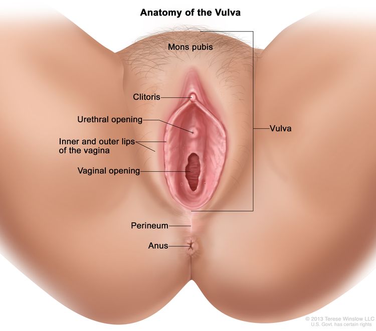


![Recombinant Anti-p53 antibody [E26] KO Tested (ab32389) | Abcam](https://www.abcam.com/ps/products/32/ab32389/Images/ab32389-389819-antip53antibodyimmunocytochemistryimmunofluorescencehap1human.jpg)


