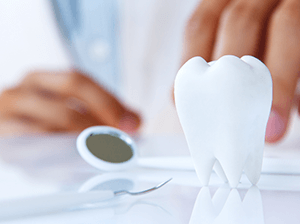
Salivary Gland Diseases
Sialadenitis
Salivary Glands
Sebaceous Gland Diseases
Lacrimal Apparatus
Submandibular Gland Diseases
Sjogren's Syndrome
Eyelid Diseases
Meibomian Glands
Salivary Glands, Minor
Submandibular Gland
Quantitative salivary gland scintigraphy. (1/117)
OBJECTIVE: Uptake of 99mTc-pertechnetate in salivary glands reflects intact salivary gland parenchyma. However, no standardized protocol for an accurate quantification of parenchymal function has been established so far. METHODS: In this paper we report on a validated acquisition protocol supplying a normal database for standardized quantitative salivary gland scintigraphy. RESULTS: The major advantage of salivary gland scintigraphy, as compared to other imaging modalities, is that both parenchymal function and excretion fraction of all four major salivary glands (i.e., parotid and submandibular glands) can be simultaneously quantified with a single intravenous injection. CONCLUSION: Quantitative salivary gland scintigraphy is demonstrated to be a suitable imaging modality for research applications in evaluating the effects of radioprotective drugs on salivary glands. Salivary gland scintigraphy is easy to perform, reproducible and well-tolerated by the patient. (+info)A zinc protein isolated from human parotid saliva. (2/117)
A zinc protein has been isolated and purified to apparent homogeneity from subjects with normal taste acuity by gel filtration and ion-exchange chromatography. The protein has a molecular weight of 37,000 and does not appear to have subunits. It is composed of 8% histidine residues and has 2 moles of zinc per mole of protein. (+info)Salivary gland P2 nucleotide receptors. (3/117)
The effects of ATP on salivary glands have been recognized since 1982. Functional and pharmacological studies of the P2 nucleotide receptors that mediate the effects of ATP and other extracellular nucleotides have been supported by the cloning of receptor cDNAs, by the expression of the receptor proteins, and by the identification in salivary gland cells of multiple P2 receptor subtypes. Currently, there is evidence obtained from pharmacological and molecular biology approaches for the expression in salivary gland of two P2X ligand-gated ion channels, P2Z/P2X7 and P2X4, and two P2Y G protein-coupled receptors, P2Y1 and P2Y2. Activation of each of these receptor subtypes increases intracellular Ca2+, a second messenger with a key role in the regulation of salivary gland secretion. Through Ca2+ regulation and other mechanisms, P2 receptors appear to regulate salivary cell volume, ion and protein secretion, and increased permeability to small molecules that may be involved in cytotoxicity. Some localization of the various salivary P2 receptor subtypes to specific cells and membrane subdomains has been reported, along with evidence for the co-expression of multiple P2 receptor subtypes within specific salivary acinar or duct cells. However, additional studies in vivo and with intact organ preparations are required to define clearly the roles the various P2 receptor subtypes play in salivary gland physiology and pathology. Opportunities for eventual utilization of these receptors as pharmacotherapeutic targets in diseases involving salivary gland dysfunction appear promising. (+info)Parotid swellings: report of 110 consecutive cases. (4/117)
Parotid swellings are uncommon. Over a twelve-year period, 110 cases of parotid swellings were treated at the Department of Plastic Surgery, Hospital Kuala Lumpur, of which 97 cases were histologically proven to be parotid tumours. 75% of these tumours were benign tumours, and 80% of the benign tumours were pleomorphic adenomas. Among the malignant tumours, 6 cases were adenoid cystic carcinoma and 5 were carcinoma ex-pleomorphic adenoma. There were equal number of male to female patients, with an age range of 14 to 83 years. There is a positive correlation between the final histological diagnosis and FNAC results in 74% of cases. Surgical treatment of choice for benign parotid tumours was near-total parotidectomy whilst for malignant tumours was total radical parotidectomy with sural nerve graft. (+info)Scintigraphic study of the major salivary glands in pediatric bone marrow transplant recipients. (5/117)
Total body irradiation (TBI) at bone marrow transplantation (BMT) is shown to cause salivary gland dysfunction in children. The aim of the investigation was to study the function of major salivary glands in long-term surviving children following treatment with TBI, using salivary gland scintigraphy (SGS). Thirteen patients (seven male, six female), who had received TBI before the age of 13 years and survived more than 4 years, participated in the study. A reference group of 10 patients (nine male, one female) was examined shortly before they were to undergo BMT. The mean age was 14.1 +/- 4.1 years in the TBI-treated group and 12.8 +/- 5.9 years in the reference group. Unstimulated and stimulated whole salivary secretion rates were measured for 15 and 5 min, respectively, before SGS was performed. The percentage of stimulated secretion was 44.7 +/- 18.1% in the TBI-treated group compared to 58.4 +/- 13.0% in the reference group (P = 0.0438). Slower reaccumulation after excretion was found in the TBI-treated patients compared to the reference group (P = 0. 0300). The function of the major salivary glands in long-term survivors treated with TBI at BMT before the age of 13 years was found to be diminished, as shown by the reduced trapping rate and reduced emptying capacity, compared to prior to BMT. Bone Marrow Transplantation (2000) 26, 775-779. (+info)Glandular and extraglandular expression of costimulatory molecules in patients with Sjogren's syndrome. (6/117)
OBJECTIVES: To investigate the expression and regulation of CD80, CD86, and CD28 costimulatory molecules in sialoadenitis and interstitial nephritis in patients with Sjogren's syndrome (SS). METHODS: Expression of CD80, CD86, and CD28 molecules was studied by immunohistochemical staining of lip biopsy specimens obtained from patients who had sialoadenitis associated with SS, and renal biopsy specimens obtained from patients who had interstitial nephritis associated with SS. To elucidate the mechanism of de novo expression of CD80 and CD86 antigens, their induction by cytokines in human salivary duct cell line (HSG) and renal cortical epithelial cells (HRCE) by cell enzyme linked immunosorbent assay (ELISA) was quantitatively investigated. RESULTS: In patients with severe sialoadenitis, CD80 and CD86 were strongly expressed on ductal epithelial cells. In contrast, these antigens were not found in the minor salivary glands of normal subjects or of patients with mild sialoadenitis. Some infiltrating cells expressed CD28. In patients who had interstitial nephritis associated with SS, some tubular epithelial cells expressed CD86 but not the CD80 antigen. Unstimulated HSG cells did not express CD80 or CD86. Interferon gamma (IFNgamma) consistently up regulated levels of CD80 and CD86. In contrast, tumour necrosis factor alpha (TNFalpha), interleukin 1beta (IL1beta), IL2, and IL4 had no effect on either CD80 or CD86 levels. Unstimulated HRCE did not express CD80 or CD86. IFNgamma consistently up regulated CD86 expression. No CD80 expression was found on tubular cells. TNFalpha, IL1beta, IL2, and IL4 had no discernible effects. CONCLUSIONS: Salivary ductal cells in patients with SS can express CD80 and CD86 costimulatory molecules in response to IFNgamma. Tubular epithelial cells in patients who have interstitial nephritis associated with SS express only CD86 molecules. In patients with SS, salivary ductal cells and tubular epithelial cells may activate infiltrating CD28 positive T lymphocytes by presenting antigens to T cells, potentially leading to tissue destruction. (+info)Pathogenesis of sialodacryoadenitis in gnotobiotic rats. (7/117)
The pathogenesis of sialodacryoadenitis was studied in gnotobiotic CD rats inoculated intranasally with the causal virus. Virus replication was detected sequentially in the nasopharynx, tracheobronchial tree, cervical lymph nodes, submaxillary and parotid salivary glands, exorbital gland, and Harderian gland. Acute rhinitis appeared within 2 days after inoculation, and salivary glands had lesions in 4 days. Early changes in salivary and exorbital glands were characterized by necrosis of ductal epithelium, which rapidly progressed to widespread acinar necrosis, marked inflammation, edema and total effacement of glandular architecture. Harderian glands also had massive necrosis of tubuloalveolar units. Repair in all glands was characterized by marked squamous metaplasia of tubuloalveolar units. Repair in all glands was characterized by marked squamous metaplasia of ducts. Neutralizing and complement-fixing antibodies were detected in 7 days, and there was a concomitant decrease in tissue-virus titers. There was no detectable evidence for hematogenous spread of virus or for retrograde infection by way of major salivary ducts. (+info)Comparative study of MR sialography and digital subtraction sialography for benign salivary gland disorders. (8/117)
BACKGROUND AND PURPOSE: MR sialography has become an alternative imaging technique for ductal salivary gland diseases. We compared the diagnostic accuracies of MR sialography and digital subtraction sialography in patients with successful completion of both examinations and benign salivary gland disorders. METHODS: In a prospective study, we attempted to examine salivary glands in 80 patients with clinically suspected diagnoses of sialadenitis and/or sialolithiasis. Each patient underwent digital subtraction sialography and MR sialography. MR sialography was obtained with a T2-weighted single-shot turbo spin-echo sequence (TR/TE 2800/1100 msec, acquisition time 7 seconds), with use of a quadrature head coil. Final diagnoses were confirmed by clinical follow-up and results of biopsy (n = 9) or surgery (n = 19). RESULTS: Failure rate was 5% (four of 80) for MR sialography and 14% (11 of 80) for digital subtraction sialography. Eighty-one salivary glands (48 parotid, 33 submandibular) in 65 patients were successfully visualized with both modalities. MR sialography depicted the main ductal system and first- and second-order branches, whereas digital subtraction sialography was able to depict third-order branches. Sensitivity and specificity to diagnose chronic sialadenitis were 70% and 98% with MR and 96% and 100% with digital subtraction sialography. MR sialography enabled diagnosis of sialolithiasis with a sensitivity of 80% and a specificity of 98% versus 90% and 98% for each with digital subtraction sialography. CONCLUSION: MR sialography with a heavily T2-weighted sequence is highly successful in the noninvasive visualization of the ductal system of major salivary glands. It is useful for diagnosing sialolithiasis and sialadenitis. Digital subtraction sialography, an invasive technique, had a substantial procedural failure rate, particularly for the submandibular duct. However, because of its higher spatial resolution, successfully completed digital subtraction sialography achieved superior diagnostic information compared with that of MR sialography. (+info)Salivary gland diseases refer to a group of conditions that affect the function and structure of the salivary glands. These glands are responsible for producing saliva, which helps in digestion, lubrication, and protection of the mouth and throat. The major salivary glands include the parotid, submandibular, and sublingual glands.
There are several types of salivary gland diseases, including:
1. Salivary Gland Infections: These are usually caused by bacteria or viruses that infect the gland, ducts, or surrounding tissues. The most common infection is called sialadenitis, which can cause pain, swelling, redness, and difficulty swallowing.
2. Salivary Gland Stones (Sialolithiasis): These are small, hard deposits that form in the ducts of the salivary glands, causing blockages and leading to swelling, pain, and infection.
3. Salivary Gland Tumors: Both benign and malignant tumors can develop in the salivary glands. Benign tumors are usually slow-growing and cause localized swelling, while malignant tumors may be more aggressive and spread to other parts of the body.
4. Salivary Gland Dysfunction: This refers to conditions that affect the production or flow of saliva, such as Sjogren's syndrome, radiation therapy, dehydration, or certain medications.
5. Autoimmune Disorders: Conditions like Sjogren's syndrome, lupus, and rheumatoid arthritis can affect the salivary glands and cause inflammation, dry mouth, and other symptoms.
6. Salivary Gland Trauma: Injuries to the face or neck can damage the salivary glands and lead to swelling, bleeding, or decreased function.
Proper diagnosis and treatment of salivary gland diseases require a thorough evaluation by a healthcare professional, often involving imaging studies, laboratory tests, and biopsies. Treatment options may include antibiotics, surgery, radiation therapy, or changes in medication or lifestyle.
Sialadenitis is a medical condition characterized by inflammation of the salivary gland. It can occur in any of the major salivary glands, including the parotid, submandibular, and sublingual glands. The inflammation may result from bacterial or viral infections, autoimmune disorders, or obstruction of the salivary ducts.
Acute sialadenitis is often caused by bacterial infections and can lead to symptoms such as pain, swelling, redness, and difficulty swallowing. Chronic sialadenitis, on the other hand, may be caused by recurrent infections, autoimmune disorders like Sjogren's syndrome, or stones in the salivary ducts. Symptoms of chronic sialadenitis can include intermittent swelling, pain, and dry mouth.
Treatment for sialadenitis depends on the underlying cause but may include antibiotics, anti-inflammatory medications, hydration, and massage of the salivary glands. In some cases, surgery may be necessary to remove obstructions or damaged tissue in the salivary gland.
Salivary glands are exocrine glands that produce saliva, which is secreted into the oral cavity to keep the mouth and throat moist, aid in digestion by initiating food breakdown, and help maintain dental health. There are three major pairs of salivary glands: the parotid glands located in the cheeks, the submandibular glands found beneath the jaw, and the sublingual glands situated under the tongue. Additionally, there are numerous minor salivary glands distributed throughout the oral cavity lining. These glands release their secretions through a system of ducts into the mouth.
Sebaceous gland diseases refer to conditions that affect the sebaceous glands, which are small glands in the skin that produce an oily substance called sebum. Sebum helps keep the skin and hair moisturized. Sebaceous gland diseases can cause a variety of symptoms, including skin inflammation, redness, pain, and the formation of bumps or cysts.
Some common types of sebaceous gland diseases include:
1. Acne: A common skin condition that occurs when the hair follicles become plugged with oil and dead skin cells, leading to whiteheads, blackheads, or pimples.
2. Seborrheic dermatitis: A skin condition that causes red, itchy, and flaky skin, often on the scalp, face, or chest.
3. Rosacea: A chronic skin condition that causes redness, pimples, and visible blood vessels on the face.
4. Sebaceous hyperplasia: A benign growth of the sebaceous glands that appears as a small, yellowish bump on the skin.
5. Sebaceous adenitis: A rare inflammatory disease that affects the sebaceous glands, causing hair loss and scaly skin.
6. Sebaceous carcinoma: A rare and aggressive form of skin cancer that develops in the sebaceous glands.
Treatment for sebaceous gland diseases depends on the specific condition and its severity. Treatments may include topical or oral medications, light therapy, or surgical removal of affected tissue. It is important to consult a healthcare provider for an accurate diagnosis and treatment plan.
Dacryocystitis is a medical condition that refers to the inflammation of the lacrimal sac, which is a small sac-like structure located in the inner corner of the eye near the nose. The lacrimal sac is responsible for draining tears from the eye into the nasal cavity.
Dacryocystitis can occur as a result of an infection or obstruction in the tear drainage system, leading to the accumulation of tears and other debris in the lacrimal sac. This can cause symptoms such as redness, swelling, pain, and tenderness in the affected area, as well as discharge from the eye or nose.
In some cases, dacryocystitis may be treated with antibiotics to clear up any infection. In more severe cases, surgery may be required to remove any blockages and improve tear drainage. If left untreated, dacryocystitis can lead to complications such as the formation of an abscess or damage to the eye.
Sweat gland diseases are medical conditions that affect the functioning or structure of sweat glands, leading to excessive sweating (hyperhidrosis), lack of sweating (anhydrosis), or abnormal sweating (e.g., foul-smelling sweat). There are two main types of sweat glands in humans: eccrine glands, which produce a watery sweat that helps regulate body temperature, and apocrine glands, which are located in the armpits and groin and produce a thicker, milky sweat that can mix with bacteria on the skin and cause body odor.
Some examples of sweat gland diseases include:
1. Hidradenitis suppurativa: A chronic skin condition characterized by inflammation and infection of the apocrine glands, leading to the formation of abscesses, nodules, and sinus tracts.
2. Primary focal hyperhidrosis: A condition that causes excessive sweating in specific areas of the body, such as the armpits, hands, feet, or face, without any underlying medical cause.
3. Secondary generalized hyperhidrosis: Excessive sweating that affects the entire body and is caused by an underlying medical condition, such as diabetes, thyroid disease, or obesity.
4. Cystic adenoma of the axilla: A benign tumor that arises from the apocrine glands in the armpit.
5. Eccrine nevus: A rare congenital condition characterized by an increased number of eccrine glands in a localized area of the skin, leading to excessive sweating.
6. Fox-Fordyce disease: A chronic inflammatory disorder that affects the apocrine glands, causing itchy papules and pustules in the armpits and groin.
7. Pachyonychia congenita: A rare genetic disorder characterized by thickened nails, palmoplantar keratoderma, and abnormalities of the eccrine glands, leading to excessive sweating and odor production.
Salivary gland neoplasms refer to abnormal growths or tumors that develop in the salivary glands. These glands are responsible for producing saliva, which helps in digestion, lubrication of food and maintaining oral health. Salivary gland neoplasms can be benign (non-cancerous) or malignant (cancerous).
Benign neoplasms are slow-growing and typically do not spread to other parts of the body. They may cause symptoms such as swelling, painless lumps, or difficulty swallowing if they grow large enough to put pressure on surrounding tissues.
Malignant neoplasms, on the other hand, can be aggressive and have the potential to invade nearby structures and metastasize (spread) to distant organs. Symptoms of malignant salivary gland neoplasms may include rapid growth, pain, numbness, or paralysis of facial nerves.
Salivary gland neoplasms can occur in any of the major salivary glands (parotid, submandibular, and sublingual glands) or in the minor salivary glands located throughout the mouth and throat. The exact cause of these neoplasms is not fully understood, but risk factors may include exposure to radiation, certain viral infections, and genetic predisposition.
The lacrimal apparatus is a complex system in the eye that produces, stores, and drains tears. It consists of several components including:
1. Lacrimal glands: These are located in the upper outer part of the eyelid and produce tears to keep the eye surface moist and protected from external agents.
2. Tear ducts (lacrimal canaliculi): These are small tubes that drain tears from the surface of the eye into the lacrimal sac.
3. Lacrimal sac: This is a small pouch-like structure located in the inner part of the eyelid, which collects tears from the tear ducts and drains them into the nasolacrimal duct.
4. Nasolacrimal duct: This is a tube that runs from the lacrimal sac to the nose and drains tears into the nasal cavity.
The lacrimal apparatus helps maintain the health and comfort of the eye by keeping it lubricated, protecting it from infection, and removing any foreign particles or debris.
Submandibular gland diseases refer to a group of disorders that affect the function or structure of the submandibular glands, which are salivary glands located beneath the jaw and produce saliva. These diseases can be categorized into inflammatory, infectious, obstructive, neoplastic (benign or malignant), and autoimmune disorders.
Some common submandibular gland diseases include:
1. Submandibular sialadenitis: Inflammation of the submandibular gland due to bacterial or viral infections, stones, or autoimmune conditions.
2. Salivary gland stones (sialolithiasis): Calcified deposits that obstruct the ducts leading from the submandibular gland, causing swelling and pain, especially during meals.
3. Submandibular gland tumors: Abnormal growths in the submandibular gland, which can be benign or malignant (cancerous). Malignant tumors may invade surrounding tissues and spread to other parts of the body.
4. Sjögren's syndrome: An autoimmune disorder that affects the exocrine glands, including the submandibular gland, leading to dry mouth and eyes.
5. IgG4-related disease: A systemic inflammatory condition characterized by the infiltration of IgG4-positive plasma cells into various organs, including the submandibular gland, causing swelling and damage.
6. Mikulicz's disease: A rare benign lymphoepithelial lesion that affects the salivary and lacrimal glands, including the submandibular gland, leading to enlargement and dryness of the affected glands.
7. Salivary gland dysfunction: Reduced or impaired saliva production due to aging, medications, radiation therapy, or systemic diseases, which can affect the submandibular gland.
Proper diagnosis and treatment of submandibular gland diseases require a thorough clinical evaluation, imaging studies, and sometimes biopsy or surgical intervention.
Sjögren's syndrome is a chronic autoimmune disorder in which the body's immune system mistakenly attacks its own moisture-producing glands, particularly the tear and salivary glands. This can lead to symptoms such as dry eyes, dry mouth, and dryness in other areas of the body. In some cases, it may also affect other organs, leading to a variety of complications.
There are two types of Sjögren's syndrome: primary and secondary. Primary Sjögren's syndrome occurs when the condition develops on its own, while secondary Sjögren's syndrome occurs when it develops in conjunction with another autoimmune disease, such as rheumatoid arthritis or lupus.
The exact cause of Sjögren's syndrome is not fully understood, but it is believed to involve a combination of genetic and environmental factors. Treatment typically focuses on relieving symptoms and may include artificial tears, saliva substitutes, medications to stimulate saliva production, and immunosuppressive drugs in more severe cases.
Eyelid diseases refer to a variety of medical conditions that affect the function and/or appearance of the eyelids. These can include structural abnormalities, such as entropion (inward turning of the eyelid) or ectropion (outward turning of the eyelid), as well as functional issues like ptosis (drooping of the upper eyelid). Other common eyelid diseases include blepharitis (inflammation of the eyelid margin), chalazion (a blocked oil gland in the eyelid), and cancerous or benign growths on the eyelid. Symptoms of eyelid diseases can vary widely, but often include redness, swelling, pain, itching, tearing, and sensitivity to light. Treatment for these conditions depends on the specific diagnosis and may range from self-care measures and medications to surgical intervention.
Meibomian glands are sebaceous glands located in the eyelids, specifically at the rim of the eyelid near the lashes. They produce an oily substance called meibum that forms the outermost layer of the tear film, helping to prevent evaporation and keep the eye surface lubricated. The Meibomian glands play a crucial role in maintaining the health and comfort of the eyes by providing stability to the tear film and protecting the eye from irritants and dryness.
Minor salivary glands are numerous small exocrine glands that produce saliva and are distributed throughout the oral cavity, nasal cavity, pharynx, larynx, and paranasal sinuses. They are classified as "minor" due to their smaller size compared to the three pairs of major salivary glands (parotid, submandibular, and sublingual). The minor salivary glands are primarily mucous glands, although some contain serous cells. They are responsible for producing approximately 5-10% of the total saliva in the mouth. These glands help moisten the oral cavity, protect the mucosal lining, and facilitate speaking, chewing, and swallowing.
The submandibular glands are one of the major salivary glands in the human body. They are located beneath the mandible (jawbone) and produce saliva that helps in digestion, lubrication, and protection of the oral cavity. The saliva produced by the submandibular glands contains enzymes like amylase and mucin, which aid in the digestion of carbohydrates and provide moisture to the mouth and throat. Any medical condition or disease that affects the submandibular gland may impact its function and could lead to problems such as dry mouth (xerostomia), swelling, pain, or infection.
 Salivary gland disease
 Diseases of oral cavity and salivary glands - ICD-10 Codes- Codify by AAPC
 Salivary Gland Disorders: MedlinePlus
 Salivary Abnormalities in Dentistry: Overview, Salivary Gland Anatomy, Salivation and Age
 NHS Royal Devon | Salivary Gland disease
 A Medicolegal Perspective on Salivary Gland Diseases - Medicolegal Partners Limited
 Google Drive) Manual of Salivary Gland Diseases PDF Free Download Archives
 Mouth health and salivary gland diseases: what you need to know
 Salivary stones: Symptoms, causes, and how to get rid of them
 OMFS Treatments & Services
 Essential Head and Neck Oncology and Surgery - Nova Science Publishers
 My Cat Won't Eat - Causes, What to Feed Them & When to Worry - Catster
 Salivary Duct Stones: Causes, Symptoms, and Diagnosis
 Elise Gervais | University at Albany
 Consultant A-Z
 Section: Human organs 2023 September
 Otolaryngology and Communication Enhancement | Alumni Fellows | Boston Children's Hospital
 General ENT Care | Johns Hopkins Otolaryngology-Head and Neck Surgery
 Advanced Search Results - Public Health Image Library(PHIL)
 People - The University of Nottingham
 Ear, Nose & Throat | NorthBay Health
![Recombinant Anti-p53 antibody [E26] KO Tested (ab32389) | Abcam](https://www.abcam.com/ps/products/32/ab32389/Images/ab32389-389819-antip53antibodyimmunocytochemistryimmunofluorescencehap1human.jpg)