Salivary Duct Calculi
Salivary Ducts
Postcholecystectomy Syndrome
Gallstones
Carcinoma, Ductal
Calculi
Urinary Calculi
Ureteral Calculi
Kidney Calculi
Urinary Bladder Calculi
Periplaneta
Dental Calculus
Salivary Glands
Salivary Gland Diseases
Submandibular Gland
Adenoma, Pleomorphic
Parotid Gland
Lithotripsy
Parotid sialolithiasis in Stensen's duct. (1/12)
Salivary duct lithiasis is a condition characterized by the obstruction of a salivary gland or its excretory duct due to the formation of calcareous concretions or sialoliths resulting in salivary ectasia and even provoking the subsequent dilation of the salivary gland. Sialolithiasis accounts for 30% of salivary diseases and most commonly involves the submaxillary gland (83 to 94%) and less frequently the parotid (4 to 10%) and sublingual glands (1 to 7%). The present study reports the case of a 45-year-old male patient complaining of bad breath and foul-tasting mouth at meal times and presenting with a salivary calculus in left Stensen's duct. Once the patient was diagnosed, the sialolith was surgically removed using local anesthesia. In this paper we have also updated a series of concepts related to the etiology, diagnosis and treatment of sialolithiasis. (+info)Modern management of obstructive salivary diseases. (2/12)
Over the last fifteen years, increasing public demand for minimally-invasive surgery and recent technological advances have led to the development of a number of conservative options for the therapeutic management of obstructive salivary disorders such as calculi and duct stenosis. These include extracorporeal shock-wave lithotripsy, sialoendoscopy, laser intra-corporeal lithotripsy, interventional radiology, the video-assisted conservative surgical removal of parotid and sub-mandibular calculi and botulinum toxin therapy. Each of these techniques may be used as a single therapeutic modality or in combination with one or more of the above-mentioned options, usually in day case or one-day case under local or general anaesthesia. The multi-modal approach is completely successful in about 80% of patients and reduces the need for gland removal in 3%, thus justifying the combination of, albeit, time-consuming and relatively expensive techniques as part of the modern and functional management of salivary calculi. With regard to the management of salivary duct anomalies, such as strictures and kinkings, interventional radiology with fluoroscopically controlled balloon ductoplasty seems to be the most suitable technique despite the use of radiation. Operative sialoendoscopy alone is the best therapeutic option for all mobile intra-luminal causes of obstruction, such as microliths, mucous plugs or foreign bodies, or for the local treatment of inflammatory conditions such as recurrent chronic parotitis or autoimmune salivary disorders. Finally, in the case of failure of one of the above techniques and regardless of the cause of obstruction, botulinum toxin injection into the parenchyma of the salivary glands using colour Doppler ultrasonographic monitoring should be considered before deciding on surgical gland removal. (+info)Microliths in the parotid of ferret investigated by electron microscopy and microanalysis. (3/12)
(+info)Extracellular Ca(2+) sensing in salivary ductal cells. (4/12)
(+info)Down's syndrome patient with multiple sialoliths in Stenson's duct. (5/12)
(+info)Sialolithiasis in a 10 year old child. (6/12)
(+info)Clinicopathological study of myoepithelial sialadenitis and chronic sialadenitis (sialolithiasis). (7/12)
To determine any overlap in pathological features between myoepithelial sialadenitis and chronic sialadenitis/sialolithiasis histological sections from 69 cases of myoepithelial sialadenitis (MESA) (n = 7) and chronic sialadenitis/sialolithiasis (n = 62) were reviewed over a 10 year period. Three of the cases with MESA contained calculi and four of those originally diagnosed as chronic sialadenitis/sialolithiasis showed epimyoepithelial island formation. The presence of calculi should not rule out a diagnosis of MESA, particularly in the parotid gland where calculi are uncommon; as the incidence of MESA may very well be underestimated and diagnosed as chronic sialadenitis, these patients, who are at increased risk of developing lymphoma, could be lost to follow up. (+info)Recurrent unilateral swelling of the parotid gland. (8/12)
The clinical features of 109 patients with recurrent unilateral parotid swelling (24 patients with Sjogren's disease were excluded) have been analysed to explore the best system of management. The cause was definitely a parotid duct calculus in 36 patients, and evidence is presented that the same diagnosis probably applied to another 59 patients. Features suggesting a diagnosis of calculus included age (greater than 29 years); duration of the attacks of pain (less than 24 hours); cessation of salivation on the affected side; and a spurt of saliva heralding the relief of symptoms. Only three patients in the definite calculus group (8.3%) had no physical signs. However, had physical examination not included inspection and palpation of the parotid duct and its orifice from within the mouth 75% of the proven calculi would have been missed. The intraoral and anteroposterior plain radiographs are likely to be helpful, and sialography even more so. A sialographic appearance of a stricture in the main duct with proximal dilatation is usually due to a claculus. It would appear that calculi are the cause of recurrent unilateral parotid swelling (after exclusion of Sjogren's disease) in an overwhelming proportion of patients with this symptom. (+info)Salivary duct calculi, also known as salivary gland stones or salivary duct stones, are small, hard deposits that form in the salivary glands or their ducts. These stones typically consist of calcium salts and other minerals, and they can range in size from tiny grains to larger pebbles.
Salivary duct calculi can cause a variety of symptoms, including pain, swelling, and difficulty swallowing. They may also lead to infection or inflammation of the salivary glands. In severe cases, surgery may be necessary to remove the stones and relieve the associated symptoms.
The formation of salivary duct calculi is thought to be related to a variety of factors, including dehydration, decreased saliva production, and changes in the composition of saliva. People who have certain medical conditions, such as gout or hyperparathyroidism, may also be at increased risk for developing these stones.
Salivary ducts are the excretory tubules that transport saliva from the major and minor salivary glands to the oral cavity. The main function of these ducts is to convey the salivary secretions, which contain enzymes and lubricants, into the mouth to aid in digestion, speech, and swallowing.
There are two pairs of major salivary glands: the parotid glands and the submandibular glands. Each pair has its own set of ducts. The parotid gland's saliva is drained through the parotid duct, also known as Stensen's duct, which opens into the oral cavity opposite the upper second molar tooth. The submandibular gland's saliva is transported through the submandibular duct, or Wharton's duct, which empties into the floor of the mouth near the base of the tongue.
Minor salivary glands are scattered throughout the oral cavity and pharynx, and their secretions are drained via small ducts directly into the oral mucosa.
Postcholecystectomy Syndrome is a condition that occurs in some patients following the surgical removal of the gallbladder (cholecystectomy). The syndrome encompasses a variety of symptoms such as abdominal pain, bloating, gas, indigestion, and diarrhea, which can be caused by several factors including:
1. Abnormal functioning or motility of the sphincter of Oddi (a muscle that controls the flow of bile and pancreatic juice into the small intestine)
2. Formation of gallstones in the bile ducts (choledocholithiasis)
3. Biliary dyskinesia (impaired functioning of the biliary tract muscles)
4. Persistent or recurrent infection or inflammation of the bile ducts (biliopathy)
5. Formation of abnormal bile-filled pouches (biliolethiasis or bile duct cysts)
6. Changes in bowel habits due to altered enterohepatic circulation of bile acids
The symptoms of Postcholecystectomy Syndrome can vary in severity and frequency, and they may appear soon after the surgery or develop months or even years later. The diagnosis of this condition typically involves a comprehensive medical evaluation, including a detailed history, physical examination, laboratory tests, and imaging studies such as ultrasound, CT scan, MRI, or endoscopic retrograde cholangiopancreatography (ERCP).
Treatment options for Postcholecystectomy Syndrome depend on the underlying cause of the symptoms and may include medications, dietary modifications, endoscopic procedures, or surgery. In some cases, the syndrome may resolve on its own without any specific treatment.
Gallstones are small, hard deposits that form in the gallbladder, a small organ located under the liver. They can range in size from as small as a grain of sand to as large as a golf ball. Gallstones can be made of cholesterol, bile pigments, or calcium salts, or a combination of these substances.
There are two main types of gallstones: cholesterol stones and pigment stones. Cholesterol stones are the most common type and are usually yellow-green in color. They form when there is too much cholesterol in the bile, which causes it to become saturated and form crystals that eventually grow into stones. Pigment stones are smaller and darker in color, ranging from brown to black. They form when there is an excess of bilirubin, a waste product produced by the breakdown of red blood cells, in the bile.
Gallstones can cause symptoms such as abdominal pain, nausea, vomiting, and bloating, especially after eating fatty foods. In some cases, gallstones can lead to serious complications, such as inflammation of the gallbladder (cholecystitis), infection, or blockage of the bile ducts, which can cause jaundice, a yellowing of the skin and eyes.
The exact cause of gallstones is not fully understood, but risk factors include being female, older age, obesity, a family history of gallstones, rapid weight loss, diabetes, and certain medical conditions such as cirrhosis or sickle cell anemia. Treatment for gallstones may involve medication to dissolve the stones, shock wave therapy to break them up, or surgery to remove the gallbladder.
Salivary gland neoplasms refer to abnormal growths or tumors that develop in the salivary glands. These glands are responsible for producing saliva, which helps in digestion, lubrication of food and maintaining oral health. Salivary gland neoplasms can be benign (non-cancerous) or malignant (cancerous).
Benign neoplasms are slow-growing and typically do not spread to other parts of the body. They may cause symptoms such as swelling, painless lumps, or difficulty swallowing if they grow large enough to put pressure on surrounding tissues.
Malignant neoplasms, on the other hand, can be aggressive and have the potential to invade nearby structures and metastasize (spread) to distant organs. Symptoms of malignant salivary gland neoplasms may include rapid growth, pain, numbness, or paralysis of facial nerves.
Salivary gland neoplasms can occur in any of the major salivary glands (parotid, submandibular, and sublingual glands) or in the minor salivary glands located throughout the mouth and throat. The exact cause of these neoplasms is not fully understood, but risk factors may include exposure to radiation, certain viral infections, and genetic predisposition.
Carcinoma, ductal refers to a type of cancer that begins in the milk ducts (tubes that carry milk from the breast to the nipple). It is most commonly found in the breast and is often referred to as "invasive ductal carcinoma" when it has spread beyond the ducts into the surrounding breast tissue. Ductal carcinoma can also occur in other organs, such as the pancreas, where it is called "pancreatic ductal adenocarcinoma." This type of cancer is usually aggressive and can metastasize (spread) to other parts of the body.
"Calculi" is a medical term that refers to abnormal concretions or hard masses formed within the body, usually in hollow organs or cavities. These masses are typically composed of minerals such as calcium oxalate, calcium phosphate, or magnesium ammonium phosphate, and can vary in size from tiny granules to large stones. The plural form of the Latin word "calculus" (meaning "pebble"), calculi are commonly known as "stones." They can occur in various locations within the body, including the kidneys, gallbladder, urinary bladder, and prostate gland. The presence of calculi can cause a range of symptoms, such as pain, obstruction, infection, or inflammation, depending on their size, location, and composition.
Urinary calculi, also known as kidney stones or nephrolithiasis, are hard deposits made of minerals and salts that form inside the urinary system. These calculi can develop in any part of the urinary system, which includes the kidneys, ureters, bladder, and urethra.
The formation of urinary calculi typically occurs when there is a concentration of certain substances, such as calcium, oxalate, uric acid, or struvite, in the urine. When these substances become highly concentrated, they can crystallize and form small seeds that gradually grow into larger stones over time.
The size of urinary calculi can vary from tiny, sand-like particles to large stones that can fill the entire renal pelvis. The symptoms associated with urinary calculi depend on the stone's size, location, and whether it is causing a blockage in the urinary tract. Common symptoms include severe pain in the flank, lower abdomen, or groin; nausea and vomiting; blood in the urine (hematuria); fever and chills; and frequent urge to urinate or painful urination.
Treatment for urinary calculi depends on the size and location of the stone, as well as the severity of symptoms. Small stones may pass spontaneously with increased fluid intake and pain management. Larger stones may require medical intervention, such as extracorporeal shock wave lithotripsy (ESWL), ureteroscopy, or percutaneous nephrolithotomy (PCNL) to break up or remove the stone. Preventive measures include maintaining adequate hydration, modifying dietary habits, and taking medications to reduce the risk of stone formation.
Ureteral calculi, also known as ureteric stones or ureteral stones, refer to the presence of solid mineral deposits (calculi) within the ureters, the tubes that transport urine from the kidneys to the bladder. These calculi can vary in size and composition, and their formation is often associated with conditions such as dehydration, urinary tract infections, or metabolic disorders. Ureteral calculi may cause symptoms like severe pain, hematuria (blood in the urine), and obstruction of urine flow, potentially leading to serious complications if left untreated.
Kidney calculi, also known as kidney stones, are hard deposits made of minerals and salts that form inside your kidneys. They can range in size from a grain of sand to a golf ball. When they're small enough, they can be passed through your urine without causing too much discomfort. However, larger stones may block the flow of urine, causing severe pain and potentially leading to serious complications such as urinary tract infections or kidney damage if left untreated.
The formation of kidney calculi is often associated with factors like dehydration, high levels of certain minerals in your urine, family history, obesity, and certain medical conditions such as gout or inflammatory bowel disease. Symptoms of kidney stones typically include severe pain in the back, side, lower abdomen, or groin; nausea and vomiting; fever and chills if an infection is present; and blood in the urine. Treatment options depend on the size and location of the stone but may include medications to help pass the stone, shock wave lithotripsy to break up the stone, or surgical removal of the stone in severe cases.
Urinary bladder calculi, also known as bladder stones, refer to the formation of solid mineral deposits within the urinary bladder. These calculi develop when urine becomes concentrated, allowing minerals to crystallize and stick together, forming a stone. Bladder stones can vary in size, ranging from tiny sand-like particles to larger ones that can occupy a significant portion of the bladder's volume.
Bladder stones typically form as a result of underlying urinary tract issues, such as bladder infection, enlarged prostate, nerve damage, or urinary retention. Symptoms may include lower abdominal pain, difficulty urinating, frequent urination, blood in the urine, and sudden, strong urges to urinate. If left untreated, bladder stones can lead to complications like urinary tract infections and kidney damage. Treatment usually involves surgical removal of the stones or using other minimally invasive procedures to break them up and remove the fragments.
"Periplaneta" is a genus name that refers to a group of large, winged insects commonly known as cockroaches. The two most common species in this genus are the American cockroach (Periplaneta americana) and the German cockroach (Periplaneta germantica). These insects are typically found in warm, humid environments and can often be seen scurrying across floors or walls in homes, restaurants, and other buildings. They are known to carry diseases and can cause allergies and asthma attacks in some people.
Parotid neoplasms refer to abnormal growths or tumors in the parotid gland, which is the largest of the salivary glands and is located in front of the ear and extends down the neck. These neoplasms can be benign (non-cancerous) or malignant (cancerous).
Benign parotid neoplasms are typically slow-growing, painless masses that may cause facial asymmetry or difficulty in chewing or swallowing if they become large enough to compress surrounding structures. The most common type of benign parotid tumor is a pleomorphic adenoma.
Malignant parotid neoplasms, on the other hand, are more aggressive and can invade nearby tissues and spread to other parts of the body. They may present as rapidly growing masses that are firm or fixed to surrounding structures. Common types of malignant parotid tumors include mucoepidermoid carcinoma, adenoid cystic carcinoma, and squamous cell carcinoma.
The diagnosis of parotid neoplasms typically involves a thorough clinical evaluation, imaging studies such as CT or MRI scans, and fine-needle aspiration biopsy (FNAB) to determine the nature of the tumor. Treatment options depend on the type, size, and location of the neoplasm but may include surgical excision, radiation therapy, and chemotherapy.
Dental calculus, also known as tartar, is a hardened deposit that forms on the surface of teeth. It's composed of mineralized plaque, which is a sticky film containing bacteria, saliva, and food particles. Over time, the minerals in saliva can cause the plaque to harden into calculus, which cannot be removed by brushing or flossing alone. Dental calculus can contribute to tooth decay and gum disease if not regularly removed by a dental professional through a process called scaling and root planing.
Salivary glands are exocrine glands that produce saliva, which is secreted into the oral cavity to keep the mouth and throat moist, aid in digestion by initiating food breakdown, and help maintain dental health. There are three major pairs of salivary glands: the parotid glands located in the cheeks, the submandibular glands found beneath the jaw, and the sublingual glands situated under the tongue. Additionally, there are numerous minor salivary glands distributed throughout the oral cavity lining. These glands release their secretions through a system of ducts into the mouth.
Salivary gland diseases refer to a group of conditions that affect the function and structure of the salivary glands. These glands are responsible for producing saliva, which helps in digestion, lubrication, and protection of the mouth and throat. The major salivary glands include the parotid, submandibular, and sublingual glands.
There are several types of salivary gland diseases, including:
1. Salivary Gland Infections: These are usually caused by bacteria or viruses that infect the gland, ducts, or surrounding tissues. The most common infection is called sialadenitis, which can cause pain, swelling, redness, and difficulty swallowing.
2. Salivary Gland Stones (Sialolithiasis): These are small, hard deposits that form in the ducts of the salivary glands, causing blockages and leading to swelling, pain, and infection.
3. Salivary Gland Tumors: Both benign and malignant tumors can develop in the salivary glands. Benign tumors are usually slow-growing and cause localized swelling, while malignant tumors may be more aggressive and spread to other parts of the body.
4. Salivary Gland Dysfunction: This refers to conditions that affect the production or flow of saliva, such as Sjogren's syndrome, radiation therapy, dehydration, or certain medications.
5. Autoimmune Disorders: Conditions like Sjogren's syndrome, lupus, and rheumatoid arthritis can affect the salivary glands and cause inflammation, dry mouth, and other symptoms.
6. Salivary Gland Trauma: Injuries to the face or neck can damage the salivary glands and lead to swelling, bleeding, or decreased function.
Proper diagnosis and treatment of salivary gland diseases require a thorough evaluation by a healthcare professional, often involving imaging studies, laboratory tests, and biopsies. Treatment options may include antibiotics, surgery, radiation therapy, or changes in medication or lifestyle.
The submandibular glands are one of the major salivary glands in the human body. They are located beneath the mandible (jawbone) and produce saliva that helps in digestion, lubrication, and protection of the oral cavity. The saliva produced by the submandibular glands contains enzymes like amylase and mucin, which aid in the digestion of carbohydrates and provide moisture to the mouth and throat. Any medical condition or disease that affects the submandibular gland may impact its function and could lead to problems such as dry mouth (xerostomia), swelling, pain, or infection.
A pleomorphic adenoma is a type of benign (non-cancerous) tumor that typically develops in the salivary glands, although they can also occur in other areas such as the nasopharynx and skin. "Pleomorphic" refers to the diverse appearance of the cells within the tumor, which can vary in size, shape, and arrangement.
Pleomorphic adenomas are composed of a mixture of epithelial and mesenchymal cells, which can form glandular structures, squamous (scale-like) cells, and areas that resemble cartilage or bone. These tumors tend to grow slowly and usually do not spread to other parts of the body.
While pleomorphic adenomas are generally not dangerous, they can cause problems if they become large enough to press on surrounding tissues or structures. In some cases, these tumors may also undergo malignant transformation, leading to a cancerous growth known as carcinoma ex pleomorphic adenoma. Surgical removal is the standard treatment for pleomorphic adenomas, and the prognosis is generally good with proper management.
The parotid gland is the largest of the major salivary glands. It is a bilobed, accessory digestive organ that secretes serous saliva into the mouth via the parotid duct (Stensen's duct), located near the upper second molar tooth. The parotid gland is primarily responsible for moistening and lubricating food to aid in swallowing and digestion.
Anatomically, the parotid gland is located in the preauricular region, extending from the zygomatic arch superiorly to the angle of the mandible inferiorly, and from the masseter muscle anteriorly to the sternocleidomastoid muscle posteriorly. It is enclosed within a fascial capsule and has a rich blood supply from the external carotid artery and a complex innervation pattern involving both parasympathetic and sympathetic fibers.
Parotid gland disorders can include salivary gland stones (sialolithiasis), infections, inflammatory conditions, benign or malignant tumors, and autoimmune diseases such as Sjögren's syndrome.
Lithotripsy is a medical procedure that uses shock waves or other high-energy sound waves to break down and remove calculi (stones) in the body, particularly in the kidneys, ureters, or gallbladder. The procedure is typically performed on an outpatient basis and does not require any incisions.
During lithotripsy, the patient lies on a cushioned table while a lithotripter, a device that generates shock waves, is positioned around the area of the stone. As the shock waves pass through the body, they break the stone into tiny fragments that can then be easily passed out of the body in urine.
Lithotripsy is generally a safe and effective procedure, but it may not be suitable for everyone. Patients with certain medical conditions, such as bleeding disorders or pregnancy, may not be able to undergo lithotripsy. Additionally, some stones may be too large or too dense to be effectively treated with lithotripsy. In these cases, other treatment options, such as surgery, may be necessary.
 Parotitis
List of MeSH codes (C07)
List of MeSH codes (C23)
Submandibular gland
Calculus (medicine)
Parotid gland
Salivary gland fistula
Xerostomia
Chronic sclerosing sialadenitis
Sialolithiasis
Salivary gland
Sialadenitis
Parotidectomy
Sialography
Sialoendoscopy
Extracorporeal shockwave therapy
Oral medicine
Medical ultrasound
List of Italian inventions and discoveries
ICD-9-CM Volume 3
Sialolithiasis11
- Salivary stones (also called sialolithiasis, or salivary duct calculus) are mainly made of calcium, but do not indicate any kind of calcium disorder. (wikipedia.org)
- 2 Sialolithiasis is the most common obstructive salivary gland disease, responsible for approximately 50% of cases. (cfp.ca)
- 1 , 6 Sialolithiasis involves formation of calculi in the ductal systems of the salivary glands and primarily affects the submandibular glands (80% to 90% of cases). (cfp.ca)
- After radiographical examination (mandibular occlusal radiograph), we found extensive radiopaque image in the ductal area, establishing therefore a diagnosis of sialolithiasis of Warton's duct. (bvsalud.org)
- Salivary stone also known as salivary calculi or sialolithiasis. (johorentspecialist.com)
- Sialolithiasis along with sialadenitis is one of the commonly occurring salivary gland disorders. (thieme-connect.de)
- Sialolithiasis is the most common disease of the salivary glands. (jrmds.in)
- Equine Sialolithiasis of the Distal Parotid Duct. (fortunepublish.com)
- Sialolithiasis accounts for the most etiology of salivary gland obstruction which leads to recurrent painful swelling of the involved gland which often exacerbates while eating. (entworld.org)
- Sialolithiasis is the most common disease of salivary glands 1 and is considered as one of the major causes of salivary gland dysfunction. (entworld.org)
- Sialolithiasis (Obstruction): This is an obstruction that is caused due to a stone (calculus) blocking the Wharton's duct (the duct that carries saliva to the mouth). (kingscollegehospitaldubai.com)
Major salivary glands5
- Parotitis is an inflammation of one or both parotid glands, the major salivary glands located on either side of the face, in humans. (wikipedia.org)
- The submandibular gland, along with the parotid and sublingual glands, comprise the major salivary glands. (medscape.com)
- The submandibular gland is the second largest (approximate weight, 10 g) of the major salivary glands (the parotid gland is the largest). (medscape.com)
- The function of the parotid gland and the other two major salivary glands is to produce and secrete saliva which is a substance that helps in digestion of food, lubrication of the mouth and inhibits dental decay. (kingscollegehospitaldubai.com)
- In contrast, tumours of the minor salivary glands and the other major salivary glands are often malignant. (kingscollegehospitaldubai.com)
Minor salivary5
- The minor salivary glands are scattered along the upper aerodigestive tract, including the lips, mucosa of the oral cavity, pharynx, and hard palate. (medscape.com)
- It is principally a disease of the submandibular gland (80-95% of cases), although it can occur in the parotid or minor salivary glands. (ndtv.com)
- It is commonly occurred in submandibular gland or duct and less commonly developed in parotid gland or even rarer in minor salivary gland or sublingual gland. (johorentspecialist.com)
- 2 The submandibular glands is most commonly involved followed by the parotid, sublingual and minor salivary glands. (entworld.org)
- The mucosa overlays several minor salivary glands. (medscape.com)
Saliva22
- Spit (saliva) is produced by the salivary glands in the mouth. (medlineplus.gov)
- The chemicals in saliva can form a hard crystal that can block the salivary ducts. (medlineplus.gov)
- When saliva cannot exit a blocked duct, it backs up into the gland. (medlineplus.gov)
- The salivary glands in mammals are exocrine glands that produce saliva . (wikidoc.org)
- Salivary glands release saliva that dilutes the acid found in ones stomach. (wikidoc.org)
- Direct sympathetic innervation of the salivary glands takes place via preganglionic nerves in the thoracic segments T1-T3 which synapse in the superior cervical ganglion with postganglionic neurons that release norepinephrine, which is then received by β-adrenergic receptors on the acinar and ductal cells of the salivary glands, leading to an increase in cyclic adenosine monophosphate (cAMP) levels and the corresponding increase of saliva secretion. (wikidoc.org)
- Medication that used to increase salivation e.g vitamin C or eating natural sour fruits such as orange or lemon may help to increase saliva formation and might be able to promote natural expulsion of the sialolith from the duct especially when the stone is small. (johorentspecialist.com)
- A salivary stone or salivary calculus (also known medically as a sialolith) is a small calcified mass of saliva, which builds up in the excretory duct of a salivary gland and may cause the duct to become blocked. (bund.de)
- However, if a stone grows larger and becomes lodged in the excretory duct, it prevents the saliva from flowing freely out of the duct, causing it to accumulate and flow back into the gland. (bund.de)
- The role of the salivary glands is to produce saliva, which is important for digestion. (bund.de)
- Saliva leaves the gland and enters the oral cavity through an excretory duct. (bund.de)
- This facilitates an accumulation of saliva, which in turn may lead to the formation of salivary stones. (bund.de)
- If the flow of saliva is thick and slow, more and more layers become deposited in the duct and the stone increases in size. (bund.de)
- Finally, the stone will block the excretory duct and saliva will become blocked in the tissues of the salivary gland. (bund.de)
- This may be because these glands produce a thicker, more calciferous consistency of saliva than the other salivary glands. (bund.de)
- In addition, the excretory ducts from these glands are very long and the saliva has to be transported upwards against gravity. (bund.de)
- The cause of sialadenitis is most commonly due to the salivary duct caliculi which is termed as sialolith or salivary stones which causes statis of saliva in the ducts. (thieme-connect.de)
- These are common areas because salivary duct openings continuously feed saliva (which includes calcium) into the mouth. (colgate.com)
- First there is a Salivary duct under the tongue that does nothing but squirt saliva on the backs of these teeth. (blogspot.com)
- Major salivary gland should be palpated and milked to assess salivary gland duct orifices patency and salivary flow and to evaluate the quality of saliva (eg, frothy versus serous). (medscape.com)
- The duct is responsible for transporting saliva from the gland to the mouth. (kingscollegehospitaldubai.com)
- Xerostomia (Dry Mouth): When the salivary glands are not functioning properly and stop or produce too little saliva, you can develop a dry mouth also medically known as xerostomia. (kingscollegehospitaldubai.com)
Stones29
- Salivary duct stones are deposits of minerals in the ducts that drain the salivary glands. (medlineplus.gov)
- Salivary duct stones are a type of salivary gland disorder . (medlineplus.gov)
- Salivary stones most often affect the submandibular glands. (medlineplus.gov)
- A new technique, called sialendoscopy, can diagnose and treat stones in the salivary gland duct using very small cameras and instruments. (medlineplus.gov)
- If stones become infected or come back often, you may need surgery to remove the salivary gland. (medlineplus.gov)
- Most of the time, salivary duct stones cause only pain or discomfort, and at times become infected. (medlineplus.gov)
- Contact your provider if you have symptoms of salivary duct stones. (medlineplus.gov)
- Presence of small calculi in the terminal salivary ducts (salivary sand), or stones (larger calculi) found in the larger ducts. (bvsalud.org)
- Stones can develop in the salivary gland ducts and these are known as sialoliths. (ndtv.com)
- Stones of the salivary glands occur in 1% of the population. (ndtv.com)
- Stones may be confirmed by palpation (feeling with the fingers) or purulent discharge from the glandular duct with massage. (ndtv.com)
- This case describes a rare coexistence of a cutaneous fistula with salivary stones located in the superficial lobe of the parotid gland that was removed under local anesthesia. (unicoc.edu.co)
- Initially, salivary endoscopy is performed to identify any stones. (medscape.com)
- Salivary stones may block the salivary flow, causing a painful swelling in the salivary gland. (bund.de)
- There are various ways to remove salivary stones. (bund.de)
- Salivary stones are often flushed out when the salivary flow is stimulated and assisted by means of massage. (bund.de)
- What are salivary stones? (bund.de)
- Salivary stones occur most commonly in the salivary gland located below the jaw bone (the submandibular gland) and usually on one side only. (bund.de)
- What are the symptoms of salivary stones? (bund.de)
- Salivary stones are between 2 and 10 millimeters large, which is roughly the size of a pea. (bund.de)
- Salivary stones may occur in several places within excretory ducts of the affected gland. (bund.de)
- Smaller salivary stones frequently cause no symptoms. (bund.de)
- Tiny calculi or bacteria are frequent triggers for salivary stones. (bund.de)
- Salivary stones are particularly likely to occur in the submandibular glands below the floor of the mouth. (bund.de)
- Which factors increase the risk of developing salivary stones? (bund.de)
- Salivary stones: symptoms, aetiology, biochemical composition and treatment. (thieme-connect.de)
- Biochemical composition of salivary stones in relation to stone- and patient-related factors. (thieme-connect.de)
- Stones may be encountered in any of the salivary glands but most frequently in the submandibular gland and its duct. (entworld.org)
- There are a number of medical conditions which can affect the salivary glands, such as dry mouth, stones, infections and tumours. (kingscollegehospitaldubai.com)
Obstruction4
- Chronic salivary gland enlargement is rarely painful unless there is obstruction or infection. (msdmanuals.com)
- Parotitis is usually caused by trauma or surgery of the salivary gland, radiation to the neck area involving the parotid gland followed subsequently by duct obstruction, or calculi of the salivary duct. (medscape.com)
- Cysts may be caused by obstruction of a duct by calculi, trauma, or foreign body. (nih.gov)
- Pain and swelling may be the cardinal signs and symptoms 6 which are more pronounced on anticipation of food due to the obstruction of salivary flow. (entworld.org)
Wharton's5
- Calculus in Wharton's duct. (nih.gov)
- Calculus in Wharton's duct, with inflammation of the salivary glands. (nih.gov)
- Secretions from the submandibular glands travel into the oral cavity via the submandibular duct (Wharton's duct). (teachmeanatomy.info)
- Siddika A, Ferdousi AM, Zaman S, Shahpor N. Removal of Unusually Large Torpedo Sialolith from Wharton's Duct under Local Analgesia: A Case Report. (unicoc.edu.co)
- The particular duct which is attached to the parotid gland is known as Wharton's duct. (kingscollegehospitaldubai.com)
Sialadenitis7
- Sources of information MEDLINE and PubMed databases were searched for English-language research on sialadenitis and other salivary gland disorders, as well as for relevant review articles and guidelines published between 1981 and 2021. (cfp.ca)
- Main message Sialadenitis refers to inflammation or infection of the salivary glands and is a condition that can be caused by a broad range of processes including infectious, obstructive, and autoimmune. (cfp.ca)
- Sialadenitis is inflammation or infection of the salivary glands that can present acutely or chronically. (cfp.ca)
- Acute suppurative sialadenitis is inflammation of the salivary glands caused by bacterial infection. (cfp.ca)
- Salivary stasis is one of the main precipitating factors of acute suppurative sialadenitis. (cfp.ca)
- Sialadenitis is an inflammatory disease of the salivary glands. (thieme-connect.de)
- Diagnostic utility of submandibular and labial salivary gland biopsy in IgG4-related sialadenitis. (thieme-connect.de)
Including the salivary glands1
- Researchers believe SLS may dry out and temporarily irritate oral membranes, including the salivary glands. (cdhp.org)
Management of salivary1
- Along with presenting the case report, this article also reviews the etiology, diagnosis, and various treatment modalities available for the management of salivary gland calculi depending on their site and size. (ijds.in)
Benign1
- Benign and surgery is excised as salivary gland malfunction. (sci-ed.org)
Involved salivary gland1
- Recurrent symptoms caused by salivary calculi may require surgical excision of the involved salivary gland. (ndtv.com)
Blockage3
- citation needed] Blockage of the main parotid duct, or one of its branches, is often a primary cause of acute parotitis, with further inflammation secondary to bacterial superinfection. (wikipedia.org)
- Salivary duct calculus may cause blockage of the ducts, causing pain and swelling of the gland. (wikidoc.org)
- Squamous metaplasia is usually the result of chronic irritation, but it can have other causes (e.g., hypovitamnosis A). In the salivary ducts, metaplasia of the normally cuboidal ductal epithelium to stratified squamous epithelium has been seen in response to chemicals, ionizing radiation, viral infections, vitamin A deficiency, and blockage of ducts by salivary calculi. (nih.gov)
Sialolith or salivary1
- The cause of the sialolith or salivary stone formation is usually unknown, however chronic infection of salivary gland, dehydration,chronic inflammation or sjogren's syndrome and increased local level of calcium would contribute to sialolith formation. (johorentspecialist.com)
Ductal system2
- A special cannula or #90 polyethylene tubing is inserted into the duct, and iodinated oil such as Ultravist (iopromide) oily Dionosil is injected into the ductal system. (medscape.com)
- The existence of calculus inside a salivary glands ductal system. (jrmds.in)
Inflammation5
- The parotid gland is the salivary gland most commonly affected by inflammation. (wikipedia.org)
- citation needed] Sjögren's syndrome: Chronic inflammation of the salivary glands may also be an autoimmune disease known as Sjögren's syndrome. (wikipedia.org)
- Treatment of the salivary stone includes medical treatment especially when inflammation and infection had set in. (johorentspecialist.com)
- This duct may become narrower naturally or due to an inflammation or injury. (bund.de)
- The location of the inflammation within the gland (acini or duct) should be indicated in the pathology narrative. (nih.gov)
Sublingual gland1
- The duct of the submaxillary gland and lingual nerve lie beneath and to the medial side of the sublingual gland. (co.ma)
Chronic5
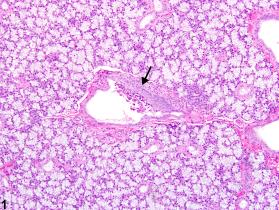
- Salivary gland, Duct - Metaplasia, Squamous in a male F344/N rat from a chronic study. (nih.gov)
- Another cause of damage to the salivary gland is from chronic alcoholism. (medscape.com)
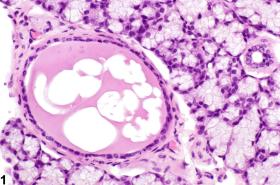
- Figure 1 Salivary gland, Duct - Cyst in a female B6C3F1 mouse from a chronic study. (nih.gov)
- A study by Wu et al indicated that technetium-99m ( 99m Tc) pertechnetate salivary gland scintigraphy can play an important part in the diagnosis of chronic obstructive parotitis and other salivary gland disorders. (medscape.com)
- A majority of sialoliths occur in the submandibular gland or its duct and are a common cause of acute and chronic infections. (ijds.in)
Secretions4
- Note that in this regard both parasympathetic and sympathetic stimuli result in an increase in salivary gland secretions. (wikidoc.org)
- Fallopian tube secretions, tears, breast milk, and sweat have amylases with a similar electrophoretic mobility of salivary isoamylase. (medscape.com)
- Their mixed serous and mucous salivary secretions are important for the lubrication of food during mastication to enable effective swallowing and aid digestion. (teachmeanatomy.info)
- Due to the viscous nature of its mucinous secretions, high calcium content, and tortuous duct, sialolith is relatively widespread (80%) in the submandibular salivary gland. (jrmds.in)
Commonly3
- Calcium deposits"- known more commonly as tartar or calculus - are a hardened layer of plaque or dental biofilm. (colgate.com)
- Sialoliths are hard concretions composed primarily of calcium carbonate and organic matter that develop within a salivary duct or much less commonly a gland [1]. (fortunepublish.com)
- In man, sialoliths are found most often in the submandibular and parotid glands and less commonly in their associated ducts [9]. (fortunepublish.com)
Disorders3
- Inflammatory disorders of the salivary glands. (medlineplus.gov)
- 1 - 3 Table 1 describes common salivary gland disorders. (cfp.ca)
- Salivary calculi are a common cause of salivary gland disorders, and they can affect any of the salivary glands at any age. (jrmds.in)
Recurrent1
- Submandibular gland excision is a common surgical procedure indicated for conditions such as submandibular gland neoplasia or recurrent calculi . (teachmeanatomy.info)
Excretory ducts2
- These drain into ducts situated between the lobes of the gland (called interlobar ducts or excretory ducts). (wikidoc.org)
- Humans have six large salivary glands, which are connected to the oral cavity by means of excretory ducts. (bund.de)
Portion of the duct2
- Critical) This program presents the surgical approach to the removal of a simple sialolith from the distal portion of the duct of the submandibular gland. (nih.gov)
- Removal of the Mandibular and Sublingual Salivary Glands and Associated Portion of the Duct. (vetsoftherockies.com)
Patency2
- Surgical removal was performed under local anesthesia , with restoration of patency of the ducts and normal salivary flow. (bvsalud.org)
- Abdominal Explore, Cholecystectomy (Removal Of Gallbladder), +/- Duodenotomy (Incision Into the Small Intestine) to Evaluate For Patency of Common Bile Duct, and Liver Biopsy. (vetsoftherockies.com)
Submaxillary duct2
- A sialogram is a radiocontrast study of a salivary duct ( parotid duct , submaxillary duct , Major sublingual duct ). (wikidoc.org)
- Immediately on either side of the part of the frenulum is the orifice of the submaxillary duct. (co.ma)
Tumors2
- Tumors of the salivary glands may occur. (wikidoc.org)
- Lee W-H, Tseng T-M, Hsu H-T, Lee F-P, Hung S-H, Chen P-Y. Salivary gland tumors: A 20-year review of clinical diagnostic accuracy at a single center. (unicoc.edu.co)
Mucous1
- The mucous membrane at the anterior part of the floor of each alveolo-glossal cus is thrown into a slight elevation, which overlies, and is caused by, the responding sublingual salivary gland. (co.ma)
Diagnosis2
- This patient provides illustration of the difficulty there may be in establishing the diagnosis of parotid calculus. (unicoc.edu.co)
- On the basis of clinical and radiological findings, a diagnosis of left submandibular duct sialolith was made. (entworld.org)
Parotid calculi2
- The majority of submandibular sialoliths are radiopaque, whereas this applies to only a minority of parotid calculi. (ndtv.com)
- The formation of calcific concretions in the salivary duct or glands is a common disorder, but parotid calculi represent only a small proportion of all salivary calculi. (unicoc.edu.co)
Glands located1
- The submandibular glands are bilateral salivary glands located in the face. (teachmeanatomy.info)
Parotitis1
- Bacterial parotitis presents as a unilateral swelling, where the gland is swollen and tender and usually produces pus at the Stensen's duct. (wikipedia.org)
Amylase activity1
- [13] The greatest S-type amylase activity is in salivary glands, where it initiates the hydrolysis of starches while food is in the mouth and esophagus. (statpearls.com)
Tortuous duct1
- 4 The submandibular gland is most frequently involved because of its anatomic location, long, tortuous duct with a narrow orifice compared to the main portion of duct. (entworld.org)
Epithelium1
- Squamous metaplasia of the salivary duct should be diagnosed and graded based on the number of areas involved and the thickness of the squamous epithelium. (nih.gov)
Removal4
- Transoral removal of an obstructing stone may be accomplished if the stone is situated near the duct opening. (ndtv.com)
- Drage NA, Brown JE, Escudier MP, McGurk M. Interventional radiology in the removal of salivary calculi. (unicoc.edu.co)
- Stone impacted over the submandibular duct on the floor of mouth could be removed via marsupiliazation of the submandibular duct and removal of the stone. (johorentspecialist.com)
- Perineal urethrostomy and +/- cystotomy for removal of cystic calculi if warranted. (vetsoftherockies.com)
Autonomic nervou1
- Salivary glands are innervated, either directly or indirectly, by the parasympathetic and sympathetic arms of the autonomic nervous system . (wikidoc.org)
Pancreas5
- Among healthy individuals, the pancreas and the salivary glands account for almost all the serum amylase, 40%-45% from the pancreas and 55%-60% from the salivary glands. (medscape.com)
- The pancreas and salivary glands contain amylase concentrations that are several orders of magnitude greater than other organs. (medscape.com)
- Electrophoresis shows that serum amylase is of two main types, as follows: (1) P-type amylase from the pancreas, and (2) S-type amylase from the salivary glands. (medscape.com)
- Amylase is a digestive enzyme predominantly secreted by the pancreas and salivary glands and found in other tissues at very small levels. (statpearls.com)
- Amylase has a wide tissue distribution, with the highest activities of the P and S-types being found in the exocrine pancreas and salivary glands, respectively. (statpearls.com)
Gland disease1
- citation needed] HIV-associated salivary gland disease can involve many diseases but often presents as enlargement of the parotid gland and a dry mouth. (wikipedia.org)
Exocrine glands1
- Salivary, lacrimal, and other exocrine glands become infiltrated with CD4 + T cells and with some B cells. (msdmanuals.com)
Acute1
- Acute viral and bacterial infections of the salivary glands. (thieme-connect.de)
Common bile1
- In patients presenting with biliary-type abdominal pain, a 3-fold increase in serum amylase levels that returns to normal within 48-72 hours suggests stone passage through the common bile duct. (medscape.com)
Infection3
- Other causes can be duct stricture (narrowing of the duct), infection or injury. (wikipedia.org)
- results in difficulty chewing and swallowing, secondary Candida infection, tooth decay, and calculi in the salivary ducts. (msdmanuals.com)
- Mumps (Viral) Infection and the parotid gland: Mumps is the most common viral infection of the salivary gland and it causes enlargement of both parotid glands. (kingscollegehospitaldubai.com)
Cysts2
- Salivary gland duct cysts tend to involve one or a few ducts and usually have a circular profile (Figure 1), whereas ductular dilation of salivary glands is typically more generalized (involving multiple ducts), and the ducts tend to have an irregular profile (see Salivary gland, Duct - Dilation). (nih.gov)
- Duct cysts should be diagnosed but are generally not graded unless there is a treatment-related effect on the size of the cysts. (nih.gov)
Lacrimal glands1
- 1999. Salivary, Harderian, and lacrimal glands. (nih.gov)
Disorder1
- Figure 1 provides an overview of how to diagnose and manage patients who present with suspected salivary gland disorder. (cfp.ca)
Biliary1
- Risk factors for developing post-ERCP hyperamylasemia appear to include difficult cannulation and pancreatography, and age younger than 60 years, as well as a previous history of diabetes, biliary duct stent placement, and nasobiliary drainage. (medscape.com)
Pancreatic3
- Elevated amylase can be seen in a variety of conditions, including pancreatic disease, salivary disease, decreased metabolic clearance, intestinal disease, and macroamylasemia. (statpearls.com)
- P-type amylase is synthesized by pancreatic acinar cells and secreted into the intestinal tract via the pancreatic duct system. (statpearls.com)
- Note stool chart and pancreatic ducts. (sci-ed.org)