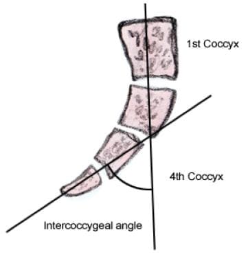
Coccyx
Sacrum
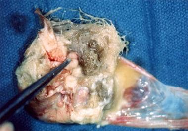
Chordoma
Teratoma
Perforator Flap
Effects of short term sacral nerve stimulation on anal and rectal function in patients with anal incontinence. (1/166)
BACKGROUND: Some patients with faecal incontinence are not amenable to simple surgical sphincter repair, due to sphincter weakness in the absence of a structural defect. AIMS: To evaluate the efficacy and possible mode of action of short term stimulation of sacral nerves in patients with faecal incontinence and a structurally intact external anal sphincter. PATIENTS: Twelve patients with faecal incontinence for solid or liquid stool at least once per week. METHODS: A stimulating electrode was placed (percutaneously in 10 patients, operatively in two) into the S3 or S4 foramen. The electrode was left in situ for a minimum of one week with chronic stimulation. RESULTS: Evaluable results were obtained in nine patients, with early electrode displacement in the other three. Incontinence ceased in seven of nine patients and improved notably in one; one patient with previous imperforate anus and sacral agenesis had no symptomatic response. Stimulation seemed to enhance maximum squeeze pressure but did not alter resting pressure. The rectum became less sensitive to distension with no change in rectal compliance. Ambulatory studies showed a possible reduction in rectal contractile activity and diminished episodes of spontaneous anal relaxation. CONCLUSIONS: Short term sacral nerve stimulation notably decreases episodes of faecal incontinence. The effect may be mediated via facilitation of striated sphincter muscle function, and via neuromodulation of sacral reflexes which regulate rectal sensitivity and contractility, and anal motility. (+info)Subcutaneous sacrococcygeal myxopapillary ependymoma. (2/166)
We report a case of myxopapillary ependymoma presenting as a primary tumor of the subcutaneous tissue in the sacrococcygeal region. The mass was large, well-encapsulated, lobulated, and multiseptated, with varying signal intensity on T1- and T2-weighted MR images caused by hemorrhagic necrosis, blood degradation products, and calcification. Only a small viable portion enhanced after administration of contrast material. Multiple lobules formed from fibrous septa and dystrophic calcification also characterize this tumor. (+info)Chemical activation of cervical cell bodies: effects on responses to colorectal distension in lumbosacral spinal cord of rats. (3/166)
We have shown that stimulation of cardiopulmonary sympathetic afferent fibers activates relays in upper cervical segments to suppress activity of lumbosacral spinal cells. The purpose of this study was to determine if chemical excitation (glutamate) of upper cervical cell bodies changes the spontaneous activity and evoked responses of lumbosacral spinal cells to colorectal distension (CRD). Extracellular potentials were recorded in pentobarbital-anesthetized male rats. CRD (80 mmHg) was produced by inflating a balloon inserted in the descending colon and rectum. A total of 135 cells in the lumbosacral segments (L(6)-S(2)) were activated by CRD. Seventy-five percent (95/126) of tested cells received convergent somatic input from the scrotum, perianal region, hindlimb, and tail; 99/135 (73%) cells were excited or excited/inhibited by CRD; and 36 (27%) cells were inhibited or inhibited/excited by CRD. A glutamate (1 M) pledget placed on the surface of C(1)-C(2) segments decreased spontaneous activity and excitatory CRD responses of 33/56 cells and increased spontaneous activity of 13/19 cells inhibited by CRD. Glutamate applied to C(6)-C(7) segments decreased activity of 10/18 cells excited by CRD, and 9 of these also were inhibited by glutamate at C(1)-C(2) segments. Glutamate at C(6)-C(7) increased activity of 4/6 cells inhibited by CRD and excited by glutamate at C(1)-C(2) segments. After transection at rostral C(1) segment, glutamate at C(1)-C(2) still reduced excitatory responses of 7/10 cells. Further, inhibitory effects of C(6)-C(7) glutamate on excitatory responses to CRD still occurred after rostral C(1) transection but were abolished after a rostral C(6) transection in 4/4 cells. These data showed that C(1)-C(2) cells activated with glutamate primarily produced inhibition of evoked responses to visceral stimulation of lumbosacral spinal cells. Inhibition resulting from activation of cells in C(6)-C(7) segments required connections in the upper cervical segments. These results provide evidence that upper cervical cells integrate information that modulates activity of distant spinal neurons responding to visceral input. (+info)Non-linear membrane properties of sacral sphincter motoneurones in the decerebrate cat. (4/166)
1. Responses to pudendal afferent stimulation and depolarizing intracellular current injection were examined in sacral sphincter motoneurones in decerebrate cats. 2. In 16 animals examined, 2-10 s trains of electrical stimulation of pudendal afferents evoked sustained sphincter motoneurone activity lasting from 5 to >50 s after stimulation. The sustained response was observed in: 11 animals in the absence of any drugs; two animals after the intravenous administration of 5-hydroxytryptophan (5-HTP; <= 20 mg kg-1); one animal in which methoxamine was perfused onto the ventral surface of the exposed spinal cord; and two animals following the administration of intravenous noradrenergic agonists. 3. Extracellular and intracellular recordings from sphincter motoneurones revealed that the persistent firing evoked by afferent stimulation could be terminated by motoneurone membrane hyperpolarization during micturition or by intracellular current injection. 4. Intracellular recordings revealed that 22/40 sphincter motoneurones examined displayed a non-linear, steep increase in the membrane potential in response to depolarizing ramp current injection. The mean voltage threshold for this non-linear membrane response was -43 +/- 3 mV. Five of the 22 cells displaying the non-linear membrane response were recorded prior to the administration of 5-HTP; 17 after the intravenous administration of 5-HTP (<= 20 mg kg-1). 5. It is concluded that sphincter motoneurones have a voltage-sensitive, non-linear membrane response to depolarization that could contribute to sustained sphincter motoneurone firing during continence. (+info)Combined anomaly of intramedullary arteriovenous malformation and lipomyelomeningocele. (5/166)
We report a rare situation in which a lipomyelomeningocele and an intramedullary arteriovenous malformation (AVM) occurred together at the T11-L1 level in a 44-year-old man. MR images showed a hypervascular lesion intradurally and a fatty component extradurally. Spinal angiography revealed this lesion to be an intramedullary AVM with multiple feeding arteries from the right T12 and left T10 intercostal artery and the left L1 lumbar artery, drained by tortuous, dilated, perimedullary veins. (+info)Sacral chordoma--a case report. (6/166)
Chordoma, a rare malignant tumour of early adulthood, rarely presents in children. We report such a case of rare malignant tumour which was diagnosed in the first decade of life. (+info)Isolated cardiac metastasis from sacral chordoma. (7/166)
A 64-year-old woman presented with right heart failure caused by a cardiac tumor centered in the free wall of the right ventricle, accompanied by pericardial effusion. A match between the biopsy specimen and tissue removed 4 years earlier resulted in the diagnosis of a cardiac metastasis from a chordoma. Immunohistochemical staining was also useful in establishing the diagnosis. To alleviate the right ventricular outflow obstruction, a palliative operation was planned, resecting the tumor and performing a right ventriculoplasty, which was cancelled due to the extent of infiltration of the tumor, and instead a right atrium to pulmonary artery shunt was attempted using a vascular prosthesis, only to fail due to an inability to maintain blood flow through the prosthesis. Presently there are no definitive treatment options available, and some palliative chemotherapy is being performed. Single cardiac metastases from a chordoma are extremely rare. (+info)Infantile arachnoid cyst compressing the sacral nerve root associated with spina bifida and lipoma--case report. (8/166)
A 2-year-old boy presented with a rare sacral arachnoid cyst manifesting as gait disturbance. Neuroimaging revealed an intradural cyst in the sacral nerve root sheath associated with spina bifida occulta and a lipoma at the same level. At surgery, the conus medullaris was situated at the L-1 level and not tethered. The highly pressurized arachnoid cyst had exposed the dural sheath of the left S-2 nerve root and compressed the adjacent nerves. An S-2 nerve root pierced through the cyst. There was no communication between the cyst and spinal arachnoid space. We thought the one-way valve mechanism had contributed to the cyst enlargement and the nerve compression. Radical resection of the cyst was not attempted. A cyst-subarachnoid shunt was placed to release the intracystic pressure. Postoperatively, his gait disturbance improved and no deterioration occurred during the 4-year follow up. Both tethered cord syndrome and sacral arachnoid cyst in the nerve root sheath should be considered in pediatric progressive gait disturbance. Cyst-subarachnoid shunt is an alternative method to cyst resection or fenestration to achieve neurological improvement. (+info)The sacrococcygeal region is the lower part of the back where the spine ends, specifically referring to the area where the sacrum (a triangular bone at the base of the spine formed by the fusion of several vertebrae) meets the coccyx (also known as the tailbone). This region is located at the very bottom of the spine and is susceptible to injury or trauma due to its position and role in supporting the body's weight. It is also a common site for birth defects, particularly in newborns.
The coccyx, also known as the tailbone, is the small triangular bone at the bottom of the spine in humans and other primates. It is formed by the fusion of several small vertebrae and serves to attach muscles and ligaments in the pelvic region. The coccyx can be a source of pain and discomfort if it is injured or becomes inflamed.
A pilonidal sinus is a small hole or tunnel in the skin that usually develops in the cleft at the top of the buttocks. It can be painful and may become infected, causing symptoms such as redness, swelling, pain, and pus discharge. The condition often affects young adults and is more common in men than women.
The term "pilonidal" comes from the Latin words "pilus," meaning hair, and "nidus," meaning nest. This refers to the fact that the sinus often contains hairs that have become embedded in the skin. The exact cause of pilonidal sinuses is not known, but they are thought to develop as a result of ingrown hairs or chronic irritation in the affected area.
Treatment for pilonidal sinuses typically involves surgical removal of the sinus and any associated hair follicles. In some cases, this may be done using a minor procedure that can be performed in a doctor's office. More complex cases may require hospitalization and a more extensive surgical procedure. After surgery, patients will need to take steps to prevent the sinus from recurring, such as keeping the area clean and avoiding prolonged periods of sitting or driving.
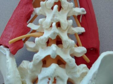
The sacrum is a triangular-shaped bone in the lower portion of the human vertebral column, located between the lumbar spine and the coccyx (tailbone). It forms through the fusion of several vertebrae during fetal development. The sacrum's base articulates with the fifth lumbar vertebra, while its apex connects with the coccyx.
The sacrum plays an essential role in supporting the spine and transmitting weight from the upper body to the pelvis and lower limbs. It also serves as an attachment site for various muscles and ligaments. The sacral region is often a focus in medical and chiropractic treatments due to its importance in spinal stability, posture, and overall health.
A chordoma is a rare, slow-growing tumor that typically develops in the bones of the spine or skull. These tumors originate from remnants of the notochord, a structure that forms during embryonic development and eventually becomes part of the spinal cord. Chordomas are usually low-grade malignancies but can be aggressive and locally invasive, potentially causing pain, neurological symptoms, or structural damage to the spine or skull. Treatment typically involves surgical resection, often combined with radiation therapy.
A teratoma is a type of germ cell tumor, which is a broad category of tumors that originate from the reproductive cells. A teratoma contains developed tissues from all three embryonic germ layers: ectoderm, mesoderm, and endoderm. This means that a teratoma can contain various types of tissue such as hair, teeth, bone, and even more complex organs like eyes, thyroid, or neural tissue.
Teratomas are usually benign (non-cancerous), but they can sometimes be malignant (cancerous) and can spread to other parts of the body. They can occur anywhere in the body, but they're most commonly found in the ovaries and testicles. When found in these areas, they are typically removed surgically.
Teratomas can also occur in other locations such as the sacrum, coccyx (tailbone), mediastinum (the area between the lungs), and pineal gland (a small gland in the brain). These types of teratomas can be more complex to treat due to their location and potential to cause damage to nearby structures.
A perforator flap is a type of surgical tissue transfer that involves the relocation of skin, fat, and sometimes muscle or fascia (the layer of connective tissue surrounding muscles) based on a specific blood vessel called a perforator. These vessels pass through the deeper fascial layers to supply the overlying skin and subcutaneous tissues.
Perforator flaps are designed to minimize donor site morbidity by preserving the underlying muscle and maximizing functional outcomes, as only the necessary amount of tissue is taken along with the perforator vessel. The versatility and reliability of these flaps have expanded their application in various reconstructive procedures, such as breast reconstruction, extremity reconstruction, and head and neck reconstruction.
The success of a perforator flap depends on careful preoperative planning, precise identification, and preservation of the perforating vessels during surgery. Commonly used techniques for perforator flaps include the deep inferior epigastric artery perforator (DIEP) flap, superior gluteal artery perforator (SGAP) flap, and anterolateral thigh (ALT) perforator flap.
Spinal neoplasms refer to abnormal growths or tumors found within the spinal column, which can be benign (non-cancerous) or malignant (cancerous). These tumors can originate in the spine itself, called primary spinal neoplasms, or they can spread to the spine from other parts of the body, known as secondary or metastatic spinal neoplasms. Spinal neoplasms can cause various symptoms, such as back pain, neurological deficits, and even paralysis, depending on their location and size. Early diagnosis and treatment are crucial to prevent or minimize long-term complications and improve the patient's prognosis.
 Sacrococcygeal symphysis - Wikipedia
Pure yolk sac tumor of sacrococcygeal region
 Article - Billing and Coding: Sacroiliac Joint Injections and Procedures (A59233)
 Congenital Spinal Deformity: Background, Embryology of Spine, Pathophysiology
 Sacrococcygeal teratoma - Getting a Diagnosis - Genetic and Rare Diseases Information Center
2012 ICD-9-CM Diagnosis Code 724.6 : Disorders of sacrum
Draft Article - Billing and Coding: Trigger Point Injections (TPI) (DA57701)
 IndexCat
M54.08 Panniculitis aff regions of neck/bk, sacr/sacrocygl region - ICD-10-CM Diagnosis Codes
 Other Special Needs Older Grant - Page 3 - Reece's Rainbow
Intracranial metastasis from a sacrococcygeal chordoma. Case report.
Epidural Nerve Block: Overview, Indications, Contraindications
 Local Anesthetic Techniques For Perineal Procedures
 Contributions to the Study of Transitional Vertebrae in Dogs - WSAVA2009 - VIN
 Proctology | KARL STORZ Endoskope | Poland
Pediatrics | KARL STORZ Endoskope | Bulgaria
 Q&A: Avoidable or Unavoidable Pressure Injury? | WoundSource
Combined laparoscopic and posterior approach resection of large sacrococcygeal cystic teratoma | Surgical Case Reports | Full...
 Orthopedic Item
 Medical Imaging | Hospital Lusíadas Lisboa
 Primary congenital sacrococcygeal neuroblastoma: A case report with immunohistochemical study and review of literature
 Saddle Region Pain
 Cunningham's textbook of anatomy - Daniel John Cunningham - Google Livres
DeCS 2016 - June 12, 2016 version
 DeCS 2019 - June 12, 2019 version
DeCS 2018 - July 31, 2018 version
DeCS 2017 - July 04, 2017 version
DeCS 2020 - June 23, 2020 version
 NCRG | E-Referral Allied
 Pain in back - lookformedical.com
Pilonidal Sinus11
- Introduction Pilonidal sinus disease is a chronic inflammatory condition typically found in the sacrococcygeal region. (researchgate.net)
- The pilonidal sinus is an acute or chronic inflammatory process in the subcutaneous adipose tissue, often occurring in the sacrococcygeal region 1 . (karlstorz.com)
- Is Histopathological Examination Necessary After Sacrococcygeal Pilonidal Sinus Excision? (ac.ir)
- Background: Pilonidal sinus is a common disease that usually occurs in the natal cleft in the sacrococcygeal region. (ac.ir)
- This study aimed to evaluate the pathology results of patients who underwent sacrococcygeal pilonidal sinus excision in our clinic. (ac.ir)
- Pilonidal sinus specimen pathologies excised from the sacrococcygeal region were examined. (ac.ir)
- Uylaş U, Gündoğdu R. Is Pathological Assessment Necessary for Excised Sacrococcygeal Pilonidal Sinus Specimens? (ac.ir)
- Study of 4085 Sacrococcygeal Pilonidal Sinus Diseases' Pathological Results in 12 Years. (ac.ir)
- Aim: Pilonidal sinus is a common disease that affects generally younger patients and occurs mostly in the sacro-coccygeal region. (eurasianmedicine.com)
- Pilonidal cyst (Synonyms: pilonidal sinus, epithelial coccygeal course) is a congenital anomaly of the development of the skin of the sacrococcygeal region associated with incomplete reduction of the vestigial caudal ligament. (pain-relief-med.com)
- The condition is also known by other names like Sacrococcygeal Fistula and Pilonidal Sinus. (hxbenefit.com)
Teratoma3
- Sacrococcygeal teratoma (SCT) is a type of tumor known as a teratoma that develops at the base of the coccyx (tailbone) and is thought to be derived from the primitive streak . (iiab.me)
- We present perinatal findings and molecular cytogenetic characterization of a prenatally detected sacrococcygeal teratoma associated with mosaic r(21). (symptoma.com)
- This is the first report of mosaic r(21) presenting with a fetal sacrococcygeal teratoma. (symptoma.com)
Spine10
- The joint is strengthened by a series of ligaments: The ventral or anterior sacrococcygeal ligament is an extension of the anterior longitudinal ligament (ALL) that runs down along the spine on the anterior sides of the bodies of the vertebrae. (wikipedia.org)
- The cervical spine is much more mobile than the thoracic or lumbar regions of the spine. (medscape.com)
- Unlike the other regions of the spine, the cervical spine has foramina in each vertebra for the arteries supplying blood to the brain. (medscape.com)
- OsteoBoost™ also targets therapeutic vibration to the hips and lower spine, two of the most common anatomic regions for osteoporotic fractures. (nih.gov)
- Similar to the same procedure done on the lumbar spine, a coccygeal discogram consists of an injection of local anesthesia in the sacrococcygeal region. (spine-health.com)
- Hypokyphosis is defined as the loss of some or all of the normal curvature in the thoracic or sacrococcygeal regions of the spine. (cure-back-pain.org)
- Patients who demonstrate a reduced kyphotic curve in the thoracic region will visually express a straight spine condition to one degree or another. (cure-back-pain.org)
- It should be noted that hypokyphotic changes can also be diagnosed in the lowest area of the spine, called the sacrococcygeal region. (cure-back-pain.org)
- The thoracic spine is also not prone to injury or degeneration like the upper and lower regions which endure a loss of curvature as a usual occurrence in virtually any chronic back pain condition. (cure-back-pain.org)
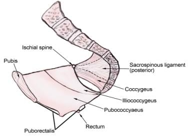
- The thickenings in the superior fascia of the pelvic diaphragm, which make up the medial puboprostatic or pubovesical ligaments, continue backward in a slight curve, concave downward, gradually diverging to the region of the ischial spine. (pediagenosis.com)
Coccyx9
- The sacrococcygeal symphysis (sacrococcygeal articulation, articulation of the sacrum and coccyx) is an amphiarthrodial joint, formed between the oval surface at the apex of the sacrum, and the base of the coccyx. (wikipedia.org)
- The interarticular or intercornual sacrococcygeal ligaments stretches from the cornu of the sacrum to the cornu of the coccyx. (wikipedia.org)
- In some cases, a doctor may choose to manipulate the coccyx manually through the rectum, in order to assess limited or excessive mobility of the sacrococcygeal joint. (spine-health.com)
- A static image of the coccyx taken by MRI or CT scan (one that does not illustrate pelvic rotation or movement) may be used if the suspected cause of pain is a fracture, tumor, or abnormal mobility of the sacrococcygeal joint. (spine-health.com)
- The body region between (and flanking) the SACRUM and COCCYX . (nih.gov)
- The coccyx, a small triangular bone at the bottom of the spinal column, can get bruised and even fractured.Coccygodynia, sometimes referred to as coccydynia, coccalgia, coccygeal neuralgia or tailbone pain, is the term used to describe the symptoms of pain that occur in the region of the coccyx. (askdrmakkar.com)
- Limited mobility of the coccyx causes the tailbone to jut outward when sitting, and can put increased pressure on the bones and the sacrococcygeal joint. (askdrmakkar.com)
- In rare cases, part of the sacrococcygeal joint may become dislocated at the front or back of the tailbone, causing coccyx pain. (askdrmakkar.com)
- Type IV: Coccyx is subluxated at sacrococcygeal joint or at intercoccygeal joint. (medscape.com)
Lumbosacral1
- The cutaneous lesions over the lumbosacral region were subcutaneous lipoma, dimples and hemangioma. (tmu.edu.tw)
Vertebrae4
- The dorsal or posterior sacrococcygeal ligament has a deep and a superficial part: The deep dorsal ligament is a flat band which corresponds to the posterior longitudinal ligament (PLL) that run down inside the vertebral canal on the posterior surfaces of the bodies of the vertebrae. (wikipedia.org)
- Primary sites include the clivus, vertebrae and sacrococcygeal region. (surgicalneurologyint.com)
- the number of vertebrae in each region of the vertebral column in abbreviated notation. (2050farm.cz)
- The vertebrae are divided into three regions: cervical C1-C7 vertebrae, thoracic T1-T12 vertebrae, and lumbar L1-L5 vertebrae. (2050farm.cz)
Ligament2
- The sacrococcygeal disc or interosseus ligament is similar to the intervertebral discs but thinner, thicker in front and behind than at the sides, and with a firmer texture. (wikipedia.org)
- Treatment consisted of mechanical force, manually assisted, short-lever (MFMA) chiropractic adjusting procedures to the coccygeal area, primarily the sacrococcygeal ligament. (activator.com)
Subcutaneous3
- a and c) A photograph showing an enlarged subcutaneous mass in the suprapubic region. (cytojournal.com)
- However, others reject the congenital nature of the disease and attribute its occurrence to the abnormal (incorrect) incorporation of growing hair into the subcutaneous tissue of the sacrococcygeal region. (pain-relief-med.com)
- The epithelial coccygeal passage is located along the midline in the intergluteal fold, blindly terminates in the subcutaneous tissue of the sacrococcygeal region, opens on the skin with one or more point holes (primary epithelial passage), and is a narrow epithelial tube. (pain-relief-med.com)
Sacrum3
- The lateral sacrococcygeal ligaments run from the lower lateral angles of the sacrum to the transverse processes of the first coccygeal vertebra to complete the foramina for the last sacral nerve. (wikipedia.org)
- When movements in the sacrum increase the anteroposterior diameter of the pelvic outlet, movements in the sacrococcygeal joint can further increase this diameter. (wikipedia.org)
- It is a disease that can arise at any region along the fold between the buttocks which extends from the sacrum or the bone at the spinal end to the anus. (hxbenefit.com)
Ligaments1
- Potential causes include injury to the pubic, sacrococcygeal, sacrotuberous, sacroiliac or iliolumbar ligaments . (getprolo.com)
Symphysis1
- The sacrococcygeal articulation is either a symphysis or a true synovial joint. (medscape.com)
Posterior2
- Also note that the epidural space is narrowest in the cervical region, with an anterior/posterior diameter of 2-3 mm. (medscape.com)
- Vertebral Column Developmental anomalies, pathological changes, or obesity can enhance the normal vertebral column curves, resulting in the development of abnormal or excessive curvatures ().Kyphosis, also referred to as humpback or hunchback, is an excessive posterior curvature of the thoracic region. (2050farm.cz)
Lumbar region1
- Caudal steroid injections should only be used for patients with leg pain of sacral origin or in whom direct access to the lumbar region is impossible. (medscape.com)
Chordoma2
- 4. Chondroid chordoma of the sacrococcygeal region. (nih.gov)
- 7 ] We report a case of a pineal region chordoma with metastasis through a ventriculoperitoneal (VP) shunt and hypothesize on the origin of these intraparenchymal chordomas. (surgicalneurologyint.com)
Coccygeal Region3
- Bone Health Technologies has intellectual property (IP) covering the method of delivering vibration through a body-worn device placed over the sacro-coccygeal region. (nih.gov)
- The pain was characterized by a continual dull ache in the coccygeal region, accompanied by intermittent sharp pain, particularly upon sitting or rising from a seated position. (activator.com)
- Coccygodynia (also referred to as coccydynia, coccalgia, coccygalgia, or coccygeal pain) is a painful syndrome affecting the tailbone (coccygeal) region. (medscape.com)
Tumor3
- Historically, sacrococcygeal teratomas present in 2 clinical patterns related to the child's age, tumor location, and likelihood of tumor malignancy. (iiab.me)
- Among infants and young children , the tumor presents as a palpable mass in the sacropelvic region compressing the bladder or rectum. (iiab.me)
- An early survey found that the rate of tumor malignancy was 48% for girls and 67% for boys older than 2 months at the time of sacrococcygeal tumor diagnosis, compared with a malignant tumor incidence of 7% for girls and 10% for boys younger than 2 months at the time of diagnosis. (iiab.me)
Benign4
- Sacrococcygeal teratomas are benign 75% of the time, malignant 12% of the time, and the remainder are considered "immature teratomas" that share benign and malignant features. (iiab.me)
- Benign sacrococcygeal teratomas are more likely to develop in younger children who are less than 5 months old, and older children are more likely to develop malignant sacrococcygeal teratomas. (iiab.me)
- Sacrococcygeal teratomas are the most common type of germ cell tumors (both benign and malignant ) diagnosed in neonates , infants , and children younger than 4 years. (iiab.me)
- In the neonatal age group most of these tumors are benign and occur mainly in the sacrococcygeal area followed by the anterior mediastinum. (ox.ac.uk)
Caudal1
- The caudal part extends to the sacrococcygeal membrane. (medscape.com)
Cervical region1
- It is thinnest in the cervical region. (medscape.com)
Pineal3
- The authors report a unique presentation encompassing the pineal region with metastasis to the peritoneum after a ventriculoperitoneal (VP) shunt procedure and review the current knowledge about their pathophysiology and management. (surgicalneurologyint.com)
- Computed tomography (CT) scans revealed a mass in the pineal region and in the abdominal cavity. (surgicalneurologyint.com)
- A heterogeneously enhanced lesion may be appreciated in the pineal region. (surgicalneurologyint.com)
Membrane1
- But germ cells can also be found elsewhere in the body, especially in the region of the tailbone and the mediastinum (a membrane separating the lungs). (zodicengineering.com)
Occur2
- Although PSD is most frequently seen in the sacrococcygeal region, it can also occur at the axilla, perineum, suprapubic regions, hands, and umbilicus. (researchgate.net)
- Even though they may occur in an extraosseous intradural location, the most common sites include the sacrococcygeal and clivus regions. (surgicalneurologyint.com)
Sacral Region1
- The maximum interface pressure in the greater trochanter in the lateral position was twice that in the sacral region in the supine position. (fujita-hu.ac.jp)
Surgical1
- A morphological study of skin areas with altered tissues of the sacrococcygeal region after radical surgical treatment of 235 patients with a coccygeal cyst of the sacrococcygeal region was carried out. (nih.gov)
Bones1
- Classify works on abnormalities of specific bones or regions with the bone or region. (nih.gov)
Suprapubic1
- A 20-year-old female presented with a swelling in the suprapubic region for the past 1 year. (cytojournal.com)
Spinal2
- This condition is described as a decrease or loss of normal spinal curvature in the thoracic region of the back. (cure-back-pain.org)
- 63650 - Two temporary spinal cord stimulator trials per anatomic spinal region (two per DOS) or (four units) per patient per lifetime (with exceptions allowed for technical limitations for the initial trials or for use of different modalities of stimulation, including new technology), in place of service office, ASC, out-patient hospital, or hospital. (gohealthcarellc.com)
Anatomy1
- Lumbopelvic region: Anatomy and biomechanics (PDF). (wikipedia.org)
Curvature1
- When the thoracic curvature is decreased, some care providers focus on the region as the source of pain. (cure-back-pain.org)
Present1
- AB - Nucleolus organizer regions (NORs) are present on the satellite stalks located on the short arms of the acrocentric chromosomes. (symptoma.com)
Grows1
- If hair grows back in the region, chances of recurrence are much higher. (hxbenefit.com)
Missing data1
- The data of 2068 patients were analyzed after excluding 23 patients who underwent excision for disease outside the sacrococcygeal region and had missing data. (ac.ir)