Rosette Formation
Immune Adherence Reaction
Erythrocytes
Receptors, Fc
Receptors, Complement
Immunoglobulin Fc Fragments
Lymphocytes
Receptors, Antigen, B-Cell
Leukemia, Lymphoid
T-Lymphocytes
Complement C3
Immunologic Techniques
Receptors, Complement 3b
Binding Sites, Antibody
Antilymphocyte Serum
Sheep
B-Lymphocytes
Immunoglobulin G
Complement System Proteins
Dinitrochlorobenzene
Immunoglobulin M
Blood Group Antigens
Complement C3b
Leukocyte Count
Plasmodium falciparum
Antigen-Antibody Complex
Monocytes
Antigen-Antibody Reactions
Immune Sera
Phagocytosis
Malaria, Falciparum
Trypsin
Lectins
Antigens, Surface
Guinea Pigs
Cell Separation
Lymphocyte Activation
Fluorescent Antibody Technique
Thymus Gland
Receptors, IgG
Cytotoxicity Tests, Immunologic
Receptors, Immunologic
Microscopy, Electron
Binding, Competitive
Macrophages
Cells, Cultured
Cell Membrane
Neutrophils
Establishment of an activated macrophage cell line, A-THP-1, and its properties. (1/1162)
A new macrophage cell line with activated character and unique morphology was isolated by selecting adherent cells from the human monocytic cell line THP-1. The original THP-1 cells had been cultured for more than 9 years using 25 cm2 flasks, when cells with a different morphology appeared, adhering to the bottoms of the culture flasks. These were selected by discarding floating nonadherent cells at every subculture. Enrichment of adherent THP-1 cells with long processes proceeded during the cultivation. These adherent THP-1 showed remarkable phenotypic changes, not only morphologically, but also functionally. Namely, increased phagocytic activity, HLA-DR expression and MLR stimulator activity were remarkable. This adherent cell line was designated as activated-THP-1 (A-THP-1), since it demonstrated characteristics of activated macrophages continuously without exogenous stimulation. A cloned A-THP-1 cell line (A-THP-1 C1) also showed the same features and contained about 10% multinucleated giant cells probably caused by cell fusion. This A-THP-1 cell line, the first activated macrophage cell line to be established, provides a good model for understanding of activation mechanisms of macrophages and multinucleation. In this paper, morphological, immunological, and biological characters of this cell line are described. (+info)Protein kinase C and a calcium-independent phospholipase are required for IgG-mediated phagocytosis by Mono-Mac-6 cells. (2/1162)
Mono-Mac-6 (MM6) human monocytes ingest IgG-opsonized particles better than other human cell lines. We compared the phagocytic signaling pathway in MM6 with human monocytes. MM6 expressed FcgammaRI at levels similar to monocytes, whereas FcRgammaII expression was approximately double. MM6 ingested IgG-opsonized erythrocytes (EIgG) in a calcium-independent manner. Incubation of MM6 with bromoenol lactone, an inhibitor of the phagocytic phospholipase (pPL), coordinately decreased phagocytosis and pPL activity. This inhibition was overcome by exogenous arachidonic acid, suggesting that phagocytosis requires pPL activation and arachidonic acid release. MM6 phagocytosis was inhibited with staurosporine and activated with diacylglycerol, supporting a role for protein kinase C (PKC) in this process. The pPL activators mastoparan and melittin restored phagocytosis to PKC-inhibited cells, suggesting that pPL lies downstream from PKC. These results suggest that the MM6 signal transduction pathway for IgG-mediated phagocytosis is similar to that of monocytes (PKC-->pPL-->arachidonic acid-->phagocytosis). The results are discussed in the context of the finding that MM6 exhibit low phagocytosis relative to monocytes and thus may represent an attractive cell line for molecular manipulation in "recovery of function" studies. (+info)Identification of residues in the CH2/CH3 domain interface of IgA essential for interaction with the human fcalpha receptor (FcalphaR) CD89. (3/1162)
Cellular receptors for IgA (FcalphaR) mediate important protective functions. An extensive panel of site-directed mutant IgAs was used to identify IgA residues critical for FcalphaR (CD89) binding and triggering. Although a tailpiece-deleted IgA1 was able to bind and trigger CD89, antibodies featuring CH3 domain exchanges between human IgA1 and IgG1 could not, indicating that both domains but not the tailpiece are required for FcalphaR recognition. To further investigate the role of the interdomain region, numerous IgA1s, each with a point substitution in either of two interdomain loops (Leu-257-Gly-259 in Calpha2; Pro-440-Phe-443 in Calpha3), were generated. With only one exception (G259R), substitutions produced either ablation (L257R, P440A, A442R, F443R) or marked reduction (P440R) in CD89 binding and triggering. Further support for involvement of these interdomain loops was provided by interspecies comparisons of IgA. Thus a human IgA1 mutant, LA441-442MN, which mimicked the mouse IgA loop sequence through substitution of two adjacent residues in the Calpha3 loop, was found, like mouse IgA, not to bind CD89. In contrast, bovine IgA1, identical to human IgA1 within these interdomain loops despite numerous differences elsewhere in the Fc region, did bind CD89. We have thus identified motifs in the interdomain region of IgA Fc critical for FcalphaR binding and triggering, significantly enhancing present understanding of the molecular basis of the IgA-FcalphaR interaction. (+info)Plasmodium falciparum malaria: rosettes are disrupted by quinine, artemisinin, mefloquine, primaquine, pyrimethamine, chloroquine and proguanil. (4/1162)
An assay was developed measuring the disruption of rosettes between Plasmodium falciparuminfected (trophozoites) and uninfected erythrocytes by the antimalarial drugs quinine, artemisinin mefloquine, primaquine, pyrimethamine, chloroquine and proguanil. At 4 hr incubation rosettes were disrupted by all the drugs in a dose dependent manner. Artemisinin and quinine were the most effective anti-malarials at disrupting rosettes at their therapeutic concentrations with South African RSA 14, 15, 17 and The Gambian FCR-3 P. falciparum strains. The least effective drugs were proguanil and chloroquine. A combination of artemisinin and mefloquine was more effective than each drug alone. The combinations of pyrimethamine or primaquine, with quinine disrupted more rosettes than quinine alone. Quinine may be an effective drug in the treatment of severe malaria because the drug efficiently reduces the number of rosettes. (+info)Cytoadherence characteristics of Plasmodium falciparum-infected erythrocytes from Malawian children with severe and uncomplicated malaria. (5/1162)
Cytoadherence of Plasmodium falciparum-infected erythrocytes to the microvascular endothelium is believed to be a key factor in the development of cerebral malaria. Erythrocyte rosette formation has been correlated with malaria severity in studies from east and west Africa. We cultured fresh isolates from Malawian children with severe (n = 76) or uncomplicated (n = 79) malaria to pigmented trophozoite stage and examined rosette formation and adherence to CD36, intercellular adhesion molecule-1 (ICAM-1), chondroitin sulfate A (CSA), and thrombomodulin (TM). Most (126 of 148) isolates bound to CD36, and 76 of 136 bound to ICAM-1. Fewer bound to CSA (40 of 148) or TM (23 of 148). After controlling for parasitemia, there was an inverse association between binding to CD36 (P = 0.004) or ICAM-1 (P = 0.001) and disease severity. Parasites from children with severe malaria anemia bound least to CD36, whereas ICAM-1 binding was lowest in children with cerebral malaria. There was no difference in rosette formation between any of the groups. In Malawian children, there was no evidence of a positive association between adherence to any of the receptors examined and disease severity. The negative association found raises the possibility that adherence to certain receptors could instead be an indicator of a less pathogenic infection. (+info)In vivo role of complement-interacting domains of herpes simplex virus type 1 glycoprotein gC. (6/1162)
Immune evasion is critical for survival of viruses that establish persistent or recurrent infections. However, at the molecular level, little is known about how viruses evade immune attack in vivo. Herpes simplex virus (HSV)-1 glycoprotein gC has two domains that are involved in modulating complement activation; one binds C3, and the other is required for blocking C5 and properdin (P) binding to C3. To evaluate the importance of these regions in vivo, HSV-1 gC mutant viruses were constructed that lacked one or both gC domains and studied in a murine model of infection. Each gC region of complement regulation contributed to virulence; however, the C3 binding domain was far more important, as virus lacking this domain was much less virulent than virus lacking the C5/P inhibitory domain and was as attenuated as virus lacking both domains. Studies in C3 knockout mice and mice reconstituted with C3 confirmed that the gC domains are inhibitors of complement activation, accounting for a 50-fold difference in virulence between mutant and wild-type viruses. We conclude that the C3 binding domain on gC is a major contributor to immune evasion and that this site explains at a molecular level why wild-type virus resists complement attack. (+info)Plasmodium falciparum rosette formation is uncommon in isolates from pregnant women. (7/1162)
We examined the formation of Plasmodium falciparum erythrocyte rosettes using parasite isolates from placental or peripheral blood of pregnant Malawian women and from peripheral blood of children. Five of 23 placental isolates, 23 of 38 maternal peripheral isolates, and 136 of 139 child peripheral isolates formed rosettes. Placental isolates formed fewer rosettes than maternal isolates (range, 0 to 7. 5% versus 0 to 33.5%; P = 0.002), and both formed fewer rosettes than isolates cultured from children (range, 0 to 56%; P < 0.0001). Rosette formation is common in infections of children but uncommon in pregnancy and rarely detected in placental isolates. (+info)Autologous T cells control B-chronic lymphocytic leukemia tumor progression in human-->mouse radiation chimera. (8/1162)
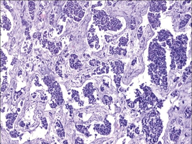
B-chronic lymphocytic leukemia (B-CLL) is characterized by the clonal accumulation of CD5+ B cells. It has been suggested that CLL cells may be regulated by inhibitory and growth-promoting signals exerted by autologous T cells. We have recently described a model for human B-CLL in which peripheral blood mononuclear cells (PBMCs) are transplanted into the peritoneal cavity of lethally irradiated mice radioprotected with bone marrow from mice with severe combined immunodeficiency. In this model, adoptive transfer of low-stage PBMCs leads to marked engraftment of T cells or combined T and CLL cell engraftment, whereas infusion of high-stage PBMCs leads to dominance of CLL cells with a miniscule level of T-cell engraftment. This mutual exclusive pattern of engraftment indicated that T cells might control the expansion of tumor cells in the peritoneum of recipient BALB/c mice. In the present study, we further investigated this question and we demonstrate that in vivo T-cell depletion, using OKT3 antibody, markedly enhances the engraftment of B-CLL cells from patients with early-stage disease. In mice receiving PBMCs from 11 donors with advanced-stage disease, the results were more heterogeneous. In five patients the results were similar to those observed in early stage, whereas in two cases no CLL cell engraftment was found in the absence of T cells. The addition of purified T cells to PBMCs led to a substantial decrease of CLL engraftment in three advanced-stage cases. These results strengthen the working hypothesis that autologous T cells can actively suppress the expansion of the pathological cells in human-->mouse radiation chimera. This effect is prominent in early-stage disease, whereas in advanced stage suppressive and/or stimulatory effects may occur in different patients. The interaction of T cells with tumor cells and the potential of autologous T cell/immune-therapy in CLL can be further explored in this model. (+info)Rosette formation is a term used in pathology and histology, which refers to the circular arrangement of cells or structures around a central point, creating a pattern that resembles a rose flower. This phenomenon can be observed in various tissues and diseases. For example, in the context of cancer, rosette formation may be seen in certain types of tumors, such as medulloblastomas or retinoblastomas, where cancer cells cluster around blood vessels or form distinctive arrangements that are characteristic of these malignancies. In some cases, rosette formation can provide valuable clues for the diagnosis and classification of neoplasms. However, it is essential to consider other histological features and clinical context when interpreting rosette formation in diagnostic pathology.
The term "Immune Adherence Reaction" is not widely used in modern immunology or medicine. It appears to be an outdated concept that refers to the attachment of immune complexes (consisting of antigens, antibodies, and complement components) to Fc receptors on phagocytic cells, such as neutrophils and monocytes. This interaction facilitates the clearance of immune complexes from circulation and helps to prevent tissue damage caused by their deposition.
However, it is important to note that this term is not commonly used in current scientific literature or clinical settings. Instead, the processes it describes are typically discussed within the broader context of immune complex-mediated inflammation, complement activation, and phagocytosis.
Erythrocytes, also known as red blood cells (RBCs), are the most common type of blood cell in circulating blood in mammals. They are responsible for transporting oxygen from the lungs to the body's tissues and carbon dioxide from the tissues to the lungs.
Erythrocytes are formed in the bone marrow and have a biconcave shape, which allows them to fold and bend easily as they pass through narrow blood vessels. They do not have a nucleus or mitochondria, which makes them more flexible but also limits their ability to reproduce or repair themselves.
In humans, erythrocytes are typically disc-shaped and measure about 7 micrometers in diameter. They contain the protein hemoglobin, which binds to oxygen and gives blood its red color. The lifespan of an erythrocyte is approximately 120 days, after which it is broken down in the liver and spleen.
Abnormalities in erythrocyte count or function can lead to various medical conditions, such as anemia, polycythemia, and sickle cell disease.
Fc receptors (FcRs) are specialized proteins found on the surface of various immune cells, including neutrophils, monocytes, macrophages, eosinophils, basophils, mast cells, and B lymphocytes. They play a crucial role in the immune response by recognizing and binding to the Fc region of antibodies (IgG, IgA, and IgE) after they have interacted with their specific antigens.
FcRs can be classified into several types based on the class of antibody they bind:
1. FcγRs - bind to the Fc region of IgG antibodies
2. FcαRs - bind to the Fc region of IgA antibodies
3. FcεRs - bind to the Fc region of IgE antibodies
The binding of antibodies to Fc receptors triggers various cellular responses, such as phagocytosis, degranulation, and antibody-dependent cellular cytotoxicity (ADCC), which contribute to the elimination of pathogens, immune complexes, and other foreign substances. Dysregulation of Fc receptor function has been implicated in several diseases, including autoimmune disorders and allergies.
Complement receptors are proteins found on the surface of various cells in the human body, including immune cells and some non-immune cells. They play a crucial role in the complement system, which is a part of the innate immune response that helps to eliminate pathogens and damaged cells from the body. Complement receptors bind to complement proteins or fragments that are generated during the activation of the complement system. This binding triggers various intracellular signaling events that can lead to diverse cellular responses, such as phagocytosis, inflammation, and immune regulation.
There are several types of complement receptors, including:
1. CR1 (CD35): A receptor found on erythrocytes, B cells, neutrophils, monocytes, macrophages, and glomerular podocytes. It functions in the clearance of immune complexes and regulates complement activation.
2. CR2 (CD21): Expressed mainly on B cells and follicular dendritic cells. It facilitates antigen presentation, B-cell activation, and immune regulation.
3. CR3 (CD11b/CD18, Mac-1): Present on neutrophils, monocytes, macrophages, and some T cells. It mediates cell adhesion, phagocytosis, and intracellular signaling.
4. CR4 (CD11c/CD18, p150,95): Expressed on neutrophils, monocytes, macrophages, and dendritic cells. It is involved in cell adhesion, phagocytosis, and intracellular signaling.
5. C5aR (CD88): Found on various immune cells, including neutrophils, monocytes, macrophages, mast cells, eosinophils, and dendritic cells. It binds to the complement protein C5a and mediates chemotaxis, degranulation, and inflammation.
6. C5L2 (GPR77): Present on various cell types, including immune cells. Its function is not well understood but may involve regulating C5a-mediated responses or acting as a receptor for other ligands.
These receptors play crucial roles in the immune response and inflammation by mediating various functions such as chemotaxis, phagocytosis, cell adhesion, and intracellular signaling. Dysregulation of these receptors has been implicated in several diseases, including autoimmune disorders, infections, and cancer.
Immunoglobulin Fc fragments are the crystallizable fragment of an antibody that is responsible for effector functions such as engagement with Fc receptors on immune cells, activation of the complement system, and neutralization of toxins. The Fc region is located at the tail end of the Y-shaped immunoglobulin molecule, and it is made up of constant regions of the heavy chains of the antibody.
When an antibody binds to its target antigen, the Fc region can interact with other proteins in the immune system, leading to a variety of responses such as phagocytosis, antibody-dependent cellular cytotoxicity (ADCC), and complement activation. These effector functions help to eliminate pathogens and infected cells from the body.
Immunoglobulin Fc fragments can be produced artificially through enzymatic digestion of intact antibodies, resulting in a fragment that retains the ability to interact with Fc receptors and other proteins involved in immune responses. These fragments have potential therapeutic applications in a variety of diseases, including autoimmune disorders, inflammatory conditions, and cancer.
Lymphocytes are a type of white blood cell that is an essential part of the immune system. They are responsible for recognizing and responding to potentially harmful substances such as viruses, bacteria, and other foreign invaders. There are two main types of lymphocytes: B-lymphocytes (B-cells) and T-lymphocytes (T-cells).
B-lymphocytes produce antibodies, which are proteins that help to neutralize or destroy foreign substances. When a B-cell encounters a foreign substance, it becomes activated and begins to divide and differentiate into plasma cells, which produce and secrete large amounts of antibodies. These antibodies bind to the foreign substance, marking it for destruction by other immune cells.
T-lymphocytes, on the other hand, are involved in cell-mediated immunity. They directly attack and destroy infected cells or cancerous cells. T-cells can also help to regulate the immune response by producing chemical signals that activate or inhibit other immune cells.
Lymphocytes are produced in the bone marrow and mature in either the bone marrow (B-cells) or the thymus gland (T-cells). They circulate throughout the body in the blood and lymphatic system, where they can be found in high concentrations in lymph nodes, the spleen, and other lymphoid organs.
Abnormalities in the number or function of lymphocytes can lead to a variety of immune-related disorders, including immunodeficiency diseases, autoimmune disorders, and cancer.
1. Receptors: In the context of physiology and medicine, receptors are specialized proteins found on the surface of cells or inside cells that detect and respond to specific molecules, known as ligands. These interactions can trigger a variety of responses within the cell, such as starting a signaling cascade or changing the cell's metabolism. Receptors play crucial roles in various biological processes, including communication between cells, regulation of immune responses, and perception of senses.
2. Antigen: An antigen is any substance (usually a protein) that can be recognized by the adaptive immune system, specifically by B-cells and T-cells. Antigens can be derived from various sources, such as microorganisms (like bacteria, viruses, or fungi), pollen, dust mites, or even components of our own cells (for instance, in autoimmune diseases). An antigen's ability to stimulate an immune response is determined by its molecular structure and whether it can be recognized by the receptors on immune cells.
3. B-Cell: B-cells are a type of white blood cell that plays a critical role in the adaptive immune system, particularly in humoral immunity. They originate from hematopoietic stem cells in the bone marrow and are responsible for producing antibodies, which are proteins that recognize and bind to specific antigens. Each B-cell has receptors on its surface called B-cell receptors (BCRs) that can recognize a unique antigen. When a B-cell encounters its specific antigen, it becomes activated, undergoes proliferation, and differentiates into plasma cells that secrete large amounts of antibodies to neutralize or eliminate the antigen.
Leukemia, lymphoid is a type of cancer that affects the lymphoid cells, which are a vital part of the body's immune system. It is characterized by the uncontrolled production of abnormal white blood cells (leukocytes or WBCs) in the bone marrow, specifically the lymphocytes. These abnormal lymphocytes accumulate and interfere with the production of normal blood cells, leading to a decrease in red blood cells (anemia), platelets (thrombocytopenia), and healthy white blood cells (leukopenia).
There are two main types of lymphoid leukemia: acute lymphoblastic leukemia (ALL) and chronic lymphocytic leukemia (CLL). Acute lymphoblastic leukemia progresses rapidly, while chronic lymphocytic leukemia has a slower onset and progression.
Symptoms of lymphoid leukemia may include fatigue, frequent infections, easy bruising or bleeding, weight loss, swollen lymph nodes, and bone pain. Treatment options depend on the type, stage, and individual patient factors but often involve chemotherapy, radiation therapy, targeted therapy, immunotherapy, or stem cell transplantation.
T-lymphocytes, also known as T-cells, are a type of white blood cell that plays a key role in the adaptive immune system's response to infection. They are produced in the bone marrow and mature in the thymus gland. There are several different types of T-cells, including CD4+ helper T-cells, CD8+ cytotoxic T-cells, and regulatory T-cells (Tregs).
CD4+ helper T-cells assist in activating other immune cells, such as B-lymphocytes and macrophages. They also produce cytokines, which are signaling molecules that help coordinate the immune response. CD8+ cytotoxic T-cells directly kill infected cells by releasing toxic substances. Regulatory T-cells help maintain immune tolerance and prevent autoimmune diseases by suppressing the activity of other immune cells.
T-lymphocytes are important in the immune response to viral infections, cancer, and other diseases. Dysfunction or depletion of T-cells can lead to immunodeficiency and increased susceptibility to infections. On the other hand, an overactive T-cell response can contribute to autoimmune diseases and chronic inflammation.
Complement C3 is a protein that plays a central role in the complement system, which is a part of the immune system that helps to clear pathogens and damaged cells from the body. Complement C3 can be activated through three different pathways: the classical pathway, the lectin pathway, and the alternative pathway. Once activated, it breaks down into two fragments, C3a and C3b.
C3a is an anaphylatoxin that helps to recruit immune cells to the site of infection or injury, while C3b plays a role in opsonization, which is the process of coating pathogens or damaged cells with proteins to make them more recognizable to the immune system. Additionally, C3b can also activate the membrane attack complex (MAC), which forms a pore in the membrane of target cells leading to their lysis or destruction.
In summary, Complement C3 is an important protein in the complement system that helps to identify and eliminate pathogens and damaged cells from the body through various mechanisms.
Immunologic techniques are a group of laboratory methods that utilize the immune system's ability to recognize and respond to specific molecules, known as antigens. These techniques are widely used in medicine, biology, and research to detect, measure, or identify various substances, including proteins, hormones, viruses, bacteria, and other antigens.
Some common immunologic techniques include:
1. Enzyme-linked Immunosorbent Assay (ELISA): A sensitive assay used to detect and quantify antigens or antibodies in a sample. This technique uses an enzyme linked to an antibody or antigen, which reacts with a substrate to produce a colored product that can be measured and quantified.
2. Immunofluorescence: A microscopic technique used to visualize the location of antigens or antibodies in tissues or cells. This technique uses fluorescent dyes conjugated to antibodies, which bind to specific antigens and emit light when excited by a specific wavelength of light.
3. Western Blotting: A laboratory technique used to detect and identify specific proteins in a sample. This technique involves separating proteins based on their size using electrophoresis, transferring them to a membrane, and then probing the membrane with antibodies that recognize the protein of interest.
4. Immunoprecipitation: A laboratory technique used to isolate and purify specific antigens or antibodies from a complex mixture. This technique involves incubating the mixture with an antibody that recognizes the antigen or antibody of interest, followed by precipitation of the antigen-antibody complex using a variety of methods.
5. Radioimmunoassay (RIA): A sensitive assay used to detect and quantify antigens or antibodies in a sample. This technique uses radioactively labeled antigens or antibodies, which bind to specific antigens or antibodies in the sample, allowing for detection and quantification using a scintillation counter.
These techniques are important tools in medical diagnosis, research, and forensic science.
Complement receptor 3b (CR3b or CD11b/CD18) is not a medical definition itself, but I can provide you with the relevant information regarding this term.
Complement receptor 3 (CR3) is a heterodimeric receptor consisting of two subunits, CD11b (also known as Mac-1 or CR3 alpha) and CD18 (also known as beta2 integrin). There are two forms of the CD11b/CD18 heterodimer: CR3a (CD11b/CD18) and CR3b (CD11b/CD18'). The difference between these two forms lies in the conformation of the CD11b subunit.
Complement receptor 3b (CR3b or CD11b/CD18') is a less common form of the CR3 receptor, which is primarily expressed on myeloid cells such as monocytes, macrophages, and neutrophils. CR3b has a higher affinity for complement component C3b and its fragments iC3b and C3dg compared to CR3a.
CR3b plays a role in various immune functions, including:
1. Phagocytosis: Binding of C3b or its fragments to CR3b facilitates the recognition and uptake of opsonized pathogens by phagocytes.
2. Adhesion: The integrin component of CR3b mediates cell-cell and cell-matrix interactions, contributing to leukocyte migration and recruitment to sites of inflammation or infection.
3. Intracellular signaling: Activation of CR3b can lead to intracellular signaling events that modulate immune responses, such as the release of pro-inflammatory cytokines and reactive oxygen species.
In summary, Complement receptor 3b (CR3b or CD11b/CD18') is a less common form of CR3 primarily expressed on myeloid cells that binds complement component C3b and its fragments with high affinity, mediating phagocytosis, adhesion, and intracellular signaling.
A binding site on an antibody refers to the specific region on the surface of the antibody molecule that can recognize and bind to a specific antigen. Antibodies are proteins produced by the immune system in response to the presence of foreign substances called antigens. They have two main functions: to neutralize the harmful effects of antigens and to help eliminate them from the body.
The binding site of an antibody is located at the tips of its Y-shaped structure, formed by the variable regions of the heavy and light chains of the antibody molecule. These regions contain unique amino acid sequences that determine the specificity of the antibody for a particular antigen. The binding site can recognize and bind to a specific epitope or region on the antigen, forming an antigen-antibody complex.
The binding between the antibody and antigen is highly specific and depends on non-covalent interactions such as hydrogen bonds, van der Waals forces, and electrostatic attractions. This interaction plays a crucial role in the immune response, as it allows the immune system to recognize and eliminate pathogens and other foreign substances from the body.
Antilymphocyte serum (ALS) is a type of immune serum that contains antibodies against human lymphocytes. It is produced by immunizing animals, such as horses or rabbits, with human lymphocytes to stimulate an immune response and the production of anti-lymphocyte antibodies. The resulting serum is then collected and can be used as a therapeutic agent to suppress the activity of the immune system in certain medical conditions.
ALS is primarily used in the treatment of transplant rejection, particularly in organ transplantation, where it helps to prevent the recipient's immune system from attacking and rejecting the transplanted organ. It can also be used in the management of autoimmune diseases, such as rheumatoid arthritis and lupus, to suppress the overactive immune response that contributes to these conditions.
It is important to note that the use of ALS carries a risk of side effects, including allergic reactions, fever, and decreased white blood cell counts. Close monitoring and appropriate management of these potential adverse events are essential during treatment with ALS.
I apologize, but "sheep" is not a term that has a medical definition. It is a common animal with the scientific name Ovis aries. If you have any medical or health-related questions, I would be happy to try and help answer those for you.
B-lymphocytes, also known as B-cells, are a type of white blood cell that plays a key role in the immune system's response to infection. They are responsible for producing antibodies, which are proteins that help to neutralize or destroy pathogens such as bacteria and viruses.
When a B-lymphocyte encounters a pathogen, it becomes activated and begins to divide and differentiate into plasma cells, which produce and secrete large amounts of antibodies specific to the antigens on the surface of the pathogen. These antibodies bind to the pathogen, marking it for destruction by other immune cells such as neutrophils and macrophages.
B-lymphocytes also have a role in presenting antigens to T-lymphocytes, another type of white blood cell involved in the immune response. This helps to stimulate the activation and proliferation of T-lymphocytes, which can then go on to destroy infected cells or help to coordinate the overall immune response.
Overall, B-lymphocytes are an essential part of the adaptive immune system, providing long-lasting immunity to previously encountered pathogens and helping to protect against future infections.
Immunoglobulin G (IgG) is a type of antibody, which is a protective protein produced by the immune system in response to foreign substances like bacteria or viruses. IgG is the most abundant type of antibody in human blood, making up about 75-80% of all antibodies. It is found in all body fluids and plays a crucial role in fighting infections caused by bacteria, viruses, and toxins.
IgG has several important functions:
1. Neutralization: IgG can bind to the surface of bacteria or viruses, preventing them from attaching to and infecting human cells.
2. Opsonization: IgG coats the surface of pathogens, making them more recognizable and easier for immune cells like neutrophils and macrophages to phagocytose (engulf and destroy) them.
3. Complement activation: IgG can activate the complement system, a group of proteins that work together to help eliminate pathogens from the body. Activation of the complement system leads to the formation of the membrane attack complex, which creates holes in the cell membranes of bacteria, leading to their lysis (destruction).
4. Antibody-dependent cellular cytotoxicity (ADCC): IgG can bind to immune cells like natural killer (NK) cells and trigger them to release substances that cause target cells (such as virus-infected or cancerous cells) to undergo apoptosis (programmed cell death).
5. Immune complex formation: IgG can form immune complexes with antigens, which can then be removed from the body through various mechanisms, such as phagocytosis by immune cells or excretion in urine.
IgG is a critical component of adaptive immunity and provides long-lasting protection against reinfection with many pathogens. It has four subclasses (IgG1, IgG2, IgG3, and IgG4) that differ in their structure, function, and distribution in the body.
The complement system is a group of proteins found in the blood and on the surface of cells that when activated, work together to help eliminate pathogens such as bacteria, viruses, and fungi from the body. The proteins are normally inactive in the bloodstream. When they encounter an invading microorganism or foreign substance, a series of reactions take place leading to the activation of the complement system. Activation results in the production of effector molecules that can punch holes in the cell membranes of pathogens, recruit and activate immune cells, and help remove debris and dead cells from the body.
There are three main pathways that can lead to complement activation: the classical pathway, the lectin pathway, and the alternative pathway. Each pathway involves a series of proteins that work together in a cascade-like manner to amplify the response and generate effector molecules. The three main effector molecules produced by the complement system are C3b, C4b, and C5b. These molecules can bind to the surface of pathogens, marking them for destruction by other immune cells.
Complement proteins also play a role in the regulation of the immune response. They help to prevent excessive activation of the complement system, which could damage host tissues. Dysregulation of the complement system has been implicated in a number of diseases, including autoimmune disorders and inflammatory conditions.
In summary, Complement System Proteins are a group of proteins that play a crucial role in the immune response by helping to eliminate pathogens and regulate the immune response. They can be activated through three different pathways, leading to the production of effector molecules that mark pathogens for destruction. Dysregulation of the complement system has been linked to various diseases.
Dinitrochlorobenzene (DNCB) is a chemical compound that is classified as an aromatic organic compound. Its medical definition relates to its use as a topical immunotherapy for the treatment of certain skin conditions. DNCB is a potent sensitizer and hapten, which means that it can cause an immune response when it comes into contact with the skin.
When applied to the skin, DNCB can stimulate the production of antibodies and activate immune cells, leading to an inflammatory reaction. This property has been exploited in the treatment of conditions such as alopecia areata, a type of hair loss that is thought to be caused by an autoimmune response. By sensitizing the patient's immune system to DNCB, it may be possible to modulate the immune response and promote hair growth.
However, the use of DNCB as a therapeutic agent is not without risks. It can cause significant local reactions, including redness, swelling, and blistering, and there is a risk of systemic toxicity if it is absorbed into the bloodstream. As such, its use is generally restricted to specialized medical settings where it can be administered under close supervision.
Cytochalasin B is a fungal metabolite that inhibits actin polymerization in cells, which can disrupt the cytoskeleton and affect various cellular processes such as cell division and motility. It is often used in research to study actin dynamics and cell shape.
Immunoglobulin M (IgM) is a type of antibody that is primarily found in the blood and lymph fluid. It is the first antibody to be produced in response to an initial exposure to an antigen, making it an important part of the body's primary immune response. IgM antibodies are large molecules that are composed of five basic units, giving them a pentameric structure. They are primarily found on the surface of B cells as membrane-bound immunoglobulins (mlgM), where they function as receptors for antigens. Once an mlgM receptor binds to an antigen, it triggers the activation and differentiation of the B cell into a plasma cell that produces and secretes large amounts of soluble IgM antibodies.
IgM antibodies are particularly effective at agglutination (clumping) and complement activation, which makes them important in the early stages of an immune response to help clear pathogens from the bloodstream. However, they are not as stable or long-lived as other types of antibodies, such as IgG, and their levels tend to decline after the initial immune response has occurred.
In summary, Immunoglobulin M (IgM) is a type of antibody that plays a crucial role in the primary immune response to antigens by agglutination and complement activation. It is primarily found in the blood and lymph fluid, and it is produced by B cells after they are activated by an antigen.
Blood group antigens are molecular markers found on the surface of red blood cells (RBCs) and sometimes other types of cells in the body. These antigens are proteins, carbohydrates, or glycoproteins that can stimulate an immune response when foreign antigens are introduced into the body.
There are several different blood group systems, but the most well-known is the ABO system, which includes A, B, AB, and O blood groups. The antigens in this system are called ABO antigens. Individuals with type A blood have A antigens on their RBCs, those with type B blood have B antigens, those with type AB blood have both A and B antigens, and those with type O blood have neither A nor B antigens.
Another important blood group system is the Rh system, which includes the D antigen. Individuals who have this antigen are considered Rh-positive, while those who do not have it are considered Rh-negative.
Blood group antigens can cause complications during blood transfusions and pregnancy if there is a mismatch between the donor's or fetus's antigens and the recipient's antibodies. For example, if a person with type A blood receives type B blood, their anti-B antibodies will attack the foreign B antigens on the donated RBCs, causing a potentially life-threatening transfusion reaction. Similarly, if an Rh-negative woman becomes pregnant with an Rh-positive fetus, her immune system may produce anti-D antibodies that can cross the placenta and attack the fetal RBCs, leading to hemolytic disease of the newborn.
It is important for medical professionals to determine a patient's blood group before performing a transfusion or pregnancy-related procedures to avoid these complications.
Complement C3b is a protein fragment that plays a crucial role in the complement system, which is a part of the immune system that helps to clear pathogens and damaged cells from the body. C3b is generated during the activation of the complement system, particularly via the classical, lectin, and alternative pathways.
Once formed, C3b can bind covalently to the surface of microbes or other target particles, marking them for destruction by other components of the immune system. Additionally, C3b can interact with other proteins in the complement system to generate the membrane attack complex (MAC), which forms pores in the membranes of targeted cells, leading to their lysis and removal.
In summary, Complement C3b is a vital protein fragment involved in the recognition, tagging, and elimination of pathogens and damaged cells during the immune response.
A leukocyte count, also known as a white blood cell (WBC) count, is a laboratory test that measures the number of leukocytes in a sample of blood. Leukocytes are a vital part of the body's immune system and help fight infection and inflammation. A high or low leukocyte count may indicate an underlying medical condition, such as an infection, inflammation, or a bone marrow disorder. The normal range for a leukocyte count in adults is typically between 4,500 and 11,000 cells per microliter (mcL) of blood. However, the normal range can vary slightly depending on the laboratory and the individual's age and sex.
'Plasmodium falciparum' is a specific species of protozoan parasite that causes malaria in humans. It is transmitted through the bites of infected female Anopheles mosquitoes and has a complex life cycle involving both human and mosquito hosts.
In the human host, the parasites infect red blood cells, where they multiply and cause damage, leading to symptoms such as fever, chills, anemia, and in severe cases, organ failure and death. 'Plasmodium falciparum' malaria is often more severe and life-threatening than other forms of malaria caused by different Plasmodium species. It is a major public health concern, particularly in tropical and subtropical regions of the world where access to prevention, diagnosis, and treatment remains limited.
An antigen-antibody complex is a type of immune complex that forms when an antibody binds to a specific antigen. An antigen is any substance that triggers an immune response, while an antibody is a protein produced by the immune system to neutralize or destroy foreign substances like antigens.
When an antibody binds to an antigen, it forms a complex that can be either soluble or insoluble. Soluble complexes are formed when the antigen is small and can move freely through the bloodstream. Insoluble complexes, on the other hand, are formed when the antigen is too large to move freely, such as when it is part of a bacterium or virus.
The formation of antigen-antibody complexes plays an important role in the immune response. Once formed, these complexes can be recognized and cleared by other components of the immune system, such as phagocytes, which help to prevent further damage to the body. However, in some cases, the formation of large numbers of antigen-antibody complexes can lead to inflammation and tissue damage, contributing to the development of certain autoimmune diseases.
Monoclonal antibodies are a type of antibody that are identical because they are produced by a single clone of cells. They are laboratory-produced molecules that act like human antibodies in the immune system. They can be designed to attach to specific proteins found on the surface of cancer cells, making them useful for targeting and treating cancer. Monoclonal antibodies can also be used as a therapy for other diseases, such as autoimmune disorders and inflammatory conditions.
Monoclonal antibodies are produced by fusing a single type of immune cell, called a B cell, with a tumor cell to create a hybrid cell, or hybridoma. This hybrid cell is then able to replicate indefinitely, producing a large number of identical copies of the original antibody. These antibodies can be further modified and engineered to enhance their ability to bind to specific targets, increase their stability, and improve their effectiveness as therapeutic agents.
Monoclonal antibodies have several mechanisms of action in cancer therapy. They can directly kill cancer cells by binding to them and triggering an immune response. They can also block the signals that promote cancer growth and survival. Additionally, monoclonal antibodies can be used to deliver drugs or radiation directly to cancer cells, increasing the effectiveness of these treatments while minimizing their side effects on healthy tissues.
Monoclonal antibodies have become an important tool in modern medicine, with several approved for use in cancer therapy and other diseases. They are continuing to be studied and developed as a promising approach to treating a wide range of medical conditions.
Monocytes are a type of white blood cell that are part of the immune system. They are large cells with a round or oval shape and a nucleus that is typically indented or horseshoe-shaped. Monocytes are produced in the bone marrow and then circulate in the bloodstream, where they can differentiate into other types of immune cells such as macrophages and dendritic cells.
Monocytes play an important role in the body's defense against infection and tissue damage. They are able to engulf and digest foreign particles, microorganisms, and dead or damaged cells, which helps to clear them from the body. Monocytes also produce cytokines, which are signaling molecules that help to coordinate the immune response.
Elevated levels of monocytes in the bloodstream can be a sign of an ongoing infection, inflammation, or other medical conditions such as cancer or autoimmune disorders.
An antigen-antibody reaction is a specific immune response that occurs when an antigen (a foreign substance, such as a protein or polysaccharide on the surface of a bacterium or virus) comes into contact with a corresponding antibody (a protective protein produced by the immune system in response to the antigen). The antigen and antibody bind together, forming an antigen-antibody complex. This interaction can neutralize the harmful effects of the antigen, mark it for destruction by other immune cells, or activate complement proteins to help eliminate the antigen from the body. Antigen-antibody reactions are a crucial part of the adaptive immune response and play a key role in the body's defense against infection and disease.
'Immune sera' refers to the serum fraction of blood that contains antibodies produced in response to an antigenic stimulus, such as a vaccine or an infection. These antibodies are proteins known as immunoglobulins, which are secreted by B cells (a type of white blood cell) and can recognize and bind to specific antigens. Immune sera can be collected from an immunized individual and used as a source of passive immunity to protect against infection or disease. It is often used in research and diagnostic settings to identify or measure the presence of specific antigens or antibodies.
Phagocytosis is the process by which certain cells in the body, known as phagocytes, engulf and destroy foreign particles, bacteria, or dead cells. This mechanism plays a crucial role in the immune system's response to infection and inflammation. Phagocytes, such as neutrophils, monocytes, and macrophages, have receptors on their surface that recognize and bind to specific molecules (known as antigens) on the target particles or microorganisms.
Once attached, the phagocyte extends pseudopodia (cell extensions) around the particle, forming a vesicle called a phagosome that completely encloses it. The phagosome then fuses with a lysosome, an intracellular organelle containing digestive enzymes and other chemicals. This fusion results in the formation of a phagolysosome, where the engulfed particle is broken down by the action of these enzymes, neutralizing its harmful effects and allowing for the removal of cellular debris or pathogens.
Phagocytosis not only serves as a crucial defense mechanism against infections but also contributes to tissue homeostasis by removing dead cells and debris.
Malaria, Falciparum is defined as a severe and often fatal form of malaria caused by the parasite Plasmodium falciparum. It is transmitted to humans through the bites of infected Anopheles mosquitoes. This type of malaria is characterized by high fever, chills, headache, muscle and joint pain, and vomiting. If left untreated, it can cause severe anemia, kidney failure, seizures, coma, and even death. It is a major public health problem in many tropical and subtropical regions of the world, particularly in Africa.
Cell adhesion refers to the binding of cells to extracellular matrices or to other cells, a process that is fundamental to the development, function, and maintenance of multicellular organisms. Cell adhesion is mediated by various cell surface receptors, such as integrins, cadherins, and immunoglobulin-like cell adhesion molecules (Ig-CAMs), which interact with specific ligands in the extracellular environment. These interactions lead to the formation of specialized junctions, such as tight junctions, adherens junctions, and desmosomes, that help to maintain tissue architecture and regulate various cellular processes, including proliferation, differentiation, migration, and survival. Disruptions in cell adhesion can contribute to a variety of diseases, including cancer, inflammation, and degenerative disorders.
The spleen is an organ in the upper left side of the abdomen, next to the stomach and behind the ribs. It plays multiple supporting roles in the body:
1. It fights infection by acting as a filter for the blood. Old red blood cells are recycled in the spleen, and platelets and white blood cells are stored there.
2. The spleen also helps to control the amount of blood in the body by removing excess red blood cells and storing platelets.
3. It has an important role in immune function, producing antibodies and removing microorganisms and damaged red blood cells from the bloodstream.
The spleen can be removed without causing any significant problems, as other organs take over its functions. This is known as a splenectomy and may be necessary if the spleen is damaged or diseased.
Trypsin is a proteolytic enzyme, specifically a serine protease, that is secreted by the pancreas as an inactive precursor, trypsinogen. Trypsinogen is converted into its active form, trypsin, in the small intestine by enterokinase, which is produced by the intestinal mucosa.
Trypsin plays a crucial role in digestion by cleaving proteins into smaller peptides at specific arginine and lysine residues. This enzyme helps to break down dietary proteins into amino acids, allowing for their absorption and utilization by the body. Additionally, trypsin can activate other zymogenic pancreatic enzymes, such as chymotrypsinogen and procarboxypeptidases, thereby contributing to overall protein digestion.
Lectins are a type of proteins that bind specifically to carbohydrates and have been found in various plant and animal sources. They play important roles in biological recognition events, such as cell-cell adhesion, and can also be involved in the immune response. Some lectins can agglutinate certain types of cells or precipitate glycoproteins, while others may have a more direct effect on cellular processes. In some cases, lectins from plants can cause adverse effects in humans if ingested, such as digestive discomfort or allergic reactions.
Surface antigens are molecules found on the surface of cells that can be recognized by the immune system as being foreign or different from the host's own cells. Antigens are typically proteins or polysaccharides that are capable of stimulating an immune response, leading to the production of antibodies and activation of immune cells such as T-cells.
Surface antigens are important in the context of infectious diseases because they allow the immune system to identify and target infected cells for destruction. For example, viruses and bacteria often display surface antigens that are distinct from those found on host cells, allowing the immune system to recognize and attack them. In some cases, these surface antigens can also be used as targets for vaccines or other immunotherapies.
In addition to their role in infectious diseases, surface antigens are also important in the context of cancer. Tumor cells often display abnormal surface antigens that differ from those found on normal cells, allowing the immune system to potentially recognize and attack them. However, tumors can also develop mechanisms to evade the immune system, making it difficult to mount an effective response.
Overall, understanding the properties and behavior of surface antigens is crucial for developing effective immunotherapies and vaccines against infectious diseases and cancer.
I must clarify that the term "Guinea Pigs" is not typically used in medical definitions. However, in colloquial or informal language, it may refer to people who are used as the first to try out a new medical treatment or drug. This is known as being a "test subject" or "in a clinical trial."
In the field of scientific research, particularly in studies involving animals, guinea pigs are small rodents that are often used as experimental subjects due to their size, cost-effectiveness, and ease of handling. They are not actually pigs from Guinea, despite their name's origins being unclear. However, they do not exactly fit the description of being used in human medical experiments.
Cell separation is a process used to separate and isolate specific cell types from a heterogeneous mixture of cells. This can be accomplished through various physical or biological methods, depending on the characteristics of the cells of interest. Some common techniques for cell separation include:
1. Density gradient centrifugation: In this method, a sample containing a mixture of cells is layered onto a density gradient medium and then centrifuged. The cells are separated based on their size, density, and sedimentation rate, with denser cells settling closer to the bottom of the tube and less dense cells remaining near the top.
2. Magnetic-activated cell sorting (MACS): This technique uses magnetic beads coated with antibodies that bind to specific cell surface markers. The labeled cells are then passed through a column placed in a magnetic field, which retains the magnetically labeled cells while allowing unlabeled cells to flow through.
3. Fluorescence-activated cell sorting (FACS): In this method, cells are stained with fluorochrome-conjugated antibodies that recognize specific cell surface or intracellular markers. The stained cells are then passed through a laser beam, which excites the fluorophores and allows for the detection and sorting of individual cells based on their fluorescence profile.
4. Filtration: This simple method relies on the physical size differences between cells to separate them. Cells can be passed through filters with pore sizes that allow smaller cells to pass through while retaining larger cells.
5. Enzymatic digestion: In some cases, cells can be separated by enzymatically dissociating tissues into single-cell suspensions and then using various separation techniques to isolate specific cell types.
These methods are widely used in research and clinical settings for applications such as isolating immune cells, stem cells, or tumor cells from biological samples.
Lymphocyte activation is the process by which B-cells and T-cells (types of lymphocytes) become activated to perform effector functions in an immune response. This process involves the recognition of specific antigens presented on the surface of antigen-presenting cells, such as dendritic cells or macrophages.
The activation of B-cells leads to their differentiation into plasma cells that produce antibodies, while the activation of T-cells results in the production of cytotoxic T-cells (CD8+ T-cells) that can directly kill infected cells or helper T-cells (CD4+ T-cells) that assist other immune cells.
Lymphocyte activation involves a series of intracellular signaling events, including the binding of co-stimulatory molecules and the release of cytokines, which ultimately result in the expression of genes involved in cell proliferation, differentiation, and effector functions. The activation process is tightly regulated to prevent excessive or inappropriate immune responses that can lead to autoimmunity or chronic inflammation.
An epitope is a specific region on the surface of an antigen (a molecule that can trigger an immune response) that is recognized by an antibody, B-cell receptor, or T-cell receptor. It is also commonly referred to as an antigenic determinant. Epitopes are typically composed of linear amino acid sequences or conformational structures made up of discontinuous amino acids in the antigen. They play a crucial role in the immune system's ability to differentiate between self and non-self molecules, leading to the targeted destruction of foreign substances like viruses and bacteria. Understanding epitopes is essential for developing vaccines, diagnostic tests, and immunotherapies.
The Fluorescent Antibody Technique (FAT) is a type of immunofluorescence assay used in laboratory medicine and pathology for the detection and localization of specific antigens or antibodies in tissues, cells, or microorganisms. In this technique, a fluorescein-labeled antibody is used to selectively bind to the target antigen or antibody, forming an immune complex. When excited by light of a specific wavelength, the fluorescein label emits light at a longer wavelength, typically visualized as green fluorescence under a fluorescence microscope.
The FAT is widely used in diagnostic microbiology for the identification and characterization of various bacteria, viruses, fungi, and parasites. It has also been applied in the diagnosis of autoimmune diseases and certain cancers by detecting specific antibodies or antigens in patient samples. The main advantage of FAT is its high sensitivity and specificity, allowing for accurate detection and differentiation of various pathogens and disease markers. However, it requires specialized equipment and trained personnel to perform and interpret the results.
The thymus gland is an essential organ of the immune system, located in the upper chest, behind the sternum and surrounding the heart. It's primarily active until puberty and begins to shrink in size and activity thereafter. The main function of the thymus gland is the production and maturation of T-lymphocytes (T-cells), which are crucial for cell-mediated immunity, helping to protect the body from infection and cancer.
The thymus gland provides a protected environment where immune cells called pre-T cells develop into mature T cells. During this process, they learn to recognize and respond appropriately to foreign substances while remaining tolerant to self-tissues, which is crucial for preventing autoimmune diseases.
Additionally, the thymus gland produces hormones like thymosin that regulate immune cell activities and contribute to the overall immune response.
IgG receptors, also known as Fcγ receptors (Fc gamma receptors), are specialized protein molecules found on the surface of various immune cells, such as neutrophils, monocytes, macrophages, and some lymphocytes. These receptors recognize and bind to the Fc region of IgG antibodies, one of the five classes of immunoglobulins in the human body.
IgG receptors play a crucial role in immune responses by mediating different effector functions, including:
1. Antibody-dependent cellular cytotoxicity (ADCC): IgG receptors on natural killer (NK) cells and other immune cells bind to IgG antibodies coated on the surface of virus-infected or cancer cells, leading to their destruction.
2. Phagocytosis: When IgG antibodies tag pathogens or foreign particles, phagocytes like neutrophils and macrophages recognize and bind to these immune complexes via IgG receptors, facilitating the engulfment and removal of the targeted particles.
3. Antigen presentation: IgG receptors on antigen-presenting cells (APCs) can internalize immune complexes, process the antigens, and present them to T cells, thereby initiating adaptive immune responses.
4. Inflammatory response regulation: IgG receptors can modulate inflammation by activating or inhibiting downstream signaling pathways in immune cells, depending on the specific type of Fcγ receptor and its activation state.
There are several types of IgG receptors (FcγRI, FcγRII, FcγRIII, and FcγRIV) with varying affinities for different subclasses of IgG antibodies (IgG1, IgG2, IgG3, and IgG4). The distinct functions and expression patterns of these receptors contribute to the complexity and fine-tuning of immune responses in the human body.
Cytotoxicity tests, immunologic are a group of laboratory assays used to measure the immune-mediated damage or destruction (cytotoxicity) of cells. These tests are often used in medical research and clinical settings to evaluate the potential toxicity of drugs, biological agents, or environmental factors on specific types of cells.
Immunologic cytotoxicity tests typically involve the use of immune effector cells, such as cytotoxic T lymphocytes (CTLs) or natural killer (NK) cells, which can recognize and kill target cells that express specific antigens on their surface. The tests may also involve the use of antibodies or other immune molecules that can bind to target cells and trigger complement-mediated cytotoxicity.
There are several types of immunologic cytotoxicity tests, including:
1. Cytotoxic T lymphocyte (CTL) assays: These tests measure the ability of CTLs to recognize and kill target cells that express specific antigens. The test involves incubating target cells with CTLs and then measuring the amount of cell death or damage.
2. Natural killer (NK) cell assays: These tests measure the ability of NK cells to recognize and kill target cells that lack self-antigens or express stress-induced antigens. The test involves incubating target cells with NK cells and then measuring the amount of cell death or damage.
3. Antibody-dependent cellular cytotoxicity (ADCC) assays: These tests measure the ability of antibodies to bind to target cells and recruit immune effector cells, such as NK cells or macrophages, to mediate cell lysis. The test involves incubating target cells with antibodies and then measuring the amount of cell death or damage.
4. Complement-dependent cytotoxicity (CDC) assays: These tests measure the ability of complement proteins to bind to target cells and form a membrane attack complex that leads to cell lysis. The test involves incubating target cells with complement proteins and then measuring the amount of cell death or damage.
Immunologic cytotoxicity tests are important tools in immunology, cancer research, and drug development. They can help researchers understand how immune cells recognize and kill infected or damaged cells, as well as how to develop new therapies that enhance or inhibit these processes.
Immunologic receptors are specialized proteins found on the surface of immune cells that recognize and bind to specific molecules, known as antigens, on the surface of pathogens or infected cells. This binding triggers a series of intracellular signaling events that activate the immune cell and initiate an immune response.
There are several types of immunologic receptors, including:
1. T-cell receptors (TCRs): These receptors are found on the surface of T cells and recognize antigens presented in the context of major histocompatibility complex (MHC) molecules.
2. B-cell receptors (BCRs): These receptors are found on the surface of B cells and recognize free antigens in solution.
3. Pattern recognition receptors (PRRs): These receptors are found inside immune cells and recognize conserved molecular patterns associated with pathogens, such as lipopolysaccharides and flagellin.
4. Fc receptors: These receptors are found on the surface of various immune cells and bind to the constant region of antibodies, mediating effector functions such as phagocytosis and antibody-dependent cellular cytotoxicity (ADCC).
Immunologic receptors play a critical role in the recognition and elimination of pathogens and infected cells, and dysregulation of these receptors can lead to immune disorders and diseases.
Lymphoma is a type of cancer that originates from the white blood cells called lymphocytes, which are part of the immune system. These cells are found in various parts of the body such as the lymph nodes, spleen, bone marrow, and other organs. Lymphoma can be classified into two main types: Hodgkin lymphoma (HL) and non-Hodgkin lymphoma (NHL).
HL is characterized by the presence of a specific type of abnormal lymphocyte called Reed-Sternberg cells, while NHL includes a diverse group of lymphomas that lack these cells. The symptoms of lymphoma may include swollen lymph nodes, fever, night sweats, weight loss, and fatigue.
The exact cause of lymphoma is not known, but it is believed to result from genetic mutations in the lymphocytes that lead to uncontrolled cell growth and division. Exposure to certain viruses, chemicals, and radiation may increase the risk of developing lymphoma. Treatment options for lymphoma depend on various factors such as the type and stage of the disease, age, and overall health of the patient. Common treatments include chemotherapy, radiation therapy, immunotherapy, and stem cell transplantation.
Electron microscopy (EM) is a type of microscopy that uses a beam of electrons to create an image of the sample being examined, resulting in much higher magnification and resolution than light microscopy. There are several types of electron microscopy, including transmission electron microscopy (TEM), scanning electron microscopy (SEM), and reflection electron microscopy (REM).
In TEM, a beam of electrons is transmitted through a thin slice of the sample, and the electrons that pass through the sample are focused to form an image. This technique can provide detailed information about the internal structure of cells, viruses, and other biological specimens, as well as the composition and structure of materials at the atomic level.
In SEM, a beam of electrons is scanned across the surface of the sample, and the electrons that are scattered back from the surface are detected to create an image. This technique can provide information about the topography and composition of surfaces, as well as the structure of materials at the microscopic level.
REM is a variation of SEM in which the beam of electrons is reflected off the surface of the sample, rather than scattered back from it. This technique can provide information about the surface chemistry and composition of materials.
Electron microscopy has a wide range of applications in biology, medicine, and materials science, including the study of cellular structure and function, disease diagnosis, and the development of new materials and technologies.
"Competitive binding" is a term used in pharmacology and biochemistry to describe the behavior of two or more molecules (ligands) competing for the same binding site on a target protein or receptor. In this context, "binding" refers to the physical interaction between a ligand and its target.
When a ligand binds to a receptor, it can alter the receptor's function, either activating or inhibiting it. If multiple ligands compete for the same binding site, they will compete to bind to the receptor. The ability of each ligand to bind to the receptor is influenced by its affinity for the receptor, which is a measure of how strongly and specifically the ligand binds to the receptor.
In competitive binding, if one ligand is present in high concentrations, it can prevent other ligands with lower affinity from binding to the receptor. This is because the higher-affinity ligand will have a greater probability of occupying the binding site and blocking access to the other ligands. The competition between ligands can be described mathematically using equations such as the Langmuir isotherm, which describes the relationship between the concentration of ligand and the fraction of receptors that are occupied by the ligand.
Competitive binding is an important concept in drug development, as it can be used to predict how different drugs will interact with their targets and how they may affect each other's activity. By understanding the competitive binding properties of a drug, researchers can optimize its dosage and delivery to maximize its therapeutic effect while minimizing unwanted side effects.
Macrophages are a type of white blood cell that are an essential part of the immune system. They are large, specialized cells that engulf and destroy foreign substances, such as bacteria, viruses, parasites, and fungi, as well as damaged or dead cells. Macrophages are found throughout the body, including in the bloodstream, lymph nodes, spleen, liver, lungs, and connective tissues. They play a critical role in inflammation, immune response, and tissue repair and remodeling.
Macrophages originate from monocytes, which are a type of white blood cell produced in the bone marrow. When monocytes enter the tissues, they differentiate into macrophages, which have a larger size and more specialized functions than monocytes. Macrophages can change their shape and move through tissues to reach sites of infection or injury. They also produce cytokines, chemokines, and other signaling molecules that help coordinate the immune response and recruit other immune cells to the site of infection or injury.
Macrophages have a variety of surface receptors that allow them to recognize and respond to different types of foreign substances and signals from other cells. They can engulf and digest foreign particles, bacteria, and viruses through a process called phagocytosis. Macrophages also play a role in presenting antigens to T cells, which are another type of immune cell that helps coordinate the immune response.
Overall, macrophages are crucial for maintaining tissue homeostasis, defending against infection, and promoting wound healing and tissue repair. Dysregulation of macrophage function has been implicated in a variety of diseases, including cancer, autoimmune disorders, and chronic inflammatory conditions.
"Cells, cultured" is a medical term that refers to cells that have been removed from an organism and grown in controlled laboratory conditions outside of the body. This process is called cell culture and it allows scientists to study cells in a more controlled and accessible environment than they would have inside the body. Cultured cells can be derived from a variety of sources, including tissues, organs, or fluids from humans, animals, or cell lines that have been previously established in the laboratory.
Cell culture involves several steps, including isolation of the cells from the tissue, purification and characterization of the cells, and maintenance of the cells in appropriate growth conditions. The cells are typically grown in specialized media that contain nutrients, growth factors, and other components necessary for their survival and proliferation. Cultured cells can be used for a variety of purposes, including basic research, drug development and testing, and production of biological products such as vaccines and gene therapies.
It is important to note that cultured cells may behave differently than they do in the body, and results obtained from cell culture studies may not always translate directly to human physiology or disease. Therefore, it is essential to validate findings from cell culture experiments using additional models and ultimately in clinical trials involving human subjects.
A cell membrane, also known as the plasma membrane, is a thin semi-permeable phospholipid bilayer that surrounds all cells in animals, plants, and microorganisms. It functions as a barrier to control the movement of substances in and out of the cell, allowing necessary molecules such as nutrients, oxygen, and signaling molecules to enter while keeping out harmful substances and waste products. The cell membrane is composed mainly of phospholipids, which have hydrophilic (water-loving) heads and hydrophobic (water-fearing) tails. This unique structure allows the membrane to be flexible and fluid, yet selectively permeable. Additionally, various proteins are embedded in the membrane that serve as channels, pumps, receptors, and enzymes, contributing to the cell's overall functionality and communication with its environment.
A cell line is a culture of cells that are grown in a laboratory for use in research. These cells are usually taken from a single cell or group of cells, and they are able to divide and grow continuously in the lab. Cell lines can come from many different sources, including animals, plants, and humans. They are often used in scientific research to study cellular processes, disease mechanisms, and to test new drugs or treatments. Some common types of human cell lines include HeLa cells (which come from a cancer patient named Henrietta Lacks), HEK293 cells (which come from embryonic kidney cells), and HUVEC cells (which come from umbilical vein endothelial cells). It is important to note that cell lines are not the same as primary cells, which are cells that are taken directly from a living organism and have not been grown in the lab.
Neutrophils are a type of white blood cell that are part of the immune system's response to infection. They are produced in the bone marrow and released into the bloodstream where they circulate and are able to move quickly to sites of infection or inflammation in the body. Neutrophils are capable of engulfing and destroying bacteria, viruses, and other foreign substances through a process called phagocytosis. They are also involved in the release of inflammatory mediators, which can contribute to tissue damage in some cases. Neutrophils are characterized by the presence of granules in their cytoplasm, which contain enzymes and other proteins that help them carry out their immune functions.
I believe there may be some confusion in your question. "Rabbits" is a common name used to refer to the Lagomorpha species, particularly members of the family Leporidae. They are small mammals known for their long ears, strong legs, and quick reproduction.
However, if you're referring to "rabbits" in a medical context, there is a term called "rabbit syndrome," which is a rare movement disorder characterized by repetitive, involuntary movements of the fingers, resembling those of a rabbit chewing. It is also known as "finger-chewing chorea." This condition is usually associated with certain medications, particularly antipsychotics, and typically resolves when the medication is stopped or adjusted.
 Platanthera yadonii
Rosette (zoology)
Rosette (schizont appearance)
Elephant trunk (astronomy)
Germ-band extension
Mollugo verticillata
Erythrocyte rosetting
Histidine-rich glycoprotein
Climate of Mount Kenya
Mount Kenya
Fusome
Primula wollastonii
Rhopalosiphum rufiabdominale
Organ Pipes National Park
Crystal Cavern
Riccia
Ectomesenchymoma
Celery mosaic virus
Palisade (pathology)
Furcraea foetida
Endocrinology of reproduction
Neosauropoda
Rosette Nebula
Astrophysical fluid dynamics
Cowtail stingray
Teekloof Formation
Craspedida
GroES
Desert rose
H. Hugh Fudenberg
 Comparative structural and computational analysis supports eighteen cellulose synthases in the plant cellulose synthesis...
Sparkly~ Blue Chalcedony Druzy Crystal Rosette Formation - AKA Conchi - Blue Star Traders-Ethically Sourced & Carved Synergy...
Platanthera yadonii - Wikipedia
Rosette formation within a proliferative nodule of an atypical combined melanocytic nevus in an adult<...
 "Rosette formation by mouse lymphocytes. IV. Fc and c3 receptors occu" by M I. Gyongyossy, villena A. Arnaiz et al.
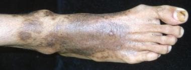
 Pseudo-Kaposi Sarcoma (Acroangiodermatitis): Practice Essentials, Pathophysiology, Etiology
Belated Annual Symposium showcases work from across Edinburgh Infectious Diseases | The University of Edinburgh
 Thieme E-Journals - Indian Journal of Neurosurgery / Abstract
 The Treasures of Orion and His Neighbors
 ENT Dressings - Ear, Nose, Throat - Olympus Medical Systems
Autoimmune Hepatitis Workup: Approach Considerations, Autoantibody Assays, Serum Proteins and Immunoglobulins
Cytoadherence characteristics of Plasmodium falciparum isolates in Thailand using an in vitro human lung endothelial cells...
Figure - Plasmodium knowlesi Infection in Traveler Returning to Canada from the Philippines, 2023 - Volume 29, Number 10...
 Ocular Pathology of Fukuyama Congenital Muscular Dystrophy | IntechOpen
 Drug-induced autoimmune liver disease: A diagnostic dilemma of an increasingly reported disease
 Collins C et al. (2021),
Tubulin acetylation promotes penetrative capaci... -
Paper
Collins C et al. (2021),
Tubulin acetylation promotes penetrative capaci... -
Paper
 Figures and data in Engineering induction of singular neural rosette emergence within hPSC-derived tissues | eLife
 Photographs of mineral No. 19254: Calcite with Sand inclusions from Turkey
 Faculty Profile | College of Graduate Studies | SUNY Upstate
Post-COVID-19 cholangiopathy: Systematic review
Neural Rosettes - Creative Biolabs
 Marin Mineral Company - Mixed Minerals
Marin Mineral Company - Selected Specimens
Retinal Dysplasia Mimicking Intraocular Tumor: MR Imaging Findings with Histopathologic Correlation | American Journal of...
The new World Health Organization classification of lung tumours | European Respiratory Society
Neogomesia
 40 Types of Roses | David Austin's Famous English Roses | HGTV
 Garden Guides | How to Kill Thistles in Grass
 Rapid Intraoperative Diagnosis of Pediatric Brain Tumors Using Stimulated Raman Histology | Cancer Research | American...
Neural rosettes14
- A ) Image of N-cadherin polarized neural rosettes structures within neuroepithelial tissues derived from hPSCs using the E6 method in 6-well TCPS plates. (elifesciences.org)
- Co-staining of neural rosettes with F-actin, ZO-1 tight junction proteins, and phospho-H3 + mitotic nuclei are provided in Figure 1-figure supplement 1 . (elifesciences.org)
- B ) Manual quantification of polarization foci and neural rosettes within micropatterned neuroepithelial tissues of varying morphology. (elifesciences.org)
- C ) Schematic of automated image analysis algorithm for quantification of polarization foci and neural rosettes. (elifesciences.org)
- Neural rosettes refer to radially organized cellular structures. (creative-biolabs.com)
- With the formation of neural rosettes, neural tube development and neural differentiation can be modeled using human pluripotent stem cells (hPSCs) in vitro . (creative-biolabs.com)
- The neural markers expressed in neural rosettes include SOX2, PAX6, and SOX1. (creative-biolabs.com)
- In addition, neural rosettes can differentiate into distinct region-specific neurons and glia. (creative-biolabs.com)
- Recent research has shown that the dismantlement of neural rosettes promotes neurogenesis and astrogenesis prematurely, which means that the complete rosette structure is essential for orderly neurodevelopment. (creative-biolabs.com)
- Fig.2 Involvement of active BMP signaling in differentiation and formation of neural rosettes. (creative-biolabs.com)
- Besides, the mechanism of neural rosettes formation relates to secondary neurulation rather than that of primary neurulation. (creative-biolabs.com)
- The hPSCs-based neural rosettes have been widely used as models for the research of neural tube development as well as neural differentiation in vitro . (creative-biolabs.com)
- Calcium signaling mediates five types of cell morphological changes to form neural rosettes. (creative-biolabs.com)
- Differentiation of neural rosettes from human pluripotent stem cells in vitro is sequentially regulated on a molecular level and accomplished by the mechanism reminiscent of secondary neurulation. (creative-biolabs.com)
Crystals10
- Recently, the trade names "Conchina Druzy" or "Conchina Chalcedony" have been used for this particular kind of formation where Chalcedony is topped with Druzy crystals. (crystal-skulls.com)
- Rosette-shaped formation of scalenohedral calcite crystals up to 3 cm. (johnbetts-fineminerals.com)
- Complete, floater cluster of relatively large Azurite crystals in a somewhat 'bow-tie' formation. (marinmineral.com)
- Beautiful Atacamite specimen featuring two rosette formations deep green crystals. (marinmineral.com)
- Rosette formations of bright 'electric' blue Azurite crystals on matrix. (marinmineral.com)
- This is a fascinating, quality, Pink Amethyst Crystals Geode specimen featuring a beautiful rosette formation, double terminated crystals perched near the center of the pocket and contrasting iron / manganese imbued calcite crystals. (crystalrockology.com)
- Near the wonderfully radiating rosette formation are intermingling, well formed double terminated crystals. (crystalrockology.com)
- A wall or bridge-like structure of secondary growth Pink Amethyst crystals grew alongside the rosette formation, appearing to protect the rosette ball. (crystalrockology.com)
- Crystal Formation - The condition of the facets, faces and terminations (tips / points) of the crystals is also is a very important factor when determining the overall quality of any Pink Amethyst Geode Crystals specimen. (crystalrockology.com)
- Hematite crystallizes as grey-black to reddish colored rhombohedral and tabular crystals, rosettes and in a variety of more unique formations, all with a metallic luster. (satyacenter.com)
Glioneuronal tumor6
- The rosette-forming glioneuronal tumor (RGNT) is an uncommon entity and carries a special character because of its mixed glial and neuronal composition in the histomorphological appearance. (thieme-connect.com)
- Rosette-forming glioneuronal tumor of the septum pellucidum with extension to the supratentorial ventricles: rare case with genetic analysis. (thieme-connect.com)
- 6 Komori T, Scheithauer BW, Hirose T. A rosette-forming glioneuronal tumor of the fourth ventricle: infratentorial form of dysembryoplastic neuroepithelial tumor? (thieme-connect.com)
- Histopathological, molecular, clinical and radiological characterization of rosette-forming glioneuronal tumor in the central nervous system. (thieme-connect.com)
- Malignant transformation of a rosette-forming glioneuronal tumor with IDH1 mutation: a case report and literature review. (thieme-connect.com)
- A rosette-forming glioneuronal tumor of the spinal cord: the first case of a rosetteforming glioneuronal tumor 217 originat- ing from the spinal cord. (thieme-connect.com)
Tumors5
- SLC44A1-PRKCA fusion in papillary and rosette-forming glioneuronal tumors. (thieme-connect.com)
- 5 Sharma P, Swain M, Padua MD, Ranjan A, Lath R. Rosette-forming glioneuronal tumors: a report of two cases. (thieme-connect.com)
- The rosette formation is a histopathological hallmark for multiple tumors, such as a primitive neuroectodermal tumor, human medulloblastoma, glioblastoma, and neuroblastoma. (creative-biolabs.com)
- Rosette-forming glioneuronal tumors (RGNT) are slow-growing WHO Grade I tumors that are characterized by mixed histology and rosette formation. (surgicalneurologyint.com)
- Rosette-forming glioneuronal tumors (RGNT) are rare, slow growing mixed cell tumors. (surgicalneurologyint.com)
Morphology3
- A six-fold assembly of the trimeric computational oligomer had the lowest potential energy per monomer and was consistent with rosette CSC morphology. (osti.gov)
- Effect of Tissue Morphology on Singular Rosette Emergence. (elifesciences.org)
- And inhibition of BMP signaling would result in the expression of these markers and the rosette morphology. (creative-biolabs.com)
Structures4
- A ) Co-localization of F-actin, ZO-1 tight junction proteins, and phospho-H3 + mitotic nuclei within N-cadherin polarized structures confirms neural rosette formation. (elifesciences.org)
- TGFß-treated cells exhibit ring-like structures (podosome rosettes, right panel), where vinculin surrounds F-actin cores (tbottom panel: higher magnification of the boxed areasl). (u-bordeaux.fr)
- Investigating the cellular architecture of the developing cortex, we observed a loss of the radial orientation of radial glial cells, likely due to the disruption of the apical band of adherens junctions, which is the first effect observed after loss of the protein, and the consequent formation of rosette like structures in the brain parenchyma. (uni-muenchen.de)
- Neurocytic rosettes and perivascular structures stained positive for synaptophysin. (surgicalneurologyint.com)
Abstract1
- abstract = "Rosette formation is a feature that has not been described as occurring in melanocytic neoplasms. (elsevierpure.com)
20191
- Gulvady, A., Forsythe, I. and Turner, C.E. (2019) Hic-5 Regulates Src-induced Invadopodia Rosette Formation and Organization. (upstate.edu)
Azurite1
- Fine quality intergrown pair of Azurite rosettes from Bisbee. (marinmineral.com)
Characterization1
- Characterization of Neural Rosette Emergence. (elifesciences.org)
Grasses1
- For the lion cub, the rosette markings act as camouflage in long grasses and bushes. (wikipedia.org)
Specimen1
- This is a high grade Pink Amethyst Geode specimen from Patagonia, Argentina, not the lower grade cut variety from southern Brazil with little crystalline defined formation that is sometimes reconstituted or reconstructed and treated with plastics, dyes, or resins. (crystalrockology.com)
Nebula3
- The Rosette Nebula is a place of intense star formation. (mhcc.edu)
- The Rosette Nebula is a star-forming region about 5,000 light years from Earth. (blogspot.com)
- By combining these results with earlier studies, the scientists conclude that the central cluster formed first, followed by expansion of the nebula, which triggered the formation of the neighboring clusters, including NGC 2237. (blogspot.com)
Elongation1
- 19.50/acre) on rosettes or prior to stem elongation. (unl.edu)
Typically1
- Common mullein spreads solely by seeds that typically germinate in the fall and exist as a rosette, a circular leaf formation at ground level, for a full growing season without producing a stalk or seed. (unl.edu)
20181
- Goreczny, G.J., Forsythe, I. and Turner, C.E. (2018) Hic-5 Regulates Fibrillar Adhesion Formation to Control Tumor Stromal Matrix Remodeling through Interaction with Tensin 1. (upstate.edu)
Representative1
- F ) Representative images depicting stages of rosette emergence and ( G ) rate of singularity within neuroepithelial tissues on select days. (elifesciences.org)
Clusters2
- Rosettes can be grouped in clusters around other spots, or may appear as blotches on the fur. (wikipedia.org)
- Not only will you find areas of intense new star formation near Orion, you can see clusters of extremely hot stars, beautiful glowing gas clouds, evidence of violent star deaths, and much more. (mhcc.edu)
Pathology2
- Rosette forming glioneu- ronal tumor: report of a chiasmal-optic nerve example in neurofibromatosis type 1: special pathology report. (thieme-connect.com)
- Pathology showed a glioneuronal neoplasm with vague neurocytic rosettes and loose perivascular pseudorosettes. (surgicalneurologyint.com)
Mechanism1
- Rosettes are used to camouflage the animal, either as a defense mechanism or as a stalking tool. (wikipedia.org)
Polarization1
- In 'Analyzed' micrograph, the purple outline indicates a rosette and green outlines indicate polarization foci as determined by the image analysis workflow. (elifesciences.org)
Negative1
- The central cell processes of the rosettes were negative for trichome, type IV collagen, neurofilament protein, glial fibrillary acidic protein, and tyrosine hydroxylase. (elsevierpure.com)
Contribution1
- We are presently exploring the contribution of these molecules to podosome formation and functioning. (u-bordeaux.fr)
Markers1
- The downregulation of BRD4 using BRD4 PROTAC, ARV-825, and BRD4 inhibitor, (+)-JQ1, inhibits mammosphere formation and reduces the levels of breast CSC markers (CD44+/CD24- and ALDH1), stem cell marker genes, and mammosphere growth. (bvsalud.org)
Distinct2
- This domesticated cat has a distinct coat pattern with a combination of rosettes, spots, and stripes. (wikipedia.org)
- While individual cheetahs (Acinonyx jubatus) do have distinct and identifiable coat patterns like leopards or jaguars, the cheetah's patterns are considered to be spots, not rosettes. (wikipedia.org)
Cells5
- In addition, there was rosette formation by the tumor cells, with a central aggregate of coarse cell processes. (elsevierpure.com)
- Endothelial podosome rosettes in cultured endothelial cells. (u-bordeaux.fr)
- Multi-cellular rosettes in the mouse visceral endoderm facilitate the ordered migration of anterior visceral endoderm cells. (ox.ac.uk)
- Using both wild-type embryos as well as mutants in which AVE migration is abnormal or arrested, we show that AVE migration is specifically linked to changes in cell packing in the VE and an increase in multi-cellular rosette arrangements (five or more cells meeting at a point). (ox.ac.uk)
- The configuration of E Rosettes with sheep red blood cells and the expression of the epitope detected for the monoclonal antibody anti-CD2 was detected in mononuclear cells from 9 Cebus apella monkeys. (bvsalud.org)
Proteins1
- To elucidate the functions of bromodomain-containing protein 4 (BRD4), nuclear PD-L1, and RelB proteins in the stemness of BCSCs, mammosphere formation was examined using BRD4 inhibitor and degrader, PD-L1 degrader, and RelB inhibitor. (bvsalud.org)
Beautiful1
- This is a beautiful Blue Chalcedony Druzy Rosette from Rio Grande do Sul, Brazil. (crystal-skulls.com)
Form3
- C) Thin smear showing P. knowlesi schizont form with presence of greenish-black pigment and lack of rosette formation. (cdc.gov)
- So a Ombre Rosette cake means gradient cake from dark to light and frosting decorations in the form of a rose. (laavyskitchen.com)
- Taken together, our model and experimental observations suggest that rosettes in the VE epithelium do not form passively in response to AVE migration. (ox.ac.uk)
Color5
- On each color variation, the leopard has rosette patterns on their backs, flanks, and limbs. (wikipedia.org)
- Heathcliffe' features large, fully double flowers of deep rosette shape as well as a deep crimson color. (hgtv.com)
- See notes about color and formation below. (crystalrockology.com)
- Using a 1M star tip from Wilton pipe little rosettes close to each other.Start from the darkest color on the bottom.Add more white frosting to remaining colored frosting so u get a lighter shade.Repeat until u reach the top. (laavyskitchen.com)
- Three ribbons are attached in the color formation of the rosette under the stars. (cavaloop.eu)
Found2
- A rosette is a rose-like marking or formation found on the fur and skin of some animals, particularly cats. (wikipedia.org)
- A Sublight flotilla of worlds arranged in a stable "Klemperer rosette" formation is found fleeing the explosion of the galactic core in Larry Niven 's Ringworld ( 1970 ). (sf-encyclopedia.com)
Mouse3
- Rosette formation by mouse lymphocytes. (jax.org)
- Blue light exposure of the R91W;Nrl −/− mouse could therefore be used to study molecular events preceding edema formation in a cone-rich environment, and thus potentially help to develop treatment strategies for edema-based complications in macular degenerations. (nature.com)
- Our results show that the formation of multi-cellular rosettes in the mouse VE is dependent on normal PCP signalling. (ox.ac.uk)
White1
- In addition, there were multiple clods of 4 white dots in rosette formations. (medscape.com)
Variety1
- Cheetah - the king cheetah variety has rosettes Bengal cat Jaguar Leopard - smaller, denser rosettes than the jaguar, lacking central spots Leopard cat Liger Liliger Lion - cubs have rosettes, which may be retained on the legs in adults Litigon Margay Ocelot Safari cat Snow leopard Tigon Leopard pattern Stripe (pattern) Schneider, Alexsandra (2012). (wikipedia.org)
Image3
- We obtained more direct evidence through generating improved transmission electron microscopy (TEM) images and image averages of the rosette-type CSC, revealing the frequent triangularity and average cross-sectional area in the plasma membrane of its individual lobes. (osti.gov)
- Six trimeric SAXS models nearly filled the space below an average FF-TEM image of the rosette CSC. (osti.gov)
- This composite image shows the Rosette star formation region, located about 5,000 light years from Earth. (blogspot.com)
Large2
- On a large base rosette, 12 large tips are mounted behind a small rosette. (cavaloop.eu)
- The large base rosette ensures the strength of the loop. (cavaloop.eu)
Results1
- Collectively, these results showed that rosette formation plays a role in host immune response by hampering leukocyte phagocytosis. (fiocruz.br)
Unknown1
- The role of one highly occurring phenotype in particular where infected reticulocytes cytoadhere to noninfected normocytes, forming rosettes, remains unknown. (fiocruz.br)
Essential1
- Simulations suggest that while rosettes are not essential for AVE migration, they are crucial for the orderliness of this migration observed in embryos. (ox.ac.uk)
Data1
- In conclusion, the multifaceted data support a rosette CSC with 18 CESAs that mediates the synthesis of a fundamental microfibril composed of 18 glucan chains. (osti.gov)
Role1
- To probe the role of rosettes during AVE migration, we develop a mathematical model of cell movement in the VE. (ox.ac.uk)
Images1
- H ) Schematic of experiment testing the effect of E6 subculture time point on neural rosette emergence within micropatterned neuroepithelial tissues, and ( I ) corresponding images of immunostained tissues 5 days after culture on micropatterned substrates. (elifesciences.org)
Journal1
- Hoang, MP , Rakheja, D & Amirkhan, RH 2003, ' Rosette formation within a proliferative nodule of an atypical combined melanocytic nevus in an adult ', American Journal of Dermatopathology , vol. 25, no. 1, pp. 35-39. (elsevierpure.com)
Center1
- Their leaves grow in a rosette formation, radiating out from the center of the plant. (allotinabox.com)
Cell2
- Fig.1 Cell morphological changes during rosette formation. (creative-biolabs.com)
- All of these functions are of importance during the formation of the cerebral neocortex, which consists of the generation of its different cell types, their migration to their destination and their maturation to a functional network. (uni-muenchen.de)
Presence1
- Apply a selective foliar herbicide to the entire lawn early in the spring when you notice the presence of thistle rosettes in your lawn, but prior to the appearance of flowering stalks. (gardenguides.com)
Application1
- Repeat the application later in the season if you notice the formation of new thistle rosettes appearing in your lawn. (gardenguides.com)
Multiple1
- A six-lobed membrane spanning cellulose synthesis complex (CSC) containing multiple cellulose synthase (CESA) glycosyltransferases mediates cellulose microfibril formation. (osti.gov)
Pattern1
- These big cats have rosettes on their bodies in random combination and pattern. (wikipedia.org)
Ideal1
- Rosette strips prevent granulation formation and are ideal for tympanum splinting. (olympus-europa.com)

.jpg)