Ribosomes
Ribosome Subunits, Small, Archaeal
Ribosome Subunits, Large, Archaeal
Ribosome Subunits
Ribosome Subunits, Large
Ribosome Subunits, Small
Ribosome Subunits, Large, Bacterial
Ribosome Subunits, Small, Bacterial
Ribosome Subunits, Large, Eukaryotic
Ribosome Subunits, Small, Eukaryotic
Protein Subunits
Ribosomal Proteins
Protein Biosynthesis
RNA, Ribosomal
Escherichia coli
Molecular Sequence Data
Ribosome Inactivating Proteins, Type 1
RNA, Transfer
Peptide Chain Elongation, Translational
Peptide Elongation Factor G
Base Sequence
Amino Acid Sequence
Peptide Chain Initiation, Translational
Macromolecular Substances
RNA, Bacterial
Puromycin
RNA, Transfer, Amino Acyl
Poly U
Polyribosomes
Nucleic Acid Conformation
RNA, Messenger
Peptide Elongation Factors
Binding Sites
Protein Binding
Models, Molecular
Mutation
Saccharomyces cerevisiae
Centrifugation, Density Gradient
RNA, Ribosomal, 23S
Electrophoresis, Polyacrylamide Gel
Guanosine Triphosphate
Peptide Biosynthesis
5' Untranslated Regions
Cryoelectron Microscopy
Peptide Initiation Factors
Protein Conformation
Peptide Elongation Factor Tu
Codon
Codon, Initiator
Saccharomyces cerevisiae Proteins
Phenylalanine
Reticulocytes
Protein Structure, Tertiary
Peptide Chain Termination, Translational
Cell Nucleolus
Thermus thermophilus
Ricin
Thiostrepton
RNA, Transfer, Met
RNA, Ribosomal, 28S
Codon, Terminator
Sequence Homology, Amino Acid
Cloning, Molecular
RNA, Transfer, Phe
Cell-Free System
GTP Phosphohydrolase-Linked Elongation Factors
Peptide Elongation Factor 2
RNA, Ribosomal, 18S
Peptidyl Transferases
Magnesium
RNA
Peptide Termination Factors
Microscopy, Electron
RNA-Binding Proteins
Transcription, Genetic
Fusidic Acid
RNA, Fungal
Plasmids
Cross-Linking Reagents
Cell Fractionation
Anticodon
Sparsomycin
Chloramphenicol
Amino Acids
Peptides
GTP-Binding Protein gamma Subunits
GTP-Binding Protein beta Subunits
Viomycin
Cytoplasm
Recombinant Fusion Proteins
RNA Processing, Post-Transcriptional
Rabbits
Protein Synthesis Inhibitors
Sequence Alignment
Eukaryotic Initiation Factor-3
Ultracentrifugation
Structure-Activity Relationship
Mutagenesis, Site-Directed
HeLa Cells
Crystallography, X-Ray
Models, Biological
Dihydrostreptomycin Sulfate
Carbon Isotopes
Tritium
RNA Caps
Chromatography, Gel
Dimerization
Temperature
RNA, Ribosomal, 5.8S
Open Reading Frames
Endoplasmic Reticulum
Conserved Sequence
Protein Structure, Quaternary
Cattle
RNA Precursors
Membrane Proteins
GTP-Binding Proteins
Streptomycin
Imidoesters
Proteins
Erythromycin
Chloroplasts
Adenosine Triphosphatases
Prokaryotic Initiation Factor-3
Frameshifting, Ribosomal
Mitochondria
Lincomycin
Endoplasmic Reticulum, Rough
Peptide Elongation Factor 1
Eukaryotic Initiation Factor-4G
Cell Membrane
Eukaryotic Initiation Factors
DNA Primers
Nuclear Proteins
Proton-Translocating ATPases
Transfection
Multiprotein Complexes
Species Specificity
Signal Recognition Particle
Liver
RNA, Ribosomal, 16S
Endoribonucleases
Picornaviridae
Eukaryotic Cells
Guanosine Diphosphate
Prokaryotic Initiation Factors
GTP-Binding Protein alpha Subunits
Peptide Fragments
Protein Transport
Protein Multimerization
Multienzyme Complexes
Substrate specificity and properties of the Escherichia coli 16S rRNA methyltransferase, RsmE. (1/117)
The small ribosome subunit of Escherichia coli contains 10 base-methylated sites distributed in important functional regions. At present, seven enzymes responsible for methylation of eight bases are known, but most of them have not been well characterized. One of these enzymes, RsmE, was recently identified and shown to specifically methylate U1498. Here we describe the enzymatic properties and substrate specificity of RsmE. The enzyme forms dimers in solution and is most active in the presence of 10-15 mM Mg(2+) and 100 mM NH(4)Cl at pH 7-9; however, in the presence of spermidine, Mg(2+) is not required for activity. While small ribosome subunits obtained from an RsmE deletion strain can be methylated by purified RsmE, neither 70S ribosomes nor 50S subunits are active. Likewise, 16S rRNA obtained from the mutant strain, synthetic 16S rRNA, and 3' minor domain RNA are all very poor or inactive as substrates. 30S particles partially depleted of proteins by treatment with high concentrations of LiCl or in vitro reconstituted intermediate particles also show little or no methyl acceptor activity. Based on these data, we conclude that RsmE requires a highly structured ribonucleoprotein particle as a substrate for methylation, and that methylation events in the 3' minor domain of 16S rRNA probably occur late during 30S ribosome assembly. (+info)Analysis of structural dynamics in the ribosome by TLS crystallographic refinement. (2/117)
A major goal in the study of ribosome structure and function is to obtain a complete description of the conformational dynamics of the ribosome during the many steps of protein synthesis. Here, we report a new approach to the study of ribosome dynamics using translation-libration-screw (TLS) refinement against experimental X-ray diffraction data. TLS analysis of complexes of the 70 S ribosome suggests that many of its structural features have an inherent tendency for anisotropic movement. Analysis of displacements of the 30 S and 50 S ribosomal subunits reveals an intrinsic bias for "ratchet-like" intersubunit rotation. The libration axes for both subunits pass through the peptidyl transferase center (PTC), indicating a tendency for structural rotations to occur around the site of peptide bond formation. The modes of anisotropic movement of ribosomal RNA components, including the head of the 30 S subunit, the L1 and L11 stalks and the two main arms of the tRNAs were found to correlate with their respective modes of movement previously inferred from comparisons of ribosomes trapped in different functional states. In the small subunit, the mobilities of features interacting with the Shine-Dalgarno helix are decreased in the presence of the Shine-Dalgarno helix, supporting the proposal that that formation of the Shine-Dalgarno helix during initiation may contribute to stabilization of the small subunit for optimal interaction with initiator tRNAfMet. The similarity of TLS parameters for two independently solved structures of similar ribosome complexes suggests that TLS analysis can provide useful information about the dynamics of very large macromolecular objects and at resolutions lower than those at which TLS refinement has commonly been applied. (+info)Structural aspects of RbfA action during small ribosomal subunit assembly. (3/117)
Ribosome binding factor A (RbfA) is a bacterial cold shock response protein, required for an efficient processing of the 5' end of the 16S ribosomal RNA (rRNA) during assembly of the small (30S) ribosomal subunit. Here we present a crystal structure of Thermus thermophilus (Tth) RbfA and a three-dimensional cryo-electron microscopic (EM) map of the Tth 30S*RbfA complex. RbfA binds to the 30S subunit in a position overlapping the binding sites of the A and P site tRNAs, and RbfA's functionally important C terminus extends toward the 5' end of the 16S rRNA. In the presence of RbfA, a portion of the 16S rRNA encompassing helix 44, which is known to be directly involved in mRNA decoding and tRNA binding, is displaced. These results shed light on the role played by RbfA during maturation of the 30S subunit, and also indicate how RbfA provides cells with a translational advantage under conditions of cold shock. (+info)Kinetic analysis of the effects of translation enhancers in translation initiation. (4/117)
Translation initiation is the most important step within a series of protein biosynthesis processes because the incorporation of ribosomes to a mRNA mainly determines efficiencies of translation. In bacteria, translation enhancers located on the 5' upstream of the Shine-Dargalno (SD) sequence on mRNAs are known to accelerate the efficiency of protein biosynthesis. To investigate the role of translation enhancers in translation initiation, we analyzed binding kinetics of a 30S ribosomal subunit to a mRNA immobilized on a 27 MHz quartz-crystal microbalance (QCM). The association constant (Ka) was rather low for the mRNA including a translation enhancer sequence compared with that for the mRNA without translation enhancers. These kinetic parameters suggest that translation enhancers destabilize the ribosome-mRNA complex on an SD sequence to move on the next step of decoding its mRNA. (+info)Interaction between RsgA and the ribosome. (5/117)
RsgA is a unique GTP hydrolytic protein that is widely found in bacteria and plants, and is activated by the small subunit of the ribosome. Disruption of the gene for RsgA from the genome affects the growth of cells, the subunit association of the ribosome in cells and maturation of 16S ribosomal RNA. Here, we investigated the interaction between EscherichiacoliRsgA and the ribosome. Several antibiotics bound to the decoding center of the small subunit inhibited the ribosome-dependent GTPase activity of RsgA, suggesting that RsgA binds to the decoding center. Chemical footprinting was also performed to further investigate the interaction. (+info)Heterogeneous rRNA molecules encoded by Streptomyces coelicolor M145 genome are all expressed and assembled into ribosomes. (6/117)
The Streptomyces coelicolor M145 genome harbors six copies of divergent rRNA operons that differ at ~0.2% and ~0.6% of the nucleotide positions in small subunit (SSU) and large subunit (LSU) rRNA genes, respectively. When these rRNA genes are expressed, a single cell may harbor three different kinds of SSU rRNA and five kinds of LSU rRNA. Primer extension analyses revealed that all of the heterogeneous rRNA molecules are expressed and assembled into ribosomes. This finding and the maintenance of the intragenomic variability of rRNA operons imply the existence of functional divergence of rRNA species in this developmentally complex microorganism. (+info)Structural basis for hygromycin B inhibition of protein biosynthesis. (7/117)
(+info)Critical residues for cofactor binding and catalytic activity in the aminoglycoside resistance methyltransferase Sgm. (8/117)
(+info)Ribosomes are complex macromolecular structures composed of ribonucleic acid (RNA) and proteins that play a crucial role in protein synthesis within cells. They serve as the site for translation, where messenger RNA (mRNA) is translated into a specific sequence of amino acids to create a polypeptide chain, which eventually folds into a functional protein.
Ribosomes consist of two subunits: a smaller subunit and a larger subunit. These subunits are composed of ribosomal RNA (rRNA) molecules and proteins. In eukaryotic cells, the smaller subunit is denoted as the 40S subunit, while the larger subunit is referred to as the 60S subunit. In prokaryotic cells, these subunits are named the 30S and 50S subunits, respectively. The ribosome's overall structure resembles a "doughnut" or a "cotton reel," with grooves and binding sites for various factors involved in protein synthesis.
Ribosomes can be found floating freely within the cytoplasm of cells or attached to the endoplasmic reticulum (ER) membrane, forming part of the rough ER. Membrane-bound ribosomes are responsible for synthesizing proteins that will be transported across the ER and ultimately secreted from the cell or inserted into the membrane. In contrast, cytoplasmic ribosomes synthesize proteins destined for use within the cytoplasm or organelles.
In summary, ribosomes are essential components of cells that facilitate protein synthesis by translating mRNA into functional polypeptide chains. They can be found in various cellular locations and exist as either free-floating entities or membrane-bound structures.
A ribosome is a complex molecular machine found in all living cells that translates messenger RNA (mRNA) into proteins. Ribosomes are composed of two subunits: a small subunit and a large subunit. The small subunit is responsible for recognizing and binding to the mRNA, as well as decoding the genetic information it contains.
Archaeal ribosomes are similar in structure and function to eukaryotic ribosomes, but they have some distinct differences in their composition and sequence. Archaeal small ribosomal subunits, like those of bacteria, are composed of a 16S rRNA molecule and approximately 20 proteins. However, the archaeal small ribosomal subunit has a unique structure and composition that is distinct from both bacterial and eukaryotic small ribosomal subunits.
The small ribosomal subunit of Archaea is referred to as the "small, archaeal" subunit. It plays a crucial role in the initiation of protein synthesis by recognizing and binding to the Shine-Dalgarno sequence in the mRNA, which helps position the start codon for translation. The small, archaeal ribosomal subunit also contains the decoding center, where the genetic information in the mRNA is translated into a corresponding amino acid sequence during protein synthesis.
Overall, the small, archaeal ribosomal subunit is an essential component of the archaeal translational machinery, responsible for accurately and efficiently decoding genetic information and initiating the synthesis of new proteins.
A large archaeal ribosomal subunit refers to the larger of the two components that make up the archaeal ribosome, which is the complex molecular machine responsible for protein synthesis in archaea. The large ribosomal subunit plays a crucial role in the elongation phase of translation, where it helps catalyze the formation of peptide bonds between amino acids during protein synthesis.
The large ribosomal subunit of archaea is composed of ribosomal RNA (rRNA) and proteins. Specifically, the archaeal large ribosomal subunit contains a 23S rRNA molecule, a 5S rRNA molecule, and around 30-40 different proteins. These components are organized into several distinct structural domains, including the central protuberance, the L1 stalk, and the peptidyl transferase center (PTC), which is where peptide bond formation occurs.
It's worth noting that while archaeal ribosomes share some similarities with eukaryotic ribosomes, they are more closely related to bacterial ribosomes in terms of their structure and composition. However, the large ribosomal subunit of archaea is still distinct from both bacterial and eukaryotic subunits in its specific rRNA sequences and protein composition.
A ribosome is a complex molecular machine found in all living cells, responsible for protein synthesis. It consists of two subunits: the smaller **ribosomal subunit** and the larger **ribosomal subunit**. These subunits are composed of ribosomal RNA (rRNA) and ribosomal proteins.
The small ribosomal subunit is responsible for decoding messenger RNA (mRNA) during protein synthesis, while the large ribosomal subunit facilitates peptide bond formation between amino acids. In eukaryotic cells, the small ribosomal subunit is composed of one 18S rRNA and approximately 30 ribosomal proteins, whereas the large ribosomal subunit contains three larger rRNAs (5S, 5.8S, and 28S or 25S) and around 45-50 ribosomal proteins.
In prokaryotic cells like bacteria, the small ribosomal subunit consists of a single 16S rRNA and approximately 21 ribosomal proteins, while the large ribosomal subunit contains three rRNAs (5S, 5.8S, and 23S) and around 30-33 ribosomal proteins.
These ribosome subunits come together during protein synthesis to form a functional ribosome, which translates the genetic code present in mRNA into a polypeptide chain (protein).
A ribosome is a complex molecular machine found in all living cells, responsible for protein synthesis. It consists of two subunits: the large subunit and the small subunit. The large ribosomal subunit plays a crucial role in the elongation phase of protein synthesis, where it helps catalyze the formation of peptide bonds between amino acids.
The Large Ribosomal Subunit, also known as the 60S subunit in eukaryotic cells (50S in prokaryotic cells), is composed of ribosomal RNA (rRNA) and numerous proteins. In humans, the large ribosomal subunit contains three rRNA molecules (28S, 5.8S, and 5S rRNA) and approximately 49 distinct proteins. Its primary function is to bind to the small ribosomal subunit and form a functional ribosome, which then translates messenger RNA (mRNA) into a polypeptide chain during protein synthesis.
The large ribosomal subunit has several key features, including the peptidyl transferase center (PTC), where peptide bonds are formed between amino acids, and the exit tunnel, through which the nascent polypeptide chain passes as it is being synthesized. The PTC is a crucial component of the large subunit, as it facilitates the transfer of activated amino acids from transfer RNA (tRNA) molecules to the growing polypeptide chain during translation.
In summary, the Large Ribosomal Subunit is a vital component of the ribosome responsible for catalyzing peptide bond formation and facilitating the synthesis of proteins within cells.
A ribosome is a complex molecular machine found in all living cells, responsible for protein synthesis. It consists of two subunits: the small and the large subunit. The small ribosomal subunit plays a crucial role in decoding the messenger RNA (mRNA) molecule and positioning transfer RNA (tRNA) molecules during translation.
The small ribosomal subunit, specifically, is composed of ribosomal RNA (rRNA) and proteins. In eukaryotic cells, the small ribosomal subunit is composed of a 18S rRNA molecule and approximately 30 distinct proteins. Its primary function is to recognize the start codon on the mRNA and facilitate the binding of the initiator tRNA (tRNAi) to begin the translation process.
Together, the small and large ribosomal subunits form a functional ribosome that translates genetic information from mRNA into proteins, contributing to the maintenance and growth of cells.
A ribosome is a complex molecular machine found in all living cells that serves as the site for protein synthesis. In bacteria, ribosomes are composed of two subunits: a smaller subunit and a larger subunit. The large bacterial ribosomal subunit is referred to as the 50S subunit.
The 50S subunit of bacterial ribosomes is a large ribonucleoprotein complex with an estimated molecular weight of approximately 1.5-2 MDa. It is composed of three ribosomal RNA (rRNA) molecules and around 30 distinct proteins. The rRNA molecules in the 50S subunit include the 23S rRNA, which plays a crucial role in peptidyl transferase activity, and the 5S rRNA, which is involved in ribosome stability and translation fidelity.
The large ribosomal subunit is responsible for catalyzing the formation of peptide bonds between amino acids during protein synthesis. It also contains binding sites for transfer RNAs (tRNAs) and various antibiotics that inhibit bacterial protein synthesis. The 50S subunit has a complex structure, with several distinct domains and functional centers, including the peptidyl transferase center, the decoding center, and the exit tunnel for nascent polypeptides.
Understanding the structure and function of the large bacterial ribosomal subunit is important for developing new antibiotics that target bacterial protein synthesis and for understanding the mechanisms of antibiotic resistance.
A small bacterial ribosomal subunit refers to a component of the ribosome in bacteria, which is responsible for protein synthesis. Specifically, it refers to the 30S subunit, which is composed of one 16S rRNA molecule and approximately 21 distinct proteins. This subunit plays a crucial role in decoding the mRNA template during translation, ensuring that the correct amino acids are added to the growing polypeptide chain. The small ribosomal subunit interacts with the mRNA and tRNAs during this process, facilitating accurate and efficient protein synthesis.
A large ribosomal subunit in eukaryotic cells is a complex macromolecular structure composed of ribosomal RNA (rRNA) and proteins. It is one of the two subunits that make up the eukaryotic ribosome, which is the site of protein synthesis in the cell. The large subunit is responsible for catalyzing the formation of peptide bonds between amino acids during protein synthesis.
In eukaryotes, the large ribosomal subunit is composed of three rRNA molecules (5S, 5.8S, and 28S) and approximately 49 proteins. The large subunit has a characteristic shape with a prominent protuberance called the "stalk" that contains proteins involved in binding translation factors and messenger RNA (mRNA).
The large ribosomal subunit plays a critical role in the elongation phase of protein synthesis, where it binds to the small ribosomal subunit and mRNA to form a functional ribosome. The large subunit moves along the mRNA, reading the genetic code and catalyzing the formation of peptide bonds between amino acids as they are brought to the ribosome by transfer RNA (tRNA) molecules.
A small ribosomal subunit in eukaryotic cells is a complex cellular structure composed of ribosomal RNA (rRNA) and proteins. It is one of the two subunits that make up the eukaryotic ribosome, which is the site of protein synthesis in the cell. The small subunit is responsible for recognizing and binding to the messenger RNA (mRNA) molecule and decoding the genetic information it contains into a specific sequence of amino acids.
In eukaryotic cells, the small ribosomal subunit is composed of a 18S rRNA molecule and approximately 30 different proteins. The 18S rRNA molecule forms the core of the subunit and provides the structural framework for the binding of the proteins. Together, the rRNA and proteins form a compact and highly organized structure that is capable of carrying out the precise and efficient decoding of mRNA.
The small ribosomal subunit plays a critical role in the initiation of protein synthesis, as it is responsible for recognizing and binding to the cap structure at the 5' end of the mRNA molecule. This interaction allows the subunit to scan along the mRNA until it encounters the start codon, which signals the beginning of the protein-coding region. Once the start codon is located, the small subunit recruits the large ribosomal subunit and initiates the process of elongation, in which the amino acids are linked together to form a polypeptide chain.
Overall, the small ribosomal subunit is an essential component of the eukaryotic protein synthesis machinery, and its proper function is critical for the maintenance of cellular homeostasis and the regulation of gene expression.
A protein subunit refers to a distinct and independently folding polypeptide chain that makes up a larger protein complex. Proteins are often composed of multiple subunits, which can be identical or different, that come together to form the functional unit of the protein. These subunits can interact with each other through non-covalent interactions such as hydrogen bonds, ionic bonds, and van der Waals forces, as well as covalent bonds like disulfide bridges. The arrangement and interaction of these subunits contribute to the overall structure and function of the protein.
Ribosomal proteins are a type of protein that play a crucial role in the structure and function of ribosomes, which are complex molecular machines found within all living cells. Ribosomes are responsible for translating messenger RNA (mRNA) into proteins during the process of protein synthesis.
Ribosomal proteins can be divided into two categories based on their location within the ribosome:
1. Large ribosomal subunit proteins: These proteins are associated with the larger of the two subunits of the ribosome, which is responsible for catalyzing peptide bond formation during protein synthesis.
2. Small ribosomal subunit proteins: These proteins are associated with the smaller of the two subunits of the ribosome, which is responsible for binding to the mRNA and decoding the genetic information it contains.
Ribosomal proteins have a variety of functions, including helping to stabilize the structure of the ribosome, assisting in the binding of substrates and cofactors necessary for protein synthesis, and regulating the activity of the ribosome. Mutations in ribosomal proteins can lead to a variety of human diseases, including developmental disorders, neurological conditions, and cancer.
Protein biosynthesis is the process by which cells generate new proteins. It involves two major steps: transcription and translation. Transcription is the process of creating a complementary RNA copy of a sequence of DNA. This RNA copy, or messenger RNA (mRNA), carries the genetic information to the site of protein synthesis, the ribosome. During translation, the mRNA is read by transfer RNA (tRNA) molecules, which bring specific amino acids to the ribosome based on the sequence of nucleotides in the mRNA. The ribosome then links these amino acids together in the correct order to form a polypeptide chain, which may then fold into a functional protein. Protein biosynthesis is essential for the growth and maintenance of all living organisms.
Ribosomal RNA (rRNA) is a type of RNA molecule that is a key component of ribosomes, which are the cellular structures where protein synthesis occurs in cells. In ribosomes, rRNA plays a crucial role in the process of translation, where genetic information from messenger RNA (mRNA) is translated into proteins.
Ribosomal RNA is synthesized in the nucleus and then transported to the cytoplasm, where it assembles with ribosomal proteins to form ribosomes. Within the ribosome, rRNA provides a structural framework for the assembly of the ribosome and also plays an active role in catalyzing the formation of peptide bonds between amino acids during protein synthesis.
There are several different types of rRNA molecules, including 5S, 5.8S, 18S, and 28S rRNA, which vary in size and function. These rRNA molecules are highly conserved across different species, indicating their essential role in protein synthesis and cellular function.
'Escherichia coli' (E. coli) is a type of gram-negative, facultatively anaerobic, rod-shaped bacterium that commonly inhabits the intestinal tract of humans and warm-blooded animals. It is a member of the family Enterobacteriaceae and one of the most well-studied prokaryotic model organisms in molecular biology.
While most E. coli strains are harmless and even beneficial to their hosts, some serotypes can cause various forms of gastrointestinal and extraintestinal illnesses in humans and animals. These pathogenic strains possess virulence factors that enable them to colonize and damage host tissues, leading to diseases such as diarrhea, urinary tract infections, pneumonia, and sepsis.
E. coli is a versatile organism with remarkable genetic diversity, which allows it to adapt to various environmental niches. It can be found in water, soil, food, and various man-made environments, making it an essential indicator of fecal contamination and a common cause of foodborne illnesses. The study of E. coli has contributed significantly to our understanding of fundamental biological processes, including DNA replication, gene regulation, and protein synthesis.
Molecular sequence data refers to the specific arrangement of molecules, most commonly nucleotides in DNA or RNA, or amino acids in proteins, that make up a biological macromolecule. This data is generated through laboratory techniques such as sequencing, and provides information about the exact order of the constituent molecules. This data is crucial in various fields of biology, including genetics, evolution, and molecular biology, allowing for comparisons between different organisms, identification of genetic variations, and studies of gene function and regulation.
Ribosome-inactivating proteins (RIPs) are a type of protein that can inhibit the function of ribosomes, which are the cellular structures responsible for protein synthesis. Ribosome-inactivating proteins are classified into two types: Type 1 and Type 2.
Type 1 Ribosome-Inactivating Proteins (RIPs) are defined as single-chain proteins that inhibit protein synthesis by depurinating a specific adenine residue in the sarcin-ricin loop of the large rRNA molecule within the ribosome. This results in the irreversible inactivation of the ribosome, preventing it from participating in further protein synthesis.
Type 1 RIPs are found in various plant species and have been identified as potential therapeutic agents for cancer treatment due to their ability to selectively inhibit protein synthesis in cancer cells. However, they can also be toxic to normal cells, which limits their clinical use. Examples of Type 1 RIPs include dianthin, gelonin, and trichosanthin.
Transfer RNA (tRNA) is a type of RNA molecule that plays a crucial role in protein synthesis, the process by which cells create proteins. In protein synthesis, tRNAs serve as adaptors, translating the genetic code present in messenger RNA (mRNA) into the corresponding amino acids required to build a protein.
Each tRNA molecule has a distinct structure, consisting of approximately 70-90 nucleotides arranged in a cloverleaf shape with several loops and stems. The most important feature of a tRNA is its anticodon, a sequence of three nucleotides located in one of the loops. This anticodon base-pairs with a complementary codon on the mRNA during translation, ensuring that the correct amino acid is added to the growing polypeptide chain.
Before tRNAs can participate in protein synthesis, they must be charged with their specific amino acids through an enzymatic process involving aminoacyl-tRNA synthetases. These enzymes recognize and bind to both the tRNA and its corresponding amino acid, forming a covalent bond between them. Once charged, the aminoacyl-tRNA complex is ready to engage in translation and contribute to protein formation.
In summary, transfer RNA (tRNA) is a small RNA molecule that facilitates protein synthesis by translating genetic information from messenger RNA into specific amino acids, ultimately leading to the creation of functional proteins within cells.
Translational peptide chain elongation is the process during protein synthesis where activated amino acids are added to the growing peptide chain in a sequence determined by the genetic code present in messenger RNA (mRNA). This process involves several steps:
1. Recognition of the start codon on the mRNA by the small ribosomal subunit, which binds to the mRNA and brings an initiator tRNA with a methionine or formylmethionine amino acid attached into the P site (peptidyl site) of the ribosome.
2. The large ribosomal subunit then joins the small subunit, forming a complete ribosome complex.
3. An incoming charged tRNA with an appropriate amino acid, complementary to the next codon on the mRNA, binds to the A site (aminoacyl site) of the ribosome.
4. Peptidyl transferase, a catalytic domain within the large ribosomal subunit, facilitates the formation of a peptide bond between the amino acids attached to the tRNAs in the P and A sites. The methionine or formylmethionine initiator amino acid is now covalently linked to the second amino acid via this peptide bond.
5. Translocation occurs, moving the tRNA with the growing peptide chain from the P site to the E site (exit site) and shifting the mRNA by one codon relative to the ribosome. The uncharged tRNA is then released from the E site.
6. The next charged tRNA carrying an appropriate amino acid binds to the A site, and the process repeats until a stop codon is reached on the mRNA.
7. Upon encountering a stop codon, release factors recognize it and facilitate the release of the completed polypeptide chain from the final tRNA in the P site. The ribosome then dissociates from the mRNA, allowing for further translational events to occur.
Translational peptide chain elongation is a crucial step in protein synthesis and requires precise coordination between various components of the translation machinery, including ribosomes, tRNAs, amino acids, and numerous accessory proteins.
Peptide Elongation Factor G is a term used in the field of molecular biology, specifically in the process of protein synthesis. It is a bacterial enzyme that plays a crucial role in the elongation stage of translation, which is the process by which genetic information encoded in messenger RNA (mRNA) is converted into a polypeptide chain or protein.
More specifically, Peptide Elongation Factor G (also known as EF-G or Translocase) is responsible for the translocation step during translation. After each amino acid is added to the growing peptide chain, the mRNA and tRNAs must move relative to the ribosome so that the next codon in the mRNA can be read. EF-G facilitates this movement by using energy from GTP hydrolysis to cause a conformational change in the ribosome, resulting in the translocation of the mRNA and tRNAs by one codon.
In summary, Peptide Elongation Factor G is a bacterial enzyme that plays an essential role in the elongation stage of protein synthesis by facilitating the movement of mRNA and tRNAs relative to the ribosome during translation.
A base sequence in the context of molecular biology refers to the specific order of nucleotides in a DNA or RNA molecule. In DNA, these nucleotides are adenine (A), guanine (G), cytosine (C), and thymine (T). In RNA, uracil (U) takes the place of thymine. The base sequence contains genetic information that is transcribed into RNA and ultimately translated into proteins. It is the exact order of these bases that determines the genetic code and thus the function of the DNA or RNA molecule.
An amino acid sequence is the specific order of amino acids in a protein or peptide molecule, formed by the linking of the amino group (-NH2) of one amino acid to the carboxyl group (-COOH) of another amino acid through a peptide bond. The sequence is determined by the genetic code and is unique to each type of protein or peptide. It plays a crucial role in determining the three-dimensional structure and function of proteins.
Peptide chain initiation in translational terms refers to the process by which the synthesis of a protein begins on a ribosome. This is the first step in translation, where the small ribosomal subunit binds to an mRNA molecule at the start codon (usually AUG), bringing with it the initiator tRNA charged with a specific amino acid (often N-formylmethionine in prokaryotes or methionine in eukaryotes). The large ribosomal subunit then joins this complex, forming a functional initiation complex. This marks the beginning of the elongation phase, where subsequent amino acids are added to the growing peptide chain until termination is reached.
Macromolecular substances, also known as macromolecules, are large, complex molecules made up of repeating subunits called monomers. These substances are formed through polymerization, a process in which many small molecules combine to form a larger one. Macromolecular substances can be naturally occurring, such as proteins, DNA, and carbohydrates, or synthetic, such as plastics and synthetic fibers.
In the context of medicine, macromolecular substances are often used in the development of drugs and medical devices. For example, some drugs are designed to bind to specific macromolecules in the body, such as proteins or DNA, in order to alter their function and produce a therapeutic effect. Additionally, macromolecular substances may be used in the creation of medical implants, such as artificial joints and heart valves, due to their strength and durability.
It is important for healthcare professionals to have an understanding of macromolecular substances and how they function in the body, as this knowledge can inform the development and use of medical treatments.
Bacterial RNA refers to the genetic material present in bacteria that is composed of ribonucleic acid (RNA). Unlike higher organisms, bacteria contain a single circular chromosome made up of DNA, along with smaller circular pieces of DNA called plasmids. These bacterial genetic materials contain the information necessary for the growth and reproduction of the organism.
Bacterial RNA can be divided into three main categories: messenger RNA (mRNA), ribosomal RNA (rRNA), and transfer RNA (tRNA). mRNA carries genetic information copied from DNA, which is then translated into proteins by the rRNA and tRNA molecules. rRNA is a structural component of the ribosome, where protein synthesis occurs, while tRNA acts as an adapter that brings amino acids to the ribosome during protein synthesis.
Bacterial RNA plays a crucial role in various cellular processes, including gene expression, protein synthesis, and regulation of metabolic pathways. Understanding the structure and function of bacterial RNA is essential for developing new antibiotics and other therapeutic strategies to combat bacterial infections.
Puromycin is an antibiotic and antiviral protein synthesis inhibitor. It works by being incorporated into the growing peptide chain during translation, causing premature termination and release of the incomplete polypeptide. This results in the inhibition of protein synthesis and ultimately leads to cell death. In research, puromycin is often used as a selective agent in cell culture to kill cells that have not been transfected with a plasmid containing a resistance gene for puromycin.
Transfer RNA (tRNA) is a type of RNA molecule that plays a crucial role in protein synthesis. It serves as the adaptor molecule that translates the genetic code present in messenger RNA (mRNA) into the corresponding amino acids, which are then linked together to form a polypeptide chain during protein synthesis.
Aminoacyl tRNA is a specific type of tRNA molecule that has been charged or activated with an amino acid. This process is called aminoacylation and is carried out by enzymes called aminoacyl-tRNA synthetases. Each synthetase specifically recognizes and attaches a particular amino acid to its corresponding tRNA, ensuring the fidelity of protein synthesis. Once an amino acid is attached to a tRNA, it forms an aminoacyl-tRNA complex, which can then participate in translation and contribute to the formation of a new protein.
I'm sorry for any confusion, but "Poly U" is not a recognized medical term or abbreviation in the English language. It could potentially refer to Polytechnic University or Hong Kong Polytechnic University, but it does not have a specific medical connotation. If you have more context or information, I'd be happy to help further!
Polyribosomes, also known as polysomes, are clusters of ribosomes that are translating the same mRNA molecule simultaneously. They can be found in the cytoplasm of eukaryotic cells and are responsible for the synthesis of proteins. The mRNA molecule serves as a template for the translation process, with multiple ribosomes moving along it and producing multiple copies of the same protein. This allows for efficient and rapid production of large quantities of a single protein. Polyribosomes can be found in high numbers in cells that are actively synthesizing proteins, such as secretory cells or cells undergoing growth and division.
Nucleic acid conformation refers to the three-dimensional structure that nucleic acids (DNA and RNA) adopt as a result of the bonding patterns between the atoms within the molecule. The primary structure of nucleic acids is determined by the sequence of nucleotides, while the conformation is influenced by factors such as the sugar-phosphate backbone, base stacking, and hydrogen bonding.
Two common conformations of DNA are the B-form and the A-form. The B-form is a right-handed helix with a diameter of about 20 Å and a pitch of 34 Å, while the A-form has a smaller diameter (about 18 Å) and a shorter pitch (about 25 Å). RNA typically adopts an A-form conformation.
The conformation of nucleic acids can have significant implications for their function, as it can affect their ability to interact with other molecules such as proteins or drugs. Understanding the conformational properties of nucleic acids is therefore an important area of research in molecular biology and medicine.
Messenger RNA (mRNA) is a type of RNA (ribonucleic acid) that carries genetic information copied from DNA in the form of a series of three-base code "words," each of which specifies a particular amino acid. This information is used by the cell's machinery to construct proteins, a process known as translation. After being transcribed from DNA, mRNA travels out of the nucleus to the ribosomes in the cytoplasm where protein synthesis occurs. Once the protein has been synthesized, the mRNA may be degraded and recycled. Post-transcriptional modifications can also occur to mRNA, such as alternative splicing and addition of a 5' cap and a poly(A) tail, which can affect its stability, localization, and translation efficiency.
Peptide elongation factors are a group of proteins that play a crucial role in the process of protein synthesis in cells, specifically during the elongation stage of translation. They assist in the addition of amino acids to the growing polypeptide chain by facilitating the binding of aminoacyl-tRNAs (transfer RNAs with attached amino acids) to the ribosome, where protein synthesis occurs.
In prokaryotic cells, there are two main peptide elongation factors: EF-Tu and EF-G. EF-Tu forms a complex with aminoacyl-tRNA and delivers it to the ribosome's acceptor site (A-site), where the incoming amino acid is matched with the corresponding codon on the mRNA. Once the correct match is made, GTP hydrolysis occurs, releasing EF-Tu from the complex, allowing for peptide bond formation between the new amino acid and the growing polypeptide chain.
EF-G then enters the scene to facilitate translocation, the movement of the ribosome along the mRNA, which shifts the newly formed peptidyl-tRNA from the A-site to the P-site (peptidyl-tRNA site) and makes room for another aminoacyl-tRNA in the A-site. This process continues until protein synthesis is complete.
In eukaryotic cells, the equivalent proteins are called EF1α, EF1β, EF1γ, and EF2 (also known as eEF1A, eEF1B, eEF1G, and eEF2). The overall function remains similar to that in prokaryotes, but the specific mechanisms and protein names differ.
In the context of medical and biological sciences, a "binding site" refers to a specific location on a protein, molecule, or cell where another molecule can attach or bind. This binding interaction can lead to various functional changes in the original protein or molecule. The other molecule that binds to the binding site is often referred to as a ligand, which can be a small molecule, ion, or even another protein.
The binding between a ligand and its target binding site can be specific and selective, meaning that only certain ligands can bind to particular binding sites with high affinity. This specificity plays a crucial role in various biological processes, such as signal transduction, enzyme catalysis, or drug action.
In the case of drug development, understanding the location and properties of binding sites on target proteins is essential for designing drugs that can selectively bind to these sites and modulate protein function. This knowledge can help create more effective and safer therapeutic options for various diseases.
Protein binding, in the context of medical and biological sciences, refers to the interaction between a protein and another molecule (known as the ligand) that results in a stable complex. This process is often reversible and can be influenced by various factors such as pH, temperature, and concentration of the involved molecules.
In clinical chemistry, protein binding is particularly important when it comes to drugs, as many of them bind to proteins (especially albumin) in the bloodstream. The degree of protein binding can affect a drug's distribution, metabolism, and excretion, which in turn influence its therapeutic effectiveness and potential side effects.
Protein-bound drugs may be less available for interaction with their target tissues, as only the unbound or "free" fraction of the drug is active. Therefore, understanding protein binding can help optimize dosing regimens and minimize adverse reactions.
Molecular models are three-dimensional representations of molecular structures that are used in the field of molecular biology and chemistry to visualize and understand the spatial arrangement of atoms and bonds within a molecule. These models can be physical or computer-generated and allow researchers to study the shape, size, and behavior of molecules, which is crucial for understanding their function and interactions with other molecules.
Physical molecular models are often made up of balls (representing atoms) connected by rods or sticks (representing bonds). These models can be constructed manually using materials such as plastic or wooden balls and rods, or they can be created using 3D printing technology.
Computer-generated molecular models, on the other hand, are created using specialized software that allows researchers to visualize and manipulate molecular structures in three dimensions. These models can be used to simulate molecular interactions, predict molecular behavior, and design new drugs or chemicals with specific properties. Overall, molecular models play a critical role in advancing our understanding of molecular structures and their functions.
In the context of medicine and pharmacology, "kinetics" refers to the study of how a drug moves throughout the body, including its absorption, distribution, metabolism, and excretion (often abbreviated as ADME). This field is called "pharmacokinetics."
1. Absorption: This is the process of a drug moving from its site of administration into the bloodstream. Factors such as the route of administration (e.g., oral, intravenous, etc.), formulation, and individual physiological differences can affect absorption.
2. Distribution: Once a drug is in the bloodstream, it gets distributed throughout the body to various tissues and organs. This process is influenced by factors like blood flow, protein binding, and lipid solubility of the drug.
3. Metabolism: Drugs are often chemically modified in the body, typically in the liver, through processes known as metabolism. These changes can lead to the formation of active or inactive metabolites, which may then be further distributed, excreted, or undergo additional metabolic transformations.
4. Excretion: This is the process by which drugs and their metabolites are eliminated from the body, primarily through the kidneys (urine) and the liver (bile).
Understanding the kinetics of a drug is crucial for determining its optimal dosing regimen, potential interactions with other medications or foods, and any necessary adjustments for special populations like pediatric or geriatric patients, or those with impaired renal or hepatic function.
A mutation is a permanent change in the DNA sequence of an organism's genome. Mutations can occur spontaneously or be caused by environmental factors such as exposure to radiation, chemicals, or viruses. They may have various effects on the organism, ranging from benign to harmful, depending on where they occur and whether they alter the function of essential proteins. In some cases, mutations can increase an individual's susceptibility to certain diseases or disorders, while in others, they may confer a survival advantage. Mutations are the driving force behind evolution, as they introduce new genetic variability into populations, which can then be acted upon by natural selection.
"Saccharomyces cerevisiae" is not typically considered a medical term, but it is a scientific name used in the field of microbiology. It refers to a species of yeast that is commonly used in various industrial processes, such as baking and brewing. It's also widely used in scientific research due to its genetic tractability and eukaryotic cellular organization.
However, it does have some relevance to medical fields like medicine and nutrition. For example, certain strains of S. cerevisiae are used as probiotics, which can provide health benefits when consumed. They may help support gut health, enhance the immune system, and even assist in the digestion of certain nutrients.
In summary, "Saccharomyces cerevisiae" is a species of yeast with various industrial and potential medical applications.
Centrifugation, Density Gradient is a medical laboratory technique used to separate and purify different components of a mixture based on their size, density, and shape. This method involves the use of a centrifuge and a density gradient medium, such as sucrose or cesium chloride, to create a stable density gradient within a column or tube.
The sample is carefully layered onto the top of the gradient and then subjected to high-speed centrifugation. During centrifugation, the particles in the sample move through the gradient based on their size, density, and shape, with heavier particles migrating faster and further than lighter ones. This results in the separation of different components of the mixture into distinct bands or zones within the gradient.
This technique is commonly used to purify and concentrate various types of biological materials, such as viruses, organelles, ribosomes, and subcellular fractions, from complex mixtures. It allows for the isolation of pure and intact particles, which can then be collected and analyzed for further study or use in downstream applications.
In summary, Centrifugation, Density Gradient is a medical laboratory technique used to separate and purify different components of a mixture based on their size, density, and shape using a centrifuge and a density gradient medium.
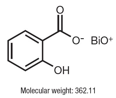
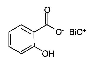
Molecular weight, also known as molecular mass, is the mass of a molecule. It is expressed in units of atomic mass units (amu) or daltons (Da). Molecular weight is calculated by adding up the atomic weights of each atom in a molecule. It is a useful property in chemistry and biology, as it can be used to determine the concentration of a substance in a solution, or to calculate the amount of a substance that will react with another in a chemical reaction.
23S Ribosomal RNA (rRNA) is a type of rRNA that is a component of the large ribosomal subunit in both prokaryotic and eukaryotic cells. In prokaryotes, the large ribosomal subunit contains 50S, which consists of 23S rRNA, 5S rRNA, and around 33 proteins. The 23S rRNA plays a crucial role in the decoding of mRNA during protein synthesis and also participates in the formation of the peptidyl transferase center, where peptide bonds are formed between amino acids.
The 23S rRNA is a long RNA molecule that contains both coding and non-coding regions. It has a complex secondary structure, which includes several domains and subdomains, as well as numerous stem-loop structures. These structures are important for the proper functioning of the ribosome during protein synthesis.
In addition to its role in protein synthesis, 23S rRNA has been used as a target for antibiotics that inhibit bacterial growth. For example, certain antibiotics bind to specific regions of the 23S rRNA and interfere with the function of the ribosome, thereby preventing bacterial protein synthesis and growth. However, because eukaryotic cells do not have a 23S rRNA equivalent, these antibiotics are generally not toxic to human cells.
Electrophoresis, polyacrylamide gel (EPG) is a laboratory technique used to separate and analyze complex mixtures of proteins or nucleic acids (DNA or RNA) based on their size and electrical charge. This technique utilizes a matrix made of cross-linked polyacrylamide, a type of gel, which provides a stable and uniform environment for the separation of molecules.
In this process:
1. The polyacrylamide gel is prepared by mixing acrylamide monomers with a cross-linking agent (bis-acrylamide) and a catalyst (ammonium persulfate) in the presence of a buffer solution.
2. The gel is then poured into a mold and allowed to polymerize, forming a solid matrix with uniform pore sizes that depend on the concentration of acrylamide used. Higher concentrations result in smaller pores, providing better resolution for separating smaller molecules.
3. Once the gel has set, it is placed in an electrophoresis apparatus containing a buffer solution. Samples containing the mixture of proteins or nucleic acids are loaded into wells on the top of the gel.
4. An electric field is applied across the gel, causing the negatively charged molecules to migrate towards the positive electrode (anode) while positively charged molecules move toward the negative electrode (cathode). The rate of migration depends on the size, charge, and shape of the molecules.
5. Smaller molecules move faster through the gel matrix and will migrate farther from the origin compared to larger molecules, resulting in separation based on size. Proteins and nucleic acids can be selectively stained after electrophoresis to visualize the separated bands.
EPG is widely used in various research fields, including molecular biology, genetics, proteomics, and forensic science, for applications such as protein characterization, DNA fragment analysis, cloning, mutation detection, and quality control of nucleic acid or protein samples.
Guanosine triphosphate (GTP) is a nucleotide that plays a crucial role in various cellular processes, such as protein synthesis, signal transduction, and regulation of enzymatic activities. It serves as an energy currency, similar to adenosine triphosphate (ATP), and undergoes hydrolysis to guanosine diphosphate (GDP) or guanosine monophosphate (GMP) to release energy required for these processes. GTP is also a precursor for the synthesis of other essential molecules, including RNA and certain signaling proteins. Additionally, it acts as a molecular switch in many intracellular signaling pathways by binding and activating specific GTPase proteins.
Peptide biosynthesis is the process by which cells synthesize peptides, short chains of amino acids. This process is mediated by enzymes called peptide synthetases, which catalyze the formation of peptide bonds between individual amino acids to create a longer chain. Peptide biosynthesis typically occurs through one of two pathways: ribosomal or non-ribosomal.
Ribosomal peptide biosynthesis involves the use of the cell's translational machinery, including the ribosome and transfer RNAs (tRNAs), to synthesize peptides from a messenger RNA (mRNA) template. This process is highly regulated and typically results in the production of small, linear peptides that are further modified by enzymes to create bioactive molecules such as hormones or neurotransmitters.
Non-ribosomal peptide biosynthesis (NRPS), on the other hand, is a more complex process that involves large multifunctional enzyme complexes called non-ribosomal peptide synthetases (NRPSs). These enzymes are capable of synthesizing a wide variety of structurally diverse peptides, including cyclic and branched peptides, as well as those containing non-proteinogenic amino acids. NRPSs typically consist of multiple modules, each responsible for adding a single amino acid to the growing peptide chain. The modular nature of NRPS systems allows for great diversity in the types of peptides that can be synthesized, making them important sources of bioactive molecules with potential therapeutic applications.
Untranslated regions (UTRs) are sections of an mRNA molecule that do not contain information for protein synthesis. There are two types of UTRs: 5' UTR, which is located at the 5' end of the mRNA molecule, and 3' UTR, which is located at the 3' end.
The 5' UTR typically contains regulatory elements that control the translation of the mRNA into protein. These elements can affect the efficiency and timing of translation, as well as the stability of the mRNA molecule. The 5' UTR may also contain upstream open reading frames (uORFs), which are short sequences that can be translated into small peptides and potentially regulate the translation of the main coding sequence.
The length and sequence composition of the 5' UTR can have significant impacts on gene expression, and variations in these regions have been associated with various diseases, including cancer and neurological disorders. Therefore, understanding the structure and function of 5' UTRs is an important area of research in molecular biology and genetics.
Cryo-electron microscopy (Cryo-EM) is a type of electron microscopy where the sample is studied at cryogenic temperatures, typically liquid nitrogen temperatures. This technique is used to investigate the structure and shape of biological molecules and complexes, viruses, and other nanoscale particles.
In Cryo-EM, the sample is rapidly frozen to preserve its natural structure and then imaged using a beam of electrons. The images are collected at different angles and then computationally combined to generate a 3D reconstruction of the sample. This technique allows researchers to visualize biological structures in their native environment with near-atomic resolution, providing valuable insights into their function and behavior.
Cryo-EM has become an increasingly popular tool in structural biology due to its ability to image large and complex structures that are difficult or impossible to crystallize for X-ray crystallography. It has been used to determine the structures of many important biological molecules, including membrane proteins, ribosomes, viruses, and protein complexes involved in various cellular processes.
Peptide initiation factors are a group of proteins involved in the process of protein synthesis in cells, specifically during the initial stage of elongation called initiation. In this phase, they assist in the assembly of the ribosome, an organelle composed of ribosomal RNA and proteins, at the start codon of a messenger RNA (mRNA) molecule. This marks the beginning of the translation process where the genetic information encoded in the mRNA is translated into a specific protein sequence.
There are three main peptide initiation factors in eukaryotic cells:
1. eIF-2 (eukaryotic Initiation Factor 2): This factor plays a crucial role in binding methionyl-tRNAi, the initiator tRNA, to the small ribosomal subunit. It does so by forming a complex with GTP and the methionyl-tRNAi, which then binds to the 40S ribosomal subunit. Once bound, eIF-2-GTP-Met-tRNAi recognizes the start codon (AUG) on the mRNA.
2. eIF-3: This is a large multiprotein complex that interacts with both the small and large ribosomal subunits and helps stabilize their interaction during initiation. It also plays a role in recruiting other initiation factors to the preinitiation complex.
3. eIF-4F: This factor is a heterotrimeric protein complex consisting of eIF-4A (an ATP-dependent RNA helicase), eIF-4E (which binds the m7G cap structure at the 5' end of most eukaryotic mRNAs), and eIF-4G (a scaffolding protein that bridges interactions between eIF-4A, eIF-4E, and other initiation factors). eIF-4F helps unwind secondary structures in the 5' untranslated region (5' UTR) of mRNAs, promoting efficient recruitment of the 43S preinitiation complex to the mRNA.
Together, these peptide initiation factors facilitate the recognition of the correct start codon and ensure efficient translation initiation in eukaryotic cells.
Bacterial proteins are a type of protein that are produced by bacteria as part of their structural or functional components. These proteins can be involved in various cellular processes, such as metabolism, DNA replication, transcription, and translation. They can also play a role in bacterial pathogenesis, helping the bacteria to evade the host's immune system, acquire nutrients, and multiply within the host.
Bacterial proteins can be classified into different categories based on their function, such as:
1. Enzymes: Proteins that catalyze chemical reactions in the bacterial cell.
2. Structural proteins: Proteins that provide structural support and maintain the shape of the bacterial cell.
3. Signaling proteins: Proteins that help bacteria to communicate with each other and coordinate their behavior.
4. Transport proteins: Proteins that facilitate the movement of molecules across the bacterial cell membrane.
5. Toxins: Proteins that are produced by pathogenic bacteria to damage host cells and promote infection.
6. Surface proteins: Proteins that are located on the surface of the bacterial cell and interact with the environment or host cells.
Understanding the structure and function of bacterial proteins is important for developing new antibiotics, vaccines, and other therapeutic strategies to combat bacterial infections.
Protein conformation refers to the specific three-dimensional shape that a protein molecule assumes due to the spatial arrangement of its constituent amino acid residues and their associated chemical groups. This complex structure is determined by several factors, including covalent bonds (disulfide bridges), hydrogen bonds, van der Waals forces, and ionic bonds, which help stabilize the protein's unique conformation.
Protein conformations can be broadly classified into two categories: primary, secondary, tertiary, and quaternary structures. The primary structure represents the linear sequence of amino acids in a polypeptide chain. The secondary structure arises from local interactions between adjacent amino acid residues, leading to the formation of recurring motifs such as α-helices and β-sheets. Tertiary structure refers to the overall three-dimensional folding pattern of a single polypeptide chain, while quaternary structure describes the spatial arrangement of multiple folded polypeptide chains (subunits) that interact to form a functional protein complex.
Understanding protein conformation is crucial for elucidating protein function, as the specific three-dimensional shape of a protein directly influences its ability to interact with other molecules, such as ligands, nucleic acids, or other proteins. Any alterations in protein conformation due to genetic mutations, environmental factors, or chemical modifications can lead to loss of function, misfolding, aggregation, and disease states like neurodegenerative disorders and cancer.
Peptide Elongation Factor Tu, also known as EF-Tu or Tuf, is a protein involved in the process of protein synthesis in prokaryotic cells. It plays a crucial role in the elongation phase of translation, where it facilitates the addition of amino acids to the growing polypeptide chain during protein synthesis.
EF-Tu functions as a binding protein for aminoacyl-tRNA (transfer RNA) complexes. In this role, EF-Tu forms a ternary complex with GTP (guanosine triphosphate) and an aminoacyl-tRNA, which then binds to the A (acceptor) site of the small ribosomal subunit. Once aligned, the GTP in the EF-Tu-tRNA complex is hydrolyzed to GDP (guanosine diphosphate), causing a conformational change that releases the aminoacyl-tRNA into the A site for peptide bond formation.
After releasing the tRNA, EF-Tu recharges with another GTP molecule and is ready to form another ternary complex, thus continuing its role in the elongation of protein synthesis. The recycling of EF-Tu between GDP and GTP forms is facilitated by another elongation factor, EF-Ts (or Tsf).
In summary, Peptide Elongation Factor Tu (EF-Tu) is a vital protein in prokaryotic cells that binds to aminoacyl-tRNA and GTP, forming a ternary complex. This complex delivers the aminoacyl-tRNA to the ribosome for peptide bond formation during protein synthesis elongation.
A codon is a sequence of three adjacent nucleotides in DNA or RNA that specifies the insertion of a particular amino acid during protein synthesis, or signals the beginning or end of translation. In DNA, these triplets are read during transcription to produce a complementary mRNA molecule, which is then translated into a polypeptide chain during translation. There are 64 possible codons in the standard genetic code, with 61 encoding for specific amino acids and three serving as stop codons that signal the termination of protein synthesis.
A codon is a sequence of three nucleotides in DNA or RNA that specifies a particular amino acid or signals the start or stop of protein synthesis. In the context of protein synthesis, an initiator codon is the specific codon that signifies the beginning of the translation process and sets the reading frame for the mRNA sequence.
The most common initiator codon in DNA and RNA is AUG, which encodes the amino acid methionine. In some cases, however, alternative initiation codons such as GUG (valine) or UUG (leucine) may be used. It's worth noting that the use of these alternative initiator codons can vary depending on the organism and the specific gene in question.
Once the initiator codon is recognized by the ribosome, the translation machinery begins to assemble and begin synthesizing the protein according to the genetic code specified by the mRNA sequence.
Saccharomyces cerevisiae proteins are the proteins that are produced by the budding yeast, Saccharomyces cerevisiae. This organism is a single-celled eukaryote that has been widely used as a model organism in scientific research for many years due to its relatively simple genetic makeup and its similarity to higher eukaryotic cells.
The genome of Saccharomyces cerevisiae has been fully sequenced, and it is estimated to contain approximately 6,000 genes that encode proteins. These proteins play a wide variety of roles in the cell, including catalyzing metabolic reactions, regulating gene expression, maintaining the structure of the cell, and responding to environmental stimuli.
Many Saccharomyces cerevisiae proteins have human homologs and are involved in similar biological processes, making this organism a valuable tool for studying human disease. For example, many of the proteins involved in DNA replication, repair, and recombination in yeast have human counterparts that are associated with cancer and other diseases. By studying these proteins in yeast, researchers can gain insights into their function and regulation in humans, which may lead to new treatments for disease.
Phenylalanine is an essential amino acid, meaning it cannot be produced by the human body and must be obtained through diet or supplementation. It's one of the building blocks of proteins and is necessary for the production of various molecules in the body, such as neurotransmitters (chemical messengers in the brain).
Phenylalanine has two forms: L-phenylalanine and D-phenylalanine. L-phenylalanine is the form found in proteins and is used by the body for protein synthesis, while D-phenylalanine has limited use in humans and is not involved in protein synthesis.
Individuals with a rare genetic disorder called phenylketonuria (PKU) must follow a low-phenylalanine diet or take special medical foods because they are unable to metabolize phenylalanine properly, leading to its buildup in the body and potential neurological damage.
Reticulocytes are immature red blood cells that still contain remnants of organelles, such as ribosomes and mitochondria, which are typically found in developing cells. These organelles are involved in the process of protein synthesis and energy production, respectively. Reticulocytes are released from the bone marrow into the bloodstream, where they continue to mature into fully developed red blood cells called erythrocytes.
Reticulocytes can be identified under a microscope by their staining characteristics, which reveal a network of fine filaments or granules known as the reticular apparatus. This apparatus is composed of residual ribosomal RNA and other proteins that have not yet been completely eliminated during the maturation process.
The percentage of reticulocytes in the blood can be used as a measure of bone marrow function and erythropoiesis, or red blood cell production. An increased reticulocyte count may indicate an appropriate response to blood loss, hemolysis, or other conditions that cause anemia, while a decreased count may suggest impaired bone marrow function or a deficiency in erythropoietin, the hormone responsible for stimulating red blood cell production.
Tertiary protein structure refers to the three-dimensional arrangement of all the elements (polypeptide chains) of a single protein molecule. It is the highest level of structural organization and results from interactions between various side chains (R groups) of the amino acids that make up the protein. These interactions, which include hydrogen bonds, ionic bonds, van der Waals forces, and disulfide bridges, give the protein its unique shape and stability, which in turn determines its function. The tertiary structure of a protein can be stabilized by various factors such as temperature, pH, and the presence of certain ions. Any changes in these factors can lead to denaturation, where the protein loses its tertiary structure and thus its function.
Peptide chain termination, translational, refers to the process in protein synthesis where the addition of new amino acids to a growing peptide chain is stopped. This event occurs when a special type of transfer RNA (tRNA), carrying a specific termination codon (UAA, UAG, or UGA) instead of an amino acid, binds to the corresponding stop codon at the ribosome.
This interaction recruits release factors, which hydrolyze the bond between the last amino acid and the tRNA, releasing the completed polypeptide chain from the ribosome. The process of peptide chain termination is essential for accurate protein synthesis and preventing errors during translation. Dysregulation or mutations in this process can lead to various genetic disorders and diseases.
'Escherichia coli (E. coli) proteins' refer to the various types of proteins that are produced and expressed by the bacterium Escherichia coli. These proteins play a critical role in the growth, development, and survival of the organism. They are involved in various cellular processes such as metabolism, DNA replication, transcription, translation, repair, and regulation.
E. coli is a gram-negative, facultative anaerobe that is commonly found in the intestines of warm-blooded organisms. It is widely used as a model organism in scientific research due to its well-studied genetics, rapid growth, and ability to be easily manipulated in the laboratory. As a result, many E. coli proteins have been identified, characterized, and studied in great detail.
Some examples of E. coli proteins include enzymes involved in carbohydrate metabolism such as lactase, sucrase, and maltose; proteins involved in DNA replication such as the polymerases, single-stranded binding proteins, and helicases; proteins involved in transcription such as RNA polymerase and sigma factors; proteins involved in translation such as ribosomal proteins, tRNAs, and aminoacyl-tRNA synthetases; and regulatory proteins such as global regulators, two-component systems, and transcription factors.
Understanding the structure, function, and regulation of E. coli proteins is essential for understanding the basic biology of this important organism, as well as for developing new strategies for combating bacterial infections and improving industrial processes involving bacteria.
The nucleolus is a structure found within the nucleus of eukaryotic cells (cells that contain a true nucleus). It plays a central role in the production and assembly of ribosomes, which are complex molecular machines responsible for protein synthesis. The nucleolus is not a distinct organelle with a membrane surrounding it, but rather a condensed region within the nucleus where ribosomal biogenesis takes place.
The process of ribosome formation begins in the nucleolus with the transcription of ribosomal DNA (rDNA) genes into long precursor RNA molecules called rRNAs (ribosomal RNAs). Within the nucleolus, these rRNA molecules are cleaved, modified, and assembled together with ribosomal proteins to form small and large ribosomal subunits. Once formed, these subunits are transported through the nuclear pores to the cytoplasm, where they come together to form functional ribosomes that can engage in protein synthesis.
In addition to its role in ribosome biogenesis, the nucleolus has been implicated in other cellular processes such as stress response, cell cycle regulation, and aging. Changes in nucleolar structure and function have been associated with various diseases, including cancer and neurodegenerative disorders.
'Thermus thermophilus' is not a medical term, but a scientific name for a species of bacteria. It is commonly used in molecular biology and genetics research. Here is the biological definition:
'Thermus thermophilus' is a gram-negative, rod-shaped, thermophilic bacterium found in hot springs and other high-temperature environments. Its optimum growth temperature ranges from 65 to 70°C (149-158°F), with some strains able to grow at temperatures as high as 85°C (185°F). The bacterium's DNA polymerase enzyme, Taq polymerase, is widely used in the Polymerase Chain Reaction (PCR) technique for amplifying and analyzing DNA. 'Thermus thermophilus' has a single circular chromosome and can also have one or more plasmids. Its genome has been fully sequenced, making it an important model organism for studying extremophiles and their adaptations to harsh environments.
Ricin is defined as a highly toxic protein that is derived from the seeds of the castor oil plant (Ricinus communis). It can be produced as a white, powdery substance or a mistable aerosol. Ricin works by getting inside cells and preventing them from making the proteins they need. Without protein, cells die. Eventually, this can cause organ failure and death.
It is not easily inhaled or absorbed through the skin, but if ingested or injected, it can be lethal in very small amounts. There is no antidote for ricin poisoning - treatment consists of supportive care. Ricin has been used as a bioterrorism agent in the past and continues to be a concern due to its relative ease of production and potential high toxicity.
Thiostrepton is an antibiotic and antiproliferative agent that is derived from the bacterium Streptomyces azureus. It belongs to the family of thiostreptons, which are cyclic oligopeptides with unique structures and various biological activities. Thiostrepton has been used primarily in veterinary medicine for the treatment of infections caused by gram-positive bacteria, such as mastitis in cows.
In addition to its antibacterial properties, thiostrepton has also been found to have antiproliferative and proapoptotic effects on various cancer cells, including breast, ovarian, and colon cancer cells. These effects are thought to be mediated by the inhibition of protein synthesis and the regulation of gene expression. However, its use as a therapeutic agent in humans is still being investigated due to its potential toxicity and limited bioavailability.
It's worth noting that thiostrepton is not commonly used in clinical practice, and its medical definition is mainly related to its chemical structure, antibacterial properties, and potential anticancer effects.
Transfer RNA (tRNA) is a type of RNA molecule that plays a crucial role in protein synthesis, the process by which cells create proteins. During protein synthesis, tRNAs serve as adaptors, translating the genetic code present in messenger RNA (mRNA) into the corresponding amino acids required to build a protein.
Each tRNA molecule has an anticodon region that can base-pair with specific codons (three-nucleotide sequences) on the mRNA. At the other end of the tRNA is the acceptor stem, which contains a binding site for the corresponding amino acid. When an amino acid attaches to the tRNA, it forms an ester bond between the carboxyl group of the amino acid and the 3'-hydroxyl group of the ribose in the tRNA. This aminoacylated tRNA then participates in the translation process, delivering the amino acid to the growing polypeptide chain at the ribosome.
In summary, transfer RNA (tRNA) is a type of RNA molecule that facilitates protein synthesis by transporting and delivering specific amino acids to the ribosome for incorporation into a polypeptide chain, based on the codon-anticodon pairing between tRNAs and messenger RNA (mRNA).
28S ribosomal RNA (rRNA) is a component of the large subunit of the eukaryotic ribosome, which is the site of protein synthesis in the cell. The ribosome is composed of two subunits, one large and one small, that come together around an mRNA molecule to translate it into a protein.
The 28S rRNA is a type of rRNA that is found in the large subunit of the eukaryotic ribosome, along with the 5S and 5.8S rRNAs. Together, these rRNAs make up the structural framework of the ribosome and play a crucial role in the process of translation.
The 28S rRNA is synthesized in the nucleolus as a precursor RNA (pre-rRNA) that undergoes several processing steps, including cleavage and modification, to produce the mature 28S rRNA molecule. The length of the 28S rRNA varies between species, but it is typically around 4700-5000 nucleotides long in humans.
Abnormalities in the structure or function of the 28S rRNA can lead to defects in protein synthesis and have been implicated in various diseases, including cancer and neurological disorders.
A codon is a sequence of three adjacent nucleotides in DNA or RNA that specifies a particular amino acid during the process of protein synthesis, or codes for the termination of translation. In DNA, these triplets are read in a 5' to 3' direction, while in mRNA, they are read in a 5' to 3' direction as well. There are 64 possible codons (4^3) in the genetic code, and 61 of them specify amino acids. The remaining three codons, UAA, UAG, and UGA, are terminator or stop codons that signal the end of protein synthesis.
Terminator codons, also known as nonsense codons, do not code for any amino acids. Instead, they cause the release of the newly synthesized polypeptide chain from the ribosome, which is the complex machinery responsible for translating the genetic code into a protein. This process is called termination or translation termination.
In prokaryotic cells, termination occurs when a release factor recognizes and binds to the stop codon in the A site of the ribosome. This triggers the hydrolysis of the peptidyl-tRNA bond, releasing the completed polypeptide chain from the tRNA and the ribosome. In eukaryotic cells, a similar process occurs, but it involves different release factors and additional steps to ensure accurate termination.
In summary, a codon is a sequence of three adjacent nucleotides in DNA or RNA that specifies an amino acid or signals the end of protein synthesis. Terminator codons are specific codons that do not code for any amino acids and instead signal the end of translation, leading to the release of the newly synthesized polypeptide chain from the ribosome.
Sequence homology, amino acid, refers to the similarity in the order of amino acids in a protein or a portion of a protein between two or more species. This similarity can be used to infer evolutionary relationships and functional similarities between proteins. The higher the degree of sequence homology, the more likely it is that the proteins are related and have similar functions. Sequence homology can be determined through various methods such as pairwise alignment or multiple sequence alignment, which compare the sequences and calculate a score based on the number and type of matching amino acids.
Molecular cloning is a laboratory technique used to create multiple copies of a specific DNA sequence. This process involves several steps:
1. Isolation: The first step in molecular cloning is to isolate the DNA sequence of interest from the rest of the genomic DNA. This can be done using various methods such as PCR (polymerase chain reaction), restriction enzymes, or hybridization.
2. Vector construction: Once the DNA sequence of interest has been isolated, it must be inserted into a vector, which is a small circular DNA molecule that can replicate independently in a host cell. Common vectors used in molecular cloning include plasmids and phages.
3. Transformation: The constructed vector is then introduced into a host cell, usually a bacterial or yeast cell, through a process called transformation. This can be done using various methods such as electroporation or chemical transformation.
4. Selection: After transformation, the host cells are grown in selective media that allow only those cells containing the vector to grow. This ensures that the DNA sequence of interest has been successfully cloned into the vector.
5. Amplification: Once the host cells have been selected, they can be grown in large quantities to amplify the number of copies of the cloned DNA sequence.
Molecular cloning is a powerful tool in molecular biology and has numerous applications, including the production of recombinant proteins, gene therapy, functional analysis of genes, and genetic engineering.
Transfer RNA (tRNA) is a type of RNA molecule that helps translate genetic information from messenger RNA (mRNA) into proteins. Each tRNA carries a specific amino acid to the growing polypeptide chain during protein synthesis, based on the anticodon sequence in its variable loop region that recognizes and binds to a complementary codon sequence in the mRNA.
Phenylalanine (Phe) is one of the twenty standard amino acids found in proteins. It has a hydrophobic side chain, which means it tends to repel water and interact with other non-polar molecules. In tRNA, phenylalanine is attached to a specific tRNA molecule known as tRNAPhe. This tRNA recognizes the mRNA codons UUC and UUU, which specify phenylalanine during protein synthesis.
A cell-free system is a biochemical environment in which biological reactions can occur outside of an intact living cell. These systems are often used to study specific cellular processes or pathways, as they allow researchers to control and manipulate the conditions in which the reactions take place. In a cell-free system, the necessary enzymes, substrates, and cofactors for a particular reaction are provided in a test tube or other container, rather than within a whole cell.
Cell-free systems can be derived from various sources, including bacteria, yeast, and mammalian cells. They can be used to study a wide range of cellular processes, such as transcription, translation, protein folding, and metabolism. For example, a cell-free system might be used to express and purify a specific protein, or to investigate the regulation of a particular metabolic pathway.
One advantage of using cell-free systems is that they can provide valuable insights into the mechanisms of cellular processes without the need for time-consuming and resource-intensive cell culture or genetic manipulation. Additionally, because cell-free systems are not constrained by the limitations of a whole cell, they offer greater flexibility in terms of reaction conditions and the ability to study complex or transient interactions between biological molecules.
Overall, cell-free systems are an important tool in molecular biology and biochemistry, providing researchers with a versatile and powerful means of investigating the fundamental processes that underlie life at the cellular level.
Ribonucleases (RNases) are a group of enzymes that catalyze the degradation of ribonucleic acid (RNA) molecules by hydrolyzing the phosphodiester bonds. These enzymes play crucial roles in various biological processes, such as RNA processing, turnover, and quality control. They can be classified into several types based on their specificities, mechanisms, and cellular localizations.
Some common classes of ribonucleases include:
1. Endoribonucleases: These enzymes cleave RNA internally, at specific sequences or structural motifs. Examples include RNase A, which targets single-stranded RNA; RNase III, which cuts double-stranded RNA at specific stem-loop structures; and RNase T1, which recognizes and cuts unpaired guanosine residues in RNA molecules.
2. Exoribonucleases: These enzymes remove nucleotides from the ends of RNA molecules. They can be further divided into 5'-3' exoribonucleases, which degrade RNA starting from the 5' end, and 3'-5' exoribonucleases, which start at the 3' end. Examples include Xrn1, a 5'-3' exoribonuclease involved in mRNA decay; and Dis3/RRP6, a 3'-5' exoribonuclease that participates in ribosomal RNA processing and degradation.
3. Specific ribonucleases: These enzymes target specific RNA molecules or regions with high precision. For example, RNase P is responsible for cleaving the 5' leader sequence of precursor tRNAs (pre-tRNAs) during their maturation; and RNase MRP is involved in the processing of ribosomal RNA and mitochondrial RNA molecules.
Dysregulation or mutations in ribonucleases have been implicated in various human diseases, such as neurological disorders, cancer, and viral infections. Therefore, understanding their functions and mechanisms is crucial for developing novel therapeutic strategies.
GTP (Guanosine Triphosphate) Phosphohydrolase-Linked Elongation Factors are a group of proteins that play a crucial role in protein synthesis, specifically in the elongation phase of translation. These factors use the energy released from GTP hydrolysis to facilitate various steps in the addition of amino acids to the growing polypeptide chain during protein synthesis.
In prokaryotic cells, there are two main GTP Phosphohydrolase-Linked Elongation Factors: EF-Tu (Elongation Factor Thermos unstable) and EF-G (Elongation Factor G).
EF-Tu forms a complex with aminoacyl-tRNA and GTP, which then binds to the ribosome. Upon correct codon-anticodon recognition, GTP is hydrolyzed to GDP, releasing EF-Tu from the ribosome and allowing for the addition of the amino acid to the polypeptide chain.
EF-G, on the other hand, facilitates the translocation of the peptidyl-tRNA from the A site to the P site of the ribosome after peptide bond formation, using GTP hydrolysis as an energy source. This movement makes room for a new aminoacyl-tRNA to bind and continue the elongation process.
In eukaryotic cells, there are functionally equivalent factors called EF1A (eEF1A) and EF2 (eEF2), which perform similar roles in protein synthesis.
Peptide Elongation Factor 2 (PEF2), also known as Elongation Factor-G (EF-G) in prokaryotes or Translation Elongation Factor 2 (TEF2) in eukaryotes, is a vital protein involved in the elongation phase of protein synthesis, specifically during translation. It facilitates the translocation of peptidyl-tRNA from the A-site to the P-site of the ribosome, thereby enabling the addition of new amino acids to the growing polypeptide chain.
During this process, PEF2/EF-G/TEF2 binds to the ribosome and utilizes the energy from GTP hydrolysis to induce a conformational change in the ribosome, leading to the translocation of peptidyl-tRNA and mRNA. After completing the translocation step, PEF2/EF-G/TEF2 is released from the ribosome and can be reused in subsequent elongation cycles.
In summary, Peptide Elongation Factor 2 (PEF2) is a crucial player in protein synthesis that facilitates the movement of peptidyl-tRNA within the ribosome during translation, allowing for the continuous addition of amino acids to the nascent polypeptide chain.
18S rRNA (ribosomal RNA) is the smaller subunit of the eukaryotic ribosome, which is the cellular organelle responsible for protein synthesis. The "18S" refers to the sedimentation coefficient of this rRNA molecule, which is a measure of its rate of sedimentation in a centrifuge and is expressed in Svedberg units (S).
The 18S rRNA is a component of the 40S subunit of the ribosome, and it plays a crucial role in the decoding of messenger RNA (mRNA) during protein synthesis. Specifically, the 18S rRNA helps to form the structure of the ribosome and contains several conserved regions that are involved in binding to mRNA and guiding the movement of transfer RNAs (tRNAs) during translation.
The 18S rRNA is also a commonly used molecular marker for evolutionary studies, as its sequence is highly conserved across different species and can be used to infer phylogenetic relationships between organisms. Additionally, the analysis of 18S rRNA gene sequences has been widely used in various fields such as ecology, environmental science, and medicine to study biodiversity, biogeography, and infectious diseases.
Peptidyl transferase is not a medical term per se, but rather a biochemical term used to describe an enzymatic function or activity. It is often mentioned in the context of molecular biology, protein synthesis, and ribosome structure.
Peptidyl transferase refers to the catalytic activity of ribosomes that facilitates the formation of peptide bonds between amino acids during protein synthesis. More specifically, peptidyl transferase is responsible for transferring the peptidyl group (the growing polypeptide chain) from the acceptor site (A-site) to the donor site (P-site) of the ribosome, creating a new peptide bond and elongating the polypeptide chain. This activity occurs within the large subunit of the ribosome, near the peptidyl transferase center (PTC).
While it is often attributed to the ribosomal RNA (rRNA) component of the ribosome, recent research suggests that both rRNA and specific ribosomal proteins contribute to this enzymatic activity.
Magnesium is an essential mineral that plays a crucial role in various biological processes in the human body. It is the fourth most abundant cation in the body and is involved in over 300 enzymatic reactions, including protein synthesis, muscle and nerve function, blood glucose control, and blood pressure regulation. Magnesium also contributes to the structural development of bones and teeth.
In medical terms, magnesium deficiency can lead to several health issues, such as muscle cramps, weakness, heart arrhythmias, and seizures. On the other hand, excessive magnesium levels can cause symptoms like diarrhea, nausea, and muscle weakness. Magnesium supplements or magnesium-rich foods are often recommended to maintain optimal magnesium levels in the body.
Some common dietary sources of magnesium include leafy green vegetables, nuts, seeds, legumes, whole grains, and dairy products. Magnesium is also available in various forms as a dietary supplement, including magnesium oxide, magnesium citrate, magnesium chloride, and magnesium glycinate.
RNA (Ribonucleic Acid) is a single-stranded, linear polymer of ribonucleotides. It is a nucleic acid present in the cells of all living organisms and some viruses. RNAs play crucial roles in various biological processes such as protein synthesis, gene regulation, and cellular signaling. There are several types of RNA including messenger RNA (mRNA), ribosomal RNA (rRNA), transfer RNA (tRNA), small nuclear RNA (snRNA), microRNA (miRNA), and long non-coding RNA (lncRNA). These RNAs differ in their structure, function, and location within the cell.
Peptide termination factors, also known as release factors, are proteins involved in the process of protein biosynthesis in cells. Specifically, they play a crucial role in the termination step of translation, which is the process by which the genetic code in messenger RNA (mRNA) is translated into a specific sequence of amino acids to form a protein.
During translation, ribosomes move along the mRNA and read the codons (three-nucleotide sequences) to add the corresponding amino acids to the growing polypeptide chain. When the ribosome encounters a stop codon (UAA, UAG, or UGA), peptide termination factors recognize it and bind to the ribosome. The specific factor that recognizes each stop codon is called a class 1 release factor.
In eukaryotic cells, there are two main class 1 release factors: eRF1 (eukaryotic release factor 1) and eRF3. eRF1 recognizes all three stop codons and promotes the hydrolysis of the peptidyl-tRNA bond, releasing the completed polypeptide chain from the ribosome. eRF3 acts as a GTPase and interacts with eRF1 to facilitate its binding to the ribosome.
Once the polypeptide is released, the ribosome dissociates from the mRNA, allowing for another round of translation or degradation of the mRNA. Peptide termination factors are essential for accurate protein synthesis and preventing errors due to premature termination or readthrough of stop codons.
Electron microscopy (EM) is a type of microscopy that uses a beam of electrons to create an image of the sample being examined, resulting in much higher magnification and resolution than light microscopy. There are several types of electron microscopy, including transmission electron microscopy (TEM), scanning electron microscopy (SEM), and reflection electron microscopy (REM).
In TEM, a beam of electrons is transmitted through a thin slice of the sample, and the electrons that pass through the sample are focused to form an image. This technique can provide detailed information about the internal structure of cells, viruses, and other biological specimens, as well as the composition and structure of materials at the atomic level.
In SEM, a beam of electrons is scanned across the surface of the sample, and the electrons that are scattered back from the surface are detected to create an image. This technique can provide information about the topography and composition of surfaces, as well as the structure of materials at the microscopic level.
REM is a variation of SEM in which the beam of electrons is reflected off the surface of the sample, rather than scattered back from it. This technique can provide information about the surface chemistry and composition of materials.
Electron microscopy has a wide range of applications in biology, medicine, and materials science, including the study of cellular structure and function, disease diagnosis, and the development of new materials and technologies.
RNA-binding proteins (RBPs) are a class of proteins that selectively interact with RNA molecules to form ribonucleoprotein complexes. These proteins play crucial roles in the post-transcriptional regulation of gene expression, including pre-mRNA processing, mRNA stability, transport, localization, and translation. RBPs recognize specific RNA sequences or structures through their modular RNA-binding domains, which can be highly degenerate and allow for the recognition of a wide range of RNA targets. The interaction between RBPs and RNA is often dynamic and can be regulated by various post-translational modifications of the proteins or by environmental stimuli, allowing for fine-tuning of gene expression in response to changing cellular needs. Dysregulation of RBP function has been implicated in various human diseases, including neurological disorders and cancer.
Genetic transcription is the process by which the information in a strand of DNA is used to create a complementary RNA molecule. This process is the first step in gene expression, where the genetic code in DNA is converted into a form that can be used to produce proteins or functional RNAs.
During transcription, an enzyme called RNA polymerase binds to the DNA template strand and reads the sequence of nucleotide bases. As it moves along the template, it adds complementary RNA nucleotides to the growing RNA chain, creating a single-stranded RNA molecule that is complementary to the DNA template strand. Once transcription is complete, the RNA molecule may undergo further processing before it can be translated into protein or perform its functional role in the cell.
Transcription can be either "constitutive" or "regulated." Constitutive transcription occurs at a relatively constant rate and produces essential proteins that are required for basic cellular functions. Regulated transcription, on the other hand, is subject to control by various intracellular and extracellular signals, allowing cells to respond to changing environmental conditions or developmental cues.
Recombinant proteins are artificially created proteins produced through the use of recombinant DNA technology. This process involves combining DNA molecules from different sources to create a new set of genes that encode for a specific protein. The resulting recombinant protein can then be expressed, purified, and used for various applications in research, medicine, and industry.
Recombinant proteins are widely used in biomedical research to study protein function, structure, and interactions. They are also used in the development of diagnostic tests, vaccines, and therapeutic drugs. For example, recombinant insulin is a common treatment for diabetes, while recombinant human growth hormone is used to treat growth disorders.
The production of recombinant proteins typically involves the use of host cells, such as bacteria, yeast, or mammalian cells, which are engineered to express the desired protein. The host cells are transformed with a plasmid vector containing the gene of interest, along with regulatory elements that control its expression. Once the host cells are cultured and the protein is expressed, it can be purified using various chromatography techniques.
Overall, recombinant proteins have revolutionized many areas of biology and medicine, enabling researchers to study and manipulate proteins in ways that were previously impossible.
A cell line is a culture of cells that are grown in a laboratory for use in research. These cells are usually taken from a single cell or group of cells, and they are able to divide and grow continuously in the lab. Cell lines can come from many different sources, including animals, plants, and humans. They are often used in scientific research to study cellular processes, disease mechanisms, and to test new drugs or treatments. Some common types of human cell lines include HeLa cells (which come from a cancer patient named Henrietta Lacks), HEK293 cells (which come from embryonic kidney cells), and HUVEC cells (which come from umbilical vein endothelial cells). It is important to note that cell lines are not the same as primary cells, which are cells that are taken directly from a living organism and have not been grown in the lab.
Fusidic Acid is a steroid antibiotic, derived from the fungus Fusidium coccineum. It is primarily used to treat skin infections and other susceptible bacterial infections. It works by inhibiting bacterial protein synthesis. In medical terms, it can be defined as:
A triterpenoid antibiotic derived from the fungus Fusidium coccineum, used primarily to treat staphylococcal and streptococcal skin infections that are resistant to other antibiotics. It inhibits bacterial protein synthesis by binding to the bacterial elongation factor EF-G, preventing translocation of peptidyl tRNA from the A site to the P site on the ribosome.
It is important to note that resistance to fusidic acid can develop and its use should be reserved for infections caused by organisms known to be susceptible to it. It is not typically used as a first-line antibiotic, but rather as a secondary option when other treatments have failed or are contraindicated.
Ribonucleic acid (RNA) is a type of nucleic acid that plays a crucial role in the process of gene expression. There are several types of RNA molecules, including messenger RNA (mRNA), ribosomal RNA (rRNA), and transfer RNA (tRNA). These RNA molecules help to transcribe DNA into mRNA, which is then translated into proteins by the ribosomes.
Fungi are a group of eukaryotic organisms that include microorganisms such as yeasts and molds, as well as larger organisms like mushrooms. Like other eukaryotes, fungi contain DNA and RNA as part of their genetic material. The RNA in fungi is similar to the RNA found in other organisms, including humans, and plays a role in gene expression and protein synthesis.
A specific medical definition of "RNA, fungal" does not exist, as RNA is a fundamental component of all living organisms, including fungi. However, RNA can be used as a target for antifungal drugs, as certain enzymes involved in RNA synthesis and processing are unique to fungi and can be inhibited by these drugs. For example, the antifungal drug flucytosine is converted into a toxic metabolite that inhibits fungal RNA and DNA synthesis.
A plasmid is a small, circular, double-stranded DNA molecule that is separate from the chromosomal DNA of a bacterium or other organism. Plasmids are typically not essential for the survival of the organism, but they can confer beneficial traits such as antibiotic resistance or the ability to degrade certain types of pollutants.
Plasmids are capable of replicating independently of the chromosomal DNA and can be transferred between bacteria through a process called conjugation. They often contain genes that provide resistance to antibiotics, heavy metals, and other environmental stressors. Plasmids have also been engineered for use in molecular biology as cloning vectors, allowing scientists to replicate and manipulate specific DNA sequences.
Plasmids are important tools in genetic engineering and biotechnology because they can be easily manipulated and transferred between organisms. They have been used to produce vaccines, diagnostic tests, and genetically modified organisms (GMOs) for various applications, including agriculture, medicine, and industry.
Cross-linking reagents are chemical agents that are used to create covalent bonds between two or more molecules, creating a network of interconnected molecules known as a cross-linked structure. In the context of medical and biological research, cross-linking reagents are often used to stabilize protein structures, study protein-protein interactions, and develop therapeutic agents.
Cross-linking reagents work by reacting with functional groups on adjacent molecules, such as amino groups (-NH2) or sulfhydryl groups (-SH), to form a covalent bond between them. This can help to stabilize protein structures and prevent them from unfolding or aggregating.
There are many different types of cross-linking reagents, each with its own specificity and reactivity. Some common examples include glutaraldehyde, formaldehyde, disuccinimidyl suberate (DSS), and bis(sulfosuccinimidyl) suberate (BS3). The choice of cross-linking reagent depends on the specific application and the properties of the molecules being cross-linked.
It is important to note that cross-linking reagents can also have unintended effects, such as modifying or disrupting the function of the proteins they are intended to stabilize. Therefore, it is essential to use them carefully and with appropriate controls to ensure accurate and reliable results.
Cell fractionation is a laboratory technique used to separate different cellular components or organelles based on their size, density, and other physical properties. This process involves breaking open the cell (usually through homogenization), and then separating the various components using various methods such as centrifugation, filtration, and ultracentrifugation.
The resulting fractions can include the cytoplasm, mitochondria, nuclei, endoplasmic reticulum, Golgi apparatus, lysosomes, peroxisomes, and other organelles. Each fraction can then be analyzed separately to study the biochemical and functional properties of the individual components.
Cell fractionation is a valuable tool in cell biology research, allowing scientists to study the structure, function, and interactions of various cellular components in a more detailed and precise manner.
An anticodon is a sequence of three ribonucleotides (RNA bases) in a transfer RNA (tRNA) molecule that pair with a complementary codon in a messenger RNA (mRNA) molecule during protein synthesis. This interaction occurs within the ribosome during translation, where the genetic code in the mRNA is translated into an amino acid sequence in a polypeptide. Specifically, each tRNA carries a specific amino acid that corresponds to its anticodon sequence, allowing for the accurate and systematic addition of amino acids to the growing polypeptide chain.
In summary, an anticodon is a crucial component of the translation machinery, facilitating the precise decoding of genetic information and enabling the synthesis of proteins according to the instructions encoded in mRNA molecules.
Sparsomycin is an antitumor antibiotic that is isolated from Streptomyces sp. It is used in research and biochemical studies as an inhibitor of the protein synthesis elongation factor-1 (EF-1) and has been investigated for its potential therapeutic use in cancer treatment. However, it has not been approved for clinical use in humans due to its narrow therapeutic index and significant toxicity.
In medical terms, sparsomycin is defined as:
"A cytotoxic antibiotic produced by Streptomyces sp., with the molecular formula C46H72N10O15P. It inhibits protein synthesis in eukaryotic cells by binding to elongation factor-1 (EF-1) and preventing the formation of the ternary complex required for peptide bond formation during translation. Sparsomycin has been studied for its potential therapeutic use in cancer treatment, but its clinical development has been limited due to its significant toxicity."
Chloramphenicol is an antibiotic medication that is used to treat a variety of bacterial infections. It works by inhibiting the ability of bacteria to synthesize proteins, which essential for their growth and survival. This helps to stop the spread of the infection and allows the body's immune system to clear the bacteria from the body.
Chloramphenicol is a broad-spectrum antibiotic, which means that it is effective against many different types of bacteria. It is often used to treat serious infections that have not responded to other antibiotics. However, because of its potential for serious side effects, including bone marrow suppression and gray baby syndrome, chloramphenicol is usually reserved for use in cases where other antibiotics are not effective or are contraindicated.
Chloramphenicol can be given by mouth, injection, or applied directly to the skin in the form of an ointment or cream. It is important to take or use chloramphenicol exactly as directed by a healthcare provider, and to complete the full course of treatment even if symptoms improve before all of the medication has been taken. This helps to ensure that the infection is fully treated and reduces the risk of antibiotic resistance.
Amino acids are organic compounds that serve as the building blocks of proteins. They consist of a central carbon atom, also known as the alpha carbon, which is bonded to an amino group (-NH2), a carboxyl group (-COOH), a hydrogen atom (H), and a variable side chain (R group). The R group can be composed of various combinations of atoms such as hydrogen, oxygen, sulfur, nitrogen, and carbon, which determine the unique properties of each amino acid.
There are 20 standard amino acids that are encoded by the genetic code and incorporated into proteins during translation. These include:
1. Alanine (Ala)
2. Arginine (Arg)
3. Asparagine (Asn)
4. Aspartic acid (Asp)
5. Cysteine (Cys)
6. Glutamine (Gln)
7. Glutamic acid (Glu)
8. Glycine (Gly)
9. Histidine (His)
10. Isoleucine (Ile)
11. Leucine (Leu)
12. Lysine (Lys)
13. Methionine (Met)
14. Phenylalanine (Phe)
15. Proline (Pro)
16. Serine (Ser)
17. Threonine (Thr)
18. Tryptophan (Trp)
19. Tyrosine (Tyr)
20. Valine (Val)
Additionally, there are several non-standard or modified amino acids that can be incorporated into proteins through post-translational modifications, such as hydroxylation, methylation, and phosphorylation. These modifications expand the functional diversity of proteins and play crucial roles in various cellular processes.
Amino acids are essential for numerous biological functions, including protein synthesis, enzyme catalysis, neurotransmitter production, energy metabolism, and immune response regulation. Some amino acids can be synthesized by the human body (non-essential), while others must be obtained through dietary sources (essential).
Peptides are short chains of amino acid residues linked by covalent bonds, known as peptide bonds. They are formed when two or more amino acids are joined together through a condensation reaction, which results in the elimination of a water molecule and the formation of an amide bond between the carboxyl group of one amino acid and the amino group of another.
Peptides can vary in length from two to about fifty amino acids, and they are often classified based on their size. For example, dipeptides contain two amino acids, tripeptides contain three, and so on. Oligopeptides typically contain up to ten amino acids, while polypeptides can contain dozens or even hundreds of amino acids.
Peptides play many important roles in the body, including serving as hormones, neurotransmitters, enzymes, and antibiotics. They are also used in medical research and therapeutic applications, such as drug delivery and tissue engineering.
GTP-binding protein (G protein) gamma subunits are a type of regulatory protein that bind to and hydrolyze guanosine triphosphate (GTP). They are a component of heterotrimeric G proteins, which are composed of alpha, beta, and gamma subunits. The gamma subunit is tightly associated with the beta subunit and together they form a stable complex called the beta-gamma dimer.
When a G protein-coupled receptor (GPCR) is activated by an agonist, it causes a conformational change in the associated G protein, allowing the alpha subunit to exchange GDP for GTP. This leads to the dissociation of the alpha subunit from the beta-gamma dimer. Both the alpha and beta-gamma subunits can then go on to activate downstream effectors, leading to a variety of cellular responses.
The gamma subunit plays a role in regulating the activity of various signaling pathways, including those involved in vision, neurotransmission, and immune function. Mutations in genes encoding gamma subunits have been associated with several human diseases, including forms of retinal degeneration and neurological disorders.
GTP-binding protein beta subunits are a type of regulatory protein that bind to and hydrolyze guanosine triphosphate (GTP) to guanosine diphosphate (GDP). These proteins are involved in intracellular signaling pathways, including those that regulate cell growth, division, and motility. The beta subunits are a component of the heterotrimeric G proteins, which consist of alpha, beta, and gamma subunits. The binding of a ligand to a G protein-coupled receptor (GPCR) causes the release of GDP from the alpha subunit and the binding of GTP, leading to the dissociation of the alpha subunit from the beta/gamma complex. This allows the alpha and beta/gamma subunits to interact with downstream effectors and modulate their activity.
Viomycin is an antibiotic that belongs to the class of drugs known as aminoglycosides. It works by binding to bacterial ribosomes and interfering with protein synthesis, leading to bacterial cell death. Viomycin is primarily used to treat tuberculosis and other mycobacterial infections that are resistant to other antibiotics. However, its use is limited due to its potential toxicity to the kidneys and hearing.
Here's a medical definition of Viomycin from Stedman's Medical Dictionary:
"A crystalline, basic polypeptide antibiotic produced by certain strains of Streptomyces floridae var. violaceusniger; used in the treatment of tuberculosis and other mycobacterial infections."
Paromomycin is an antiprotozoal medication, which belongs to the class of aminoglycoside antibiotics. It is primarily used to treat various intestinal infectious diseases caused by protozoa, such as amebiasis (an infection caused by Entamoeba histolytica) and giardiasis (an infection caused by Giardia lamblia). Paromomycin works by inhibiting the protein synthesis in the parasites, leading to their death. It is not typically used to treat bacterial infections in humans, as other aminoglycosides are.
It's important to note that paromomycin has limited systemic absorption and is primarily active within the gastrointestinal tract when taken orally. This makes it a valuable option for treating intestinal parasitic infections without causing significant harm to the beneficial bacteria in the gut or systemically affecting other organs.
Paromomycin is also used in veterinary medicine to treat various protozoal infections in animals, including leishmaniasis in dogs. The medication is available in different forms, such as tablets, capsules, and powder for oral suspension. As with any medication, paromomycin should be taken under the supervision of a healthcare professional, and its use may be subject to specific dosage, frequency, and duration guidelines.
Cytoplasm is the material within a eukaryotic cell (a cell with a true nucleus) that lies between the nuclear membrane and the cell membrane. It is composed of an aqueous solution called cytosol, in which various organelles such as mitochondria, ribosomes, endoplasmic reticulum, Golgi apparatus, lysosomes, and vacuoles are suspended. Cytoplasm also contains a variety of dissolved nutrients, metabolites, ions, and enzymes that are involved in various cellular processes such as metabolism, signaling, and transport. It is where most of the cell's metabolic activities take place, and it plays a crucial role in maintaining the structure and function of the cell.
Recombinant fusion proteins are artificially created biomolecules that combine the functional domains or properties of two or more different proteins into a single protein entity. They are generated through recombinant DNA technology, where the genes encoding the desired protein domains are linked together and expressed as a single, chimeric gene in a host organism, such as bacteria, yeast, or mammalian cells.
The resulting fusion protein retains the functional properties of its individual constituent proteins, allowing for novel applications in research, diagnostics, and therapeutics. For instance, recombinant fusion proteins can be designed to enhance protein stability, solubility, or immunogenicity, making them valuable tools for studying protein-protein interactions, developing targeted therapies, or generating vaccines against infectious diseases or cancer.
Examples of recombinant fusion proteins include:
1. Etaglunatide (ABT-523): A soluble Fc fusion protein that combines the heavy chain fragment crystallizable region (Fc) of an immunoglobulin with the extracellular domain of the human interleukin-6 receptor (IL-6R). This fusion protein functions as a decoy receptor, neutralizing IL-6 and its downstream signaling pathways in rheumatoid arthritis.
2. Etanercept (Enbrel): A soluble TNF receptor p75 Fc fusion protein that binds to tumor necrosis factor-alpha (TNF-α) and inhibits its proinflammatory activity, making it a valuable therapeutic option for treating autoimmune diseases like rheumatoid arthritis, ankylosing spondylitis, and psoriasis.
3. Abatacept (Orencia): A fusion protein consisting of the extracellular domain of cytotoxic T-lymphocyte antigen 4 (CTLA-4) linked to the Fc region of an immunoglobulin, which downregulates T-cell activation and proliferation in autoimmune diseases like rheumatoid arthritis.
4. Belimumab (Benlysta): A monoclonal antibody that targets B-lymphocyte stimulator (BLyS) protein, preventing its interaction with the B-cell surface receptor and inhibiting B-cell activation in systemic lupus erythematosus (SLE).
5. Romiplostim (Nplate): A fusion protein consisting of a thrombopoietin receptor agonist peptide linked to an immunoglobulin Fc region, which stimulates platelet production in patients with chronic immune thrombocytopenia (ITP).
6. Darbepoetin alfa (Aranesp): A hyperglycosylated erythropoiesis-stimulating protein that functions as a longer-acting form of recombinant human erythropoietin, used to treat anemia in patients with chronic kidney disease or cancer.
7. Palivizumab (Synagis): A monoclonal antibody directed against the F protein of respiratory syncytial virus (RSV), which prevents RSV infection and is administered prophylactically to high-risk infants during the RSV season.
8. Ranibizumab (Lucentis): A recombinant humanized monoclonal antibody fragment that binds and inhibits vascular endothelial growth factor A (VEGF-A), used in the treatment of age-related macular degeneration, diabetic retinopathy, and other ocular disorders.
9. Cetuximab (Erbitux): A chimeric monoclonal antibody that binds to epidermal growth factor receptor (EGFR), used in the treatment of colorectal cancer and head and neck squamous cell carcinoma.
10. Adalimumab (Humira): A fully humanized monoclonal antibody that targets tumor necrosis factor-alpha (TNF-α), used in the treatment of various inflammatory diseases, including rheumatoid arthritis, psoriasis, and Crohn's disease.
11. Bevacizumab (Avastin): A recombinant humanized monoclonal antibody that binds to VEGF-A, used in the treatment of various cancers, including colorectal, lung, breast, and kidney cancer.
12. Trastuzumab (Herceptin): A humanized monoclonal antibody that targets HER2/neu receptor, used in the treatment of breast cancer.
13. Rituximab (Rituxan): A chimeric monoclonal antibody that binds to CD20 antigen on B cells, used in the treatment of non-Hodgkin's lymphoma and rheumatoid arthritis.
14. Palivizumab (Synagis): A humanized monoclonal antibody that binds to the F protein of respiratory syncytial virus, used in the prevention of respiratory syncytial virus infection in high-risk infants.
15. Infliximab (Remicade): A chimeric monoclonal antibody that targets TNF-α, used in the treatment of various inflammatory diseases, including Crohn's disease, ulcerative colitis, rheumatoid arthritis, and ankylosing spondylitis.
16. Natalizumab (Tysabri): A humanized monoclonal antibody that binds to α4β1 integrin, used in the treatment of multiple sclerosis and Crohn's disease.
17. Adalimumab (Humira): A fully human monoclonal antibody that targets TNF-α, used in the treatment of various inflammatory diseases, including rheumatoid arthritis, psoriatic arthritis, ankylosing spondylitis, Crohn's disease, and ulcerative colitis.
18. Golimumab (Simponi): A fully human monoclonal antibody that targets TNF-α, used in the treatment of rheumatoid arthritis, psoriatic arthritis, ankylosing spondylitis, and ulcerative colitis.
19. Certolizumab pegol (Cimzia): A PEGylated Fab' fragment of a humanized monoclonal antibody that targets TNF-α, used in the treatment of rheumatoid arthritis, psoriatic arthritis, ankylosing spondylitis, and Crohn's disease.
20. Ustekinumab (Stelara): A fully human monoclonal antibody that targets IL-12 and IL-23, used in the treatment of psoriasis, psoriatic arthritis, and Crohn's disease.
21. Secukinumab (Cosentyx): A fully human monoclonal antibody that targets IL-17A, used in the treatment of psoriasis, psoriatic arthritis, and ankylosing spondylitis.
22. Ixekizumab (Taltz): A fully human monoclonal antibody that targets IL-17A, used in the treatment of psoriasis and psoriatic arthritis.
23. Brodalumab (Siliq): A fully human monoclonal antibody that targets IL-17 receptor A, used in the treatment of psoriasis.
24. Sarilumab (Kevzara): A fully human monoclonal antibody that targets the IL-6 receptor, used in the treatment of rheumatoid arthritis.
25. Tocilizumab (Actemra): A humanized monoclonal antibody that targets the IL-6 receptor, used in the treatment of rheumatoid arthritis, systemic juvenile idiopathic arthritis, polyarticular juvenile idiopathic arthritis, giant cell arteritis, and chimeric antigen receptor T-cell-induced cytokine release syndrome.
26. Siltuximab (Sylvant): A chimeric monoclonal antibody that targets IL-6, used in the treatment of multicentric Castleman disease.
27. Satralizumab (Enspryng): A humanized monoclonal antibody that targets IL-6 receptor alpha, used in the treatment of neuromyelitis optica spectrum disorder.
28. Sirukumab (Plivensia): A human monoclonal antibody that targets IL-6, used in the treatment
Post-transcriptional RNA processing refers to the modifications and regulations that occur on RNA molecules after the transcription of DNA into RNA. This process includes several steps:
1. 5' capping: The addition of a cap structure, usually a methylated guanosine triphosphate (GTP), to the 5' end of the RNA molecule. This helps protect the RNA from degradation and plays a role in its transport, stability, and translation.
2. 3' polyadenylation: The addition of a string of adenosine residues (poly(A) tail) to the 3' end of the RNA molecule. This process is important for mRNA stability, export from the nucleus, and translation initiation.
3. Intron removal and exon ligation: Eukaryotic pre-messenger RNAs (pre-mRNAs) contain intronic sequences that do not code for proteins. These introns are removed by a process called splicing, where the flanking exons are joined together to form a continuous mRNA sequence. Alternative splicing can lead to different mature mRNAs from a single pre-mRNA, increasing transcriptomic and proteomic diversity.
4. RNA editing: Specific nucleotide changes in RNA molecules that alter the coding potential or regulatory functions of RNA. This process is catalyzed by enzymes like ADAR (Adenosine Deaminases Acting on RNA) and APOBEC (Apolipoprotein B mRNA Editing Catalytic Polypeptide-like).
5. Chemical modifications: Various chemical modifications can occur on RNA nucleotides, such as methylation, pseudouridination, and isomerization. These modifications can influence RNA stability, localization, and interaction with proteins or other RNAs.
6. Transport and localization: Mature mRNAs are transported from the nucleus to the cytoplasm for translation. In some cases, specific mRNAs are localized to particular cellular compartments to ensure local protein synthesis.
7. Degradation: RNA molecules have finite lifetimes and undergo degradation by various ribonucleases (RNases). The rate of degradation can be influenced by factors such as RNA structure, modifications, or interactions with proteins.
I believe there may be some confusion in your question. "Rabbits" is a common name used to refer to the Lagomorpha species, particularly members of the family Leporidae. They are small mammals known for their long ears, strong legs, and quick reproduction.
However, if you're referring to "rabbits" in a medical context, there is a term called "rabbit syndrome," which is a rare movement disorder characterized by repetitive, involuntary movements of the fingers, resembling those of a rabbit chewing. It is also known as "finger-chewing chorea." This condition is usually associated with certain medications, particularly antipsychotics, and typically resolves when the medication is stopped or adjusted.
Protein synthesis inhibitors are a class of medications or chemical substances that interfere with the process of protein synthesis in cells. Protein synthesis is the biological process by which cells create proteins, essential components for the structure, function, and regulation of tissues and organs. This process involves two main stages: transcription and translation.
Translation is the stage where the genetic information encoded in messenger RNA (mRNA) is translated into a specific sequence of amino acids, resulting in a protein molecule. Protein synthesis inhibitors work by targeting various components of the translation machinery, such as ribosomes, transfer RNAs (tRNAs), or translation factors, thereby preventing or disrupting the formation of new proteins.
These inhibitors have clinical applications in treating various conditions, including bacterial and viral infections, cancer, and autoimmune disorders. Some examples of protein synthesis inhibitors include:
1. Antibiotics: Certain antibiotics, like tetracyclines, macrolides, aminoglycosides, and chloramphenicol, target bacterial ribosomes and inhibit their ability to synthesize proteins, thereby killing or inhibiting the growth of bacteria.
2. Antiviral drugs: Protein synthesis inhibitors are used to treat viral infections by targeting various stages of the viral replication cycle, including protein synthesis. For example, ribavirin is an antiviral drug that can inhibit viral RNA-dependent RNA polymerase and mRNA capping, which are essential for viral protein synthesis.
3. Cancer therapeutics: Some chemotherapeutic agents target rapidly dividing cancer cells by interfering with their protein synthesis machinery. For instance, puromycin is an aminonucleoside antibiotic that can be incorporated into elongating polypeptide chains during translation, causing premature termination and inhibiting overall protein synthesis in cancer cells.
4. Immunosuppressive drugs: Protein synthesis inhibitors are also used as immunosuppressants to treat autoimmune disorders and prevent organ rejection after transplantation. For example, tacrolimus and cyclosporine bind to and inhibit the activity of calcineurin, a protein phosphatase that plays a crucial role in T-cell activation and cytokine production.
In summary, protein synthesis inhibitors are valuable tools for treating various diseases, including bacterial and viral infections, cancer, and autoimmune disorders. By targeting the protein synthesis machinery of pathogens or abnormal cells, these drugs can selectively inhibit their growth and proliferation while minimizing harm to normal cells.
Hydrolysis is a chemical process, not a medical one. However, it is relevant to medicine and biology.
Hydrolysis is the breakdown of a chemical compound due to its reaction with water, often resulting in the formation of two or more simpler compounds. In the context of physiology and medicine, hydrolysis is a crucial process in various biological reactions, such as the digestion of food molecules like proteins, carbohydrates, and fats. Enzymes called hydrolases catalyze these hydrolysis reactions to speed up the breakdown process in the body.
In genetics, sequence alignment is the process of arranging two or more DNA, RNA, or protein sequences to identify regions of similarity or homology between them. This is often done using computational methods to compare the nucleotide or amino acid sequences and identify matching patterns, which can provide insight into evolutionary relationships, functional domains, or potential genetic disorders. The alignment process typically involves adjusting gaps and mismatches in the sequences to maximize the similarity between them, resulting in an aligned sequence that can be visually represented and analyzed.
Eukaryotic Initiation Factor-3 (eIF-3) is a multi-subunit protein complex that plays a crucial role in the initiation phase of eukaryotic translation, the process by which genetic information encoded in mRNA is translated into proteins. Specifically, eIF-3 is involved in the assembly of the 43S preinitiation complex (43S PIC), which includes the small ribosomal subunit, various initiation factors, and methionyl-tRNAi (met-tRNAi).
The eIF-3 complex consists of at least 12 different subunits, designated as eIF-3a through eIF-3m. These subunits are believed to play a role in regulating the assembly and disassembly of the 43S PIC, promoting the scanning of mRNA for initiation codons, and facilitating the recruitment of the large ribosomal subunit during translation initiation.
Dysregulation of eIF-3 function has been implicated in various human diseases, including cancer, neurodegenerative disorders, and viral infections. Therefore, understanding the molecular mechanisms underlying eIF-3 function is an important area of research with potential implications for the development of novel therapeutic strategies.
Ultracentrifugation is a medical and laboratory technique used for the separation of particles of different sizes, densities, or shapes from a mixture based on their sedimentation rates. This process involves the use of a specialized piece of equipment called an ultracentrifuge, which can generate very high centrifugal forces, much greater than those produced by a regular centrifuge.
In ultracentrifugation, a sample is placed in a special tube and spun at extremely high speeds, causing the particles within the sample to separate based on their size, shape, and density. The larger or denser particles will sediment faster and accumulate at the bottom of the tube, while smaller or less dense particles will remain suspended in the solution or sediment more slowly.
Ultracentrifugation is a valuable tool in various fields, including biochemistry, molecular biology, and virology. It can be used to purify and concentrate viruses, subcellular organelles, membrane fractions, ribosomes, DNA, and other macromolecules from complex mixtures. The technique can also provide information about the size, shape, and density of these particles, making it a crucial method for characterizing and studying their properties.
A viral RNA (ribonucleic acid) is the genetic material found in certain types of viruses, as opposed to viruses that contain DNA (deoxyribonucleic acid). These viruses are known as RNA viruses. The RNA can be single-stranded or double-stranded and can exist as several different forms, such as positive-sense, negative-sense, or ambisense RNA. Upon infecting a host cell, the viral RNA uses the host's cellular machinery to translate the genetic information into proteins, leading to the production of new virus particles and the continuation of the viral life cycle. Examples of human diseases caused by RNA viruses include influenza, COVID-19 (SARS-CoV-2), hepatitis C, and polio.
Fungal proteins are a type of protein that is specifically produced and present in fungi, which are a group of eukaryotic organisms that include microorganisms such as yeasts and molds. These proteins play various roles in the growth, development, and survival of fungi. They can be involved in the structure and function of fungal cells, metabolism, pathogenesis, and other cellular processes. Some fungal proteins can also have important implications for human health, both in terms of their potential use as therapeutic targets and as allergens or toxins that can cause disease.
Fungal proteins can be classified into different categories based on their functions, such as enzymes, structural proteins, signaling proteins, and toxins. Enzymes are proteins that catalyze chemical reactions in fungal cells, while structural proteins provide support and protection for the cell. Signaling proteins are involved in communication between cells and regulation of various cellular processes, and toxins are proteins that can cause harm to other organisms, including humans.
Understanding the structure and function of fungal proteins is important for developing new treatments for fungal infections, as well as for understanding the basic biology of fungi. Research on fungal proteins has led to the development of several antifungal drugs that target specific fungal enzymes or other proteins, providing effective treatment options for a range of fungal diseases. Additionally, further study of fungal proteins may reveal new targets for drug development and help improve our ability to diagnose and treat fungal infections.
A Structure-Activity Relationship (SAR) in the context of medicinal chemistry and pharmacology refers to the relationship between the chemical structure of a drug or molecule and its biological activity or effect on a target protein, cell, or organism. SAR studies aim to identify patterns and correlations between structural features of a compound and its ability to interact with a specific biological target, leading to a desired therapeutic response or undesired side effects.
By analyzing the SAR, researchers can optimize the chemical structure of lead compounds to enhance their potency, selectivity, safety, and pharmacokinetic properties, ultimately guiding the design and development of novel drugs with improved efficacy and reduced toxicity.
Site-directed mutagenesis is a molecular biology technique used to introduce specific and targeted changes to a specific DNA sequence. This process involves creating a new variant of a gene or a specific region of interest within a DNA molecule by introducing a planned, deliberate change, or mutation, at a predetermined site within the DNA sequence.
The methodology typically involves the use of molecular tools such as PCR (polymerase chain reaction), restriction enzymes, and/or ligases to introduce the desired mutation(s) into a plasmid or other vector containing the target DNA sequence. The resulting modified DNA molecule can then be used to transform host cells, allowing for the production of large quantities of the mutated gene or protein for further study.
Site-directed mutagenesis is a valuable tool in basic research, drug discovery, and biotechnology applications where specific changes to a DNA sequence are required to understand gene function, investigate protein structure/function relationships, or engineer novel biological properties into existing genes or proteins.
HeLa cells are a type of immortalized cell line used in scientific research. They are derived from a cancer that developed in the cervical tissue of Henrietta Lacks, an African-American woman, in 1951. After her death, cells taken from her tumor were found to be capable of continuous division and growth in a laboratory setting, making them an invaluable resource for medical research.
HeLa cells have been used in a wide range of scientific studies, including research on cancer, viruses, genetics, and drug development. They were the first human cell line to be successfully cloned and are able to grow rapidly in culture, doubling their population every 20-24 hours. This has made them an essential tool for many areas of biomedical research.
It is important to note that while HeLa cells have been instrumental in numerous scientific breakthroughs, the story of their origin raises ethical questions about informed consent and the use of human tissue in research.
X-ray crystallography is a technique used in structural biology to determine the three-dimensional arrangement of atoms in a crystal lattice. In this method, a beam of X-rays is directed at a crystal and diffracts, or spreads out, into a pattern of spots called reflections. The intensity and angle of each reflection are measured and used to create an electron density map, which reveals the position and type of atoms in the crystal. This information can be used to determine the molecular structure of a compound, including its shape, size, and chemical bonds. X-ray crystallography is a powerful tool for understanding the structure and function of biological macromolecules such as proteins and nucleic acids.
Biological models, also known as physiological models or organismal models, are simplified representations of biological systems, processes, or mechanisms that are used to understand and explain the underlying principles and relationships. These models can be theoretical (conceptual or mathematical) or physical (such as anatomical models, cell cultures, or animal models). They are widely used in biomedical research to study various phenomena, including disease pathophysiology, drug action, and therapeutic interventions.
Examples of biological models include:
1. Mathematical models: These use mathematical equations and formulas to describe complex biological systems or processes, such as population dynamics, metabolic pathways, or gene regulation networks. They can help predict the behavior of these systems under different conditions and test hypotheses about their underlying mechanisms.
2. Cell cultures: These are collections of cells grown in a controlled environment, typically in a laboratory dish or flask. They can be used to study cellular processes, such as signal transduction, gene expression, or metabolism, and to test the effects of drugs or other treatments on these processes.
3. Animal models: These are living organisms, usually vertebrates like mice, rats, or non-human primates, that are used to study various aspects of human biology and disease. They can provide valuable insights into the pathophysiology of diseases, the mechanisms of drug action, and the safety and efficacy of new therapies.
4. Anatomical models: These are physical representations of biological structures or systems, such as plastic models of organs or tissues, that can be used for educational purposes or to plan surgical procedures. They can also serve as a basis for developing more sophisticated models, such as computer simulations or 3D-printed replicas.
Overall, biological models play a crucial role in advancing our understanding of biology and medicine, helping to identify new targets for therapeutic intervention, develop novel drugs and treatments, and improve human health.
Dihydrostreptomycin sulfate is an antibiotic that is derived from streptomycin, a naturally occurring antibiotic produced by the bacterium Streptomyces griseus. Dihydrostreptomycin is a semi-synthetic derivative of streptomycin, in which one of the amino groups has been reduced to a hydroxyl group, resulting in improved water solubility and stability compared to streptomycin.
Dihydrostreptomycin sulfate is used primarily to treat severe infections caused by gram-negative bacteria, such as tuberculosis, typhoid fever, and other bacterial infections that are resistant to other antibiotics. It works by binding to the 30S subunit of the bacterial ribosome, inhibiting protein synthesis and ultimately leading to bacterial cell death.
Like all antibiotics, dihydrostreptomycin sulfate should be used only under the direction of a healthcare provider, as misuse can lead to antibiotic resistance and other serious health consequences.
Carbon isotopes are variants of the chemical element carbon that have different numbers of neutrons in their atomic nuclei. The most common and stable isotope of carbon is carbon-12 (^{12}C), which contains six protons and six neutrons. However, carbon can also come in other forms, known as isotopes, which contain different numbers of neutrons.
Carbon-13 (^{13}C) is a stable isotope of carbon that contains seven neutrons in its nucleus. It makes up about 1.1% of all carbon found on Earth and is used in various scientific applications, such as in tracing the metabolic pathways of organisms or in studying the age of fossilized materials.
Carbon-14 (^{14}C), also known as radiocarbon, is a radioactive isotope of carbon that contains eight neutrons in its nucleus. It is produced naturally in the atmosphere through the interaction of cosmic rays with nitrogen gas. Carbon-14 has a half-life of about 5,730 years, which makes it useful for dating organic materials, such as archaeological artifacts or fossils, up to around 60,000 years old.
Carbon isotopes are important in many scientific fields, including geology, biology, and medicine, and are used in a variety of applications, from studying the Earth's climate history to diagnosing medical conditions.
Tritium is not a medical term, but it is a term used in the field of nuclear physics and chemistry. Tritium (symbol: T or 3H) is a radioactive isotope of hydrogen with two neutrons and one proton in its nucleus. It is also known as heavy hydrogen or superheavy hydrogen.
Tritium has a half-life of about 12.3 years, which means that it decays by emitting a low-energy beta particle (an electron) to become helium-3. Due to its radioactive nature and relatively short half-life, tritium is used in various applications, including nuclear weapons, fusion reactors, luminous paints, and medical research.
In the context of medicine, tritium may be used as a radioactive tracer in some scientific studies or medical research, but it is not a term commonly used to describe a medical condition or treatment.
GTP (Guanosine Triphosphate) Phosphohydrolases are a group of enzymes that catalyze the hydrolysis of GTP to GDP (Guanosine Diphosphate) and inorganic phosphate. This reaction plays a crucial role in regulating various cellular processes, including signal transduction pathways, protein synthesis, and vesicle trafficking.
The human genome encodes several different types of GTP Phosphohydrolases, such as GTPase-activating proteins (GAPs), GTPase effectors, and G protein-coupled receptors (GPCRs). These enzymes share a common mechanism of action, in which they utilize the energy released from GTP hydrolysis to drive conformational changes that enable them to interact with downstream effector molecules and modulate their activity.
Dysregulation of GTP Phosphohydrolases has been implicated in various human diseases, including cancer, neurodegenerative disorders, and infectious diseases. Therefore, understanding the structure, function, and regulation of these enzymes is essential for developing novel therapeutic strategies to target these conditions.
RNA caps are structures found at the 5' end of RNA molecules, including messenger RNA (mRNA), ribosomal RNA (rRNA), and transfer RNA (tRNA). These caps consist of a modified guanine nucleotide (called 7-methylguanosine) that is linked to the first nucleotide of the RNA chain through a triphosphate bridge. The RNA cap plays several important roles in regulating RNA metabolism, including protecting the RNA from degradation by exonucleases, promoting the recognition and binding of the RNA by ribosomes during translation, and modulating the stability and transport of the RNA within the cell.
Gel chromatography is a type of liquid chromatography that separates molecules based on their size or molecular weight. It uses a stationary phase that consists of a gel matrix made up of cross-linked polymers, such as dextran, agarose, or polyacrylamide. The gel matrix contains pores of various sizes, which allow smaller molecules to penetrate deeper into the matrix while larger molecules are excluded.
In gel chromatography, a mixture of molecules is loaded onto the top of the gel column and eluted with a solvent that moves down the column by gravity or pressure. As the sample components move down the column, they interact with the gel matrix and get separated based on their size. Smaller molecules can enter the pores of the gel and take longer to elute, while larger molecules are excluded from the pores and elute more quickly.
Gel chromatography is commonly used to separate and purify proteins, nucleic acids, and other biomolecules based on their size and molecular weight. It is also used in the analysis of polymers, colloids, and other materials with a wide range of applications in chemistry, biology, and medicine.
Dimerization is a process in which two molecules, usually proteins or similar structures, bind together to form a larger complex. This can occur through various mechanisms, such as the formation of disulfide bonds, hydrogen bonding, or other non-covalent interactions. Dimerization can play important roles in cell signaling, enzyme function, and the regulation of gene expression.
In the context of medical research and therapy, dimerization is often studied in relation to specific proteins that are involved in diseases such as cancer. For example, some drugs have been developed to target and inhibit the dimerization of certain proteins, with the goal of disrupting their function and slowing or stopping the progression of the disease.
Temperature, in a medical context, is a measure of the degree of hotness or coldness of a body or environment. It is usually measured using a thermometer and reported in degrees Celsius (°C), degrees Fahrenheit (°F), or kelvin (K). In the human body, normal core temperature ranges from about 36.5-37.5°C (97.7-99.5°F) when measured rectally, and can vary slightly depending on factors such as time of day, physical activity, and menstrual cycle. Elevated body temperature is a common sign of infection or inflammation, while abnormally low body temperature can indicate hypothermia or other medical conditions.
5.8S ribosomal RNA (rRNA) is a type of structural RNA molecule that is a component of the large subunit of eukaryotic ribosomes. It is one of the several rRNA species that are present in the ribosome, which also include the 18S rRNA in the small subunit and the 28S and 5S rRNAs in the large subunit. The 5.8S rRNA plays a role in the translation process, where it helps in the decoding of messenger RNA (mRNA) during protein synthesis. It is transcribed from DNA as part of a larger precursor RNA molecule, which is then processed to produce the mature 5.8S rRNA. The length of the 5.8S rRNA varies slightly between species, but it is generally around 160 nucleotides long in humans.
An open reading frame (ORF) is a continuous stretch of DNA or RNA sequence that has the potential to be translated into a protein. It begins with a start codon (usually "ATG" in DNA, which corresponds to "AUG" in RNA) and ends with a stop codon ("TAA", "TAG", or "TGA" in DNA; "UAA", "UAG", or "UGA" in RNA). The sequence between these two points is called a coding sequence (CDS), which, when transcribed into mRNA and translated into amino acids, forms a polypeptide chain.
In eukaryotic cells, ORFs can be located in either protein-coding genes or non-coding regions of the genome. In prokaryotic cells, multiple ORFs may be present on a single strand of DNA, often organized into operons that are transcribed together as a single mRNA molecule.
It's important to note that not all ORFs necessarily represent functional proteins; some may be pseudogenes or result from errors in genome annotation. Therefore, additional experimental evidence is typically required to confirm the expression and functionality of a given ORF.
Ribonucleoproteins (RNPs) are complexes composed of ribonucleic acid (RNA) and proteins. They play crucial roles in various cellular processes, including gene expression, RNA processing, transport, stability, and degradation. Different types of RNPs exist, such as ribosomes, spliceosomes, and signal recognition particles, each having specific functions in the cell.
Ribosomes are large RNP complexes responsible for protein synthesis, where messenger RNA (mRNA) is translated into proteins. They consist of two subunits: a smaller subunit containing ribosomal RNA (rRNA) and proteins that recognize the start codon on mRNA, and a larger subunit with rRNA and proteins that facilitate peptide bond formation during translation.
Spliceosomes are dynamic RNP complexes involved in pre-messenger RNA (pre-mRNA) splicing, where introns (non-coding sequences) are removed, and exons (coding sequences) are joined together to form mature mRNA. Spliceosomes consist of five small nuclear ribonucleoproteins (snRNPs), each containing a specific small nuclear RNA (snRNA) and several proteins, as well as numerous additional proteins.
Other RNP complexes include signal recognition particles (SRPs), which are responsible for targeting secretory and membrane proteins to the endoplasmic reticulum during translation, and telomerase, an enzyme that maintains the length of telomeres (the protective ends of chromosomes) by adding repetitive DNA sequences using its built-in RNA component.
In summary, ribonucleoproteins are essential complexes in the cell that participate in various aspects of RNA metabolism and protein synthesis.
The endoplasmic reticulum (ER) is a network of interconnected tubules and sacs that are present in the cytoplasm of eukaryotic cells. It is a continuous membranous organelle that plays a crucial role in the synthesis, folding, modification, and transport of proteins and lipids.
The ER has two main types: rough endoplasmic reticulum (RER) and smooth endoplasmic reticulum (SER). RER is covered with ribosomes, which give it a rough appearance, and is responsible for protein synthesis. On the other hand, SER lacks ribosomes and is involved in lipid synthesis, drug detoxification, calcium homeostasis, and steroid hormone production.
In summary, the endoplasmic reticulum is a vital organelle that functions in various cellular processes, including protein and lipid metabolism, calcium regulation, and detoxification.
A conserved sequence in the context of molecular biology refers to a pattern of nucleotides (in DNA or RNA) or amino acids (in proteins) that has remained relatively unchanged over evolutionary time. These sequences are often functionally important and are highly conserved across different species, indicating strong selection pressure against changes in these regions.
In the case of protein-coding genes, the corresponding amino acid sequence is deduced from the DNA sequence through the genetic code. Conserved sequences in proteins may indicate structurally or functionally important regions, such as active sites or binding sites, that are critical for the protein's activity. Similarly, conserved non-coding sequences in DNA may represent regulatory elements that control gene expression.
Identifying conserved sequences can be useful for inferring evolutionary relationships between species and for predicting the function of unknown genes or proteins.
Quaternary protein structure refers to the arrangement and interaction of multiple folded protein molecules in a multi-subunit complex. These subunits can be identical or different forms of the same protein or distinctly different proteins that associate to form a functional complex. The quaternary structure is held together by non-covalent interactions, such as hydrogen bonds, ionic bonds, and van der Waals forces. Understanding quaternary structure is crucial for comprehending the function, regulation, and assembly of many protein complexes involved in various cellular processes.
"Cattle" is a term used in the agricultural and veterinary fields to refer to domesticated animals of the genus *Bos*, primarily *Bos taurus* (European cattle) and *Bos indicus* (Zebu). These animals are often raised for meat, milk, leather, and labor. They are also known as bovines or cows (for females), bulls (intact males), and steers/bullocks (castrated males). However, in a strict medical definition, "cattle" does not apply to humans or other animals.
RNA precursors, also known as primary transcripts or pre-messenger RNAs (pre-mRNAs), refer to the initial RNA molecules that are synthesized during the transcription process in which DNA is copied into RNA. These precursor molecules still contain non-coding sequences and introns, which need to be removed through a process called splicing, before they can become mature and functional RNAs such as messenger RNAs (mRNAs), ribosomal RNAs (rRNAs), or transfer RNAs (tRNAs).
Pre-mRNAs undergo several processing steps, including 5' capping, 3' polyadenylation, and splicing, to generate mature mRNA molecules that can be translated into proteins. The accurate and efficient production of RNA precursors and their subsequent processing are crucial for gene expression and regulation in cells.
Membrane proteins are a type of protein that are embedded in the lipid bilayer of biological membranes, such as the plasma membrane of cells or the inner membrane of mitochondria. These proteins play crucial roles in various cellular processes, including:
1. Cell-cell recognition and signaling
2. Transport of molecules across the membrane (selective permeability)
3. Enzymatic reactions at the membrane surface
4. Energy transduction and conversion
5. Mechanosensation and signal transduction
Membrane proteins can be classified into two main categories: integral membrane proteins, which are permanently associated with the lipid bilayer, and peripheral membrane proteins, which are temporarily or loosely attached to the membrane surface. Integral membrane proteins can further be divided into three subcategories based on their topology:
1. Transmembrane proteins, which span the entire width of the lipid bilayer with one or more alpha-helices or beta-barrels.
2. Lipid-anchored proteins, which are covalently attached to lipids in the membrane via a glycosylphosphatidylinositol (GPI) anchor or other lipid modifications.
3. Monotopic proteins, which are partially embedded in the membrane and have one or more domains exposed to either side of the bilayer.
Membrane proteins are essential for maintaining cellular homeostasis and are targets for various therapeutic interventions, including drug development and gene therapy. However, their structural complexity and hydrophobicity make them challenging to study using traditional biochemical methods, requiring specialized techniques such as X-ray crystallography, nuclear magnetic resonance (NMR) spectroscopy, and single-particle cryo-electron microscopy (cryo-EM).
GTP-binding proteins, also known as G proteins, are a family of molecular switches present in many organisms, including humans. They play a crucial role in signal transduction pathways, particularly those involved in cellular responses to external stimuli such as hormones, neurotransmitters, and sensory signals like light and odorants.
G proteins are composed of three subunits: α, β, and γ. The α-subunit binds GTP (guanosine triphosphate) and acts as the active component of the complex. When a G protein-coupled receptor (GPCR) is activated by an external signal, it triggers a conformational change in the associated G protein, allowing the α-subunit to exchange GDP (guanosine diphosphate) for GTP. This activation leads to dissociation of the G protein complex into the GTP-bound α-subunit and the βγ-subunit pair. Both the α-GTP and βγ subunits can then interact with downstream effectors, such as enzymes or ion channels, to propagate and amplify the signal within the cell.
The intrinsic GTPase activity of the α-subunit eventually hydrolyzes the bound GTP to GDP, which leads to re-association of the α and βγ subunits and termination of the signal. This cycle of activation and inactivation makes G proteins versatile signaling elements that can respond quickly and precisely to changing environmental conditions.
Defects in G protein-mediated signaling pathways have been implicated in various diseases, including cancer, neurological disorders, and cardiovascular diseases. Therefore, understanding the function and regulation of GTP-binding proteins is essential for developing targeted therapeutic strategies.
Streptomycin is an antibiotic drug derived from the actinobacterium Streptomyces griseus. It belongs to the class of aminoglycosides and works by binding to the 30S subunit of the bacterial ribosome, thereby inhibiting protein synthesis and leading to bacterial death.
Streptomycin is primarily used to treat a variety of infections caused by gram-negative and gram-positive bacteria, including tuberculosis, brucellosis, plague, tularemia, and certain types of bacterial endocarditis. It is also used as part of combination therapy for the treatment of multidrug-resistant tuberculosis (MDR-TB).
Like other aminoglycosides, streptomycin has a narrow therapeutic index and can cause ototoxicity (hearing loss) and nephrotoxicity (kidney damage) with prolonged use or high doses. Therefore, its use is typically limited to cases where other antibiotics are ineffective or contraindicated.
It's important to note that the use of streptomycin requires careful monitoring of drug levels and kidney function, as well as regular audiometric testing to detect any potential hearing loss.
Imidoesters are organic compounds that contain the functional group -N=C(O)R, where R is an organic group. They are derivatives of imidic acids and can be considered as esters of imidic acids. These compounds are reactive and can undergo various chemical reactions, including hydrolysis and condensation with other reagents. Imidoesters have been used in the synthesis of heterocyclic compounds and other organic compounds. They may also have potential applications in medicinal chemistry and drug discovery. However, they are not a commonly used class of compounds in medical or clinical settings.
Proteins are complex, large molecules that play critical roles in the body's functions. They are made up of amino acids, which are organic compounds that are the building blocks of proteins. Proteins are required for the structure, function, and regulation of the body's tissues and organs. They are essential for the growth, repair, and maintenance of body tissues, and they play a crucial role in many biological processes, including metabolism, immune response, and cellular signaling. Proteins can be classified into different types based on their structure and function, such as enzymes, hormones, antibodies, and structural proteins. They are found in various foods, especially animal-derived products like meat, dairy, and eggs, as well as plant-based sources like beans, nuts, and grains.
Erythromycin is a type of antibiotic known as a macrolide, which is used to treat various types of bacterial infections. It works by inhibiting the bacteria's ability to produce proteins, which are necessary for the bacteria to survive and multiply. Erythromycin is often used to treat respiratory tract infections, skin infections, and sexually transmitted diseases. It may also be used to prevent endocarditis (inflammation of the lining of the heart) in people at risk of this condition.
Erythromycin is generally considered safe for most people, but it can cause side effects such as nausea, vomiting, and diarrhea. It may also interact with other medications, so it's important to tell your doctor about all the drugs you are taking before starting erythromycin.
Like all antibiotics, erythromycin should only be used to treat bacterial infections, as it is not effective against viral infections such as the common cold or flu. Overuse of antibiotics can lead to antibiotic resistance, which makes it harder to treat infections in the future.
Chloroplasts are specialized organelles found in the cells of green plants, algae, and some protists. They are responsible for carrying out photosynthesis, which is the process by which these organisms convert light energy from the sun into chemical energy in the form of organic compounds, such as glucose.
Chloroplasts contain the pigment chlorophyll, which absorbs light energy from the sun. They also contain a system of membranes and enzymes that convert carbon dioxide and water into glucose and oxygen through a series of chemical reactions known as the Calvin cycle. This process not only provides energy for the organism but also releases oxygen as a byproduct, which is essential for the survival of most life forms on Earth.
Chloroplasts are believed to have originated from ancient cyanobacteria that were engulfed by early eukaryotic cells and eventually became integrated into their host's cellular machinery through a process called endosymbiosis. Over time, chloroplasts evolved to become an essential component of plant and algal cells, contributing to their ability to carry out photosynthesis and thrive in a wide range of environments.
Adenosine triphosphatases (ATPases) are a group of enzymes that catalyze the conversion of adenosine triphosphate (ATP) into adenosine diphosphate (ADP) and inorganic phosphate. This reaction releases energy, which is used to drive various cellular processes such as muscle contraction, transport of ions across membranes, and synthesis of proteins and nucleic acids.
ATPases are classified into several types based on their structure, function, and mechanism of action. Some examples include:
1. P-type ATPases: These ATPases form a phosphorylated intermediate during the reaction cycle and are involved in the transport of ions across membranes, such as the sodium-potassium pump and calcium pumps.
2. F-type ATPases: These ATPases are found in mitochondria, chloroplasts, and bacteria, and are responsible for generating a proton gradient across the membrane, which is used to synthesize ATP.
3. V-type ATPases: These ATPases are found in vacuolar membranes and endomembranes, and are involved in acidification of intracellular compartments.
4. A-type ATPases: These ATPases are found in the plasma membrane and are involved in various functions such as cell signaling and ion transport.
Overall, ATPases play a crucial role in maintaining the energy balance of cells and regulating various physiological processes.
The Prokaryotic Initiation Factor-3 (IF3) is a protein factor involved in the initiation phase of protein synthesis in prokaryotic organisms, such as bacteria. Specifically, IF3 plays a crucial role in the accurate selection and binding of initiator tetra codon (AUG) during the formation of the initiation complex on the small ribosomal subunit.
In prokaryotes, protein synthesis begins with the formation of a 30S initiation complex, which consists of the 30S ribosomal subunit, initiator tRNA (tRNA^fMet^), mRNA, and various initiation factors, including IF3. The primary function of IF3 is to prevent non-initiator tRNAs from binding to the P site on the 30S ribosomal subunit, ensuring that only the initiator tRNA can bind to the correct start codon (AUG) during initiation.
IF3 has two distinct domains: an N-terminal domain responsible for interacting with the 30S ribosomal subunit and a C-terminal domain involved in binding to the initiator tRNA. After the formation of the 30S initiation complex, IF3 is released from the complex following the hydrolysis of GTP by another initiation factor (IF2). This release allows for the joining of the large ribosomal subunit and the beginning of elongation phase of protein synthesis.
In summary, Prokaryotic Initiation Factor-3 is a critical player in prokaryotic translation, ensuring accurate initiation by promoting the binding of initiator tRNA to the correct start codon on the small ribosomal subunit.
'Frameshifting, ribosomal' refers to a type of genetic modification that occurs during translation, the process by which messenger RNA (mRNA) is translated into a protein. Specifically, frameshifting is a type of error or programmed change in the reading frame of the mRNA as it is being translated by the ribosome.
In ribosomal frameshifting, the ribosome shifts the reading frame of the mRNA by one or two nucleotides, resulting in an entirely different sequence of amino acids being incorporated into the growing polypeptide chain. This can lead to the production of a truncated or elongated protein, or a completely different protein altogether.
There are two types of ribosomal frameshifting: programmed -1 frameshifting and programmed +1 frameshifting. Programmed -1 frameshifting involves a -1 shift in the reading frame, resulting in the incorporation of a different set of three nucleotides (a codon) into the polypeptide chain. Programmed +1 frameshifting involves a +1 shift in the reading frame, with similar consequences.
Ribosomal frameshifting is a tightly regulated process that plays an important role in gene expression and can have significant consequences for protein function and cellular physiology. It is also implicated in certain genetic diseases and viral infections.
Mitochondria are specialized structures located inside cells that convert the energy from food into ATP (adenosine triphosphate), which is the primary form of energy used by cells. They are often referred to as the "powerhouses" of the cell because they generate most of the cell's supply of chemical energy. Mitochondria are also involved in various other cellular processes, such as signaling, differentiation, and apoptosis (programmed cell death).
Mitochondria have their own DNA, known as mitochondrial DNA (mtDNA), which is inherited maternally. This means that mtDNA is passed down from the mother to her offspring through the egg cells. Mitochondrial dysfunction has been linked to a variety of diseases and conditions, including neurodegenerative disorders, diabetes, and aging.
Lincomycin is defined as an antibiotic produced by Streptomyces lincolnensis. It is primarily bacteriostatic, inhibiting protein synthesis in sensitive bacteria by binding to the 50S ribosomal subunit. Lincomycin is used clinically to treat a variety of infections caused by susceptible gram-positive organisms, including some anaerobes. It has activity against many strains of streptococci, pneumococci, and staphylococci, but not enterococci. Common side effects include gastrointestinal symptoms such as nausea, vomiting, and diarrhea.
The rough endoplasmic reticulum (RER) is a type of organelle found in eukaryotic cells, which are characterized by the presence of ribosomes on their cytoplasmic surface. These ribosomes give the RER a "rough" appearance and are responsible for the synthesis of proteins that are destined to be exported from the cell or targeted to various organelles within the cell.
The RER is involved in several important cellular processes, including:
1. Protein folding and modification: Once proteins are synthesized by ribosomes on the RER, they are transported into the lumen of the RER where they undergo folding and modifications such as glycosylation.
2. Quality control: The RER plays a crucial role in ensuring that only properly folded and modified proteins are transported to their final destinations within the cell or exported from the cell. Misfolded or improperly modified proteins are retained within the RER and targeted for degradation.
3. Transport: Proteins that are synthesized on the RER are packaged into vesicles and transported to the Golgi apparatus, where they undergo further modifications and sorting before being transported to their final destinations.
Overall, the rough endoplasmic reticulum is a critical organelle for protein synthesis, folding, modification, and transport in eukaryotic cells.
Peptide Elongation Factor 1 (PEF1) is not a commonly used medical term, but it is a term used in biochemistry and molecular biology. Here's the definition:
Peptide Elongation Factor 1 (also known as EF-Tu in prokaryotes or EFT1A/EFT1B in eukaryotes) is a protein involved in the elongation phase of protein synthesis, specifically during translation. It plays a crucial role in delivering aminoacyl-tRNAs to the ribosome, enabling the addition of new amino acids to the growing polypeptide chain.
In eukaryotic cells, EF1A and EF1B (also known as EF-Ts) form a complex that helps facilitate the binding of aminoacyl-tRNAs to the ribosome. In prokaryotic cells, EF-Tu forms a complex with GTP and aminoacyl-tRNA, which then binds to the ribosome. Once bound, GTP is hydrolyzed to GDP, causing a conformational change that releases the aminoacyl-tRNA into the acceptor site of the ribosome, allowing for peptide bond formation. The EF-Tu/GDP complex then dissociates from the ribosome and is recycled by another protein called EF-G (EF-G in prokaryotes or EFL1 in eukaryotes).
Therefore, Peptide Elongation Factor 1 plays a critical role in ensuring that the correct amino acids are added to the growing peptide chain during protein synthesis.
Eukaryotic Initiation Factor-4G (eIF4G) is a large protein in eukaryotic cells that plays a crucial role in the initiation phase of protein synthesis, also known as translation. It serves as a scaffold or platform that brings together various components required for the assembly of the translation initiation complex.
The eIF4G protein interacts with several other proteins involved in translation initiation, including eIF4E, eIF4A, and the poly(A)-binding protein (PABP). The binding of eIF4G to eIF4E helps recruit the methionine initiator tRNA (tRNAiMet) to the 5' cap structure of mRNA, while its interaction with eIF4A promotes the unwinding of secondary structures in the 5' untranslated region (5' UTR) of mRNA. The association of eIF4G with PABP at the 3' poly(A) tail of mRNA facilitates circularization of the mRNA, promoting efficient translation initiation and recycling of ribosomes.
There are multiple isoforms of eIF4G in eukaryotic cells, such as eIF4GI and eIF4GII, which share structural similarities but may have distinct functions or interact with different sets of proteins during the translation process. Dysregulation of eIF4G function has been implicated in various human diseases, including cancer and neurological disorders.
A cell membrane, also known as the plasma membrane, is a thin semi-permeable phospholipid bilayer that surrounds all cells in animals, plants, and microorganisms. It functions as a barrier to control the movement of substances in and out of the cell, allowing necessary molecules such as nutrients, oxygen, and signaling molecules to enter while keeping out harmful substances and waste products. The cell membrane is composed mainly of phospholipids, which have hydrophilic (water-loving) heads and hydrophobic (water-fearing) tails. This unique structure allows the membrane to be flexible and fluid, yet selectively permeable. Additionally, various proteins are embedded in the membrane that serve as channels, pumps, receptors, and enzymes, contributing to the cell's overall functionality and communication with its environment.
Eukaryotic initiation factors (eIFs) are a group of proteins that play a crucial role in the process of protein synthesis, also known as translation, in eukaryotic cells. During the initiation phase of translation, these factors help to assemble the necessary components for the formation of the initiation complex on the small ribosomal subunit and facilitate the recruitment of messenger RNA (mRNA) and the transfer RNA carrying the initiator methionine (tRNAi^Met).
There are several eukaryotic initiation factors, each with a specific function in the initiation process. Some of the key eIFs include:
1. eIF1: helps to maintain the correct conformation of the 40S ribosomal subunit and prevents premature binding of tRNAi^Met.
2. eIF1A: stabilizes the interaction between eIF1 and the 40S ribosomal subunit, and also promotes the recruitment of tRNAi^Met.
3. eIF2: forms a ternary complex with GTP and tRNAi^Met, which binds to the 40S ribosomal subunit in an AUG-specific manner.
4. eIF3: interacts with the 40S ribosomal subunit and helps to recruit other initiation factors, including eIF1, eIF1A, and eIF2.
5. eIF4F: a heterotrimeric complex that includes eIF4E (cap-binding protein), eIF4A (DEAD-box RNA helicase), and eIF4G (scaffolding protein). This complex recognizes the 5' cap structure of mRNAs and facilitates their recruitment to the ribosome.
6. eIF5: promotes the hydrolysis of GTP in the eIF2-GTP-tRNAi^Met ternary complex, leading to the dissociation of eIF2-GDP and the formation of a stable 43S preinitiation complex.
7. eIF5B: catalyzes the joining of the 60S ribosomal subunit to form an 80S initiation complex and facilitates the release of eIF1A, eIF2-GDP, and eIF5 from the complex.
These initiation factors play crucial roles in ensuring accurate translation initiation, maintaining translational fidelity, and regulating gene expression at the level of translation. Dysregulation of these processes can lead to various human diseases, including cancer, neurodegenerative disorders, and viral infections.
DNA primers are short single-stranded DNA molecules that serve as a starting point for DNA synthesis. They are typically used in laboratory techniques such as the polymerase chain reaction (PCR) and DNA sequencing. The primer binds to a complementary sequence on the DNA template through base pairing, providing a free 3'-hydroxyl group for the DNA polymerase enzyme to add nucleotides and synthesize a new strand of DNA. This allows for specific and targeted amplification or analysis of a particular region of interest within a larger DNA molecule.
Nuclear proteins are a category of proteins that are primarily found in the nucleus of a eukaryotic cell. They play crucial roles in various nuclear functions, such as DNA replication, transcription, repair, and RNA processing. This group includes structural proteins like lamins, which form the nuclear lamina, and regulatory proteins, such as histones and transcription factors, that are involved in gene expression. Nuclear localization signals (NLS) often help target these proteins to the nucleus by interacting with importin proteins during active transport across the nuclear membrane.
Proton-translocating ATPases are complex, multi-subunit enzymes found in the membranes of many organisms, from bacteria to humans. They play a crucial role in energy transduction processes within cells.
In simpler terms, these enzymes help convert chemical energy into a form that can be used to perform mechanical work, such as moving molecules across membranes against their concentration gradients. This is achieved through a process called chemiosmosis, where the movement of ions (in this case, protons or hydrogen ions) down their electrochemical gradient drives the synthesis of ATP, an essential energy currency for cellular functions.
Proton-translocating ATPases consist of two main domains: a catalytic domain responsible for ATP binding and hydrolysis, and a membrane domain that contains the ion transport channel. The enzyme operates in either direction depending on the energy status of the cell: it can use ATP to pump protons out of the cell when there's an excess of chemical energy or utilize the proton gradient to generate ATP during times of energy deficit.
These enzymes are essential for various biological processes, including nutrient uptake, pH regulation, and maintaining ion homeostasis across membranes. In humans, they are primarily located in the inner mitochondrial membrane (forming the F0F1-ATP synthase) and plasma membranes of certain cells (as V-type ATPases). Dysfunction of these enzymes has been linked to several diseases, including neurological disorders and cancer.
Transfection is a term used in molecular biology that refers to the process of deliberately introducing foreign genetic material (DNA, RNA or artificial gene constructs) into cells. This is typically done using chemical or physical methods, such as lipofection or electroporation. Transfection is widely used in research and medical settings for various purposes, including studying gene function, producing proteins, developing gene therapies, and creating genetically modified organisms. It's important to note that transfection is different from transduction, which is the process of introducing genetic material into cells using viruses as vectors.
Medical Definition of "Multiprotein Complexes" :
Multiprotein complexes are large molecular assemblies composed of two or more proteins that interact with each other to carry out specific cellular functions. These complexes can range from relatively simple dimers or trimers to massive structures containing hundreds of individual protein subunits. They are formed through a process known as protein-protein interaction, which is mediated by specialized regions on the protein surface called domains or motifs.
Multiprotein complexes play critical roles in many cellular processes, including signal transduction, gene regulation, DNA replication and repair, protein folding and degradation, and intracellular transport. The formation of these complexes is often dynamic and regulated in response to various stimuli, allowing for precise control of their function.
Disruption of multiprotein complexes can lead to a variety of diseases, including cancer, neurodegenerative disorders, and infectious diseases. Therefore, understanding the structure, composition, and regulation of these complexes is an important area of research in molecular biology and medicine.
Species specificity is a term used in the field of biology, including medicine, to refer to the characteristic of a biological entity (such as a virus, bacterium, or other microorganism) that allows it to interact exclusively or preferentially with a particular species. This means that the biological entity has a strong affinity for, or is only able to infect, a specific host species.
For example, HIV is specifically adapted to infect human cells and does not typically infect other animal species. Similarly, some bacterial toxins are species-specific and can only affect certain types of animals or humans. This concept is important in understanding the transmission dynamics and host range of various pathogens, as well as in developing targeted therapies and vaccines.
A Signal Recognition Particle (SRP) is a complex molecular machine found in the cytosol of eukaryotic cells and on the bacterial cytoplasmic membrane. It plays a crucial role in the co-translational targeting and translocation of secretory and membrane proteins.
The SRP is composed of two main components: a small RNA molecule called 7SL RNA, and six proteins (SRP9, SRP14, SRP54, SRP68, SRP72, and SRP19 in humans). The 7SL RNA provides the binding site for the SRP proteins, while SRP54 contains the Alu domain that recognizes the signal sequence of nascent polypeptide chains as they emerge from ribosomes during translation.
When a signal sequence is exposed on a nascent polypeptide chain, it interacts with the SRP54 component of the SRP, causing the entire SRP to bind to the ribosome-nascent chain complex. This interaction leads to the arrest of protein synthesis and the recruitment of the SRP receptor (SR). The SRP-SR complex then targets the ribosome-nascent chain complex to the Sec61 translocon on the endoplasmic reticulum membrane in eukaryotes or the plasma membrane in bacteria. Upon docking, the SRP is released from the complex, and protein synthesis resumes, allowing for the translocation of the nascent polypeptide chain across the membrane into the lumen of the endoplasmic reticulum or the periplasmic space in bacteria.
In summary, a Signal Recognition Particle is a ribonucleoprotein complex that plays an essential role in recognizing signal sequences on nascent polypeptide chains and targeting them to the appropriate translocation machinery for secretion or membrane integration.
The liver is a large, solid organ located in the upper right portion of the abdomen, beneath the diaphragm and above the stomach. It plays a vital role in several bodily functions, including:
1. Metabolism: The liver helps to metabolize carbohydrates, fats, and proteins from the food we eat into energy and nutrients that our bodies can use.
2. Detoxification: The liver detoxifies harmful substances in the body by breaking them down into less toxic forms or excreting them through bile.
3. Synthesis: The liver synthesizes important proteins, such as albumin and clotting factors, that are necessary for proper bodily function.
4. Storage: The liver stores glucose, vitamins, and minerals that can be released when the body needs them.
5. Bile production: The liver produces bile, a digestive juice that helps to break down fats in the small intestine.
6. Immune function: The liver plays a role in the immune system by filtering out bacteria and other harmful substances from the blood.
Overall, the liver is an essential organ that plays a critical role in maintaining overall health and well-being.
A bacterial gene is a segment of DNA (or RNA in some viruses) that contains the genetic information necessary for the synthesis of a functional bacterial protein or RNA molecule. These genes are responsible for encoding various characteristics and functions of bacteria such as metabolism, reproduction, and resistance to antibiotics. They can be transmitted between bacteria through horizontal gene transfer mechanisms like conjugation, transformation, and transduction. Bacterial genes are often organized into operons, which are clusters of genes that are transcribed together as a single mRNA molecule.
It's important to note that the term "bacterial gene" is used to describe genetic elements found in bacteria, but not all genetic elements in bacteria are considered genes. For example, some DNA sequences may not encode functional products and are therefore not considered genes. Additionally, some bacterial genes may be plasmid-borne or phage-borne, rather than being located on the bacterial chromosome.
Ribosomal RNA (rRNA) is a type of RNA that combines with proteins to form ribosomes, which are complex structures inside cells where protein synthesis occurs. The "16S" refers to the sedimentation coefficient of the rRNA molecule, which is a measure of its size and shape. In particular, 16S rRNA is a component of the smaller subunit of the prokaryotic ribosome (found in bacteria and archaea), and is often used as a molecular marker for identifying and classifying these organisms due to its relative stability and conservation among species. The sequence of 16S rRNA can be compared across different species to determine their evolutionary relationships and taxonomic positions.
Endoribonucleases are enzymes that cleave RNA molecules internally, meaning they cut the phosphodiester bond between nucleotides within the RNA chain. These enzymes play crucial roles in various cellular processes, such as RNA processing, degradation, and quality control. Different endoribonucleases recognize specific sequences or structural features in RNA substrates, allowing them to target particular regions for cleavage. Some well-known examples of endoribonucleases include RNase III, RNase T1, and RNase A, each with distinct substrate preferences and functions.
Picornaviridae is a family of small, single-stranded RNA viruses that are non-enveloped and have an icosahedral symmetry. The name "picornavirus" is derived from "pico," meaning small, and "RNA." These viruses are responsible for a variety of human and animal diseases, including the common cold, poliomyelitis, hepatitis A, hand-foot-and-mouth disease, and myocarditis. The genome of picornaviruses is around 7.5 to 8.5 kilobases in length and encodes a single polyprotein that is processed into structural and nonstructural proteins by viral proteases. Picornaviridae includes several important genera, such as Enterovirus, Rhinovirus, Hepatovirus, Cardiovirus, Aphthovirus, and Erbovirus.
Eukaryotic cells are complex cells that characterize the cells of all living organisms except bacteria and archaea. They are typically larger than prokaryotic cells and contain a true nucleus and other membrane-bound organelles. The nucleus houses the genetic material, DNA, which is organized into chromosomes. Other organelles include mitochondria, responsible for energy production; chloroplasts, present in plant cells and responsible for photosynthesis; endoplasmic reticulum, involved in protein synthesis; Golgi apparatus, involved in the processing and transport of proteins and lipids; lysosomes, involved in digestion and waste disposal; and vacuoles, involved in storage and waste management. Eukaryotic cells also have a cytoskeleton made up of microtubules, intermediate filaments, and actin filaments that provide structure, support, and mobility to the cell.
Guanosine diphosphate (GDP) is a nucleotide that consists of a guanine base, a sugar molecule called ribose, and two phosphate groups. It is an ester of pyrophosphoric acid with the hydroxy group of the ribose sugar at the 5' position. GDP plays a crucial role as a secondary messenger in intracellular signaling pathways and also serves as an important intermediate in the synthesis of various biomolecules, such as proteins and polysaccharides.
In cells, GDP is formed from the hydrolysis of guanosine triphosphate (GTP) by enzymes called GTPases, which convert GTP to GDP and release energy that can be used to power various cellular processes. The conversion of GDP back to GTP can be facilitated by nucleotide diphosphate kinases, allowing for the recycling of these nucleotides within the cell.
It is important to note that while guanosine diphosphate has a significant role in biochemical processes, it is not typically associated with medical conditions or diseases directly. However, understanding its function and regulation can provide valuable insights into various physiological and pathophysiological mechanisms.
Prokaryotic initiation factors are a group of proteins that play an essential role in the initiation phase of protein synthesis in prokaryotes, such as bacteria. These factors help to assemble the ribosome complex and facilitate the binding of messenger RNA (mRNA) and transfer RNA (tRNA) during the start of translation, the process by which genetic information encoded in mRNA is converted into a protein sequence.
There are three main prokaryotic initiation factors:
1. IF1 (InfA): This factor binds to the 30S ribosomal subunit and prevents it from prematurely binding to the 50S ribosomal subunit before the mRNA is properly positioned. It also helps in the correct positioning of the initiator tRNA (tRNAi) during initiation.
2. IF2 (InfB): This factor plays a crucial role in recognizing and binding the initiator tRNA to the 30S ribosomal subunit, forming the 70S initiation complex. It also hydrolyzes GTP during this process, which provides energy for the reaction.
3. IF3 (InfC): This factor helps in the dissociation of the 70S ribosome into its individual 30S and 50S subunits after translation is complete. During initiation, it binds to the 30S subunit and prevents incorrect mRNA binding while promoting the correct positioning of the initiator tRNA.
These prokaryotic initiation factors work together to ensure accurate and efficient protein synthesis in bacteria and other prokaryotes.
GTP-binding protein (G protein) alpha subunits are a family of proteins that play a crucial role in cell signaling pathways, particularly those involved in the transmission of signals across the plasma membrane in response to hormones, neurotransmitters, and other extracellular signals. These proteins bind to guanosine triphosphate (GTP) and undergo conformational changes upon activation, which enables them to interact with downstream effectors and modulate various cellular responses.
There are several classes of G protein alpha subunits, including Gs, Gi/o, Gq/11, and G12/13, each of which activates distinct signaling cascades upon activation. For instance, Gs alpha subunits activate adenylyl cyclase, leading to increased levels of cAMP and the activation of protein kinase A (PKA), while Gi/o alpha subunits inhibit adenylyl cyclase and reduce cAMP levels. Gq/11 alpha subunits activate phospholipase C-beta (PLC-β), which leads to the production of inositol trisphosphate (IP3) and diacylglycerol (DAG), while G12/13 alpha subunits modulate cytoskeletal rearrangements through activation of Rho GTPases.
Mutations in G protein alpha subunits have been implicated in various human diseases, including cancer, neurological disorders, and cardiovascular disease. Therefore, understanding the structure, function, and regulation of these proteins is essential for developing novel therapeutic strategies to target these conditions.
A peptide fragment is a short chain of amino acids that is derived from a larger peptide or protein through various biological or chemical processes. These fragments can result from the natural breakdown of proteins in the body during regular physiological processes, such as digestion, or they can be produced experimentally in a laboratory setting for research or therapeutic purposes.
Peptide fragments are often used in research to map the structure and function of larger peptides and proteins, as well as to study their interactions with other molecules. In some cases, peptide fragments may also have biological activity of their own and can be developed into drugs or diagnostic tools. For example, certain peptide fragments derived from hormones or neurotransmitters may bind to receptors in the body and mimic or block the effects of the full-length molecule.
Protein transport, in the context of cellular biology, refers to the process by which proteins are actively moved from one location to another within or between cells. This is a crucial mechanism for maintaining proper cell function and regulation.
Intracellular protein transport involves the movement of proteins within a single cell. Proteins can be transported across membranes (such as the nuclear envelope, endoplasmic reticulum, Golgi apparatus, or plasma membrane) via specialized transport systems like vesicles and transport channels.
Intercellular protein transport refers to the movement of proteins from one cell to another, often facilitated by exocytosis (release of proteins in vesicles) and endocytosis (uptake of extracellular substances via membrane-bound vesicles). This is essential for communication between cells, immune response, and other physiological processes.
It's important to note that any disruption in protein transport can lead to various diseases, including neurological disorders, cancer, and metabolic conditions.
Protein multimerization refers to the process where multiple protein subunits assemble together to form a complex, repetitive structure called a multimer or oligomer. This can involve the association of identical or similar protein subunits through non-covalent interactions such as hydrogen bonding, ionic bonding, and van der Waals forces. The resulting multimeric structures can have various shapes, sizes, and functions, including enzymatic activity, transport, or structural support. Protein multimerization plays a crucial role in many biological processes and is often necessary for the proper functioning of proteins within cells.
Anti-bacterial agents, also known as antibiotics, are a type of medication used to treat infections caused by bacteria. These agents work by either killing the bacteria or inhibiting their growth and reproduction. There are several different classes of anti-bacterial agents, including penicillins, cephalosporins, fluoroquinolones, macrolides, and tetracyclines, among others. Each class of antibiotic has a specific mechanism of action and is used to treat certain types of bacterial infections. It's important to note that anti-bacterial agents are not effective against viral infections, such as the common cold or flu. Misuse and overuse of antibiotics can lead to antibiotic resistance, which is a significant global health concern.
Multienzyme complexes are specialized protein structures that consist of multiple enzymes closely associated or bound together, often with other cofactors and regulatory subunits. These complexes facilitate the sequential transfer of substrates along a series of enzymatic reactions, also known as a metabolic pathway. By keeping the enzymes in close proximity, multienzyme complexes enhance reaction efficiency, improve substrate specificity, and maintain proper stoichiometry between different enzymes involved in the pathway. Examples of multienzyme complexes include the pyruvate dehydrogenase complex, the citrate synthase complex, and the fatty acid synthetase complex.
 Mitochondrial ribosome
Polysome
Ribosome
Kanamycin A
Hfq protein
Prokaryotic small ribosomal subunit
Roxithromycin
Eukaryotic large ribosomal subunit (60S)
Translation initiation factor IF-3
Svedberg
Eukaryotic small ribosomal subunit (40S)
Eukaryotic ribosome
Prokaryotic large ribosomal subunit
Chloroplast
Bacterial cell structure
Translation (biology)
Helix 69
Kozak consensus sequence
Ribosome-binding site
RNA polymerase
Eukaryotic initiation factor
MRPS18A
EF-G
Riboregulator
MRPS18B
Bacterial translation
Bacterial initiation factor
Odilorhabdin
Signal recognition particle receptor
Ribosomal RNA
Mitochondrial ribosome - Wikipedia
 MAFFT multiple sequence alignment software version 7: improvements in performance and usability
 PRIME PubMed | Crystal structure of the S15-rRNA complex
Mechanism of ribosome shutdown by RsfS in Staphylococcus aureus revealed by integrative structural biology approach
 AI in Chemical Biology: New Frontiers | The New York Academy of Sciences
 Aquarium Medications Part 2 | Antibiotic & Antimicrobial Treatments
 A late-stage assembly checkpoint of the human mitochondrial ribosome large subunit | Nature Communications
 Ochman Lab: LINKS
 RPSA gene: MedlinePlus Genetics
 Protein synthesis inhibitors: Tetracyclines: Video | Osmosis
 Pesquisa | Portal Regional da BVS
 Initiation of Translation and Initiation Factors | Molecular Biology | JoVE
Analysis of translating mitoribosome reveals functional characteristics of translation in mitochondria of fungi | Nature...
NEW (2008) DeCS DESCRIPTORS WITH SCOPE NOTES (UNIT RECORD FORMAT; 21/02/2008
NEW (2008) DeCS DESCRIPTORS WITH SCOPE NOTES (UNIT RECORD FORMAT; 21/02/2008
NEW (2008) DeCS DESCRIPTORS WITH SCOPE NOTES (UNIT RECORD FORMAT; 21/02/2008
NEW (2008) DeCS DESCRIPTORS WITH SCOPE NOTES (UNIT RECORD FORMAT; 21/02/2008
 Shigella Infection: Practice Essentials, Pathophysiology, Etiology
 The alpha2 cDNA sequence of human haptoglobin carries a bacterial promoter functional in vivo | RTI
 repository.title.prefixÇıkarma tarihi ile LEE- Kimya Mühendisliği-Doktora'a göz atma
 LEAPS Meets Life Sciences Conference (14-May 19, 2023) · Agenda (Indico)
RNA interference and receptors - IBMC
 BE.109:Systems engineering/Tools for systems engineering - OpenWetWare
Miscellaneous protein synthesis inhibitors: Video | Osmosis
 SCOPe 2.01: Domain d2qbfn1: 2qbf N:1-100
 Streptomycin - Estreptomicina - La vaca cega desconfiada
1 - Leverage di forex adalah
ribosomal rna analysis
SCOPe 2.08: Domain d2qam41: 2qam 4:1-38
 NEET AIPMT Biology Chapter Wise Solutions - Biological Classification | Recruitment Topper
Proteins29
- While mitochondrial rRNAs are encoded in the mitochondrial genome, the proteins that make up mitoribosomes are encoded in the nucleus and assembled by cytoplasmic ribosomes before being implanted into the mitochondria. (wikipedia.org)
- Most mitochondrial proteins are synthesized via cytoplasmic ribosomes. (wikipedia.org)
- In configuration, the mitoribosome includes additional proteins in both its large and small subunits. (wikipedia.org)
- Below is a table showing some properties of different ribosomes: As the mitoribosome is responsible for the manufacture of proteins necessary for the electron transport chain, malfunctions in the mitoribosome can result in metabolic disease. (wikipedia.org)
- In bacterial ribosomes, the small (30S) ribosomal subunit is composed of 16S rRNA and 21 distinct proteins. (unboundmedicine.com)
- This process is favored by the reversible binding of small stress-induced proteins to the ribosome to prevent unnecessary translation. (nih.gov)
- Here, a genome-wide analysis of the human mitochondrial transcriptome shows that 2'- O -methylation is limited to residues of the mitoribosomal large subunit (mtLSU) 16S mt-rRNA, introduced by MRM1, MRM2 and MRM3, with the modifications installed by the latter two proteins being interdependent. (nature.com)
- These proteins come together to form structures called ribosomes. (medlineplus.gov)
- Ribosomes process the cell's genetic instructions to create proteins. (medlineplus.gov)
- The specific roles of each of the ribosomal proteins within the ribosome are not entirely understood. (medlineplus.gov)
- Some ribosomal proteins are involved in the assembly or stability of ribosomes. (medlineplus.gov)
- Research suggests that ribosomal protein SA helps the ribosome control the production of certain proteins, many of which are likely important for development before birth. (medlineplus.gov)
- Tetracyclines are antimicrobial antibiotics that inhibit bacterial ribosomes which are the organelles that make proteins. (osmosis.org)
- Since these proteins are different, we can created medications that selectively interfere with the bacterial ones. (osmosis.org)
- Binding of mRNA requires extended mitoribosomal proteins of the small subunit, and movement of tRNA is realized through additional proteins of the large subunit. (nature.com)
- Evolutionary analysis comparing mitoribosomes from different species with bacterial counterparts, illustrates that the exit tunnel evolves via deletions in the rRNA and extensions of mitoribosomal proteins. (nature.com)
- Protein synthesis inhibitors include many different classes of medications that prevent bacterial ribosomes from synthesizing proteins. (osmosis.org)
- Protein synthesis inhibitors are a class of antibiotics which prevent bacterial ribosomes from synthesizing proteins. (osmosis.org)
- [3] It works by blocking the ability of 30S ribosomal subunits to make proteins, which results in bacterial death . (histo.cat)
- rRNA genes contain regions of variable DNA sequence that are unique to the species carrying the … Ribosomal RNA is transcribed from ribosomal DNA (rDNA) and then bound to ribosomal proteins to form small and large ribosome subunits. (printerresource.com)
- Since all life forms must synthesize proteins, all have ribosomes and all have SSU rRNA. (ontogenie.com)
- Small heat shock proteins (sHsps) are molecular chaperones that prevent the aggregation of nonnative proteins. (cipsm.de)
- Chymotrypsin -- An enzyme that catalyses the hydrolysis (the breakdown) of proteins into peptides or amino acids in the small intestine. (nih.gov)
- The larger 50S subunit in turn comprises 33 different proteins and 2 ribonucleic acid molecules. (charite.de)
- The Golgi body receives proteins, synthesized by ribosomes on the rough endoplasmic reticulum, via transport vesicles. (visiblebody.com)
- Ribosomes are made mostly of rRNA (ribosomal ribonucleic acid), and bacterial cells contain more than 50 associated proteins, whereas plant and animal cells contain 80 associated proteins. (visiblebody.com)
- Proteins synthesized by ribosomes are used by organelles in the cell, by the plasma membrane, or by structures outside the cell. (visiblebody.com)
- Ribosomes deposit proteins into the lumen of the rough ER, where they can be modified. (visiblebody.com)
- This agent inhibits bacterial cell wall synthesis by binding to penicillin-binding proteins. (medscape.com)
RRNA8
- Ribosomal protein S15 is of particular interest because it binds primarily to 16S rRNA and is required for assembly of the small subunit and for intersubunit association, thus representing a key element in the assembly of a whole ribosome. (unboundmedicine.com)
- The Ribosomal Database Project (RDP) provides ribosome related data and services to the scientific community, including online data analysis and aligned and annotated 16S rRNA sequences. (utexas.edu)
- Release 9 contains 108,781 aligned and annotated Bacterial small-subunit rRNA sequences, and is updated frequently. (utexas.edu)
- This consensus AGGAGGU sequence serves as the ribosomal binding site by base pairing with a complementary sequence on the 16S rRNA of the small ribosomal subunit. (jove.com)
- Since it binds to ribosomes and precipitates out of solution, it serves as a method for removing rRNA, mRNA, and even DNA if the extract is from a prokaryote. (histo.cat)
- Distel, D. L., DeLong, E. F., and Waterbury, J. Epub … Bacterial Identification by 16S rRNA Gene Sequence Analysis Background All bacteria contain 16S ribosomal RNA (rRNA) genes of approximately 1500 base pairs (bp) in length. (printerresource.com)
- Carl Woese is credited with the now standard method of comparing species based on the DNA sequence of their small subunit ribosomal RNA (SSU rRNA). (ontogenie.com)
- RNA polymerase I (RNAP I) synthesizes pre-rRNA 45S, which matures into the ribosome 's 28S, 18S, and 5.8S subunits. (sciencefacts.net)
MRNA11
- In bacteria, initiation occurs when the 50S and 30S subunits bind to the mRNA sequence to form a ribosome-mRNA complex. (osmosis.org)
- The complete ribosome-mRNA complex has 3 sites where tRNA can enter and bind. (osmosis.org)
- In the final stage of elongation, the ribosome slides across the mRNA, and the A site sits above a new codon, the tRNAs that was in the A site slides over to the P site, and the tRNA in the P site slides over to the E site. (osmosis.org)
- Instead, each bacterial mRNA contains a leader sequence upstream of the first AUG codon, called the Shine-Dalgarno sequence. (jove.com)
- Initiator tRNA, ribosomal subunits, and eukaryotic initiation factors (eIFs) are all required to assemble on the initiation codon of mRNA. (jove.com)
- Before the preinitiation complex binds the mRNA, to make sure that a correctly processed mRNA is translated, the cell uses initial recognition of the 5' cap of the mRNA by the eIF4E subunit of eIF4F. (jove.com)
- However, how mRNA binds to its dedicated channel, and tRNA moves as the mitoribosomal subunit rotate with respect to each other is not understood. (nature.com)
- The models of the active mitoribosome explain how mRNA binds through a dedicated protein platform on the small subunit, tRNA is translocated with the help of the protein mL108, bridging it with L1 stalk on the large subunit, and nascent polypeptide paths through a newly shaped exit tunnel involving a series of structural rearrangements. (nature.com)
- Both Hp promoter signals are followed by potential mRNA start sites and ribosome binding sites at a compatible distance from initiation codons. (rti.org)
- Additionally, some of the sites around the neck where tRNAs and mRNA bind to the ribosome are also flexible. (itu.edu.tr)
- This cap protects the mRNA from degradation and facilitates its transport and recognition by the ribosome. (stemcelldaily.com)
Bacteria7
- Like mitochondria itself is descended from bacteria, mitochondrial ribosomes are descended from bacterial ribosomes. (wikipedia.org)
- Folic acid is a large molecule and is unable to enter bacterial cells, so the bacteria must synthesize the compound intracellularly. (americanaquariumproducts.com)
- Additionally, there may be small circular DNA outside the genomic DNA in some bacteria. (excellup.com)
- The aim of the study was to obtain detailed information about the way in which these numerous different molecules form 50S subunits in bacteria. (charite.de)
- They are of medical importance because some fimbriae mediate the attachment of bacteria to cells via adhesins (ADHESINS, BACTERIAL). (childrensmercy.org)
- An envelope of loose gel surrounding a bacterial cell which is associated with the virulence of pathogenic bacteria. (nih.gov)
- Gentamycin is an aminoglycoside antibiotic that inhibits protein synthesis by binding to the ribosome and preventing translocation Although it is readily transported into many bacteria, gentamycin does not efficiently cross eukaryotic cytoplasmic membrane. (diseasepdf.com)
Binds6
- One example is the conserved bacterial ribosome silencing factor (RsfS) that binds to uL14 protein onto the large ribosomal subunit and prevents its association with the small subunit. (nih.gov)
- Together with eIF2 and GTP, the initiator tRNA binds the P site of the small ribosomal subunit forming the eukaryotic pre-initiation complex. (jove.com)
- It binds to the 50S sub unit of bacterial ribosome and inhibits translocation.ie: they: interfere with the transfer of the newly formed peptide chain from the A site to the P site and fails to expose the A site .So that A site is unable to bind with the next aminoacyl t RNA complex. (vizitdoc.com)
- To bind to the promoter, the core RNAP binds to the sigma (σ), forming the holoenzyme (ββ′α2ω σ) with 6 subunits. (sciencefacts.net)
- It binds to the 50S subunit of the bacterial ribosome and inhibits translocation. (cyno.co.in)
- Tetracycline reversibly binds to the small subunit of ribosomes and interfere with binding of aminoacyl -tRNA to the Acceptor site. (diseasepdf.com)
Antibiotic4
- Streptomycin is an antibiotic medication used to treat a number of bacterial infections, [3] including tuberculosis , Mycobacterium avium complex , endocarditis , brucellosis , Burkholderia infection , plague , tularemia , and rat bite fever . (histo.cat)
- Streptomycin, in combination with penicillin, is used in a standard antibiotic cocktail to prevent bacterial infection in cell culture. (histo.cat)
- Cyrox 150mg Tablet is an antibiotic used to treat various types of bacterial infections. (cyno.co.in)
- The slight differences between human ribosomes which are not bound by these antibiotics and bacterial ribosomes make this type of antibiotic ideal for treating many illnesses. (icr.org)
Biogenesis4
- Many cellular processes, including ribosome biogenesis, are regulated through post-transcriptional RNA modifications. (nature.com)
- An assembly intermediate is modeled with the maturation factor Atp25, providing insight into the biogenesis of the mitoribosomal large subunit and translation regulation. (nature.com)
- Toward a Whole-Cell Model of Ribosome Biogenesis: Kinetic Modeling of SSU Assembly. (scripps.edu)
- Discovery of a small molecule that inhibits bacterial ribosome biogenesis. (scripps.edu)
TRNA7
- This causes a conformational change in the ribosome which unlocks the A site for the next tRNA. (osmosis.org)
- Tetracyclines are a class of antibiotics that inhibit bacterial protein synthesis by binding to the 30s subunit of their ribosomes and preventing tRNA from binding. (osmosis.org)
- Now, a new tRNA, carrying the second amino acid, can bind to the A-site on the ribosome and protein synthesis can begin. (jove.com)
- ratchet-like rotation (both in T. thermophilus and H. sapiens), 30S/40S head rotation (both), anti-correlated movement of L1 and L7/L12 stalks (T. thermophilus), 40S head and beak regions towards E-tRNA (H. sapiens and T. thermophilus with PDB ID:4v9h), 40S body rotation around vertical axis perpendicular to classical rotation axis called as "subunit rolling" (H. sapiens) are observed. (itu.edu.tr)
- The complete ribosome has 3 sites where tRNA can enter and bind. (osmosis.org)
- Erythromycin inhibits bacterial growth, possibly by blocking dissociation of peptidyl tRNA from ribosomes, causing RNA-dependent protein synthesis to arrest. (medscape.com)
- Azithromycin acts by binding to the 50S ribosomal subunit of susceptible microorganisms and blocks dissociation of peptidyl tRNA from ribosomes, causing RNA-dependent protein synthesis to arrest. (medscape.com)
Complete ribosome1
- Now, the 50S ribosomal subunit can bind to the initiation complex, with the complete ribosome ready to begin translation. (jove.com)
Structures2
- We determined subnanometer-resolution cryo-electron microscopy structures of eukaryotic ribosome-Sec61 complexes. (cipsm.de)
- Ribosomes, the structures where protein synthesis is catalyzed, are the targets of many other Streptomyces antibiotics such as spectinomycin, tetracycline, and streptomycin. (icr.org)
Pathogen3
- The understanding of the detailed landscape of RsfS-uL14 interactions within the ribosome shed light on the mechanism of ribosome shutdown in the human pathogen S. aureus and might deliver a novel target for pharmacological drug development and treatment of bacterial infections. (nih.gov)
- Sulfathiazole is very effective for basic Fin Rot infections with Pseudomonas as the bacterial pathogen. (americanaquariumproducts.com)
- Frequent, watery stools are more consistent with viral gastroenteritis, while stools with blood or mucous are indicative of a bacterial pathogen. (medscape.com)
Mitochondrial7
- The mitochondrial ribosome, or mitoribosome, is a protein complex that is active in mitochondria and functions as a riboprotein for translating mitochondrial mRNAs encoded in mtDNA. (wikipedia.org)
- Similar to other systems, the mitochondrial ribosome is composed of a small (mtSSU) and a large (mtLSU) subunit, with their core rRNAs, 12S and 16S mitochondrial (mt-) rRNAs, respectively, surrounded by MRPs (30 for the mtSSU and 52 for the mtLSU). (nature.com)
- The mammalian mitochondrial ribosome is endowed with a number of specific features. (nature.com)
- While RNA makes up most of the composition of bacterial and cytosolic eukaryotic ribosomes, mammalian mitochondrial ribosomes present a more elaborate protein shell, which aids coping with the oxidative microenvironment. (nature.com)
- Almost half of these MRPs are evolutionarily exclusive to mitochondrial ribosomes, some of which were repurposed and accreted during reductive genome evolution 4 , 5 . (nature.com)
- The Cox6 subunit of Saccharomyces cerevisiae cytochrome oxidase (COX) and the Atp9 subunit of the ATP synthase are encoded in nuclear and mitochondrial DNA, respectively. (life-science-alliance.org)
- COX, responsible for the reduction of oxygen to water, consists of three catalytic core subunits (Cox1, Cox2, and Cox3), each derived from a mitochondrial gene and of 8-10 additional structural subunits that are expressed from nuclear genes. (life-science-alliance.org)
Infections1
- Without this organ, affected individuals are highly susceptible to bacterial infections, which can be life-threatening. (medlineplus.gov)
Complexes2
- The work shows that mitoribosomes acquire cofactors and subunits associated with the respiratory complexes, such as NAD and IF 1 . (nature.com)
- This thesis employs coarse-grained (CG) computational techniques that focuses on differences/similarities in especially dynamical behavior between bacterial and human ribosomal complexes at different stages of translation. (itu.edu.tr)
Escherichia4
- Crystal structure of the bacterial ribosome from Escherichia coli in complex with ribosome recycling factor (RRF). (berkeley.edu)
- Expression of the Escherichia coli tryptophanase operon depends on ribosome stalling during translation of the upstream TnaC leader peptide, a process for which interactions between the TnaC nascent chain and the ribosomal exit tunnel are critical. (cipsm.de)
- Crystal structure of the bacterial ribosome from Escherichia coli at 3.5 A resolution. (bgsu.edu)
- The typical bacterial scenario seemed to be a two-component sHsps system of two homologous sHsps, such as the Escherichia coli sHsps IbpA and IbpB. (cipsm.de)
Antibiotics2
Cytoplasmic2
- Mitoribosomes, like cytoplasmic ribosomes, consist of two subunits - large (mt-LSU) and small (mt-SSU). (wikipedia.org)
- This contrast is probably due to differences in membrane potential maintained across the bacterial cytoplasmic membrane vs the eukaryotic cytoplasmic membrane). (diseasepdf.com)
Bind2
- Next, the eIF2/GTP/Met-tRNAi ternary complex and other eIFs bind to the small ribosomal subunit to form a 43S preinitiation complex. (jove.com)
- that bind to the 30S subunit of the ribosome, thus inhibiting bacterial protein synthesis. (msdmanuals.com)
Translocation1
- Kanamycin inhibits protein synthesis by binding to the 30s ribosomal subunit and preventing translocation. (diseasepdf.com)
Bound3
- This file contains the 50S subunit of the first 70S ribosome, with neomycin bound. (berkeley.edu)
- Each Golgi body contains stacks of small, flattened, membrane-bound sacs called cisternae. (visiblebody.com)
- Cox6, on the other hand, is a subunit of yeast COX that is peripherally bound to this complex on the matrix side of the inner membrane ( Rathore et al, 2019 ). (life-science-alliance.org)
Large11
- Mammalian mitoribosomes have small 28S and large 39S subunits, together forming a 55S mitoribosome. (wikipedia.org)
- Plant mitoribosomes have small 33S and large 50S subunits, together forming a 78S mitoribosome. (wikipedia.org)
- Here we describe the binding mode of Staphylococcus aureus RsfS to the large ribosomal subunit and present a 3.2 Å resolution cryo-EM reconstruction of the 50S-RsfS complex together with the crystal structure of uL14-RsfS complex solved at 2.3 Å resolution. (nih.gov)
- Each ribosome is made up of two parts (subunits) called the large subunit and the small subunit. (medlineplus.gov)
- Upon codon-anticodon recognition, GTP is hydrolyzed and the initiation factors dissociate, allowing the large ribosomal subunit to join the complex and form an intact ribosome. (jove.com)
- Nicotinamide adenine dinucleotide (NAD) is found in the central protuberance of the large subunit, and the ATPase inhibitory factor 1 (IF 1 ) in the small subunit. (nature.com)
- Most fluctuating parts are found at the solvent-exposed sites on both 30S (small subunit) and 50S (large subunit). (itu.edu.tr)
- however, only molecular models of large 50S subunits have been reported for archaea. (cipsm.de)
- β is the second large subunit and is encoded by the rpoB gene. (sciencefacts.net)
- Cells contain a large number of small organelles called ribosomes. (visiblebody.com)
- Modular Assembly of the Bacterial Large Ribosomal Subunit. (scripps.edu)
Vitro2
- It is important to re-analyze in vitro translation with ribosomes containing only intact bL31. (bvsalud.org)
- Structural visualization of the formation and activation of the 50S ribosomal subunit during in vitro reconstitution. (charite.de)
Prokaryotic2
- Prokaryotic cells are usually smaller than eukaryotic cells. (excellup.com)
- These two subunits together form 70S prokaryotic ribosomes. (excellup.com)
Inhibit1
- these drugs could therefore be used to inhibit all bacterial growth. (charite.de)
Coli1
- A combined quantitative mass spectrometry and electron microscopy analysis of ribosomal 30S subunit assembly in E. coli. (scripps.edu)
Operon1
- A perennial favorite is BBa_R0010, the promoter from the bacterial lac operon, and you're already familiar with BBa_R0085 from your protein engineering work since this part is the promoter recognized by the T7 RNA polymerase. (openwetware.org)
Functional1
- Functional domains of the 50S subunit mature late in the assembly process. (scripps.edu)
Inhibits protein synthesis2
- Chloramphenicol inhibits protein synthesis by binding to the 50s ribosomal subunit and blocking the peptidyltransferase reaction. (diseasepdf.com)
- Spectinomycin inhibits protein synthesis by binding to the Sr protein of the 30s ribosomal subunit and blocking translation. (diseasepdf.com)
Eukaryotes1
- The small subunit of the 80s ribosome of eukaryotes. (musc.edu)
Cells5
- ΔompT cells, which lack protease 7, only contained intact bL31, suggesting that protease 7 cleaves intact bL31 and generates short bL31 during ribosome preparation from wild-type cells. (bvsalud.org)
- Bacterial cells can be motile or non-motile. (excellup.com)
- In all living cells, protein synthesis occurs on ribonucleoprotein particles called ribosomes. (cipsm.de)
- Chloroplast in eukaryotic cells contains an RNAP similar to bacterial RNAP. (sciencefacts.net)
- To test this assumption, we are currently developing methods capable of analyzing the structure of 50S ribosomal subunit precursors, which we will obtain directly from cells. (charite.de)
Organelles2
- Translation is also known as protein synthesis, and it's when organelles called ribosomes assemble the protein from amino acids within the cytoplasm. (osmosis.org)
- Organelles are specialized subunits in a cell that are contained within the cytoplasm. (visiblebody.com)
Translational2
- The present structure gives insights into the dual role of S15 in ribosome assembly and translational regulation. (unboundmedicine.com)
- The translational activities of wild-type and ΔrpmE ribosomes were 20% and 40% lower than those of ΔompT ribosomes, which contained one copy of intact bL31. (bvsalud.org)
Assemble1
- ω is the smallest of all subunits that helps assemble and provide stability to the core enzyme. (sciencefacts.net)
Polymerase1
- Most promoters for RNA polymerase II also have a conserved sequence called the TATA box, which is recognized by a subunit of the transcription factor TFIID. (stemcelldaily.com)
Polypeptide1
- In addition to the expected 45,000 dalton polypeptide synthesized after induction of the PR promoter, the complete alpha2FSbeta constructions constitutively express a smaller polypeptide of approx 30,000 dalton corresponding to a truncated Hp protein. (rti.org)
Metabolism1
- Metabolism: The small part undergoes metabolism. (cyno.co.in)
Amino1
- Intact bL31 was required for subunit association, and its eight cleaved C-terminal amino acids contributed to this function. (bvsalud.org)
Nucleus1
- In Saccharomyces cerevisiae , Atp9 is a mitochondrially encoded subunit that oligomerizes into a 10-subunit ring of the F 1 -F o ATP synthase ( Dautant et al, 2010 ), whereas in humans, Atp9 is encoded by the nucleus and forms an 8-subunit ring ( Watt et al, 2010 ). (life-science-alliance.org)
Molecular2
- The ribosome is a molecular machine that catalyzes protein synthesis in three kingdoms of life. (itu.edu.tr)
- Prior to Woese's molecular taxonomy work, the archaea were considered part of the bacterial kingdom. (ontogenie.com)
Cell4
- We can't yet make a lowly bacterial cell let alone a human cell even though we know the genome sequence. (openwetware.org)
- Fimbriae are small bristle like fibres sprouting out of the cell. (excellup.com)
- Ribosomes are the 'protein factories' of the cell and are composed of two units: a larger 50S subunit, and a smaller 30S subunit. (charite.de)
- Dr. Nikolay plans to conduct additional research to test whether the process of 50S subunit assembly inside the living cell is the same as that observed outside the cell. (charite.de)
Fimbriae4
- Fimbriae, Bacterial" is a descriptor in the National Library of Medicine's controlled vocabulary thesaurus, MeSH (Medical Subject Headings) . (childrensmercy.org)
- Bacterial fimbriae refer to common pili, to be distinguished from the preferred use of "pili", which is confined to sex pili (PILI, SEX). (childrensmercy.org)
- This graph shows the total number of publications written about "Fimbriae, Bacterial" by people in this website by year, and whether "Fimbriae, Bacterial" was a major or minor topic of these publications. (childrensmercy.org)
- Below are the most recent publications written about "Fimbriae, Bacterial" by people in Profiles. (childrensmercy.org)
Archaeal1
- All of the rest of the tree represents bacterial and archaeal species. (ontogenie.com)
Differences1
- As mitochondria evolved however, the mitoribosome has significantly diverged from its bacterial cousins leading to differences in configuration and function. (wikipedia.org)
Human1
- Various constructions of human haptoglobin (Hp) cDNA coding either for the complete alpha2FSbeta precursor protein or only for the beta subunit have been placed under the control of the λPR promoter in the bacterial expression vector pCQV2 (Queen, 1983). (rti.org)
Form2
- Bacterial ribosomes are made up of a 50S subunit and a 30S subunit which combine to form a 70S ribosome. (osmosis.org)
- Eukaryotic ribosomes are made up of a 60S and a 40S subunits that form an 80S ribosome. (osmosis.org)