Red Nucleus
Cell Nucleus
Cerebellar Nuclei
Abducens Nerve
Trochlear Nerve
Cats
Subthalamus
Mesencephalon
Oculomotor Nerve
Brain Stem
Electrolysis
Pyramidal Tracts
Pons
Chloralose
Movement
Neurons
Spinal Cord
Septum of Brain
Cerebellar Cortex
Nucleus Accumbens
Blinking
Islets of Langerhans Transplantation
Islets of Langerhans
Pancreas, Artificial
Encyclopedias as Topic
Diabetes Mellitus, Type 1
Insulin
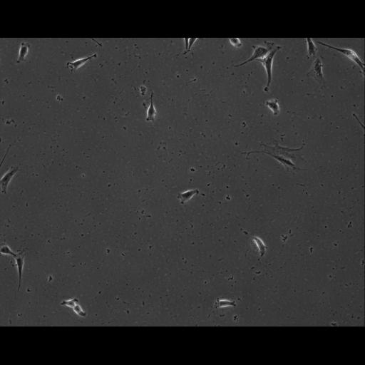
Effects of red nucleus microstimulation on the locomotor pattern and timing in the intact cat: a comparison with the motor cortex. (1/117)
Effects of red nucleus microstimulation on the locomotor pattern and timing in the intact cat: a comparison with the motor cortex. To determine the extent to which the rubrospinal tract is capable of modifying locomotion in the intact cat, we applied microstimulation (cathodal current, 330 Hz; pulse duration 0.2 ms; maximal current, 25 microA) to the red nucleus during locomotion. The stimuli were applied either as short trains (33 ms) of impulses to determine the capacity of the rubrospinal tract to modify the level of electromyographic (EMG) activity in different flexors and extensors at different phases of the step cycle or as long trains (200 ms) of pulses to determine the effect of the red nucleus on cycle timing. Stimuli were also applied with the cat at rest (33-ms train). This latter stimulation evoked short-latency (average = 11.8-19.0 ms) facilitatory responses in all of the physiological flexor muscles of the forelimb that were recorded; facilitatory responses were also common in the elbow extensor, lateral head of triceps but were rare in the physiological wrist and digit extensor, palmaris longus. Responses were still evoked in most muscles when the current was decreased to near threshold (3-10 microA). Stimulation during locomotion with the short trains of stimuli evoked shorter-latency (average = 6.0-12.5 ms) facilitatory responses in flexor muscles during the swing phase of locomotion and, except in the case of the extensor digitorum communis, evoked substantially smaller responses in stance. The same stimuli also evoked facilitatory responses in the extensor muscles during swing and produced more complex effects involving both facilitation and suppression in stance. Increasing the duration of the train to 200 ms modified the amplitude and duration of the EMG activity of both flexors and extensors but had little significant effect on the cycle duration. In contrast, whereas stimulation of the motor cortex with short trains of stimuli during locomotion had very similar effects to that of the red nucleus, increasing the train duration to 200 ms frequently produced a marked reset of the step cycle by curtailing stance and initiating a new period of swing. The results suggest that whereas both the motor cortex and the red nucleus have access to the interneuronal circuits responsible for controlling the structure of the EMG activity in the step cycle, only the motor cortex has access to the circuits responsible for controlling cycle timing. (+info)Genetic analysis of a dentatorubral-pallidoluysian atrophy family: relevance to apparent sporadic cases. (2/117)
Dentatorubral-pallidoluysian atrophy (DRPLA) is associated with an unstable CAG trinucleotide sequence. We describe a DRPLA family whose members have an allele containing an expanded CAG repeat, even in an elderly neurologically normal individual. The proband developed DRPLA at age 14. She was initially considered a sporadic case, but later her sister became symptomatic. Investigation of the number of CAG repeat units in her family revealed the 81-year-old father to have an expanded CAG repeat of 51 units. To our knowledge, such an advanced aged unaffected patient has not been previously documented. The present example may explain apparent sporadic cases. (+info)Transplants of fibroblasts genetically modified to express BDNF promote regeneration of adult rat rubrospinal axons and recovery of forelimb function. (3/117)
Adult mammalian CNS neurons do not normally regenerate their severed axons. This failure has been attributed to scar tissue and inhibitory molecules at the injury site that block the regenerating axons, a lack of trophic support for the axotomized neurons, and intrinsic neuronal changes that follow axotomy, including cell atrophy and death. We studied whether transplants of fibroblasts genetically engineered to produce brain-derived neurotrophic factor (BDNF) would promote rubrospinal tract (RST) regeneration in adult rats. Primary fibroblasts were modified by retroviral-mediated transfer of a DNA construct encoding the human BDNF gene, an internal ribosomal entry site, and a fusion gene of lacZ and neomycin resistance genes. The modified fibroblasts produce biologically active BDNF in vitro. These cells were grafted into a partial cervical hemisection cavity that completely interrupted one RST. One and two months after lesion and transplantation, RST regeneration was demonstrated with retrograde and anterograde tracing techniques. Retrograde tracing with fluorogold showed that approximately 7% of RST neurons regenerated axons at least three to four segments caudal to the transplants. Anterograde tracing with biotinylated dextran amine revealed that the RST axons regenerated through and around the transplants, grew for long distances within white matter caudal to the transplant, and terminated in spinal cord gray matter regions that are the normal targets of RST axons. Transplants of unmodified primary fibroblasts or Gelfoam alone did not elicit regeneration. Behavioral tests demonstrated that recipients of BDNF-producing fibroblasts showed significant recovery of forelimb usage, which was abolished by a second lesion that transected the regenerated axons. (+info)Perineuronal nets of proteoglycans in the adult mouse brain are digested by collagenase. (4/117)
As our previous study has indicated, perineuronal proteoglycans in the adult mouse brain are associated with some collagenous molecules which can be stained with Gomori's ammoniacal silver and are resistant to hyaluronidase digestion. The present study demonstrated that these molecules are thoroughly digested with collagenase, and suggests that they represent a hyaluronic acid-binding domain of the ligand proteoglycans connecting the perineuronal proteoglycans and nerve cell surface glycoproteins. (+info)Reciprocal connections between the red nucleus and the trigeminal nuclei: a retrograde and anterograde tracing study. (5/117)
An anterograde biocytin and a retrograde WGA-colloidal gold study in the rat can provide information about reciprocal communication pathways between the red nucleus and the trigeminal sensory complex. No terminals were found within the trigeminal motor nucleus, in contrast with the facial motor nucleus. A dense terminal field was observed in the parvicellular reticular formation ventrally to the trigeminal motor nucleus. The parvicellular area may be important for the control of jaw movements by rubrotrigeminal inputs. On the other hand, the contralateral rostral parvicellular part of the red nucleus receives terminals from the same zone in the rostral part of the trigeminal sensory complex, where retrogradely labelled neurones were found after tracer injections into the red nucleus. Such relationships could be part of a control loop for somatosensory information from the orofacial area. (+info)The effects of reversible inactivation of the red nucleus on learning-related and auditory-evoked unit activity in the pontine nuclei of classically conditioned rabbits. (6/117)
The pontine nuclei carry auditory conditioned stimulus information to the cerebellum during classical conditioning of the nictitating membrane response in rabbits. In well-trained animals learning-related as well as stimulus-evoked unit activity can be recorded throughout the pontine nuclei but particularly in the lateral and dorsolateral pons. Recent work in our laboratory has provided evidence that the learning-related unit activity in the pons is dependent on the interpositus nucleus and that the pons is not a site of essential plasticity for the learned response. In the present study we considered the question of whether learning-related unit activity might be projected from the interpositus nucleus to the pons through the red nucleus, a primary output target of the interpositus and a structure known to be essential for expression of the learned response. Multiple unit recordings were taken from lateral and dorsolateral pontine locations in well-trained rabbits before and during cooling of the red nucleus. Analysis of pooled data for all recording locations within the lateral and dorsolateral pons indicated that reversible inactivation of red nucleus abolished both stimulus-evoked and learning-related unit activity. However, we also found discrete recording locations where stimulus-evoked and learning-related unit activity were attenuated but not abolished by red nucleus cooling. (+info)Summation of effective synaptic currents and firing rate modulation in cat spinal motoneurons. (7/117)
The aim of this study was to examine how cat spinal motoneurons integrate the synaptic currents generated by the concurrent activation of large groups of presynaptic neurons. We obtained intracellular recordings from cat triceps surae motoneurons and measured the effects of repetitive activity in different sets of presynaptic neurons produced by electrical stimulation of descending fibers or peripheral nerves and by longitudinal vibration of the triceps surae muscles (to activate primary muscle spindle Ia afferent fibers). We combined synaptic activation with subthreshold injected currents to obtain estimates of effective synaptic currents at the resting potential (I(Nrest)) and at the threshold for repetitive discharge (I(Nthresh)). We then superimposed synaptic activation on suprathreshold injected current steps to measure the synaptically evoked change in firing rate. We studied eight different pairs of synaptic inputs. When any two synaptic inputs were activated concurrently, both the effective synaptic currents (I(Nrest)) and the synaptically evoked changes in firing rate generally were equal to or slightly less than the linear sum of the effects produced by activating each input alone. However, there were several instances in which the summation was substantially less than linear. In some motoneurons, we induced a partial blockade of potassium channels by adding tetraethylammonium (TEA) or cesium to the electrolyte solution in the intracellular pipette. In these cells, persistent inward currents were evoked by depolarization that led to instances of substantially greater-than linear summation of injected and synaptic currents. Overall our results indicate that the spatial distribution of synaptic boutons on motoneurons acts to minimize electrical interactions between synaptic sites permitting near linear summation of synaptic currents. However, modulation of voltage-gated conductances on the soma and dendrites of the motoneuron can lead to marked nonlinearities in synaptic integration. (+info)Comparison of the effects of electrolytic and chemical destruction of the red nucleus on the compensatory capacity of rats with rubrospinal tract lesions. (8/117)
Transection of the rubrospinal tract in rats, performed before lesion of the red nucleus, resulted in the facilitated recovery of motor activity and operantly conditioned reflexes. Such facilitation was absent when the red nucleus is lesioned alone. This phenomenon is explained by the switching of descending influences on the corticospinal tract through the participation of the following system: red nucleus--inferior olive--cerebellum--ventrolateral thalamic nucleus--cerebral cortex. The above mentioned facilitating influence on the recovery process was particularly prominent in rats with quinolinic acid-induced lesion of the red nucleus. Under these conditions, the cerebellar ascending fibers to the ventrolateral thalamic nucleus were preserved. Decreased facilitated recovery following electrolytic lesion of the red nucleus suggests the existence of additional cerebello-cortical pathways for the realization of the switching phenomenon. (+info)The red nucleus is a round-shaped collection of neurons located in the midbrain, specifically in the rostral part of the mesencephalon. It is called "red" due to its deep red color, which comes from the rich vascularization and numerous iron-containing red blood cells present in the region.
The red nucleus plays a crucial role in the motor system, primarily involved in controlling and coordinating movements, particularly on the contralateral side of the body. It is part of the rubrospinal tract, which descends from the red nucleus to the spinal cord and helps regulate fine motor movements and muscle tone.
There are two main types of neurons present in the red nucleus: magnocellular (large cells) and parvocellular (small cells). Magnocellular neurons form the rubrospinal tract, while parvocellular neurons project to the inferior olivary nucleus, which is part of the cerebellum. The connections between the red nucleus, cerebellum, and spinal cord allow for the integration and coordination of motor information and the execution of smooth movements.
Damage to the red nucleus can result in various motor impairments, such as ataxia (lack of muscle coordination), tremors, and weakness on the contralateral side of the body.
The cell nucleus is a membrane-bound organelle found in the eukaryotic cells (cells with a true nucleus). It contains most of the cell's genetic material, organized as DNA molecules in complex with proteins, RNA molecules, and histones to form chromosomes.
The primary function of the cell nucleus is to regulate and control the activities of the cell, including growth, metabolism, protein synthesis, and reproduction. It also plays a crucial role in the process of mitosis (cell division) by separating and protecting the genetic material during this process. The nuclear membrane, or nuclear envelope, surrounding the nucleus is composed of two lipid bilayers with numerous pores that allow for the selective transport of molecules between the nucleoplasm (nucleus interior) and the cytoplasm (cell exterior).
The cell nucleus is a vital structure in eukaryotic cells, and its dysfunction can lead to various diseases, including cancer and genetic disorders.
The cerebellar nuclei are clusters of neurons located within the white matter of the cerebellum, a region of the brain responsible for motor coordination, balance, and fine movement regulation. There are four main pairs of cerebellar nuclei: the fastigial, interpositus, dentate, and vestibular nuclei. These nuclei receive input from various parts of the cerebellar cortex and project to different areas of the brainstem and thalamus, contributing to the regulation of muscle tone, posture, and movement.
A forelimb is a term used in animal anatomy to refer to the upper limbs located in the front of the body, primarily involved in movement and manipulation of the environment. In humans, this would be equivalent to the arms, while in quadrupedal animals (those that move on four legs), it includes the structures that are comparable to both the arms and legs of humans, such as the front legs of dogs or the forepaws of cats. The bones that make up a typical forelimb include the humerus, radius, ulna, carpals, metacarpals, and phalanges.
The abducens nerve, also known as the sixth cranial nerve (CN VI), is a motor nerve that controls the lateral rectus muscle of the eye. This muscle is responsible for moving the eye away from the midline (towards the temple) and enables the eyes to look towards the side while keeping them aligned. Any damage or dysfunction of the abducens nerve can result in strabismus, where the eyes are misaligned and point in different directions, specifically an adduction deficit, also known as abducens palsy or sixth nerve palsy.
The trochlear nerve, also known as the fourth cranial nerve (CN IV), is a nerve that originates in the midbrain and innervates the superior oblique muscle of the eye. This muscle helps with the downward and outward movement of the eye, playing a crucial role in controlling eye movements and maintaining binocular vision. The trochlear nerve's main function is to provide motor (efferent) innervation to the superior oblique muscle, enabling fine-tuning of eye movements during activities such as reading, writing, or driving. Damage to this nerve can result in vertical diplopia (double vision), strabismus (eye misalignment), and other visual impairments.
"Cat" is a common name that refers to various species of small carnivorous mammals that belong to the family Felidae. The domestic cat, also known as Felis catus or Felis silvestris catus, is a popular pet and companion animal. It is a subspecies of the wildcat, which is found in Europe, Africa, and Asia.
Domestic cats are often kept as pets because of their companionship, playful behavior, and ability to hunt vermin. They are also valued for their ability to provide emotional support and therapy to people. Cats are obligate carnivores, which means that they require a diet that consists mainly of meat to meet their nutritional needs.
Cats are known for their agility, sharp senses, and predatory instincts. They have retractable claws, which they use for hunting and self-defense. Cats also have a keen sense of smell, hearing, and vision, which allow them to detect prey and navigate their environment.
In medical terms, cats can be hosts to various parasites and diseases that can affect humans and other animals. Some common feline diseases include rabies, feline leukemia virus (FeLV), feline immunodeficiency virus (FIV), and toxoplasmosis. It is important for cat owners to keep their pets healthy and up-to-date on vaccinations and preventative treatments to protect both the cats and their human companions.
The subthalamus is a region in the brain that is located deep beneath the thalamus and above the midbrain. It is a part of the basal ganglia, which are a group of structures involved in the control of movement. The subthalamus contains several different types of neurons, including glutamatergic and GABAergic neurons, and plays a role in regulating movement, reward, and motivation. It is also thought to be involved in the pathophysiology of certain neurological disorders such as Parkinson's disease.
The subthalamic nucleus (STN) is a specific structure within the subthalamus that has been the target of deep brain stimulation surgery for the treatment of movement disorders like Parkinson's disease and dystonia. The STN is responsible for regulating the activity of other structures in the basal ganglia, and its overactivity can lead to symptoms such as tremors, rigidity, and difficulty initiating movements. By implanting electrodes in the STN and delivering electrical impulses, deep brain stimulation can help to regulate the activity of the STN and alleviate some of these symptoms.
The mesencephalon, also known as the midbrain, is the middle portion of the brainstem that connects the hindbrain (rhombencephalon) and the forebrain (prosencephalon). It plays a crucial role in several important functions including motor control, vision, hearing, and the regulation of consciousness and sleep-wake cycles. The mesencephalon contains several important structures such as the cerebral aqueduct, tectum, tegmentum, cerebral peduncles, and several cranial nerve nuclei (III and IV).
The oculomotor nerve, also known as the third cranial nerve (CN III), is a motor nerve that originates from the midbrain. It controls the majority of the eye muscles, including the levator palpebrae superioris muscle that raises the upper eyelid, and the extraocular muscles that enable various movements of the eye such as looking upward, downward, inward, and outward. Additionally, it carries parasympathetic fibers responsible for pupillary constriction and accommodation (focusing on near objects). Damage to this nerve can result in various ocular motor disorders, including strabismus, ptosis, and pupillary abnormalities.
The brainstem is the lower part of the brain that connects to the spinal cord. It consists of the midbrain, pons, and medulla oblongata. The brainstem controls many vital functions such as heart rate, breathing, and blood pressure. It also serves as a relay center for sensory and motor information between the cerebral cortex and the rest of the body. Additionally, several cranial nerves originate from the brainstem, including those that control eye movements, facial movements, and hearing.
Electrolysis is a medical procedure that involves the use of electrical current to permanently remove hair growth. It works by passing a thin, solid metal electrode (called a probe) into the natural opening of the hair follicle and applying an electrical charge to destroy the hair root. This process can be used to remove hair from any part of the body, including the face, legs, arms, underarms, and bikini area.
During electrolysis, a trained professional called an electrologist inserts a small needle into the hair follicle and applies a mild electrical current. The current heats up and destroys the hair root, preventing future growth. Multiple treatments are usually necessary to achieve permanent hair removal, as only one or two hairs can be treated at a time.
Electrolysis is considered a safe and effective method for permanent hair removal, but it can cause some discomfort during and after treatment. Common side effects include redness, swelling, and tenderness in the treated area. These side effects typically resolve within a few hours to a few days after treatment.
It's important to note that electrolysis should only be performed by a licensed and trained electrologist. Improper technique can cause scarring, infection, or other complications. Before undergoing electrolysis, it's recommended to consult with a dermatologist or other healthcare provider to discuss the risks and benefits of the procedure.
The pyramidal tracts, also known as the corticospinal tracts, are bundles of nerve fibers that run through the brainstem and spinal cord, originating from the cerebral cortex. These tracts are responsible for transmitting motor signals from the brain to the muscles, enabling voluntary movement and control of the body.
The pyramidal tracts originate from the primary motor cortex in the frontal lobe of the brain and decussate (cross over) in the lower medulla oblongata before continuing down the spinal cord. The left pyramidal tract controls muscles on the right side of the body, while the right pyramidal tract controls muscles on the left side of the body.
Damage to the pyramidal tracts can result in various motor impairments, such as weakness or paralysis, spasticity, and loss of fine motor control, depending on the location and extent of the damage.
Neural pathways, also known as nerve tracts or fasciculi, refer to the highly organized and specialized routes through which nerve impulses travel within the nervous system. These pathways are formed by groups of neurons (nerve cells) that are connected in a series, creating a continuous communication network for electrical signals to transmit information between different regions of the brain, spinal cord, and peripheral nerves.
Neural pathways can be classified into two main types: sensory (afferent) and motor (efferent). Sensory neural pathways carry sensory information from various receptors in the body (such as those for touch, temperature, pain, and vision) to the brain for processing. Motor neural pathways, on the other hand, transmit signals from the brain to the muscles and glands, controlling movements and other effector functions.
The formation of these neural pathways is crucial for normal nervous system function, as it enables efficient communication between different parts of the body and allows for complex behaviors, cognitive processes, and adaptive responses to internal and external stimuli.
The pons is a part of the brainstem that lies between the medulla oblongata and the midbrain. Its name comes from the Latin word "ponte" which means "bridge," as it serves to connect these two regions of the brainstem. The pons contains several important structures, including nerve fibers that carry signals between the cerebellum (the part of the brain responsible for coordinating muscle movements) and the rest of the nervous system. It also contains nuclei (clusters of neurons) that help regulate various functions such as respiration, sleep, and facial movements.
Chloralose is not a medical term commonly used in modern medicine. However, historically, it is a chemical compound that has been used in research and veterinary medicine as an sedative and hypnotic agent. It is a combination of chloral hydrate and sodium pentobarbital.
Chloralose has been used in research to study the effects of sedation on various physiological processes, such as respiration and circulation. In veterinary medicine, it has been used as an anesthetic for small animals during surgical procedures. However, due to its potential for serious side effects, including respiratory depression and cardiac arrest, chloralose is not commonly used in clinical practice today.
In the context of medicine and healthcare, "movement" refers to the act or process of changing physical location or position. It involves the contraction and relaxation of muscles, which allows for the joints to move and the body to be in motion. Movement can also refer to the ability of a patient to move a specific body part or limb, which is assessed during physical examinations. Additionally, "movement" can describe the progression or spread of a disease within the body.
Neurons, also known as nerve cells or neurocytes, are specialized cells that constitute the basic unit of the nervous system. They are responsible for receiving, processing, and transmitting information and signals within the body. Neurons have three main parts: the dendrites, the cell body (soma), and the axon. The dendrites receive signals from other neurons or sensory receptors, while the axon transmits these signals to other neurons, muscles, or glands. The junction between two neurons is called a synapse, where neurotransmitters are released to transmit the signal across the gap (synaptic cleft) to the next neuron. Neurons vary in size, shape, and structure depending on their function and location within the nervous system.
The spinal cord is a major part of the nervous system, extending from the brainstem and continuing down to the lower back. It is a slender, tubular bundle of nerve fibers (axons) and support cells (glial cells) that carries signals between the brain and the rest of the body. The spinal cord primarily serves as a conduit for motor information, which travels from the brain to the muscles, and sensory information, which travels from the body to the brain. It also contains neurons that can independently process and respond to information within the spinal cord without direct input from the brain.
The spinal cord is protected by the bony vertebral column (spine) and is divided into 31 segments: 8 cervical, 12 thoracic, 5 lumbar, 5 sacral, and 1 coccygeal. Each segment corresponds to a specific region of the body and gives rise to pairs of spinal nerves that exit through the intervertebral foramina at each level.
The spinal cord is responsible for several vital functions, including:
1. Reflexes: Simple reflex actions, such as the withdrawal reflex when touching a hot surface, are mediated by the spinal cord without involving the brain.
2. Muscle control: The spinal cord carries motor signals from the brain to the muscles, enabling voluntary movement and muscle tone regulation.
3. Sensory perception: The spinal cord transmits sensory information, such as touch, temperature, pain, and vibration, from the body to the brain for processing and awareness.
4. Autonomic functions: The sympathetic and parasympathetic divisions of the autonomic nervous system originate in the thoracolumbar and sacral regions of the spinal cord, respectively, controlling involuntary physiological responses like heart rate, blood pressure, digestion, and respiration.
Damage to the spinal cord can result in various degrees of paralysis or loss of sensation below the level of injury, depending on the severity and location of the damage.
The term "septum" in the context of the brain refers to the septal nuclei, which are a collection of neurons located in the basal forebrain. Specifically, they make up the septal area, which is part of the limbic system and plays a role in reward, reinforcement, and positive motivational states.
There isn't a structure called the "septum of brain" in medical terminology. However, there are several structures in the brain that contain a septum or have a partitioning septum within them, such as:
1. Septal nuclei (as mentioned above)
2. The nasal septum, which is a thin wall of bone and cartilage that separates the two nostrils in the nose
3. The interventricular septum, which is a thin muscular wall that separates the left and right lateral ventricles within the brain
4. The membranous septum, a part of the heart's structure that separates the left and right ventricles
Confusion might arise due to the term "septum" being used in different contexts. In this case, there is no specific medical definition for 'Septum of Brain'.
Eyelid conditioning, also known as eyelid classical conditioning or Ursinus' phenomenon, is a type of reflex conditioning that involves associating a neutral stimulus with the natural act of blinking. This concept was first described by Russian physiologist Ivan Pavlov and later studied in detail by German ophthalmologist Hermann Ludwig Ferdinand von Helmholtz and Austrian physician Sigmund Exner.
In this procedure, a conditioned stimulus (like a sound or light) is repeatedly presented just before the unconditioned stimulus (such as a puff of air directed at the eye), which naturally triggers the blink reflex. Over time, the subject begins to associate the conditioned stimulus with the blinking response and will start to blink even when only the conditioned stimulus is presented, without the presence of the unconditioned stimulus. This learning process is an example of classical conditioning and can be used in various research and clinical applications.
The cerebellar cortex is the outer layer of the cerebellum, which is a part of the brain that plays a crucial role in motor control, balance, and coordination of muscle movements. The cerebellar cortex contains numerous small neurons called granule cells, as well as other types of neurons such as Purkinje cells, basket cells, and stellate cells. These neurons are organized into distinct layers and microcircuits that process information related to motor function and possibly other functions such as cognition and emotion. The cerebellar cortex receives input from various sources, including the spinal cord, vestibular system, and cerebral cortex, and sends output to brainstem nuclei and thalamus, which in turn project to the cerebral cortex. Damage to the cerebellar cortex can result in ataxia, dysmetria, dysdiadochokinesia, and other motor symptoms.
Electric stimulation, also known as electrical nerve stimulation or neuromuscular electrical stimulation, is a therapeutic treatment that uses low-voltage electrical currents to stimulate nerves and muscles. It is often used to help manage pain, promote healing, and improve muscle strength and mobility. The electrical impulses can be delivered through electrodes placed on the skin or directly implanted into the body.
In a medical context, electric stimulation may be used for various purposes such as:
1. Pain management: Electric stimulation can help to block pain signals from reaching the brain and promote the release of endorphins, which are natural painkillers produced by the body.
2. Muscle rehabilitation: Electric stimulation can help to strengthen muscles that have become weak due to injury, illness, or surgery. It can also help to prevent muscle atrophy and improve range of motion.
3. Wound healing: Electric stimulation can promote tissue growth and help to speed up the healing process in wounds, ulcers, and other types of injuries.
4. Urinary incontinence: Electric stimulation can be used to strengthen the muscles that control urination and reduce symptoms of urinary incontinence.
5. Migraine prevention: Electric stimulation can be used as a preventive treatment for migraines by applying electrical impulses to specific nerves in the head and neck.
It is important to note that electric stimulation should only be administered under the guidance of a qualified healthcare professional, as improper use can cause harm or discomfort.
The nucleus accumbens is a part of the brain that is located in the ventral striatum, which is a key region of the reward circuitry. It is made up of two subregions: the shell and the core. The nucleus accumbens receives inputs from various sources, including the prefrontal cortex, amygdala, and hippocampus, and sends outputs to the ventral pallidum and other areas.
The nucleus accumbens is involved in reward processing, motivation, reinforcement learning, and addiction. It plays a crucial role in the release of the neurotransmitter dopamine, which is associated with pleasure and reinforcement. Dysfunction in the nucleus accumbens has been implicated in various neurological and psychiatric conditions, including substance use disorders, depression, and obsessive-compulsive disorder.
Blinking is the rapid and repetitive closing and reopening of the eyelids. It is a normal physiological process that helps to keep the eyes moist, protected and comfortable by spreading tears over the surface of the eye and removing any foreign particles or irritants that may have accumulated on the eyelid or the conjunctiva (the mucous membrane that covers the front of the eye and lines the inside of the eyelids).
Blinking is controlled by the facial nerve (cranial nerve VII), which sends signals to the muscles that control the movement of the eyelids. On average, people blink about 15-20 times per minute, but this rate can vary depending on factors such as mood, level of attention, and visual tasks. For example, people tend to blink less frequently when they are concentrating on a visual task or looking at a screen, which can lead to dry eye symptoms.
Islets of Langerhans transplantation is a surgical procedure that involves the transplantation of isolated islets from a deceased donor's pancreas into another person with type 1 diabetes. The islets of Langerhans are clusters of cells within the pancreas that produce hormones, including insulin, which regulates blood sugar levels.
In type 1 diabetes, the body's immune system mistakenly attacks and destroys these insulin-producing cells, leading to high blood sugar levels. Islet transplantation aims to replace the damaged islets with healthy ones from a donor, allowing the recipient's body to produce and regulate its own insulin again.
The procedure involves extracting the islets from the donor pancreas and infusing them into the recipient's liver through a small incision in the abdomen. Once inside the liver, the islets can sense glucose levels in the bloodstream and release insulin as needed to maintain normal blood sugar levels.
Islet transplantation has shown promising results in improving blood sugar control and reducing the risk of severe hypoglycemia (low blood sugar) in people with type 1 diabetes. However, it requires long-term immunosuppressive therapy to prevent rejection of the transplanted islets, which can have side effects and increase the risk of infections.
The Islets of Langerhans are clusters of specialized cells within the pancreas, an organ located behind the stomach. These islets are named after Paul Langerhans, who first identified them in 1869. They constitute around 1-2% of the total mass of the pancreas and are distributed throughout its substance.
The Islets of Langerhans contain several types of cells, including:
1. Alpha (α) cells: These produce and release glucagon, a hormone that helps to regulate blood sugar levels by promoting the conversion of glycogen to glucose in the liver when blood sugar levels are low.
2. Beta (β) cells: These produce and release insulin, a hormone that promotes the uptake and utilization of glucose by cells throughout the body, thereby lowering blood sugar levels.
3. Delta (δ) cells: These produce and release somatostatin, a hormone that inhibits the release of both insulin and glucagon and helps regulate their secretion in response to changing blood sugar levels.
4. PP cells (gamma or γ cells): These produce and release pancreatic polypeptide, which plays a role in regulating digestive enzyme secretion and gastrointestinal motility.
Dysfunction of the Islets of Langerhans can lead to various endocrine disorders, such as diabetes mellitus, where insulin-producing beta cells are damaged or destroyed, leading to impaired blood sugar regulation.
An artificial pancreas is not a literal organ like a biological pancreas. Instead, it refers to a closed-loop system that integrates a continuous glucose monitor (CGM) with an insulin pump to automatically regulate blood glucose levels in individuals with diabetes. This system mimics the functions of a healthy pancreas by constantly monitoring blood sugar levels and delivering the appropriate amount of insulin as needed, without requiring manual input from the user.
The artificial pancreas is still an area of active research and development, and various prototypes and systems are being tested in clinical trials to improve their accuracy, safety, and effectiveness. The ultimate goal of developing an artificial pancreas is to provide a more effective and convenient way to manage diabetes, reduce the risk of complications, and improve quality of life for people with diabetes.
An encyclopedia is a comprehensive reference work containing articles on various topics, usually arranged in alphabetical order. In the context of medicine, a medical encyclopedia is a collection of articles that provide information about a wide range of medical topics, including diseases and conditions, treatments, tests, procedures, and anatomy and physiology. Medical encyclopedias may be published in print or electronic formats and are often used as a starting point for researching medical topics. They can provide reliable and accurate information on medical subjects, making them useful resources for healthcare professionals, students, and patients alike. Some well-known examples of medical encyclopedias include the Merck Manual and the Stedman's Medical Dictionary.
Pancreas transplantation is a surgical procedure that involves implanting a healthy pancreas from a deceased donor into a recipient with diabetes. The primary goal of this procedure is to restore the recipient's insulin production and eliminate the need for insulin injections, thereby improving their quality of life and reducing the risk of long-term complications associated with diabetes.
There are three main types of pancreas transplantation:
1. Simultaneous pancreas-kidney (SPK) transplantation: This is the most common type of pancreas transplant, performed simultaneously with a kidney transplant in patients with diabetes and end-stage renal disease (ESRD). The new pancreas not only restores insulin production but also helps prevent further kidney damage.
2. Pancreas after kidney (PAK) transplantation: In this procedure, a patient receives a kidney transplant first, followed by a pancreas transplant at a later time. This is typically performed in patients who have already undergone a successful kidney transplant and wish to improve their diabetes management.
3. Pancreas transplantation alone (PTA): In rare cases, a pancreas transplant may be performed without a concurrent kidney transplant. This is usually considered for patients with brittle diabetes who experience severe hypoglycemic episodes despite optimal medical management and lifestyle modifications.
The success of pancreas transplantation has significantly improved over the years, thanks to advancements in surgical techniques, immunosuppressive medications, and post-transplant care. However, it is essential to weigh the benefits against the risks, such as potential complications related to surgery, infection, rejection, and long-term use of immunosuppressive drugs. Ultimately, the decision to undergo pancreas transplantation should be made in consultation with a multidisciplinary team of healthcare professionals, considering each patient's unique medical history and personal circumstances.
Diabetes Mellitus, Type 1 is a chronic autoimmune disease characterized by the destruction of insulin-producing beta cells in the pancreas, leading to an absolute deficiency of insulin. This results in an inability to regulate blood glucose levels, causing hyperglycemia (high blood sugar). Type 1 diabetes typically presents in childhood or early adulthood, although it can develop at any age. It is usually managed with regular insulin injections or the use of an insulin pump, along with monitoring of blood glucose levels and adjustments to diet and physical activity. Uncontrolled type 1 diabetes can lead to serious complications such as kidney damage, nerve damage, blindness, and cardiovascular disease.
Insulin is a hormone produced by the beta cells of the pancreatic islets, primarily in response to elevated levels of glucose in the circulating blood. It plays a crucial role in regulating blood glucose levels and facilitating the uptake and utilization of glucose by peripheral tissues, such as muscle and adipose tissue, for energy production and storage. Insulin also inhibits glucose production in the liver and promotes the storage of excess glucose as glycogen or triglycerides.
Deficiency in insulin secretion or action leads to impaired glucose regulation and can result in conditions such as diabetes mellitus, characterized by chronic hyperglycemia and associated complications. Exogenous insulin is used as a replacement therapy in individuals with diabetes to help manage their blood glucose levels and prevent long-term complications.
 Red nucleus - Wikipedia
 'Red and Yellow Nucleus' by Tim Woolcock |...
Postnatal Maturation of the Red Nucleus Motor Map Depends on Rubrospinal Connections with Forelimb Motor Pools | Journal of...
 Riverside Portfolio Company | Red Nucleus | www.riversidecompany.com
 Student Photography Exhibition, featuring a performance by Red Nucleus - North Carolina Botanical Garden
Demonstration of the Medullary Lamellae of the Human Red Nucleus with High-resolution Gradient-echo MR Imaging | American...
 The 'Red Radio Ring': ionized and molecular gas in a starburst/active galactic nucleus at z similar to 2.55 - Kölner...
 Tilta Nucleus-M RED CTRL Run/Stop Cable
- Dedotec Schweiz
Tilta Nucleus-M RED CTRL Run/Stop Cable
- Dedotec Schweiz
Solitary nucleus - Wikipedia
 INVIVO is now Red Nucleus
 Pentagon Nucleus Liner Jacket Maroon Red
 red nucleus tower of fantasy Archives - Wholepost
 Coco Bongo Slot by Nucleus - Play Online | Red Dog Casino
Functional Relationships Between the Red Nucleus and the Brachium Gonjunctivum | Neurology
 Atomic nucleus - New World Encyclopedia
 Cerebellar plasticity and motor learning deficits in a copy-number variation mouse model of autism | Nature Communications
 OC1024</span><span class=" 3LUMH 1NhtS v2z8p" style="cursor:pointer" role=...
 Frontiers | The Pathogenic R3052W BRCA2 Variant Disrupts Homology-Directed Repair by Failing to Localize to the Nucleus
 Gamma-aminobutyric Acid and Glutamate/Glutamine Levels in the Dentate Nucleus and Periaqueductal Gray With Episodic and Chronic...
Figure 2 - Isolation of Prion with BSE Properties from Farmed Goat - Volume 17, Number 12-December 2011 - Emerging Infectious...
 2,944 Nuclei Acids PPTs View free & download | PowerShow.com
Pancreatic islets - Wikipedia
Complete human day 14 post-implantation embryo models from naive ES cells | Nature
 MM+M Agency Showcase 2023 - MM+M - Medical Marketing and Media
 Warren Ellis Now A Hollywood Red | ComicMix
 Alcian blue staining kit, pH 2.5 for the detection of acidic mucosubstance for microscopy | Sigma-Aldrich
'Red' Adds McMahon, Borgnine to Cast |...
Daubnet Community • Berek, Ines, Emet and Grok Dominica
 2017 List of Best Places to Work in Pennsylvania Revealed - Team PA Foundation
Cell nuclei5
- Nucleic Acids and Nucleotides - Nucleic Acids and Nucleotides Nucleic acids are long, slightly acidic molecules originally identified in cell nuclei. (powershow.com)
- Cell nuclei are outlined in blue. (genengnews.com)
- TGF-β1 activated accumulation of phosphorylated SMAD2 (pSMAD2-C) at centrioles of motile cilia and at cell nuclei. (springer.com)
- This triggered an increase in paracellular permeability via cellular redistribution of claudin 3 (CLDN3) from TJs into cell nuclei followed by disruption of epithelial integrity and formation of epithelial lesions. (springer.com)
- In summary, we demonstrate a role of motile cilia in TGF-β1 sensing and showed that TGF-β1 disturbs TJ permeability of conductive airway epithelia by redistributing CLDN3 from TJs into cell nuclei. (springer.com)
Dentate nucleus1
- To explore the pathogenesis of migraine chronification, we measured gamma-aminobutyric acid (GABA) and glutamate/glutamine (Glx) levels in the dentate nucleus (DN) and PAG of patients with episodic and chronic migraine and healthy subjects. (researchsquare.com)
Subthalamic nucleus1
- Among possible targets, the non-motor subthalamic nucleus (STN) is a key node of the basal ganglia circuitry, strongly connected to limbic cortical areas known to be involved in OCD. (bmj.com)
Chromosomes4
- Each cell (except for red blood cells) contains a nucleus that houses these chromosomes. (cdc.gov)
- Genes are contained in chromosomes, which are in the cell nucleus. (msdmanuals.com)
- DNA (deoxyribonucleic acid) is the cell's genetic material, contained in chromosomes within the cell nucleus and mitochondria. (msdmanuals.com)
- Except for certain cells (for example, sperm and egg cells and red blood cells), the cell nucleus contains 23 pairs of chromosomes. (msdmanuals.com)
Rubrospinal tract3
- In humans, the red nucleus also has limited control over hands, as the rubrospinal tract is more involved in large muscle movement such as that for the arms (but not for the legs, as the tract terminates in the superior thoracic region of the spinal cord). (wikipedia.org)
- The red nucleus (RN) and rubrospinal tract (RST) are important for forelimb motor control. (jneurosci.org)
- In maturity, motor skills are dependent on the corticospinal tract (CST), originating principally from the motor cortex (M1), and the rubrospinal tract (RST), originating from the red nucleus (RN), which are collectively termed the lateral motor system ( Kuypers, 1981 ). (jneurosci.org)
Corticospinal tract2
- In a vertebrate without a significant corticospinal tract, gait is mainly controlled by the red nucleus. (wikipedia.org)
- Fine control of the fingers is not modified by the functioning of the red nucleus but relies on the corticospinal tract. (wikipedia.org)
Atoms2
- However, the nuclei of hydrogen and helium atoms are exceptions, as the nuclear charge is concentrated most highly at the central point. (newworldencyclopedia.org)
- The diameter of the nucleus is in the range of 1.6 femtometer (fm) ( 1.6 × 10 −15 m ) (for a proton in light hydrogen) to about 15 fm (for the heaviest atoms, such as uranium). (newworldencyclopedia.org)
Amygdala2
- The solitary nucleus projects to a large number of other regions of the brain including the paraventricular nucleus of the hypothalamus , the central nucleus of the amygdala , as well as other nuclei in the brainstem (such as the parabrachial area , locus coeruleus , dorsal raphe nucleus , and other visceral motor or respiratory networks). (wikipedia.org)
- This disrupts both GABAergic and glutamatergic transmission through amygdala circuits, including reward-related outputs to the nucleus accumbens. (jneurosci.org)
Cytoskeleton1
- Cytoskeleton (green), nucleus (blue), and drusen deposits (red) are shown. (nih.gov)
Acidic1
- Acidic mucosubstances are selectively stained in the tissue, after which the specimen is counterstained with nuclear fast red. (sigmaaldrich.com)
Medulla3
- The majority of red nucleus axons do not project to the spinal cord but, via its parvocellular part, relay information from the motor cortex to the cerebellum through the inferior olivary complex, an important relay center in the medulla. (wikipedia.org)
- The solitary nucleus (also called nucleus of the solitary tract , nucleus solitarius, or nucleus tractus solitarii (SN or NTS) ) [1] [2] is a series of sensory nuclei (clusters of nerve cell bodies) forming a vertical column of grey matter in the medulla oblongata of the brainstem. (wikipedia.org)
- The nucleus solitarius is a series of purely sensory nuclei forming a vertical column of grey matter embedded within the medulla oblongata . (wikipedia.org)
Neurons1
- [6] [7] Some neuronal subpopulations in the SN, such as the noradrenergic cell group A2 and the aldosterone -sensitive HSD2 neurons project as far ventral as the bed nucleus of the stria terminalis . (wikipedia.org)
Uniformly2
- However, previous MR studies, using conventional spin-echo sequences, have shown the human red nucleus as a uniformly hypointense area on both T1- and T2-weighted images (6, 7) . (ajnr.org)
- Each proton carries a single positive charge, and the total electrical charge of the nucleus is usually spread fairly uniformly throughout its body. (newworldencyclopedia.org)
Starburst2
- Analysis of data from the Chandra X-ray Observatory for the double nucleus ULIRG Mrk 273 reveals an absorbed hard X-ray source coincident with the southwest nucleus, implying that this unresolved, near-infrared source is where an active nucleus resides, while the northern nuclear region contains a powerful starburst that dominates the far infrared luminosity. (aanda.org)
- 2007 ) spectra originate in the N nucleus, in agreement with a strong starburst. (aanda.org)
Efferents1
- The red nucleus has two sets of efferents: In humans, the majority of the output goes to the bundle of fibers continues through the medial tegmental field toward the inferior olive of the same side, to form part of a pathway that ultimately influence the cerebellum. (wikipedia.org)
Lateral1
- The red nucleus receives many inputs from the cerebellum (interposed nucleus and the lateral cerebellar nucleus) of the opposite side and an input from the motor cortex of the same side. (wikipedia.org)
Dense2
- The nucleus of an atom is the very dense region at the center of the atom, consisting of particles known as protons and neutrons (collectively called nucleons). (newworldencyclopedia.org)
- DSRCT is composed of small cells with round hyperchromatic nuclei and a dense fibrous or spindle cell stroma. (medscape.com)
Blue4
- In the nucleus, the two protons are shown in red and neutrons blue. (newworldencyclopedia.org)
- insulin in red, nuclei in blue. (wikipedia.org)
- The nuclei were counterstained with Hoechst 33342 (blue). (genengnews.com)
- Flashing lights that are red and blue, floating type movement, with no noise. (nuforc.org)
Neuronal1
- Organ specific regions of neuronal architecture are preserved in the solitary nucleus. (wikipedia.org)
Revision1
- Wikipedia Red Nucleus Revision https://sites.google.com/site/childrenoftheamphioxus/table-of-contents/wikipedia-red-nucleus-revision Drayer, B. (wikipedia.org)
Bind1
- The branch of physics concerned with studying and understanding the atomic nucleus, including its composition and the forces which bind it together, is called nuclear physics . (newworldencyclopedia.org)
Blood3
- P. ovale gametocytes are round to oval and may almost fill the red blood cells. (cdc.gov)
- Red blood cells may show cloverleaf nuclei when systemic symptoms are present. (medscape.com)
- It produces hormones that affect the function and metabolism of all red blood cells containing a nucleus. (medicalnewstoday.com)
Nuclear3
- In the atomic nucleus, protons and neutrons are bound together by the nuclear force known as the residual strong force . (newworldencyclopedia.org)
- Counterstaining takes place with a nuclear fast red/aluminium sulfate solution. (sigmaaldrich.com)
- The nuclear region appears complex, and two nuclei with a projected separation of ~1′′ have been revealed by infrared imaging (Majewski et al. (aanda.org)
Pale1
- The red nucleus is pale pink, which is believed to be due to the presence of iron in at least two different forms: hemoglobin and ferritin. (wikipedia.org)
Warren Ellis2
- Our step-brothers over at Cinematical report Warren Ellis' Red has been optioned by Summit Entertainment. (comicmix.com)
- The adaptation of Warren Ellis and Cully Hamner's Red miniseries has moved ahead and added Julian McMahon ( Nip/Tuck, Fantastic Four ), Richard Dreyfuss, Brian Cox, and the great Ernest Borgnine, who will play the keeper of the CIA's darkest secrets. (comicmix.com)
Content1
- The nucleus itself has higher iron content than its surrounding neural fibers. (ajnr.org)
Green1
- Pluripotency of live human stem cell colonies was verified using a NorthernLights™ (NL) 493-conjugated Mouse Anti-Human SSEA-4 Monoclonal Antibody (green) and a NL557-conjugated Mouse Anti-Human TRA-1-60(R) Monoclonal Antibody (red). (genengnews.com)
Small5
- Classical images of separate particles thus fail to model known charge distributions in very small nuclei. (newworldencyclopedia.org)
- Almost all of the mass of an atom is attributable to the protons and neutrons in the nucleus, with a very small contribution from the electrons that occupy orbitals around the nucleus. (newworldencyclopedia.org)
- The strong force is highly attractive at very small distances, and this overwhelms the repulsion between protons due to the electromagnetic force, thus allowing nuclei to exist. (newworldencyclopedia.org)
- In the red nucleus, small PrP Sc aggregates were observed in the suspected case (B) and in the BSE controls (D and F), whereas the same nucleus seem to be unaffected in the scrapie control despite evident PrP Sc deposits in the surrounding area. (cdc.gov)
- Red Light flickering 3 flickers or so per second, small craft from afar by New York city, New Jersey area. (nuforc.org)
Fibers3
- Since various bands of fibers (fibers of the fasciculus retroflexus, rootlets of the oculomotor nerve, and fibers of the superior cerebellar peduncle) pass through the red nucleus (3, 4) , it has been proposed that these myelinated fiber bundles constitute the lamellae (1) . (ajnr.org)
- Myoclonic epilepsy with ragged-red fibers (MERRF) is a disorder that affects many parts of the body, particularly the muscles and nervous system. (medlineplus.gov)
- These abnormal muscle cells are called ragged-red fibers. (medlineplus.gov)
Shown3
- Anatomic sections of the human brain have also shown the presence of lamellae in the red nucleus (2) . (ajnr.org)
- In addition, previous studies, using histochemical methods, have shown inhomogeneous deposition of iron in high concentration in the human red nucleus (5) . (ajnr.org)
- The androgen receptor is shown in dark red. (genengnews.com)
Cells1
- A critical role of autophagy was identified in rat nucleus pulposus (NP) cells: Inhibition of autophagy suppresses, while activation of autophagy enhances, the catabolic effect of cytokines. (spandidos-publications.com)
Function1
- Note that this cable was specifically designed for the Nucleus-M so the R/S function only works with the Nucleus-M's own hand unit and hand grips. (dedotec.ch)
Center1
- This depiction shows the particles as separate, whereas in an actual helium atom, the protons are superimposed in space and most likely found at the very center of the nucleus, and the same is true of the two neutrons. (newworldencyclopedia.org)
Large2
- Yes, the developer Nucleus Gaming has made this game compatible with a large number and a variety of mobile devices, including tablets, iOS, and Android devices alike. (reddogcasino.com)
- P. ovale schizonts have 6 to 14 merozoites with large nuclei, clustered around a mass of dark-brown pigment. (cdc.gov)
Tower1
- Hi- thanks for the great article about Red Tower! (comicmix.com)
Made2
- Lightweight and breathable, the Nucleus Liner Jacket from Pentagon is made of high-density and water-resistant Nylon fabric with padding for added warmth. (military1st.ie)
- Nucleus Gaming has made some very popular slot games that are frequently played in online casinos on the internet. (reddogcasino.com)
Dark1
- Don't miss out on awesome rewards including free accessories, red nucleus, dark crystals, and more! (apple.com)
Human6
- Human brain frontal (coronal) section Red nucleus Cerebral peduncle, optic chasm, cerebral aqueduct. (wikipedia.org)
- The human red nucleus is not a uniform structure. (ajnr.org)
- Because iron is deposited in the human red nucleus and gradient-echo sequences are more sensitive than spin-echo sequences in the detection of iron, a high-resolution gradient-echo MR sequence was used in this study to determine whether the medullary lamellae of the normal human red nucleus could be detected in vivo. (ajnr.org)
- With the lamellae, the parvicellular subnucleus of the human red nucleus can be divided into its subdivisions. (ajnr.org)
- High-resolution gradient-echo MR imaging is capable of depicting the medullary lamellae of the normal human red nucleus in vivo. (ajnr.org)
- Histologically, the parvicellular subnucleus of the human red nucleus can be divided into three parts by medullary lamellae: the pars oralis, the pars dorsomedialis, and the pars caudalis (1) ( Figs 1 and 2 ). (ajnr.org)
Components2
- IVDs are composed of two distinct components: The inner gel-like core nucleus pulposus (NP) and the outer firm annulus fibrosus (AF). (spandidos-publications.com)
- 1991), and the northern (N) and southwest (SW) components are identified with the two infrared nuclei, while the southeast (SE) component is likely a star cluster seen in the Hubble Space Telescope (HST hereafter) ACS and NICMOS images (Scoville et al. (aanda.org)
Activity1
- Upon phosphorylation, R-SMADs recruit the co-SMAD SMAD-4 to form protein complexes that translocate into the nucleus and exhibit transcriptional activity. (springer.com)
Motor2
- The red nucleus or nucleus ruber is a structure in the rostral midbrain involved in motor coordination. (wikipedia.org)
- The red nucleus and substantia nigra are subcortical centers of the extrapyramidal motor system. (wikipedia.org)
Additional1
- The red nucleus may play an additional role in controlling muscles of the shoulder and upper arm via projections of its magnocellular part. (wikipedia.org)
Exist1
- A few exciting features exist for players who try Joker City at Red Dog Casino. (reddogcasino.com)
Present1
- There is evidence of a slight image extension in the 6-7 keV band, where an Fe K line is present, towards the northern nucleus. (aanda.org)
Structure1
- The released bacterial DNA was condensed into a single nucleus-like structure, and the droplet interior infiltrated with a cytoskeletal-like network of protein filaments and membrane-bounded water vacuoles. (nanowerk.com)
Knowledge1
- To the best of our knowledge, the medullary lamellae in the red nucleus have not previously been demonstrated by means of a noninvasive imaging technique. (ajnr.org)
Performance1
- Red Nucleus is a provider of strategic learning, performance, and process solutions exclusively for the life sciences industry in accordance with each client's unique culture and process. (riversidecompany.com)
Light1
- Bright Red/Orange Light flying across sky weeving in and Out, Slowing down and Speeding up. (nuforc.org)


.JPG)