Radiation Leukemia Virus
Leukemia, Radiation-Induced
Leukemia Virus, Murine
Leukemia, Experimental
Thymoma
Preleukemia
AKR murine leukemia virus
Moloney murine leukemia virus
Thymus Gland
Leukemia Virus, Feline
Leukemia Virus, Bovine
T-Lymphocytes
Leukemia
Friend murine leukemia virus
Leukemia, Myeloid, Acute
Radiation Dosage
Human T-lymphotropic virus 1
Radiation, Ionizing
Mice, Inbred C57BL
Radiation Injuries
Abelson murine leukemia virus
Receptors, Virus
Leukemia, Lymphocytic, Chronic, B-Cell
Virus Replication
Leukemia Virus, Gibbon Ape
Gene Products, tax
Rauscher Virus
Leukemia, Lymphoid
Leukemic Infiltration
Cutis Laxa
Neoplasms, Plasma Cell
Skin
Epidermis
Identification of a common site of provirus integration in radiation leukemia virus-induced T-cell lymphomas in mice. (1/13)
The BL/VL(3) Kaplan radiation leukemia virus (RadLV-VL(3)) is a nondefective retrovirus that induces T cell lymphomas in several strains of mice. By using DNA probes derived from RadLV/VL(3) provirus-flanking sequences cloned from the BL/VL(3) cell line, we identified a DNA region rearranged in 5 of 19 tumors analysed (25%). All proviruses were integrated in the same 5'-to-3' orientation in a small DNA region called Kis1 (Kaplan integration site 1). This region was localized on distal mouse chromosome 2 in a region not previously identified as important to lymphomagenesis. The cells rearranged at the Kis1 locus represent a clonal subpopulation of the clonal tumor masses examined, indicating a probable role of Kis1 in tumor progression. (+info)U3 long terminal repeat-mediated induction of intracellular immunity by a murine retrovirus: a novel model of latency for retroviruses. (2/13)
BL/VL3 radiation leukemia virus (RadLV) is a thymotropic, highly leukemogenic murine leukemia virus (MuLV) which is unable to replicate in vitro in mouse fibroblasts. We have previously reported that the U3 long terminal repeat region of its genome is responsible for this block (E. Rassart, Y. Paquette, and P. Jolicoeur, J. Virol. 62:3840-3848, 1988). By using hybrids of permissive and resistant cells infected with BL/VL3 RadLV or fibrotropic MuLV, we found that the resistant phenotype was dominant. Investigation to determine at which step of the virus cycle the block operates revealed that integration, transcription, and translation of the BL/VL3 viral genome occurred at normal levels in nonpermissive cells. The BL/VL3 RadLV Pr65gag proteins made in nonpermissive cells were also myristylated and located at the membrane, and the levels of their cleaved products were similar to those of fibrotropic MuLV. However, processing of BL/VL3 RadLV Pr85env was impaired in nonpermissive cells. Virions were not released into the culture medium of nonpermissive cells, as measured by reverse transcriptase activity and by content in p30 or gp70 protein and as documented by lower levels of budding particles seen by electron microscopy. These results indicate that BL/VL3 RadLV replication is blocked at a late stage of the virus cycle, i.e., at virion assembly. Interestingly, these BL/VL3 RadLV-infected nonpermissive fibroblasts were resistant to superinfection by fibrotropic Moloney MuLV, and this resistance also occurred at a late step of the Moloney virus cycle. Since this block is dominant, it appears that the U3 long terminal repeat region of the BL/VL3 viral genome has the ability to induce a cellular suppressor factor(s), thus bringing intracellular immunity against itself and against other ecotropic MuLVs. (+info)Cyclo-oxygenase type 2-dependent prostaglandin E2 secretion is involved in retrovirus-induced T-cell dysfunction in mice. (3/13)
MAIDS (murine AIDS) is caused by infection with the murine leukaemia retrovirus RadLV-Rs and is characterized by a severe immunodeficiency and T-cell anergy combined with a lymphoproliferative disease affecting both B- and T-cells. Hyperactivation of the cAMP-protein kinase A pathway is involved in the T-cell dysfunction of MAIDS and HIV by inhibiting T-cell activation through the T-cell receptor. In the present study, we show that MAIDS involves a strong and selective up-regulation of cyclo-oxygenase type 2 in the CD11b+ subpopulation of T- and B-cells of the lymph nodes, leading to increased levels of PGE2 (prostaglandin E2). PGE2 activates the cAMP pathway through G-protein-coupled receptors. Treatment with cyclo-oxygenase type 2 inhibitors reduces the level of PGE2 and thereby reverses the T-cell anergy, restores the T-cell immune function and ameliorates the lymphoproliferative disease. (+info)Radiation leukemia virus common integration at the Kis2 locus: simultaneous overexpression of a novel noncoding RNA and of the proximal Phf6 gene. (4/13)
Retroviral tagging has been used extensively and successfully to identify genes implicated in cancer pathways. In order to find oncogenes implicated in T-cell leukemia, we used the highly leukemogenic radiation leukemia retrovirus VL3 (RadLV/VL3). We applied the inverted PCR technique to isolate and analyze sequences flanking proviral integrations in RadLV/VL3-induced T lymphomas. We found retroviral integrations in c-myc and Pim1 as already reported but we also identified for the first time Notch1 as a RadLV common integration site. More interestingly, we found a new RadLV common integration site that is situated on mouse chromosome X (XA4 region, bp 45091000). This site has also been reported as an SL3-3 and Moloney murine leukemia virus integration site, which strengthens its implication in murine leukemia virus-induced T lymphomas. This locus, named Kis2 (Kaplan Integration Site 2), was found rearranged in 11% of the tumors analyzed. In this article, we report not only the alteration of the Kis2 gene located nearby in response to RadLV integration but also the induction of the expression of Phf6, situated about 250 kbp from the integration site. The Kis2 gene encodes five different alternatively spliced noncoding RNAs and the Phf6 gene codes for a 365-amino-acid protein which contains two plant homology domain fingers, recently implicated in the Borjeson-Forssman-Lehmann syndrome in humans. With the recent release of the mouse genome sequence, high-throughput retroviral tagging emerges as a powerful tool in the quest for oncogenes. It also allows the analysis of large DNA regions surrounding the integration locus. (+info)Determinants of thymotropism in Kaplan radiation leukemia virus and nucleotide sequence of its envelope region. (5/13)
Radiation leukemia viruses (RadLVs) are a group of murine leukemia viruses which are induced by radiation and cause T-cell leukemia. Viral clones isolated from the BL/VL3 lymphoid cell line derived from a thymoma show variable tropism and leukemogenic potential. We have constructed chimeric viruses by in vitro recombination between two viruses, a RadLV that is thymotropic and an endogenous ecotropic virus that is nonthymotropic. We show here that, in contrast to thymotropism determinants identified previously, which lie in the long terminal repeat (LTR), it is the envelope region that is responsible for the thymotropism of BL/VL3 RadLV. The nonthymotropic virus which we have rendered thymotropic by transfer of the env region of RadLV in the present study has been shown previously to become thymotropic when the LTR of another thymotropic virus is inserted in its genome. Thus, the LTR and envelope gene may be involved in complementary action to lead to thymotropism. (+info)Treatment of premalignancy: prevention of lymphoma in radiation leukemia virus-inoculated mice by cyclosporin A and immunotoxin. (6/13)
Radiation leukemia virus (RadLV)-induced preleukemic (PL) latency is characterized by the appearance of virus-infected PL cells in the thymus. The survival of these PL cells is dependent upon autostimulation with interleukin 4 (IL-4). We have intervened prophylactically in RadLV-induced preleukemia by using cyclosporin-A (CSA), which inhibits IL-4 production, and an immunotoxin (ITx) that kills PL cells. CSA efficiently inhibited IL-4 secretion from RadLV-induced PL and leukemic cells, and its administration to PL mice caused a significant delay in their death. An ITx consisting of anti-RadLV glycoprotein-70 (gp70) antibody coupled to ricin A chain efficiently inhibited protein synthesis in virus-infected cells in vitro and, when injected into PL mice, also delayed their death. Combined treatment with CSA and ITx prevented 75% of the treated PL mice from developing lymphoma. These results show that the development of malignancy from a premalignant state can be averted by a combination of therapeutic modalities that decrease the size and growth rate of the premalignant cell population. (+info)Identification of a novel gene, Vin-1, in murine leukemia virus-induced T-cell leukemias by provirus insertional mutagenesis. (7/13)
The BL/VL3 radiation leukemia virus is a nondefective retrovirus which induces clonal or oligoclonal T-cell leukemia in mice. To study the role of provirus insertional mutagenesis in the development of these neoplasias, we searched for common provirus integration sites in BL/VL3 radiation leukemia virus-induced tumors. Using cellular sequences flanking a provirus cloned from one of these thymomas, we found that the viral genome was integrated into a common region, designated Vin-1, in a low percentage (5%) of these tumors. The proviruses found in this locus were integrated in the same orientation, close to a CpG-rich island, at proximity of a transcriptional unit encoding a 6-kb RNA. Vin-1 RNA was detected in several organs of the adult mouse. Vin-1 RNA levels were high in tumors having a provirus inserted within the Vin-1 region but were also high in some other tumors whose Vin-1 region was not found to be rearranged. Vin-1 was found to be well conserved among mammalian species and was mapped to mouse chromosome 6, between raf and K-ras-2. Vin-1 appears to be a novel gene which may be involved in tumor development. (+info)Oncogenic potential of the miR-106-363 cluster and its implication in human T-cell leukemia. (8/13)
We previously reported the identification of the Kis2 common retrovirus integration site, located on mouse chromosome X, in radiation leukemia virus-induced T-cell leukemias. Tumors with a provirus at the Kis2 locus overexpressed a novel noncoding RNA (ncRNA) with a complex splicing pattern and no polyA tail. Database upgrade revealed the presence of a microRNA (miRNA) cluster, miR-106-363, just downstream of the Kis2 ncRNAs. We found that Kis2 ncRNAs are the pri-miRNA of miR-106-363, and we present evidence that Kis2 ncRNA overexpression in mouse tumors results in miR-106a, miR-19b-2, miR-92-2, and miR-20b accumulation. We show the oncogenic potential of those miRNAs in anchorage independence assay and confirm pri-miR-106-363 overexpression in 46% of human T-cell leukemias tested. This overexpression contributes in rising miR-92 and miR-19 levels, as this is the case for miR-17-92 cluster overexpression. Furthermore, we identified myosin regulatory light chain-interacting protein, retinoblastoma-binding protein 1-like, and possibly homeodomain-interacting protein kinase 3 as target genes of this miRNA cluster, which establishes a link between these genes and T-cell leukemia for the first time. (+info)The Radiation Leukemia Virus (RLV) is not a recognized medical term or a virus with clinical significance in human medicine. However, it appears to be a term used in some scientific research, particularly in the field of molecular biology and genetics, where it refers to a retrovirus that was first isolated from mice exposed to high levels of radiation.
Radiation Leukemia Virus (RLV) is a murine leukemia virus that was originally discovered in 1976 in mice that had been exposed to high doses of radiation. RLV is a retrovirus, which means it contains RNA as its genetic material and uses an enzyme called reverse transcriptase to create DNA copies of its genome that can integrate into the host cell's chromosomes.
RLV has been used in laboratory studies to investigate the mechanisms of retroviral infection, gene regulation, and tumorigenesis. However, it is not a human virus and does not cause leukemia or any other diseases in humans.
Radiation-induced leukemia is a type of cancer that affects the blood-forming tissues of the body, such as the bone marrow. It is caused by exposure to high levels of radiation, which can damage the DNA of cells and lead to their uncontrolled growth and division.
There are several types of radiation-induced leukemia, depending on the specific type of blood cell that becomes cancerous. The most common types are acute myeloid leukemia (AML) and acute lymphoblastic leukemia (ALL). These forms of leukemia tend to progress quickly and require prompt treatment.
Radiation-induced leukemia is a rare complication of radiation therapy, which is used to treat many types of cancer. The risk of developing this type of leukemia increases with the dose and duration of radiation exposure. It is important to note that the benefits of radiation therapy in treating cancer generally outweigh the small increased risk of developing radiation-induced leukemia.
Symptoms of radiation-induced leukemia may include fatigue, fever, frequent infections, easy bruising or bleeding, and weight loss. If you have been exposed to high levels of radiation and are experiencing these symptoms, it is important to seek medical attention promptly. A diagnosis of radiation-induced leukemia is typically made through a combination of physical exam, medical history, and laboratory tests, such as blood counts and bone marrow biopsy. Treatment may include chemotherapy, radiation therapy, and/or stem cell transplantation.
Medical Definition:
Murine leukemia virus (MLV) is a type of retrovirus that primarily infects and causes various types of malignancies such as leukemias and lymphomas in mice. It is a complex genus of viruses, with many strains showing different pathogenic properties.
MLV contains two identical single-stranded RNA genomes and has the ability to reverse transcribe its RNA into DNA upon infection, integrating this proviral DNA into the host cell's genome. This is facilitated by an enzyme called reverse transcriptase, which MLV carries within its viral particle.
The virus can be horizontally transmitted between mice through close contact with infected saliva, urine, or milk. Vertical transmission from mother to offspring can also occur either in-utero or through the ingestion of infected breast milk.
MLV has been extensively studied as a model system for retroviral pathogenesis and tumorigenesis, contributing significantly to our understanding of oncogenes and their role in cancer development. It's important to note that Murine Leukemia Virus does not infect humans.
Experimental leukemia refers to the stage of research or clinical trials where new therapies, treatments, or diagnostic methods are being studied for leukemia. Leukemia is a type of cancer that affects the blood and bone marrow, leading to an overproduction of abnormal white blood cells.
In the experimental stage, researchers investigate various aspects of leukemia, such as its causes, progression, and potential treatments. They may conduct laboratory studies using cell cultures or animal models to understand the disease better and test new therapeutic approaches. Additionally, clinical trials may be conducted to evaluate the safety and efficacy of novel treatments in human patients with leukemia.
Experimental research in leukemia is crucial for advancing our understanding of the disease and developing more effective treatment strategies. It involves a rigorous and systematic process that adheres to ethical guidelines and scientific standards to ensure the validity and reliability of the findings.
Thymoma is a type of tumor that originates from the thymus gland, which is a part of the immune system located in the chest behind the breastbone. Thymomas are typically slow-growing and often do not cause any symptoms until they have grown quite large or spread to other parts of the body.
Thymomas can be classified into different types based on their appearance under a microscope, such as type A, AB, B1, B2, and B3. These classifications are important because they can help predict how aggressive the tumor is likely to be and how it should be treated.
Symptoms of thymoma may include cough, chest pain, difficulty breathing, or swelling in the face or neck. Thymomas can also be associated with autoimmune disorders such as myasthenia gravis, which affects muscle strength and mobility. Treatment for thymoma typically involves surgical removal of the tumor, often followed by radiation therapy or chemotherapy to help prevent recurrence.
Thymus neoplasms are abnormal growths in the thymus gland that result from uncontrolled cell division. The term "neoplasm" refers to any new and abnormal growth of tissue, also known as a tumor. Thymus neoplasms can be benign or malignant (cancerous).
Malignant thymus neoplasms are called thymomas or thymic carcinomas. Thymomas are the most common type and tend to grow slowly, invading nearby tissues and organs. They can also spread (metastasize) to other parts of the body. Thymic carcinomas are rarer and more aggressive, growing and spreading more quickly than thymomas.
Symptoms of thymus neoplasms may include coughing, chest pain, difficulty breathing, or swelling in the neck or upper chest. Treatment options for thymus neoplasms depend on the type, size, location, and stage of the tumor, as well as the patient's overall health. Treatment may include surgery, radiation therapy, chemotherapy, or a combination of these approaches.
Lymphoma is a type of cancer that originates from the white blood cells called lymphocytes, which are part of the immune system. These cells are found in various parts of the body such as the lymph nodes, spleen, bone marrow, and other organs. Lymphoma can be classified into two main types: Hodgkin lymphoma (HL) and non-Hodgkin lymphoma (NHL).
HL is characterized by the presence of a specific type of abnormal lymphocyte called Reed-Sternberg cells, while NHL includes a diverse group of lymphomas that lack these cells. The symptoms of lymphoma may include swollen lymph nodes, fever, night sweats, weight loss, and fatigue.
The exact cause of lymphoma is not known, but it is believed to result from genetic mutations in the lymphocytes that lead to uncontrolled cell growth and division. Exposure to certain viruses, chemicals, and radiation may increase the risk of developing lymphoma. Treatment options for lymphoma depend on various factors such as the type and stage of the disease, age, and overall health of the patient. Common treatments include chemotherapy, radiation therapy, immunotherapy, and stem cell transplantation.
"Preleukemia" is a term that was used historically to describe conditions characterized by the presence of preleukemic cells or certain genetic changes that could potentially progress into acute leukemia. However, this terminology has largely been replaced in modern medicine.
Currently, the preferred terms are "clonal hematopoiesis" or "clonal cytopenias of undetermined significance (CCUS)" for conditions where there is an expansion of blood cells with certain genetic mutations but without evidence of progression to acute leukemia.
One example of this is a condition called "clonal hematopoiesis of indeterminate potential" (CHIP), which is defined by the presence of certain somatic mutations in hematopoietic stem cells, but without evidence of cytopenias or progression to malignancy.
It's important to note that not all individuals with CHIP will develop leukemia, and many may never experience any symptoms related to this condition. However, the presence of CHIP has been associated with an increased risk of hematologic cancers, as well as cardiovascular disease.
The AKR murine leukemia virus (AKR MLV) is a type of retrovirus that naturally infects mice of the AKR strain. It is a member of the gammaretrovirus genus and is closely related to other murine leukemia viruses (MLVs).
AKR MLV is transmitted horizontally through close contact with infected animals, as well as vertically from mother to offspring. The virus primarily infects hematopoietic cells, including lymphocytes and macrophages, and can cause a variety of diseases, most notably leukemia and lymphoma.
The AKR MLV genome contains three main structural genes: gag, pol, and env, which encode the viral matrix, capsid, nucleocapsid, reverse transcriptase, integrase, and envelope proteins, respectively. Additionally, the virus carries accessory genes, such as rex and sor, that play a role in regulating viral gene expression and replication.
AKR MLV has been extensively studied as a model system for retrovirus biology and pathogenesis, and its study has contributed significantly to our understanding of the mechanisms of retroviral infection, replication, and disease.
The Moloney murine leukemia virus (Mo-MLV) is a type of retrovirus, specifically a gammaretrovirus, that is commonly found in mice. It was first discovered and isolated by John Moloney in 1960. Mo-MLV is known to cause various types of cancerous conditions, particularly leukemia, in susceptible mouse strains.
Mo-MLV has a single-stranded RNA genome that is reverse transcribed into double-stranded DNA upon infection of the host cell. This viral DNA then integrates into the host's genome and utilizes the host's cellular machinery to produce new virus particles. The Mo-MLV genome encodes for several viral proteins, including gag (group-specific antigen), pol (polymerase), and env (envelope) proteins, which are essential for the replication cycle of the virus.
Mo-MLV is widely used in laboratory research as a model retrovirus to study various aspects of viral replication, gene therapy, and oncogenesis. It has also been engineered as a vector for gene delivery applications due to its ability to efficiently integrate into the host genome and deliver large DNA sequences. However, it is important to note that Mo-MLV and other retroviruses have the potential to cause insertional mutagenesis, which can lead to unintended genetic alterations and adverse effects in some cases.
The thymus gland is an essential organ of the immune system, located in the upper chest, behind the sternum and surrounding the heart. It's primarily active until puberty and begins to shrink in size and activity thereafter. The main function of the thymus gland is the production and maturation of T-lymphocytes (T-cells), which are crucial for cell-mediated immunity, helping to protect the body from infection and cancer.
The thymus gland provides a protected environment where immune cells called pre-T cells develop into mature T cells. During this process, they learn to recognize and respond appropriately to foreign substances while remaining tolerant to self-tissues, which is crucial for preventing autoimmune diseases.
Additionally, the thymus gland produces hormones like thymosin that regulate immune cell activities and contribute to the overall immune response.
Feline Leukemia Virus (FeLV) is a retrovirus that primarily infects cats, causing a variety of diseases and disorders. It is the causative agent of feline leukemia, a name given to a syndrome characterized by a variety of symptoms such as lymphoma (cancer of the lymphatic system), anemia, immunosuppression, and reproductive disorders. FeLV is typically transmitted through close contact with infected cats, such as through saliva, nasal secretions, urine, and milk. It can also be spread through shared litter boxes and feeding dishes.
FeLV infects cells of the immune system, leading to a weakened immune response and making the cat more susceptible to other infections. The virus can also integrate its genetic material into the host's DNA, potentially causing cancerous changes in infected cells. FeLV is a significant health concern for cats, particularly those that are exposed to outdoor environments or come into contact with other cats. Vaccination and regular veterinary care can help protect cats from this virus.
Bovine Leukemia Virus (BLV) is a retrovirus that infects cattle and causes enzootic bovine leukosis, a neoplastic disease characterized by the proliferation of malignant B-lymphocytes. The virus primarily targets the animal's immune system, leading to a decrease in the number of white blood cells (leukopenia) and an increased susceptibility to other infections.
The virus is transmitted horizontally through close contact with infected animals or vertically from mother to offspring via infected milk or colostrum. The majority of BLV-infected cattle remain asymptomatic carriers, but a small percentage develop clinical signs such as lymphoma, weight loss, and decreased milk production.
BLV is closely related to human T-cell leukemia virus (HTLV), and both viruses belong to the Retroviridae family, genus Deltaretrovirus. However, it's important to note that BLV does not cause leukemia or any other neoplastic diseases in humans.
T-lymphocytes, also known as T-cells, are a type of white blood cell that plays a key role in the adaptive immune system's response to infection. They are produced in the bone marrow and mature in the thymus gland. There are several different types of T-cells, including CD4+ helper T-cells, CD8+ cytotoxic T-cells, and regulatory T-cells (Tregs).
CD4+ helper T-cells assist in activating other immune cells, such as B-lymphocytes and macrophages. They also produce cytokines, which are signaling molecules that help coordinate the immune response. CD8+ cytotoxic T-cells directly kill infected cells by releasing toxic substances. Regulatory T-cells help maintain immune tolerance and prevent autoimmune diseases by suppressing the activity of other immune cells.
T-lymphocytes are important in the immune response to viral infections, cancer, and other diseases. Dysfunction or depletion of T-cells can lead to immunodeficiency and increased susceptibility to infections. On the other hand, an overactive T-cell response can contribute to autoimmune diseases and chronic inflammation.
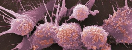
Leukemia is a type of cancer that originates from the bone marrow - the soft, inner part of certain bones where new blood cells are made. It is characterized by an abnormal production of white blood cells, known as leukocytes or blasts. These abnormal cells accumulate in the bone marrow and interfere with the production of normal blood cells, leading to a decrease in red blood cells (anemia), platelets (thrombocytopenia), and healthy white blood cells (leukopenia).
There are several types of leukemia, classified based on the specific type of white blood cell affected and the speed at which the disease progresses:
1. Acute Leukemias - These types of leukemia progress rapidly, with symptoms developing over a few weeks or months. They involve the rapid growth and accumulation of immature, nonfunctional white blood cells (blasts) in the bone marrow and peripheral blood. The two main categories are:
- Acute Lymphoblastic Leukemia (ALL) - Originates from lymphoid progenitor cells, primarily affecting children but can also occur in adults.
- Acute Myeloid Leukemia (AML) - Develops from myeloid progenitor cells and is more common in older adults.
2. Chronic Leukemias - These types of leukemia progress slowly, with symptoms developing over a period of months to years. They involve the production of relatively mature, but still abnormal, white blood cells that can accumulate in large numbers in the bone marrow and peripheral blood. The two main categories are:
- Chronic Lymphocytic Leukemia (CLL) - Affects B-lymphocytes and is more common in older adults.
- Chronic Myeloid Leukemia (CML) - Originates from myeloid progenitor cells, characterized by the presence of a specific genetic abnormality called the Philadelphia chromosome. It can occur at any age but is more common in middle-aged and older adults.
Treatment options for leukemia depend on the type, stage, and individual patient factors. Treatments may include chemotherapy, targeted therapy, immunotherapy, stem cell transplantation, or a combination of these approaches.
Friend murine leukemia virus (F-MuLV) is a type of retrovirus that specifically infects mice. It was first discovered by Charlotte Friend in the 1950s and has since been widely used as a model system to study retroviral pathogenesis, oncogenesis, and immune responses.
F-MuLV is a complex retrovirus that contains several accessory genes, including gag, pol, env, and others. The virus can cause leukemia and other malignancies in susceptible mice, particularly when it is transmitted from mother to offspring through the milk.
The virus is also known to induce immunosuppression, which makes infected mice more susceptible to other infections and diseases. F-MuLV has been used extensively in laboratory research to investigate various aspects of retroviral biology, including viral entry, replication, gene expression, and host immune responses.
It is important to note that Friend murine leukemia virus only infects mice and is not known to cause any disease in humans or other animals.
Acute myeloid leukemia (AML) is a type of cancer that originates in the bone marrow, the soft inner part of certain bones where new blood cells are made. In AML, the immature cells, called blasts, in the bone marrow fail to mature into normal blood cells. Instead, these blasts accumulate and interfere with the production of normal blood cells, leading to a shortage of red blood cells (anemia), platelets (thrombocytopenia), and normal white blood cells (leukopenia).
AML is called "acute" because it can progress quickly and become severe within days or weeks without treatment. It is a type of myeloid leukemia, which means that it affects the myeloid cells in the bone marrow. Myeloid cells are a type of white blood cell that includes monocytes and granulocytes, which help fight infection and defend the body against foreign invaders.
In AML, the blasts can build up in the bone marrow and spread to other parts of the body, including the blood, lymph nodes, liver, spleen, and brain. This can cause a variety of symptoms, such as fatigue, fever, frequent infections, easy bruising or bleeding, and weight loss.
AML is typically treated with a combination of chemotherapy, radiation therapy, and/or stem cell transplantation. The specific treatment plan will depend on several factors, including the patient's age, overall health, and the type and stage of the leukemia.
A cell line is a culture of cells that are grown in a laboratory for use in research. These cells are usually taken from a single cell or group of cells, and they are able to divide and grow continuously in the lab. Cell lines can come from many different sources, including animals, plants, and humans. They are often used in scientific research to study cellular processes, disease mechanisms, and to test new drugs or treatments. Some common types of human cell lines include HeLa cells (which come from a cancer patient named Henrietta Lacks), HEK293 cells (which come from embryonic kidney cells), and HUVEC cells (which come from umbilical vein endothelial cells). It is important to note that cell lines are not the same as primary cells, which are cells that are taken directly from a living organism and have not been grown in the lab.
Radiation dosage, in the context of medical physics, refers to the amount of radiation energy that is absorbed by a material or tissue, usually measured in units of Gray (Gy), where 1 Gy equals an absorption of 1 Joule of radiation energy per kilogram of matter. In the clinical setting, radiation dosage is used to plan and assess the amount of radiation delivered to a patient during treatments such as radiotherapy. It's important to note that the biological impact of radiation also depends on other factors, including the type and energy level of the radiation, as well as the sensitivity of the irradiated tissues or organs.
Human T-lymphotropic virus 1 (HTLV-1) is a complex retrovirus that infects CD4+ T lymphocytes and can cause adult T-cell leukemia/lymphoma (ATLL) and HTLV-1-associated myelopathy/tropical spastic paraparesis (HAM/TSP). The virus is primarily transmitted through breastfeeding, sexual contact, or contaminated blood products. After infection, the virus integrates into the host's genome and can remain latent for years or even decades before leading to disease. HTLV-1 is endemic in certain regions of the world, including Japan, the Caribbean, Central and South America, and parts of Africa.
Ionizing radiation is a type of radiation that carries enough energy to ionize atoms or molecules, which means it can knock electrons out of their orbits and create ions. These charged particles can cause damage to living tissue and DNA, making ionizing radiation dangerous to human health. Examples of ionizing radiation include X-rays, gamma rays, and some forms of subatomic particles such as alpha and beta particles. The amount and duration of exposure to ionizing radiation are important factors in determining the potential health effects, which can range from mild skin irritation to an increased risk of cancer and other diseases.
C57BL/6 (C57 Black 6) is an inbred strain of laboratory mouse that is widely used in biomedical research. The term "inbred" refers to a strain of animals where matings have been carried out between siblings or other closely related individuals for many generations, resulting in a population that is highly homozygous at most genetic loci.
The C57BL/6 strain was established in 1920 by crossing a female mouse from the dilute brown (DBA) strain with a male mouse from the black strain. The resulting offspring were then interbred for many generations to create the inbred C57BL/6 strain.
C57BL/6 mice are known for their robust health, longevity, and ease of handling, making them a popular choice for researchers. They have been used in a wide range of biomedical research areas, including studies of cancer, immunology, neuroscience, cardiovascular disease, and metabolism.
One of the most notable features of the C57BL/6 strain is its sensitivity to certain genetic modifications, such as the introduction of mutations that lead to obesity or impaired glucose tolerance. This has made it a valuable tool for studying the genetic basis of complex diseases and traits.
Overall, the C57BL/6 inbred mouse strain is an important model organism in biomedical research, providing a valuable resource for understanding the genetic and molecular mechanisms underlying human health and disease.
Radiation injuries refer to the damages that occur to living tissues as a result of exposure to ionizing radiation. These injuries can be acute, occurring soon after exposure to high levels of radiation, or chronic, developing over a longer period after exposure to lower levels of radiation. The severity and type of injury depend on the dose and duration of exposure, as well as the specific tissues affected.
Acute radiation syndrome (ARS), also known as radiation sickness, is the most severe form of acute radiation injury. It can cause symptoms such as nausea, vomiting, diarrhea, fatigue, fever, and skin burns. In more severe cases, it can lead to neurological damage, hemorrhage, infection, and death.
Chronic radiation injuries, on the other hand, may not appear until months or even years after exposure. They can cause a range of symptoms, including fatigue, weakness, skin changes, cataracts, reduced fertility, and an increased risk of cancer.
Radiation injuries can be treated with supportive care, such as fluids and electrolytes replacement, antibiotics, wound care, and blood transfusions. In some cases, surgery may be necessary to remove damaged tissue or control bleeding. Prevention is the best approach to radiation injuries, which includes limiting exposure through proper protective measures and monitoring radiation levels in the environment.
The Abelson murine leukemia virus (Abelson murine leukemia virus, or A-MuLV) is a type of retrovirus that can cause cancer in mice. It was first discovered in 1970 and has since been widely studied as a model system for understanding the mechanisms of retroviral infection and cancer development.
A-MuLV is named after Peter Nowell and David A. Harrison, who first described the virus and its ability to cause leukemia in mice. The virus contains an oncogene called "v-abl," which encodes a tyrosine kinase enzyme that can activate various signaling pathways involved in cell growth and division. When the v-abl oncogene is integrated into the genome of an infected mouse cell, it can cause uncontrolled cell growth and division, leading to the development of leukemia.
A-MuLV has been used extensively in laboratory research to study the molecular mechanisms of cancer development and to develop new therapies for treating cancer. It has also been used as a tool for introducing specific genetic modifications into mouse cells, allowing researchers to study the effects of those modifications on cell behavior and function.
Virus receptors are specific molecules (commonly proteins) on the surface of host cells that viruses bind to in order to enter and infect those cells. This interaction between the virus and its receptor is a critical step in the infection process. Different types of viruses have different receptor requirements, and identifying these receptors can provide important insights into the biology of the virus and potential targets for antiviral therapies.
Chronic lymphocytic leukemia (CLL) is a type of cancer that starts from cells that become certain white blood cells (called lymphocytes) in the bone marrow. The cancer (leukemia) cells start in the bone marrow but then go into the blood.
In CLL, the leukemia cells often build up slowly. Many people don't have any symptoms for at least a few years. But over time, the cells can spread to other parts of the body, including the lymph nodes, liver, and spleen.
The "B-cell" part of the name refers to the fact that the cancer starts in a type of white blood cell called a B lymphocyte or B cell. The "chronic" part means that this leukemia usually progresses more slowly than other types of leukemia.
It's important to note that chronic lymphocytic leukemia is different from chronic myelogenous leukemia (CML). Although both are cancers of the white blood cells, they start in different types of white blood cells and progress differently.
Virus replication is the process by which a virus produces copies or reproduces itself inside a host cell. This involves several steps:
1. Attachment: The virus attaches to a specific receptor on the surface of the host cell.
2. Penetration: The viral genetic material enters the host cell, either by invagination of the cell membrane or endocytosis.
3. Uncoating: The viral genetic material is released from its protective coat (capsid) inside the host cell.
4. Replication: The viral genetic material uses the host cell's machinery to produce new viral components, such as proteins and nucleic acids.
5. Assembly: The newly synthesized viral components are assembled into new virus particles.
6. Release: The newly formed viruses are released from the host cell, often through lysis (breaking) of the cell membrane or by budding off the cell membrane.
The specific mechanisms and details of virus replication can vary depending on the type of virus. Some viruses, such as DNA viruses, use the host cell's DNA polymerase to replicate their genetic material, while others, such as RNA viruses, use their own RNA-dependent RNA polymerase or reverse transcriptase enzymes. Understanding the process of virus replication is important for developing antiviral therapies and vaccines.
Gibbon Ape Leukemia Virus (GaLV) is not exactly a "leukemia virus" in the sense that it directly causes leukemia in humans. Instead, GaLV is a type of retrovirus that primarily infects gibbons and some other non-human primates. It's important to note that GaLV is not known to infect or cause disease in healthy human beings.
GaLV has four subtypes (A, B, C, and D), with A and B being the most well-studied. These viruses have a close genetic relationship with certain human retroviruses, such as Human T-cell Leukemia Virus types I and II (HTLV-I/II). Although GaLV is not known to cause leukemia or any other diseases in humans directly, it has served as an important model for understanding the biology and pathogenesis of retroviruses, including those that can cause leukemia and other malignancies in humans.
The term "leukemia virus" is often used to describe retroviruses that can cause leukemia or lymphoma, such as HTLV-I/II and Human Immunodeficiency Virus (HIV). GaLV does not fit into this category for humans, but it's essential to understand its role in the context of retroviral research and comparative primatology.
RNA viruses are a type of virus that contain ribonucleic acid (RNA) as their genetic material, as opposed to deoxyribonucleic acid (DNA). RNA viruses replicate by using an enzyme called RNA-dependent RNA polymerase to transcribe and replicate their RNA genome.
There are several different groups of RNA viruses, including:
1. Negative-sense single-stranded RNA viruses: These viruses have a genome that is complementary to the mRNA and must undergo transcription to produce mRNA before translation can occur. Examples include influenza virus, measles virus, and rabies virus.
2. Positive-sense single-stranded RNA viruses: These viruses have a genome that can serve as mRNA and can be directly translated into protein after entry into the host cell. Examples include poliovirus, rhinoviruses, and coronaviruses.
3. Double-stranded RNA viruses: These viruses have a genome consisting of double-stranded RNA and use a complex replication strategy involving both transcription and reverse transcription. Examples include rotaviruses and reoviruses.
RNA viruses are known to cause a wide range of human diseases, ranging from the common cold to more severe illnesses such as hepatitis C, polio, and COVID-19. Due to their high mutation rates and ability to adapt quickly to new environments, RNA viruses can be difficult to control and treat with antiviral drugs or vaccines.
A gene product is the biochemical material, such as a protein or RNA, that is produced by the expression of a gene. Gene products are the result of the translation and transcription of genetic information encoded in DNA or RNA.
In the context of "tax," this term is not typically used in a medical definition of gene products. However, it may refer to the concept of taxing or regulating gene products in the context of genetic engineering or synthetic biology. This could involve imposing fees or restrictions on the production, use, or sale of certain gene products, particularly those that are genetically modified or engineered. The regulation of gene products is an important aspect of ensuring their safe and effective use in various applications, including medical treatments, agricultural production, and industrial processes.
I'm sorry for any confusion, but there seems to be no established medical definition or recognition of a "Rauscher Virus" in human or veterinary medicine. It is possible that you may have misspelled or misremembered the name of a specific virus or medical term. If you have more information or context about where this term was used, I'd be happy to help you further research the topic.
Leukemia, lymphoid is a type of cancer that affects the lymphoid cells, which are a vital part of the body's immune system. It is characterized by the uncontrolled production of abnormal white blood cells (leukocytes or WBCs) in the bone marrow, specifically the lymphocytes. These abnormal lymphocytes accumulate and interfere with the production of normal blood cells, leading to a decrease in red blood cells (anemia), platelets (thrombocytopenia), and healthy white blood cells (leukopenia).
There are two main types of lymphoid leukemia: acute lymphoblastic leukemia (ALL) and chronic lymphocytic leukemia (CLL). Acute lymphoblastic leukemia progresses rapidly, while chronic lymphocytic leukemia has a slower onset and progression.
Symptoms of lymphoid leukemia may include fatigue, frequent infections, easy bruising or bleeding, weight loss, swollen lymph nodes, and bone pain. Treatment options depend on the type, stage, and individual patient factors but often involve chemotherapy, radiation therapy, targeted therapy, immunotherapy, or stem cell transplantation.
Leukemic infiltration is the abnormal spread and accumulation of malignant white blood cells (leukemia cells) in various tissues and organs outside the bone marrow. The bone marrow is the spongy tissue inside bones where blood cells are normally produced. In leukemia, the bone marrow produces large numbers of abnormal white blood cells that do not function properly. These abnormal cells can sometimes spill into the bloodstream and infiltrate other organs, such as the lymph nodes, spleen, liver, and central nervous system (brain and spinal cord). Leukemic infiltration can cause damage to these organs and lead to various symptoms. The pattern of organ involvement and the severity of infiltration depend on the type and stage of leukemia.
Cutis laxa is a group of rare connective tissue disorders characterized by loose, sagging, and inelastic skin. The term "cutis laxa" comes from Latin, meaning "loose skin." This condition can affect both the skin and the internal organs. Inherited forms of cutis laxa are caused by mutations in various genes involved in the structure and function of connective tissue, while acquired forms can be associated with autoimmune disorders, cancer, or certain medications.
The main features of cutis laxa include:
1. Sagging, redundant skin: The skin appears loose and wrinkled, especially on the face, neck, hands, and feet. This is due to a deficiency in elastic fibers, which provide flexibility and resilience to the skin.
2. Premature aging appearance: The sagging skin can give an individual a prematurely aged appearance, with deep wrinkles and folds around the eyes, mouth, and neck.
3. Pulmonary involvement: Recurrent respiratory infections, bronchiectasis (permanent enlargement of the airways), and emphysema can occur due to weakened lung tissue.
4. Gastrointestinal issues: Weakened intestinal walls may lead to hernias, bowel obstructions, or malabsorption.
5. Cardiovascular problems: The aorta and other major blood vessels may become weakened and dilated, leading to an increased risk of aneurysms and dissections (tears in the vessel wall).
6. Ophthalmic complications: Eye abnormalities such as blue sclerae (transparent blue appearance of the whites of the eyes) and strabismus (crossed eyes) can occur.
7. Skeletal abnormalities: Individuals with cutis laxa may have joint hypermobility, scoliosis (curvature of the spine), or hip dislocations.
8. Neurological issues: Rarely, cutis laxa can be associated with developmental delays, intellectual disability, or seizures.
There is no cure for cutis laxa, and treatment focuses on managing symptoms and preventing complications. This may include skin care, physical therapy, medications to control blood pressure, and surgery to repair hernias or aneurysms. Regular follow-up with a multidisciplinary team of healthcare professionals is essential to monitor disease progression and address any emerging issues.
Plasma cell neoplasms are a type of cancer that originates from plasma cells, which are a type of white blood cell found in the bone marrow. These cells are responsible for producing antibodies to help fight off infections. When plasma cells become cancerous and multiply out of control, they can form a tumor called a plasmacytoma.
There are two main types of plasma cell neoplasms: solitary plasmacytoma and multiple myeloma. Solitary plasmacytoma is a localized tumor that typically forms in the bone, while multiple myeloma is a systemic disease that affects multiple bones and can cause a variety of symptoms such as bone pain, fatigue, and anemia.
Plasma cell neoplasms are diagnosed through a combination of tests, including blood tests, imaging studies, and bone marrow biopsy. Treatment options depend on the stage and extent of the disease, but may include radiation therapy, chemotherapy, and stem cell transplantation.
In medical terms, the skin is the largest organ of the human body. It consists of two main layers: the epidermis (outer layer) and dermis (inner layer), as well as accessory structures like hair follicles, sweat glands, and oil glands. The skin plays a crucial role in protecting us from external factors such as bacteria, viruses, and environmental hazards, while also regulating body temperature and enabling the sense of touch.
The epidermis is the outermost layer of the skin, composed mainly of stratified squamous epithelium. It forms a protective barrier that prevents water loss and inhibits the entry of microorganisms. The epidermis contains no blood vessels, and its cells are nourished by diffusion from the underlying dermis. The bottom-most layer of the epidermis, called the stratum basale, is responsible for generating new skin cells that eventually move up to replace dead cells on the surface. This process of cell turnover takes about 28 days in adults.
The most superficial part of the epidermis consists of dead cells called squames, which are constantly shed and replaced. The exact rate at which this happens varies depending on location; for example, it's faster on the palms and soles than elsewhere. Melanocytes, the pigment-producing cells, are also located in the epidermis, specifically within the stratum basale layer.
In summary, the epidermis is a vital part of our integumentary system, providing not only physical protection but also playing a crucial role in immunity and sensory perception through touch receptors called Pacinian corpuscles.
 Biphenotypic acute leukaemia
 Leukemia cutis: Symptoms, pictures, treatment, and outlook
 General Recommendations on Immunization: Recommendations of the Advisory Committee on Immunization
Practices (ACIP) and the...
General Recommendations on Immunization: Recommendations of the Advisory Committee on Immunization
Practices (ACIP) and the... Leukemia Cutis: Background, Pathophysiology, Etiology
 ACIP: General Recommendations on Immunization
 Bone marrow transplant: MedlinePlus Medical Encyclopedia
 Are Stem Cell Transplants A Cure For HIV? Not So Fast
 Timothy Ray Brown, first man cured of HIV, dies of leukemia :: Bay Area Reporter
 Wireless Mind, Gullible Mind
 Flashcards - Adult health 2
 National Association of Special Education Teachers: Blood Disorder Anemia Blood Loss Aplastic Anemia Symptom NASET
 Lymphoma Causes and Diagnoses | Northwestern Medicine
 Different Types of Disorders That Can Affect Blood Cells | HPMP-HealthCare
 Microwave News | News Center: Main Articles Archive
 HIV-positive man in U.K. seems to be 2nd person ever cured after transplant - National | Globalnews.ca
 Leukemia.pptx
 People with blood-related cancers more likely to experience COVID breakthrough infections
 Influenza vacc,tri 2003(live) nasal Patient Assistance Program
 Leukemia | Cancer Support Community
 Best Acute Leukemia Treatment Cost in India Top Surgeon Doctor Hospital
 Leukemia, Radiation-Induced - Medical Dictionary online-medical-dictionary.org