RNA Interference
RNA, Small Interfering
RNA
RNA, Double-Stranded
RNA Editing
RNA Splicing
RNA, Messenger
Gene Silencing
RNA, Ribosomal
RNA, Bacterial
DNA-Directed RNA Polymerases
Molecular Sequence Data
Base Sequence
RNA, Antisense
RNA Stability
HeLa Cells
RNA, Catalytic
Gene Knockdown Techniques
RNA Polymerase II
RNA Processing, Post-Transcriptional
RNA, Untranslated
Ribonuclease III
Microscopy, Interference
RNA, Fungal
RNA-Induced Silencing Complex
Argonaute Proteins
Transfection
Nucleic Acid Conformation
Caenorhabditis elegans
RNA, Protozoan
RNA Precursors
RNA, Small Nuclear
RNA, Plant
RNA, Helminth
Mutation
Amino Acid Sequence
RNA-Binding Proteins
RNA, Transfer
Reverse Transcriptase Polymerase Chain Reaction
Sequence Analysis, RNA
Down-Regulation
DEAD-box RNA Helicases
Trypanosoma brucei brucei
Protein Binding
Transcription, Genetic
Gene Expression Regulation
Lentivirus
Signal Transduction
RNA Caps
Genetic Vectors
Cells, Cultured
Virus Replication
Caenorhabditis elegans Proteins
Promoter Regions, Genetic
Transcription Factors
Drosophila Proteins
Blotting, Western
Apoptosis
Green Fluorescent Proteins
Phenotype
Plasmids
DNA Primers
RNA, Guide
Nuclear Proteins
RNA Polymerase III
Drosophila melanogaster
Protein Structure, Tertiary
Cell Nucleus
RNA Transport
RNA Ligase (ATP)
Drosophila
Models, Biological
Plants, Genetically Modified
Binding Sites
Endoribonucleases
Gene Expression
Genes, Reporter
Gene Expression Profiling
Immunoprecipitation
Phosphorylation
Gene Expression Regulation, Neoplastic
Protein Transport
RNA Polymerase I
Animals, Genetically Modified
Microscopy, Fluorescence
Models, Genetic
MicroRNAs
DNA
RNA, Nuclear
Protein Biosynthesis
Sequence Homology, Amino Acid
RNA, Ribosomal, 28S
Oligonucleotides
RNA, Spliced Leader
RNA Cleavage
Cell Survival
Gene Expression Regulation, Developmental
Cell Cycle Proteins
RNA, Ribosomal, 18S
Membrane Proteins
Nodaviridae
DNA-Binding Proteins
Repressor Proteins
Gene Targeting
Escherichia coli
Genetic Techniques
RNA Virus Infections
DNA, Complementary
Blotting, Northern
Cell Movement
Gene Expression Regulation, Plant
Protein-Serine-Threonine Kinases
Carrier Proteins
Genetic Therapy
RNA, Ribosomal, 23S
Up-Regulation
Recombinant Fusion Proteins
Luciferases
Cytoplasm
HEK293 Cells
Cercopithecus aethiops
RNA, Satellite
Polymerase Chain Reaction
Mitosis
Tribolium
Oligonucleotide Array Sequence Analysis
Gene Expression Regulation, Viral
Cell Cycle
RNA, Archaeal
Cell Differentiation
Nucleic Acid Hybridization
Plant Proteins
Microtubule-Associated Proteins
Oligoribonucleotides
RNA, Ribosomal, 16S
Gene Expression Regulation, Enzymologic
RNA, Small Untranslated
Immunoblotting
Models, Molecular
Larva
RNA, Long Noncoding
Two-Hybrid System Techniques
Host-Pathogen Interactions
Mice, Nude
Transcriptional Activation
Chromatin Immunoprecipitation
Arabidopsis
Electroporation
RNA, Small Nucleolar
Real-Time Polymerase Chain Reaction
NIH 3T3 Cells
Uridine
Adaptor Proteins, Signal Transducing
Mitochondria
Base Pairing
Exoribonucleases
Conserved Sequence
Tobacco
Fibroblasts
Embryo, Nonmammalian
Protein Isoforms
Enzyme Activation
Trans-Activators
HIV-1
Cricetinae
3' Untranslated Regions
Histones
Flow Cytometry
Gene Deletion
Planarians
Arabidopsis Proteins
RNA, Small Cytoplasmic
Templates, Genetic
Proteins
Saccharomyces cerevisiae
Eukaryotic Initiation Factors
RNA, Heterogeneous Nuclear
Methylation
Neoplasm Proteins
Exon skipping in IVD RNA processing in isovaleric acidemia caused by point mutations in the coding region of the IVD gene. (1/17357)
Isovaleric acidemia (IVA) is a recessive disorder caused by a deficiency of isovaleryl-CoA dehydrogenase (IVD). We have reported elsewhere nine point mutations in the IVD gene in fibroblasts of patients with IVA, which lead to abnormalities in IVD protein processing and activity. In this report, we describe eight IVD gene mutations identified in seven IVA patients that result in abnormal splicing of IVD RNA. Four mutations in the coding region lead to aberrantly spliced mRNA species in patient fibroblasts. Three of these are amino acid altering point mutations, whereas one is a single-base insertion that leads to a shift in the reading frame of the mRNA. Two of the coding mutations strengthen pre-existing cryptic splice acceptors adjacent to the natural splice junctions and apparently interfere with exon recognition, resulting in exon skipping. This mechanism for missplicing has not been reported elsewhere. Four other mutations alter either the conserved gt or ag dinucleotide splice sites in the IVD gene. Exon skipping and cryptic splicing were confirmed by transfection of these mutations into a Cos-7 cell line model splicing system. Several of the mutations were predicted by individual information analysis to inactivate or significantly weaken adjacent donor or acceptor sites. The high frequency of splicing mutations identified in these patients is unusual, as is the finding of missplicing associated with missense mutations in exons. These results may lead to a better understanding of the phenotypic complexity of IVA, as well as provide insight into those factors important in defining intron/exon boundaries in vivo. (+info)Characterisation and expression of a PP1 serine/threonine protein phosphatase (PfPP1) from the malaria parasite, Plasmodium falciparum: demonstration of its essential role using RNA interference. (2/17357)
BACKGROUND: Reversible protein phosphorylation is relatively unexplored in the intracellular protozoa of the Apicomplexa family that includes the genus Plasmodium, to which belong the causative agents of malaria. Members of the PP1 family represent the most highly conserved protein phosphatase sequences in phylogeny and play essential regulatory roles in various cellular pathways. Previous evidence suggested a PP1-like activity in Plasmodium falciparum, not yet identified at the molecular level. RESULTS: We have identified a PP1 catalytic subunit from P. falciparum and named it PfPP1. The predicted primary structure of the 304-amino acid long protein was highly similar to PP1 sequences of other species, and showed conservation of all the signature motifs. The purified recombinant protein exhibited potent phosphatase activity in vitro. Its sensitivity to specific phosphatase inhibitors was characteristic of the PP1 class. The authenticity of the PfPP1 cDNA was further confirmed by mutational analysis of strategic amino acid residues important in catalysis. The protein was expressed in all erythrocytic stages of the parasite. Abrogation of PP1 expression by synthetic short interfering RNA (siRNA) led to inhibition of parasite DNA synthesis. CONCLUSIONS: The high sequence similarity of PfPP1 with other PP1 members suggests conservation of function. Phenotypic gene knockdown studies using siRNA confirmed its essential role in the parasite. Detailed studies of PfPP1 and its regulation may unravel the role of reversible protein phosphorylation in the signalling pathways of the parasite, including glucose metabolism and parasitic cell division. The use of siRNA could be an important tool in the functional analysis of Apicomplexan genes. (+info)Frequent germline mutations and somatic repeat instability in DNA mismatch-repair-deficient Caenorhabditis elegans. (3/17357)
Mismatch-repair-deficient mutants were initially recognized as mutation-prone derivatives of bacteria, and later mismatch repair deficiency was found to predispose humans to colon cancers (HNPCC). We generated mismatch-repair-deficient Caenorhabditis elegans by deleting the msh-6 gene and analyzed the fidelity of transmission of genetic information to subsequent generations. msh-6-defective animals show an elevated level of spontaneous mutants in both the male and female germline; also repeated DNA tracts are unstable. To monitor DNA repeat instability in somatic tissue, we developed a sensitive system, making use of heat-shock promoter-driven lacZ transgenes, but with a repeat that puts this reporter gene out of frame. In genetic msh-6-deficient animals lacZ+ patches are observed as a result of somatic repeat instability. RNA interference by feeding wild-type animals dsRNA homologous to msh-2 or msh-6 also resulted in somatic DNA instability, as well as in germline mutagenesis, indicating that one can use C. elegans as a model system to discover genes involved in maintaining DNA stability by large-scale RNAi screens. (+info)The dynamic localisation of the Drosophila APC/C: evidence for the existence of multiple complexes that perform distinct functions and are differentially localised. (4/17357)
In Drosophila cells, the destruction of cyclin B is spatially regulated. In cellularised embryos, cyclin B is initially degraded on the mitotic spindle and is then degraded in the cytoplasm. In syncytial embryos, only the spindle-associated cyclin B is degraded at the end of mitosis. The anaphase promoting complex/cyclosome (APC/C) targets cyclin B for destruction, but its subcellular localisation remains controversial. We constructed GFP fusions of two core APC/C subunits, Cdc16 and Cdc27. These fusion proteins were incorporated into the endogenous APC/C and were largely localised in the cytoplasm during interphase in living syncytial embryos. Both fusion proteins rapidly accumulated in the nucleus prior to nuclear envelope breakdown but only weakly associated with mitotic spindles throughout mitosis. Thus, the global activation of a spatially restricted APC/C cannot explain the spatially regulated destruction of cyclin B. Instead, different subpopulations of the APC/C must be activated at different times to degrade cyclin B. Surprisingly, we noticed that GFP-Cdc27 associated with mitotic chromosomes, whereas GFP-Cdc16 did not. Moreover, reducing the levels of Cdc16 or Cdc27 by >90% in tissue culture cells led to a transient mitotic arrest that was both biochemically and morphologically distinct. Taken together, our results raise the intriguing possibility that there could be multiple forms of the APC/C that are differentially localised and perform distinct functions. (+info)A novel linker histone-like protein is associated with cytoplasmic filaments in Caenorhabditis elegans. (5/17357)
The histone H1 complement of Caenorhabditis elegans contains a single unusual protein, H1.X. Although H1.X possesses the globular domain and the canonical three-domain structure of linker histones, the amino acid composition of H1.X is distinctly different from conventional linker histones in both terminal domains. We have characterized H1.X in C. elegans by antibody labeling, green fluorescent protein fusion protein expression and RNA interference. Unlike normal linker histones, H1.X is a cytoplasmic as well as a nuclear protein and is not associated with chromosomes. H1.X is most prominently expressed in the marginal cells of the pharynx and is associated with a peculiar cytoplasmic cytoskeletal structure therein, the tonofilaments. Additionally H1.X::GFP is expressed in the cytoplasm of body and vulva muscle cells, neurons, excretory cells and in the nucleoli of embryonic blastomeres and adult gut cells. RNA interference with H1.X results in uncoordinated and egg laying defective animals, as well as in a longitudinally enlarged pharynx. These phenotypes indicate a cytoplasmic role of H1.X in muscle growth and muscle function. (+info)Cathepsin B expression and down-regulation by gene silencing and antisense DNA in human chondrocytes. (6/17357)
Cathepsin B, a marker of the dedifferentiated chondrocyte phenotype, contributes to cartilage destruction in osteoarthritis and pathological proteolysis in rheumatoid arthritis and cancer. In search of possible means for neutralizing the action of this enzyme, we compared its expression, biosynthesis and distribution in articular chondrocytes and two lines of immortalized human chondrocytes. Native articular chondrocytes in primary culture and the polyclonal T/C-28a2 chondrocyte cell line were similar with respect to the number of endosomes and lysosomes, the distribution of three alternatively spliced cathepsin B mRNA forms, and the cathepsin B activity. In contrast, the clonal C-28/I2 cell line contained four times higher levels of intracellular cathepsin B activity, slightly higher numbers of endosomes and lysosomes, and uniform distribution of all three cathepsin B transcripts and thus resembled subcultured chondrocytes at an early stage of dedifferentiation. Transfection of T/C-28a2 chondrocytes with double-stranded cathepsin B mRNA resulted in inhibition of cathepsin B biosynthesis by up to 70% due to RNA interference, and single-stranded antisense DNAs of various sizes decreased cathepsin B biosynthesis by up to 78%. An antisense oligonucleotide designed to hybridize to the end of cathepsin B's exons 1 and the beginning of exon 3 was successful in specifically inhibiting the mRNA splice variant lacking exon 2. These results indicate that cathepsin B expression and activity may be targeted for gene silencing by RNA interference and antisense DNA in chondrocytes. Furthermore, the differential expression and distribution of cathepsin B and presence of the necessary molecular apparatus for gene silencing in the immortalized human chondrocyte cell lines indicate that they may serve as a useful model for studying the function of relevant enzymes in cartilage pathologies. (+info)Inscuteable-independent apicobasally oriented asymmetric divisions in the Drosophila embryonic CNS. (7/17357)
Inscuteable is the founding member of a protein complex localised to the apical cortex of Drosophila neural progenitors that controls their asymmetric division. Aspects of asymmetric divisions of all identified apicobasally oriented neural progenitors characterised to date, in both the central and peripheral nervous systems, require inscuteable. Here we examine the generality of this requirement. We show that many identified neuroblast lineages, in fact, do not require inscuteable for normal morphological development. To elucidate the requirements for apicobasal asymmetric divisions in a context where inscuteable is not essential, we focused on the MP2 > dMP2 + vMP2 division. We show that for MP2 divisions, asymmetric localisation and segregation of Numb and the specification of distinct dMP2 and vMP2 identities require bazooka but not inscuteable. We conclude that inscuteable is not required for all apicobasally oriented asymmetric divisions and that, in some cellular contexts, bazooka can mediate apicobasal asymmetric divisions without inscuteable. (+info)Requirements of high levels of Hedgehog signaling activity for medial-region cell fate determination in Drosophila legs: identification of pxb, a putative Hedgehog signaling attenuator gene repressed along the anterior-posterior compartment boundary. (8/17357)
We show that high levels of Hedgehog signaling activity are essential for medial-region patterning in Drosophila legs. In mid-to-late third instar leg discs, high levels of Hedgehog signals repress the transcription of pxb, a newly identified gene encoding a transmembrane protein expressed specifically in the anterior compartment. Misexpression experiments indicate that Pxb may serve as a Hedgehog signaling attenuator capable of acting prior to Hedgehog-Patched interactions, suggesting that Hedgehog signaling in leg discs includes a pxb-repression-mediated positive feedback loop. RNA interference and clonal analysis show that neither Wingless nor Decapentaplegic signaling is required for pxb repression but high levels of Wingless signaling activity are essential for patterning in the leg ventral medial region. (+info)RNA interference (RNAi) is a biological process in which RNA molecules inhibit the expression of specific genes. This process is mediated by small RNA molecules, including microRNAs (miRNAs) and small interfering RNAs (siRNAs), that bind to complementary sequences on messenger RNA (mRNA) molecules, leading to their degradation or translation inhibition.
RNAi plays a crucial role in regulating gene expression and defending against foreign genetic elements, such as viruses and transposons. It has also emerged as an important tool for studying gene function and developing therapeutic strategies for various diseases, including cancer and viral infections.
Small interfering RNA (siRNA) is a type of short, double-stranded RNA molecule that plays a role in the RNA interference (RNAi) pathway. The RNAi pathway is a natural cellular process that regulates gene expression by targeting and destroying specific messenger RNA (mRNA) molecules, thereby preventing the translation of those mRNAs into proteins.
SiRNAs are typically 20-25 base pairs in length and are generated from longer double-stranded RNA precursors called hairpin RNAs or dsRNAs by an enzyme called Dicer. Once generated, siRNAs associate with a protein complex called the RNA-induced silencing complex (RISC), which uses one strand of the siRNA (the guide strand) to recognize and bind to complementary sequences in the target mRNA. The RISC then cleaves the target mRNA, leading to its degradation and the inhibition of protein synthesis.
SiRNAs have emerged as a powerful tool for studying gene function and have shown promise as therapeutic agents for a variety of diseases, including viral infections, cancer, and genetic disorders. However, their use as therapeutics is still in the early stages of development, and there are challenges associated with delivering siRNAs to specific cells and tissues in the body.
RNA (Ribonucleic Acid) is a single-stranded, linear polymer of ribonucleotides. It is a nucleic acid present in the cells of all living organisms and some viruses. RNAs play crucial roles in various biological processes such as protein synthesis, gene regulation, and cellular signaling. There are several types of RNA including messenger RNA (mRNA), ribosomal RNA (rRNA), transfer RNA (tRNA), small nuclear RNA (snRNA), microRNA (miRNA), and long non-coding RNA (lncRNA). These RNAs differ in their structure, function, and location within the cell.
A viral RNA (ribonucleic acid) is the genetic material found in certain types of viruses, as opposed to viruses that contain DNA (deoxyribonucleic acid). These viruses are known as RNA viruses. The RNA can be single-stranded or double-stranded and can exist as several different forms, such as positive-sense, negative-sense, or ambisense RNA. Upon infecting a host cell, the viral RNA uses the host's cellular machinery to translate the genetic information into proteins, leading to the production of new virus particles and the continuation of the viral life cycle. Examples of human diseases caused by RNA viruses include influenza, COVID-19 (SARS-CoV-2), hepatitis C, and polio.
I'm happy to help! However, it seems there might be a slight misunderstanding in your question. RNA, or ribonucleic acid, is typically single-stranded. Unlike DNA, which is double-stranded and forms a double helix, RNA usually exists as a single strand of nucleotides.
There are exceptions to this rule, such as certain types of RNA molecules that can form double-stranded structures in specific contexts. For example:
1. Double-Stranded RNA (dsRNA) viruses: These viruses have genomes made entirely of RNA, which is double-stranded throughout or partially double-stranded. The dsRNA viruses include important pathogens such as rotaviruses and reoviruses.
2. Hairpin loops in RNA structures: Some single-stranded RNA molecules can fold back on themselves to form short double-stranded regions, called hairpin loops, within their overall structure. These are often found in ribosomal RNA (rRNA), transfer RNA (tRNA), and messenger RNA (mRNA) molecules.
So, while 'double-stranded RNA' is not a standard medical definition for RNA itself, there are specific instances where RNA can form double-stranded structures as described above.
RNA editing is a process that alters the sequence of a transcribed RNA molecule after it has been synthesized from DNA, but before it is translated into protein. This can result in changes to the amino acid sequence of the resulting protein or to the regulation of gene expression. The most common type of RNA editing in mammals is the hydrolytic deamination of adenosine (A) to inosine (I), catalyzed by a family of enzymes called adenosine deaminases acting on RNA (ADARs). Inosine is recognized as guanosine (G) by the translation machinery, leading to A-to-G changes in the RNA sequence. Other types of RNA editing include cytidine (C) to uridine (U) deamination and insertion/deletion of nucleotides. RNA editing is a crucial mechanism for generating diversity in gene expression and has been implicated in various biological processes, including development, differentiation, and disease.
RNA splicing is a post-transcriptional modification process in which the non-coding sequences (introns) are removed and the coding sequences (exons) are joined together in a messenger RNA (mRNA) molecule. This results in a continuous mRNA sequence that can be translated into a single protein. Alternative splicing, where different combinations of exons are included or excluded, allows for the creation of multiple proteins from a single gene.
Messenger RNA (mRNA) is a type of RNA (ribonucleic acid) that carries genetic information copied from DNA in the form of a series of three-base code "words," each of which specifies a particular amino acid. This information is used by the cell's machinery to construct proteins, a process known as translation. After being transcribed from DNA, mRNA travels out of the nucleus to the ribosomes in the cytoplasm where protein synthesis occurs. Once the protein has been synthesized, the mRNA may be degraded and recycled. Post-transcriptional modifications can also occur to mRNA, such as alternative splicing and addition of a 5' cap and a poly(A) tail, which can affect its stability, localization, and translation efficiency.
Gene silencing is a process by which the expression of a gene is blocked or inhibited, preventing the production of its corresponding protein. This can occur naturally through various mechanisms such as RNA interference (RNAi), where small RNAs bind to and degrade specific mRNAs, or DNA methylation, where methyl groups are added to the DNA molecule, preventing transcription. Gene silencing can also be induced artificially using techniques such as RNAi-based therapies, antisense oligonucleotides, or CRISPR-Cas9 systems, which allow for targeted suppression of gene expression in research and therapeutic applications.
Ribosomal RNA (rRNA) is a type of RNA molecule that is a key component of ribosomes, which are the cellular structures where protein synthesis occurs in cells. In ribosomes, rRNA plays a crucial role in the process of translation, where genetic information from messenger RNA (mRNA) is translated into proteins.
Ribosomal RNA is synthesized in the nucleus and then transported to the cytoplasm, where it assembles with ribosomal proteins to form ribosomes. Within the ribosome, rRNA provides a structural framework for the assembly of the ribosome and also plays an active role in catalyzing the formation of peptide bonds between amino acids during protein synthesis.
There are several different types of rRNA molecules, including 5S, 5.8S, 18S, and 28S rRNA, which vary in size and function. These rRNA molecules are highly conserved across different species, indicating their essential role in protein synthesis and cellular function.
Bacterial RNA refers to the genetic material present in bacteria that is composed of ribonucleic acid (RNA). Unlike higher organisms, bacteria contain a single circular chromosome made up of DNA, along with smaller circular pieces of DNA called plasmids. These bacterial genetic materials contain the information necessary for the growth and reproduction of the organism.
Bacterial RNA can be divided into three main categories: messenger RNA (mRNA), ribosomal RNA (rRNA), and transfer RNA (tRNA). mRNA carries genetic information copied from DNA, which is then translated into proteins by the rRNA and tRNA molecules. rRNA is a structural component of the ribosome, where protein synthesis occurs, while tRNA acts as an adapter that brings amino acids to the ribosome during protein synthesis.
Bacterial RNA plays a crucial role in various cellular processes, including gene expression, protein synthesis, and regulation of metabolic pathways. Understanding the structure and function of bacterial RNA is essential for developing new antibiotics and other therapeutic strategies to combat bacterial infections.
DNA-directed RNA polymerases are enzymes that synthesize RNA molecules using a DNA template in a process called transcription. These enzymes read the sequence of nucleotides in a DNA molecule and use it as a blueprint to construct a complementary RNA strand.
The RNA polymerase moves along the DNA template, adding ribonucleotides one by one to the growing RNA chain. The synthesis is directional, starting at the promoter region of the DNA and moving towards the terminator region.
In bacteria, there is a single type of RNA polymerase that is responsible for transcribing all types of RNA (mRNA, tRNA, and rRNA). In eukaryotic cells, however, there are three different types of RNA polymerases: RNA polymerase I, II, and III. Each type is responsible for transcribing specific types of RNA.
RNA polymerases play a crucial role in gene expression, as they link the genetic information encoded in DNA to the production of functional proteins. Inhibition or mutation of these enzymes can have significant consequences for cellular function and survival.
Molecular sequence data refers to the specific arrangement of molecules, most commonly nucleotides in DNA or RNA, or amino acids in proteins, that make up a biological macromolecule. This data is generated through laboratory techniques such as sequencing, and provides information about the exact order of the constituent molecules. This data is crucial in various fields of biology, including genetics, evolution, and molecular biology, allowing for comparisons between different organisms, identification of genetic variations, and studies of gene function and regulation.
RNA viruses are a type of virus that contain ribonucleic acid (RNA) as their genetic material, as opposed to deoxyribonucleic acid (DNA). RNA viruses replicate by using an enzyme called RNA-dependent RNA polymerase to transcribe and replicate their RNA genome.
There are several different groups of RNA viruses, including:
1. Negative-sense single-stranded RNA viruses: These viruses have a genome that is complementary to the mRNA and must undergo transcription to produce mRNA before translation can occur. Examples include influenza virus, measles virus, and rabies virus.
2. Positive-sense single-stranded RNA viruses: These viruses have a genome that can serve as mRNA and can be directly translated into protein after entry into the host cell. Examples include poliovirus, rhinoviruses, and coronaviruses.
3. Double-stranded RNA viruses: These viruses have a genome consisting of double-stranded RNA and use a complex replication strategy involving both transcription and reverse transcription. Examples include rotaviruses and reoviruses.
RNA viruses are known to cause a wide range of human diseases, ranging from the common cold to more severe illnesses such as hepatitis C, polio, and COVID-19. Due to their high mutation rates and ability to adapt quickly to new environments, RNA viruses can be difficult to control and treat with antiviral drugs or vaccines.
A base sequence in the context of molecular biology refers to the specific order of nucleotides in a DNA or RNA molecule. In DNA, these nucleotides are adenine (A), guanine (G), cytosine (C), and thymine (T). In RNA, uracil (U) takes the place of thymine. The base sequence contains genetic information that is transcribed into RNA and ultimately translated into proteins. It is the exact order of these bases that determines the genetic code and thus the function of the DNA or RNA molecule.
A cell line is a culture of cells that are grown in a laboratory for use in research. These cells are usually taken from a single cell or group of cells, and they are able to divide and grow continuously in the lab. Cell lines can come from many different sources, including animals, plants, and humans. They are often used in scientific research to study cellular processes, disease mechanisms, and to test new drugs or treatments. Some common types of human cell lines include HeLa cells (which come from a cancer patient named Henrietta Lacks), HEK293 cells (which come from embryonic kidney cells), and HUVEC cells (which come from umbilical vein endothelial cells). It is important to note that cell lines are not the same as primary cells, which are cells that are taken directly from a living organism and have not been grown in the lab.
Antisense RNA is a type of RNA molecule that is complementary to another RNA called sense RNA. In the context of gene expression, sense RNA is the RNA transcribed from a protein-coding gene, which serves as a template for translation into a protein. Antisense RNA, on the other hand, is transcribed from the opposite strand of the DNA and is complementary to the sense RNA.
Antisense RNA can bind to its complementary sense RNA through base-pairing, forming a double-stranded RNA structure. This interaction can prevent the sense RNA from being translated into protein or can target it for degradation by cellular machinery, thereby reducing the amount of protein produced from the gene. Antisense RNA can be used as a tool in molecular biology to study gene function or as a therapeutic strategy to silence disease-causing genes.
RNA stability refers to the duration that a ribonucleic acid (RNA) molecule remains intact and functional within a cell before it is degraded or broken down into its component nucleotides. Various factors can influence RNA stability, including:
1. Primary sequence: Certain sequences in the RNA molecule may be more susceptible to degradation by ribonucleases (RNases), enzymes that break down RNA.
2. Secondary structure: The formation of stable secondary structures, such as hairpins or stem-loop structures, can protect RNA from degradation.
3. Presence of RNA-binding proteins: Proteins that bind to RNA can either stabilize or destabilize the RNA molecule, depending on the type and location of the protein-RNA interaction.
4. Chemical modifications: Modifications to the RNA nucleotides, such as methylation, can increase RNA stability by preventing degradation.
5. Subcellular localization: The subcellular location of an RNA molecule can affect its stability, with some locations providing more protection from ribonucleases than others.
6. Cellular conditions: Changes in cellular conditions, such as pH or temperature, can also impact RNA stability.
Understanding RNA stability is important for understanding gene regulation and the function of non-coding RNAs, as well as for developing RNA-based therapeutic strategies.
A cell line that is derived from tumor cells and has been adapted to grow in culture. These cell lines are often used in research to study the characteristics of cancer cells, including their growth patterns, genetic changes, and responses to various treatments. They can be established from many different types of tumors, such as carcinomas, sarcomas, and leukemias. Once established, these cell lines can be grown and maintained indefinitely in the laboratory, allowing researchers to conduct experiments and studies that would not be feasible using primary tumor cells. It is important to note that tumor cell lines may not always accurately represent the behavior of the original tumor, as they can undergo genetic changes during their time in culture.
HeLa cells are a type of immortalized cell line used in scientific research. They are derived from a cancer that developed in the cervical tissue of Henrietta Lacks, an African-American woman, in 1951. After her death, cells taken from her tumor were found to be capable of continuous division and growth in a laboratory setting, making them an invaluable resource for medical research.
HeLa cells have been used in a wide range of scientific studies, including research on cancer, viruses, genetics, and drug development. They were the first human cell line to be successfully cloned and are able to grow rapidly in culture, doubling their population every 20-24 hours. This has made them an essential tool for many areas of biomedical research.
It is important to note that while HeLa cells have been instrumental in numerous scientific breakthroughs, the story of their origin raises ethical questions about informed consent and the use of human tissue in research.
A catalytic RNA, often referred to as a ribozyme, is a type of RNA molecule that has the ability to act as an enzyme and catalyze chemical reactions. These RNA molecules contain specific sequences and structures that allow them to bind to other molecules and accelerate chemical reactions without being consumed in the process.
Ribozymes play important roles in various biological processes, such as RNA splicing, translation regulation, and gene expression. One of the most well-known ribozymes is the self-splicing intron found in certain RNA molecules, which can excise itself from the host RNA and then ligase the flanking exons together.
The discovery of catalytic RNAs challenged the central dogma of molecular biology, which held that proteins were solely responsible for carrying out biological catalysis. The finding that RNA could also function as an enzyme opened up new avenues of research and expanded our understanding of the complexity and versatility of biological systems.
RNA helicases are a class of enzymes that are capable of unwinding RNA secondary structures using the energy derived from ATP hydrolysis. They play crucial roles in various cellular processes involving RNA, such as transcription, splicing, translation, ribosome biogenesis, and RNA degradation. RNA helicases can be divided into several superfamilies based on their sequence and structural similarities, with the two largest being superfamily 1 (SF1) and superfamily 2 (SF2). These enzymes typically contain conserved motifs that are involved in ATP binding and hydrolysis, as well as RNA binding. By unwinding RNA structures, RNA helicases facilitate the access of other proteins to their target RNAs, thereby enabling the coordinated regulation of RNA metabolism.
Gene knockdown techniques are methods used to reduce the expression or function of specific genes in order to study their role in biological processes. These techniques typically involve the use of small RNA molecules, such as siRNAs (small interfering RNAs) or shRNAs (short hairpin RNAs), which bind to and promote the degradation of complementary mRNA transcripts. This results in a decrease in the production of the protein encoded by the targeted gene.
Gene knockdown techniques are often used as an alternative to traditional gene knockout methods, which involve completely removing or disrupting the function of a gene. Knockdown techniques allow for more subtle and reversible manipulation of gene expression, making them useful for studying genes that are essential for cell survival or have redundant functions.
These techniques are widely used in molecular biology research to investigate gene function, genetic interactions, and disease mechanisms. However, it is important to note that gene knockdown can have off-target effects and may not completely eliminate the expression of the targeted gene, so results should be interpreted with caution.
RNA Polymerase II is a type of enzyme responsible for transcribing DNA into RNA in eukaryotic cells. It plays a crucial role in the process of gene expression, where the information stored in DNA is used to create proteins. Specifically, RNA Polymerase II transcribes protein-coding genes to produce precursor messenger RNA (pre-mRNA), which is then processed into mature mRNA. This mature mRNA serves as a template for protein synthesis during translation.
RNA Polymerase II has a complex structure, consisting of multiple subunits, and it requires the assistance of various transcription factors and coactivators to initiate and regulate transcription. The enzyme recognizes specific promoter sequences in DNA, unwinds the double-stranded DNA, and synthesizes a complementary RNA strand using one of the unwound DNA strands as a template. This process results in the formation of a nascent RNA molecule that is further processed into mature mRNA for protein synthesis or other functional RNAs involved in gene regulation.
Post-transcriptional RNA processing refers to the modifications and regulations that occur on RNA molecules after the transcription of DNA into RNA. This process includes several steps:
1. 5' capping: The addition of a cap structure, usually a methylated guanosine triphosphate (GTP), to the 5' end of the RNA molecule. This helps protect the RNA from degradation and plays a role in its transport, stability, and translation.
2. 3' polyadenylation: The addition of a string of adenosine residues (poly(A) tail) to the 3' end of the RNA molecule. This process is important for mRNA stability, export from the nucleus, and translation initiation.
3. Intron removal and exon ligation: Eukaryotic pre-messenger RNAs (pre-mRNAs) contain intronic sequences that do not code for proteins. These introns are removed by a process called splicing, where the flanking exons are joined together to form a continuous mRNA sequence. Alternative splicing can lead to different mature mRNAs from a single pre-mRNA, increasing transcriptomic and proteomic diversity.
4. RNA editing: Specific nucleotide changes in RNA molecules that alter the coding potential or regulatory functions of RNA. This process is catalyzed by enzymes like ADAR (Adenosine Deaminases Acting on RNA) and APOBEC (Apolipoprotein B mRNA Editing Catalytic Polypeptide-like).
5. Chemical modifications: Various chemical modifications can occur on RNA nucleotides, such as methylation, pseudouridination, and isomerization. These modifications can influence RNA stability, localization, and interaction with proteins or other RNAs.
6. Transport and localization: Mature mRNAs are transported from the nucleus to the cytoplasm for translation. In some cases, specific mRNAs are localized to particular cellular compartments to ensure local protein synthesis.
7. Degradation: RNA molecules have finite lifetimes and undergo degradation by various ribonucleases (RNases). The rate of degradation can be influenced by factors such as RNA structure, modifications, or interactions with proteins.
Untranslated regions (UTRs) of RNA are the non-coding sequences that are present in mRNA (messenger RNA) molecules, which are located at both the 5' end (5' UTR) and the 3' end (3' UTR) of the mRNA, outside of the coding sequence (CDS). These regions do not get translated into proteins. They contain regulatory elements that play a role in the regulation of gene expression by affecting the stability, localization, and translation efficiency of the mRNA molecule. The 5' UTR typically contains the Shine-Dalgarno sequence in prokaryotes or the Kozak consensus sequence in eukaryotes, which are important for the initiation of translation. The 3' UTR often contains regulatory elements such as AU-rich elements (AREs) and microRNA (miRNA) binding sites that can affect mRNA stability and translation.
Ribonuclease III, also known as RNase III or double-stranded RNA specific endonuclease, is an enzyme that belongs to the endoribonuclease family. This enzyme is responsible for cleaving double-stranded RNA (dsRNA) molecules into smaller fragments of approximately 20-25 base pairs in length. The resulting fragments are called small interfering RNAs (siRNAs), which play a crucial role in the regulation of gene expression through a process known as RNA interference (RNAi).
Ribonuclease III functions by recognizing and binding to specific stem-loop structures within dsRNA molecules, followed by cleaving both strands at precise locations. This enzyme is highly conserved across various species, including bacteria, yeast, plants, and animals, indicating its fundamental role in cellular processes. In addition to its involvement in RNAi, ribonuclease III has been implicated in the maturation of other non-coding RNAs, such as microRNAs (miRNAs) and transfer RNAs (tRNAs).
Interference microscopy is a type of microscopy that uses the interference of light waves to enhance contrast and visualize details in a specimen. It is often used to measure thin transparent samples, such as cells or tissues, with very high precision. One common method of interference microscopy is phase contrast microscopy, which converts differences in the optical path length of light passing through the sample into changes in amplitude and/or phase of the transmitted light. This results in enhanced contrast and visibility of details that may be difficult to see using other forms of microscopy. Other types of interference microscopy include differential interference contrast (DIC) microscopy, which uses polarized light to enhance contrast, and holographic microscopy, which records and reconstructs the wavefront of light passing through the sample to create a 3D image.
Ribonucleic acid (RNA) is a type of nucleic acid that plays a crucial role in the process of gene expression. There are several types of RNA molecules, including messenger RNA (mRNA), ribosomal RNA (rRNA), and transfer RNA (tRNA). These RNA molecules help to transcribe DNA into mRNA, which is then translated into proteins by the ribosomes.
Fungi are a group of eukaryotic organisms that include microorganisms such as yeasts and molds, as well as larger organisms like mushrooms. Like other eukaryotes, fungi contain DNA and RNA as part of their genetic material. The RNA in fungi is similar to the RNA found in other organisms, including humans, and plays a role in gene expression and protein synthesis.
A specific medical definition of "RNA, fungal" does not exist, as RNA is a fundamental component of all living organisms, including fungi. However, RNA can be used as a target for antifungal drugs, as certain enzymes involved in RNA synthesis and processing are unique to fungi and can be inhibited by these drugs. For example, the antifungal drug flucytosine is converted into a toxic metabolite that inhibits fungal RNA and DNA synthesis.
RNA folding, also known as RNA structure formation or RNA tertiary structure prediction, refers to the process by which an RNA molecule folds into a specific three-dimensional shape based on its primary sequence. This shape is determined by intramolecular interactions between nucleotides within the RNA chain, including base pairing (through hydrogen bonding) and stacking interactions. The folded structure of RNA plays a crucial role in its function, as it can create specific binding sites for proteins or other molecules, facilitate or inhibit enzymatic activity, or influence the stability and localization of the RNA within the cell.
RNA folding is a complex process that can be influenced by various factors such as temperature, ionic conditions, and molecular crowding. The folded structure of an RNA molecule can be predicted using computational methods, such as thermodynamic modeling and machine learning algorithms, which take into account the primary sequence and known patterns of base pairing and stacking interactions to generate a model of the three-dimensional structure. However, experimental techniques, such as chemical probing and crystallography, are often necessary to validate and refine these predictions.
The RNA-induced silencing complex (RISC) is a multiprotein complex that plays a central role in the RNA interference (RNAi) pathway, which is a post-transcriptional gene regulatory mechanism. The RISC complex mediates sequence-specific mRNA degradation or translational repression through the interaction with small non-coding RNAs called small interfering RNAs (siRNAs) or microRNAs (miRNAs).
The siRNAs are double-stranded RNAs that are generated from long, perfectly complementary dsRNA precursors by the enzyme Dicer. Once incorporated into the RISC complex, one strand of the siRNA duplex is removed, and the remaining single-stranded RNA guides the RISC to target mRNAs with complementary sequences. The binding of the RISC-siRNA complex to the target mRNA results in its cleavage or translational repression, leading to gene silencing.
The miRNAs, on the other hand, are single-stranded RNAs that are generated from hairpin precursors by Dicer. Unlike siRNAs, miRNAs typically have imperfect complementarity to their target mRNAs. The RISC-miRNA complex binds to the 3' untranslated region (UTR) of the target mRNA and represses its translation or induces its degradation, depending on the degree of complementarity between the miRNA and the target mRNA.
Overall, the RISC complex is a critical component of the RNAi pathway that plays a crucial role in regulating gene expression at the post-transcriptional level.
Argonaute proteins are a family of conserved proteins that play a crucial role in the RNA interference (RNAi) pathway, which is a cellular process that regulates gene expression by post-transcriptional silencing of specific mRNAs. In this pathway, Argonaute proteins function as key components of the RNA-induced silencing complex (RISC), where they bind to small non-coding RNAs such as microRNAs (miRNAs) or small interfering RNAs (siRNAs).
The argonaute protein then uses this small RNA guide to recognize and cleave complementary mRNA targets, leading to their degradation or translational repression. Argonaute proteins contain several domains, including the PIWI domain, which possesses endonuclease activity responsible for the cleavage of target mRNAs.
In addition to their role in RNAi, argonaute proteins have also been implicated in other cellular processes, such as DNA damage repair and transposable element silencing. There are eight argonaute proteins in humans (AGO1-4 and AGO6-8), each with distinct functions and expression patterns. Dysregulation of argonaute proteins has been associated with various diseases, including cancer and neurological disorders.
Transfection is a term used in molecular biology that refers to the process of deliberately introducing foreign genetic material (DNA, RNA or artificial gene constructs) into cells. This is typically done using chemical or physical methods, such as lipofection or electroporation. Transfection is widely used in research and medical settings for various purposes, including studying gene function, producing proteins, developing gene therapies, and creating genetically modified organisms. It's important to note that transfection is different from transduction, which is the process of introducing genetic material into cells using viruses as vectors.
Nucleic acid conformation refers to the three-dimensional structure that nucleic acids (DNA and RNA) adopt as a result of the bonding patterns between the atoms within the molecule. The primary structure of nucleic acids is determined by the sequence of nucleotides, while the conformation is influenced by factors such as the sugar-phosphate backbone, base stacking, and hydrogen bonding.
Two common conformations of DNA are the B-form and the A-form. The B-form is a right-handed helix with a diameter of about 20 Å and a pitch of 34 Å, while the A-form has a smaller diameter (about 18 Å) and a shorter pitch (about 25 Å). RNA typically adopts an A-form conformation.
The conformation of nucleic acids can have significant implications for their function, as it can affect their ability to interact with other molecules such as proteins or drugs. Understanding the conformational properties of nucleic acids is therefore an important area of research in molecular biology and medicine.
'Caenorhabditis elegans' is a species of free-living, transparent nematode (roundworm) that is widely used as a model organism in scientific research, particularly in the fields of biology and genetics. It has a simple anatomy, short lifespan, and fully sequenced genome, making it an ideal subject for studying various biological processes and diseases.
Some notable features of C. elegans include:
* Small size: Adult hermaphrodites are about 1 mm in length.
* Short lifespan: The average lifespan of C. elegans is around 2-3 weeks, although some strains can live up to 4 weeks under laboratory conditions.
* Development: C. elegans has a well-characterized developmental process, with adults developing from eggs in just 3 days at 20°C.
* Transparency: The transparent body of C. elegans allows researchers to observe its internal structures and processes easily.
* Genetics: C. elegans has a fully sequenced genome, which contains approximately 20,000 genes. Many of these genes have human homologs, making it an excellent model for studying human diseases.
* Neurobiology: C. elegans has a simple nervous system, with only 302 neurons in the hermaphrodite and 383 in the male. This simplicity makes it an ideal organism for studying neural development, function, and behavior.
Research using C. elegans has contributed significantly to our understanding of various biological processes, including cell division, apoptosis, aging, learning, and memory. Additionally, studies on C. elegans have led to the discovery of many genes associated with human diseases such as cancer, neurodegenerative disorders, and metabolic conditions.
I'd be happy to help! However, I believe there might be a slight confusion in your question. RNA stands for Ribonucleic Acid, which is a type of nucleic acid involved in various biological roles in the coding, decoding, regulation, and expression of genes. On the other hand, protozoan refers to a unicellular organism from the kingdom Protista, which includes a wide variety of simple eukaryotic organisms such as amoebas, paramecia, and plasmodium (the malaria-causing parasite).
There isn't a specific medical definition for "RNA, protozoan" since RNA is a molecule present in all living cells, including human cells, and protozoans are a group of organisms. However, I can tell you that RNA plays crucial roles in protozoan biology, such as acting as a messenger between DNA and ribosomes during protein synthesis or regulating gene expression.
If you have any further questions or need more specific information about RNA in protozoans, please let me know!
RNA precursors, also known as primary transcripts or pre-messenger RNAs (pre-mRNAs), refer to the initial RNA molecules that are synthesized during the transcription process in which DNA is copied into RNA. These precursor molecules still contain non-coding sequences and introns, which need to be removed through a process called splicing, before they can become mature and functional RNAs such as messenger RNAs (mRNAs), ribosomal RNAs (rRNAs), or transfer RNAs (tRNAs).
Pre-mRNAs undergo several processing steps, including 5' capping, 3' polyadenylation, and splicing, to generate mature mRNA molecules that can be translated into proteins. The accurate and efficient production of RNA precursors and their subsequent processing are crucial for gene expression and regulation in cells.
Small nuclear RNA (snRNA) are a type of RNA molecules that are typically around 100-300 nucleotides in length. They are found within the nucleus of eukaryotic cells and are components of small nuclear ribonucleoproteins (snRNPs), which play important roles in various aspects of RNA processing, including splicing of pre-messenger RNA (pre-mRNA) and regulation of transcription.
There are several classes of snRNAs, each with a distinct function. The most well-studied class is the spliceosomal snRNAs, which include U1, U2, U4, U5, and U6 snRNAs. These snRNAs form complexes with proteins to form small nuclear ribonucleoprotein particles (snRNPs) that recognize specific sequences in pre-mRNA and catalyze the removal of introns during splicing.
Other classes of snRNAs include signal recognition particle (SRP) RNA, which is involved in targeting proteins to the endoplasmic reticulum, and Ro60 RNA, which is associated with autoimmune diseases such as systemic lupus erythematosus.
Overall, small nuclear RNAs are essential components of the cellular machinery that regulates gene expression and protein synthesis in eukaryotic cells.
Ribonucleic acid (RNA) in plants refers to the long, single-stranded molecules that are essential for the translation of genetic information from deoxyribonucleic acid (DNA) into proteins. RNA is a nucleic acid, like DNA, and it is composed of a ribose sugar backbone with attached nitrogenous bases (adenine, uracil, guanine, and cytosine).
In plants, there are several types of RNA that play specific roles in the gene expression process:
1. Messenger RNA (mRNA): This type of RNA carries genetic information copied from DNA in the form of a sequence of three-base code units called codons. These codons specify the order of amino acids in a protein.
2. Transfer RNA (tRNA): tRNAs are small RNA molecules that serve as adaptors between the mRNA and the amino acids during protein synthesis. Each tRNA has a specific anticodon sequence that base-pairs with a complementary codon on the mRNA, and it carries a specific amino acid that corresponds to that codon.
3. Ribosomal RNA (rRNA): rRNAs are structural components of ribosomes, which are large macromolecular complexes where protein synthesis occurs. In plants, there are several types of rRNAs, including the 18S, 5.8S, and 25S/28S rRNAs, that form the core of the ribosome and help catalyze peptide bond formation during protein synthesis.
4. Small nuclear RNA (snRNA): These are small RNA molecules that play a role in RNA processing, such as splicing, where introns (non-coding sequences) are removed from pre-mRNA and exons (coding sequences) are joined together to form mature mRNAs.
5. MicroRNA (miRNA): These are small non-coding RNAs that regulate gene expression by binding to complementary sequences in target mRNAs, leading to their degradation or translation inhibition.
Overall, these different types of RNAs play crucial roles in various aspects of RNA metabolism, gene regulation, and protein synthesis in plants.
RNA (Ribonucleic acid) is a single-stranded molecule that plays a crucial role in the process of gene expression. It acts as a messenger carrying genetic information copied from DNA to the ribosomes, where proteins are synthesized. RNA is also involved in catalyzing chemical reactions and regulating gene expression.
Helminths, on the other hand, refer to parasitic worms that infect humans and animals. They belong to various phyla, including Nematoda (roundworms), Platyhelminthes (flatworms), and Acanthocephala (spiny-headed worms). Helminth infections can cause a range of diseases and conditions, such as intestinal inflammation, anemia, stunted growth, and cognitive impairment.
There is no medical definition for "RNA, Helminth" since RNA is a type of molecule found in all living organisms, including helminths. However, researchers have studied the genetic material of various helminth species to better understand their biology, evolution, and pathogenesis. This includes sequencing and analyzing the RNA transcriptome of these parasites, which can provide insights into their gene expression patterns and help identify potential drug targets for developing new treatments.
A mutation is a permanent change in the DNA sequence of an organism's genome. Mutations can occur spontaneously or be caused by environmental factors such as exposure to radiation, chemicals, or viruses. They may have various effects on the organism, ranging from benign to harmful, depending on where they occur and whether they alter the function of essential proteins. In some cases, mutations can increase an individual's susceptibility to certain diseases or disorders, while in others, they may confer a survival advantage. Mutations are the driving force behind evolution, as they introduce new genetic variability into populations, which can then be acted upon by natural selection.
An amino acid sequence is the specific order of amino acids in a protein or peptide molecule, formed by the linking of the amino group (-NH2) of one amino acid to the carboxyl group (-COOH) of another amino acid through a peptide bond. The sequence is determined by the genetic code and is unique to each type of protein or peptide. It plays a crucial role in determining the three-dimensional structure and function of proteins.
RNA-binding proteins (RBPs) are a class of proteins that selectively interact with RNA molecules to form ribonucleoprotein complexes. These proteins play crucial roles in the post-transcriptional regulation of gene expression, including pre-mRNA processing, mRNA stability, transport, localization, and translation. RBPs recognize specific RNA sequences or structures through their modular RNA-binding domains, which can be highly degenerate and allow for the recognition of a wide range of RNA targets. The interaction between RBPs and RNA is often dynamic and can be regulated by various post-translational modifications of the proteins or by environmental stimuli, allowing for fine-tuning of gene expression in response to changing cellular needs. Dysregulation of RBP function has been implicated in various human diseases, including neurological disorders and cancer.
Transfer RNA (tRNA) is a type of RNA molecule that plays a crucial role in protein synthesis, the process by which cells create proteins. In protein synthesis, tRNAs serve as adaptors, translating the genetic code present in messenger RNA (mRNA) into the corresponding amino acids required to build a protein.
Each tRNA molecule has a distinct structure, consisting of approximately 70-90 nucleotides arranged in a cloverleaf shape with several loops and stems. The most important feature of a tRNA is its anticodon, a sequence of three nucleotides located in one of the loops. This anticodon base-pairs with a complementary codon on the mRNA during translation, ensuring that the correct amino acid is added to the growing polypeptide chain.
Before tRNAs can participate in protein synthesis, they must be charged with their specific amino acids through an enzymatic process involving aminoacyl-tRNA synthetases. These enzymes recognize and bind to both the tRNA and its corresponding amino acid, forming a covalent bond between them. Once charged, the aminoacyl-tRNA complex is ready to engage in translation and contribute to protein formation.
In summary, transfer RNA (tRNA) is a small RNA molecule that facilitates protein synthesis by translating genetic information from messenger RNA into specific amino acids, ultimately leading to the creation of functional proteins within cells.
Reverse Transcriptase Polymerase Chain Reaction (RT-PCR) is a laboratory technique used in molecular biology to amplify and detect specific DNA sequences. This technique is particularly useful for the detection and quantification of RNA viruses, as well as for the analysis of gene expression.
The process involves two main steps: reverse transcription and polymerase chain reaction (PCR). In the first step, reverse transcriptase enzyme is used to convert RNA into complementary DNA (cDNA) by reading the template provided by the RNA molecule. This cDNA then serves as a template for the PCR amplification step.
In the second step, the PCR reaction uses two primers that flank the target DNA sequence and a thermostable polymerase enzyme to repeatedly copy the targeted cDNA sequence. The reaction mixture is heated and cooled in cycles, allowing the primers to anneal to the template, and the polymerase to extend the new strand. This results in exponential amplification of the target DNA sequence, making it possible to detect even small amounts of RNA or cDNA.
RT-PCR is a sensitive and specific technique that has many applications in medical research and diagnostics, including the detection of viruses such as HIV, hepatitis C virus, and SARS-CoV-2 (the virus that causes COVID-19). It can also be used to study gene expression, identify genetic mutations, and diagnose genetic disorders.
RNA Sequence Analysis is a branch of bioinformatics that involves the determination and analysis of the nucleotide sequence of Ribonucleic Acid (RNA) molecules. This process includes identifying and characterizing the individual RNA molecules, determining their functions, and studying their evolutionary relationships.
RNA Sequence Analysis typically involves the use of high-throughput sequencing technologies to generate large datasets of RNA sequences, which are then analyzed using computational methods. The analysis may include comparing the sequences to reference databases to identify known RNA molecules or discovering new ones, identifying patterns and features in the sequences, such as motifs or domains, and predicting the secondary and tertiary structures of the RNA molecules.
RNA Sequence Analysis has many applications in basic research, including understanding gene regulation, identifying novel non-coding RNAs, and studying evolutionary relationships between organisms. It also has practical applications in clinical settings, such as diagnosing and monitoring diseases, developing new therapies, and personalized medicine.
Down-regulation is a process that occurs in response to various stimuli, where the number or sensitivity of cell surface receptors or the expression of specific genes is decreased. This process helps maintain homeostasis within cells and tissues by reducing the ability of cells to respond to certain signals or molecules.
In the context of cell surface receptors, down-regulation can occur through several mechanisms:
1. Receptor internalization: After binding to their ligands, receptors can be internalized into the cell through endocytosis. Once inside the cell, these receptors may be degraded or recycled back to the cell surface in smaller numbers.
2. Reduced receptor synthesis: Down-regulation can also occur at the transcriptional level, where the expression of genes encoding for specific receptors is decreased, leading to fewer receptors being produced.
3. Receptor desensitization: Prolonged exposure to a ligand can lead to a decrease in receptor sensitivity or affinity, making it more difficult for the cell to respond to the signal.
In the context of gene expression, down-regulation refers to the decreased transcription and/or stability of specific mRNAs, leading to reduced protein levels. This process can be induced by various factors, including microRNA (miRNA)-mediated regulation, histone modification, or DNA methylation.
Down-regulation is an essential mechanism in many physiological processes and can also contribute to the development of several diseases, such as cancer and neurodegenerative disorders.
DEAD-box RNA helicases are a family of proteins that are involved in unwinding RNA secondary structures and displacing proteins bound to RNA molecules. They get their name from the conserved amino acid sequence motif "DEAD" (Asp-Glu-Ala-Asp) found within their catalytic core, which is responsible for ATP-dependent helicase activity. These enzymes play crucial roles in various aspects of RNA metabolism, including pre-mRNA splicing, ribosome biogenesis, translation initiation, and RNA decay. DEAD-box helicases are also implicated in a number of human diseases, such as cancer and neurological disorders.
Trypanosoma brucei brucei is a species of protozoan flagellate parasite that causes African trypanosomiasis, also known as sleeping sickness in humans and Nagana in animals. This parasite is transmitted through the bite of an infected tsetse fly (Glossina spp.). The life cycle of T. b. brucei involves two main stages: the insect-dwelling procyclic trypomastigote stage and the mammalian-dwelling bloodstream trypomastigote stage.
The distinguishing feature of T. b. brucei is its ability to change its surface coat, which helps it evade the host's immune system. This allows the parasite to establish a long-term infection in the mammalian host. However, T. b. brucei is not infectious to humans; instead, two other subspecies, Trypanosoma brucei gambiense and Trypanosoma brucei rhodesiense, are responsible for human African trypanosomiasis.
In summary, Trypanosoma brucei brucei is a non-human-infective subspecies of the parasite that causes African trypanosomiasis in animals and serves as an essential model organism for understanding the biology and pathogenesis of related human-infective trypanosomes.
Protein binding, in the context of medical and biological sciences, refers to the interaction between a protein and another molecule (known as the ligand) that results in a stable complex. This process is often reversible and can be influenced by various factors such as pH, temperature, and concentration of the involved molecules.
In clinical chemistry, protein binding is particularly important when it comes to drugs, as many of them bind to proteins (especially albumin) in the bloodstream. The degree of protein binding can affect a drug's distribution, metabolism, and excretion, which in turn influence its therapeutic effectiveness and potential side effects.
Protein-bound drugs may be less available for interaction with their target tissues, as only the unbound or "free" fraction of the drug is active. Therefore, understanding protein binding can help optimize dosing regimens and minimize adverse reactions.
Genetic transcription is the process by which the information in a strand of DNA is used to create a complementary RNA molecule. This process is the first step in gene expression, where the genetic code in DNA is converted into a form that can be used to produce proteins or functional RNAs.
During transcription, an enzyme called RNA polymerase binds to the DNA template strand and reads the sequence of nucleotide bases. As it moves along the template, it adds complementary RNA nucleotides to the growing RNA chain, creating a single-stranded RNA molecule that is complementary to the DNA template strand. Once transcription is complete, the RNA molecule may undergo further processing before it can be translated into protein or perform its functional role in the cell.
Transcription can be either "constitutive" or "regulated." Constitutive transcription occurs at a relatively constant rate and produces essential proteins that are required for basic cellular functions. Regulated transcription, on the other hand, is subject to control by various intracellular and extracellular signals, allowing cells to respond to changing environmental conditions or developmental cues.
'Gene expression regulation' refers to the processes that control whether, when, and where a particular gene is expressed, meaning the production of a specific protein or functional RNA encoded by that gene. This complex mechanism can be influenced by various factors such as transcription factors, chromatin remodeling, DNA methylation, non-coding RNAs, and post-transcriptional modifications, among others. Proper regulation of gene expression is crucial for normal cellular function, development, and maintaining homeostasis in living organisms. Dysregulation of gene expression can lead to various diseases, including cancer and genetic disorders.
A lentivirus is a type of slow-acting retrovirus that can cause chronic diseases and cancers. The term "lentivirus" comes from the Latin word "lentus," which means slow. Lentiviruses are characterized by their ability to establish a persistent infection, during which they continuously produce new viral particles.
Lentiviruses have a complex genome that includes several accessory genes, in addition to the typical gag, pol, and env genes found in all retroviruses. These accessory genes play important roles in regulating the virus's replication cycle and evading the host's immune response.
One of the most well-known lentiviruses is the human immunodeficiency virus (HIV), which causes AIDS. Other examples include the feline immunodeficiency virus (FIV) and the simian immunodeficiency virus (SIV). Lentiviruses have also been used as vectors for gene therapy, as they can efficiently introduce new genes into both dividing and non-dividing cells.
Signal transduction is the process by which a cell converts an extracellular signal, such as a hormone or neurotransmitter, into an intracellular response. This involves a series of molecular events that transmit the signal from the cell surface to the interior of the cell, ultimately resulting in changes in gene expression, protein activity, or metabolism.
The process typically begins with the binding of the extracellular signal to a receptor located on the cell membrane. This binding event activates the receptor, which then triggers a cascade of intracellular signaling molecules, such as second messengers, protein kinases, and ion channels. These molecules amplify and propagate the signal, ultimately leading to the activation or inhibition of specific cellular responses.
Signal transduction pathways are highly regulated and can be modulated by various factors, including other signaling molecules, post-translational modifications, and feedback mechanisms. Dysregulation of these pathways has been implicated in a variety of diseases, including cancer, diabetes, and neurological disorders.
RNA caps are structures found at the 5' end of RNA molecules, including messenger RNA (mRNA), ribosomal RNA (rRNA), and transfer RNA (tRNA). These caps consist of a modified guanine nucleotide (called 7-methylguanosine) that is linked to the first nucleotide of the RNA chain through a triphosphate bridge. The RNA cap plays several important roles in regulating RNA metabolism, including protecting the RNA from degradation by exonucleases, promoting the recognition and binding of the RNA by ribosomes during translation, and modulating the stability and transport of the RNA within the cell.
A genetic vector is a vehicle, often a plasmid or a virus, that is used to introduce foreign DNA into a host cell as part of genetic engineering or gene therapy techniques. The vector contains the desired gene or genes, along with regulatory elements such as promoters and enhancers, which are needed for the expression of the gene in the target cells.
The choice of vector depends on several factors, including the size of the DNA to be inserted, the type of cell to be targeted, and the efficiency of uptake and expression required. Commonly used vectors include plasmids, adenoviruses, retroviruses, and lentiviruses.
Plasmids are small circular DNA molecules that can replicate independently in bacteria. They are often used as cloning vectors to amplify and manipulate DNA fragments. Adenoviruses are double-stranded DNA viruses that infect a wide range of host cells, including human cells. They are commonly used as gene therapy vectors because they can efficiently transfer genes into both dividing and non-dividing cells.
Retroviruses and lentiviruses are RNA viruses that integrate their genetic material into the host cell's genome. This allows for stable expression of the transgene over time. Lentiviruses, a subclass of retroviruses, have the advantage of being able to infect non-dividing cells, making them useful for gene therapy applications in post-mitotic tissues such as neurons and muscle cells.
Overall, genetic vectors play a crucial role in modern molecular biology and medicine, enabling researchers to study gene function, develop new therapies, and modify organisms for various purposes.
"Cells, cultured" is a medical term that refers to cells that have been removed from an organism and grown in controlled laboratory conditions outside of the body. This process is called cell culture and it allows scientists to study cells in a more controlled and accessible environment than they would have inside the body. Cultured cells can be derived from a variety of sources, including tissues, organs, or fluids from humans, animals, or cell lines that have been previously established in the laboratory.
Cell culture involves several steps, including isolation of the cells from the tissue, purification and characterization of the cells, and maintenance of the cells in appropriate growth conditions. The cells are typically grown in specialized media that contain nutrients, growth factors, and other components necessary for their survival and proliferation. Cultured cells can be used for a variety of purposes, including basic research, drug development and testing, and production of biological products such as vaccines and gene therapies.
It is important to note that cultured cells may behave differently than they do in the body, and results obtained from cell culture studies may not always translate directly to human physiology or disease. Therefore, it is essential to validate findings from cell culture experiments using additional models and ultimately in clinical trials involving human subjects.
Virus replication is the process by which a virus produces copies or reproduces itself inside a host cell. This involves several steps:
1. Attachment: The virus attaches to a specific receptor on the surface of the host cell.
2. Penetration: The viral genetic material enters the host cell, either by invagination of the cell membrane or endocytosis.
3. Uncoating: The viral genetic material is released from its protective coat (capsid) inside the host cell.
4. Replication: The viral genetic material uses the host cell's machinery to produce new viral components, such as proteins and nucleic acids.
5. Assembly: The newly synthesized viral components are assembled into new virus particles.
6. Release: The newly formed viruses are released from the host cell, often through lysis (breaking) of the cell membrane or by budding off the cell membrane.
The specific mechanisms and details of virus replication can vary depending on the type of virus. Some viruses, such as DNA viruses, use the host cell's DNA polymerase to replicate their genetic material, while others, such as RNA viruses, use their own RNA-dependent RNA polymerase or reverse transcriptase enzymes. Understanding the process of virus replication is important for developing antiviral therapies and vaccines.
'Caenorhabditis elegans' (C. elegans) is a type of free-living, transparent nematode (roundworm) that is often used as a model organism in scientific research. C. elegans proteins refer to the various types of protein molecules that are produced by the organism's genes and play crucial roles in maintaining its biological functions.
Proteins are complex molecules made up of long chains of amino acids, and they are involved in virtually every cellular process, including metabolism, DNA replication, signal transduction, and transportation of molecules within the cell. In C. elegans, proteins are encoded by genes, which are transcribed into messenger RNA (mRNA) molecules that are then translated into protein sequences by ribosomes.
Studying C. elegans proteins is important for understanding the basic biology of this organism and can provide insights into more complex biological systems, including humans. Because C. elegans has a relatively simple nervous system and a short lifespan, it is often used to study neurobiology, aging, and development. Additionally, because many of the genes and proteins in C. elegans have counterparts in other organisms, including humans, studying them can provide insights into human disease processes and potential therapeutic targets.
Promoter regions in genetics refer to specific DNA sequences located near the transcription start site of a gene. They serve as binding sites for RNA polymerase and various transcription factors that regulate the initiation of gene transcription. These regulatory elements help control the rate of transcription and, therefore, the level of gene expression. Promoter regions can be composed of different types of sequences, such as the TATA box and CAAT box, and their organization and composition can vary between different genes and species.
Transcription factors are proteins that play a crucial role in regulating gene expression by controlling the transcription of DNA to messenger RNA (mRNA). They function by binding to specific DNA sequences, known as response elements, located in the promoter region or enhancer regions of target genes. This binding can either activate or repress the initiation of transcription, depending on the properties and interactions of the particular transcription factor. Transcription factors often act as part of a complex network of regulatory proteins that determine the precise spatiotemporal patterns of gene expression during development, differentiation, and homeostasis in an organism.
'Drosophila proteins' refer to the proteins that are expressed in the fruit fly, Drosophila melanogaster. This organism is a widely used model system in genetics, developmental biology, and molecular biology research. The study of Drosophila proteins has contributed significantly to our understanding of various biological processes, including gene regulation, cell signaling, development, and aging.
Some examples of well-studied Drosophila proteins include:
1. HSP70 (Heat Shock Protein 70): A chaperone protein involved in protein folding and protection from stress conditions.
2. TUBULIN: A structural protein that forms microtubules, important for cell division and intracellular transport.
3. ACTIN: A cytoskeletal protein involved in muscle contraction, cell motility, and maintenance of cell shape.
4. BETA-GALACTOSIDASE (LACZ): A reporter protein often used to monitor gene expression patterns in transgenic flies.
5. ENDOGLIN: A protein involved in the development of blood vessels during embryogenesis.
6. P53: A tumor suppressor protein that plays a crucial role in preventing cancer by regulating cell growth and division.
7. JUN-KINASE (JNK): A signaling protein involved in stress response, apoptosis, and developmental processes.
8. DECAPENTAPLEGIC (DPP): A member of the TGF-β (Transforming Growth Factor Beta) superfamily, playing essential roles in embryonic development and tissue homeostasis.
These proteins are often studied using various techniques such as biochemistry, genetics, molecular biology, and structural biology to understand their functions, interactions, and regulation within the cell.
RNA (Ribonucleic acid) is a single-stranded molecule similar in structure to DNA, involved in the process of protein synthesis in the cell. It acts as a messenger carrying genetic information from DNA to the ribosomes, where proteins are produced.
A neoplasm, on the other hand, is an abnormal growth of cells, which can be benign or malignant. Benign neoplasms are not cancerous and do not invade nearby tissues or spread to other parts of the body. Malignant neoplasms, however, are cancerous and have the potential to invade surrounding tissues and spread to distant sites in the body through a process called metastasis.
Therefore, an 'RNA neoplasm' is not a recognized medical term as RNA is not a type of growth or tumor. However, there are certain types of cancer-causing viruses known as oncoviruses that contain RNA as their genetic material and can cause neoplasms. For example, human T-cell leukemia virus (HTLV-1) and hepatitis C virus (HCV) are RNA viruses that can cause certain types of cancer in humans.
Western blotting is a laboratory technique used in molecular biology to detect and quantify specific proteins in a mixture of many different proteins. This technique is commonly used to confirm the expression of a protein of interest, determine its size, and investigate its post-translational modifications. The name "Western" blotting distinguishes this technique from Southern blotting (for DNA) and Northern blotting (for RNA).
The Western blotting procedure involves several steps:
1. Protein extraction: The sample containing the proteins of interest is first extracted, often by breaking open cells or tissues and using a buffer to extract the proteins.
2. Separation of proteins by electrophoresis: The extracted proteins are then separated based on their size by loading them onto a polyacrylamide gel and running an electric current through the gel (a process called sodium dodecyl sulfate-polyacrylamide gel electrophoresis or SDS-PAGE). This separates the proteins according to their molecular weight, with smaller proteins migrating faster than larger ones.
3. Transfer of proteins to a membrane: After separation, the proteins are transferred from the gel onto a nitrocellulose or polyvinylidene fluoride (PVDF) membrane using an electric current in a process called blotting. This creates a replica of the protein pattern on the gel but now immobilized on the membrane for further analysis.
4. Blocking: The membrane is then blocked with a blocking agent, such as non-fat dry milk or bovine serum albumin (BSA), to prevent non-specific binding of antibodies in subsequent steps.
5. Primary antibody incubation: A primary antibody that specifically recognizes the protein of interest is added and allowed to bind to its target protein on the membrane. This step may be performed at room temperature or 4°C overnight, depending on the antibody's properties.
6. Washing: The membrane is washed with a buffer to remove unbound primary antibodies.
7. Secondary antibody incubation: A secondary antibody that recognizes the primary antibody (often coupled to an enzyme or fluorophore) is added and allowed to bind to the primary antibody. This step may involve using a horseradish peroxidase (HRP)-conjugated or alkaline phosphatase (AP)-conjugated secondary antibody, depending on the detection method used later.
8. Washing: The membrane is washed again to remove unbound secondary antibodies.
9. Detection: A detection reagent is added to visualize the protein of interest by detecting the signal generated from the enzyme-conjugated or fluorophore-conjugated secondary antibody. This can be done using chemiluminescent, colorimetric, or fluorescent methods.
10. Analysis: The resulting image is analyzed to determine the presence and quantity of the protein of interest in the sample.
Western blotting is a powerful technique for identifying and quantifying specific proteins within complex mixtures. It can be used to study protein expression, post-translational modifications, protein-protein interactions, and more. However, it requires careful optimization and validation to ensure accurate and reproducible results.
Cell proliferation is the process by which cells increase in number, typically through the process of cell division. In the context of biology and medicine, it refers to the reproduction of cells that makes up living tissue, allowing growth, maintenance, and repair. It involves several stages including the transition from a phase of quiescence (G0 phase) to an active phase (G1 phase), DNA replication in the S phase, and mitosis or M phase, where the cell divides into two daughter cells.
Abnormal or uncontrolled cell proliferation is a characteristic feature of many diseases, including cancer, where deregulated cell cycle control leads to excessive and unregulated growth of cells, forming tumors that can invade surrounding tissues and metastasize to distant sites in the body.
Apoptosis is a programmed and controlled cell death process that occurs in multicellular organisms. It is a natural process that helps maintain tissue homeostasis by eliminating damaged, infected, or unwanted cells. During apoptosis, the cell undergoes a series of morphological changes, including cell shrinkage, chromatin condensation, and fragmentation into membrane-bound vesicles called apoptotic bodies. These bodies are then recognized and engulfed by neighboring cells or phagocytic cells, preventing an inflammatory response. Apoptosis is regulated by a complex network of intracellular signaling pathways that involve proteins such as caspases, Bcl-2 family members, and inhibitors of apoptosis (IAPs).
Green Fluorescent Protein (GFP) is not a medical term per se, but a scientific term used in the field of molecular biology. GFP is a protein that exhibits bright green fluorescence when exposed to light, particularly blue or ultraviolet light. It was originally discovered in the jellyfish Aequorea victoria.
In medical and biological research, scientists often use recombinant DNA technology to introduce the gene for GFP into other organisms, including bacteria, plants, and animals, including humans. This allows them to track the expression and localization of specific genes or proteins of interest in living cells, tissues, or even whole organisms.
The ability to visualize specific cellular structures or processes in real-time has proven invaluable for a wide range of research areas, from studying the development and function of organs and organ systems to understanding the mechanisms of diseases and the effects of therapeutic interventions.
A phenotype is the physical or biochemical expression of an organism's genes, or the observable traits and characteristics resulting from the interaction of its genetic constitution (genotype) with environmental factors. These characteristics can include appearance, development, behavior, and resistance to disease, among others. Phenotypes can vary widely, even among individuals with identical genotypes, due to differences in environmental influences, gene expression, and genetic interactions.
A plasmid is a small, circular, double-stranded DNA molecule that is separate from the chromosomal DNA of a bacterium or other organism. Plasmids are typically not essential for the survival of the organism, but they can confer beneficial traits such as antibiotic resistance or the ability to degrade certain types of pollutants.
Plasmids are capable of replicating independently of the chromosomal DNA and can be transferred between bacteria through a process called conjugation. They often contain genes that provide resistance to antibiotics, heavy metals, and other environmental stressors. Plasmids have also been engineered for use in molecular biology as cloning vectors, allowing scientists to replicate and manipulate specific DNA sequences.
Plasmids are important tools in genetic engineering and biotechnology because they can be easily manipulated and transferred between organisms. They have been used to produce vaccines, diagnostic tests, and genetically modified organisms (GMOs) for various applications, including agriculture, medicine, and industry.
DNA primers are short single-stranded DNA molecules that serve as a starting point for DNA synthesis. They are typically used in laboratory techniques such as the polymerase chain reaction (PCR) and DNA sequencing. The primer binds to a complementary sequence on the DNA template through base pairing, providing a free 3'-hydroxyl group for the DNA polymerase enzyme to add nucleotides and synthesize a new strand of DNA. This allows for specific and targeted amplification or analysis of a particular region of interest within a larger DNA molecule.
A guide RNA (gRNA) is not a type of RNA itself, but rather a term used to describe various types of RNAs that guide other molecules to specific target sites in the genome or transcriptome. The most well-known example of a guide RNA is the CRISPR RNA (crRNA) used in the CRISPR-Cas system for targeted gene editing.
The crRNA contains a sequence complementary to the target DNA or RNA, and it guides the Cas endonuclease to the correct location in the genome where cleavage and modification can occur. Other types of guide RNAs include small interfering RNAs (siRNAs) and microRNAs (miRNAs), which guide the RNA-induced silencing complex (RISC) to specific mRNA targets for degradation or translational repression.
Overall, guide RNAs play crucial roles in various cellular processes, including gene regulation, genome editing, and defense against foreign genetic elements.
Nuclear proteins are a category of proteins that are primarily found in the nucleus of a eukaryotic cell. They play crucial roles in various nuclear functions, such as DNA replication, transcription, repair, and RNA processing. This group includes structural proteins like lamins, which form the nuclear lamina, and regulatory proteins, such as histones and transcription factors, that are involved in gene expression. Nuclear localization signals (NLS) often help target these proteins to the nucleus by interacting with importin proteins during active transport across the nuclear membrane.
RNA Polymerase III is a type of enzyme that carries out the transcription of DNA into RNA, specifically functioning in the synthesis of small, stable RNAs. These RNAs include 5S rRNA, transfer RNAs (tRNAs), and other small nuclear RNAs (snRNAs). The enzyme recognizes specific promoter sequences in DNA and catalyzes the formation of phosphodiester bonds between ribonucleotides to create a complementary RNA strand. RNA Polymerase III is essential for protein synthesis and cell survival, and its activity is tightly regulated within the cell.
'Drosophila melanogaster' is the scientific name for a species of fruit fly that is commonly used as a model organism in various fields of biological research, including genetics, developmental biology, and evolutionary biology. Its small size, short generation time, large number of offspring, and ease of cultivation make it an ideal subject for laboratory studies. The fruit fly's genome has been fully sequenced, and many of its genes have counterparts in the human genome, which facilitates the understanding of genetic mechanisms and their role in human health and disease.
Here is a brief medical definition:
Drosophila melanogaster (droh-suh-fih-luh meh-lon-guh-ster): A species of fruit fly used extensively as a model organism in genetic, developmental, and evolutionary research. Its genome has been sequenced, revealing many genes with human counterparts, making it valuable for understanding genetic mechanisms and their role in human health and disease.
Tertiary protein structure refers to the three-dimensional arrangement of all the elements (polypeptide chains) of a single protein molecule. It is the highest level of structural organization and results from interactions between various side chains (R groups) of the amino acids that make up the protein. These interactions, which include hydrogen bonds, ionic bonds, van der Waals forces, and disulfide bridges, give the protein its unique shape and stability, which in turn determines its function. The tertiary structure of a protein can be stabilized by various factors such as temperature, pH, and the presence of certain ions. Any changes in these factors can lead to denaturation, where the protein loses its tertiary structure and thus its function.
The cell nucleus is a membrane-bound organelle found in the eukaryotic cells (cells with a true nucleus). It contains most of the cell's genetic material, organized as DNA molecules in complex with proteins, RNA molecules, and histones to form chromosomes.
The primary function of the cell nucleus is to regulate and control the activities of the cell, including growth, metabolism, protein synthesis, and reproduction. It also plays a crucial role in the process of mitosis (cell division) by separating and protecting the genetic material during this process. The nuclear membrane, or nuclear envelope, surrounding the nucleus is composed of two lipid bilayers with numerous pores that allow for the selective transport of molecules between the nucleoplasm (nucleus interior) and the cytoplasm (cell exterior).
The cell nucleus is a vital structure in eukaryotic cells, and its dysfunction can lead to various diseases, including cancer and genetic disorders.
RNA transport refers to the process by which messenger RNA (mRNA) molecules are transferred from the nucleus to the cytoplasm in eukaryotic cells. After being transcribed in the nucleus, mRNA molecules must be transported to the cytoplasm where they can be translated into proteins on ribosomes. This process is essential for gene expression and involves a complex network of proteins and RNA-binding factors that facilitate the recognition, packaging, and transport of mRNA through the nuclear pore complex.
The transport of mRNA is a highly regulated process that ensures the proper localization and translation of specific mRNAs in response to various cellular signals. Abnormalities in RNA transport have been implicated in several neurological disorders, including amyotrophic lateral sclerosis (ALS) and spinal muscular atrophy (SMA).
Genes are the fundamental units of heredity in living organisms. They are made up of DNA (deoxyribonucleic acid) and are located on chromosomes. Genes carry the instructions for the development and function of an organism, including its physical and behavioral traits.
Helminths, also known as parasitic worms, are a type of parasite that can infect various organs and tissues in humans and animals. They have complex life cycles that involve multiple hosts and stages of development. Examples of helminths include roundworms, tapeworms, and flukes.
In the context of genetics, genes from helminths are studied to understand their role in the biology and evolution of these parasites, as well as to identify potential targets for the development of new drugs or vaccines to control or eliminate helminth infections. This involves studying the genetic makeup of helminths, including their DNA, RNA, and proteins, and how they interact with their hosts and the environment.
"Drosophila" is a genus of small flies, also known as fruit flies. The most common species used in scientific research is "Drosophila melanogaster," which has been a valuable model organism for many areas of biological and medical research, including genetics, developmental biology, neurobiology, and aging.
The use of Drosophila as a model organism has led to numerous important discoveries in genetics and molecular biology, such as the identification of genes that are associated with human diseases like cancer, Parkinson's disease, and obesity. The short reproductive cycle, large number of offspring, and ease of genetic manipulation make Drosophila a powerful tool for studying complex biological processes.
Biological models, also known as physiological models or organismal models, are simplified representations of biological systems, processes, or mechanisms that are used to understand and explain the underlying principles and relationships. These models can be theoretical (conceptual or mathematical) or physical (such as anatomical models, cell cultures, or animal models). They are widely used in biomedical research to study various phenomena, including disease pathophysiology, drug action, and therapeutic interventions.
Examples of biological models include:
1. Mathematical models: These use mathematical equations and formulas to describe complex biological systems or processes, such as population dynamics, metabolic pathways, or gene regulation networks. They can help predict the behavior of these systems under different conditions and test hypotheses about their underlying mechanisms.
2. Cell cultures: These are collections of cells grown in a controlled environment, typically in a laboratory dish or flask. They can be used to study cellular processes, such as signal transduction, gene expression, or metabolism, and to test the effects of drugs or other treatments on these processes.
3. Animal models: These are living organisms, usually vertebrates like mice, rats, or non-human primates, that are used to study various aspects of human biology and disease. They can provide valuable insights into the pathophysiology of diseases, the mechanisms of drug action, and the safety and efficacy of new therapies.
4. Anatomical models: These are physical representations of biological structures or systems, such as plastic models of organs or tissues, that can be used for educational purposes or to plan surgical procedures. They can also serve as a basis for developing more sophisticated models, such as computer simulations or 3D-printed replicas.
Overall, biological models play a crucial role in advancing our understanding of biology and medicine, helping to identify new targets for therapeutic intervention, develop novel drugs and treatments, and improve human health.
Genetically modified plants (GMPs) are plants that have had their DNA altered through genetic engineering techniques to exhibit desired traits. These modifications can be made to enhance certain characteristics such as increased resistance to pests, improved tolerance to environmental stresses like drought or salinity, or enhanced nutritional content. The process often involves introducing genes from other organisms, such as bacteria or viruses, into the plant's genome. Examples of GMPs include Bt cotton, which has a gene from the bacterium Bacillus thuringiensis that makes it resistant to certain pests, and golden rice, which is engineered to contain higher levels of beta-carotene, a precursor to vitamin A. It's important to note that genetically modified plants are subject to rigorous testing and regulation to ensure their safety for human consumption and environmental impact before they are approved for commercial use.
In the context of medical and biological sciences, a "binding site" refers to a specific location on a protein, molecule, or cell where another molecule can attach or bind. This binding interaction can lead to various functional changes in the original protein or molecule. The other molecule that binds to the binding site is often referred to as a ligand, which can be a small molecule, ion, or even another protein.
The binding between a ligand and its target binding site can be specific and selective, meaning that only certain ligands can bind to particular binding sites with high affinity. This specificity plays a crucial role in various biological processes, such as signal transduction, enzyme catalysis, or drug action.
In the case of drug development, understanding the location and properties of binding sites on target proteins is essential for designing drugs that can selectively bind to these sites and modulate protein function. This knowledge can help create more effective and safer therapeutic options for various diseases.
Endoribonucleases are enzymes that cleave RNA molecules internally, meaning they cut the phosphodiester bond between nucleotides within the RNA chain. These enzymes play crucial roles in various cellular processes, such as RNA processing, degradation, and quality control. Different endoribonucleases recognize specific sequences or structural features in RNA substrates, allowing them to target particular regions for cleavage. Some well-known examples of endoribonucleases include RNase III, RNase T1, and RNase A, each with distinct substrate preferences and functions.
Gene expression is the process by which the information encoded in a gene is used to synthesize a functional gene product, such as a protein or RNA molecule. This process involves several steps: transcription, RNA processing, and translation. During transcription, the genetic information in DNA is copied into a complementary RNA molecule, known as messenger RNA (mRNA). The mRNA then undergoes RNA processing, which includes adding a cap and tail to the mRNA and splicing out non-coding regions called introns. The resulting mature mRNA is then translated into a protein on ribosomes in the cytoplasm through the process of translation.
The regulation of gene expression is a complex and highly controlled process that allows cells to respond to changes in their environment, such as growth factors, hormones, and stress signals. This regulation can occur at various stages of gene expression, including transcriptional activation or repression, RNA processing, mRNA stability, and translation. Dysregulation of gene expression has been implicated in many diseases, including cancer, genetic disorders, and neurological conditions.
A "reporter gene" is a type of gene that is linked to a gene of interest in order to make the expression or activity of that gene detectable. The reporter gene encodes for a protein that can be easily measured and serves as an indicator of the presence and activity of the gene of interest. Commonly used reporter genes include those that encode for fluorescent proteins, enzymes that catalyze colorimetric reactions, or proteins that bind to specific molecules.
In the context of genetics and genomics research, a reporter gene is often used in studies involving gene expression, regulation, and function. By introducing the reporter gene into an organism or cell, researchers can monitor the activity of the gene of interest in real-time or after various experimental treatments. The information obtained from these studies can help elucidate the role of specific genes in biological processes and diseases, providing valuable insights for basic research and therapeutic development.
Gene expression profiling is a laboratory technique used to measure the activity (expression) of thousands of genes at once. This technique allows researchers and clinicians to identify which genes are turned on or off in a particular cell, tissue, or organism under specific conditions, such as during health, disease, development, or in response to various treatments.
The process typically involves isolating RNA from the cells or tissues of interest, converting it into complementary DNA (cDNA), and then using microarray or high-throughput sequencing technologies to determine which genes are expressed and at what levels. The resulting data can be used to identify patterns of gene expression that are associated with specific biological states or processes, providing valuable insights into the underlying molecular mechanisms of diseases and potential targets for therapeutic intervention.
In recent years, gene expression profiling has become an essential tool in various fields, including cancer research, drug discovery, and personalized medicine, where it is used to identify biomarkers of disease, predict patient outcomes, and guide treatment decisions.
Immunoprecipitation (IP) is a research technique used in molecular biology and immunology to isolate specific antigens or antibodies from a mixture. It involves the use of an antibody that recognizes and binds to a specific antigen, which is then precipitated out of solution using various methods, such as centrifugation or chemical cross-linking.
In this technique, an antibody is first incubated with a sample containing the antigen of interest. The antibody specifically binds to the antigen, forming an immune complex. This complex can then be captured by adding protein A or G agarose beads, which bind to the constant region of the antibody. The beads are then washed to remove any unbound proteins, leaving behind the precipitated antigen-antibody complex.
Immunoprecipitation is a powerful tool for studying protein-protein interactions, post-translational modifications, and signal transduction pathways. It can also be used to detect and quantify specific proteins in biological samples, such as cells or tissues, and to identify potential biomarkers of disease.
Phosphorylation is the process of adding a phosphate group (a molecule consisting of one phosphorus atom and four oxygen atoms) to a protein or other organic molecule, which is usually done by enzymes called kinases. This post-translational modification can change the function, localization, or activity of the target molecule, playing a crucial role in various cellular processes such as signal transduction, metabolism, and regulation of gene expression. Phosphorylation is reversible, and the removal of the phosphate group is facilitated by enzymes called phosphatases.
Neoplastic gene expression regulation refers to the processes that control the production of proteins and other molecules from genes in neoplastic cells, or cells that are part of a tumor or cancer. In a normal cell, gene expression is tightly regulated to ensure that the right genes are turned on or off at the right time. However, in cancer cells, this regulation can be disrupted, leading to the overexpression or underexpression of certain genes.
Neoplastic gene expression regulation can be affected by a variety of factors, including genetic mutations, epigenetic changes, and signals from the tumor microenvironment. These changes can lead to the activation of oncogenes (genes that promote cancer growth and development) or the inactivation of tumor suppressor genes (genes that prevent cancer).
Understanding neoplastic gene expression regulation is important for developing new therapies for cancer, as targeting specific genes or pathways involved in this process can help to inhibit cancer growth and progression.
Protein transport, in the context of cellular biology, refers to the process by which proteins are actively moved from one location to another within or between cells. This is a crucial mechanism for maintaining proper cell function and regulation.
Intracellular protein transport involves the movement of proteins within a single cell. Proteins can be transported across membranes (such as the nuclear envelope, endoplasmic reticulum, Golgi apparatus, or plasma membrane) via specialized transport systems like vesicles and transport channels.
Intercellular protein transport refers to the movement of proteins from one cell to another, often facilitated by exocytosis (release of proteins in vesicles) and endocytosis (uptake of extracellular substances via membrane-bound vesicles). This is essential for communication between cells, immune response, and other physiological processes.
It's important to note that any disruption in protein transport can lead to various diseases, including neurological disorders, cancer, and metabolic conditions.
In the field of medicine, "time factors" refer to the duration of symptoms or time elapsed since the onset of a medical condition, which can have significant implications for diagnosis and treatment. Understanding time factors is crucial in determining the progression of a disease, evaluating the effectiveness of treatments, and making critical decisions regarding patient care.
For example, in stroke management, "time is brain," meaning that rapid intervention within a specific time frame (usually within 4.5 hours) is essential to administering tissue plasminogen activator (tPA), a clot-busting drug that can minimize brain damage and improve patient outcomes. Similarly, in trauma care, the "golden hour" concept emphasizes the importance of providing definitive care within the first 60 minutes after injury to increase survival rates and reduce morbidity.
Time factors also play a role in monitoring the progression of chronic conditions like diabetes or heart disease, where regular follow-ups and assessments help determine appropriate treatment adjustments and prevent complications. In infectious diseases, time factors are crucial for initiating antibiotic therapy and identifying potential outbreaks to control their spread.
Overall, "time factors" encompass the significance of recognizing and acting promptly in various medical scenarios to optimize patient outcomes and provide effective care.
RNA Polymerase I is a type of enzyme that carries out the transcription of ribosomal RNA (rRNA) genes in eukaryotic cells. These enzymes are responsible for synthesizing the rRNA molecules, which are crucial components of ribosomes, the cellular structures where protein synthesis occurs. RNA Polymerase I is found in the nucleolus, a specialized region within the nucleus of eukaryotic cells, and it primarily transcribes the 5S, 18S, and 28S rRNA genes. The enzyme binds to the promoter regions of these genes and synthesizes the rRNA molecules by adding ribonucleotides in a template-directed manner, using DNA as a template. This process is essential for maintaining normal cellular function and for the production of proteins required for growth, development, and homeostasis.
Genetically modified animals (GMAs) are those whose genetic makeup has been altered using biotechnological techniques. This is typically done by introducing one or more genes from another species into the animal's genome, resulting in a new trait or characteristic that does not naturally occur in that species. The introduced gene is often referred to as a transgene.
The process of creating GMAs involves several steps:
1. Isolation: The desired gene is isolated from the DNA of another organism.
2. Transfer: The isolated gene is transferred into the target animal's cells, usually using a vector such as a virus or bacterium.
3. Integration: The transgene integrates into the animal's chromosome, becoming a permanent part of its genetic makeup.
4. Selection: The modified cells are allowed to multiply, and those that contain the transgene are selected for further growth and development.
5. Breeding: The genetically modified individuals are bred to produce offspring that carry the desired trait.
GMAs have various applications in research, agriculture, and medicine. In research, they can serve as models for studying human diseases or testing new therapies. In agriculture, GMAs can be developed to exhibit enhanced growth rates, improved disease resistance, or increased nutritional value. In medicine, GMAs may be used to produce pharmaceuticals or other therapeutic agents within their bodies.
Examples of genetically modified animals include mice with added genes for specific proteins that make them useful models for studying human diseases, goats that produce a human protein in their milk to treat hemophilia, and pigs with enhanced resistance to certain viruses that could potentially be used as organ donors for humans.
It is important to note that the use of genetically modified animals raises ethical concerns related to animal welfare, environmental impact, and potential risks to human health. These issues must be carefully considered and addressed when developing and implementing GMA technologies.
Fluorescence microscopy is a type of microscopy that uses fluorescent dyes or proteins to highlight and visualize specific components within a sample. In this technique, the sample is illuminated with high-energy light, typically ultraviolet (UV) or blue light, which excites the fluorescent molecules causing them to emit lower-energy, longer-wavelength light, usually visible light in the form of various colors. This emitted light is then collected by the microscope and detected to produce an image.
Fluorescence microscopy has several advantages over traditional brightfield microscopy, including the ability to visualize specific structures or molecules within a complex sample, increased sensitivity, and the potential for quantitative analysis. It is widely used in various fields of biology and medicine, such as cell biology, neuroscience, and pathology, to study the structure, function, and interactions of cells and proteins.
There are several types of fluorescence microscopy techniques, including widefield fluorescence microscopy, confocal microscopy, two-photon microscopy, and total internal reflection fluorescence (TIRF) microscopy, each with its own strengths and limitations. These techniques can provide valuable insights into the behavior of cells and proteins in health and disease.
Genetic models are theoretical frameworks used in genetics to describe and explain the inheritance patterns and genetic architecture of traits, diseases, or phenomena. These models are based on mathematical equations and statistical methods that incorporate information about gene frequencies, modes of inheritance, and the effects of environmental factors. They can be used to predict the probability of certain genetic outcomes, to understand the genetic basis of complex traits, and to inform medical management and treatment decisions.
There are several types of genetic models, including:
1. Mendelian models: These models describe the inheritance patterns of simple genetic traits that follow Mendel's laws of segregation and independent assortment. Examples include autosomal dominant, autosomal recessive, and X-linked inheritance.
2. Complex trait models: These models describe the inheritance patterns of complex traits that are influenced by multiple genes and environmental factors. Examples include heart disease, diabetes, and cancer.
3. Population genetics models: These models describe the distribution and frequency of genetic variants within populations over time. They can be used to study evolutionary processes, such as natural selection and genetic drift.
4. Quantitative genetics models: These models describe the relationship between genetic variation and phenotypic variation in continuous traits, such as height or IQ. They can be used to estimate heritability and to identify quantitative trait loci (QTLs) that contribute to trait variation.
5. Statistical genetics models: These models use statistical methods to analyze genetic data and infer the presence of genetic associations or linkage. They can be used to identify genetic risk factors for diseases or traits.
Overall, genetic models are essential tools in genetics research and medical genetics, as they allow researchers to make predictions about genetic outcomes, test hypotheses about the genetic basis of traits and diseases, and develop strategies for prevention, diagnosis, and treatment.
MicroRNAs (miRNAs) are a class of small non-coding RNAs, typically consisting of around 20-24 nucleotides, that play crucial roles in post-transcriptional regulation of gene expression. They primarily bind to the 3' untranslated region (3' UTR) of target messenger RNAs (mRNAs), leading to mRNA degradation or translational repression. MicroRNAs are involved in various biological processes, including development, differentiation, proliferation, and apoptosis, and have been implicated in numerous diseases, such as cancers and neurological disorders. They can be found in various organisms, from plants to animals, and are often conserved across species. MicroRNAs are usually transcribed from DNA sequences located in introns or exons of protein-coding genes or in intergenic regions. After transcription, they undergo a series of processing steps, including cleavage by ribonucleases Drosha and Dicer, to generate mature miRNA molecules capable of binding to their target mRNAs.
Deoxyribonucleic acid (DNA) is the genetic material present in the cells of organisms where it is responsible for the storage and transmission of hereditary information. DNA is a long molecule that consists of two strands coiled together to form a double helix. Each strand is made up of a series of four nucleotide bases - adenine (A), guanine (G), cytosine (C), and thymine (T) - that are linked together by phosphate and sugar groups. The sequence of these bases along the length of the molecule encodes genetic information, with A always pairing with T and C always pairing with G. This base-pairing allows for the replication and transcription of DNA, which are essential processes in the functioning and reproduction of all living organisms.
'RNA, Nuclear' refers to Ribonucleic Acid that is located within the nucleus of a eukaryotic cell. It plays a crucial role in the process of gene expression, specifically in the transcription of DNA into messenger RNA (mRNA). During this process, a segment of DNA is copied into a complementary RNA strand, known as a primary transcript. This primary transcript then undergoes various processing steps within the nucleus, such as splicing and capping, to produce mature, functional mRNA. Nuclear RNA also includes other non-coding RNAs, such as ribosomal RNA (rRNA), transfer RNA (tRNA), and small nuclear RNA (snRNA), which are involved in various cellular processes including protein synthesis and regulation of gene expression.
I'm sorry for any confusion, but "Insect Proteins" is not a recognized medical term. However, I can provide some information about insect protein from a nutritional and food science perspective.
Insect proteins refer to the proteins that are obtained from insects. Insects are a rich source of protein, and their protein content varies by species. For example, mealworms and crickets have been found to contain approximately 47-63% and 60-72% protein by dry weight, respectively.
In recent years, insect proteins have gained attention as a potential sustainable source of nutrition due to their high protein content, low environmental impact, and the ability to convert feed into protein more efficiently compared to traditional livestock. Insect proteins can be used in various applications such as food and feed additives, nutritional supplements, and even cosmetics.
However, it's important to note that the use of insect proteins in human food is not widely accepted in many Western countries due to cultural and regulatory barriers. Nonetheless, research and development efforts continue to explore the potential benefits and applications of insect proteins in the global food system.
Protein biosynthesis is the process by which cells generate new proteins. It involves two major steps: transcription and translation. Transcription is the process of creating a complementary RNA copy of a sequence of DNA. This RNA copy, or messenger RNA (mRNA), carries the genetic information to the site of protein synthesis, the ribosome. During translation, the mRNA is read by transfer RNA (tRNA) molecules, which bring specific amino acids to the ribosome based on the sequence of nucleotides in the mRNA. The ribosome then links these amino acids together in the correct order to form a polypeptide chain, which may then fold into a functional protein. Protein biosynthesis is essential for the growth and maintenance of all living organisms.
Sequence homology, amino acid, refers to the similarity in the order of amino acids in a protein or a portion of a protein between two or more species. This similarity can be used to infer evolutionary relationships and functional similarities between proteins. The higher the degree of sequence homology, the more likely it is that the proteins are related and have similar functions. Sequence homology can be determined through various methods such as pairwise alignment or multiple sequence alignment, which compare the sequences and calculate a score based on the number and type of matching amino acids.
28S ribosomal RNA (rRNA) is a component of the large subunit of the eukaryotic ribosome, which is the site of protein synthesis in the cell. The ribosome is composed of two subunits, one large and one small, that come together around an mRNA molecule to translate it into a protein.
The 28S rRNA is a type of rRNA that is found in the large subunit of the eukaryotic ribosome, along with the 5S and 5.8S rRNAs. Together, these rRNAs make up the structural framework of the ribosome and play a crucial role in the process of translation.
The 28S rRNA is synthesized in the nucleolus as a precursor RNA (pre-rRNA) that undergoes several processing steps, including cleavage and modification, to produce the mature 28S rRNA molecule. The length of the 28S rRNA varies between species, but it is typically around 4700-5000 nucleotides long in humans.
Abnormalities in the structure or function of the 28S rRNA can lead to defects in protein synthesis and have been implicated in various diseases, including cancer and neurological disorders.
Oligonucleotides are short sequences of nucleotides, the building blocks of DNA and RNA. They typically contain fewer than 100 nucleotides, and can be synthesized chemically to have specific sequences. Oligonucleotides are used in a variety of applications in molecular biology, including as probes for detecting specific DNA or RNA sequences, as inhibitors of gene expression, and as components of diagnostic tests and therapies. They can also be used in the study of protein-nucleic acid interactions and in the development of new drugs.
"Spliced leader RNA (SL-RNA)" is a type of RNA molecule that is present in some single-celled eukaryotic organisms, such as trypanosomes and nematodes. In these organisms, spliced leader RNAs play a critical role in the process of gene expression by providing a "leader" sequence that is added to the beginning of messenger RNA (mRNA) molecules during the process of RNA splicing.
SL-RNAs are typically composed of two regions: a conserved 5' " leader" sequence, which is added to the beginning of mRNAs, and a variable 3' " trailer" sequence, which contains the sequences required for recognition and cleavage by the splicing machinery. During RNA splicing, the spliced leader RNA is joined to the target mRNA through a process called trans-splicing, in which the leader sequence of the SL-RNA is ligated to the 5' end of the target mRNA, replacing the original 5' exon.
The addition of the spliced leader sequence to mRNAs can have several important consequences for gene expression. For example, it can help ensure that all mRNAs produced from a given gene contain the same 5' end, even if the gene is transcribed from multiple promoters or undergoes alternative splicing. Additionally, the presence of the conserved leader sequence can serve as a recognition site for RNA-binding proteins, which can regulate mRNA stability, localization, and translation.
Overall, spliced leader RNAs are an important component of the gene expression machinery in many eukaryotic organisms, and their study has provided valuable insights into the mechanisms of RNA processing and regulation.
RNA cleavage is a biological process in which RNA molecules are cut or split into smaller fragments by enzymes known as ribonucleases (RNases). This process can occur co-transcriptionally, during splicing, or as a means of regulation of RNA stability and function. Cleavage sites are often defined by specific sequences or structures within the RNA molecule. The cleavage products may have various fates, including degradation, further processing, or serving as functional RNA molecules.
Cell survival refers to the ability of a cell to continue living and functioning normally, despite being exposed to potentially harmful conditions or treatments. This can include exposure to toxins, radiation, chemotherapeutic drugs, or other stressors that can damage cells or interfere with their normal processes.
In scientific research, measures of cell survival are often used to evaluate the effectiveness of various therapies or treatments. For example, researchers may expose cells to a particular drug or treatment and then measure the percentage of cells that survive to assess its potential therapeutic value. Similarly, in toxicology studies, measures of cell survival can help to determine the safety of various chemicals or substances.
It's important to note that cell survival is not the same as cell proliferation, which refers to the ability of cells to divide and multiply. While some treatments may promote cell survival, they may also inhibit cell proliferation, making them useful for treating diseases such as cancer. Conversely, other treatments may be designed to specifically target and kill cancer cells, even if it means sacrificing some healthy cells in the process.
Developmental gene expression regulation refers to the processes that control the activation or repression of specific genes during embryonic and fetal development. These regulatory mechanisms ensure that genes are expressed at the right time, in the right cells, and at appropriate levels to guide proper growth, differentiation, and morphogenesis of an organism.
Developmental gene expression regulation is a complex and dynamic process involving various molecular players, such as transcription factors, chromatin modifiers, non-coding RNAs, and signaling molecules. These regulators can interact with cis-regulatory elements, like enhancers and promoters, to fine-tune the spatiotemporal patterns of gene expression during development.
Dysregulation of developmental gene expression can lead to various congenital disorders and developmental abnormalities. Therefore, understanding the principles and mechanisms governing developmental gene expression regulation is crucial for uncovering the etiology of developmental diseases and devising potential therapeutic strategies.
Cell cycle proteins are a group of regulatory proteins that control the progression of the cell cycle, which is the series of events that take place in a eukaryotic cell leading to its division and duplication. These proteins can be classified into several categories based on their functions during different stages of the cell cycle.
The major groups of cell cycle proteins include:
1. Cyclin-dependent kinases (CDKs): CDKs are serine/threonine protein kinases that regulate key transitions in the cell cycle. They require binding to a regulatory subunit called cyclin to become active. Different CDK-cyclin complexes are activated at different stages of the cell cycle.
2. Cyclins: Cyclins are a family of regulatory proteins that bind and activate CDKs. Their levels fluctuate throughout the cell cycle, with specific cyclins expressed during particular phases. For example, cyclin D is important for the G1 to S phase transition, while cyclin B is required for the G2 to M phase transition.
3. CDK inhibitors (CKIs): CKIs are regulatory proteins that bind to and inhibit CDKs, thereby preventing their activation. CKIs can be divided into two main families: the INK4 family and the Cip/Kip family. INK4 family members specifically inhibit CDK4 and CDK6, while Cip/Kip family members inhibit a broader range of CDKs.
4. Anaphase-promoting complex/cyclosome (APC/C): APC/C is an E3 ubiquitin ligase that targets specific proteins for degradation by the 26S proteasome. During the cell cycle, APC/C regulates the metaphase to anaphase transition and the exit from mitosis by targeting securin and cyclin B for degradation.
5. Other regulatory proteins: Several other proteins play crucial roles in regulating the cell cycle, such as p53, a transcription factor that responds to DNA damage and arrests the cell cycle, and the polo-like kinases (PLKs), which are involved in various aspects of mitosis.
Overall, cell cycle proteins work together to ensure the proper progression of the cell cycle, maintain genomic stability, and prevent uncontrolled cell growth, which can lead to cancer.
18S rRNA (ribosomal RNA) is the smaller subunit of the eukaryotic ribosome, which is the cellular organelle responsible for protein synthesis. The "18S" refers to the sedimentation coefficient of this rRNA molecule, which is a measure of its rate of sedimentation in a centrifuge and is expressed in Svedberg units (S).
The 18S rRNA is a component of the 40S subunit of the ribosome, and it plays a crucial role in the decoding of messenger RNA (mRNA) during protein synthesis. Specifically, the 18S rRNA helps to form the structure of the ribosome and contains several conserved regions that are involved in binding to mRNA and guiding the movement of transfer RNAs (tRNAs) during translation.
The 18S rRNA is also a commonly used molecular marker for evolutionary studies, as its sequence is highly conserved across different species and can be used to infer phylogenetic relationships between organisms. Additionally, the analysis of 18S rRNA gene sequences has been widely used in various fields such as ecology, environmental science, and medicine to study biodiversity, biogeography, and infectious diseases.
Membrane proteins are a type of protein that are embedded in the lipid bilayer of biological membranes, such as the plasma membrane of cells or the inner membrane of mitochondria. These proteins play crucial roles in various cellular processes, including:
1. Cell-cell recognition and signaling
2. Transport of molecules across the membrane (selective permeability)
3. Enzymatic reactions at the membrane surface
4. Energy transduction and conversion
5. Mechanosensation and signal transduction
Membrane proteins can be classified into two main categories: integral membrane proteins, which are permanently associated with the lipid bilayer, and peripheral membrane proteins, which are temporarily or loosely attached to the membrane surface. Integral membrane proteins can further be divided into three subcategories based on their topology:
1. Transmembrane proteins, which span the entire width of the lipid bilayer with one or more alpha-helices or beta-barrels.
2. Lipid-anchored proteins, which are covalently attached to lipids in the membrane via a glycosylphosphatidylinositol (GPI) anchor or other lipid modifications.
3. Monotopic proteins, which are partially embedded in the membrane and have one or more domains exposed to either side of the bilayer.
Membrane proteins are essential for maintaining cellular homeostasis and are targets for various therapeutic interventions, including drug development and gene therapy. However, their structural complexity and hydrophobicity make them challenging to study using traditional biochemical methods, requiring specialized techniques such as X-ray crystallography, nuclear magnetic resonance (NMR) spectroscopy, and single-particle cryo-electron microscopy (cryo-EM).
Nodaviridae is a family of small, non-enveloped viruses with icosahedral symmetry. The genome consists of two positive-sense, single-stranded RNA segments: RNA1 (3.1 kb) encodes the RNA-dependent RNA polymerase and RNA2 (1.4 kb) encodes the capsid protein. A subgenomic RNA3 is also produced from RNA1 during replication, which encodes a non-structural protein involved in viral replication. Nodaviruses infect insects and fish and can cause diseases such as encephalopathy and retinopathy in fish. They are transmitted horizontally through the fecal-oral route and vertically through the egg. Nodaviridae is a member of the order Picornavirales.
DNA-binding proteins are a type of protein that have the ability to bind to DNA (deoxyribonucleic acid), the genetic material of organisms. These proteins play crucial roles in various biological processes, such as regulation of gene expression, DNA replication, repair and recombination.
The binding of DNA-binding proteins to specific DNA sequences is mediated by non-covalent interactions, including electrostatic, hydrogen bonding, and van der Waals forces. The specificity of binding is determined by the recognition of particular nucleotide sequences or structural features of the DNA molecule.
DNA-binding proteins can be classified into several categories based on their structure and function, such as transcription factors, histones, and restriction enzymes. Transcription factors are a major class of DNA-binding proteins that regulate gene expression by binding to specific DNA sequences in the promoter region of genes and recruiting other proteins to modulate transcription. Histones are DNA-binding proteins that package DNA into nucleosomes, the basic unit of chromatin structure. Restriction enzymes are DNA-binding proteins that recognize and cleave specific DNA sequences, and are widely used in molecular biology research and biotechnology applications.
Repressor proteins are a type of regulatory protein in molecular biology that suppress the transcription of specific genes into messenger RNA (mRNA) by binding to DNA. They function as part of gene regulation processes, often working in conjunction with an operator region and a promoter region within the DNA molecule. Repressor proteins can be activated or deactivated by various signals, allowing for precise control over gene expression in response to changing cellular conditions.
There are two main types of repressor proteins:
1. DNA-binding repressors: These directly bind to specific DNA sequences (operator regions) near the target gene and prevent RNA polymerase from transcribing the gene into mRNA.
2. Allosteric repressors: These bind to effector molecules, which then cause a conformational change in the repressor protein, enabling it to bind to DNA and inhibit transcription.
Repressor proteins play crucial roles in various biological processes, such as development, metabolism, and stress response, by controlling gene expression patterns in cells.
Gene targeting is a research technique in molecular biology used to precisely modify specific genes within the genome of an organism. This technique allows scientists to study gene function by creating targeted genetic changes, such as insertions, deletions, or mutations, in a specific gene of interest. The process typically involves the use of engineered nucleases, such as CRISPR-Cas9 or TALENs, to introduce double-stranded breaks at desired locations within the genome. These breaks are then repaired by the cell's own DNA repair machinery, often leading to the incorporation of designed changes in the targeted gene. Gene targeting is a powerful tool for understanding gene function and has wide-ranging applications in basic research, agriculture, and therapeutic development.
'Escherichia coli' (E. coli) is a type of gram-negative, facultatively anaerobic, rod-shaped bacterium that commonly inhabits the intestinal tract of humans and warm-blooded animals. It is a member of the family Enterobacteriaceae and one of the most well-studied prokaryotic model organisms in molecular biology.
While most E. coli strains are harmless and even beneficial to their hosts, some serotypes can cause various forms of gastrointestinal and extraintestinal illnesses in humans and animals. These pathogenic strains possess virulence factors that enable them to colonize and damage host tissues, leading to diseases such as diarrhea, urinary tract infections, pneumonia, and sepsis.
E. coli is a versatile organism with remarkable genetic diversity, which allows it to adapt to various environmental niches. It can be found in water, soil, food, and various man-made environments, making it an essential indicator of fecal contamination and a common cause of foodborne illnesses. The study of E. coli has contributed significantly to our understanding of fundamental biological processes, including DNA replication, gene regulation, and protein synthesis.
Genetic techniques refer to a variety of methods and tools used in the field of genetics to study, manipulate, and understand genes and their functions. These techniques can be broadly categorized into those that allow for the identification and analysis of specific genes or genetic variations, and those that enable the manipulation of genes in order to understand their function or to modify them for therapeutic purposes.
Some examples of genetic analysis techniques include:
1. Polymerase Chain Reaction (PCR): a method used to amplify specific DNA sequences, allowing researchers to study small amounts of DNA.
2. Genome sequencing: the process of determining the complete DNA sequence of an organism's genome.
3. Genotyping: the process of identifying and analyzing genetic variations or mutations in an individual's DNA.
4. Linkage analysis: a method used to identify genetic loci associated with specific traits or diseases by studying patterns of inheritance within families.
5. Expression profiling: the measurement of gene expression levels in cells or tissues, often using microarray technology.
Some examples of genetic manipulation techniques include:
1. Gene editing: the use of tools such as CRISPR-Cas9 to modify specific genes or genetic sequences.
2. Gene therapy: the introduction of functional genes into cells or tissues to replace missing or nonfunctional genes.
3. Transgenic technology: the creation of genetically modified organisms (GMOs) by introducing foreign DNA into their genomes.
4. RNA interference (RNAi): the use of small RNA molecules to silence specific genes and study their function.
5. Induced pluripotent stem cells (iPSCs): the creation of stem cells from adult cells through genetic reprogramming, allowing for the study of development and disease in vitro.
RNA virus infections refer to diseases or conditions caused by the invasion and replication of RNA (Ribonucleic acid) viruses in host cells. These viruses use RNA as their genetic material, which is different from DNA (Deoxyribonucleic acid) viruses. Upon entering a host cell, the RNA virus releases its genetic material, which then uses the host cell's machinery to produce new viral components and replicate. This process can lead to various outcomes, depending on the specific virus and the host's immune response:
1. Asymptomatic infection: Some RNA virus infections may not cause any noticeable symptoms and may only be discovered through diagnostic testing.
2. Acute infection: Many RNA viruses cause acute infections, characterized by the rapid onset of symptoms that typically last for a short period (days to weeks). Examples include the common cold (caused by rhinoviruses), influenza (caused by orthomyxoviruses), and some gastrointestinal infections (caused by noroviruses or rotaviruses).
3. Chronic infection: A few RNA viruses can establish chronic infections, where the virus persists in the host for an extended period, sometimes leading to long-term health complications. Examples include HIV (Human Immunodeficiency Virus), HCV (Hepatitis C Virus), and HTLV-1 (Human T-lymphotropic virus type 1).
4. Latent infection: Some RNA viruses, like herpesviruses, can establish latency in the host, where they remain dormant for extended periods but can reactivate under certain conditions, causing recurrent symptoms or diseases.
5. Oncogenic potential: Certain RNA viruses have oncogenic properties and can contribute to the development of cancer. For example, retroviruses like HTLV-1 can cause leukemia and lymphoma by integrating their genetic material into the host cell's DNA and altering gene expression.
Treatment for RNA virus infections varies depending on the specific virus and the severity of the infection. Antiviral medications, immunotherapy, and supportive care are common treatment strategies. Vaccines are also available to prevent some RNA virus infections, such as measles, mumps, rubella, influenza, and hepatitis A and B.
Complementary DNA (cDNA) is a type of DNA that is synthesized from a single-stranded RNA molecule through the process of reverse transcription. In this process, the enzyme reverse transcriptase uses an RNA molecule as a template to synthesize a complementary DNA strand. The resulting cDNA is therefore complementary to the original RNA molecule and is a copy of its coding sequence, but it does not contain non-coding regions such as introns that are present in genomic DNA.
Complementary DNA is often used in molecular biology research to study gene expression, protein function, and other genetic phenomena. For example, cDNA can be used to create cDNA libraries, which are collections of cloned cDNA fragments that represent the expressed genes in a particular cell type or tissue. These libraries can then be screened for specific genes or gene products of interest. Additionally, cDNA can be used to produce recombinant proteins in heterologous expression systems, allowing researchers to study the structure and function of proteins that may be difficult to express or purify from their native sources.
Helminth proteins refer to the proteins that are produced and expressed by helminths, which are parasitic worms that cause diseases in humans and animals. These proteins can be found on the surface or inside the helminths and play various roles in their biology, such as in development, reproduction, and immune evasion. Some helminth proteins have been identified as potential targets for vaccines or drug development, as blocking their function may help to control or eliminate helminth infections. Examples of helminth proteins that have been studied include the antigen Bm86 from the cattle tick Boophilus microplus, and the tetraspanin protein Sm22.6 from the blood fluke Schistosoma mansoni.
Northern blotting is a laboratory technique used in molecular biology to detect and analyze specific RNA molecules (such as mRNA) in a mixture of total RNA extracted from cells or tissues. This technique is called "Northern" blotting because it is analogous to the Southern blotting method, which is used for DNA detection.
The Northern blotting procedure involves several steps:
1. Electrophoresis: The total RNA mixture is first separated based on size by running it through an agarose gel using electrical current. This separates the RNA molecules according to their length, with smaller RNA fragments migrating faster than larger ones.
2. Transfer: After electrophoresis, the RNA bands are denatured (made single-stranded) and transferred from the gel onto a nitrocellulose or nylon membrane using a technique called capillary transfer or vacuum blotting. This step ensures that the order and relative positions of the RNA fragments are preserved on the membrane, similar to how they appear in the gel.
3. Cross-linking: The RNA is then chemically cross-linked to the membrane using UV light or heat treatment, which helps to immobilize the RNA onto the membrane and prevent it from washing off during subsequent steps.
4. Prehybridization: Before adding the labeled probe, the membrane is prehybridized in a solution containing blocking agents (such as salmon sperm DNA or yeast tRNA) to minimize non-specific binding of the probe to the membrane.
5. Hybridization: A labeled nucleic acid probe, specific to the RNA of interest, is added to the prehybridization solution and allowed to hybridize (form base pairs) with its complementary RNA sequence on the membrane. The probe can be either a DNA or an RNA molecule, and it is typically labeled with a radioactive isotope (such as ³²P) or a non-radioactive label (such as digoxigenin).
6. Washing: After hybridization, the membrane is washed to remove unbound probe and reduce background noise. The washing conditions (temperature, salt concentration, and detergent concentration) are optimized based on the stringency required for specific hybridization.
7. Detection: The presence of the labeled probe is then detected using an appropriate method, depending on the type of label used. For radioactive probes, this typically involves exposing the membrane to X-ray film or a phosphorimager screen and analyzing the resulting image. For non-radioactive probes, detection can be performed using colorimetric, chemiluminescent, or fluorescent methods.
8. Data analysis: The intensity of the signal is quantified and compared to controls (such as housekeeping genes) to determine the relative expression level of the RNA of interest. This information can be used for various purposes, such as identifying differentially expressed genes in response to a specific treatment or comparing gene expression levels across different samples or conditions.
Cell movement, also known as cell motility, refers to the ability of cells to move independently and change their location within tissue or inside the body. This process is essential for various biological functions, including embryonic development, wound healing, immune responses, and cancer metastasis.
There are several types of cell movement, including:
1. **Crawling or mesenchymal migration:** Cells move by extending and retracting protrusions called pseudopodia or filopodia, which contain actin filaments. This type of movement is common in fibroblasts, immune cells, and cancer cells during tissue invasion and metastasis.
2. **Amoeboid migration:** Cells move by changing their shape and squeezing through tight spaces without forming protrusions. This type of movement is often observed in white blood cells (leukocytes) as they migrate through the body to fight infections.
3. **Pseudopodial extension:** Cells extend pseudopodia, which are temporary cytoplasmic projections containing actin filaments. These protrusions help the cell explore its environment and move forward.
4. **Bacterial flagellar motion:** Bacteria use a whip-like structure called a flagellum to propel themselves through their environment. The rotation of the flagellum is driven by a molecular motor in the bacterial cell membrane.
5. **Ciliary and ependymal movement:** Ciliated cells, such as those lining the respiratory tract and fallopian tubes, have hair-like structures called cilia that beat in coordinated waves to move fluids or mucus across the cell surface.
Cell movement is regulated by a complex interplay of signaling pathways, cytoskeletal rearrangements, and adhesion molecules, which enable cells to respond to environmental cues and navigate through tissues.
Gene expression regulation in plants refers to the processes that control the production of proteins and RNA from the genes present in the plant's DNA. This regulation is crucial for normal growth, development, and response to environmental stimuli in plants. It can occur at various levels, including transcription (the first step in gene expression, where the DNA sequence is copied into RNA), RNA processing (such as alternative splicing, which generates different mRNA molecules from a single gene), translation (where the information in the mRNA is used to produce a protein), and post-translational modification (where proteins are chemically modified after they have been synthesized).
In plants, gene expression regulation can be influenced by various factors such as hormones, light, temperature, and stress. Plants use complex networks of transcription factors, chromatin remodeling complexes, and small RNAs to regulate gene expression in response to these signals. Understanding the mechanisms of gene expression regulation in plants is important for basic research, as well as for developing crops with improved traits such as increased yield, stress tolerance, and disease resistance.
Protein-Serine-Threonine Kinases (PSTKs) are a type of protein kinase that catalyzes the transfer of a phosphate group from ATP to the hydroxyl side chains of serine or threonine residues on target proteins. This phosphorylation process plays a crucial role in various cellular signaling pathways, including regulation of metabolism, gene expression, cell cycle progression, and apoptosis. PSTKs are involved in many physiological and pathological processes, and their dysregulation has been implicated in several diseases, such as cancer, diabetes, and neurodegenerative disorders.
Carrier proteins, also known as transport proteins, are a type of protein that facilitates the movement of molecules across cell membranes. They are responsible for the selective and active transport of ions, sugars, amino acids, and other molecules from one side of the membrane to the other, against their concentration gradient. This process requires energy, usually in the form of ATP (adenosine triphosphate).
Carrier proteins have a specific binding site for the molecule they transport, and undergo conformational changes upon binding, which allows them to move the molecule across the membrane. Once the molecule has been transported, the carrier protein returns to its original conformation, ready to bind and transport another molecule.
Carrier proteins play a crucial role in maintaining the balance of ions and other molecules inside and outside of cells, and are essential for many physiological processes, including nerve impulse transmission, muscle contraction, and nutrient uptake.
Genetic therapy, also known as gene therapy, is a medical intervention that involves the use of genetic material, such as DNA or RNA, to treat or prevent diseases. It works by introducing functional genes into cells to replace missing or faulty ones caused by genetic disorders or mutations. The introduced gene is incorporated into the recipient's genome, allowing for the production of a therapeutic protein that can help manage the disease symptoms or even cure the condition.
There are several approaches to genetic therapy, including:
1. Replacing a faulty gene with a healthy one
2. Inactivating or "silencing" a dysfunctional gene causing a disease
3. Introducing a new gene into the body to help fight off a disease, such as cancer
Genetic therapy holds great promise for treating various genetic disorders, including cystic fibrosis, muscular dystrophy, hemophilia, and certain types of cancer. However, it is still an evolving field with many challenges, such as efficient gene delivery, potential immune responses, and ensuring the safety and long-term effectiveness of the therapy.
23S Ribosomal RNA (rRNA) is a type of rRNA that is a component of the large ribosomal subunit in both prokaryotic and eukaryotic cells. In prokaryotes, the large ribosomal subunit contains 50S, which consists of 23S rRNA, 5S rRNA, and around 33 proteins. The 23S rRNA plays a crucial role in the decoding of mRNA during protein synthesis and also participates in the formation of the peptidyl transferase center, where peptide bonds are formed between amino acids.
The 23S rRNA is a long RNA molecule that contains both coding and non-coding regions. It has a complex secondary structure, which includes several domains and subdomains, as well as numerous stem-loop structures. These structures are important for the proper functioning of the ribosome during protein synthesis.
In addition to its role in protein synthesis, 23S rRNA has been used as a target for antibiotics that inhibit bacterial growth. For example, certain antibiotics bind to specific regions of the 23S rRNA and interfere with the function of the ribosome, thereby preventing bacterial protein synthesis and growth. However, because eukaryotic cells do not have a 23S rRNA equivalent, these antibiotics are generally not toxic to human cells.
Up-regulation is a term used in molecular biology and medicine to describe an increase in the expression or activity of a gene, protein, or receptor in response to a stimulus. This can occur through various mechanisms such as increased transcription, translation, or reduced degradation of the molecule. Up-regulation can have important functional consequences, for example, enhancing the sensitivity or response of a cell to a hormone, neurotransmitter, or drug. It is a normal physiological process that can also be induced by disease or pharmacological interventions.
Recombinant fusion proteins are artificially created biomolecules that combine the functional domains or properties of two or more different proteins into a single protein entity. They are generated through recombinant DNA technology, where the genes encoding the desired protein domains are linked together and expressed as a single, chimeric gene in a host organism, such as bacteria, yeast, or mammalian cells.
The resulting fusion protein retains the functional properties of its individual constituent proteins, allowing for novel applications in research, diagnostics, and therapeutics. For instance, recombinant fusion proteins can be designed to enhance protein stability, solubility, or immunogenicity, making them valuable tools for studying protein-protein interactions, developing targeted therapies, or generating vaccines against infectious diseases or cancer.
Examples of recombinant fusion proteins include:
1. Etaglunatide (ABT-523): A soluble Fc fusion protein that combines the heavy chain fragment crystallizable region (Fc) of an immunoglobulin with the extracellular domain of the human interleukin-6 receptor (IL-6R). This fusion protein functions as a decoy receptor, neutralizing IL-6 and its downstream signaling pathways in rheumatoid arthritis.
2. Etanercept (Enbrel): A soluble TNF receptor p75 Fc fusion protein that binds to tumor necrosis factor-alpha (TNF-α) and inhibits its proinflammatory activity, making it a valuable therapeutic option for treating autoimmune diseases like rheumatoid arthritis, ankylosing spondylitis, and psoriasis.
3. Abatacept (Orencia): A fusion protein consisting of the extracellular domain of cytotoxic T-lymphocyte antigen 4 (CTLA-4) linked to the Fc region of an immunoglobulin, which downregulates T-cell activation and proliferation in autoimmune diseases like rheumatoid arthritis.
4. Belimumab (Benlysta): A monoclonal antibody that targets B-lymphocyte stimulator (BLyS) protein, preventing its interaction with the B-cell surface receptor and inhibiting B-cell activation in systemic lupus erythematosus (SLE).
5. Romiplostim (Nplate): A fusion protein consisting of a thrombopoietin receptor agonist peptide linked to an immunoglobulin Fc region, which stimulates platelet production in patients with chronic immune thrombocytopenia (ITP).
6. Darbepoetin alfa (Aranesp): A hyperglycosylated erythropoiesis-stimulating protein that functions as a longer-acting form of recombinant human erythropoietin, used to treat anemia in patients with chronic kidney disease or cancer.
7. Palivizumab (Synagis): A monoclonal antibody directed against the F protein of respiratory syncytial virus (RSV), which prevents RSV infection and is administered prophylactically to high-risk infants during the RSV season.
8. Ranibizumab (Lucentis): A recombinant humanized monoclonal antibody fragment that binds and inhibits vascular endothelial growth factor A (VEGF-A), used in the treatment of age-related macular degeneration, diabetic retinopathy, and other ocular disorders.
9. Cetuximab (Erbitux): A chimeric monoclonal antibody that binds to epidermal growth factor receptor (EGFR), used in the treatment of colorectal cancer and head and neck squamous cell carcinoma.
10. Adalimumab (Humira): A fully humanized monoclonal antibody that targets tumor necrosis factor-alpha (TNF-α), used in the treatment of various inflammatory diseases, including rheumatoid arthritis, psoriasis, and Crohn's disease.
11. Bevacizumab (Avastin): A recombinant humanized monoclonal antibody that binds to VEGF-A, used in the treatment of various cancers, including colorectal, lung, breast, and kidney cancer.
12. Trastuzumab (Herceptin): A humanized monoclonal antibody that targets HER2/neu receptor, used in the treatment of breast cancer.
13. Rituximab (Rituxan): A chimeric monoclonal antibody that binds to CD20 antigen on B cells, used in the treatment of non-Hodgkin's lymphoma and rheumatoid arthritis.
14. Palivizumab (Synagis): A humanized monoclonal antibody that binds to the F protein of respiratory syncytial virus, used in the prevention of respiratory syncytial virus infection in high-risk infants.
15. Infliximab (Remicade): A chimeric monoclonal antibody that targets TNF-α, used in the treatment of various inflammatory diseases, including Crohn's disease, ulcerative colitis, rheumatoid arthritis, and ankylosing spondylitis.
16. Natalizumab (Tysabri): A humanized monoclonal antibody that binds to α4β1 integrin, used in the treatment of multiple sclerosis and Crohn's disease.
17. Adalimumab (Humira): A fully human monoclonal antibody that targets TNF-α, used in the treatment of various inflammatory diseases, including rheumatoid arthritis, psoriatic arthritis, ankylosing spondylitis, Crohn's disease, and ulcerative colitis.
18. Golimumab (Simponi): A fully human monoclonal antibody that targets TNF-α, used in the treatment of rheumatoid arthritis, psoriatic arthritis, ankylosing spondylitis, and ulcerative colitis.
19. Certolizumab pegol (Cimzia): A PEGylated Fab' fragment of a humanized monoclonal antibody that targets TNF-α, used in the treatment of rheumatoid arthritis, psoriatic arthritis, ankylosing spondylitis, and Crohn's disease.
20. Ustekinumab (Stelara): A fully human monoclonal antibody that targets IL-12 and IL-23, used in the treatment of psoriasis, psoriatic arthritis, and Crohn's disease.
21. Secukinumab (Cosentyx): A fully human monoclonal antibody that targets IL-17A, used in the treatment of psoriasis, psoriatic arthritis, and ankylosing spondylitis.
22. Ixekizumab (Taltz): A fully human monoclonal antibody that targets IL-17A, used in the treatment of psoriasis and psoriatic arthritis.
23. Brodalumab (Siliq): A fully human monoclonal antibody that targets IL-17 receptor A, used in the treatment of psoriasis.
24. Sarilumab (Kevzara): A fully human monoclonal antibody that targets the IL-6 receptor, used in the treatment of rheumatoid arthritis.
25. Tocilizumab (Actemra): A humanized monoclonal antibody that targets the IL-6 receptor, used in the treatment of rheumatoid arthritis, systemic juvenile idiopathic arthritis, polyarticular juvenile idiopathic arthritis, giant cell arteritis, and chimeric antigen receptor T-cell-induced cytokine release syndrome.
26. Siltuximab (Sylvant): A chimeric monoclonal antibody that targets IL-6, used in the treatment of multicentric Castleman disease.
27. Satralizumab (Enspryng): A humanized monoclonal antibody that targets IL-6 receptor alpha, used in the treatment of neuromyelitis optica spectrum disorder.
28. Sirukumab (Plivensia): A human monoclonal antibody that targets IL-6, used in the treatment
A transgene is a segment of DNA that has been artificially transferred from one organism to another, typically between different species, to introduce a new trait or characteristic. The term "transgene" specifically refers to the genetic material that has been transferred and has become integrated into the host organism's genome. This technology is often used in genetic engineering and biomedical research, including the development of genetically modified organisms (GMOs) for agricultural purposes or the creation of animal models for studying human diseases.
Transgenes can be created using various techniques, such as molecular cloning, where a desired gene is isolated, manipulated, and then inserted into a vector (a small DNA molecule, such as a plasmid) that can efficiently enter the host organism's cells. Once inside the cell, the transgene can integrate into the host genome, allowing for the expression of the new trait in the resulting transgenic organism.
It is important to note that while transgenes can provide valuable insights and benefits in research and agriculture, their use and release into the environment are subjects of ongoing debate due to concerns about potential ecological impacts and human health risks.
Luciferases are a class of enzymes that catalyze the oxidation of their substrates, leading to the emission of light. This bioluminescent process is often associated with certain species of bacteria, insects, and fish. The term "luciferase" comes from the Latin word "lucifer," which means "light bearer."
The most well-known example of luciferase is probably that found in fireflies, where the enzyme reacts with a compound called luciferin to produce light. This reaction requires the presence of oxygen and ATP (adenosine triphosphate), which provides the energy needed for the reaction to occur.
Luciferases have important applications in scientific research, particularly in the development of sensitive assays for detecting gene expression and protein-protein interactions. By labeling a protein or gene of interest with luciferase, researchers can measure its activity by detecting the light emitted during the enzymatic reaction. This allows for highly sensitive and specific measurements, making luciferases valuable tools in molecular biology and biochemistry.
Cytoplasm is the material within a eukaryotic cell (a cell with a true nucleus) that lies between the nuclear membrane and the cell membrane. It is composed of an aqueous solution called cytosol, in which various organelles such as mitochondria, ribosomes, endoplasmic reticulum, Golgi apparatus, lysosomes, and vacuoles are suspended. Cytoplasm also contains a variety of dissolved nutrients, metabolites, ions, and enzymes that are involved in various cellular processes such as metabolism, signaling, and transport. It is where most of the cell's metabolic activities take place, and it plays a crucial role in maintaining the structure and function of the cell.
HEK293 cells, also known as human embryonic kidney 293 cells, are a line of cells used in scientific research. They were originally derived from human embryonic kidney cells and have been adapted to grow in a lab setting. HEK293 cells are widely used in molecular biology and biochemistry because they can be easily transfected (a process by which DNA is introduced into cells) and highly express foreign genes. As a result, they are often used to produce proteins for structural and functional studies. It's important to note that while HEK293 cells are derived from human tissue, they have been grown in the lab for many generations and do not retain the characteristics of the original embryonic kidney cells.
Ribonucleoproteins (RNPs) are complexes composed of ribonucleic acid (RNA) and proteins. They play crucial roles in various cellular processes, including gene expression, RNA processing, transport, stability, and degradation. Different types of RNPs exist, such as ribosomes, spliceosomes, and signal recognition particles, each having specific functions in the cell.
Ribosomes are large RNP complexes responsible for protein synthesis, where messenger RNA (mRNA) is translated into proteins. They consist of two subunits: a smaller subunit containing ribosomal RNA (rRNA) and proteins that recognize the start codon on mRNA, and a larger subunit with rRNA and proteins that facilitate peptide bond formation during translation.
Spliceosomes are dynamic RNP complexes involved in pre-messenger RNA (pre-mRNA) splicing, where introns (non-coding sequences) are removed, and exons (coding sequences) are joined together to form mature mRNA. Spliceosomes consist of five small nuclear ribonucleoproteins (snRNPs), each containing a specific small nuclear RNA (snRNA) and several proteins, as well as numerous additional proteins.
Other RNP complexes include signal recognition particles (SRPs), which are responsible for targeting secretory and membrane proteins to the endoplasmic reticulum during translation, and telomerase, an enzyme that maintains the length of telomeres (the protective ends of chromosomes) by adding repetitive DNA sequences using its built-in RNA component.
In summary, ribonucleoproteins are essential complexes in the cell that participate in various aspects of RNA metabolism and protein synthesis.
'Cercopithecus aethiops' is the scientific name for the monkey species more commonly known as the green monkey. It belongs to the family Cercopithecidae and is native to western Africa. The green monkey is omnivorous, with a diet that includes fruits, nuts, seeds, insects, and small vertebrates. They are known for their distinctive greenish-brown fur and long tail. Green monkeys are also important animal models in biomedical research due to their susceptibility to certain diseases, such as SIV (simian immunodeficiency virus), which is closely related to HIV.
A satellite RNA is a type of non-coding RNA that does not encode proteins but instead plays a role in the regulation of gene expression. It is so named because it can exist as a separate, smaller molecule that "satellites" around a larger RNA molecule called the helper RNA. Satellite RNAs are often associated with viruses and can affect their replication and packaging. They can also be found in some eukaryotic cells, where they may play a role in regulating the expression of certain genes or in the development of diseases such as cancer.
Polymerase Chain Reaction (PCR) is a laboratory technique used to amplify specific regions of DNA. It enables the production of thousands to millions of copies of a particular DNA sequence in a rapid and efficient manner, making it an essential tool in various fields such as molecular biology, medical diagnostics, forensic science, and research.
The PCR process involves repeated cycles of heating and cooling to separate the DNA strands, allow primers (short sequences of single-stranded DNA) to attach to the target regions, and extend these primers using an enzyme called Taq polymerase, resulting in the exponential amplification of the desired DNA segment.
In a medical context, PCR is often used for detecting and quantifying specific pathogens (viruses, bacteria, fungi, or parasites) in clinical samples, identifying genetic mutations or polymorphisms associated with diseases, monitoring disease progression, and evaluating treatment effectiveness.
In the context of medicine and pharmacology, "kinetics" refers to the study of how a drug moves throughout the body, including its absorption, distribution, metabolism, and excretion (often abbreviated as ADME). This field is called "pharmacokinetics."
1. Absorption: This is the process of a drug moving from its site of administration into the bloodstream. Factors such as the route of administration (e.g., oral, intravenous, etc.), formulation, and individual physiological differences can affect absorption.
2. Distribution: Once a drug is in the bloodstream, it gets distributed throughout the body to various tissues and organs. This process is influenced by factors like blood flow, protein binding, and lipid solubility of the drug.
3. Metabolism: Drugs are often chemically modified in the body, typically in the liver, through processes known as metabolism. These changes can lead to the formation of active or inactive metabolites, which may then be further distributed, excreted, or undergo additional metabolic transformations.
4. Excretion: This is the process by which drugs and their metabolites are eliminated from the body, primarily through the kidneys (urine) and the liver (bile).
Understanding the kinetics of a drug is crucial for determining its optimal dosing regimen, potential interactions with other medications or foods, and any necessary adjustments for special populations like pediatric or geriatric patients, or those with impaired renal or hepatic function.
Mitosis is a type of cell division in which the genetic material of a single cell, called the mother cell, is equally distributed into two identical daughter cells. It's a fundamental process that occurs in multicellular organisms for growth, maintenance, and repair, as well as in unicellular organisms for reproduction.
The process of mitosis can be broken down into several stages: prophase, prometaphase, metaphase, anaphase, and telophase. During prophase, the chromosomes condense and become visible, and the nuclear envelope breaks down. In prometaphase, the nuclear membrane is completely disassembled, and the mitotic spindle fibers attach to the chromosomes at their centromeres.
During metaphase, the chromosomes align at the metaphase plate, an imaginary line equidistant from the two spindle poles. In anaphase, sister chromatids are pulled apart by the spindle fibers and move toward opposite poles of the cell. Finally, in telophase, new nuclear envelopes form around each set of chromosomes, and the chromosomes decondense and become less visible.
Mitosis is followed by cytokinesis, a process that divides the cytoplasm of the mother cell into two separate daughter cells. The result of mitosis and cytokinesis is two genetically identical cells, each with the same number and kind of chromosomes as the original parent cell.
"Tribolium" is not a term commonly used in medical definitions. It is actually the name of a genus of beetles, also known as flour beetles, which are often used in scientific research, particularly in the fields of genetics and evolution. If you have any confusion with a specific medical context where this term was used, I would recommend checking the source again for clarification.
Oligonucleotide Array Sequence Analysis is a type of microarray analysis that allows for the simultaneous measurement of the expression levels of thousands of genes in a single sample. In this technique, oligonucleotides (short DNA sequences) are attached to a solid support, such as a glass slide, in a specific pattern. These oligonucleotides are designed to be complementary to specific target mRNA sequences from the sample being analyzed.
During the analysis, labeled RNA or cDNA from the sample is hybridized to the oligonucleotide array. The level of hybridization is then measured and used to determine the relative abundance of each target sequence in the sample. This information can be used to identify differences in gene expression between samples, which can help researchers understand the underlying biological processes involved in various diseases or developmental stages.
It's important to note that this technique requires specialized equipment and bioinformatics tools for data analysis, as well as careful experimental design and validation to ensure accurate and reproducible results.
I'm sorry for any confusion, but "Protozoan Proteins" is not a specific medical or scientific term. Protozoa are single-celled eukaryotic organisms, and proteins are large biological molecules consisting of one or more chains of amino acid residues. Therefore, "Protozoan Proteins" generally refers to the various types of proteins found in protozoa.
However, if you're looking for information about proteins specific to certain protozoan parasites with medical relevance (such as Plasmodium falciparum, which causes malaria), I would be happy to help! Please provide more context or specify the particular protozoan of interest.
Gene expression regulation, viral, refers to the processes that control the production of viral gene products, such as proteins and nucleic acids, during the viral life cycle. This can involve both viral and host cell factors that regulate transcription, RNA processing, translation, and post-translational modifications of viral genes.
Viral gene expression regulation is critical for the virus to replicate and produce progeny virions. Different types of viruses have evolved diverse mechanisms to regulate their gene expression, including the use of promoters, enhancers, transcription factors, RNA silencing, and epigenetic modifications. Understanding these regulatory processes can provide insights into viral pathogenesis and help in the development of antiviral therapies.
The cell cycle is a series of events that take place in a cell leading to its division and duplication. It consists of four main phases: G1 phase, S phase, G2 phase, and M phase.
During the G1 phase, the cell grows in size and synthesizes mRNA and proteins in preparation for DNA replication. In the S phase, the cell's DNA is copied, resulting in two complete sets of chromosomes. During the G2 phase, the cell continues to grow and produces more proteins and organelles necessary for cell division.
The M phase is the final stage of the cell cycle and consists of mitosis (nuclear division) and cytokinesis (cytoplasmic division). Mitosis results in two genetically identical daughter nuclei, while cytokinesis divides the cytoplasm and creates two separate daughter cells.
The cell cycle is regulated by various checkpoints that ensure the proper completion of each phase before progressing to the next. These checkpoints help prevent errors in DNA replication and division, which can lead to mutations and cancer.
Archaeal RNA refers to the Ribonucleic acid (RNA) molecules that are present in archaea, which are a domain of single-celled microorganisms. RNA is a nucleic acid that plays a crucial role in various biological processes, such as protein synthesis, gene expression, and regulation of cellular activities.
Archaeal RNAs can be categorized into different types based on their functions, including:
1. Messenger RNA (mRNA): It carries genetic information from DNA to the ribosome, where it is translated into proteins.
2. Transfer RNA (tRNA): It helps in translating the genetic code present in mRNA into specific amino acids during protein synthesis.
3. Ribosomal RNA (rRNA): It is a structural and functional component of ribosomes, where protein synthesis occurs.
4. Non-coding RNA: These are RNAs that do not code for proteins but have regulatory functions in gene expression and other cellular processes.
Archaeal RNAs share similarities with both bacterial and eukaryotic RNAs, but they also possess unique features that distinguish them from the other two domains of life. For example, archaeal rRNAs contain unique sequence motifs and secondary structures that are not found in bacteria or eukaryotes. These differences suggest that archaeal RNAs have evolved to adapt to the extreme environments where many archaea live.
Overall, understanding the structure, function, and evolution of archaeal RNA is essential for gaining insights into the biology of these unique microorganisms and their roles in various cellular processes.
Cell differentiation is the process by which a less specialized cell, or stem cell, becomes a more specialized cell type with specific functions and structures. This process involves changes in gene expression, which are regulated by various intracellular signaling pathways and transcription factors. Differentiation results in the development of distinct cell types that make up tissues and organs in multicellular organisms. It is a crucial aspect of embryonic development, tissue repair, and maintenance of homeostasis in the body.
A viral genome is the genetic material (DNA or RNA) that is present in a virus. It contains all the genetic information that a virus needs to replicate itself and infect its host. The size and complexity of viral genomes can vary greatly, ranging from a few thousand bases to hundreds of thousands of bases. Some viruses have linear genomes, while others have circular genomes. The genome of a virus also contains the information necessary for the virus to hijack the host cell's machinery and use it to produce new copies of the virus. Understanding the genetic makeup of viruses is important for developing vaccines and antiviral treatments.
Nucleic acid hybridization is a process in molecular biology where two single-stranded nucleic acids (DNA, RNA) with complementary sequences pair together to form a double-stranded molecule through hydrogen bonding. The strands can be from the same type of nucleic acid or different types (i.e., DNA-RNA or DNA-cDNA). This process is commonly used in various laboratory techniques, such as Southern blotting, Northern blotting, polymerase chain reaction (PCR), and microarray analysis, to detect, isolate, and analyze specific nucleic acid sequences. The hybridization temperature and conditions are critical to ensure the specificity of the interaction between the two strands.
"Plant proteins" refer to the proteins that are derived from plant sources. These can include proteins from legumes such as beans, lentils, and peas, as well as proteins from grains like wheat, rice, and corn. Other sources of plant proteins include nuts, seeds, and vegetables.
Plant proteins are made up of individual amino acids, which are the building blocks of protein. While animal-based proteins typically contain all of the essential amino acids that the body needs to function properly, many plant-based proteins may be lacking in one or more of these essential amino acids. However, by consuming a variety of plant-based foods throughout the day, it is possible to get all of the essential amino acids that the body needs from plant sources alone.
Plant proteins are often lower in calories and saturated fat than animal proteins, making them a popular choice for those following a vegetarian or vegan diet, as well as those looking to maintain a healthy weight or reduce their risk of chronic diseases such as heart disease and cancer. Additionally, plant proteins have been shown to have a number of health benefits, including improving gut health, reducing inflammation, and supporting muscle growth and repair.
Phylogeny is the evolutionary history and relationship among biological entities, such as species or genes, based on their shared characteristics. In other words, it refers to the branching pattern of evolution that shows how various organisms have descended from a common ancestor over time. Phylogenetic analysis involves constructing a tree-like diagram called a phylogenetic tree, which depicts the inferred evolutionary relationships among organisms or genes based on molecular sequence data or other types of characters. This information is crucial for understanding the diversity and distribution of life on Earth, as well as for studying the emergence and spread of diseases.
Genes in insects refer to the hereditary units of DNA that are passed down from parents to offspring and contain the instructions for the development, function, and reproduction of an organism. These genetic materials are located within the chromosomes in the nucleus of insect cells. They play a crucial role in determining various traits such as physical characteristics, behavior, and susceptibility to diseases.
Insect genes, like those of other organisms, consist of exons (coding regions) that contain information for protein synthesis and introns (non-coding regions) that are removed during the process of gene expression. The expression of insect genes is regulated by various factors such as transcription factors, enhancers, and silencers, which bind to specific DNA sequences to activate or repress gene transcription.
Understanding the genetic makeup of insects has important implications for various fields, including agriculture, public health, and evolutionary biology. For example, genes associated with insect pests' resistance to pesticides can be identified and targeted to develop more effective control strategies. Similarly, genes involved in disease transmission by insect vectors such as mosquitoes can be studied to develop novel interventions for preventing the spread of infectious diseases.
Medical Definition:
Microtubule-associated proteins (MAPs) are a diverse group of proteins that bind to microtubules, which are key components of the cytoskeleton in eukaryotic cells. MAPs play crucial roles in regulating microtubule dynamics and stability, as well as in mediating interactions between microtubules and other cellular structures. They can be classified into several categories based on their functions, including:
1. Microtubule stabilizers: These MAPs promote the assembly of microtubules and protect them from disassembly by enhancing their stability. Examples include tau proteins and MAP2.
2. Microtubule dynamics regulators: These MAPs modulate the rate of microtubule polymerization and depolymerization, allowing for dynamic reorganization of the cytoskeleton during cell division and other processes. Examples include stathmin and XMAP215.
3. Microtubule motor proteins: These MAPs use energy from ATP hydrolysis to move along microtubules, transporting various cargoes within the cell. Examples include kinesin and dynein.
4. Adapter proteins: These MAPs facilitate interactions between microtubules and other cellular structures, such as membranes, organelles, or signaling molecules. Examples include MAP4 and CLASPs.
Dysregulation of MAPs has been implicated in several diseases, including neurodegenerative disorders like Alzheimer's disease (where tau proteins form abnormal aggregates called neurofibrillary tangles) and cancer (where altered microtubule dynamics can contribute to uncontrolled cell division).
Oligoribonucleotides are short, synthetic chains of ribonucleotides, which are the building blocks of RNA (ribonucleic acid). These chains typically contain fewer than 20 ribonucleotide units, and can be composed of all four types of nucleotides found in RNA: adenine (A), uracil (U), guanine (G), and cytosine (C). They are often used in research for various purposes, such as studying RNA function, regulating gene expression, or serving as potential therapeutic agents.
Recombinant proteins are artificially created proteins produced through the use of recombinant DNA technology. This process involves combining DNA molecules from different sources to create a new set of genes that encode for a specific protein. The resulting recombinant protein can then be expressed, purified, and used for various applications in research, medicine, and industry.
Recombinant proteins are widely used in biomedical research to study protein function, structure, and interactions. They are also used in the development of diagnostic tests, vaccines, and therapeutic drugs. For example, recombinant insulin is a common treatment for diabetes, while recombinant human growth hormone is used to treat growth disorders.
The production of recombinant proteins typically involves the use of host cells, such as bacteria, yeast, or mammalian cells, which are engineered to express the desired protein. The host cells are transformed with a plasmid vector containing the gene of interest, along with regulatory elements that control its expression. Once the host cells are cultured and the protein is expressed, it can be purified using various chromatography techniques.
Overall, recombinant proteins have revolutionized many areas of biology and medicine, enabling researchers to study and manipulate proteins in ways that were previously impossible.
Ribosomal RNA (rRNA) is a type of RNA that combines with proteins to form ribosomes, which are complex structures inside cells where protein synthesis occurs. The "16S" refers to the sedimentation coefficient of the rRNA molecule, which is a measure of its size and shape. In particular, 16S rRNA is a component of the smaller subunit of the prokaryotic ribosome (found in bacteria and archaea), and is often used as a molecular marker for identifying and classifying these organisms due to its relative stability and conservation among species. The sequence of 16S rRNA can be compared across different species to determine their evolutionary relationships and taxonomic positions.
Gene expression regulation, enzymologic refers to the biochemical processes and mechanisms that control the transcription and translation of specific genes into functional proteins or enzymes. This regulation is achieved through various enzymatic activities that can either activate or repress gene expression at different levels, such as chromatin remodeling, transcription factor activation, mRNA processing, and protein degradation.
Enzymologic regulation of gene expression involves the action of specific enzymes that catalyze chemical reactions involved in these processes. For example, histone-modifying enzymes can alter the structure of chromatin to make genes more or less accessible for transcription, while RNA polymerase and its associated factors are responsible for transcribing DNA into mRNA. Additionally, various enzymes are involved in post-transcriptional modifications of mRNA, such as splicing, capping, and tailing, which can affect the stability and translation of the transcript.
Overall, the enzymologic regulation of gene expression is a complex and dynamic process that allows cells to respond to changes in their environment and maintain proper physiological function.
Small untranslated region (UTR) of RNA refers to the non-coding sequences located at the 5' end (5' UTR) or 3' end (3' UTR) of an mRNA molecule that do not contain information for protein synthesis. These regions play a role in the regulation of translation, stability, and localization of the mRNA. The small untranslated regions are so named because they are typically shorter in length compared to other regulatory elements found within the mRNA.
Immunoblotting, also known as western blotting, is a laboratory technique used in molecular biology and immunogenetics to detect and quantify specific proteins in a complex mixture. This technique combines the electrophoretic separation of proteins by gel electrophoresis with their detection using antibodies that recognize specific epitopes (protein fragments) on the target protein.
The process involves several steps: first, the protein sample is separated based on size through sodium dodecyl sulfate-polyacrylamide gel electrophoresis (SDS-PAGE). Next, the separated proteins are transferred onto a nitrocellulose or polyvinylidene fluoride (PVDF) membrane using an electric field. The membrane is then blocked with a blocking agent to prevent non-specific binding of antibodies.
After blocking, the membrane is incubated with a primary antibody that specifically recognizes the target protein. Following this, the membrane is washed to remove unbound primary antibodies and then incubated with a secondary antibody conjugated to an enzyme such as horseradish peroxidase (HRP) or alkaline phosphatase (AP). The enzyme catalyzes a colorimetric or chemiluminescent reaction that allows for the detection of the target protein.
Immunoblotting is widely used in research and clinical settings to study protein expression, post-translational modifications, protein-protein interactions, and disease biomarkers. It provides high specificity and sensitivity, making it a valuable tool for identifying and quantifying proteins in various biological samples.
Ribonucleases (RNases) are a group of enzymes that catalyze the degradation of ribonucleic acid (RNA) molecules by hydrolyzing the phosphodiester bonds. These enzymes play crucial roles in various biological processes, such as RNA processing, turnover, and quality control. They can be classified into several types based on their specificities, mechanisms, and cellular localizations.
Some common classes of ribonucleases include:
1. Endoribonucleases: These enzymes cleave RNA internally, at specific sequences or structural motifs. Examples include RNase A, which targets single-stranded RNA; RNase III, which cuts double-stranded RNA at specific stem-loop structures; and RNase T1, which recognizes and cuts unpaired guanosine residues in RNA molecules.
2. Exoribonucleases: These enzymes remove nucleotides from the ends of RNA molecules. They can be further divided into 5'-3' exoribonucleases, which degrade RNA starting from the 5' end, and 3'-5' exoribonucleases, which start at the 3' end. Examples include Xrn1, a 5'-3' exoribonuclease involved in mRNA decay; and Dis3/RRP6, a 3'-5' exoribonuclease that participates in ribosomal RNA processing and degradation.
3. Specific ribonucleases: These enzymes target specific RNA molecules or regions with high precision. For example, RNase P is responsible for cleaving the 5' leader sequence of precursor tRNAs (pre-tRNAs) during their maturation; and RNase MRP is involved in the processing of ribosomal RNA and mitochondrial RNA molecules.
Dysregulation or mutations in ribonucleases have been implicated in various human diseases, such as neurological disorders, cancer, and viral infections. Therefore, understanding their functions and mechanisms is crucial for developing novel therapeutic strategies.
Molecular models are three-dimensional representations of molecular structures that are used in the field of molecular biology and chemistry to visualize and understand the spatial arrangement of atoms and bonds within a molecule. These models can be physical or computer-generated and allow researchers to study the shape, size, and behavior of molecules, which is crucial for understanding their function and interactions with other molecules.
Physical molecular models are often made up of balls (representing atoms) connected by rods or sticks (representing bonds). These models can be constructed manually using materials such as plastic or wooden balls and rods, or they can be created using 3D printing technology.
Computer-generated molecular models, on the other hand, are created using specialized software that allows researchers to visualize and manipulate molecular structures in three dimensions. These models can be used to simulate molecular interactions, predict molecular behavior, and design new drugs or chemicals with specific properties. Overall, molecular models play a critical role in advancing our understanding of molecular structures and their functions.
A larva is a distinct stage in the life cycle of various insects, mites, and other arthropods during which they undergo significant metamorphosis before becoming adults. In a medical context, larvae are known for their role in certain parasitic infections. Specifically, some helminth (parasitic worm) species use larval forms to infect human hosts. These invasions may lead to conditions such as cutaneous larva migrans, visceral larva migrans, or gnathostomiasis, depending on the specific parasite involved and the location of the infection within the body.
The larval stage is characterized by its markedly different morphology and behavior compared to the adult form. Larvae often have a distinct appearance, featuring unsegmented bodies, simple sense organs, and undeveloped digestive systems. They are typically adapted for a specific mode of life, such as free-living or parasitic existence, and rely on external sources of nutrition for their development.
In the context of helminth infections, larvae may be transmitted to humans through various routes, including ingestion of contaminated food or water, direct skin contact with infective stages, or transmission via an intermediate host (such as a vector). Once inside the human body, these parasitic larvae can cause tissue damage and provoke immune responses, leading to the clinical manifestations of disease.
It is essential to distinguish between the medical definition of 'larva' and its broader usage in biology and zoology. In those fields, 'larva' refers to any juvenile form that undergoes metamorphosis before reaching adulthood, regardless of whether it is parasitic or not.
Long non-coding RNA (lncRNA) is a type of RNA molecule that is longer than 200 nucleotides and does not encode for proteins. They are involved in various cellular processes such as regulation of gene expression, chromosome remodeling, and modulation of protein function. LncRNAs can be located in the nucleus or cytoplasm and can interact with DNA, RNA, and proteins to bring about their functions. Dysregulation of lncRNAs has been implicated in various human diseases, including cancer.
A two-hybrid system technique is a type of genetic screening method used in molecular biology to identify protein-protein interactions within an organism, most commonly baker's yeast (Saccharomyces cerevisiae) or Escherichia coli. The name "two-hybrid" refers to the fact that two separate proteins are being examined for their ability to interact with each other.
The technique is based on the modular nature of transcription factors, which typically consist of two distinct domains: a DNA-binding domain (DBD) and an activation domain (AD). In a two-hybrid system, one protein of interest is fused to the DBD, while the second protein of interest is fused to the AD. If the two proteins interact, the DBD and AD are brought in close proximity, allowing for transcriptional activation of a reporter gene that is linked to a specific promoter sequence recognized by the DBD.
The main components of a two-hybrid system include:
1. Bait protein (fused to the DNA-binding domain)
2. Prey protein (fused to the activation domain)
3. Reporter gene (transcribed upon interaction between bait and prey proteins)
4. Promoter sequence (recognized by the DBD when brought in proximity due to interaction)
The two-hybrid system technique has several advantages, including:
1. Ability to screen large libraries of potential interacting partners
2. High sensitivity for detecting weak or transient interactions
3. Applicability to various organisms and protein types
4. Potential for high-throughput analysis
However, there are also limitations to the technique, such as false positives (interactions that do not occur in vivo) and false negatives (lack of detection of true interactions). Additionally, the fusion proteins may not always fold or localize correctly, leading to potential artifacts. Despite these limitations, two-hybrid system techniques remain a valuable tool for studying protein-protein interactions and have contributed significantly to our understanding of various cellular processes.
Host-pathogen interactions refer to the complex and dynamic relationship between a living organism (the host) and a disease-causing agent (the pathogen). This interaction can involve various molecular, cellular, and physiological processes that occur between the two entities. The outcome of this interaction can determine whether the host will develop an infection or not, as well as the severity and duration of the illness.
During host-pathogen interactions, the pathogen may release virulence factors that allow it to evade the host's immune system, colonize tissues, and obtain nutrients for its survival and replication. The host, in turn, may mount an immune response to recognize and eliminate the pathogen, which can involve various mechanisms such as inflammation, phagocytosis, and the production of antimicrobial agents.
Understanding the intricacies of host-pathogen interactions is crucial for developing effective strategies to prevent and treat infectious diseases. This knowledge can help identify new targets for therapeutic interventions, inform vaccine design, and guide public health policies to control the spread of infectious agents.
"Nude mice" is a term used in the field of laboratory research to describe a strain of mice that have been genetically engineered to lack a functional immune system. Specifically, nude mice lack a thymus gland and have a mutation in the FOXN1 gene, which results in a failure to develop a mature T-cell population. This means that they are unable to mount an effective immune response against foreign substances or organisms.
The name "nude" refers to the fact that these mice also have a lack of functional hair follicles, resulting in a hairless or partially hairless phenotype. This feature is actually a secondary consequence of the same genetic mutation that causes their immune deficiency.
Nude mice are commonly used in research because their weakened immune system makes them an ideal host for transplanted tumors, tissues, and cells from other species, including humans. This allows researchers to study the behavior of these foreign substances in a living organism without the complication of an immune response. However, it's important to note that because nude mice lack a functional immune system, they must be kept in sterile conditions and are more susceptible to infection than normal mice.
Transcriptional activation is the process by which a cell increases the rate of transcription of specific genes from DNA to RNA. This process is tightly regulated and plays a crucial role in various biological processes, including development, differentiation, and response to environmental stimuli.
Transcriptional activation occurs when transcription factors (proteins that bind to specific DNA sequences) interact with the promoter region of a gene and recruit co-activator proteins. These co-activators help to remodel the chromatin structure around the gene, making it more accessible for the transcription machinery to bind and initiate transcription.
Transcriptional activation can be regulated at multiple levels, including the availability and activity of transcription factors, the modification of histone proteins, and the recruitment of co-activators or co-repressors. Dysregulation of transcriptional activation has been implicated in various diseases, including cancer and genetic disorders.
Chromatin Immunoprecipitation (ChIP) is a molecular biology technique used to analyze the interaction between proteins and DNA in the cell. It is a powerful tool for studying protein-DNA binding, such as transcription factor binding to specific DNA sequences, histone modification, and chromatin structure.
In ChIP assays, cells are first crosslinked with formaldehyde to preserve protein-DNA interactions. The chromatin is then fragmented into small pieces using sonication or other methods. Specific antibodies against the protein of interest are added to precipitate the protein-DNA complexes. After reversing the crosslinking, the DNA associated with the protein is purified and analyzed using PCR, sequencing, or microarray technologies.
ChIP assays can provide valuable information about the regulation of gene expression, epigenetic modifications, and chromatin structure in various biological processes and diseases, including cancer, development, and differentiation.
'Arabidopsis' is a genus of small flowering plants that are part of the mustard family (Brassicaceae). The most commonly studied species within this genus is 'Arabidopsis thaliana', which is often used as a model organism in plant biology and genetics research. This plant is native to Eurasia and Africa, and it has a small genome that has been fully sequenced. It is known for its short life cycle, self-fertilization, and ease of growth, making it an ideal subject for studying various aspects of plant biology, including development, metabolism, and response to environmental stresses.
Electroporation is a medical procedure that involves the use of electrical fields to create temporary pores or openings in the cell membrane, allowing for the efficient uptake of molecules, drugs, or genetic material into the cell. This technique can be used for various purposes, including delivering genes in gene therapy, introducing drugs for cancer treatment, or transforming cells in laboratory research. The electrical pulses are carefully controlled to ensure that they are strong enough to create pores in the membrane without causing permanent damage to the cell. After the electrical field is removed, the pores typically close and the cell membrane returns to its normal state.
Small nucleolar RNAs (snoRNAs) are a specific class of small RNA molecules that range in size from 60 to 300 nucleotides. They are primarily located in the dense granules of the nucleus called nucleoli, which are membrane-less organelles where ribosome biogenesis occurs.
SnoRNAs guide the chemical modification of other RNA molecules, mainly ribosomal RNAs (rRNAs) and small nuclear RNAs (snRNAs). They function as guides for site-specific post-transcriptional modifications, such as 2'-O-methylation and pseudouridination, of their target RNAs. These modifications are essential for the stability, structure, and functionality of the target RNAs.
SnoRNAs can be classified into two main groups based on their secondary structures and sequence motifs:
1. C/D box snoRNAs: These snoRNAs contain conserved sequence motifs known as the C (RUGAUGA) and D (CUGA) boxes, which are located in the 5' and 3' ends of the snoRNA, respectively. They typically guide 2'-O-methylation of their target RNAs.
2. H/ACA box snoRNAs: These snoRNAs contain conserved sequence motifs known as the H (ANANNA) and ACA boxes, which are located in the 5' and 3' ends of the snoRNA, respectively. They typically guide pseudouridination of their target RNAs.
SnoRNAs are encoded by either host genes or as independent transcription units. In some cases, they can be found within introns of protein-coding or non-protein-coding genes and are processed from the primary transcript (pre-mRNA or intron lariat) during splicing.
In summary, small nucleolar RNAs (snoRNAs) are a class of small RNA molecules that guide post-transcriptional modifications, mainly 2'-O-methylation and pseudouridination, of other RNA molecules such as ribosomal RNAs (rRNAs), small nuclear RNAs (snRNAs), and messenger RNAs (mRNAs).
Real-Time Polymerase Chain Reaction (RT-PCR) is a laboratory technique used in molecular biology to amplify and detect specific DNA sequences in real-time. It is a sensitive and specific method that allows for the quantification of target nucleic acids, such as DNA or RNA, through the use of fluorescent reporter molecules.
The RT-PCR process involves several steps: first, the template DNA is denatured to separate the double-stranded DNA into single strands. Then, primers (short sequences of DNA) specific to the target sequence are added and allowed to anneal to the template DNA. Next, a heat-stable enzyme called Taq polymerase adds nucleotides to the annealed primers, extending them along the template DNA until a new double-stranded DNA molecule is formed.
During each amplification cycle, fluorescent reporter molecules are added that bind specifically to the newly synthesized DNA. As more and more copies of the target sequence are generated, the amount of fluorescence increases in proportion to the number of copies present. This allows for real-time monitoring of the PCR reaction and quantification of the target nucleic acid.
RT-PCR is commonly used in medical diagnostics, research, and forensics to detect and quantify specific DNA or RNA sequences. It has been widely used in the diagnosis of infectious diseases, genetic disorders, and cancer, as well as in the identification of microbial pathogens and the detection of gene expression.
NIH 3T3 cells are a type of mouse fibroblast cell line that was developed by the National Institutes of Health (NIH). The "3T3" designation refers to the fact that these cells were derived from embryonic Swiss mouse tissue and were able to be passaged (i.e., subcultured) more than three times in tissue culture.
NIH 3T3 cells are widely used in scientific research, particularly in studies involving cell growth and differentiation, signal transduction, and gene expression. They have also been used as a model system for studying the effects of various chemicals and drugs on cell behavior. NIH 3T3 cells are known to be relatively easy to culture and maintain, and they have a stable, flat morphology that makes them well-suited for use in microscopy studies.
It is important to note that, as with any cell line, it is essential to verify the identity and authenticity of NIH 3T3 cells before using them in research, as contamination or misidentification can lead to erroneous results.
Uridine is a nucleoside that consists of a pyrimidine base (uracil) linked to a pentose sugar (ribose). It is a component of RNA, where it pairs with adenine. Uridine can also be found in various foods such as beer, broccoli, yeast, and meat. In the body, uridine can be synthesized from orotate or from the breakdown of RNA. It has several functions, including acting as a building block for RNA, contributing to energy metabolism, and regulating cell growth and differentiation. Uridine is also available as a dietary supplement and has been studied for its potential benefits in various health conditions.
Adaptor proteins are a type of protein that play a crucial role in intracellular signaling pathways by serving as a link between different components of the signaling complex. Specifically, "signal transducing adaptor proteins" refer to those adaptor proteins that are involved in signal transduction processes, where they help to transmit signals from the cell surface receptors to various intracellular effectors. These proteins typically contain modular domains that allow them to interact with multiple partners, thereby facilitating the formation of large signaling complexes and enabling the integration of signals from different pathways.
Signal transducing adaptor proteins can be classified into several families based on their structural features, including the Src homology 2 (SH2) domain, the Src homology 3 (SH3) domain, and the phosphotyrosine-binding (PTB) domain. These domains enable the adaptor proteins to recognize and bind to specific motifs on other signaling molecules, such as receptor tyrosine kinases, G protein-coupled receptors, and cytokine receptors.
One well-known example of a signal transducing adaptor protein is the growth factor receptor-bound protein 2 (Grb2), which contains an SH2 domain that binds to phosphotyrosine residues on activated receptor tyrosine kinases. Grb2 also contains an SH3 domain that interacts with proline-rich motifs on other signaling proteins, such as the guanine nucleotide exchange factor SOS. This interaction facilitates the activation of the Ras small GTPase and downstream signaling pathways involved in cell growth, differentiation, and survival.
Overall, signal transducing adaptor proteins play a critical role in regulating various cellular processes by modulating intracellular signaling pathways in response to extracellular stimuli. Dysregulation of these proteins has been implicated in various diseases, including cancer and inflammatory disorders.
Mitochondria are specialized structures located inside cells that convert the energy from food into ATP (adenosine triphosphate), which is the primary form of energy used by cells. They are often referred to as the "powerhouses" of the cell because they generate most of the cell's supply of chemical energy. Mitochondria are also involved in various other cellular processes, such as signaling, differentiation, and apoptosis (programmed cell death).
Mitochondria have their own DNA, known as mitochondrial DNA (mtDNA), which is inherited maternally. This means that mtDNA is passed down from the mother to her offspring through the egg cells. Mitochondrial dysfunction has been linked to a variety of diseases and conditions, including neurodegenerative disorders, diabetes, and aging.
Base pairing is a specific type of chemical bonding that occurs between complementary base pairs in the nucleic acid molecules DNA and RNA. In DNA, these bases are adenine (A), thymine (T), guanine (G), and cytosine (C). Adenine always pairs with thymine via two hydrogen bonds, while guanine always pairs with cytosine via three hydrogen bonds. This precise base pairing is crucial for the stability of the double helix structure of DNA and for the accurate replication and transcription of genetic information. In RNA, uracil (U) takes the place of thymine and pairs with adenine.
Exoribonucleases are a type of enzyme that degrade RNA molecules in a process called exoribonucleolysis. They remove nucleotides from the end of an RNA strand, working their way inwards towards the middle of the strand. Exoribonucleases can be specific for single-stranded or double-stranded RNA, and some can discriminate between different types of RNA molecules based on sequence or structure. They play important roles in various cellular processes, including RNA degradation, quality control, and maturation.
A conserved sequence in the context of molecular biology refers to a pattern of nucleotides (in DNA or RNA) or amino acids (in proteins) that has remained relatively unchanged over evolutionary time. These sequences are often functionally important and are highly conserved across different species, indicating strong selection pressure against changes in these regions.
In the case of protein-coding genes, the corresponding amino acid sequence is deduced from the DNA sequence through the genetic code. Conserved sequences in proteins may indicate structurally or functionally important regions, such as active sites or binding sites, that are critical for the protein's activity. Similarly, conserved non-coding sequences in DNA may represent regulatory elements that control gene expression.
Identifying conserved sequences can be useful for inferring evolutionary relationships between species and for predicting the function of unknown genes or proteins.
Tobacco is not a medical term, but it refers to the leaves of the plant Nicotiana tabacum that are dried and fermented before being used in a variety of ways. Medically speaking, tobacco is often referred to in the context of its health effects. According to the World Health Organization (WHO), "tobacco" can also refer to any product prepared from the leaf of the tobacco plant for smoking, sucking, chewing or snuffing.
Tobacco use is a major risk factor for a number of diseases, including cancer, heart disease, stroke, lung disease, and various other medical conditions. The smoke produced by burning tobacco contains thousands of chemicals, many of which are toxic and can cause serious health problems. Nicotine, one of the primary active constituents in tobacco, is highly addictive and can lead to dependence.
Fibroblasts are specialized cells that play a critical role in the body's immune response and wound healing process. They are responsible for producing and maintaining the extracellular matrix (ECM), which is the non-cellular component present within all tissues and organs, providing structural support and biochemical signals for surrounding cells.
Fibroblasts produce various ECM proteins such as collagens, elastin, fibronectin, and laminins, forming a complex network of fibers that give tissues their strength and flexibility. They also help in the regulation of tissue homeostasis by controlling the turnover of ECM components through the process of remodeling.
In response to injury or infection, fibroblasts become activated and start to proliferate rapidly, migrating towards the site of damage. Here, they participate in the inflammatory response, releasing cytokines and chemokines that attract immune cells to the area. Additionally, they deposit new ECM components to help repair the damaged tissue and restore its functionality.
Dysregulation of fibroblast activity has been implicated in several pathological conditions, including fibrosis (excessive scarring), cancer (where they can contribute to tumor growth and progression), and autoimmune diseases (such as rheumatoid arthritis).
A nonmammalian embryo refers to the developing organism in animals other than mammals, from the fertilized egg (zygote) stage until hatching or birth. In nonmammalian species, the developmental stages and terminology differ from those used in mammals. The term "embryo" is generally applied to the developing organism up until a specific stage of development that is characterized by the formation of major organs and structures. After this point, the developing organism is referred to as a "larva," "juvenile," or other species-specific terminology.
The study of nonmammalian embryos has played an important role in our understanding of developmental biology and evolutionary developmental biology (evo-devo). By comparing the developmental processes across different animal groups, researchers can gain insights into the evolutionary origins and diversification of body plans and structures. Additionally, nonmammalian embryos are often used as model systems for studying basic biological processes, such as cell division, gene regulation, and pattern formation.
Protein isoforms are different forms or variants of a protein that are produced from a single gene through the process of alternative splicing, where different exons (or parts of exons) are included in the mature mRNA molecule. This results in the production of multiple, slightly different proteins that share a common core structure but have distinct sequences and functions. Protein isoforms can also arise from genetic variations such as single nucleotide polymorphisms or mutations that alter the protein-coding sequence of a gene. These differences in protein sequence can affect the stability, localization, activity, or interaction partners of the protein isoform, leading to functional diversity and specialization within cells and organisms.
Enzyme activation refers to the process by which an enzyme becomes biologically active and capable of carrying out its specific chemical or biological reaction. This is often achieved through various post-translational modifications, such as proteolytic cleavage, phosphorylation, or addition of cofactors or prosthetic groups to the enzyme molecule. These modifications can change the conformation or structure of the enzyme, exposing or creating a binding site for the substrate and allowing the enzymatic reaction to occur.
For example, in the case of proteolytic cleavage, an inactive precursor enzyme, known as a zymogen, is cleaved into its active form by a specific protease. This is seen in enzymes such as trypsin and chymotrypsin, which are initially produced in the pancreas as inactive precursors called trypsinogen and chymotrypsinogen, respectively. Once they reach the small intestine, they are activated by enteropeptidase, a protease that cleaves a specific peptide bond, releasing the active enzyme.
Phosphorylation is another common mechanism of enzyme activation, where a phosphate group is added to a specific serine, threonine, or tyrosine residue on the enzyme by a protein kinase. This modification can alter the conformation of the enzyme and create a binding site for the substrate, allowing the enzymatic reaction to occur.
Enzyme activation is a crucial process in many biological pathways, as it allows for precise control over when and where specific reactions take place. It also provides a mechanism for regulating enzyme activity in response to various signals and stimuli, such as hormones, neurotransmitters, or changes in the intracellular environment.
Trans-activators are proteins that increase the transcriptional activity of a gene or a set of genes. They do this by binding to specific DNA sequences and interacting with the transcription machinery, thereby enhancing the recruitment and assembly of the complexes needed for transcription. In some cases, trans-activators can also modulate the chromatin structure to make the template more accessible to the transcription machinery.
In the context of HIV (Human Immunodeficiency Virus) infection, the term "trans-activator" is often used specifically to refer to the Tat protein. The Tat protein is a viral regulatory protein that plays a critical role in the replication of HIV by activating the transcription of the viral genome. It does this by binding to a specific RNA structure called the Trans-Activation Response Element (TAR) located at the 5' end of all nascent HIV transcripts, and recruiting cellular cofactors that enhance the processivity and efficiency of RNA polymerase II, leading to increased viral gene expression.
HIV-1 (Human Immunodeficiency Virus type 1) is a species of the retrovirus genus that causes acquired immunodeficiency syndrome (AIDS). It is primarily transmitted through sexual contact, exposure to infected blood or blood products, and from mother to child during pregnancy, childbirth, or breastfeeding. HIV-1 infects vital cells in the human immune system, such as CD4+ T cells, macrophages, and dendritic cells, leading to a decline in their numbers and weakening of the immune response over time. This results in the individual becoming susceptible to various opportunistic infections and cancers that ultimately cause death if left untreated. HIV-1 is the most prevalent form of HIV worldwide and has been identified as the causative agent of the global AIDS pandemic.
Cricetinae is a subfamily of rodents that includes hamsters, gerbils, and relatives. These small mammals are characterized by having short limbs, compact bodies, and cheek pouches for storing food. They are native to various parts of the world, particularly in Europe, Asia, and Africa. Some species are popular pets due to their small size, easy care, and friendly nature. In a medical context, understanding the biology and behavior of Cricetinae species can be important for individuals who keep them as pets or for researchers studying their physiology.
3' Untranslated Regions (3' UTRs) are segments of messenger RNA (mRNA) that do not code for proteins. They are located after the last exon, which contains the coding sequence for a protein, and before the poly-A tail in eukaryotic mRNAs.
The 3' UTR plays several important roles in regulating gene expression, including:
1. Stability of mRNA: The 3' UTR contains sequences that can bind to proteins that either stabilize or destabilize the mRNA, thereby controlling its half-life and abundance.
2. Localization of mRNA: Some 3' UTRs contain sequences that direct the localization of the mRNA to specific cellular compartments, such as the synapse in neurons.
3. Translation efficiency: The 3' UTR can also contain regulatory elements that affect the translation efficiency of the mRNA into protein. For example, microRNAs (miRNAs) can bind to complementary sequences in the 3' UTR and inhibit translation or promote degradation of the mRNA.
4. Alternative polyadenylation: The 3' UTR can also contain multiple alternative polyadenylation sites, which can lead to different lengths of the 3' UTR and affect gene expression.
Overall, the 3' UTR plays a critical role in post-transcriptional regulation of gene expression, and mutations or variations in the 3' UTR can contribute to human diseases.
Histones are highly alkaline proteins found in the chromatin of eukaryotic cells. They are rich in basic amino acid residues, such as arginine and lysine, which give them their positive charge. Histones play a crucial role in packaging DNA into a more compact structure within the nucleus by forming a complex with it called a nucleosome. Each nucleosome contains about 146 base pairs of DNA wrapped around an octamer of eight histone proteins (two each of H2A, H2B, H3, and H4). The N-terminal tails of these histones are subject to various post-translational modifications, such as methylation, acetylation, and phosphorylation, which can influence chromatin structure and gene expression. Histone variants also exist, which can contribute to the regulation of specific genes and other nuclear processes.
Neoplasm invasiveness is a term used in pathology and oncology to describe the aggressive behavior of cancer cells as they invade surrounding tissues and organs. This process involves the loss of cell-to-cell adhesion, increased motility and migration, and the ability of cancer cells to degrade the extracellular matrix (ECM) through the production of enzymes such as matrix metalloproteinases (MMPs).
Invasive neoplasms are cancers that have spread beyond the original site where they first developed and have infiltrated adjacent tissues or structures. This is in contrast to non-invasive or in situ neoplasms, which are confined to the epithelial layer where they originated and have not yet invaded the underlying basement membrane.
The invasiveness of a neoplasm is an important prognostic factor in cancer diagnosis and treatment, as it can indicate the likelihood of metastasis and the potential effectiveness of various therapies. In general, more invasive cancers are associated with worse outcomes and require more aggressive treatment approaches.
Flow cytometry is a medical and research technique used to measure physical and chemical characteristics of cells or particles, one cell at a time, as they flow in a fluid stream through a beam of light. The properties measured include:
* Cell size (light scatter)
* Cell internal complexity (granularity, also light scatter)
* Presence or absence of specific proteins or other molecules on the cell surface or inside the cell (using fluorescent antibodies or other fluorescent probes)
The technique is widely used in cell counting, cell sorting, protein engineering, biomarker discovery and monitoring disease progression, particularly in hematology, immunology, and cancer research.
Gene deletion is a type of mutation where a segment of DNA, containing one or more genes, is permanently lost or removed from a chromosome. This can occur due to various genetic mechanisms such as homologous recombination, non-homologous end joining, or other types of genomic rearrangements.
The deletion of a gene can have varying effects on the organism, depending on the function of the deleted gene and its importance for normal physiological processes. If the deleted gene is essential for survival, the deletion may result in embryonic lethality or developmental abnormalities. However, if the gene is non-essential or has redundant functions, the deletion may not have any noticeable effects on the organism's phenotype.
Gene deletions can also be used as a tool in genetic research to study the function of specific genes and their role in various biological processes. For example, researchers may use gene deletion techniques to create genetically modified animal models to investigate the impact of gene deletion on disease progression or development.
A plant virus is not defined in medical terms, as viruses are typically considered within the context of living organisms and plants are not classified as such. However, I can provide a general virology definition for you:
Plant viruses are small infectious agents that consist of nucleic acid (DNA or RNA) enclosed in a protein coat. They infect various plant species, causing a wide range of symptoms and diseases, which can result in significant economic losses in agriculture and horticulture. Plant viruses lack the ability to replicate outside a host cell, and they rely on the host's metabolic machinery for their reproduction. They can be transmitted through various means, such as insect vectors, seeds, or mechanical contact.
Planarians are not a medical term, but rather a type of flatworms that belong to the phylum Platyhelminthes. They are known for their ability to regenerate and reproduce asexually. Planarians are often studied in the fields of biology and regenerative medicine due to their unique capacity to regrow lost body parts. However, some planarian species can also be parasitic and infect humans, causing diseases such as intestinal schistosomiasis or cercarial dermatitis. Therefore, while planarians themselves are not a medical term, they have relevance to certain medical fields.
Arabidopsis proteins refer to the proteins that are encoded by the genes in the Arabidopsis thaliana plant, which is a model organism commonly used in plant biology research. This small flowering plant has a compact genome and a short life cycle, making it an ideal subject for studying various biological processes in plants.
Arabidopsis proteins play crucial roles in many cellular functions, such as metabolism, signaling, regulation of gene expression, response to environmental stresses, and developmental processes. Research on Arabidopsis proteins has contributed significantly to our understanding of plant biology and has provided valuable insights into the molecular mechanisms underlying various agronomic traits.
Some examples of Arabidopsis proteins include transcription factors, kinases, phosphatases, receptors, enzymes, and structural proteins. These proteins can be studied using a variety of techniques, such as biochemical assays, protein-protein interaction studies, and genetic approaches, to understand their functions and regulatory mechanisms in plants.
"Small cytoplasmic RNAs" (scRNAs) are a heterogeneous group of non-coding RNA molecules that are typically 100-300 nucleotides in length and are located within the cytoplasm of cells. They play various roles in post-transcriptional regulation of gene expression, including serving as components of ribonucleoprotein complexes involved in mRNA splicing, stability, and translation.
Some specific types of scRNAs include small nuclear RNAs (snRNAs), which are involved in spliceosomal complexes that remove introns from pre-mRNA; small nucleolar RNAs (snoRNAs), which guide chemical modifications of other RNA molecules, such as ribosomal RNAs (rRNAs); and microRNAs (miRNAs), which bind to target mRNAs and inhibit their translation or promote their degradation.
It's worth noting that the term "small cytoplasmic RNA" is a broad category, and individual scRNAs can have distinct functions and characteristics.
A genetic template refers to the sequence of DNA or RNA that contains the instructions for the development and function of an organism or any of its components. These templates provide the code for the synthesis of proteins and other functional molecules, and determine many of the inherited traits and characteristics of an individual. In this sense, genetic templates serve as the blueprint for life and are passed down from one generation to the next through the process of reproduction.
In molecular biology, the term "template" is used to describe the strand of DNA or RNA that serves as a guide or pattern for the synthesis of a complementary strand during processes such as transcription and replication. During transcription, the template strand of DNA is transcribed into a complementary RNA molecule, while during replication, each parental DNA strand serves as a template for the synthesis of a new complementary strand.
In genetic engineering and synthetic biology, genetic templates can be manipulated and modified to introduce new functions or alter existing ones in organisms. This is achieved through techniques such as gene editing, where specific sequences in the genetic template are targeted and altered using tools like CRISPR-Cas9. Overall, genetic templates play a crucial role in shaping the structure, function, and evolution of all living organisms.
Proteins are complex, large molecules that play critical roles in the body's functions. They are made up of amino acids, which are organic compounds that are the building blocks of proteins. Proteins are required for the structure, function, and regulation of the body's tissues and organs. They are essential for the growth, repair, and maintenance of body tissues, and they play a crucial role in many biological processes, including metabolism, immune response, and cellular signaling. Proteins can be classified into different types based on their structure and function, such as enzymes, hormones, antibodies, and structural proteins. They are found in various foods, especially animal-derived products like meat, dairy, and eggs, as well as plant-based sources like beans, nuts, and grains.
"Saccharomyces cerevisiae" is not typically considered a medical term, but it is a scientific name used in the field of microbiology. It refers to a species of yeast that is commonly used in various industrial processes, such as baking and brewing. It's also widely used in scientific research due to its genetic tractability and eukaryotic cellular organization.
However, it does have some relevance to medical fields like medicine and nutrition. For example, certain strains of S. cerevisiae are used as probiotics, which can provide health benefits when consumed. They may help support gut health, enhance the immune system, and even assist in the digestion of certain nutrients.
In summary, "Saccharomyces cerevisiae" is a species of yeast with various industrial and potential medical applications.
Eukaryotic initiation factors (eIFs) are a group of proteins that play a crucial role in the process of protein synthesis, also known as translation, in eukaryotic cells. During the initiation phase of translation, these factors help to assemble the necessary components for the formation of the initiation complex on the small ribosomal subunit and facilitate the recruitment of messenger RNA (mRNA) and the transfer RNA carrying the initiator methionine (tRNAi^Met).
There are several eukaryotic initiation factors, each with a specific function in the initiation process. Some of the key eIFs include:
1. eIF1: helps to maintain the correct conformation of the 40S ribosomal subunit and prevents premature binding of tRNAi^Met.
2. eIF1A: stabilizes the interaction between eIF1 and the 40S ribosomal subunit, and also promotes the recruitment of tRNAi^Met.
3. eIF2: forms a ternary complex with GTP and tRNAi^Met, which binds to the 40S ribosomal subunit in an AUG-specific manner.
4. eIF3: interacts with the 40S ribosomal subunit and helps to recruit other initiation factors, including eIF1, eIF1A, and eIF2.
5. eIF4F: a heterotrimeric complex that includes eIF4E (cap-binding protein), eIF4A (DEAD-box RNA helicase), and eIF4G (scaffolding protein). This complex recognizes the 5' cap structure of mRNAs and facilitates their recruitment to the ribosome.
6. eIF5: promotes the hydrolysis of GTP in the eIF2-GTP-tRNAi^Met ternary complex, leading to the dissociation of eIF2-GDP and the formation of a stable 43S preinitiation complex.
7. eIF5B: catalyzes the joining of the 60S ribosomal subunit to form an 80S initiation complex and facilitates the release of eIF1A, eIF2-GDP, and eIF5 from the complex.
These initiation factors play crucial roles in ensuring accurate translation initiation, maintaining translational fidelity, and regulating gene expression at the level of translation. Dysregulation of these processes can lead to various human diseases, including cancer, neurodegenerative disorders, and viral infections.
Heterogeneous Nuclear RNA (hnRNA) is a type of RNA molecule found in the nucleus of eukaryotic cells during the early stages of gene expression. The term "heterogeneous" refers to the diverse range of sizes and structures that these RNAs exhibit, which can vary from several hundred to tens of thousands of nucleotides in length.
HnRNA is transcribed from DNA templates by the enzyme RNA polymerase II and includes both introns (non-coding sequences) and exons (coding sequences) that will eventually be spliced together to form mature mRNA molecules. HnRNA also contains additional sequences, such as 5' cap structures and 3' poly(A) tails, which are added during post-transcriptional processing.
Because hnRNA is a precursor to mature mRNA, it is often used as a marker for transcriptionally active genes. However, not all hnRNA molecules are ultimately processed into mRNA; some may be degraded or converted into other types of RNA, such as microRNAs or long non-coding RNAs.
Overall, hnRNA plays a critical role in the regulation and expression of genes in eukaryotic cells.
Methylation, in the context of genetics and epigenetics, refers to the addition of a methyl group (CH3) to a molecule, usually to the nitrogenous base of DNA or to the side chain of amino acids in proteins. In DNA methylation, this process typically occurs at the 5-carbon position of cytosine residues that precede guanine residues (CpG sites) and is catalyzed by enzymes called DNA methyltransferases (DNMTs).
DNA methylation plays a crucial role in regulating gene expression, genomic imprinting, X-chromosome inactivation, and suppression of repetitive elements. Hypermethylation or hypomethylation of specific genes can lead to altered gene expression patterns, which have been associated with various human diseases, including cancer.
In summary, methylation is a fundamental epigenetic modification that influences genomic stability, gene regulation, and cellular function by introducing methyl groups to DNA or proteins.
A neoplasm is a tumor or growth that is formed by an abnormal and excessive proliferation of cells, which can be benign or malignant. Neoplasm proteins are therefore any proteins that are expressed or produced in these neoplastic cells. These proteins can play various roles in the development, progression, and maintenance of neoplasms.
Some neoplasm proteins may contribute to the uncontrolled cell growth and division seen in cancer, such as oncogenic proteins that promote cell cycle progression or inhibit apoptosis (programmed cell death). Others may help the neoplastic cells evade the immune system, allowing them to proliferate undetected. Still others may be involved in angiogenesis, the formation of new blood vessels that supply the tumor with nutrients and oxygen.
Neoplasm proteins can also serve as biomarkers for cancer diagnosis, prognosis, or treatment response. For example, the presence or level of certain neoplasm proteins in biological samples such as blood or tissue may indicate the presence of a specific type of cancer, help predict the likelihood of cancer recurrence, or suggest whether a particular therapy will be effective.
Overall, understanding the roles and behaviors of neoplasm proteins can provide valuable insights into the biology of cancer and inform the development of new diagnostic and therapeutic strategies.
'Tumor cells, cultured' refers to the process of removing cancerous cells from a tumor and growing them in controlled laboratory conditions. This is typically done by isolating the tumor cells from a patient's tissue sample, then placing them in a nutrient-rich environment that promotes their growth and multiplication.
The resulting cultured tumor cells can be used for various research purposes, including the study of cancer biology, drug development, and toxicity testing. They provide a valuable tool for researchers to better understand the behavior and characteristics of cancer cells outside of the human body, which can lead to the development of more effective cancer treatments.
It is important to note that cultured tumor cells may not always behave exactly the same way as they do in the human body, so findings from cell culture studies must be validated through further research, such as animal models or clinical trials.
Heterochromatin is a type of chromatin (the complex of DNA, RNA, and proteins that make up chromosomes) that is characterized by its tightly packed structure and reduced genetic activity. It is often densely stained with certain dyes due to its high concentration of histone proteins and other chromatin-associated proteins. Heterochromatin can be further divided into two subtypes: constitutive heterochromatin, which is consistently highly condensed and transcriptionally inactive throughout the cell cycle, and facultative heterochromatin, which can switch between a condensed, inactive state and a more relaxed, active state depending on the needs of the cell. Heterochromatin plays important roles in maintaining the stability and integrity of the genome by preventing the transcription of repetitive DNA sequences and protecting against the spread of transposable elements.
A protein subunit refers to a distinct and independently folding polypeptide chain that makes up a larger protein complex. Proteins are often composed of multiple subunits, which can be identical or different, that come together to form the functional unit of the protein. These subunits can interact with each other through non-covalent interactions such as hydrogen bonds, ionic bonds, and van der Waals forces, as well as covalent bonds like disulfide bridges. The arrangement and interaction of these subunits contribute to the overall structure and function of the protein.
Cell division is the process by which a single eukaryotic cell (a cell with a true nucleus) divides into two identical daughter cells. This complex process involves several stages, including replication of DNA, separation of chromosomes, and division of the cytoplasm. There are two main types of cell division: mitosis and meiosis.
Mitosis is the type of cell division that results in two genetically identical daughter cells. It is a fundamental process for growth, development, and tissue repair in multicellular organisms. The stages of mitosis include prophase, prometaphase, metaphase, anaphase, and telophase, followed by cytokinesis, which divides the cytoplasm.
Meiosis, on the other hand, is a type of cell division that occurs in the gonads (ovaries and testes) during the production of gametes (sex cells). Meiosis results in four genetically unique daughter cells, each with half the number of chromosomes as the parent cell. This process is essential for sexual reproduction and genetic diversity. The stages of meiosis include meiosis I and meiosis II, which are further divided into prophase, prometaphase, metaphase, anaphase, and telophase.
In summary, cell division is the process by which a single cell divides into two daughter cells, either through mitosis or meiosis. This process is critical for growth, development, tissue repair, and sexual reproduction in multicellular organisms.
Luminescent proteins are a type of protein that emit light through a chemical reaction, rather than by absorbing and re-emitting light like fluorescent proteins. This process is called bioluminescence. The light emitted by luminescent proteins is often used in scientific research as a way to visualize and track biological processes within cells and organisms.
One of the most well-known luminescent proteins is Green Fluorescent Protein (GFP), which was originally isolated from jellyfish. However, GFP is actually a fluorescent protein, not a luminescent one. A true example of a luminescent protein is the enzyme luciferase, which is found in fireflies and other bioluminescent organisms. When luciferase reacts with its substrate, luciferin, it produces light through a process called oxidation.
Luminescent proteins have many applications in research, including as reporters for gene expression, as markers for protein-protein interactions, and as tools for studying the dynamics of cellular processes. They are also used in medical imaging and diagnostics, as well as in the development of new therapies.
A plant disease is a disorder that affects the normal growth and development of plants, caused by pathogenic organisms such as bacteria, viruses, fungi, parasites, or nematodes, as well as environmental factors like nutrient deficiencies, extreme temperatures, or physical damage. These diseases can cause various symptoms, including discoloration, wilting, stunted growth, necrosis, and reduced yield or productivity, which can have significant economic and ecological impacts.
A "gene library" is not a recognized term in medical genetics or molecular biology. However, the closest concept that might be referred to by this term is a "genomic library," which is a collection of DNA clones that represent the entire genetic material of an organism. These libraries are used for various research purposes, such as identifying and studying specific genes or gene functions.
C57BL/6 (C57 Black 6) is an inbred strain of laboratory mouse that is widely used in biomedical research. The term "inbred" refers to a strain of animals where matings have been carried out between siblings or other closely related individuals for many generations, resulting in a population that is highly homozygous at most genetic loci.
The C57BL/6 strain was established in 1920 by crossing a female mouse from the dilute brown (DBA) strain with a male mouse from the black strain. The resulting offspring were then interbred for many generations to create the inbred C57BL/6 strain.
C57BL/6 mice are known for their robust health, longevity, and ease of handling, making them a popular choice for researchers. They have been used in a wide range of biomedical research areas, including studies of cancer, immunology, neuroscience, cardiovascular disease, and metabolism.
One of the most notable features of the C57BL/6 strain is its sensitivity to certain genetic modifications, such as the introduction of mutations that lead to obesity or impaired glucose tolerance. This has made it a valuable tool for studying the genetic basis of complex diseases and traits.
Overall, the C57BL/6 inbred mouse strain is an important model organism in biomedical research, providing a valuable resource for understanding the genetic and molecular mechanisms underlying human health and disease.
Proactive inhibition is a term used in neuropsychology and refers to the ability to temporarily suppress or inhibit a dominant, automatic, or prepotent response in order to perform a less dominant, non-automatic response. It is a form of cognitive control that allows for flexibility in behavior and is an important aspect of executive function.
In a medical context, proactive inhibition may be assessed through neuropsychological tests, such as the Stroop task or the Go/No-Go task, which require individuals to inhibit a dominant response (e.g., reading a word) in order to perform a less dominant response (e.g., naming the color of the ink). Deficits in proactive inhibition have been associated with various neurological and psychiatric conditions, including attention deficit hyperactivity disorder (ADHD), traumatic brain injury, and dementia.
"RNA 3' end processing" refers to the post-transcriptional modifications that occur at the 3' end of RNA transcripts. While "RNA 3' end processing" is not a specific medical term, it is a fundamental biological process that has implications in various areas of medicine, such as gene regulation and disease pathogenesis.
During RNA 3' end processing, several enzymatic activities take place to generate a mature and functional RNA molecule. These modifications typically include the removal of unnecessary sequences, the addition of a poly(A) tail, and sometimes the incorporation of a specific nucleotide called a "cap."
1. Removal of unnecessary sequences: In many cases, the initial RNA transcript contains non-coding regions (introns) that need to be removed to generate a mature RNA molecule. This process is known as splicing, and it results in the formation of an mRNA (messenger RNA) or other types of functional RNAs, such as rRNA (ribosomal RNA), tRNA (transfer RNA), or snRNA (small nuclear RNA).
2. Addition of a poly(A) tail: After splicing, the 3' end of the RNA molecule is further processed by adding a string of adenine nucleotides, known as a poly(A) tail. This modification is catalyzed by an enzyme called poly(A) polymerase and plays a crucial role in stabilizing the RNA molecule, promoting its export from the nucleus to the cytoplasm, and facilitating translation.
3. Incorporation of a cap: At the 5' end of the RNA molecule, a special structure called a "cap" is added. This cap consists of a modified guanine nucleotide that is linked to the first nucleotide of the RNA via a triphosphate bridge. The cap helps protect the RNA from degradation and plays a role in translation initiation by recruiting ribosomes and other translation factors.
Dysregulation of RNA 3' end processing has been implicated in various diseases, including cancer, neurological disorders, and viral infections. Understanding the molecular mechanisms underlying these processes can provide valuable insights into disease pathogenesis and potential therapeutic targets.
Molecular cloning is a laboratory technique used to create multiple copies of a specific DNA sequence. This process involves several steps:
1. Isolation: The first step in molecular cloning is to isolate the DNA sequence of interest from the rest of the genomic DNA. This can be done using various methods such as PCR (polymerase chain reaction), restriction enzymes, or hybridization.
2. Vector construction: Once the DNA sequence of interest has been isolated, it must be inserted into a vector, which is a small circular DNA molecule that can replicate independently in a host cell. Common vectors used in molecular cloning include plasmids and phages.
3. Transformation: The constructed vector is then introduced into a host cell, usually a bacterial or yeast cell, through a process called transformation. This can be done using various methods such as electroporation or chemical transformation.
4. Selection: After transformation, the host cells are grown in selective media that allow only those cells containing the vector to grow. This ensures that the DNA sequence of interest has been successfully cloned into the vector.
5. Amplification: Once the host cells have been selected, they can be grown in large quantities to amplify the number of copies of the cloned DNA sequence.
Molecular cloning is a powerful tool in molecular biology and has numerous applications, including the production of recombinant proteins, gene therapy, functional analysis of genes, and genetic engineering.
"Poly A" is an abbreviation for "poly(A) tail" or "polyadenylation." It refers to the addition of multiple adenine (A) nucleotides to the 3' end of eukaryotic mRNA molecules during the process of transcription. This poly(A) tail plays a crucial role in various aspects of mRNA metabolism, including stability, transport, and translation. The length of the poly(A) tail can vary from around 50 to 250 nucleotides depending on the cell type and developmental stage.
The Fluorescent Antibody Technique (FAT) is a type of immunofluorescence assay used in laboratory medicine and pathology for the detection and localization of specific antigens or antibodies in tissues, cells, or microorganisms. In this technique, a fluorescein-labeled antibody is used to selectively bind to the target antigen or antibody, forming an immune complex. When excited by light of a specific wavelength, the fluorescein label emits light at a longer wavelength, typically visualized as green fluorescence under a fluorescence microscope.
The FAT is widely used in diagnostic microbiology for the identification and characterization of various bacteria, viruses, fungi, and parasites. It has also been applied in the diagnosis of autoimmune diseases and certain cancers by detecting specific antibodies or antigens in patient samples. The main advantage of FAT is its high sensitivity and specificity, allowing for accurate detection and differentiation of various pathogens and disease markers. However, it requires specialized equipment and trained personnel to perform and interpret the results.
Tumor suppressor protein p53, also known as p53 or tumor protein p53, is a nuclear phosphoprotein that plays a crucial role in preventing cancer development and maintaining genomic stability. It does so by regulating the cell cycle and acting as a transcription factor for various genes involved in apoptosis (programmed cell death), DNA repair, and cell senescence (permanent cell growth arrest).
In response to cellular stress, such as DNA damage or oncogene activation, p53 becomes activated and accumulates in the nucleus. Activated p53 can then bind to specific DNA sequences and promote the transcription of target genes that help prevent the proliferation of potentially cancerous cells. These targets include genes involved in cell cycle arrest (e.g., CDKN1A/p21), apoptosis (e.g., BAX, PUMA), and DNA repair (e.g., GADD45).
Mutations in the TP53 gene, which encodes p53, are among the most common genetic alterations found in human cancers. These mutations often lead to a loss or reduction of p53's tumor suppressive functions, allowing cancer cells to proliferate uncontrollably and evade apoptosis. As a result, p53 has been referred to as "the guardian of the genome" due to its essential role in preventing tumorigenesis.
I'm sorry for any confusion, but "Active Transport, Cell Nucleus" is not a widely recognized or established medical term. Active transport typically refers to the energy-dependent process by which cells move molecules across their membranes against their concentration gradient. This process is facilitated by transport proteins and requires ATP as an energy source. However, this process primarily occurs in the cell membrane and not in the cell nucleus.
The cell nucleus, on the other hand, contains genetic material (DNA) and is responsible for controlling various cellular activities such as gene expression, replication, and repair. While there are transport processes that occur within the nucleus, they do not typically involve active transport in the same way that it occurs at the cell membrane.
Therefore, a medical definition of "Active Transport, Cell Nucleus" would not be applicable or informative in this context.
Nerve tissue proteins are specialized proteins found in the nervous system that provide structural and functional support to nerve cells, also known as neurons. These proteins include:
1. Neurofilaments: These are type IV intermediate filaments that provide structural support to neurons and help maintain their shape and size. They are composed of three subunits - NFL (light), NFM (medium), and NFH (heavy).
2. Neuronal Cytoskeletal Proteins: These include tubulins, actins, and spectrins that provide structural support to the neuronal cytoskeleton and help maintain its integrity.
3. Neurotransmitter Receptors: These are specialized proteins located on the postsynaptic membrane of neurons that bind neurotransmitters released by presynaptic neurons, triggering a response in the target cell.
4. Ion Channels: These are transmembrane proteins that regulate the flow of ions across the neuronal membrane and play a crucial role in generating and transmitting electrical signals in neurons.
5. Signaling Proteins: These include enzymes, receptors, and adaptor proteins that mediate intracellular signaling pathways involved in neuronal development, differentiation, survival, and death.
6. Adhesion Proteins: These are cell surface proteins that mediate cell-cell and cell-matrix interactions, playing a crucial role in the formation and maintenance of neural circuits.
7. Extracellular Matrix Proteins: These include proteoglycans, laminins, and collagens that provide structural support to nerve tissue and regulate neuronal migration, differentiation, and survival.
Inhibitor of Apoptosis Proteins (IAPs) are a family of proteins that play a crucial role in regulating programmed cell death, also known as apoptosis. These proteins function by binding to and inhibiting the activity of caspases, which are enzymes that drive the execution phase of apoptosis.
There are eight known human IAPs, including X-linked IAP (XIAP), cellular IAP1 (cIAP1), cIAP2, survivin, melanoma IAP (ML-IAP), ILP-2, NAIP, and Bruce. Each IAP contains at least one baculoviral IAP repeat (BIR) domain, which is responsible for binding to caspases and other regulatory proteins.
In addition to inhibiting caspases, some IAPs have been shown to regulate other cellular processes, such as inflammation, innate immunity, and cell cycle progression. Dysregulation of IAP function has been implicated in various diseases, including cancer, neurodegenerative disorders, and autoimmune diseases. Therefore, IAPs are considered important targets for the development of new therapeutic strategies aimed at modulating apoptosis and other cellular processes.
Viral DNA refers to the genetic material present in viruses that consist of DNA as their core component. Deoxyribonucleic acid (DNA) is one of the two types of nucleic acids that are responsible for storing and transmitting genetic information in living organisms. Viruses are infectious agents much smaller than bacteria that can only replicate inside the cells of other organisms, called hosts.
Viral DNA can be double-stranded (dsDNA) or single-stranded (ssDNA), depending on the type of virus. Double-stranded DNA viruses have a genome made up of two complementary strands of DNA, while single-stranded DNA viruses contain only one strand of DNA.
Examples of dsDNA viruses include Adenoviruses, Herpesviruses, and Poxviruses, while ssDNA viruses include Parvoviruses and Circoviruses. Viral DNA plays a crucial role in the replication cycle of the virus, encoding for various proteins necessary for its multiplication and survival within the host cell.
Cytoplasmic structures refer to the various organelles and inclusions present within the cytoplasm of a eukaryotic cell, excluding the nucleus. These structures are involved in different cellular functions, such as energy production, protein synthesis, waste management, and intracellular transport.
Some examples of cytoplasmic structures include:
1. Mitochondria - organelles that generate energy for the cell through cellular respiration.
2. Ribosomes - complexes composed of ribosomal RNA (rRNA) and proteins that facilitate protein synthesis.
3. Endoplasmic reticulum (ER) - a network of membranous tubules involved in lipid and protein synthesis, folding, and transport.
4. Golgi apparatus - a series of stacked membrane sacs responsible for modifying, sorting, and packaging proteins and lipids for transport to their destinations.
5. Lysosomes - membrane-bound organelles that contain enzymes for breaking down waste materials, cellular debris, and foreign substances.
6. Peroxisomes - single-membrane bound organelles involved in various metabolic processes, including the breakdown of fatty acids and hydrogen peroxide detoxification.
7. Vacuoles - membrane-bound compartments that store water, nutrients, waste products, or enzymes. In plant cells, vacuoles also help maintain turgor pressure.
8. Cytoskeleton - a network of protein filaments (actin microfilaments, intermediate filaments, and microtubules) responsible for maintaining cell shape, providing structural support, and enabling intracellular transport and movement.
9. Inclusions - various membrane-less structures composed of aggregated proteins or other molecules, such as lipid droplets, glycogen granules, and pigment granules (e.g., melanosomes in melanocytes).
These cytoplasmic structures contribute to the overall functioning and maintenance of a eukaryotic cell.
The kinetoplast is a unique structure found in the single, mitochondrion of certain protozoan parasites, including those of the genera Trypanosoma and Leishmania. It consists of a network of circular DNA molecules that are highly concentrated and tightly packed. These DNA molecules contain genetic information necessary for the functioning of the unique mitochondrion in these organisms.
The kinetoplast DNA (kDNA) is organized into thousands of maxicircles and minicircles, which vary in size and number depending on the species. Maxicircles are similar to mammalian mitochondrial DNA and encode proteins involved in oxidative phosphorylation, while minicircles contain sequences that code for guide RNAs involved in the editing of maxicircle transcripts.
The kDNA undergoes dynamic rearrangements during the life cycle of these parasites, which involves different morphological and metabolic forms. The study of kDNA has provided valuable insights into the biology and evolution of these important pathogens and has contributed to the development of novel therapeutic strategies.
Immunohistochemistry (IHC) is a technique used in pathology and laboratory medicine to identify specific proteins or antigens in tissue sections. It combines the principles of immunology and histology to detect the presence and location of these target molecules within cells and tissues. This technique utilizes antibodies that are specific to the protein or antigen of interest, which are then tagged with a detection system such as a chromogen or fluorophore. The stained tissue sections can be examined under a microscope, allowing for the visualization and analysis of the distribution and expression patterns of the target molecule in the context of the tissue architecture. Immunohistochemistry is widely used in diagnostic pathology to help identify various diseases, including cancer, infectious diseases, and immune-mediated disorders.
Untranslated regions (UTRs) are sections of an mRNA molecule that do not contain information for protein synthesis. There are two types of UTRs: 5' UTR, which is located at the 5' end of the mRNA molecule, and 3' UTR, which is located at the 3' end.
The 5' UTR typically contains regulatory elements that control the translation of the mRNA into protein. These elements can affect the efficiency and timing of translation, as well as the stability of the mRNA molecule. The 5' UTR may also contain upstream open reading frames (uORFs), which are short sequences that can be translated into small peptides and potentially regulate the translation of the main coding sequence.
The length and sequence composition of the 5' UTR can have significant impacts on gene expression, and variations in these regions have been associated with various diseases, including cancer and neurological disorders. Therefore, understanding the structure and function of 5' UTRs is an important area of research in molecular biology and genetics.
5.8S ribosomal RNA (rRNA) is a type of structural RNA molecule that is a component of the large subunit of eukaryotic ribosomes. It is one of the several rRNA species that are present in the ribosome, which also include the 18S rRNA in the small subunit and the 28S and 5S rRNAs in the large subunit. The 5.8S rRNA plays a role in the translation process, where it helps in the decoding of messenger RNA (mRNA) during protein synthesis. It is transcribed from DNA as part of a larger precursor RNA molecule, which is then processed to produce the mature 5.8S rRNA. The length of the 5.8S rRNA varies slightly between species, but it is generally around 160 nucleotides long in humans.
In genetics, sequence alignment is the process of arranging two or more DNA, RNA, or protein sequences to identify regions of similarity or homology between them. This is often done using computational methods to compare the nucleotide or amino acid sequences and identify matching patterns, which can provide insight into evolutionary relationships, functional domains, or potential genetic disorders. The alignment process typically involves adjusting gaps and mismatches in the sequences to maximize the similarity between them, resulting in an aligned sequence that can be visually represented and analyzed.
High-throughput screening (HTS) assays are a type of biochemical or cell-based assay that are designed to quickly and efficiently identify potential hits or active compounds from large libraries of chemicals or biological molecules. In HTS, automated equipment is used to perform the assay in a parallel or high-throughput format, allowing for the screening of thousands to millions of compounds in a relatively short period of time.
HTS assays typically involve the use of robotics, liquid handling systems, and detection technologies such as microplate readers, imagers, or flow cytometers. These assays are often used in drug discovery and development to identify lead compounds that modulate specific biological targets, such as enzymes, receptors, or ion channels.
HTS assays can be used to measure a variety of endpoints, including enzyme activity, binding affinity, cell viability, gene expression, and protein-protein interactions. The data generated from HTS assays are typically analyzed using statistical methods and bioinformatics tools to prioritize and optimize hit compounds for further development.
Overall, high-throughput screening assays are a powerful tool in modern drug discovery and development, enabling researchers to rapidly identify and characterize potential therapeutic agents with improved efficiency and accuracy.
Sequence homology in nucleic acids refers to the similarity or identity between the nucleotide sequences of two or more DNA or RNA molecules. It is often used as a measure of biological relationship between genes, organisms, or populations. High sequence homology suggests a recent common ancestry or functional constraint, while low sequence homology may indicate a more distant relationship or different functions.
Nucleic acid sequence homology can be determined by various methods such as pairwise alignment, multiple sequence alignment, and statistical analysis. The degree of homology is typically expressed as a percentage of identical or similar nucleotides in a given window of comparison.
It's important to note that the interpretation of sequence homology depends on the biological context and the evolutionary distance between the sequences compared. Therefore, functional and experimental validation is often necessary to confirm the significance of sequence homology.
Substrate specificity in the context of medical biochemistry and enzymology refers to the ability of an enzyme to selectively bind and catalyze a chemical reaction with a particular substrate (or a group of similar substrates) while discriminating against other molecules that are not substrates. This specificity arises from the three-dimensional structure of the enzyme, which has evolved to match the shape, charge distribution, and functional groups of its physiological substrate(s).
Substrate specificity is a fundamental property of enzymes that enables them to carry out highly selective chemical transformations in the complex cellular environment. The active site of an enzyme, where the catalysis takes place, has a unique conformation that complements the shape and charge distribution of its substrate(s). This ensures efficient recognition, binding, and conversion of the substrate into the desired product while minimizing unwanted side reactions with other molecules.
Substrate specificity can be categorized as:
1. Absolute specificity: An enzyme that can only act on a single substrate or a very narrow group of structurally related substrates, showing no activity towards any other molecule.
2. Group specificity: An enzyme that prefers to act on a particular functional group or class of compounds but can still accommodate minor structural variations within the substrate.
3. Broad or promiscuous specificity: An enzyme that can act on a wide range of structurally diverse substrates, albeit with varying catalytic efficiencies.
Understanding substrate specificity is crucial for elucidating enzymatic mechanisms, designing drugs that target specific enzymes or pathways, and developing biotechnological applications that rely on the controlled manipulation of enzyme activities.
"Oryza sativa" is the scientific name for Asian rice, which is a species of grass and one of the most important food crops in the world. It is a staple food for more than half of the global population, providing a significant source of calories and carbohydrates. There are several varieties of Oryza sativa, including indica and japonica, which differ in their genetic makeup, growth habits, and grain characteristics.
Oryza sativa is an annual plant that grows to a height of 1-2 meters and produces long slender leaves and clusters of flowers at the top of the stem. The grains are enclosed within a tough husk, which must be removed before consumption. Rice is typically grown in flooded fields or paddies, which provide the necessary moisture for germination and growth.
Rice is an important source of nutrition for people around the world, particularly in developing countries where it may be one of the few reliable sources of food. It is rich in carbohydrates, fiber, and various vitamins and minerals, including thiamin, riboflavin, niacin, iron, and magnesium. However, rice can also be a significant source of arsenic, a toxic heavy metal that can accumulate in the grain during growth.
In medical terms, Oryza sativa may be used as a component of nutritional interventions for individuals who are at risk of malnutrition or who have specific dietary needs. It may also be studied in clinical trials to evaluate its potential health benefits or risks.
The nucleolus is a structure found within the nucleus of eukaryotic cells (cells that contain a true nucleus). It plays a central role in the production and assembly of ribosomes, which are complex molecular machines responsible for protein synthesis. The nucleolus is not a distinct organelle with a membrane surrounding it, but rather a condensed region within the nucleus where ribosomal biogenesis takes place.
The process of ribosome formation begins in the nucleolus with the transcription of ribosomal DNA (rDNA) genes into long precursor RNA molecules called rRNAs (ribosomal RNAs). Within the nucleolus, these rRNA molecules are cleaved, modified, and assembled together with ribosomal proteins to form small and large ribosomal subunits. Once formed, these subunits are transported through the nuclear pores to the cytoplasm, where they come together to form functional ribosomes that can engage in protein synthesis.
In addition to its role in ribosome biogenesis, the nucleolus has been implicated in other cellular processes such as stress response, cell cycle regulation, and aging. Changes in nucleolar structure and function have been associated with various diseases, including cancer and neurodegenerative disorders.
Chromosomal proteins, non-histone, are a diverse group of proteins that are associated with chromatin, the complex of DNA and histone proteins, but do not have the characteristic structure of histones. These proteins play important roles in various nuclear processes such as DNA replication, transcription, repair, recombination, and chromosome condensation and segregation during cell division. They can be broadly classified into several categories based on their functions, including architectural proteins, enzymes, transcription factors, and structural proteins. Examples of non-histone chromosomal proteins include high mobility group (HMG) proteins, poly(ADP-ribose) polymerases (PARPs), and condensins.
Viral nonstructural proteins (NS) are viral proteins that are not part of the virion structure. They play various roles in the viral life cycle, such as replication of the viral genome, transcription, translation regulation, and modulation of the host cell environment to favor virus replication. These proteins are often produced in large quantities during infection and can manipulate or disrupt various cellular pathways to benefit the virus. They may also be involved in evasion of the host's immune response. The specific functions of viral nonstructural proteins vary depending on the type of virus.
Neurons, also known as nerve cells or neurocytes, are specialized cells that constitute the basic unit of the nervous system. They are responsible for receiving, processing, and transmitting information and signals within the body. Neurons have three main parts: the dendrites, the cell body (soma), and the axon. The dendrites receive signals from other neurons or sensory receptors, while the axon transmits these signals to other neurons, muscles, or glands. The junction between two neurons is called a synapse, where neurotransmitters are released to transmit the signal across the gap (synaptic cleft) to the next neuron. Neurons vary in size, shape, and structure depending on their function and location within the nervous system.
Cell growth processes refer to the series of events that occur within a cell leading to an increase in its size, mass, and number of organelles. These processes are essential for the development, maintenance, and reproduction of all living organisms. The main cell growth processes include:
1. Cell Cycle: It is the sequence of events that a eukaryotic cell goes through from one cell division (mitosis) to the next. The cell cycle consists of four distinct phases: G1 phase (growth and preparation for DNA replication), S phase (DNA synthesis), G2 phase (preparation for mitosis), and M phase (mitosis or meiosis).
2. DNA Replication: It is the process by which a cell makes an identical copy of its DNA molecule before cell division. This ensures that each daughter cell receives an exact replica of the parent cell's genetic material.
3. Protein Synthesis: Cells grow by increasing their protein content, which is achieved through the process of protein synthesis. This involves transcribing DNA into mRNA (transcription) and then translating that mRNA into a specific protein sequence (translation).
4. Cellular Metabolism: It refers to the sum total of all chemical reactions that occur within a cell to maintain life. These reactions include catabolic processes, which break down nutrients to release energy, and anabolic processes, which use energy to build complex molecules like proteins, lipids, and carbohydrates.
5. Cell Signaling: Cells communicate with each other through intricate signaling pathways that help coordinate growth, differentiation, and survival. These signals can come from within the cell (intracellular) or from outside the cell (extracellular).
6. Cell Division: Also known as mitosis, it is the process by which a single cell divides into two identical daughter cells. This ensures that each new cell contains an exact copy of the parent cell's genetic material and allows for growth and repair of tissues.
7. Apoptosis: It is a programmed cell death process that helps maintain tissue homeostasis by eliminating damaged or unnecessary cells. Dysregulation of apoptosis can lead to diseases such as cancer and autoimmune disorders.
Genomics is the scientific study of genes and their functions. It involves the sequencing and analysis of an organism's genome, which is its complete set of DNA, including all of its genes. Genomics also includes the study of how genes interact with each other and with the environment. This field of study can provide important insights into the genetic basis of diseases and can lead to the development of new diagnostic tools and treatments.
Alternative splicing is a process in molecular biology that occurs during the post-transcriptional modification of pre-messenger RNA (pre-mRNA) molecules. It involves the removal of non-coding sequences, known as introns, and the joining together of coding sequences, or exons, to form a mature messenger RNA (mRNA) molecule that can be translated into a protein.
In alternative splicing, different combinations of exons are selected and joined together to create multiple distinct mRNA transcripts from a single pre-mRNA template. This process increases the diversity of proteins that can be produced from a limited number of genes, allowing for greater functional complexity in organisms.
Alternative splicing is regulated by various cis-acting elements and trans-acting factors that bind to specific sequences in the pre-mRNA molecule and influence which exons are included or excluded during splicing. Abnormal alternative splicing has been implicated in several human diseases, including cancer, neurological disorders, and cardiovascular disease.
Confocal microscopy is a powerful imaging technique used in medical and biological research to obtain high-resolution, contrast-rich images of thick samples. This super-resolution technology provides detailed visualization of cellular structures and processes at various depths within a specimen.
In confocal microscopy, a laser beam focused through a pinhole illuminates a small spot within the sample. The emitted fluorescence or reflected light from this spot is then collected by a detector, passing through a second pinhole that ensures only light from the focal plane reaches the detector. This process eliminates out-of-focus light, resulting in sharp images with improved contrast compared to conventional widefield microscopy.
By scanning the laser beam across the sample in a raster pattern and collecting fluorescence at each point, confocal microscopy generates optical sections of the specimen. These sections can be combined to create three-dimensional reconstructions, allowing researchers to study cellular architecture and interactions within complex tissues.
Confocal microscopy has numerous applications in medical research, including studying protein localization, tracking intracellular dynamics, analyzing cell morphology, and investigating disease mechanisms at the cellular level. Additionally, it is widely used in clinical settings for diagnostic purposes, such as analyzing skin lesions or detecting pathogens in patient samples.
COS cells are a type of cell line that are commonly used in molecular biology and genetic research. The name "COS" is an acronym for "CV-1 in Origin," as these cells were originally derived from the African green monkey kidney cell line CV-1. COS cells have been modified through genetic engineering to express high levels of a protein called SV40 large T antigen, which allows them to efficiently take up and replicate exogenous DNA.
There are several different types of COS cells that are commonly used in research, including COS-1, COS-3, and COS-7 cells. These cells are widely used for the production of recombinant proteins, as well as for studies of gene expression, protein localization, and signal transduction.
It is important to note that while COS cells have been a valuable tool in scientific research, they are not without their limitations. For example, because they are derived from monkey kidney cells, there may be differences in the way that human genes are expressed or regulated in these cells compared to human cells. Additionally, because COS cells express SV40 large T antigen, they may have altered cell cycle regulation and other phenotypic changes that could affect experimental results. Therefore, it is important to carefully consider the choice of cell line when designing experiments and interpreting results.
Complementary RNA refers to a single-stranded RNA molecule that is complementary to another RNA or DNA sequence in terms of base pairing. In other words, it is the nucleic acid strand that can form a double-stranded structure with another strand through hydrogen bonding between complementary bases (A-U and G-C). Complementary RNAs play crucial roles in various biological processes such as transcription, translation, and gene regulation. For example, during transcription, the DNA template strand serves as the template for the synthesis of a complementary RNA strand, known as the primary transcript or pre-mRNA. This pre-mRNA then undergoes processing to remove non-coding sequences and generate a mature mRNA that is complementary to the DNA template strand. Complementary RNAs are also involved in RNA interference (RNAi), where small interfering RNAs (siRNAs) or microRNAs (miRNAs) bind to complementary sequences in target mRNAs, leading to their degradation or translation inhibition.
Cell death is the process by which cells cease to function and eventually die. There are several ways that cells can die, but the two most well-known and well-studied forms of cell death are apoptosis and necrosis.
Apoptosis is a programmed form of cell death that occurs as a normal and necessary process in the development and maintenance of healthy tissues. During apoptosis, the cell's DNA is broken down into small fragments, the cell shrinks, and the membrane around the cell becomes fragmented, allowing the cell to be easily removed by phagocytic cells without causing an inflammatory response.
Necrosis, on the other hand, is a form of cell death that occurs as a result of acute tissue injury or overwhelming stress. During necrosis, the cell's membrane becomes damaged and the contents of the cell are released into the surrounding tissue, causing an inflammatory response.
There are also other forms of cell death, such as autophagy, which is a process by which cells break down their own organelles and proteins to recycle nutrients and maintain energy homeostasis, and pyroptosis, which is a form of programmed cell death that occurs in response to infection and involves the activation of inflammatory caspases.
Cell death is an important process in many physiological and pathological processes, including development, tissue homeostasis, and disease. Dysregulation of cell death can contribute to the development of various diseases, including cancer, neurodegenerative disorders, and autoimmune diseases.
Chromosome segregation is the process that occurs during cell division (mitosis or meiosis) where replicated chromosomes are separated and distributed equally into two daughter cells. Each chromosome consists of two sister chromatids, which are identical copies of genetic material. During chromosome segregation, these sister chromatids are pulled apart by a structure called the mitotic spindle and moved to opposite poles of the cell. This ensures that each new cell receives one copy of each chromosome, preserving the correct number and composition of chromosomes in the organism.
Proto-oncogene proteins are normal cellular proteins that play crucial roles in various cellular processes, such as signal transduction, cell cycle regulation, and apoptosis (programmed cell death). They are involved in the regulation of cell growth, differentiation, and survival under physiological conditions.
When proto-oncogene proteins undergo mutations or aberrations in their expression levels, they can transform into oncogenic forms, leading to uncontrolled cell growth and division. These altered proteins are then referred to as oncogene products or oncoproteins. Oncogenic mutations can occur due to various factors, including genetic predisposition, environmental exposures, and aging.
Examples of proto-oncogene proteins include:
1. Ras proteins: Involved in signal transduction pathways that regulate cell growth and differentiation. Activating mutations in Ras genes are found in various human cancers.
2. Myc proteins: Regulate gene expression related to cell cycle progression, apoptosis, and metabolism. Overexpression of Myc proteins is associated with several types of cancer.
3. EGFR (Epidermal Growth Factor Receptor): A transmembrane receptor tyrosine kinase that regulates cell proliferation, survival, and differentiation. Mutations or overexpression of EGFR are linked to various malignancies, such as lung cancer and glioblastoma.
4. Src family kinases: Intracellular tyrosine kinases that regulate signal transduction pathways involved in cell proliferation, survival, and migration. Dysregulation of Src family kinases is implicated in several types of cancer.
5. Abl kinases: Cytoplasmic tyrosine kinases that regulate various cellular processes, including cell growth, differentiation, and stress responses. Aberrant activation of Abl kinases, as seen in chronic myelogenous leukemia (CML), leads to uncontrolled cell proliferation.
Understanding the roles of proto-oncogene proteins and their dysregulation in cancer development is essential for developing targeted cancer therapies that aim to inhibit or modulate these aberrant signaling pathways.
Introns are non-coding sequences of DNA that are present within the genes of eukaryotic organisms, including plants, animals, and humans. Introns are removed during the process of RNA splicing, in which the initial RNA transcript is cut and reconnected to form a mature, functional RNA molecule.
After the intron sequences are removed, the remaining coding sequences, known as exons, are joined together to create a continuous stretch of genetic information that can be translated into a protein or used to produce non-coding RNAs with specific functions. The removal of introns allows for greater flexibility in gene expression and regulation, enabling the generation of multiple proteins from a single gene through alternative splicing.
In summary, introns are non-coding DNA sequences within genes that are removed during RNA processing to create functional RNA molecules or proteins.
Chromatin is the complex of DNA, RNA, and proteins that make up the chromosomes in the nucleus of a cell. It is responsible for packaging the long DNA molecules into a more compact form that fits within the nucleus. Chromatin is made up of repeating units called nucleosomes, which consist of a histone protein octamer wrapped tightly by DNA. The structure of chromatin can be altered through chemical modifications to the histone proteins and DNA, which can influence gene expression and other cellular processes.
A gene in plants, like in other organisms, is a hereditary unit that carries genetic information from one generation to the next. It is a segment of DNA (deoxyribonucleic acid) that contains the instructions for the development and function of an organism. Genes in plants determine various traits such as flower color, plant height, resistance to diseases, and many others. They are responsible for encoding proteins and RNA molecules that play crucial roles in the growth, development, and reproduction of plants. Plant genes can be manipulated through traditional breeding methods or genetic engineering techniques to improve crop yield, enhance disease resistance, and increase nutritional value.
A sequence deletion in a genetic context refers to the removal or absence of one or more nucleotides (the building blocks of DNA or RNA) from a specific region in a DNA or RNA molecule. This type of mutation can lead to the loss of genetic information, potentially resulting in changes in the function or expression of a gene. If the deletion involves a critical portion of the gene, it can cause diseases, depending on the role of that gene in the body. The size of the deleted sequence can vary, ranging from a single nucleotide to a large segment of DNA.
Intracellular signaling peptides and proteins are molecules that play a crucial role in transmitting signals within cells, which ultimately lead to changes in cell behavior or function. These signals can originate from outside the cell (extracellular) or within the cell itself. Intracellular signaling molecules include various types of peptides and proteins, such as:
1. G-protein coupled receptors (GPCRs): These are seven-transmembrane domain receptors that bind to extracellular signaling molecules like hormones, neurotransmitters, or chemokines. Upon activation, they initiate a cascade of intracellular signals through G proteins and secondary messengers.
2. Receptor tyrosine kinases (RTKs): These are transmembrane receptors that bind to growth factors, cytokines, or hormones. Activation of RTKs leads to autophosphorylation of specific tyrosine residues, creating binding sites for intracellular signaling proteins such as adapter proteins, phosphatases, and enzymes like Ras, PI3K, and Src family kinases.
3. Second messenger systems: Intracellular second messengers are small molecules that amplify and propagate signals within the cell. Examples include cyclic adenosine monophosphate (cAMP), cyclic guanosine monophosphate (cGMP), diacylglycerol (DAG), inositol triphosphate (IP3), calcium ions (Ca2+), and nitric oxide (NO). These second messengers activate or inhibit various downstream effectors, leading to changes in cellular responses.
4. Signal transduction cascades: Intracellular signaling proteins often form complex networks of interacting molecules that relay signals from the plasma membrane to the nucleus. These cascades involve kinases (protein kinases A, B, C, etc.), phosphatases, and adapter proteins, which ultimately regulate gene expression, cell cycle progression, metabolism, and other cellular processes.
5. Ubiquitination and proteasome degradation: Intracellular signaling pathways can also control protein stability by modulating ubiquitin-proteasome degradation. E3 ubiquitin ligases recognize specific substrates and conjugate them with ubiquitin molecules, targeting them for proteasomal degradation. This process regulates the abundance of key signaling proteins and contributes to signal termination or amplification.
In summary, intracellular signaling pathways involve a complex network of interacting proteins that relay signals from the plasma membrane to various cellular compartments, ultimately regulating gene expression, metabolism, and other cellular processes. Dysregulation of these pathways can contribute to disease development and progression, making them attractive targets for therapeutic intervention.
Sindbis virus is an alphavirus that belongs to the Togaviridae family. It's named after the location where it was first isolated, in Sindbis, Egypt, in 1952. This virus is primarily transmitted by mosquitoes and can infect a wide range of animals, including birds and humans. In humans, Sindbis virus infection often causes a mild flu-like illness characterized by fever, rash, and joint pain. However, some people may develop more severe symptoms, such as neurological disorders, although this is relatively rare. There is no specific treatment for Sindbis virus infection, and management typically involves supportive care to alleviate symptoms.
A virus is a small infectious agent that replicates inside the living cells of an organism. It is not considered to be a living organism itself, as it lacks the necessary components to independently maintain its own metabolic functions. Viruses are typically composed of genetic material, either DNA or RNA, surrounded by a protein coat called a capsid. Some viruses also have an outer lipid membrane known as an envelope.
Viruses can infect all types of organisms, from animals and plants to bacteria and archaea. They cause various diseases by invading the host cell, hijacking its machinery, and using it to produce numerous copies of themselves, which can then infect other cells. The resulting infection and the immune response it triggers can lead to a range of symptoms, depending on the virus and the host organism.
Viruses are transmitted through various means, such as respiratory droplets, bodily fluids, contaminated food or water, and vectors like insects. Prevention methods include vaccination, practicing good hygiene, using personal protective equipment, and implementing public health measures to control their spread.
Adenoviridae is a family of viruses that includes many species that can cause various types of illnesses in humans and animals. These viruses are non-enveloped, meaning they do not have a lipid membrane, and have an icosahedral symmetry with a diameter of approximately 70-90 nanometers.
The genome of Adenoviridae is composed of double-stranded DNA, which contains linear chromosomes ranging from 26 to 45 kilobases in length. The family is divided into five genera: Mastadenovirus, Aviadenovirus, Atadenovirus, Siadenovirus, and Ichtadenovirus.
Human adenoviruses are classified under the genus Mastadenovirus and can cause a wide range of illnesses, including respiratory infections, conjunctivitis, gastroenteritis, and upper respiratory tract infections. Some serotypes have also been associated with more severe diseases such as hemorrhagic cystitis, hepatitis, and meningoencephalitis.
Adenoviruses are highly contagious and can be transmitted through respiratory droplets, fecal-oral route, or by contact with contaminated surfaces. They can also be spread through contaminated water sources. Infections caused by adenoviruses are usually self-limiting, but severe cases may require hospitalization and supportive care.
Drug resistance in neoplasms (also known as cancer drug resistance) refers to the ability of cancer cells to withstand the effects of chemotherapeutic agents or medications designed to kill or inhibit the growth of cancer cells. This can occur due to various mechanisms, including changes in the cancer cell's genetic makeup, alterations in drug targets, increased activity of drug efflux pumps, and activation of survival pathways.
Drug resistance can be intrinsic (present at the beginning of treatment) or acquired (developed during the course of treatment). It is a significant challenge in cancer therapy as it often leads to reduced treatment effectiveness, disease progression, and poor patient outcomes. Strategies to overcome drug resistance include the use of combination therapies, development of new drugs that target different mechanisms, and personalized medicine approaches that consider individual patient and tumor characteristics.
Epithelial cells are types of cells that cover the outer surfaces of the body, line the inner surfaces of organs and glands, and form the lining of blood vessels and body cavities. They provide a protective barrier against the external environment, regulate the movement of materials between the internal and external environments, and are involved in the sense of touch, temperature, and pain. Epithelial cells can be squamous (flat and thin), cuboidal (square-shaped and of equal height), or columnar (tall and narrow) in shape and are classified based on their location and function.
Homeodomain proteins are a group of transcription factors that play crucial roles in the development and differentiation of cells in animals and plants. They are characterized by the presence of a highly conserved DNA-binding domain called the homeodomain, which is typically about 60 amino acids long. The homeodomain consists of three helices, with the third helix responsible for recognizing and binding to specific DNA sequences.
Homeodomain proteins are involved in regulating gene expression during embryonic development, tissue maintenance, and organismal growth. They can act as activators or repressors of transcription, depending on the context and the presence of cofactors. Mutations in homeodomain proteins have been associated with various human diseases, including cancer, congenital abnormalities, and neurological disorders.
Some examples of homeodomain proteins include PAX6, which is essential for eye development, HOX genes, which are involved in body patterning, and NANOG, which plays a role in maintaining pluripotency in stem cells.
DNA Sequence Analysis is the systematic determination of the order of nucleotides in a DNA molecule. It is a critical component of modern molecular biology, genetics, and genetic engineering. The process involves determining the exact order of the four nucleotide bases - adenine (A), guanine (G), cytosine (C), and thymine (T) - in a DNA molecule or fragment. This information is used in various applications such as identifying gene mutations, studying evolutionary relationships, developing molecular markers for breeding, and diagnosing genetic diseases.
The process of DNA Sequence Analysis typically involves several steps, including DNA extraction, PCR amplification (if necessary), purification, sequencing reaction, and electrophoresis. The resulting data is then analyzed using specialized software to determine the exact sequence of nucleotides.
In recent years, high-throughput DNA sequencing technologies have revolutionized the field of genomics, enabling the rapid and cost-effective sequencing of entire genomes. This has led to an explosion of genomic data and new insights into the genetic basis of many diseases and traits.
Actin is a type of protein that forms part of the contractile apparatus in muscle cells, and is also found in various other cell types. It is a globular protein that polymerizes to form long filaments, which are important for many cellular processes such as cell division, cell motility, and the maintenance of cell shape. In muscle cells, actin filaments interact with another type of protein called myosin to enable muscle contraction. Actins can be further divided into different subtypes, including alpha-actin, beta-actin, and gamma-actin, which have distinct functions and expression patterns in the body.
Genetic recombination is the process by which genetic material is exchanged between two similar or identical molecules of DNA during meiosis, resulting in new combinations of genes on each chromosome. This exchange occurs during crossover, where segments of DNA are swapped between non-sister homologous chromatids, creating genetic diversity among the offspring. It is a crucial mechanism for generating genetic variability and facilitating evolutionary change within populations. Additionally, recombination also plays an essential role in DNA repair processes through mechanisms such as homologous recombinational repair (HRR) and non-homologous end joining (NHEJ).
Species specificity is a term used in the field of biology, including medicine, to refer to the characteristic of a biological entity (such as a virus, bacterium, or other microorganism) that allows it to interact exclusively or preferentially with a particular species. This means that the biological entity has a strong affinity for, or is only able to infect, a specific host species.
For example, HIV is specifically adapted to infect human cells and does not typically infect other animal species. Similarly, some bacterial toxins are species-specific and can only affect certain types of animals or humans. This concept is important in understanding the transmission dynamics and host range of various pathogens, as well as in developing targeted therapies and vaccines.
Cell adhesion refers to the binding of cells to extracellular matrices or to other cells, a process that is fundamental to the development, function, and maintenance of multicellular organisms. Cell adhesion is mediated by various cell surface receptors, such as integrins, cadherins, and immunoglobulin-like cell adhesion molecules (Ig-CAMs), which interact with specific ligands in the extracellular environment. These interactions lead to the formation of specialized junctions, such as tight junctions, adherens junctions, and desmosomes, that help to maintain tissue architecture and regulate various cellular processes, including proliferation, differentiation, migration, and survival. Disruptions in cell adhesion can contribute to a variety of diseases, including cancer, inflammation, and degenerative disorders.
Viral genes refer to the genetic material present in viruses that contains the information necessary for their replication and the production of viral proteins. In DNA viruses, the genetic material is composed of double-stranded or single-stranded DNA, while in RNA viruses, it is composed of single-stranded or double-stranded RNA.
Viral genes can be classified into three categories: early, late, and structural. Early genes encode proteins involved in the replication of the viral genome, modulation of host cell processes, and regulation of viral gene expression. Late genes encode structural proteins that make up the viral capsid or envelope. Some viruses also have structural genes that are expressed throughout their replication cycle.
Understanding the genetic makeup of viruses is crucial for developing antiviral therapies and vaccines. By targeting specific viral genes, researchers can develop drugs that inhibit viral replication and reduce the severity of viral infections. Additionally, knowledge of viral gene sequences can inform the development of vaccines that stimulate an immune response to specific viral proteins.
BALB/c is an inbred strain of laboratory mouse that is widely used in biomedical research. The strain was developed at the Institute of Cancer Research in London by Henry Baldwin and his colleagues in the 1920s, and it has since become one of the most commonly used inbred strains in the world.
BALB/c mice are characterized by their black coat color, which is determined by a recessive allele at the tyrosinase locus. They are also known for their docile and friendly temperament, making them easy to handle and work with in the laboratory.
One of the key features of BALB/c mice that makes them useful for research is their susceptibility to certain types of tumors and immune responses. For example, they are highly susceptible to developing mammary tumors, which can be induced by chemical carcinogens or viral infection. They also have a strong Th2-biased immune response, which makes them useful models for studying allergic diseases and asthma.
BALB/c mice are also commonly used in studies of genetics, neuroscience, behavior, and infectious diseases. Because they are an inbred strain, they have a uniform genetic background, which makes it easier to control for genetic factors in experiments. Additionally, because they have been bred in the laboratory for many generations, they are highly standardized and reproducible, making them ideal subjects for scientific research.
Microtubules are hollow, cylindrical structures composed of tubulin proteins in the cytoskeleton of eukaryotic cells. They play crucial roles in various cellular processes such as maintaining cell shape, intracellular transport, and cell division (mitosis and meiosis). Microtubules are dynamic, undergoing continuous assembly and disassembly, which allows them to rapidly reorganize in response to cellular needs. They also form part of important cellular structures like centrioles, basal bodies, and cilia/flagella.
Ribosomes are complex macromolecular structures composed of ribonucleic acid (RNA) and proteins that play a crucial role in protein synthesis within cells. They serve as the site for translation, where messenger RNA (mRNA) is translated into a specific sequence of amino acids to create a polypeptide chain, which eventually folds into a functional protein.
Ribosomes consist of two subunits: a smaller subunit and a larger subunit. These subunits are composed of ribosomal RNA (rRNA) molecules and proteins. In eukaryotic cells, the smaller subunit is denoted as the 40S subunit, while the larger subunit is referred to as the 60S subunit. In prokaryotic cells, these subunits are named the 30S and 50S subunits, respectively. The ribosome's overall structure resembles a "doughnut" or a "cotton reel," with grooves and binding sites for various factors involved in protein synthesis.
Ribosomes can be found floating freely within the cytoplasm of cells or attached to the endoplasmic reticulum (ER) membrane, forming part of the rough ER. Membrane-bound ribosomes are responsible for synthesizing proteins that will be transported across the ER and ultimately secreted from the cell or inserted into the membrane. In contrast, cytoplasmic ribosomes synthesize proteins destined for use within the cytoplasm or organelles.
In summary, ribosomes are essential components of cells that facilitate protein synthesis by translating mRNA into functional polypeptide chains. They can be found in various cellular locations and exist as either free-floating entities or membrane-bound structures.
Mutagenesis is the process by which the genetic material (DNA or RNA) of an organism is changed in a way that can alter its phenotype, or observable traits. These changes, known as mutations, can be caused by various factors such as chemicals, radiation, or viruses. Some mutations may have no effect on the organism, while others can cause harm, including diseases and cancer. Mutagenesis is a crucial area of study in genetics and molecular biology, with implications for understanding evolution, genetic disorders, and the development of new medical treatments.
Retroviridae is a family of viruses that includes human immunodeficiency virus (HIV) and other viruses that primarily use RNA as their genetic material. The name "retrovirus" comes from the fact that these viruses reverse transcribe their RNA genome into DNA, which then becomes integrated into the host cell's genome. This is a unique characteristic of retroviruses, as most other viruses use DNA as their genetic material.
Retroviruses can cause a variety of diseases in animals and humans, including cancer, neurological disorders, and immunodeficiency syndromes like AIDS. They have a lipid membrane envelope that contains glycoprotein spikes, which allow them to attach to and enter host cells. Once inside the host cell, the viral RNA is reverse transcribed into DNA by the enzyme reverse transcriptase, which is then integrated into the host genome by the enzyme integrase.
Retroviruses can remain dormant in the host genome for extended periods of time, and may be reactivated under certain conditions to produce new viral particles. This ability to integrate into the host genome has also made retroviruses useful tools in molecular biology, where they are used as vectors for gene therapy and other genetic manipulations.
The spindle apparatus is a microtubule-based structure that plays a crucial role in the process of cell division, specifically during mitosis and meiosis. It consists of three main components:
1. The spindle poles: These are organized structures composed of microtubules and associated proteins that serve as the anchoring points for the spindle fibers. In animal cells, these poles are typically formed by centrosomes, while in plant cells, they form around nucleation sites called microtubule-organizing centers (MTOCs).
2. The spindle fibers: These are dynamic arrays of microtubules that extend between the two spindle poles. They can be categorized into three types: kinetochore fibers, which connect to the kinetochores on chromosomes; astral fibers, which radiate from the spindle poles and help position the spindle within the cell; and interpolar fibers, which lie between the two spindle poles and contribute to their separation during anaphase.
3. Regulatory proteins: Various motor proteins, such as dynein and kinesin, as well as non-motor proteins like tubulin and septins, are involved in the assembly, maintenance, and dynamics of the spindle apparatus. These proteins help to generate forces that move chromosomes, position the spindle, and ultimately segregate genetic material between two daughter cells during cell division.
The spindle apparatus is essential for ensuring accurate chromosome separation and maintaining genomic stability during cell division. Dysfunction of the spindle apparatus can lead to various abnormalities, including aneuploidy (abnormal number of chromosomes) and chromosomal instability, which have been implicated in several diseases, such as cancer and developmental disorders.
Amino acid motifs are recurring patterns or sequences of amino acids in a protein molecule. These motifs can be identified through various sequence analysis techniques and often have functional or structural significance. They can be as short as two amino acids in length, but typically contain at least three to five residues.
Some common examples of amino acid motifs include:
1. Active site motifs: These are specific sequences of amino acids that form the active site of an enzyme and participate in catalyzing chemical reactions. For example, the catalytic triad in serine proteases consists of three residues (serine, histidine, and aspartate) that work together to hydrolyze peptide bonds.
2. Signal peptide motifs: These are sequences of amino acids that target proteins for secretion or localization to specific organelles within the cell. For example, a typical signal peptide consists of a positively charged n-region, a hydrophobic h-region, and a polar c-region that directs the protein to the endoplasmic reticulum membrane for translocation.
3. Zinc finger motifs: These are structural domains that contain conserved sequences of amino acids that bind zinc ions and play important roles in DNA recognition and regulation of gene expression.
4. Transmembrane motifs: These are sequences of hydrophobic amino acids that span the lipid bilayer of cell membranes and anchor transmembrane proteins in place.
5. Phosphorylation sites: These are specific serine, threonine, or tyrosine residues that can be phosphorylated by protein kinases to regulate protein function.
Understanding amino acid motifs is important for predicting protein structure and function, as well as for identifying potential drug targets in disease-associated proteins.
A dose-response relationship in the context of drugs refers to the changes in the effects or symptoms that occur as the dose of a drug is increased or decreased. Generally, as the dose of a drug is increased, the severity or intensity of its effects also increases. Conversely, as the dose is decreased, the effects of the drug become less severe or may disappear altogether.
The dose-response relationship is an important concept in pharmacology and toxicology because it helps to establish the safe and effective dosage range for a drug. By understanding how changes in the dose of a drug affect its therapeutic and adverse effects, healthcare providers can optimize treatment plans for their patients while minimizing the risk of harm.
The dose-response relationship is typically depicted as a curve that shows the relationship between the dose of a drug and its effect. The shape of the curve may vary depending on the drug and the specific effect being measured. Some drugs may have a steep dose-response curve, meaning that small changes in the dose can result in large differences in the effect. Other drugs may have a more gradual dose-response curve, where larger changes in the dose are needed to produce significant effects.
In addition to helping establish safe and effective dosages, the dose-response relationship is also used to evaluate the potential therapeutic benefits and risks of new drugs during clinical trials. By systematically testing different doses of a drug in controlled studies, researchers can identify the optimal dosage range for the drug and assess its safety and efficacy.
Cytokinesis is the part of the cell division process (mitosis or meiosis) in which the cytoplasm of a single eukaryotic cell divides into two daughter cells. It usually begins after telophase, and it involves the constriction of a contractile ring composed of actin filaments and myosin motor proteins that forms at the equatorial plane of the cell. This results in the formation of a cleavage furrow, which deepens and eventually leads to the physical separation of the two daughter cells. Cytokinesis is essential for cell reproduction and growth in multicellular organisms, and its failure can lead to various developmental abnormalities or diseases.
In situ hybridization (ISH) is a molecular biology technique used to detect and localize specific nucleic acid sequences, such as DNA or RNA, within cells or tissues. This technique involves the use of a labeled probe that is complementary to the target nucleic acid sequence. The probe can be labeled with various types of markers, including radioisotopes, fluorescent dyes, or enzymes.
During the ISH procedure, the labeled probe is hybridized to the target nucleic acid sequence in situ, meaning that the hybridization occurs within the intact cells or tissues. After washing away unbound probe, the location of the labeled probe can be visualized using various methods depending on the type of label used.
In situ hybridization has a wide range of applications in both research and diagnostic settings, including the detection of gene expression patterns, identification of viral infections, and diagnosis of genetic disorders.
Reproducibility of results in a medical context refers to the ability to obtain consistent and comparable findings when a particular experiment or study is repeated, either by the same researcher or by different researchers, following the same experimental protocol. It is an essential principle in scientific research that helps to ensure the validity and reliability of research findings.
In medical research, reproducibility of results is crucial for establishing the effectiveness and safety of new treatments, interventions, or diagnostic tools. It involves conducting well-designed studies with adequate sample sizes, appropriate statistical analyses, and transparent reporting of methods and findings to allow other researchers to replicate the study and confirm or refute the results.
The lack of reproducibility in medical research has become a significant concern in recent years, as several high-profile studies have failed to produce consistent findings when replicated by other researchers. This has led to increased scrutiny of research practices and a call for greater transparency, rigor, and standardization in the conduct and reporting of medical research.
Temperature, in a medical context, is a measure of the degree of hotness or coldness of a body or environment. It is usually measured using a thermometer and reported in degrees Celsius (°C), degrees Fahrenheit (°F), or kelvin (K). In the human body, normal core temperature ranges from about 36.5-37.5°C (97.7-99.5°F) when measured rectally, and can vary slightly depending on factors such as time of day, physical activity, and menstrual cycle. Elevated body temperature is a common sign of infection or inflammation, while abnormally low body temperature can indicate hypothermia or other medical conditions.
Gene transfer techniques, also known as gene therapy, refer to medical procedures where genetic material is introduced into an individual's cells or tissues to treat or prevent diseases. This can be achieved through various methods:
1. **Viral Vectors**: The most common method uses modified viruses, such as adenoviruses, retroviruses, or lentiviruses, to carry the therapeutic gene into the target cells. The virus infects the cell and inserts the new gene into the cell's DNA.
2. **Non-Viral Vectors**: These include methods like electroporation (using electric fields to create pores in the cell membrane), gene guns (shooting gold particles coated with DNA into cells), or liposomes (tiny fatty bubbles that can enclose DNA).
3. **Direct Injection**: In some cases, the therapeutic gene can be directly injected into a specific tissue or organ.
The goal of gene transfer techniques is to supplement or replace a faulty gene with a healthy one, thereby correcting the genetic disorder. However, these techniques are still largely experimental and have their own set of challenges, including potential immune responses, issues with accurate targeting, and risks of mutations or cancer development.
Electrophoresis, polyacrylamide gel (EPG) is a laboratory technique used to separate and analyze complex mixtures of proteins or nucleic acids (DNA or RNA) based on their size and electrical charge. This technique utilizes a matrix made of cross-linked polyacrylamide, a type of gel, which provides a stable and uniform environment for the separation of molecules.
In this process:
1. The polyacrylamide gel is prepared by mixing acrylamide monomers with a cross-linking agent (bis-acrylamide) and a catalyst (ammonium persulfate) in the presence of a buffer solution.
2. The gel is then poured into a mold and allowed to polymerize, forming a solid matrix with uniform pore sizes that depend on the concentration of acrylamide used. Higher concentrations result in smaller pores, providing better resolution for separating smaller molecules.
3. Once the gel has set, it is placed in an electrophoresis apparatus containing a buffer solution. Samples containing the mixture of proteins or nucleic acids are loaded into wells on the top of the gel.
4. An electric field is applied across the gel, causing the negatively charged molecules to migrate towards the positive electrode (anode) while positively charged molecules move toward the negative electrode (cathode). The rate of migration depends on the size, charge, and shape of the molecules.
5. Smaller molecules move faster through the gel matrix and will migrate farther from the origin compared to larger molecules, resulting in separation based on size. Proteins and nucleic acids can be selectively stained after electrophoresis to visualize the separated bands.
EPG is widely used in various research fields, including molecular biology, genetics, proteomics, and forensic science, for applications such as protein characterization, DNA fragment analysis, cloning, mutation detection, and quality control of nucleic acid or protein samples.
Apoptosis regulatory proteins are a group of proteins that play an essential role in the regulation and execution of apoptosis, also known as programmed cell death. This process is a normal part of development and tissue homeostasis, allowing for the elimination of damaged or unnecessary cells. The balance between pro-apoptotic and anti-apoptotic proteins determines whether a cell will undergo apoptosis.
Pro-apoptotic proteins, such as BAX, BID, and PUMA, promote apoptosis by neutralizing or counteracting the effects of anti-apoptotic proteins or by directly activating the apoptotic pathway. These proteins can be activated in response to various stimuli, including DNA damage, oxidative stress, and activation of the death receptor pathway.
Anti-apoptotic proteins, such as BCL-2, BCL-XL, and MCL-1, inhibit apoptosis by binding and neutralizing pro-apoptotic proteins or by preventing the release of cytochrome c from the mitochondria, which is a key step in the intrinsic apoptotic pathway.
Dysregulation of apoptosis regulatory proteins has been implicated in various diseases, including cancer, neurodegenerative disorders, and autoimmune diseases. Therefore, understanding the role of these proteins in apoptosis regulation is crucial for developing new therapeutic strategies to treat these conditions.
Eukaryotic Initiation Factor-2 (eIF-2) is a crucial protein complex in the process of protein synthesis, also known as translation, in eukaryotic cells. It plays a role in the initiation phase of translation, where it helps to recruit and position the initiator tRNA (tRNAiMet) at the start codon on the mRNA molecule.
The eIF-2 complex is made up of three subunits: α, β, and γ. Phosphorylation of the α subunit (eIF-2α) plays a regulatory role in protein synthesis. When eIF-2α is phosphorylated by one of several eIF-2 kinases in response to various stress signals, it leads to a decrease in global protein synthesis, allowing the cell to conserve resources and survive during times of stress. This process is known as the integrated stress response (ISR).
In summary, Eukaryotic Initiation Factor-2 (eIF-2) is a protein complex that plays a critical role in the initiation phase of protein synthesis in eukaryotic cells, and its activity can be regulated by phosphorylation of the α subunit.
Endocytosis is the process by which cells absorb substances from their external environment by engulfing them in membrane-bound structures, resulting in the formation of intracellular vesicles. This mechanism allows cells to take up large molecules, such as proteins and lipids, as well as small particles, like bacteria and viruses. There are two main types of endocytosis: phagocytosis (cell eating) and pinocytosis (cell drinking). Phagocytosis involves the engulfment of solid particles, while pinocytosis deals with the uptake of fluids and dissolved substances. Other specialized forms of endocytosis include receptor-mediated endocytosis and caveolae-mediated endocytosis, which allow for the specific internalization of molecules through the interaction with cell surface receptors.
Tumor suppressor proteins are a type of regulatory protein that helps control the cell cycle and prevent cells from dividing and growing in an uncontrolled manner. They work to inhibit tumor growth by preventing the formation of tumors or slowing down their progression. These proteins can repair damaged DNA, regulate gene expression, and initiate programmed cell death (apoptosis) if the damage is too severe for repair.
Mutations in tumor suppressor genes, which provide the code for these proteins, can lead to a decrease or loss of function in the resulting protein. This can result in uncontrolled cell growth and division, leading to the formation of tumors and cancer. Examples of tumor suppressor proteins include p53, Rb (retinoblastoma), and BRCA1/2.
Ubiquitin-protein ligases, also known as E3 ubiquitin ligases, are a group of enzymes that play a crucial role in the ubiquitination process. Ubiquitination is a post-translational modification where ubiquitin molecules are attached to specific target proteins, marking them for degradation by the proteasome or for other regulatory functions.
Ubiquitin-protein ligases catalyze the final step in this process by binding to both the ubiquitin protein and the target protein, facilitating the transfer of ubiquitin from an E2 ubiquitin-conjugating enzyme to the target protein. There are several different types of ubiquitin-protein ligases, each with their own specificity for particular target proteins and regulatory functions.
Ubiquitin-protein ligases have been implicated in various cellular processes such as protein degradation, DNA repair, signal transduction, and regulation of the cell cycle. Dysregulation of ubiquitination has been associated with several diseases, including cancer, neurodegenerative disorders, and inflammatory responses. Therefore, understanding the function and regulation of ubiquitin-protein ligases is an important area of research in biology and medicine.
"Aedes" is a genus of mosquitoes that are known to transmit various diseases, including Zika virus, dengue fever, chikungunya, and yellow fever. These mosquitoes are typically found in tropical and subtropical regions around the world. They are distinguished by their black and white striped legs and thorax. Aedes aegypti is the most common species associated with disease transmission, although other species such as Aedes albopictus can also transmit diseases. It's important to note that only female mosquitoes bite and feed on blood, while males feed solely on nectar and plant juices.
Site-directed mutagenesis is a molecular biology technique used to introduce specific and targeted changes to a specific DNA sequence. This process involves creating a new variant of a gene or a specific region of interest within a DNA molecule by introducing a planned, deliberate change, or mutation, at a predetermined site within the DNA sequence.
The methodology typically involves the use of molecular tools such as PCR (polymerase chain reaction), restriction enzymes, and/or ligases to introduce the desired mutation(s) into a plasmid or other vector containing the target DNA sequence. The resulting modified DNA molecule can then be used to transform host cells, allowing for the production of large quantities of the mutated gene or protein for further study.
Site-directed mutagenesis is a valuable tool in basic research, drug discovery, and biotechnology applications where specific changes to a DNA sequence are required to understand gene function, investigate protein structure/function relationships, or engineer novel biological properties into existing genes or proteins.
A "knockout" mouse is a genetically engineered mouse in which one or more genes have been deleted or "knocked out" using molecular biology techniques. This allows researchers to study the function of specific genes and their role in various biological processes, as well as potential associations with human diseases. The mice are generated by introducing targeted DNA modifications into embryonic stem cells, which are then used to create a live animal. Knockout mice have been widely used in biomedical research to investigate gene function, disease mechanisms, and potential therapeutic targets.
An open reading frame (ORF) is a continuous stretch of DNA or RNA sequence that has the potential to be translated into a protein. It begins with a start codon (usually "ATG" in DNA, which corresponds to "AUG" in RNA) and ends with a stop codon ("TAA", "TAG", or "TGA" in DNA; "UAA", "UAG", or "UGA" in RNA). The sequence between these two points is called a coding sequence (CDS), which, when transcribed into mRNA and translated into amino acids, forms a polypeptide chain.
In eukaryotic cells, ORFs can be located in either protein-coding genes or non-coding regions of the genome. In prokaryotic cells, multiple ORFs may be present on a single strand of DNA, often organized into operons that are transcribed together as a single mRNA molecule.
It's important to note that not all ORFs necessarily represent functional proteins; some may be pseudogenes or result from errors in genome annotation. Therefore, additional experimental evidence is typically required to confirm the expression and functionality of a given ORF.
Phosphoproteins are proteins that have been post-translationally modified by the addition of a phosphate group (-PO3H2) onto specific amino acid residues, most commonly serine, threonine, or tyrosine. This process is known as phosphorylation and is mediated by enzymes called kinases. Phosphoproteins play crucial roles in various cellular processes such as signal transduction, cell cycle regulation, metabolism, and gene expression. The addition or removal of a phosphate group can activate or inhibit the function of a protein, thereby serving as a switch to control its activity. Phosphoproteins can be detected and quantified using techniques such as Western blotting, mass spectrometry, and immunofluorescence.
Enzyme inhibitors are substances that bind to an enzyme and decrease its activity, preventing it from catalyzing a chemical reaction in the body. They can work by several mechanisms, including blocking the active site where the substrate binds, or binding to another site on the enzyme to change its shape and prevent substrate binding. Enzyme inhibitors are often used as drugs to treat various medical conditions, such as high blood pressure, abnormal heart rhythms, and bacterial infections. They can also be found naturally in some foods and plants, and can be used in research to understand enzyme function and regulation.
I believe there may be a slight misunderstanding in your question. "Plant leaves" are not a medical term, but rather a general biological term referring to a specific organ found in plants.
Leaves are organs that are typically flat and broad, and they are the primary site of photosynthesis in most plants. They are usually green due to the presence of chlorophyll, which is essential for capturing sunlight and converting it into chemical energy through photosynthesis.
While leaves do not have a direct medical definition, understanding their structure and function can be important in various medical fields, such as pharmacognosy (the study of medicinal plants) or environmental health. For example, certain plant leaves may contain bioactive compounds that have therapeutic potential, while others may produce allergens or toxins that can impact human health.
Protein-kinase B, also known as AKT, is a group of intracellular proteins that play a crucial role in various cellular processes such as glucose metabolism, apoptosis, cell proliferation, transcription, and cell migration. The AKT family includes three isoforms: AKT1, AKT2, and AKT3, which are encoded by the genes PKBalpha, PKBbeta, and PKBgamma, respectively.
Proto-oncogene proteins c-AKT refer to the normal, non-mutated forms of these proteins that are involved in the regulation of cell growth and survival under physiological conditions. However, when these genes are mutated or overexpressed, they can become oncogenes, leading to uncontrolled cell growth and cancer development.
Activation of c-AKT occurs through a signaling cascade that begins with the binding of extracellular ligands such as insulin-like growth factor 1 (IGF-1) or epidermal growth factor (EGF) to their respective receptors on the cell surface. This triggers a series of phosphorylation events that ultimately lead to the activation of c-AKT, which then phosphorylates downstream targets involved in various cellular processes.
In summary, proto-oncogene proteins c-AKT are normal intracellular proteins that play essential roles in regulating cell growth and survival under physiological conditions. However, their dysregulation can contribute to cancer development and progression.
I'm happy to help! However, I believe there might be a slight confusion in your question. RNA and chloroplasts are two different biological concepts. Let me clarify:
1. RNA (Ribonucleic Acid): It is a type of nucleic acid that plays several crucial roles in the process of translation, decoding the genetic information stored in DNA into proteins. There are three main types of RNA: messenger RNA (mRNA), ribosomal RNA (rRNA), and transfer RNA (tRNA).
2. Chloroplasts: They are specialized organelles found in the cells of green plants, algae, and some protists. Chloroplasts are responsible for capturing sunlight's energy and converting it into chemical energy through photosynthesis, which is used to produce glucose and oxygen.
There is no medical definition directly related to 'RNA, chloroplast' as they are separate concepts in biology. However, RNA does play a role in the expression of genes found within the chloroplast genome during photosynthesis. If you have any further questions or need more information about either concept, please let me know!
I apologize for any confusion, but "Schizosaccharomyces" is not a medical term. It is a genus name in the field of microbiology and genetics, referring to a group of budding, tear-shaped yeasts that are widely used as model organisms in scientific research. The most well-known species within this genus is Schizosaccharomyces pombe, which has been extensively studied for its cell cycle regulation, DNA repair mechanisms, and other fundamental biological processes.
If you have any questions related to medical terminology or health-related topics, I would be happy to help.
Defective viruses are viruses that have lost the ability to complete a full replication cycle and produce progeny virions independently. These viruses require the assistance of a helper virus, which provides the necessary functions for replication. Defective viruses can arise due to mutations, deletions, or other genetic changes that result in the loss of essential genes. They are often non-infectious and cannot cause disease on their own, but they may interfere with the replication of the helper virus and modulate the course of infection. Defective viruses can be found in various types of viruses, including retroviruses, bacteriophages, and DNA viruses.
Hepacivirus is a genus of viruses in the family Flaviviridae. The most well-known member of this genus is Hepatitis C virus (HCV), which is a major cause of liver disease worldwide. HCV infection can lead to chronic hepatitis, cirrhosis, and liver cancer.
Hepaciviruses are enveloped viruses with a single-stranded, positive-sense RNA genome. They have a small icosahedral capsid and infect a variety of hosts, including humans, non-human primates, horses, and birds. The virus enters the host cell by binding to specific receptors on the cell surface and is then internalized through endocytosis.
HCV has a high degree of genetic diversity and is classified into seven major genotypes and numerous subtypes based on differences in its RNA sequence. This genetic variability can affect the virus's ability to evade the host immune response, making treatment more challenging.
In addition to HCV, other hepaciviruses have been identified in various animal species, including equine hepacivirus (EHCV), rodent hepacivirus (RHV), and bat hepacivirus (BtHepCV). These viruses are being studied to better understand the biology of hepaciviruses and their potential impact on human health.
Inverted repeat sequences in a genetic context refer to a pattern of nucleotides (the building blocks of DNA or RNA) where a specific sequence appears in the reverse complementary orientation in the same molecule. This means that if you read the sequence from one end, it will be identical to the sequence read from the other end, but in the opposite direction.
For example, if a DNA segment is 5'-ATGCAT-3', an inverted repeat sequence would be 5'-GTACTC-3' on the same strand or its complementary sequence 3'-CAGTA-5' on the other strand.
These sequences can play significant roles in genetic regulation and expression, as they are often involved in forming hairpin or cruciform structures in single-stranded DNA or RNA molecules. They also have implications in genome rearrangements and stability, including deletions, duplications, and translocations.
Viral proteins are the proteins that are encoded by the viral genome and are essential for the viral life cycle. These proteins can be structural or non-structural and play various roles in the virus's replication, infection, and assembly process. Structural proteins make up the physical structure of the virus, including the capsid (the protein shell that surrounds the viral genome) and any envelope proteins (that may be present on enveloped viruses). Non-structural proteins are involved in the replication of the viral genome and modulation of the host cell environment to favor viral replication. Overall, a thorough understanding of viral proteins is crucial for developing antiviral therapies and vaccines.
I apologize for any confusion, but "plants" is not a term that has a medical definition. The term "plants" refers to a large and diverse group of organisms that obtain their energy through photosynthesis, which is the process of converting sunlight into chemical energy. Plants are typically characterized by having cells with cell walls containing cellulose, chloroplasts containing the pigment chlorophyll, and the ability to synthesize their own food through photosynthesis.
In a medical or biological context, you might be thinking of "plant-based" or "phytomedicine," which refer to the use of plants or plant extracts as a form of medicine or treatment. Phytomedicines have been used for thousands of years in many traditional systems of medicine, and some plant-derived compounds have been found to have therapeutic benefits in modern medicine as well. However, "plants" itself does not have a medical definition.
Molecular evolution is the process of change in the DNA sequence or protein structure over time, driven by mechanisms such as mutation, genetic drift, gene flow, and natural selection. It refers to the evolutionary study of changes in DNA, RNA, and proteins, and how these changes accumulate and lead to new species and diversity of life. Molecular evolution can be used to understand the history and relationships among different organisms, as well as the functional consequences of genetic changes.
Neoplasms are abnormal growths of cells or tissues in the body that serve no physiological function. They can be benign (non-cancerous) or malignant (cancerous). Benign neoplasms are typically slow growing and do not spread to other parts of the body, while malignant neoplasms are aggressive, invasive, and can metastasize to distant sites.
Neoplasms occur when there is a dysregulation in the normal process of cell division and differentiation, leading to uncontrolled growth and accumulation of cells. This can result from genetic mutations or other factors such as viral infections, environmental exposures, or hormonal imbalances.
Neoplasms can develop in any organ or tissue of the body and can cause various symptoms depending on their size, location, and type. Treatment options for neoplasms include surgery, radiation therapy, chemotherapy, immunotherapy, and targeted therapy, among others.
A genome is the complete set of genetic material (DNA, or in some viruses, RNA) present in a single cell of an organism. It includes all of the genes, both coding and noncoding, as well as other regulatory elements that together determine the unique characteristics of that organism. The human genome, for example, contains approximately 3 billion base pairs and about 20,000-25,000 protein-coding genes.
The term "genome" was first coined by Hans Winkler in 1920, derived from the word "gene" and the suffix "-ome," which refers to a complete set of something. The study of genomes is known as genomics.
Understanding the genome can provide valuable insights into the genetic basis of diseases, evolution, and other biological processes. With advancements in sequencing technologies, it has become possible to determine the entire genomic sequence of many organisms, including humans, and use this information for various applications such as personalized medicine, gene therapy, and biotechnology.
NF-κB (Nuclear Factor kappa-light-chain-enhancer of activated B cells) is a protein complex that plays a crucial role in regulating the immune response to infection and inflammation, as well as in cell survival, differentiation, and proliferation. It is composed of several subunits, including p50, p52, p65 (RelA), c-Rel, and RelB, which can form homodimers or heterodimers that bind to specific DNA sequences called κB sites in the promoter regions of target genes.
Under normal conditions, NF-κB is sequestered in the cytoplasm by inhibitory proteins known as IκBs (inhibitors of κB). However, upon stimulation by various signals such as cytokines, bacterial or viral products, and stress, IκBs are phosphorylated, ubiquitinated, and degraded, leading to the release and activation of NF-κB. Activated NF-κB then translocates to the nucleus, where it binds to κB sites and regulates the expression of target genes involved in inflammation, immunity, cell survival, and proliferation.
Dysregulation of NF-κB signaling has been implicated in various pathological conditions such as cancer, chronic inflammation, autoimmune diseases, and neurodegenerative disorders. Therefore, targeting NF-κB signaling has emerged as a potential therapeutic strategy for the treatment of these diseases.
Animal disease models are specialized animals, typically rodents such as mice or rats, that have been genetically engineered or exposed to certain conditions to develop symptoms and physiological changes similar to those seen in human diseases. These models are used in medical research to study the pathophysiology of diseases, identify potential therapeutic targets, test drug efficacy and safety, and understand disease mechanisms.
The genetic modifications can include knockout or knock-in mutations, transgenic expression of specific genes, or RNA interference techniques. The animals may also be exposed to environmental factors such as chemicals, radiation, or infectious agents to induce the disease state.
Examples of animal disease models include:
1. Mouse models of cancer: Genetically engineered mice that develop various types of tumors, allowing researchers to study cancer initiation, progression, and metastasis.
2. Alzheimer's disease models: Transgenic mice expressing mutant human genes associated with Alzheimer's disease, which exhibit amyloid plaque formation and cognitive decline.
3. Diabetes models: Obese and diabetic mouse strains like the NOD (non-obese diabetic) or db/db mice, used to study the development of type 1 and type 2 diabetes, respectively.
4. Cardiovascular disease models: Atherosclerosis-prone mice, such as ApoE-deficient or LDLR-deficient mice, that develop plaque buildup in their arteries when fed a high-fat diet.
5. Inflammatory bowel disease models: Mice with genetic mutations affecting intestinal barrier function and immune response, such as IL-10 knockout or SAMP1/YitFc mice, which develop colitis.
Animal disease models are essential tools in preclinical research, but it is important to recognize their limitations. Differences between species can affect the translatability of results from animal studies to human patients. Therefore, researchers must carefully consider the choice of model and interpret findings cautiously when applying them to human diseases.
A "fat body" is not a medical term that is typically used to describe human anatomy. It is more commonly used in the context of insects and other invertebrates, where it refers to a specialized tissue that functions to store energy in the form of fat.
However, in humans, we do have adipose tissue, which is the medical term for body fat. Adipose tissue is found throughout the body, but is particularly concentrated in certain areas such as the abdomen, hips, and thighs. It serves a variety of functions, including storing energy, insulating the body, and producing hormones that regulate metabolism and appetite.
If you are looking for information on obesity or excess body fat in humans, there are many medical resources available to help you understand these topics better.
Genetic engineering, also known as genetic modification, is a scientific process where the DNA or genetic material of an organism is manipulated to bring about a change in its characteristics. This is typically done by inserting specific genes into the organism's genome using various molecular biology techniques. These new genes may come from the same species (cisgenesis) or a different species (transgenesis). The goal is to produce a desired trait, such as resistance to pests, improved nutritional content, or increased productivity. It's widely used in research, medicine, and agriculture. However, it's important to note that the use of genetically engineered organisms can raise ethical, environmental, and health concerns.
"Autoanalysis" is not a term that is widely used in the medical field. However, in psychology and psychotherapy, "autoanalysis" refers to the process of self-analysis or self-examination, where an individual analyzes their own thoughts, feelings, behaviors, and experiences to gain insight into their unconscious mind and understand their motivations, conflicts, and emotional patterns.
Self-analysis can involve various techniques such as introspection, journaling, meditation, dream analysis, and reflection on past experiences. While autoanalysis can be a useful tool for personal growth and self-awareness, it is generally considered less reliable and comprehensive than professional psychotherapy or psychoanalysis, which involves a trained therapist or analyst who can provide objective feedback, interpretation, and guidance.
A genetic complementation test is a laboratory procedure used in molecular genetics to determine whether two mutated genes can complement each other's function, indicating that they are located at different loci and represent separate alleles. This test involves introducing a normal or wild-type copy of one gene into a cell containing a mutant version of the same gene, and then observing whether the presence of the normal gene restores the normal function of the mutated gene. If the introduction of the normal gene results in the restoration of the normal phenotype, it suggests that the two genes are located at different loci and can complement each other's function. However, if the introduction of the normal gene does not restore the normal phenotype, it suggests that the two genes are located at the same locus and represent different alleles of the same gene. This test is commonly used to map genes and identify genetic interactions in a variety of organisms, including bacteria, yeast, and animals.
An oocyte, also known as an egg cell or female gamete, is a large specialized cell found in the ovary of female organisms. It contains half the number of chromosomes as a normal diploid cell, as it is the product of meiotic division. Oocytes are surrounded by follicle cells and are responsible for the production of female offspring upon fertilization with sperm. The term "oocyte" specifically refers to the immature egg cell before it reaches full maturity and is ready for fertilization, at which point it is referred to as an ovum or egg.
Germ cells are the reproductive cells, also known as sex cells, that combine to form offspring in sexual reproduction. In females, germ cells are called ova or egg cells, and in males, they are called spermatozoa or sperm cells. These cells are unique because they carry half the genetic material necessary for creating new life. They are produced through a process called meiosis, which reduces their chromosome number by half, ensuring that when two germ cells combine during fertilization, the normal diploid number of chromosomes is restored.
Mitochondrial proteins are any proteins that are encoded by the nuclear genome or mitochondrial genome and are located within the mitochondria, an organelle found in eukaryotic cells. These proteins play crucial roles in various cellular processes including energy production, metabolism of lipids, amino acids, and steroids, regulation of calcium homeostasis, and programmed cell death or apoptosis.
Mitochondrial proteins can be classified into two main categories based on their origin:
1. Nuclear-encoded mitochondrial proteins (NEMPs): These are proteins that are encoded by genes located in the nucleus, synthesized in the cytoplasm, and then imported into the mitochondria through specific import pathways. NEMPs make up about 99% of all mitochondrial proteins and are involved in various functions such as oxidative phosphorylation, tricarboxylic acid (TCA) cycle, fatty acid oxidation, and mitochondrial dynamics.
2. Mitochondrial DNA-encoded proteins (MEPs): These are proteins that are encoded by the mitochondrial genome, synthesized within the mitochondria, and play essential roles in the electron transport chain (ETC), a key component of oxidative phosphorylation. The human mitochondrial genome encodes only 13 proteins, all of which are subunits of complexes I, III, IV, and V of the ETC.
Defects in mitochondrial proteins can lead to various mitochondrial disorders, which often manifest as neurological, muscular, or metabolic symptoms due to impaired energy production. These disorders are usually caused by mutations in either nuclear or mitochondrial genes that encode mitochondrial proteins.
Ubiquitin is a small protein that is present in all eukaryotic cells and plays a crucial role in the regulation of various cellular processes, such as protein degradation, DNA repair, and stress response. It is involved in marking proteins for destruction by attaching to them, a process known as ubiquitination. This modification can target proteins for degradation by the proteasome, a large protein complex that breaks down unneeded or damaged proteins in the cell. Ubiquitin also has other functions, such as regulating the localization and activity of certain proteins. The ability of ubiquitin to modify many different proteins and play a role in multiple cellular processes makes it an essential player in maintaining cellular homeostasis.
A cell membrane, also known as the plasma membrane, is a thin semi-permeable phospholipid bilayer that surrounds all cells in animals, plants, and microorganisms. It functions as a barrier to control the movement of substances in and out of the cell, allowing necessary molecules such as nutrients, oxygen, and signaling molecules to enter while keeping out harmful substances and waste products. The cell membrane is composed mainly of phospholipids, which have hydrophilic (water-loving) heads and hydrophobic (water-fearing) tails. This unique structure allows the membrane to be flexible and fluid, yet selectively permeable. Additionally, various proteins are embedded in the membrane that serve as channels, pumps, receptors, and enzymes, contributing to the cell's overall functionality and communication with its environment.
The proteasome endopeptidase complex is a large protein complex found in the cells of eukaryotic organisms, as well as in archaea and some bacteria. It plays a crucial role in the degradation of damaged or unneeded proteins through a process called proteolysis. The proteasome complex contains multiple subunits, including both regulatory and catalytic particles.
The catalytic core of the proteasome is composed of four stacked rings, each containing seven subunits, forming a structure known as the 20S core particle. Three of these rings are made up of beta-subunits that contain the proteolytic active sites, while the fourth ring consists of alpha-subunits that control access to the interior of the complex.
The regulatory particles, called 19S or 11S regulators, cap the ends of the 20S core particle and are responsible for recognizing, unfolding, and translocating targeted proteins into the catalytic chamber. The proteasome endopeptidase complex can cleave peptide bonds in various ways, including hydrolysis of ubiquitinated proteins, which is an essential mechanism for maintaining protein quality control and regulating numerous cellular processes, such as cell cycle progression, signal transduction, and stress response.
In summary, the proteasome endopeptidase complex is a crucial intracellular machinery responsible for targeted protein degradation through proteolysis, contributing to various essential regulatory functions in cells.
A cell-free system is a biochemical environment in which biological reactions can occur outside of an intact living cell. These systems are often used to study specific cellular processes or pathways, as they allow researchers to control and manipulate the conditions in which the reactions take place. In a cell-free system, the necessary enzymes, substrates, and cofactors for a particular reaction are provided in a test tube or other container, rather than within a whole cell.
Cell-free systems can be derived from various sources, including bacteria, yeast, and mammalian cells. They can be used to study a wide range of cellular processes, such as transcription, translation, protein folding, and metabolism. For example, a cell-free system might be used to express and purify a specific protein, or to investigate the regulation of a particular metabolic pathway.
One advantage of using cell-free systems is that they can provide valuable insights into the mechanisms of cellular processes without the need for time-consuming and resource-intensive cell culture or genetic manipulation. Additionally, because cell-free systems are not constrained by the limitations of a whole cell, they offer greater flexibility in terms of reaction conditions and the ability to study complex or transient interactions between biological molecules.
Overall, cell-free systems are an important tool in molecular biology and biochemistry, providing researchers with a versatile and powerful means of investigating the fundamental processes that underlie life at the cellular level.
Peptides are short chains of amino acid residues linked by covalent bonds, known as peptide bonds. They are formed when two or more amino acids are joined together through a condensation reaction, which results in the elimination of a water molecule and the formation of an amide bond between the carboxyl group of one amino acid and the amino group of another.
Peptides can vary in length from two to about fifty amino acids, and they are often classified based on their size. For example, dipeptides contain two amino acids, tripeptides contain three, and so on. Oligopeptides typically contain up to ten amino acids, while polypeptides can contain dozens or even hundreds of amino acids.
Peptides play many important roles in the body, including serving as hormones, neurotransmitters, enzymes, and antibiotics. They are also used in medical research and therapeutic applications, such as drug delivery and tissue engineering.
Retroelements are a type of mobile genetic element that can move within a host genome by reverse transcription of an RNA intermediate. They are called "retro" because they replicate through a retrotransposition process, which involves the reverse transcription of their RNA into DNA, and then integration of the resulting cDNA into a new location in the genome.
Retroelements are typically divided into two main categories: long terminal repeat (LTR) retrotransposons and non-LTR retrotransposons. LTR retrotransposons have direct repeats of several hundred base pairs at their ends, similar to retroviruses, while non-LTR retrotransposons lack these repeats.
Retroelements are widespread in eukaryotic genomes and can make up a significant fraction of the DNA content. They are thought to play important roles in genome evolution, including the creation of new genes and the regulation of gene expression. However, they can also cause genetic instability and disease when they insert into or near functional genes.
DNA damage refers to any alteration in the structure or composition of deoxyribonucleic acid (DNA), which is the genetic material present in cells. DNA damage can result from various internal and external factors, including environmental exposures such as ultraviolet radiation, tobacco smoke, and certain chemicals, as well as normal cellular processes such as replication and oxidative metabolism.
Examples of DNA damage include base modifications, base deletions or insertions, single-strand breaks, double-strand breaks, and crosslinks between the two strands of the DNA helix. These types of damage can lead to mutations, genomic instability, and chromosomal aberrations, which can contribute to the development of diseases such as cancer, neurodegenerative disorders, and aging-related conditions.
The body has several mechanisms for repairing DNA damage, including base excision repair, nucleotide excision repair, mismatch repair, and double-strand break repair. However, if the damage is too extensive or the repair mechanisms are impaired, the cell may undergo apoptosis (programmed cell death) to prevent the propagation of potentially harmful mutations.
Single-strand specific DNA and RNA endonucleases are enzymes that cleave or cut single-stranded DNA or RNA molecules at specific sites, leaving a free 3'-hydroxyl group and a 5'-phosphate group on the resulting fragments. These enzymes recognize and bind to particular nucleotide sequences or structural motifs in single-stranded nucleic acids, making them useful tools for various molecular biology techniques such as DNA and RNA mapping, sequencing, and manipulation.
Examples of single-strand specific endonucleases include S1 nuclease (specific to single-stranded DNA), mung bean nuclease (specific to single-stranded DNA with a preference for 3'-overhangs), and RNase A (specific to single-stranded RNA). These enzymes have distinct substrate specificities, cleavage patterns, and optimal reaction conditions, which should be carefully considered when selecting them for specific applications.
Nucleotides are the basic structural units of nucleic acids, such as DNA and RNA. They consist of a nitrogenous base (adenine, guanine, cytosine, thymine or uracil), a pentose sugar (ribose in RNA and deoxyribose in DNA) and one to three phosphate groups. Nucleotides are linked together by phosphodiester bonds between the sugar of one nucleotide and the phosphate group of another, forming long chains known as polynucleotides. The sequence of these nucleotides determines the genetic information carried in DNA and RNA, which is essential for the functioning, reproduction and survival of all living organisms.
Transgenic mice are genetically modified rodents that have incorporated foreign DNA (exogenous DNA) into their own genome. This is typically done through the use of recombinant DNA technology, where a specific gene or genetic sequence of interest is isolated and then introduced into the mouse embryo. The resulting transgenic mice can then express the protein encoded by the foreign gene, allowing researchers to study its function in a living organism.
The process of creating transgenic mice usually involves microinjecting the exogenous DNA into the pronucleus of a fertilized egg, which is then implanted into a surrogate mother. The offspring that result from this procedure are screened for the presence of the foreign DNA, and those that carry the desired genetic modification are used to establish a transgenic mouse line.
Transgenic mice have been widely used in biomedical research to model human diseases, study gene function, and test new therapies. They provide a valuable tool for understanding complex biological processes and developing new treatments for a variety of medical conditions.
Breast neoplasms refer to abnormal growths in the breast tissue that can be benign or malignant. Benign breast neoplasms are non-cancerous tumors or growths, while malignant breast neoplasms are cancerous tumors that can invade surrounding tissues and spread to other parts of the body.
Breast neoplasms can arise from different types of cells in the breast, including milk ducts, milk sacs (lobules), or connective tissue. The most common type of breast cancer is ductal carcinoma, which starts in the milk ducts and can spread to other parts of the breast and nearby structures.
Breast neoplasms are usually detected through screening methods such as mammography, ultrasound, or MRI, or through self-examination or clinical examination. Treatment options for breast neoplasms depend on several factors, including the type and stage of the tumor, the patient's age and overall health, and personal preferences. Treatment may include surgery, radiation therapy, chemotherapy, hormone therapy, or targeted therapy.
Extracellular signal-regulated mitogen-activated protein kinases (ERKs or Extracellular signal-regulated kinases) are a subfamily of the MAPK (mitogen-activated protein kinase) family, which are serine/threonine protein kinases that regulate various cellular processes such as proliferation, differentiation, migration, and survival in response to extracellular signals.
ERKs are activated by a cascade of phosphorylation events initiated by the binding of growth factors, hormones, or other extracellular molecules to their respective receptors. This activation results in the formation of a complex signaling pathway that involves the sequential activation of several protein kinases, including Ras, Raf, MEK (MAPK/ERK kinase), and ERK.
Once activated, ERKs translocate to the nucleus where they phosphorylate and activate various transcription factors, leading to changes in gene expression that ultimately result in the appropriate cellular response. Dysregulation of the ERK signaling pathway has been implicated in a variety of diseases, including cancer, diabetes, and neurological disorders.
A mammalian embryo is the developing offspring of a mammal, from the time of implantation of the fertilized egg (blastocyst) in the uterus until the end of the eighth week of gestation. During this period, the embryo undergoes rapid cell division and organ differentiation to form a complex structure with all the major organs and systems in place. This stage is followed by fetal development, which continues until birth. The study of mammalian embryos is important for understanding human development, evolution, and reproductive biology.
Protein kinases are a group of enzymes that play a crucial role in many cellular processes by adding phosphate groups to other proteins, a process known as phosphorylation. This modification can activate or deactivate the target protein's function, thereby regulating various signaling pathways within the cell. Protein kinases are essential for numerous biological functions, including metabolism, signal transduction, cell cycle progression, and apoptosis (programmed cell death). Abnormal regulation of protein kinases has been implicated in several diseases, such as cancer, diabetes, and neurological disorders.
Antineoplastic agents are a class of drugs used to treat malignant neoplasms or cancer. These agents work by inhibiting the growth and proliferation of cancer cells, either by killing them or preventing their division and replication. Antineoplastic agents can be classified based on their mechanism of action, such as alkylating agents, antimetabolites, topoisomerase inhibitors, mitotic inhibitors, and targeted therapy agents.
Alkylating agents work by adding alkyl groups to DNA, which can cause cross-linking of DNA strands and ultimately lead to cell death. Antimetabolites interfere with the metabolic processes necessary for DNA synthesis and replication, while topoisomerase inhibitors prevent the relaxation of supercoiled DNA during replication. Mitotic inhibitors disrupt the normal functioning of the mitotic spindle, which is essential for cell division. Targeted therapy agents are designed to target specific molecular abnormalities in cancer cells, such as mutated oncogenes or dysregulated signaling pathways.
It's important to note that antineoplastic agents can also affect normal cells and tissues, leading to various side effects such as nausea, vomiting, hair loss, and myelosuppression (suppression of bone marrow function). Therefore, the use of these drugs requires careful monitoring and management of their potential adverse effects.
Nematoda is a phylum of pseudocoelomate, unsegmented worms with a round or filiform body shape. They are commonly known as roundworms or threadworms. Nematodes are among the most diverse and numerous animals on earth, with estimates of over 1 million species, of which only about 25,000 have been described.
Nematodes are found in a wide range of habitats, including marine, freshwater, and terrestrial environments. Some nematode species are free-living, while others are parasitic, infecting a variety of hosts, including plants, animals, and humans. Parasitic nematodes can cause significant disease and economic losses in agriculture, livestock production, and human health.
The medical importance of nematodes lies primarily in their role as parasites that infect humans and animals. Some common examples of medically important nematodes include:
* Ascaris lumbricoides (human roundworm)
* Trichuris trichiura (whipworm)
* Ancylostoma duodenale and Necator americanus (hookworms)
* Enterobius vermicularis (pinworm or threadworm)
* Wuchereria bancrofti, Brugia malayi, and Loa loa (filarial nematodes that cause lymphatic filariasis, onchocerciasis, and loiasis, respectively)
Nematode infections can cause a range of clinical symptoms, depending on the species and the location of the parasite in the body. Common symptoms include gastrointestinal disturbances, anemia, skin rashes, and lymphatic swelling. In some cases, nematode infections can lead to serious complications or even death if left untreated.
Medical management of nematode infections typically involves the use of anthelmintic drugs, which are medications that kill or expel parasitic worms from the body. The choice of drug depends on the species of nematode and the severity of the infection. In some cases, preventive measures such as improved sanitation and hygiene can help reduce the risk of nematode infections.
The "tat" gene in the Human Immunodeficiency Virus (HIV) produces the Tat protein, which is a regulatory protein that plays a crucial role in the replication of the virus. The Tat protein functions by enhancing the transcription of the viral genome, increasing the production of viral RNA and ultimately leading to an increase in the production of new virus particles. This protein is essential for the efficient replication of HIV and is a target for potential antiretroviral therapies.
The liver is a large, solid organ located in the upper right portion of the abdomen, beneath the diaphragm and above the stomach. It plays a vital role in several bodily functions, including:
1. Metabolism: The liver helps to metabolize carbohydrates, fats, and proteins from the food we eat into energy and nutrients that our bodies can use.
2. Detoxification: The liver detoxifies harmful substances in the body by breaking them down into less toxic forms or excreting them through bile.
3. Synthesis: The liver synthesizes important proteins, such as albumin and clotting factors, that are necessary for proper bodily function.
4. Storage: The liver stores glucose, vitamins, and minerals that can be released when the body needs them.
5. Bile production: The liver produces bile, a digestive juice that helps to break down fats in the small intestine.
6. Immune function: The liver plays a role in the immune system by filtering out bacteria and other harmful substances from the blood.
Overall, the liver is an essential organ that plays a critical role in maintaining overall health and well-being.
A "false positive reaction" in medical testing refers to a situation where a diagnostic test incorrectly indicates the presence of a specific condition or disease in an individual who does not actually have it. This occurs when the test results give a positive outcome, while the true health status of the person is negative or free from the condition being tested for.
False positive reactions can be caused by various factors including:
1. Presence of unrelated substances that interfere with the test result (e.g., cross-reactivity between similar molecules).
2. Low specificity of the test, which means it may detect other conditions or irrelevant factors as positive.
3. Contamination during sample collection, storage, or analysis.
4. Human errors in performing or interpreting the test results.
False positive reactions can have significant consequences, such as unnecessary treatments, anxiety, and increased healthcare costs. Therefore, it is essential to confirm any positive test result with additional tests or clinical evaluations before making a definitive diagnosis.
Root nodules in plants refer to the specialized structures formed through the symbiotic relationship between certain leguminous plants and nitrogen-fixing bacteria, most commonly belonging to the genus Rhizobia. These nodules typically develop on the roots of the host plant, providing an ideal environment for the bacteria to convert atmospheric nitrogen into ammonia, a form that can be directly utilized by the plant for growth and development.
The formation of root nodules begins with the infection of the plant's root hair cells by Rhizobia bacteria. This interaction triggers a series of molecular signals leading to the differentiation of root cortical cells into nodule primordia, which eventually develop into mature nodules. The nitrogen-fixing bacteria reside within these nodules in membrane-bound compartments called symbiosomes, where they reduce atmospheric nitrogen into ammonia through an enzyme called nitrogenase.
The plant, in turn, provides the bacteria with carbon sources and other essential nutrients required for their growth and survival within the nodules. The fixed nitrogen is then transported from the root nodules to other parts of the plant, enhancing its overall nitrogen nutrition and promoting sustainable growth without the need for external nitrogen fertilizers.
In summary, root nodules in plants are essential structures formed through symbiotic associations with nitrogen-fixing bacteria, allowing leguminous plants to convert atmospheric nitrogen into a usable form while also benefiting the environment by reducing the reliance on chemical nitrogen fertilizers.
Indicators and reagents are terms commonly used in the field of clinical chemistry and laboratory medicine. Here are their definitions:
1. Indicator: An indicator is a substance that changes its color or other physical properties in response to a chemical change, such as a change in pH, oxidation-reduction potential, or the presence of a particular ion or molecule. Indicators are often used in laboratory tests to monitor or signal the progress of a reaction or to indicate the end point of a titration. A familiar example is the use of phenolphthalein as a pH indicator in acid-base titrations, which turns pink in basic solutions and colorless in acidic solutions.
2. Reagent: A reagent is a substance that is added to a system (such as a sample or a reaction mixture) to bring about a chemical reaction, test for the presence or absence of a particular component, or measure the concentration of a specific analyte. Reagents are typically chemicals with well-defined and consistent properties, allowing them to be used reliably in analytical procedures. Examples of reagents include enzymes, antibodies, dyes, metal ions, and organic compounds. In laboratory settings, reagents are often prepared and standardized according to strict protocols to ensure their quality and performance in diagnostic tests and research applications.
A gene is a specific sequence of nucleotides in DNA that carries genetic information. Genes are the fundamental units of heredity and are responsible for the development and function of all living organisms. They code for proteins or RNA molecules, which carry out various functions within cells and are essential for the structure, function, and regulation of the body's tissues and organs.
Each gene has a specific location on a chromosome, and each person inherits two copies of every gene, one from each parent. Variations in the sequence of nucleotides in a gene can lead to differences in traits between individuals, including physical characteristics, susceptibility to disease, and responses to environmental factors.
Medical genetics is the study of genes and their role in health and disease. It involves understanding how genes contribute to the development and progression of various medical conditions, as well as identifying genetic risk factors and developing strategies for prevention, diagnosis, and treatment.
Innate immunity, also known as non-specific immunity or natural immunity, is the inherent defense mechanism that provides immediate protection against potentially harmful pathogens (like bacteria, viruses, fungi, and parasites) without the need for prior exposure. This type of immunity is present from birth and does not adapt to specific threats over time.
Innate immune responses involve various mechanisms such as:
1. Physical barriers: Skin and mucous membranes prevent pathogens from entering the body.
2. Chemical barriers: Enzymes, stomach acid, and lysozyme in tears, saliva, and sweat help to destroy or inhibit the growth of microorganisms.
3. Cellular responses: Phagocytic cells (neutrophils, monocytes, macrophages) recognize and engulf foreign particles and pathogens, while natural killer (NK) cells target and eliminate virus-infected or cancerous cells.
4. Inflammatory response: When an infection occurs, the innate immune system triggers inflammation to increase blood flow, recruit immune cells, and remove damaged tissue.
5. Complement system: A group of proteins that work together to recognize and destroy pathogens directly or enhance phagocytosis by coating them with complement components (opsonization).
Innate immunity plays a crucial role in initiating the adaptive immune response, which is specific to particular pathogens and provides long-term protection through memory cells. Both innate and adaptive immunity work together to maintain overall immune homeostasis and protect the body from infections and diseases.
Sprague-Dawley rats are a strain of albino laboratory rats that are widely used in scientific research. They were first developed by researchers H.H. Sprague and R.C. Dawley in the early 20th century, and have since become one of the most commonly used rat strains in biomedical research due to their relatively large size, ease of handling, and consistent genetic background.
Sprague-Dawley rats are outbred, which means that they are genetically diverse and do not suffer from the same limitations as inbred strains, which can have reduced fertility and increased susceptibility to certain diseases. They are also characterized by their docile nature and low levels of aggression, making them easier to handle and study than some other rat strains.
These rats are used in a wide variety of research areas, including toxicology, pharmacology, nutrition, cancer, and behavioral studies. Because they are genetically diverse, Sprague-Dawley rats can be used to model a range of human diseases and conditions, making them an important tool in the development of new drugs and therapies.
Neoplastic cell transformation is a process in which a normal cell undergoes genetic alterations that cause it to become cancerous or malignant. This process involves changes in the cell's DNA that result in uncontrolled cell growth and division, loss of contact inhibition, and the ability to invade surrounding tissues and metastasize (spread) to other parts of the body.
Neoplastic transformation can occur as a result of various factors, including genetic mutations, exposure to carcinogens, viral infections, chronic inflammation, and aging. These changes can lead to the activation of oncogenes or the inactivation of tumor suppressor genes, which regulate cell growth and division.
The transformation of normal cells into cancerous cells is a complex and multi-step process that involves multiple genetic and epigenetic alterations. It is characterized by several hallmarks, including sustained proliferative signaling, evasion of growth suppressors, resistance to cell death, enabling replicative immortality, induction of angiogenesis, activation of invasion and metastasis, reprogramming of energy metabolism, and evading immune destruction.
Neoplastic cell transformation is a fundamental concept in cancer biology and is critical for understanding the molecular mechanisms underlying cancer development and progression. It also has important implications for cancer diagnosis, prognosis, and treatment, as identifying the specific genetic alterations that underlie neoplastic transformation can help guide targeted therapies and personalized medicine approaches.
Bacterial proteins are a type of protein that are produced by bacteria as part of their structural or functional components. These proteins can be involved in various cellular processes, such as metabolism, DNA replication, transcription, and translation. They can also play a role in bacterial pathogenesis, helping the bacteria to evade the host's immune system, acquire nutrients, and multiply within the host.
Bacterial proteins can be classified into different categories based on their function, such as:
1. Enzymes: Proteins that catalyze chemical reactions in the bacterial cell.
2. Structural proteins: Proteins that provide structural support and maintain the shape of the bacterial cell.
3. Signaling proteins: Proteins that help bacteria to communicate with each other and coordinate their behavior.
4. Transport proteins: Proteins that facilitate the movement of molecules across the bacterial cell membrane.
5. Toxins: Proteins that are produced by pathogenic bacteria to damage host cells and promote infection.
6. Surface proteins: Proteins that are located on the surface of the bacterial cell and interact with the environment or host cells.
Understanding the structure and function of bacterial proteins is important for developing new antibiotics, vaccines, and other therapeutic strategies to combat bacterial infections.
A genome in the context of insects refers to the complete set of genetic material, including all of the DNA and RNA, that is present in the cells of an insect. The genome contains all of the genes that provide the instructions for the development, growth, and function of the insect. It also includes non-coding regions of DNA that may have regulatory functions or may be the result of historical processes.
The genome of an insect is typically divided into several chromosomes, which are structures in the cell's nucleus that contain long stretches of DNA. The number and appearance of these chromosomes can vary between different species of insects. For example, some insects may have a diploid number of two sets of chromosomes (one set from each parent), while others may have a haploid number of a single set of chromosomes.
The genome size of insects can also vary significantly, with some species having genomes that are only a few hundred million base pairs in length, while others have genomes that are several billion base pairs long. The genome sequence of an insect can provide valuable insights into its evolutionary history, as well as information about the genes and regulatory elements that are important for its biology and behavior.
Phosphatidylinositol 3-Kinases (PI3Ks) are a family of enzymes that play a crucial role in intracellular signal transduction. They phosphorylate the 3-hydroxyl group of the inositol ring in phosphatidylinositol and its derivatives, which results in the production of second messengers that regulate various cellular processes such as cell growth, proliferation, differentiation, motility, and survival.
PI3Ks are divided into three classes based on their structure and substrate specificity. Class I PI3Ks are further subdivided into two categories: class IA and class IB. Class IA PI3Ks are heterodimers consisting of a catalytic subunit (p110α, p110β, or p110δ) and a regulatory subunit (p85α, p85β, p55γ, or p50γ). They are primarily activated by receptor tyrosine kinases and G protein-coupled receptors. Class IB PI3Ks consist of a catalytic subunit (p110γ) and a regulatory subunit (p101 or p84/87). They are mainly activated by G protein-coupled receptors.
Dysregulation of PI3K signaling has been implicated in various human diseases, including cancer, diabetes, and autoimmune disorders. Therefore, PI3Ks have emerged as important targets for drug development in these areas.
Subcellular fractions refer to the separation and collection of specific parts or components of a cell, including organelles, membranes, and other structures, through various laboratory techniques such as centrifugation and ultracentrifugation. These fractions can be used in further biochemical and molecular analyses to study the structure, function, and interactions of individual cellular components. Examples of subcellular fractions include nuclear extracts, mitochondrial fractions, microsomal fractions (membrane vesicles), and cytosolic fractions (cytoplasmic extracts).
Molecular weight, also known as molecular mass, is the mass of a molecule. It is expressed in units of atomic mass units (amu) or daltons (Da). Molecular weight is calculated by adding up the atomic weights of each atom in a molecule. It is a useful property in chemistry and biology, as it can be used to determine the concentration of a substance in a solution, or to calculate the amount of a substance that will react with another in a chemical reaction.
The Stroop Test is a neuropsychological test that measures the ability to inhibit cognitive interference, or the ability to selectively focus on one task while suppressing irrelevant information. It was developed by John Ridley Stroop in 1935.
In this test, individuals are presented with three cards in each trial:
1. The first card displays a list of color names (e.g., "red," "green," "blue") printed in black ink.
2. The second card shows rectangles filled with different colors (e.g., red rectangle, green rectangle, blue rectangle).
3. The third card has words from the first card, but each word is written in a color that does not match its name (e.g., "red" may be printed in green ink, "green" in blue ink, and "blue" in red ink).
Participants are asked to name the color of the ink for each word on the third card as quickly and accurately as possible while ignoring the written word itself. The time it takes to complete this task is compared to the time taken to perform a control task (e.g., reading the words on the first card or naming the colors on the second card).
The difference in reaction times between these tasks reflects cognitive interference, which occurs when there is a conflict between two simultaneously competing mental processes. The Stroop Test has been widely used in both clinical and research settings to assess various aspects of cognition, including attention, executive function, and processing speed.
RNA-dependent RNA polymerase, also known as RNA replicase, is an enzyme that catalyzes the production of RNA from an RNA template. It plays a crucial role in the replication of certain viruses, such as positive-strand RNA viruses and retroviruses, which use RNA as their genetic material. The enzyme uses the existing RNA strand as a template to create a new complementary RNA strand, effectively replicating the viral genome. This process is essential for the propagation of these viruses within host cells and is a target for antiviral therapies.
A centromere is a specialized region found on chromosomes that plays a crucial role in the separation of replicated chromosomes during cell division. It is the point where the sister chromatids (the two copies of a chromosome formed during DNA replication) are joined together. The centromere contains highly repeated DNA sequences and proteins that form a complex structure known as the kinetochore, which serves as an attachment site for microtubules of the mitotic spindle during cell division.
During mitosis or meiosis, the kinetochore facilitates the movement of chromosomes by interacting with the microtubules, allowing for the accurate distribution of genetic material to the daughter cells. Centromeres can vary in their position and structure among different species, ranging from being located near the middle of the chromosome (metacentric) to being positioned closer to one end (acrocentric). The precise location and characteristics of centromeres are essential for proper chromosome segregation and maintenance of genomic stability.
Clathrin Heavy Chains are the major structural components of clathrin coated vesicles, which are involved in intracellular trafficking and transport of proteins and lipids between different cellular compartments. These chains combine with light chains to form triskelions, a three-legged structure that polymerizes to form a cage-like lattice surrounding the vesicle membrane during the process of vesicle formation. The heavy chains are large polypeptides with a molecular weight of approximately 190 kDa and are subject to post-translational modifications such as phosphorylation, which can regulate their function in clathrin-mediated endocytosis.
A xenograft model antitumor assay is a type of preclinical cancer research study that involves transplanting human tumor cells or tissues into an immunodeficient mouse. This model allows researchers to study the effects of various treatments, such as drugs or immune therapies, on human tumors in a living organism.
In this assay, human tumor cells or tissues are implanted into the mouse, typically under the skin or in another organ, where they grow and form a tumor. Once the tumor has established, the mouse is treated with the experimental therapy, and the tumor's growth is monitored over time. The response of the tumor to the treatment is then assessed by measuring changes in tumor size or weight, as well as other parameters such as survival rate and metastasis.
Xenograft model antitumor assays are useful for evaluating the efficacy and safety of new cancer therapies before they are tested in human clinical trials. They provide valuable information on how the tumors respond to treatment, drug pharmacokinetics, and toxicity, which can help researchers optimize dosing regimens and identify potential side effects. However, it is important to note that xenograft models have limitations, such as differences in tumor biology between mice and humans, and may not always predict how well a therapy will work in human patients.
The brain is the central organ of the nervous system, responsible for receiving and processing sensory information, regulating vital functions, and controlling behavior, movement, and cognition. It is divided into several distinct regions, each with specific functions:
1. Cerebrum: The largest part of the brain, responsible for higher cognitive functions such as thinking, learning, memory, language, and perception. It is divided into two hemispheres, each controlling the opposite side of the body.
2. Cerebellum: Located at the back of the brain, it is responsible for coordinating muscle movements, maintaining balance, and fine-tuning motor skills.
3. Brainstem: Connects the cerebrum and cerebellum to the spinal cord, controlling vital functions such as breathing, heart rate, and blood pressure. It also serves as a relay center for sensory information and motor commands between the brain and the rest of the body.
4. Diencephalon: A region that includes the thalamus (a major sensory relay station) and hypothalamus (regulates hormones, temperature, hunger, thirst, and sleep).
5. Limbic system: A group of structures involved in emotional processing, memory formation, and motivation, including the hippocampus, amygdala, and cingulate gyrus.
The brain is composed of billions of interconnected neurons that communicate through electrical and chemical signals. It is protected by the skull and surrounded by three layers of membranes called meninges, as well as cerebrospinal fluid that provides cushioning and nutrients.
Saccharomyces cerevisiae proteins are the proteins that are produced by the budding yeast, Saccharomyces cerevisiae. This organism is a single-celled eukaryote that has been widely used as a model organism in scientific research for many years due to its relatively simple genetic makeup and its similarity to higher eukaryotic cells.
The genome of Saccharomyces cerevisiae has been fully sequenced, and it is estimated to contain approximately 6,000 genes that encode proteins. These proteins play a wide variety of roles in the cell, including catalyzing metabolic reactions, regulating gene expression, maintaining the structure of the cell, and responding to environmental stimuli.
Many Saccharomyces cerevisiae proteins have human homologs and are involved in similar biological processes, making this organism a valuable tool for studying human disease. For example, many of the proteins involved in DNA replication, repair, and recombination in yeast have human counterparts that are associated with cancer and other diseases. By studying these proteins in yeast, researchers can gain insights into their function and regulation in humans, which may lead to new treatments for disease.
Meiosis is a type of cell division that results in the formation of four daughter cells, each with half the number of chromosomes as the parent cell. It is a key process in sexual reproduction, where it generates gametes or sex cells (sperm and eggs).
The process of meiosis involves one round of DNA replication followed by two successive nuclear divisions, meiosis I and meiosis II. In meiosis I, homologous chromosomes pair, form chiasma and exchange genetic material through crossing over, then separate from each other. In meiosis II, sister chromatids separate, leading to the formation of four haploid cells. This process ensures genetic diversity in offspring by shuffling and recombining genetic information during the formation of gametes.
Sensitivity and specificity are statistical measures used to describe the performance of a diagnostic test or screening tool in identifying true positive and true negative results.
* Sensitivity refers to the proportion of people who have a particular condition (true positives) who are correctly identified by the test. It is also known as the "true positive rate" or "recall." A highly sensitive test will identify most or all of the people with the condition, but may also produce more false positives.
* Specificity refers to the proportion of people who do not have a particular condition (true negatives) who are correctly identified by the test. It is also known as the "true negative rate." A highly specific test will identify most or all of the people without the condition, but may also produce more false negatives.
In medical testing, both sensitivity and specificity are important considerations when evaluating a diagnostic test. High sensitivity is desirable for screening tests that aim to identify as many cases of a condition as possible, while high specificity is desirable for confirmatory tests that aim to rule out the condition in people who do not have it.
It's worth noting that sensitivity and specificity are often influenced by factors such as the prevalence of the condition in the population being tested, the threshold used to define a positive result, and the reliability and validity of the test itself. Therefore, it's important to consider these factors when interpreting the results of a diagnostic test.
Exons are the coding regions of DNA that remain in the mature, processed mRNA after the removal of non-coding intronic sequences during RNA splicing. These exons contain the information necessary to encode proteins, as they specify the sequence of amino acids within a polypeptide chain. The arrangement and order of exons can vary between different genes and even between different versions of the same gene (alternative splicing), allowing for the generation of multiple protein isoforms from a single gene. This complexity in exon structure and usage significantly contributes to the diversity and functionality of the proteome.
I'm sorry for any confusion, but "Hemiptera" is not a medical term. It is a taxonomic order in the classification of living things, also known as "true bugs." This group includes species such as cicadas, aphids, and bedbugs. If you have a medical term in mind, please provide it so I can give you an accurate definition.
Computational biology is a branch of biology that uses mathematical and computational methods to study biological data, models, and processes. It involves the development and application of algorithms, statistical models, and computational approaches to analyze and interpret large-scale molecular and phenotypic data from genomics, transcriptomics, proteomics, metabolomics, and other high-throughput technologies. The goal is to gain insights into biological systems and processes, develop predictive models, and inform experimental design and hypothesis testing in the life sciences. Computational biology encompasses a wide range of disciplines, including bioinformatics, systems biology, computational genomics, network biology, and mathematical modeling of biological systems.
Crossing over, genetic is a process that occurs during meiosis, where homologous chromosomes exchange genetic material with each other. It is a crucial mechanism for generating genetic diversity in sexually reproducing organisms.
Here's a more detailed explanation:
During meiosis, homologous chromosomes pair up and align closely with each other. At this point, sections of the chromosomes can break off and reattach to the corresponding section on the homologous chromosome. This exchange of genetic material is called crossing over or genetic recombination.
The result of crossing over is that the two resulting chromosomes are no longer identical to each other or to the original chromosomes. Instead, they contain a unique combination of genetic material from both parents. Crossing over can lead to new combinations of alleles (different forms of the same gene) and can increase genetic diversity in the population.
Crossing over is a random process, so the location and frequency of crossover events vary between individuals and between chromosomes. The number and position of crossovers can affect the likelihood that certain genes will be inherited together or separated, which is an important consideration in genetic mapping and breeding studies.
The endoplasmic reticulum (ER) is a network of interconnected tubules and sacs that are present in the cytoplasm of eukaryotic cells. It is a continuous membranous organelle that plays a crucial role in the synthesis, folding, modification, and transport of proteins and lipids.
The ER has two main types: rough endoplasmic reticulum (RER) and smooth endoplasmic reticulum (SER). RER is covered with ribosomes, which give it a rough appearance, and is responsible for protein synthesis. On the other hand, SER lacks ribosomes and is involved in lipid synthesis, drug detoxification, calcium homeostasis, and steroid hormone production.
In summary, the endoplasmic reticulum is a vital organelle that functions in various cellular processes, including protein and lipid metabolism, calcium regulation, and detoxification.
Hemocytes are specialized cells found in the open circulatory system of invertebrates, including insects, crustaceans, and mollusks. They play crucial roles in the immune response and defense mechanisms of these organisms. Hemocytes can be categorized into several types based on their functions and morphologies, such as phagocytic cells, encapsulating cells, and clotting cells. These cells are responsible for various immunological activities, including recognition and removal of foreign particles, pathogens, and debris; production of immune effector molecules; and contribution to the formation of blood clots to prevent excessive bleeding. In some invertebrates, hemocytes also participate in wound healing, tissue repair, and other physiological processes.
DNA replication is the biological process by which DNA makes an identical copy of itself during cell division. It is a fundamental mechanism that allows genetic information to be passed down from one generation of cells to the next. During DNA replication, each strand of the double helix serves as a template for the synthesis of a new complementary strand. This results in the creation of two identical DNA molecules. The enzymes responsible for DNA replication include helicase, which unwinds the double helix, and polymerase, which adds nucleotides to the growing strands.
Doxycycline is a broad-spectrum antibiotic, which is a type of medication used to treat infections caused by bacteria and other microorganisms. It belongs to the tetracycline class of antibiotics. Doxycycline works by inhibiting the production of proteins that bacteria need to survive and multiply.
Doxycycline is used to treat a wide range of bacterial infections, including respiratory infections, skin infections, urinary tract infections, sexually transmitted diseases, and severe acne. It is also used to prevent malaria in travelers who are visiting areas where malaria is common.
Like all antibiotics, doxycycline should be taken exactly as directed by a healthcare professional. Misuse of antibiotics can lead to the development of drug-resistant bacteria, which can make infections harder to treat in the future.
It's important to note that doxycycline can cause photosensitivity, so it is recommended to avoid prolonged sun exposure and use sun protection while taking this medication. Additionally, doxycycline should not be taken during pregnancy or by children under the age of 8 due to potential dental and bone development issues.
Post-translational protein processing refers to the modifications and changes that proteins undergo after their synthesis on ribosomes, which are complex molecular machines responsible for protein synthesis. These modifications occur through various biochemical processes and play a crucial role in determining the final structure, function, and stability of the protein.
The process begins with the translation of messenger RNA (mRNA) into a linear polypeptide chain, which is then subjected to several post-translational modifications. These modifications can include:
1. Proteolytic cleavage: The removal of specific segments or domains from the polypeptide chain by proteases, resulting in the formation of mature, functional protein subunits.
2. Chemical modifications: Addition or modification of chemical groups to the side chains of amino acids, such as phosphorylation (addition of a phosphate group), glycosylation (addition of sugar moieties), methylation (addition of a methyl group), acetylation (addition of an acetyl group), and ubiquitination (addition of a ubiquitin protein).
3. Disulfide bond formation: The oxidation of specific cysteine residues within the polypeptide chain, leading to the formation of disulfide bonds between them. This process helps stabilize the three-dimensional structure of proteins, particularly in extracellular environments.
4. Folding and assembly: The acquisition of a specific three-dimensional conformation by the polypeptide chain, which is essential for its function. Chaperone proteins assist in this process to ensure proper folding and prevent aggregation.
5. Protein targeting: The directed transport of proteins to their appropriate cellular locations, such as the nucleus, mitochondria, endoplasmic reticulum, or plasma membrane. This is often facilitated by specific signal sequences within the protein that are recognized and bound by transport machinery.
Collectively, these post-translational modifications contribute to the functional diversity of proteins in living organisms, allowing them to perform a wide range of cellular processes, including signaling, catalysis, regulation, and structural support.
Dominant genes refer to the alleles (versions of a gene) that are fully expressed in an individual's phenotype, even if only one copy of the gene is present. In dominant inheritance patterns, an individual needs only to receive one dominant allele from either parent to express the associated trait. This is in contrast to recessive genes, where both copies of the gene must be the recessive allele for the trait to be expressed. Dominant genes are represented by uppercase letters (e.g., 'A') and recessive genes by lowercase letters (e.g., 'a'). If an individual inherits one dominant allele (A) from either parent, they will express the dominant trait (A).
Plant root nodulation is a type of symbiotic relationship between certain plants (mostly legumes) and nitrogen-fixing bacteria, such as Rhizobia species. This process involves the formation of specialized structures called nodules on the roots of the host plant. The bacteria inhabit these nodules and convert atmospheric nitrogen into ammonia, a form of nitrogen that plants can use for growth. In return, the plant provides the bacteria with carbon sources and a protected environment for growth. This mutualistic relationship helps improve soil fertility and promotes sustainable agriculture.
Proto-oncogene proteins c-bcl-2 are a group of proteins that play a role in regulating cell death (apoptosis). The c-bcl-2 gene produces one of these proteins, which helps to prevent cells from undergoing apoptosis. This protein is located on the membrane of mitochondria and endoplasmic reticulum and it can inhibit the release of cytochrome c, a key player in the activation of caspases, which are enzymes that trigger apoptosis.
In normal cells, the regulation of c-bcl-2 protein helps to maintain a balance between cell proliferation and cell death, ensuring proper tissue homeostasis. However, when the c-bcl-2 gene is mutated or its expression is dysregulated, it can contribute to cancer development by allowing cancer cells to survive and proliferate. High levels of c-bcl-2 protein have been found in many types of cancer, including leukemia, lymphoma, and carcinomas, and are often associated with a poor prognosis.
Embryonic development is the series of growth and developmental stages that occur during the formation and early growth of the embryo. In humans, this stage begins at fertilization (when the sperm and egg cell combine) and continues until the end of the 8th week of pregnancy. During this time, the fertilized egg (now called a zygote) divides and forms a blastocyst, which then implants into the uterus. The cells in the blastocyst begin to differentiate and form the three germ layers: the ectoderm, mesoderm, and endoderm. These germ layers will eventually give rise to all of the different tissues and organs in the body.
Embryonic development is a complex and highly regulated process that involves the coordinated interaction of genetic and environmental factors. It is characterized by rapid cell division, migration, and differentiation, as well as programmed cell death (apoptosis) and tissue remodeling. Abnormalities in embryonic development can lead to birth defects or other developmental disorders.
It's important to note that the term "embryo" is used to describe the developing organism from fertilization until the end of the 8th week of pregnancy in humans, after which it is called a fetus.
A multigene family is a group of genetically related genes that share a common ancestry and have similar sequences or structures. These genes are arranged in clusters on a chromosome and often encode proteins with similar functions. They can arise through various mechanisms, including gene duplication, recombination, and transposition. Multigene families play crucial roles in many biological processes, such as development, immunity, and metabolism. Examples of multigene families include the globin genes involved in oxygen transport, the immune system's major histocompatibility complex (MHC) genes, and the cytochrome P450 genes associated with drug metabolism.
Genetic transduction is a process in molecular biology that describes the transfer of genetic material from one bacterium to another by a viral vector called a bacteriophage (or phage). In this process, the phage infects one bacterium and incorporates a portion of the bacterial DNA into its own genetic material. When the phage then infects a second bacterium, it can transfer the incorporated bacterial DNA to the new host. This can result in the horizontal gene transfer (HGT) of traits such as antibiotic resistance or virulence factors between bacteria.
There are two main types of transduction: generalized and specialized. In generalized transduction, any portion of the bacterial genome can be packaged into the phage particle, leading to a random assortment of genetic material being transferred. In specialized transduction, only specific genes near the site where the phage integrates into the bacterial chromosome are consistently transferred.
It's important to note that genetic transduction is not to be confused with transformation or conjugation, which are other mechanisms of HGT in bacteria.
A centrosome is a microtubule-organizing center found in animal cells. It consists of two barrel-shaped structures called centrioles, which are surrounded by a protein matrix called the pericentriolar material. The centrosome plays a crucial role in organizing the microtubules that form the cell's cytoskeleton and help to shape the cell, as well as in separating the chromosomes during cell division.
During mitosis, the two centrioles of the centrosome separate and move to opposite poles of the cell, where they nucleate the formation of the spindle fibers that pull the chromosomes apart. The centrosome also helps to ensure that the genetic material is equally distributed between the two resulting daughter cells.
It's worth noting that while centrioles are present in many animal cells, they are not always present in all types of cells. For example, plant cells do not have centrioles or centrosomes, and instead rely on other mechanisms to organize their microtubules.
Hu paraneoplastic encephalomyelitis antigens are a group of neuronal intracellular antigens associated with paraneoplastic neurological disorders (PNDs). PNDs are a group of rare, degenerative conditions that affect the nervous system and can occur in patients with cancer. The Hu antigens are part of a family of proteins known as onconeural antigens, which are expressed in both cancer cells and normal neurons.
The Hu antigens include three main proteins: HuD, HuC, and Rb/p75. These proteins are involved in the regulation of gene expression and are found in the nucleus and cytoplasm of neuronal cells. In patients with PNDs associated with Hu antigens, the immune system mistakenly recognizes these antigens as foreign and mounts an immune response against them. This leads to inflammation and damage to the nervous system, resulting in various neurological symptoms such as muscle weakness, sensory loss, and autonomic dysfunction.
Paraneoplastic encephalomyelitis is a specific type of PND that affects both the brain (encephalitis) and spinal cord (myelitis). It is often associated with small cell lung cancer but can also occur in other types of cancer. The presence of Hu antibodies in the blood or cerebrospinal fluid is a useful diagnostic marker for this condition, although not all patients with Hu-associated PNDs will have detectable Hu antibodies.
A "cell line, transformed" is a type of cell culture that has undergone a stable genetic alteration, which confers the ability to grow indefinitely in vitro, outside of the organism from which it was derived. These cells have typically been immortalized through exposure to chemical or viral carcinogens, or by introducing specific oncogenes that disrupt normal cell growth regulation pathways.
Transformed cell lines are widely used in scientific research because they offer a consistent and renewable source of biological material for experimentation. They can be used to study various aspects of cell biology, including signal transduction, gene expression, drug discovery, and toxicity testing. However, it is important to note that transformed cells may not always behave identically to their normal counterparts, and results obtained using these cells should be validated in more physiologically relevant systems when possible.
Genetic suppression is a concept in genetics that refers to the phenomenon where the expression or function of one gene is reduced or silenced by another gene. This can occur through various mechanisms such as:
* Allelic exclusion: When only one allele (version) of a gene is expressed, while the other is suppressed.
* Epigenetic modifications: Chemical changes to the DNA or histone proteins that package DNA can result in the suppression of gene expression.
* RNA interference: Small RNAs can bind to and degrade specific mRNAs (messenger RNAs), preventing their translation into proteins.
* Transcriptional repression: Proteins called transcription factors can bind to DNA and prevent the recruitment of RNA polymerase, which is necessary for gene transcription.
Genetic suppression plays a crucial role in regulating gene expression and maintaining proper cellular function. It can also contribute to diseases such as cancer when genes that suppress tumor growth are suppressed themselves.
Interferons (IFNs) are a group of signaling proteins made and released by host cells in response to the presence of pathogens such as viruses, bacteria, parasites, or tumor cells. They belong to the larger family of cytokines and are crucial for the innate immune system's defense against infections. Interferons exist in multiple forms, classified into three types: type I (alpha and beta), type II (gamma), and type III (lambda). These proteins play a significant role in modulating the immune response, inhibiting viral replication, regulating cell growth, and promoting apoptosis of infected cells. Interferons are used as therapeutic agents for various medical conditions, including certain viral infections, cancers, and autoimmune diseases.
HCT116 cells are a type of human colon cancer cell line that is widely used in scientific research. They were originally established in the early 1980s from a primary colon tumor that had metastasized to the liver. HCT116 cells are known for their stability, robust growth, and susceptibility to various genetic manipulations, making them a popular choice for studying cancer biology, drug discovery, and gene function.
These cells have several important features that make them useful in research. For example, they harbor mutations in key genes involved in colorectal cancer development, such as the adenomatous polyposis coli (APC) gene and the KRAS oncogene. Additionally, HCT116 cells can be easily cultured in the lab and are amenable to a variety of experimental techniques, including genetic modification, drug screening, and protein analysis.
It is important to note that while HCT116 cells provide valuable insights into colon cancer biology, they represent only one type of cancer cell line, and their behavior may not necessarily reflect the complexity of human tumors in vivo. Therefore, researchers must exercise caution when interpreting results obtained from these cells and consider other complementary approaches to validate their findings.
Transfer RNA (tRNA) is a type of RNA molecule that helps translate genetic information from messenger RNA (mRNA) into proteins. Each tRNA carries a specific amino acid to the growing polypeptide chain during protein synthesis, based on the anticodon sequence in its variable loop region that recognizes and binds to a complementary codon sequence in the mRNA.
Phenylalanine (Phe) is one of the twenty standard amino acids found in proteins. It has a hydrophobic side chain, which means it tends to repel water and interact with other non-polar molecules. In tRNA, phenylalanine is attached to a specific tRNA molecule known as tRNAPhe. This tRNA recognizes the mRNA codons UUC and UUU, which specify phenylalanine during protein synthesis.
Caspases are a family of protease enzymes that play essential roles in programmed cell death, also known as apoptosis. These enzymes are produced as inactive precursors and are activated when cells receive signals to undergo apoptosis. Once activated, caspases cleave specific protein substrates, leading to the characteristic morphological changes and DNA fragmentation associated with apoptotic cell death. Caspases also play roles in other cellular processes, including inflammation and differentiation. There are two types of caspases: initiator caspases (caspase-2, -8, -9, and -10) and effector caspases (caspase-3, -6, and -7). Initiator caspases are activated in response to various apoptotic signals and then activate the effector caspases, which carry out the proteolytic cleavage of cellular proteins. Dysregulation of caspase activity has been implicated in a variety of diseases, including neurodegenerative disorders, ischemic injury, and cancer.
Heterogeneous Nuclear Ribonucleoproteins (hnRNPs) are a type of nuclear protein complex associated with nascent RNA transcripts in the nucleus of eukaryotic cells. They play crucial roles in various aspects of RNA metabolism, including processing, transport, stability, and translation.
The term "heterogeneous" refers to the diverse range of proteins that make up these complexes, while "nuclear" indicates their location within the nucleus. The hnRNPs are composed of a core protein component and associated RNA molecules, primarily heterogeneous nuclear RNAs (hnRNAs) or pre-messenger RNAs (pre-mRNAs).
There are over 20 different hnRNP proteins identified so far, each with distinct functions and structures. Some of the well-known hnRNPs include hnRNP A1, hnRNP C, and hnRNP U. These proteins contain several domains that facilitate RNA binding, protein-protein interactions, and post-translational modifications.
The primary function of hnRNPs is to regulate gene expression at the post-transcriptional level by interacting with RNA molecules. They participate in splicing, 3' end processing, export, localization, stability, and translation of mRNAs. Dysregulation of hnRNP function has been implicated in various human diseases, including neurological disorders and cancer.
I'm happy to help! Here are the definitions for each of those terms:
1. RNA (Ribonucleic Acid): RNA is a nucleic acid that plays a crucial role in the process of gene expression. It carries genetic information from DNA to the ribosomes, where proteins are synthesized. RNA is made up of a long chain of nucleotides, just like DNA, but with some key differences in its structure and composition.
2. Transfer: In the context of biochemistry, "transfer" refers to the movement or transport of molecules from one location to another within a cell or between cells. This process is often facilitated by specific proteins or other molecular carriers.
3. Lys (Lysine): Lysine is an essential amino acid that cannot be synthesized by the human body and must be obtained through diet. It plays important roles in various biological processes, including protein synthesis, enzyme function, hormone production, and energy metabolism. In molecular biology, lysine is often used as a marker for certain types of modifications to proteins or nucleic acids.
Therefore, "RNA, Transfer, Lys" could refer to the transfer RNA (tRNA) molecule that carries a specific amino acid, such as lysine, to the ribosome during protein synthesis. The tRNA molecule recognizes a specific codon on the messenger RNA (mRNA) and brings the corresponding amino acid to the growing polypeptide chain, allowing for the translation of genetic information into a functional protein.
Sp transcription factors are a group of proteins that play crucial roles in the regulation of gene expression during the development and differentiation of various organisms, including humans. The term "Sp" stands for "specificity protein," which refers to their ability to bind to specific DNA sequences and control the transcription of nearby genes.
Sp transcription factors contain a highly conserved DNA-binding domain known as the zinc finger domain. This domain consists of multiple tandem repeats of a short sequence, typically containing cysteine and histidine residues that coordinate with zinc ions to form a stable, folded structure. The zinc finger domains of Sp transcription factors recognize and bind to specific DNA sequences called GC-rich boxes or SP sites, which are often located in the promoter regions of target genes.
There are several members of the Sp family of transcription factors, including Sp1, Sp2, Sp3, and Sp4. These proteins share a high degree of sequence similarity within their zinc finger domains but can differ significantly in their transactivation domains, which interact with other proteins to modulate gene expression.
Sp transcription factors have been implicated in various cellular processes, such as cell growth, differentiation, and apoptosis. Dysregulation of Sp transcription factors has been associated with several human diseases, including cancer, neurodegenerative disorders, and cardiovascular diseases. Therefore, understanding the functions and regulatory mechanisms of Sp transcription factors is essential for developing novel therapeutic strategies to treat these conditions.
Tenuivirus is a genus of negative-stranded RNA viruses that are transmitted by planthopper insects and cause serious diseases in crops such as rice, maize, and sorghum. The genus Tenuivirus is part of the family Phenuiviridae in the order Bunyavirales.
The virions of Tenuivirus are enveloped and filamentous, with a length of 800-1200 nm and a diameter of 3-4 nm. The genome consists of four single-stranded RNA segments, which are encapsidated in separate nucleocapsids.
Tenuiviruses are known to cause several important diseases in plants, including rice stripe disease, maize stripe disease, and sorghum stripe disease. These diseases can result in significant yield losses and economic damage to agriculture.
The transmission of Tenuivirus occurs through the feeding of infected planthoppers on healthy plants. The viruses replicate in both the insect vector and the plant host, and can be transmitted vertically from parent to offspring in the insect vector. Control measures for Tenuivirus diseases include the use of resistant crop varieties, chemical control of the vector population, and cultural practices such as crop rotation and sanitation.
A helminth genome refers to the complete set of genetic information present in the DNA of a helminth organism. Helminths are parasitic worms that include nematodes (roundworms), cestodes (tapeworms), and trematodes (flukes). The genome of a helminth includes all of the genes that code for proteins, as well as non-coding DNA sequences that regulate gene expression and other functions.
The study of helminth genomics has provided important insights into the biology and evolution of these parasites, as well as their interactions with their hosts. For example, genomic studies have identified potential drug targets and vaccine candidates, and have helped to elucidate the mechanisms of host-parasite coevolution.
It's worth noting that the size and complexity of helminth genomes can vary widely depending on the species. Some helminth genomes are relatively small and compact, while others are large and complex, with a high degree of genetic diversity. The human whipworm (Trichuris trichiura), for example, has a genome size of approximately 120 megabases, while the tapeworm Schistosoma mansoni has a genome size of over 360 megabases.
Overall, the study of helminth genomics is an important area of research that has the potential to inform the development of new strategies for preventing and treating helminth infections, which affect millions of people worldwide.
Medical Definition of "Multiprotein Complexes" :
Multiprotein complexes are large molecular assemblies composed of two or more proteins that interact with each other to carry out specific cellular functions. These complexes can range from relatively simple dimers or trimers to massive structures containing hundreds of individual protein subunits. They are formed through a process known as protein-protein interaction, which is mediated by specialized regions on the protein surface called domains or motifs.
Multiprotein complexes play critical roles in many cellular processes, including signal transduction, gene regulation, DNA replication and repair, protein folding and degradation, and intracellular transport. The formation of these complexes is often dynamic and regulated in response to various stimuli, allowing for precise control of their function.
Disruption of multiprotein complexes can lead to a variety of diseases, including cancer, neurodegenerative disorders, and infectious diseases. Therefore, understanding the structure, composition, and regulation of these complexes is an important area of research in molecular biology and medicine.
Chromosomes are thread-like structures that exist in the nucleus of cells, carrying genetic information in the form of genes. They are composed of DNA and proteins, and are typically present in pairs in the nucleus, with one set inherited from each parent. In humans, there are 23 pairs of chromosomes for a total of 46 chromosomes. Chromosomes come in different shapes and forms, including sex chromosomes (X and Y) that determine the biological sex of an individual. Changes or abnormalities in the number or structure of chromosomes can lead to genetic disorders and diseases.
Tubulin is a type of protein that forms microtubules, which are hollow cylindrical structures involved in the cell's cytoskeleton. These structures play important roles in various cellular processes, including maintaining cell shape, cell division, and intracellular transport. There are two main types of tubulin proteins: alpha-tubulin and beta-tubulin. They polymerize to form heterodimers, which then assemble into microtubules. The assembly and disassembly of microtubules are dynamic processes that are regulated by various factors, including GTP hydrolysis, motor proteins, and microtubule-associated proteins (MAPs). Tubulin is an essential component of the eukaryotic cell and has been a target for anti-cancer drugs such as taxanes and vinca alkaloids.
The G2 phase, also known as the "gap 2 phase," is a stage in the cell cycle that occurs after DNA replication (S phase) and before cell division (mitosis). During this phase, the cell prepares for mitosis by completing the synthesis of proteins and organelles needed for chromosome separation. The cell also checks for any errors or damage to the DNA before entering mitosis. This phase is a critical point in the cell cycle where proper regulation ensures the faithful transmission of genetic information from one generation of cells to the next. If significant DNA damage is detected during G2, the cell may undergo programmed cell death (apoptosis) instead of dividing.
The transcriptome refers to the complete set of RNA molecules, including messenger RNA (mRNA), ribosomal RNA (rRNA), transfer RNA (tRNA), and other non-coding RNAs, that are present in a cell or a population of cells at a given point in time. It reflects the genetic activity and provides information about which genes are being actively transcribed and to what extent. The transcriptome can vary under different conditions, such as during development, in response to environmental stimuli, or in various diseases, making it an important area of study in molecular biology and personalized medicine.
Jurkat cells are a type of human immortalized T lymphocyte (a type of white blood cell) cell line that is commonly used in scientific research. They were originally isolated from the peripheral blood of a patient with acute T-cell leukemia. Jurkat cells are widely used as a model system to study T-cell activation, signal transduction, and apoptosis (programmed cell death). They are also used in the study of HIV infection and replication, as they can be infected with the virus and used to investigate viral replication and host cell responses.
A virion is the complete, infectious form of a virus outside its host cell. It consists of the viral genome (DNA or RNA) enclosed within a protein coat called the capsid, which is often surrounded by a lipid membrane called the envelope. The envelope may contain viral proteins and glycoproteins that aid in attachment to and entry into host cells during infection. The term "virion" emphasizes the infectious nature of the virus particle, as opposed to non-infectious components like individual capsid proteins or naked viral genome.
Adenosine Deaminase (ADA) is an enzyme that plays a crucial role in the immune system by helping to regulate the levels of certain chemicals called purines within cells. Specifically, ADA helps to break down adenosine, a type of purine, into another compound called inosine. This enzyme is found in all tissues of the body, but it is especially active in the immune system's white blood cells, where it helps to support their growth, development, and function.
ADA deficiency is a rare genetic disorder that can lead to severe combined immunodeficiency (SCID), a condition in which babies are born with little or no functional immune system. This makes them extremely vulnerable to infections, which can be life-threatening. ADA deficiency can be treated with enzyme replacement therapy, bone marrow transplantation, or gene therapy.
Antisense oligonucleotides (ASOs) are short synthetic single stranded DNA-like molecules that are designed to complementarily bind to a specific RNA sequence through base-pairing, with the goal of preventing the translation of the target RNA into protein or promoting its degradation.
The antisense oligonucleotides work by hybridizing to the targeted messenger RNA (mRNA) molecule and inducing RNase H-mediated degradation, sterically blocking ribosomal translation, or modulating alternative splicing of the pre-mRNA.
ASOs have shown promise as therapeutic agents for various genetic diseases, viral infections, and cancers by specifically targeting disease-causing genes. However, their clinical application is still facing challenges such as off-target effects, stability, delivery, and potential immunogenicity.
 RNA interference
DNA-directed RNA interference
Silence Therapeutics
Benjamin Weiss (scientist)
RNA therapeutics
RNA-based evolution
Nucleic acid methods
RNA silencing
Gregory Hannon
Small interfering RNA
Persomics
RNA-induced silencing complex
RDE-1
Spatiotemporal gene expression
David Baulcombe
Non-coding RNA
Stable nucleic acid lipid particle
Microprocessor complex
RNA
Genetically modified animal
Short hairpin RNA
Genetically modified organism
RNA-induced transcriptional silencing
Papaya ringspot virus
Genome-wide CRISPR-Cas9 knockout screens
WormBase
RNA activation
List of RNAs
Immune system
Nucleic acid quaternary structure
RNA interference - Wikipedia
 RNA Interference
 Cre-lox-regulated conditional RNA interference from transgenes
 Center for RNA Interference and ncRNAs - Publications | MD Anderson Cancer Center
 New method powerfully boosts efficiency of RNA interference (RNAi) in shutting down genes | ScienceDaily
JCI - RNA interference therapeutics targeting angiotensinogen ameliorate preeclamptic phenotype in rodent models
 T 1354/15 (RNA interference mediators II/WHITEHEAD INSTITUTE) 01-03-2018 | Epo.org
 Researchers interested in RNA Interference | Internal Medicine
 Viruses | Free Full-Text | Antiviral RNA Interference Activity in Cells of the Predatory Mosquito, Toxorhynchites amboinensis
 Monsanto suppresses information about dangers of RNA interference technology - YogaEsoteric
 RNA interference as a potential control of peste des petits ruminants | RNA CUREPPR | Project | News & Multimedia | FP6 |...
The study of gene families in C. elegans by combinatioral RNA interference | COMBINATORIAL RNAI | Project | Fact sheet | FP6 |...
 Study provides proof in humans of RNA interference using targeted nanoparticles
 The MISO Sequence Ontology Browser - GENE SILENCED BY RNA INTERFERENCE
 RNA Interference Screening in Drosophila Primary Cells for Genes Involved in Muscle Assembly and Maintenance
 All rna interference articles | Chemistry World
 The development of RNA interference (RNAi) in gastrointestinal nematodes | Moredun
 RNA Interference and Viruses
Desensitization, internalization, and signaling functions of beta-arrestins demonstrated by RNA interference.
RNA Interference-Mediated Knockdown of Dynamin 2 Reduces Endocannabinoid Uptake into Neuronal dCAD Cells | Molecular...
 RNA Interference for Therapy | JN Learning | AMA Ed Hub
 RNA interference | NAL Agricultural Thesaurus
Depletion of SMN by RNA interference in HeLa cells induces defects in Cajal body formation - IGMM
 Combinatorial RNA interference in Caenorhabditis elegans reveals that redundancy between gene duplicates can be maintained for...
 Stable suppression of tumorigenicity by virus-mediated RNA interference
Low mismatch rate between double-stranded RNA and target mRNA does not affect RNA interference efficiency in Colorado potato...
 RNA interference by production of short hairpin dsRNA in ES cells, their differentiated derivatives, and in somatic cell lines ...
 Germline AGO2 mutations impair RNA interference and human neurological development<...
 The promises and pitfalls of RNA-interference-based therapeutics - Wikidata
Combining RNA interference and kinase inhibitors against cell signalling components involved in cancer | BMC Cancer | Full Text
RNAi30
- RNA interference (RNAi) is a biological process in which RNA molecules are involved in sequence-specific suppression of gene expression by double-stranded RNA, through translational or transcriptional repression. (wikipedia.org)
- Two types of small ribonucleic acid (RNA) molecules, microRNA (miRNA) and small interfering RNA (siRNA), are central to components to the RNAi pathway. (wikipedia.org)
- RNAi is an RNA-dependent gene silencing process that is controlled by RISC and is initiated by short double-stranded RNA molecules in a cell's cytoplasm, where they interact with the catalytic RISC component Argonaute. (wikipedia.org)
- Exogenous dsRNA initiates RNAi by activating the ribonuclease protein Dicer, which binds and cleaves dsRNAs in plants, or short hairpin RNAs (shRNAs) in humans, to produce double-stranded fragments of 20-25 base pairs with a 2-nucleotide overhang at the 3′ end. (wikipedia.org)
- This accomplishment will now allow biologists to fully exploit RNA interference (RNAi), a natural cellular mechanism that has already been co-opted by scientists for myriad purposes such as hunting for cancer genes, stopping viral infections and more recently, treating diseases in clinical trials. (sciencedaily.com)
- For every gene, depending on the size of its protein-coding RNA, there are potentially 500 to 5000 different small RNAs that can trigger RNAi," explains Hannon. (sciencedaily.com)
- Based on an idea by Hannon, a pioneer in RNAi technology, and using molecular tools developed in the Lowe laboratory, the team designed an assay that tests thousands of short hairpin RNA (shRNA) molecules at a time for their ability to shut down genes of interest in cells and identifies the most potent RNAi triggers. (sciencedaily.com)
- ShRNAs that were inefficient at triggering RNAi failed to spur the destruction of their target (or sensor) genes' RNA and that of the fluorescent marker. (sciencedaily.com)
- But shRNAs that were potent RNAi triggers caused the efficient destruction of the target gene's RNA and that of the fluorescent marker as well. (sciencedaily.com)
- In particular, the RNA interference (RNAi) pathways are of notable significance for the control of viral infections. (mdpi.com)
- The consensus was pro-RNA interference, seeing no risk: "No plausible risk hypotheses were identified that can be considered unique to RNAi [RNA interference] mechanisms when compared to other genetically engineered plants with similar traits. (yogaesoteric.net)
- The strategy is based on the RNA interference (RNAi), a process which has been the winning research of the Nobel Prize in Physiology or Medicine in 2006. (europa.eu)
- RNAi is activated when RNA molecules occur as double-stranded pairs in the cell. (europa.eu)
- Published in the March 21, 2010 advance online edition of the journal Nature , the results demonstrate the feasibility of using both nanoparticles and RNA interference (RNAi)-based therapeutics in patients. (nanowerk.com)
- It's the first time anyone has found an RNA fragment from a patient's cells showing the mRNA was cut at exactly the right base via the RNAi mechanism," Davis said. (nanowerk.com)
- To facilitate the genetic analysis of muscle assembly and maintenance, we have developed a method for efficient RNA interference (RNAi) in Drosophila primary cells using double-stranded RNAs (dsRNAs). (harvard.edu)
- We established conditions for RNA interference (RNAi) in C. elegans to target multiple genes simultaneously in a high-throughput setting. (biomedcentral.com)
- RNA interference (RNAi)-based technology has been proven as a novel approach for insect pest control. (potatobeetle.org)
- Resistance to such inhibitors has already been observed, guiding the prediction of their use in combination therapies with other targeted agents such as RNA interference (RNAi). (biomedcentral.com)
- RNAi is a natural system evolved by organisms to inhibit gene expression, either by cleaving a particular RNA or inhibiting the translation of a particular RNA. (biologyonline.com)
- A large industry has grown around synthetic exogenous RNA molecules like siRNA, shRNA and other RNAi strategies. (biologyonline.com)
- Calantha, a product of the company GreenLight Biosciences, contains the active ingredient ledprona, which uses a mechanism called RNA interference (RNAi) to kill the Colorado potato beetle, a notorious pest, by turning off genes it needs to survive. (thenewlede.org)
- RNAi interference (RNAi) is an evolutionarily conserved post-transcriptional gene silencing mechanism and has been well recognized as an important antiviral immunity in eukaryotes. (virosin.org)
- In this study, we screened all the nonstructural proteins of HCV and found that HCV NS2 could suppress RNAi induced either by small hairpin RNAs (shRNAs) or small interfering RNAs (siRNAs) in mammalian cells. (virosin.org)
- Moreover, we demonstrated that NS2 could suppress RNAi via its direct interaction with double-stranded RNAs (dsRNAs) and siRNAs, and further identified that the cysteine 184 of NS2 is required for the RNAi suppression activity through a serial of point mutation analyses. (virosin.org)
- Kakumani PK, Ponia SS, Sood V, Chinnappan M, Banerjea AC, Medigeshi GR, Malhotra P, Mukherjee SK, Bhatnagar RK (2013) Role of RNA interference (RNAi) in dengue virus replication and identification of NS4B as an RNAi suppressor. (virosin.org)
- RNA interference (RNAi), by which disease genes such as those identified for spino-cerebellar ataxia and Huntington's disease can be specifically silenced, has great potential in becoming a successful therapeutic strategy for these diseases. (ox.ac.uk)
- RNA interference (RNAi) is a natural process that occurs in cells where small RNA molecules 'silence' or decrease the activity of specific genes by preventing them from making proteins. (sciencelearn.org.nz)
- RNAi is also a biotechnology tool in which double-stranded RNA sequences ( dsRNA ) are synthesised and then trimmed into tiny fragments called small interfering RNA. (sciencelearn.org.nz)
- This study examines the limitations of oral RNA interference ( RNAi ) as a strategy to disrupt viral transmission by Ae. (bvsalud.org)
SiRNA10
- Each siRNA is unwound into two single-stranded RNAs (ssRNAs), the passenger (sense) strand and the guide (antisense) strand. (wikipedia.org)
- R2D2 carries tandem double-stranded RNA-binding domains to recognize the thermodynamically stable terminus of siRNA duplexes, whereas Dicer-2 the other less stable extremity. (wikipedia.org)
- With this adenovirus vector, an active siRNA sequences (N1-PPRV) was transcribed into the infected cells as a short hairpin RNA precursor (shRNA, with approximately 50 nt). (europa.eu)
- It proves that the RNA interference mechanism can happen using siRNA in a human. (nanowerk.com)
- The specific dsRNAs are processed into small interfering RNA (siRNA) which serves as a guide for cleavage of the homologous mRNA in the RNA-induced silencing complex. (usda.gov)
- Bennasser Y, Le SY, Benkirane M, Jeang KT (2005) Evidence that HIV-1 encodes an siRNA and a suppressor of RNA silencing. (virosin.org)
- In a noteworthy development in the pharmaceutical industry, SiSaf Ltd, a renowned RNA delivery and therapeutics firm, recently announced that the U.S. FDA has granted Orphan Drug Designation and Rare Pediatric Disease Designation to its novel siRNA therapeutic, SIS-101-ADO, for the treatment of Autosomal Dominant Osteopetrosis Type 2 (ADO2). (biopharmatrend.com)
- This biological process involves the use of small interfering RNA (siRNA) molecules that can downregulate or suppress the expression of specific genes. (biopharmatrend.com)
- The short interfering RNA (siRNA) agent SLN360 was well tolerated and lowered lipoprotein(a) by up to 98% in volunteers without cardiovascular disease but with elevated Lp(a) in the small dose-ranging APOLLO trial. (medscape.com)
- In a recently initiated project we are also investigating how short interfering RNAs (siRNA) might be used in combination with the metal-based drugs to optimize anticancer activity. (lu.se)
Genes9
- The initiating dsRNA can also be endogenous (originating in the cell), as in pre-microRNAs expressed from RNA-coding genes in the genome. (wikipedia.org)
- A professor from South Carolina University (SCU) who conducts medical research on plant genes is bringing forth new findings on RNA interference technology that have Monsanto squirming. (yogaesoteric.net)
- Furthermore, the research showed that the RNA technology actually interferes with mammalian genes, regulating their expressions. (yogaesoteric.net)
- I mean, you're eating a plant and taking in plant RNAs and they are regulating the expression of your genes? (yogaesoteric.net)
- Cells can inhibit the expression of intrinsic or foreign genes at the mRNA level by RNA interference. (europa.eu)
- RNA interference occurs in plants, animals, and humans and it is already being used as a method to study the function of genes and as a defence strategy against diseases. (europa.eu)
- The discovery of RNA interference, the mechanism by which double strands of RNA silence genes, won researchers Andrew Fire and Craig Mello the 2006 Nobel Prize in Physiology or Medicine. (nanowerk.com)
- The effect of K(ATP) channels is dependent on the RNA interference genes Dcr-2, AGO2, and r2d2, indicating that an activity associated with this potassium channel participates in this antiviral pathway in Drosophila. (cnrs.fr)
- We further demonstrated that suppression of autophagy using either pharmacological inhibitors or RNA interference of essential autophagy genes enhanced cell death induced by IM in cell lines and primary CML cells. (lu.se)
Double-stranded8
- It is initiated by the enzyme Dicer, which cleaves long double-stranded RNA (dsRNA) molecules into short double-stranded fragments of approximately 21 to 23 nucleotide siRNAs. (wikipedia.org)
- Double-stranded RNA (dsRNA) activates a biochemical machinery into the cell which degrades those mRNA molecules with a genetic code complementary to that of the dsRNA. (europa.eu)
- RNA interference can be induced by synthetic short double-stranded RNA molecules containing around 21-25 nucleotides (short interfering RNAs - siRNAs). (europa.eu)
- Nanowerk News ) A team of researchers and clinicians from UCLA's Jonsson Comprehensive Cancer Center and the California Institute of Technology (Caltech) has published the first proof that a targeted nanoparticle-used as an experimental therapeutic and injected directly into a patient's bloodstream-can navigate into tumors, deliver double-stranded small interfering RNAs (siRNAs) and turn off an important cancer gene. (nanowerk.com)
- A gene silencing phenomenon whereby specific dsRNAs (RNA, double-stranded) trigger the degradation of homologous mRNA (RNA, messenger). (usda.gov)
- Small interfering RNAs induce macrophage migration inhibitory factor production and proliferation in breast cancer cells via a double-stranded RNA-dependent protein kinase-dependent mechanism. (wikidata.org)
- These include modulation of the expression of unintended partially-complementary mRNAs and activation of an innate immune response by binding of double-stranded RNA to Toll-like receptors. (biologyonline.com)
- We hypothesized that double-stranded RNA (dsRNA) targeting the Zika virus ( ZIKV ) or chikungunya virus (CHIKV) genomes produced by engineered bacterial symbionts could trigger an antiviral response. (bvsalud.org)
SiRNAs1
- Does the understanding of immune activation by RNA predict the design of safe siRNAs? (wikidata.org)
MRNA4
- Such silencing RNA molecules can be introduced into the cell and activate the RNA interference machinery to silence the complementary mRNA sequence. (europa.eu)
- After transcription, the shRNA is processed into active 21-nucleotides RNAs by a cellular enzyme called Dicer, and it recognises and destroys the viral mRNA target. (europa.eu)
- In contrast, RNA interference targets the messenger RNA (mRNA), which encodes the information needed to make a protein. (nanowerk.com)
- The RISC complex binds to a small strand of RNA and uses this strand to find target mRNA. (biologyonline.com)
Molecules4
- Scientists have developed a powerful method that allows them to sift through thousands of candidate hairpin-shaped RNA molecules at a time and pull out only those RNAs that potently shut down the activity of a target gene. (sciencedaily.com)
- A research team led by scientists at Cold Spring Harbor Laboratory (CSHL) has developed a powerful method that allows them to sift through thousands of candidate hairpin-shaped RNA molecules at a time and pull out only those RNAs that potently shut down the activity of a target gene. (sciencedaily.com)
- The research showed that mice take up small RNA molecules when they eat genetically engineered plants. (yogaesoteric.net)
- Further investigation revealed RNA molecules in both mice and human blood tissue. (yogaesoteric.net)
Investigational RNA interference2
- Drug maker Alnylam Pharmaceuticals has announced that the ILLUMINATE-A trial, a phase 3 study of lumasiran, an investigational RNA interference product targeting glycolate oxidase, met its primary end point in a study of patients with primary hyperoxaluria type 1 (PH1). (ajmc.com)
- Zilebesiran, an investigational RNA interference therapeutic agent with a prolonged duration of action, inhibits hepatic angiotensinogen synthesis. (cdc.gov)
Hairpin2
- One vector allows for conditional activation, whereas the other permits conditional inactivation of short hairpin RNA (shRNA) expression. (nih.gov)
- All we had to do then was to sort these cells out, pull out each cell's genetic material and sequence the short hairpin RNA," explains graduate student Christof Fellman, who together with post-doctoral fellow Johannes Zuber led these efforts. (sciencedaily.com)
DsRNA1
- When the dsRNA is exogenous (coming from infection by a virus with an RNA genome or laboratory manipulations), the RNA is imported directly into the cytoplasm and cleaved to short fragments by Dicer. (wikipedia.org)
Pathway3
- ARGONAUTE-2 and associated miRNAs form the RNA-induced silencing complex (RISC), which targets mRNAs for translational silencing and degradation as part of the RNA interference pathway. (rug.nl)
- Fabozzi G, Nabel CS, Dolan MA, Sullivan NJ (2011) Ebolavirus proteins suppress the effects of small interfering RNA by direct interaction with the mammalian RNA interference pathway. (virosin.org)
- The Drosophila piRNA pathway provides an RNA-based immune system that defends the germline genome against selfish genetic elements. (nih.gov)
Drosophila2
Protein7
- The trigger is a tiny piece of RNA, which, by attaching to a matching piece of the target gene's RNA, spurs its destruction, thereby shutting down the production of protein from that gene. (sciencedaily.com)
- One specific RNA molecule from rice proved to inhibit a protein that functions to remove bad LDL cholesterol from the blood. (yogaesoteric.net)
- Aqil M, Naqvi AR, Bano AS, Jameel S (2013) The HIV-1 Nef protein binds argonaute-2 and functions as a viral suppressor of RNA interference. (virosin.org)
- Haasnoot J, de Vries W, Geutjes EJ, Prins M, de Haan P, Berkhout B (2007) The Ebola virus VP35 protein is a suppressor of RNA silencing. (virosin.org)
- These tiny fragments match with messenger RNA, which carries the genetic instructions for making a specific protein, and turn off protein production. (sciencelearn.org.nz)
- Author Manuscript and RNA repair protein AlkB-derived enzyme mixture was first used to remove methylations at the Watson-Crick face. (cdc.gov)
- After 4 weeks of conversion, we performed global analyses of RNA and protein levels by RNA-sequencing and mass spectrometry. (lu.se)
Humans2
- According to Zhang's research, RNA interference technology could impact heart disease in humans. (yogaesoteric.net)
- Still, there have been many potential roadblocks to applying RNA interference technology in humans. (nanowerk.com)
Mechanism2
- This accomplishment will now allow biologists to fully exploit RNA interference, a natural cellular mechanism that has already been co-opted by scientists for myriad purposes. (sciencedaily.com)
- To comprehend the potential of SIS-101-ADO, one needs to understand the basic working mechanism of RNA interference. (biopharmatrend.com)
Proteins1
- Ji J, Glaser A, Wernli M, Berke JM, Moradpour D, Erb P (2008) Suppression of short interfering RNA-mediated gene silencing by the structural proteins of hepatitis C virus. (virosin.org)
Suppression1
- Recently, we developed a vector that mediates suppression of gene expression through RNA interference. (uu.nl)
RISC1
- The passenger strand is degraded and the guide strand is incorporated into the RNA-induced silencing complex (RISC). (wikipedia.org)
Silence1
- SLN360 is designed to lower Lp(a) production by using RNA interference to silence messenger RNA transcribed from the LPA gene in liver cells. (medscape.com)
Nanoparticles1
- It builds on lipid nanoparticle technology by adding silicon for structural integrity and durability, resulting in nanoparticles with improved RNA loading capacity and protection from hydrolysis. (biopharmatrend.com)
Small6
- There is still very little that is known about small RNA biogenesis," says Hannon. (sciencedaily.com)
- These results indicate that viral delivery of small interfering RNAs can be used for tumor-specific gene therapy to reverse the oncogenic phenotype of cancer cells. (uu.nl)
- miRNAs are a family of small non-coding RNAs that emerged as significant and versatile regulators involved in regulation of immune responses. (frontiersin.org)
- MicroRNAs (miRNAs) are highly conserved, single-stranded, small non-coding RNAs that have been shown to act as essential regulators of gene expression at the posttranscriptional level. (frontiersin.org)
- Guo Z, Li Y, Ding SW (2019) Small RNA-based antimicrobial immunity. (virosin.org)
- Mosquitoes mono-colonized with Escherichia coli producing dsZIK or dsCHIK did not display reduced viral titers following exposure to virus -contaminated bloodmeals and failed to generate dsZIK- or dsCHIK-derived small interfering RNAs. (bvsalud.org)
AGO21
- Our data emphasize the importance of gene expression regulation through the dynamic AGO2-RNA association for human neuronal development. (rug.nl)
Viral1
- Furthermore, to facilitate the transfer of the functional interfering RNA sequences to infected animals, we have constructed a recombinant DNA viral vector commonly used as a vaccine. (europa.eu)
Viruses1
- They are targeting the messenger RNA of the nucleoprotein gene of these viruses, then blocking the virus multiplication process. (europa.eu)
Therapies2
- The FDA's recognition of SIS-101-ADO and the underlying RNA interference technology reinforces the potential of RNA-based therapies in managing a wide range of medical conditions. (biopharmatrend.com)
- The development of therapies targeting messenger RNA has made possible significant lowering of lipoprotein(a). (medscape.com)
Cells2
- In this article, we have used an RNA interference approach in order to analyse the effects of SMN depletion on snRNP assembly in HeLa cells. (cnrs.fr)
- Dataset contains Excel files with raw data underlying Figures 1B, 1C, 1D, 1E, 2B, 3A, 3B and 4 of paper 'Antiviral RNA interference activity in cells of the predatory mosquito, Toxorhynchites amboinensis. (gla.ac.uk)
Synthesis2
- Despite its biological importance, transfer RNA (tRNA) could not be adequately sequenced by Author Manuscript standard methods due to abundant post-transcriptional modifications and stable structure, which interfere with cDNA synthesis. (cdc.gov)
- Widely used RNA-seq methods start with adapter ligation and cDNA synthesis of biological RNA samples followed by PCR amplification to generate sequencing libraries1. (cdc.gov)
Antiviral1
- Limitations in harnessing oral RNA interference as an antiviral strategy in Aedes aegypti . (bvsalud.org)
Neurological1
- RNA interference based gene therapy for neurological disease. (ox.ac.uk)
Cellular1
- Author Manuscript class of cellular RNA for which the standard sequencing methods cannot yet be applied efficiently and quantitatively, although attempts have been made (e.g. ref 2). (cdc.gov)
Therapeutic2
- Antisense RNA produced intracellularly by an expression vector may be developed and find utility as novel therapeutic agents. (wikipedia.org)
- Zilebesiran, an RNA Interference Therapeutic Agent for Hypertension. (cdc.gov)
Targets1
- We aim at identifying targets on DNA- or RNA-level that may facilitate the development of novel generations of highly efficient metal-based protocols for treatment of cancers. (lu.se)
Transfection1
- As an alternative to the knockout model, RNA interference was used to decrease UOX expression using stable transfection in a mouse hepatic cell line (ATCC, FL83B). (cdc.gov)
Mechanisms2
Therapy2
- By suppressing its expression, the RNA therapy aims to restore bone mass and quality to nearly normal levels. (biopharmatrend.com)
- As SiSaf prepares for first-in-human clinical trials for SIS-101-ADO, it's evident that this groundbreaking RNA therapy could well pave the way for new treatment options for rare bone and skeletal diseases. (biopharmatrend.com)
Mice2
- We also demonstrate the usefulness of this approach to achieve conditional, tissue-specific RNA interference in Cre-expressing transgenic mice. (nih.gov)
- We designed plants that make a cocktail of three human tumor suppressor RNAs and then fed those plants to mice", says Vance. (yogaesoteric.net)
Insects1
- Her research lines up with findings from Chinese researcher Dr. Chen-Yu Zhang, who has confirmed new dangers of RNA interference technology in plants engineered to kill insects. (yogaesoteric.net)
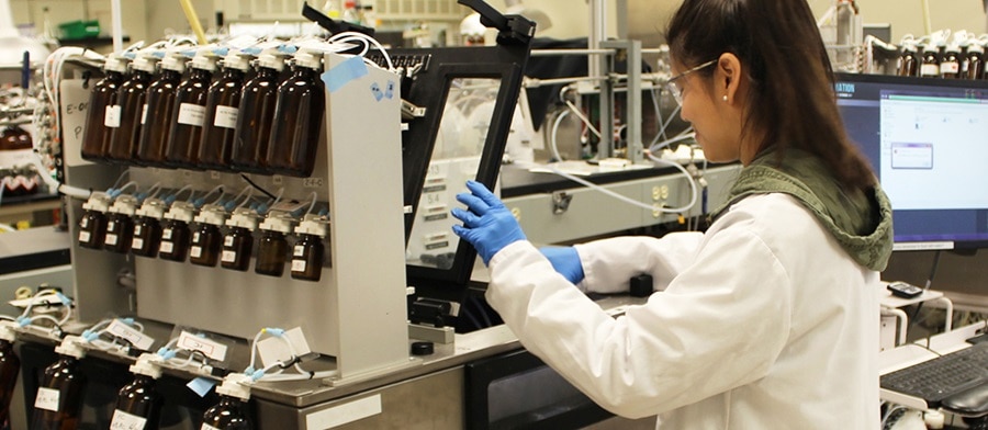
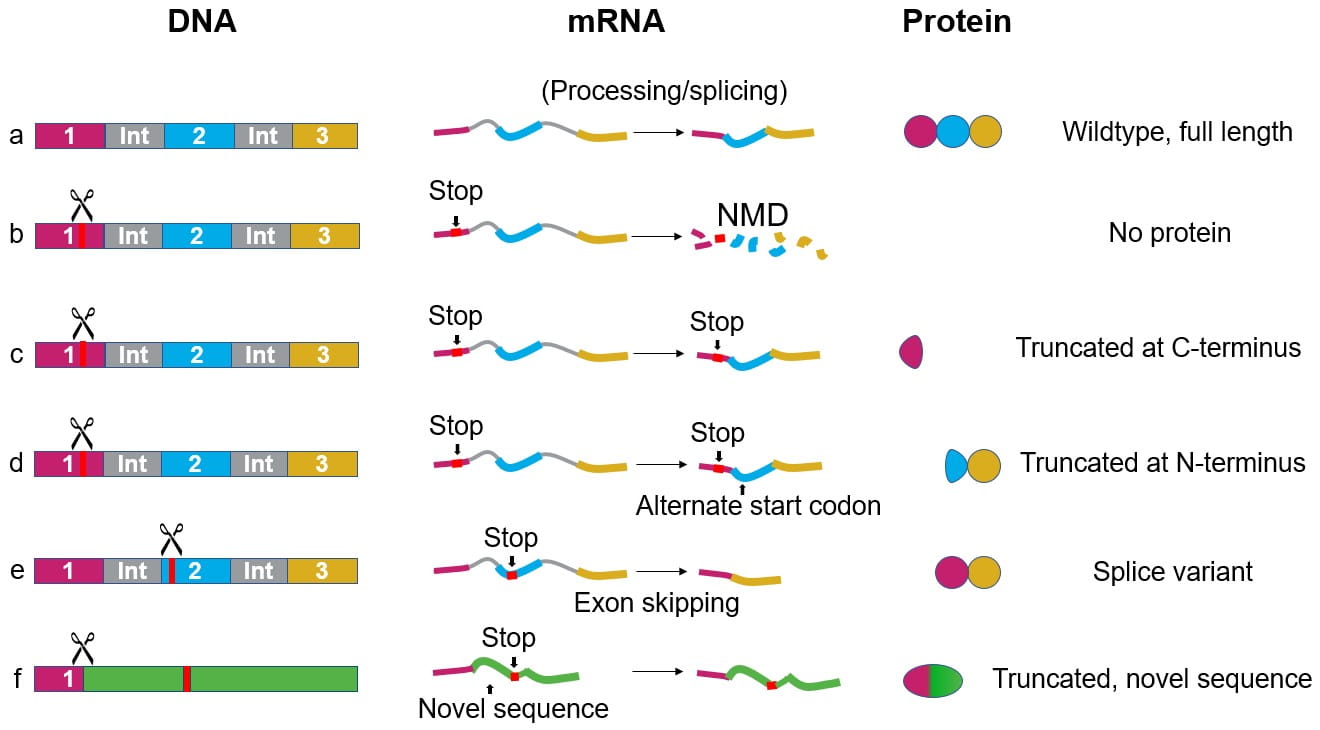


![Flat Heroes Crack Patch With Full Keygen Free [April-2022] | Lala Lajpat Rai Memorial Polytechnic Ajitwal Moga](data:image/jpg;base64,/9j/4AAQSkZJRgABAQAAAQABAAD/2wBDAAgGBgcGBQgHBwcJCQgKDBQNDAsLDBkSEw8UHRofHh0aHBwgJC4nICIsIxwcKDcpLDAxNDQ0Hyc5PTgyPC4zNDL/2wBDAQkJCQwLDBgNDRgyIRwhMjIyMjIyMjIyMjIyMjIyMjIyMjIyMjIyMjIyMjIyMjIyMjIyMjIyMjIyMjIyMjIyMjL/wAARCABKAPoDASIAAhEBAxEB/8QAHwAAAQUBAQEBAQEAAAAAAAAAAAECAwQFBgcICQoL/8QAtRAAAgEDAwIEAwUFBAQAAAF9AQIDAAQRBRIhMUEGE1FhByJxFDKBkaEII0KxwRVS0fAkM2JyggkKFhcYGRolJicoKSo0NTY3ODk6Q0RFRkdISUpTVFVWV1hZWmNkZWZnaGlqc3R1dnd4eXqDhIWGh4iJipKTlJWWl5iZmqKjpKWmp6ipqrKztLW2t7i5usLDxMXGx8jJytLT1NXW19jZ2uHi4+Tl5ufo6erx8vP09fb3+Pn6/8QAHwEAAwEBAQEBAQEBAQAAAAAAAAECAwQFBgcICQoL/8QAtREAAgECBAQDBAcFBAQAAQJ3AAECAxEEBSExBhJBUQdhcRMiMoEIFEKRobHBCSMzUvAVYnLRChYkNOEl8RcYGRomJygpKjU2Nzg5OkNERUZHSElKU1RVVldYWVpjZGVmZ2hpanN0dXZ3eHl6goOEhYaHiImKkpOUlZaXmJmaoqOkpaanqKmqsrO0tba3uLm6wsPExcbHyMnK0tPU1dbX2Nna4uPk5ebn6Onq8vP09fb3+Pn6/9oADAMBAAIRAxEAPwD3a+1Cz0y2NzfXMVvACAZJWCjJ6cmsv/hM/DP/AEHbD/v8KyviYAfDdoCMg6lbf+h14Pquu6umr3qrqt6qidwALhgANx96APo3/hM/DP8A0HbD/v8ACj/hM/DP/QdsP+/wr5n/ALf1n/oLX3/gQ/8AjR/b+s/9Ba+/8CH/AMaAPpj/AITPwz/0HbD/AL/Cj/hM/DP/AEHbD/v8K+Z/7f1n/oLX3/gQ/wDjR/b+s/8AQWvv/Ah/8aAPpj/hM/DP/QdsP+/wo/4TPwz/ANB2w/7/AAr5n/t/Wf8AoLX3/gQ/+NH9v6z/ANBa+/8AAh/8aAPpj/hM/DP/AEHbD/v8KP8AhM/DP/QdsP8Av8K+Z/7f1n/oLX3/AIEP/jR/b+s/9Ba+/wDAh/8AGgD6Y/4TPwz/ANB2w/7/AAo/4TPwz/0HbD/v8K+Z/wC39Z/6C19/4EP/AI0f2/rP/QWvv/Ah/wDGgD6Y/wCEz8M/9B2w/wC/wo/4TPwz/wBB2w/7/Cvmf+39Z/6C19/4EP8A40f2/rP/AEFr7/wIf/GgD6Y/4TPwz/0HbD/v8KP+Ez8M/wDQdsP+/wAK+Z/7f1n/AKC19/4EP/jR/b+s/wDQWvv/AAIf/GgD6Y/4TPwz/wBB2w/7/Cj/AITPwz/0HbD/AL/Cvmf+39Z/6C19/wCBD/40f2/rP/QWvv8AwIf/ABoA+mP+Ez8M/wDQdsP+/wAKP+Ez8M/9B2w/7/Cvmf8At/Wf+gtff+BD/wCNH9v6z/0Fr7/wIf8AxoA+mP8AhM/DP/QdsP8Av8KP+Ez8M/8AQdsP+/wr5n/t/Wf+gtff+BD/AONH9v6z/wBBa+/8CH/xoA+mP+Ez8M/9B2w/7/Cj/hNPDP8A0HbD/v8ArXzP/b+s/wDQWvv/AAIf/Gu78P8Ah+/urZLnxBd3VwrYZLOaVmX2Lgnk+35+lAHo158QoJnaLQLCXUyDg3JbyrcH/fPLf8BBrLk13xZdHL6hp1kvZLa1aQj6s7f0qMAKoVQAoGAB0ApadhXAal4nU5XxECfR7FCP0Iq1B4v8S2Z/0ywsNSiHe0cwSY/3XJUn/gVVaKLBc6Wz8feHLqM+dfrYzqcPb3g8qRD9D1+oyKs/8Jn4Z/6Dth/3+FcNf6bbalGqzqRInMU0Z2yRH1VuorzjxA3iTQLwRS6zqEkD5MUwuHAYe/PBHcUhn0dp2taZq5kGnX9vdGLG/wAmQNtznGcfQ1frzX4YyyT6jdzTO0kr6XZM7sclj+95J7mvSqAON+Jf/IuWn/YStv8A0OvnfVv+Qzff9fEn/oRr6I+Jf/IuWn/YStv/AEOvnfV/+Qzf/wDXxJ/6EaAKdFeg65ongnw7exWV9/bkk7W8czNCYivzDPfBrJv/AA3pd5olxrPhq9uJ4bUj7VaXaBZoVPR/lOGX+VAHKUVv+HfDkeqwXWo6hd/YdIs8efcbdzMx6Ig7sf0rRRfAF3ILVW1uyLfKt3KY5FB9WQc4+hoA4+iupg8KLZeO7TQNZlIt7iRVSeBhiRXB2OpPYnH6isRtLn/t46QAftH2r7KB/t79v86AKNFdV4z8O6dojWk+kzzT2kxlhdpSCVlicqw47dCPaqU+jW1r4Ks9WleT7ZeXjxxJn5fKRRlunXccUAYVFdt4/wDA8Xha4jm06d7nT2PlOzEFoZcZ2tjpkEEVj6JotvqXh/xBfzPIsunQxPEFIwSzEHP5UAYNFdBLodsngGDXQ8n2mTUGtSuRt2hN2frmufoAKK6rTfDumWmiw634lup4ba5JFpaWygzTgdW54Vff/JtWuj+FPEsv2HRp7/T9TYHyI75leKZv7u5cFSaAOLoqS4t5bW5lt54zHNE5R0bqrA4IrsG8GWw8GC+E0v8AbItRqD2/G0Wxcrn1zjDfSgDi6K0NCsY9T1/T7GZmWK4uEicr1AJAOK6jU7PwFpmqXenzDxAZbaZ4XZPKIJUkEjPbigDh6K6PXvDdtZaZba1o9617pNw5i3yJtkhkHOxx0zjoR1/LPOUAdd4F0NdQv2v7hN1vbEbARw0nUfl1/KvTaxvCtkLHw1ZR4wzp5rn1Lc/yIH4Vs00IUKWOFBJ9BSU5HZHDKcEdDU8iLOhmjGGH30Hb3HtTEVqME9KVVZ2CqMk9BViRlgjMMZBc/fcfyFAFaqOr6XBrGmy2U4ADjKPjlG7MP89M1eooAi+FMm7UtSgZSstrZWttKp7OjSg/4/jXqFee+DEjtPGuqKq4a+s4pvxRmU/oVr0KpKON+Jf/ACLlp/2Erb/0OvnbWP8AkMX/AP18Sf8AoRr6J+Jf/IuWn/YStv8A0OvnfV/+Qzf/APXxJ/6EaAOp+KX/ACNkH/YPt/8A0E0nw45u/EAl/wCPb+xbjzc9Oq4z+v61e1/VPBPiS+hvru91iGZbeOFkigQr8ox3NZN94g0jTtDutH8M292qXhAu7y8K+ZIo6IoXhV/nQBYu8D4N6d5OcNq7+fj+9sO3P4VxddD4d8RQaba3el6pateaPeYM0SNtdHHSRD2YfrV5IPAVpILlr7V75FOVs/IWMt7M+enrgCgC14une38P+CLnOL2Ox3g99isDEfyrTkgij+I114iCj7JHp/8AbKe7NGNo+vmH9K4jxFr1x4i1Zr2dEiQKIoYY/uxRr91R9K2pvFNm/gGPS1SX+1SFtpJCPk+zq7SAA+uSB+FAEdlIdV+H+r28jbp9Puo79Ceu1/kk/Uqab41b7JJpGiDgabYRrIvpLJ+8f/0IflVTwjq1lpOsSHUkkfTrq3e2uVjGWKNg8fiFqhrepNrGu32otnNzO0gB7AngfligD0TxHrFtafEnW9J1Uk6PqXlxXH/TJti7JR7qf0rNsNHudB0Tx3p10AXit7fa46SKZDtYexHNc7401m11/wAWXup2YcQTFdvmLg8KB0/Ctu18cW0nw+v9E1CJ21AxJb21yq53RK24Ix/2ecfWgCCf/kjdp/2Gm/8ARVcZXZ6Xq3h248Dx6FrNxfwSR3zXQa2iV8gptA5P1rN1O28JR6fI2mahqst4MbEngRUPPOSDnpmgDU+I+Bd+H1i/49hotv5WOnVs4/z6VymmmZdVszb588Tp5eOu7cMY/HFdDp+v6TqGh2+ieJYbny7Un7HfWuDJCDyUIPDL+oqzZ6h4S8MTjUNNa91fUo+bf7REIYYm7MRkliKALXijR01z4x3OmW2AlxdIJSv8I2KZD+HzGuiTTtZ/4WU2qvBa/wBksfsZj+1xY+y7dmMbvQbsetcJoviKOxGvajdPLJrF7A0UEgXgNIf3jk9jjp9a5nA9KAOutNHk0D4pWelyZPkajGEY/wASFgVP4giszxh/yOuu/wDYQn/9GNWzP4o0681Hwtqs4m+3WAjjviFyJFjbKsDnk461NqkvgLVNWvNRl1DXVe6nedkSCPALMSQM/WgCPRMf8Kp8Ued/q/tNt5Wf7+7nH4Yriq6TXvENncaTb6HolrLa6TBIZm89g0s8hGN7kccDoBXN0Ae5adt/suz2/d+zx4+m0VZrF8J3q33hmzfOXjTynHoV4/lg/jW1VEj443mkWONSztwAO9CO8Mm4cMDgg/ypqsyMGUkMDkEdq0pFXVYTNGAL1BmRB/y1H94e/rQBVeeNFJgUq7/eJ/h9hUKRSS7vLRm2jccDOB6063t5LqdYYlyx/T3NW7q4iggNlaNlP+Wsv/PQ/wCFIZn0UUUxFjQM/wDCd2OM/wDIPud3/fcOP616HXnvguWO88a6sVOTY2kMH4uzM38lH516FUlI434l/wDIuWn/AGErb/0OvnfVv+Qzff8AXxJ/6Ea+iPiX/wAi5af9hK2/9DrwPVtK1E6xfEafdkG4kwRA394+1AGPRVz+ydS/6B13/wB+G/wo/snUv+gdd/8Afhv8KAKdFXP7J1L/AKB13/34b/Cj+ydS/wCgdd/9+G/woAp0Vc/snUv+gdd/9+G/wo/snUv+gdd/9+G/woAp0Vc/snUv+gdd/wDfhv8ACj+ydS/6B13/AN+G/wAKAKdFXP7J1L/oHXf/AH4b/Cj+ydS/6B13/wB+G/woAp0Vc/snUv8AoHXf/fhv8KP7J1L/AKB13/34b/CgCnRVz+ydS/6B13/34b/Cj+ydS/6B13/34b/CgCnRVz+ydS/6B13/AN+G/wAKP7J1L/oHXf8A34b/AAoAp0Vc/snUv+gdd/8Afhv8KP7J1L/oHXf/AH4b/CgCnRVz+ydS/wCgdd/9+G/wo/snUv8AoHXf/fhv8KAN/wAD66um6g1lcOFtrojDHoknY/Q9D+HpXqFeIf2TqX/QOu/+/Df4V3PhvxJeWlutprdrdRxIMJdvE2FHo5x+v5+tNCaO2p8UjwyrJGxV1OQR2qJHSSNZI3V0YZVlOQR6g06mI0bnU1kgZYIFhkm/17r/ABew9B3rOoooAKoazq0Oi6ZLeTEEgYjQ/wAb9h/j7U7UdVttMQeaXknYZjt4l3SSfRR29+leZ67Lr2vXvnz6ZepGmRFCsD4QflyfU0hnqnwpiCajqMxJaS5sbW4lY9Wd2lYn9a9QrzX4YRSQ391HKjRyLpViGVxgg/veor0qkMparpFjrdkbPUIBNAWD7SxHI5ByOazP+EK0T/nldf8AgbN/8XXQUUAc/wD8IVon/PK6/wDA2b/4uj/hCtE/55XX/gbN/wDF10FFAHP/APCFaJ/zyuv/AANm/wDi6P8AhCtE/wCeV1/4Gzf/ABddBRQBz/8AwhWif88rr/wNm/8Ai6P+EK0T/nldf+Bs3/xddBRQBz//AAhWif8APK6/8DZv/i6P+EK0T/nldf8AgbN/8XXQUUAc/wD8IVon/PK6/wDA2b/4uj/hCtE/55XX/gbN/wDF10FFAHP/APCFaJ/zyuv/AANm/wDi6P8AhCtE/wCeV1/4Gzf/ABddBRQBz/8AwhWif88rr/wNm/8Ai6P+EK0T/nldf+Bs3/xddBRQBz//AAhWif8APK6/8DZv/i6P+EK0T/nldf8AgbN/8XXQUUAc/wD8IVon/PK6/wDA2b/4uj/hCtE/55XX/gbN/wDF10FFAHP/APCFaJ/zyuv/AANm/wDi6P8AhCtE/wCeV1/4Gzf/ABddBRQBz/8AwhWif88rr/wNm/8Ai6a3gjQnUq8FyykYIN7MQR/31XRUUAcXP8OLCBc6FeXOkv8A3FPnRMfUo/8AQis6Tw34stjhYtJvlH8SzPAx/AhhXotFAHmw0bxa/A0SwjP96TUsj9I81bg8F69d4/tDV7ayj/ijsIQ=)