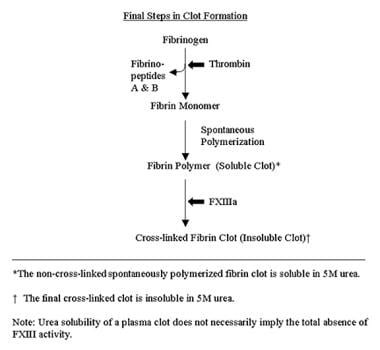
Pregnancy Proteins
Pregnancy
Pregnancy Outcome
Pregnancy Complications
Pregnancy, Animal
Pregnancy, Ectopic
Pregnancy Trimester, First
Pregnancy Rate
Pregnancy Trimester, Third
Pregnancy Complications, Cardiovascular
Pregnancy Trimesters
Pregnancy in Diabetics
Pregnancy, Tubal
Pregnancy Trimester, Second
Pregnancy Complications, Neoplastic
Pregnancy Complications, Infectious
Abortion, Spontaneous
Pregnancy, Unplanned
Pregnancy, High-Risk
Gestational Age
Pregnancy Complications, Hematologic
Pregnancy, Prolonged
Pre-Eclampsia
Placenta
Abortion, Induced
Pregnancy, Abdominal
Uterus
Pregnancy Complications, Parasitic
Ultrasonography, Prenatal
Embryo Transfer
Prenatal Care
Fertilization in Vitro
Maternal-Fetal Exchange
Parity
Prenatal Exposure Delayed Effects
Birth Weight
Pregnancy Reduction, Multifetal
Hypertension, Pregnancy-Induced
Fetal Growth Retardation
Progesterone
Premature Birth
Abnormalities, Drug-Induced
Lactation
Decidua
Fetus
Labor, Obstetric
Obstetric Labor, Premature
Twins
Prenatal Diagnosis
Maternal Exposure
Chorionic Gonadotropin
Diabetes, Gestational
Abortion, Therapeutic
Trophoblasts
Prospective Studies
Insemination, Artificial
Delivery, Obstetric
Fetal Diseases
Puerperal Disorders
Endometrium
Sperm Injections, Intracytoplasmic
Risk Factors
Fertility
Infant, Low Birth Weight
Preconception Care
Amniotic Fluid
Infertility
Gravidity
Reproductive Techniques, Assisted
Chorionic Gonadotropin, beta Subunit, Human
Cohort Studies
Maternal Nutritional Physiological Phenomena
Retrospective Studies
Ovulation Induction
Pregnancy Tests, Immunologic
Live Birth
Abortion, Legal
Placental Circulation
Contraception
Abortion, Threatened
Hydatidiform Mole
Infant, Small for Gestational Age
Triplets
Uterine Hemorrhage
Placentation
Reproductive Techniques
Uterine Artery
Fetal Resorption
Corpus Luteum
Estradiol
Oocyte Donation
Chorionic Villi
Maternal Welfare
Hyperemesis Gravidarum
Estrus
Fetal Weight
Eclampsia
Amniocentesis
Family Planning Services
Obstetric Labor Complications
Fetal Blood
Fallopian Tubes
Cryopreservation
Case-Control Studies
Crown-Rump Length
Fetal Macrosomia
HELLP Syndrome
Fetal Monitoring
Infectious Disease Transmission, Vertical
Estriol
Ovary
Pseudopregnancy
Sheep
Morning Sickness
Logistic Models
Chorion
Estrous Cycle
Questionnaires
Cervix Uteri
Fetal Viability
Fertilization
Misoprostol
Abruptio Placentae
Menstrual Cycle
Relaxin
Infant Mortality
Odds Ratio
Down Syndrome
Maternal Mortality
Infertility, Male
Umbilical Arteries
Neural Tube Defects
Dietary Supplements
Insemination, Artificial, Homologous
Placental Lactogen
Socioeconomic Factors
Blastocyst
Body Weight
Down-regulation of oxytocin-induced cyclooxygenase-2 and prostaglandin F synthase expression by interferon-tau in bovine endometrial cells. (1/1380)
Oxytocin (OT) is responsible for the episodic release of luteolytic prostaglandin (PG) F2alpha from the uterus in ruminants. The attenuation of OT-stimulated uterine PGF2alpha secretion by interferon-tau (IFN-tau) is essential for prevention of luteolysis during pregnancy in cows. To better understand the mechanisms involved, the effect of recombinant bovine IFN-tau (rbIFN-tau) on OT-induced PG production and cyclooxygenase-2 (COX-2) and PGF synthase (PGFS) expression in cultured endometrial epithelial cells was investigated. Cells were obtained from cows at Days 1-3 of the estrous cycle and cultured to confluence in RPMI medium supplemented with 5% steroid-free fetal calf serum. The cells were then incubated in the presence or absence of either 100 ng/ml OT or OT+100 ng/ml rbIFN-tau for 3, 6, 12, and 24 h. OT significantly increased PGF2alpha and PGE2 secretion at all time points (p < 0.01), while rbIFN-tau inhibited the OT-induced PG production and reduced OT receptor binding in a time-dependent manner. OT increased the steady-state level of COX-2 mRNA, measured by Northern blot, which was maximal at 3 h (9-fold increase) and then decreased with time (p < 0.01). OT also caused an increase in COX-2 protein, which peaked at 12 h (11-fold increase), as measured by Western blot. Addition of rbIFN-tau suppressed the induction of COX-2 mRNA (89%, p < 0.01) and COX-2 protein (50%, p < 0.01) by OT. OT also increased PGFS mRNA, and this stimulation was attenuated by rbIFN-tau (p < 0.01). To ensure that the decrease in COX-2 was not solely due to down-regulation of the OT receptor, cells were stimulated with a phorbol ester (phorbol 12-myristate 13-acetate; PMA) in the presence and absence of rbIFN-tau. The results showed that rbIFN-tau also decreased PMA-stimulated PG production and COX-2 protein. It can be concluded that rbIFN-tau inhibition of OT-stimulated PG production is due to down-regulation of OT receptor, COX-2, and PGFS. (+info)Tissue factor pathway inhibitor-2 is a novel mitogen for vascular smooth muscle cells. (2/1380)
A mitogen for growth-arrested cultured bovine aortic smooth muscle cells was purified to homogeneity from the supernatant of cultured human umbilical vein endothelial cells by heparin affinity chromatography and reverse-phase high performance liquid chromatography. This mitogen was revealed to be tissue factor pathway inhibitor-2 (TFPI-2), which is a Kunitz-type serine protease inhibitor. TFPI-2 was expressed in baby hamster kidney cells using a mammalian expression vector. Recombinant TFPI-2 (rTFPI-2) stimulated DNA synthesis and cell proliferation in a dose-dependent manner (1-500 nM). rTFPI-2 activated mitogen-activated protein kinase (MAPK) activity and stimulated early proto-oncogene c-fos mRNA expression in smooth muscle cells. MAPK, c-fos expression and the mitogenic activity were inhibited by a specific inhibitor of MAPK kinase, PD098059. Thus, the mitogenic function of rTFPI-2 is considered to be mediated through MAPK pathway. TFPI has been reported to exhibit antiproliferative action after vascular smooth muscle injury in addition to the ability to inhibit activation of the extrinsic coagulation cascade. However, structurally similar TFPI-2 was found to have a mitogenic activity for the smooth muscle cell. (+info)Modulation of the baboon (Papio anubis) uterine endometrium by chorionic gonadotrophin during the period of uterine receptivity. (3/1380)
This study was undertaken to determine the modulation of uterine function by chorionic gonadotrophin (CG) in a nonhuman primate. Infusion of recombinant human CG (hCG) between days 6 and 10 post ovulation initiated the endoreplication of the uterine surface epithelium to form distinct epithelial plaques. These plaque cells stained intensely for cytokeratin and the proliferating cell nuclear antigen. The stromal fibroblasts below the epithelial plaques stained positively for alpha-smooth muscle actin (alphaSMA). Expression of alphaSMA is associated with the initiation of decidualization in the baboon endometrium. Synthesis of the glandular secretory protein glycodelin, as assessed by Western blot analysis, was markedly up-regulated by hCG, and this increase was confirmed by immunocytochemistry, Northern blot analysis, and reverse transcriptase-PCR. To determine whether hCG directly modulated these uterine responses, we treated ovariectomized baboons sequentially with estradiol and progesterone to mimic the hormonal profile of the normal menstrual cycle. Infusion of hCG into the oviduct of steroid-hormone-treated ovariectomized baboons induced the expression of alphaSMA in the stromal cells and glycodelin in the glandular epithelium. The epithelial plaque reaction, however, was not readily evident. These studies demonstrate a physiological effect of CG on the uterine endometrium in vivo and suggest that the primate blastocyst signal, like the blastocyst signals of other species, modulates the uterine environment prior to implantation. (+info)Pregnancy detection and the effects of age, body weight, and previous reproductive performance on pregnancy status and weaning rates of farmed fallow deer (Dama dama). (4/1380)
Fallow does (n = 502) of different ages (mature, 2-yr-old, and yearling) were maintained with bucks for a 60-d breeding season to determine whether previous reproductive performance and changes in BW affect doe pregnancy rates and to compare the effectiveness of ultrasonography and serum pregnancy-specific protein B (PSPB) for the detection of pregnancy in fallow does. Ultrasonography was performed, blood samples collected, and BW recorded at buck removal (d 0) and at 30 and 90 d after buck removal. Lactational status (lactating = WET; nonlactating = DRY) were determined from farm records taken at weaning prior to each breeding season (autumn 1990 through autumn 1994). Ultrasonography and PSPB for determining pregnancy were in agreement 93% of the time. Overall pregnancy rates did not differ (P>.10) relative to age of the doe; the combined pregnancy rate was 92%. We also determined that 82.9% of does conceived early in the breeding season and that the incidence of embryonal-fetal mortality during the first 90 d after buck removal was 2.8%. In general, mature and 2-yr-old DRY does were heavier and had lower pregnancy rates than WET does. The overall weaning rate for all does was 77.9%. Loss in the number of fawns from pregnancy detection to weaning was equivalent to 14.8% for mature does, 24.7% for 2 yr old does, and 42.5% for yearling does. These data indicate that even though pregnancy rates were relatively high, further study is needed to determine the causes associated with subsequent fawn losses, particularly among yearling does. As a production tool, lactational WET/ DRY status testing was found to be an acceptable means for determining the reproductive potential of individual does within the herd. In addition, serum PSPB may be used in place of ultrasonography for pregnancy diagnosis in fallow deer as early as d 30 after buck removal. (+info)Increased expression of regeneration and tolerance factor in individuals with human immunodeficiency virus infection. (5/1380)
Regeneration and tolerance factor (RTF) plays a pivotal role in successful pregnancy outcome and has potent immunomodulating properties. During pregnancy, it is abundantly expressed in the placenta and on peripheral B lymphocytes. Several lines of evidence suggest that both successful pregnancy outcome and progression from human immunodeficiency virus (HIV) infection to AIDS are associated with a Th2-type response. As a result, we hypothesized that the cellular expression of RTF may also be increased during infection with HIV. Using flow cytometric analysis, we showed a significantly (P < 0.01) increased expression of RTF on CD3(+) cells obtained from individuals with HIV over that for individuals without HIV. On average, 32.1% of the CD3(+) cells from individuals with HIV expressed high levels of RTF. In contrast, an average of only 6.7% of the CD3(+) cells from individuals without HIV expressed high levels of RTF. Similar results were obtained when CD19(+) cells from individuals with (mean, 44.1%) and without (mean, 25.8%) HIV were evaluated. Linear regression analysis suggested that high levels of RTF expression by CD3(+) cells correlated better with viral load (r value, 0.46) than with absolute CD4 count (r value, 0.09). While additional experiments are necessary to delineate the precise immunologic role of RTF, our current data suggest that RTF expression during HIV infection may be a useful marker of immune activation. (+info)Chemotactic, mitogenic, and angiogenic actions of UTP on vascular endothelial cells. (6/1380)
Endothelial cells express receptors for ATP and UTP, and both UTP and ATP elicit endothelial release of vasoactive compounds such as prostacyclin and nitric oxide; however, the distinction between purine and pyrimidine nucleotide signaling is not known. We hypothesized that UTP plays a more important role in endothelial mitogenesis and chemotaxis than does ATP and that UTP is angiogenic. In cultured endothelial cells from guinea pig cardiac vasculature (CEC), both UTP and vascular endothelial growth factor (VEGF) were significant mitogenic and chemotactic factors; in contrast, ATP demonstrated no significant chemotaxis in CEC. In chick chorioallantoic membranes (CAM), UTP and VEGF treatments produced statistically significant increases in CAM vascularity compared with controls. These findings are the first evidence of chemotactic or angiogenic effects of pyrimidines; they suggest a role for pyrimidine nucleotides that is distinct from those assumed by purine nucleotides and provide for the possibility that UTP serves as an extracellular signal for processes such as endothelial repair and angiogenesis. (+info)Signal transduction and biological function of placenta growth factor in primary human trophoblast. (7/1380)
Placenta growth factor (PlGF), a member of the vascular endothelial growth factor family of angiogenic factors, is prominently expressed by trophoblast. In addition to its role as a paracrine angiogenic factor within the placenta and endometrium, presence of its receptor, Flt-1, on trophoblast suggests that PlGF also may have an autocrine role(s) in regulating trophoblast function. To elucidate its role in trophoblast, we examined the signal transduction and functional responses of primary human trophoblast to PlGF. Exogenous PlGF induced specific activation of the stress-activated protein kinase (SAPK) pathways, c-Jun-N terminal kinase (JNK) and p38 kinase, in primary term trophoblast with little to no induction of the extracellular signal regulated kinase (ERK-1 and -2) pathways. In contrast, PlGF induced significant ERK-1 and -2 activity in human umbilical vein endothelial cells but did not induce JNK or p38 activity. PlGF-induced activation of the SAPK signaling pathways protected trophoblast from growth factor withdrawal-induced apoptosis, but it did not protect trophoblast from apoptosis induced by the pro-inflammatory cytokines, interferon gamma and tumor necrosis factor alpha. These results provide the first direct evidence of a biochemical and functional role for PlGF/Flt-1 in normal trophoblast and suggest that aberrant PlGF expression during pregnancy may impact upon trophoblast function as well as vascularity within the placental bed. (+info)Are human placental bed giant cells merely aggregates of small mononuclear trophoblast cells? An ultrastructural and immunocytochemical study. (8/1380)
The ultrastructure of placental bed giant cells in early human pregnancies of 7-12 weeks gestational age is described. Their nature and function was further characterized by confocal immunofluorescence microscopy of paraffin sections labelled for cytokeratin, gap junction connexins (CX) 32 or 43, and placental hormones, alpha-human chorionic gonadotrophin (alpha-HCG) and human placental lactogen (HPL). Placental bed giant cells were observed with two phenotypes; as single large trophoblast cells containing one or more nuclear profiles in a voluminous cytoplasm, and as cell aggregates comprising mononuclear trophoblast cells in close apposition separated by narrow intercellular spaces. Cells within the aggregates are attached to one another by desmosomes, and also possess gap junctions as shown by immunolabelling for CX32 and CX43. By contrast, gap junctions were absent in the true multinucleated giant cells. Organelles present within the cytoplasm of the giant cells and their immunoreactivity for HPL and alpha-HCG suggest protein synthesis. (+info)"Pregnancy proteins" is not a standard medical term, but it may refer to specific proteins that are produced or have increased levels during pregnancy. Two common pregnancy-related proteins are:
1. Human Chorionic Gonadotropin (hCG): A hormone produced by the placenta shortly after fertilization. It is often detected in urine or blood tests to confirm pregnancy. Its primary function is to maintain the corpus luteum, which produces progesterone and estrogen during early pregnancy until the placenta takes over these functions.
2. Pregnancy-Specific beta-1 Glycoprotein (SP1): A protein produced by the placental trophoblasts during pregnancy. Its function is not well understood, but it may play a role in implantation, placentation, and protection against the mother's immune system. SP1 levels increase throughout pregnancy and are used as a marker for fetal growth and well-being.
These proteins have clinical significance in monitoring pregnancy progression, detecting potential complications, and diagnosing certain pregnancy-related conditions.
Pregnancy is a physiological state or condition where a fertilized egg (zygote) successfully implants and grows in the uterus of a woman, leading to the development of an embryo and finally a fetus. This process typically spans approximately 40 weeks, divided into three trimesters, and culminates in childbirth. Throughout this period, numerous hormonal and physical changes occur to support the growing offspring, including uterine enlargement, breast development, and various maternal adaptations to ensure the fetus's optimal growth and well-being.
Pregnancy outcome refers to the final result or status of a pregnancy, including both the health of the mother and the newborn baby. It can be categorized into various types such as:
1. Live birth: The delivery of one or more babies who show signs of life after separation from their mother.
2. Stillbirth: The delivery of a baby who has died in the womb after 20 weeks of pregnancy.
3. Miscarriage: The spontaneous loss of a pregnancy before the 20th week.
4. Abortion: The intentional termination of a pregnancy before the fetus can survive outside the uterus.
5. Ectopic pregnancy: A pregnancy that develops outside the uterus, usually in the fallopian tube, which is not viable and requires medical attention.
6. Preterm birth: The delivery of a baby before 37 weeks of gestation, which can lead to various health issues for the newborn.
7. Full-term birth: The delivery of a baby between 37 and 42 weeks of gestation.
8. Post-term pregnancy: The delivery of a baby after 42 weeks of gestation, which may increase the risk of complications for both mother and baby.
The pregnancy outcome is influenced by various factors such as maternal age, health status, lifestyle habits, genetic factors, and access to quality prenatal care.
Pregnancy complications refer to any health problems that arise during pregnancy which can put both the mother and the baby at risk. These complications may occur at any point during the pregnancy, from conception until childbirth. Some common pregnancy complications include:
1. Gestational diabetes: a type of diabetes that develops during pregnancy in women who did not have diabetes before becoming pregnant.
2. Preeclampsia: a pregnancy complication characterized by high blood pressure and damage to organs such as the liver or kidneys.
3. Placenta previa: a condition where the placenta covers the cervix, which can cause bleeding and may require delivery via cesarean section.
4. Preterm labor: when labor begins before 37 weeks of gestation, which can lead to premature birth and other complications.
5. Intrauterine growth restriction (IUGR): a condition where the fetus does not grow at a normal rate inside the womb.
6. Multiple pregnancies: carrying more than one baby, such as twins or triplets, which can increase the risk of premature labor and other complications.
7. Rh incompatibility: a condition where the mother's blood type is different from the baby's, which can cause anemia and jaundice in the newborn.
8. Pregnancy loss: including miscarriage, stillbirth, or ectopic pregnancy, which can be emotionally devastating for the parents.
It is important to monitor pregnancy closely and seek medical attention promptly if any concerning symptoms arise. With proper care and management, many pregnancy complications can be treated effectively, reducing the risk of harm to both the mother and the baby.
"Animal pregnancy" is not a term that is typically used in medical definitions. However, in biological terms, animal pregnancy refers to the condition where a fertilized egg (or eggs) implants and develops inside the reproductive tract of a female animal, leading to the birth of offspring (live young).
The specific details of animal pregnancy can vary widely between different species, with some animals exhibiting phenomena such as placental development, gestation periods, and hormonal changes that are similar to human pregnancy, while others may have very different reproductive strategies.
It's worth noting that the study of animal pregnancy and reproduction is an important area of biological research, as it can provide insights into fundamental mechanisms of embryonic development, genetics, and evolution.
Ectopic pregnancy is a type of abnormal pregnancy that occurs outside the uterine cavity. The most common site for an ectopic pregnancy is the fallopian tube, accounting for about 95% of cases. This condition is also known as tubal pregnancy. Other less common sites include the ovary, cervix, and abdominal cavity.
In a normal pregnancy, the fertilized egg travels down the fallopian tube and implants itself in the lining of the uterus. However, in an ectopic pregnancy, the fertilized egg implants and starts to develop somewhere other than the uterus. The growing embryo cannot survive outside the uterus, and if left untreated, an ectopic pregnancy can cause life-threatening bleeding due to the rupture of the fallopian tube or other organs.
Symptoms of ectopic pregnancy may include abdominal pain, vaginal bleeding, shoulder pain, lightheadedness, fainting, and in severe cases, shock. Diagnosis is usually made through a combination of medical history, physical examination, ultrasound, and blood tests to measure the levels of human chorionic gonadotropin (hCG), a hormone produced during pregnancy.
Treatment for ectopic pregnancy depends on several factors, including the location, size, and growth rate of the ectopic mass, as well as the patient's overall health and desire for future pregnancies. Treatment options may include medication to stop the growth of the embryo or surgery to remove the ectopic tissue. In some cases, both methods may be used together. Early diagnosis and treatment can help prevent serious complications and improve the chances of preserving fertility in future pregnancies.
The first trimester of pregnancy is defined as the period of gestational development that extends from conception (fertilization of the egg by sperm) to the end of the 13th week. This critical phase marks significant transformations in both the mother's body and the growing embryo/fetus.
During the first trimester, the fertilized egg implants into the uterine lining (implantation), initiating a series of complex interactions leading to the formation of the placenta - an organ essential for providing nutrients and oxygen to the developing fetus while removing waste products. Simultaneously, the embryo undergoes rapid cell division and differentiation, giving rise to various organs and systems. By the end of the first trimester, most major structures are present, although they continue to mature and grow throughout pregnancy.
The mother may experience several physiological changes during this time, including:
- Morning sickness (nausea and vomiting)
- Fatigue
- Breast tenderness
- Frequent urination
- Food aversions or cravings
- Mood swings
Additionally, hormonal shifts can cause various symptoms and prepare the body for potential changes in lactation, posture, and pelvic alignment as pregnancy progresses. Regular prenatal care is crucial during this period to monitor both maternal and fetal wellbeing, identify any potential complications early on, and provide appropriate guidance and support throughout the pregnancy.
The pregnancy rate is a measure used in reproductive medicine to determine the frequency or efficiency of conception following certain treatments, interventions, or under specific conditions. It is typically defined as the number of pregnancies per 100 women exposed to the condition being studied over a specified period of time. A pregnancy is confirmed when a woman has a positive result on a pregnancy test or through the detection of a gestational sac on an ultrasound exam.
In clinical trials and research, the pregnancy rate helps healthcare professionals evaluate the effectiveness of various fertility treatments such as in vitro fertilization (IVF), intrauterine insemination (IUI), or ovulation induction medications. The pregnancy rate can also be used to assess the impact of lifestyle factors, environmental exposures, or medical conditions on fertility and conception.
It is important to note that pregnancy rates may vary depending on several factors, including age, the cause of infertility, the type and quality of treatment provided, and individual patient characteristics. Therefore, comparing pregnancy rates between different studies should be done cautiously, considering these potential confounding variables.
The third trimester of pregnancy is the final stage of pregnancy that lasts from week 29 until birth, which typically occurs around the 40th week. During this period, the fetus continues to grow and mature, gaining weight rapidly. The mother's body also prepares for childbirth by dilating the cervix and producing milk in preparation for breastfeeding. Regular prenatal care is crucial during this time to monitor the health of both the mother and the developing fetus, as well as to prepare for delivery.
Cardiovascular complications in pregnancy refer to conditions that affect the heart and blood vessels, which can arise during pregnancy, childbirth, or after delivery. These complications can be pre-existing or new-onset and can range from mild to severe, potentially threatening the life of both the mother and the fetus. Some examples of cardiovascular complications in pregnancy include:
1. Hypertension disorders: This includes chronic hypertension (high blood pressure before pregnancy), gestational hypertension (high blood pressure that develops after 20 weeks of pregnancy), and preeclampsia/eclampsia (a pregnancy-specific disorder characterized by high blood pressure, proteinuria, and potential organ damage).
2. Cardiomyopathy: A condition in which the heart muscle becomes weakened, leading to an enlarged heart and reduced pumping efficiency. Peripartum cardiomyopathy is a specific type that occurs during pregnancy or in the months following delivery.
3. Arrhythmias: Irregularities in the heart's rhythm, such as tachycardia (rapid heartbeat) or bradycardia (slow heartbeat), can occur during pregnancy and may require medical intervention.
4. Valvular heart disease: Pre-existing valve disorders, like mitral stenosis or aortic insufficiency, can worsen during pregnancy due to increased blood volume and cardiac output. Additionally, new valve issues might develop during pregnancy.
5. Venous thromboembolism (VTE): Pregnancy increases the risk of developing blood clots in the veins, particularly deep vein thrombosis (DVT) or pulmonary embolism (PE).
6. Ischemic heart disease: Although rare, coronary artery disease and acute coronary syndrome can occur during pregnancy, especially in women with risk factors such as obesity, diabetes, or smoking history.
7. Heart failure: Severe cardiac dysfunction leading to fluid accumulation, shortness of breath, and reduced exercise tolerance may develop due to any of the above conditions or other underlying heart diseases.
Early recognition, monitoring, and appropriate management of these cardiovascular complications in pregnancy are crucial for maternal and fetal well-being.
Pregnancy trimesters are a way to divide the duration of pregnancy into three 3-month (or approximately 13-week) segments. This division can help healthcare providers monitor and discuss specific developmental stages, symptoms, and care needs during each phase of the pregnancy. Here's a brief overview of what typically occurs in each trimester:
1. First Trimester (Week 1 - Week 12): During this period, major organs and structures begin to form in the developing fetus. Common symptoms experienced by the pregnant individual may include morning sickness, fatigue, breast tenderness, and frequent urination. Regular prenatal care should start during these early weeks to monitor both the mother's and baby's health.
2. Second Trimester (Week 13 - Week 26): This phase is often considered more comfortable for many pregnant individuals as some symptoms from the first trimester improve. The fetus continues to grow, and movement can be felt. Organs and systems continue to develop, and the fetus becomes more active. Common symptoms during this time include back pain, stretch marks, and swelling of the ankles and feet.
3. Third Trimester (Week 27 - Birth): The final trimester is marked by significant growth and weight gain for both the mother and baby. The fetus will turn into a head-down position in preparation for birth. Common symptoms during this time include shortness of breath, heartburn, difficulty sleeping, and contractions (which can indicate early labor). Regular prenatal care remains crucial to monitor the health of both the mother and baby as delivery approaches.
A pregnancy test is a medical diagnostic tool used to determine whether or not a woman is pregnant. These tests detect the presence of human chorionic gonadotropin (hCG), a hormone produced by the placenta after fertilization. Pregnancy tests can be performed using a variety of methods, including urine tests and blood tests.
Urine pregnancy tests are typically performed at home and involve either dipping a test strip into a sample of urine or holding the strip under a stream of urine for several seconds. The test strip contains antibodies that react with hCG, producing a visual signal such as a line or plus sign if hCG is present.
Blood pregnancy tests are performed by a healthcare provider and can detect lower levels of hCG than urine tests. There are two types of blood pregnancy tests: qualitative and quantitative. Qualitative tests simply detect the presence or absence of hCG, while quantitative tests measure the exact amount of hCG present in the blood.
Pregnancy tests are generally very accurate when used correctly, but false positives and false negatives can occur. False positives may occur due to certain medical conditions or medications that contain hCG. False negatives may occur if the test is taken too early or if it is not performed correctly. It is important to follow the instructions carefully and consult with a healthcare provider if there is any uncertainty about the results.
Multiple pregnancy is a type of gestation where more than one fetus is carried simultaneously in the uterus. The most common forms of multiple pregnancies are twins (two fetuses), triplets (three fetuses), and quadruplets (four fetuses). Multiple pregnancies can occur when a single fertilized egg splits into two or more embryos (monozygotic) or when more than one egg is released and gets fertilized during ovulation (dizygotic). The risk of multiple pregnancies increases with the use of assisted reproductive technologies, such as in vitro fertilization. Multiple pregnancies are associated with higher risks for both the mother and the fetuses, including preterm labor, low birth weight, and other complications.
'Pregnancy in Diabetics' refers to the condition where an individual with pre-existing diabetes mellitus becomes pregnant. This can be further categorized into two types:
1. Pre-gestational diabetes: This is when a woman is diagnosed with diabetes before she becomes pregnant. It includes both Type 1 and Type 2 diabetes. Proper control of blood sugar levels prior to conception and during pregnancy is crucial to reduce the risk of complications for both the mother and the baby.
2. Gestational diabetes: This is when a woman develops high blood sugar levels during pregnancy, typically in the second or third trimester. While it usually resolves after delivery, women with gestational diabetes have a higher risk of developing Type 2 diabetes later in life. Proper management of gestational diabetes is essential to ensure a healthy pregnancy and reduce the risk of complications for both the mother and the baby.
Tubal pregnancy, also known as an ectopic pregnancy, is a type of pregnancy that occurs outside the uterus, usually in the fallopian tube. The fertilized egg implants and starts to develop in the tube instead of the uterine lining. This condition is not viable and can be life-threatening if not treated promptly.
The symptoms of a tubal pregnancy may include abdominal pain, vaginal bleeding, shoulder pain, dizziness or fainting, and pelvic discomfort or tenderness. If you suspect that you have a tubal pregnancy, it is important to seek medical attention immediately. Treatment options for tubal pregnancies include medication or surgery to remove the embryo and repair or remove the affected fallopian tube.
The second trimester of pregnancy is the period between the completion of 12 weeks (the end of the first trimester) and 26 weeks (the beginning of the third trimester) of gestational age. It is often considered the most comfortable period for many pregnant women as the risk of miscarriage decreases significantly, and the symptoms experienced during the first trimester, such as nausea and fatigue, typically improve.
During this time, the uterus expands above the pubic bone, allowing more space for the growing fetus. The fetal development in the second trimester includes significant growth in size and weight, formation of all major organs, and the beginning of movement sensations that the mother can feel. Additionally, the fetus starts to hear, swallow and kick, and the skin is covered with a protective coating called vernix.
Prenatal care during this period typically includes regular prenatal appointments to monitor the mother's health and the baby's growth and development. These appointments may include measurements of the uterus, fetal heart rate monitoring, and screening tests for genetic disorders or other potential issues.
Neoplastic pregnancy complications refer to the abnormal growth of cells (neoplasia) that can occur during pregnancy. These growths can be benign or malignant and can arise from any type of tissue in the body. However, when they occur in pregnant women, they can pose unique challenges due to the potential effects on the developing fetus and the changes in the mother's body.
Some common neoplastic pregnancy complications include:
1. Gestational trophoblastic disease (GTD): This is a group of rare tumors that occur in the uterus during pregnancy. GTD can range from benign conditions like hydatidiform mole to malignant forms like choriocarcinoma.
2. Breast cancer: Pregnancy-associated breast cancer (PABC) is a type of breast cancer that occurs during pregnancy or within one year after delivery. It can be aggressive and challenging to diagnose due to the changes in the breast tissue during pregnancy.
3. Cervical cancer: Cervical cancer can occur during pregnancy, and its management depends on the stage of the disease and the gestational age. In some cases, treatment may need to be delayed until after delivery.
4. Lung cancer: Pregnancy does not increase the risk of lung cancer, but it can make diagnosis and treatment more challenging.
5. Melanoma: Melanoma is the most common malignant skin cancer during pregnancy. It can spread quickly and requires prompt treatment.
The management of neoplastic pregnancy complications depends on several factors, including the type and stage of the tumor, gestational age, and the patient's wishes. In some cases, surgery, chemotherapy, or radiation therapy may be necessary. However, these treatments can have potential risks to the developing fetus, so a multidisciplinary team of healthcare providers is often involved in the care of pregnant women with neoplastic complications.
Pregnancy in adolescence, also known as teenage pregnancy, refers to a pregnancy that occurs in females under the age of 20. This can be further categorized into early adolescent pregnancy (occurring between ages 10-14), middle adolescent pregnancy (occurring between ages 15-17), and late adolescent pregnancy (occurring between ages 18-19). Teenage pregnancy is associated with higher risks of complications for both the mother and the baby, including preterm birth, low birth weight, and increased risk of neonatal mortality. Additionally, teenage mothers are more likely to drop out of school and face socioeconomic challenges.
Infectious pregnancy complications refer to infections that occur during pregnancy and can affect the mother, fetus, or both. These infections can lead to serious consequences such as preterm labor, low birth weight, birth defects, stillbirth, or even death. Some common infectious agents that can cause pregnancy complications include:
1. Bacteria: Examples include group B streptococcus, Escherichia coli, and Listeria monocytogenes, which can cause sepsis, meningitis, or pneumonia in the mother and lead to preterm labor or stillbirth.
2. Viruses: Examples include cytomegalovirus, rubella, varicella-zoster, and HIV, which can cause congenital anomalies, developmental delays, or transmission of the virus to the fetus.
3. Parasites: Examples include Toxoplasma gondii, which can cause severe neurological damage in the fetus if transmitted during pregnancy.
4. Fungi: Examples include Candida albicans, which can cause fungal infections in the mother and lead to preterm labor or stillbirth.
Preventive measures such as vaccination, good hygiene practices, and avoiding high-risk behaviors can help reduce the risk of infectious pregnancy complications. Prompt diagnosis and treatment of infections during pregnancy are also crucial to prevent adverse outcomes.
Spontaneous abortion, also known as miscarriage, is the unintentional expulsion of a nonviable fetus from the uterus before the 20th week of gestation. It is a common complication of early pregnancy, with most miscarriages occurring during the first trimester. Spontaneous abortion can have various causes, including chromosomal abnormalities, maternal health conditions, infections, hormonal imbalances, and structural issues of the uterus or cervix. In many cases, the exact cause may remain unknown.
The symptoms of spontaneous abortion can vary but often include vaginal bleeding, which may range from light spotting to heavy bleeding; abdominal pain or cramping; and the passing of tissue or clots from the vagina. While some miscarriages occur suddenly and are immediately noticeable, others may progress slowly over several days or even weeks.
In medical practice, healthcare providers often use specific terminology to describe different stages and types of spontaneous abortion. For example:
* Threatened abortion: Vaginal bleeding during early pregnancy, but the cervix remains closed, and there is no evidence of fetal demise or passing of tissue.
* Inevitable abortion: Vaginal bleeding with an open cervix, indicating that a miscarriage is imminent or already in progress.
* Incomplete abortion: The expulsion of some but not all products of conception from the uterus, requiring medical intervention to remove any remaining tissue.
* Complete abortion: The successful passage of all products of conception from the uterus, often confirmed through an ultrasound or pelvic examination.
* Missed abortion: The death of a fetus in the uterus without any expulsion of the products of conception, which may be discovered during routine prenatal care.
* Septic abortion: A rare and life-threatening complication of spontaneous abortion characterized by infection of the products of conception and the surrounding tissues, requiring prompt medical attention and antibiotic treatment.
Healthcare providers typically monitor patients who experience a spontaneous abortion to ensure that all products of conception have been expelled and that there are no complications, such as infection or excessive bleeding. In some cases, medication or surgical intervention may be necessary to remove any remaining tissue or address other issues related to the miscarriage. Counseling and support services are often available for individuals and couples who experience a spontaneous abortion, as they may face emotional challenges and concerns about future pregnancies.
Unplanned pregnancy is a pregnancy that is not intended or expected by the woman or couple. It is also sometimes referred to as an "unintended" or "unwanted" pregnancy. This can occur when contraceptive methods fail or are not used, or when there is a lack of knowledge about or access to effective family planning resources. Unplanned pregnancies can present various physical, emotional, and social challenges for the individuals involved, and may also have implications for public health and societal well-being. It's important to note that unplanned pregnancies can still result in wanted and loved children, but the circumstances surrounding their conception may bring additional stressors and considerations.
High-risk pregnancy is a term used to describe a situation where the mother or the fetus has an increased risk of developing complications during pregnancy, labor, delivery, or in the postpartum period. These risks may be due to pre-existing medical conditions in the mother, such as diabetes, hypertension, heart disease, kidney disease, autoimmune disorders, or infectious diseases like HIV/AIDS. Other factors that can contribute to a high-risk pregnancy include advanced maternal age (35 years and older), obesity, multiple gestations (twins, triplets, etc.), fetal growth restriction, placental issues, and a history of previous pregnancy complications or preterm labor.
High-risk pregnancies require specialized care and monitoring by healthcare professionals, often involving maternal-fetal medicine specialists, obstetricians, perinatologists, and neonatologists. Regular prenatal care, frequent checkups, ultrasound monitoring, and sometimes additional testing and interventions may be necessary to ensure the best possible outcomes for both the mother and the baby.
Unwanted pregnancy is a situation where a person becomes pregnant despite not planning or desiring to conceive at that time. This can occur due to various reasons such as lack of access to effective contraception, failure of contraceptive methods, sexual assault, or a change in circumstances that makes the pregnancy untimely or inconvenient. Unwanted pregnancies can have significant physical, emotional, and socioeconomic impacts on individuals and families. It is important to address unwanted pregnancies through comprehensive sexuality education, access to affordable and effective contraception, and supportive services for those who experience unintended pregnancies.
Gestational age is the length of time that has passed since the first day of the last menstrual period (LMP) in pregnant women. It is the standard unit used to estimate the age of a pregnancy and is typically expressed in weeks. This measure is used because the exact date of conception is often not known, but the start of the last menstrual period is usually easier to recall.
It's important to note that since ovulation typically occurs around two weeks after the start of the LMP, gestational age is approximately two weeks longer than fetal age, which is the actual time elapsed since conception. Medical professionals use both gestational and fetal age to track the development and growth of the fetus during pregnancy.
Hematologic pregnancy complications refer to disorders related to the blood and blood-forming tissues that occur during pregnancy. These complications can have serious consequences for both the mother and the fetus if not properly managed. Some common hematologic pregnancy complications include:
1. Anemia: A condition characterized by a decrease in the number of red blood cells or hemoglobin in the blood, which can lead to fatigue, weakness, and shortness of breath. Iron-deficiency anemia is the most common type of anemia during pregnancy.
2. Thrombocytopenia: A condition characterized by a decrease in the number of platelets (cells that help blood clot) in the blood. Mild thrombocytopenia is relatively common during pregnancy, but severe thrombocytopenia can increase the risk of bleeding during delivery.
3. Gestational thrombotic thrombocytopenic purpura (GTTP): A rare but serious disorder that can cause blood clots to form in small blood vessels throughout the body, leading to a decrease in the number of platelets and red blood cells. GTTP can cause serious complications such as stroke, kidney failure, and even death if not promptly diagnosed and treated.
4. Disseminated intravascular coagulation (DIC): A condition characterized by abnormal clotting and bleeding throughout the body. DIC can be triggered by various conditions such as severe infections, pregnancy complications, or cancer.
5. Hemolysis, elevated liver enzymes, and low platelets (HELLP) syndrome: A serious complication of pregnancy that can cause damage to the liver and lead to bleeding. HELLP syndrome is often associated with preeclampsia, a condition characterized by high blood pressure and damage to organs such as the liver and kidneys.
It's important for pregnant women to receive regular prenatal care to monitor for these and other potential complications, and to seek prompt medical attention if any concerning symptoms arise.
Prolonged pregnancy, also known as post-term pregnancy, is a medical condition defined as a pregnancy that continues beyond 42 weeks (294 days) of gestation from the first day of the last menstrual period. It is important to note that this definition is based on the estimated date of delivery and not the actual conception date. Prolonged pregnancies are associated with increased risks for both the mother and the fetus, including stillbirth, meconium aspiration, fetal distress, and difficulty during labor and delivery. Therefore, healthcare providers closely monitor pregnant women who reach 41 weeks of gestation to ensure timely delivery if necessary.
A newborn infant is a baby who is within the first 28 days of life. This period is also referred to as the neonatal period. Newborns require specialized care and attention due to their immature bodily systems and increased vulnerability to various health issues. They are closely monitored for signs of well-being, growth, and development during this critical time.
Pre-eclampsia is a pregnancy-related disorder, typically characterized by the onset of high blood pressure (hypertension) and damage to organs, such as the kidneys, after the 20th week of pregnancy. It is often accompanied by proteinuria, which is the presence of excess protein in the urine. Pre-eclampsia can lead to serious complications for both the mother and the baby if left untreated or unmanaged.
The exact causes of pre-eclampsia are not fully understood, but it is believed that placental issues, genetic factors, and immune system problems may contribute to its development. Risk factors include first-time pregnancies, history of pre-eclampsia in previous pregnancies, chronic hypertension, obesity, older age (35 or older), and assisted reproductive technology (ART) pregnancies.
Pre-eclampsia can progress to a more severe form called eclampsia, which is characterized by the onset of seizures. HELLP syndrome, another severe complication, involves hemolysis (breaking down of red blood cells), elevated liver enzymes, and low platelet count.
Early detection and management of pre-eclampsia are crucial to prevent severe complications. Regular prenatal care, including frequent blood pressure checks and urine tests, can help identify early signs of the condition. Treatment typically involves close monitoring, medication to lower blood pressure, corticosteroids to promote fetal lung maturity, and, in some cases, delivery of the baby if the mother's or baby's health is at risk.
Twin pregnancy refers to a type of multiple pregnancy where a woman is carrying two fetuses simultaneously. There are two types of twin pregnancies: monozygotic (identical) and dizygotic (fraternal). Monoygotic twins occur when a single fertilized egg (zygote) splits and develops into two separate embryos, resulting in identical twins who share the same genetic material. Dizygotic twins, on the other hand, result from the fertilization of two separate eggs by two different sperm cells, leading to non-identical twins who have their own unique genetic material.
Twin pregnancies are associated with higher risks of complications compared to singleton pregnancies, including preterm labor, low birth weight, gestational diabetes, and preeclampsia. Close monitoring by healthcare providers is essential to ensure the best possible outcomes for both the mother and the twins.
The placenta is an organ that develops in the uterus during pregnancy and provides oxygen and nutrients to the growing baby through the umbilical cord. It also removes waste products from the baby's blood. The placenta attaches to the wall of the uterus, and the baby's side of the placenta contains many tiny blood vessels that connect to the baby's circulatory system. This allows for the exchange of oxygen, nutrients, and waste between the mother's and baby's blood. After the baby is born, the placenta is usually expelled from the uterus in a process called afterbirth.
Fetal death, also known as stillbirth or intrauterine fetal demise, is defined as the death of a fetus at 20 weeks of gestation or later. The criteria for defining fetal death may vary slightly by country and jurisdiction, but in general, it refers to the loss of a pregnancy after the point at which the fetus is considered viable outside the womb.
Fetal death can occur for a variety of reasons, including chromosomal abnormalities, placental problems, maternal health conditions, infections, and umbilical cord accidents. In some cases, the cause of fetal death may remain unknown.
The diagnosis of fetal death is typically made through ultrasound or other imaging tests, which can confirm the absence of a heartbeat or movement in the fetus. Once fetal death has been diagnosed, medical professionals will work with the parents to determine the best course of action for managing the pregnancy and delivering the fetus. This may involve waiting for labor to begin naturally, inducing labor, or performing a cesarean delivery.
Experiencing a fetal death can be a very difficult and emotional experience for parents, and it is important for them to receive supportive care from their healthcare providers, family members, and friends. Grief counseling and support groups may also be helpful in coping with the loss.
Induced abortion is a medical procedure that intentionally terminates a pregnancy before the fetus can survive outside the womb. It can be performed either surgically or medically through the use of medications. The timing of an induced abortion is typically based on the gestational age of the pregnancy, with different methods used at different stages.
The most common surgical procedure for induced abortion is vacuum aspiration, which is usually performed during the first trimester (up to 12-13 weeks of gestation). This procedure involves dilating the cervix and using a vacuum device to remove the pregnancy tissue from the uterus. Other surgical procedures, such as dilation and evacuation (D&E), may be used in later stages of pregnancy.
Medical abortion involves the use of medications to induce the termination of a pregnancy. The most common regimen involves the use of two drugs: mifepristone and misoprostol. Mifepristone works by blocking the action of progesterone, a hormone necessary for maintaining pregnancy. Misoprostol causes the uterus to contract and expel the pregnancy tissue. This method is typically used during the first 10 weeks of gestation.
Induced abortion is a safe and common medical procedure, with low rates of complications when performed by trained healthcare providers in appropriate settings. Access to induced abortion varies widely around the world, with some countries restricting or prohibiting the practice entirely.
"Abdominal pregnancy" is a type of ectopic pregnancy, which is a rare and potentially life-threatening condition in which a fertilized egg implants outside the uterus. In an abdominal pregnancy, the embryo implants and grows in the abdominal cavity, rather than in the fallopian tube or uterus.
Abdominal pregnancies can be caused by various factors, including previous abdominal surgeries, pelvic inflammatory disease, or a history of ectopic pregnancies. They are often difficult to diagnose and can pose significant risks to both the mother and the fetus, as they can lead to internal bleeding, infection, and other complications.
Treatment for abdominal pregnancy typically involves surgery to remove the embryo and repair any damage to surrounding organs. In some cases, it may be possible to preserve the fallopian tube and uterus, but in others, removal of one or both may be necessary. It is important for women who have had an ectopic pregnancy to receive appropriate follow-up care and counseling to reduce their risk of future ectopic pregnancies.
The uterus, also known as the womb, is a hollow, muscular organ located in the female pelvic cavity, between the bladder and the rectum. It has a thick, middle layer called the myometrium, which is composed of smooth muscle tissue, and an inner lining called the endometrium, which provides a nurturing environment for the fertilized egg to develop into a fetus during pregnancy.
The uterus is where the baby grows and develops until it is ready for birth through the cervix, which is the lower, narrow part of the uterus that opens into the vagina. The uterus plays a critical role in the menstrual cycle as well, by shedding its lining each month if pregnancy does not occur.
Parasitic pregnancy complications refer to a rare condition where a parasitic twin takes over the development of the dominant twin's reproductive system and becomes pregnant. This condition is also known as fetus in fetu or vanishing twin syndrome with a parasitic twin. The parasitic twin may have some organs developed, but it is not fully formed and relies on the dominant twin for survival. The pregnancy can pose risks to the dominant twin, such as abnormal growth patterns, organ damage, and complications during childbirth. This condition is usually detected during prenatal ultrasound examinations.
Pregnancy maintenance refers to the ongoing process and care required to support and sustain a healthy pregnancy until childbirth. This includes regular prenatal check-ups to monitor the health of both the mother and the developing fetus, proper nutrition, regular exercise, and avoiding harmful behaviors such as smoking or consuming alcohol. In some cases, pregnancy maintenance may also include medical interventions such as hormone treatments or bed rest. The goal of pregnancy maintenance is to ensure the best possible outcome for both the mother and the baby.
Prenatal ultrasonography, also known as obstetric ultrasound, is a medical diagnostic procedure that uses high-frequency sound waves to create images of the developing fetus, placenta, and amniotic fluid inside the uterus. It is a non-invasive and painless test that is widely used during pregnancy to monitor the growth and development of the fetus, detect any potential abnormalities or complications, and determine the due date.
During the procedure, a transducer (a small handheld device) is placed on the mother's abdomen and moved around to capture images from different angles. The sound waves travel through the mother's body and bounce back off the fetus, producing echoes that are then converted into electrical signals and displayed as images on a screen.
Prenatal ultrasonography can be performed at various stages of pregnancy, including early pregnancy to confirm the pregnancy and detect the number of fetuses, mid-pregnancy to assess the growth and development of the fetus, and late pregnancy to evaluate the position of the fetus and determine if it is head down or breech. It can also be used to guide invasive procedures such as amniocentesis or chorionic villus sampling.
Overall, prenatal ultrasonography is a valuable tool in modern obstetrics that helps ensure the health and well-being of both the mother and the developing fetus.
Embryo implantation is the process by which a fertilized egg, or embryo, becomes attached to the wall of the uterus (endometrium) and begins to receive nutrients from the mother's blood supply. This process typically occurs about 6-10 days after fertilization and is a critical step in the establishment of a successful pregnancy.
During implantation, the embryo secretes enzymes that help it to burrow into the endometrium, while the endometrium responds by producing receptors for the embryo's enzymes and increasing blood flow to the area. The embryo then begins to grow and develop, eventually forming the placenta, which will provide nutrients and oxygen to the developing fetus throughout pregnancy.
Implantation is a complex process that requires precise timing and coordination between the embryo and the mother's body. Factors such as age, hormonal imbalances, and uterine abnormalities can affect implantation and increase the risk of miscarriage or difficulty becoming pregnant.
Embryo transfer is a medical procedure that involves the transfer of an embryo, which is typically created through in vitro fertilization (IVF), into the uterus of a woman with the aim of establishing a pregnancy. The embryo may be created using the intended parent's own sperm and eggs or those from donors. After fertilization and early cell division, the resulting embryo is transferred into the uterus of the recipient mother through a thin catheter that is inserted through the cervix. This procedure is typically performed under ultrasound guidance to ensure proper placement of the embryo. Embryo transfer is a key step in assisted reproductive technology (ART) and is often used as a treatment for infertility.
Prenatal care is a type of preventive healthcare that focuses on providing regular check-ups and medical care to pregnant women, with the aim of ensuring the best possible health outcomes for both the mother and the developing fetus. It involves routine prenatal screenings and tests, such as blood pressure monitoring, urine analysis, weight checks, and ultrasounds, to assess the progress of the pregnancy and identify any potential health issues or complications early on.
Prenatal care also includes education and counseling on topics such as nutrition, exercise, and lifestyle choices that can affect pregnancy outcomes. It may involve referrals to specialists, such as obstetricians, perinatologists, or maternal-fetal medicine specialists, for high-risk pregnancies.
Overall, prenatal care is an essential component of ensuring a healthy pregnancy and reducing the risk of complications during childbirth and beyond.
Fertilization in vitro, also known as in-vitro fertilization (IVF), is a medical procedure where an egg (oocyte) and sperm are combined in a laboratory dish to facilitate fertilization. The fertilized egg (embryo) is then transferred to a uterus with the hope of establishing a successful pregnancy. This procedure is often used when other assisted reproductive technologies have been unsuccessful or are not applicable, such as in cases of blocked fallopian tubes, severe male factor infertility, and unexplained infertility. The process involves ovarian stimulation, egg retrieval, fertilization, embryo culture, and embryo transfer. In some cases, additional techniques such as intracytoplasmic sperm injection (ICSI) or preimplantation genetic testing (PGT) may be used to increase the chances of success.
Maternal-fetal exchange, also known as maternal-fetal transport or placental transfer, refers to the physiological process by which various substances are exchanged between the mother and fetus through the placenta. This exchange includes the transfer of oxygen and nutrients from the mother's bloodstream to the fetal bloodstream, as well as the removal of waste products and carbon dioxide from the fetal bloodstream to the mother's bloodstream.
The process occurs via passive diffusion, facilitated diffusion, and active transport mechanisms across the placental barrier, which is composed of fetal capillary endothelial cells, the extracellular matrix, and the syncytiotrophoblast layer of the placenta. The maternal-fetal exchange is crucial for the growth, development, and survival of the fetus throughout pregnancy.
In medical terms, parity refers to the number of times a woman has given birth to a viable fetus, usually defined as a pregnancy that reaches at least 20 weeks' gestation. It is often used in obstetrics and gynecology to describe a woman's childbearing history and to assess potential risks associated with childbirth.
Parity is typically categorized as follows:
* Nulliparous: A woman who has never given birth to a viable fetus.
* Primiparous: A woman who has given birth to one viable fetus.
* Multiparous: A woman who has given birth to more than one viable fetus.
In some cases, parity may also consider the number of pregnancies that resulted in stillbirths or miscarriages, although this is not always the case. It's important to note that parity does not necessarily reflect the total number of pregnancies a woman has had, only those that resulted in viable births.
"Prenatal exposure delayed effects" refer to the adverse health outcomes or symptoms that become apparent in an individual during their development or later in life, which are caused by exposure to certain environmental factors or substances while they were still in the womb. These effects may not be immediately observable at birth and can take weeks, months, years, or even decades to manifest. They can result from maternal exposure to various agents such as infectious diseases, medications, illicit drugs, tobacco smoke, alcohol, or environmental pollutants during pregnancy. The delayed effects can impact multiple organ systems and may include physical, cognitive, behavioral, and developmental abnormalities. It is important to note that the risk and severity of these effects can depend on several factors, including the timing, duration, and intensity of the exposure, as well as the individual's genetic susceptibility.
Birth weight refers to the first weight of a newborn infant, usually taken immediately after birth. It is a critical vital sign that indicates the baby's health status and is used as a predictor for various short-term and long-term health outcomes.
Typically, a full-term newborn's weight ranges from 5.5 to 8.8 pounds (2.5 to 4 kg), although normal birth weights can vary significantly based on factors such as gestational age, genetics, maternal health, and nutrition. Low birth weight is defined as less than 5.5 pounds (2.5 kg), while high birth weight is greater than 8.8 pounds (4 kg).
Low birth weight babies are at a higher risk for various medical complications, including respiratory distress syndrome, jaundice, infections, and developmental delays. High birth weight babies may face challenges with delivery, increased risk of obesity, and potential metabolic issues later in life. Regular prenatal care is essential to monitor fetal growth and ensure a healthy pregnancy and optimal birth weight for the baby.
Pregnancy reduction, multifetal refers to the medical procedure used to decrease the number of fetuses in a multiple pregnancy, such as twins or higher-order multiples (triplets, quadruplets, etc.). This is also known as selective reduction or selective termination. The goal of this procedure is to reduce the risk of complications associated with multifetal pregnancies, including preterm labor, low birth weight, and pregnancy loss.
The procedure typically involves an ultrasound-guided injection of a medication that stops the development of one or more fetuses. This is usually performed during the first trimester of pregnancy. The decision to undergo pregnancy reduction is often based on a variety of factors, including maternal age, medical history, and personal preferences. It's important to note that this procedure carries its own risks, such as infection, bleeding, and loss of the remaining fetuses, so it should be carefully considered and discussed with healthcare providers.
Maternal age is a term used to describe the age of a woman at the time she becomes pregnant or gives birth. It is often used in medical and epidemiological contexts to discuss the potential risks, complications, and outcomes associated with pregnancy and childbirth at different stages of a woman's reproductive years.
Advanced maternal age typically refers to women who become pregnant or give birth at 35 years of age or older. This group faces an increased risk for certain chromosomal abnormalities, such as Down syndrome, and other pregnancy-related complications, including gestational diabetes, preeclampsia, and cesarean delivery.
On the other end of the spectrum, adolescent pregnancies (those that occur in women under 20 years old) also come with their own set of potential risks and complications, such as preterm birth, low birth weight, and anemia.
It's important to note that while maternal age can influence pregnancy outcomes, many other factors – including genetics, lifestyle choices, and access to quality healthcare – can also play a significant role in determining the health of both mother and baby during pregnancy and childbirth.
Pregnancy-induced hypertension (PIH), also known as gestational hypertension, is a condition characterized by the new onset of high blood pressure (≥140 mm Hg systolic or ≥90 mm Hg diastolic) after 20 weeks of pregnancy in a woman who was normotensive before. It can sometimes progress to more severe conditions like preeclampsia and eclampsia, which are associated with damage to other organ systems such as the liver and kidneys.
PIH is typically classified into two types:
1. Gestational hypertension: This is when a woman develops high blood pressure after 20 weeks of pregnancy without any protein in the urine or evidence of damage to other organ systems. Women with gestational hypertension are at increased risk for preeclampsia and may require closer monitoring.
2. Preeclampsia: This is a more severe form of PIH, characterized by high blood pressure and proteinuria (≥0.3 g in a 24-hour urine collection) after 20 weeks of pregnancy. Preeclampsia can also involve damage to other organ systems, such as the liver, kidneys, or brain, and may progress to eclampsia, a life-threatening condition characterized by seizures.
The exact causes of PIH are not fully understood, but it is thought to be related to problems with the development and function of the blood vessels that supply the placenta. Risk factors for developing PIH include first-time pregnancies, obesity, older age, a history of chronic hypertension or kidney disease, and carrying multiples (twins, triplets, etc.).
Treatment for PIH depends on the severity of the condition and the gestational age of the pregnancy. In mild cases, close monitoring of blood pressure, urine protein levels, and fetal growth may be sufficient. More severe cases may require medication to lower blood pressure, corticosteroids to promote fetal lung maturity, or early delivery of the baby to prevent further complications.
A Cesarean section, often referred to as a C-section, is a surgical procedure used to deliver a baby. It involves making an incision through the mother's abdomen and uterus to remove the baby. This procedure may be necessary when a vaginal delivery would put the mother or the baby at risk.
There are several reasons why a C-section might be recommended, including:
* The baby is in a breech position (feet first) or a transverse position (sideways) and cannot be turned to a normal head-down position.
* The baby is too large to safely pass through the mother's birth canal.
* The mother has a medical condition, such as heart disease or high blood pressure, that could make vaginal delivery risky.
* The mother has an infection, such as HIV or herpes, that could be passed to the baby during a vaginal delivery.
* The labor is not progressing and there are concerns about the health of the mother or the baby.
C-sections are generally safe for both the mother and the baby, but like any surgery, they do carry some risks. These can include infection, bleeding, blood clots, and injury to nearby organs. In addition, women who have a C-section are more likely to experience complications in future pregnancies, such as placenta previa or uterine rupture.
If you have questions about whether a C-section is necessary for your delivery, it's important to discuss your options with your healthcare provider.
Fetal growth retardation, also known as intrauterine growth restriction (IUGR), is a condition in which a fetus fails to grow at the expected rate during pregnancy. This can be caused by various factors such as maternal health problems, placental insufficiency, chromosomal abnormalities, and genetic disorders. The fetus may be smaller than expected for its gestational age, have reduced movement, and may be at risk for complications during labor and delivery. It is important to monitor fetal growth and development closely throughout pregnancy to detect any potential issues early on and provide appropriate medical interventions.
Progesterone is a steroid hormone that is primarily produced in the ovaries during the menstrual cycle and in pregnancy. It plays an essential role in preparing the uterus for implantation of a fertilized egg and maintaining the early stages of pregnancy. Progesterone works to thicken the lining of the uterus, creating a nurturing environment for the developing embryo.
During the menstrual cycle, progesterone is produced by the corpus luteum, a temporary structure formed in the ovary after an egg has been released from a follicle during ovulation. If pregnancy does not occur, the levels of progesterone will decrease, leading to the shedding of the uterine lining and menstruation.
In addition to its reproductive functions, progesterone also has various other effects on the body, such as helping to regulate the immune system, supporting bone health, and potentially influencing mood and cognition. Progesterone can be administered medically in the form of oral pills, intramuscular injections, or vaginal suppositories for various purposes, including hormone replacement therapy, contraception, and managing certain gynecological conditions.
A premature birth is defined as the delivery of a baby before 37 weeks of gestation. This can occur spontaneously or as a result of medical intervention due to maternal or fetal complications. Premature babies, also known as preemies, may face various health challenges depending on how early they are born and their weight at birth. These challenges can include respiratory distress syndrome, jaundice, anemia, issues with feeding and digestion, developmental delays, and vision problems. With advancements in medical care and neonatal intensive care units (NICUs), many premature babies survive and go on to lead healthy lives.
Female infertility is a condition characterized by the inability to conceive after 12 months or more of regular, unprotected sexual intercourse or the inability to carry a pregnancy to a live birth. The causes of female infertility can be multifactorial and may include issues with ovulation, damage to the fallopian tubes or uterus, endometriosis, hormonal imbalances, age-related factors, and other medical conditions.
Some common causes of female infertility include:
1. Ovulation disorders: Conditions such as polycystic ovary syndrome (PCOS), thyroid disorders, premature ovarian failure, and hyperprolactinemia can affect ovulation and lead to infertility.
2. Damage to the fallopian tubes: Pelvic inflammatory disease, endometriosis, or previous surgeries can cause scarring and blockages in the fallopian tubes, preventing the egg and sperm from meeting.
3. Uterine abnormalities: Structural issues with the uterus, such as fibroids, polyps, or congenital defects, can interfere with implantation and pregnancy.
4. Age-related factors: As women age, their fertility declines due to a decrease in the number and quality of eggs.
5. Other medical conditions: Certain medical conditions, such as diabetes, celiac disease, and autoimmune disorders, can contribute to infertility.
In some cases, female infertility can be treated with medications, surgery, or assisted reproductive technologies (ART) like in vitro fertilization (IVF). A thorough evaluation by a healthcare professional is necessary to determine the underlying cause and develop an appropriate treatment plan.
"Drug-induced abnormalities" refer to physical or physiological changes that occur as a result of taking medication or drugs. These abnormalities can affect various organs and systems in the body and can range from minor symptoms, such as nausea or dizziness, to more serious conditions, such as liver damage or heart rhythm disturbances.
Drug-induced abnormalities can occur for several reasons, including:
1. Direct toxicity: Some drugs can directly damage cells and tissues in the body, leading to abnormalities.
2. Altered metabolism: Drugs can interfere with normal metabolic processes in the body, leading to the accumulation of harmful substances or the depletion of essential nutrients.
3. Hormonal imbalances: Some drugs can affect hormone levels in the body, leading to abnormalities.
4. Allergic reactions: Some people may have allergic reactions to certain drugs, which can cause a range of symptoms, including rashes, swelling, and difficulty breathing.
5. Interactions with other drugs: Taking multiple medications or drugs at the same time can increase the risk of drug-induced abnormalities.
It is important for healthcare providers to monitor patients closely for signs of drug-induced abnormalities and to adjust medication dosages or switch to alternative treatments as necessary. Patients should also inform their healthcare providers of any symptoms they experience while taking medication, as these may be related to drug-induced abnormalities.
Lactation is the process by which milk is produced and secreted from the mammary glands of female mammals, including humans, for the nourishment of their young. This physiological function is initiated during pregnancy and continues until it is deliberately stopped or weaned off. The primary purpose of lactation is to provide essential nutrients, antibodies, and other bioactive components that support the growth, development, and immune system of newborns and infants.
The process of lactation involves several hormonal and physiological changes in a woman's body. During pregnancy, the hormones estrogen and progesterone stimulate the growth and development of the mammary glands. After childbirth, the levels of these hormones drop significantly, allowing another hormone called prolactin to take over. Prolactin is responsible for triggering the production of milk in the alveoli, which are tiny sacs within the breast tissue.
Another hormone, oxytocin, plays a crucial role in the release or "let-down" of milk from the alveoli to the nipple during lactation. This reflex is initiated by suckling or thinking about the baby, which sends signals to the brain to release oxytocin. The released oxytocin then binds to receptors in the mammary glands, causing the smooth muscles around the alveoli to contract and push out the milk through the ducts and into the nipple.
Lactation is a complex and highly regulated process that ensures the optimal growth and development of newborns and infants. It provides not only essential nutrients but also various bioactive components, such as immunoglobulins, enzymes, and growth factors, which protect the infant from infections and support their immune system.
In summary, lactation is the physiological process by which milk is produced and secreted from the mammary glands of female mammals for the nourishment of their young. It involves hormonal changes, including the actions of prolactin, oxytocin, estrogen, and progesterone, to regulate the production, storage, and release of milk.
Fetal development is the process in which a fertilized egg grows and develops into a fetus, which is a developing human being from the end of the eighth week after conception until birth. This complex process involves many different stages, including:
1. Fertilization: The union of a sperm and an egg to form a zygote.
2. Implantation: The movement of the zygote into the lining of the uterus, where it will begin to grow and develop.
3. Formation of the embryo: The development of the basic structures of the body, including the neural tube (which becomes the brain and spinal cord), heart, gastrointestinal tract, and sensory organs.
4. Differentiation of tissues and organs: The process by which different cells and tissues become specialized to perform specific functions.
5. Growth and maturation: The continued growth and development of the fetus, including the formation of bones, muscles, and other tissues.
Fetal development is a complex and highly regulated process that involves the interaction of genetic and environmental factors. Proper nutrition, prenatal care, and avoidance of harmful substances such as tobacco, alcohol, and drugs are important for ensuring healthy fetal development.
The decidua is a specialized type of tissue that lines the uterus during pregnancy. It forms after the implantation of a fertilized egg (embryo) into the uterine lining, and it plays an important role in supporting the growth and development of the embryo and fetus.
The decidua is composed of several layers, including the decidual capsularis, which surrounds the embryo, and the decidual parietalis, which lines the rest of the uterus. The tissue is rich in blood vessels and contains a variety of immune cells that help to protect the developing fetus from infection.
During pregnancy, the decidua produces various hormones and growth factors that support the growth of the placenta, which provides nutrients and oxygen to the fetus. After the birth of the baby, the decidua is shed along with the placenta in a process called childbirth or parturition.
It's worth noting that abnormalities in the decidua can contribute to pregnancy complications such as preeclampsia, preterm labor, and miscarriage.
A fetus is the developing offspring in a mammal, from the end of the embryonic period (approximately 8 weeks after fertilization in humans) until birth. In humans, the fetal stage of development starts from the eleventh week of pregnancy and continues until childbirth, which is termed as full-term pregnancy at around 37 to 40 weeks of gestation. During this time, the organ systems become fully developed and the body grows in size. The fetus is surrounded by the amniotic fluid within the amniotic sac and is connected to the placenta via the umbilical cord, through which it receives nutrients and oxygen from the mother. Regular prenatal care is essential during this period to monitor the growth and development of the fetus and ensure a healthy pregnancy and delivery.
'Labor, Obstetric' refers to the physiological process that occurs during childbirth, leading to the expulsion of the fetus from the uterus. It is divided into three stages:
1. The first stage begins with the onset of regular contractions and cervical dilation and effacement (thinning and shortening) until full dilation is reached (approximately 10 cm). This stage can last from hours to days, particularly in nulliparous women (those who have not given birth before).
2. The second stage starts with complete cervical dilation and ends with the delivery of the baby. During this stage, the mother experiences strong contractions that help push the fetus down the birth canal. This stage typically lasts from 20 minutes to two hours but can take longer in some cases.
3. The third stage involves the delivery of the placenta (afterbirth) and membranes, which usually occurs within 15-30 minutes after the baby's birth. However, it can sometimes take up to an hour for the placenta to be expelled completely.
Obstetric labor is a complex process that requires careful monitoring and management by healthcare professionals to ensure the safety and well-being of both the mother and the baby.
Premature obstetric labor, also known as preterm labor, is defined as regular contractions leading to cervical changes that begin before 37 weeks of gestation. This condition can result in premature birth and potentially complications for the newborn, depending on how early the delivery occurs. It's important to note that premature labor requires medical attention and intervention to try to stop or delay it, if possible, to allow for further fetal development.
Congenital abnormalities, also known as birth defects, are structural or functional anomalies that are present at birth. These abnormalities can develop at any point during fetal development, and they can affect any part of the body. They can be caused by genetic factors, environmental influences, or a combination of both.
Congenital abnormalities can range from mild to severe and may include structural defects such as heart defects, neural tube defects, and cleft lip and palate, as well as functional defects such as intellectual disabilities and sensory impairments. Some congenital abnormalities may be visible at birth, while others may not become apparent until later in life.
In some cases, congenital abnormalities may be detected through prenatal testing, such as ultrasound or amniocentesis. In other cases, they may not be diagnosed until after the baby is born. Treatment for congenital abnormalities varies depending on the type and severity of the defect, and may include surgery, therapy, medication, or a combination of these approaches.
In the field of medicine, twins are defined as two offspring produced by the same pregnancy. They can be either monozygotic (identical) or dizygotic (fraternal). Monozygotic twins develop from a single fertilized egg that splits into two separate embryos, resulting in individuals who share identical genetic material. Dizygotic twins, on the other hand, result from the fertilization of two separate eggs by two different sperm cells, leading to siblings who share about 50% of their genetic material, similar to non-twin siblings.
Prenatal diagnosis is the medical testing of fetuses, embryos, or pregnant women to detect the presence or absence of certain genetic disorders or birth defects. These tests can be performed through various methods such as chorionic villus sampling (CVS), amniocentesis, or ultrasound. The goal of prenatal diagnosis is to provide early information about the health of the fetus so that parents and healthcare providers can make informed decisions about pregnancy management and newborn care. It allows for early intervention, treatment, or planning for the child's needs after birth.
"Maternal exposure" is a medical term that refers to the contact or interaction of a pregnant woman with various environmental factors, such as chemicals, radiation, infectious agents, or physical environments, which could potentially have an impact on the developing fetus. This exposure can occur through different routes, including inhalation, ingestion, dermal contact, or even transplacentally. The effects of maternal exposure on the fetus can vary widely depending on the type, duration, and intensity of the exposure, as well as the stage of pregnancy at which it occurs. It is important to monitor and minimize maternal exposure to potentially harmful substances or environments during pregnancy to ensure the best possible outcomes for both the mother and developing fetus.
Chorionic Gonadotropin (hCG) is a hormone that is produced during pregnancy. It is produced by the placenta after implantation of the fertilized egg in the uterus. The main function of hCG is to prevent the disintegration of the corpus luteum, which is a temporary endocrine structure that forms in the ovary after ovulation and produces progesterone during early pregnancy. Progesterone is essential for maintaining the lining of the uterus and supporting the pregnancy.
hCG can be detected in the blood or urine as early as 10 days after conception, and its levels continue to rise throughout the first trimester of pregnancy. In addition to its role in maintaining pregnancy, hCG is also used as a clinical marker for pregnancy and to monitor certain medical conditions such as gestational trophoblastic diseases.
Gestational diabetes is a type of diabetes that occurs during pregnancy. It is characterized by an increase in blood sugar levels that begins or is first recognized during pregnancy. The condition usually develops around the 24th week of gestation and is caused by the body's inability to produce enough insulin to meet the increased demands of pregnancy.
Gestational diabetes typically resolves after delivery, but women who have had gestational diabetes are at an increased risk of developing type 2 diabetes later in life. It is important for women with gestational diabetes to manage their blood sugar levels during pregnancy to reduce the risk of complications for both the mother and the baby.
Management of gestational diabetes may include lifestyle modifications such as dietary changes and exercise, as well as monitoring blood sugar levels and potentially using insulin or other medications to control blood sugar levels. Regular prenatal care is essential for women with gestational diabetes to ensure that their blood sugar levels are properly managed and to monitor the growth and development of the fetus.
A therapeutic abortion is the deliberate termination of a pregnancy before viability (the ability of the fetus to survive outside the womb), which is generally considered to be around 24 weeks of gestation. The term "therapeutic" is used to describe abortions that are performed for medical reasons, such as to protect the life or health of the pregnant individual, or in cases where the fetus has a severe abnormality and cannot survive outside the womb.
Therapeutic abortions may be recommended in situations where continuing the pregnancy poses a significant risk to the health or life of the pregnant individual. For example, if a pregnant person has a serious medical condition such as heart disease, cancer, or severe pre-eclampsia, continuing the pregnancy could worsen their condition and put them at risk of serious complications or even death. In these cases, a therapeutic abortion may be necessary to protect the health or life of the pregnant individual.
Therapeutic abortions may also be recommended in cases where the fetus has a severe abnormality that is not compatible with life outside the womb. For example, if the fetus has a condition such as anencephaly (a neural tube defect where the brain and skull do not form properly), or a chromosomal abnormality such as Trisomy 13 or 18, continuing the pregnancy may result in a stillbirth or a short, painful life for the infant after birth. In these cases, a therapeutic abortion may be considered a compassionate option to prevent unnecessary suffering.
It's important to note that the decision to undergo a therapeutic abortion is a deeply personal one, and should be made in consultation with medical professionals and trusted family members or support networks. Ultimately, the decision should be based on what is best for the physical and emotional health of the pregnant individual, taking into account their values, beliefs, and circumstances.
Trophoblasts are specialized cells that make up the outer layer of a blastocyst, which is a hollow ball of cells that forms in the earliest stages of embryonic development. In humans, this process occurs about 5-6 days after fertilization. The blastocyst consists of an inner cell mass (which will eventually become the embryo) and an outer layer of trophoblasts.
Trophoblasts play a crucial role in implantation, which is the process by which the blastocyst attaches to and invades the lining of the uterus. Once implanted, the trophoblasts differentiate into two main layers: the cytotrophoblasts (which are closer to the inner cell mass) and the syncytiotrophoblasts (which form a multinucleated layer that is in direct contact with the maternal tissues).
The cytotrophoblasts proliferate and fuse to form the syncytiotrophoblasts, which have several important functions. They secrete enzymes that help to degrade and remodel the extracellular matrix of the uterine lining, allowing the blastocyst to implant more deeply. They also form a barrier between the maternal and fetal tissues, helping to protect the developing embryo from the mother's immune system.
Additionally, trophoblasts are responsible for the formation of the placenta, which provides nutrients and oxygen to the developing fetus and removes waste products. The syncytiotrophoblasts in particular play a key role in this process by secreting hormones such as human chorionic gonadotropin (hCG), which helps to maintain pregnancy, and by forming blood vessels that allow for the exchange of nutrients and waste between the mother and fetus.
Abnormalities in trophoblast development or function can lead to a variety of pregnancy-related complications, including preeclampsia, intrauterine growth restriction, and gestational trophoblastic diseases such as hydatidiform moles and choriocarcinomas.
The medical definition of "Habitual Abortion" refers to a woman who has three or more consecutive pregnancies that end in spontaneous miscarriages before 20 weeks of gestation. The cause of habitual abortions can be difficult to determine and may involve genetic, anatomical, hormonal, or immune system factors. Treatment is often aimed at addressing any underlying issues that may be contributing to the recurrent miscarriages. It's important to note that the terminology has changed over time and the term "recurrent pregnancy loss" is now more commonly used in place of "habitual abortion".
Prospective studies, also known as longitudinal studies, are a type of cohort study in which data is collected forward in time, following a group of individuals who share a common characteristic or exposure over a period of time. The researchers clearly define the study population and exposure of interest at the beginning of the study and follow up with the participants to determine the outcomes that develop over time. This type of study design allows for the investigation of causal relationships between exposures and outcomes, as well as the identification of risk factors and the estimation of disease incidence rates. Prospective studies are particularly useful in epidemiology and medical research when studying diseases with long latency periods or rare outcomes.
Non-steroidal abortifacient agents are medications or substances that can cause abortion by interfering with the normal functioning of the hormones in the reproductive system. These agents do not contain steroids and work primarily by preventing the implantation of a fertilized egg in the uterus or by causing the shedding of the uterine lining, leading to the termination of an early pregnancy.
Examples of non-steroidal abortifacient agents include:
1. Mifepristone (RU-486): This medication works by blocking the action of progesterone, a hormone necessary for maintaining pregnancy. When used in combination with another medication called misoprostol, it can cause an abortion during the early stages of pregnancy.
2. Misoprostol: This medication is primarily used to prevent and treat stomach ulcers but can also be used as an abortifacient agent. It causes uterine contractions and cervical dilation, leading to the expulsion of the contents of the uterus.
3. High-dose estrogen and progestin: These hormones can interfere with the normal functioning of the reproductive system and cause an early abortion when taken in high doses.
4. Herbal remedies: Certain herbs, such as pennyroyal, tansy, and savin, have been used traditionally as abortifacient agents. However, their effectiveness and safety are not well-established, and they can cause serious side effects or even death when taken in large quantities.
It is important to note that the use of non-steroidal abortifacient agents for the purpose of inducing an abortion should only be done under the supervision of a licensed healthcare provider, as there are potential risks and complications associated with their use. Additionally, some of these agents may be restricted or illegal in certain jurisdictions, so it is essential to comply with local laws and regulations regarding their use.
Artificial insemination (AI) is a medical procedure that involves the introduction of sperm into a female's cervix or uterus for the purpose of achieving pregnancy. This procedure can be performed using sperm from a partner or a donor. It is often used when there are issues with male fertility, such as low sperm count or poor sperm motility, or in cases where natural conception is not possible due to various medical reasons.
There are two types of artificial insemination: intracervical insemination (ICI) and intrauterine insemination (IUI). ICI involves placing the sperm directly into the cervix, while IUI involves placing the sperm directly into the uterus using a catheter. The choice of procedure depends on various factors, including the cause of infertility and the preferences of the individuals involved.
Artificial insemination is a relatively simple and low-risk procedure that can be performed in a doctor's office or clinic. It may be combined with fertility drugs to increase the chances of pregnancy. The success rate of artificial insemination varies depending on several factors, including the age and fertility of the individuals involved, the cause of infertility, and the type of procedure used.
"Delivery, Obstetric" is a medical term that refers to the process of giving birth to a baby. It involves the passage of the fetus through the mother's vagina or via Caesarean section (C-section), which is a surgical procedure.
The obstetric delivery process typically includes three stages:
1. The first stage begins with the onset of labor and ends when the cervix is fully dilated.
2. The second stage starts with full dilation of the cervix and ends with the birth of the baby.
3. The third stage involves the delivery of the placenta, which is the organ that provides oxygen and nutrients to the developing fetus during pregnancy.
Obstetric delivery requires careful monitoring and management by healthcare professionals to ensure the safety and well-being of both the mother and the baby. Various interventions and techniques may be used during the delivery process to facilitate a safe and successful outcome, including the use of medications, assisted delivery with forceps or vacuum extraction, and C-section.
Fetal diseases are medical conditions or abnormalities that affect a fetus during pregnancy. These diseases can be caused by genetic factors, environmental influences, or a combination of both. They can range from mild to severe and may impact various organ systems in the developing fetus. Examples of fetal diseases include congenital heart defects, neural tube defects, chromosomal abnormalities such as Down syndrome, and infectious diseases such as toxoplasmosis or rubella. Fetal diseases can be diagnosed through prenatal testing, including ultrasound, amniocentesis, and chorionic villus sampling. Treatment options may include medication, surgery, or delivery of the fetus, depending on the nature and severity of the disease.
Puerperal disorders are a group of medical conditions that can affect women during the period following childbirth, also known as the puerperium. The puerperium typically lasts for six to eight weeks after delivery. These disorders can be complications of childbirth or postpartum infections and include:
1. Puerperal fever: This is a febrile illness that occurs during the puerperium, usually caused by a bacterial infection. The most common causative organisms are group A streptococcus, Staphylococcus aureus, and Escherichia coli.
2. Puerperal sepsis: This is a severe form of puerperal fever characterized by the presence of bacteria in the blood (bacteremia) and widespread inflammation throughout the body. It can lead to organ failure and even death if not treated promptly with antibiotics.
3. Puerperal endometritis: This is an infection of the lining of the uterus (endometrium) that occurs during the puerperium. Symptoms may include fever, abdominal pain, and foul-smelling vaginal discharge.
4. Puerperal mastitis: This is an inflammation of the breast tissue that can occur during lactation, often caused by a bacterial infection. It is more common in women who are breastfeeding but can also occur in non-lactating women.
5. Puerperal psychosis: This is a rare but serious mental health disorder that can occur after childbirth. It is characterized by symptoms such as delusions, hallucinations, and disorganized thinking.
6. Puerperal thromboembolism: This is a blood clot that forms during the puerperium, usually in the deep veins of the legs (deep vein thrombosis) or in the lungs (pulmonary embolism). It can be a serious complication of childbirth and requires prompt medical attention.
Overall, puerperal disorders are a significant cause of maternal morbidity and mortality worldwide, particularly in low-income countries where access to healthcare is limited. Prompt diagnosis and treatment are essential for improving outcomes and reducing the risk of long-term complications.
The endometrium is the innermost layer of the uterus, which lines the uterine cavity and has a critical role in the menstrual cycle and pregnancy. It is composed of glands and blood vessels that undergo cyclic changes under the influence of hormones, primarily estrogen and progesterone. During the menstrual cycle, the endometrium thickens in preparation for a potential pregnancy. If fertilization does not occur, it will break down and be shed, resulting in menstruation. In contrast, if implantation takes place, the endometrium provides essential nutrients to support the developing embryo and placenta throughout pregnancy.
Intracytoplasmic Sperm Injection (ICSI) is a specialized form of assisted reproductive technology (ART), specifically used in the context of in vitro fertilization (IVF). It involves the direct injection of a single sperm into the cytoplasm of a mature egg (oocyte) to facilitate fertilization. This technique is often used when there are issues with male infertility, such as low sperm count or poor sperm motility, to increase the chances of successful fertilization. The resulting embryos can then be transferred to the uterus in hopes of achieving a pregnancy.
Medical Definition:
"Risk factors" are any attribute, characteristic or exposure of an individual that increases the likelihood of developing a disease or injury. They can be divided into modifiable and non-modifiable risk factors. Modifiable risk factors are those that can be changed through lifestyle choices or medical treatment, while non-modifiable risk factors are inherent traits such as age, gender, or genetic predisposition. Examples of modifiable risk factors include smoking, alcohol consumption, physical inactivity, and unhealthy diet, while non-modifiable risk factors include age, sex, and family history. It is important to note that having a risk factor does not guarantee that a person will develop the disease, but rather indicates an increased susceptibility.
Fertility is the natural ability to conceive or to cause conception of offspring. In humans, it is the capacity of a woman and a man to reproduce through sexual reproduction. For women, fertility usually takes place during their reproductive years, which is from adolescence until menopause. A woman's fertility depends on various factors including her age, overall health, and the health of her reproductive system.
For men, fertility can be affected by a variety of factors such as age, genetics, general health, sexual function, and environmental factors that may affect sperm production or quality. Factors that can negatively impact male fertility include exposure to certain chemicals, radiation, smoking, alcohol consumption, drug use, and sexually transmitted infections (STIs).
Infertility is a common medical condition affecting about 10-15% of couples trying to conceive. Infertility can be primary or secondary. Primary infertility refers to the inability to conceive after one year of unprotected sexual intercourse, while secondary infertility refers to the inability to conceive following a previous pregnancy.
Infertility can be treated with various medical and surgical interventions depending on the underlying cause. These may include medications to stimulate ovulation, intrauterine insemination (IUI), in vitro fertilization (IVF), or surgery to correct anatomical abnormalities.
Low birth weight is a term used to describe babies who are born weighing less than 5 pounds, 8 ounces (2,500 grams). It's often defined as a birth weight of 2,499 grams or less. This can be further categorized into very low birth weight (less than 1,500 grams) and extremely low birth weight (less than 1,000 grams). Low birth weight is most commonly caused by premature birth, but it can also be caused by growth restriction in the womb. These babies are at risk for numerous health complications, both in the short and long term.
Preconception care (PCC) refers to a set of interventions that aim to identify and modify biomedical, behavioral, and social risks to a woman's health or pregnancy outcome through prevention and management, before conception. PCC is designed to optimize the health status of women of reproductive age, and includes counseling and education about lifestyle modifications such as improving nutrition, achieving a healthy weight, stopping smoking and alcohol consumption, controlling chronic diseases, and avoiding teratogenic exposures. The goal of PCC is to reduce risks and improve the chances of a healthy pregnancy and baby.
Placental diseases, also known as placental pathologies, refer to a group of conditions that affect the development and function of the placenta during pregnancy. The placenta is an organ that develops in the uterus during pregnancy and provides oxygen and nutrients to the developing fetus while removing waste products.
Placental diseases can have serious consequences for both the mother and the fetus, including preterm labor, growth restriction, stillbirth, and long-term health problems for the child. Some common placental diseases include:
1. Placental abruption: This occurs when the placenta separates from the uterine wall before delivery, causing bleeding and potentially harming the fetus.
2. Placental previa: This is a condition where the placenta implants in the lower part of the uterus, covering the cervix. It can cause bleeding and may require cesarean delivery.
3. Preeclampsia: This is a pregnancy-related disorder characterized by high blood pressure and damage to organs such as the liver and kidneys. Placental dysfunction is thought to play a role in its development.
4. Intrauterine growth restriction (IUGR): This occurs when the fetus does not grow properly due to poor placental function, leading to low birth weight and potential health problems.
5. Chorioamnionitis: This is an infection of the membranes surrounding the fetus, which can lead to preterm labor and other complications.
6. Placental infarction: This occurs when a portion of the placenta dies due to a lack of blood flow, which can lead to growth restriction or stillbirth.
Prompt diagnosis and treatment of placental diseases are essential for ensuring the best possible outcomes for both the mother and the fetus.
Amniotic fluid is a clear, slightly yellowish liquid that surrounds and protects the developing baby in the uterus. It is enclosed within the amniotic sac, which is a thin-walled sac that forms around the embryo during early pregnancy. The fluid is composed of fetal urine, lung secretions, and fluids that cross over from the mother's bloodstream through the placenta.
Amniotic fluid plays several important roles in pregnancy:
1. It provides a shock-absorbing cushion for the developing baby, protecting it from injury caused by movement or external forces.
2. It helps to maintain a constant temperature around the fetus, keeping it warm and comfortable.
3. It allows the developing baby to move freely within the uterus, promoting normal growth and development of the muscles and bones.
4. It provides a source of nutrients and hydration for the fetus, helping to support its growth and development.
5. It helps to prevent infection by providing a barrier between the fetus and the outside world.
Throughout pregnancy, the volume of amniotic fluid increases as the fetus grows. The amount of fluid typically peaks around 34-36 weeks of gestation, after which it begins to gradually decrease. Abnormalities in the volume of amniotic fluid can indicate problems with the developing baby or the pregnancy itself, and may require medical intervention.
Infertility is a reproductive health disorder defined as the failure to achieve a clinical pregnancy after 12 months or more of regular, unprotected sexual intercourse or due to an impairment of a person's capacity to reproduce either as an individual or with their partner. It can be caused by various factors in both men and women, including hormonal imbalances, structural abnormalities, genetic issues, infections, age, lifestyle factors, and others. Infertility can have significant emotional and psychological impacts on individuals and couples experiencing it, and medical intervention may be necessary to help them conceive.
'Pregnant women' refers to female individuals who have conceived and are in the process of carrying a developing fetus inside their womb (uterus) until childbirth. This state is typically marked by various physiological changes, including hormonal fluctuations, weight gain, and growth of the uterus and breasts, among others. Pregnancy usually lasts for about 40 weeks, starting from the first day of the woman's last menstrual period (LMP) and is divided into three trimesters. Each trimester is characterized by different developmental milestones in the fetus. Regular prenatal care is essential to monitor the health and wellbeing of both the mother and the developing fetus, and to address any potential complications that may arise during pregnancy.
A stillbirth is defined as the delivery of a baby who has died in the womb after 20 weeks of pregnancy. The baby may die at any time during the pregnancy, but death must occur after 20 weeks to be classified as a stillbirth. Stillbirths can have many different causes, including problems with the placenta or umbilical cord, chromosomal abnormalities, infections, and birth defects. In some cases, the cause of a stillbirth may not be able to be determined.
Stillbirth is a tragic event that can have significant emotional and psychological impacts on the parents and other family members. It is important for healthcare providers to offer support and resources to help families cope with their loss. This may include counseling, support groups, and information about memorializing their baby.
Gravidity is a medical term that refers to the number of times a woman has been pregnant, regardless of the outcome of the pregnancies. It's a way to quantify a woman's childbearing experience and is often used in obstetrics and gynecology to assess potential risks and complications during pregnancy and childbirth.
For example, a woman who has been pregnant once before would have a gravidity of 1, while a woman who has been pregnant twice would have a gravidity of 2. This term is distinct from parity, which refers to the number of pregnancies that have reached a viable gestational age and resulted in a live birth.
Assisted reproductive techniques (ART) are medical procedures that involve the handling of human sperm and ova to establish a pregnancy. These techniques are used when other methods of achieving pregnancy have failed or are not available. Examples of ART include in vitro fertilization (IVF), intracytoplasmic sperm injection (ICSI), gamete intrafallopian transfer (GIFT), and zygote intrafallopian transfer (ZIFT). These procedures may be used to treat infertility, prevent genetic disorders, or to help same-sex couples or single people have children. It is important to note that the use of ART can involve significant physical, emotional, and financial costs, and it may not always result in a successful pregnancy.
Chorionic Gonadotropin, beta Subunit, Human (β-hCG) is a protein that is produced by the placenta during pregnancy. It is a component of human chorionic gonadotropin (hCG), which is a hormone that is composed of two subunits: alpha and beta. The β-hCG subunit is specific to hCG and is not found in other hormones, making it a useful marker for pregnancy and certain medical conditions.
During early pregnancy, the levels of β-hCG increase rapidly and can be detected in the blood and urine. This has led to the development of pregnancy tests that detect the presence of β-hCG to confirm pregnancy. In addition to its role in pregnancy, β-hCG is also used as a tumor marker for certain types of cancer, such as germ cell tumors and choriocarcinoma.
Elevated levels of β-hCG may indicate the presence of a molar pregnancy, a condition in which a fertilized egg implants in the uterus but does not develop properly. In some cases, a molar pregnancy can become cancerous and require treatment. Therefore, monitoring β-hCG levels during pregnancy is important for detecting any potential complications.
Parturition is the process of giving birth, or the act of delivering newborn offspring. In medical terms, it refers to the expulsion of the products of conception (such as the fetus, placenta, and membranes) from the uterus of a pregnant woman during childbirth. This process is regulated by hormonal changes and involves complex interactions between the mother's body and the developing fetus. Parturition typically occurs after a full-term pregnancy, which is approximately 40 weeks in humans.
A cohort study is a type of observational study in which a group of individuals who share a common characteristic or exposure are followed up over time to determine the incidence of a specific outcome or outcomes. The cohort, or group, is defined based on the exposure status (e.g., exposed vs. unexposed) and then monitored prospectively to assess for the development of new health events or conditions.
Cohort studies can be either prospective or retrospective in design. In a prospective cohort study, participants are enrolled and followed forward in time from the beginning of the study. In contrast, in a retrospective cohort study, researchers identify a cohort that has already been assembled through medical records, insurance claims, or other sources and then look back in time to assess exposure status and health outcomes.
Cohort studies are useful for establishing causality between an exposure and an outcome because they allow researchers to observe the temporal relationship between the two. They can also provide information on the incidence of a disease or condition in different populations, which can be used to inform public health policy and interventions. However, cohort studies can be expensive and time-consuming to conduct, and they may be subject to bias if participants are not representative of the population or if there is loss to follow-up.
Maternal nutritional physiological phenomena refer to the various changes and processes that occur in a woman's body during pregnancy, lactation, and postpartum periods to meet the increased nutritional demands and support the growth and development of the fetus or infant. These phenomena involve complex interactions between maternal nutrition, hormonal regulation, metabolism, and physiological functions to ensure optimal pregnancy outcomes and offspring health.
Examples of maternal nutritional physiological phenomena include:
1. Adaptations in maternal nutrient metabolism: During pregnancy, the mother's body undergoes various adaptations to increase the availability of essential nutrients for fetal growth and development. For instance, there are increased absorption and utilization of glucose, amino acids, and fatty acids, as well as enhanced storage of glycogen and lipids in maternal tissues.
2. Placental transfer of nutrients: The placenta plays a crucial role in facilitating the exchange of nutrients between the mother and fetus. It selectively transports essential nutrients such as glucose, amino acids, fatty acids, vitamins, and minerals from the maternal circulation to the fetal compartment while removing waste products.
3. Maternal weight gain: Pregnant women typically experience an increase in body weight due to the growth of the fetus, placenta, amniotic fluid, and maternal tissues such as the uterus and breasts. Adequate gestational weight gain is essential for ensuring optimal pregnancy outcomes and reducing the risk of adverse perinatal complications.
4. Changes in maternal hormonal regulation: Pregnancy is associated with significant changes in hormonal profiles, including increased levels of estrogen, progesterone, human chorionic gonadotropin (hCG), and other hormones that regulate various physiological functions such as glucose metabolism, appetite regulation, and maternal-fetal immune tolerance.
5. Lactation: Following childbirth, the mother's body undergoes further adaptations to support lactation and breastfeeding. This involves the production and secretion of milk, which contains essential nutrients and bioactive components that promote infant growth, development, and immunity.
6. Nutrient requirements: Pregnancy and lactation increase women's nutritional demands for various micronutrients such as iron, calcium, folate, vitamin D, and omega-3 fatty acids. Meeting these increased nutritional needs is crucial for ensuring optimal pregnancy outcomes and supporting maternal health during the postpartum period.
Understanding these physiological adaptations and their implications for maternal and fetal health is essential for developing evidence-based interventions to promote positive pregnancy outcomes, reduce the risk of adverse perinatal complications, and support women's health throughout the reproductive lifespan.
Retrospective studies, also known as retrospective research or looking back studies, are a type of observational study that examines data from the past to draw conclusions about possible causal relationships between risk factors and outcomes. In these studies, researchers analyze existing records, medical charts, or previously collected data to test a hypothesis or answer a specific research question.
Retrospective studies can be useful for generating hypotheses and identifying trends, but they have limitations compared to prospective studies, which follow participants forward in time from exposure to outcome. Retrospective studies are subject to biases such as recall bias, selection bias, and information bias, which can affect the validity of the results. Therefore, retrospective studies should be interpreted with caution and used primarily to generate hypotheses for further testing in prospective studies.
Ovulation induction is a medical procedure that involves the stimulation of ovulation (the release of an egg from the ovaries) in women who have difficulties conceiving due to ovulatory disorders. This is typically achieved through the use of medications such as clomiphene citrate or gonadotropins, which promote the development and maturation of follicles in the ovaries containing eggs. The process is closely monitored through regular ultrasounds and hormone tests to ensure appropriate response and minimize the risk of complications like multiple pregnancies. Ovulation induction may be used as a standalone treatment or in conjunction with other assisted reproductive technologies (ART), such as intrauterine insemination (IUI) or in vitro fertilization (IVF).
Immunologic pregnancy tests are a type of diagnostic test used to determine the presence of human chorionic gonadotropin (hCG) in a patient's urine or blood. hCG is a hormone produced by the placenta during pregnancy, and its levels increase rapidly in early pregnancy. Immunologic pregnancy tests use antibodies to detect the presence of hCG and produce a positive or negative result based on the amount of hCG detected. These tests are widely used as an initial screening tool for pregnancy and can provide accurate results within a few days of missed menstrual period.
A live birth is the complete expulsion or extraction from its mother of a product of human conception, irrespective of the duration of the pregnancy, that, after such separation, breathes or shows any other evidence of life - such as beating of the heart, pulsation of the umbilical cord, or definite movement of voluntary muscles - whether or not the umbilical cord has been cut or the placenta is attached.
This definition is used by the World Health Organization (WHO) and most national statistical agencies to distinguish live births from stillbirths. It's important to note that in some medical contexts, a different definition of live birth may be used.
The myometrium is the middle and thickest layer of the uterine wall, composed mainly of smooth muscle cells. It is responsible for the strong contractions during labor and can also contribute to bleeding during menstruation or childbirth. The myometrium is able to stretch and expand to accommodate a growing fetus and then contract during labor to help push the baby out. It also plays a role in maintaining the structure and shape of the uterus, and in protecting the internal organs within the pelvic cavity.
A legal abortion is the deliberate termination of a pregnancy through medical or surgical means, carried out in accordance with the laws and regulations of a particular jurisdiction. In countries where abortion is legal, it is typically restricted to certain circumstances, such as:
* To protect the life or health of the pregnant person
* In cases of fetal anomalies that are incompatible with life outside the womb
* When the pregnancy is the result of rape or incest
* When the continuation of the pregnancy would pose a significant risk to the physical or mental health of the pregnant person
The specific circumstances under which abortion is legal, as well as the procedures and regulations that govern it, vary widely from one country to another. In some places, such as the United States, abortion is protected as a fundamental right under certain conditions; while in other countries, such as those with highly restrictive abortion laws, it may only be allowed in very limited circumstances or not at all.
Placental circulation refers to the specialized circulatory system that develops during pregnancy to allow for the exchange of nutrients, oxygen, and waste products between the mother's blood and the fetal blood in the placenta. The placenta is a highly vascular organ that grows within the uterus and is connected to the developing fetus via the umbilical cord.
In the maternal side of the placenta, the spiral arteries branch into smaller vessels called the intervillous spaces, where they come in close contact with the fetal blood vessels within the villi (finger-like projections) of the placenta. The intervillous spaces are filled with maternal blood that flows around the villi, allowing for the exchange of gases and nutrients between the two circulations.
On the fetal side, the umbilical cord contains two umbilical arteries that carry oxygen-depleted blood from the fetus to the placenta, and one umbilical vein that returns oxygenated blood back to the fetus. The umbilical arteries branch into smaller vessels within the villi, where they exchange gases and nutrients with the maternal blood in the intervillous spaces.
Overall, the placental circulation is a crucial component of fetal development, allowing for the growing fetus to receive the necessary oxygen and nutrients to support its growth and development.
Contraception is the use of various devices, methods, or medications to prevent pregnancy. The term is derived from the Latin words "contra" meaning "against" and "conceptio" meaning "conception." Contraceptive methods can be broadly categorized into temporary and permanent methods. Temporary methods include barriers such as condoms, diaphragms, cervical caps, and sponges; hormonal methods like the pill, patch, ring, injection, and emergency contraception; and fertility awareness-based methods that involve tracking ovulation and avoiding intercourse during fertile periods. Permanent methods include surgical procedures such as tubal ligation for women and vasectomy for men.
The primary goal of contraception is to prevent the sperm from reaching and fertilizing the egg, thereby preventing pregnancy. However, some contraceptive methods also offer additional benefits such as reducing the risk of sexually transmitted infections (STIs) and regulating menstrual cycles. It's important to note that while contraception can prevent pregnancy, it does not protect against STIs, so using condoms is still recommended for individuals who are at risk of contracting STIs.
When choosing a contraceptive method, it's essential to consider factors such as effectiveness, safety, ease of use, cost, and personal preferences. It's also important to consult with a healthcare provider to determine the most appropriate method based on individual health history and needs.
I'm sorry for any confusion, but "Prenatal Nutritional Physiological Phenomena" is not a standard or widely recognized medical term. It seems to be a rather broad and vague phrase that combines several concepts: prenatal (occurring before birth), nutritional (relating to nutrition), physiological (relating to the functioning of living organisms and their parts), and phenomena (observable events or occurrences).
If you're interested in a specific aspect of maternal and fetal nutrition, physiology, or related processes during pregnancy, I would be happy to help further if you could provide a more precise term or question. For example, prenatal nutritional physiological phenomena could refer to the process of how certain nutrients are transported across the placenta, how maternal nutrition affects fetal growth and development, or how various hormonal and metabolic changes occur during pregnancy.
In the field of medicine, "time factors" refer to the duration of symptoms or time elapsed since the onset of a medical condition, which can have significant implications for diagnosis and treatment. Understanding time factors is crucial in determining the progression of a disease, evaluating the effectiveness of treatments, and making critical decisions regarding patient care.
For example, in stroke management, "time is brain," meaning that rapid intervention within a specific time frame (usually within 4.5 hours) is essential to administering tissue plasminogen activator (tPA), a clot-busting drug that can minimize brain damage and improve patient outcomes. Similarly, in trauma care, the "golden hour" concept emphasizes the importance of providing definitive care within the first 60 minutes after injury to increase survival rates and reduce morbidity.
Time factors also play a role in monitoring the progression of chronic conditions like diabetes or heart disease, where regular follow-ups and assessments help determine appropriate treatment adjustments and prevent complications. In infectious diseases, time factors are crucial for initiating antibiotic therapy and identifying potential outbreaks to control their spread.
Overall, "time factors" encompass the significance of recognizing and acting promptly in various medical scenarios to optimize patient outcomes and provide effective care.
A "threatened abortion" is a medical term used to describe a situation in which there are symptoms that suggest an impending miscarriage, such as vaginal bleeding and/or cramping during early pregnancy, but the cervix remains closed and the fetal heartbeat is still present. This condition is estimated to occur in up to 20-30% of all pregnancies, and while it can be a source of anxiety for pregnant individuals, it does not necessarily mean that a miscarriage will definitely occur.
It's important to note that if you are experiencing any symptoms of a threatened abortion, you should contact your healthcare provider right away for evaluation and guidance on how to manage the situation. They may recommend bed rest, pelvic rest, or other treatments to help support the pregnancy and reduce the risk of miscarriage.
A hydatidiform mole, also known as a molar pregnancy, is a type of gestational trophoblastic disease (GTD), which is a group of rare disorders that involve abnormal growth of the placental tissue.
In a hydatidiform mole, there is an abnormal fertilization event leading to the growth of a mass of grapelike cysts in the uterus instead of a normal pregnancy. The chromosomes from the sperm and egg do not combine properly, resulting in an extra set of chromosomes, which leads to the development of the mole.
Hydatidiform moles can be complete or partial:
* Complete hydatidiform mole (CHM): This type arises when an egg without a nucleus is fertilized by one or two sperm, leading to the growth of abnormal placental tissue with no embryo. The chromosomes come from the father only, and there are typically 46 chromosomes, all of paternal origin.
* Partial hydatidiform mole (PHM): This type occurs when an egg is fertilized by two sperm or a single sperm that duplicates itself, resulting in an abnormal placenta with some fetal tissue. The chromosomes are of both maternal and paternal origin, and the placental tissue has a mix of normal and abnormal cells.
Hydatidiform moles can cause vaginal bleeding, rapid uterine enlargement, and high levels of human chorionic gonadotropin (hCG) hormone in the blood. They are usually detected during an ultrasound exam and require medical treatment to prevent complications such as gestational trophoblastic neoplasia, a malignant form of GTD that can spread to other organs.
Small for Gestational Age (SGA) is a term used in pediatrics to describe newborn infants who are smaller in size than expected for the number of weeks they have been in the womb. It is typically defined as a baby whose weight is below the 10th percentile for its gestational age. SGA can be further classified into two categories: constitutionally small (also known as physiologically small) and pathologically small. Constitutionally small infants are those who are genetically predisposed to being smaller, while pathologically small infants have a growth restriction due to factors such as placental insufficiency, maternal hypertension, or chromosomal abnormalities.
It is important to note that SGA is not the same as premature birth. Premature babies are those born before 37 weeks of gestation, regardless of their size. However, a baby can be both premature and SGA.
In medical terms, triplets are a type of multiple pregnancy, where three offsprings (fetuses) develop simultaneously in the uterus of a single pregnant woman. This occurs when a woman releases more than one egg during ovulation, and all three eggs get fertilized by separate sperm cells. Triplets can also occur through the use of assisted reproductive technologies such as in vitro fertilization (IVF) where multiple embryos are transferred into the uterus.
Triplet pregnancies carry a higher risk of complications for both the mother and the offsprings compared to singleton or twin pregnancies, including preterm labor, low birth weight, and developmental issues. As such, they often require close monitoring and specialized care throughout the pregnancy.
Uterine hemorrhage, also known as uterine bleeding or gynecological bleeding, is an abnormal loss of blood from the uterus. It can occur in various clinical settings such as menstruation (known as menorrhagia), postpartum period (postpartum hemorrhage), or in non-pregnant women (dysfunctional uterine bleeding). The bleeding may be light to heavy, intermittent or continuous, and can be accompanied by symptoms such as pain, dizziness, or fainting. Uterine hemorrhage is a common gynecological problem that can have various underlying causes, including hormonal imbalances, structural abnormalities, coagulopathies, and malignancies. It is important to seek medical attention if experiencing heavy or prolonged uterine bleeding to determine the cause and receive appropriate treatment.
Placentation is the process by which the placenta, an organ that provides nutrients and oxygen to the developing fetus and removes waste products, is formed and develops during pregnancy. It involves the attachment of the fertilized egg (embryo) to the uterine wall and the development of specialized structures that facilitate the exchange of gases, nutrients, and waste between the mother and the fetus.
In humans, placentation begins when the embryo implants into the endometrium, or the lining of the uterus, about 6-10 days after fertilization. The outer layer of the embryo, called the trophoblast, invades the endometrial tissue and forms a structure called the placenta.
The placenta consists of both maternal and fetal tissues. The fetal portion of the placenta is derived from the chorionic villi, which are finger-like projections that develop on the surface of the embryo and increase the surface area for exchange. The maternal portion of the placenta is made up of modified endometrial tissue called decidua.
The placenta grows and develops throughout pregnancy, providing a vital connection between the mother and fetus. Proper placentation is essential for a healthy pregnancy and fetal development. Abnormalities in placentation can lead to complications such as preeclampsia, preterm labor, and intrauterine growth restriction.
Reproductive techniques refer to various methods and procedures used to assist individuals or couples in achieving pregnancy, carrying a pregnancy to term, or preserving fertility. These techniques can be broadly categorized into assisted reproductive technology (ART) and fertility preservation.
Assisted reproductive technology (ART) includes procedures such as:
1. In vitro fertilization (IVF): A process where an egg is fertilized by sperm outside the body in a laboratory dish, and then the resulting embryo is transferred to a woman's uterus.
2. Intracytoplasmic sperm injection (ICSI): A procedure where a single sperm is directly injected into an egg to facilitate fertilization.
3. Embryo culture and cryopreservation: The process of growing embryos in a laboratory for a few days before freezing them for later use.
4. Donor gametes: Using eggs, sperm, or embryos from a known or anonymous donor to achieve pregnancy.
5. Gestational surrogacy: A method where a woman carries and gives birth to a baby for another individual or couple who cannot carry a pregnancy themselves.
Fertility preservation techniques include:
1. Sperm banking: The process of freezing and storing sperm for future use in artificial reproduction.
2. Egg (oocyte) freezing: A procedure where a woman's eggs are extracted, frozen, and stored for later use in fertility treatments.
3. Embryo freezing: The cryopreservation of embryos created through IVF for future use.
4. Ovarian tissue cryopreservation: The freezing and storage of ovarian tissue to restore fertility after cancer treatment or other conditions that may affect fertility.
5. Testicular tissue cryopreservation: The collection and storage of testicular tissue in prepubertal boys undergoing cancer treatment to preserve their future fertility potential.
I believe there may be a misunderstanding in your question. "Mothers" is a term that refers to individuals who have given birth to and raised children. It is not a medical term with a specific definition. If you are referring to a different word or term, please clarify so I can provide a more accurate response.
The uterine artery is a paired branch of the internal iliac (hip) artery that supplies blood to the uterus and vagina. It anastomoses (joins) with the ovarian artery to form a rich vascular network that nourishes the female reproductive organs. The right and left uterine arteries run along the sides of the uterus, where they divide into several branches to supply oxygenated blood and nutrients to the myometrium (uterine muscle), endometrium (lining), and cervix. These arteries undergo significant changes in size and structure during pregnancy to accommodate the growing fetus and placenta, making them crucial for maintaining a healthy pregnancy.
An abortifacient agent is a substance or drug that causes abortion by inducing the uterus to contract and expel a fetus. These agents can be chemical or herbal substances, and they work by interfering with the implantation of the fertilized egg in the uterine lining or by stimulating uterine contractions to expel the developing embryo or fetus.
Examples of abortifacient agents include misoprostol, mifepristone, and certain herbs such as pennyroyal, tansy, and black cohosh. It is important to note that the use of abortifacient agents can have serious health consequences, including infection, bleeding, and damage to the reproductive system. Therefore, it is essential to consult with a healthcare provider before using any abortifacient agent.
Fetal resorption, also known as fetal demise or intrauterine fetal death, is a medical term that refers to the absorption of a nonviable fetus by the mother's body after its death in utero. This process typically occurs before the 20th week of gestation and may go unnoticed if it happens early in pregnancy.
During fetal resorption, the fetal tissue is broken down and absorbed by the mother's body, leaving no visible remains of the fetus. The placenta and other surrounding tissues may still be present, but they often undergo changes as well. In some cases, a small amount of fetal tissue may be expelled from the uterus during the resorption process.
The causes of fetal resorption can vary, including chromosomal abnormalities, maternal health conditions, infections, and environmental factors. It is essential to seek medical attention if a woman suspects fetal resorption or experiences any unusual symptoms during pregnancy, such as vaginal bleeding or decreased fetal movement, to ensure proper diagnosis and management.
Contraception behavior refers to the actions and decisions made by individuals or couples to prevent pregnancy. This can include the use of various contraceptive methods, such as hormonal birth control (e.g., pills, patches, rings), barrier methods (e.g., condoms, diaphragms), intrauterine devices (IUDs), and natural family planning techniques (e.g., fertility awareness-based methods).
Contraception behavior can be influenced by various factors, including personal beliefs, cultural norms, relationship dynamics, access to healthcare services, and knowledge about contraceptive options. It is an important aspect of sexual and reproductive health, as it allows individuals and couples to plan their families and make informed choices about whether and when to have children.
It's worth noting that while the term "contraception behavior" typically refers to actions taken specifically to prevent pregnancy, some contraceptive methods may also provide protection against sexually transmitted infections (STIs). For example, condoms are effective at preventing both pregnancy and STIs when used consistently and correctly.
The corpus luteum is a temporary endocrine structure that forms in the ovary after an oocyte (egg) has been released from a follicle during ovulation. It's formed by the remaining cells of the ruptured follicle, which transform into large, hormone-secreting cells.
The primary function of the corpus luteum is to produce progesterone and, to a lesser extent, estrogen during the menstrual cycle or pregnancy. Progesterone plays a crucial role in preparing the uterus for potential implantation of a fertilized egg and maintaining the early stages of pregnancy. If pregnancy does not occur, the corpus luteum will typically degenerate and stop producing hormones after approximately 10-14 days, leading to menstruation.
However, if pregnancy occurs, the developing embryo starts to produce human chorionic gonadotropin (hCG), which signals the corpus luteum to continue secreting progesterone and estrogen until the placenta takes over hormonal production, usually around the end of the first trimester.
Estradiol is a type of estrogen, which is a female sex hormone. It is the most potent and dominant form of estrogen in humans. Estradiol plays a crucial role in the development and maintenance of secondary sexual characteristics in women, such as breast development and regulation of the menstrual cycle. It also helps maintain bone density, protect the lining of the uterus, and is involved in cognition and mood regulation.
Estradiol is produced primarily by the ovaries, but it can also be synthesized in smaller amounts by the adrenal glands and fat cells. In men, estradiol is produced from testosterone through a process called aromatization. Abnormal levels of estradiol can contribute to various health issues, such as hormonal imbalances, infertility, osteoporosis, and certain types of cancer.
Oocyte donation is a medical procedure in which mature oocytes (or immature oocytes that are matured in the lab) are donated by one woman to another woman for the purpose of assisted reproduction. The recipient woman typically receives hormonal treatments to prepare her uterus for embryo implantation. The donated oocytes are then fertilized with sperm from the recipient's partner or a sperm donor in a laboratory, and the resulting embryos are transferred into the recipient's uterus.
Oocyte donation is often recommended for women who have poor ovarian function or who have a high risk of passing on genetic disorders to their offspring. It is also used in cases where previous attempts at in vitro fertilization (IVF) using the woman's own eggs have been unsuccessful.
The process of oocyte donation involves rigorous screening and evaluation of both the donor and recipient, including medical, psychological, and genetic evaluations, to ensure the safety and success of the procedure. The donor's ovaries are stimulated with hormonal medications to produce multiple mature oocytes, which are then retrieved through a minor surgical procedure.
Overall, oocyte donation is a complex and emotionally charged process that requires careful consideration and counseling for both the donor and recipient. It offers hope for many women who would otherwise be unable to conceive a biological child.
Chorionic villi are finger-like projections of the chorion, which is the outermost extraembryonic membrane in a developing embryo. These structures are composed of both fetal and maternal tissues and play a crucial role in the early stages of pregnancy by providing a site for exchange of nutrients and waste products between the mother and the developing fetus.
Chorionic villi contain fetal blood vessels that are surrounded by stromal cells, trophoblasts, and connective tissue. They are formed during the process of implantation, when the fertilized egg attaches to the uterine wall. The chorionic villi continue to grow and multiply as the placenta develops, eventually forming a highly vascular and specialized organ that supports fetal growth and development throughout pregnancy.
One important function of chorionic villi is to serve as the site for the production of human chorionic gonadotropin (hCG), a hormone that can be detected in the mother's blood and urine during early pregnancy. This hormone plays a critical role in maintaining pregnancy by signaling the corpus luteum to continue producing progesterone, which helps to prevent menstruation and support fetal growth.
Abnormalities in chorionic villi can lead to various pregnancy complications, such as miscarriage, stillbirth, or intrauterine growth restriction. For this reason, chorionic villus sampling (CVS) is a diagnostic procedure that may be performed during early pregnancy to obtain fetal cells for genetic testing and diagnosis of chromosomal abnormalities or other genetic disorders.
Embryonic and fetal development is the process of growth and development that occurs from fertilization of the egg (conception) to birth. The terms "embryo" and "fetus" are used to describe different stages of this development:
* Embryonic development: This stage begins at fertilization and continues until the end of the 8th week of pregnancy. During this time, the fertilized egg (zygote) divides and forms a blastocyst, which implants in the uterus and begins to develop into a complex structure called an embryo. The embryo consists of three layers of cells that will eventually form all of the organs and tissues of the body. During this stage, the basic structures of the body, including the nervous system, heart, and gastrointestinal tract, begin to form.
* Fetal development: This stage begins at the end of the 8th week of pregnancy and continues until birth. During this time, the embryo is called a fetus, and it grows and develops rapidly. The organs and tissues that were formed during the embryonic stage continue to mature and become more complex. The fetus also begins to move and kick, and it can hear and respond to sounds from outside the womb.
Overall, embryonic and fetal development is a complex and highly regulated process that involves the coordinated growth and differentiation of cells and tissues. It is a critical period of development that lays the foundation for the health and well-being of the individual throughout their life.
Maternal welfare is not a term that has a specific medical definition. However, in a general sense, it refers to the physical, mental, and social well-being of a woman during pregnancy, childbirth, and the postpartum period. It encompasses various factors such as access to quality healthcare services, nutrition, emotional support, and a safe and healthy environment.
Maternal welfare is an essential component of maternal health, which aims to ensure that women have a positive and safe pregnancy and childbirth experience, free from complications and harm. It involves addressing issues related to maternal mortality and morbidity, prenatal care, family planning, and reproductive rights.
Promoting maternal welfare requires a multidisciplinary approach that includes healthcare providers, policymakers, community leaders, and families working together to ensure that women have access to the resources and support they need to maintain their health and well-being during pregnancy and beyond.
Hyperemesis Gravidarum is a severe form of nausea and vomiting in pregnancy that is more extreme than the typical morning sickness. It's characterized by persistent vomiting, dehydration, weight loss, and electrolyte imbalance. If left untreated, it can lead to serious complications for both the mother and the baby. The exact cause is not known, but it may be related to high levels of hormones or other substances in the pregnant woman's body. Treatment often involves hospitalization for rehydration, medication to control vomiting, and nutritional support.
Estrus is a term used in veterinary medicine to describe the physiological and behavioral state of female mammals that are ready to mate and conceive. It refers to the period of time when the female's reproductive system is most receptive to fertilization.
During estrus, the female's ovaries release one or more mature eggs (ovulation) into the fallopian tubes, where they can be fertilized by sperm from a male. This phase of the estrous cycle is often accompanied by changes in behavior and physical appearance, such as increased vocalization, restlessness, and swelling of the genital area.
The duration and frequency of estrus vary widely among different species of mammals. In some animals, such as dogs and cats, estrus occurs regularly at intervals of several weeks or months, while in others, such as cows and mares, it may only occur once or twice a year.
It's important to note that the term "estrus" is not used to describe human reproductive physiology. In humans, the equivalent phase of the menstrual cycle is called ovulation.
Fetal weight is the calculated weight of a fetus during pregnancy, typically estimated through ultrasound measurements. It is a crucial indicator of fetal growth and development throughout pregnancy. The weight is determined by measuring various parameters such as the head circumference, abdominal circumference, and femur length, which are then used in conjunction with specific formulas to estimate the fetal weight. Regular monitoring of fetal weight helps healthcare providers assess fetal health, identify potential growth restrictions or abnormalities, and determine appropriate delivery timing. Low fetal weight can indicate intrauterine growth restriction (IUGR), while high fetal weight might suggest macrosomia, both of which may require specialized care and management.
Eclampsia is a serious pregnancy complication characterized by the onset of seizures or convulsions in a woman who has already developed preeclampsia, which is a condition marked by high blood pressure and damage to organs such as the liver and kidneys. Eclampsia can occur before, during, or after delivery and is considered a medical emergency that requires immediate treatment. It can pose significant risks to both the mother and the baby, including premature birth, fetal growth restriction, and even maternal and fetal death.
The exact causes of eclampsia are not fully understood, but it is thought to be related to problems with the placenta and abnormal blood vessel development in the uterus. Risk factors for developing eclampsia include preexisting medical conditions such as chronic hypertension or diabetes, a history of preeclampsia or eclampsia in previous pregnancies, multiple gestation (carrying more than one baby), and certain genetic factors.
Treatment for eclampsia typically involves delivering the baby as soon as possible to prevent further complications. In some cases, medication may be given to manage seizures and prevent their recurrence. Close monitoring of both the mother and the baby is essential to ensure the best possible outcomes.
Amniocentesis is a medical procedure in which a small amount of amniotic fluid, which contains fetal cells, is withdrawn from the uterus through a hollow needle inserted into the abdomen of a pregnant woman. This procedure is typically performed between the 16th and 20th weeks of pregnancy.
The main purpose of amniocentesis is to diagnose genetic disorders and chromosomal abnormalities in the developing fetus, such as Down syndrome, Edwards syndrome, and neural tube defects. The fetal cells obtained from the amniotic fluid can be cultured and analyzed for various genetic characteristics, including chromosomal structure and number, as well as specific gene mutations.
Amniocentesis carries a small risk of complications, such as miscarriage, infection, or injury to the fetus. Therefore, it is generally offered to women who have an increased risk of having a baby with a genetic disorder or chromosomal abnormality, such as those over the age of 35, those with a family history of genetic disorders, or those who have had a previous pregnancy affected by a genetic condition.
It's important to note that while amniocentesis can provide valuable information about the health of the fetus, it does not guarantee a completely normal baby, and there are some risks associated with the procedure. Therefore, the decision to undergo amniocentesis should be made carefully, in consultation with a healthcare provider, taking into account the individual circumstances and preferences of each woman.
Family planning services refer to comprehensive healthcare programs and interventions that aim to help individuals and couples prevent or achieve pregnancies, according to their desired number and spacing of children. These services typically include:
1. Counseling and education: Providing information about various contraceptive methods, their effectiveness, side effects, and appropriate use. This may also include counseling on reproductive health, sexually transmitted infections (STIs), and preconception care.
2. Contraceptive services: Making a wide range of contraceptive options available to clients, including barrier methods (condoms, diaphragms), hormonal methods (pills, patches, injectables, implants), intrauterine devices (IUDs), and permanent methods (tubal ligation, vasectomy).
3. Screening and testing: Offering STI screening and testing, as well as cervical cancer screening for eligible clients.
4. Preconception care: Providing counseling and interventions to help women achieve optimal health before becoming pregnant, including folic acid supplementation, management of chronic conditions, and avoidance of harmful substances (tobacco, alcohol, drugs).
5. Fertility services: Addressing infertility issues through diagnostic testing, counseling, and medical or surgical treatments when appropriate.
6. Menstrual regulation: Providing manual vacuum aspiration or medication to safely and effectively manage incomplete miscarriages or unwanted pregnancies within the first trimester.
7. Pregnancy options counseling: Offering unbiased information and support to help individuals make informed decisions about their pregnancy, including parenting, adoption, or abortion.
8. Community outreach and education: Engaging in community-based initiatives to increase awareness of family planning services and promote reproductive health.
9. Advocacy: Working to remove barriers to accessing family planning services, such as policy changes, reducing stigma, and increasing funding for programs.
Family planning services are an essential component of sexual and reproductive healthcare and contribute significantly to improving maternal and child health outcomes, reducing unintended pregnancies, and empowering individuals to make informed choices about their reproductive lives.
Obstetric labor complications refer to any physical or physiological difficulties that arise during the process of childbirth (labor) and can pose risks to the health of the mother, baby, or both. These complications may result from various factors such as pre-existing medical conditions, fetal distress, prolonged labor, abnormal positioning of the fetus, or issues related to the size or weight of the baby.
Some examples of obstetric labor complications include:
1. Fetal distress: This occurs when the fetus is not receiving adequate oxygen supply or is in danger during labor. It can be caused by various factors such as umbilical cord compression, placental abruption, or maternal anemia.
2. Prolonged labor: When labor lasts for more than 20 hours in first-time mothers or more than 14 hours in subsequent pregnancies, it is considered prolonged labor. This can lead to fatigue, infection, and other complications for both the mother and baby.
3. Abnormal positioning of the fetus: Normally, the fetus should be positioned head-down (vertex) before delivery. However, if the fetus is in a breech or transverse position, it can lead to difficult labor and increased risk of complications during delivery.
4. Shoulder dystocia: This occurs when the baby's shoulders get stuck behind the mother's pubic bone during delivery, making it challenging to deliver the baby. It can cause injuries to both the mother and the baby.
5. Placental abruption: This is a serious complication where the placenta separates from the uterus before delivery, leading to bleeding and potential oxygen deprivation for the fetus.
6. Uterine rupture: A rare but life-threatening complication where the uterus tears during labor, causing severe bleeding and potentially endangering both the mother and baby's lives.
7. Preeclampsia/eclampsia: This is a pregnancy-related hypertensive disorder that can lead to complications such as seizures, organ failure, or even maternal death if left untreated.
8. Postpartum hemorrhage: Excessive bleeding after delivery can be life-threatening and requires immediate medical attention.
9. Infections: Maternal infections during pregnancy or childbirth can lead to complications for both the mother and baby, including preterm labor, low birth weight, and even fetal death.
10. Anesthesia complications: Adverse reactions to anesthesia during delivery can cause respiratory depression, allergic reactions, or other complications that may endanger the mother's life.
Fetal blood refers to the blood circulating in a fetus during pregnancy. It is essential for the growth and development of the fetus, as it carries oxygen and nutrients from the placenta to the developing tissues and organs. Fetal blood also removes waste products, such as carbon dioxide, from the fetal tissues and transports them to the placenta for elimination.
Fetal blood has several unique characteristics that distinguish it from adult blood. For example, fetal hemoglobin (HbF) is the primary type of hemoglobin found in fetal blood, whereas adults primarily have adult hemoglobin (HbA). Fetal hemoglobin has a higher affinity for oxygen than adult hemoglobin, which allows it to more efficiently extract oxygen from the maternal blood in the placenta.
Additionally, fetal blood contains a higher proportion of reticulocytes (immature red blood cells) and nucleated red blood cells compared to adult blood. These differences reflect the high turnover rate of red blood cells in the developing fetus and the need for rapid growth and development.
Examination of fetal blood can provide important information about the health and well-being of the fetus during pregnancy. For example, fetal blood sampling (also known as cordocentesis or percutaneous umbilical blood sampling) can be used to diagnose genetic disorders, infections, and other conditions that may affect fetal development. However, this procedure carries risks, including preterm labor, infection, and fetal loss, and is typically only performed when there is a significant risk of fetal compromise or when other diagnostic tests have been inconclusive.
A "missed abortion" is a medical term used to describe a pregnancy in which the fetus has died or failed to develop, but the products of conception (i.e., the placenta and gestational sac) remain in the uterus. This condition is also sometimes referred to as a "silent miscarriage" or "delayed miscarriage." In a missed abortion, there may be no symptoms or only very mild ones, such as vaginal bleeding or the passing of tissue. The diagnosis is typically made through an ultrasound exam that shows an empty gestational sac or a non-viable fetus. Treatment options include waiting for the body to expel the products of conception naturally, taking medication to induce expulsion, or undergoing a surgical procedure to remove the products of conception.
The Fallopian tubes, also known as uterine tubes or oviducts, are a pair of slender tubular structures in the female reproductive system. They play a crucial role in human reproduction by providing a passageway for the egg (ovum) from the ovary to the uterus (womb).
Each Fallopian tube is typically around 7.6 to 10 centimeters long and consists of four parts: the interstitial part, the isthmus, the ampulla, and the infundibulum. The fimbriated end of the infundibulum, which resembles a fringe or frill, surrounds and captures the released egg from the ovary during ovulation.
Fertilization usually occurs in the ampulla when sperm meets the egg after sexual intercourse. Once fertilized, the zygote (fertilized egg) travels through the Fallopian tube toward the uterus for implantation and further development. The cilia lining the inner surface of the Fallopian tubes help propel the egg and the zygote along their journey.
In some cases, abnormalities or blockages in the Fallopian tubes can lead to infertility or ectopic pregnancies, which are pregnancies that develop outside the uterus, typically within the Fallopian tube itself.
Cryopreservation is a medical procedure that involves the preservation of cells, tissues, or organs by cooling them to very low temperatures, typically below -150°C. This is usually achieved using liquid nitrogen. The low temperature slows down or stops biological activity, including chemical reactions and cellular metabolism, which helps to prevent damage and decay.
The cells, tissues, or organs that are being cryopreserved must be treated with a cryoprotectant solution before cooling to prevent the formation of ice crystals, which can cause significant damage. Once cooled, the samples are stored in specialized containers or tanks until they are needed for use.
Cryopreservation is commonly used in assisted reproductive technologies, such as the preservation of sperm, eggs, and embryos for fertility treatments. It is also used in research, including the storage of cell lines and stem cells, and in clinical settings, such as the preservation of skin grafts and corneas for transplantation.
A case-control study is an observational research design used to identify risk factors or causes of a disease or health outcome. In this type of study, individuals with the disease or condition (cases) are compared with similar individuals who do not have the disease or condition (controls). The exposure history or other characteristics of interest are then compared between the two groups to determine if there is an association between the exposure and the disease.
Case-control studies are often used when it is not feasible or ethical to conduct a randomized controlled trial, as they can provide valuable insights into potential causes of diseases or health outcomes in a relatively short period of time and at a lower cost than other study designs. However, because case-control studies rely on retrospective data collection, they are subject to biases such as recall bias and selection bias, which can affect the validity of the results. Therefore, it is important to carefully design and conduct case-control studies to minimize these potential sources of bias.
Crown-rump length (CRL) is a medical measurement used in obstetrics to estimate the age of a developing fetus. It refers to the length from the top of the head (crown) to the bottom of the buttocks (rump). This measurement is typically taken during an ultrasound examination in the first trimester of pregnancy, between 8 and 13 weeks of gestation.
The CRL is used to calculate the estimated due date and to monitor fetal growth and development. It can also help identify potential issues or abnormalities in fetal development. As the pregnancy progresses, other measurements such as head circumference, abdominal circumference, and femur length are used to assess fetal growth and development.
Fetal macrosomia is a medical condition where the fetus in the womb is significantly larger than normal. While there is no consensus on an exact weight that defines macrosomia, it is generally defined as a fetus with an estimated weight of 4,000 grams (8 pounds 13 ounces) or more at birth.
Fetal macrosomia can be caused by several factors, including maternal diabetes, post-term pregnancy, excessive weight gain during pregnancy, and prior history of macrosomic infants. Macrosomic infants are at an increased risk for complications during labor and delivery, such as shoulder dystocia, birth injuries, and hypoglycemia.
It is important for healthcare providers to monitor fetal growth carefully during pregnancy, particularly in women who have risk factors for macrosomia. Regular prenatal care, including ultrasound measurements of the fetus, can help identify cases of fetal macrosomia and allow for appropriate management and delivery planning.
HELLP syndrome is a serious complication in pregnancy, characterized by Hemolysis (the breakdown of red blood cells), Elevated Liver enzymes, and Low Platelet count. It is often considered a variant of severe preeclampsia or eclampsia, although it can also occur without these conditions.
The symptoms of HELLP syndrome include headache, nausea and vomiting, upper right abdominal pain, and visual disturbances. It can lead to serious complications for both the mother and the baby, such as liver failure, placental abruption, disseminated intravascular coagulation (DIC), and even death if not promptly diagnosed and treated.
The exact cause of HELLP syndrome is not known, but it is thought to be related to problems with the blood vessels that supply the placenta. Treatment typically involves delivering the baby as soon as possible, even if the baby is premature. Women who have had HELLP syndrome are at increased risk for complications in future pregnancies.
Fetal monitoring is a procedure used during labor and delivery to assess the well-being of the fetus. It involves the use of electronic devices to measure and record the fetal heart rate and uterine contractions. The information obtained from fetal monitoring can help healthcare providers identify any signs of fetal distress, such as a decreased fetal heart rate, which may indicate the need for interventions or an emergency cesarean delivery.
There are two main types of fetal monitoring: external and internal. External fetal monitoring involves placing sensors on the mother's abdomen to detect the fetal heart rate and uterine contractions. Internal fetal monitoring, which is typically used during high-risk deliveries, involves inserting an electrode into the fetus' scalp to measure the fetal heart rate more accurately.
Fetal monitoring can provide valuable information about the fetus's well-being during labor and delivery, but it is important to note that it has limitations and may not always detect fetal distress in a timely manner. Therefore, healthcare providers must use their clinical judgment and other assessment tools, such as fetal movement counting and visual examination of the fetus, to ensure the safe delivery of the baby.
The birth rate is the number of live births that occur in a population during a specific period, usually calculated as the number of live births per 1,000 people per year. It is an important demographic indicator used to measure the growth or decline of a population over time. A higher birth rate indicates a younger population and faster population growth, while a lower birth rate suggests an older population and slower growth.
The birth rate can be affected by various factors, including socioeconomic conditions, cultural attitudes towards childbearing, access to healthcare services, and government policies related to family planning and reproductive health. It is also influenced by the age structure of the population, as women in their reproductive years (typically ages 15-49) are more likely to give birth.
It's worth noting that while the birth rate is an important indicator of population growth, it does not provide a complete picture of fertility rates or demographic trends. Other measures, such as the total fertility rate (TFR), which estimates the average number of children a woman would have during her reproductive years, are also used to analyze fertility patterns and population dynamics.
Vertical transmission of infectious diseases refers to the spread of an infection from an infected mother to her offspring during pregnancy, childbirth, or breastfeeding. This mode of transmission can occur through several pathways:
1. Transplacental transmission: The infection crosses the placenta and reaches the fetus while it is still in the womb. Examples include HIV, syphilis, and toxoplasmosis.
2. Intrauterine infection: The mother's infection causes direct damage to the developing fetus or its surrounding tissues, leading to complications such as congenital defects. Examples include rubella and cytomegalovirus (CMV).
3. Perinatal transmission: This occurs during childbirth when the infant comes into contact with the mother's infected genital tract or bodily fluids. Examples include group B streptococcus, herpes simplex virus (HSV), and hepatitis B.
4. Postnatal transmission: This occurs after birth, often through breastfeeding, when the infant ingests infected milk or comes into contact with the mother's contaminated bodily fluids. Examples include HIV and HTLV-I (human T-lymphotropic virus type I).
Vertical transmission is a significant concern in public health, as it can lead to severe complications, congenital disabilities, or even death in newborns. Preventive measures, such as prenatal screening, vaccination, and antimicrobial treatment, are crucial for reducing the risk of vertical transmission and ensuring better outcomes for both mothers and their offspring.
Estriol is a type of estrogen, which is a female sex hormone. It is produced in the placenta during pregnancy and is used as a marker for fetal growth and development. Estriol levels can be measured in the mother's urine or blood to assess fetal well-being during pregnancy. Additionally, synthetic forms of estriol are sometimes used in hormone replacement therapy to treat symptoms of menopause.
An ovary is a part of the female reproductive system in which ova or eggs are produced through the process of oogenesis. They are a pair of solid, almond-shaped structures located one on each side of the uterus within the pelvic cavity. Each ovary measures about 3 to 5 centimeters in length and weighs around 14 grams.
The ovaries have two main functions: endocrine (hormonal) function and reproductive function. They produce and release eggs (ovulation) responsible for potential fertilization and development of an embryo/fetus during pregnancy. Additionally, they are essential in the production of female sex hormones, primarily estrogen and progesterone, which regulate menstrual cycles, sexual development, and reproduction.
During each menstrual cycle, a mature egg is released from one of the ovaries into the fallopian tube, where it may be fertilized by sperm. If not fertilized, the egg, along with the uterine lining, will be shed, leading to menstruation.
Pseudopregnancy, also known as pseudocyesis or phantom pregnancy, is a psychological condition where an individual (most commonly in women) believes they are pregnant when they are not. This belief is often accompanied by various physical symptoms such as weight gain, abdominal distention, and breast enlargement that mimic those of a genuine pregnancy, despite there being no actual fetal development. These symptoms are caused by the body's hormonal and physiological responses to the individual's strong belief of being pregnant. It is important to note that this condition is rare and can be resolved with proper medical evaluation, counseling, and support.
I apologize, but "sheep" is not a term that has a medical definition. It is a common animal with the scientific name Ovis aries. If you have any medical or health-related questions, I would be happy to try and help answer those for you.
Smoking is not a medical condition, but it's a significant health risk behavior. Here is the definition from a public health perspective:
Smoking is the act of inhaling and exhaling the smoke of burning tobacco that is commonly consumed through cigarettes, pipes, and cigars. The smoke contains over 7,000 chemicals, including nicotine, tar, carbon monoxide, and numerous toxic and carcinogenic substances. These toxins contribute to a wide range of diseases and health conditions, such as lung cancer, heart disease, stroke, chronic obstructive pulmonary disease (COPD), and various other cancers, as well as adverse reproductive outcomes and negative impacts on the developing fetus during pregnancy. Smoking is highly addictive due to the nicotine content, which makes quitting smoking a significant challenge for many individuals.
Morning sickness is a common condition during pregnancy, typically characterized by nausea and vomiting. It usually occurs in the morning, although it can happen at any time of the day. The exact cause is not known, but it's thought to be due to the hormonal changes that occur during early pregnancy. For most women, morning sickness improves or goes away after the first trimester. However, for some, it may last longer. While it can be unpleasant and uncomfortable, morning sickness is generally not harmful to the mother or baby, unless it's severe and leads to dehydration or weight loss. In such cases, medical attention is required.
Logistic models, specifically logistic regression models, are a type of statistical analysis used in medical and epidemiological research to identify the relationship between the risk of a certain health outcome or disease (dependent variable) and one or more independent variables, such as demographic factors, exposure variables, or other clinical measurements.
In contrast to linear regression models, logistic regression models are used when the dependent variable is binary or dichotomous in nature, meaning it can only take on two values, such as "disease present" or "disease absent." The model uses a logistic function to estimate the probability of the outcome based on the independent variables.
Logistic regression models are useful for identifying risk factors and estimating the strength of associations between exposures and health outcomes, adjusting for potential confounders, and predicting the probability of an outcome given certain values of the independent variables. They can also be used to develop clinical prediction rules or scores that can aid in decision-making and patient care.
The chorion is the outermost fetal membrane that surrounds the developing conceptus (the embryo or fetus and its supporting structures). It forms early in pregnancy as an extraembryonic structure, meaning it arises from cells that will not become part of the actual body of the developing organism. The chorion plays a crucial role in pregnancy by contributing to the formation of the placenta, which provides nutrients and oxygen to the growing embryo/fetus and removes waste products.
One of the most important functions of the chorion is to produce human chorionic gonadotropin (hCG), a hormone that signals the presence of pregnancy and maintains the corpus luteum, a temporary endocrine structure in the ovary that produces progesterone during early pregnancy. Progesterone is essential for preparing the uterus for implantation and maintaining the pregnancy.
The chorion consists of two layers: an inner cytotrophoblast layer and an outer syncytiotrophoblast layer. The cytotrophoblast layer is made up of individual cells, while the syncytiotrophoblast layer is a multinucleated mass of fused cytotrophoblast cells. These layers interact with the maternal endometrium (the lining of the uterus) to form the placenta and facilitate exchange between the mother and the developing fetus.
In summary, the chorion is a vital extraembryonic structure in pregnancy that contributes to the formation of the placenta, produces hCG, and interacts with the maternal endometrium to support fetal development.
The estrous cycle is the reproductive cycle in certain mammals, characterized by regular changes in the reproductive tract and behavior, which are regulated by hormonal fluctuations. It is most commonly observed in non-primate mammals such as dogs, cats, cows, pigs, and horses.
The estrous cycle consists of several stages:
1. Proestrus: This stage lasts for a few days and is characterized by the development of follicles in the ovaries and an increase in estrogen levels. During this time, the female may show signs of sexual receptivity, but will not allow mating to occur.
2. Estrus: This is the period of sexual receptivity, during which the female allows mating to take place. It typically lasts for a few days and is marked by a surge in luteinizing hormone (LH) and follicle-stimulating hormone (FSH), which triggers ovulation.
3. Metestrus: This stage follows ovulation and is characterized by the formation of a corpus luteum, a structure that produces progesterone to support pregnancy. If fertilization does not occur, the corpus luteum will eventually regress, leading to the next phase.
4. Diestrus: This is the final stage of the estrous cycle and can last for several weeks or months. During this time, the female's reproductive tract returns to its resting state, and she is not sexually receptive. If pregnancy has occurred, the corpus luteum will continue to produce progesterone until the placenta takes over this function later in pregnancy.
It's important to note that the human menstrual cycle is different from the estrous cycle. While both cycles involve hormonal fluctuations and changes in the reproductive tract, the menstrual cycle includes a shedding of the uterine lining (menstruation) if fertilization does not occur, which is not a feature of the estrous cycle.
A questionnaire in the medical context is a standardized, systematic, and structured tool used to gather information from individuals regarding their symptoms, medical history, lifestyle, or other health-related factors. It typically consists of a series of written questions that can be either self-administered or administered by an interviewer. Questionnaires are widely used in various areas of healthcare, including clinical research, epidemiological studies, patient care, and health services evaluation to collect data that can inform diagnosis, treatment planning, and population health management. They provide a consistent and organized method for obtaining information from large groups or individual patients, helping to ensure accurate and comprehensive data collection while minimizing bias and variability in the information gathered.
The cervix uteri, often simply referred to as the cervix, is the lower part of the uterus (womb) that connects to the vagina. It has an opening called the external os through which menstrual blood exits the uterus and sperm enters during sexual intercourse. During childbirth, the cervix dilates or opens to allow for the passage of the baby through the birth canal.
Fetal viability is the point in pregnancy at which a fetus is considered capable of surviving outside the uterus, given appropriate medical support. Although there is no precise gestational age that defines fetal viability, it is generally considered to occur between 24 and 28 weeks of gestation. At this stage, the fetus has developed sufficient lung maturity and body weight, and the risk of neonatal mortality and morbidity significantly decreases. However, the exact definition of fetal viability may vary depending on regional standards, medical facilities, and individual clinical assessments.
Fertilization is the process by which a sperm cell (spermatozoon) penetrates and fuses with an egg cell (ovum), resulting in the formation of a zygote. This fusion of genetic material from both the male and female gametes initiates the development of a new organism. In human biology, fertilization typically occurs in the fallopian tube after sexual intercourse, when a single sperm out of millions is able to reach and penetrate the egg released from the ovary during ovulation. The successful fusion of these two gametes marks the beginning of pregnancy.
Misoprostol is a synthetic prostaglandin E1 analog used in obstetrics and gynecology to prevent and treat ulcers caused by nonsteroidal anti-inflammatory drugs (NSAIDs), reduce the risk of gastric ulcers in patients taking NSAIDs long term, induce labor, manage postpartum hemorrhage, and cause abortion. It is also used off-label for cervical ripening before gynecologic surgery and to treat miscarriage.
In addition, Misoprostol has been found to be effective in reducing the risk of gastric ulcers and NSAID-induced dyspepsia (upper abdominal pain or discomfort) in patients with rheumatoid arthritis and other inflammatory conditions who require long-term NSAID therapy.
It is important to note that Misoprostol should not be used during pregnancy unless under the supervision of a healthcare provider for specific medical indications, such as preventing or treating stomach ulcers in pregnant women taking NSAIDs or inducing labor. It can cause miscarriage and birth defects if taken during early pregnancy.
Abruptio placentae, also known as placental abruption, is a medical condition that occurs when the placenta separates from the uterus before the baby is born. The placenta is an organ that develops in the uterus during pregnancy to provide oxygen and nutrients to the growing fetus.
In abruptio placentae, the separation of the placenta from the uterus can cause bleeding, which can be serious or life-threatening for both the mother and the baby. The severity of the condition depends on how much of the placenta has separated from the uterus and how much bleeding has occurred.
Abruptio placentae can cause a range of symptoms, including vaginal bleeding, abdominal pain, contractions, and fetal distress. In severe cases, it can lead to preterm labor, low birth weight, and even stillbirth. The exact cause of abruptio placentae is not always known, but risk factors include high blood pressure, smoking, cocaine use, trauma to the abdomen, and advanced maternal age. Treatment may involve hospitalization, bed rest, medication to prevent contractions, or delivery of the baby if the pregnancy is at term.
An "eugenic abortion" is not a medical term, but rather a descriptive phrase that combines two concepts: eugenics and abortion.
Eugenics refers to the belief and practice of improving the human species by encouraging reproduction of individuals with desired traits and preventing reproduction of those with undesired traits. This concept has been widely criticized for its potential to be used as a tool for discrimination and oppression.
Abortion, on the other hand, is the medical procedure to end a pregnancy before the fetus can survive outside the womb.
A "eugenic abortion," therefore, generally refers to the practice of terminating a pregnancy based on the perceived genetic traits or characteristics of the fetus, such as disability, race, or sex. This phrase is often used in discussions about the ethics and morality of selective abortions, and it raises important questions about discrimination, reproductive rights, and medical ethics. It's worth noting that the vast majority of abortions are not performed for eugenic reasons, but rather due to a variety of personal, medical, and socioeconomic factors.
Weight gain is defined as an increase in body weight over time, which can be attributed to various factors such as an increase in muscle mass, fat mass, or total body water. It is typically measured in terms of pounds or kilograms and can be intentional or unintentional. Unintentional weight gain may be a cause for concern if it's significant or accompanied by other symptoms, as it could indicate an underlying medical condition such as hypothyroidism, diabetes, or heart disease.
It is important to note that while body mass index (BMI) can be used as a general guideline for weight status, it does not differentiate between muscle mass and fat mass. Therefore, an increase in muscle mass through activities like strength training could result in a higher BMI, but this may not necessarily be indicative of increased health risks associated with excess body fat.
The menstrual cycle is a series of natural changes that occur in the female reproductive system over an approximate 28-day interval, marking the body's preparation for potential pregnancy. It involves the interplay of hormones that regulate the growth and disintegration of the uterine lining (endometrium) and the release of an egg (ovulation) from the ovaries.
The menstrual cycle can be divided into three main phases:
1. Menstrual phase: The cycle begins with the onset of menstruation, where the thickened uterine lining is shed through the vagina, lasting typically for 3-7 days. This shedding occurs due to a decrease in estrogen and progesterone levels, which are hormones essential for maintaining the endometrium during the previous cycle.
2. Follicular phase: After menstruation, the follicular phase commences with the pituitary gland releasing follicle-stimulating hormone (FSH). FSH stimulates the growth of several ovarian follicles, each containing an immature egg. One dominant follicle usually becomes selected to mature and release an egg during ovulation. Estrogen levels rise as the dominant follicle grows, causing the endometrium to thicken in preparation for a potential pregnancy.
3. Luteal phase: Following ovulation, the ruptured follicle transforms into the corpus luteum, which produces progesterone and estrogen to further support the endometrial thickening. If fertilization does not occur within approximately 24 hours after ovulation, the corpus luteum will degenerate, leading to a decline in hormone levels. This drop triggers the onset of menstruation, initiating a new menstrual cycle.
Understanding the menstrual cycle is crucial for monitoring reproductive health and planning or preventing pregnancies. Variations in cycle length and symptoms are common among women, but persistent irregularities may indicate underlying medical conditions requiring further evaluation by a healthcare professional.
Breastfeeding is the process of providing nutrition to an infant or young child by feeding them breast milk directly from the mother's breast. It is also known as nursing. Breast milk is the natural food for newborns and infants, and it provides all the nutrients they need to grow and develop during the first six months of life.
Breastfeeding has many benefits for both the mother and the baby. For the baby, breast milk contains antibodies that help protect against infections and diseases, and it can also reduce the risk of sudden infant death syndrome (SIDS), allergies, and obesity. For the mother, breastfeeding can help her lose weight after pregnancy, reduce the risk of certain types of cancer, and promote bonding with her baby.
Breastfeeding is recommended exclusively for the first six months of an infant's life, and then continued along with appropriate complementary foods until the child is at least two years old or beyond. However, it is important to note that every mother and baby pair is unique, and what works best for one may not work as well for another. It is recommended that mothers consult with their healthcare provider to determine the best feeding plan for themselves and their baby.
Relaxin is a hormone produced by the ovaries and, during pregnancy, also by the placenta and the fetal membranes. Its primary function is to relax the uterus and pelvic joints in preparation for childbirth, hence its name. It does this by softening the connective tissues and increasing their elasticity, which allows them to stretch more easily. Relaxin also plays a role in the cardiovascular system during pregnancy, helping to maintain healthy blood pressure levels.
Additionally, relaxin has been shown to have effects on other parts of the body, such as reducing muscle stiffness and joint pain, increasing flexibility, and potentially even playing a role in bone metabolism. However, more research is needed to fully understand all of its functions and potential therapeutic uses.
Contraceptive agents are substances or medications that are used to prevent pregnancy by interfering with the normal process of conception and fertilization or the development and implantation of the fertilized egg. They can be divided into two main categories: hormonal and non-hormonal methods.
Hormonal contraceptive agents include combined oral contraceptives (COCs), progestin-only pills, patches, rings, injections, and implants. These methods work by releasing synthetic hormones that mimic the natural hormones estrogen and progesterone in a woman's body. By doing so, they prevent ovulation, thicken cervical mucus to make it harder for sperm to reach the egg, and thin the lining of the uterus to make it less likely for a fertilized egg to implant.
Non-hormonal contraceptive agents include barrier methods such as condoms, diaphragms, cervical caps, and sponges, which prevent sperm from reaching the egg by creating a physical barrier. Other non-hormonal methods include intrauterine devices (IUDs), which are inserted into the uterus to prevent pregnancy, and fertility awareness-based methods, which involve tracking ovulation and avoiding intercourse during fertile periods.
Emergency contraceptive agents, such as Plan B or ella, can also be used to prevent pregnancy after unprotected sex or contraceptive failure. These methods work by preventing or delaying ovulation, preventing fertilization, or preventing implantation of a fertilized egg.
It's important to note that while contraceptive agents are effective at preventing pregnancy, they do not protect against sexually transmitted infections (STIs). Using condoms in addition to other forms of contraception can help reduce the risk of STIs.
Infant Mortality is the death of a baby before their first birthday. The infant mortality rate is typically expressed as the number of deaths per 1,000 live births. This is a key indicator of the overall health of a population and is often used to measure the well-being of children in a society.
Infant mortality can be further categorized into neonatal mortality (death within the first 28 days of life) and postneonatal mortality (death after 28 days of life but before one year). The main causes of infant mortality vary by country and region, but generally include premature birth, low birth weight, congenital anomalies, sudden infant death syndrome (SIDS), and infectious diseases.
Reducing infant mortality is a major public health goal for many countries, and efforts to improve maternal and child health, access to quality healthcare, and socioeconomic conditions are crucial in achieving this goal.
Litter size is a term used in veterinary medicine, particularly in relation to breeding of animals. It refers to the number of offspring that are born to an animal during one pregnancy. For example, in the case of dogs or cats, it would be the number of kittens or puppies born in a single litter. The size of the litter can vary widely depending on the species, breed, age, and health status of the parent animals.
The odds ratio (OR) is a statistical measure used in epidemiology and research to estimate the association between an exposure and an outcome. It represents the odds that an event will occur in one group versus the odds that it will occur in another group, assuming that all other factors are held constant.
In medical research, the odds ratio is often used to quantify the strength of the relationship between a risk factor (exposure) and a disease outcome. An OR of 1 indicates no association between the exposure and the outcome, while an OR greater than 1 suggests that there is a positive association between the two. Conversely, an OR less than 1 implies a negative association.
It's important to note that the odds ratio is not the same as the relative risk (RR), which compares the incidence rates of an outcome in two groups. While the OR can approximate the RR when the outcome is rare, they are not interchangeable and can lead to different conclusions about the association between an exposure and an outcome.
Down syndrome is a genetic disorder caused by the presence of all or part of a third copy of chromosome 21. It is characterized by intellectual and developmental disabilities, distinctive facial features, and sometimes physical growth delays and health problems. The condition affects approximately one in every 700 babies born in the United States.
Individuals with Down syndrome have varying degrees of cognitive impairment, ranging from mild to moderate or severe. They may also have delayed development, including late walking and talking, and may require additional support and education services throughout their lives.
People with Down syndrome are at increased risk for certain health conditions, such as congenital heart defects, respiratory infections, hearing loss, vision problems, gastrointestinal issues, and thyroid disorders. However, many individuals with Down syndrome live healthy and fulfilling lives with appropriate medical care and support.
The condition is named after John Langdon Down, an English physician who first described the syndrome in 1866.
Maternal mortality is defined by the World Health Organization (WHO) as "the death of a woman while pregnant or within 42 days of termination of pregnancy, irrespective of the duration and site of the pregnancy, from any cause related to or aggravated by the pregnancy or its management but not from accidental or incidental causes."
This definition highlights that maternal mortality is a preventable death that occurs during pregnancy, childbirth, or in the postpartum period, and it can be caused by various factors related to or worsened by the pregnancy or its management. The WHO also collects data on maternal deaths due to direct obstetric causes (such as hemorrhage, hypertensive disorders, sepsis, and unsafe abortion) and indirect causes (such as malaria, anemia, and HIV/AIDS).
Maternal mortality is a significant public health issue worldwide, particularly in low- and middle-income countries. Reducing maternal mortality is one of the Sustainable Development Goals (SDGs) set by the United Nations, with a target to reduce the global maternal mortality ratio to less than 70 per 100,000 live births by 2030.
Male infertility is a condition characterized by the inability to cause pregnancy in a fertile female. It is typically defined as the failure to achieve a pregnancy after 12 months or more of regular unprotected sexual intercourse.
The causes of male infertility can be varied and include issues with sperm production, such as low sperm count or poor sperm quality, problems with sperm delivery, such as obstructions in the reproductive tract, or hormonal imbalances that affect sperm production. Other factors that may contribute to male infertility include genetic disorders, environmental exposures, lifestyle choices, and certain medical conditions or treatments.
It is important to note that male infertility can often be treated or managed with medical interventions, such as medication, surgery, or assisted reproductive technologies (ART). A healthcare provider can help diagnose the underlying cause of male infertility and recommend appropriate treatment options.
The umbilical arteries are a pair of vessels that develop within the umbilical cord during fetal development. They carry oxygenated and nutrient-rich blood from the mother to the developing fetus through the placenta. These arteries arise from the internal iliac arteries in the fetus and pass through the umbilical cord to connect with the two umbilical veins within the placenta. After birth, the umbilical arteries become ligaments (the medial umbilical ligaments) that run along the inner abdominal wall.
Maternal behavior refers to the nurturing and protective behaviors exhibited by a female animal towards its offspring. In humans, this term is often used to describe the natural instincts and actions of a woman during pregnancy, childbirth, and early child-rearing. It encompasses a broad range of activities such as feeding, grooming, protecting, and teaching the young.
In the context of medical and psychological research, maternal behavior is often studied to understand the factors that influence its development, expression, and outcomes for both the mother and offspring. Factors that can affect maternal behavior include hormonal changes during pregnancy and childbirth, as well as social, cultural, and environmental influences.
Abnormal or atypical maternal behavior may indicate underlying mental health issues, such as postpartum depression or anxiety, and can have negative consequences for both the mother and the child's development and well-being. Therefore, it is important to monitor and support healthy maternal behaviors in new mothers to promote positive outcomes for both parties.
Mammary glands are specialized exocrine glands found in mammals, including humans and other animals. These glands are responsible for producing milk, which is used to nurse offspring after birth. The mammary glands are located in the breast region of female mammals and are usually rudimentary or absent in males.
In animals, mammary glands can vary in number and location depending on the species. For example, humans and other primates have two mammary glands, one in each breast. Cows, goats, and sheep, on the other hand, have multiple pairs of mammary glands located in their lower abdominal region.
Mammary glands are made up of several structures, including lobules, ducts, and connective tissue. The lobules contain clusters of milk-secreting cells called alveoli, which produce and store milk. The ducts transport the milk from the lobules to the nipple, where it is released during lactation.
Mammary glands are an essential feature of mammals, as they provide a source of nutrition for newborn offspring. They also play a role in the development and maintenance of the mother-infant bond, as nursing provides opportunities for physical contact and bonding between the mother and her young.
Neural Tube Defects (NTDs) are a group of birth defects that affect the brain, spine, or spinal cord. They occur when the neural tube, which forms the early brain and spinal cord of the embryo, does not close properly during fetal development. This can result in various conditions such as:
1. Anencephaly: a severe defect where most of the brain and skull are missing. Infants with anencephaly are usually stillborn or die shortly after birth.
2. Spina bifida: a condition where the spine does not close properly, leaving a portion of the spinal cord and nerves exposed. This can result in various neurological problems, including paralysis, bladder and bowel dysfunction, and hydrocephalus (fluid buildup in the brain).
3. Encephalocele: a condition where the skull does not close properly, allowing the brain to protrude through an opening in the skull. This can result in various neurological problems, including developmental delays, vision and hearing impairments, and seizures.
NTDs are thought to be caused by a combination of genetic and environmental factors, such as folic acid deficiency, obesity, diabetes, and exposure to certain medications during pregnancy. Folic acid supplementation before and during early pregnancy has been shown to reduce the risk of NTDs.
A dietary supplement is a product that contains nutrients, such as vitamins, minerals, amino acids, herbs or other botanicals, and is intended to be taken by mouth, to supplement the diet. Dietary supplements can include a wide range of products, such as vitamin and mineral supplements, herbal supplements, and sports nutrition products.
Dietary supplements are not intended to treat, diagnose, cure, or alleviate the effects of diseases. They are intended to be used as a way to add extra nutrients to the diet or to support specific health functions. It is important to note that dietary supplements are not subject to the same rigorous testing and regulations as drugs, so it is important to choose products carefully and consult with a healthcare provider if you have any questions or concerns about using them.
Artificial insemination, homologous is a medical procedure where sperm from a woman's partner (the husband or male partner in a heterosexual relationship) is collected, processed and then inserted into the woman's reproductive tract through various methods to achieve fertilization and pregnancy. This method is often used when the male partner has issues with infertility, such as low sperm count or poor sperm motility, or when there are physical barriers that prevent natural conception from occurring. It is a type of artificial insemination that utilizes sperm from a genetically related source, as opposed to artificial insemination with donor (AID) sperm, which uses sperm from an anonymous or known donor.
Placental lactogen is a hormone produced by the placenta during pregnancy in humans and some other mammals. It is similar in structure to human growth hormone and prolactin, and has both growth-promoting and lactogenic (milk-producing) properties. Placental lactogen plays an important role in regulating maternal metabolism during pregnancy, promoting the growth and development of the fetus, and preparing the mother's body for lactation after birth. It helps to stimulate the growth of the mammary glands and the production of milk by increasing the availability of nutrients such as glucose, amino acids, and fatty acids in the mother's bloodstream. Placental lactogen also helps to regulate the mother's insulin sensitivity, which can affect her energy levels and the growth of the fetus.
Socioeconomic factors are a range of interconnected conditions and influences that affect the opportunities and resources a person or group has to maintain and improve their health and well-being. These factors include:
1. Economic stability: This includes employment status, job security, income level, and poverty status. Lower income and lack of employment are associated with poorer health outcomes.
2. Education: Higher levels of education are generally associated with better health outcomes. Education can affect a person's ability to access and understand health information, as well as their ability to navigate the healthcare system.
3. Social and community context: This includes factors such as social support networks, discrimination, and community safety. Strong social supports and positive community connections are associated with better health outcomes, while discrimination and lack of safety can negatively impact health.
4. Healthcare access and quality: Access to affordable, high-quality healthcare is an important socioeconomic factor that can significantly impact a person's health. Factors such as insurance status, availability of providers, and cultural competency of healthcare systems can all affect healthcare access and quality.
5. Neighborhood and built environment: The physical conditions in which people live, work, and play can also impact their health. Factors such as housing quality, transportation options, availability of healthy foods, and exposure to environmental hazards can all influence health outcomes.
Socioeconomic factors are often interrelated and can have a cumulative effect on health outcomes. For example, someone who lives in a low-income neighborhood with limited access to healthy foods and safe parks may also face challenges related to employment, education, and healthcare access that further impact their health. Addressing socioeconomic factors is an important part of promoting health equity and reducing health disparities.
A blastocyst is a stage in the early development of a fertilized egg, or embryo, in mammals. It occurs about 5-6 days after fertilization and consists of an outer layer of cells called trophoblasts, which will eventually form the placenta, and an inner cell mass, which will give rise to the fetus. The blastocyst is characterized by a fluid-filled cavity called the blastocoel. This stage is critical for the implantation of the embryo into the uterine lining.
Body weight is the measure of the force exerted on a scale or balance by an object's mass, most commonly expressed in units such as pounds (lb) or kilograms (kg). In the context of medical definitions, body weight typically refers to an individual's total weight, which includes their skeletal muscle, fat, organs, and bodily fluids.
Healthcare professionals often use body weight as a basic indicator of overall health status, as it can provide insights into various aspects of a person's health, such as nutritional status, metabolic function, and risk factors for certain diseases. For example, being significantly underweight or overweight can increase the risk of developing conditions like malnutrition, diabetes, heart disease, and certain types of cancer.
It is important to note that body weight alone may not provide a complete picture of an individual's health, as it does not account for factors such as muscle mass, bone density, or body composition. Therefore, healthcare professionals often use additional measures, such as body mass index (BMI), waist circumference, and blood tests, to assess overall health status more comprehensively.
Reproduction, in the context of biology and medicine, refers to the process by which organisms produce offspring. It is a complex process that involves the creation, development, and growth of new individuals from parent organisms. In sexual reproduction, this process typically involves the combination of genetic material from two parents through the fusion of gametes (sex cells) such as sperm and egg cells. This results in the formation of a zygote, which then develops into a new individual with a unique genetic makeup.
In contrast, asexual reproduction does not involve the fusion of gametes and can occur through various mechanisms such as budding, fragmentation, or parthenogenesis. Asexual reproduction results in offspring that are genetically identical to the parent organism.
Reproduction is a fundamental process that ensures the survival and continuation of species over time. It is also an area of active research in fields such as reproductive medicine, where scientists and clinicians work to understand and address issues related to human fertility, contraception, and genetic disorders.
Female fertility agents are medications or treatments that are used to enhance or restore female fertility. They can work in various ways such as stimulating ovulation, improving the quality of eggs, facilitating the implantation of a fertilized egg in the uterus, or addressing issues related to the reproductive system.
Some examples of female fertility agents include:
1. Clomiphene citrate (Clomid, Serophene): This medication stimulates ovulation by causing the pituitary gland to release more follicle-stimulating hormone (FSH) and luteinizing hormone (LH).
2. Gonadotropins: These are hormonal medications that contain FSH and LH, which stimulate the ovaries to produce mature eggs. Examples include human menopausal gonadotropin (hMG) and follicle-stimulating hormone (FSH).
3. Letrozole (Femara): This medication is an aromatase inhibitor that can be used off-label to stimulate ovulation in women who do not respond to clomiphene citrate.
4. Metformin (Glucophage): This medication is primarily used to treat type 2 diabetes, but it can also improve fertility in women with polycystic ovary syndrome (PCOS) by regulating insulin levels and promoting ovulation.
5. Bromocriptine (Parlodel): This medication is used to treat infertility caused by hyperprolactinemia, a condition characterized by high levels of prolactin in the blood.
6. Assisted reproductive technologies (ART): These include procedures such as in vitro fertilization (IVF), intracytoplasmic sperm injection (ICSI), and gamete intrafallopian transfer (GIFT). They involve manipulating eggs and sperm outside the body to facilitate fertilization and implantation.
It is important to consult with a healthcare provider or reproductive endocrinologist to determine the most appropriate fertility agent for individual needs, as these medications can have side effects and potential risks.
Uterine neoplasms refer to abnormal growths in the uterus, which can be benign (non-cancerous) or malignant (cancerous). These growths can originate from different types of cells within the uterus, leading to various types of uterine neoplasms. The two main categories of uterine neoplasms are endometrial neoplasms and uterine sarcomas.
Endometrial neoplasms develop from the endometrium, which is the inner lining of the uterus. Most endometrial neoplasms are classified as endometrioid adenocarcinomas, arising from glandular cells in the endometrium. Other types include serous carcinoma, clear cell carcinoma, and mucinous carcinoma.
Uterine sarcomas, on the other hand, are less common and originate from the connective tissue (stroma) or muscle (myometrium) of the uterus. Uterine sarcomas can be further divided into several subtypes, such as leiomyosarcoma, endometrial stromal sarcoma, and undifferentiated uterine sarcoma.
Uterine neoplasms can cause various symptoms, including abnormal vaginal bleeding or discharge, pelvic pain, and difficulty urinating or having bowel movements. The diagnosis typically involves a combination of imaging tests (such as ultrasound, CT, or MRI scans) and tissue biopsies to determine the type and extent of the neoplasm. Treatment options depend on the type, stage, and patient's overall health but may include surgery, radiation therapy, chemotherapy, or hormone therapy.
A uterine contraction is a rhythmic, involuntary muscle tightening that occurs in the uterus. These contractions are primarily caused by the activation of smooth muscle cells within the uterine wall, known as myometrial cells. They play a crucial role in various reproductive processes, including menstruation, implantation of a fertilized egg, and childbirth (labor).
During labor, strong and frequent uterine contractions help to dilate the cervix and efface (thin) the lower part of the uterus. As the contractions become more intense and regular, they assist in moving the baby down through the birth canal, ultimately resulting in delivery. Uterine contractions are regulated by a complex interplay of hormones, neurotransmitters, and other signaling molecules, ensuring proper coordination and timing throughout the reproductive process.
Intravaginal administration refers to the delivery of medications or other substances directly into the vagina. This route of administration can be used for local treatment of vaginal infections or inflammation, or to deliver systemic medication that is absorbed through the vaginal mucosa.
Medications can be administered intravaginally using a variety of dosage forms, including creams, gels, foams, suppositories, and films. The choice of dosage form depends on several factors, such as the drug's physicochemical properties, the desired duration of action, and patient preference.
Intravaginal administration offers several advantages over other routes of administration. It allows for direct delivery of medication to the site of action, which can result in higher local concentrations and fewer systemic side effects. Additionally, some medications may be more effective when administered intravaginally due to their ability to bypass first-pass metabolism in the liver.
However, there are also potential disadvantages to intravaginal administration. Some women may find it uncomfortable or inconvenient to use this route of administration, and there is a risk of leakage or expulsion of the medication. Additionally, certain medications may cause local irritation or allergic reactions when administered intravaginally.
Overall, intravaginal administration can be a useful route of administration for certain medications and conditions, but it is important to consider the potential benefits and risks when choosing this method.
 Pregnancy zone protein
Acetomepregenol
Cymegesolate
Megestrol caproate
Alpha-2-Macroglobulin
Maternal physiological changes in pregnancy
Pappalysin-2
PIBF1
Endometrial intraepithelial neoplasia
Pregnancy-associated malaria
Cervical effacement
Lysinuric protein intolerance
TGF beta 2
HLA-DQ4
Mir-330 microRNA precursor family
GJA4
Cystatin C
Pregnancy-specific beta-1-glycoprotein 1
Simon Langley-Evans
Paul Ashwood
Major basic protein
TFPI2
Nutriepigenomics
Growth hormone-binding protein
GNAQ
Hypertension
Nutritional epigenetics
Maternal effect
Sodium- and chloride-dependent taurine transporter
ABCB4
Pregnancy zone protein - Wikipedia
 Animal Protein, Pregnancy, and Childhood Obesity
Two New Serum Proteins: Pregnancy-Associated α2 - and β1-Macroglobulins (α2-and β1-Pam) | Clinical Science | Portland Press
 Protein Z in normal pregnancy - Zurich Open Repository and Archive
Muscle ribosomal protein synthesis in normal pregnancy: implication for carrier detection in Duchenne muscular dystrophy |...
 The effect of dietary protein source on the metabolism and performance of ewes in late pregnancy and early lactation - Open...
 Amazonia Raw Protein Pregnancy Plus Smooth Vanilla 500g
 RePub, Erasmus University Repository:
Maternal C-Reactive Protein Concentration in Early Pregnancy and Child Autistic Traits...
RePub, Erasmus University Repository:
Maternal C-Reactive Protein Concentration in Early Pregnancy and Child Autistic Traits...
Gestational diabetes: higher animal protein intake during pregnancy is associated with increased risk, and higher vegetable...
 24-Hour Protein Excretion (Urine) During Pregnancy | babyMed.com
JKU-FoDok Forschungsdokumentation der Universit t Linz - Publikation - Pregnancy-associated plasma protein-A as a marker for...
 Protein Helps Pregnancy Proceed | Women's Health Research Institute
 Plasma Protein Tests: Purpose, Results, and More
 Protein & Pregnancy- Pure Product Australia
Protein (5) - BodyFabulous Pregnancy Women's Fitness
 Pregnancy & Breastfeeding Protein Powder | Boobie Superfoods
- Boobie*
Pregnancy & Breastfeeding Protein Powder | Boobie Superfoods
- Boobie*
Protein Helps Pregnancy Proceed | Women's Health Research Institute
Unjury Pregnancy Protein Supplement - New Life Wellness Center NYC
PROTEIN REQUIREMENTS DURING YOUR PREGNANCY | BY STAR OF BABY
Spike Proteins in Covid Injections Compromise Not Only Current but All Future Pregnancies
Boobie Body Superfood Protein Shake for Moms, Pregnancy Protein Powder
 Protein in urine during pregnancy - norm and causes
-
MedExpress.pl
Protein in urine during pregnancy - norm and causes
-
MedExpress.pl
Hyperhomocysteinemia and protein S deficiency in complicated pregnancies<...
Protein Powder and Pregnancy: Everything You Need To Know in 2023
 Amazonia Raw Protein Pregnancy Plus, 500g (Smooth Vanilla) - NZ Health Store
 Pregnancy-associated plasma protein-A (PAPP-A) is a key component of an interactive cellular mechanism promoting pulmonary...
 The proform of eosinophil major basic protein: a new maternal serum marker for Down syndrome
 6 of the Best Protein Powder Shakes for Pregnancy - Mama Did It
 20 Best Pregnancy Safe Protein Powders in 2021 (Reviews and Buying Guide)
Nutrients16
- On the list of vital nutrients are proteins [2] , carbohydrates, and fats. (healthcanal.com)
- Among the nutrients studied, protein in particular appears to play a major role for fetal growth. (biomedcentral.com)
- Adding various types of protein sources in your day allows for a richer amount of nutrients for you and baby. (proteinproadvice.com)
- Protein shakes are an efficient way to increase the supply of these nutrients during pregnancy. (youaremom.com)
- It's best to achieve a sufficient intake of protein through food , as this will achieve a more complete intake of nutrients. (youaremom.com)
- During pregnancy, your body must be prepared to provide the nutrients and vitamins that the fetus needs. (youaremom.com)
- Out of those most essential nutrients to consume during pregnancy, protein is by far the most important. (amanibirth.com)
- Along with protein rich foods, don't forget to eat other nutrients that make up all the requirements of a healthy diet during pregnancy. (wellnesskeen.com)
- These healthy pregnancy snacks are real-food based , loaded with nutrients, and will help nourish both mom and babe. (fitasamamabear.com)
- The best pregnancy snacks are ones that you'll actually enjoy while giving your baby the nutrients it needs- and you can find them below! (fitasamamabear.com)
- Your babe is in need of a lot of nutrients to grow , and let's face it, you either want to eat everything in sight or vomit, so having healthy pregnancy snacks on hand can be a savior (just like learning about using a dairy-free breastfeeding diet to curb fussy babes). (fitasamamabear.com)
- Choosing healthy pregnancy snacks that are high in quality nutrients is important. (fitasamamabear.com)
- A good source of protein and other essential nutrients is protein powder. (myactivetribe.com)
- In addition to aiding in weight management, whey protein can also provide essential nutrients for both the mother and the developing fetus. (atlasbars.com)
- Whey protein can help to support fetal development by providing the necessary amino acids and nutrients. (atlasbars.com)
- Meat can be a valuable source of nutrients, in particular protein, iron , zinc and vitamin B12 . (nidirect.gov.uk)
20231
- Summary of European Union decisions on marketing authorisations in respect of medicinal products from 1 October 2022 to 31 October 2022 (https://eur-lex.europa.eu/legal-content/EN/TXT/PDF/?uri=CELEX:52022XC1130(03)&from=EN, accessed in January 2023). (who.int)
Powders8
- Best Pregnancy-Safe Protein Powders For Moms On The Market In (November. (healthcanal.com)
- Protein consumption becomes simple and convenient by using the best protein powders for pregnancy. (healthcanal.com)
- Shop our range of all-natural pregnancy-safe protein powders, wholefood supplements and frozen superfoods. (amazonia.com)
- Why women should take protein powders during pregnancy. (curivohealthcare.com)
- Also, some protein powders may have ingredients like caffeine and creatine that could hurt the development of the embryo. (myactivetribe.com)
- Women who are sensitive to dairy products can switch to alternative organic protein powders that are suitable for intake during pregnancy. (myactivetribe.com)
- It is because the majority of powders are made out of proteins like whey, casein, and soy. (myactivetribe.com)
- Fillers are chemicals that are added to protein powders to give protein shakes more volume. (myactivetribe.com)
Amino12
- So the trick to getting the right amount of protein is to get the complete amino acid components, which is done by consuming a wide range [4] of plant foods. (healthcanal.com)
- Contains a complete amino acid profile plus Zinc, Phosphorous and up to 99% RDI Iron, formulated with certified organic sprouted and fermented plant protein for optimal digestion. (amazonia.com)
- One amino acid, called glycine, can be especially helpful for supporting healthy blood pressure, and needs increase during pregnancy. (proteinproadvice.com)
- The building blocks of protein, called amino acids, play countless roles in the body, from maintaining the structure of our muscles, skin and bones to producing critical hormones for growth. (proteinproadvice.com)
- Eating enough protein during pregnancy also supports your baby's development, since amino acids are required for normal cell growth and function. (proteinproadvice.com)
- Pregnant women need to eat what's considered 'quality' protein - -that is, protein containing large amounts of all the required amino acids. (pampers.ae)
- It is important to choose a variety of protein sources to ensure that you are getting all the essential amino acids that your body needs. (atlasbars.com)
- Whey protein is a high-quality protein that comes from milk, and it contains all the essential amino acids required by the body. (atlasbars.com)
- Whey protein is a rich source of amino acids, which are the building blocks of protein. (atlasbars.com)
- Limited N-terminal sequence analysis of these two isoforms showed that the protein had complete homology with bovine placental and plasma retinol-binding protein (RBP) over the first 20 amino acids. (animbiosci.org)
- Many of the HADHA mutations change one of the protein building blocks (amino acids) used to make the alpha subunit. (nih.gov)
- Proteins consist of amino acids, which your body uses to repair skin, organs, muscles and glands. (livestrong.com)
Amount of protein4
- Your body uses protein to build and repair cells, so it makes sense that when you're pregnant and building a human, you need a sufficient amount of protein. (bestnestwellness.com)
- You should pay attention not only to the amount of protein you consume but to the type of protein as well. (pampers.ae)
- So, the amount of protein that a pregnant woman needs increases significantly . (myactivetribe.com)
- I'll list these foods by the most abundant to the least abundant (although, each food has different serving sizes to get that amount of protein). (feedingourflamingos.com)
Supplementation3
- Thus dietary modulation of gastric myoelectric rhythm with protein supplementation may provide symptomatic benefit in nausea of pregnancy. (nih.gov)
- For this reason, we can say that protein supplementation is capable of improving body composition, especially when the diet doesn't meet the daily requirements of this nutrient. (youaremom.com)
- Background: A previous randomized dietary intervention in pregnant women from the 1970s, the Harlem Trial, reported retarded fetal growth and excesses of very early preterm births and neonatal deaths among those receiving high-protein supplementation. (pure.fo)
Trimester2
- If anything, pregnancy hormones could be the culprit, as cravings tend to be particularly strong during the first trimester and pass after that. (proteinproadvice.com)
- During pregnancy (especially the second and third trimester), women should eat an additional 25 grams, or around 70 grams of protein per day . (pcrm.org)
Preeclampsia8
- In studies of mice and nonhuman primates, researchers funded by the National Institutes of Health have used a technique known as RNA interference to reduce high levels of a protein that can cause preeclampsia, a potentially fatal high blood pressure disorder of pregnancy. (nih.gov)
- Preeclampsia can result from overproduction of the protein sFlt1 in the placenta. (nih.gov)
- Similarly, the researchers used this RNA interference technique to reduce sFLT1 levels in pregnant baboons with a laboratory-induced form of preeclampsia, restoring the animals' blood pressure to normal levels and eliminating the abnormal quantities of urinary protein that occurs in the condition. (nih.gov)
- Protein-rich foods supply the building blocks to help your body meet these increased demands, and decrease some of the risk factors for developing preeclampsia, like hypertension and high blood sugar. (proteinproadvice.com)
- It turns out that eating a high-protein diet during pregnancy is a great way to stave off preeclampsia, high blood pressure, swelling, premature labor, and other not-so-good pregnancy issues. (proteinproadvice.com)
- Not having enough protein can result in maternal fatigue and moodiness, as well as low fetal birth weight, slowed fetal brain development, and other pregnancy complications such as preeclampsia and toxemia. (amanibirth.com)
- Protein (Albumin): Preeclampsia is associated with either high levels of protein in the urine and/or the new development of decreased blood platelets. (baby4you.co.nz)
- Preeclampsia, which is high blood pressure during pregnancy, raises your risk of developing coronary heart disease later in life. (nih.gov)
Outcomes9
- Periodontitis has been associated with increased risk of adverse pregnancy outcomes and elevated C-reactive protein (CRP) concentrations in non-pregnant adults. (nih.gov)
- CRP could potentially mediate the association of periodontitis with adverse pregnancy outcomes. (nih.gov)
- This study aimed to investigate the associations of different dietary protein sources (total protein, animal protein, plant protein, and major dietary protein sources) during pregnancy with birth weight and the related adverse birth outcomes. (biomedcentral.com)
- Multilevel linear or logistic regression models were used to estimate birth weight changes or ORs (95% CIs) for adverse birth outcomes associated with different dietary protein sources during pregnancy. (biomedcentral.com)
- We observed no associations of plant protein and other major dietary protein sources with birth weight and the above birth outcomes. (biomedcentral.com)
- However, the measurement of dietary protein intake during pregnancy has been mostly limited to total protein intake in the published human studies about birth weight and the related adverse birth outcomes. (biomedcentral.com)
- Diet was recorded in mid-pregnancy and information on birth outcomes was extracted from national birth registries. (pure.fo)
- C1q expression can have both positive and negative effects on pregnancy outcomes. (explorationpub.com)
- Background: In trials testing the efficacy of diet and exercise modifications during pregnancy on health outcomes, assessment of participant adherence to interventions of diet and exercise is rarely reported, with few standard methods existing to measure adherence. (researchgate.net)
Consume6
- During pregnancy our body's need for protein increases dramatically to meet the demands of our growing baby so it's recommended to consume 80-100 grams each day. (amanibirth.com)
- Also, consuming more protein doesn't mean you have to consume more fat (although fat isn't necessarily a bad thing during pregnancy either). (amanibirth.com)
- A pregnant woman should consume at least 60 g of proteins everyday. (wellnesskeen.com)
- That's why experts recommend that pregnant women consume animal protein every day. (pampers.ae)
- However, you must take protein powder into account if you are a vegan or do not consume meat. (myactivetribe.com)
- The Dietary Reference Intake (DRI) recommends that pregnant women should consume 71 grams of protein per day. (atlasbars.com)
Vegan6
- This last option is valid for vegan women, although they're not entirely recommended during pregnancy. (youaremom.com)
- Many brands of protein powder that say they are vegan or good for your diet also add vitamins and caffeine. (myactivetribe.com)
- Nutrition is of course essential and high quality vegan protein during pregnancy like Nature Zen is a key for maintaining your vitality and baby's health during your pregnancy and after. (nature-zen.com)
- 2. A vegan protein perfect for an active life as a mother. (nature-zen.com)
- This vegan and organic protein is there to provide you with the energy you need on a daily basis, with the 20 vitamins and minerals of the Essential range and its 12 flavors, it becomes easy to ensure a good energy on a daily basis, there is even vitamin B12! (nature-zen.com)
- he has always eaten vegan protein and whoever meets him, they can see that he is in top health. (nature-zen.com)
Complications3
- While a shift away from animal protein to plant-sourced protein is protective against cardiovascular problems and cholesterol complications [5] , in the pregnant woman, it may lead to deficiencies [6] in protein, iron, vitamin D, calcium, iodine, omega-3, and vitamin B-12. (healthcanal.com)
- Meeting your daily protein needs may also lower the risk of complications like fetal growth restriction and preterm labor. (proteinproadvice.com)
- By virtue of the local production of C1q by immune and non-immune cells, its role in pregnancy and its complications has also been examined recently [ 4 , 5 ]. (explorationpub.com)
Placenta5
- Pregnancy-associated plasma protein A (PAPP-A) is a cell membrane-associated proteinase produced by the placenta that promotes fetal growth by inducing insulinlike growth factor (IGF) signaling. (nih.gov)
- PSG1 is a protein synthesized by the placenta at the onset of pregnancy. (nih.gov)
- After exposure to P. falciparum parasites that sequester in the placenta ( 9 ), antibodies against VAR2CSA, a multidomain variant antigen of the P. falciparum erythrocyte membrane protein 1 family, develop in pregnant women ( 10 ). (cdc.gov)
- In a knock-out mouse model, C1q deficiency caused the development of a dysfunctional placenta and pre-eclampsia (PE)-like symptoms, suggesting that lack of C1q could be one of the causes of defective placentation and PE in human pregnancies [ 5 ]. (explorationpub.com)
- During pregnancy, estradiol is mainly formed in the placenta. (cdc.gov)
Albumin6
- These highlights do not include all the information needed to use PACLITAXEL PROTEIN-BOUND PARTICLES FOR INJECTABLE SUSPENSION (ALBUMIN-BOUND) safely and effectively. (nih.gov)
- Do not substitute Paclitaxel Protein-Bound Particles for Injectable Suspension (Albumin-Bound) for other paclitaxel products. (nih.gov)
- Recommended dosage of Paclitaxel Protein-Bound Particles for Injectable Suspension (Albumin-Bound) is 260 mg/m 2 intravenously over 30 minutes every 3 weeks. (nih.gov)
- administer carboplatin on Day 1 of each 21-day cycle immediately after Paclitaxel Protein-Bound Particles for Injectable Suspension (Albumin-Bound). (nih.gov)
- Severe hypersensitivity reactions to Paclitaxel Protein-Bound Particles for Injectable Suspension (Albumin-Bound). (nih.gov)
- About 98% of estradiol is bound to transport proteins (SHBG and albumin). (cdc.gov)
Shakes14
- Even as premade protein shakes for pregnancy, a powder protein supplement can help you meet your protein needs. (healthcanal.com)
- Protein Shakes During Pregnancy, Are They Safe? (youaremom.com)
- The consumption of protein shakes has become quite popular in recent years. (youaremom.com)
- The first thing to be clear about is that, for the most part, protein shakes are products derived from milk . (youaremom.com)
- Protein shakes are normally consumed in the context of physical exercise in order to enhance recovery. (youaremom.com)
- In fact, myths have been dismantled that linked the intake of these protein shakes with the possibility of developing kidney or liver damage in the medium term. (youaremom.com)
- As a general rule, protein shakes are considered safe products for the entire population. (youaremom.com)
- As we discussed earlier, protein shakes don't contain any particular substances that could be dangerous to the mother or the fetus. (youaremom.com)
- There are no specific trials evaluating the benefits of protein shakes in pregnant women. (youaremom.com)
- But in cases where this isn't possible or when you show signs of malnutrition, protein shakes could be really helpful. (youaremom.com)
- There are many different types of protein shakes on the market. (youaremom.com)
- There are also protein shakes that don't come from milk. (youaremom.com)
- And, as we already mentioned, before introducing protein shakes into your diet, we recommend consulting with a nutritionist. (youaremom.com)
- Sick of protein shakes? (myactivetribe.com)
Nausea3
- Stay full, fueled, and beat nausea with these high protein pregnancy snacks ! (fitasamamabear.com)
- However, know this also: protein also helps keep you full throughout the day and reduce the frequency of nausea. (fitasamamabear.com)
- Taking fish oil during pregnancy seems to reduce the risk of the child having nausea and vomiting from an infection of the intestines. (komen.org)
Intake during pr3
- We know low-protein intake during pregnancy tends to lead to a 28% decrease in the number of the offspring's nephrons, the structures that filter blood in the kidneys. (fapesp.br)
- Animal studies have shown that both insufficient and excessive dietary protein intake during pregnancy produced offspring with low birth weight [ 3 ]. (biomedcentral.com)
- However, findings from human observational studies on the effect of dietary protein intake during pregnancy on birth weight are inconsistent. (biomedcentral.com)
Healthy21
- A premium blend of Marine Collagen and organic sprouted and fermented plant protein to support collagen production and connective tissue structure for healthy hair, skin, and nails. (amazonia.com)
- Enjoy these fruits as part of a healthy pregnancy diet in salads, in smoothies, over yogurt, or as a snack anytime. (proteinproadvice.com)
- Moms and growing babies need protein for healthy growth and development and Mama Bird Protein Powder is science-backed nutrition you can count on. (bestnestwellness.com)
- Protein is the main ingredient for growing healthy cells and manufacturing hormones that help regulate life. (amanibirth.com)
- But remember that we're talking about pregnancy here, a time when moms are supposed to gain weight to keep themselves and their budding babes healthy. (amanibirth.com)
- The current weight gain recommendation for a healthy single pregnancy is 25-35 pounds (11.5-16 kilos), most of that being gained during the second and third trimesters when your baby is growing the fastest. (amanibirth.com)
- All these foods contain a high amount of proteins, which is crucial for a healthy pregnancy. (wellnesskeen.com)
- struggle to find healthy sources of proteins. (wellnesskeen.com)
- Reaching up to this level will definitely be hard for you, if you are not aware of the healthy, high protein food sources. (wellnesskeen.com)
- Now that you know the protein rich foods to eat during pregnancy, it is time for you to create a healthy meal plan for high protein diet for pregnancy. (wellnesskeen.com)
- I'm a foodie so when I was pregnant as well as a Certified Nutrition Coach , I was all about having healthy, high-protein pregnancy snacks on stand by - and I wanted to share! (fitasamamabear.com)
- It also doesn't mean that each healthy pregnancy snack idea needs to be fully protein-based . (fitasamamabear.com)
- I also aim to have healthy pregnancy desserts on hand because. (fitasamamabear.com)
- Fats, healthy ones, of course, are a staple when it comes to healthy snacks for pregnancy. (fitasamamabear.com)
- It can be cooked, baked, or on the go in a shake… Also it is sugar free, you are free to add the sweetener you prefer, you have the peace of mind of not gaining weight with our organic plant-based protein, activity and a healthy diet. (nature-zen.com)
- Protein is the building block of life, and it is vital for the healthy growth and development of the fetus. (atlasbars.com)
- During pregnancy, many women experience weight gain, which is normal and healthy. (atlasbars.com)
- The developing fetus requires protein for healthy development, particularly during the second and third trimesters. (atlasbars.com)
- While whey protein can be a beneficial addition to a healthy pregnancy diet, it is important to ensure that it is consumed in moderation and does not interfere with any other aspects of prenatal care. (atlasbars.com)
- How Do Health Schemas Inform Healthy Behaviours During Pregnancy? (researchgate.net)
- Eating too much meat can cause high cholesterol, so choosing beans as a protein source is part of a healthy diet. (livestrong.com)
Tissues6
- Protein is particularly critical in pregnancy since it aids in constructing, supporting, and maintaining cells, tissues, and muscles in your body and your baby's developing body. (healthcanal.com)
- By comparing RNA expression of cell surface proteins in EWS (n = 120) versus normal tissues (n = 42), we comprehensively characterized the surfaceome of EWS to identify highly differentially expressed molecules. (nih.gov)
- Proteins are important parts of all cells and tissues. (medlineplus.gov)
- Throughout pregnancy, your body is building more blood, vessels, and various tissues to help support you and your growing baby. (proteinproadvice.com)
- Protein is essential for the development of the baby's organs, muscles, and tissues. (atlasbars.com)
- Two Stat proteins, Stat 5a and Stat5b, are expressed in mammary tissues during pregnancy. (nih.gov)
Deficiencies3
- Protein S and protein C deficiencies significantly elevate the risks for thrombosis when compared with the modest increase in thrombosis seen with factor V Leiden mutation. (medscape.com)
- Soare AM, Popa C. Deficiencies of proteins C, S and antithrombin and activated protein C resistance--their involvement in the occurrence of Arterial thromboses. (medscape.com)
- No one really knows why pregnancy cravings occur, but theyâre not generally thought to be caused by food deficiencies. (proteinproadvice.com)
Gestational4
- P = 0.06) in women with periodontitis (2.46 +/- 0.52 mg/l) than in controls (1.49 +/- 0.22 mg/l), adjusting for factors related to CRP levels, including age, race/ethnicity, pre-pregnancy body mass index, alcohol intake, education, income, and gestational age at blood collection. (nih.gov)
- Additionally, whey protein can help to regulate blood sugar levels, which is important for preventing gestational diabetes. (atlasbars.com)
- 15. Pregnancy-specific proteins as markers for gestational trophoblastic disease. (nih.gov)
- Seventy-seven pregnancies associated with Down's syndrome each matched with five controls (except in two cases that were matched with four controls) for maternal age (same five year age groups), duration of storage of the serum sample (same calendar year), and gestational age (usually same week of pregnancy). (nih.gov)
Baby's development2
- Thus, it serves as an essential component of a baby's development throughout pregnancy. (healthcanal.com)
- Get Dr. Greene's Pregnancy Newsletter Sign up for Dr. Greene's FREE week-by-week newsletter, timed to your pregnancy to keep you up to date on every stage of your baby's development. (drgreene.com)
Metabolism3
- Caused disorders of increased metabolism and also by unbalanced diets such as high protein / low carb, anorexia, fasting and extreme morning sickness. (baby4you.co.nz)
- Whey protein can help to increase metabolism, reduce appetite, and support fat burning, which can aid in weight management. (atlasbars.com)
- Your metabolism also uses proteins to build new cells, especially during childhood development and pregnancy. (livestrong.com)
High-protein diet d2
- High protein diet during pregnancy: good or bad? (proteinproadvice.com)
- There are a lot of theories that make us believe in the concept of a high protein diet during pregnancy. (wellnesskeen.com)
Animal protein5
- The mean percentage of energy from total protein was 11.4% (SD 2.2), with only 27.4% of total protein derived from animal protein. (biomedcentral.com)
- Per 3% increase in energy from total protein, animal protein, and dairy protein was associated with birth weight increases of 19.4 g (95% CI 6.0-32.9), 20.6 g (4.8-36.5), and 18.2 g (4.7-31.7), respectively. (biomedcentral.com)
- Among Chinese pregnant women with low intake of protein, higher intake of dietary protein, in particular animal protein and dairy protein, is associated with higher birth weight and lower risks of LBW, SGA, and IUGR. (biomedcentral.com)
- Most animal protein is quality protein, while most plant protein is not. (pampers.ae)
- Quinoa grains are adequate substitute from the shortage of animal protein within the Andean Area. (pohmelya.net)
Early pregnancy5
- We examined the relationship between periodontitis and CRP among women who provided dental radiographs and had blood collected during early pregnancy, excluding smokers and diabetic patients. (nih.gov)
- In the present study, proteins synthesized and secreted by caprine endometrium during periattachment period of early pregnancy were characterized. (animbiosci.org)
- Secretion of RBP by caprine endometrium of days 13, 15 and 21 of pregnancy suggested that retinol played an important role in conceptus development during periattachment period of early pregnancy. (animbiosci.org)
- 7. Pregnancy-specific beta 1-glycoprotein in complicated early pregnancy and trophoblastic disease. (nih.gov)
- If NGF can cultivate the gland the way it does in llamas, cattle, and mice, it's possible that frequent sex - and thus a steady supply of semen - during early pregnancy could help prevent miscarriage. (babyafter40.com)
Grams of protein per day1
- For an average woman this would be about 46 grams of protein per day. (pcrm.org)
Woman's6
- Using mouse genomic data, genome editing, and bioinformatics, a team led by NIEHS scientists determined that a protein in humans and mice called SOX17 may be critical to a woman's ability to become pregnant. (nih.gov)
- A pregnant woman's need for protein grows over the course of the pregnancy and is highest in the second and third trimesters when the baby is growing rapidly. (myactivetribe.com)
- The optimal body weight for a woman's height is taken into consideration when determining her protein needs, not her actual weight. (myactivetribe.com)
- Pregnancy is a time when a woman's body undergoes significant physical and emotional changes. (atlasbars.com)
- During pregnancy, a woman's body requires more protein than usual to support the physical changes and the growth of the baby. (atlasbars.com)
- In a small number of cases, HADHA mutations appear to increase a woman's risk of developing two serious liver disorders during pregnancy, known as acute fatty liver of pregnancy (AFLP) and HELLP syndrome. (nih.gov)
Globulin1
- PZP is part of the alpha-2 globulin family of proteins. (wikipedia.org)
Anticoagulant3
- For women with a prior history of VTE who are already receiving oral anticoagulants at the time of pregnancy, full anticoagulant dosing of LMWH is recommended with transition back to oral anticoagulant postpartum. (medscape.com)
- Plasma protein S contains zinc essential for efficient activated protein C-independent anticoagulant activity and binding to factor Xa, but not for efficient binding to tissue factor pathway inhibitor. (medscape.com)
- Genetic principles underlying disorders of procoagulant and anticoagulant proteins. (medscape.com)
Eggs2
- 6 eggs) of protein per day. (nature-zen.com)
- Beans, pulses, fish, eggs and meat are all good sources of protein, vitamins and minerals so it is important to eat some foods from this group every day. (nidirect.gov.uk)
Eating8
- Keep in mind that eating high protein meals doesn't mean one should eat them in an uncontrolled manner. (wellnesskeen.com)
- Plan on eating at least 6 oz (170g) worth of protein rich foods every day. (wellnesskeen.com)
- However, if you're eating a balanced diet , including the right amount of quality protein and carbs , you don't need to worry about putting on weight. (pampers.ae)
- Your need for protein will rise while you are pregnant, and you can satisfy that need by eating a variety of foods, both plant-based and animal-based. (myactivetribe.com)
- I would love to take a stab at how much protein you can get by eating one serving, but I'll let you figure that out, since it could take me several hours for all these recipes! (feedingourflamingos.com)
- Pulses are particularly important for people who don't get protein by eating meat, fish or dairy products. (nidirect.gov.uk)
- During pregnancy, eating fish is good for your health and the development of your baby. (nidirect.gov.uk)
- During my first pregnancy I was told that I needed more protein, so I started eating dairy again. (drmcdougall.com)
Postpartum6
- The risk of VTE during pregnancy and for the first 6 weeks postpartum varies among the hereditary thrombophilic states. (medscape.com)
- Many experts recommend that women with protein S deficiency and a history of fetal loss, and severe or recurrent eclampsia, receive low-dose aspirin and prophylactic-dose low molecular weight heparin (LMWH) therapy during pregnancy, with the LMWH prophylaxis extending for 6 weeks postpartum. (medscape.com)
- Some experts recommend VTE prophylaxis only during the 6 weeks postpartum (the highest risk period for VTE) unless the pregnancy is complicated. (medscape.com)
- Others recommend prophylaxis for the entire pregnancy and 6 weeks postpartum. (medscape.com)
- For women with no prior history of VTE and protein S deficiency plus any other thrombophilic defect, active prophylaxis with LMWH should be given during pregnancy and for 6 weeks postpartum. (medscape.com)
- For women with a prior VTE history and confirmed protein S deficiency, experts recommend prophylactic or intermediate dosing of LMWH during pregnancy and for 6 weeks postpartum. (medscape.com)
Fats3
- Carbohydrates and fats increased electrogastrographic power to similar degrees as proteins, whereas responses to noncaloric meals were less. (nih.gov)
- Mitochondrial trifunctional protein is required to metabolize a group of fats called long-chain fatty acids. (nih.gov)
- These beans contain no trans fats and are rich in vitamins, minerals, protein and fiber. (livestrong.com)
Smoothie3
- Try these smoothie and protein bar recipes with Mama Bird Protein Powder to provide an optimized blend of plant-based, pea and hemp protein. (bestnestwellness.com)
- his is already a fan of his morning smoothie or protein bowl. (nature-zen.com)
- Simply adjust protein to a more reasonable proportion (half a spoon) in a shake or smoothie for breakfast or to take to school and that will do the trick. (nature-zen.com)
Fetal growth2
- Moreover, little is known about the effects of dietary protein intake from different sources on fetal growth. (biomedcentral.com)
- Maternal nutrition during pregnancy, as an important modifiable factor, is critical for fetal growth. (biomedcentral.com)
Increase during pr2
- Levels of factor IX (9) do not increase during pregnancy. (cdc.gov)
- Demands for folate increase during pregnancy because of folate's role in the methylation and synthesis of DNA and RNA, which is essential for the growth and proliferation of all body cells. (bestnestwellness.com)
Hormones2
- The molecule, nerve growth factor, kick-starts egg release and revs up pregnancy-protective hormones in llamas, researchers report online August 20 in the Proceedings of the National Academy of Sciences. (babyafter40.com)
- This data will allow for analysis of the selected steroid hormones and related binding protein that can be used to assist in disease diagnosis, treatment, and prevention of diseases, such as Polycystic Ovary Syndrome (PCOS), androgen deficiency, certain cancers, and hormone imbalances. (cdc.gov)
Enough protein2
- While the majority of people do not have trouble getting enough protein, pregnant women may find it difficult to satisfy these greater needs since their personal protein requirements rise during pregnancy. (myactivetribe.com)
- If your diet already has enough protein, taking a high-protein supplement could hurt your baby by making him or her have a low birth weight, be born early, or not grow well. (myactivetribe.com)
Source of protein3
- Red meat is probably the best-known source of protein, but you don't need to eat a steak at every meal to meet your quota. (pampers.ae)
- Fish is a good source of protein. (nidirect.gov.uk)
- They are a good source of protein and vitamins A and D. (nidirect.gov.uk)
Nutrition in pregna1
- With the research documenting diet and nutrition in pregnancy, we've come a long way from just letting cravings and aversions guide the way. (healthcanal.com)
Prenatal2
- Some studies indicate that a well-balanced supplement with up to 20% of calories from protein is the best approach to incorporating protein supplements into a prenatal diet. (myactivetribe.com)
- Prenatal multivitamin + plant-based protein for before and during pregnancy. (ritual.com)
Supplements2
- Supplements are vital if your body cannot assimilate enough proteins from your food sources. (healthcanal.com)
- Since protein powder supplements often contain added sugars like sucralose, it is impossible to conclude that they are entirely safe for new moms. (myactivetribe.com)
Powder8
- Easy to mix, this collagen powder adds a protein punch to anything you pair it with. (fitasamamabear.com)
- Is Protein Powder Safe for Pregnancy? (myactivetribe.com)
- Home » Best » Is Protein Powder Safe for Pregnancy? (myactivetribe.com)
- Since protein powder during pregnancy has an effect on both the mother's and the unborn child's health, it is given further consideration. (myactivetribe.com)
- It is crucial to combine protein powder with additional substances that contain fat and carbohydrates in order to achieve this using protein powder, which is frequently 70% to almost 100% protein. (myactivetribe.com)
- You may also mix your protein powder into oatmeal, yoghurt parfaits, or homemade soups. (myactivetribe.com)
- It is suggested that you consult with your doctor if you wish to use protein powder as a supplement during pregnancy. (myactivetribe.com)
- If you can get all the protein you need from meals, we don't advise using protein powder. (myactivetribe.com)
Diet15
- Besides being underweight, babies born to women whose diet lacked sufficient protein during pregnancy tend to have kidney problems resulting from alterations that occurred while their organs were forming during the embryonic stage of their development. (fapesp.br)
- They obtained this knowledge by sequencing microRNAs (often called miRNAs) from the offspring of rats fed a low-protein diet while gestating. (fapesp.br)
- Compared analysis between rats fed a regular protein diet (17% of daily calorie intake) and a second group fed a low-protein diet (6%) during pregnancy revealed alterations in 44 miRNAs - seven of which in genes associated with the proliferation and differentiation of cells essential to nephron development, researchers found. (fapesp.br)
- With my first pregnancy, I ate a low to moderate protein diet, and I found that my feet swelled a little bit in the last 6 weeks. (proteinproadvice.com)
- In general terms, these products are safe and effective when it comes to improving protein intake in the diet , something very necessary for the correct development of the fetus. (youaremom.com)
- Try a few of the following ideas to add a punch of protein to your diet without padding your hips. (amanibirth.com)
- If you are also looking for a good high protein diet plan, then read on. (wellnesskeen.com)
- We all have heard and known a lot about the importance of a balanced diet during pregnancy, and its positive effects on the baby. (wellnesskeen.com)
- Your pregnancy diet should also contain at least 300 more calories than a regular diet. (wellnesskeen.com)
- In order to prepare the best pregnancy diet plan, add two servings of protein rich foods in your diet. (wellnesskeen.com)
- According to the expert nutritionists, your pregnancy diet plan should contain at least 6 oz of proteins, 3 to 5 servings of vegetables, 2 to 4 servings of fresh fruits, 6 to 10 servings of whole grains, and at least 3 servings of milk and dairy products! (wellnesskeen.com)
- It is important to note that pregnant women should consult with their healthcare provider before adding whey protein to their diet. (atlasbars.com)
- A plant-based diet is a healthful choice at every stage of life, including pregnancy and breastfeeding. (pcrm.org)
- However, women in the second half of pregnancy sometimes need to take a supplement regardless of the type of diet they follow. (pcrm.org)
- Comparative assessment of diet quality and adherence to a structured nutrition and exercise intervention versus usual care in pregnancy in a randomized trial. (researchgate.net)
Cravings2
- Before books were written on optimal pregnancy nutrition, evolution encouraged certain foods via cravings and discouraged others via morning sickness. (healthcanal.com)
- Foods that are rich in protein, also keep you satiated for a longer period of time, and benefit you during the pregnancy food cravings. (wellnesskeen.com)
Crucial3
- Just like when you're not pregnant, protein is crucial for growth . (fitasamamabear.com)
- Consuming protein while pregnant is crucial. (fitasamamabear.com)
- Furthermore, whey protein is a rich source of calcium, which is crucial for the development of the baby's bones and teeth. (atlasbars.com)
Dairy1
- dairy protein: 0.78, 0.66-0.92). (biomedcentral.com)
Body12
- Some studies have indicated that mother's excess body weight in pregnancy can influence offspring weight through dietary habits and gut microbiota [ 2 ]. (biomedcentral.com)
- Protein is a caloric nutrient, meaning it does have the potential to add pounds to your body. (amanibirth.com)
- Protein is one of the building blocks of the body, with an ability to give boost to proper functioning of the entire body system. (wellnesskeen.com)
- For instance, if a woman weighs 70 kg but the WHO considers 55 kg to be her ideal body weight, her protein needs would be adjusted to 55 kg. (myactivetribe.com)
- But what we tend to forget is our need for protein during pregnancy, because our body is tested, turned upside down during pregnancy… So we need to nourish it deeply to meet all the care it needs before, during and post post-partum. (nature-zen.com)
- Everything we eat during pregnancy and afterwards goes directly to the production of breast milk by our body, so we understand how important it is to eat well so that our child has all the essential elements it needs to grow well. (nature-zen.com)
- With Nature Zen's plant-based and organic protein, the mother makes sure that she does not put any chemical products in her body, because our protein is organic, vegetable and even gluten free. (nature-zen.com)
- An adult who is not pregnant and not physically active should get about 0.36 grams of protein per pound of body weight. (pcrm.org)
- During pregnancy you add both lean body mass (protein and water weight) and fat. (drgreene.com)
- It turns out, though, that the added lean body mass is the weight that has been shown to benefit you the most, your baby the most, and to disappear easily after pregnancy. (drgreene.com)
- In this study, gains in lean body mass literally vanished, having no effect on post-pregnancy weight. (drgreene.com)
- But gradual weight gain accompanied by moderate, weight -bearing exercise throughout pregnancy can help pack the extra weight on as lean body mass - a great gift to yourself and your baby . (drgreene.com)
Consuming7
- Consuming protein in its natural state also benefits other essential food elements, including fibre, vitamins, and minerals. (myactivetribe.com)
- However, during pregnancy, consuming whey protein can provide many benefits. (atlasbars.com)
- Moreover, consuming whey protein during pregnancy can help to support the immune system, which is essential for both the mother and the developing fetus. (atlasbars.com)
- Consuming whey protein can help to promote weight management during pregnancy. (atlasbars.com)
- Consuming whey protein can help to maintain muscle mass and prevent muscle wasting, which can improve overall health and well-being during pregnancy. (atlasbars.com)
- Additionally, consuming whey protein during pregnancy can aid in the development of the baby's brain, nervous system, and eyes. (atlasbars.com)
- Consuming whey protein may help to reduce the risk of developing pre-eclampsia, as it has been shown to have a positive effect on blood pressure levels. (atlasbars.com)
Childbirth2
- Want to know more about pregnancy nutrition, childbirth and more? (amanibirth.com)
- Miscarriage is an inability to complete the normal process of pregnancy and childbirth and represents a major concern for pregnant women that can be an emotionally devastating event. (researchgate.net)
Peanut butter1
- If you're like me, chicken as a pretty big no-go until 18 weeks haha (but something like this peanut butter protein chia pudding works! (fitasamamabear.com)
Breastfeeding1
- While the average person only needs about 40-50 grams of protein a day, pregnant and breastfeeding women need at least 70 and ideally more like 80-100 grams IN ONE DAY! (feedingourflamingos.com)
Milk1
- To put the idea of 'grams' into perspective, a litre of milk has about 30 grams of protein. (pampers.ae)
Regulation4
- PZP is believed to play a role in immune-regulation during pregnancy, however many aspects of its mechanism, function and structure are yet to be determined. (wikipedia.org)
- The identification of a combination of proteins required for pregnancy and their regulation of gene expression opens up a new research front in the study of uterine functions,' Wu said. (nih.gov)
- Regulation of coagulation by protein S. Curr Opin Hematol . (medscape.com)
- Activin is involved in pituitary hormone regulation and its pituitary actions can be nullified by local production of its binding protein follistatin. (frontiersin.org)
Pregnant woman1
- How Much Protein Does a Pregnant Woman Need? (myactivetribe.com)
Whey4
- First, whey protein can help with satiety, which can be particularly helpful for pregnant women who experience hunger pangs. (atlasbars.com)
- Additionally, whey protein can help to reduce the risk of pre-eclampsia, a serious pregnancy complication that can lead to high blood pressure and damage to organs. (atlasbars.com)
- Furthermore, whey protein can help to prevent muscle loss during pregnancy. (atlasbars.com)
- Research has also shown that whey protein can help to prevent pre-eclampsia, a potentially dangerous condition that can occur during pregnancy. (atlasbars.com)
Levels in Pregnancy1
- These findings suggest that periodontitis may increase CRP levels in pregnancy. (nih.gov)
Plant-based protein1
- Yes our plant-based protein is also good for kids! (nature-zen.com)
Important7
- Why is protein important during pregnancy? (bestnestwellness.com)
- However, this macronutrient becomes even more important during pregnancy. (fitasamamabear.com)
- Protein is especially important because it helps both your and your child's bodies build and repair cells and organs as they grow. (myactivetribe.com)
- It wasn't until about half way through my second pregnancy that I really started to understand how important protein is in pregnancy. (feedingourflamingos.com)
- Taking calls, going to work, preparing meals, going to daycare, rocking baby to sleep, laughing as a family, feeling tired with joy and then starting a new day… That's why it's important to eat enough of protein during pregnancy, to have all the energy you need. (nature-zen.com)
- Calcium is important during pregnancy, but the amount you need does not increase. (pcrm.org)
- it's really important to eat this protein-rich food during pregnancy , particularly fatty fish like herring, mackerel, salmon, sardines and trout. (todaysparent.com)
Foods4
- The primary sources of dietary protein are animal-based foods and plant-based foods [ 18 ]. (biomedcentral.com)
- I thought it would be incredibly helpful for you and me to get a good idea of what foods contain protein and their protein amounts, and then share some wonderful protein-loaded pregnancy snacks in case you need some great ideas. (feedingourflamingos.com)
- Let's go down a list of foods that have protein. (feedingourflamingos.com)
- Now that you have a basic idea of what foods to eat to get more protein, here are some excellent protein-loaded pregnancy snacks to get you through to the next meal! (feedingourflamingos.com)
Preterm1
- Conclusions: High protein intake was weakly associated with preterm delivery. (pure.fo)
Liver2
- Positive correlations were found between activities of liver enzymes and percentage of the distribution in 76 kDa, 55 kDa proteins. (omu.edu.tr)
- In contrast, there was a negative correlation between the 66 kDa protein and liver enzymes. (omu.edu.tr)
Humans4
- Pregnancy zone protein (PZP), also known as the pregnancy-associated α2-glycoprotein (α2-PAG or PAα2G), is a protein which in humans is encoded by the PZP gene on chromosome 12. (wikipedia.org)
- NIEHS scientists and colleagues discovered that a protein in mice and humans called SOX17 may be critical for a woman to become pregnant. (nih.gov)
- The protein is also found in the semen of bulls and humans. (babyafter40.com)
- In addition to tweaking ovulation timing, NGF could boost growth of the pregnancy-protecting corpus luteum in humans. (babyafter40.com)
Serum8
- We conducted this study to describe the serum protein pattern associated with the pregnancy toxemia in ewes. (omu.edu.tr)
- In this study, the electrophoretic pattern of serum proteins of 15 ewes with naturally occuring pregnancy toxemia and 12 ewes with uncomplicated pregnant were investigated by Sodium Dodecyl Sulfate Polyacrylamide Gel Electrophoresis (SDS-PAGE). (omu.edu.tr)
- Serum protein patterns were mainly characterized by four bands and located in the 76 kDa, 66 kDa, 55 kDa and 29 kDa both diseased and control groups. (omu.edu.tr)
- 3. Diagnostic reliability of simultaneous measurements of beta human chorionic gonadotropin and pregnancy-specific beta-1-glycoprotein in serum of patients with trophoblastic disease. (nih.gov)
- 5. [Clinical significance of pregnancy-specific beta glycoprotein, human placental lactogen and beta-human chorionic gonadotropin in the serum of patients with trophoblastic diseases]. (nih.gov)
- Serum screening for Down's syndrome between 8 and 14 weeks of pregnancy. (nih.gov)
- To determine the value of serum screening for Down's syndrome at 8-14 weeks of pregnancy using seven potential serum markers (alpha-fetoprotein, unconjugated oestriol, total human chorionic gonadotrophin (hCG), free alpha-hCG, free beta-hCG, pregnancy associated plasma protein A (PAPP-A), and dimeric inhibin A). (nih.gov)
- The performance of screening using maternal age and serum-free beta-hCG and PAPP-A at 10 weeks of pregnancy was better than the double test (alpha-fetoprotein and hCG with maternal age) and similar to the triple test (alpha-fetoprotein, unconjugated oestriol and hCG with maternal age) at 15-22 weeks. (nih.gov)
Enzymes2
- Proteins are the building blocks of the enzymes that run our bodies, powering countless chemical reactions that keep you alive. (healthcanal.com)
- As the name suggests, mitochondrial trifunctional protein contains three enzymes that each perform a different function. (nih.gov)