Prefrontal Cortex
Cerebral Cortex
Brain Mapping
Magnetic Resonance Imaging
Visual Cortex
Motor Cortex
Auditory Cortex
Somatosensory Cortex
Gyrus Cinguli
Brain
Psychomotor Performance
Frontal Lobe
Memory
Functional Laterality
Photic Stimulation
Image Processing, Computer-Assisted
Neurons
Nerve Net
Amygdala
Rats, Sprague-Dawley
Reward
Parietal Lobe
Analysis of Variance
Dopamine
Limbic System
Pyramidal Cells
Neuropsychological Tests
Entorhinal Cortex
Cues
Emotions
Rats, Long-Evans
Schizophrenia
Microdialysis
Choice Behavior
Attention
Kidney Cortex
Temporal Lobe
Nucleus Accumbens
Hippocampus
Models, Neurological
Macaca mulatta
Dopamine Antagonists
Functional Neuroimaging
Fear
Visual Perception
Receptors, Dopamine D1
Extinction, Psychological
Mediodorsal Thalamic Nucleus
Oxygen
Action Potentials
Macaca
Cerebellar Cortex
Neuronal Plasticity
Transcranial Magnetic Stimulation
Task Performance and Analysis
Association Learning
Neural Inhibition
Conditioning, Operant
Decision Making
Parvalbumins
Reversal Learning
Recognition (Psychology)
Synaptic Transmission
Set (Psychology)
Receptor, Serotonin, 5-HT2A
Executive Function
Corpus Striatum
Pattern Recognition, Visual
Basal Ganglia
Phencyclidine
Learning
Rats, Wistar
Adrenal Cortex
Neostriatum
Receptors, Dopamine D2
Judgment
Evoked Potentials
Interneurons
Conditioning, Classical
Occipital Lobe
Electroencephalography
Macaca fascicularis
Muscimol
Receptors, N-Methyl-D-Aspartate
Glutamic Acid
Discrimination Learning
Clozapine
Cocaine
Theory of Mind
Haloperidol
Dendritic Spines
Electrodes, Implanted
Maze Learning
Excitatory Amino Acid Antagonists
Inhibition (Psychology)
Receptor, Serotonin, 5-HT1A
Serotonin
Antipsychotic Agents
Ibotenic Acid
Brain Chemistry
Catechol O-Methyltransferase
Exploratory Behavior
Dopamine Uptake Inhibitors
Aging
Synapses
Ventral Tegmental Area
Microinjections
Cognition Disorders
Tomography, Emission-Computed
Memory Disorders
Restraint, Physical
Visual Pathways
Electrophysiology
Parahippocampal Gyrus
Excitatory Postsynaptic Potentials
Thalamic Nuclei
Auditory Perception
Biogenic Monoamines
Dendrites
Bipolar Disorder
Caudate Nucleus
Proto-Oncogene Proteins c-fos
Haplorhini
Gambling
Conflict (Psychology)
Guanfacine
Problem Solving
Games, Experimental
Face
Spatial Behavior
Association
Glutamate Decarboxylase
Cerebellum
Lie Detection
Central Nervous System Stimulants
Amphetamine
Word Association Tests
Saccades
Head Injuries, Penetrating
Depressive Disorder, Major
Afferent Pathways
Evoked Potentials, Visual
Social Perception
Disease Models, Animal
Receptors, Dopamine D5
Oncogene Proteins v-fos
Thalamus
Vibrissae
Neuroimaging
Magnetoencephalography
Impulsive Behavior
Dizocilpine Maleate
Habituation, Psychophysiologic
Serotonin Antagonists
Receptors, Serotonin
Self Administration
GABA Agonists
Electroshock
Cats
Prosencephalon
Dopamine Agents
Movement
Hallucinogens
Receptors, Dopamine
Mice, Inbred C57BL
Patch-Clamp Techniques
Statistics as Topic
Comparison of effects of haloperidol administration on amphetamine-stimulated dopamine release in the rat medial prefrontal cortex and dorsal striatum. (1/5053)
Research has shown that there are important neurochemical differences between the mesocortical and mesostriatal dopamine systems. The work reported in this paper has sought to compare the regulation of dopamine release in the medial prefrontal cortex and the anterior caudate-putamen. In vivo microdialysis was used to recover dialysate fluid for subsequent assay for dopamine concentrations. The responses to D2 antagonist (haloperidol) administration, which has been shown to increase impulse-dependent dopamine release, were compared. Results demonstrated a diminished effect of systemic haloperidol administration on dopamine efflux in the prefrontal cortex. The responses to systemic administration of a nonimpulse-dependent, transporter-mediated, dopamine releaser (d-amphetamine) were also contrasted. Results again demonstrated a diminished pharmacological effect in the cortex. The potential interaction of stimulation of these two types of dopamine release was examined by coadministration of these compounds. Haloperidol pretreatment dramatically potentiated the dopamine-releasing effect of amphetamine administration. This effect was observed in both the cortex and the striatum. Subsequent work demonstrated that this effect of haloperidol was mediated by D2-like receptors in the prefrontal cortex. These results are discussed in relation to other neurochemical and neuroanatomical studies demonstrating sparse densities of dopamine transporter sites and dopamine D2 receptors in the cortex compared with the striatum. They demonstrate a functional correlate to the recently reported, largely extrasynaptic localization of dopamine transporter sites in the prefrontal cortex. Furthermore, they demonstrate the existence of cortical D2-like autoreceptors that may normally be "silent" under basal conditions. (+info)Lateralized effects of medial prefrontal cortex lesions on neuroendocrine and autonomic stress responses in rats. (2/5053)
The medial prefrontal cortex (mPFC) is highly activated by stress and modulates neuroendocrine and autonomic function. Dopaminergic inputs to mPFC facilitate coping ability and demonstrate considerable hemispheric functional lateralization. The present study investigated the potentially lateralized regulation of stress responses at the level of mPFC output neurons, using ibotenic acid lesions. Neuroendocrine function was assessed by plasma corticosterone increases in response to acute or repeated 20 min restraint stress. The primary index of autonomic activation was gastric ulcer development during a separate cold restraint stress. Restraint-induced defecation was also monitored. Plasma corticosterone levels were markedly lower in response to repeated versus acute restraint stress. In acutely restrained animals, right or bilateral, but not left mPFC lesions, decreased prestress corticosterone levels, whereas in repeatedly restrained rats, the same lesions significantly reduced the peak stress-induced corticosterone response. Stress ulcer development (after a single cold restraint stress) was greatly reduced by either right or bilateral mPFC lesions but was unaffected by left lesions. Restraint-induced defecation was elevated in animals with left mPFC lesions. Finally, a left-biased asymmetry in adrenal gland weights was observed across animals, which was unaffected by mPFC lesions. The results suggest that mPFC output neurons demonstrate an intrinsic right brain specialization in both neuroendocrine and autonomic activation. Such findings may be particularly relevant to clinical depression which is associated with both disturbances in stress regulatory systems and hemispheric imbalances in prefrontal function. (+info)Dissociable deficits in the decision-making cognition of chronic amphetamine abusers, opiate abusers, patients with focal damage to prefrontal cortex, and tryptophan-depleted normal volunteers: evidence for monoaminergic mechanisms. (3/5053)
We used a novel computerized decision-making task to compare the decision-making behavior of chronic amphetamine abusers, chronic opiate abusers, and patients with focal lesions of orbital prefrontal cortex (PFC) or dorsolateral/medial PFC. We also assessed the effects of reducing central 5-hydroxytryptamine (5-HT) activity using a tryptophan-depleting amino acid drink in normal volunteers. Chronic amphetamine abusers showed suboptimal decisions (correlated with years of abuse), and deliberated for significantly longer before making their choices. The opiate abusers exhibited only the second of these behavioral changes. Importantly, both sub-optimal choices and increased deliberation times were evident in the patients with damage to orbitofrontal PFC but not other sectors of PFC. Qualitatively, the performance of the subjects with lowered plasma tryptophan was similar to that associated with amphetamine abuse, consistent with recent reports of depleted 5-HT in the orbital regions of PFC of methamphetamine abusers. Overall, these data suggest that chronic amphetamine abusers show similar decision-making deficits to those seen after focal damage to orbitofrontal PFC. These deficits may reflect altered neuromodulation of the orbitofrontal PFC and interconnected limbic-striatal systems by both the ascending 5-HT and mesocortical dopamine (DA) projections. (+info)A PET study of sequential finger movements of varying length in patients with Parkinson's disease. (4/5053)
To study the difficulty that patients with Parkinson's disease have in performing long sequential movements, we used H2(15)O PET to assess the regional cerebral blood flow (rCBF) associated with the performance of simple repetitive movements, well-learned sequential finger movements of varying length and self-selected movements. Sequential finger movements in the Parkinson's disease patients were associated with an activation pattern similar to that found in normal subjects, but Parkinson's disease patients showed relative overactivity in the precuneus, premotor and parietal cortices. Increasing the complexity of movements resulted in increased rCBF in the premotor and parietal cortices of normal subjects; the Parkinson's disease patients showed greater increases in these same regions and had additional significant increases in the anterior supplementary motor area (SMA)/cingulate. Performance of self-selected movements induced significant activation of the anterior SMA/cingulate in normal subjects but not in Parkinson's disease patients. We conclude that in Parkinson's disease patients more cortical areas are recruited to perform sequential finger movements; this may be the result of increasing corticocortical activity to compensate for striatal dysfunction. (+info)Electrophysiological examination of the effects of sustained flibanserin administration on serotonin receptors in rat brain. (5/5053)
5-HT1A receptor agonists have proven to be effective antidepressant medications, however they suffer from a significant therapeutic lag before depressive symptoms abate. Flibanserin is a 5-HT1A receptor agonist and 5-HT2A receptor antagonist developed to possibly induce a more rapid onset of antidepressant action through its preferential postsynaptic 5-HT1A receptor agonism. Flibanserin antagonized the effect of microiontophoretically-applied DOI in the medial prefrontal cortex (mPFC) following 2 days of administration, indicating antagonism of postsynaptic 5-HT2A receptors. This reduction in the effect of locally-applied DOI was no longer present following 7-day flibanserin administration. Two-day flibanserin administration only marginally reduced the firing activity of dorsal raphe (DRN) 5-HT neurons. Following 7 days of administration, 5-HT neuronal firing activity had returned to normal and the somatodendritic 5-HT1A autoreceptors were desensitized. The responsiveness of postsynaptic 5-HT1A receptors located on CA3 hippocampus pyramidal neurons and mPFC neurons, examined using microiontophoretically-applied 5-HT and gepirone, was unchanged following a 7-day flibanserin treatment. As demonstrated by the ability of the 5-HT1A receptor antagonist WAY 100635 to selectively increase the firing of hippocampal neurons in 2- and 7-day treated rats, flibanserin enhanced the tonic activation of postsynaptic 5-HT1A receptors in this brain region. The results suggest that flibanserin could be a therapeutically useful compound putatively endowed with a more rapid onset of antidepressant action. (+info)Clozapine preferentially increases dopamine release in the rhesus monkey prefrontal cortex compared with the caudate nucleus. (6/5053)
Despite substantial differences between species in the organization and elaboration of the cortical dopamine innervation, little is known about the pharmacological response of cortical or striatal sites to antipsychotic medications in nonhuman primates. To examine this issue, rhesus monkeys were chronically implanted with guide cannulae directed at the principal sulcus, medial prefrontal cortex, premotor cortex, and caudate nucleus. Alterations in dopamine release in these discrete brain regions were measured in response to administration of clozapine or haloperidol. Clozapine produced significant and long-lasting increases in dopamine release in the principal sulcus, and to a lesser extent, in the caudate nucleus. Haloperidol did not produce a consistent effect on dopamine release in the principal sulcus, although it increased dopamine release in the caudate. Clozapine's preferential augmentation of dopamine release in the dorsolateral prefrontal cortex supports the idea that clozapine exerts its therapeutic effects in part by increasing cortical dopamine neurotransmission. (+info)Isodirectional tuning of adjacent interneurons and pyramidal cells during working memory: evidence for microcolumnar organization in PFC. (7/5053)
Studies on the cellular mechanisms of working memory demonstrated that neurons in dorsolateral prefrontal cortex (dPFC) exhibit directionally tuned activity during an oculomotor delayed response. To determine the particular contributions of pyramidal cells and interneurons to spatial tuning in dPFC, we examined both individually and in pairs the tuning properties of regular-spiking (RS) and fast-spiking (FS) units that represent putative pyramidal cells and interneurons, respectively. Our main finding is that FS units possess spatially tuned sensory, motor, and delay activity (i. e., "memory fields") similar to those found in RS units. Furthermore, when recorded simultaneously at the same site, the majority of neighboring neurons, whether FS or RS, displayed isodirectional tuning, i.e., they shared very similar tuning angles for the sensory and delay phases of the task. As the trial entered the response phase of the task, many FS units shifted their direction of tuning and became cross-directional to adjacent RS units by the end of the trial. These results establish that a large part of inhibition in prefrontal cortex is spatially oriented rather than being untuned and simply regulating the threshold response of pyramidal cell output. Moreover, the isodirectional tuning between adjacent neurons supports a functional microcolumnar organization in dPFC for spatial memory fields similar to that found in other areas of cortex for sensory receptive fields. (+info)Impairment of EEG desynchronisation before and during movement and its relation to bradykinesia in Parkinson's disease. (8/5053)
OBJECTIVE: It has been suggested that the basal ganglia act to release cortical elements from idling (alpha) rhythms so that they may become coherent in the gamma range, thereby binding together those distributed activities necessary for the effective selection and execution of a motor act. This hypothesis was tested in 10 patients with idiopathic Parkinson's disease. METHODS: Surface EEG was recorded during self paced squeezing of the hand and elbow flexion performed separately, simultaneously, or sequentially. Recordings were made after overnight withdrawal of medication and, again, 1 hour after levodopa. The medication related improvement in EEG desynchronisation (in the 7.5-12.5 Hz band) over the 1 second before movement and during movement were separately correlated with the improvement in movement time for each electrode site. Correlation coefficients (r) > 0.632 were considered significant (p<0.05). RESULTS: Improvement in premovement desynchronisation correlated with reduction in bradykinesia over the contralateral sensorimotor cortex and supplementary motor area in flexion and squeeze, respectively. However, when both movements were combined either simultaneously or sequentially, this correlation shifted anteriorly, to areas overlying prefrontal cortex. Improvement in EEG desynchronisation during movement only correlated with reduction in bradykinesia in two tasks. Correlation was seen over the supplementary motor area during flexion, and central prefrontal and ipsilateral premotor areas during simultaneous flex and squeeze. CONCLUSIONS: The results are consistent with the idea that the basal ganglia liberate frontal cortex from idling rhythms, and that this effect is focused and specific in so far as it changes with the demands of the task. In particular, the effective selection and execution of more complex tasks is associated with changes over the prefrontal cortex. (+info)The prefrontal cortex is the anterior (frontal) part of the frontal lobe in the brain, involved in higher-order cognitive processes such as planning complex cognitive behavior, personality expression, decision making, and moderating social behavior. It also plays a significant role in working memory and executive functions. The prefrontal cortex is divided into several subregions, each associated with specific cognitive and emotional functions. Damage to the prefrontal cortex can result in various impairments, including difficulties with planning, decision making, and social behavior regulation.
The cerebral cortex is the outermost layer of the brain, characterized by its intricate folded structure and wrinkled appearance. It is a region of great importance as it plays a key role in higher cognitive functions such as perception, consciousness, thought, memory, language, and attention. The cerebral cortex is divided into two hemispheres, each containing four lobes: the frontal, parietal, temporal, and occipital lobes. These areas are responsible for different functions, with some regions specializing in sensory processing while others are involved in motor control or associative functions. The cerebral cortex is composed of gray matter, which contains neuronal cell bodies, and is covered by a layer of white matter that consists mainly of myelinated nerve fibers.
Brain mapping is a broad term that refers to the techniques used to understand the structure and function of the brain. It involves creating maps of the various cognitive, emotional, and behavioral processes in the brain by correlating these processes with physical locations or activities within the nervous system. Brain mapping can be accomplished through a variety of methods, including functional magnetic resonance imaging (fMRI), positron emission tomography (PET) scans, electroencephalography (EEG), and others. These techniques allow researchers to observe which areas of the brain are active during different tasks or thoughts, helping to shed light on how the brain processes information and contributes to our experiences and behaviors. Brain mapping is an important area of research in neuroscience, with potential applications in the diagnosis and treatment of neurological and psychiatric disorders.
Medical Definition:
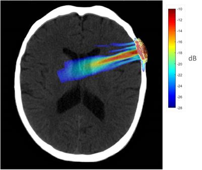
Magnetic Resonance Imaging (MRI) is a non-invasive diagnostic imaging technique that uses a strong magnetic field and radio waves to create detailed cross-sectional or three-dimensional images of the internal structures of the body. The patient lies within a large, cylindrical magnet, and the scanner detects changes in the direction of the magnetic field caused by protons in the body. These changes are then converted into detailed images that help medical professionals to diagnose and monitor various medical conditions, such as tumors, injuries, or diseases affecting the brain, spinal cord, heart, blood vessels, joints, and other internal organs. MRI does not use radiation like computed tomography (CT) scans.
The visual cortex is the part of the brain that processes visual information. It is located in the occipital lobe, which is at the back of the brain. The visual cortex is responsible for receiving and interpreting signals from the retina, which are then transmitted through the optic nerve and optic tract.
The visual cortex contains several areas that are involved in different aspects of visual processing, such as identifying shapes, colors, and movements. These areas work together to help us recognize and understand what we see. Damage to the visual cortex can result in various visual impairments, such as blindness or difficulty with visual perception.
The motor cortex is a region in the frontal lobe of the brain that is responsible for controlling voluntary movements. It is involved in planning, initiating, and executing movements of the limbs, body, and face. The motor cortex contains neurons called Betz cells, which have large cell bodies and are responsible for transmitting signals to the spinal cord to activate muscles. Damage to the motor cortex can result in various movement disorders such as hemiplegia or paralysis on one side of the body.
Neural pathways, also known as nerve tracts or fasciculi, refer to the highly organized and specialized routes through which nerve impulses travel within the nervous system. These pathways are formed by groups of neurons (nerve cells) that are connected in a series, creating a continuous communication network for electrical signals to transmit information between different regions of the brain, spinal cord, and peripheral nerves.
Neural pathways can be classified into two main types: sensory (afferent) and motor (efferent). Sensory neural pathways carry sensory information from various receptors in the body (such as those for touch, temperature, pain, and vision) to the brain for processing. Motor neural pathways, on the other hand, transmit signals from the brain to the muscles and glands, controlling movements and other effector functions.
The formation of these neural pathways is crucial for normal nervous system function, as it enables efficient communication between different parts of the body and allows for complex behaviors, cognitive processes, and adaptive responses to internal and external stimuli.
The auditory cortex is the region of the brain that is responsible for processing and analyzing sounds, including speech. It is located in the temporal lobe of the cerebral cortex, specifically within the Heschl's gyrus and the surrounding areas. The auditory cortex receives input from the auditory nerve, which carries sound information from the inner ear to the brain.
The auditory cortex is divided into several subregions that are responsible for different aspects of sound processing, such as pitch, volume, and location. These regions work together to help us recognize and interpret sounds in our environment, allowing us to communicate with others and respond appropriately to our surroundings. Damage to the auditory cortex can result in hearing loss or difficulty understanding speech.
The somatosensory cortex is a part of the brain located in the postcentral gyrus of the parietal lobe, which is responsible for processing sensory information from the body. It receives and integrates tactile, proprioceptive, and thermoception inputs from the skin, muscles, joints, and internal organs, allowing us to perceive and interpret touch, pressure, pain, temperature, vibration, position, and movement of our body parts. The somatosensory cortex is organized in a map-like manner, known as the sensory homunculus, where each body part is represented according to its relative sensitivity and density of innervation. This organization allows for precise localization and discrimination of tactile stimuli across the body surface.
Reaction time, in the context of medicine and physiology, refers to the time period between the presentation of a stimulus and the subsequent initiation of a response. This complex process involves the central nervous system, particularly the brain, which perceives the stimulus, processes it, and then sends signals to the appropriate muscles or glands to react.
There are different types of reaction times, including simple reaction time (responding to a single, expected stimulus) and choice reaction time (choosing an appropriate response from multiple possibilities). These measures can be used in clinical settings to assess various aspects of neurological function, such as cognitive processing speed, motor control, and alertness.
However, it is important to note that reaction times can be influenced by several factors, including age, fatigue, attention, and the use of certain medications or substances.
The gyrus cinguli, also known as the cingulate gyrus, is a structure located in the brain. It forms part of the limbic system and plays a role in various functions such as emotion, memory, and perception of pain. The gyrus cinguli is situated in the medial aspect of the cerebral hemisphere, adjacent to the corpus callosum, and curves around the frontal portion of the corpus callosum, forming a C-shaped structure. It has been implicated in several neurological and psychiatric conditions, including depression, anxiety disorders, and chronic pain syndromes.
The brain is the central organ of the nervous system, responsible for receiving and processing sensory information, regulating vital functions, and controlling behavior, movement, and cognition. It is divided into several distinct regions, each with specific functions:
1. Cerebrum: The largest part of the brain, responsible for higher cognitive functions such as thinking, learning, memory, language, and perception. It is divided into two hemispheres, each controlling the opposite side of the body.
2. Cerebellum: Located at the back of the brain, it is responsible for coordinating muscle movements, maintaining balance, and fine-tuning motor skills.
3. Brainstem: Connects the cerebrum and cerebellum to the spinal cord, controlling vital functions such as breathing, heart rate, and blood pressure. It also serves as a relay center for sensory information and motor commands between the brain and the rest of the body.
4. Diencephalon: A region that includes the thalamus (a major sensory relay station) and hypothalamus (regulates hormones, temperature, hunger, thirst, and sleep).
5. Limbic system: A group of structures involved in emotional processing, memory formation, and motivation, including the hippocampus, amygdala, and cingulate gyrus.
The brain is composed of billions of interconnected neurons that communicate through electrical and chemical signals. It is protected by the skull and surrounded by three layers of membranes called meninges, as well as cerebrospinal fluid that provides cushioning and nutrients.
Psychomotor performance refers to the integration and coordination of mental processes (cognitive functions) with physical movements. It involves the ability to perform complex tasks that require both cognitive skills, such as thinking, remembering, and perceiving, and motor skills, such as gross and fine motor movements. Examples of psychomotor performances include driving a car, playing a musical instrument, or performing surgical procedures.
In a medical context, psychomotor performance is often used to assess an individual's ability to perform activities of daily living (ADLs) and instrumental activities of daily living (IADLs), such as bathing, dressing, cooking, cleaning, and managing medications. Deficits in psychomotor performance can be a sign of neurological or psychiatric disorders, such as dementia, Parkinson's disease, or depression.
Assessment of psychomotor performance may involve tests that measure reaction time, coordination, speed, precision, and accuracy of movements, as well as cognitive functions such as attention, memory, and problem-solving skills. These assessments can help healthcare professionals develop appropriate treatment plans and monitor the progression of diseases or the effectiveness of interventions.
The frontal lobe is the largest lobes of the human brain, located at the front part of each cerebral hemisphere and situated in front of the parietal and temporal lobes. It plays a crucial role in higher cognitive functions such as decision making, problem solving, planning, parts of social behavior, emotional expressions, physical reactions, and motor function. The frontal lobe is also responsible for what's known as "executive functions," which include the ability to focus attention, understand rules, switch focus, plan actions, and inhibit inappropriate behaviors. It is divided into five areas, each with its own specific functions: the primary motor cortex, premotor cortex, Broca's area, prefrontal cortex, and orbitofrontal cortex. Damage to the frontal lobe can result in a wide range of impairments, depending on the location and extent of the injury.
In the context of medical and clinical neuroscience, memory is defined as the brain's ability to encode, store, retain, and recall information or experiences. Memory is a complex cognitive process that involves several interconnected regions of the brain and can be categorized into different types based on various factors such as duration and the nature of the information being remembered.
The major types of memory include:
1. Sensory memory: The shortest form of memory, responsible for holding incoming sensory information for a brief period (less than a second to several seconds) before it is either transferred to short-term memory or discarded.
2. Short-term memory (also called working memory): A temporary storage system that allows the brain to hold and manipulate information for approximately 20-30 seconds, although this duration can be extended through rehearsal strategies. Short-term memory has a limited capacity, typically thought to be around 7±2 items.
3. Long-term memory: The memory system responsible for storing large amounts of information over extended periods, ranging from minutes to a lifetime. Long-term memory has a much larger capacity compared to short-term memory and is divided into two main categories: explicit (declarative) memory and implicit (non-declarative) memory.
Explicit (declarative) memory can be further divided into episodic memory, which involves the recollection of specific events or episodes, including their temporal and spatial contexts, and semantic memory, which refers to the storage and retrieval of general knowledge, facts, concepts, and vocabulary, independent of personal experience or context.
Implicit (non-declarative) memory encompasses various forms of learning that do not require conscious awareness or intention, such as procedural memory (skills and habits), priming (facilitated processing of related stimuli), classical conditioning (associative learning), and habituation (reduced responsiveness to repeated stimuli).
Memory is a crucial aspect of human cognition and plays a significant role in various aspects of daily life, including learning, problem-solving, decision-making, social interactions, and personal identity. Memory dysfunction can result from various neurological and psychiatric conditions, such as dementia, Alzheimer's disease, stroke, traumatic brain injury, and depression.
Functional laterality, in a medical context, refers to the preferential use or performance of one side of the body over the other for specific functions. This is often demonstrated in hand dominance, where an individual may be right-handed or left-handed, meaning they primarily use their right or left hand for tasks such as writing, eating, or throwing.
However, functional laterality can also apply to other bodily functions and structures, including the eyes (ocular dominance), ears (auditory dominance), or legs. It's important to note that functional laterality is not a strict binary concept; some individuals may exhibit mixed dominance or no strong preference for one side over the other.
In clinical settings, assessing functional laterality can be useful in diagnosing and treating various neurological conditions, such as stroke or traumatic brain injury, where understanding any resulting lateralized impairments can inform rehabilitation strategies.
Photic stimulation is a medical term that refers to the exposure of the eyes to light, specifically repetitive pulses of light, which is used as a method in various research and clinical settings. In neuroscience, it's often used in studies related to vision, circadian rhythms, and brain function.
In a clinical context, photic stimulation is sometimes used in the diagnosis of certain medical conditions such as seizure disorders (like epilepsy). By observing the response of the brain to this light stimulus, doctors can gain valuable insights into the functioning of the brain and the presence of any neurological disorders.
However, it's important to note that photic stimulation should be conducted under the supervision of a trained healthcare professional, as improper use can potentially trigger seizures in individuals who are susceptible to them.
Computer-assisted image processing is a medical term that refers to the use of computer systems and specialized software to improve, analyze, and interpret medical images obtained through various imaging techniques such as X-ray, CT (computed tomography), MRI (magnetic resonance imaging), ultrasound, and others.
The process typically involves several steps, including image acquisition, enhancement, segmentation, restoration, and analysis. Image processing algorithms can be used to enhance the quality of medical images by adjusting contrast, brightness, and sharpness, as well as removing noise and artifacts that may interfere with accurate diagnosis. Segmentation techniques can be used to isolate specific regions or structures of interest within an image, allowing for more detailed analysis.
Computer-assisted image processing has numerous applications in medical imaging, including detection and characterization of lesions, tumors, and other abnormalities; assessment of organ function and morphology; and guidance of interventional procedures such as biopsies and surgeries. By automating and standardizing image analysis tasks, computer-assisted image processing can help to improve diagnostic accuracy, efficiency, and consistency, while reducing the potential for human error.
Neurons, also known as nerve cells or neurocytes, are specialized cells that constitute the basic unit of the nervous system. They are responsible for receiving, processing, and transmitting information and signals within the body. Neurons have three main parts: the dendrites, the cell body (soma), and the axon. The dendrites receive signals from other neurons or sensory receptors, while the axon transmits these signals to other neurons, muscles, or glands. The junction between two neurons is called a synapse, where neurotransmitters are released to transmit the signal across the gap (synaptic cleft) to the next neuron. Neurons vary in size, shape, and structure depending on their function and location within the nervous system.
A nerve net, also known as a neural net or neuronal network, is not a medical term per se, but rather a concept in neuroscience and artificial intelligence (AI). It refers to a complex network of interconnected neurons that process and transmit information. In the context of the human body, the nervous system can be thought of as a type of nerve net, with the brain and spinal cord serving as the central processing unit and peripheral nerves carrying signals to and from various parts of the body.
In the field of AI, artificial neural networks are computational models inspired by the structure and function of biological nerve nets. These models consist of interconnected nodes or "neurons" that process information and learn patterns through a process of training and adaptation. They have been used in a variety of applications, including image recognition, natural language processing, and machine learning.
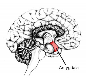
The amygdala is an almond-shaped group of nuclei located deep within the temporal lobe of the brain, specifically in the anterior portion of the temporal lobes and near the hippocampus. It forms a key component of the limbic system and plays a crucial role in processing emotions, particularly fear and anxiety. The amygdala is involved in the integration of sensory information with emotional responses, memory formation, and decision-making processes.
In response to emotionally charged stimuli, the amygdala can modulate various physiological functions, such as heart rate, blood pressure, and stress hormone release, via its connections to the hypothalamus and brainstem. Additionally, it contributes to social behaviors, including recognizing emotional facial expressions and responding appropriately to social cues. Dysfunctions in amygdala function have been implicated in several psychiatric and neurological conditions, such as anxiety disorders, depression, post-traumatic stress disorder (PTSD), and autism spectrum disorder (ASD).
Cognition refers to the mental processes involved in acquiring, processing, and utilizing information. These processes include perception, attention, memory, language, problem-solving, and decision-making. Cognitive functions allow us to interact with our environment, understand and respond to stimuli, learn new skills, and remember experiences.
In a medical context, cognitive function is often assessed as part of a neurological or psychiatric evaluation. Impairments in cognition can be caused by various factors, such as brain injury, neurodegenerative diseases (e.g., Alzheimer's disease), infections, toxins, and mental health conditions. Assessing cognitive function helps healthcare professionals diagnose conditions, monitor disease progression, and develop treatment plans.
Short-term memory, also known as primary or active memory, is the system responsible for holding and processing limited amounts of information for brief periods of time, typically on the order of seconds to minutes. It has a capacity of around 7±2 items, as suggested by George Miller's "magic number" theory. Short-term memory allows us to retain and manipulate information temporarily while we are using it, such as remembering a phone number while dialing or following a set of instructions. Information in short-term memory can be maintained through rehearsal, which is the conscious repetition of the information. Over time, if the information is not transferred to long-term memory through consolidation processes, it will be forgotten.
Sprague-Dawley rats are a strain of albino laboratory rats that are widely used in scientific research. They were first developed by researchers H.H. Sprague and R.C. Dawley in the early 20th century, and have since become one of the most commonly used rat strains in biomedical research due to their relatively large size, ease of handling, and consistent genetic background.
Sprague-Dawley rats are outbred, which means that they are genetically diverse and do not suffer from the same limitations as inbred strains, which can have reduced fertility and increased susceptibility to certain diseases. They are also characterized by their docile nature and low levels of aggression, making them easier to handle and study than some other rat strains.
These rats are used in a wide variety of research areas, including toxicology, pharmacology, nutrition, cancer, and behavioral studies. Because they are genetically diverse, Sprague-Dawley rats can be used to model a range of human diseases and conditions, making them an important tool in the development of new drugs and therapies.
In the context of medicine, particularly in behavioral neuroscience and psychology, "reward" is not typically used as a definitive medical term. However, it generally refers to a positive outcome or incentive that reinforces certain behaviors, making them more likely to be repeated in the future. This can involve various stimuli such as food, water, sexual activity, social interaction, or drug use, among others.
In the brain, rewards are associated with the activation of the reward system, primarily the mesolimbic dopamine pathway, which includes the ventral tegmental area (VTA) and the nucleus accumbens (NAcc). The release of dopamine in these areas is thought to reinforce and motivate behavior linked to rewards.
It's important to note that while "reward" has a specific meaning in this context, it is not a formal medical diagnosis or condition. Instead, it is a concept used to understand the neural and psychological mechanisms underlying motivation, learning, and addiction.
The parietal lobe is a region of the brain that is located in the posterior part of the cerebral cortex, covering the upper and rear portions of the brain. It is involved in processing sensory information from the body, such as touch, temperature, and pain, as well as spatial awareness and perception, visual-spatial cognition, and the integration of different senses.
The parietal lobe can be divided into several functional areas, including the primary somatosensory cortex (which receives tactile information from the body), the secondary somatosensory cortex (which processes more complex tactile information), and the posterior parietal cortex (which is involved in spatial attention, perception, and motor planning).
Damage to the parietal lobe can result in various neurological symptoms, such as neglect of one side of the body, difficulty with spatial orientation, problems with hand-eye coordination, and impaired mathematical and language abilities.
Analysis of Variance (ANOVA) is a statistical technique used to compare the means of two or more groups and determine whether there are any significant differences between them. It is a way to analyze the variance in a dataset to determine whether the variability between groups is greater than the variability within groups, which can indicate that the groups are significantly different from one another.
ANOVA is based on the concept of partitioning the total variance in a dataset into two components: variance due to differences between group means (also known as "between-group variance") and variance due to differences within each group (also known as "within-group variance"). By comparing these two sources of variance, ANOVA can help researchers determine whether any observed differences between groups are statistically significant, or whether they could have occurred by chance.
ANOVA is a widely used technique in many areas of research, including biology, psychology, engineering, and business. It is often used to compare the means of two or more experimental groups, such as a treatment group and a control group, to determine whether the treatment had a significant effect. ANOVA can also be used to compare the means of different populations or subgroups within a population, to identify any differences that may exist between them.
Dopamine is a type of neurotransmitter, which is a chemical messenger that transmits signals in the brain and nervous system. It plays several important roles in the body, including:
* Regulation of movement and coordination
* Modulation of mood and motivation
* Control of the reward and pleasure centers of the brain
* Regulation of muscle tone
* Involvement in memory and attention
Dopamine is produced in several areas of the brain, including the substantia nigra and the ventral tegmental area. It is released by neurons (nerve cells) and binds to specific receptors on other neurons, where it can either excite or inhibit their activity.
Abnormalities in dopamine signaling have been implicated in several neurological and psychiatric conditions, including Parkinson's disease, schizophrenia, and addiction.
The limbic system is a complex set of structures in the brain that includes the hippocampus, amygdala, fornix, cingulate gyrus, and other nearby areas. It's associated with emotional responses, instinctual behaviors, motivation, long-term memory formation, and olfaction (smell). The limbic system is also involved in the modulation of visceral functions and drives, such as hunger, thirst, and sexual drive.
The structures within the limbic system communicate with each other and with other parts of the brain, particularly the hypothalamus and the cortex, to regulate various physiological and psychological processes. Dysfunctions in the limbic system can lead to a range of neurological and psychiatric conditions, including depression, anxiety disorders, post-traumatic stress disorder (PTSD), and certain types of memory impairment.
Pyramidal cells, also known as pyramidal neurons, are a type of multipolar neuron found in the cerebral cortex and hippocampus of the brain. They have a characteristic triangular or pyramid-like shape with a single apical dendrite that extends from the apex of the cell body towards the pial surface, and multiple basal dendrites that branch out from the base of the cell body.
Pyramidal cells are excitatory neurons that play a crucial role in information processing and transmission within the brain. They receive inputs from various sources, including other neurons and sensory receptors, and generate action potentials that are transmitted to other neurons through their axons. The apical dendrite of pyramidal cells receives inputs from distant cortical areas, while the basal dendrites receive inputs from local circuits.
Pyramidal cells are named after their pyramid-like shape and are among the largest neurons in the brain. They are involved in various cognitive functions, including learning, memory, attention, and perception. Dysfunction of pyramidal cells has been implicated in several neurological disorders, such as Alzheimer's disease, epilepsy, and schizophrenia.
Neuropsychological tests are a type of psychological assessment that measures cognitive functions, such as attention, memory, language, problem-solving, and perception. These tests are used to help diagnose and understand the cognitive impact of neurological conditions, including dementia, traumatic brain injury, stroke, Parkinson's disease, and other disorders that affect the brain.
The tests are typically administered by a trained neuropsychologist and can take several hours to complete. They may involve paper-and-pencil tasks, computerized tasks, or interactive activities. The results of the tests are compared to normative data to help identify any areas of cognitive weakness or strength.
Neuropsychological testing can provide valuable information for treatment planning, rehabilitation, and assessing response to treatment. It can also be used in research to better understand the neural basis of cognition and the impact of neurological conditions on cognitive function.
The entorhinal cortex is a region in the brain that is located in the medial temporal lobe and is part of the limbic system. It plays a crucial role in memory, navigation, and the processing of sensory information. The entorhinal cortex is closely connected to the hippocampus, which is another important structure for memory and spatial cognition.
The entorhinal cortex can be divided into several subregions, including the lateral, medial, and posterior sections. These subregions have distinct connectivity patterns and may contribute differently to various cognitive functions. One of the most well-known features of the entorhinal cortex is the presence of "grid cells," which are neurons that fire in response to specific spatial locations and help to form a cognitive map of the environment.
Damage to the entorhinal cortex has been linked to several neurological and psychiatric conditions, including Alzheimer's disease, epilepsy, and schizophrenia.
In the context of medicine, "cues" generally refer to specific pieces of information or signals that can help healthcare professionals recognize and respond to a particular situation or condition. These cues can come in various forms, such as:
1. Physical examination findings: For example, a patient's abnormal heart rate or blood pressure reading during a physical exam may serve as a cue for the healthcare professional to investigate further.
2. Patient symptoms: A patient reporting chest pain, shortness of breath, or other concerning symptoms can act as a cue for a healthcare provider to consider potential diagnoses and develop an appropriate treatment plan.
3. Laboratory test results: Abnormal findings on laboratory tests, such as elevated blood glucose levels or abnormal liver function tests, may serve as cues for further evaluation and diagnosis.
4. Medical history information: A patient's medical history can provide valuable cues for healthcare professionals when assessing their current health status. For example, a history of smoking may increase the suspicion for chronic obstructive pulmonary disease (COPD) in a patient presenting with respiratory symptoms.
5. Behavioral or environmental cues: In some cases, behavioral or environmental factors can serve as cues for healthcare professionals to consider potential health risks. For instance, exposure to secondhand smoke or living in an area with high air pollution levels may increase the risk of developing respiratory conditions.
Overall, "cues" in a medical context are essential pieces of information that help healthcare professionals make informed decisions about patient care and treatment.
Emotions are complex psychological states that involve three distinct components: a subjective experience, a physiological response, and a behavioral or expressive response. Emotions can be short-lived, such as a flash of anger, or more long-lasting, such as enduring sadness. They can also vary in intensity, from mild irritation to intense joy or fear.
Emotions are often distinguished from other psychological states, such as moods and temperament, which may be less specific and more enduring. Emotions are typically thought to have a clear cause or object, such as feeling happy when you receive good news or feeling anxious before a job interview.
There are many different emotions that people can experience, including happiness, sadness, anger, fear, surprise, disgust, and shame. These emotions are often thought to serve important adaptive functions, helping individuals respond to challenges and opportunities in their environment.
In medical contexts, emotions may be relevant to the diagnosis and treatment of various mental health conditions, such as depression, anxiety disorders, and bipolar disorder. Abnormalities in emotional processing and regulation have been implicated in many psychiatric illnesses, and therapies that target these processes may be effective in treating these conditions.
"Long-Evans" is a strain of laboratory rats commonly used in scientific research. They are named after their developers, the scientists Long and Evans. This strain is albino, with a brownish-black hood over their eyes and ears, and they have an agouti (salt-and-pepper) color on their backs. They are often used as a model organism due to their size, ease of handling, and genetic similarity to humans. However, I couldn't find any specific medical definition related to "Long-Evans rats" as they are not a medical condition or disease.
Schizophrenia is a severe mental disorder characterized by disturbances in thought, perception, emotion, and behavior. It often includes hallucinations (usually hearing voices), delusions, paranoia, and disorganized speech and behavior. The onset of symptoms typically occurs in late adolescence or early adulthood. Schizophrenia is a complex, chronic condition that requires ongoing treatment and management. It significantly impairs social and occupational functioning, and it's often associated with reduced life expectancy due to comorbid medical conditions. The exact causes of schizophrenia are not fully understood, but research suggests that genetic, environmental, and neurodevelopmental factors play a role in its development.
Microdialysis is a minimally invasive technique used in clinical and research settings to continuously monitor the concentration of various chemicals, such as neurotransmitters, drugs, or metabolites, in biological fluids (e.g., extracellular fluid of tissues, blood, or cerebrospinal fluid). This method involves inserting a small, flexible catheter with a semipermeable membrane into the region of interest. A physiological solution is continuously perfused through the catheter, allowing molecules to diffuse across the membrane based on their concentration gradient. The dialysate that exits the catheter is then collected and analyzed for target compounds using various analytical techniques (e.g., high-performance liquid chromatography, mass spectrometry).
In summary, microdialysis is a valuable tool for monitoring real-time changes in chemical concentrations within biological systems, enabling better understanding of physiological processes or pharmacokinetic properties of drugs.
'Animal behavior' refers to the actions or responses of animals to various stimuli, including their interactions with the environment and other individuals. It is the study of the actions of animals, whether they are instinctual, learned, or a combination of both. Animal behavior includes communication, mating, foraging, predator avoidance, and social organization, among other things. The scientific study of animal behavior is called ethology. This field seeks to understand the evolutionary basis for behaviors as well as their physiological and psychological mechanisms.
Choice behavior refers to the selection or decision-making process in which an individual consciously or unconsciously chooses one option over others based on their preferences, values, experiences, and motivations. In a medical context, choice behavior may relate to patients' decisions about their healthcare, such as selecting a treatment option, choosing a healthcare provider, or adhering to a prescribed medication regimen. Understanding choice behavior is essential in shaping health policies, developing patient-centered care models, and improving overall health outcomes.
In a medical or psychological context, attention is the cognitive process of selectively concentrating on certain aspects of the environment while ignoring other things. It involves focusing mental resources on specific stimuli, sensory inputs, or internal thoughts while blocking out irrelevant distractions. Attention can be divided into different types, including:
1. Sustained attention: The ability to maintain focus on a task or stimulus over time.
2. Selective attention: The ability to concentrate on relevant stimuli while ignoring irrelevant ones.
3. Divided attention: The capacity to pay attention to multiple tasks or stimuli simultaneously.
4. Alternating attention: The skill of shifting focus between different tasks or stimuli as needed.
Deficits in attention are common symptoms of various neurological and psychiatric conditions, such as ADHD, dementia, depression, and anxiety disorders. Assessment of attention is an essential part of neuropsychological evaluations and can be measured using various tests and tasks.
The kidney cortex is the outer region of the kidney where most of the functional units called nephrons are located. It plays a crucial role in filtering blood and regulating water, electrolyte, and acid-base balance in the body. The kidney cortex contains the glomeruli, proximal tubules, loop of Henle, and distal tubules, which work together to reabsorb necessary substances and excrete waste products into the urine.
The temporal lobe is one of the four main lobes of the cerebral cortex in the brain, located on each side of the head roughly level with the ears. It plays a major role in auditory processing, memory, and emotion. The temporal lobe contains several key structures including the primary auditory cortex, which is responsible for analyzing sounds, and the hippocampus, which is crucial for forming new memories. Damage to the temporal lobe can result in various neurological symptoms such as hearing loss, memory impairment, and changes in emotional behavior.
The nucleus accumbens is a part of the brain that is located in the ventral striatum, which is a key region of the reward circuitry. It is made up of two subregions: the shell and the core. The nucleus accumbens receives inputs from various sources, including the prefrontal cortex, amygdala, and hippocampus, and sends outputs to the ventral pallidum and other areas.
The nucleus accumbens is involved in reward processing, motivation, reinforcement learning, and addiction. It plays a crucial role in the release of the neurotransmitter dopamine, which is associated with pleasure and reinforcement. Dysfunction in the nucleus accumbens has been implicated in various neurological and psychiatric conditions, including substance use disorders, depression, and obsessive-compulsive disorder.
The hippocampus is a complex, curved formation in the brain that resembles a seahorse (hence its name, from the Greek word "hippos" meaning horse and "kampos" meaning sea monster). It's part of the limbic system and plays crucial roles in the formation of memories, particularly long-term ones.
This region is involved in spatial navigation and cognitive maps, allowing us to recognize locations and remember how to get to them. Additionally, it's one of the first areas affected by Alzheimer's disease, which often results in memory loss as an early symptom.
Anatomically, it consists of two main parts: the Ammon's horn (or cornu ammonis) and the dentate gyrus. These structures are made up of distinct types of neurons that contribute to different aspects of learning and memory.
Neurological models are simplified representations or simulations of various aspects of the nervous system, including its structure, function, and processes. These models can be theoretical, computational, or physical and are used to understand, explain, and predict neurological phenomena. They may focus on specific neurological diseases, disorders, or functions, such as memory, learning, or movement. The goal of these models is to provide insights into the complex workings of the nervous system that cannot be easily observed or understood through direct examination alone.
"Macaca mulatta" is the scientific name for the Rhesus macaque, a species of monkey that is native to South, Central, and Southeast Asia. They are often used in biomedical research due to their genetic similarity to humans.
Dopamine antagonists are a class of drugs that block the action of dopamine, a neurotransmitter in the brain associated with various functions including movement, motivation, and emotion. These drugs work by binding to dopamine receptors and preventing dopamine from attaching to them, which can help to reduce the symptoms of certain medical conditions such as schizophrenia, bipolar disorder, and gastroesophageal reflux disease (GERD).
There are several types of dopamine antagonists, including:
1. Typical antipsychotics: These drugs are primarily used to treat psychosis, including schizophrenia and delusional disorders. Examples include haloperidol, chlorpromazine, and fluphenazine.
2. Atypical antipsychotics: These drugs are also used to treat psychosis but have fewer side effects than typical antipsychotics. They may also be used to treat bipolar disorder and depression. Examples include risperidone, olanzapine, and quetiapine.
3. Antiemetics: These drugs are used to treat nausea and vomiting. Examples include metoclopramide and prochlorperazine.
4. Dopamine agonists: While not technically dopamine antagonists, these drugs work by stimulating dopamine receptors and can be used to treat conditions such as Parkinson's disease. However, they can also have the opposite effect and block dopamine receptors in high doses, making them functionally similar to dopamine antagonists.
Common side effects of dopamine antagonists include sedation, weight gain, and movement disorders such as tardive dyskinesia. It's important to use these drugs under the close supervision of a healthcare provider to monitor for side effects and adjust the dosage as needed.
Functional neuroimaging is a branch of medical imaging that involves the use of various techniques to measure and visualize the metabolic activity or blood flow in different regions of the brain. These measurements can be used to infer the level of neural activation in specific brain areas, allowing researchers and clinicians to study the functioning of the brain in various states, such as during rest, cognitive tasks, or disease processes.
Some common functional neuroimaging techniques include:
1. Functional Magnetic Resonance Imaging (fMRI): This technique uses magnetic fields and radio waves to measure changes in blood flow and oxygenation levels in the brain, which are associated with neural activity.
2. Positron Emission Tomography (PET): This technique involves the injection of a small amount of radioactive tracer into the body, which is taken up by active brain cells. The resulting gamma rays are then detected and used to create images of brain activity.
3. Single-Photon Emission Computed Tomography (SPECT): Similar to PET, SPECT uses a radioactive tracer to measure blood flow in the brain, but with lower resolution and sensitivity.
4. Functional Near-Infrared Spectroscopy (fNIRS): This technique uses near-infrared light to measure changes in oxygenation levels in the brain, providing a non-invasive and relatively inexpensive method for studying brain function.
Functional neuroimaging has numerous applications in both research and clinical settings, including the study of cognitive processes, the diagnosis and monitoring of neurological and psychiatric disorders, and the development of new treatments and interventions.
Fear is a basic human emotion that is typically characterized by a strong feeling of anxiety, apprehension, or distress in response to a perceived threat or danger. It is a natural and adaptive response that helps individuals identify and respond to potential dangers in their environment, and it can manifest as physical, emotional, and cognitive symptoms.
Physical symptoms of fear may include increased heart rate, rapid breathing, sweating, trembling, and muscle tension. Emotional symptoms may include feelings of anxiety, worry, or panic, while cognitive symptoms may include difficulty concentrating, racing thoughts, and intrusive thoughts about the perceived threat.
Fear can be a normal and adaptive response to real dangers, but it can also become excessive or irrational in some cases, leading to phobias, anxiety disorders, and other mental health conditions. In these cases, professional help may be necessary to manage and overcome the fear.
Visual perception refers to the ability to interpret and organize information that comes from our eyes to recognize and understand what we are seeing. It involves several cognitive processes such as pattern recognition, size estimation, movement detection, and depth perception. Visual perception allows us to identify objects, navigate through space, and interact with our environment. Deficits in visual perception can lead to learning difficulties and disabilities.
Dopamine D1 receptors are a type of G protein-coupled receptor that bind to the neurotransmitter dopamine. They are classified as D1-like receptors, along with D5 receptors, and are activated by dopamine through a stimulatory G protein (Gs).
D1 receptors are widely expressed in the central nervous system, including the striatum, prefrontal cortex, hippocampus, and amygdala. They play important roles in various physiological functions, such as movement control, motivation, reward processing, working memory, and cognition.
Activation of D1 receptors leads to increased levels of intracellular cyclic adenosine monophosphate (cAMP) and activation of protein kinase A (PKA), which in turn modulate the activity of various downstream signaling pathways. Dysregulation of dopamine D1 receptor function has been implicated in several neurological and psychiatric disorders, including Parkinson's disease, schizophrenia, attention deficit hyperactivity disorder (ADHD), and drug addiction.
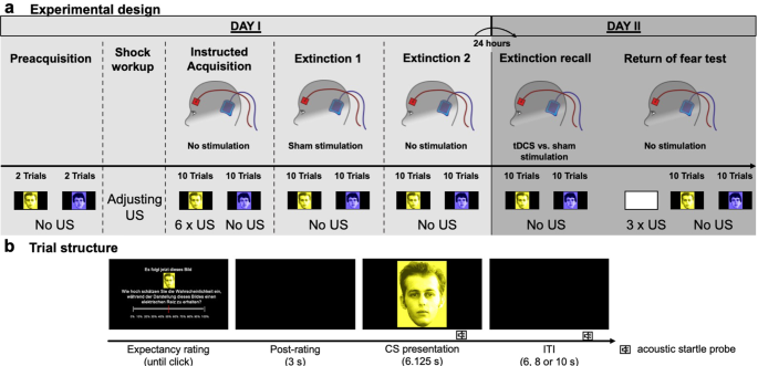
"Extinction, Psychological" refers to the process by which a conditioned response or behavior becomes weakened and eventually disappears when the behavior is no longer reinforced or rewarded. It is a fundamental concept in learning theory and conditioning.
In classical conditioning, extinction occurs when the conditioned stimulus (CS) is repeatedly presented without the unconditioned stimulus (US), leading to the gradual weakening and eventual disappearance of the conditioned response (CR). For example, if a person learns to associate a tone (CS) with a puff of air to the eye (US), causing blinking (CR), but then the tone is presented several times without the puff of air, the blinking response will become weaker and eventually disappear.
In operant conditioning, extinction occurs when a reinforcer is no longer provided following a behavior, leading to the gradual weakening and eventual disappearance of that behavior. For example, if a child receives candy every time they clean their room (reinforcement), but then the candy is withheld, the child may eventually stop cleaning their room (extinction).
It's important to note that extinction can be a slow process and may require multiple trials or repetitions. Additionally, behaviors that have been extinguished can sometimes reappear in certain circumstances, a phenomenon known as spontaneous recovery.
The mediodorsal thalamic nucleus (MDTN) is a collection of neurons located in the dorsal part of the thalamus, a region of the brain that serves as a relay station for sensory and motor signals to the cerebral cortex. The MDTN is primarily involved in cognitive functions such as memory, attention, and emotion regulation.
The MDTN receives inputs from various regions of the brain, including the prefrontal cortex, amygdala, and hippocampus, and projects to the same areas of the cerebral cortex. It has been implicated in several neurological and psychiatric conditions, such as Alzheimer's disease, Parkinson's disease, schizophrenia, and depression.
Anatomically, the MDTN is divided into several subnuclei, including the parvocellular, magnocellular, and intermediate parts, each with distinct connectivity patterns and functions. Overall, the MDTN plays a crucial role in integrating information from different brain regions to facilitate higher-order cognitive processes.
'Behavior' is a term used in the medical and scientific community to describe the actions or reactions of an individual in response to internal or external stimuli. It can be observed and measured, and it involves all the responses of a person, including motor responses, emotional responses, and cognitive responses. Behaviors can be voluntary or involuntary, adaptive or maladaptive, and normal or abnormal. They can also be influenced by genetic, physiological, environmental, and social factors. In a medical context, the study of behavior is often relevant to understanding and treating various mental health conditions, such as anxiety disorders, mood disorders, and personality disorders.
Oxygen is a colorless, odorless, tasteless gas that constitutes about 21% of the earth's atmosphere. It is a crucial element for human and most living organisms as it is vital for respiration. Inhaled oxygen enters the lungs and binds to hemoglobin in red blood cells, which carries it to tissues throughout the body where it is used to convert nutrients into energy and carbon dioxide, a waste product that is exhaled.
Medically, supplemental oxygen therapy may be provided to patients with conditions such as chronic obstructive pulmonary disease (COPD), pneumonia, heart failure, or other medical conditions that impair the body's ability to extract sufficient oxygen from the air. Oxygen can be administered through various devices, including nasal cannulas, face masks, and ventilators.
An action potential is a brief electrical signal that travels along the membrane of a nerve cell (neuron) or muscle cell. It is initiated by a rapid, localized change in the permeability of the cell membrane to specific ions, such as sodium and potassium, resulting in a rapid influx of sodium ions and a subsequent efflux of potassium ions. This ion movement causes a brief reversal of the electrical potential across the membrane, which is known as depolarization. The action potential then propagates along the cell membrane as a wave, allowing the electrical signal to be transmitted over long distances within the body. Action potentials play a crucial role in the communication and functioning of the nervous system and muscle tissue.
I'm sorry for any confusion, but "Macaca" is not a medical term. It is the name of a genus that includes several species of monkeys, commonly known as macaques. These primates are often used in biomedical research due to their similarities with humans in terms of genetics and physiology. If you have any questions related to medicine or health, I would be happy to try to help answer them.
The cerebellar cortex is the outer layer of the cerebellum, which is a part of the brain that plays a crucial role in motor control, balance, and coordination of muscle movements. The cerebellar cortex contains numerous small neurons called granule cells, as well as other types of neurons such as Purkinje cells, basket cells, and stellate cells. These neurons are organized into distinct layers and microcircuits that process information related to motor function and possibly other functions such as cognition and emotion. The cerebellar cortex receives input from various sources, including the spinal cord, vestibular system, and cerebral cortex, and sends output to brainstem nuclei and thalamus, which in turn project to the cerebral cortex. Damage to the cerebellar cortex can result in ataxia, dysmetria, dysdiadochokinesia, and other motor symptoms.
Neuronal plasticity, also known as neuroplasticity or neural plasticity, refers to the ability of the brain and nervous system to change and adapt as a result of experience, learning, injury, or disease. This can involve changes in the structure, organization, and function of neurons (nerve cells) and their connections (synapses) in the central and peripheral nervous systems.
Neuronal plasticity can take many forms, including:
* Synaptic plasticity: Changes in the strength or efficiency of synaptic connections between neurons. This can involve the formation, elimination, or modification of synapses.
* Neural circuit plasticity: Changes in the organization and connectivity of neural circuits, which are networks of interconnected neurons that process information.
* Structural plasticity: Changes in the physical structure of neurons, such as the growth or retraction of dendrites (branches that receive input from other neurons) or axons (projections that transmit signals to other neurons).
* Functional plasticity: Changes in the physiological properties of neurons, such as their excitability, responsiveness, or sensitivity to stimuli.
Neuronal plasticity is a fundamental property of the nervous system and plays a crucial role in many aspects of brain function, including learning, memory, perception, and cognition. It also contributes to the brain's ability to recover from injury or disease, such as stroke or traumatic brain injury.
Transcranial Magnetic Stimulation (TMS) is a non-invasive form of brain stimulation where a magnetic field is generated via an electromagnetic coil placed on the scalp. This magnetic field induces an electric current in the underlying brain tissue, which can lead to neuronal activation or inhibition, depending on the frequency and intensity of the stimulation. TMS has been used as a therapeutic intervention for various neurological and psychiatric conditions, such as depression, migraine, and tinnitus, among others. It is also used in research settings to investigate brain function and connectivity.
'Task Performance and Analysis' is not a commonly used medical term, but it can be found in the field of rehabilitation medicine and ergonomics. It refers to the process of evaluating and understanding how a specific task is performed, in order to identify any physical or cognitive demands placed on an individual during the performance of that task. This information can then be used to inform the design of interventions, such as workplace modifications or rehabilitation programs, aimed at improving task performance or reducing the risk of injury.
In a medical context, task performance and analysis may be used in the assessment and treatment of individuals with disabilities or injuries, to help them return to work or other activities of daily living. The analysis involves breaking down the task into its component parts, observing and measuring the physical and cognitive demands of each part, and evaluating the individual's ability to perform those demands. Based on this analysis, recommendations may be made for modifications to the task or the environment, training or education, or assistive devices that can help the individual perform the task more safely and efficiently.
Overall, task performance and analysis is a valuable tool in promoting safe and effective task performance, reducing the risk of injury, and improving functional outcomes for individuals with disabilities or injuries.
Association learning, also known as associative learning, is a type of learning in which an individual learns to associate two stimuli or a response with a particular outcome. This can occur through classical conditioning or operant conditioning.
In classical conditioning, first described by Ivan Pavlov, an initially neutral stimulus (the conditioned stimulus) is repeatedly paired with a biologically significant stimulus (the unconditioned stimulus), until the conditioned stimulus elicits a response (the conditioned response) similar to that of the unconditioned stimulus. For example, a dog may learn to salivate at the sound of a bell if the bell is repeatedly rung just before it is fed.
In operant conditioning, described by B.F. Skinner, behavior is modified by its consequences, with desired behaviors being reinforced and undesired behaviors being punished. For example, a child may learn to put their toys away if they are given a reward for doing so.
Association learning is an important mechanism in the acquisition of many types of knowledge and skills, and it plays a key role in the development and modification of behavior.
Neural inhibition is a process in the nervous system that decreases or prevents the activity of neurons (nerve cells) in order to regulate and control communication within the nervous system. It is a fundamental mechanism that allows for the balance of excitation and inhibition necessary for normal neural function. Inhibitory neurotransmitters, such as GABA (gamma-aminobutyric acid) and glycine, are released from the presynaptic neuron and bind to receptors on the postsynaptic neuron, reducing its likelihood of firing an action potential. This results in a decrease in neural activity and can have various effects depending on the specific neurons and brain regions involved. Neural inhibition is crucial for many functions including motor control, sensory processing, attention, memory, and emotional regulation.
Operant conditioning is a type of learning in which behavior is modified by its consequences, either reinforcing or punishing the behavior. It was first described by B.F. Skinner and involves an association between a response (behavior) and a consequence (either reward or punishment). There are two types of operant conditioning: positive reinforcement, in which a desirable consequence follows a desired behavior, increasing the likelihood that the behavior will occur again; and negative reinforcement, in which a undesirable consequence is removed following a desired behavior, also increasing the likelihood that the behavior will occur again.
For example, if a child cleans their room (response) and their parent gives them praise or a treat (positive reinforcement), the child is more likely to clean their room again in the future. If a child is buckling their seatbelt in the car (response) and the annoying buzzer stops (negative reinforcement), the child is more likely to buckle their seatbelt in the future.
It's important to note that operant conditioning is a form of learning, not motivation. The behavior is modified by its consequences, regardless of the individual's internal state or intentions.
Decision-making is the cognitive process of selecting a course of action from among multiple alternatives. In a medical context, decision-making refers to the process by which healthcare professionals and patients make choices about medical tests, treatments, or management options based on a thorough evaluation of available information, including the patient's preferences, values, and circumstances.
The decision-making process in medicine typically involves several steps:
1. Identifying the problem or issue that requires a decision.
2. Gathering relevant information about the patient's medical history, current condition, diagnostic test results, treatment options, and potential outcomes.
3. Considering the benefits, risks, and uncertainties associated with each option.
4. Evaluating the patient's preferences, values, and goals.
5. Selecting the most appropriate course of action based on a careful weighing of the available evidence and the patient's individual needs and circumstances.
6. Communicating the decision to the patient and ensuring that they understand the rationale behind it, as well as any potential risks or benefits.
7. Monitoring the outcomes of the decision and adjusting the course of action as needed based on ongoing evaluation and feedback.
Effective decision-making in medicine requires a thorough understanding of medical evidence, clinical expertise, and patient preferences. It also involves careful consideration of ethical principles, such as respect for autonomy, non-maleficence, beneficence, and justice. Ultimately, the goal of decision-making in healthcare is to promote the best possible outcomes for patients while minimizing harm and respecting their individual needs and values.
Parvalbumins are a group of calcium-binding proteins that are primarily found in muscle and nerve tissues. They belong to the EF-hand superfamily, which is characterized by a specific structure containing helix-loop-helix motifs that bind calcium ions. Parvalbumins have a high affinity for calcium and play an essential role in regulating intracellular calcium concentrations during muscle contraction and nerve impulse transmission.
In muscle tissue, parvalbumins are found in fast-twitch fibers and help to facilitate rapid relaxation after muscle contraction by binding calcium ions and removing them from the cytoplasm. In nerve tissue, parvalbumins are expressed in inhibitory interneurons and modulate neuronal excitability by regulating intracellular calcium concentrations during synaptic transmission.
Parvalbumins have also been identified as potential allergens in certain foods, such as fish and shellfish, and may cause allergic reactions in sensitive individuals.
Acoustic stimulation refers to the use of sound waves or vibrations to elicit a response in an individual, typically for the purpose of assessing or treating hearing, balance, or neurological disorders. In a medical context, acoustic stimulation may involve presenting pure tones, speech sounds, or other types of auditory signals through headphones, speakers, or specialized devices such as bone conduction transducers.
The response to acoustic stimulation can be measured using various techniques, including electrophysiological tests like auditory brainstem responses (ABRs) or otoacoustic emissions (OAEs), behavioral observations, or functional imaging methods like fMRI. Acoustic stimulation is also used in therapeutic settings, such as auditory training programs for hearing impairment or vestibular rehabilitation for balance disorders.
It's important to note that acoustic stimulation should be administered under the guidance of a qualified healthcare professional to ensure safety and effectiveness.
In the field of medicine, "time factors" refer to the duration of symptoms or time elapsed since the onset of a medical condition, which can have significant implications for diagnosis and treatment. Understanding time factors is crucial in determining the progression of a disease, evaluating the effectiveness of treatments, and making critical decisions regarding patient care.
For example, in stroke management, "time is brain," meaning that rapid intervention within a specific time frame (usually within 4.5 hours) is essential to administering tissue plasminogen activator (tPA), a clot-busting drug that can minimize brain damage and improve patient outcomes. Similarly, in trauma care, the "golden hour" concept emphasizes the importance of providing definitive care within the first 60 minutes after injury to increase survival rates and reduce morbidity.
Time factors also play a role in monitoring the progression of chronic conditions like diabetes or heart disease, where regular follow-ups and assessments help determine appropriate treatment adjustments and prevent complications. In infectious diseases, time factors are crucial for initiating antibiotic therapy and identifying potential outbreaks to control their spread.
Overall, "time factors" encompass the significance of recognizing and acting promptly in various medical scenarios to optimize patient outcomes and provide effective care.
Electric stimulation, also known as electrical nerve stimulation or neuromuscular electrical stimulation, is a therapeutic treatment that uses low-voltage electrical currents to stimulate nerves and muscles. It is often used to help manage pain, promote healing, and improve muscle strength and mobility. The electrical impulses can be delivered through electrodes placed on the skin or directly implanted into the body.
In a medical context, electric stimulation may be used for various purposes such as:
1. Pain management: Electric stimulation can help to block pain signals from reaching the brain and promote the release of endorphins, which are natural painkillers produced by the body.
2. Muscle rehabilitation: Electric stimulation can help to strengthen muscles that have become weak due to injury, illness, or surgery. It can also help to prevent muscle atrophy and improve range of motion.
3. Wound healing: Electric stimulation can promote tissue growth and help to speed up the healing process in wounds, ulcers, and other types of injuries.
4. Urinary incontinence: Electric stimulation can be used to strengthen the muscles that control urination and reduce symptoms of urinary incontinence.
5. Migraine prevention: Electric stimulation can be used as a preventive treatment for migraines by applying electrical impulses to specific nerves in the head and neck.
It is important to note that electric stimulation should only be administered under the guidance of a qualified healthcare professional, as improper use can cause harm or discomfort.
Mental processes, also referred to as cognitive processes, are the ways in which our minds perceive, process, and understand information from the world around us. These processes include:
1. Attention: The ability to focus on specific stimuli while ignoring others.
2. Perception: The way in which we interpret and organize sensory information.
3. Memory: The storage and retrieval of information.
4. Learning: The process of acquiring new knowledge or skills.
5. Language: The ability to understand, produce and communicate using words and symbols.
6. Thinking: The process of processing information, reasoning, problem-solving, and decision making.
7. Intelligence: The capacity to understand, learn, and adapt to new situations.
8. Emotion: The ability to experience and respond to different feelings.
9. Consciousness: The state of being aware of and able to think and perceive one's surroundings, thoughts, and feelings.
These mental processes are interconnected and influence each other in complex ways. They allow us to interact with our environment, make decisions, and communicate with others. Disorders in these mental processes can lead to various neurological and psychiatric conditions.
Reversal learning is a neuropsychological concept that refers to the ability to adjust behavioral responses when reward contingencies are changed or reversed. In other words, it is the capacity to learn and adapt to new rules when the previous ones no longer apply or are no longer reinforced. This cognitive process is often studied in animal models and human subjects using various learning paradigms, such as classical or operant conditioning tasks.
In a typical reversal learning task, a subject is initially trained to associate a particular stimulus (e.g., visual cue, sound, or action) with a reward (e.g., food or water). Once the subject has learned this association and responds consistently to the stimulus, the reinforcement contingency is reversed, so that the previously reinforced stimulus is now unreinforced, and the previously unreinforced stimulus is now reinforced. The subject must then learn and adapt to this new reward contingency.
Reversal learning involves several cognitive processes, including attention, memory, motivation, and executive functions. It requires the ability to inhibit a previously learned response, update working memory with new information, and flexibly adjust behavior based on changing environmental demands. Deficits in reversal learning have been observed in various neurological and psychiatric conditions, such as Parkinson's disease, Huntington's disease, schizophrenia, and substance use disorders, suggesting that this cognitive process may be a useful marker of brain dysfunction in these conditions.
"Mental recall," also known as "memory recall," refers to the ability to retrieve or bring information from your memory storage into your conscious mind, so you can think about, use, or apply it. This process involves accessing and retrieving stored memories in response to certain cues or prompts. It is a fundamental cognitive function that allows individuals to remember and recognize people, places, events, facts, and experiences.
In the context of medical terminology, mental recall may be used to assess an individual's cognitive abilities, particularly in relation to memory function. Impairments in memory recall can be indicative of various neurological or psychological conditions, such as dementia, Alzheimer's disease, or amnesia.
Space perception, in the context of neuroscience and psychology, refers to the ability to perceive and understand the spatial arrangement of objects and their relationship to oneself. It involves integrating various sensory inputs such as visual, auditory, tactile, and proprioceptive information to create a coherent three-dimensional representation of our environment.
This cognitive process enables us to judge distances, sizes, shapes, and movements of objects around us. It also helps us navigate through space, reach for objects, avoid obstacles, and maintain balance. Disorders in space perception can lead to difficulties in performing everyday activities and may be associated with neurological conditions such as stroke, brain injury, or neurodevelopmental disorders like autism.
Synaptic transmission is the process by which a neuron communicates with another cell, such as another neuron or a muscle cell, across a junction called a synapse. It involves the release of neurotransmitters from the presynaptic terminal of the neuron, which then cross the synaptic cleft and bind to receptors on the postsynaptic cell, leading to changes in the electrical or chemical properties of the target cell. This process is critical for the transmission of signals within the nervous system and for controlling various physiological functions in the body.
A serotonin receptor, specifically the 5-HT2A subtype (5-hydroxytryptamine 2A receptor), is a type of G protein-coupled receptor found in the cell membrane. It is activated by the neurotransmitter serotonin and plays a role in regulating various physiological processes, including mood, cognition, sleep, and sensory perception.
The 5-HT2A receptor is widely distributed throughout the central nervous system and has been implicated in several neurological and psychiatric disorders, such as depression, anxiety, schizophrenia, and migraine. It is also the primary target of several psychoactive drugs, including hallucinogens like LSD and psilocybin, as well as atypical antipsychotics used to treat conditions like schizophrenia.
The 5-HT2A receptor signals through a G protein called Gq, which activates a signaling cascade that ultimately leads to the activation of phospholipase C and the production of second messengers such as inositol trisphosphate (IP3) and diacylglycerol (DAG). These second messengers then go on to modulate various cellular processes, including the release of neurotransmitters and the regulation of gene expression.
Executive function is a term used to describe a set of cognitive processes that are necessary for the control and regulation of thought and behavior. These functions include:
1. Working memory: The ability to hold and manipulate information in mind over short periods of time.
2. Cognitive flexibility: The ability to switch between tasks or mental sets, and to adapt to new rules and situations.
3. Inhibitory control: The ability to inhibit or delay automatic responses, and to resist impulses and distractions.
4. Planning and organization: The ability to plan and organize actions, and to manage time and resources effectively.
5. Problem-solving: The ability to analyze problems, generate solutions, and evaluate the outcomes of actions.
6. Decision-making: The ability to weigh risks and benefits, and to make informed choices based on available information.
7. Emotional regulation: The ability to manage and regulate emotions, and to respond appropriately to social cues and situations.
Executive functions are primarily controlled by the frontal lobes of the brain, and they play a critical role in goal-directed behavior, problem-solving, decision-making, and self-regulation. Deficits in executive function can have significant impacts on daily life, including difficulties with academic performance, work productivity, social relationships, and mental health.
The corpus striatum is a part of the brain that plays a crucial role in movement, learning, and cognition. It consists of two structures called the caudate nucleus and the putamen, which are surrounded by the external and internal segments of the globus pallidus. Together, these structures form the basal ganglia, a group of interconnected neurons that help regulate voluntary movement.
The corpus striatum receives input from various parts of the brain, including the cerebral cortex, thalamus, and other brainstem nuclei. It processes this information and sends output to the globus pallidus and substantia nigra, which then project to the thalamus and back to the cerebral cortex. This feedback loop helps coordinate and fine-tune movements, allowing for smooth and coordinated actions.
Damage to the corpus striatum can result in movement disorders such as Parkinson's disease, Huntington's disease, and dystonia. These conditions are characterized by abnormal involuntary movements, muscle stiffness, and difficulty initiating or controlling voluntary movements.
Visual pattern recognition is the ability to identify and interpret patterns in visual information. In a medical context, it often refers to the process by which healthcare professionals recognize and diagnose medical conditions based on visible signs or symptoms. This can involve recognizing the characteristic appearance of a rash, wound, or other physical feature associated with a particular disease or condition. It may also involve recognizing patterns in medical images such as X-rays, CT scans, or MRIs.
In the field of radiology, for example, visual pattern recognition is a critical skill. Radiologists are trained to recognize the typical appearances of various diseases and conditions in medical images. This allows them to make accurate diagnoses based on the patterns they see. Similarly, dermatologists use visual pattern recognition to identify skin abnormalities and diseases based on the appearance of rashes, lesions, or other skin changes.
Overall, visual pattern recognition is an essential skill in many areas of medicine, allowing healthcare professionals to quickly and accurately diagnose medical conditions based on visible signs and symptoms.
The basal ganglia are a group of interconnected nuclei, or clusters of neurons, located in the base of the brain. They play a crucial role in regulating motor function, cognition, and emotion. The main components of the basal ganglia include the striatum (made up of the caudate nucleus, putamen, and ventral striatum), globus pallidus (divided into external and internal segments), subthalamic nucleus, and substantia nigra (with its pars compacta and pars reticulata).
The basal ganglia receive input from various regions of the cerebral cortex and other brain areas. They process this information and send output back to the thalamus and cortex, helping to modulate and coordinate movement. The basal ganglia also contribute to higher cognitive functions such as learning, decision-making, and habit formation. Dysfunction in the basal ganglia can lead to neurological disorders like Parkinson's disease, Huntington's disease, and dystonia.
Phencyclidine (PCP) is a dissociative drug that was originally developed as an intravenous anesthetic in the 1950s. It can lead to distortions of time, space and body image, hallucinations, and a sense of physical invulnerability.
It can also cause numbness, loss of coordination, and aggressive behavior. High doses can lead to seizures, coma, and death. Long-term use can lead to memory loss, difficulties with speech and thinking, and mental health issues such as depression and suicidal thoughts. It is classified as a Schedule II drug in the United States, indicating it has a high potential for abuse but also an accepted medical use.
Dopamine agonists are a class of medications that mimic the action of dopamine, a neurotransmitter in the brain that regulates movement, emotion, motivation, and reinforcement of rewarding behaviors. These medications bind to dopamine receptors in the brain and activate them, leading to an increase in dopaminergic activity.
Dopamine agonists are used primarily to treat Parkinson's disease, a neurological disorder characterized by motor symptoms such as tremors, rigidity, bradykinesia (slowness of movement), and postural instability. By increasing dopaminergic activity in the brain, dopamine agonists can help alleviate some of these symptoms.
Examples of dopamine agonists include:
1. Pramipexole (Mirapex)
2. Ropinirole (Requip)
3. Rotigotine (Neupro)
4. Apomorphine (Apokyn)
Dopamine agonists may also be used off-label to treat other conditions, such as restless legs syndrome or certain types of dopamine-responsive dystonia. However, these medications can have significant side effects, including nausea, dizziness, orthostatic hypotension, compulsive behaviors (such as gambling, shopping, or sexual addiction), and hallucinations. Therefore, they should be used with caution and under the close supervision of a healthcare provider.
In the context of medicine and healthcare, learning is often discussed in relation to learning abilities or disabilities that may impact an individual's capacity to acquire, process, retain, and apply new information or skills. Learning can be defined as the process of acquiring knowledge, understanding, behaviors, and skills through experience, instruction, or observation.
Learning disorders, also known as learning disabilities, are a type of neurodevelopmental disorder that affects an individual's ability to learn and process information in one or more areas, such as reading, writing, mathematics, or reasoning. These disorders are not related to intelligence or motivation but rather result from differences in the way the brain processes information.
It is important to note that learning can also be influenced by various factors, including age, cognitive abilities, physical and mental health status, cultural background, and educational experiences. Therefore, a comprehensive assessment of an individual's learning abilities and needs should take into account these various factors to provide appropriate support and interventions.
"Wistar rats" are a strain of albino rats that are widely used in laboratory research. They were developed at the Wistar Institute in Philadelphia, USA, and were first introduced in 1906. Wistar rats are outbred, which means that they are genetically diverse and do not have a fixed set of genetic characteristics like inbred strains.
Wistar rats are commonly used as animal models in biomedical research because of their size, ease of handling, and relatively low cost. They are used in a wide range of research areas, including toxicology, pharmacology, nutrition, cancer, cardiovascular disease, and behavioral studies. Wistar rats are also used in safety testing of drugs, medical devices, and other products.
Wistar rats are typically larger than many other rat strains, with males weighing between 500-700 grams and females weighing between 250-350 grams. They have a lifespan of approximately 2-3 years. Wistar rats are also known for their docile and friendly nature, making them easy to handle and work with in the laboratory setting.
The adrenal cortex is the outer portion of the adrenal gland, which is located on top of the kidneys. It plays a crucial role in producing hormones that are essential for various bodily functions. The adrenal cortex is divided into three zones:
1. Zona glomerulosa: This outermost zone produces mineralocorticoids, primarily aldosterone. Aldosterone helps regulate sodium and potassium balance and thus influences blood pressure by controlling the amount of fluid in the body.
2. Zona fasciculata: The middle layer is responsible for producing glucocorticoids, with cortisol being the most important one. Cortisol regulates metabolism, helps manage stress responses, and has anti-inflammatory properties. It also plays a role in blood sugar regulation and maintaining the body's response to injury and illness.
3. Zona reticularis: The innermost zone produces androgens, primarily dehydroepiandrosterone (DHEA) and its sulfate form (DHEAS). These androgens are weak compared to those produced by the gonads (ovaries or testes), but they can be converted into more potent androgens or estrogens in peripheral tissues.
Disorders related to the adrenal cortex can lead to hormonal imbalances, affecting various bodily functions. Examples include Addison's disease (insufficient adrenal cortical hormone production) and Cushing's syndrome (excessive glucocorticoid levels).
The neostriatum is a component of the basal ganglia, a group of subcortical nuclei in the brain that are involved in motor control, procedural learning, and other cognitive functions. It is composed primarily of two types of neurons: medium spiny neurons and aspiny interneurons. The neostriatum receives input from various regions of the cerebral cortex and projects to other parts of the basal ganglia, forming an important part of the cortico-basal ganglia-thalamo-cortical loop.
In medical terminology, the neostriatum is often used interchangeably with the term "striatum," although some sources reserve the term "neostriatum" for the caudate nucleus and putamen specifically, while using "striatum" to refer to the entire structure including the ventral striatum (also known as the nucleus accumbens).
Damage to the neostriatum has been implicated in various neurological conditions, such as Huntington's disease and Parkinson's disease.
Dopamine D2 receptor is a type of metabotropic G protein-coupled receptor that binds to the neurotransmitter dopamine. It is one of five subtypes of dopamine receptors (D1-D5) and is encoded by the gene DRD2. The activation of D2 receptors leads to a decrease in the activity of adenylyl cyclase, which results in reduced levels of cAMP and modulation of ion channels.
D2 receptors are widely distributed throughout the central nervous system (CNS) and play important roles in various physiological functions, including motor control, reward processing, emotion regulation, and cognition. They are also involved in several neurological and psychiatric disorders, such as Parkinson's disease, schizophrenia, drug addiction, and Tourette syndrome.
D2 receptors have two main subtypes: D2 short (D2S) and D2 long (D2L). The D2S subtype is primarily located in the presynaptic terminals and functions as an autoreceptor that regulates dopamine release, while the D2L subtype is mainly found in the postsynaptic neurons and modulates intracellular signaling pathways.
Antipsychotic drugs, which are used to treat schizophrenia and other psychiatric disorders, work by blocking D2 receptors. However, excessive blockade of these receptors can lead to side effects such as extrapyramidal symptoms (EPS), tardive dyskinesia, and hyperprolactinemia. Therefore, the development of drugs that selectively target specific subtypes of dopamine receptors is an active area of research in the field of neuropsychopharmacology.
In the context of medical definitions, "judgment" generally refers to the ability to make decisions or form opinions regarding a patient's condition or treatment. It involves critical thinking, clinical reasoning, and knowledge of medical principles and practices. In some cases, it may also refer to a medical professional's assessment or evaluation of a patient's health status or response to treatment.
However, it is important to note that "judgment" is not a term with a specific medical definition, and its meaning can vary depending on the context in which it is used. In general, it refers to the ability to make sound decisions based on evidence, experience, and expertise.
Evoked potentials (EPs) are medical tests that measure the electrical activity in the brain or spinal cord in response to specific sensory stimuli, such as sight, sound, or touch. These tests are often used to help diagnose and monitor conditions that affect the nervous system, such as multiple sclerosis, brainstem tumors, and spinal cord injuries.
There are several types of EPs, including:
1. Visual Evoked Potentials (VEPs): These are used to assess the function of the visual pathway from the eyes to the back of the brain. A patient is typically asked to look at a patterned image or flashing light while electrodes placed on the scalp record the electrical responses.
2. Brainstem Auditory Evoked Potentials (BAEPs): These are used to evaluate the function of the auditory nerve and brainstem. Clicking sounds are presented to one or both ears, and electrodes placed on the scalp measure the response.
3. Somatosensory Evoked Potentials (SSEPs): These are used to assess the function of the peripheral nerves and spinal cord. Small electrical shocks are applied to a nerve at the wrist or ankle, and electrodes placed on the scalp record the response as it travels up the spinal cord to the brain.
4. Motor Evoked Potentials (MEPs): These are used to assess the function of the motor pathways in the brain and spinal cord. A magnetic or electrical stimulus is applied to the brain or spinal cord, and electrodes placed on a muscle measure the response as it travels down the motor pathway.
EPs can help identify abnormalities in the nervous system that may not be apparent through other diagnostic tests, such as imaging studies or clinical examinations. They are generally safe, non-invasive procedures with few risks or side effects.
Interneurons are a type of neuron that is located entirely within the central nervous system (CNS), including the brain and spinal cord. They are called "inter" neurons because they connect and communicate with other nearby neurons, forming complex networks within the CNS. Interneurons receive input from sensory neurons and/or other interneurons and then send output signals to motor neurons or other interneurons.
Interneurons are responsible for processing information and modulating neural circuits in the CNS. They can have either excitatory or inhibitory effects on their target neurons, depending on the type of neurotransmitters they release. Excitatory interneurons release neurotransmitters such as glutamate that increase the likelihood of an action potential in the postsynaptic neuron, while inhibitory interneurons release neurotransmitters such as GABA (gamma-aminobutyric acid) or glycine that decrease the likelihood of an action potential.
Interneurons are diverse and can be classified based on various criteria, including their morphology, electrophysiological properties, neurochemical characteristics, and connectivity patterns. They play crucial roles in many aspects of CNS function, such as sensory processing, motor control, cognition, and emotion regulation. Dysfunction or damage to interneurons has been implicated in various neurological and psychiatric disorders, including epilepsy, Parkinson's disease, schizophrenia, and autism spectrum disorder.
Classical conditioning is a type of learning process that occurs when two stimuli are repeatedly paired together, leading to an association between them. This concept was first introduced by Ivan Pavlov, a Russian physiologist, in his studies on classical conditioning in the late 19th and early 20th centuries.
In classical conditioning, there are typically two types of stimuli involved: the unconditioned stimulus (US) and the neutral stimulus (NS). The US is a stimulus that naturally triggers a response, known as the unconditioned response (UR), in an organism. For example, food is an US that triggers salivation, which is the UR, in dogs.
The NS, on the other hand, is a stimulus that does not initially trigger any response in the organism. However, when the NS is repeatedly paired with the US, it becomes a conditioned stimulus (CS) and begins to elicit a conditioned response (CR). The CR is similar to the UR but is triggered by the CS instead of the US.
For example, if Pavlov repeatedly rang a bell (NS) just before presenting food (US) to a dog, the dog would eventually start salivating (CR) in response to the bell (CS) even when food was not presented. This is an example of classical conditioning.
Classical conditioning has been widely studied and is believed to play a role in various physiological processes, such as learning, memory, and emotion regulation. It has also been used in various applications, including behavioral therapy and advertising.
The occipital lobe is the portion of the cerebral cortex that lies at the back of the brain (posteriorly) and is primarily involved in visual processing. It contains areas that are responsible for the interpretation and integration of visual stimuli, including color, form, movement, and recognition of objects. The occipital lobe is divided into several regions, such as the primary visual cortex (V1), secondary visual cortex (V2 to V5), and the visual association cortex, which work together to process different aspects of visual information. Damage to the occipital lobe can lead to various visual deficits, including blindness or partial loss of vision, known as a visual field cut.
Electroencephalography (EEG) is a medical procedure that records electrical activity in the brain. It uses small, metal discs called electrodes, which are attached to the scalp with paste or a specialized cap. These electrodes detect tiny electrical charges that result from the activity of brain cells, and the EEG machine then amplifies and records these signals.
EEG is used to diagnose various conditions related to the brain, such as seizures, sleep disorders, head injuries, infections, and degenerative diseases like Alzheimer's or Parkinson's. It can also be used during surgery to monitor brain activity and ensure that surgical procedures do not interfere with vital functions.
EEG is a safe and non-invasive procedure that typically takes about 30 minutes to an hour to complete, although longer recordings may be necessary in some cases. Patients are usually asked to relax and remain still during the test, as movement can affect the quality of the recording.
"Macaca fascicularis" is the scientific name for the crab-eating macaque, also known as the long-tailed macaque. It's a species of monkey that is native to Southeast Asia. They are called "crab-eating" macaques because they are known to eat crabs and other crustaceans. These monkeys are omnivorous and their diet also includes fruits, seeds, insects, and occasionally smaller vertebrates.
Crab-eating macaques are highly adaptable and can be found in a wide range of habitats, including forests, grasslands, and wetlands. They are also known to live in close proximity to human settlements and are often considered pests due to their tendency to raid crops and steal food from humans.
These monkeys are social animals and live in large groups called troops. They have a complex social structure with a clear hierarchy and dominant males. Crab-eating macaques are also known for their intelligence and problem-solving abilities.
In medical research, crab-eating macaques are often used as animal models due to their close genetic relationship to humans. They are used in studies related to infectious diseases, neuroscience, and reproductive biology, among others.
Muscimol is defined as a cyclic psychoactive ingredient found in certain mushrooms, including Amanita muscaria and Amanita pantherina. It acts as a potent agonist at GABA-A receptors, which are involved in inhibitory neurotransmission in the central nervous system. Muscimol can cause symptoms such as altered consciousness, delirium, hallucinations, and seizures. It is used in research but has no medical applications.
N-Methyl-D-Aspartate (NMDA) receptors are a type of ionotropic glutamate receptor, which are found in the membranes of excitatory neurons in the central nervous system. They play a crucial role in synaptic plasticity, learning, and memory processes. NMDA receptors are ligand-gated channels that are permeable to calcium ions (Ca2+) and other cations.
NMDA receptors are composed of four subunits, which can be a combination of NR1, NR2A-D, and NR3A-B subunits. The binding of the neurotransmitter glutamate to the NR2 subunit and glycine to the NR1 subunit leads to the opening of the ion channel and the influx of Ca2+ ions.
NMDA receptors have a unique property in that they require both agonist binding and membrane depolarization for full activation, making them sensitive to changes in the electrical activity of the neuron. This property allows NMDA receptors to act as coincidence detectors, playing a critical role in synaptic plasticity and learning.
Abnormal functioning of NMDA receptors has been implicated in various neurological disorders, including Alzheimer's disease, Parkinson's disease, epilepsy, and chronic pain. Therefore, NMDA receptors are a common target for drug development in the treatment of these conditions.
Glutamic acid is an alpha-amino acid, which is one of the 20 standard amino acids in the genetic code. The systematic name for this amino acid is (2S)-2-Aminopentanedioic acid. Its chemical formula is HO2CCH(NH2)CH2CH2CO2H.
Glutamic acid is a crucial excitatory neurotransmitter in the human brain, and it plays an essential role in learning and memory. It's also involved in the metabolism of sugars and amino acids, the synthesis of proteins, and the removal of waste nitrogen from the body.
Glutamic acid can be found in various foods such as meat, fish, beans, eggs, dairy products, and vegetables. In the human body, glutamic acid can be converted into gamma-aminobutyric acid (GABA), another important neurotransmitter that has a calming effect on the nervous system.
Discrimination learning is a type of learning in which an individual learns to distinguish between two or more stimuli and respond differently to each. It involves the ability to recognize the differences between similar stimuli and to respond appropriately based on the specific characteristics of each stimulus. This type of learning is important for many aspects of cognition, including perception, language, and problem-solving.
In discrimination learning, an individual may be presented with two or more stimuli and reinforced for responding differently to each. For example, a person might be trained to press a button in response to the color red and to do nothing in response to the color green. Through this process of differential reinforcement, the individual learns to discriminate between the two colors and to respond appropriately to each.
Discrimination learning is often studied in animals as well as humans, and it is thought to involve a range of cognitive processes, including attention, memory, and perception. It is an important aspect of many forms of learning and plays a role in a wide variety of behaviors.
Clozapine is an atypical antipsychotic medication that is primarily used to treat schizophrenia in patients who have not responded to other antipsychotic treatments. It is also used off-label for the treatment of severe aggression, suicidal ideation, and self-injurious behavior in individuals with developmental disorders.
Clozapine works by blocking dopamine receptors in the brain, particularly the D4 receptor, which is thought to be involved in the development of schizophrenia. It also has a strong affinity for serotonin receptors, which contributes to its unique therapeutic profile.
Clozapine is considered a medication of last resort due to its potential side effects, which can include agranulocytosis (a severe decrease in white blood cell count), myocarditis (inflammation of the heart muscle), seizures, orthostatic hypotension (low blood pressure upon standing), and weight gain. Because of these risks, patients taking clozapine must undergo regular monitoring of their blood counts and other vital signs.
Despite its potential side effects, clozapine is often effective in treating treatment-resistant schizophrenia and has been shown to reduce the risk of suicide in some patients. It is available in tablet and orally disintegrating tablet formulations.
Cocaine is a highly addictive stimulant drug derived from the leaves of the coca plant (Erythroxylon coca). It is a powerful central nervous system stimulant that affects the brain and body in many ways. When used recreationally, cocaine can produce feelings of euphoria, increased energy, and mental alertness; however, it can also cause serious negative consequences, including addiction, cardiovascular problems, seizures, and death.
Cocaine works by increasing the levels of dopamine in the brain, a neurotransmitter associated with pleasure and reward. This leads to the pleasurable effects that users seek when they take the drug. However, cocaine also interferes with the normal functioning of the brain's reward system, making it difficult for users to experience pleasure from natural rewards like food or social interactions.
Cocaine can be taken in several forms, including powdered form (which is usually snorted), freebase (a purer form that is often smoked), and crack cocaine (a solid form that is typically heated and smoked). Each form of cocaine has different risks and potential harms associated with its use.
Long-term use of cocaine can lead to a number of negative health consequences, including addiction, heart problems, malnutrition, respiratory issues, and mental health disorders like depression or anxiety. It is important to seek help if you or someone you know is struggling with cocaine use or addiction.
Theory of Mind (ToM) is not a medical term per se, but rather a concept from psychology and cognitive science. It refers to the ability to attribute mental states to oneself and others, understanding that others have beliefs, desires, intentions, and perspectives that are different from one's own. This cognitive skill enables us to explain and predict people's behaviors based on their mental states, fostering social cognition and interaction.
While ToM is not a medical definition itself, impairments in Theory of Mind have been associated with various medical and neurodevelopmental conditions, such as autism spectrum disorder (ASD), schizophrenia, and other psychiatric disorders. In these cases, difficulties in understanding others' mental states may lead to challenges in social communication and interaction.
Haloperidol is an antipsychotic medication, which is primarily used to treat schizophrenia and symptoms of psychosis, such as delusions, hallucinations, paranoia, or disordered thought. It may also be used to manage Tourette's disorder, tics, agitation, aggression, and hyperactivity in children with developmental disorders.
Haloperidol works by blocking the action of dopamine, a neurotransmitter in the brain, which helps to regulate mood and behavior. It is available in various forms, including tablets, liquid, and injectable solutions. The medication can cause side effects such as drowsiness, restlessness, muscle stiffness, and uncontrolled movements. In rare cases, it may also lead to more serious neurological side effects.
As with any medication, haloperidol should be taken under the supervision of a healthcare provider, who will consider the individual's medical history, current medications, and other factors before prescribing it.
Dendritic spines are small, specialized protrusions found on the dendrites of neurons, which are cells that transmit information in the nervous system. These structures receive and process signals from other neurons. Dendritic spines have a small head connected to the dendrite by a thin neck, and they vary in shape, size, and number depending on the type of neuron and its function. They are dynamic structures that can change their morphology and strength of connections with other neurons in response to various stimuli, such as learning and memory processes.
Implanted electrodes are medical devices that are surgically placed inside the body to interface directly with nerves, neurons, or other electrically excitable tissue for various therapeutic purposes. These electrodes can be used to stimulate or record electrical activity from specific areas of the body, depending on their design and application.
There are several types of implanted electrodes, including:
1. Deep Brain Stimulation (DBS) electrodes: These are placed deep within the brain to treat movement disorders such as Parkinson's disease, essential tremor, and dystonia. DBS electrodes deliver electrical impulses that modulate abnormal neural activity in targeted brain regions.
2. Spinal Cord Stimulation (SCS) electrodes: These are implanted along the spinal cord to treat chronic pain syndromes. SCS electrodes emit low-level electrical pulses that interfere with pain signals traveling to the brain, providing relief for patients.
3. Cochlear Implant electrodes: These are surgically inserted into the cochlea of the inner ear to restore hearing in individuals with severe to profound hearing loss. The electrodes stimulate the auditory nerve directly, bypassing damaged hair cells within the cochlea.
4. Retinal Implant electrodes: These are implanted in the retina to treat certain forms of blindness caused by degenerative eye diseases like retinitis pigmentosa. The electrodes convert visual information from a camera into electrical signals, which stimulate remaining retinal cells and transmit the information to the brain via the optic nerve.
5. Sacral Nerve Stimulation (SNS) electrodes: These are placed near the sacral nerves in the lower back to treat urinary or fecal incontinence and overactive bladder syndrome. SNS electrodes deliver electrical impulses that regulate the function of the affected muscles and nerves.
6. Vagus Nerve Stimulation (VNS) electrodes: These are wrapped around the vagus nerve in the neck to treat epilepsy and depression. VNS electrodes provide intermittent electrical stimulation to the vagus nerve, which has connections to various regions of the brain involved in these conditions.
Overall, implanted electrodes serve as a crucial component in many neuromodulation therapies, offering an effective treatment option for numerous neurological and sensory disorders.
Gamma-Aminobutyric Acid (GABA) is a major inhibitory neurotransmitter in the mammalian central nervous system. It plays a crucial role in regulating neuronal excitability and preventing excessive neuronal firing, which helps to maintain neural homeostasis and reduce the risk of seizures. GABA functions by binding to specific receptors (GABA-A, GABA-B, and GABA-C) on the postsynaptic membrane, leading to hyperpolarization of the neuronal membrane and reduced neurotransmitter release from presynaptic terminals.
In addition to its role in the central nervous system, GABA has also been identified as a neurotransmitter in the peripheral nervous system, where it is involved in regulating various physiological processes such as muscle relaxation, hormone secretion, and immune function.
GABA can be synthesized in neurons from glutamate, an excitatory neurotransmitter, through the action of the enzyme glutamic acid decarboxylase (GAD). Once synthesized, GABA is stored in synaptic vesicles and released into the synapse upon neuronal activation. After release, GABA can be taken up by surrounding glial cells or degraded by the enzyme GABA transaminase (GABA-T) into succinic semialdehyde, which is further metabolized to form succinate and enter the Krebs cycle for energy production.
Dysregulation of GABAergic neurotransmission has been implicated in various neurological and psychiatric disorders, including epilepsy, anxiety, depression, and sleep disturbances. Therefore, modulating GABAergic signaling through pharmacological interventions or other therapeutic approaches may offer potential benefits for the treatment of these conditions.
Maze learning is not a medical term per se, but it is a concept that is often used in the field of neuroscience and psychology. It refers to the process by which an animal or human learns to navigate through a complex environment, such as a maze, in order to find its way to a goal or target.
Maze learning involves several cognitive processes, including spatial memory, learning, and problem-solving. As animals or humans navigate through the maze, they encode information about the location of the goal and the various landmarks within the environment. This information is then used to form a cognitive map that allows them to navigate more efficiently in subsequent trials.
Maze learning has been widely used as a tool for studying learning and memory processes in both animals and humans. For example, researchers may use maze learning tasks to investigate the effects of brain damage or disease on cognitive function, or to evaluate the efficacy of various drugs or interventions for improving cognitive performance.
Excitatory amino acid antagonists are a class of drugs that block the action of excitatory neurotransmitters, particularly glutamate and aspartate, in the brain. These drugs work by binding to and blocking the receptors for these neurotransmitters, thereby reducing their ability to stimulate neurons and produce an excitatory response.
Excitatory amino acid antagonists have been studied for their potential therapeutic benefits in a variety of neurological conditions, including stroke, epilepsy, traumatic brain injury, and neurodegenerative disorders such as Alzheimer's disease and Parkinson's disease. However, their use is limited by the fact that blocking excitatory neurotransmission can also have negative effects on cognitive function and memory.
There are several types of excitatory amino acid receptors, including N-methyl-D-aspartate (NMDA), alpha-amino-3-hydroxy-5-methyl-4-isoxazolepropionic acid (AMPA), and kainite receptors. Different excitatory amino acid antagonists may target one or more of these receptor subtypes, depending on their specific mechanism of action.
Examples of excitatory amino acid antagonists include ketamine, memantine, and dextromethorphan. These drugs have been used in clinical practice for various indications, such as anesthesia, sedation, and treatment of neurological disorders. However, their use must be carefully monitored due to potential side effects and risks associated with blocking excitatory neurotransmission.
A serotonin receptor, specifically the 5-HT1A subtype, is a type of G protein-coupled receptor found in the central and peripheral nervous systems. These receptors are activated by the neurotransmitter serotonin (5-hydroxytryptamine or 5-HT) and play important roles in regulating various physiological processes, including neurotransmission, neuronal excitability, and neuroendocrine function.
The 5-HT1A receptor is widely distributed throughout the brain and spinal cord, where it is involved in modulating mood, anxiety, cognition, memory, and pain perception. Activation of this receptor can have both inhibitory and excitatory effects on neuronal activity, depending on the location and type of neuron involved.
In addition to its role in normal physiology, the 5-HT1A receptor has been implicated in various pathological conditions, including depression, anxiety disorders, schizophrenia, and drug addiction. As a result, drugs that target this receptor have been developed for the treatment of these conditions. These drugs include selective serotonin reuptake inhibitors (SSRIs), which increase the availability of serotonin in the synaptic cleft and enhance 5-HT1A receptor activation, as well as direct agonists of the 5-HT1A receptor, such as buspirone, which is used to treat anxiety disorders.
Serotonin, also known as 5-hydroxytryptamine (5-HT), is a monoamine neurotransmitter that is found primarily in the gastrointestinal (GI) tract, blood platelets, and the central nervous system (CNS) of humans and other animals. It is produced by the conversion of the amino acid tryptophan to 5-hydroxytryptophan (5-HTP), and then to serotonin.
In the CNS, serotonin plays a role in regulating mood, appetite, sleep, memory, learning, and behavior, among other functions. It also acts as a vasoconstrictor, helping to regulate blood flow and blood pressure. In the GI tract, it is involved in peristalsis, the contraction and relaxation of muscles that moves food through the digestive system.
Serotonin is synthesized and stored in serotonergic neurons, which are nerve cells that use serotonin as their primary neurotransmitter. These neurons are found throughout the brain and spinal cord, and they communicate with other neurons by releasing serotonin into the synapse, the small gap between two neurons.
Abnormal levels of serotonin have been linked to a variety of disorders, including depression, anxiety, schizophrenia, and migraines. Medications that affect serotonin levels, such as selective serotonin reuptake inhibitors (SSRIs), are commonly used to treat these conditions.
Antipsychotic agents are a class of medications used to manage and treat psychosis, which includes symptoms such as delusions, hallucinations, paranoia, disordered thought processes, and agitated behavior. These drugs work by blocking the action of dopamine, a neurotransmitter in the brain that is believed to play a role in the development of psychotic symptoms. Antipsychotics can be broadly divided into two categories: first-generation antipsychotics (also known as typical antipsychotics) and second-generation antipsychotics (also known as atypical antipsychotics).
First-generation antipsychotics, such as chlorpromazine, haloperidol, and fluphenazine, were developed in the 1950s and have been widely used for several decades. They are generally effective in reducing positive symptoms of psychosis (such as hallucinations and delusions) but can cause significant side effects, including extrapyramidal symptoms (EPS), such as rigidity, tremors, and involuntary movements, as well as weight gain, sedation, and orthostatic hypotension.
Second-generation antipsychotics, such as clozapine, risperidone, olanzapine, quetiapine, and aripiprazole, were developed more recently and are considered to have a more favorable side effect profile than first-generation antipsychotics. They are generally effective in reducing both positive and negative symptoms of psychosis (such as apathy, anhedonia, and social withdrawal) and cause fewer EPS. However, they can still cause significant weight gain, metabolic disturbances, and sedation.
Antipsychotic agents are used to treat various psychiatric disorders, including schizophrenia, bipolar disorder, major depressive disorder with psychotic features, delusional disorder, and other conditions that involve psychosis or agitation. They can be administered orally, intramuscularly, or via long-acting injectable formulations. The choice of antipsychotic agent depends on the individual patient's needs, preferences, and response to treatment, as well as the potential for side effects. Regular monitoring of patients taking antipsychotics is essential to ensure their safety and effectiveness.
Ibotenic acid is a naturally occurring neurotoxin that can be found in certain species of mushrooms, including the Amanita muscaria and Amanita pantherina. It is a type of glutamate receptor agonist, which means it binds to and activates certain receptors in the brain called N-methyl-D-aspartate (NMDA) receptors.
Ibotenic acid has been used in scientific research as a tool for studying the brain and nervous system. It can cause excitotoxicity, which is a process of excessive stimulation of nerve cells leading to their damage or death. This property has been exploited in studies involving neurodegenerative disorders, where ibotenic acid is used to selectively destroy specific populations of neurons to understand the functional consequences and potential therapeutic interventions for these conditions.
However, it's important to note that ibotenic acid is not used as a treatment or therapy in humans due to its neurotoxic effects. It should only be handled and used by trained professionals in controlled laboratory settings for research purposes.
Brain chemistry refers to the chemical processes that occur within the brain, particularly those involving neurotransmitters, neuromodulators, and neuropeptides. These chemicals are responsible for transmitting signals between neurons (nerve cells) in the brain, allowing for various cognitive, emotional, and physical functions.
Neurotransmitters are chemical messengers that transmit signals across the synapse (the tiny gap between two neurons). Examples of neurotransmitters include dopamine, serotonin, norepinephrine, GABA (gamma-aminobutyric acid), and glutamate. Each neurotransmitter has a specific role in brain function, such as regulating mood, motivation, attention, memory, and movement.
Neuromodulators are chemicals that modify the effects of neurotransmitters on neurons. They can enhance or inhibit the transmission of signals between neurons, thereby modulating brain activity. Examples of neuromodulators include acetylcholine, histamine, and substance P.
Neuropeptides are small protein-like molecules that act as neurotransmitters or neuromodulators. They play a role in various physiological functions, such as pain perception, stress response, and reward processing. Examples of neuropeptides include endorphins, enkephalins, and oxytocin.
Abnormalities in brain chemistry can lead to various neurological and psychiatric conditions, such as depression, anxiety disorders, schizophrenia, Parkinson's disease, and Alzheimer's disease. Understanding brain chemistry is crucial for developing effective treatments for these conditions.
In the context of medicine, particularly in the setting of developing a care plan for patients, "goals" refer to specific, measurable, and achievable outcomes that healthcare providers and patients aim to accomplish through treatment or management strategies. These goals are often centered around improving symptoms, enhancing quality of life, promoting functional ability, preventing complications, and extending survival. Goals should be individualized to each patient's unique needs, values, and preferences and may be adjusted over time based on the patient's progress and changing circumstances.
Catechol-O-methyltransferase (COMT) is an enzyme that plays a role in the metabolism of catecholamines, which are neurotransmitters and hormones such as dopamine, norepinephrine, and epinephrine. COMT mediates the transfer of a methyl group from S-adenosylmethionine (SAM) to a catechol functional group in these molecules, resulting in the formation of methylated products that are subsequently excreted.
The methylation of catecholamines by COMT regulates their concentration and activity in the body, and genetic variations in the COMT gene can affect enzyme function and contribute to individual differences in the metabolism of these neurotransmitters. This has been implicated in various neurological and psychiatric conditions, including Parkinson's disease, schizophrenia, and attention deficit hyperactivity disorder (ADHD).
Exploratory behavior refers to the actions taken by an individual to investigate and gather information about their environment. This type of behavior is often driven by curiosity and a desire to understand new or unfamiliar situations, objects, or concepts. In a medical context, exploratory behavior may refer to a patient's willingness to learn more about their health condition, try new treatments, or engage in self-care activities. It can also refer to the behaviors exhibited by young children as they explore their world and develop their cognitive and motor skills. Exploratory behavior is an important aspect of learning and development, and it can have a positive impact on overall health and well-being.
Cerebral dominance is a concept in neuropsychology that refers to the specialization of one hemisphere of the brain over the other for certain cognitive functions. In most people, the left hemisphere is dominant for language functions such as speaking and understanding spoken or written language, while the right hemisphere is dominant for non-verbal functions such as spatial ability, face recognition, and artistic ability.
Cerebral dominance does not mean that the non-dominant hemisphere is incapable of performing the functions of the dominant hemisphere, but rather that it is less efficient or specialized in those areas. The concept of cerebral dominance has been used to explain individual differences in cognitive abilities and learning styles, as well as the laterality of brain damage and its effects on cognition and behavior.
It's important to note that cerebral dominance is a complex phenomenon that can vary between individuals and can be influenced by various factors such as genetics, environment, and experience. Additionally, recent research has challenged the strict lateralization of functions and suggested that there is more functional overlap and interaction between the two hemispheres than previously thought.
Dopamine uptake inhibitors are a class of medications that work by blocking the reuptake of dopamine, a neurotransmitter, into the presynaptic neuron. This results in an increased concentration of dopamine in the synapse, leading to enhanced dopaminergic transmission and activity.
These drugs are used in various medical conditions where dopamine is implicated, such as depression, attention deficit hyperactivity disorder (ADHD), and neurological disorders like Parkinson's disease. They can also be used to treat substance abuse disorders, such as cocaine addiction, by blocking the reuptake of dopamine and reducing the rewarding effects of the drug.
Examples of dopamine uptake inhibitors include:
* Bupropion (Wellbutrin), which is used to treat depression and ADHD
* Methylphenidate (Ritalin, Concerta), which is used to treat ADHD
* Amantadine (Symmetrel), which is used to treat Parkinson's disease and also has antiviral properties.
It's important to note that dopamine uptake inhibitors can have side effects, including increased heart rate, blood pressure, and anxiety. They may also have the potential for abuse and dependence, particularly in individuals with a history of substance abuse. Therefore, these medications should be used under the close supervision of a healthcare provider.
Benzazepines are a class of heterocyclic compounds that contain a benzene fused to a diazepine ring. In the context of pharmaceuticals, benzazepines refer to a group of drugs with various therapeutic uses, such as antipsychotics and antidepressants. Some examples of benzazepine-derived drugs include clozapine, olanzapine, and loxoprofen. These drugs have complex mechanisms of action, often involving multiple receptor systems in the brain.
Aging is a complex, progressive and inevitable process of bodily changes over time, characterized by the accumulation of cellular damage and degenerative changes that eventually lead to increased vulnerability to disease and death. It involves various biological, genetic, environmental, and lifestyle factors that contribute to the decline in physical and mental functions. The medical field studies aging through the discipline of gerontology, which aims to understand the underlying mechanisms of aging and develop interventions to promote healthy aging and extend the human healthspan.
A synapse is a structure in the nervous system that allows for the transmission of signals from one neuron (nerve cell) to another. It is the point where the axon terminal of one neuron meets the dendrite or cell body of another, and it is here that neurotransmitters are released and received. The synapse includes both the presynaptic and postsynaptic elements, as well as the cleft between them.
At the presynaptic side, an action potential travels down the axon and triggers the release of neurotransmitters into the synaptic cleft through exocytosis. These neurotransmitters then bind to receptors on the postsynaptic side, which can either excite or inhibit the receiving neuron. The strength of the signal between two neurons is determined by the number and efficiency of these synapses.
Synapses play a crucial role in the functioning of the nervous system, allowing for the integration and processing of information from various sources. They are also dynamic structures that can undergo changes in response to experience or injury, which has important implications for learning, memory, and recovery from neurological disorders.
The Ventral Tegmental Area (VTA) is a collection of neurons located in the midbrain that is part of the dopamine system. It is specifically known as the A10 group and is the largest source of dopaminergic neurons in the brain. These neurons project to various regions, including the prefrontal cortex, amygdala, hippocampus, and nucleus accumbens, and are involved in reward, motivation, addiction, and various cognitive functions. The VTA also contains GABAergic and glutamatergic neurons that modulate dopamine release and have various other functions.
A facial expression is a result of the contraction or relaxation of muscles in the face that change the physical appearance of an individual's face to convey various emotions, intentions, or physical sensations. Facial expressions can be voluntary or involuntary and are a form of non-verbal communication that plays a crucial role in social interaction and conveying a person's state of mind.
The seven basic facial expressions of emotion, as proposed by Paul Ekman, include happiness, sadness, fear, disgust, surprise, anger, and contempt. These facial expressions are universally recognized across cultures and can be detected through the interpretation of specific muscle movements in the face, known as action units, which are measured and analyzed in fields such as psychology, neurology, and computer vision.
Microinjection is a medical technique that involves the use of a fine, precise needle to inject small amounts of liquid or chemicals into microscopic structures, cells, or tissues. This procedure is often used in research settings to introduce specific substances into individual cells for study purposes, such as introducing DNA or RNA into cell nuclei to manipulate gene expression.
In clinical settings, microinjections may be used in various medical and cosmetic procedures, including:
1. Intracytoplasmic Sperm Injection (ICSI): A type of assisted reproductive technology where a single sperm is injected directly into an egg to increase the chances of fertilization during in vitro fertilization (IVF) treatments.
2. Botulinum Toxin Injections: Microinjections of botulinum toxin (Botox, Dysport, or Xeomin) are used for cosmetic purposes to reduce wrinkles and fine lines by temporarily paralyzing the muscles responsible for their formation. They can also be used medically to treat various neuromuscular disorders, such as migraines, muscle spasticity, and excessive sweating (hyperhidrosis).
3. Drug Delivery: Microinjections may be used to deliver drugs directly into specific tissues or organs, bypassing the systemic circulation and potentially reducing side effects. This technique can be particularly useful in treating localized pain, delivering growth factors for tissue regeneration, or administering chemotherapy agents directly into tumors.
4. Gene Therapy: Microinjections of genetic material (DNA or RNA) can be used to introduce therapeutic genes into cells to treat various genetic disorders or diseases, such as cystic fibrosis, hemophilia, or cancer.
Overall, microinjection is a highly specialized and precise technique that allows for the targeted delivery of substances into small structures, cells, or tissues, with potential applications in research, medical diagnostics, and therapeutic interventions.
Cognitive disorders are a category of mental health disorders that primarily affect cognitive abilities including learning, memory, perception, and problem-solving. These disorders can be caused by various factors such as brain injury, degenerative diseases, infection, substance abuse, or developmental disabilities. Examples of cognitive disorders include dementia, amnesia, delirium, and intellectual disability. It's important to note that the specific definition and diagnostic criteria for cognitive disorders may vary depending on the medical source or classification system being used.
Emission computed tomography (ECT) is a type of tomographic imaging technique in which an emission signal from within the body is detected to create cross-sectional images of that signal's distribution. In Emission-Computed Tomography (ECT), a radionuclide is introduced into the body, usually through injection, inhalation or ingestion. The radionuclide emits gamma rays that are then detected by external gamma cameras.
The data collected from these cameras is then used to create cross-sectional images of the distribution of the radiopharmaceutical within the body. This allows for the identification and quantification of functional information about specific organs or systems within the body, such as blood flow, metabolic activity, or receptor density.
One common type of Emission-Computed Tomography is Single Photon Emission Computed Tomography (SPECT), which uses a single gamma camera that rotates around the patient to collect data from multiple angles. Another type is Positron Emission Tomography (PET), which uses positron-emitting radionuclides and detects the coincident gamma rays emitted by the annihilation of positrons and electrons.
Overall, ECT is a valuable tool in medical imaging for diagnosing and monitoring various diseases, including cancer, heart disease, and neurological disorders.
Psychological stress is the response of an individual's mind and body to challenging or demanding situations. It can be defined as a state of emotional and physical tension resulting from adversity, demand, or change. This response can involve a variety of symptoms, including emotional, cognitive, behavioral, and physiological components.
Emotional responses may include feelings of anxiety, fear, anger, sadness, or frustration. Cognitive responses might involve difficulty concentrating, racing thoughts, or negative thinking patterns. Behaviorally, psychological stress can lead to changes in appetite, sleep patterns, social interactions, and substance use. Physiologically, the body's "fight-or-flight" response is activated, leading to increased heart rate, blood pressure, muscle tension, and other symptoms.
Psychological stress can be caused by a wide range of factors, including work or school demands, financial problems, relationship issues, traumatic events, chronic illness, and major life changes. It's important to note that what causes stress in one person may not cause stress in another, as individual perceptions and coping mechanisms play a significant role.
Chronic psychological stress can have negative effects on both mental and physical health, increasing the risk of conditions such as anxiety disorders, depression, heart disease, diabetes, and autoimmune diseases. Therefore, it's essential to identify sources of stress and develop effective coping strategies to manage and reduce its impact.
Memory disorders are a category of cognitive impairments that affect an individual's ability to acquire, store, retain, and retrieve memories. These disorders can be caused by various underlying medical conditions, including neurological disorders, psychiatric illnesses, substance abuse, or even normal aging processes. Some common memory disorders include:
1. Alzheimer's disease: A progressive neurodegenerative disorder that primarily affects older adults and is characterized by a decline in cognitive abilities, including memory, language, problem-solving, and decision-making skills.
2. Dementia: A broader term used to describe a group of symptoms associated with a decline in cognitive function severe enough to interfere with daily life. Alzheimer's disease is the most common cause of dementia, but other causes include vascular dementia, Lewy body dementia, and frontotemporal dementia.
3. Amnesia: A memory disorder characterized by difficulties in forming new memories or recalling previously learned information due to brain damage or disease. Amnesia can be temporary or permanent and may result from head trauma, stroke, infection, or substance abuse.
4. Mild cognitive impairment (MCI): A condition where an individual experiences mild but noticeable memory or cognitive difficulties that are greater than expected for their age and education level. While some individuals with MCI may progress to dementia, others may remain stable or even improve over time.
5. Korsakoff's syndrome: A memory disorder often caused by alcohol abuse and thiamine deficiency, characterized by severe short-term memory loss, confabulation (making up stories to fill in memory gaps), and disorientation.
It is essential to consult a healthcare professional if you or someone you know experiences persistent memory difficulties, as early diagnosis and intervention can help manage symptoms and improve quality of life.
Physical restraint, in a medical context, refers to the use of physical force or equipment to limit a person's movements or access to their own body. This is typically done to prevent harm to the individual themselves or to others. It can include various devices such as wrist restraints, vest restraints, or bed rails. The use of physical restraints should be a last resort and must be in accordance with established guidelines and regulations to ensure the safety and rights of the patient are respected.
Visual pathways, also known as the visual system or the optic pathway, refer to the series of specialized neurons in the nervous system that transmit visual information from the eyes to the brain. This complex network includes the retina, optic nerve, optic chiasma, optic tract, lateral geniculate nucleus, pulvinar, and the primary and secondary visual cortices located in the occipital lobe of the brain.
The process begins when light enters the eye and strikes the photoreceptor cells (rods and cones) in the retina, converting the light energy into electrical signals. These signals are then transmitted to bipolar cells and subsequently to ganglion cells, whose axons form the optic nerve. The fibers from each eye's nasal hemiretina cross at the optic chiasma, while those from the temporal hemiretina continue without crossing. This results in the formation of the optic tract, which carries visual information from both eyes to the opposite side of the brain.
The majority of fibers in the optic tract synapse with neurons in the lateral geniculate nucleus (LGN), a part of the thalamus. The LGN sends this information to the primary visual cortex, also known as V1 or Brodmann area 17, located in the occipital lobe. Here, simple features like lines and edges are initially processed. Further processing occurs in secondary (V2) and tertiary (V3-V5) visual cortices, where more complex features such as shape, motion, and depth are analyzed. Ultimately, this information is integrated to form our perception of the visual world.
Electrophysiology is a branch of medicine that deals with the electrical activities of the body, particularly the heart. In a medical context, electrophysiology studies (EPS) are performed to assess abnormal heart rhythms (arrhythmias) and to evaluate the effectiveness of certain treatments, such as medication or pacemakers.
During an EPS, electrode catheters are inserted into the heart through blood vessels in the groin or neck. These catheters can record the electrical activity of the heart and stimulate it to help identify the source of the arrhythmia. The information gathered during the study can help doctors determine the best course of treatment for each patient.
In addition to cardiac electrophysiology, there are also other subspecialties within electrophysiology, such as neuromuscular electrophysiology, which deals with the electrical activity of the nervous system and muscles.
The parahippocampal gyrus is a region within the brain's temporal lobe that plays a significant role in memory encoding and retrieval, as well as in the processing of spatial navigation and visual perception. It is located next to the hippocampus, which is another crucial structure for long-term memory formation. The parahippocampal gyrus contains several subregions, including the entorhinal cortex, perirhinal cortex, and the posterior cingulate cortex, all of which contribute to various aspects of learning and memory. Damage to this area can lead to memory impairments, particularly in the context of recognizing places or objects (source: Nieuwenhuis & De Dreu, 2016).
"Postmortem changes," also known as "autolysis" or "decomposition," refer to the natural biological processes that occur in a deceased body after death. These changes include various chemical, physical, and biological alterations such as livor mortis (pooling of blood), algor mortis (drop in body temperature), rigor mortis (stiffening of muscles), putrefaction (breakdown by microorganisms), and decomposition by insects and other animals. These changes help forensic experts estimate the time since death, known as the postmortem interval.
Excitatory postsynaptic potentials (EPSPs) are electrical signals that occur in the dendrites and cell body of a neuron, or nerve cell. They are caused by the activation of excitatory synapses, which are connections between neurons that allow for the transmission of information.
When an action potential, or electrical impulse, reaches the end of an axon, it triggers the release of neurotransmitters into the synaptic cleft, the small gap between the presynaptic and postsynaptic membranes. The excitatory neurotransmitters then bind to receptors on the postsynaptic membrane, causing a local depolarization of the membrane potential. This depolarization is known as an EPSP.
EPSPs are responsible for increasing the likelihood that an action potential will be generated in the postsynaptic neuron. When multiple EPSPs occur simultaneously or in close succession, they can summate and cause a large enough depolarization to trigger an action potential. This allows for the transmission of information from one neuron to another.
It's important to note that there are also inhibitory postsynaptic potentials (IPSPs) which decrease the likelihood that an action potential will be generated in the postsynaptic neuron, by causing a local hyperpolarization of the membrane potential.
"Newborn animals" refers to the very young offspring of animals that have recently been born. In medical terminology, newborns are often referred to as "neonates," and they are classified as such from birth until about 28 days of age. During this time period, newborn animals are particularly vulnerable and require close monitoring and care to ensure their survival and healthy development.
The specific needs of newborn animals can vary widely depending on the species, but generally, they require warmth, nutrition, hydration, and protection from harm. In many cases, newborns are unable to regulate their own body temperature or feed themselves, so they rely heavily on their mothers for care and support.
In medical settings, newborn animals may be examined and treated by veterinarians to ensure that they are healthy and receiving the care they need. This can include providing medical interventions such as feeding tubes, antibiotics, or other treatments as needed to address any health issues that arise. Overall, the care and support of newborn animals is an important aspect of animal medicine and conservation efforts.
Thalamic nuclei refer to specific groupings of neurons within the thalamus, a key relay station in the brain that receives sensory information from various parts of the body and transmits it to the cerebral cortex for processing. The thalamus is divided into several distinct nuclei, each with its own unique functions and connections. These nuclei can be broadly categorized into three groups:
1. Sensory relay nuclei: These nuclei receive sensory information from different modalities such as vision, audition, touch, and taste, and project this information to specific areas of the cerebral cortex for further processing. Examples include the lateral geniculate nucleus (vision), medial geniculate nucleus (audition), and ventral posterior nucleus (touch and taste).
2. Association nuclei: These nuclei are involved in higher-order cognitive functions, such as attention, memory, and executive control. They receive inputs from various cortical areas and project back to those same areas, forming closed loops that facilitate information processing and integration. Examples include the mediodorsal nucleus and pulvinar.
3. Motor relay nuclei: These nuclei are involved in motor control and coordination. They receive inputs from the cerebral cortex and basal ganglia and project to the brainstem and spinal cord, helping to regulate movement and posture. Examples include the ventral anterior and ventral lateral nuclei.
Overall, thalamic nuclei play a crucial role in integrating sensory, motor, and cognitive information, allowing for adaptive behavior and conscious experience.
Auditory perception refers to the process by which the brain interprets and makes sense of the sounds we hear. It involves the recognition and interpretation of different frequencies, intensities, and patterns of sound waves that reach our ears through the process of hearing. This allows us to identify and distinguish various sounds such as speech, music, and environmental noises.
The auditory system includes the outer ear, middle ear, inner ear, and the auditory nerve, which transmits electrical signals to the brain's auditory cortex for processing and interpretation. Auditory perception is a complex process that involves multiple areas of the brain working together to identify and make sense of sounds in our environment.
Disorders or impairments in auditory perception can result in difficulties with hearing, understanding speech, and identifying environmental sounds, which can significantly impact communication, learning, and daily functioning.
Biogenic monoamines are a type of neurotransmitter, which are chemical messengers that transmit signals in the brain and other parts of the nervous system. They are called "biogenic" because they are derived from biological substances, and "monoamines" because they contain one amine group (-NH2) and are derived from the aromatic amino acids: tryptophan, tyrosine, and phenylalanine.
Examples of biogenic monoamines include:
1. Serotonin (5-hydroxytryptamine or 5-HT): synthesized from the amino acid tryptophan and plays a crucial role in regulating mood, appetite, sleep, memory, and learning.
2. Dopamine: formed from tyrosine and is involved in reward, motivation, motor control, and reinforcement of behavior.
3. Norepinephrine (noradrenaline): also derived from tyrosine and functions as a neurotransmitter and hormone that modulates attention, arousal, and stress responses.
4. Epinephrine (adrenaline): synthesized from norepinephrine and serves as a crucial hormone and neurotransmitter in the body's fight-or-flight response to stress or danger.
5. Histamine: produced from the amino acid histidine, it acts as a neurotransmitter and mediates allergic reactions, immune responses, and regulates wakefulness and appetite.
Imbalances in biogenic monoamines have been linked to various neurological and psychiatric disorders, such as depression, anxiety, Parkinson's disease, and schizophrenia. Therefore, medications that target these neurotransmitters, like selective serotonin reuptake inhibitors (SSRIs) for depression or levodopa for Parkinson's disease, are often used in the treatment of these conditions.
Dendrites are the branched projections of a neuron that receive and process signals from other neurons. They are typically short and highly branching, increasing the surface area for receiving incoming signals. Dendrites are covered in small protrusions called dendritic spines, which can form connections with the axon terminals of other neurons through chemical synapses. The structure and function of dendrites play a critical role in the integration and processing of information in the nervous system.
Bipolar disorder, also known as manic-depressive illness, is a mental health condition that causes extreme mood swings that include emotional highs (mania or hypomania) and lows (depression). When you become depressed, you may feel sad or hopeless and lose interest or pleasure in most activities. When your mood shifts to mania or hypomania (a less severe form of mania), you may feel euphoric, full of energy, or unusually irritable. These mood swings can significantly affect your job, school, relationships, and overall quality of life.
Bipolar disorder is typically characterized by the presence of one or more manic or hypomanic episodes, often accompanied by depressive episodes. The episodes may be separated by periods of normal mood, but in some cases, a person may experience rapid cycling between mania and depression.
There are several types of bipolar disorder, including:
* Bipolar I Disorder: This type is characterized by the occurrence of at least one manic episode, which may be preceded or followed by hypomanic or major depressive episodes.
* Bipolar II Disorder: This type involves the presence of at least one major depressive episode and at least one hypomanic episode, but no manic episodes.
* Cyclothymic Disorder: This type is characterized by numerous periods of hypomania and depression that are not severe enough to meet the criteria for a full manic or depressive episode.
* Other Specified and Unspecified Bipolar and Related Disorders: These categories include bipolar disorders that do not fit the criteria for any of the other types.
The exact cause of bipolar disorder is unknown, but it appears to be related to a combination of genetic, environmental, and neurochemical factors. Treatment typically involves a combination of medication, psychotherapy, and lifestyle changes to help manage symptoms and prevent relapses.
The caudate nucleus is a part of the brain located within the basal ganglia, a group of structures that are important for movement control and cognition. It has a distinctive C-shaped appearance and plays a role in various functions such as learning, memory, emotion, and motivation. The caudate nucleus receives inputs from several areas of the cerebral cortex and sends outputs to other basal ganglia structures, contributing to the regulation of motor behavior and higher cognitive processes.
Proto-oncogene proteins, such as c-Fos, are normal cellular proteins that play crucial roles in various biological processes including cell growth, differentiation, and survival. They can be activated or overexpressed due to genetic alterations, leading to the formation of cancerous cells. The c-Fos protein is a nuclear phosphoprotein involved in signal transduction pathways and forms a heterodimer with c-Jun to create the activator protein-1 (AP-1) transcription factor complex. This complex binds to specific DNA sequences, thereby regulating the expression of target genes that contribute to various cellular responses, including proliferation, differentiation, and apoptosis. Dysregulation of c-Fos can result in uncontrolled cell growth and malignant transformation, contributing to tumor development and progression.
Haplorhini is a term used in the field of primatology and physical anthropology to refer to a parvorder of simian primates, which includes humans, apes (both great and small), and Old World monkeys. The name "Haplorhini" comes from the Greek words "haploos," meaning single or simple, and "rhinos," meaning nose.
The defining characteristic of Haplorhini is the presence of a simple, dry nose, as opposed to the wet, fleshy noses found in other primates, such as New World monkeys and strepsirrhines (which include lemurs and lorises). The nostrils of haplorhines are located close together at the tip of the snout, and they lack the rhinarium or "wet nose" that is present in other primates.
Haplorhini is further divided into two infraorders: Simiiformes (which includes apes and Old World monkeys) and Tarsioidea (which includes tarsiers). These groups are distinguished by various anatomical and behavioral differences, such as the presence or absence of a tail, the structure of the hand and foot, and the degree of sociality.
Overall, Haplorhini is a group of primates that share a number of distinctive features related to their sensory systems, locomotion, and social behavior. Understanding the evolutionary history and diversity of this group is an important area of research in anthropology, biology, and psychology.
I'm sorry for any confusion, but "gambling" is not a term that has a specific medical definition. Gambling is generally defined as the act of betting or wagering money or something of value on an event with an uncertain outcome, with the primary intent of winning money or material goods. It can become a problematic behavior leading to financial, emotional, and social consequences for some individuals. If you have any questions related to health or medicine, I'd be happy to help answer those!
"Cocaine-Related Disorders" is a term used in the medical and psychiatric fields to refer to a group of conditions related to the use of cocaine, a powerful stimulant drug. These disorders are classified and diagnosed based on the criteria outlined in the Diagnostic and Statistical Manual of Mental Disorders (DSM-5), published by the American Psychiatric Association.
The two main categories of Cocaine-Related Disorders are:
1. Cocaine Use Disorder: This disorder is characterized by a problematic pattern of cocaine use leading to clinically significant impairment or distress, as manifested by at least two symptoms within a 12-month period. These symptoms may include using larger amounts of cocaine over a longer period than intended, persistent desire or unsuccessful efforts to cut down or control cocaine use, spending a great deal of time obtaining, using, or recovering from the effects of cocaine, and continued use despite physical or psychological problems caused or exacerbated by cocaine.
2. Cocaine-Induced Disorders: These disorders are directly caused by the acute effects of cocaine intoxication or withdrawal. They include:
* Cocaine Intoxication: Presents with a reversible syndrome due to recent use of cocaine, characterized by euphoria, increased energy, and psychomotor agitation. It may also cause elevated heart rate, blood pressure, and body temperature, as well as pupillary dilation.
* Cocaine Withdrawal: Occurs when an individual who has been using cocaine heavily for a prolonged period abruptly stops or significantly reduces their use. Symptoms include depressed mood, fatigue, increased appetite, vivid and unpleasant dreams, and insomnia.
Cocaine-Related Disorders can have severe negative consequences on an individual's physical health, mental wellbeing, and social functioning. They often require professional treatment to manage and overcome.
Guanfacine is an oral, selective alpha-2A adrenergic receptor agonist, which is primarily used in the treatment of high blood pressure (hypertension). It can also be used off-label to treat attention deficit hyperactivity disorder (ADHD) due to its ability to increase central nervous system norepinephrine levels and improve neurotransmitter activity in the prefrontal cortex, leading to improved attention, impulse control, and working memory.
Guanfacine is available under various brand names, such as Tenex (immediate-release) and Intuniv (extended-release). The extended-release formulation is typically preferred for treating ADHD due to its once-daily dosing.
Common side effects of guanfacine include drowsiness, dry mouth, headache, fatigue, and dizziness. Rare but serious side effects may include low blood pressure, a slow heart rate, or fainting. It is essential to follow the prescribing physician's instructions carefully when taking guanfacine and report any concerning symptoms promptly.
In the context of medicine, problem-solving refers to the cognitive process by which healthcare professionals identify, analyze, and address clinical issues or challenges in order to provide optimal care for their patients. This may involve gathering relevant information, generating potential solutions, evaluating their feasibility and risks, selecting the most appropriate course of action, and implementing and monitoring the chosen intervention. Effective problem-solving skills are essential for making informed decisions, improving patient outcomes, and reducing medical errors.
I could not find a specific medical definition for "Experimental Games" as it is not a term commonly used in the medical field. However, I can provide some context that might help you understand the possible relationship between experimental games and medicine.
Experimental games generally refer to games designed specifically to explore, test, or validate certain concepts, hypotheses, or theories. These games often involve novel mechanics, rules, or systems that are intended to create unique experiences, engage players in specific ways, or teach particular skills or knowledge.
In the context of medicine and healthcare, experimental games might be used for various purposes, such as:
1. Medical education and training: Experimental games can help medical professionals learn new skills, understand complex concepts, or practice decision-making in a safe, controlled environment. These games could simulate various medical scenarios, allowing players to develop their expertise and improve patient outcomes.
2. Therapeutic interventions: Experimental games might be used as a form of therapy for patients with physical, cognitive, or emotional challenges. By engaging patients in gameplay that targets specific areas of need, these games can help improve various aspects of health and well-being. For example, therapeutic gaming applications have been developed to assist with rehabilitation, pain management, stress reduction, and mental health conditions like anxiety and depression.
3. Research: Experimental games could be used in medical research to investigate various aspects of human behavior, cognition, or physiology. By observing how players interact with the game and its mechanics, researchers can gain insights into factors that influence health, decision-making, or treatment outcomes.
In summary, while "Experimental Games" is not a standard medical term, it generally refers to games designed to explore, test, or validate specific concepts, hypotheses, or theories. In the context of medicine and healthcare, experimental games might be used for medical education, therapeutic interventions, or research purposes.
In medical terms, the face refers to the front part of the head that is distinguished by the presence of the eyes, nose, and mouth. It includes the bones of the skull (frontal bone, maxilla, zygoma, nasal bones, lacrimal bones, palatine bones, inferior nasal conchae, and mandible), muscles, nerves, blood vessels, skin, and other soft tissues. The face plays a crucial role in various functions such as breathing, eating, drinking, speaking, seeing, smelling, and expressing emotions. It also serves as an important identifier for individuals, allowing them to be recognized by others.
I'm sorry for any confusion, but "spatial behavior" is not a term that has a specific medical definition. It is a concept that is used in various fields, including psychology, neuroscience, and robotics, to refer to the way that organisms or machines navigate and interact with their environment based on spatial relationships and coordinates.
In a broader context, "spatial behavior" can sometimes be used to describe certain aspects of human behavior related to how people move and interact within spaces, such as in architecture, urban planning, or ergonomics. However, it is not a term that is typically used in medical diagnoses or treatments.
If you have any specific questions about a concept related to medicine or health, I would be happy to try to help answer them for you!
In medical terms, "association" is a relationship between two or more variables, conditions, or factors in which they consistently occur together more often than would be expected by chance. This does not necessarily mean that one causes the other, but simply that they are connected in some way. The association can be positive (meaning that as one variable increases, so does the other) or negative (meaning that as one variable increases, the other decreases).
For example, there is a well-known association between smoking and lung cancer, meaning that people who smoke are more likely to develop lung cancer than those who do not. However, this does not mean that smoking causes lung cancer, only that the two are linked in some way. Further research is needed to establish causality.
Glutamate decarboxylase (GAD) is an enzyme that plays a crucial role in the synthesis of the neurotransmitter gamma-aminobutyric acid (GABA) in the brain. GABA is an inhibitory neurotransmitter that helps to balance the excitatory effects of glutamate, another neurotransmitter.
Glutamate decarboxylase catalyzes the conversion of glutamate to GABA by removing a carboxyl group from the glutamate molecule. This reaction occurs in two steps, with the enzyme first converting glutamate to glutamic acid semialdehyde and then converting that intermediate product to GABA.
There are two major isoforms of glutamate decarboxylase, GAD65 and GAD67, which differ in their molecular weight, subcellular localization, and function. GAD65 is primarily responsible for the synthesis of GABA in neuronal synapses, while GAD67 is responsible for the synthesis of GABA in the cell body and dendrites of neurons.
Glutamate decarboxylase is an important target for research in neurology and psychiatry because dysregulation of GABAergic neurotransmission has been implicated in a variety of neurological and psychiatric disorders, including epilepsy, anxiety, depression, and schizophrenia.
The cerebellum is a part of the brain that lies behind the brainstem and is involved in the regulation of motor movements, balance, and coordination. It contains two hemispheres and a central portion called the vermis. The cerebellum receives input from sensory systems and other areas of the brain and spinal cord and sends output to motor areas of the brain. Damage to the cerebellum can result in problems with movement, balance, and coordination.
'Lie detection' is not a term that is typically used in medical definitions. However, it is often associated with the use of technologies and techniques to determine whether a person is being truthful or deceptive. The most common method of lie detection is through the use of a polygraph machine, which measures and records several physiological responses such as heart rate, blood pressure, respiration rate, and skin conductivity while a series of questions are asked. It's important to note that the results of a polygraph test are not always accurate and can be influenced by various factors, including the examiner's experience and skill, the subject's emotional state, and the presence of certain medical conditions or medications. Therefore, polygraph tests are generally not admissible as evidence in court proceedings in many jurisdictions.
Central nervous system (CNS) stimulants are a class of drugs that increase alertness, attention, energy, and/or mood by directly acting on the brain. They can be prescribed to treat medical conditions such as narcolepsy, attention deficit hyperactivity disorder (ADHD), and depression that has not responded to other treatments.
Examples of CNS stimulants include amphetamine (Adderall), methylphenidate (Ritalin, Concerta), and modafinil (Provigil). These medications work by increasing the levels of certain neurotransmitters, such as dopamine and norepinephrine, in the brain.
In addition to their therapeutic uses, CNS stimulants are also sometimes misused for non-medical reasons, such as to enhance cognitive performance or to get high. However, it's important to note that misusing these drugs can lead to serious health consequences, including addiction, cardiovascular problems, and mental health issues.
Amphetamine is a central nervous system stimulant drug that works by increasing the levels of certain neurotransmitters (chemical messengers) in the brain, such as dopamine and norepinephrine. It is used medically to treat conditions such as attention deficit hyperactivity disorder (ADHD), narcolepsy, and obesity, due to its appetite-suppressing effects.
Amphetamines can be prescribed in various forms, including tablets, capsules, or liquids, and are available under several brand names, such as Adderall, Dexedrine, and Vyvanse. They are also known by their street names, such as speed, uppers, or wake-ups, and can be abused for their euphoric effects and ability to increase alertness, energy, and concentration.
Long-term use of amphetamines can lead to dependence, tolerance, and addiction, as well as serious health consequences, such as cardiovascular problems, mental health disorders, and malnutrition. It is essential to use amphetamines only under the supervision of a healthcare provider and follow their instructions carefully.
Cerebrovascular circulation refers to the network of blood vessels that supply oxygenated blood and nutrients to the brain tissue, and remove waste products. It includes the internal carotid arteries, vertebral arteries, circle of Willis, and the intracranial arteries that branch off from them.
The internal carotid arteries and vertebral arteries merge to form the circle of Willis, a polygonal network of vessels located at the base of the brain. The anterior cerebral artery, middle cerebral artery, posterior cerebral artery, and communicating arteries are the major vessels that branch off from the circle of Willis and supply blood to different regions of the brain.
Interruptions or abnormalities in the cerebrovascular circulation can lead to various neurological conditions such as stroke, transient ischemic attack (TIA), and vascular dementia.
Excitatory amino acid agonists are substances that bind to and activate excitatory amino acid receptors, leading to an increase in the excitation or activation of neurons. The most common excitatory amino acids in the central nervous system are glutamate and aspartate.
Agonists of excitatory amino acid receptors can be divided into two main categories: ionotropic and metabotropic. Ionotropic receptors, such as N-methyl-D-aspartate (NMDA), α-amino-3-hydroxy-5-methyl-4-isoxazolepropionic acid (AMPA), and kainite receptors, are ligand-gated ion channels that directly mediate fast excitatory synaptic transmission. Metabotropic receptors, on the other hand, are G protein-coupled receptors that modulate synaptic activity through second messenger systems.
Excitatory amino acid agonists have been implicated in various physiological and pathophysiological processes, including learning and memory, neurodevelopment, and neurodegenerative disorders such as stroke, epilepsy, and Alzheimer's disease. They are also used in research to study the functions of excitatory amino acid receptors and their roles in neuronal signaling. However, due to their potential neurotoxic effects, the therapeutic use of excitatory amino acid agonists is limited.
Physical stimulation, in a medical context, refers to the application of external forces or agents to the body or its tissues to elicit a response. This can include various forms of touch, pressure, temperature, vibration, or electrical currents. The purpose of physical stimulation may be therapeutic, as in the case of massage or physical therapy, or diagnostic, as in the use of reflex tests. It is also used in research settings to study physiological responses and mechanisms.
In a broader sense, physical stimulation can also refer to the body's exposure to physical activity or exercise, which can have numerous health benefits, including improving cardiovascular function, increasing muscle strength and flexibility, and reducing the risk of chronic diseases.
A Word Association Test is not a medical term per se, but it is a psychological and neuropsychological testing procedure. It is used to assess various aspects of cognitive functioning, particularly language, memory, and executive functions. In this test, the examiner provides a word, and the person being tested is asked to quickly respond with the first word that comes to mind. The responses are then analyzed for any patterns or inconsistencies, which can provide insights into the person's cognitive processes and psychological state.
Word Association Tests have been used in various forms and contexts, including clinical evaluations, research settings, and even in some employment screenings. They can help identify language disorders, memory impairments, thought disorders, and other cognitive or emotional disturbances. However, it's important to note that these tests should be administered and interpreted by trained professionals, as they require a solid understanding of the underlying psychological principles and potential confounding factors.
A saccade is a quick, rapid, and ballistic conjugate eye movement that shifts the point of fixation from one target to another. It helps in rapidly repositioning the fovea (the central part of the retina with the highest visual acuity) to focus on different targets of interest in the visual scene. Saccades are essential for efficient scanning and exploration of our environment, allowing us to direct our high-resolution vision towards various points of interest. They typically take only about 20-200 milliseconds to complete and can reach peak velocities of up to 500 degrees per second or more, depending on the amplitude of the movement. Saccades are a critical component of normal visual function and are often studied in fields such as ophthalmology, neurology, and neuroscience.
Penetrating head injuries are a type of traumatic brain injury (TBI) that occurs when an object pierces the skull and enters the brain tissue. This can result in damage to specific areas of the brain, depending on the location and trajectory of the penetrating object. Penetrating head injuries can be caused by various objects, such as bullets, knives, or sharp debris from accidents. They are often severe and require immediate medical attention, as they can lead to significant neurological deficits, disability, or even death.
Major Depressive Disorder (MDD), also simply referred to as depression, is a serious mental health condition characterized by the presence of one or more major depressive episodes. A major depressive episode is a period of at least two weeks during which an individual experiences a severely depressed mood and/or loss of interest or pleasure in nearly all activities, accompanied by at least four additional symptoms such as significant changes in appetite or weight, sleep disturbances, psychomotor agitation or retardation, fatigue or loss of energy, feelings of worthlessness or excessive guilt, difficulty thinking, concentrating, or making decisions, and recurrent thoughts of death or suicide.
MDD can significantly impair an individual's ability to function in daily life, and it is associated with increased risks of suicide, substance abuse, and other mental health disorders. The exact cause of MDD is not fully understood, but it is believed to result from a complex interplay of genetic, biological, environmental, and psychological factors. Treatment typically involves a combination of psychotherapy (such as cognitive-behavioral therapy) and medication (such as selective serotonin reuptake inhibitors or tricyclic antidepressants).
Afferent pathways, also known as sensory pathways, refer to the neural connections that transmit sensory information from the peripheral nervous system to the central nervous system (CNS), specifically to the brain and spinal cord. These pathways are responsible for carrying various types of sensory information, such as touch, temperature, pain, pressure, vibration, hearing, vision, and taste, to the CNS for processing and interpretation.
The afferent pathways begin with sensory receptors located throughout the body, which detect changes in the environment and convert them into electrical signals. These signals are then transmitted via afferent neurons, also known as sensory neurons, to the spinal cord or brainstem. Within the CNS, the information is further processed and integrated with other neural inputs before being relayed to higher cognitive centers for conscious awareness and response.
Understanding the anatomy and physiology of afferent pathways is essential for diagnosing and treating various neurological conditions that affect sensory function, such as neuropathies, spinal cord injuries, and brain disorders.
Evoked potentials, visual, also known as visually evoked potentials (VEPs), are electrical responses recorded from the brain following the presentation of a visual stimulus. These responses are typically measured using electroencephalography (EEG) and can provide information about the functioning of the visual pathways in the brain.
There are several types of VEPs, including pattern-reversal VEPs and flash VEPs. Pattern-reversal VEPs are elicited by presenting alternating checkerboard patterns, while flash VEPs are elicited by flashing a light. The responses are typically analyzed in terms of their latency (the time it takes for the response to occur) and amplitude (the size of the response).
VEPs are often used in clinical settings to help diagnose and monitor conditions that affect the visual system, such as multiple sclerosis, optic neuritis, and brainstem tumors. They can also be used in research to study the neural mechanisms underlying visual perception.
Social perception, in the context of psychology and social sciences, refers to the ability to interpret and understand other people's behavior, emotions, and intentions. It is the process by which we make sense of the social world around us, by observing and interpreting cues such as facial expressions, body language, tone of voice, and situational context.
In medical terminology, social perception is not a specific diagnosis or condition, but rather a cognitive skill that can be affected in various mental and neurological disorders, such as autism spectrum disorder, schizophrenia, and dementia. For example, individuals with autism may have difficulty interpreting social cues and understanding other people's emotions and intentions, while those with schizophrenia may have distorted perceptions of social situations and interactions.
Healthcare professionals who work with patients with cognitive or neurological disorders may assess their social perception skills as part of a comprehensive evaluation, in order to develop appropriate interventions and support strategies.
Animal disease models are specialized animals, typically rodents such as mice or rats, that have been genetically engineered or exposed to certain conditions to develop symptoms and physiological changes similar to those seen in human diseases. These models are used in medical research to study the pathophysiology of diseases, identify potential therapeutic targets, test drug efficacy and safety, and understand disease mechanisms.
The genetic modifications can include knockout or knock-in mutations, transgenic expression of specific genes, or RNA interference techniques. The animals may also be exposed to environmental factors such as chemicals, radiation, or infectious agents to induce the disease state.
Examples of animal disease models include:
1. Mouse models of cancer: Genetically engineered mice that develop various types of tumors, allowing researchers to study cancer initiation, progression, and metastasis.
2. Alzheimer's disease models: Transgenic mice expressing mutant human genes associated with Alzheimer's disease, which exhibit amyloid plaque formation and cognitive decline.
3. Diabetes models: Obese and diabetic mouse strains like the NOD (non-obese diabetic) or db/db mice, used to study the development of type 1 and type 2 diabetes, respectively.
4. Cardiovascular disease models: Atherosclerosis-prone mice, such as ApoE-deficient or LDLR-deficient mice, that develop plaque buildup in their arteries when fed a high-fat diet.
5. Inflammatory bowel disease models: Mice with genetic mutations affecting intestinal barrier function and immune response, such as IL-10 knockout or SAMP1/YitFc mice, which develop colitis.
Animal disease models are essential tools in preclinical research, but it is important to recognize their limitations. Differences between species can affect the translatability of results from animal studies to human patients. Therefore, researchers must carefully consider the choice of model and interpret findings cautiously when applying them to human diseases.
"Motor activity" is a general term used in the field of medicine and neuroscience to refer to any kind of physical movement or action that is generated by the body's motor system. The motor system includes the brain, spinal cord, nerves, and muscles that work together to produce movements such as walking, talking, reaching for an object, or even subtle actions like moving your eyes.
Motor activity can be voluntary, meaning it is initiated intentionally by the individual, or involuntary, meaning it is triggered automatically by the nervous system without conscious control. Examples of voluntary motor activity include deliberately lifting your arm or kicking a ball, while examples of involuntary motor activity include heartbeat, digestion, and reflex actions like jerking your hand away from a hot stove.
Abnormalities in motor activity can be a sign of neurological or muscular disorders, such as Parkinson's disease, cerebral palsy, or multiple sclerosis. Assessment of motor activity is often used in the diagnosis and treatment of these conditions.
Dopamine D5 receptor is a type of dopamine receptor that belongs to the family of G protein-coupled receptors. It is also known as D5R or DRD5. These receptors are found in various parts of the brain, including the cortex and the hippocampus.
The activation of Dopamine D5 receptors leads to the stimulation of several intracellular signaling pathways, including the cAMP-dependent pathway, which results in the modulation of neuronal excitability, neurotransmitter release, and other cellular functions.
Dopamine D5 receptors have been implicated in various physiological processes, such as cognition, emotion, motor control, and reward processing. They have also been associated with several neurological and psychiatric disorders, including schizophrenia, Parkinson's disease, attention deficit hyperactivity disorder (ADHD), and drug addiction.
The medical definition of "Receptors, Dopamine D5" can be summarized as follows:
Dopamine D5 receptor is a type of G protein-coupled receptor that binds dopamine and activates several intracellular signaling pathways, leading to the modulation of various physiological processes. These receptors have been implicated in several neurological and psychiatric disorders and are a target for drug development.
In the context of medicine and healthcare, "individuality" refers to the unique characteristics, traits, and needs that distinguish one person from another. This concept recognizes that each patient is a distinct individual with their own genetic makeup, lifestyle factors, personal history, and social circumstances, all of which can influence their health status and response to medical interventions.
Individuality in healthcare emphasizes the importance of tailoring medical treatments and care plans to meet the specific needs and preferences of each patient, rather than relying on a one-size-fits-all approach. This personalized approach can lead to better outcomes, improved patient satisfaction, and reduced healthcare costs.
Factors that contribute to an individual's medical individuality include their genetic makeup, epigenetic factors, environmental exposures, lifestyle choices (such as diet, exercise, and substance use), and social determinants of health (such as income, education, and access to care). All of these factors can interact in complex ways to influence a person's health status and risk for disease.
Recognizing and respecting individuality is essential for providing high-quality, patient-centered care. Healthcare providers who take the time to understand their patients' unique needs and preferences are better able to build trust, promote adherence to treatment plans, and achieve positive outcomes.
In the context of medical and clinical psychology, particularly in the field of applied behavior analysis (ABA), "verbal behavior" is a term used to describe the various functions or purposes of spoken language. It was first introduced by the psychologist B.F. Skinner in his 1957 book "Verbal Behavior."
Skinner proposed that verbal behavior could be classified into several categories based on its function, including:
1. Mand: A verbal operant in which a person requests or demands something from another person. For example, saying "I would like a glass of water" is a mand.
2. Tact: A verbal operant in which a person describes or labels something in their environment. For example, saying "That's a red apple" is a tact.
3. Echoic: A verbal operant in which a person repeats or imitates what they have heard. For example, saying "Hello" after someone says hello to you is an echoic.
4. Intraverbal: A verbal operant in which a person responds to another person's verbal behavior with their own verbal behavior, without simply repeating or imitating what they have heard. For example, answering a question like "What's the capital of France?" is an intraverbal.
5. Textual: A verbal operant in which a person reads or writes text. For example, reading a book or writing a letter are textual.
Understanding the function of verbal behavior can be helpful in assessing and treating communication disorders, such as those seen in autism spectrum disorder (ASD). By identifying the specific functions of a child's verbal behavior, therapists can develop targeted interventions to help them communicate more effectively.
I'm sorry for any confusion, but "semantics" is not a term that has a specific medical definition. Semantics is actually a branch of linguistics that deals with the study of meaning, reference, and the interpretation of signs and symbols, either individually or in combination. It is used in various fields including computer science, anthropology, psychology, and philosophy.
However, if you have any medical terms or concepts that you would like me to explain, I'd be happy to help!
Medical Definition of Rest:
1. A state of motionless, inactivity, or repose of the body.
2. A period during which such a state is experienced, usually as a result of sleep or relaxation.
3. The cessation of mental or physical activity; a pause or interval of rest is a period of time in which one does not engage in work or exertion.
4. In medical contexts, rest may also refer to the treatment or management strategy that involves limiting physical activity or exertion in order to allow an injury or illness to heal, reduce pain or prevent further harm. This can include bed rest, where a person is advised to stay in bed for a certain period of time.
5. In physiology, rest refers to the state of the body when it is not engaged in physical activity and the muscles are at their resting length and tension. During rest, the body's systems have an opportunity to recover from the demands placed on them during activity, allowing for optimal functioning and overall health.
An oncogene protein, specifically the v-fos protein, is a product of the v-fos gene found in the FBJ murine osteosarcoma virus. This viral oncogene can transform cells and cause cancer in animals. The normal cellular counterpart of v-fos is the c-fos gene, which encodes a nuclear protein that forms a heterodimer with other proteins to function as a transcription factor, regulating the expression of target genes involved in various cellular processes such as proliferation, differentiation, and transformation.
However, when the v-fos gene is integrated into the viral genome and expressed at high levels, it can lead to unregulated and constitutive activation of these cellular processes, resulting in oncogenic transformation and tumor formation. The v-fos protein can interact with other cellular proteins and modify their functions, leading to aberrant signaling pathways that contribute to the development of cancer.
The thalamus is a large, paired structure in the brain that serves as a relay station for sensory and motor signals to the cerebral cortex. It is located in the dorsal part of the diencephalon and is made up of two symmetrical halves, each connected to the corresponding cerebral hemisphere.
The thalamus receives inputs from almost all senses, except for the olfactory system, and processes them before sending them to specific areas in the cortex. It also plays a role in regulating consciousness, sleep, and alertness. Additionally, the thalamus is involved in motor control by relaying information between the cerebellum and the motor cortex.
The thalamus is divided into several nuclei, each with distinct connections and functions. Some of these nuclei are involved in sensory processing, while others are involved in motor function or regulation of emotions and cognition. Overall, the thalamus plays a critical role in integrating information from various brain regions and modulating cognitive and emotional processes.
Vibrissae are stiff, tactile hairs that are highly sensitive to touch and movement. They are primarily found in various mammals, including humans (in the form of eyelashes and eyebrows), but they are especially prominent in certain animals such as cats, rats, and seals. These hairs are deeply embedded in skin and have a rich supply of nerve endings that provide the animal with detailed information about its environment. They are often used for detecting nearby objects, navigating in the dark, and maintaining balance.
Neuroimaging is a medical term that refers to the use of various techniques to either directly or indirectly image the structure, function, or pharmacology of the nervous system. It includes techniques such as computed tomography (CT), magnetic resonance imaging (MRI), functional MRI (fMRI), positron emission tomography (PET), single-photon emission computed tomography (SPECT), and diffusion tensor imaging (DTI). These techniques are used to diagnose and monitor various neurological and psychiatric conditions, as well as to understand the underlying mechanisms of brain function in health and disease.
Magnetoencephalography (MEG) is a non-invasive functional neuroimaging technique used to measure the magnetic fields produced by electrical activity in the brain. These magnetic fields are detected by very sensitive devices called superconducting quantum interference devices (SQUIDs), which are cooled to extremely low temperatures to enhance their sensitivity. MEG provides direct and real-time measurement of neural electrical activity with high temporal resolution, typically on the order of milliseconds, allowing for the investigation of brain function during various cognitive, sensory, and motor tasks. It is often used in conjunction with other neuroimaging techniques, such as fMRI, to provide complementary information about brain structure and function.
Social behavior, in the context of medicine and psychology, refers to the ways in which individuals interact and engage with others within their social environment. It involves various actions, communications, and responses that are influenced by cultural norms, personal values, emotional states, and cognitive processes. These behaviors can include but are not limited to communication, cooperation, competition, empathy, altruism, aggression, and conformity.
Abnormalities in social behavior may indicate underlying mental health conditions such as autism spectrum disorder, schizophrenia, or personality disorders. Therefore, understanding and analyzing social behavior is an essential aspect of diagnosing and treating various psychological and psychiatric conditions.
Impulsive behavior can be defined medically as actions performed without proper thought or consideration of the consequences, driven by immediate needs, desires, or urges. It often involves risky or inappropriate behaviors that may lead to negative outcomes. In a clinical context, impulsivity is frequently associated with certain mental health conditions such as ADHD (Attention Deficit Hyperactivity Disorder), bipolar disorder, borderline personality disorder, and some neurological conditions. It's important to note that everyone can exhibit impulsive behavior at times, but when it becomes a persistent pattern causing distress or functional impairment, it may indicate an underlying condition requiring professional assessment and treatment.
Dizocilpine maleate is a chemical compound that is commonly known as an N-methyl-D-aspartate (NMDA) receptor antagonist. It is primarily used in research settings to study the role of NMDA receptors in various physiological processes, including learning and memory.
The chemical formula for dizocilpine maleate is C16H24Cl2N2O4·C4H4O4. The compound is a white crystalline powder that is soluble in water and alcohol. It has potent psychoactive effects and has been investigated as a potential treatment for various neurological and psychiatric disorders, although it has not been approved for clinical use.
Dizocilpine maleate works by blocking the action of glutamate, a neurotransmitter that plays a key role in learning and memory, at NMDA receptors in the brain. By doing so, it can alter various cognitive processes and has been shown to have anticonvulsant, analgesic, and neuroprotective effects in animal studies. However, its use is associated with significant side effects, including hallucinations, delusions, and memory impairment, which have limited its development as a therapeutic agent.
Habituation, psychophysiologic, refers to the decrease in autonomic nervous system response to repeated exposure to a stimulus. It is a form of learning that occurs when an individual is exposed to a stimulus repeatedly over time, leading to a reduced reaction or no reaction at all. This process involves the decreased responsiveness of both the sympathetic and parasympathetic branches of the autonomic nervous system.
Examples of psychophysiologic habituation include the decreased heart rate and skin conductance response that occurs with repeated exposure to a startling stimulus, such as a loud noise. This form of habituation is thought to be an adaptive mechanism that allows individuals to respond appropriately to novel or important stimuli while reducing the response to non-significant or irrelevant stimuli.
It's worth noting that habituation can also occur in other systems and contexts, such as sensory habituation (decreased response to repeated sensory stimulation) or cognitive habituation (reduced attention or memory for repeated exposure to a stimulus). However, the term "psychophysiologic habituation" specifically refers to the decreased autonomic nervous system response that occurs with repeated exposure to a stimulus.
Serotonin antagonists are a class of drugs that block the action of serotonin, a neurotransmitter, at specific receptor sites in the brain and elsewhere in the body. They work by binding to the serotonin receptors without activating them, thereby preventing the natural serotonin from binding and transmitting signals.
Serotonin antagonists are used in the treatment of various conditions such as psychiatric disorders, migraines, and nausea and vomiting associated with cancer chemotherapy. They can have varying degrees of affinity for different types of serotonin receptors (e.g., 5-HT2A, 5-HT3, etc.), which contributes to their specific therapeutic effects and side effect profiles.
Examples of serotonin antagonists include ondansetron (used to treat nausea and vomiting), risperidone and olanzapine (used to treat psychiatric disorders), and methysergide (used to prevent migraines). It's important to note that these medications should be used under the supervision of a healthcare provider, as they can have potential risks and interactions with other drugs.
Serotonin receptors are a type of cell surface receptor that bind to the neurotransmitter serotonin (5-hydroxytryptamine, 5-HT). They are widely distributed throughout the body, including the central and peripheral nervous systems, where they play important roles in regulating various physiological processes such as mood, appetite, sleep, memory, learning, and cognition.
There are seven different classes of serotonin receptors (5-HT1 to 5-HT7), each with multiple subtypes, that exhibit distinct pharmacological properties and signaling mechanisms. These receptors are G protein-coupled receptors (GPCRs) or ligand-gated ion channels, which activate intracellular signaling pathways upon serotonin binding.
Serotonin receptors have been implicated in various neurological and psychiatric disorders, including depression, anxiety, schizophrenia, and migraine. Therefore, selective serotonin receptor agonists or antagonists are used as therapeutic agents for the treatment of these conditions.
Self-administration, in the context of medicine and healthcare, refers to the act of an individual administering medication or treatment to themselves. This can include various forms of delivery such as oral medications, injections, or topical treatments. It is important that individuals who self-administer are properly trained and understand the correct dosage, timing, and technique to ensure safety and effectiveness. Self-administration promotes independence, allows for timely treatment, and can improve overall health outcomes.
GABA (gamma-aminobutyric acid) agonists are substances that bind to and activate GABA receptors in the brain, mimicking the actions of GABA, which is the primary inhibitory neurotransmitter in the central nervous system. These agents can produce various effects such as sedation, anxiolysis, muscle relaxation, and anticonvulsant activity by enhancing the inhibitory tone in the brain. They are used clinically to treat conditions such as anxiety disorders, seizures, and muscle spasticity. Examples of GABA agonists include benzodiazepines, barbiturates, and certain non-benzodiazepine hypnotics.
Electroshock, also known as electroconvulsive therapy (ECT), is a medical procedure in which electric currents are passed through the brain to treat certain mental health conditions. It is primarily used to treat severe forms of depression that have not responded to other treatments, and it may also be used to treat bipolar disorder and schizophrenia.
During an ECT procedure, electrodes are placed on the patient's head, and a carefully controlled electric current is passed through the brain, intentionally triggering a seizure. The patient is under general anesthesia and given muscle relaxants to prevent physical injury from the seizure.
ECT is typically administered in a series of treatments, usually two or three times a week for several weeks. While the exact mechanism of action is not fully understood, ECT is thought to affect brain chemistry and help regulate mood and other symptoms. It is generally considered a safe and effective treatment option for certain mental health conditions when other treatments have failed. However, it can have side effects, including short-term memory loss and confusion, and it may not be appropriate for everyone.
"Cat" is a common name that refers to various species of small carnivorous mammals that belong to the family Felidae. The domestic cat, also known as Felis catus or Felis silvestris catus, is a popular pet and companion animal. It is a subspecies of the wildcat, which is found in Europe, Africa, and Asia.
Domestic cats are often kept as pets because of their companionship, playful behavior, and ability to hunt vermin. They are also valued for their ability to provide emotional support and therapy to people. Cats are obligate carnivores, which means that they require a diet that consists mainly of meat to meet their nutritional needs.
Cats are known for their agility, sharp senses, and predatory instincts. They have retractable claws, which they use for hunting and self-defense. Cats also have a keen sense of smell, hearing, and vision, which allow them to detect prey and navigate their environment.
In medical terms, cats can be hosts to various parasites and diseases that can affect humans and other animals. Some common feline diseases include rabies, feline leukemia virus (FeLV), feline immunodeficiency virus (FIV), and toxoplasmosis. It is important for cat owners to keep their pets healthy and up-to-date on vaccinations and preventative treatments to protect both the cats and their human companions.
The prosencephalon is a term used in the field of neuroembryology, which refers to the developmental stage of the forebrain in the embryonic nervous system. It is one of the three primary vesicles that form during the initial stages of neurulation, along with the mesencephalon (midbrain) and rhombencephalon (hindbrain).
The prosencephalon further differentiates into two secondary vesicles: the telencephalon and diencephalon. The telencephalon gives rise to structures such as the cerebral cortex, basal ganglia, and olfactory bulbs, while the diencephalon develops into structures like the thalamus, hypothalamus, and epithalamus.
It is important to note that 'prosencephalon' itself is not used as a medical term in adult neuroanatomy, but it is crucial for understanding the development of the human brain during embryogenesis.
Dopamine agents are medications that act on dopamine receptors in the brain. Dopamine is a neurotransmitter, a chemical messenger that transmits signals in the brain and other areas of the body. It plays important roles in many functions, including movement, motivation, emotion, and cognition.
Dopamine agents can be classified into several categories based on their mechanism of action:
1. Dopamine agonists: These medications bind to dopamine receptors and mimic the effects of dopamine. They are used to treat conditions such as Parkinson's disease, restless legs syndrome, and certain types of dopamine-responsive dystonia. Examples include pramipexole, ropinirole, and rotigotine.
2. Dopamine precursors: These medications provide the building blocks for the body to produce dopamine. Levodopa is a commonly used dopamine precursor that is converted to dopamine in the brain. It is often used in combination with carbidopa, which helps to prevent levodopa from being broken down before it reaches the brain.
3. Dopamine antagonists: These medications block the action of dopamine at its receptors. They are used to treat conditions such as schizophrenia and certain types of nausea and vomiting. Examples include haloperidol, risperidone, and metoclopramide.
4. Dopamine reuptake inhibitors: These medications increase the amount of dopamine available in the synapse (the space between two neurons) by preventing its reuptake into the presynaptic neuron. They are used to treat conditions such as attention deficit hyperactivity disorder (ADHD) and depression. Examples include bupropion and nomifensine.
5. Dopamine release inhibitors: These medications prevent the release of dopamine from presynaptic neurons. They are used to treat conditions such as Tourette's syndrome and certain types of chronic pain. Examples include tetrabenazine and deutetrabenazine.
It is important to note that dopamine agents can have significant side effects, including addiction, movement disorders, and psychiatric symptoms. Therefore, they should be used under the close supervision of a healthcare provider.
In the context of medicine and healthcare, "movement" refers to the act or process of changing physical location or position. It involves the contraction and relaxation of muscles, which allows for the joints to move and the body to be in motion. Movement can also refer to the ability of a patient to move a specific body part or limb, which is assessed during physical examinations. Additionally, "movement" can describe the progression or spread of a disease within the body.
Hallucinogens are a class of psychoactive substances that alter perception, mood, and thought, often causing hallucinations, which are profound distortions in a person's perceptions of reality. These substances work by disrupting the normal functioning of the brain, particularly the parts that regulate mood, sensory perception, sleep, hunger, and sexual behavior.
Hallucinogens can be found in various forms, including plants, mushrooms, and synthetic compounds. Some common examples of hallucinogens include LSD (d-lysergic acid diethylamide), psilocybin (found in certain species of mushrooms), DMT (dimethyltryptamine), and ayahuasca (a plant-based brew from South America).
The effects of hallucinogens can vary widely depending on the specific substance, the dose, the individual's personality, mood, and expectations, and the environment in which the drug is taken. These effects can range from pleasant sensory experiences and heightened emotional awareness to terrifying hallucinations and overwhelming feelings of anxiety or despair.
It's important to note that hallucinogens can be dangerous, particularly when taken in high doses or in combination with other substances. They can also cause long-term psychological distress and may trigger underlying mental health conditions. As such, they should only be used under the guidance of a trained medical professional for therapeutic purposes.
Dopamine receptors are a type of G protein-coupled receptor that bind to and respond to the neurotransmitter dopamine. There are five subtypes of dopamine receptors (D1-D5), which are classified into two families based on their structure and function: D1-like (D1 and D5) and D2-like (D2, D3, and D4).
Dopamine receptors play a crucial role in various physiological processes, including movement, motivation, reward, cognition, emotion, and neuroendocrine regulation. They are widely distributed throughout the central nervous system, with high concentrations found in the basal ganglia, limbic system, and cortex.
Dysfunction of dopamine receptors has been implicated in several neurological and psychiatric disorders, such as Parkinson's disease, schizophrenia, attention deficit hyperactivity disorder (ADHD), drug addiction, and depression. Therefore, drugs targeting dopamine receptors have been developed for the treatment of these conditions.
C57BL/6 (C57 Black 6) is an inbred strain of laboratory mouse that is widely used in biomedical research. The term "inbred" refers to a strain of animals where matings have been carried out between siblings or other closely related individuals for many generations, resulting in a population that is highly homozygous at most genetic loci.
The C57BL/6 strain was established in 1920 by crossing a female mouse from the dilute brown (DBA) strain with a male mouse from the black strain. The resulting offspring were then interbred for many generations to create the inbred C57BL/6 strain.
C57BL/6 mice are known for their robust health, longevity, and ease of handling, making them a popular choice for researchers. They have been used in a wide range of biomedical research areas, including studies of cancer, immunology, neuroscience, cardiovascular disease, and metabolism.
One of the most notable features of the C57BL/6 strain is its sensitivity to certain genetic modifications, such as the introduction of mutations that lead to obesity or impaired glucose tolerance. This has made it a valuable tool for studying the genetic basis of complex diseases and traits.
Overall, the C57BL/6 inbred mouse strain is an important model organism in biomedical research, providing a valuable resource for understanding the genetic and molecular mechanisms underlying human health and disease.
Patch-clamp techniques are a group of electrophysiological methods used to study ion channels and other electrical properties of cells. These techniques were developed by Erwin Neher and Bert Sakmann, who were awarded the Nobel Prize in Physiology or Medicine in 1991 for their work. The basic principle of patch-clamp techniques involves creating a high resistance seal between a glass micropipette and the cell membrane, allowing for the measurement of current flowing through individual ion channels or groups of channels.
There are several different configurations of patch-clamp techniques, including:
1. Cell-attached configuration: In this configuration, the micropipette is attached to the outer surface of the cell membrane, and the current flowing across a single ion channel can be measured. This configuration allows for the study of the properties of individual channels in their native environment.
2. Whole-cell configuration: Here, the micropipette breaks through the cell membrane, creating a low resistance electrical connection between the pipette and the inside of the cell. This configuration allows for the measurement of the total current flowing across all ion channels in the cell membrane.
3. Inside-out configuration: In this configuration, the micropipette is pulled away from the cell after establishing a seal, resulting in the exposure of the inner surface of the cell membrane to the solution in the pipette. This configuration allows for the study of the properties of ion channels in isolation from other cellular components.
4. Outside-out configuration: Here, the micropipette is pulled away from the cell after establishing a seal, resulting in the exposure of the outer surface of the cell membrane to the solution in the pipette. This configuration allows for the study of the properties of ion channels in their native environment, but with the ability to control the composition of the extracellular solution.
Patch-clamp techniques have been instrumental in advancing our understanding of ion channel function and have contributed to numerous breakthroughs in neuroscience, pharmacology, and physiology.
Statistics, as a topic in the context of medicine and healthcare, refers to the scientific discipline that involves the collection, analysis, interpretation, and presentation of numerical data or quantifiable data in a meaningful and organized manner. It employs mathematical theories and models to draw conclusions, make predictions, and support evidence-based decision-making in various areas of medical research and practice.
Some key concepts and methods in medical statistics include:
1. Descriptive Statistics: Summarizing and visualizing data through measures of central tendency (mean, median, mode) and dispersion (range, variance, standard deviation).
2. Inferential Statistics: Drawing conclusions about a population based on a sample using hypothesis testing, confidence intervals, and statistical modeling.
3. Probability Theory: Quantifying the likelihood of events or outcomes in medical scenarios, such as diagnostic tests' sensitivity and specificity.
4. Study Designs: Planning and implementing various research study designs, including randomized controlled trials (RCTs), cohort studies, case-control studies, and cross-sectional surveys.
5. Sampling Methods: Selecting a representative sample from a population to ensure the validity and generalizability of research findings.
6. Multivariate Analysis: Examining the relationships between multiple variables simultaneously using techniques like regression analysis, factor analysis, or cluster analysis.
7. Survival Analysis: Analyzing time-to-event data, such as survival rates in clinical trials or disease progression.
8. Meta-Analysis: Systematically synthesizing and summarizing the results of multiple studies to provide a comprehensive understanding of a research question.
9. Biostatistics: A subfield of statistics that focuses on applying statistical methods to biological data, including medical research.
10. Epidemiology: The study of disease patterns in populations, which often relies on statistical methods for data analysis and interpretation.
Medical statistics is essential for evidence-based medicine, clinical decision-making, public health policy, and healthcare management. It helps researchers and practitioners evaluate the effectiveness and safety of medical interventions, assess risk factors and outcomes associated with diseases or treatments, and monitor trends in population health.
Serotonin receptor agonists are a class of medications that bind to and activate serotonin receptors in the body, mimicking the effects of the neurotransmitter serotonin. These drugs can have various effects depending on which specific serotonin receptors they act upon. Some serotonin receptor agonists are used to treat conditions such as migraines, cluster headaches, and Parkinson's disease, while others may be used to stimulate appetite or reduce anxiety. It is important to note that some serotonin receptor agonists can have serious side effects, particularly when taken in combination with other medications that affect serotonin levels, such as selective serotonin reuptake inhibitors (SSRIs) or monoamine oxidase inhibitors (MAOIs). This can lead to a condition called serotonin syndrome, which is characterized by symptoms such as agitation, confusion, rapid heart rate, high blood pressure, and muscle stiffness.
I'm sorry for any confusion, but "touch" is not a term that has a specific medical definition in the context you've provided. In a general sense, touch refers to the ability to perceive things through physically contacting them, which is a function of our nervous system. However, it's not a term used to describe a specific medical condition, diagnosis, treatment, or procedure. If you have any more specific context or question in mind, I'd be happy to try and help further!
Dopamine D4 receptor (DRD4) is a type of dopamine receptor that belongs to the family of G protein-coupled receptors. It is activated by the neurotransmitter dopamine and plays a role in various physiological functions, including regulation of movement, motivation, reward processing, cognition, and emotional responses.
The DRD4 gene contains a variable number of tandem repeats (VNTR) polymorphism in its coding region, which results in different isoforms of the receptor with varying lengths of the third intracellular loop. This genetic variation has been associated with several neuropsychiatric disorders, such as attention-deficit/hyperactivity disorder (ADHD), substance use disorders, and personality traits like novelty seeking.
The D4 receptor is widely expressed in the brain, particularly in the limbic system, prefrontal cortex, hippocampus, and amygdala. It has a lower affinity for dopamine than other dopamine receptors (D1-D3) and exhibits a slower rate of dissociation from dopamine, suggesting that it may act as a modulator of dopaminergic signaling rather than a primary mediator.
In summary, the Dopamine D4 receptor is a type of dopamine receptor involved in various physiological functions and has been associated with several neuropsychiatric disorders due to genetic variations in its coding region.
Positron-Emission Tomography (PET) is a type of nuclear medicine imaging that uses small amounts of radioactive material, called a radiotracer, to produce detailed, three-dimensional images. This technique measures metabolic activity within the body, such as sugar metabolism, to help distinguish between healthy and diseased tissue, identify cancerous cells, or examine the function of organs.
During a PET scan, the patient is injected with a radiotracer, typically a sugar-based compound labeled with a positron-emitting radioisotope, such as fluorine-18 (^18^F). The radiotracer accumulates in cells that are metabolically active, like cancer cells. As the radiotracer decays, it emits positrons, which then collide with electrons in nearby tissue, producing gamma rays. A special camera, called a PET scanner, detects these gamma rays and uses this information to create detailed images of the body's internal structures and processes.
PET is often used in conjunction with computed tomography (CT) or magnetic resonance imaging (MRI) to provide both functional and anatomical information, allowing for more accurate diagnosis and treatment planning. Common applications include detecting cancer recurrence, staging and monitoring cancer, evaluating heart function, and assessing brain function in conditions like dementia and epilepsy.
Sulpiride is an antipsychotic drug that belongs to the chemical class of benzamides. It primarily acts as a selective dopamine D2 and D3 receptor antagonist. Sulpiride is used in the treatment of various psychiatric disorders such as schizophrenia, psychosis, anxiety, and depression. In addition, it has been found to be effective in managing gastrointestinal disorders like gastroparesis due to its prokinetic effects on the gastrointestinal tract.
The medical definition of Sulpiride is as follows:
Sulpiride (INN, BAN), also known as Sultopride (USAN) or SP, is a selective dopamine D2 and D3 receptor antagonist used in the treatment of various psychiatric disorders such as schizophrenia, psychosis, anxiety, and depression. It has been found to be effective in managing gastrointestinal disorders like gastroparesis due to its prokinetic effects on the gastrointestinal tract. Sulpiride is available under various brand names worldwide, including Dogmatil, Sulpitac, and Espirid."
Please note that this definition includes information about the drug's therapeutic uses, which are essential aspects of understanding a medication in its entirety.
A dose-response relationship in the context of drugs refers to the changes in the effects or symptoms that occur as the dose of a drug is increased or decreased. Generally, as the dose of a drug is increased, the severity or intensity of its effects also increases. Conversely, as the dose is decreased, the effects of the drug become less severe or may disappear altogether.
The dose-response relationship is an important concept in pharmacology and toxicology because it helps to establish the safe and effective dosage range for a drug. By understanding how changes in the dose of a drug affect its therapeutic and adverse effects, healthcare providers can optimize treatment plans for their patients while minimizing the risk of harm.
The dose-response relationship is typically depicted as a curve that shows the relationship between the dose of a drug and its effect. The shape of the curve may vary depending on the drug and the specific effect being measured. Some drugs may have a steep dose-response curve, meaning that small changes in the dose can result in large differences in the effect. Other drugs may have a more gradual dose-response curve, where larger changes in the dose are needed to produce significant effects.
In addition to helping establish safe and effective dosages, the dose-response relationship is also used to evaluate the potential therapeutic benefits and risks of new drugs during clinical trials. By systematically testing different doses of a drug in controlled studies, researchers can identify the optimal dosage range for the drug and assess its safety and efficacy.
Near-infrared spectroscopy (NIRS) is a non-invasive optical technique that uses the near-infrared region of the electromagnetic spectrum (approximately 700-2500 nanometers) to analyze various chemical and physical properties of materials, primarily in the fields of biomedical research and industry. In medicine, NIRS is often used to measure tissue oxygenation, hemodynamics, and metabolism, providing valuable information about organ function and physiology. This technique is based on the principle that different molecules absorb and scatter near-infrared light differently, allowing for the identification and quantification of specific chromophores, such as oxyhemoglobin, deoxyhemoglobin, and cytochrome c oxidase. NIRS can be employed in a variety of clinical settings, including monitoring cerebral or muscle oxygenation during surgery, assessing tissue viability in wound healing, and studying brain function in neuroscience research.
Nerve tissue proteins are specialized proteins found in the nervous system that provide structural and functional support to nerve cells, also known as neurons. These proteins include:
1. Neurofilaments: These are type IV intermediate filaments that provide structural support to neurons and help maintain their shape and size. They are composed of three subunits - NFL (light), NFM (medium), and NFH (heavy).
2. Neuronal Cytoskeletal Proteins: These include tubulins, actins, and spectrins that provide structural support to the neuronal cytoskeleton and help maintain its integrity.
3. Neurotransmitter Receptors: These are specialized proteins located on the postsynaptic membrane of neurons that bind neurotransmitters released by presynaptic neurons, triggering a response in the target cell.
4. Ion Channels: These are transmembrane proteins that regulate the flow of ions across the neuronal membrane and play a crucial role in generating and transmitting electrical signals in neurons.
5. Signaling Proteins: These include enzymes, receptors, and adaptor proteins that mediate intracellular signaling pathways involved in neuronal development, differentiation, survival, and death.
6. Adhesion Proteins: These are cell surface proteins that mediate cell-cell and cell-matrix interactions, playing a crucial role in the formation and maintenance of neural circuits.
7. Extracellular Matrix Proteins: These include proteoglycans, laminins, and collagens that provide structural support to nerve tissue and regulate neuronal migration, differentiation, and survival.
Corticosterone is a hormone produced by the adrenal gland in many animals, including humans. It is a type of glucocorticoid steroid hormone that plays an important role in the body's response to stress, immune function, metabolism, and regulation of inflammation. Corticosterone helps to regulate the balance of sodium and potassium in the body and also plays a role in the development and functioning of the nervous system. It is the primary glucocorticoid hormone in rodents, while cortisol is the primary glucocorticoid hormone in humans and other primates.
Autoradiography is a medical imaging technique used to visualize and localize the distribution of radioactively labeled compounds within tissues or organisms. In this process, the subject is first exposed to a radioactive tracer that binds to specific molecules or structures of interest. The tissue is then placed in close contact with a radiation-sensitive film or detector, such as X-ray film or an imaging plate.
As the radioactive atoms decay, they emit particles (such as beta particles) that interact with the film or detector, causing chemical changes and leaving behind a visible image of the distribution of the labeled compound. The resulting autoradiogram provides information about the location, quantity, and sometimes even the identity of the molecules or structures that have taken up the radioactive tracer.
Autoradiography has been widely used in various fields of biology and medical research, including pharmacology, neuroscience, genetics, and cell biology, to study processes such as protein-DNA interactions, gene expression, drug metabolism, and neuronal connectivity. However, due to the use of radioactive materials and potential hazards associated with them, this technique has been gradually replaced by non-radioactive alternatives like fluorescence in situ hybridization (FISH) or immunofluorescence techniques.
Reference values, also known as reference ranges or reference intervals, are the set of values that are considered normal or typical for a particular population or group of people. These values are often used in laboratory tests to help interpret test results and determine whether a patient's value falls within the expected range.
The process of establishing reference values typically involves measuring a particular biomarker or parameter in a large, healthy population and then calculating the mean and standard deviation of the measurements. Based on these statistics, a range is established that includes a certain percentage of the population (often 95%) and excludes extreme outliers.
It's important to note that reference values can vary depending on factors such as age, sex, race, and other demographic characteristics. Therefore, it's essential to use reference values that are specific to the relevant population when interpreting laboratory test results. Additionally, reference values may change over time due to advances in measurement technology or changes in the population being studied.
In a medical or physiological context, "arousal" refers to the state of being awake and responsive to stimuli. It involves the activation of the nervous system, particularly the autonomic nervous system, which prepares the body for action. Arousal levels can vary from low (such as during sleep) to high (such as during states of excitement or stress). In clinical settings, changes in arousal may be assessed to help diagnose conditions such as coma, brain injury, or sleep disorders. It is also used in the context of sexual response, where it refers to the level of physical and mental awareness and readiness for sexual activity.
GABA-A receptor agonists are substances that bind to and activate GABA-A receptors, which are ligand-gated ion channels found in the central nervous system. GABA (gamma-aminobutyric acid) is the primary inhibitory neurotransmitter in the brain, and its activation via GABA-A receptors results in hyperpolarization of neurons and reduced neuronal excitability.
GABA-A receptor agonists can be classified into two categories: GABAergic compounds and non-GABAergic compounds. GABAergic compounds, such as muscimol and isoguvacine, are structurally similar to GABA and directly activate the receptors. Non-GABAergic compounds, on the other hand, include benzodiazepines, barbiturates, and neurosteroids, which allosterically modulate the receptor's affinity for GABA, thereby enhancing its inhibitory effects.
These agents are used in various clinical settings to treat conditions such as anxiety, insomnia, seizures, and muscle spasticity. However, they can also produce adverse effects, including sedation, cognitive impairment, respiratory depression, and physical dependence, particularly when used at high doses or for prolonged periods.
"Cell count" is a medical term that refers to the process of determining the number of cells present in a given volume or sample of fluid or tissue. This can be done through various laboratory methods, such as counting individual cells under a microscope using a specialized grid called a hemocytometer, or using automated cell counters that use light scattering and electrical impedance techniques to count and classify different types of cells.
Cell counts are used in a variety of medical contexts, including hematology (the study of blood and blood-forming tissues), microbiology (the study of microscopic organisms), and pathology (the study of diseases and their causes). For example, a complete blood count (CBC) is a routine laboratory test that includes a white blood cell (WBC) count, red blood cell (RBC) count, hemoglobin level, hematocrit value, and platelet count. Abnormal cell counts can indicate the presence of various medical conditions, such as infections, anemia, or leukemia.
A brain injury is defined as damage to the brain that occurs following an external force or trauma, such as a blow to the head, a fall, or a motor vehicle accident. Brain injuries can also result from internal conditions, such as lack of oxygen or a stroke. There are two main types of brain injuries: traumatic and acquired.
Traumatic brain injury (TBI) is caused by an external force that results in the brain moving within the skull or the skull being fractured. Mild TBIs may result in temporary symptoms such as headaches, confusion, and memory loss, while severe TBIs can cause long-term complications, including physical, cognitive, and emotional impairments.
Acquired brain injury (ABI) is any injury to the brain that occurs after birth and is not hereditary, congenital, or degenerative. ABIs are often caused by medical conditions such as strokes, tumors, anoxia (lack of oxygen), or infections.
Both TBIs and ABIs can range from mild to severe and may result in a variety of physical, cognitive, and emotional symptoms that can impact a person's ability to perform daily activities and function independently. Treatment for brain injuries typically involves a multidisciplinary approach, including medical management, rehabilitation, and supportive care.
The Raphe Nuclei are clusters of neurons located in the brainstem, specifically in the midline of the pons, medulla oblongata, and mesencephalon (midbrain). These neurons are characterized by their ability to synthesize and release serotonin, a neurotransmitter that plays a crucial role in regulating various functions such as mood, appetite, sleep, and pain perception.
The Raphe Nuclei project axons widely throughout the central nervous system, allowing serotonin to modulate the activity of other neurons. There are several subdivisions within the Raphe Nuclei, each with distinct connections and functions. Dysfunction in the Raphe Nuclei has been implicated in several neurological and psychiatric disorders, including depression, anxiety, and chronic pain.
I'm sorry for any confusion, but "thinking" is not a term that has a specific medical definition. It is a cognitive process, which is a general term used to describe various mental activities related to perception, reasoning, memory, attention, language use, learning, and problem-solving. These processes are studied across many fields, including psychology, neuroscience, philosophy, and linguistics.
If you're looking for medical definitions of cognitive processes or conditions that affect cognition, I'd be happy to help! Please provide more details.
Stilbamidines are a class of chemical compounds that are primarily used as veterinary medicines, specifically as parasiticides for the treatment and prevention of ectoparasites such as ticks and lice in livestock animals. Stilbamidines belong to the family of chemicals known as formamidines, which are known to have insecticidal and acaricidal properties.
The most common stilbamidine compound is chlorphentermine, which has been used as an appetite suppressant in human medicine. However, its use as a weight loss drug was discontinued due to its addictive properties and potential for serious side effects.
It's important to note that Stilbamidines are not approved for use in humans and should only be used under the supervision of a veterinarian for the intended purpose of treating and preventing ectoparasites in animals.
Callithrix is a genus of New World monkeys, also known as marmosets. They are small, active primates found in the forests of South and Central America. The term "Callithrix" itself is derived from the Greek words "kallis" meaning beautiful and "thrix" meaning hair, referring to their thick, vibrantly colored fur.
Marmosets in the genus Callithrix are characterized by their slender bodies, long, bushy tails, and specialized dental structures that allow them to gouge tree bark to extract sap and exudates, which form a significant part of their diet. They also consume fruits, insects, and small vertebrates.
Some well-known species in this genus include the common marmoset (Callithrix jacchus), the white-headed marmoset (Callithrix geoffroyi), and the buffy-tufted-ear marmoset (Callithrix aurita). Marmosets are popular subjects of research due to their small size, short gestation period, and ease of breeding in captivity.
Computer-assisted image interpretation is the use of computer algorithms and software to assist healthcare professionals in analyzing and interpreting medical images. These systems use various techniques such as pattern recognition, machine learning, and artificial intelligence to help identify and highlight abnormalities or patterns within imaging data, such as X-rays, CT scans, MRI, and ultrasound images. The goal is to increase the accuracy, consistency, and efficiency of image interpretation, while also reducing the potential for human error. It's important to note that these systems are intended to assist healthcare professionals in their decision making process and not to replace them.
Statistical data interpretation involves analyzing and interpreting numerical data in order to identify trends, patterns, and relationships. This process often involves the use of statistical methods and tools to organize, summarize, and draw conclusions from the data. The goal is to extract meaningful insights that can inform decision-making, hypothesis testing, or further research.
In medical contexts, statistical data interpretation is used to analyze and make sense of large sets of clinical data, such as patient outcomes, treatment effectiveness, or disease prevalence. This information can help healthcare professionals and researchers better understand the relationships between various factors that impact health outcomes, develop more effective treatments, and identify areas for further study.
Some common statistical methods used in data interpretation include descriptive statistics (e.g., mean, median, mode), inferential statistics (e.g., hypothesis testing, confidence intervals), and regression analysis (e.g., linear, logistic). These methods can help medical professionals identify patterns and trends in the data, assess the significance of their findings, and make evidence-based recommendations for patient care or public health policy.
In the context of healthcare and medical psychology, motivation refers to the driving force behind an individual's goal-oriented behavior. It is the internal or external stimuli that initiate, direct, and sustain a person's actions towards achieving their desired outcomes. Motivation can be influenced by various factors such as biological needs, personal values, emotional states, and social contexts.
In clinical settings, healthcare professionals often assess patients' motivation to engage in treatment plans, adhere to medical recommendations, or make lifestyle changes necessary for improving their health status. Enhancing a patient's motivation can significantly impact their ability to manage chronic conditions, recover from illnesses, and maintain overall well-being. Various motivational interviewing techniques and interventions are employed by healthcare providers to foster intrinsic motivation and support patients in achieving their health goals.
Brain waves, also known as electroencephalography (EEG) waves, are the rhythmic electrical activity produced by the brain's neurons. These waves are detected by placing electrodes on the scalp and can be visualized using an EEG machine. Brain waves are typically categorized into different frequency bands, including:
1. Delta waves (0.5-4 Hz): Slow waves that are typically seen during deep sleep or in pathological states such as coma.
2. Theta waves (4-8 Hz): Slower waves that are associated with drowsiness, meditation, and creative thinking.
3. Alpha waves (8-13 Hz): These waves are present during relaxed wakefulness and can be seen during eyes-closed rest.
4. Beta waves (13-30 Hz): Faster waves that are associated with active thinking, focus, and alertness.
5. Gamma waves (30-100 Hz): The fastest waves, which are associated with higher cognitive functions such as attention, perception, and problem-solving.
Abnormalities in brain wave patterns can be indicative of various neurological conditions, including epilepsy, sleep disorders, brain injuries, and neurodegenerative diseases.
Ocular fixation is a term used in ophthalmology and optometry to refer to the ability of the eyes to maintain steady gaze or visual focus on an object. It involves the coordinated movement of the extraocular muscles that control eye movements, allowing for clear and stable vision.
In medical terminology, fixation specifically refers to the state in which the eyes are aligned and focused on a single point in space. This is important for maintaining visual perception and preventing blurring or double vision. Ocular fixation can be affected by various factors such as muscle weakness, nerve damage, or visual processing disorders.
Assessment of ocular fixation is often used in eye examinations to evaluate visual acuity, eye alignment, and muscle function. Abnormalities in fixation may indicate the presence of underlying eye conditions or developmental delays that require further investigation and treatment.
Optogenetics is not a term with a specific medical definition, but it is a scientific technique that is used in biomedical research. Here's a general definition:
Optogenetics is a neuroscientific technique that involves the use of light to control and manipulate the activity of individual neurons or groups of neurons in living organisms, typically using genetic modification to introduce light-sensitive proteins into specific cells. This allows researchers to precisely control the electrical activity of targeted neurons with high temporal resolution, providing insights into their function and connectivity in various physiological and pathological processes.
Optogenetics has been used to study a wide range of neurological disorders, including epilepsy, Parkinson's disease, and addiction, among others. It is an interdisciplinary field that combines optics, genetics, molecular biology, and neuroscience.
Wakefulness is a state of consciousness in which an individual is alert and aware of their surroundings. It is characterized by the ability to perceive, process, and respond to stimuli in a purposeful manner. In a medical context, wakefulness is often assessed using measures such as the electroencephalogram (EEG) to evaluate brain activity patterns associated with consciousness.
Wakefulness is regulated by several interconnected neural networks that promote arousal and attention. These networks include the ascending reticular activating system (ARAS), which consists of a group of neurons located in the brainstem that project to the thalamus and cerebral cortex, as well as other regions involved in regulating arousal and attention, such as the basal forebrain and hypothalamus.
Disorders of wakefulness can result from various underlying conditions, including neurological disorders, sleep disorders, medication side effects, or other medical conditions that affect brain function. Examples of such disorders include narcolepsy, insomnia, hypersomnia, and various forms of encephalopathy or brain injury.
Extracellular fluid (ECF) is the fluid that exists outside of the cells in the body. It makes up about 20-25% of the total body weight in a healthy adult. ECF can be further divided into two main components: interstitial fluid and intravascular fluid.
Interstitial fluid is the fluid that surrounds the cells and fills the spaces between them. It provides nutrients to the cells, removes waste products, and helps maintain a balanced environment around the cells.
Intravascular fluid, also known as plasma, is the fluid component of blood that circulates in the blood vessels. It carries nutrients, hormones, and waste products throughout the body, and helps regulate temperature, pH, and osmotic pressure.
Maintaining the proper balance of ECF is essential for normal bodily functions. Disruptions in this balance can lead to various medical conditions, such as dehydration, edema, and heart failure.
Avoidance learning is a type of conditioning in which an individual learns to act in a certain way to avoid experiencing an unpleasant or aversive stimulus. It is a form of learning that occurs when an organism changes its behavior to avoid a negative outcome or situation. This can be seen in both animals and humans, and it is often studied in the field of psychology and neuroscience.
In avoidance learning, the individual learns to associate a particular cue or stimulus with the unpleasant experience. Over time, they learn to perform an action to escape or avoid the cue, thereby preventing the negative outcome from occurring. For example, if a rat receives an electric shock every time it hears a certain tone, it may eventually learn to press a lever to turn off the tone and avoid the shock.
Avoidance learning can be adaptive in some situations, as it allows individuals to avoid dangerous or harmful stimuli. However, it can also become maladaptive if it leads to excessive fear or anxiety, or if it interferes with an individual's ability to function in daily life. For example, a person who has been attacked may develop a phobia of public places and avoid them altogether, even though this limits their ability to engage in social activities and live a normal life.
In summary, avoidance learning is a type of conditioning in which an individual learns to act in a certain way to avoid experiencing an unpleasant or aversive stimulus. It can be adaptive in some situations but can also become maladaptive if it leads to excessive fear or anxiety or interferes with daily functioning.
In medical and psychological terms, "affect" refers to a person's emotional or expressive state, mood, or dispositions that are outwardly manifested in their behavior, facial expressions, demeanor, or speech. Affect can be described as being congruent or incongruent with an individual's thoughts and experiences.
There are different types of affect, including:
1. Neutral affect: When a person shows no apparent emotion or displays minimal emotional expressiveness.
2. Positive affect: When a person exhibits positive emotions such as happiness, excitement, or enthusiasm.
3. Negative affect: When a person experiences and displays negative emotions like sadness, anger, or fear.
4. Blunted affect: When a person's emotional response is noticeably reduced or diminished, often observed in individuals with certain mental health conditions, such as schizophrenia.
5. Flat affect: When a person has an almost complete absence of emotional expressiveness, which can be indicative of severe depression or other mental health disorders.
6. Labile affect: When a person's emotional state fluctuates rapidly and frequently between positive and negative emotions, often observed in individuals with certain neurological conditions or mood disorders.
Clinicians may assess a patient's affect during an interview or examination to help diagnose mental health conditions, evaluate treatment progress, or monitor overall well-being.
Addictive behavior is a pattern of repeated self-destructive behavior, often identified by the individual's inability to stop despite negative consequences. It can involve a variety of actions such as substance abuse (e.g., alcohol, drugs), gambling, sex, shopping, or using technology (e.g., internet, social media, video games).
These behaviors activate the brain's reward system, leading to feelings of pleasure and satisfaction. Over time, the individual may require more of the behavior to achieve the same level of pleasure, resulting in tolerance. If the behavior is stopped or reduced, withdrawal symptoms may occur.
Addictive behaviors can have serious consequences on an individual's physical, emotional, social, and financial well-being. They are often associated with mental health disorders such as depression, anxiety, and bipolar disorder. Treatment typically involves a combination of behavioral therapy, medication, and support groups to help the individual overcome the addiction and develop healthy coping mechanisms.
Stereotaxic techniques are minimally invasive surgical procedures used in neuroscience and neurology that allow for precise targeting and manipulation of structures within the brain. These methods use a stereotactic frame, which is attached to the skull and provides a three-dimensional coordinate system to guide the placement of instruments such as electrodes, cannulas, or radiation sources. The main goal is to reach specific brain areas with high precision and accuracy, minimizing damage to surrounding tissues. Stereotaxic techniques are widely used in research, diagnosis, and treatment of various neurological disorders, including movement disorders, pain management, epilepsy, and psychiatric conditions.
Messenger RNA (mRNA) is a type of RNA (ribonucleic acid) that carries genetic information copied from DNA in the form of a series of three-base code "words," each of which specifies a particular amino acid. This information is used by the cell's machinery to construct proteins, a process known as translation. After being transcribed from DNA, mRNA travels out of the nucleus to the ribosomes in the cytoplasm where protein synthesis occurs. Once the protein has been synthesized, the mRNA may be degraded and recycled. Post-transcriptional modifications can also occur to mRNA, such as alternative splicing and addition of a 5' cap and a poly(A) tail, which can affect its stability, localization, and translation efficiency.
A case-control study is an observational research design used to identify risk factors or causes of a disease or health outcome. In this type of study, individuals with the disease or condition (cases) are compared with similar individuals who do not have the disease or condition (controls). The exposure history or other characteristics of interest are then compared between the two groups to determine if there is an association between the exposure and the disease.
Case-control studies are often used when it is not feasible or ethical to conduct a randomized controlled trial, as they can provide valuable insights into potential causes of diseases or health outcomes in a relatively short period of time and at a lower cost than other study designs. However, because case-control studies rely on retrospective data collection, they are subject to biases such as recall bias and selection bias, which can affect the validity of the results. Therefore, it is important to carefully design and conduct case-control studies to minimize these potential sources of bias.
The extracellular space is the region outside of cells within a tissue or organ, where various biological molecules and ions exist in a fluid medium. This space is filled with extracellular matrix (ECM), which includes proteins like collagen and elastin, glycoproteins, and proteoglycans that provide structural support and biochemical cues to surrounding cells. The ECM also contains various ions, nutrients, waste products, signaling molecules, and growth factors that play crucial roles in cell-cell communication, tissue homeostasis, and regulation of cell behavior. Additionally, the extracellular space includes the interstitial fluid, which is the fluid component of the ECM, and the lymphatic and vascular systems, through which cells exchange nutrients, waste products, and signaling molecules with the rest of the body. Overall, the extracellular space is a complex and dynamic microenvironment that plays essential roles in maintaining tissue structure, function, and homeostasis.
The putamen is a round, egg-shaped structure that is a part of the basal ganglia, located in the forebrain. It is situated laterally to the globus pallidus and medially to the internal capsule. The putamen plays a crucial role in regulating movement and is involved in various functions such as learning, motivation, and habit formation.
It receives input from the cerebral cortex via the corticostriatal pathway and sends output to the globus pallidus and substantia nigra pars reticulata, which are also part of the basal ganglia circuitry. The putamen is heavily innervated by dopaminergic neurons from the substantia nigra pars compacta, and degeneration of these neurons in Parkinson's disease leads to a significant reduction in dopamine levels in the putamen, resulting in motor dysfunction.
A serotonin receptor, specifically the 5-HT2C (5-hydroxytryptamine 2C) receptor, is a type of G protein-coupled receptor found in the central and peripheral nervous systems. These receptors are activated by the neurotransmitter serotonin (also known as 5-hydroxytryptamine or 5-HT) and play crucial roles in various physiological processes, including mood regulation, appetite control, sleep, and memory.
The 5-HT2C receptor is primarily located on postsynaptic neurons and can also be found on presynaptic terminals. When serotonin binds to the 5-HT2C receptor, it triggers a signaling cascade that ultimately influences the electrical activity of the neuron. This receptor has been associated with several neurological and psychiatric conditions, such as depression, anxiety, schizophrenia, and eating disorders.
Pharmacological agents targeting the 5-HT2C receptor have been developed for the treatment of various diseases. For example, some antipsychotic drugs work as antagonists at this receptor to alleviate positive symptoms of schizophrenia. Additionally, agonists at the 5-HT2C receptor have shown potential in treating obesity due to their anorexigenic effects. However, further research is needed to fully understand the therapeutic and side effects of these compounds.
Time perception, in the context of medicine and neuroscience, refers to the subjective experience and cognitive representation of time intervals. It is a complex process that involves the integration of various sensory, attentional, and emotional factors.
Disorders or injuries to certain brain regions, such as the basal ganglia, thalamus, or cerebellum, can affect time perception, leading to symptoms such as time distortion, where time may seem to pass more slowly or quickly than usual. Additionally, some neurological and psychiatric conditions, such as Parkinson's disease, attention deficit hyperactivity disorder (ADHD), and depression, have been associated with altered time perception.
Assessment of time perception is often used in neuropsychological evaluations to help diagnose and monitor the progression of certain neurological disorders. Various tests exist to measure time perception, such as the temporal order judgment task, where individuals are asked to judge which of two stimuli occurred first, or the duration estimation task, where individuals are asked to estimate the duration of a given stimulus.
The Stroop Test is a neuropsychological test that measures the ability to inhibit cognitive interference, or the ability to selectively focus on one task while suppressing irrelevant information. It was developed by John Ridley Stroop in 1935.
In this test, individuals are presented with three cards in each trial:
1. The first card displays a list of color names (e.g., "red," "green," "blue") printed in black ink.
2. The second card shows rectangles filled with different colors (e.g., red rectangle, green rectangle, blue rectangle).
3. The third card has words from the first card, but each word is written in a color that does not match its name (e.g., "red" may be printed in green ink, "green" in blue ink, and "blue" in red ink).
Participants are asked to name the color of the ink for each word on the third card as quickly and accurately as possible while ignoring the written word itself. The time it takes to complete this task is compared to the time taken to perform a control task (e.g., reading the words on the first card or naming the colors on the second card).
The difference in reaction times between these tasks reflects cognitive interference, which occurs when there is a conflict between two simultaneously competing mental processes. The Stroop Test has been widely used in both clinical and research settings to assess various aspects of cognition, including attention, executive function, and processing speed.
Evoked potentials, motor, are a category of tests used in clinical neurophysiology to measure the electrical activity generated by the nervous system in response to a stimulus that specifically activates the motor pathways. These tests can help assess the integrity and function of the motor neurons, which are responsible for controlling voluntary muscle movements.
During a motor evoked potentials test, electrodes are placed on the scalp or directly on the surface of the brain or spinal cord. A stimulus is then applied to the motor cortex or peripheral nerves, causing the muscles to contract. The resulting electrical signals are recorded and analyzed to evaluate the conduction velocity, amplitude, and latency of the motor responses.
Motor evoked potentials tests can be useful in diagnosing various neurological conditions, such as multiple sclerosis, spinal cord injuries, and motor neuron diseases. They can also help monitor the progression of these conditions and assess the effectiveness of treatments.
AMPA (α-amino-3-hydroxy-5-methyl-4-isoxazolepropionic acid) receptors are ligand-gated ion channels found in the postsynaptic membrane of excitatory synapses in the central nervous system. They play a crucial role in fast synaptic transmission and are responsible for the majority of the fast excitatory postsynaptic currents (EPSCs) in the brain.
AMPA receptors are tetramers composed of four subunits, which can be any combination of GluA1-4 (previously known as GluR1-4). When the neurotransmitter glutamate binds to the AMPA receptor, it causes a conformational change that opens the ion channel, allowing the flow of sodium and potassium ions. This leads to depolarization of the postsynaptic membrane and the generation of an action potential if the depolarization is sufficient.
In addition to their role in synaptic transmission, AMPA receptors are also involved in synaptic plasticity, which is the ability of synapses to strengthen or weaken over time in response to changes in activity. This process is thought to underlie learning and memory.
Galvanic Skin Response (GSR), also known as Electrodermal Activity (EDA), is a physiological response that reflects the activation of the sympathetic nervous system. It measures changes in the electrical properties of the skin, which are influenced by the sweat gland activity. GSR is often used as an indicator of emotional arousal or psychological stress in various research and clinical settings.
In psychology, the term "ego" is used to describe a part of the personality that deals with the conscious mind and includes the senses of self and reality. It is one of the three components of Freud's structural model of the psyche, along with the id and the superego. The ego serves as the mediator between the unconscious desires of the id and the demands of the real world, helping to shape behavior that is socially acceptable and adaptive.
It's important to note that this definition of "ego" is specific to the field of psychology and should not be confused with other uses of the term in different contexts, such as its use in popular culture to refer to an inflated sense of self-importance or self-centeredness.
Empathy is the ability to understand and share the feelings of another being. In a medical or clinical context, empathy refers to the healthcare provider's capacity to comprehend and respond to a patient's emotional experiences, perspectives, and concerns. Empathy involves not only cognitive understanding but also the emotional resonance with the patient's situation. It is a crucial component of the physician-patient relationship, fostering trust, satisfaction, adherence to treatment plans, and better healthcare outcomes.
Sensory gating is a term used in neuroscience and psychology to describe the brain's ability to filter out redundant or unnecessary sensory information. It is a fundamental process that allows the nervous system to focus attention on relevant stimuli while suppressing irrelevant ones, thereby preventing overwhelming of the brain with too much information.
In medical terms, sensory gating is often assessed through the use of electrophysiological measures such as event-related potentials (ERPs) or auditory evoked potentials (AEPs). One commonly used measure of sensory gating is the P50 suppression ratio, which compares the amplitude of the P50 waveform in response to the first and second stimuli in a paired-stimulus paradigm. A reduced P50 suppression ratio indicates impaired sensory gating, which has been associated with various neurological and psychiatric conditions such as schizophrenia, bipolar disorder, and attention deficit hyperactivity disorder (ADHD).
Overall, sensory gating is a crucial mechanism for maintaining appropriate sensory processing and cognitive functioning in everyday life.
In a medical context, "orientation" typically refers to an individual's awareness and understanding of their personal identity, place, time, and situation. It is a critical component of cognitive functioning and mental status. Healthcare professionals often assess a person's orientation during clinical evaluations, using tests that inquire about their name, location, the current date, and the circumstances of their hospitalization or visit.
There are different levels of orientation:
1. Person (or self): The individual knows their own identity, including their name, age, and other personal details.
2. Place: The individual is aware of where they are, such as the name of the city, hospital, or healthcare facility.
3. Time: The individual can accurately state the current date, day of the week, month, and year.
4. Situation or event: The individual understands why they are in the healthcare setting, what happened leading to their hospitalization or visit, and the nature of any treatments or procedures they are undergoing.
Impairments in orientation can be indicative of various neurological or psychiatric conditions, such as delirium, dementia, or substance intoxication or withdrawal. It is essential for healthcare providers to monitor and address orientation issues to ensure appropriate diagnosis, treatment, and patient safety.
I'm sorry for any confusion, but "morals" is not a term that has a medical definition. Morals refer to personal or societal beliefs about right and wrong behavior. It is a concept that falls under the realm of ethics, philosophy, and sociology rather than medicine. If you have any questions related to medical terminologies or concepts, I would be happy to help clarify those for you.
Immunohistochemistry (IHC) is a technique used in pathology and laboratory medicine to identify specific proteins or antigens in tissue sections. It combines the principles of immunology and histology to detect the presence and location of these target molecules within cells and tissues. This technique utilizes antibodies that are specific to the protein or antigen of interest, which are then tagged with a detection system such as a chromogen or fluorophore. The stained tissue sections can be examined under a microscope, allowing for the visualization and analysis of the distribution and expression patterns of the target molecule in the context of the tissue architecture. Immunohistochemistry is widely used in diagnostic pathology to help identify various diseases, including cancer, infectious diseases, and immune-mediated disorders.
A startle reaction is a natural, defensive response to an unexpected stimulus that is characterized by a sudden contraction of muscles, typically in the face, neck, and arms. It's a reflexive action that occurs involuntarily and is mediated by the brainstem. The startle reaction can be observed in many different species, including humans, and is thought to have evolved as a protective mechanism to help organisms respond quickly to potential threats. In addition to the muscle contraction, the startle response may also include other physiological changes such as an increase in heart rate and blood pressure.
Piperidines are not a medical term per se, but they are a class of organic compounds that have important applications in the pharmaceutical industry. Medically relevant piperidines include various drugs such as some antihistamines, antidepressants, and muscle relaxants.
A piperidine is a heterocyclic amine with a six-membered ring containing five carbon atoms and one nitrogen atom. The structure can be described as a cyclic secondary amine. Piperidines are found in some natural alkaloids, such as those derived from the pepper plant (Piper nigrum), which gives piperidines their name.
In a medical context, it is more common to encounter specific drugs that belong to the class of piperidines rather than the term itself.
I must clarify that there is no such thing as "Schizophrenic Psychology." The term schizophrenia is used to describe a specific and serious mental disorder that affects how a person thinks, feels, and behaves. It's important not to use the term casually or inaccurately, as it can perpetuate stigma and misunderstanding about the condition.
Schizophrenia is characterized by symptoms such as hallucinations (hearing or seeing things that aren't there), delusions (false beliefs that are not based on reality), disorganized speech, and grossly disorganized or catatonic behavior. These symptoms can impair a person's ability to function in daily life, maintain relationships, and experience emotions appropriately.
If you have any questions related to mental health conditions or psychology, I would be happy to provide accurate information and definitions.
Theta rhythm is a type of electrical brain activity that can be detected through an electroencephalogram (EEG), which measures the electrical impulses generated by the brain's neurons. Theta waves have a frequency range of 4-8 Hz and are typically observed in the EEG readings of children, as well as adults during states of drowsiness, light sleep, or deep meditation.
Theta rhythm is thought to be involved in several cognitive processes, including memory consolidation, spatial navigation, and emotional regulation. It has also been associated with various mental states, such as creativity, intuition, and heightened suggestibility. However, more research is needed to fully understand the functional significance of theta rhythm and its role in brain function.
The midline thalamic nuclei are a group of nuclei located in the thalamus, which is a part of the diencephalon in the brain. The thalamus serves as a relay station for sensory and motor signals to the cerebral cortex. The midline thalamic nuclei are situated in the most medial portion of the thalamus, along the midline. They include several distinct nuclei, such as the paraventricular nucleus, the reuniens nucleus, the rhomboid nucleus, and the central medial nucleus. These nuclei have complex connections with various brain regions, including the hypothalamus, the hippocampus, and the prefrontal cortex. They are involved in a variety of functions, such as memory, emotion, and sleep regulation.
Fluoxetine is a selective serotonin reuptake inhibitor (SSRI) medication that is primarily used to treat major depressive disorder, obsessive-compulsive disorder, bulimia nervosa, panic disorder, and premenstrual dysphoric disorder. It works by increasing the levels of serotonin, a neurotransmitter in the brain that helps maintain mental balance.
Fluoxetine is available under the brand name Prozac and is also available as a generic medication. It comes in various forms, including capsules, tablets, delayed-release capsules, and liquid solution. The typical starting dose for adults with depression is 20 mg per day, but the dosage may be adjusted based on individual patient needs and response to treatment.
Fluoxetine has a relatively long half-life, which means it stays in the body for an extended period of time. This can be beneficial for patients who may have difficulty remembering to take their medication daily, as they may only need to take it once or twice a week. However, it also means that it may take several weeks for the full effects of the medication to become apparent.
As with any medication, fluoxetine can cause side effects, including nausea, dry mouth, sleepiness, insomnia, dizziness, and headache. In some cases, it may also increase the risk of suicidal thoughts or behavior in children, adolescents, and young adults, particularly during the initial stages of treatment. It is important for patients to discuss any concerns about side effects with their healthcare provider.
Metabotropic glutamate receptors (mGluRs) are a type of G protein-coupled receptor (GPCR) that are activated by the neurotransmitter glutamate, which is the primary excitatory neurotransmitter in the central nervous system. There are eight different subtypes of mGluRs, labeled mGluR1 through mGluR8, which are classified into three groups (Group I, II, and III) based on their sequence homology, downstream signaling pathways, and pharmacological properties.
Group I mGluRs include mGluR1 and mGluR5, which are primarily located postsynaptically in the central nervous system. Activation of Group I mGluRs leads to increased intracellular calcium levels and activation of protein kinases, which can modulate synaptic transmission and plasticity.
Group II mGluRs include mGluR2 and mGluR3, which are primarily located presynaptically in the central nervous system. Activation of Group II mGluRs inhibits adenylyl cyclase activity and reduces neurotransmitter release.
Group III mGluRs include mGluR4, mGluR6, mGluR7, and mGluR8, which are also primarily located presynaptically in the central nervous system. Activation of Group III mGluRs inhibits adenylyl cyclase activity and voltage-gated calcium channels, reducing neurotransmitter release.
Overall, metabotropic glutamate receptors play important roles in modulating synaptic transmission and plasticity, and have been implicated in various neurological disorders, including epilepsy, pain, anxiety, depression, and neurodegenerative diseases.
Electrophysiological phenomena refer to the electrical properties and activities of biological tissues, cells, or organ systems, particularly in relation to nerve and muscle function. These phenomena can be studied using various techniques such as electrocardiography (ECG), electromyography (EMG), and electroencephalography (EEG).
In the context of cardiology, electrophysiological phenomena are often used to describe the electrical activity of the heart. The ECG is a non-invasive test that measures the electrical activity of the heart as it contracts and relaxes. By analyzing the patterns of electrical activity, doctors can diagnose various heart conditions such as arrhythmias, myocardial infarction, and electrolyte imbalances.
In neurology, electrophysiological phenomena are used to study the electrical activity of the brain. The EEG is a non-invasive test that measures the electrical activity of the brain through sensors placed on the scalp. By analyzing the patterns of electrical activity, doctors can diagnose various neurological conditions such as epilepsy, sleep disorders, and brain injuries.
Overall, electrophysiological phenomena are an important tool in medical diagnostics and research, providing valuable insights into the function of various organ systems.
I'm sorry for any confusion, but "Indophenol" is not a medical term. It is a term used in chemistry to describe a deep blue compound that is formed when certain phenolic compounds are treated with potassium permanganate (KMnO4) under acidic conditions and then reduced with oxalic acid or ferrous sulfate.
This reaction is sometimes used as a qualitative test for the presence of phenolic compounds in chemical analysis. If you have any questions related to the field of medicine, I'd be happy to try to help answer those!
Efferent pathways refer to the neural connections that carry signals from the central nervous system (CNS), which includes the brain and spinal cord, to the peripheral effectors such as muscles and glands. These pathways are responsible for the initiation and control of motor responses, as well as regulating various autonomic functions.
Efferent pathways can be divided into two main types:
1. Somatic efferent pathways: These pathways carry signals from the CNS to the skeletal muscles, enabling voluntary movements and postural control. The final common pathway for somatic motor innervation is the alpha-motor neuron, which synapses directly onto skeletal muscle fibers.
2. Autonomic efferent pathways: These pathways regulate the function of internal organs, smooth muscles, and glands. They are further divided into two subtypes: sympathetic and parasympathetic. The sympathetic system is responsible for the 'fight or flight' response, while the parasympathetic system promotes rest and digestion. Both systems use a two-neuron chain to transmit signals from the CNS to the effector organs. The preganglionic neuron has its cell body in the CNS and synapses with the postganglionic neuron in an autonomic ganglion located near the effector organ. The postganglionic neuron then innervates the target organ or tissue.
In summary, efferent pathways are the neural connections that carry signals from the CNS to peripheral effectors, enabling motor responses and regulating various autonomic functions. They can be divided into somatic and autonomic efferent pathways, with further subdivisions within the autonomic system.
Episodic memory is a type of declarative (explicit) memory that involves the ability to recall and mentally reexperience specific events or episodes, including their temporal and spatial contexts. It is the memory for particular events or episodes that are embedded in a personal autobiographical timeline, along with the details of what happened, where it happened, who was involved, and when it happened. Episodic memories are often formed through conscious effort and can be voluntarily retrieved. They are susceptible to interference and decay over time, making them less reliable than other types of memory.
A microelectrode is a small electrode with dimensions ranging from several micrometers to a few tens of micrometers in diameter. They are used in various biomedical applications, such as neurophysiological studies, neuromodulation, and brain-computer interfaces. In these applications, microelectrodes serve to record electrical activity from individual or small groups of neurons or deliver electrical stimuli to specific neural structures with high spatial resolution.
Microelectrodes can be fabricated using various materials, including metals (e.g., tungsten, stainless steel, platinum), metal alloys, carbon fibers, and semiconductor materials like silicon. The design of microelectrodes may vary depending on the specific application, with some common types being sharpened metal wires, glass-insulated metal microwires, and silicon-based probes with multiple recording sites.
The development and use of microelectrodes have significantly contributed to our understanding of neural function in health and disease, enabling researchers and clinicians to investigate the underlying mechanisms of neurological disorders and develop novel therapies for conditions such as Parkinson's disease, epilepsy, and hearing loss.
Pirenzepine is a medication that belongs to a class of drugs called anticholinergics or parasympatholytics. It works by blocking the action of acetylcholine, a neurotransmitter in the body, on certain types of muscarinic receptors.
Pirenzepine is primarily used to treat peptic ulcers and gastroesophageal reflux disease (GERD) by reducing the production of stomach acid. It may also be used to manage symptoms of irritable bowel syndrome, such as abdominal pain and diarrhea.
The medication is available in the form of tablets or gel for topical application. Side effects of pirenzepine may include dry mouth, blurred vision, constipation, dizziness, and difficulty urinating. It should be used with caution in people with glaucoma, benign prostatic hyperplasia, or other conditions that may be exacerbated by anticholinergic drugs.
It is important to note that this definition is for informational purposes only and should not be taken as medical advice. Always consult with a healthcare professional before starting any new medication.
Appetitive behavior is a term used in the field of psychology and neuroscience to refer to actions or behaviors that are performed in order to obtain a reward or positive reinforcement. These behaviors are often driven by basic biological needs, such as hunger, thirst, or the need for social interaction. They can also be influenced by learned associations and past experiences.
In the context of medical terminology, appetitive behavior may be used to describe a patient's level of interest in food or their desire to eat. For example, a patient with a good appetite may have a strong desire to eat and may seek out food regularly, while a patient with a poor appetite may have little interest in food and may need to be encouraged to eat.
Appetitive behavior is regulated by a complex interplay of hormonal, neural, and psychological factors. Disruptions in these systems can lead to changes in appetitive behavior, such as increased or decreased hunger and eating. Appetitive behavior is an important area of study in the field of obesity research, as it is thought that understanding the underlying mechanisms that drive appetitive behavior may help to develop more effective treatments for weight management.
A "freezing reaction" or "cataleptic reaction" is not a formally recognized medical term in psychiatry or neurology. However, the term "catalepsy" is used in neurology to describe a state of immobility and stupor, often associated with certain mental disorders or as a side effect of some medications.
Catalepsy is characterized by:
1. Waxy flexibility: The limbs or body can be placed in unusual positions, which are then maintained for a long time.
2. Stupor: A decreased responsiveness to external stimuli and reduced initiation of voluntary movements.
3. Rigidity: Increased muscle tone and resistance to passive movement.
In the context you provided, "freezing reaction" might refer to an involuntary immobility or stillness, possibly related to anxiety, fear, or stress. However, without more context, it is difficult to provide a precise medical definition for this term.
Somatosensory evoked potentials (SEPs) are electrical signals generated in the brain and spinal cord in response to the stimulation of peripheral nerves. These responses are recorded and measured to assess the functioning of the somatosensory system, which is responsible for processing sensations such as touch, temperature, vibration, and proprioception (the sense of the position and movement of body parts).
SEPs are typically elicited by applying electrical stimuli to peripheral nerves in the arms or legs. The resulting neural responses are then recorded using electrodes placed on the scalp or other locations on the body. These recordings can provide valuable information about the integrity and function of the nervous system, and are often used in clinical settings to diagnose and monitor conditions such as nerve damage, spinal cord injury, multiple sclerosis, and other neurological disorders.
SEPs can be further categorized based on the specific type of stimulus used and the location of the recording electrodes. For example, short-latency SEPs (SLSEPs) are those that occur within the first 50 milliseconds after stimulation, and are typically recorded from the scalp over the primary sensory cortex. These responses reflect the earliest stages of sensory processing and can be used to assess the integrity of the peripheral nerves and the ascending sensory pathways in the spinal cord.
In contrast, long-latency SEPs (LLSEPs) occur after 50 milliseconds and are typically recorded from more posterior regions of the scalp over the parietal cortex. These responses reflect later stages of sensory processing and can be used to assess higher-level cognitive functions such as attention, memory, and perception.
Overall, SEPs provide a valuable tool for clinicians and researchers seeking to understand the functioning of the somatosensory system and diagnose or monitor neurological disorders.
I must clarify that "symbolism" is not a term that has a specific medical definition within the field of medicine or healthcare. Instead, symbolism is a concept that is more commonly used in the humanities, such as literature, art, and psychology.
However, I can provide you with a general definition of symbolism:
Symbolism is the use of symbols to represent ideas or qualities. In literature, art, and other forms of expression, symbolism is often employed to convey complex meanings that are beyond the scope of literal representation. Symbols may be objects, characters, colors, or events that carry a deeper significance and evoke particular associations or emotions in the viewer or reader.
In a medical or healthcare context, symbols might be used to represent certain concepts, such as international symbols for accessibility or hazard warnings. But, again, "symbolism" itself does not have a specific medical definition.
Adrenergic uptake inhibitors are a class of medications that work by blocking the reuptake of neurotransmitters, such as norepinephrine and dopamine, into the presynaptic neuron. This results in an increase in the amount of neurotransmitter available to bind to postsynaptic receptors, leading to an enhancement of adrenergic transmission.
These medications are used in the treatment of various medical conditions, including depression, attention deficit hyperactivity disorder (ADHD), and narcolepsy. Some examples of adrenergic uptake inhibitors include:
* Tricyclic antidepressants (TCAs): These medications, such as imipramine and amitriptyline, were developed in the 1950s and are used to treat depression, anxiety disorders, and chronic pain.
* Selective serotonin-norepinephrine reuptake inhibitors (SNRIs): These medications, such as venlafaxine and duloxetine, were developed in the 1990s and are used to treat depression, anxiety disorders, and chronic pain.
* Norepinephrine-dopamine reuptake inhibitors (NDRIs): These medications, such as bupropion, are used to treat depression and ADHD.
It's important to note that these medications can have side effects and should be used under the supervision of a healthcare provider.
Atrophy is a medical term that refers to the decrease in size and wasting of an organ or tissue due to the disappearance of cells, shrinkage of cells, or decreased number of cells. This process can be caused by various factors such as disuse, aging, degeneration, injury, or disease.
For example, if a muscle is immobilized for an extended period, it may undergo atrophy due to lack of use. Similarly, certain medical conditions like diabetes, cancer, and heart failure can lead to the wasting away of various tissues and organs in the body.
Atrophy can also occur as a result of natural aging processes, leading to decreased muscle mass and strength in older adults. In general, atrophy is characterized by a decrease in the volume or weight of an organ or tissue, which can have significant impacts on its function and overall health.
Brain-Derived Neurotrophic Factor (BDNF) is a type of protein called a neurotrophin, which is involved in the growth and maintenance of neurons (nerve cells) in the brain. BDNFA is encoded by the BDNF gene and is widely expressed throughout the central nervous system. It plays an essential role in supporting the survival of existing neurons, encouraging the growth and differentiation of new neurons and synapses, and contributing to neuroplasticity - the ability of the brain to change and adapt as a result of experience. Low levels of BDNF have been associated with several neurological disorders, including depression, Alzheimer's disease, and Huntington's disease.
GABA (gamma-aminobutyric acid) antagonists are substances that block the action of GABA, which is the primary inhibitory neurotransmitter in the central nervous system. GABA plays a crucial role in regulating neuronal excitability and reducing the transmission of nerve impulses.
GABA antagonists work by binding to the GABA receptors without activating them, thereby preventing the normal function of GABA and increasing neuronal activity. These agents can cause excitation of the nervous system, leading to various effects depending on the specific type of GABA receptor they target.
GABA antagonists are used in medical treatments for certain conditions, such as sleep disorders, depression, and cognitive enhancement. However, they can also have adverse effects, including anxiety, agitation, seizures, and even neurotoxicity at high doses. Examples of GABA antagonists include picrotoxin, bicuculline, and flumazenil.
Chronic brain damage is a condition characterized by long-term, persistent injury to the brain that results in cognitive, physical, and behavioral impairments. It can be caused by various factors such as trauma, hypoxia (lack of oxygen), infection, toxic exposure, or degenerative diseases. The effects of chronic brain damage may not be immediately apparent and can worsen over time, leading to significant disability and reduced quality of life.
The symptoms of chronic brain damage can vary widely depending on the severity and location of the injury. They may include:
* Cognitive impairments such as memory loss, difficulty concentrating, trouble with problem-solving and decision-making, and decreased learning ability
* Motor impairments such as weakness, tremors, poor coordination, and balance problems
* Sensory impairments such as hearing or vision loss, numbness, tingling, or altered sense of touch
* Speech and language difficulties such as aphasia (problems with understanding or producing speech) or dysarthria (slurred or slow speech)
* Behavioral changes such as irritability, mood swings, depression, anxiety, and personality changes
Chronic brain damage can be diagnosed through a combination of medical history, physical examination, neurological evaluation, and imaging studies such as MRI or CT scans. Treatment typically focuses on managing symptoms and maximizing function through rehabilitation therapies such as occupational therapy, speech therapy, and physical therapy. In some cases, medication or surgery may be necessary to address specific symptoms or underlying causes of the brain damage.
Auditory evoked potentials (AEP) are medical tests that measure the electrical activity in the brain in response to sound stimuli. These tests are often used to assess hearing function and neural processing in individuals, particularly those who cannot perform traditional behavioral hearing tests.
There are several types of AEP tests, including:
1. Brainstem Auditory Evoked Response (BAER) or Brainstem Auditory Evoked Potentials (BAEP): This test measures the electrical activity generated by the brainstem in response to a click or tone stimulus. It is often used to assess the integrity of the auditory nerve and brainstem pathways, and can help diagnose conditions such as auditory neuropathy and retrocochlear lesions.
2. Middle Latency Auditory Evoked Potentials (MLAEP): This test measures the electrical activity generated by the cortical auditory areas of the brain in response to a click or tone stimulus. It is often used to assess higher-level auditory processing, and can help diagnose conditions such as auditory processing disorders and central auditory dysfunction.
3. Long Latency Auditory Evoked Potentials (LLAEP): This test measures the electrical activity generated by the cortical auditory areas of the brain in response to a complex stimulus, such as speech. It is often used to assess language processing and cognitive function, and can help diagnose conditions such as learning disabilities and dementia.
Overall, AEP tests are valuable tools for assessing hearing and neural function in individuals who cannot perform traditional behavioral hearing tests or who have complex neurological conditions.
The neocortex, also known as the isocortex, is the most recently evolved and outermost layer of the cerebral cortex in mammalian brains. It plays a crucial role in higher cognitive functions such as sensory perception, spatial reasoning, conscious thought, language, and memory. The neocortex is characterized by its six-layered structure and is divided into several functional regions, including the primary motor, somatosensory, and visual cortices. It is highly expanded in humans and other primates, reflecting our advanced cognitive abilities compared to other animals.
Amphetamines are a type of central nervous system stimulant drug that increases alertness, wakefulness, and energy levels. They work by increasing the activity of certain neurotransmitters (chemical messengers) in the brain, such as dopamine and norepinephrine. Amphetamines can be prescribed for medical conditions such as attention deficit hyperactivity disorder (ADHD) and narcolepsy, but they are also commonly abused for their ability to produce euphoria, increase confidence, and improve performance in tasks that require sustained attention.
Some common examples of amphetamines include:
* Adderall: a combination of amphetamine and dextroamphetamine, used to treat ADHD and narcolepsy
* Dexedrine: a brand name for dextroamphetamine, used to treat ADHD and narcolepsy
* Vyvanse: a long-acting formulation of lisdexamfetamine, a prodrug that is converted to dextroamphetamine in the body, used to treat ADHD
Amphetamines can be taken orally, snorted, smoked, or injected. Long-term use or abuse of amphetamines can lead to a number of negative health consequences, including addiction, cardiovascular problems, malnutrition, mental health disorders, and memory loss.
Methylphenidate is a central nervous system (CNS) stimulant drug that is primarily used in the treatment of attention deficit hyperactivity disorder (ADHD) and narcolepsy. It works by increasing the levels of neurotransmitters, such as dopamine and norepinephrine, in the brain, which helps to improve focus, concentration, and alertness.
Methylphenidate is available under various brand names, including Ritalin, Concerta, and Methylin, among others. It comes in different forms, such as tablets, capsules, or extended-release formulations, and is typically taken orally. The dosage and duration of treatment are usually individualized based on the patient's response to the medication and any potential side effects.
It is important to note that methylphenidate has a high potential for abuse and addiction, and its use should be closely monitored by a healthcare professional. Additionally, it can interact with other medications and medical conditions, so it is essential to inform your doctor of any health concerns before starting treatment with methylphenidate.
Salicylamides are organic compounds that consist of a salicylic acid molecule (a type of phenolic acid) linked to an amide group. They are derivatives of salicylic acid and are known for their analgesic, anti-inflammatory, and antipyretic properties. Salicylamides have been used in various pharmaceutical and therapeutic applications, including the treatment of pain, fever, and inflammation. However, they have largely been replaced by other compounds such as acetylsalicylic acid (aspirin) due to their lower potency and potential side effects.
Serial learning is a form of learning in which new information or skills are acquired and organized in a sequential manner, with each piece of information building on the previous one. In other words, it involves learning items or concepts one at a time, in a specific order, rather than all at once. This type of learning is often used in situations where the material to be learned has a clear sequence, such as learning the alphabet, numbers, or days of the week.
In a medical context, serial learning may be used to teach complex medical procedures or concepts that have multiple steps or components. For example, a medical student may learn how to perform a physical examination by first learning how to take a patient's vital signs, then moving on to inspecting various parts of the body in a specific order. Through repeated practice and reinforcement, the student gradually builds up a sequence of skills and knowledge that becomes integrated into their long-term memory.
It is worth noting that some individuals may find serial learning more challenging than other forms of learning, particularly if they have difficulty with sequential processing or working memory limitations. Therefore, individualized instruction and accommodations may be necessary to support learners who struggle with serial learning tasks.
Calbindin 2 is a calcium-binding protein that belongs to the calbindin family and is found in various tissues, including the brain and intestines. It has a molecular weight of approximately 28 kDa and plays a crucial role in regulating intracellular calcium levels, neurotransmitter release, and protecting neurons from excitotoxicity. Calbindin 2 is also known as calbindin D-28k or calbindin-D9k, depending on the species and its molecular weight. It has multiple isoforms generated by alternative splicing and is involved in various physiological processes, including muscle contraction, hormone secretion, and cell proliferation. In the nervous system, calbindin 2 is expressed in specific populations of neurons and glial cells, where it functions as a neuroprotective agent and modulates synaptic plasticity.
Reality testing is a psychological concept that refers to the ability to distinguish between what is real and what is not. It is the process of comparing one's thoughts, beliefs, expectations, or perceptions with external reality, to determine their accuracy and validity. This skill is essential for effective functioning in daily life, as it helps individuals to navigate their environment, make sound decisions, and respond appropriately to different situations.
In psychiatric and psychological assessments, reality testing is often evaluated as a measure of cognitive and emotional stability. Individuals with impaired reality testing may have difficulty distinguishing between what is real and what is imagined, which can be indicative of various mental health conditions, such as psychosis, schizophrenia, or severe depression. Reality testing is an essential aspect of maintaining a grasp on consensual reality and is crucial for successful social interactions and interpersonal relationships.
Dopamine plasma membrane transport proteins, also known as dopamine transporters (DAT), are a type of protein found in the cell membrane that play a crucial role in the regulation of dopamine neurotransmission. They are responsible for the reuptake of dopamine from the synaptic cleft back into the presynaptic neuron, thereby terminating the signal transduction of dopamine and regulating the amount of dopamine available for further release.
Dopamine transporters belong to the family of sodium-dependent neurotransmitter transporters and are encoded by the SLC6A3 gene in humans. Abnormalities in dopamine transporter function have been implicated in several neurological and psychiatric disorders, including Parkinson's disease, attention deficit hyperactivity disorder (ADHD), and substance use disorders.
In summary, dopamine plasma membrane transport proteins are essential for the regulation of dopamine neurotransmission by mediating the reuptake of dopamine from the synaptic cleft back into the presynaptic neuron.
I'm sorry for any confusion, but "imagnation" is not a term typically used in medical definitions. Imagination generally refers to the ability to form mental images or concepts of things that are not present or have never been experienced. It involves the cognitive process of creating new ideas, scenarios, or concepts from existing knowledge and experiences.
However, if you meant to ask for a medical term related to imagination, one possibility could be "**productive thinking**" or **"generative cognitive processes"**. These terms are used in neuropsychology and cognitive science to describe the mental activities involved in creating new ideas, problem-solving, and generating novel responses.
If you had something specific in mind or if there's a different context you'd like me to consider, please provide more information, and I will do my best to help.
Physiological adaptation refers to the changes or modifications that occur in an organism's biological functions or structures as a result of environmental pressures or changes. These adaptations enable the organism to survive and reproduce more successfully in its environment. They can be short-term, such as the constriction of blood vessels in response to cold temperatures, or long-term, such as the evolution of longer limbs in animals that live in open environments.
In the context of human physiology, examples of physiological adaptation include:
1. Acclimatization: The process by which the body adjusts to changes in environmental conditions, such as altitude or temperature. For example, when a person moves to a high-altitude location, their body may produce more red blood cells to compensate for the lower oxygen levels, leading to improved oxygen delivery to tissues.
2. Exercise adaptation: Regular physical activity can lead to various physiological adaptations, such as increased muscle strength and endurance, enhanced cardiovascular function, and improved insulin sensitivity.
3. Hormonal adaptation: The body can adjust hormone levels in response to changes in the environment or internal conditions. For instance, during prolonged fasting, the body releases stress hormones like cortisol and adrenaline to help maintain energy levels and prevent muscle wasting.
4. Sensory adaptation: Our senses can adapt to different stimuli over time. For example, when we enter a dark room after being in bright sunlight, it takes some time for our eyes to adjust to the new light level. This process is known as dark adaptation.
5. Aging-related adaptations: As we age, various physiological changes occur that help us adapt to the changing environment and maintain homeostasis. These include changes in body composition, immune function, and cognitive abilities.
The cerebrum is the largest part of the brain, located in the frontal part of the skull. It is divided into two hemispheres, right and left, which are connected by a band of nerve fibers called the corpus callosum. The cerebrum is responsible for higher cognitive functions such as thinking, learning, memory, language, perception, and consciousness.
The outer layer of the cerebrum is called the cerebral cortex, which is made up of gray matter containing billions of neurons. This region is responsible for processing sensory information, generating motor commands, and performing higher-level cognitive functions. The cerebrum also contains several subcortical structures such as the thalamus, hypothalamus, hippocampus, and amygdala, which play important roles in various brain functions.
Damage to different parts of the cerebrum can result in a range of neurological symptoms, depending on the location and severity of the injury. For example, damage to the left hemisphere may affect language function, while damage to the right hemisphere may affect spatial perception and visual-spatial skills.
A cannabinoid receptor, CB1, is a G protein-coupled receptor that is primarily found in the brain and central nervous system. It is one of the two main types of cannabinoid receptors, the other being CB2, and is activated by the endocannabinoid anandamide and the phytocannabinoid Delta-9-tetrahydrocannabinol (THC), which is the primary psychoactive component of cannabis. The activation of CB1 receptors is responsible for many of the psychological effects of cannabis, including euphoria, altered sensory perception, and memory impairment. CB1 receptors are also found in peripheral tissues, such as the adipose tissue, liver, and muscles, where they play a role in regulating energy metabolism, appetite, and pain perception.
Maternal deprivation is a psychoanalytic term that refers to the lack of adequate emotional nurturing and care from a mother or primary caregiver during early childhood. It can also refer to the physical separation of a child from their mother shortly after birth, which can lead to attachment issues and developmental delays if not addressed promptly.
The concept of maternal deprivation was first introduced by British pediatrician and psychoanalyst John Bowlby in his 1951 book "Maternal Care and Mental Health." Bowlby argued that the early bond between a child and their mother is critical for healthy emotional and social development, and that prolonged separation or inadequate care can lead to serious psychological consequences.
It's important to note that maternal deprivation can also occur in cases where the mother is physically present but emotionally unavailable or neglectful, and that fathers, other family members, and caregivers can also play a critical role in providing emotional nurturing and support to children.
Anxiety: A feeling of worry, nervousness, or unease, typically about an imminent event or something with an uncertain outcome. In a medical context, anxiety refers to a mental health disorder characterized by feelings of excessive and persistent worry, fear, or panic that interfere with daily activities. It can also be a symptom of other medical conditions, such as heart disease, diabetes, or substance abuse disorders. Anxiety disorders include generalized anxiety disorder, panic disorder, social anxiety disorder, and phobias.
Raclopride is not a medical condition but a drug that belongs to the class of dopamine receptor antagonists. It's primarily used in research and diagnostic settings as a radioligand in positron emission tomography (PET) scans to visualize and measure the distribution and availability of dopamine D2 and D3 receptors in the brain.
In simpler terms, Raclopride is a compound that can be labeled with a radioactive isotope and then introduced into the body to track the interaction between the radioligand and specific receptors (in this case, dopamine D2 and D3 receptors) in the brain. This information can help researchers and clinicians better understand neurochemical processes and disorders related to dopamine dysfunction, such as Parkinson's disease, schizophrenia, and drug addiction.
It is important to note that Raclopride is not used as a therapeutic agent in clinical practice due to its short half-life and the potential for side effects associated with dopamine receptor blockade.
N-Methyl-D-Aspartate (NMDA) is not a medication but a type of receptor, specifically a glutamate receptor, found in the post-synaptic membrane in the central nervous system. Glutamate is a major excitatory neurotransmitter in the brain. NMDA receptors are involved in various functions such as synaptic plasticity, learning, and memory. They also play a role in certain neurological disorders like epilepsy, neurodegenerative diseases, and chronic pain.
NMDA receptors are named after N-Methyl-D-Aspartate, a synthetic analog of the amino acid aspartic acid, which is a selective agonist for this type of receptor. An agonist is a substance that binds to a receptor and causes a response similar to that of the natural ligand (in this case, glutamate).
S100 calcium binding protein G, also known as calgranulin A or S100A8, is a member of the S100 family of proteins. These proteins are characterized by their ability to bind calcium ions and play a role in intracellular signaling and regulation of various cellular processes.
S100 calcium binding protein G forms a heterodimer with S100 calcium binding protein B (S100A9) and is involved in the inflammatory response, immune function, and tumor growth and progression. The S100A8/A9 heterocomplex has been shown to play a role in neutrophil activation and recruitment, as well as the regulation of cytokine production and cell proliferation.
Elevated levels of S100 calcium binding protein G have been found in various inflammatory conditions, such as rheumatoid arthritis, Crohn's disease, and psoriasis, as well as in several types of cancer, including breast, lung, and colon cancer. Therefore, it has been suggested that S100 calcium binding protein G may be a useful biomarker for the diagnosis and prognosis of these conditions.
Long-term potentiation (LTP) is a persistent strengthening of synapses following high-frequency stimulation of their afferents. It is a cellular mechanism for learning and memory, where the efficacy of neurotransmission is increased at synapses in the hippocampus and other regions of the brain. LTP can last from hours to days or even weeks, depending on the type and strength of stimulation. It involves complex biochemical processes, including changes in the number and sensitivity of receptors for neurotransmitters, as well as alterations in the structure and function of synaptic connections between neurons. LTP is widely studied as a model for understanding the molecular basis of learning and memory.
Presynaptic terminals, also known as presynaptic boutons or nerve terminals, refer to the specialized structures located at the end of axons in neurons. These terminals contain numerous small vesicles filled with neurotransmitters, which are chemical messengers that transmit signals between neurons.
When an action potential reaches the presynaptic terminal, it triggers the influx of calcium ions into the terminal, leading to the fusion of the vesicles with the presynaptic membrane and the release of neurotransmitters into the synaptic cleft, a small gap between the presynaptic and postsynaptic terminals.
The released neurotransmitters then bind to receptors on the postsynaptic terminal, leading to the generation of an electrical or chemical signal that can either excite or inhibit the postsynaptic neuron. Presynaptic terminals play a crucial role in regulating synaptic transmission and are targets for various drugs and toxins that modulate neuronal communication.
Calbindins are a family of calcium-binding proteins that are widely distributed in various tissues, including the gastrointestinal tract, brain, and kidney. They play important roles in regulating intracellular calcium levels and modulating calcium-dependent signaling pathways. Calbindin D28k, one of the major isoforms, is particularly abundant in the central nervous system and has been implicated in neuroprotection, neuronal plasticity, and regulation of neurotransmitter release. Deficiencies or alterations in calbindins have been associated with various pathological conditions, including neurological disorders and cancer.
"Cebus" is a genus of New World monkeys, also known as capuchin monkeys. They are small to medium-sized primates that are native to Central and South America. Capuchin monkeys are named after the Order of Friars Minor Capuchin, because of their similarity in color to the robes worn by the friars.
Capuchin monkeys are highly intelligent and social animals, living in groups of up to 30 individuals. They have a diverse diet that includes fruits, nuts, seeds, insects, and small vertebrates. Capuchin monkeys are known for their problem-solving abilities and have been observed using tools in the wild.
There are several species of capuchin monkeys, including the white-fronted capuchin (Cebus albifrons), the tufted capuchin (Cebus apella), and the weeper capuchin (Cebus olivaceus). They vary in size, coloration, and behavior, but all share the characteristic cap of hair on their heads that gives them their name.
3,4-Dihydroxyphenylacetic Acid (3,4-DOPAC) is a major metabolite of dopamine, which is a neurotransmitter in the brain. Dopamine is metabolized by the enzyme monoamine oxidase to form dihydroxyphenylacetaldehyde, which is then further metabolized to 3,4-DOPAC by the enzyme aldehyde dehydrogenase.
3,4-DOPAC is found in the urine and can be used as a marker for dopamine turnover in the brain. Changes in the levels of 3,4-DOPAC have been associated with various neurological disorders such as Parkinson's disease and schizophrenia. Additionally, 3,4-DOPAC has been shown to have antioxidant properties and may play a role in protecting against oxidative stress in the brain.
Cortical synchronization refers to the phenomenon of coordinated neural activity in the cerebral cortex, the brain region responsible for higher cognitive functions. It is characterized by the synchronized firing of neurons in various cortical areas, leading to the generation of rhythmic electrical patterns. These rhythms can be observed using electroencephalography (EEG) and other neuroimaging techniques.
Cortical synchronization plays a crucial role in various cognitive processes, such as attention, perception, memory, and consciousness. It is also involved in the pathophysiology of several neurological and psychiatric disorders, including epilepsy, schizophrenia, and Parkinson's disease.
The degree of cortical synchronization can be modulated by various factors, such as sensory stimulation, attention, arousal, and cognitive load. The precise mechanisms underlying cortical synchronization are still not fully understood but are thought to involve complex interactions between excitatory and inhibitory neurons, as well as the modulation of synaptic strength and connectivity.
Verbal learning is a type of learning that involves the acquisition, processing, and retrieval of information presented in a verbal or written form. It is often assessed through tasks such as list learning, where an individual is asked to remember a list of words or sentences after a single presentation or multiple repetitions. Verbal learning is an important aspect of cognitive functioning and is commonly evaluated in neuropsychological assessments to help identify any memory or learning impairments.
"Age factors" refer to the effects, changes, or differences that age can have on various aspects of health, disease, and medical care. These factors can encompass a wide range of issues, including:
1. Physiological changes: As people age, their bodies undergo numerous physical changes that can affect how they respond to medications, illnesses, and medical procedures. For example, older adults may be more sensitive to certain drugs or have weaker immune systems, making them more susceptible to infections.
2. Chronic conditions: Age is a significant risk factor for many chronic diseases, such as heart disease, diabetes, cancer, and arthritis. As a result, age-related medical issues are common and can impact treatment decisions and outcomes.
3. Cognitive decline: Aging can also lead to cognitive changes, including memory loss and decreased decision-making abilities. These changes can affect a person's ability to understand and comply with medical instructions, leading to potential complications in their care.
4. Functional limitations: Older adults may experience physical limitations that impact their mobility, strength, and balance, increasing the risk of falls and other injuries. These limitations can also make it more challenging for them to perform daily activities, such as bathing, dressing, or cooking.
5. Social determinants: Age-related factors, such as social isolation, poverty, and lack of access to transportation, can impact a person's ability to obtain necessary medical care and affect their overall health outcomes.
Understanding age factors is critical for healthcare providers to deliver high-quality, patient-centered care that addresses the unique needs and challenges of older adults. By taking these factors into account, healthcare providers can develop personalized treatment plans that consider a person's age, physical condition, cognitive abilities, and social circumstances.
Comparative physiology is a branch of biology that deals with the study of the similarities and differences in the functioning of organs and systems in various species. It involves comparing the physiological processes and functions across different organisms, from simple to complex, to understand the evolutionary adaptations and mechanisms that allow them to survive in their specific environments. This field helps to provide insights into the fundamental principles that govern living organisms, as well as the development and adaptation of physiological systems throughout evolution.
Auditory pathways refer to the series of structures and nerves in the body that are involved in processing sound and transmitting it to the brain for interpretation. The process begins when sound waves enter the ear and cause vibrations in the eardrum, which then move the bones in the middle ear. These movements stimulate hair cells in the cochlea, a spiral-shaped structure in the inner ear, causing them to release neurotransmitters that activate auditory nerve fibers.
The auditory nerve carries these signals to the brainstem, where they are relayed through several additional structures before reaching the auditory cortex in the temporal lobe of the brain. Here, the signals are processed and interpreted as sounds, allowing us to hear and understand speech, music, and other environmental noises.
Damage or dysfunction at any point along the auditory pathway can lead to hearing loss or impairment.
Inhibitory postsynaptic potentials (IPSPs) are electrical signals that occur in the postsynaptic neuron when an inhibitory neurotransmitter is released from the presynaptic neuron and binds to receptors on the postsynaptic membrane. This binding causes a decrease in the excitability of the postsynaptic neuron, making it less likely to fire an action potential.
IPSPs are typically caused by neurotransmitters such as gamma-aminobutyric acid (GABA) and glycine, which open chloride channels in the postsynaptic membrane. The influx of negatively charged chloride ions into the neuron causes a hyperpolarization of the membrane potential, making it more difficult for the neuron to reach the threshold needed to generate an action potential.
IPSPs play an important role in regulating the activity of neural circuits and controlling the flow of information through the nervous system. By inhibiting the activity of certain neurons, IPSPs can help to sharpen the signals that are transmitted between neurons and prevent unwanted noise or interference from disrupting communication within the circuit.
Deception is not a medical term, but it is a concept that can be studied and applied in various fields including psychology, sociology, and forensics. In the context of medicine and healthcare, deception may refer to the act of misleading or providing false information to patients, research subjects, or healthcare providers. This can include situations where a patient is not fully informed about their medical condition or treatment options, or where researchers manipulate data or results in clinical trials. Deception can have serious ethical and legal implications, and it is generally considered unacceptable in medical practice and research.
Physiological stress is a response of the body to a demand or threat that disrupts homeostasis and activates the autonomic nervous system and hypothalamic-pituitary-adrenal (HPA) axis. This results in the release of stress hormones such as adrenaline, cortisol, and noradrenaline, which prepare the body for a "fight or flight" response. Increased heart rate, rapid breathing, heightened sensory perception, and increased alertness are some of the physiological changes that occur during this response. Chronic stress can have negative effects on various bodily functions, including the immune, cardiovascular, and nervous systems.
Neuropil refers to the complex network of interwoven nerve cell processes (dendrites, axons, and their synaptic connections) in the central nervous system that forms the basis for information processing and transmission. It is the part of the brain or spinal cord where the neuronal cell bodies are not present, and it mainly consists of unmyelinated axons, dendrites, and synapses. Neuropil plays a crucial role in neural communication and is often the site of various neurochemical interactions.
Color perception refers to the ability to detect, recognize, and differentiate various colors and color patterns in the visual field. This complex process involves the functioning of both the eyes and the brain.
The eye's retina contains two types of photoreceptor cells called rods and cones. Rods are more sensitive to light and dark changes and help us see in low-light conditions, but they do not contribute much to color vision. Cones, on the other hand, are responsible for color perception and function best in well-lit conditions.
There are three types of cone cells, each sensitive to a particular range of wavelengths corresponding to blue, green, and red colors. The combination of signals from these three types of cones allows us to perceive a wide spectrum of colors.
The brain then interprets these signals and translates them into the perception of different colors and hues. It is important to note that color perception can be influenced by various factors, including cultural background, personal experiences, and even language. Some individuals may also have deficiencies in color perception due to genetic or acquired conditions, such as color blindness or cataracts.
 Prefrontal cortex
 prefrontal cortex Articles - Neuromarketing
 Fluid network dynamics in the prefrontal cortex during multiple strategy switching | Nature Communications
 A controlled trial of daily left prefrontal cortex TMS for treating depression
 dorsolateral prefrontal cortex News Research Articles
 Frontiers | Prefrontal cortex and executive function in young children: a review of NIRS studies
 pre-frontal cortex - IDE Corp.
 The hierarchical organization of the lateral prefrontal cortex | eLife
 IJMS | Free Full-Text | Synaptoproteomic Analysis of the Prefrontal Cortex Reveals Spatio-Temporal Changes in SYNGAP1 Following...
 Functional connectivity between prefrontal and parietal cortex drives visuo-spatial attention shifts - Zurich Open...
 brain - Why is the prefrontal cortex called such? - Biology Stack Exchange
 How Does PTSD Affect the Brain? The Physical Effects of Trauma
 urges.....primitive brain/pre-frontal cortex - Get Coached
 Prefrontal cortex: functions and associated disorders - virtualpsychcentre.com
 Pre frontal cortex Unisex Graphic T-Shirt - GeekDawn
 The guided activation theory of prefrontal cortex / Dr Simon Moss /
 Matthias Michel & Jorge Morales, Minority Reports: Consciousness and the Prefrontal Cortex - PhilArchive
 dorsolateral prefrontal cortices Archives - Healthinnovations- Latest Innovative Health & Medical News