Prealbumin
Thyroxine-Binding Proteins
Serum Albumin
Retinol-Binding Proteins
Retinol-Binding Proteins, Plasma
Protein-Energy Malnutrition
Amyloidosis
Rats, Inbred BUF
Thyroxine
Norethandrolone
Vitamin A
Malnutrition
Blood Proteins
Hepatic Insufficiency
Nutrition Assessment
Kwashiorkor
Transferrin
Immunoelectrophoresis
Nephelometry and Turbidimetry
Thymus Hormones
Nutrition Disorders
Amyloid
Ceruloplasmin
Enteral Nutrition
Naphthalenesulfonates
Choroid Plexus
Nervous System Diseases
Parenteral Nutrition
Renal Dialysis
Immunodiffusion
Anthropometry
Parenteral Nutrition, Total
Electrophoresis, Disc
Iodine Isotopes
Anilino Naphthalenesulfonates
C-Reactive Protein
Orosomucoid
Liver Function Tests
Encyclopedias as Topic
gamma-Glutamyltransferase
Liver
Prothrombin Time
Aspartate Aminotransferases
Neuromyelitis Optica
Bulbar Palsy, Progressive
Aquaporin 4
Referral and Consultation
Myelitis, Transverse
Inhibition by lead of production and secretion of transthyretin in the choroid plexus: its relation to thyroxine transport at blood-CSF barrier. (1/852)
Long-term, low-dose Pb exposure in rats is associated with a significant decrease in transthyretin (TTR) concentrations in the CSF. Since CSF TTR, a primary carrier of thyroxine in brain, is produced and secreted by the choroid plexus, in vitro studies were conducted to test whether Pb exposure interferes with TTR production and/or secretion by the choroid plexus, leading to an impaired thyroxine transport at the blood-CSF barrier. Newly synthesized TTR molecules in the cultured choroidal epithelial cells were pulse-labeled with [35S]methionine. [35S]TTR in the cell lysates and culture media was immunoprecipitated and separated by SDS-PAGE, and quantitated by autoradiography and liquid scintillation counting. Pb treatment did not significantly alter the protein concentrations in the culture, but inhibited the synthesis of total [35S]TTR (cells + media), particularly during the later chase phase. Two-way ANOVA of the chase phase revealed that Pb exposure (30 microM) significantly suppressed the rate of secretion of [35S]TTR compared to the controls (p < 0.05). Accordingly, Pb treatment caused a retention of [35S]TTR by the cells. In a two-chamber transport system with a monolayer of epithelial barrier, Pb exposure (30 microM) reduced the initial release rate constant (kr) of [125I]T4 from the cell monolayer to the culture media and impeded the transepithelial transport of [125I]T4 from the basal to apical side of epithelial cells by 27%. Taken together, these in vitro data suggest that sequestration of Pb in the choroid plexus hinders the production and secretion of TTR by this tissue. Consequently, this may alter the transport of thyroxine across this blood-CSF barrier. (+info)Subtilisin-like proprotein convertases, PACE4 and PC8, as well as furin, are endogenous proalbumin convertases in HepG2 cells. (2/852)
Serum albumin is synthesized as a larger precursor form, proalbumin, which undergoes proteolytic processing at a dibasic site by a hepatic proprotein convertase within the secretory pathway to generate the mature form. Although furin, a member of the subtilisin-like proprotein convertase (SPC) family, was thought to be the only candidate hepatic convertase for proalbumin, SPC family members other than furin were recently suggested to also be involved in proalbumin processing. This study was designed to identify the endogenous proprotein convertases involved in proalbumin processing. Since human hepatoma HepG2 cells are highly differentiated and produce major plasma proteins, this cell line was used as a model for hepatocytes. Northern blot analysis revealed that PACE4, furin and PC8 of the SPC family were expressed in HepG2 cells as well as in the liver. Ribonuclease protection assay showed that PACE4A-II mRNA is the major transcript in HepG2 cells among the PACE4 isoforms. The coexpression studies showed that furin, PACE4A-II and PC8 were all able to convert proalbumin to albumin correctly. To elucidate the roles of these endogenous SPC family members in proalbumin processing, the antisense RNA for PACE4, furin and PC8 was stably expressed in HepG2 cells, respectively. The expression of each antisense RNA resulted in approximately 30% inhibition of endogenous proalbumin processing. We therefore concluded that PACE4 and PC8, as well as furin, are involved in the processing of proalbumin in HepG2 cells, and that these SPC family members are functionally redundant in this processing. (+info)Transthyretin Leu12Pro is associated with systemic, neuropathic and leptomeningeal amyloidosis. (3/852)
We report a middle-aged woman with a novel transthyretin (TTR) variant, Leu12Pro. She had extensive amyloid deposition in the leptomeninges and liver as well as the involvement of the heart and peripheral nervous system which characterizes familial amyloid polyneuropathy caused by variant TTR. Clinical features attributed to her leptomeningeal amyloid included radiculopathy, central hypoventilation, recurrent subarachnoid haemorrhage, depression, seizures and periods of decreased consciousness. MRI showed a marked enhancement throughout her meninges and ependyma, and TTR amyloid deposition was confirmed by meningeal biopsy. The simultaneous presence of extensive visceral amyloid and clinically significant deposits affecting both the peripheral and central nervous system extends the spectrum of amyloid-related disease associated with TTR mutations. The unusual association of severe peripheral neuropathy with symptoms of leptomeningeal amyloid indicates that leptomeningeal amyloidosis should be considered part of the syndrome of TTR-related familial amyloid polyneuropathy. (+info)Exposure of cryptic epitopes on transthyretin only in amyloid and in amyloidogenic mutants. (4/852)
The structural requirements for generation of amyloid from the plasma protein transthyretin (TTR) are not known, although it is assumed that TTR is partly misfolded in amyloid. In a search for structural determinants important for amyloid formation, we generated a TTR mutant with high potential to form amyloid. We demonstrated that the mutant represents an intermediate in a series of conformational changes leading to amyloid. Two monoclonal antibodies were generated against this mutant; each displayed affinity to ex vivo TTR and TTR mutants with amyloidogenic folding but not to wild-type TTR or mutants exhibiting the wild-type fold. Two cryptic epitopes were mapped to a domain of TTR, where most mutations associated with amyloidosis occur and which we propose is displaced at the initial phase of amyloid formation, opening up new surfaces necessary for autoaggregation of TTR monomers. The results provide direct biochemical evidence for structural changes in an amyloidogenic intermediate of TTR. (+info)Potential mechanisms of thyroid disruption in humans: interaction of organochlorine compounds with thyroid receptor, transthyretin, and thyroid-binding globulin. (5/852)
Organochlorine compounds, particularly polychlorinated biphenyls (PCBs), alter serum thyroid hormone levels in humans. Hydroxylated organochlorines have relatively high affinities for the serum transport protein transthyretin, but the ability of these compounds to interact with the human thyroid receptor is unknown. Using a baculovirus expression system in insect cells (Sf9 cells), we produced recombinant human thyroid receptor ss (hTRss). In competitive binding experiments, the recombinant receptor had the expected relative affinity for thyroid hormones and their analogs. In competitive inhibition experiments with PCBs, hydroxylated PCBs (OH-PCBs), DDT and its metabolites, and several organochlorine herbicides, only the OH-PCBs competed for binding. The affinity of hTRss for OH-PCBs was 10,000-fold lower (Ki = 20-50 microM) than its affinity for thyroid hormone (3,3',5-triiodothyronine, T3; Ki = 10 nM). Because their relative affinity for the receptor was low, we tested the ability of OH-PCBs to interact with the serum transport proteins--transthyretin and thyroid-binding globulin (TBG). With the exception of one compound, the OH-PCBs had the same affinity (Ki = 10-80 nM) for transthyretin as thyroid hormone (thyroxine; T4). Only two of the OH-PCBs bound TBG (Ki = 3-7 microM), but with a 100-fold lower affinity than T4. Hydroxylated PCBs have relatively low affinities for the human thyroid receptor in vitro, but they have a thyroid hormonelike affinity for the serum transport protein transthyretin. Based on these results, OH-PCBs in vivo are more likely to compete for binding to serum transport proteins than for binding to the thyroid receptor. (+info)Biochemical but not clinical vitamin A deficiency results from mutations in the gene for retinol binding protein. (6/852)
BACKGROUND: Two German sisters aged 14 and 17 y were admitted to the Tubingen eye hospital with a history of night blindness. In both siblings, plasma retinol binding protein (RBP) concentrations were below the limit of detection (<0.6 micromol/L) and plasma retinol concentrations were extremely low (0.19 micromol/L). Interestingly, intestinal absorption of retinyl esters was normal. In addition, other factors associated with low retinol concentrations (eg, low plasma transthyretin or zinc concentrations or mutations in the transthyretin gene) were not present. Neither sibling had a history of systemic disease. OBJECTIVE: Our aim was to investigate the cause of the retinol deficiency in these 2 siblings. DESIGN: The 2 siblings and their mother were examined clinically, including administration of the relative-dose-response test, DNA sequencing of the RBP gene, and routine laboratory testing. RESULTS: Genomic DNA sequence analysis revealed 2 point mutations in the RBP gene: a T-to-A substitution at nucleotide 1282 of exon 3 and a G-to-A substitution at nucleotide 1549 of exon 4. These mutations resulted in amino acid substitutions of asparagine for isoleucine at position 41 (Ile41-->Asn) and of aspartate for glycine at position 74 (Gly74-->Asp). Sequence analysis of cloned polymerase chain reaction products spanning exons 3 and 4 showed that these mutations were localized on different alleles. The genetic defect induced severe biochemical vitamin A deficiency but only mild clinical symptoms (night blindness and a modest retinal dystrophy without effects on growth). CONCLUSIONS: We conclude that the cellular supply of vitamin A to target tissues might be bypassed in these siblings via circulating retinyl esters, beta-carotene, or retinoic acid, thereby maintaining the health of peripheral tissues. (+info)1H-NMR structural studies of a cystine-linked peptide containing residues 71-93 of transthyretin and effects of a Ser84 substitution implicated in familial amyloidotic polyneuropathy. (7/852)
The Ile-->Ser84 substitution in the thyroid hormone transport protein transthyretin is one of over 50 variations found to be associated with familial amyloid polyneuropathy, a hereditary type of lethal amyloidosis. Using a peptide analogue of the loop containing residue 84 in transthyretin, we have examined the putative local structural effects of this substitution using 1H-NMR spectroscopy. The peptide, containing residues 71-93 of transthyretin with its termini linked via a disulfide bond, was found to possess the same helix-turn motif as in the corresponding region of the crystallographically derived structure of transthyretin in 20% trifluoroethanol (TFE) solution. It therefore, represents a useful model with which to examine the effects of amyloidogenic substitutions. In a peptide analogue containing the Ile84-->Ser substitution it was found that the substitution does not greatly disrupt the overall three-dimensional structure, but leads to minor local differences at the turn in which residue 84 is involved. Coupling constant and NOE measurements indicate that the helix-turn motif is still present, but differences in chemical shifts and amide-exchange rates reflect a small distortion. This is in keeping with observations that several other mutant forms of transthyretin display similar subunit interactions and those that have been structurally analysed possess a near native structure. We propose that the Ser84 mutation induces only subtle perturbations to the transthyretin structure which predisposes the protein to amyloid formation. (+info)Initiation of mammalian liver development from endoderm by fibroblast growth factors. (8/852)
The signaling molecules that elicit embryonic induction of the liver from the mammalian gut endoderm or induction of other gut-derived organs are unknown. Close proximity of cardiac mesoderm, which expresses fibroblast growth factors (FGFs) 1, 2, and 8, causes the foregut endoderm to develop into the liver. Treatment of isolated foregut endoderm from mouse embryos with FGF1 or FGF2, but not FGF8, was sufficient to replace cardiac mesoderm as an inducer of the liver gene expression program, the latter being the first step of hepatogenesis. The hepatogenic response was restricted to endoderm tissue, which selectively coexpresses FGF receptors 1 and 4. Further studies with FGFs and their specific inhibitors showed that FGF8 contributes to the morphogenetic outgrowth of the hepatic endoderm. Thus, different FGF signals appear to initiate distinct phases of liver development during mammalian organogenesis. (+info)Prealbumin, also known as transthyretin, is a protein produced primarily in the liver and circulates in the blood. It plays a role in transporting thyroid hormones and vitamin A throughout the body. Prealbumin levels are often used as an indicator of nutritional status and liver function. Low prealbumin levels may suggest malnutrition or inflammation, while increased levels can be seen in certain conditions like hyperthyroidism. It is important to note that prealbumin levels should be interpreted in conjunction with other clinical findings and laboratory tests for a more accurate assessment of a patient's health status.
Thyroxine-binding proteins (TBPs) are specialized transport proteins in the blood that bind and carry thyroid hormones, primarily Thyroxine (T4), but also Triiodothyronine (T3) to a lesser extent. The majority of T4 and T3 in the blood are bound to these proteins, while only a small fraction (0.03% of T4 and 0.3% of T3) remains unbound or free, which is the biologically active form that can enter cells and tissues to exert its physiological effects.
There are three main types of thyroxine-binding proteins:
1. Thyroxine-binding globulin (TBG): This is the major thyroid hormone transport protein, synthesized in the liver and accounting for approximately 70-80% of T4 and T3 binding. TBG has a high affinity but low capacity for thyroid hormones.
2. Transthyretin (TTR), also known as prealbumin: This protein accounts for around 10-20% of T4 and T3 binding. It has a lower affinity but higher capacity for thyroid hormones compared to TBG.
3. Albumin: This is the most abundant protein in the blood and binds approximately 15-20% of T4 and a smaller fraction of T3. Although albumin has a low affinity for thyroid hormones, its high concentration allows it to contribute significantly to their transport.
The binding of thyroid hormones to these proteins helps maintain stable levels in the blood and ensures a steady supply to tissues. Additionally, TBPs protect thyroid hormones from degradation and rapid clearance by the kidneys, thereby extending their half-life in the circulation.
Serum albumin is the most abundant protein in human blood plasma, synthesized by the liver. It plays a crucial role in maintaining the oncotic pressure or colloid osmotic pressure of blood, which helps to regulate the fluid balance between the intravascular and extravascular spaces.
Serum albumin has a molecular weight of around 66 kDa and is composed of a single polypeptide chain. It contains several binding sites for various endogenous and exogenous substances, such as bilirubin, fatty acids, hormones, and drugs, facilitating their transport throughout the body. Additionally, albumin possesses antioxidant properties, protecting against oxidative damage.
Albumin levels in the blood are often used as a clinical indicator of liver function, nutritional status, and overall health. Low serum albumin levels may suggest liver disease, malnutrition, inflammation, or kidney dysfunction.
Retinol-binding proteins (RBPs) are specialized transport proteins that bind and carry retinol (vitamin A alcohol) in the bloodstream. The most well-known and studied RBP is serum retinol-binding protein 4 (RBP4), which is primarily produced in the liver and circulates in the bloodstream.
RBP4 plays a crucial role in delivering retinol to target tissues, where it gets converted into active forms of vitamin A, such as retinal and retinoic acid, which are essential for various physiological functions, including vision, immune response, cell growth, and differentiation. RBP4 binds to retinol in a 1:1 molar ratio, forming a complex that is stable and soluble in the bloodstream.
Additionally, RBP4 has been identified as an adipokine, a protein hormone produced by adipose tissue, and has been associated with insulin resistance, metabolic syndrome, and type 2 diabetes. However, the precise mechanisms through which RBP4 contributes to these conditions are not yet fully understood.
Retinol-binding proteins (RBPs) are a group of transport proteins found in plasma that bind and carry retinol (vitamin A alcohol) in the bloodstream. The major form of RBP in humans is known as RBP4, which is synthesized primarily in the liver and secreted into the bloodstream bound to retinol.
RBP4 plays a critical role in delivering retinol from the liver to peripheral tissues, where it is converted to retinal and then to retinoic acid, which are essential for various physiological functions such as vision, immune response, and cell differentiation. RBP4 is also considered a potential biomarker for insulin resistance and metabolic syndrome.
In summary, Retinol-Binding Proteins, Plasma refer to the proteins in the blood that bind and transport retinol (vitamin A alcohol) to peripheral tissues for further metabolism and physiological functions.
Protein-Energy Malnutrition (PEM) is a serious condition that occurs when an individual's diet does not provide enough protein or calories to meet their body's needs. It can lead to impaired physical and cognitive development, decreased immune function, increased susceptibility to infections, and in severe cases, death.
PEM can be caused by a variety of factors, including poverty, food insecurity, digestive disorders, chronic diseases, and eating disorders. The two most common forms of PEM are marasmus and kwashiorkor. Marasmus is characterized by extreme weight loss, muscle wasting, and decreased fat stores, while kwashiorkor is marked by swelling (edema), fluid accumulation in the abdomen, and a distended belly.
In medical terms, PEM is defined as a state of nutrient deficiency that results from a lack of adequate protein and energy intake over an extended period. It can be diagnosed through a combination of clinical assessment, medical history, physical examination, and laboratory tests. Treatment typically involves providing the individual with a balanced diet that is high in both protein and calories, as well as addressing any underlying medical conditions that may be contributing to their malnutrition.
Amyloidosis is a medical condition characterized by the abnormal accumulation of insoluble proteins called amyloid in various tissues and organs throughout the body. These misfolded protein deposits can disrupt the normal function of affected organs, leading to a range of symptoms depending on the location and extent of the amyloid deposition.
There are different types of amyloidosis, classified based on the specific proteins involved:
1. Primary (AL) Amyloidosis: This is the most common form, accounting for around 80% of cases. It results from the overproduction and misfolding of immunoglobulin light chains, typically by clonal plasma cells in the bone marrow. The amyloid deposits can affect various organs, including the heart, kidneys, liver, and nervous system.
2. Secondary (AA) Amyloidosis: This form is associated with chronic inflammatory diseases, such as rheumatoid arthritis, tuberculosis, or familial Mediterranean fever. The amyloid fibrils are composed of serum amyloid A protein (SAA), an acute-phase reactant produced during the inflammatory response. The kidneys are commonly affected in this type of amyloidosis.
3. Hereditary or Familial Amyloidosis: These forms are caused by genetic mutations that result in the production of abnormal proteins prone to misfolding and amyloid formation. Examples include transthyretin (TTR) amyloidosis, fibrinogen amyloidosis, and apolipoprotein AI amyloidosis. These forms can affect various organs, including the heart, nerves, and kidneys.
4. Dialysis-Related Amyloidosis: This form is seen in patients undergoing long-term dialysis for chronic kidney disease. The amyloid fibrils are composed of beta-2 microglobulin, a protein that accumulates due to impaired clearance during dialysis. The joints and bones are commonly affected in this type of amyloidosis.
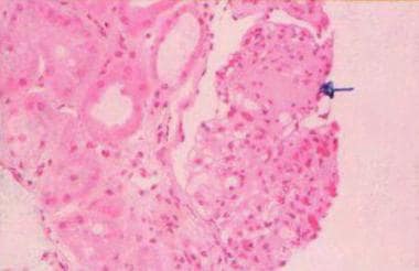
The diagnosis of amyloidosis typically involves a combination of clinical evaluation, imaging studies, and tissue biopsy with the demonstration of amyloid deposition using special stains (e.g., Congo red). Treatment depends on the specific type and extent of organ involvement and may include supportive care, medications to target the underlying cause (e.g., chemotherapy, immunomodulatory agents), and organ transplantation in some cases.
I'm sorry for any confusion, but "Rats, Inbred BUF" is not a standard medical term or abbreviation in human or animal medicine that I'm aware of. It's possible that you may be referring to a specific strain of inbred rats used in scientific research. "BUF" could potentially stand for "Buehler University of Florida," which is a strain of inbred rats developed at the University of Florida. These rats are often used in studies related to cardiovascular and renal physiology. However, I would recommend consulting the original source or contacting a professional in the field to confirm the specific context and accurate definition.
Nutritional status is a concept that refers to the condition of an individual in relation to their nutrient intake, absorption, metabolism, and excretion. It encompasses various aspects such as body weight, muscle mass, fat distribution, presence of any deficiencies or excesses of specific nutrients, and overall health status.
A comprehensive assessment of nutritional status typically includes a review of dietary intake, anthropometric measurements (such as height, weight, waist circumference, blood pressure), laboratory tests (such as serum albumin, total protein, cholesterol levels, vitamin and mineral levels), and clinical evaluation for signs of malnutrition or overnutrition.
Malnutrition can result from inadequate intake or absorption of nutrients, increased nutrient requirements due to illness or injury, or excessive loss of nutrients due to medical conditions. On the other hand, overnutrition can lead to obesity and related health problems such as diabetes, cardiovascular disease, and certain types of cancer.
Therefore, maintaining a good nutritional status is essential for overall health and well-being, and it is an important consideration in the prevention, diagnosis, and treatment of various medical conditions.
Blood protein electrophoresis (BPE) is a laboratory test that separates and measures the different proteins in the blood, such as albumin, alpha-1 globulins, alpha-2 globulins, beta globulins, and gamma globulins. This test is often used to help diagnose or monitor conditions related to abnormal protein levels, such as multiple myeloma, macroglobulinemia, and other plasma cell disorders.
In this test, a sample of the patient's blood is placed on a special gel and an electric current is applied. The proteins in the blood migrate through the gel based on their electrical charge and size, creating bands that can be visualized and measured. By comparing the band patterns to reference ranges, doctors can identify any abnormal protein levels or ratios, which may indicate underlying medical conditions.
It's important to note that while BPE is a useful diagnostic tool, it should be interpreted in conjunction with other clinical findings and laboratory tests for accurate diagnosis and management of the patient's condition.
Thyroxine (T4) is a type of hormone produced and released by the thyroid gland, a small butterfly-shaped endocrine gland located in the front of your neck. It is one of two major hormones produced by the thyroid gland, with the other being triiodothyronine (T3).
Thyroxine plays a crucial role in regulating various metabolic processes in the body, including growth, development, and energy expenditure. Specifically, T4 helps to control the rate at which your body burns calories for energy, regulates protein, fat, and carbohydrate metabolism, and influences the body's sensitivity to other hormones.
T4 is produced by combining iodine and tyrosine, an amino acid found in many foods. Once produced, T4 circulates in the bloodstream and gets converted into its active form, T3, in various tissues throughout the body. Thyroxine has a longer half-life than T3, which means it remains active in the body for a more extended period.
Abnormal levels of thyroxine can lead to various medical conditions, such as hypothyroidism (underactive thyroid) or hyperthyroidism (overactive thyroid). These conditions can cause a range of symptoms, including weight gain or loss, fatigue, mood changes, and changes in heart rate and blood pressure.
Norethandrolone is a synthetic anabolic-androgenic steroid, which is a type of drug that is similar to the male hormone testosterone. It is used in the treatment of conditions such as breast cancer in women, delayed puberty in boys, and wasting syndrome in people with HIV/AIDS. Norethandrolone works by promoting the growth of muscle tissue, increasing appetite, and changing the way the body uses certain nutrients.
It is important to note that anabolic-androgenic steroids are controlled substances in many countries due to their potential for abuse and serious side effects, including liver damage, cardiovascular disease, and hormonal imbalances. They should only be used under the close supervision of a healthcare provider.
Medical Definition of Vitamin A:
Vitamin A is a fat-soluble vitamin that is essential for normal vision, immune function, and cell growth. It is also an antioxidant that helps protect the body's cells from damage caused by free radicals. Vitamin A can be found in two main forms: preformed vitamin A, which is found in animal products such as dairy, fish, and meat, particularly liver; and provitamin A carotenoids, which are found in plant-based foods such as fruits, vegetables, and vegetable oils.
The most active form of vitamin A is retinoic acid, which plays a critical role in the development and maintenance of the heart, lungs, kidneys, and other organs. Vitamin A deficiency can lead to night blindness, dry skin, and increased susceptibility to infections. Chronic vitamin A toxicity can cause nausea, dizziness, headaches, coma, and even death.
Malnutrition is a condition that results from eating a diet in which one or more nutrients are either not enough or are too much such that the body's function is not maintained. It can also refer to a deficiency or excess of vitamins, minerals, protein, energy, and/or water. This condition can have negative effects on physical and mental health. Malnutrition includes undernutrition (wasting, stunting, underweight), overnutrition (overweight, obesity) and micronutrient deficiencies or excesses.
It's important to note that malnutrition is different from malabsorption, which is the inability to absorb nutrients from food. Malabsorption can also lead to malnutrition if it results in a lack of necessary nutrients for the body's function.
Blood proteins, also known as serum proteins, are a group of complex molecules present in the blood that are essential for various physiological functions. These proteins include albumin, globulins (alpha, beta, and gamma), and fibrinogen. They play crucial roles in maintaining oncotic pressure, transporting hormones, enzymes, vitamins, and minerals, providing immune defense, and contributing to blood clotting.
Albumin is the most abundant protein in the blood, accounting for about 60% of the total protein mass. It functions as a transporter of various substances, such as hormones, fatty acids, and drugs, and helps maintain oncotic pressure, which is essential for fluid balance between the blood vessels and surrounding tissues.
Globulins are divided into three main categories: alpha, beta, and gamma globulins. Alpha and beta globulins consist of transport proteins like lipoproteins, hormone-binding proteins, and enzymes. Gamma globulins, also known as immunoglobulins or antibodies, are essential for the immune system's defense against pathogens.
Fibrinogen is a protein involved in blood clotting. When an injury occurs, fibrinogen is converted into fibrin, which forms a mesh to trap platelets and form a clot, preventing excessive bleeding.
Abnormal levels of these proteins can indicate various medical conditions, such as liver or kidney disease, malnutrition, infections, inflammation, or autoimmune disorders. Blood protein levels are typically measured through laboratory tests like serum protein electrophoresis (SPE) and immunoelectrophoresis (IEP).
Hepatic insufficiency, also known as liver insufficiency, refers to the reduced ability of the liver to perform its vital functions due to damage or disease. The liver plays a crucial role in metabolism, detoxification, synthesis, storage, and secretion. When it becomes insufficient, it can lead to various complications such as:
1. Impaired metabolism of carbohydrates, fats, and proteins
2. Buildup of toxic substances in the blood due to reduced detoxification capacity
3. Decreased synthesis of essential proteins, including clotting factors
4. Reduced glycogen storage and impaired glucose regulation
5. Fluid accumulation in the abdomen (ascites) and legs (edema) due to decreased production of albumin and increased pressure in the portal vein
6. Impaired immune function, making the individual more susceptible to infections
7. Hormonal imbalances leading to various symptoms such as changes in appetite, weight loss, and sexual dysfunction
Hepatic insufficiency can range from mild to severe, and if left untreated, it may progress to liver failure, a life-threatening condition requiring immediate medical attention.
A Nutrition Assessment is a systematic and comprehensive evaluation of an individual's nutritional status, which is carried out by healthcare professionals such as registered dietitians or nutritionists. The assessment typically involves collecting and analyzing data related to various factors that influence nutritional health, including:
1. Anthropometric measurements: These include height, weight, waist circumference, blood pressure, and other physical measures that can provide insights into an individual's overall health status and risk of chronic diseases.
2. Dietary intake assessment: This involves evaluating an individual's dietary patterns, food preferences, and eating habits to determine whether they are meeting their nutritional needs through their diet.
3. Biochemical assessments: These include blood tests and other laboratory measures that can provide information about an individual's nutrient status, such as serum levels of vitamins, minerals, and other nutrients.
4. Clinical assessment: This involves reviewing an individual's medical history, current medications, and any symptoms or health conditions that may be impacting their nutritional health.
5. Social and economic assessment: This includes evaluating an individual's access to food, income, education level, and other social determinants of health that can affect their ability to obtain and consume a healthy diet.
The goal of a Nutrition Assessment is to identify any nutritional risks or deficiencies and develop a personalized nutrition plan to address them. This may involve making dietary recommendations, providing education and counseling, or referring the individual to other healthcare professionals for further evaluation and treatment.
Kwashiorkor is a severe form of protein-energy malnutrition characterized by edema (fluid accumulation in the body's tissues), a distended belly, and a weakened immune system. It typically occurs in children between the ages of 1 and 3 who experience a sudden stop in breastfeeding and are switched to a diet that is low in protein but high in carbohydrates. The lack of protein impairs the body's ability to produce essential enzymes and hormones, leading to the characteristic symptoms of Kwashiorkor. It can also result in liver enlargement, skin lesions, hair changes, and impaired growth and development. Immediate medical attention is required for individuals with Kwashiorkor to prevent further complications and promote recovery.
Transferrin is a glycoprotein that plays a crucial role in the transport and homeostasis of iron in the body. It's produced mainly in the liver and has the ability to bind two ferric (Fe3+) ions in its N-lobe and C-lobe, thus creating transferrin saturation.
This protein is essential for delivering iron to cells while preventing the harmful effects of free iron, which can catalyze the formation of reactive oxygen species through Fenton reactions. Transferrin interacts with specific transferrin receptors on the surface of cells, particularly in erythroid precursors and brain endothelial cells, to facilitate iron uptake via receptor-mediated endocytosis.
In addition to its role in iron transport, transferrin also has antimicrobial properties due to its ability to sequester free iron, making it less available for bacterial growth and survival. Transferrin levels can be used as a clinical marker of iron status, with decreased levels indicating iron deficiency anemia and increased levels potentially signaling inflammation or liver disease.
Immunoelectrophoresis (IEP) is a laboratory technique used in the field of clinical pathology and immunology. It is a method for separating and identifying proteins, particularly immunoglobulins or antibodies, in a sample. This technique combines the principles of electrophoresis, which separates proteins based on their electric charge and size, with immunological reactions, which detect specific proteins using antigen-antibody interactions.
In IEP, a protein sample is first separated by electrophoresis in an agarose or agar gel matrix on a glass slide or in a test tube. After separation, an antibody specific to the protein of interest is layered on top of the gel and allowed to diffuse towards the separated proteins. This creates a reaction between the antigen (protein) and the antibody, forming a visible precipitate at the point where they meet. The precipitate line's position and intensity can then be analyzed to identify and quantify the protein of interest.
Immunoelectrophoresis is particularly useful in diagnosing various medical conditions, such as immunodeficiency disorders, monoclonal gammopathies (like multiple myeloma), and other plasma cell dyscrasias. It can help detect abnormal protein patterns, quantify specific immunoglobulins, and identify the presence of M-proteins or Bence Jones proteins, which are indicative of monoclonal gammopathies.
Nephelometry and turbidimetry are methods used in clinical laboratories to measure the amount of particles, such as proteins or cells, present in a liquid sample. The main difference between these two techniques lies in how they detect and quantify the particles.
1. Nephelometry: This is a laboratory method that measures the amount of light scattered by suspended particles in a liquid medium at a 90-degree angle to the path of the incident light. When light passes through a sample containing particles, some of the light is absorbed, while some is scattered in various directions. In nephelometry, a light beam is shone into the sample, and a detector measures the intensity of the scattered light at a right angle to the light source. The more particles present in the sample, the higher the intensity of scattered light, which correlates with the concentration of particles in the sample. Nephelometry is often used to measure the levels of immunoglobulins, complement components, and other proteins in serum or plasma.
2. Turbidimetry: This is another laboratory method that measures the amount of light blocked or absorbed by suspended particles in a liquid medium. In turbidimetry, a light beam is shone through the sample, and the intensity of the transmitted light is measured. The more particles present in the sample, the more light is absorbed or scattered, resulting in lower transmitted light intensity. Turbidimetric measurements are typically reported as percent transmittance, which is the ratio of the intensity of transmitted light to that of the incident light expressed as a percentage. Turbidimetry can be used to measure various substances, such as proteins, cells, and crystals, in body fluids like urine, serum, or plasma.
In summary, nephelometry measures the amount of scattered light at a 90-degree angle, while turbidimetry quantifies the reduction in transmitted light intensity due to particle presence. Both methods are useful for determining the concentration of particles in liquid samples and are commonly used in clinical laboratories for diagnostic purposes.
Thymus hormones, also known as thymic factors or thymic humoral factors, refer to the biologically active molecules secreted by the thymus gland. The two main thymus hormones are thymosin and thymopoietin. These hormones play crucial roles in the differentiation, maturation, and function of T-cells, which are a type of white blood cell responsible for cell-mediated immunity. Thymosin is involved in the maturation of T-cells, helping them to distinguish between self and non-self antigens, while thymopoietin contributes to the differentiation of T-cells into their various subsets and supports their proliferation and activation.
The thymus gland is a primary lymphoid organ located in the upper chest region, anterior to the heart. It plays a critical role in the adaptive immune system, particularly during fetal development and early childhood. The thymus gland begins to atrophy after puberty, leading to a decrease in the production of thymus hormones. This natural decline in thymic function is believed to contribute to the decreased immune response observed in older individuals.
Supplementation with thymus hormones has been explored as a potential therapeutic approach for enhancing immune function in various clinical settings, including immunodeficiency disorders, cancer, and aging. However, more research is needed to fully understand their mechanisms of action and potential benefits and risks.
Nutrition disorders refer to conditions that result from eating, drinking, or absorbing nutrients in a way that is not consistent with human physiological needs. These disorders can manifest as both undernutrition and overnutrition. Undernutrition includes disorders such as protein-energy malnutrition, vitamin deficiencies, and mineral deficiencies, while overnutrition includes conditions such as obesity and diet-related noncommunicable diseases like diabetes, cardiovascular disease, and certain types of cancer.
Malnutrition is the broad term used to describe a state in which a person's nutrient intake is insufficient or excessive, leading to negative consequences for their health. Malnutrition can be caused by a variety of factors, including poverty, food insecurity, lack of education, cultural practices, and chronic diseases.
In addition to under- and overnutrition, disordered eating patterns such as anorexia nervosa, bulimia nervosa, binge eating disorder, and other specified feeding or eating disorders can also be considered nutrition disorders. These conditions are characterized by abnormal eating habits that can lead to serious health consequences, including malnutrition, organ damage, and mental health problems.
Overall, nutrition disorders are complex conditions that can have significant impacts on a person's physical and mental health. They require careful assessment, diagnosis, and treatment by healthcare professionals with expertise in nutrition and dietetics.
Amyloid is a term used in medicine to describe abnormally folded protein deposits that can accumulate in various tissues and organs of the body. These misfolded proteins can form aggregates known as amyloid fibrils, which have a characteristic beta-pleated sheet structure. Amyloid deposits can be composed of different types of proteins, depending on the specific disease associated with the deposit.
In some cases, amyloid deposits can cause damage to organs and tissues, leading to various clinical symptoms. Some examples of diseases associated with amyloidosis include Alzheimer's disease (where amyloid-beta protein accumulates in the brain), systemic amyloidosis (where amyloid fibrils deposit in various organs such as the heart, kidneys, and liver), and type 2 diabetes (where amyloid deposits form in the pancreas).
It's important to note that not all amyloid deposits are harmful or associated with disease. However, when they do cause problems, treatment typically involves managing the underlying condition that is leading to the abnormal protein accumulation.
Ceruloplasmin is a protein found in blood plasma that binds and transports copper ions. It plays a crucial role in copper metabolism, including the oxidation of ferrous iron to ferric iron, which is necessary for the incorporation of iron into transferrin, another protein responsible for transporting iron throughout the body. Ceruloplasmin also acts as an antioxidant by scavenging free radicals and has been implicated in neurodegenerative disorders like Alzheimer's disease and Wilson's disease, a genetic disorder characterized by abnormal copper accumulation in various organs.
Enteral nutrition refers to the delivery of nutrients to a person through a tube that is placed into the gastrointestinal tract, specifically into the stomach or small intestine. This type of nutrition is used when a person is unable to consume food or liquids by mouth due to various medical conditions such as swallowing difficulties, malabsorption, or gastrointestinal disorders.
Enteral nutrition can be provided through different types of feeding tubes, including nasogastric tubes, which are inserted through the nose and down into the stomach, and gastrostomy or jejunostomy tubes, which are placed directly into the stomach or small intestine through a surgical incision.
The nutrients provided through enteral nutrition may include commercially prepared formulas that contain a balance of carbohydrates, proteins, fats, vitamins, and minerals, or blenderized whole foods that are pureed and delivered through the feeding tube. The choice of formula or type of feed depends on the individual's nutritional needs, gastrointestinal function, and medical condition.
Enteral nutrition is a safe and effective way to provide nutrition support to people who are unable to meet their nutritional needs through oral intake alone. It can help prevent malnutrition, promote wound healing, improve immune function, and enhance overall health and quality of life.
Naphthalenesulfonates are a group of chemical compounds that consist of a naphthalene ring, which is a bicyclic aromatic hydrocarbon, substituted with one or more sulfonate groups. Sulfonates are salts or esters of sulfuric acid. Naphthalenesulfonates are commonly used as detergents, dyes, and research chemicals.
In the medical field, naphthalenesulfonates may be used in diagnostic tests to detect certain enzyme activities or metabolic disorders. For example, 1-naphthyl sulfate is a substrate for the enzyme arylsulfatase A, which is deficient in individuals with the genetic disorder metachromatic leukodystrophy. By measuring the activity of this enzyme using 1-naphthyl sulfate as a substrate, doctors can diagnose or monitor the progression of this disease.
It's worth noting that some naphthalenesulfonates have been found to have potential health hazards and environmental concerns. For instance, sodium naphthalenesulfonate has been classified as a possible human carcinogen by the International Agency for Research on Cancer (IARC). Therefore, their use should be handled with caution and in accordance with established safety protocols.
The choroid plexus is a network of blood vessels and tissue located within each ventricle (fluid-filled space) of the brain. It plays a crucial role in the production of cerebrospinal fluid (CSF), which provides protection and nourishment to the brain and spinal cord.
The choroid plexus consists of modified ependymal cells, called plexus epithelial cells, that line the ventricular walls. These cells have finger-like projections called villi, which increase their surface area for efficient CSF production. The blood vessels within the choroid plexus transport nutrients, ions, and water to these epithelial cells, where they are actively secreted into the ventricles to form CSF.
In addition to its role in CSF production, the choroid plexus also acts as a barrier between the blood and the central nervous system (CNS), regulating the exchange of substances between them. This barrier function is primarily attributed to tight junctions present between the epithelial cells, which limit the paracellular movement of molecules.
Abnormalities in the choroid plexus can lead to various neurological conditions, such as hydrocephalus (excessive accumulation of CSF) or certain types of brain tumors.
Nervous system diseases, also known as neurological disorders, refer to a group of conditions that affect the nervous system, which includes the brain, spinal cord, nerves, and muscles. These diseases can affect various functions of the body, such as movement, sensation, cognition, and behavior. They can be caused by genetics, infections, injuries, degeneration, or tumors. Examples of nervous system diseases include Alzheimer's disease, Parkinson's disease, multiple sclerosis, epilepsy, migraine, stroke, and neuroinfections like meningitis and encephalitis. The symptoms and severity of these disorders can vary widely, ranging from mild to severe and debilitating.
Parenteral nutrition (PN) is a medical term used to describe the delivery of nutrients directly into a patient's bloodstream through a vein, bypassing the gastrointestinal tract. It is a specialized medical treatment that is typically used when a patient cannot receive adequate nutrition through enteral feeding, which involves the ingestion and digestion of food through the mouth or a feeding tube.
PN can be used to provide essential nutrients such as carbohydrates, proteins, fats, vitamins, minerals, and electrolytes to patients who have conditions that prevent them from absorbing nutrients through their gut, such as severe gastrointestinal tract disorders, malabsorption syndromes, or short bowel syndrome.
PN is administered through a catheter that is inserted into a vein, typically in the chest or arm. The nutrient solution is prepared under sterile conditions and delivered through an infusion pump to ensure accurate and controlled delivery of the solution.
While PN can be a life-saving intervention for some patients, it also carries risks such as infection, inflammation, and organ damage. Therefore, it should only be prescribed and administered by healthcare professionals with specialized training in this area.
Renal dialysis is a medical procedure that is used to artificially remove waste products, toxins, and excess fluids from the blood when the kidneys are no longer able to perform these functions effectively. This process is also known as hemodialysis.
During renal dialysis, the patient's blood is circulated through a special machine called a dialyzer or an artificial kidney, which contains a semi-permeable membrane that filters out waste products and excess fluids from the blood. The cleaned blood is then returned to the patient's body.
Renal dialysis is typically recommended for patients with advanced kidney disease or kidney failure, such as those with end-stage renal disease (ESRD). It is a life-sustaining treatment that helps to maintain the balance of fluids and electrolytes in the body, prevent the buildup of waste products and toxins, and control blood pressure.
There are two main types of renal dialysis: hemodialysis and peritoneal dialysis. Hemodialysis is the most common type and involves using a dialyzer to filter the blood outside the body. Peritoneal dialysis, on the other hand, involves placing a catheter in the abdomen and using the lining of the abdomen (peritoneum) as a natural filter to remove waste products and excess fluids from the body.
Overall, renal dialysis is an essential treatment option for patients with kidney failure, helping them to maintain their quality of life and prolong their survival.
Immunodiffusion is a laboratory technique used in immunology to detect and measure the presence of specific antibodies or antigens in a sample. It is based on the principle of diffusion, where molecules move from an area of high concentration to an area of low concentration until they reach equilibrium. In this technique, a sample containing an unknown quantity of antigen or antibody is placed in a gel or agar medium that contains a known quantity of antibody or antigen, respectively.
The two substances then diffuse towards each other and form a visible precipitate at the point where they meet and reach equivalence, which indicates the presence and quantity of the specific antigen or antibody in the sample. There are several types of immunodiffusion techniques, including radial immunodiffusion (RID) and double immunodiffusion (Ouchterlony technique). These techniques are widely used in diagnostic laboratories to identify and measure various antigens and antibodies, such as those found in infectious diseases, autoimmune disorders, and allergic reactions.
"Autoanalysis" is not a term that is widely used in the medical field. However, in psychology and psychotherapy, "autoanalysis" refers to the process of self-analysis or self-examination, where an individual analyzes their own thoughts, feelings, behaviors, and experiences to gain insight into their unconscious mind and understand their motivations, conflicts, and emotional patterns.
Self-analysis can involve various techniques such as introspection, journaling, meditation, dream analysis, and reflection on past experiences. While autoanalysis can be a useful tool for personal growth and self-awareness, it is generally considered less reliable and comprehensive than professional psychotherapy or psychoanalysis, which involves a trained therapist or analyst who can provide objective feedback, interpretation, and guidance.
Anthropometry is the scientific study of measurements and proportions of the human body. It involves the systematic measurement and analysis of various physical characteristics, such as height, weight, blood pressure, waist circumference, and other body measurements. These measurements are used in a variety of fields, including medicine, ergonomics, forensics, and fashion design, to assess health status, fitness level, or to design products and environments that fit the human body. In a medical context, anthropometry is often used to assess growth and development, health status, and disease risk factors in individuals and populations.
Total Parenteral Nutrition (TPN) is a medical term used to describe a specialized nutritional support system that is delivered through a vein (intravenously). It provides all the necessary nutrients that a patient needs, such as carbohydrates, proteins, fats, vitamins, and minerals. TPN is typically used when a patient cannot eat or digest food through their gastrointestinal tract for various reasons, such as severe malabsorption, intestinal obstruction, or inflammatory bowel disease. The term "total" indicates that the nutritional support is complete and meets all of the patient's nutritional needs.
Thyroid function tests (TFTs) are a group of blood tests that assess the functioning of the thyroid gland, which is a small butterfly-shaped gland located in the front of the neck. The thyroid gland produces hormones that regulate metabolism, growth, and development in the body.
TFTs typically include the following tests:
1. Thyroid-stimulating hormone (TSH) test: This test measures the level of TSH, a hormone produced by the pituitary gland that regulates the production of thyroid hormones. High levels of TSH may indicate an underactive thyroid gland (hypothyroidism), while low levels may indicate an overactive thyroid gland (hyperthyroidism).
2. Thyroxine (T4) test: This test measures the level of T4, a hormone produced by the thyroid gland. High levels of T4 may indicate hyperthyroidism, while low levels may indicate hypothyroidism.
3. Triiodothyronine (T3) test: This test measures the level of T3, another hormone produced by the thyroid gland. High levels of T3 may indicate hyperthyroidism, while low levels may indicate hypothyroidism.
4. Thyroid peroxidase antibody (TPOAb) test: This test measures the level of TPOAb, an antibody that attacks the thyroid gland and can cause hypothyroidism.
5. Thyroglobulin (Tg) test: This test measures the level of Tg, a protein produced by the thyroid gland. It is used to monitor the treatment of thyroid cancer.
These tests help diagnose and manage various thyroid disorders, including hypothyroidism, hyperthyroidism, thyroiditis, and thyroid cancer.
Disc electrophoresis is a type of electrophoresis technique used to separate and analyze DNA, RNA, or proteins based on their size and electrical charge. In this method, the samples are placed in a gel matrix (usually agarose or polyacrylamide) and an electric field is applied. The smaller and/or more negatively charged molecules migrate faster through the gel and separate from larger and/or less charged molecules, creating a pattern of bands that can be visualized and analyzed.
The term "disc" refers to the characteristic disc-shaped pattern that is often seen in the separated protein bands when using this technique. This pattern is created by the interaction between the size, charge, and shape of the proteins, resulting in a distinct banding pattern that can be used for identification and analysis.
Disc electrophoresis is widely used in molecular biology and genetics research, as well as in diagnostic testing and forensic science.
Iodine isotopes are different forms of the chemical element iodine, which have different numbers of neutrons in their nuclei. Iodine has a total of 53 protons in its nucleus, and its stable isotope, iodine-127, has 74 neutrons, giving it a mass number of 127. However, there are also radioactive isotopes of iodine, which have different numbers of neutrons and are therefore unstable.
Radioactive isotopes of iodine emit radiation as they decay towards a stable state. For example, iodine-131 is a commonly used isotope in medical imaging and therapy, with a half-life of about 8 days. It decays by emitting beta particles and gamma rays, making it useful for treating thyroid cancer and other conditions that involve overactive thyroid glands.
Other radioactive iodine isotopes include iodine-123, which has a half-life of about 13 hours and is used in medical imaging, and iodine-125, which has a half-life of about 60 days and is used in brachytherapy (a type of radiation therapy that involves placing radioactive sources directly into or near tumors).
It's important to note that exposure to radioactive iodine isotopes can be harmful, especially if it occurs through inhalation or ingestion. This is because the iodine can accumulate in the thyroid gland and cause damage over time. Therefore, appropriate safety measures must be taken when handling or working with radioactive iodine isotopes.
In medical terms, "tears" are a clear, salty liquid that is produced by the tear glands (lacrimal glands) in our eyes. They serve to keep the eyes moist, protect against dust and other foreign particles, and help to provide clear vision by maintaining a smooth surface on the front of the eye. Tears consist of water, oil, and mucus, which help to prevent evaporation and ensure that the tears spread evenly across the surface of the eye. Emotional or reflexive responses, such as crying or yawning, can also stimulate the production of tears.
Anilino Naphthalenesulfonates are a group of compounds that contain both aniline and naphthalene sulfonate components. Aniline is a organic compound with the formula C6H5NH2, and naphthalene sulfonate is the sodium salt of naphthalene-1,5-disulfonic acid.
Anilino Naphthalenesulfonates are commonly used as fluorescent dyes in various applications such as histology, microscopy, and flow cytometry. These compounds exhibit strong fluorescence under ultraviolet light and can be used to label and visualize specific structures or molecules of interest. Examples of Anilino Naphthalenesulfonates include Propidium Iodide, Acridine Orange, and Hoechst 33258.
It is important to note that while these compounds are widely used in research and diagnostic settings, they may also have potential hazards and should be handled with appropriate safety precautions.
C-reactive protein (CRP) is a protein produced by the liver in response to inflammation or infection in the body. It is named after its ability to bind to the C-polysaccharide of pneumococcus, a type of bacteria. CRP levels can be measured with a simple blood test and are often used as a marker of inflammation or infection. Elevated CRP levels may indicate a variety of conditions, including infections, tissue damage, and chronic diseases such as rheumatoid arthritis and cancer. However, it is important to note that CRP is not specific to any particular condition, so additional tests are usually needed to make a definitive diagnosis.
Orosomucoid, also known as α-1-acid glycoprotein or AAG, is a protein found in human plasma. It's a member of the acute phase proteins, which are produced in higher amounts during inflammation and infection. Orosomucoid has a molecular weight of approximately 41-43 kDa and is composed of a single polypeptide chain with five N-linked glycosylation sites. It plays a role in protecting tissues from various harmful substances, such as proteases and oxidants, by binding to them and preventing their interaction with cells. Additionally, orosomucoid has been studied as a potential biomarker for several diseases due to its altered levels during inflammation and cancer.
Liver function tests (LFTs) are a group of blood tests that are used to assess the functioning and health of the liver. These tests measure the levels of various enzymes, proteins, and waste products that are produced or metabolized by the liver. Some common LFTs include:
1. Alanine aminotransferase (ALT): An enzyme found primarily in the liver, ALT is released into the bloodstream in response to liver cell damage. Elevated levels of ALT may indicate liver injury or disease.
2. Aspartate aminotransferase (AST): Another enzyme found in various tissues, including the liver, heart, and muscles. Like ALT, AST is released into the bloodstream following tissue damage. High AST levels can be a sign of liver damage or other medical conditions.
3. Alkaline phosphatase (ALP): An enzyme found in several organs, including the liver, bile ducts, and bones. Elevated ALP levels may indicate a blockage in the bile ducts, liver disease, or bone disorders.
4. Gamma-glutamyl transferase (GGT): An enzyme found mainly in the liver, pancreas, and biliary system. Increased GGT levels can suggest liver disease, alcohol consumption, or the use of certain medications.
5. Bilirubin: A yellowish pigment produced when hemoglobin from red blood cells is broken down. Bilirubin is processed by the liver and excreted through bile. High bilirubin levels can indicate liver dysfunction, bile duct obstruction, or certain types of anemia.
6. Albumin: A protein produced by the liver that helps maintain fluid balance in the body and transports various substances in the blood. Low albumin levels may suggest liver damage, malnutrition, or kidney disease.
7. Total protein: A measure of all proteins present in the blood, including albumin and other types of proteins produced by the liver. Decreased total protein levels can indicate liver dysfunction or other medical conditions.
These tests are often ordered together as part of a routine health checkup or when evaluating symptoms related to liver function or disease. The results should be interpreted in conjunction with clinical findings, medical history, and other diagnostic tests.
An encyclopedia is a comprehensive reference work containing articles on various topics, usually arranged in alphabetical order. In the context of medicine, a medical encyclopedia is a collection of articles that provide information about a wide range of medical topics, including diseases and conditions, treatments, tests, procedures, and anatomy and physiology. Medical encyclopedias may be published in print or electronic formats and are often used as a starting point for researching medical topics. They can provide reliable and accurate information on medical subjects, making them useful resources for healthcare professionals, students, and patients alike. Some well-known examples of medical encyclopedias include the Merck Manual and the Stedman's Medical Dictionary.
Gamma-glutamyltransferase (GGT), also known as gamma-glutamyl transpeptidase, is an enzyme found in many tissues, including the liver, bile ducts, and pancreas. GGT is involved in the metabolism of certain amino acids and plays a role in the detoxification of various substances in the body.
GGT is often measured as a part of a panel of tests used to evaluate liver function. Elevated levels of GGT in the blood may indicate liver disease or injury, bile duct obstruction, or alcohol consumption. However, it's important to note that several other factors can also affect GGT levels, so abnormal results should be interpreted in conjunction with other clinical findings and diagnostic tests.
Liver diseases refer to a wide range of conditions that affect the normal functioning of the liver. The liver is a vital organ responsible for various critical functions such as detoxification, protein synthesis, and production of biochemicals necessary for digestion.
Liver diseases can be categorized into acute and chronic forms. Acute liver disease comes on rapidly and can be caused by factors like viral infections (hepatitis A, B, C, D, E), drug-induced liver injury, or exposure to toxic substances. Chronic liver disease develops slowly over time, often due to long-term exposure to harmful agents or inherent disorders of the liver.
Common examples of liver diseases include hepatitis, cirrhosis (scarring of the liver tissue), fatty liver disease, alcoholic liver disease, autoimmune liver diseases, genetic/hereditary liver disorders (like Wilson's disease and hemochromatosis), and liver cancers. Symptoms may vary widely depending on the type and stage of the disease but could include jaundice, abdominal pain, fatigue, loss of appetite, nausea, and weight loss.
Early diagnosis and treatment are essential to prevent progression and potential complications associated with liver diseases.
The liver is a large, solid organ located in the upper right portion of the abdomen, beneath the diaphragm and above the stomach. It plays a vital role in several bodily functions, including:
1. Metabolism: The liver helps to metabolize carbohydrates, fats, and proteins from the food we eat into energy and nutrients that our bodies can use.
2. Detoxification: The liver detoxifies harmful substances in the body by breaking them down into less toxic forms or excreting them through bile.
3. Synthesis: The liver synthesizes important proteins, such as albumin and clotting factors, that are necessary for proper bodily function.
4. Storage: The liver stores glucose, vitamins, and minerals that can be released when the body needs them.
5. Bile production: The liver produces bile, a digestive juice that helps to break down fats in the small intestine.
6. Immune function: The liver plays a role in the immune system by filtering out bacteria and other harmful substances from the blood.
Overall, the liver is an essential organ that plays a critical role in maintaining overall health and well-being.
Prothrombin time (PT) is a medical laboratory test that measures the time it takes for blood to clot. It's often used to evaluate the functioning of the extrinsic and common pathways of the coagulation system, which is responsible for blood clotting. Specifically, PT measures how long it takes for prothrombin (a protein produced by the liver) to be converted into thrombin, an enzyme that converts fibrinogen into fibrin and helps form a clot.
Prolonged PT may indicate a bleeding disorder or a deficiency in coagulation factors, such as vitamin K deficiency or the use of anticoagulant medications like warfarin. It's important to note that PT is often reported with an international normalized ratio (INR), which allows for standardization and comparison of results across different laboratories and reagent types.
Aspartate aminotransferases (ASTs) are a group of enzymes found in various tissues throughout the body, including the heart, liver, and muscles. They play a crucial role in the metabolic process of transferring amino groups between different molecules.
In medical terms, AST is often used as a blood test to measure the level of this enzyme in the serum. Elevated levels of AST can indicate damage or injury to tissues that contain this enzyme, such as the liver or heart. For example, liver disease, including hepatitis and cirrhosis, can cause elevated AST levels due to damage to liver cells. Similarly, heart attacks can also result in increased AST levels due to damage to heart muscle tissue.
It is important to note that an AST test alone cannot diagnose a specific medical condition, but it can provide valuable information when used in conjunction with other diagnostic tests and clinical evaluation.
Neuromyelitis optica (NMO), also known as Devic's disease, is an autoimmune disorder that affects the central nervous system (CNS). It primarily causes inflammation and damage to the optic nerves (which transmit visual signals from the eye to the brain) and the spinal cord. This results in optic neuritis (inflammation of the optic nerve, causing vision loss) and myelitis (inflammation of the spinal cord, leading to motor, sensory, and autonomic dysfunction).
A key feature of NMO is the presence of autoantibodies against aquaporin-4 (AQP4-IgG), a water channel protein found in astrocytes (a type of glial cell) in the CNS. These antibodies play a crucial role in the development of the disease, as they target and damage the AQP4 proteins, leading to inflammation, demyelination (loss of the protective myelin sheath around nerve fibers), and subsequent neurological dysfunction.
NMO is distinct from multiple sclerosis (MS), another autoimmune disorder affecting the CNS, as it has different clinical features, radiological findings, and treatment responses. However, NMO can sometimes be misdiagnosed as MS due to overlapping symptoms in some cases. Accurate diagnosis of NMO is essential for appropriate management and treatment, which often includes immunosuppressive therapies to control the autoimmune response and prevent further damage to the nervous system.
Progressive bulbar palsy (PBP) is a form of motor neuron disease (MND), also known as Amyotrophic Lateral Sclerosis (ALS). It is characterized by the progressive degeneration of the motor neurons in the brainstem, which control vital functions such as swallowing, speaking, chewing, and breathing.
In PBP, these symptoms gradually worsen over time, often resulting in severe disability and ultimately death due to respiratory failure. The progression of the disease can vary from person to person, but it typically advances more slowly than other forms of ALS. There is currently no cure for PBP or any other form of MND, and treatment is focused on managing symptoms and maintaining quality of life.
Aquaporin 4 (AQP4) is a water channel protein that is primarily found in the membranes of astrocytes, which are a type of glial cell in the central nervous system. AQP4 plays a crucial role in the regulation of water homeostasis and the clearance of excess fluid from the brain and spinal cord. It also facilitates the rapid movement of water across the blood-brain barrier and between astrocytes, which is important for maintaining proper neuronal function and protecting the brain from edema or swelling.
Mutations in the AQP4 gene can lead to various neurological disorders, such as neurodegenerative diseases and neuromyelitis optica spectrum disorder (NMOSD), a severe autoimmune condition that affects the optic nerves and spinal cord. In NMOSD, the immune system mistakenly attacks AQP4 proteins, causing inflammation, demyelination, and damage to the nervous tissue.
**Referral:**
A referral in the medical context is the process where a healthcare professional (such as a general practitioner or primary care physician) sends or refers a patient to another healthcare professional who has specialized knowledge and skills to address the patient's specific health condition or concern. This could be a specialist, a consultant, or a facility that provides specialized care. The referral may involve transferring the patient's care entirely to the other professional or may simply be for a consultation and advice.
**Consultation:**
A consultation in healthcare is a process where a healthcare professional seeks the opinion or advice of another professional regarding a patient's medical condition. This can be done in various ways, such as face-to-face meetings, phone calls, or written correspondence. The consulting professional provides their expert opinion to assist in the diagnosis, treatment plan, or management of the patient's condition. The ultimate decision and responsibility for the patient's care typically remain with the referring or primary healthcare provider.
Transverse Myelitis is a neurological disorder that involves inflammation of the spinal cord, leading to damage in both sides of the cord. This results in varying degrees of motor, sensory, and autonomic dysfunction, typically defined by the level of the spine that's affected. Symptoms may include a sudden onset of lower back pain, muscle weakness, paraesthesia or loss of sensation, and bowel/bladder dysfunction. The exact cause is often unknown but can be associated with infections, autoimmune disorders, or other underlying conditions.
I'm sorry for any confusion, but "Suburban Population" is not a medical term. It refers to the people living in suburban areas, which are typically residential districts or communities that lie outside a city center. They are often characterized by single-family homes and less population density than in urban areas. If you have any questions related to medical terminology, I would be happy to help with those!
Autoantibodies are defined as antibodies that are produced by the immune system and target the body's own cells, tissues, or organs. These antibodies mistakenly identify certain proteins or molecules in the body as foreign invaders and attack them, leading to an autoimmune response. Autoantibodies can be found in various autoimmune diseases such as rheumatoid arthritis, lupus, and thyroiditis. The presence of autoantibodies can also be used as a diagnostic marker for certain conditions.
Transthyretin5
- Prealbumin, also called transthyretin, is one of the major proteins in the blood and is produced primarily by the liver. (testing.com)
- 2] "Rat transthyretin (prealbumin). (tcdb.org)
- 3] "Cloning and nucleotide sequencing of transthyretin (prealbumin) cDNA from rat choroid plexus and liver. (tcdb.org)
- 5] "Structure and expression of the rat transthyretin (prealbumin) gene. (tcdb.org)
- 1. Transthyretin, aka prealbumin, is made in the liver and transports what proteins? (jotform.com)
Serum4
- What is the difference between prealbumin, serum/plasma albumin, and urine albumin tests? (testing.com)
- Our aim was to evaluate serum micronutrients and prealbumin in a cohort of 113 solid-cancer patients receiving platinum and taxane compounds during the development and recovery of neuropathy, up to 1 year after finishing treatment. (nih.gov)
- After chemotherapy treatment, the improvement of patients displaying symptomatic neuropathy is related to vitamin E and prealbumin serum levels. (nih.gov)
- The authors refer to the use of serum proteins (albumin, prealbumin, transferrin) to assess the adequacy of nourishment. (cancernetwork.com)
Thyroxine-binding Pr1
- Distribution - Circulating thyroid hormones are greater than 99% bound to plasma proteins, including thyroxine-binding globulin (TBG), thyroxine-binding prealbumin (TBPA), and albumin (TBA), whose capacities and affinities vary for each hormone. (nih.gov)
High prealbumin3
- If you have high prealbumin levels, your provider may order other tests to diagnose your condition. (medlineplus.gov)
- High Prealbumin levels could happen due to Hodgkin's disease, Kidney failure, and alcohol use disorder. (redcliffelabs.com)
- High Prealbumin range can be a sign that can be related to the chronic kidney disease. (healthcaretip.com)
Malnutrition11
- Historically, prealbumin has been ordered to help detect protein-calorie malnutrition and to monitor the effectiveness of parenteral (for example, intravenous) nutrition. (testing.com)
- No current consensus exists on when to get tested, although prealbumin may be ordered, along with assessments of nutritional intake, when a healthcare practitioner suspects that someone is malnourished or is at risk of malnutrition. (testing.com)
- Although commonly used as a marker of malnutrition, research is continuing in order to better understand the role(s) of prealbumin in the body, especially the reasons for changes observed during illness, and the clinical utility of prealbumin testing. (testing.com)
- Until recently, the prealbumin test was believed to be a useful marker of nutritional status and was used to help detect and diagnose protein-calorie malnutrition as well as to monitor people receiving total parenteral nutrition (TPN, getting nutrition via a solution injected into a vein). (testing.com)
- As such, it has been suggested by some health professionals that the prealbumin test should no longer be used to assess nutritional status or diagnose malnutrition. (testing.com)
- With the caveats stated above, a prealbumin test may be ordered by some health care practitioners when signs and symptoms of malnutrition are present or when a person is felt to be at risk for malnutrition, such as during a critical or chronic illness, hospitalization, or when receiving parenteral nutrition or undergoing hemodialysis. (testing.com)
- If your prealbumin levels are lower than normal, it may be a sign of malnutrition . (medlineplus.gov)
- Some medical experts don't think a prealbumin test is the best way to diagnose or monitor malnutrition. (medlineplus.gov)
- Prealbumin may reflect things like inflammation, infections and trauma more accurately than malnutrition. (labwork365.com)
- To detect nutritional health problems, Diagnose malnutrition and other related concerns due to too much or too low Prealbumin levels, Find what kind of nutritional support you might need while undergoing a specific surgery or treatment, To check how much protein you have lost (in the case of people with an eating disorder). (redcliffelabs.com)
- During the screening procedure adopted in Prealbumin tests in order to have an assessment of malnutrition. (healthcaretip.com)
Albumin and prealbumin1
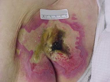
- Laboratory evaluation of her albumin and prealbumin values showed that they were within normal limits. (woundsource.com)
Human Prealbumin1
- 1] "The primary structure of rabbit and rat prealbumin and a comparison with the tertiary structure of human prealbumin. (tcdb.org)
Proteins5
- Prealbumin is one of the principal proteins that is found in the blood. (labwork365.com)
- Prealbumin is a crucial protein that your liver produces to make other proteins. (redcliffelabs.com)
- The liver produces Prealbumin in order to develop the required level of specific proteins for the body. (healthcaretip.com)
- Prealbumin test is suggested to the patients that have nutritional deficiency or have low level of proteins. (healthcaretip.com)
- The results that are associated with the level of Albumin give a long term and clear picture of the nutrition values, whereas the Prealbumin reflects the changes in the level of proteins in shorter time period. (healthcaretip.com)
Creatinine1
- Doctors may order this test along with other tests, such as a blood urea nitrogen test, a creatinine test, and a prealbumin test. (brighthub.com)
Inflammation2
- however, there is controversy because changes in prealbumin may actually reflect other conditions such as inflammation, infection, or trauma. (testing.com)
- For example, measures of inflammation, such as C-reactive protein (CRP) , may be ordered to aid in interpretation of the prealbumin results. (testing.com)
Blood test8
- What is a prealbumin blood test? (medlineplus.gov)
- A prealbumin blood test measures prealbumin levels in your blood. (medlineplus.gov)
- Why do I need a prealbumin blood test? (medlineplus.gov)
- Is there anything else I need to know about a prealbumin blood test? (medlineplus.gov)
- The Prealbumin blood test for nutrition has no fasting requirements. (labwork365.com)
- A Prealbumin blood test determines if a person is getting adequate nutrients in the diet. (redcliffelabs.com)
- So, skip all your delays and book a Prealbumin Blood Test in Faridabad with Redcliffe Labs today. (redcliffelabs.com)
- Prealbumin is a simple blood test that may cause slight pain or a red spot at the punctured site, which goes away quickly. (redcliffelabs.com)
Test18
- Reasons for ordering the prealbumin test are undergoing reassessment as understanding of prealbumin is evolving. (testing.com)
- Newer evidence suggests that the reasons for changes in prealbumin levels may be more complex and the test should not be interpreted as a simple indicator of nutritional status. (testing.com)
- This test measures the level of prealbumin in the blood. (testing.com)
- Given the ongoing discussion of the appropriate use of this test as researchers continue to explore the role of prealbumin in the body and what changes in its level in the body reflect, interpretation of prealbumin results is challenging. (testing.com)
- A high level of prealbumin may be seen in certain conditions, but the test is not used for diagnosis or monitoring in these situations. (testing.com)
- Your provider may order a prealbumin test to help diagnose and monitor problems with your nutritional health. (medlineplus.gov)
- You don't need any special preparations for a prealbumin test. (medlineplus.gov)
- More research is needed to understand how prealbumin works in the body and what abnormal test results may mean. (medlineplus.gov)
- Turnaround time for the Prealbumin test is typically 1-2 business days. (labwork365.com)
- If doctors suspect your body is not getting adequate protein or you have symptoms like sudden weight loss, weakness, joint aches, dizziness, dry, brittle hair, or hair loss, they might suggest Prealbumin Test . (redcliffelabs.com)
- Therefore, if your doctor has suggested this test, Redcliffe Labs, a leading diagnostic centre, is where you should book a Prealbumin Test in Faridabad . (redcliffelabs.com)
- Therefore, with us, Prealbumin Test Cost in Faridabad is Rs 3000 with a free home sample collection. (redcliffelabs.com)
- Prealbumin is a type of test that is carried out to find the level of protein in the diet. (healthcaretip.com)
- The purpose of the Prealbumin test is to make it sure that there is no deficiency of the nutrients inside the body. (healthcaretip.com)
- The levels of the protein in Prealbumin test are measured in milligrams per deciliter. (healthcaretip.com)
- There is an age consideration as well that can influence the range of the Prealbumin test. (healthcaretip.com)
- Moreover, the Prealbumin Test can also indicate that if there is any injury done to the liver because once the liver is damaged it can lower the production of Prealbumin protein. (healthcaretip.com)
- It's called a prealbumin test, not to be confused with an albumin test. (sunwarrior.com)
Clinical1
- Some suggest that a single prealbumin result is less meaningful that a series of measurements taken several days apart, along with other clinical assessments and laboratory tests. (testing.com)
Kidney2
- Prealbumin levels can also be high in Hodgkin disease, kidney failure, iron deficiency, pregnancy, and with hyperactive adrenal glands. (testing.com)
- Levels of prealbumin, C-peptide, and insulin, along with measures of kidney function, were all decreased in symptomatic patients, but not in asymptomatic ones. (medscape.com)
Decrease2
- Drugs such as amiodarone, estrogens, and oral contraceptives (birth control pills) can decrease prealbumin levels. (testing.com)
- By T12, symptomatic patients without recovery had a decrease in vitamin E levels (p = 0.019) and prealbumin (p = 0.062) compared with those symptomatic that improved. (nih.gov)
Plasma1
- 6] "Rat plasma prealbumin. (tcdb.org)
Vitamin2
- Prealbumin helps carry thyroid hormones and vitamin A through your bloodstream. (medlineplus.gov)
- A correlation between the variation of vitamin E and prealbumin at T0-T1 (r = 0.626, p = 0.001) and T1-T12 (r = 0.411, p = 0.06) was observed. (nih.gov)
Levels in your1
- It helps measure Prealbumin levels in your blood and diagnose related diseases. (redcliffelabs.com)
Diagnose1
- Prealbumin testing is not used to diagnose or monitor these conditions. (medlineplus.gov)
Normal3
- Normal prealbumin levels vary according to age and sex. (testing.com)
- If your prealbumin levels are not normal, it doesn't always mean you have a condition that needs treatment. (medlineplus.gov)
- These levels are compared with the normal values of Prealbumin. (healthcaretip.com)
Tests3
- But many providers still find prealbumin blood tests useful for monitoring nutrition, especially in people who are seriously ill or in the hospital. (medlineplus.gov)
- Like the method used in the tests, there are some predefined levels of Prealbumin. (healthcaretip.com)
- The Prealbumin tests can have multiple ranges. (healthcaretip.com)
Names1
- Although the names are similar, prealbumin and albumin are completely different molecules. (testing.com)
Health1
- The levels of the Prealbumin is to be monitored because Prealbumin is a protein that is vital for health and is found in the blood. (healthcaretip.com)
Found2
- Low levels of prealbumin were found to be associated with liver disorders, malignancy, collagen diseases, congestive cardiac failure, and burns. (bmj.com)
- In reality, Prealbumin is a type of protein that is generally found in the liver. (healthcaretip.com)
Values1
- As far as the reliability is concerned, prealbumin is more reliable in having the assessment of nutritional values. (healthcaretip.com)
Patients1
- Many patients and families are asking for levels of prealbumin to be measured but it is not clear whether levels are routinely measured, consistent, or what the measurements can reveal. (agsd.org.uk)
Type1
- Prealbumin is a type of protein that also carries the thyroid hormones. (healthcaretip.com)
Disease1
- Low Prealbumin range is an indication that the patient is having a liver disease. (healthcaretip.com)
Longer1
- Albumin is a protein that has the longer half-life as compared to the Prealbumin. (healthcaretip.com)