Polysomnography
Sleep Apnea, Obstructive
Sleep Apnea Syndromes
Sleep Stages
Sleep
Sleep Disorders
Sleep, REM
Continuous Positive Airway Pressure
Disorders of Excessive Somnolence
Sleep Initiation and Maintenance Disorders
Nocturnal Myoclonus Syndrome
Sleep Apnea, Central
Parasomnias
Wakefulness
Oximetry
Arousal
Severity of Illness Index
Narcolepsy
Cheyne-Stokes Respiration
Monitoring, Ambulatory
Positive-Pressure Respiration
Somnambulism
Respiration
Mandibular Advancement
Rhinomanometry
Restless Legs Syndrome
Sleep Deprivation
Azabicyclo Compounds
Body Mass Index
Circadian Rhythm
Monitoring, Physiologic
Electroencephalography
Respiration Disorders
Sleep Disorders, Circadian Rhythm
Palate, Soft
Prospective Studies
Delta Rhythm
Pierre Robin Syndrome
Cataplexy
Dyssomnias
Diagnosis, Computer-Assisted
Sensitivity and Specificity
Questionnaires
Kleine-Levin Syndrome
Sleep Arousal Disorders
Pharynx
Hypnotics and Sedatives
Oxygen
REM Sleep Behavior Disorder
Electrooculography
Nasal Obstruction
Reproducibility of Results
Fluid Shifts
Comorbidity
Pulmonary Ventilation
Oropharynx
Treatment Outcome
Insomnia, Fatal Familial
Plethysmography, Impedance
Case-Control Studies
Orthodontic Appliances, Removable
Prevalence
Signal Processing, Computer-Assisted
Dreams
Cross-Sectional Studies
Trazodone
Airway Resistance
Orthodontic Appliances
Fatigue
Follow-Up Studies
Nose
Electromyography
Analysis of Variance
Predictive Value of Tests
Statistics, Nonparametric
Respiratory Physiological Phenomena
Palatine Tonsil
Oxyhemoglobins
Placebo Effect
Reference Values
Sudden Infant Death
ROC Curve
Cohort Studies
Risk Factors
Obesity
Masks
Cross-Over Studies
Respiratory Rate
Arnold-Chiari Malformation
Hypercapnia
Double-Blind Method
Age Factors
Tongue
Retrospective Studies
Melatonin
Autonomic Nervous System
Effects of truss mattress upon sleep and bed climate. (1/2955)
The purpose of this study was to examine the effects of a truss mattress upon sleep and bed climate. The truss mattress which has been designed to decrease the pressure and bed climate humidity was tested. Six healthy female volunteers with a mean age of 23.3 years, served as subjects. The experiment was carried out under two conditions: a truss mattress (T) and a futon (F) (Japanese bedding). The ambient temperature and relative humidity were controlled at 19-20 degrees C, and RH 50-60% respectively. Sleep was monitored by an EEG machine and the rectal temperature, skin temperature and bed climate were also measured continuously. Subjective evaluations of bed and sleep were obtained before and after the recording sessions. No significant difference was observed in the sleep parameters and time spent in each sleep stage. Rectal temperature was significantly lower in T than F. Although there was no significant difference in bed climate over the T/F, the temperature under T/F was significantly higher in T. No significant difference was observed in subjective sleep evaluation. The subjective feeling of the mattress was significantly warmer in F than T before sleep. These results suggest that although T does not disturb the sleep parameters and the bed climate is maintained at the same level as with F, it may affect rectal temperature which can be due to low thermal insulation. (+info)Effect of obesity and erect/supine posture on lateral cephalometry: relationship to sleep-disordered breathing. (2/2955)
Craniofacial and upper airway anatomy, obesity and posture may all play a role in compromising upper airway patency in patients with the sleep apnoea/hypopnoea syndrome. The aim of this study was to investigate the relationship between obesity, facial structure and severity of sleep-disordered breathing using lateral cephalometric measurements and to assess the effect of body posture on cephalometric measurements of upper airway calibre variables in obese and non-obese subjects. Lateral cephalometry was carried out in erect and supine postures in 73 awake male subjects randomly selected from patients referred for polysomnography who had a wide range of apnoea/hypopnoea frequencies (1-131 events x h sleep(-1)). Subjects were divided into non-obese (body mass index (BMI) < 30 kg x m(-2); n=42) and obese (BMI > or = 30 kg x m(-2); n=31) groups. Significant but weak correlations were found between apnoea/hypopnoea index (AHI) and measurements reflecting upper airway dimensions: uvular protrusion-posterior pharyngeal wall (r=-0.26, p<0.05) and hyoid-posterior pharyngeal wall (r=0.26, p<0.05). Multiple regression using both upper airway dimensions improved the correlation to AHI (r=0.34, p=0.01). Obese subjects had greater hyoid-posterior pharyngeal wall distances than non-obese subjects, both erect (42+/-5 versus 39+/-4 mm, respectively (mean+/-SD) p<0.01) and supine (43+/-5 versus 40+/-4 mm, p<0.05). Skeletal craniofacial structure was similar in obese and non-obese subjects. In conclusion, measurements reflecting upper airway size were correlated with the severity of sleep-disordered breathing. Differences in upper airway size measurements between obese and non-obese subjects were independent of bony craniofacial structure. (+info)Cephalometric abnormalities in non-obese and obese patients with obstructive sleep apnoea. (3/2955)
The aim of this work was to comprehensively evaluate the cephalometric features in Japanese patients with obstructive sleep apnoea (OSA) and to elucidate the relationship between cephalometric variables and severity of apnoea. Forty-eight cephalometric variables were measured in 37 healthy males and 114 male OSA patients, who were classed into 54 non-obese (body mass index (BMI) <27 kg x m(-2), apnoea-hypopnoea index (AHI)=25.3+/-16.1 events x h(-1)) and 60 obese (BMI > or = 27 kg x m(-2), AHI=45.6+/-28.0 events h(-1)) groups. Diagnostic polysomnography was carried out in all of the OSA patients and in 19 of the normal controls. The non-obese OSA patients showed several cephalometric defects compared with their BMI-matched normal controls: 1) decreased facial A-P distance at cranial base, maxilla and mandible levels and decreased bony pharynx width; 2) enlarged tongue and inferior shift of the tongue volume; 3) enlarged soft palate; 4) inferiorly positioned hyoid bone; and 5) decreased upper airway width at four different levels. More extensive and severe soft tissue abnormalities with a few defects in craniofacial bony structures were found in the obese OSA group. For the non-obese OSA group, the stepwise regression model on AHI was significant with two bony structure variables as determinants: anterior cranial base length (S-N) and mandibular length (Me-Go). Although the regression model retained only linear distance between anterior vertebra and hyoid bone (H-VL) as an explainable determinant for AHI in the obese OSA group, H-VL was significantly correlated with soft tissue measurements such as overall tongue area (Ton), inferior tongue area (Ton2) and pharyngeal airway length (PNS-V). In conclusion, Japanese obstructive sleep apnoea patients have a series of cephalometric abnormalities similar to those described in Caucasian patients, and that the aetiology of obstructive sleep apnoea in obese patients may be different from that in non-obese patients. In obese patients, upper airway soft tissue enlargement may play a more important role in the development of obstructive sleep apnoea, whereas in non-obese patients, bony structure discrepancies may be the dominant contributing factors for obstructive sleep apnoea. (+info)Craniofacial modifications in children with habitual snoring and obstructive sleep apnoea: a case-control study. (4/2955)
Habitual snoring and obstructive sleep apnoea in children, which are frequently associated with adenotonsillar hypertrophy, may begin early in life and in relation with orocraniofacial features. The aim of this study was to detect the presence of early bone craniofacial modifications in young children with a long history of habitual snoring. Twenty-six habitually snoring children (mean age 4.6 yrs) were studied by nocturnal portable recording or diurnal polysomnography, cephalometry and orthodontic evaluation. A comparison of cephalometric findings was made between the studied group and 26 age-matched children (mean age 5.1 yrs) with no history of snoring or respiratory problems during sleep. The cephalometric analyses showed a significant increase in craniomandibular intermaxillar, lower and upper goniac angles with a retroposition and posterior rotation of the mandible (high angle face) and a reduction in the rhinopharynx space caused by higher thickness of adenoids in habitually snoring children compared with controls. Cross-bites and labial incompetence as well as daytime symptoms and familiarity for habitual snoring were found in most of the studied group of snorers compared with controls. The results indicate that upper airway obstruction during sleep is associated with mild but significant cephalometric and craniofacial modifications in children complaining of habitual snoring. Whether this skeletal conformation is genetically determined or influenced by the early onset of habitual snoring remains to be assessed. (+info)The association between sleep apnea and the risk of traffic accidents. Cooperative Group Burgos-Santander. (5/2955)
BACKGROUND AND METHODS: Drowsiness and lack of concentration may contribute to traffic accidents. We conducted a case-control study of the relation between sleep apnea and the risk of traffic accidents. The case patients were 102 drivers who received emergency treatment at hospitals in Burgos or Santander, Spain, after highway traffic accidents between April and December 1995. The controls were 152 patients randomly selected from primary care centers in the same cities and matched with the case patients for age and sex. Respiratory polygraphy was used to screen the patients for sleep apnea at home, and conventional polysomnography was used to confirm the diagnosis. The apnea-hypopnea index (the total number of episodes of apnea and hypopnea divided by the number of hours of sleep) was calculated for each participant. RESULTS: The mean age of the participants was 44 years; 77 percent were men. As compared with those without sleep apnea, patients with an apnea-hypopnea index of 10 or higher had an odds ratio of 6.3 (95 percent confidence interval, 2.4 to 16.2) for having a traffic accident. This relation remained significant after adjustment for potential confounders, such as alcohol consumption, visual-refraction disorders, body-mass index, years of driving, age, history with respect to traffic accidents, use of medications causing drowsiness, and sleep schedule. Among subjects with an apnea-hypopnea index of 10 or more, the risk of an accident was higher among those who had consumed alcohol on the day of the accident than among those who had not. CONCLUSIONS: There is a strong association between sleep apnea, as measured by the apnea-hypopnea index, and the risk of traffic accidents. (+info)Time course of sleep inertia dissipation in human performance and alertness. (6/2955)
Alertness and performance on a wide variety of tasks are impaired immediately upon waking from sleep due to sleep inertia, which has been found to dissipate in an asymptotic manner following waketime. It has been suggested that behavioural or environmental factors, as well as sleep stage at awakening, may affect the severity of sleep inertia. In order to determine the time course of sleep inertia dissipation under normal entrained conditions, subjective alertness and cognitive throughput were measured during the first 4 h after habitual waketime from a full 8-h sleep episode on 3 consecutive days. We investigated whether this time course was affected by either sleep stage at awakening or behavioural/environmental factors. Sleep inertia dissipated in an asymptotic manner and took 2-4 h to near the asymptote. Saturating exponential functions fitted the sleep inertia data well, with time constants of 0.67 h for subjective alertness and 1.17 h for cognitive performance. Most awakenings occurred out of stage rapid eye movement (REM), 2 or 1 sleep, and no effect of sleep stage at awakening on either the severity of sleep inertia or the time course of its dissipation could be detected. Subjective alertness and cognitive throughput were significantly impaired upon awakening regardless of whether subjects got out of bed, ate breakfast, showered and were exposed to ordinary indoor room light (approximately 150 lux) or whether subjects participated in a constant routine (CR) protocol in which they remained in bed, ate small hourly snacks and were exposed to very dim light (10-15 lux). These findings allow for the refinement of models of alertness and performance, and have important implications for the scheduling of work immediately upon awakening in many occupational settings. (+info)Heart period and heart period variability during sleep on the MIR space station. (7/2955)
The long-term acclimation of cardiac rhythms to microgravity was studied in four astronauts aboard the Russian space station MIR during wakefulness and sleep. Sleep polygraphies were obtained between the third and the 30th day in space and, in addition, prior to mission on the ground. From each of the sleep polygraphies, beat-to-beat intervals of cardiac rhythms were determined. The response of heart period and heart period variability to the stimulus microgravity was tested during sleep across sleep stages and during waking. A lengthening of heart period by about 100 ms was found in space compared to measurements on the ground. The slowing of heart rate was more pronounced for non-REM sleep than for REM sleep. A systematic change in heart period in relation to the duration of the stay in space could not be detected. An analysis of heart period variability in the high frequency (respiratory sinus arrhythmia) band supports the hypothesis that the decrease of heart rate under microgravity is produced by an increase in parasympathetic activity. Testing the response of cardiac rhythms to microgravity across distinct behavioural states seems to be a powerful tool to investigate the cardiovascular system. (+info)Prediction of sleep-disordered breathing by unattended overnight oximetry. (8/2955)
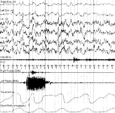
Between January 1994 and July 1997, 793 patients suspected of having sleep-disordered breathing had unattended overnight oximetry in their homes followed by laboratory polysomnography. From the oximetry data we extracted cumulative percentage time at SaO2 < 90% (CT90) and a saturation variability index (delta Index, the sum of the differences between successive readings divided by the number of readings - 1). CT90 was weakly correlated with polysomnographic apnea/hypopnea index (AHI). (Spearman rho = 0.36, P < 0.0001) and with delta Index (rho = 0.71, P < 0.0001). delta Index was more closely correlated with AHI (rho = 0.59, P < 0.0001). In a multivariate model, only delta Index was significantly related to AHI, the relationship being AHI = 18.8 delta Index + 7.7. The 95% CI for the coefficient were 16.2, 21.4, and for the constant were 5.8, 9.7. The sensitivity of a delta Index cut-off of 0.4 for the detection of AHI > or = 15 was 88%, for detection of AHI > or = 20 was 90% and for the detection of AHI > or = 25 was 91%. The specificity of delta Index > or = 0.4 for AHI > or = 15 was 40%. In 113 further patients, oximetry was performed simultaneously with laboratory polysomnography. Under these circumstances delta Index was more closely correlated with AHI (rho = 0.74, P < 0.0001), as was CT90 (rho = 0.58, P < 0.0001). Sensitivity of delta Index > or = 0.4 for detection of AHI > or = 15 was not improved at 88%, but specificity was better at 70%. We concluded that oximetry using a saturation variability index is sensitive but nonspecific for the detection of obstructive sleep apnea, and that few false negative but a significant proportion of false positive results arise from night-to-night variability. (+info)Polysomnography (PSG) is a comprehensive sleep study that monitors various body functions during sleep, including brain activity, eye movement, muscle tone, heart rate, respirations, and oxygen levels. It is typically conducted in a sleep laboratory under the supervision of a trained technologist. The data collected during PSG is used to diagnose and manage various sleep disorders such as sleep-related breathing disorders (e.g., sleep apnea), movement disorders (e.g., periodic limb movement disorder), parasomnias, and narcolepsy.
The study usually involves the attachment of electrodes to different parts of the body, such as the scalp, face, chest, and legs, to record electrical signals from the brain, eye movements, muscle activity, and heartbeats. Additionally, sensors may be placed on or near the nose and mouth to measure airflow, and a belt may be worn around the chest and abdomen to monitor breathing efforts. Oxygen levels are also monitored through a sensor attached to the finger or ear.
Polysomnography is often recommended when a sleep disorder is suspected based on symptoms or medical history, and other diagnostic tests have been inconclusive. The results of the study can help guide treatment decisions and improve overall sleep health.
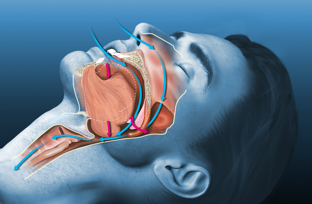
Obstructive Sleep Apnea (OSA) is a sleep-related breathing disorder that occurs when the upper airway becomes partially or completely blocked during sleep, leading to pauses in breathing or shallow breaths. These episodes, known as apneas or hypopneas, can last for 10 seconds or longer and may occur multiple times throughout the night, disrupting normal sleep patterns and causing oxygen levels in the blood to drop.
The obstruction in OSA is typically caused by the relaxation of the muscles in the back of the throat during sleep, which allows the soft tissues to collapse and block the airway. This can result in snoring, choking, gasping for air, or awakening from sleep with a start.
Contributing factors to OSA may include obesity, large neck circumference, enlarged tonsils or adenoids, alcohol consumption, smoking, and use of sedatives or muscle relaxants. Untreated OSA can lead to serious health consequences such as high blood pressure, heart disease, stroke, diabetes, and cognitive impairment. Treatment options for OSA include lifestyle changes, oral appliances, positive airway pressure therapy, and surgery.
Sleep apnea syndromes refer to a group of disorders characterized by abnormal breathing patterns during sleep. These patterns can result in repeated pauses in breathing (apneas) or shallow breaths (hypopneas), causing interruptions in sleep and decreased oxygen supply to the body. There are three main types of sleep apnea syndromes:
1. Obstructive Sleep Apnea (OSA): This is the most common form, caused by the collapse or obstruction of the upper airway during sleep, often due to relaxation of the muscles in the throat and tongue.
2. Central Sleep Apnea (CSA): This type is less common and results from the brain's failure to send proper signals to the breathing muscles. It can be associated with conditions such as heart failure, stroke, or certain medications.
3. Complex/Mixed Sleep Apnea: In some cases, a person may experience both obstructive and central sleep apnea symptoms, known as complex or mixed sleep apnea.
Symptoms of sleep apnea syndromes can include loud snoring, excessive daytime sleepiness, fatigue, morning headaches, difficulty concentrating, and mood changes. Diagnosis typically involves a sleep study (polysomnography) to monitor breathing patterns, heart rate, brain activity, and other physiological factors during sleep. Treatment options may include lifestyle modifications, oral appliances, positive airway pressure therapy, or even surgery in severe cases.
Sleep stages are distinct patterns of brain activity that occur during sleep, as measured by an electroencephalogram (EEG). They are part of the sleep cycle and are used to describe the different types of sleep that humans go through during a normal night's rest. The sleep cycle includes several repeating stages:
1. Stage 1 (N1): This is the lightest stage of sleep, where you transition from wakefulness to sleep. During this stage, muscle activity and brain waves begin to slow down.
2. Stage 2 (N2): In this stage, your heart rate slows, body temperature decreases, and eye movements stop. Brain wave activity becomes slower, with occasional bursts of electrical activity called sleep spindles.
3. Stage 3 (N3): Also known as deep non-REM sleep, this stage is characterized by slow delta waves. It is during this stage that the body undergoes restorative processes such as tissue repair, growth, and immune function enhancement.
4. REM (Rapid Eye Movement) sleep: This is the stage where dreaming typically occurs. Your eyes move rapidly beneath closed eyelids, heart rate and respiration become irregular, and brain wave activity increases to levels similar to wakefulness. REM sleep is important for memory consolidation and learning.
The sleep cycle progresses through these stages multiple times during the night, with REM sleep periods becoming longer towards morning. Understanding sleep stages is crucial in diagnosing and treating various sleep disorders.
Sleep is a complex physiological process characterized by altered consciousness, relatively inhibited sensory activity, reduced voluntary muscle activity, and decreased interaction with the environment. It's typically associated with specific stages that can be identified through electroencephalography (EEG) patterns. These stages include rapid eye movement (REM) sleep, associated with dreaming, and non-rapid eye movement (NREM) sleep, which is further divided into three stages.
Sleep serves a variety of functions, including restoration and strengthening of the immune system, support for growth and development in children and adolescents, consolidation of memory, learning, and emotional regulation. The lack of sufficient sleep or poor quality sleep can lead to significant health problems, such as obesity, diabetes, cardiovascular disease, and even cognitive decline.
The American Academy of Sleep Medicine (AASM) defines sleep as "a period of daily recurring natural rest during which consciousness is suspended and metabolic processes are reduced." However, it's important to note that the exact mechanisms and purposes of sleep are still being researched and debated among scientists.
Sleep disorders are a group of conditions that affect the ability to sleep well on a regular basis. They can include problems with falling asleep, staying asleep, or waking up too early in the morning. These disorders can be caused by various factors such as stress, anxiety, depression, medical conditions, or substance abuse.
The American Academy of Sleep Medicine (AASM) recognizes over 80 distinct sleep disorders, which are categorized into the following major groups:
1. Insomnia - difficulty falling asleep or staying asleep.
2. Sleep-related breathing disorders - abnormal breathing during sleep such as obstructive sleep apnea.
3. Central disorders of hypersomnolence - excessive daytime sleepiness, including narcolepsy.
4. Circadian rhythm sleep-wake disorders - disruption of the internal body clock that regulates the sleep-wake cycle.
5. Parasomnias - abnormal behaviors during sleep such as sleepwalking or night terrors.
6. Sleep-related movement disorders - repetitive movements during sleep such as restless legs syndrome.
7. Isolated symptoms and normal variants - brief and occasional symptoms that do not warrant a specific diagnosis.
Sleep disorders can have significant impacts on an individual's quality of life, productivity, and overall health. If you suspect that you may have a sleep disorder, it is recommended to consult with a healthcare professional or a sleep specialist for proper evaluation and treatment.
REM sleep, or Rapid Eye Movement sleep, is a stage of sleep characterized by rapid eye movements, low muscle tone, and active brain activity. It is one of the two main types of sleep along with non-REM sleep and is marked by vivid dreaming, increased brain metabolism, and altered brain wave patterns. REM sleep is often referred to as "paradoxical sleep" because of the seemingly contradictory nature of its characteristics - an active brain in a state of relaxation. It is thought to play a role in memory consolidation, learning, and mood regulation. A typical night's sleep cycle includes several episodes of REM sleep, with each episode becoming longer as the night progresses.
Snoring is defined as the vibration of respiratory structures and the resulting sound, due to obstructed air movement during breathing while sleeping. It occurs when the tissues at the back of the throat relax and narrow during sleep, partially blocking the airway. The airflow causes these tissues to vibrate, leading to the snoring sound. Snoring can be a sign of various conditions such as obstructive sleep apnea or other respiratory disorders. It can also be influenced by factors such as alcohol consumption, obesity, and sleeping position.
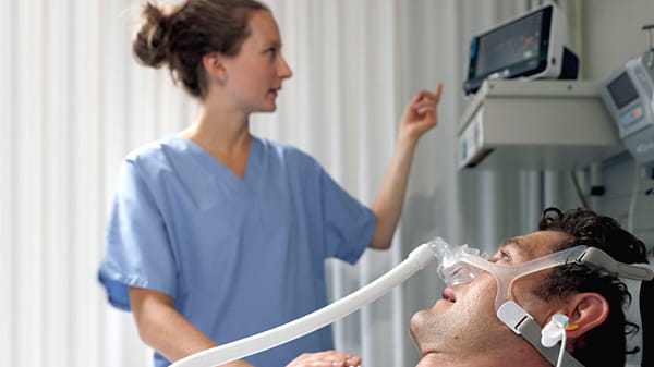
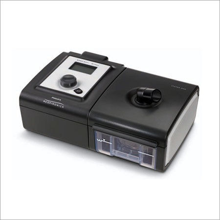
Continuous Positive Airway Pressure (CPAP) is a mode of non-invasive ventilation that delivers pressurized room air or oxygen to maintain airway patency and increase functional residual capacity in patients with respiratory disorders. A CPAP device, which typically includes a flow generator, tubing, and a mask, provides a constant positive pressure throughout the entire respiratory cycle, preventing the collapse of the upper airway during inspiration and expiration.
CPAP is commonly used to treat obstructive sleep apnea (OSA), a condition characterized by repetitive narrowing or closure of the upper airway during sleep, leading to intermittent hypoxia, hypercapnia, and sleep fragmentation. By delivering positive pressure, CPAP helps to stent open the airway, ensuring unobstructed breathing and reducing the frequency and severity of apneic events.
Additionally, CPAP can be used in other clinical scenarios, such as managing acute respiratory distress syndrome (ARDS), chronic obstructive pulmonary disease (COPD) exacerbations, or postoperative respiratory insufficiency, to improve oxygenation and reduce the work of breathing. The specific pressure settings and device configurations are tailored to each patient's needs based on their underlying condition, severity of symptoms, and response to therapy.
Disorders of excessive somnolence (DES) are a group of medical conditions characterized by an increased tendency to fall asleep or experience excessive daytime sleepiness (EDS), despite having adequate opportunity and circumstances for sleep. These disorders are typically classified as central disorders of hypersomnolence according to the International Classification of Sleep Disorders (ICSD-3).
The most common DES is narcolepsy, a chronic neurological disorder caused by the brain's inability to regulate sleep-wake cycles normally. Other DES include idiopathic hypersomnia, Kleine-Levin syndrome, and recurrent hypersomnia. These disorders can significantly impact an individual's daily functioning, quality of life, and overall health.
Narcolepsy is further divided into two types: narcolepsy type 1 (NT1) and narcolepsy type 2 (NT2). NT1 is characterized by the presence of cataplexy, a sudden loss of muscle tone triggered by strong emotions, while NT2 does not include cataplexy. Both types of narcolepsy involve excessive daytime sleepiness, sleep paralysis, hypnagogic/hypnopompic hallucinations, and fragmented nighttime sleep.
Idiopathic hypersomnia is a DES without the presence of REM-related symptoms like cataplexy or sleep paralysis. Individuals with idiopathic hypersomnia experience excessive daytime sleepiness and prolonged nighttime sleep, often lasting 10 to 14 hours, but do not feel refreshed upon waking.
Kleine-Levin syndrome is a rare DES characterized by recurrent episodes of excessive sleepiness, often accompanied by cognitive impairment, altered perception, hyperphagia (excessive eating), and hypersexuality during the episodes. These episodes can last days to weeks and typically occur multiple times per year.
Recurrent hypersomnia is another rare DES with recurring episodes of excessive sleepiness lasting for several days, followed by a period of normal or reduced sleepiness. The episodes are not as predictable or consistent as those seen in Kleine-Levin syndrome.
Treatment for DES typically involves pharmacological interventions to manage symptoms and improve daytime alertness. Modafinil, armodafinil, and traditional stimulants like amphetamine salts are commonly used to treat excessive daytime sleepiness. Additionally, antidepressants may be prescribed to manage REM-related symptoms like cataplexy or sleep paralysis. Non-pharmacological interventions, such as scheduled napping and good sleep hygiene practices, can also help improve symptoms.
Sleep initiation and maintenance disorders are a category of sleep disorders that involve difficulty falling asleep and staying asleep throughout the night. This category includes:
1. Insomnia disorder: A persistent difficulty in initiating or maintaining sleep, or early morning awakening, despite adequate opportunity and circumstances for sleep, which causes clinically significant distress or impairment.
2. Narcolepsy: A chronic neurological disorder characterized by excessive daytime sleepiness, cataplexy (sudden loss of muscle tone triggered by strong emotions), hypnagogic hallucinations (vivid, dream-like experiences that occur while falling asleep) and sleep paralysis (temporary inability to move or speak while falling asleep or waking up).
3. Breathing-related sleep disorders: A group of disorders that involve abnormal breathing patterns during sleep, such as obstructive sleep apnea and central sleep apnea, which can lead to difficulty initiating and maintaining sleep.
4. Circadian rhythm sleep-wake disorders: A group of disorders that involve a misalignment between the individual's internal circadian rhythm and the external environment, leading to difficulty falling asleep and staying asleep at desired times.
5. Parasomnias: A group of disorders that involve abnormal behaviors or experiences during sleep, such as sleepwalking, night terrors, and REM sleep behavior disorder, which can disrupt sleep initiation and maintenance.
These disorders can have significant impacts on an individual's quality of life, daytime functioning, and overall health, and should be evaluated and managed by a healthcare professional with expertise in sleep medicine.
Nocturnal Myoclonus Syndrome, also known as Periodic Limb Movement Disorder (PLMD), is a condition characterized by recurring involuntary jerking movements of the limbs during sleep, particularly the legs. These movements typically occur every 20-40 seconds and can last for an hour or more throughout the night. They often disrupt normal sleep patterns, causing insomnia or excessive daytime sleepiness.
The movements are usually jerky, rapid, and rhythmic, involving extension of the big toe and flexion of the ankle, knee, or hip. In some cases, these movements can be so forceful that they cause the person to wake up, although often individuals with this condition may not be aware of their nighttime leg movements.
Nocturnal Myoclonus Syndrome is different from another common sleep disorder called Restless Legs Syndrome (RLS), as RLS primarily causes discomfort or an irresistible urge to move the legs while awake and still, whereas Nocturnal Myoclonus Syndrome involves involuntary movements during sleep. However, up to 80% of people with RLS also have PLMD.
The exact cause of Nocturnal Myoclonus Syndrome is not fully understood, but it may be associated with abnormalities in the brain's regulation of muscle activity during sleep. Certain medications, neurological conditions, and iron deficiency anemia have been linked to an increased risk of developing this disorder. Treatment options include medication, lifestyle changes, and addressing any underlying medical conditions that may contribute to the development or worsening of symptoms.
Adenoidectomy is a surgical procedure in which the adenoids are removed. The adenoids are a patch of tissue located behind the nasal cavity, near the roof of the mouth. They help to filter out germs that are breathed in through the nose. However, sometimes the adenoids can become enlarged or infected, leading to problems such as difficulty breathing through the nose, recurrent ear infections, and sleep apnea. In these cases, an adenoidectomy may be recommended to remove the adenoids and alleviate these symptoms.
The procedure is typically performed on an outpatient basis, which means that the patient can go home the same day as the surgery. The surgeon will use a special instrument to remove the adenoids through the mouth, without making any external incisions. After the surgery, the patient may experience some discomfort, sore throat, and difficulty swallowing for a few days. However, these symptoms usually resolve within a week or two.
It is important to note that an adenoidectomy is not the same as a tonsillectomy, which is the surgical removal of the tonsils. While the tonsils and adenoids are both part of the immune system and located in the same area of the mouth, they serve different functions and may be removed separately or together depending on the individual's medical needs.
Central sleep apnea (CSA) is a type of sleep-disordered breathing characterized by repeated cessations in breathing during sleep due to the brain's failure to transmit signals to the respiratory muscles that control breathing. Unlike obstructive sleep apnea (OSA), which results from airway obstruction, CSA occurs when the brain fails to send the necessary signals to the diaphragm and intercostal muscles to initiate or maintain respiratory efforts during sleep.
Central sleep apneas are usually associated with decreased oxygen saturation levels and can lead to frequent arousals from sleep, causing excessive daytime sleepiness, fatigue, and impaired cognitive function. CSA is often related to underlying medical conditions such as heart failure, stroke, or brainstem injury, and it may also be caused by the use of certain medications, including opioids.
There are several types of central sleep apnea, including:
1. Primary Central Sleep Apnea: This type occurs without any underlying medical condition or medication use.
2. Cheyne-Stokes Breathing: A pattern of central sleep apnea commonly seen in individuals with heart failure or stroke. It is characterized by a crescendo-decrescendo pattern of breathing, with periods of hyperventilation followed by hypoventilation and apnea.
3. High-Altitude Periodic Breathing: This type occurs at high altitudes due to the reduced oxygen levels and is usually reversible upon returning to lower altitudes.
4. Complex or Mixed Sleep Apnea: A combination of both central and obstructive sleep apneas, often observed in patients with OSA who are treated with continuous positive airway pressure (CPAP) therapy. In some cases, the central component may resolve over time with continued CPAP use.
Diagnosis of CSA typically involves a sleep study (polysomnography), which monitors various physiological parameters during sleep, such as brain waves, eye movements, muscle activity, heart rate, and breathing patterns. Treatment options for central sleep apnea depend on the underlying cause and may include medications, adjustments in medication dosages, or the use of devices that assist with breathing, such as adaptive servo-ventilation (ASV) or bilevel positive airway pressure (BiPAP) therapy.
Actigraphy is a non-invasive method used to estimate sleep-wake patterns and physical activity levels over extended periods, typically ranging from several days to weeks. It involves the use of a small device called an actigraph, which is usually worn on the wrist like a watch.
The actigraph contains an accelerometer that detects movement and records the intensity and duration of motion. This data is then analyzed using specialized software to provide information about sleep and wake times, as well as patterns of physical activity.
Actigraphy can be useful in assessing various sleep disorders, such as insomnia, circadian rhythm disorders, and sleep-related breathing disorders. It can also help evaluate the effectiveness of treatments for these conditions. However, it is important to note that actigraphy is not a substitute for a formal sleep study (polysomnography) and should be used in conjunction with other assessment tools and clinical evaluations.
A tonsillectomy is a surgical procedure in which the tonsils, two masses of lymphoid tissue located on both sides of the back of the throat, are removed. This procedure is typically performed to treat recurrent or severe cases of tonsillitis (inflammation of the tonsils), sleep-disordered breathing such as obstructive sleep apnea, and other conditions where the tonsils are causing problems or complications. The surgery can be done under general anesthesia, and there are various methods for removing the tonsils, including traditional scalpel excision, electrocautery, and laser surgery. After a tonsillectomy, patients may experience pain, swelling, and difficulty swallowing, but these symptoms typically improve within 1-2 weeks post-surgery.
Parasomnias are a category of sleep disorders that involve unwanted physical events or experiences that occur while falling asleep, sleeping, or waking up. These behaviors can include abnormal movements, talk, emotions, perceptions, or dreams. Parasomnias can be caused by various factors such as stress, alcohol, certain medications, or underlying medical conditions. Some examples of parasomnias are sleepwalking, night terrors, sleep talking, and REM sleep behavior disorder. These disorders can disrupt sleep and cause distress to the individual and their bed partner.
Wakefulness is a state of consciousness in which an individual is alert and aware of their surroundings. It is characterized by the ability to perceive, process, and respond to stimuli in a purposeful manner. In a medical context, wakefulness is often assessed using measures such as the electroencephalogram (EEG) to evaluate brain activity patterns associated with consciousness.
Wakefulness is regulated by several interconnected neural networks that promote arousal and attention. These networks include the ascending reticular activating system (ARAS), which consists of a group of neurons located in the brainstem that project to the thalamus and cerebral cortex, as well as other regions involved in regulating arousal and attention, such as the basal forebrain and hypothalamus.
Disorders of wakefulness can result from various underlying conditions, including neurological disorders, sleep disorders, medication side effects, or other medical conditions that affect brain function. Examples of such disorders include narcolepsy, insomnia, hypersomnia, and various forms of encephalopathy or brain injury.
Pulse oximetry is a noninvasive method for monitoring a person's oxygen saturation (SO2) and pulse rate. It uses a device called a pulse oximeter, which measures the amount of oxygen-carrying hemoglobin in the blood compared to the amount of hemoglobin that is not carrying oxygen. This measurement is expressed as a percentage, known as oxygen saturation (SpO2). Normal oxygen saturation levels are generally 95% or above at sea level. Lower levels may indicate hypoxemia, a condition where there is not enough oxygen in the blood to meet the body's needs. Pulse oximetry is commonly used in hospitals and other healthcare settings to monitor patients during surgery, in intensive care units, and in sleep studies to detect conditions such as sleep apnea. It can also be used by individuals with certain medical conditions, such as chronic obstructive pulmonary disease (COPD), to monitor their oxygen levels at home.
In a medical or physiological context, "arousal" refers to the state of being awake and responsive to stimuli. It involves the activation of the nervous system, particularly the autonomic nervous system, which prepares the body for action. Arousal levels can vary from low (such as during sleep) to high (such as during states of excitement or stress). In clinical settings, changes in arousal may be assessed to help diagnose conditions such as coma, brain injury, or sleep disorders. It is also used in the context of sexual response, where it refers to the level of physical and mental awareness and readiness for sexual activity.
A Severity of Illness Index is a measurement tool used in healthcare to assess the severity of a patient's condition and the risk of mortality or other adverse outcomes. These indices typically take into account various physiological and clinical variables, such as vital signs, laboratory values, and co-morbidities, to generate a score that reflects the patient's overall illness severity.
Examples of Severity of Illness Indices include the Acute Physiology and Chronic Health Evaluation (APACHE) system, the Simplified Acute Physiology Score (SAPS), and the Mortality Probability Model (MPM). These indices are often used in critical care settings to guide clinical decision-making, inform prognosis, and compare outcomes across different patient populations.
It is important to note that while these indices can provide valuable information about a patient's condition, they should not be used as the sole basis for clinical decision-making. Rather, they should be considered in conjunction with other factors, such as the patient's overall clinical presentation, treatment preferences, and goals of care.
Narcolepsy is a chronic neurological disorder that affects the control of sleep and wakefulness. It's characterized by excessive daytime sleepiness (EDS), where people experience sudden, uncontrollable episodes of falling asleep during the day. These "sleep attacks" can occur at any time - while working, talking, eating, or even driving.
In addition to EDS, narcolepsy often includes cataplexy, a condition that causes loss of muscle tone, leading to weakness and sometimes collapse, often triggered by strong emotions like laughter or surprise. Other common symptoms are sleep paralysis (a temporary inability to move or speak while falling asleep or waking up), vivid hallucinations during the transitions between sleep and wakefulness, and fragmented nighttime sleep.
The exact cause of narcolepsy is not fully understood, but it's believed to involve genetic and environmental factors, as well as problems with certain neurotransmitters in the brain, such as hypocretin/orexin, which regulate sleep-wake cycles. Narcolepsy can significantly impact a person's quality of life, making it essential to seek medical attention for proper diagnosis and management.
Cheyne-Stokes respiration is a pattern of breathing characterized by cyclical changes in the depth and rate of respirations. It is often associated with various medical conditions that affect the brainstem, such as stroke, brain injury, or certain neurological disorders.
In Cheyne-Stokes respiration, the individual's breathing starts with a series of deeper and faster breaths (hyperventilation), which gradually become shallower and slower (hypoventilation). This cycle repeats every few minutes, resulting in a pattern of waxing and waning of the depth and rate of respirations.
The underlying mechanism for Cheyne-Stokes respiration is related to the regulation of breathing by the brainstem. When there are abnormalities in this area, it can lead to instability in the control of breathing, resulting in the cyclical pattern of hyperventilation and hypoventilation.
Cheyne-Stokes respiration can be a sign of serious underlying medical conditions, and it is important to seek medical attention if you or someone else experiences this type of breathing pattern. Treatment may involve addressing the underlying cause, such as managing heart failure or reducing intracranial pressure in patients with brain injury or stroke.
Ambulatory monitoring is a medical practice that involves the continuous or intermittent recording of physiological parameters in a patient who is mobile and able to perform their usual activities while outside of a hospital or clinical setting. This type of monitoring allows healthcare professionals to evaluate a patient's condition over an extended period, typically 24 hours or more, in their natural environment.
Ambulatory monitoring can be used to diagnose and manage various medical conditions such as hypertension, cardiac arrhythmias, sleep disorders, and mobility issues. Common methods of ambulatory monitoring include:
1. Holter monitoring: A small, portable device that records the electrical activity of the heart for 24-48 hours or more.
2. Ambulatory blood pressure monitoring (ABPM): A device that measures blood pressure at regular intervals throughout the day and night.
3. Event monitors: Devices that record heart rhythms only when symptoms occur or when activated by the patient.
4. Actigraphy: A non-invasive method of monitoring sleep-wake patterns, physical activity, and circadian rhythms using a wristwatch-like device.
5. Continuous glucose monitoring (CGM): A device that measures blood sugar levels continuously throughout the day and night.
Overall, ambulatory monitoring provides valuable information about a patient's physiological status in their natural environment, allowing healthcare professionals to make informed decisions regarding diagnosis, treatment, and management of medical conditions.
Positive-pressure respiration is a type of mechanical ventilation where positive pressure is applied to the airway and lungs, causing them to expand and inflate. This can be used to support or replace spontaneous breathing in patients who are unable to breathe effectively on their own due to conditions such as respiratory failure, neuromuscular disorders, or sedation for surgery.
During positive-pressure ventilation, a mechanical ventilator delivers breaths to the patient through an endotracheal tube or a tracheostomy tube. The ventilator is set to deliver a specific volume or pressure of air with each breath, and the patient's breathing is synchronized with the ventilator to ensure proper delivery of the breaths.
Positive-pressure ventilation can help improve oxygenation and remove carbon dioxide from the lungs, but it can also have potential complications such as barotrauma (injury to lung tissue due to excessive pressure), volutrauma (injury due to overdistention of the lungs), hemodynamic compromise (decreased blood pressure and cardiac output), and ventilator-associated pneumonia. Therefore, careful monitoring and adjustment of ventilator settings are essential to minimize these risks and provide safe and effective respiratory support.
Somnambulism is defined as a parasomnia, which is a type of sleep disorder, that involves walking or performing other complex behaviors while asleep. It's more commonly known as sleepwalking. During a sleepwalking episode, a person will have their eyes open and may appear to be awake and aware of their surroundings, but they are actually in a state of low consciousness.
Sleepwalking can range from simply sitting up in bed and looking around, to walking around the house, dressing or undressing, or even leaving the house. Episodes usually occur during deep non-REM sleep early in the night and can last from several minutes to an hour.
Although it is more common in children, especially those between the ages of 3 and 7, somnambulism can also affect adults. Factors that may contribute to sleepwalking include stress, fatigue, fever, certain medications, alcohol consumption, and underlying medical or psychiatric conditions such as sleep apnea, restless leg syndrome, gastroesophageal reflux disease (GERD), post-traumatic stress disorder (PTSD), or dissociative states.
Most of the time, somnambulism is not a cause for concern and does not require treatment. However, if sleepwalking leads to potential harm or injury, or if it frequently disrupts sleep, medical advice should be sought to address any underlying conditions and ensure safety measures are in place during sleep.
Hypoventilation is a medical condition that refers to the decreased rate and depth of breathing, which leads to an inadequate exchange of oxygen and carbon dioxide in the lungs. As a result, there is an increase in the levels of carbon dioxide (hypercapnia) and a decrease in the levels of oxygen (hypoxemia) in the blood. Hypoventilation can occur due to various reasons such as respiratory muscle weakness, sedative or narcotic overdose, chest wall deformities, neuromuscular disorders, obesity hypoventilation syndrome, and sleep-disordered breathing. Prolonged hypoventilation can lead to serious complications such as respiratory failure, cardiac arrhythmias, and even death.
Medical Definition of Respiration:
Respiration, in physiology, is the process by which an organism takes in oxygen and gives out carbon dioxide. It's also known as breathing. This process is essential for most forms of life because it provides the necessary oxygen for cellular respiration, where the cells convert biochemical energy from nutrients into adenosine triphosphate (ATP), and releases waste products, primarily carbon dioxide.
In humans and other mammals, respiration is a two-stage process:
1. Breathing (or external respiration): This involves the exchange of gases with the environment. Air enters the lungs through the mouth or nose, then passes through the pharynx, larynx, trachea, and bronchi, finally reaching the alveoli where the actual gas exchange occurs. Oxygen from the inhaled air diffuses into the blood, while carbon dioxide, a waste product of metabolism, diffuses from the blood into the alveoli to be exhaled.
2. Cellular respiration (or internal respiration): This is the process by which cells convert glucose and other nutrients into ATP, water, and carbon dioxide in the presence of oxygen. The carbon dioxide produced during this process then diffuses out of the cells and into the bloodstream to be exhaled during breathing.
In summary, respiration is a vital physiological function that enables organisms to obtain the necessary oxygen for cellular metabolism while eliminating waste products like carbon dioxide.
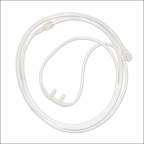
Mandibular advancement is a treatment approach used in dentistry and sleep medicine, which involves the surgical or non-surgical forward movement of the mandible (lower jaw) to address certain medical conditions. The most common use of mandibular advancement is in the treatment of obstructive sleep apnea (OSA), where the tongue and soft tissues at the back of the throat can collapse into the airway during sleep, causing obstruction and breathing difficulties.
Mandibular advancement devices (MADs) are often used in non-surgical treatments. These custom-made oral appliances look similar to mouthguards or sports guards and are worn during sleep. They work by holding the lower jaw in a slightly forward position, which helps to keep the airway open and prevents the tongue and soft tissues from collapsing into it.
Surgical mandibular advancement is another option for patients with severe OSA who cannot tolerate or do not respond well to MADs or other treatments like continuous positive airway pressure (CPAP). In this procedure, the jaw is surgically moved forward and stabilized in that position using plates, screws, or wires. This creates more space in the airway and reduces the risk of obstruction during sleep.
In summary, mandibular advancement refers to the movement of the lower jaw forward, either through non-surgical means like MADs or surgical interventions, with the primary goal of treating obstructive sleep apnea by maintaining a patent airway during sleep.
Rhinomanometry is a medical diagnostic procedure that measures the pressure and flow of air through the nasal passages. It is used to assess the nasal airway resistance and function, and can help diagnose and monitor conditions such as nasal congestion, deviated septum, sinusitis, and other disorders that affect nasal breathing.
During the procedure, a small catheter or mask is placed over the nose, and the patient is asked to breathe normally while the pressure and airflow are measured. The data is then analyzed to determine any abnormalities in nasal function, such as increased resistance or asymmetry between the two sides of the nose.
Rhinomanometry can be performed using either anterior or posterior methods, depending on whether the measurement is taken at the entrance or exit of the nasal passages. The results of the test can help guide treatment decisions and assess the effectiveness of therapies such as medications or surgery.
The uvula is a small, conical piece of soft tissue that hangs down from the middle part of the back of the soft palate (the rear-most portion of the roof of the mouth). It contains muscle fibers and mucous glands, and its function is associated with swallowing, speaking, and protecting the airway. During swallowing, the uvula helps to prevent food and liquids from entering the nasal cavity by blocking the opening between the oral and nasal cavities (the nasopharynx). In speech, it plays a role in shaping certain sounds like "a" and "u."
Restless Legs Syndrome (RLS) is a neurological disorder characterized by an irresistible urge to move one's body to stop uncomfortable or odd sensations. It most commonly affects the legs. The condition worsens during periods of rest, particularly when lying or sitting.
The symptoms typically include:
1. An uncontrollable need or urge to move the legs to relieve uncomfortable sensations such as crawling, creeping, tingling, pulling, or painful feelings.
2. Symptoms begin or intensify during rest or inactivity.
3. Symptoms are partially or totally relieved by movement, such as walking or stretching, at least as long as the activity continues.
4. Symptoms are worse in the evening or night, often leading to disturbed sleep.
The exact cause of RLS is unknown, but it may be related to abnormalities in the brain's dopamine pathways that control muscle movements. It can also be associated with certain medical conditions like iron deficiency, kidney disease, diabetes, and pregnancy. Treatment often involves addressing any underlying conditions and using medications to manage symptoms.
Apnea is a medical condition defined as the cessation of breathing for 10 seconds or more. It can occur during sleep (sleep apnea) or while awake (wakeful apnea). There are different types of sleep apnea, including obstructive sleep apnea, central sleep apnea, and complex sleep apnea syndrome. Obstructive sleep apnea occurs when the airway becomes blocked during sleep, while central sleep apnea occurs when the brain fails to signal the muscles to breathe. Complex sleep apnea syndrome, also known as treatment-emergent central sleep apnea, is a combination of obstructive and central sleep apneas. Sleep apnea can lead to various complications, such as fatigue, difficulty concentrating, high blood pressure, heart disease, and stroke.
Sleep deprivation is a condition that occurs when an individual fails to get sufficient quality sleep or the recommended amount of sleep, typically 7-9 hours for adults. This can lead to various physical and mental health issues. It can be acute, lasting for one night or a few days, or chronic, persisting over a longer period.
The consequences of sleep deprivation include:
1. Fatigue and lack of energy
2. Difficulty concentrating or remembering things
3. Mood changes, such as irritability or depression
4. Weakened immune system
5. Increased appetite and potential weight gain
6. Higher risk of accidents due to decreased reaction time
7. Health problems like high blood pressure, diabetes, and heart disease over time
Sleep deprivation can be caused by various factors, including stress, shift work, sleep disorders like insomnia or sleep apnea, poor sleep hygiene, and certain medications. It's essential to address the underlying causes of sleep deprivation to ensure proper rest and overall well-being.
Azabicyclo compounds are a type of organic compound that contain at least one nitrogen atom (azacycle) and two rings fused together (bicyclic). The nitrogen atom can be part of either a saturated or unsaturated ring, and the rings themselves can be composed of carbon atoms only or contain other heteroatoms such as oxygen or sulfur.
The term "azabicyclo" is often followed by a set of three numbers that specify the number of atoms in each of the three rings involved in the fusion. For example, azabicyclo[3.2.1]octane is a compound with two fused rings containing 3 and 2 carbon atoms, respectively, and one nitrogen atom forming the third ring of 1 carbon atom.
These compounds have a wide range of applications in pharmaceuticals, agrochemicals, and materials science due to their unique structures and properties. In particular, azabicyclo compounds are often used as building blocks for the synthesis of complex natural products and bioactive molecules.
Body Mass Index (BMI) is a measure used to assess whether a person has a healthy weight for their height. It's calculated by dividing a person's weight in kilograms by the square of their height in meters. Here is the medical definition:
Body Mass Index (BMI) = weight(kg) / [height(m)]^2
According to the World Health Organization, BMI categories are defined as follows:
* Less than 18.5: Underweight
* 18.5-24.9: Normal or healthy weight
* 25.0-29.9: Overweight
* 30.0 and above: Obese
It is important to note that while BMI can be a useful tool for identifying weight issues in populations, it does have limitations when applied to individuals. For example, it may not accurately reflect body fat distribution or muscle mass, which can affect health risks associated with excess weight. Therefore, BMI should be used as one of several factors when evaluating an individual's health status and risk for chronic diseases.
A circadian rhythm is a roughly 24-hour biological cycle that regulates various physiological and behavioral processes in living organisms. It is driven by the body's internal clock, which is primarily located in the suprachiasmatic nucleus (SCN) of the hypothalamus in the brain.
The circadian rhythm controls many aspects of human physiology, including sleep-wake cycles, hormone secretion, body temperature, and metabolism. It helps to synchronize these processes with the external environment, particularly the day-night cycle caused by the rotation of the Earth.
Disruptions to the circadian rhythm can have negative effects on health, leading to conditions such as insomnia, sleep disorders, depression, bipolar disorder, and even increased risk of chronic diseases like cancer, diabetes, and cardiovascular disease. Factors that can disrupt the circadian rhythm include shift work, jet lag, irregular sleep schedules, and exposure to artificial light at night.
Physiological monitoring is the continuous or intermittent observation and measurement of various body functions or parameters in a patient, with the aim of evaluating their health status, identifying any abnormalities or changes, and guiding clinical decision-making and treatment. This may involve the use of specialized medical equipment, such as cardiac monitors, pulse oximeters, blood pressure monitors, and capnographs, among others. The data collected through physiological monitoring can help healthcare professionals assess the effectiveness of treatments, detect complications early, and make timely adjustments to patient care plans.
Electroencephalography (EEG) is a medical procedure that records electrical activity in the brain. It uses small, metal discs called electrodes, which are attached to the scalp with paste or a specialized cap. These electrodes detect tiny electrical charges that result from the activity of brain cells, and the EEG machine then amplifies and records these signals.
EEG is used to diagnose various conditions related to the brain, such as seizures, sleep disorders, head injuries, infections, and degenerative diseases like Alzheimer's or Parkinson's. It can also be used during surgery to monitor brain activity and ensure that surgical procedures do not interfere with vital functions.
EEG is a safe and non-invasive procedure that typically takes about 30 minutes to an hour to complete, although longer recordings may be necessary in some cases. Patients are usually asked to relax and remain still during the test, as movement can affect the quality of the recording.
Respiratory disorders are a group of conditions that affect the respiratory system, including the nose, throat (pharynx), windpipe (trachea), bronchi, lungs, and diaphragm. These disorders can make it difficult for a person to breathe normally and may cause symptoms such as coughing, wheezing, shortness of breath, and chest pain.
There are many different types of respiratory disorders, including:
1. Asthma: A chronic inflammatory disease that causes the airways to become narrow and swollen, leading to difficulty breathing.
2. Chronic obstructive pulmonary disease (COPD): A group of lung diseases, including emphysema and chronic bronchitis, that make it hard to breathe.
3. Pneumonia: An infection of the lungs that can cause coughing, chest pain, and difficulty breathing.
4. Lung cancer: A type of cancer that forms in the tissues of the lungs and can cause symptoms such as coughing, chest pain, and shortness of breath.
5. Tuberculosis (TB): A bacterial infection that mainly affects the lungs but can also affect other parts of the body.
6. Sleep apnea: A disorder that causes a person to stop breathing for short periods during sleep.
7. Interstitial lung disease: A group of disorders that cause scarring of the lung tissue, leading to difficulty breathing.
8. Pulmonary fibrosis: A type of interstitial lung disease that causes scarring of the lung tissue and makes it hard to breathe.
9. Pleural effusion: An abnormal accumulation of fluid in the space between the lungs and chest wall.
10. Lung transplantation: A surgical procedure to replace a diseased or failing lung with a healthy one from a donor.
Respiratory disorders can be caused by a variety of factors, including genetics, exposure to environmental pollutants, smoking, and infections. Treatment for respiratory disorders may include medications, oxygen therapy, breathing exercises, and lifestyle changes. In some cases, surgery may be necessary to treat the disorder.
A Circadian Rhythm Sleep Disorder (CRSD) is a condition in which a person's sleep-wake cycle is out of sync with the typical 24-hour day. This means that their internal "body clock" that regulates sleep and wakefulness does not align with the external environment, leading to difficulties sleeping, staying awake, or functioning at appropriate times.
CRSDs can be caused by a variety of factors, including genetic predisposition, environmental influences, and medical conditions. Some common types of CRSDs include Delayed Sleep Phase Syndrome (DSPS), Advanced Sleep Phase Syndrome (ASPS), Non-24-Hour Sleep-Wake Rhythm Disorder, and Shift Work Disorder.
Symptoms of CRSDs may include difficulty falling asleep or staying asleep at the desired time, excessive sleepiness during the day, difficulty concentrating or functioning at work or school, and mood disturbances. Treatment for CRSDs may involve lifestyle changes, such as adjusting sleep schedules or exposure to light at certain times of day, as well as medications or other therapies.
The soft palate, also known as the velum, is the rear portion of the roof of the mouth that is made up of muscle and mucous membrane. It extends from the hard palate (the bony front part of the roof of the mouth) to the uvula, which is the small piece of tissue that hangs down at the back of the throat.
The soft palate plays a crucial role in speech, swallowing, and breathing. During swallowing, it moves upward and backward to block off the nasal cavity, preventing food and liquids from entering the nose. In speech, it helps to direct the flow of air from the mouth into the nose, which is necessary for producing certain sounds.
Anatomically, the soft palate consists of several muscles that allow it to change shape and move. These muscles include the tensor veli palatini, levator veli palatini, musculus uvulae, palatopharyngeus, and palatoglossus. The soft palate also contains a rich supply of blood vessels and nerves that provide sensation and help regulate its function.
Prospective studies, also known as longitudinal studies, are a type of cohort study in which data is collected forward in time, following a group of individuals who share a common characteristic or exposure over a period of time. The researchers clearly define the study population and exposure of interest at the beginning of the study and follow up with the participants to determine the outcomes that develop over time. This type of study design allows for the investigation of causal relationships between exposures and outcomes, as well as the identification of risk factors and the estimation of disease incidence rates. Prospective studies are particularly useful in epidemiology and medical research when studying diseases with long latency periods or rare outcomes.
A "delta rhythm" is a term used in electroencephalography (EEG) to describe a pattern of brain waves that are typically seen in the delta frequency range (0.5-4 Hz) and are maximal over the posterior regions of the head. This rhythm is often observed during deep sleep stages, specifically stage 3 and stage 4 of non-rapid eye movement (NREM) sleep, also known as slow-wave sleep.
Delta waves are characterized by their high amplitude and slow frequency, making them easily distinguishable from other brain wave patterns. The presence of a robust delta rhythm during sleep is thought to reflect the restorative processes that occur during this stage of sleep, including memory consolidation and physical restoration.
However, it's important to note that abnormal delta rhythms can also be observed in certain neurological conditions, such as epilepsy or encephalopathy, where they may indicate underlying brain dysfunction or injury. In these cases, the presence of delta rhythm may have different clinical implications and require further evaluation by a medical professional.
Pierre Robin Syndrome is a congenital condition characterized by a set of distinctive features including:
1. Micrognathia: This is the term for an abnormally small lower jaw (mandible). In Pierre Robin Syndrome, this feature is present at birth and can lead to breathing difficulties due to the tongue falling back and obstructing the airway.
2. Glossoptosis: This refers to the displacement of the tongue towards the back of the mouth. Because of the small jaw, the tongue has limited space and tends to fall back and block the airway, especially during sleep.
3. Cleft Palate: A cleft palate is a birth defect where there is an opening in the roof of the mouth (palate). This occurs because the two sides of the palate do not fuse together properly during fetal development.
The syndrome can vary in severity among individuals, and some may also have other associated conditions such as hearing problems, heart defects, or learning disabilities. The exact cause of Pierre Robin Syndrome is unknown, but it's often associated with genetic syndromes like Stickler syndrome and velocardiofacial syndrome. Treatment typically involves addressing the airway issues first, often through positioning, prone sleeping, or in severe cases, a surgical procedure to bring the jaw forward (distraction osteogenesis). The cleft palate is usually repaired with surgery within the first year of life.
Cataplexy is a medical condition characterized by sudden and temporary loss of muscle tone or strength, typically triggered by strong emotions such as laughter, anger, or surprise. This can result in symptoms ranging from a slight slackening of the muscles to complete collapse. Cataplexy is often associated with narcolepsy, which is a neurological disorder that affects sleep-wake cycles. It's important to note that cataplexy is different from syncope (fainting), as it specifically involves muscle weakness rather than loss of consciousness.
Dyssomnias are a category of sleep disorders that involve problems with the amount, quality, or timing of sleep. They can be broken down into several subcategories, including:
1. Insomnia: This is characterized by difficulty falling asleep or staying asleep, despite adequate opportunity and circumstances to do so. It can result in distress, impairment in social, occupational, or other areas of functioning, and/or feelings of dissatisfaction with sleep.
2. Hypersomnias: These are disorders that involve excessive sleepiness during the day, even after having adequate opportunity for sleep. Narcolepsy is an example of a hypersomnia.
3. Sleep-related breathing disorders: These include conditions such as obstructive sleep apnea, in which breathing is repeatedly interrupted during sleep, leading to poor sleep quality and excessive daytime sleepiness.
4. Circadian rhythm sleep-wake disorders: These involve disruptions to the body's internal clock, which can result in difficulty falling asleep or staying asleep at desired times. Jet lag and shift work disorder are examples of circadian rhythm sleep-wake disorders.
5. Parasomnias: These are disruptive sleep-related events that occur during various stages of sleep, such as sleepwalking, night terrors, and REM sleep behavior disorder.
Dyssomnias can have significant impacts on a person's quality of life, and it is important to seek medical evaluation if you are experiencing symptoms. Treatment may involve lifestyle changes, medication, or other interventions depending on the specific type of dyssomnia.
Computer-assisted diagnosis (CAD) is the use of computer systems to aid in the diagnostic process. It involves the use of advanced algorithms and data analysis techniques to analyze medical images, laboratory results, and other patient data to help healthcare professionals make more accurate and timely diagnoses. CAD systems can help identify patterns and anomalies that may be difficult for humans to detect, and they can provide second opinions and flag potential errors or uncertainties in the diagnostic process.
CAD systems are often used in conjunction with traditional diagnostic methods, such as physical examinations and patient interviews, to provide a more comprehensive assessment of a patient's health. They are commonly used in radiology, pathology, cardiology, and other medical specialties where imaging or laboratory tests play a key role in the diagnostic process.
While CAD systems can be very helpful in the diagnostic process, they are not infallible and should always be used as a tool to support, rather than replace, the expertise of trained healthcare professionals. It's important for medical professionals to use their clinical judgment and experience when interpreting CAD results and making final diagnoses.
Sensitivity and specificity are statistical measures used to describe the performance of a diagnostic test or screening tool in identifying true positive and true negative results.
* Sensitivity refers to the proportion of people who have a particular condition (true positives) who are correctly identified by the test. It is also known as the "true positive rate" or "recall." A highly sensitive test will identify most or all of the people with the condition, but may also produce more false positives.
* Specificity refers to the proportion of people who do not have a particular condition (true negatives) who are correctly identified by the test. It is also known as the "true negative rate." A highly specific test will identify most or all of the people without the condition, but may also produce more false negatives.
In medical testing, both sensitivity and specificity are important considerations when evaluating a diagnostic test. High sensitivity is desirable for screening tests that aim to identify as many cases of a condition as possible, while high specificity is desirable for confirmatory tests that aim to rule out the condition in people who do not have it.
It's worth noting that sensitivity and specificity are often influenced by factors such as the prevalence of the condition in the population being tested, the threshold used to define a positive result, and the reliability and validity of the test itself. Therefore, it's important to consider these factors when interpreting the results of a diagnostic test.
A questionnaire in the medical context is a standardized, systematic, and structured tool used to gather information from individuals regarding their symptoms, medical history, lifestyle, or other health-related factors. It typically consists of a series of written questions that can be either self-administered or administered by an interviewer. Questionnaires are widely used in various areas of healthcare, including clinical research, epidemiological studies, patient care, and health services evaluation to collect data that can inform diagnosis, treatment planning, and population health management. They provide a consistent and organized method for obtaining information from large groups or individual patients, helping to ensure accurate and comprehensive data collection while minimizing bias and variability in the information gathered.
Kleine-Levin Syndrome (KLS) is a rare and complex neurological disorder characterized by recurring episodes of excessive sleep (hypersomnia), often accompanied by cognitive impairment, altered perception, and behavioral changes. These episodes can last for days or even weeks. The exact cause of KLS remains unknown, but it's thought to involve dysfunction in the hypothalamus and/or thalamus regions of the brain. It primarily affects adolescents, with males being more commonly affected than females. Diagnosis is typically made based on clinical symptoms, as there are no specific diagnostic tests for KLS. Treatment usually involves managing individual symptoms and may include stimulant medications to help reduce excessive sleepiness during episodes.
Sleep arousal disorders are a category of sleep disorders that involve the partial or complete awakening from sleep, often accompanied by confusion and disorientation. These disorders are characterized by an abnormal arousal process during sleep, which can result in brief periods of wakefulness or full awakenings. The most common types of sleep arousal disorders include sleepwalking (somnambulism), sleep talking (somniloquy), and night terrors (pavor nocturnus).
In sleepwalking, the individual may get out of bed and walk around while still asleep, often with a blank stare and without any memory of the event. Sleep talking can occur in various levels of sleep and may range from simple sounds to complex conversations. Night terrors are episodes of intense fear and agitation during sleep, often accompanied by screams or cries for help, rapid heart rate, and sweating.
These disorders can be caused by a variety of factors, including stress, anxiety, fever, certain medications, alcohol consumption, and underlying medical conditions such as sleep apnea or restless leg syndrome. They can also occur as a result of genetic predisposition. Sleep arousal disorders can have significant impacts on an individual's quality of life, leading to fatigue, daytime sleepiness, impaired cognitive function, and decreased overall well-being. Treatment options may include behavioral therapy, medication, or addressing any underlying medical conditions.
The pharynx is a part of the digestive and respiratory systems that serves as a conduit for food and air. It is a musculo-membranous tube extending from the base of the skull to the level of the sixth cervical vertebra where it becomes continuous with the esophagus.
The pharynx has three regions: the nasopharynx, oropharynx, and laryngopharynx. The nasopharynx is the uppermost region, which lies above the soft palate and is connected to the nasal cavity. The oropharynx is the middle region, which includes the area between the soft palate and the hyoid bone, including the tonsils and base of the tongue. The laryngopharynx is the lowest region, which lies below the hyoid bone and connects to the larynx.
The primary function of the pharynx is to convey food from the oral cavity to the esophagus during swallowing and to allow air to pass from the nasal cavity to the larynx during breathing. It also plays a role in speech, taste, and immune defense.
Hypnotics and sedatives are classes of medications that have depressant effects on the central nervous system, leading to sedation (calming or inducing sleep), reduction in anxiety, and in some cases, decreased awareness or memory. These agents work by affecting the neurotransmitter GABA (gamma-aminobutyric acid) in the brain, which results in inhibitory effects on neuronal activity.
Hypnotics are primarily used for the treatment of insomnia and other sleep disorders, while sedatives are often prescribed to manage anxiety or to produce a calming effect before medical procedures. Some medications can function as both hypnotics and sedatives, depending on the dosage and specific formulation. Common examples of these medications include benzodiazepines (such as diazepam and lorazepam), non-benzodiazepine hypnotics (such as zolpidem and eszopiclone), barbiturates, and certain antihistamines.
It is essential to use these medications under the guidance of a healthcare professional, as they can have potential side effects, such as drowsiness, dizziness, confusion, and impaired coordination. Additionally, long-term use or high doses may lead to tolerance, dependence, and withdrawal symptoms upon discontinuation.
Oxygen is a colorless, odorless, tasteless gas that constitutes about 21% of the earth's atmosphere. It is a crucial element for human and most living organisms as it is vital for respiration. Inhaled oxygen enters the lungs and binds to hemoglobin in red blood cells, which carries it to tissues throughout the body where it is used to convert nutrients into energy and carbon dioxide, a waste product that is exhaled.
Medically, supplemental oxygen therapy may be provided to patients with conditions such as chronic obstructive pulmonary disease (COPD), pneumonia, heart failure, or other medical conditions that impair the body's ability to extract sufficient oxygen from the air. Oxygen can be administered through various devices, including nasal cannulas, face masks, and ventilators.
REM Sleep Behavior Disorder (RBD) is a parasomnia, which is a disorder that involves undesirable experiences or abnormal behaviors during sleep. Specifically, RBD is a type of rapid eye movement (REM) sleep parasomnia where the muscle atonia (lack of muscle tone) that normally occurs during REM sleep is absent or incomplete, allowing for the emergence of motor behaviors and vivid dreaming. These dreams can be quite intense and may result in the individual physically acting out their dreams, leading to potential harm for themselves or their bed partner. RBD can occur in isolation or as a symptom of another neurological condition.
Electrooculography (EOG) is a technique for measuring the resting potential of the eye and the changes in this potential that occur with eye movements. It involves placing electrodes near the eyes to detect the small electric fields generated by the movement of the eyeball within the surrounding socket. This technique is used in research and clinical settings to study eye movements and their control, as well as in certain diagnostic applications such as assessing the function of the oculomotor system in patients with neurological disorders.
Nasal obstruction is a medical condition that refers to any blockage or restriction in the normal flow of air through the nasal passages. This can be caused by various factors such as inflammation, swelling, or physical abnormalities in the nasal cavity. Common causes of nasal obstruction include allergies, sinusitis, deviated septum, enlarged turbinates, and nasal polyps. Symptoms may include difficulty breathing through the nose, nasal congestion, and nasal discharge. Treatment options depend on the underlying cause and may include medications, surgery, or lifestyle changes.
Adenoids are a pair of masses of lymphoid tissue located in the nasopharynx, which is the upper part of the throat behind the nose. They are part of the immune system and help to protect against infection. Adenoids are largest in children and tend to shrink in size as people get older. In some cases, adenoids can become enlarged or infected, leading to problems such as breathing difficulties, ear infections, and sleep disorders. Treatment for enlarged or infected adenoids may include antibiotics, medications to reduce swelling, or surgical removal of the adenoids (adenoidectomy).
Reproducibility of results in a medical context refers to the ability to obtain consistent and comparable findings when a particular experiment or study is repeated, either by the same researcher or by different researchers, following the same experimental protocol. It is an essential principle in scientific research that helps to ensure the validity and reliability of research findings.
In medical research, reproducibility of results is crucial for establishing the effectiveness and safety of new treatments, interventions, or diagnostic tools. It involves conducting well-designed studies with adequate sample sizes, appropriate statistical analyses, and transparent reporting of methods and findings to allow other researchers to replicate the study and confirm or refute the results.
The lack of reproducibility in medical research has become a significant concern in recent years, as several high-profile studies have failed to produce consistent findings when replicated by other researchers. This has led to increased scrutiny of research practices and a call for greater transparency, rigor, and standardization in the conduct and reporting of medical research.
Fluid shifts, in a medical context, refer to the movement or redistribution of fluids between different compartments within the body. The human body is composed of two main fluid compartments: the intracellular fluid (ICF), which is present inside the cells, and the extracellular fluid (ECF), which is outside the cells. The ECF is further divided into interstitial fluid (present in the spaces between cells) and intravascular fluid (present within the blood vessels).
Fluid shifts can occur due to various physiological and pathological conditions, such as changes in hydrostatic pressure, oncotic pressure, or permeability of the capillary membranes. These shifts can have significant impacts on various body systems, particularly the cardiovascular, respiratory, and renal systems. For example, fluid shifting from the intravascular space to the interstitial space can lead to edema (swelling), while fluid shifts into the alveoli in the lungs can cause pulmonary edema and impair breathing.
In a clinical setting, healthcare professionals monitor and manage fluid shifts through various strategies, such as administering intravenous fluids, using diuretics, or implementing mechanical ventilation, depending on the underlying cause and the specific effects of the fluid shift on the patient's condition.
Airway obstruction is a medical condition that occurs when the normal flow of air into and out of the lungs is partially or completely blocked. This blockage can be caused by a variety of factors, including swelling of the tissues in the airway, the presence of foreign objects or substances, or abnormal growths such as tumors.
When the airway becomes obstructed, it can make it difficult for a person to breathe normally. They may experience symptoms such as shortness of breath, wheezing, coughing, and chest tightness. In severe cases, airway obstruction can lead to respiratory failure and other life-threatening complications.
There are several types of airway obstruction, including:
1. Upper airway obstruction: This occurs when the blockage is located in the upper part of the airway, such as the nose, throat, or voice box.
2. Lower airway obstruction: This occurs when the blockage is located in the lower part of the airway, such as the trachea or bronchi.
3. Partial airway obstruction: This occurs when the airway is partially blocked, allowing some air to flow in and out of the lungs.
4. Complete airway obstruction: This occurs when the airway is completely blocked, preventing any air from flowing into or out of the lungs.
Treatment for airway obstruction depends on the underlying cause of the condition. In some cases, removing the obstruction may be as simple as clearing the airway of foreign objects or mucus. In other cases, more invasive treatments such as surgery may be necessary.
Comorbidity is the presence of one or more additional health conditions or diseases alongside a primary illness or condition. These co-occurring health issues can have an impact on the treatment plan, prognosis, and overall healthcare management of an individual. Comorbidities often interact with each other and the primary condition, leading to more complex clinical situations and increased healthcare needs. It is essential for healthcare professionals to consider and address comorbidities to provide comprehensive care and improve patient outcomes.
Pulmonary ventilation, also known as pulmonary respiration or simply ventilation, is the process of moving air into and out of the lungs to facilitate gas exchange. It involves two main phases: inhalation (or inspiration) and exhalation (or expiration). During inhalation, the diaphragm and external intercostal muscles contract, causing the chest volume to increase and the pressure inside the chest to decrease, which then draws air into the lungs. Conversely, during exhalation, these muscles relax, causing the chest volume to decrease and the pressure inside the chest to increase, which pushes air out of the lungs. This process ensures that oxygen-rich air from the atmosphere enters the alveoli (air sacs in the lungs), where it can diffuse into the bloodstream, while carbon dioxide-rich air from the bloodstream in the capillaries surrounding the alveoli is expelled out of the body.
The oropharynx is the part of the throat (pharynx) that is located immediately behind the mouth and includes the back one-third of the tongue, the soft palate, the side and back walls of the throat, and the tonsils. It serves as a passageway for both food and air, and is also an important area for the immune system due to the presence of tonsils.
Treatment outcome is a term used to describe the result or effect of medical treatment on a patient's health status. It can be measured in various ways, such as through symptoms improvement, disease remission, reduced disability, improved quality of life, or survival rates. The treatment outcome helps healthcare providers evaluate the effectiveness of a particular treatment plan and make informed decisions about future care. It is also used in clinical research to compare the efficacy of different treatments and improve patient care.
Fatal Familial Insomnia (FFI) is a rare, inherited prion disease characterized by progressive insomnia that leads to death, typically within a year of onset. It is caused by a mutation in the PRNP gene, which encodes the prion protein. The mutation results in an abnormal form of the prion protein that accumulates in the brain and causes damage to nerve cells.
The symptoms of FFI usually begin in middle age and include progressive insomnia, rapid weight loss, hallucinations, confusion, and eventually dementia. As the disease progresses, patients may experience muscle spasms, rigidity, and difficulty swallowing. There is no cure for FFI, and treatment is focused on managing symptoms and providing supportive care.
FFI is inherited in an autosomal dominant manner, meaning that a person has a 50% chance of inheriting the mutated gene from an affected parent. The disease affects both men and women and has been reported in families worldwide.
Impedance plethysmography is a non-invasive method used to measure changes in blood volume or flow in a particular area of the body. It works by passing a small electrical current through the tissue and measuring the opposition (impedance) to that current, which varies with the amount of blood present in the area.
In impedance cardiography, this technique is used to estimate cardiac output, stroke volume, and other hemodynamic parameters. The changes in impedance are measured across the chest wall, which correlate with the ventricular ejection of blood during each heartbeat. This allows for the calculation of various cardiovascular variables, such as the amount of blood pumped by the heart per minute (cardiac output) and the resistance to blood flow in the systemic circulation (systemic vascular resistance).
Impedance plethysmography is a safe and reliable method for assessing cardiovascular function, and it has been widely used in clinical settings to evaluate patients with various cardiovascular disorders, including heart failure, hypertension, and peripheral arterial disease.
A case-control study is an observational research design used to identify risk factors or causes of a disease or health outcome. In this type of study, individuals with the disease or condition (cases) are compared with similar individuals who do not have the disease or condition (controls). The exposure history or other characteristics of interest are then compared between the two groups to determine if there is an association between the exposure and the disease.
Case-control studies are often used when it is not feasible or ethical to conduct a randomized controlled trial, as they can provide valuable insights into potential causes of diseases or health outcomes in a relatively short period of time and at a lower cost than other study designs. However, because case-control studies rely on retrospective data collection, they are subject to biases such as recall bias and selection bias, which can affect the validity of the results. Therefore, it is important to carefully design and conduct case-control studies to minimize these potential sources of bias.
Orthodontic appliances, removable, are dental devices that can be removed and inserted by the patient as needed or directed. These appliances are designed to align and straighten teeth, correct bite issues, and improve the function and appearance of the teeth and jaws. They are typically made from materials such as plastic, metal, or acrylic and may include components like wires, springs, or screws. Examples of removable orthodontic appliances include aligners, retainers, and space maintainers. The specific type and design of the appliance will depend on the individual patient's orthodontic needs and treatment goals.
Prevalence, in medical terms, refers to the total number of people in a given population who have a particular disease or condition at a specific point in time, or over a specified period. It is typically expressed as a percentage or a ratio of the number of cases to the size of the population. Prevalence differs from incidence, which measures the number of new cases that develop during a certain period.
Computer-assisted signal processing is a medical term that refers to the use of computer algorithms and software to analyze, interpret, and extract meaningful information from biological signals. These signals can include physiological data such as electrocardiogram (ECG) waves, electromyography (EMG) signals, electroencephalography (EEG) readings, or medical images.
The goal of computer-assisted signal processing is to automate the analysis of these complex signals and extract relevant features that can be used for diagnostic, monitoring, or therapeutic purposes. This process typically involves several steps, including:
1. Signal acquisition: Collecting raw data from sensors or medical devices.
2. Preprocessing: Cleaning and filtering the data to remove noise and artifacts.
3. Feature extraction: Identifying and quantifying relevant features in the signal, such as peaks, troughs, or patterns.
4. Analysis: Applying statistical or machine learning algorithms to interpret the extracted features and make predictions about the underlying physiological state.
5. Visualization: Presenting the results in a clear and intuitive way for clinicians to review and use.
Computer-assisted signal processing has numerous applications in healthcare, including:
* Diagnosing and monitoring cardiac arrhythmias or other heart conditions using ECG signals.
* Assessing muscle activity and function using EMG signals.
* Monitoring brain activity and diagnosing neurological disorders using EEG readings.
* Analyzing medical images to detect abnormalities, such as tumors or fractures.
Overall, computer-assisted signal processing is a powerful tool for improving the accuracy and efficiency of medical diagnosis and monitoring, enabling clinicians to make more informed decisions about patient care.
Dreams are a series of thoughts, images, and sensations occurring in a person's mind during sleep. They can be vivid or vague, positive or negative, and may involve memories, emotions, and fears. The scientific study of dreams is called oneirology. While the exact purpose and function of dreams remain a topic of debate among researchers, some theories suggest that dreaming may help with memory consolidation, problem-solving, emotional processing, and learning.
Dreams usually occur during the rapid eye movement (REM) stage of sleep, although they can also happen in non-REM stages. They are typically associated with complex brain activities, involving areas such as the amygdala, hippocampus, and the neocortex. The content of dreams can be influenced by various factors, including a person's thoughts, experiences, emotions, physical state, and environmental conditions.
It is important to note that dreaming is a natural and universal human experience, and understanding dreams can provide insights into our cognitive processes, emotional well-being, and mental health.
Equipment design, in the medical context, refers to the process of creating and developing medical equipment and devices, such as surgical instruments, diagnostic machines, or assistive technologies. This process involves several stages, including:
1. Identifying user needs and requirements
2. Concept development and brainstorming
3. Prototyping and testing
4. Design for manufacturing and assembly
5. Safety and regulatory compliance
6. Verification and validation
7. Training and support
The goal of equipment design is to create safe, effective, and efficient medical devices that meet the needs of healthcare providers and patients while complying with relevant regulations and standards. The design process typically involves a multidisciplinary team of engineers, clinicians, designers, and researchers who work together to develop innovative solutions that improve patient care and outcomes.
A cross-sectional study is a type of observational research design that examines the relationship between variables at one point in time. It provides a snapshot or a "cross-section" of the population at a particular moment, allowing researchers to estimate the prevalence of a disease or condition and identify potential risk factors or associations.
In a cross-sectional study, data is collected from a sample of participants at a single time point, and the variables of interest are measured simultaneously. This design can be used to investigate the association between exposure and outcome, but it cannot establish causality because it does not follow changes over time.
Cross-sectional studies can be conducted using various data collection methods, such as surveys, interviews, or medical examinations. They are often used in epidemiology to estimate the prevalence of a disease or condition in a population and to identify potential risk factors that may contribute to its development. However, because cross-sectional studies only provide a snapshot of the population at one point in time, they cannot account for changes over time or determine whether exposure preceded the outcome.
Therefore, while cross-sectional studies can be useful for generating hypotheses and identifying potential associations between variables, further research using other study designs, such as cohort or case-control studies, is necessary to establish causality and confirm any findings.
Trazodone is an antidepressant medication that belongs to the class of drugs called serotonin antagonist and reuptake inhibitors (SARIs). It works by increasing the levels of the neurotransmitter serotonin in the brain, which helps to improve mood and reduce symptoms of depression.
Trazodone is primarily used to treat major depressive disorder, but it may also be prescribed for anxiety, insomnia, and other conditions. The medication comes in various forms, including tablets and an extended-release formulation, and is typically taken orally one to three times a day. Common side effects of trazodone include dizziness, dry mouth, and sedation.
It's important to note that trazodone can interact with other medications and substances, so it's essential to inform your healthcare provider about all the drugs you are taking before starting treatment. Additionally, trazodone may increase the risk of suicidal thoughts or behavior in some people, particularly during the initial stages of treatment, so close monitoring is necessary.
The supine position is a term used in medicine to describe a body posture where an individual is lying down on their back, with their face and torso facing upwards. This position is often adopted during various medical procedures, examinations, or when resting, as it allows for easy access to the front of the body. It is also the position automatically assumed by most people who are falling asleep.
It's important to note that in the supine position, the head can be flat on the surface or raised with the use of pillows or specialized medical equipment like a hospital bed. This can help to alleviate potential issues such as breathing difficulties or swelling in the face and head.
Airway resistance is a measure of the opposition to airflow during breathing, which is caused by the friction between the air and the walls of the respiratory tract. It is an important parameter in respiratory physiology because it can affect the work of breathing and gas exchange.
Airway resistance is usually expressed in units of cm H2O/L/s or Pa·s/m, and it can be measured during spontaneous breathing or during forced expiratory maneuvers, such as those used in pulmonary function testing. Increased airway resistance can result from a variety of conditions, including asthma, chronic obstructive pulmonary disease (COPD), bronchitis, and bronchiectasis. Decreased airway resistance can be seen in conditions such as emphysema or after a successful bronchodilator treatment.
A pressure transducer is a device that converts a mechanical force or pressure exerted upon it into an electrical signal which can be measured and standardized. In medical terms, pressure transducers are often used to measure various bodily pressures such as blood pressure, intracranial pressure, or intraocular pressure. These transducers typically consist of a diaphragm that is deflected by the pressure being measured, which then generates an electrical signal proportional to the amount of deflection. This signal can be processed and displayed in various ways, such as on a monitor or within an electronic medical record system.
Anoxia is a medical condition that refers to the absence or complete lack of oxygen supply in the body or a specific organ, tissue, or cell. This can lead to serious health consequences, including damage or death of cells and tissues, due to the vital role that oxygen plays in supporting cellular metabolism and energy production.
Anoxia can occur due to various reasons, such as respiratory failure, cardiac arrest, severe blood loss, carbon monoxide poisoning, or high altitude exposure. Prolonged anoxia can result in hypoxic-ischemic encephalopathy, a serious condition that can cause brain damage and long-term neurological impairments.
Medical professionals use various diagnostic tests, such as blood gas analysis, pulse oximetry, and electroencephalography (EEG), to assess oxygen levels in the body and diagnose anoxia. Treatment for anoxia typically involves addressing the underlying cause, providing supplemental oxygen, and supporting vital functions, such as breathing and circulation, to prevent further damage.
Orthodontic appliances are devices used in orthodontics, a branch of dentistry focused on the diagnosis, prevention, and treatment of dental and facial irregularities. These appliances can be fixed or removable and are used to align teeth, correct jaw relationships, or modify dental forces. They can include braces, aligners, palatal expanders, space maintainers, and headgear, among others. The specific type of appliance used depends on the individual patient's needs and the treatment plan developed by the orthodontist.
Heart rate is the number of heartbeats per unit of time, often expressed as beats per minute (bpm). It can vary significantly depending on factors such as age, physical fitness, emotions, and overall health status. A resting heart rate between 60-100 bpm is generally considered normal for adults, but athletes and individuals with high levels of physical fitness may have a resting heart rate below 60 bpm due to their enhanced cardiovascular efficiency. Monitoring heart rate can provide valuable insights into an individual's health status, exercise intensity, and response to various treatments or interventions.
Fatigue is a state of feeling very tired, weary, or exhausted, which can be physical, mental, or both. It is a common symptom that can be caused by various factors, including lack of sleep, poor nutrition, stress, medical conditions (such as anemia, diabetes, heart disease, or cancer), medications, and substance abuse. Fatigue can also be a symptom of depression or other mental health disorders. In medical terms, fatigue is often described as a subjective feeling of tiredness that is not proportional to recent activity levels and interferes with usual functioning. It is important to consult a healthcare professional if experiencing persistent or severe fatigue to determine the underlying cause and develop an appropriate treatment plan.
In medical terms, the "neck" is defined as the portion of the body that extends from the skull/head to the thorax or chest region. It contains 7 cervical vertebrae, muscles, nerves, blood vessels, lymphatic vessels, and glands (such as the thyroid gland). The neck is responsible for supporting the head, allowing its movement in various directions, and housing vital structures that enable functions like respiration and circulation.
Follow-up studies are a type of longitudinal research that involve repeated observations or measurements of the same variables over a period of time, in order to understand their long-term effects or outcomes. In medical context, follow-up studies are often used to evaluate the safety and efficacy of medical treatments, interventions, or procedures.
In a typical follow-up study, a group of individuals (called a cohort) who have received a particular treatment or intervention are identified and then followed over time through periodic assessments or data collection. The data collected may include information on clinical outcomes, adverse events, changes in symptoms or functional status, and other relevant measures.
The results of follow-up studies can provide important insights into the long-term benefits and risks of medical interventions, as well as help to identify factors that may influence treatment effectiveness or patient outcomes. However, it is important to note that follow-up studies can be subject to various biases and limitations, such as loss to follow-up, recall bias, and changes in clinical practice over time, which must be carefully considered when interpreting the results.
A nose, in a medical context, refers to the external part of the human body that is located on the face and serves as the primary organ for the sense of smell. It is composed of bone and cartilage, with a thin layer of skin covering it. The nose also contains nasal passages that are lined with mucous membranes and tiny hairs known as cilia. These structures help to filter, warm, and moisturize the air we breathe in before it reaches our lungs. Additionally, the nose plays an essential role in the process of verbal communication by shaping the sounds we make when we speak.
Cephalometry is a medical term that refers to the measurement and analysis of the skull, particularly the head face relations. It is commonly used in orthodontics and maxillofacial surgery to assess and plan treatment for abnormalities related to the teeth, jaws, and facial structures. The process typically involves taking X-ray images called cephalograms, which provide a lateral view of the head, and then using various landmarks and reference lines to make measurements and evaluate skeletal and dental relationships. This information can help clinicians diagnose problems, plan treatment, and assess treatment outcomes.
Electromyography (EMG) is a medical diagnostic procedure that measures the electrical activity of skeletal muscles during contraction and at rest. It involves inserting a thin needle electrode into the muscle to record the electrical signals generated by the muscle fibers. These signals are then displayed on an oscilloscope and may be heard through a speaker.
EMG can help diagnose various neuromuscular disorders, such as muscle weakness, numbness, or pain, and can distinguish between muscle and nerve disorders. It is often used in conjunction with other diagnostic tests, such as nerve conduction studies, to provide a comprehensive evaluation of the nervous system.
EMG is typically performed by a neurologist or a physiatrist, and the procedure may cause some discomfort or pain, although this is usually minimal. The results of an EMG can help guide treatment decisions and monitor the progression of neuromuscular conditions over time.
Analysis of Variance (ANOVA) is a statistical technique used to compare the means of two or more groups and determine whether there are any significant differences between them. It is a way to analyze the variance in a dataset to determine whether the variability between groups is greater than the variability within groups, which can indicate that the groups are significantly different from one another.
ANOVA is based on the concept of partitioning the total variance in a dataset into two components: variance due to differences between group means (also known as "between-group variance") and variance due to differences within each group (also known as "within-group variance"). By comparing these two sources of variance, ANOVA can help researchers determine whether any observed differences between groups are statistically significant, or whether they could have occurred by chance.
ANOVA is a widely used technique in many areas of research, including biology, psychology, engineering, and business. It is often used to compare the means of two or more experimental groups, such as a treatment group and a control group, to determine whether the treatment had a significant effect. ANOVA can also be used to compare the means of different populations or subgroups within a population, to identify any differences that may exist between them.
The Predictive Value of Tests, specifically the Positive Predictive Value (PPV) and Negative Predictive Value (NPV), are measures used in diagnostic tests to determine the probability that a positive or negative test result is correct.
Positive Predictive Value (PPV) is the proportion of patients with a positive test result who actually have the disease. It is calculated as the number of true positives divided by the total number of positive results (true positives + false positives). A higher PPV indicates that a positive test result is more likely to be a true positive, and therefore the disease is more likely to be present.
Negative Predictive Value (NPV) is the proportion of patients with a negative test result who do not have the disease. It is calculated as the number of true negatives divided by the total number of negative results (true negatives + false negatives). A higher NPV indicates that a negative test result is more likely to be a true negative, and therefore the disease is less likely to be present.
The predictive value of tests depends on the prevalence of the disease in the population being tested, as well as the sensitivity and specificity of the test. A test with high sensitivity and specificity will generally have higher predictive values than a test with low sensitivity and specificity. However, even a highly sensitive and specific test can have low predictive values if the prevalence of the disease is low in the population being tested.
A medical definition of the wrist is the complex joint that connects the forearm to the hand, composed of eight carpal bones arranged in two rows. The wrist allows for movement and flexibility in the hand, enabling us to perform various activities such as grasping, writing, and typing. It also provides stability and support for the hand during these movements. Additionally, numerous ligaments, tendons, and nerves pass through or near the wrist, making it susceptible to injuries and conditions like carpal tunnel syndrome.
Nonparametric statistics is a branch of statistics that does not rely on assumptions about the distribution of variables in the population from which the sample is drawn. In contrast to parametric methods, nonparametric techniques make fewer assumptions about the data and are therefore more flexible in their application. Nonparametric tests are often used when the data do not meet the assumptions required for parametric tests, such as normality or equal variances.
Nonparametric statistical methods include tests such as the Wilcoxon rank-sum test (also known as the Mann-Whitney U test) for comparing two independent groups, the Wilcoxon signed-rank test for comparing two related groups, and the Kruskal-Wallis test for comparing more than two independent groups. These tests use the ranks of the data rather than the actual values to make comparisons, which allows them to be used with ordinal or continuous data that do not meet the assumptions of parametric tests.
Overall, nonparametric statistics provide a useful set of tools for analyzing data in situations where the assumptions of parametric methods are not met, and can help researchers draw valid conclusions from their data even when the data are not normally distributed or have other characteristics that violate the assumptions of parametric tests.
Inhalation is the act or process of breathing in where air or other gases are drawn into the lungs. It's also known as inspiration. This process involves several muscles, including the diaphragm and intercostal muscles between the ribs, working together to expand the chest cavity and decrease the pressure within the thorax, which then causes air to flow into the lungs.
In a medical context, inhalation can also refer to the administration of medications or therapeutic gases through the respiratory tract, typically using an inhaler or nebulizer. This route of administration allows for direct delivery of the medication to the lungs, where it can be quickly absorbed into the bloodstream and exert its effects.
Respiratory physiological phenomena refer to the various mechanical, chemical, and biological processes and functions that occur in the respiratory system during breathing and gas exchange. These phenomena include:
1. Ventilation: The movement of air into and out of the lungs, which is achieved through the contraction and relaxation of the diaphragm and intercostal muscles.
2. Gas Exchange: The diffusion of oxygen (O2) from the alveoli into the bloodstream and carbon dioxide (CO2) from the bloodstream into the alveoli.
3. Respiratory Mechanics: The physical properties and forces that affect the movement of air in and out of the lungs, such as lung compliance, airway resistance, and chest wall elasticity.
4. Control of Breathing: The regulation of ventilation by the central nervous system through the integration of sensory information from chemoreceptors and mechanoreceptors in the respiratory system.
5. Acid-Base Balance: The maintenance of a stable pH level in the blood through the regulation of CO2 elimination and bicarbonate balance by the respiratory and renal systems.
6. Oxygen Transport: The binding of O2 to hemoglobin in the red blood cells and its delivery to the tissues for metabolic processes.
7. Defense Mechanisms: The various protective mechanisms that prevent the entry and colonization of pathogens and foreign particles into the respiratory system, such as mucociliary clearance, cough reflex, and immune responses.
The palatine tonsils, also known as the "tonsils," are two masses of lymphoid tissue located on either side of the oropharynx, at the back of the throat. They are part of the immune system and play a role in protecting the body from inhaled or ingested pathogens. Each tonsil has a surface covered with crypts and follicles that contain lymphocytes, which help to filter out bacteria and viruses that enter the mouth and nose.
The palatine tonsils are visible through the mouth and can be seen during a routine physical examination. They vary in size, but typically are about the size of a large olive or almond. Swelling or inflammation of the tonsils is called tonsillitis, which can cause symptoms such as sore throat, difficulty swallowing, fever, and swollen lymph nodes in the neck. In some cases, enlarged tonsils may need to be removed through a surgical procedure called a tonsillectomy.
Oxyhemoglobin is the form of hemoglobin that is combined with oxygen in red blood cells. It's created when oxygen molecules bind to the iron-containing heme groups of the hemoglobin protein inside the lungs, allowing for the transportation of oxygen from the lungs to body tissues. The affinity of hemoglobin for oxygen is influenced by factors such as pH, carbon dioxide concentration, and temperature, which can affect the release of oxygen from oxyhemoglobin in different parts of the body based on their specific needs.
The placebo effect is a psychological or psychophysiological phenomenon in which a person's symptoms improve following a treatment but this improvement is not attributable to the properties of the treatment itself. Instead, it is believed to be due to the mind's belief in the effectiveness of the treatment, often influenced by positive expectations and the ritualistic aspects of the therapy itself.
Placebos are often used in clinical trials as a control group to compare against the actual treatment. The placebo effect can make it challenging to determine whether an observed improvement is truly due to the treatment or other factors.
Reference values, also known as reference ranges or reference intervals, are the set of values that are considered normal or typical for a particular population or group of people. These values are often used in laboratory tests to help interpret test results and determine whether a patient's value falls within the expected range.
The process of establishing reference values typically involves measuring a particular biomarker or parameter in a large, healthy population and then calculating the mean and standard deviation of the measurements. Based on these statistics, a range is established that includes a certain percentage of the population (often 95%) and excludes extreme outliers.
It's important to note that reference values can vary depending on factors such as age, sex, race, and other demographic characteristics. Therefore, it's essential to use reference values that are specific to the relevant population when interpreting laboratory test results. Additionally, reference values may change over time due to advances in measurement technology or changes in the population being studied.
Sudden Infant Death Syndrome (SIDS) is defined by the American Academy of Pediatrics as "the sudden unexpected death of an infant
A Receiver Operating Characteristic (ROC) curve is a graphical representation used in medical decision-making and statistical analysis to illustrate the performance of a binary classifier system, such as a diagnostic test or a machine learning algorithm. It's a plot that shows the tradeoff between the true positive rate (sensitivity) and the false positive rate (1 - specificity) for different threshold settings.
The x-axis of an ROC curve represents the false positive rate (the proportion of negative cases incorrectly classified as positive), while the y-axis represents the true positive rate (the proportion of positive cases correctly classified as positive). Each point on the curve corresponds to a specific decision threshold, with higher points indicating better performance.
The area under the ROC curve (AUC) is a commonly used summary measure that reflects the overall performance of the classifier. An AUC value of 1 indicates perfect discrimination between positive and negative cases, while an AUC value of 0.5 suggests that the classifier performs no better than chance.
ROC curves are widely used in healthcare to evaluate diagnostic tests, predictive models, and screening tools for various medical conditions, helping clinicians make informed decisions about patient care based on the balance between sensitivity and specificity.
In the field of medicine, "time factors" refer to the duration of symptoms or time elapsed since the onset of a medical condition, which can have significant implications for diagnosis and treatment. Understanding time factors is crucial in determining the progression of a disease, evaluating the effectiveness of treatments, and making critical decisions regarding patient care.
For example, in stroke management, "time is brain," meaning that rapid intervention within a specific time frame (usually within 4.5 hours) is essential to administering tissue plasminogen activator (tPA), a clot-busting drug that can minimize brain damage and improve patient outcomes. Similarly, in trauma care, the "golden hour" concept emphasizes the importance of providing definitive care within the first 60 minutes after injury to increase survival rates and reduce morbidity.
Time factors also play a role in monitoring the progression of chronic conditions like diabetes or heart disease, where regular follow-ups and assessments help determine appropriate treatment adjustments and prevent complications. In infectious diseases, time factors are crucial for initiating antibiotic therapy and identifying potential outbreaks to control their spread.
Overall, "time factors" encompass the significance of recognizing and acting promptly in various medical scenarios to optimize patient outcomes and provide effective care.
A cohort study is a type of observational study in which a group of individuals who share a common characteristic or exposure are followed up over time to determine the incidence of a specific outcome or outcomes. The cohort, or group, is defined based on the exposure status (e.g., exposed vs. unexposed) and then monitored prospectively to assess for the development of new health events or conditions.
Cohort studies can be either prospective or retrospective in design. In a prospective cohort study, participants are enrolled and followed forward in time from the beginning of the study. In contrast, in a retrospective cohort study, researchers identify a cohort that has already been assembled through medical records, insurance claims, or other sources and then look back in time to assess exposure status and health outcomes.
Cohort studies are useful for establishing causality between an exposure and an outcome because they allow researchers to observe the temporal relationship between the two. They can also provide information on the incidence of a disease or condition in different populations, which can be used to inform public health policy and interventions. However, cohort studies can be expensive and time-consuming to conduct, and they may be subject to bias if participants are not representative of the population or if there is loss to follow-up.
Medical Definition:
"Risk factors" are any attribute, characteristic or exposure of an individual that increases the likelihood of developing a disease or injury. They can be divided into modifiable and non-modifiable risk factors. Modifiable risk factors are those that can be changed through lifestyle choices or medical treatment, while non-modifiable risk factors are inherent traits such as age, gender, or genetic predisposition. Examples of modifiable risk factors include smoking, alcohol consumption, physical inactivity, and unhealthy diet, while non-modifiable risk factors include age, sex, and family history. It is important to note that having a risk factor does not guarantee that a person will develop the disease, but rather indicates an increased susceptibility.
Obesity is a complex disease characterized by an excess accumulation of body fat to the extent that it negatively impacts health. It's typically defined using Body Mass Index (BMI), a measure calculated from a person's weight and height. A BMI of 30 or higher is indicative of obesity. However, it's important to note that while BMI can be a useful tool for identifying obesity in populations, it does not directly measure body fat and may not accurately reflect health status in individuals. Other factors such as waist circumference, blood pressure, cholesterol levels, and blood sugar levels should also be considered when assessing health risks associated with weight.
In a medical context, masks are typically used as personal protective equipment (PPE) to protect the wearer from inhaling airborne particles and contaminants. They can also help prevent the spread of respiratory droplets from the wearer to others, which is particularly important in clinical settings where patients may have infectious diseases.
There are several types of masks used in medical settings, including:
1. Medical Masks: These are loose-fitting, disposable masks that create a physical barrier between the mouth and nose of the wearer and potential contaminants in the immediate environment. They are commonly used by healthcare professionals during medical procedures to protect themselves and patients from respiratory droplets and aerosols.
2. N95 Respirators: These are tight-fitting masks that can filter out both large droplets and small aerosol particles, including those containing viruses. They offer a higher level of protection than medical masks and are recommended for use in healthcare settings where there is a risk of exposure to airborne contaminants, such as during certain medical procedures or when caring for patients with infectious diseases like tuberculosis or COVID-19.
3. Surgical N95 Respirators: These are a specialized type of N95 respirator designed for use in surgical settings. They have a clear plastic window that allows the wearer's mouth and nose to be visible, which is useful during surgery where clear communication and identification of the wearer's facial features are important.
4. Powered Air-Purifying Respirators (PAPRs): These are motorized masks that use a fan to draw air through a filter, providing a continuous supply of clean air to the wearer. They offer a high level of protection and are often used in healthcare settings where there is a risk of exposure to highly infectious diseases or hazardous substances.
It's important to note that masks should be used in conjunction with other infection prevention measures, such as hand hygiene and social distancing, to provide the best possible protection against respiratory illnesses.
A cross-over study is a type of experimental design in which participants receive two or more interventions in a specific order. After a washout period, each participant receives the opposite intervention(s). The primary advantage of this design is that it controls for individual variability by allowing each participant to act as their own control.
In medical research, cross-over studies are often used to compare the efficacy or safety of two treatments. For example, a researcher might conduct a cross-over study to compare the effectiveness of two different medications for treating high blood pressure. Half of the participants would be randomly assigned to receive one medication first and then switch to the other medication after a washout period. The other half of the participants would receive the opposite order of treatments.
Cross-over studies can provide valuable insights into the relative merits of different interventions, but they also have some limitations. For example, they may not be suitable for studying conditions that are chronic or irreversible, as it may not be possible to completely reverse the effects of the first intervention before administering the second one. Additionally, carryover effects from the first intervention can confound the results if they persist into the second treatment period.
Overall, cross-over studies are a useful tool in medical research when used appropriately and with careful consideration of their limitations.
Respiratory rate is the number of breaths a person takes per minute. It is typically measured by counting the number of times the chest rises and falls in one minute. Normal respiratory rate at rest for an adult ranges from 12 to 20 breaths per minute. An increased respiratory rate (tachypnea) or decreased respiratory rate (bradypnea) can be a sign of various medical conditions, such as lung disease, heart failure, or neurological disorders. It is an important vital sign that should be regularly monitored in clinical settings.
Arnold-Chiari malformation is a structural abnormality of the brain and skull base, specifically the cerebellum and brainstem. It is characterized by the descent of the cerebellar tonsils and sometimes parts of the brainstem through the foramen magnum (the opening at the base of the skull) into the upper spinal canal. This can cause pressure on the brainstem and cerebellum, potentially leading to a range of symptoms such as headaches, neck pain, unsteady gait, swallowing difficulties, hearing or balance problems, and in severe cases, neurological deficits. There are four types of Arnold-Chiari malformations, with type I being the most common and least severe form. Types II, III, and IV are progressively more severe and involve varying degrees of hindbrain herniation and associated neural tissue damage. Surgical intervention is often required to alleviate symptoms and prevent further neurological deterioration.
Hypercapnia is a state of increased carbon dioxide (CO2) concentration in the blood, typically defined as an arterial CO2 tension (PaCO2) above 45 mmHg. It is often associated with conditions that impair gas exchange or eliminate CO2 from the body, such as chronic obstructive pulmonary disease (COPD), severe asthma, respiratory failure, or certain neuromuscular disorders. Hypercapnia can cause symptoms such as headache, confusion, shortness of breath, and in severe cases, it can lead to life-threatening complications such as respiratory acidosis, coma, and even death if not promptly treated.
The double-blind method is a study design commonly used in research, including clinical trials, to minimize bias and ensure the objectivity of results. In this approach, both the participants and the researchers are unaware of which group the participants are assigned to, whether it be the experimental group or the control group. This means that neither the participants nor the researchers know who is receiving a particular treatment or placebo, thus reducing the potential for bias in the evaluation of outcomes. The assignment of participants to groups is typically done by a third party not involved in the study, and the codes are only revealed after all data have been collected and analyzed.
"Age factors" refer to the effects, changes, or differences that age can have on various aspects of health, disease, and medical care. These factors can encompass a wide range of issues, including:
1. Physiological changes: As people age, their bodies undergo numerous physical changes that can affect how they respond to medications, illnesses, and medical procedures. For example, older adults may be more sensitive to certain drugs or have weaker immune systems, making them more susceptible to infections.
2. Chronic conditions: Age is a significant risk factor for many chronic diseases, such as heart disease, diabetes, cancer, and arthritis. As a result, age-related medical issues are common and can impact treatment decisions and outcomes.
3. Cognitive decline: Aging can also lead to cognitive changes, including memory loss and decreased decision-making abilities. These changes can affect a person's ability to understand and comply with medical instructions, leading to potential complications in their care.
4. Functional limitations: Older adults may experience physical limitations that impact their mobility, strength, and balance, increasing the risk of falls and other injuries. These limitations can also make it more challenging for them to perform daily activities, such as bathing, dressing, or cooking.
5. Social determinants: Age-related factors, such as social isolation, poverty, and lack of access to transportation, can impact a person's ability to obtain necessary medical care and affect their overall health outcomes.
Understanding age factors is critical for healthcare providers to deliver high-quality, patient-centered care that addresses the unique needs and challenges of older adults. By taking these factors into account, healthcare providers can develop personalized treatment plans that consider a person's age, physical condition, cognitive abilities, and social circumstances.
In medical terms, the tongue is a muscular organ in the oral cavity that plays a crucial role in various functions such as taste, swallowing, and speech. It's covered with a mucous membrane and contains papillae, which are tiny projections that contain taste buds to help us perceive different tastes - sweet, salty, sour, and bitter. The tongue also assists in the initial process of digestion by moving food around in the mouth for chewing and mixing with saliva. Additionally, it helps in forming words and speaking clearly by shaping the sounds produced in the mouth.
Patient compliance, also known as medication adherence or patient adherence, refers to the degree to which a patient's behavior matches the agreed-upon recommendations from their healthcare provider. This includes taking medications as prescribed (including the correct dosage, frequency, and duration), following dietary restrictions, making lifestyle changes, and attending follow-up appointments. Poor patient compliance can negatively impact treatment outcomes and lead to worsening of symptoms, increased healthcare costs, and development of drug-resistant strains in the case of antibiotics. It is a significant challenge in healthcare and efforts are being made to improve patient education, communication, and support to enhance compliance.
Retrospective studies, also known as retrospective research or looking back studies, are a type of observational study that examines data from the past to draw conclusions about possible causal relationships between risk factors and outcomes. In these studies, researchers analyze existing records, medical charts, or previously collected data to test a hypothesis or answer a specific research question.
Retrospective studies can be useful for generating hypotheses and identifying trends, but they have limitations compared to prospective studies, which follow participants forward in time from exposure to outcome. Retrospective studies are subject to biases such as recall bias, selection bias, and information bias, which can affect the validity of the results. Therefore, retrospective studies should be interpreted with caution and used primarily to generate hypotheses for further testing in prospective studies.
Melatonin is a hormone that is produced by the pineal gland in the brain. It helps regulate sleep-wake cycles and is often referred to as the "hormone of darkness" because its production is stimulated by darkness and inhibited by light. Melatonin plays a key role in synchronizing the circadian rhythm, the body's internal clock that regulates various biological processes over a 24-hour period.
Melatonin is primarily released at night, and its levels in the blood can rise and fall in response to changes in light and darkness in an individual's environment. Supplementing with melatonin has been found to be helpful in treating sleep disorders such as insomnia, jet lag, and delayed sleep phase syndrome. It may also have other benefits, including antioxidant properties and potential uses in the treatment of certain neurological conditions.
It is important to note that while melatonin supplements are available over-the-counter in many countries, they should still be used under the guidance of a healthcare professional, as their use can have potential side effects and interactions with other medications.
The Autonomic Nervous System (ANS) is a part of the peripheral nervous system that operates largely below the level of consciousness and controls visceral functions. It is divided into two main subdivisions: the sympathetic and parasympathetic nervous systems, which generally have opposing effects and maintain homeostasis in the body.
The Sympathetic Nervous System (SNS) prepares the body for stressful or emergency situations, often referred to as the "fight or flight" response. It increases heart rate, blood pressure, respiratory rate, and metabolic rate, while also decreasing digestive activity. This response helps the body respond quickly to perceived threats.
The Parasympathetic Nervous System (PNS), on the other hand, promotes the "rest and digest" state, allowing the body to conserve energy and restore itself after the stress response has subsided. It decreases heart rate, blood pressure, and respiratory rate, while increasing digestive activity and promoting relaxation.
These two systems work together to maintain balance in the body by adjusting various functions based on internal and external demands. Disorders of the Autonomic Nervous System can lead to a variety of symptoms, such as orthostatic hypotension, gastroparesis, and cardiac arrhythmias, among others.
 Polysomnography - Wikipedia
 Polysomnography: MedlinePlus Medical Encyclopedia
 Keyword: respiratory polysomnography (rPSG)
 Polysomnography Certificate : Thompson Rivers University, Open Learning
 Polysomnography | Lakeland Community College - myLakeland
 Tri-C Electroneurodiagnostic Technology Polysomnography Track Program: Cleveland
 Clinical Atlas of Polysomnography | Sleep Review
 Alvin Community College Polysomnography (Sleep Medicine) Program | Alvin Community College
 Nocturnal Polysomnography | hartfordhealthcare.org | Hartford HealthCare | CT
 RePub, Erasmus University Repository:
Comparison of two types of Actiwatch with polysomnography in older adults with...
RePub, Erasmus University Repository:
Comparison of two types of Actiwatch with polysomnography in older adults with...
Instantaneous Heart Rate based sleep staging using deep learning models as a convenient alternative to Polysomnography |...
 The utility of polysomnography for the diagnosis of NREM parasomnias: an observational study over 4 years of clinical practice....
 JCDR -
Predictability of STOP-Bang Questionnaire and Epworth Sleepiness Scale in Identifying Obstructive Sleep Apnoea...
JCDR -
Predictability of STOP-Bang Questionnaire and Epworth Sleepiness Scale in Identifying Obstructive Sleep Apnoea...
 polysomnography
 Case Study: Extended Polysomnography Confirms an Idiopathic Hypersomnia Diagnosis - Consult QD
 Polysomnography Tech II
Ambulatory polysomnography: Technical aspects and normative values - Fingerprint - Augusta University Research Profiles
 Physiologic Approach in Snoring and Obstructive Sleep Apnea: Practice Essentials, Terminology in Sleep-Disordered Breathing,...
Sleep-Disordered Breathing and CPAP Workup: Laboratory Studies, Imaging Studies, Epworth Sleepiness Scale
 Polysomnography - Integra Health PC Temecula CA
 Polysomnography Test on in khanpur | 8368886992
 Watchpat Polysomnography | Sleep Apnea Treatments | ENT Lab
 Health Professional Boards
 Obstructive Sleep Apnea in Children | AAFP
 Examples of 'Polysomnography' in a Sentence | YourDictionary.com
 Arquivos Polysomnography - Associação Brasileira de Medicina do Sono
 Polysomnography and Treatment-related Outcomes of Childhood Sleep Apnea
 Nox A1s Portable Polysomnography Machine, Nox Medical | CPAPstore.eu
Registered Polysomnography Tech1
- Overview Atlantic Health System is seeking a per-diem Registered Polysomnography Tech for the Sleep Center at Chilton Medical Center to work varies shift including weekend as required by department.Responsibilities Essential Function/Reponsibilities:As a Per Diem polysomnographic technologist, you may be responsible for conducting daytime responsibilities including MSLTs, MWTs, Home Sleep Testing education, and monitoring patients during daytime Polysomnography testing when needed or conducting overnight Polysomnograms. (hrsonline.org)
Technologists2
- Polysomnography should only be performed by technicians and technologists who are specifically accredited in sleep medicine. (wikipedia.org)
- Polysomnography technologists, also known as sleep techs, specialize in the diagnosis and treatment of patients suffering from sleep disorders. (tru.ca)
Medical Instrument Technician3
- The VA Northeast Ohio Healthcare System is recruiting for a Medical Instrument Technician (Polysomnography). (abcardio.org)
- Medical Instrument Technician (Polysomnography) will function in Medicine Service. (abcardio.org)
- The Medical Instrument Technician (Polysomnography) will work as a technician in the Sleep Disorders Lab performing a range of diagnostic studies, including Polysomnography, Home Sleep Apnea Testing, and Out of Center Sleep tests. (coloradoacc.org)
Career in polysomnography1
- Persons seeking a career in polysomnography must have strong communication and interpersonal skills because they are required to assist and attain information from patients. (tru.ca)
Disorders8
- Polysomnography is used to diagnose sleep disorders. (medlineplus.gov)
- Polysomnography (PSG) is a special procedure, which monitors a person's sleep pattern, breathing, heart activity, and limb movements to help diagnose various sleep disorders. (tri-c.edu)
- Each polysomnography was analysed for features of NREM parasomnia or different sleep disorders and for presence of potential precipitants. (ucl.ac.uk)
- A test used to identify sleep disorders is polysomnography, also referred to as a sleep study. (phhealthcares.com)
- Sleep study or polysomnography is the typical medical test for identifying the disorders that are related to sleep. (vikramenthospital.com)
- We at Vikram Hospital have a team of doctors who are specialists in performing polysomnography, and who can solve all the problems connected with sleep disorders. (vikramenthospital.com)
- Polysomnography can be used to help diagnose sleep disorders as well as conduct research into sleep. (yourdictionary.com)
- Complete in-laboratory polysomnography (PSG) studies are essential to exclude other sleep disorders, particularly sleep breathing disorder, periodic limb movement disorder, and narcolepsy. (medscape.com)
Obstructive sleep2
- Nocturnal polysomnography, also known as a sleep study, is the most commonly used test to diagnose obstructive sleep apnea syndrome (OSAS). (hartfordhealthcare.org)
- After using the device for 4 weeks, the patient underwent another polysomnography, which showed the absence of obstructive sleep apnea. (bvsalud.org)
Actigraphy2
- Sleep was evaluated with polysomnography, actigraphy, and questionnaires. (scielo.br)
- [ 11 ] Sleep can be monitored by subjective (questionnaires), objective (polysomnography [PSG] or actigraphy), or multimodal ways. (medscape.com)
Respiratory2
- However, at times nurses and respiratory therapists perform polysomnography without specific knowledge and training in the field. (wikipedia.org)
- Are very experienced in all aspects of Respiratory care, to include completion of Sleep Study Course, (Polysomnography), in 2009. (linode.com)
Technologist II1
- The Polysomnography Technologist II (NYS license) exercises skill, knowledge and judgment in the principles and methods of polysomnography. (universityofrochester-veterans.jobs)
Diagnostic8
- Polysomnography (PSG), a type of sleep study, is a multi-parameter study of sleep and a diagnostic tool in sleep medicine. (wikipedia.org)
- Polysomnography (PSG) is considered the gold standard for diagnosis of non-rapid eye movement (NREM) parasomnias, however its diagnostic yield has been rarely reported. (ucl.ac.uk)
- We aimed to assess the diagnostic value of polysomnography in different categories of patients with suspected NREM parasomnia and define variables that can affect the outcome. (ucl.ac.uk)
- Dr. Foldvary-Schaefer has been speaking at medical meetings to raise awareness of the shortcomings of the MSLT for idiopathic hypersomnia and to make the case for wider U.S. acceptance of alternate diagnostic modalities, including an extended polysomnography protocol, for select patients. (clevelandclinic.org)
- Polysomnography (PSG) is the criterion standard diagnostic test for OSAS. (medscape.com)
- A polysomnography (PSG) is a diagnostic sleep study. (integrahealthpc.com)
- Polysomnographic video recording is the most important diagnostic test for RBD because although patients with RBD typically have tonic or phasic abnormalities of muscle tone, polysomnography can reveal at least some during REM sleep accompanying any attacks. (medscape.com)
- Standard sleep diagnostic procedure (standard care pathway): Single night polygraphy or polysomnography both at baseline. (who.int)
Overnight6
- Type I polysomnography is a sleep study performed overnight while being continuously monitored by a credentialed technologist. (wikipedia.org)
- The authors say that the Clinical Atlas of Polysomnography aims to act as a guide through the fundamental aspects of: types of overnight sleep studies, establishing a sleep laboratory, preparing the patient for a sleep evaluation study, placement of electrodes and leads, and the scientific aspects of such placement. (sleepreviewmag.com)
- however, the gold standard evaluation is overnight polysomnography. (aafp.org)
- Polysomnography Test - A monitored, overnight test during which the patient's sleep is analyzed to evaluate sleep disturbances such as sleep apnea and sleep-related breathing problems. (yourdictionary.com)
- 35%) were screened with overnight oximetry and the diagnosis of CSAS was established with polysomnography in 18. (ersjournals.com)
- An in-lab sleep study is called polysomnography (PSG) , which is performed in a healthcare setting where you sleep overnight in a lab. (psychcentral.com)
Clinical6
- The utility of polysomnography for the diagnosis of NREM parasomnias: an observational study over 4 years of clinical practice. (ucl.ac.uk)
- 124 adults referred for polysomnography for suspected NREM parasomnia were retrospectively identified and divided into clinical categories based on their history. (ucl.ac.uk)
- The Polysomnography Technologist I (NYS Permit) under the supervision and direction of Medical Director, completes polysomnographic hookups monitoring sleep/wake physiology and sleep pathology in laboratory setting recognizing when clinical and/or technical interventions are needed to ensure quality data and patient safety. (universityofrochester-veterans.jobs)
- Polysomnography (PSG) monitoring is an important method for clinical diagnosis of diseases such as insomnia , apnea and so on. (bvsalud.org)
- We assessed potential correlations of clinical data with polysomnography findings and response to treatment. (cdc.gov)
- Through an interpretation of the polysomnography examination, case history, and clinical examination and by performing cephalometric analysis for sleep apnea, a safe and effective treatment using an intraoral device was indicated. (bvsalud.org)
10,0001
- Registered Polysomnography Technologist, $10,000 Sign On Bonus and New Rates of Pay! (aset.org)
Underwent1
- A subgroup of participants also underwent polysomnography testing. (medscape.com)
Diagnosis1
- Although the diagnosis of sleep apnea is in part based on a doctor's evaluation of symptoms, doctors usually use polysomnography to confirm the diagnosis and determine the severity. (msdmanuals.com)
Study4
- Polysomnography is a sleep study. (medlineplus.gov)
- It was the date for my polysomnography - sleep study, or in Dutch, slaaponderzoek , meaning sleep investigation. (theuninspiredmom.com)
- Study on the method of polysomnography sleep stage staging based on attention mechanism and bidirectional gate recurrent unit]. (bvsalud.org)
- The study consisted of two meetings, including a full-night polysomnography. (medscape.com)
Daytime1
- The use of polysomnography as a screening test for persons having excessive daytime sleepiness as a sole presenting complaint is controversial. (wikipedia.org)
Pittsburgh Sleep Quality Index1
- The primary outcome measures include Pittsburgh Sleep Quality Index scores and polysomnography. (biomedcentral.com)
Certificate1
- Or, if you're excited and ready to apply, click the button below and under "Select Degree/Certificate" scroll down to "3250-Polysomnography cert" to declare your program intent. (lakelandcc.edu)
Airflow2
- A patient participates in sleep studies that measure airflow during a polysomnography . (yourdictionary.com)
- The training included the following topics: preparation of the selected patient for sleep research - placement of polysomnography research accessories on the body (EEG, EMG, ECG, Thorax's, Abdominal, SpO2, Airflow, Body Position Sensors). (ahtbilisi.com)
Phasic1
- Polysomnography no longer includes NPT monitoring for erectile dysfunction, as it is reported that all male patients will experience erections during phasic REM sleep, regardless of dream content. (wikipedia.org)
Sleepiness1
- While helpful, these scales remain essentially subjective in nature, which raises questions about the characteristics of sleepiness as assessed by subjective methods (e.g., the Epworth and Stanford sleepiness scales) versus objective ones (e.g., polysomnography and the Multiple Sleep Latency Test). (medscape.com)
Disorder2
- Polysomnography may also be used to help adjust your treatment plan if you've already been diagnosed with a sleep disorder. (integrahealthpc.com)
- If you suffer from a sleep disorder, or think you may have a sleep disorder a polysomnography test might be right for you. (integrahealthpc.com)
Subjective1
- Despite changes in polysomnography, OSA appears to have no impact on subjective sleep quality in COPD patients. (bvsalud.org)
Symptoms1
- The experimental setting (Sleep Revolution pathway) includes three nights of self-applied polysomnography, app-based assessment of symptoms, sleep diary, cognitive function and physical activity. (who.int)
Adults1
- Method Two Actiwatch types were compared to polysomnography (PSG) in 10 older adults with mild ID, using a 1-min epoch-to-epoch comparison. (eur.nl)
Device1
- The Nox A1s is a small, lightweight Polysomnography device that shows reliable, high-quality signals and is designed for an easy hookup experience and patient comfort. (cpapstore.eu)
Parasomnias1
- Video-EEG polysomnography which combines polysomnography with video recording has been described as more effective than only polysomnography for the evaluation of sleep troubles such as parasomnias, because it allows easier correlation of EEG and polysomnography with bodily motion. (wikipedia.org)
Degree1
- Preferred: Current enrollment in an accredited educational program leading to an associate degree with an emphasis in polysomnography. (baptistonline.org)
Search1
- Click Here to search for polysomnography in our 2.4M jobs. (linode.com)
Data2
- Polysomnography data can be directly related to sleep onset latency (SOL), REM-sleep onset latency, the number of awakenings during the sleep period, the total sleep duration, percentages and durations of every sleep stage, and the number of arousals. (wikipedia.org)
- The Nox SAS solution is a new, flexible way to record EEG, ECG and EMG data during polysomnography sleep studies conducted with the Nox A1s. (noxmedical.com)
Pathway1
- Polysomnography (Sleep Medicine) is part of our Health Career Pathway. (alvincollege.edu)
Sign On Bonus1
- Non Reg Polysomnography Tech, $10,.000 Sign On Bonus! (neohospitals.org)
Variables1
- Home sleep tests are not indicated because polysomnography involves the simultaneous recording of many physiologic variables, including EEG. (medscape.com)
Program1
- He established a regional program for polysomnography workshops and advanced scoring courses in 2010. (sleepreviewmag.com)
Time1
- The average turnaround time for polysomnography test results is 3 days. (phhealthcares.com)
Procedure1
- Due to the fact that a polysomnography test is painless, it is regarded as a risk-free procedure. (phhealthcares.com)
Results2
- Your provider will contact you to go over the results of your nocturnal polysomnography. (hartfordhealthcare.org)
- Were you looking for polysomnography job results? (linode.com)
Patient1
- A polysomnography test may be advised by the doctor if a patient moves or walks rhythmically while they are asleep. (phhealthcares.com)