Pituitary Gland
Pituitary Gland, Anterior
Pituitary Neoplasms
Pituitary Diseases
Pituitary Hormones
Pituitary Gland, Posterior
Prolactin
Pituitary Hormones, Anterior
Pituitary Gland, Intermediate
Salivary Glands
Pituitary Apoplexy
Hypopituitarism
Growth Hormone
Adrenocorticotropic Hormone
Luteinizing Hormone
Gonadotropins, Pituitary
Exocrine Glands
Submandibular Gland
Gonadotropin-Releasing Hormone
Prolactinoma
Hypophysectomy
Sella Turcica
Pro-Opiomelanocortin
Adrenal Glands
Follicle Stimulating Hormone
Pituitary Adenylate Cyclase-Activating Polypeptide
Hypothalamus
Parotid Gland
Thyrotropin-Releasing Hormone
Gonadotrophs
Receptors, LHRH
Thyrotropin
ACTH-Secreting Pituitary Adenoma
Adenoma, Chromophobe
Follicle Stimulating Hormone, beta Subunit
Cranial Fossa, Posterior
Sweat Glands
Dwarfism, Pituitary
Pituitary ACTH Hypersecretion
beta-Lipotropin
Magnetic Resonance Imaging
Hypothalamo-Hypophyseal System
Sebaceous Glands
Growth Hormone-Releasing Hormone
Pituitary Hormone-Releasing Hormones
RNA, Messenger
Transcription Factor Pit-1
Radioimmunoassay
Endorphins
Bromocriptine
Sphenoid Sinus
Luteinizing Hormone, beta Subunit
Thyrotropin, beta Subunit
Harderian Gland
Growth Hormone-Secreting Pituitary Adenoma
Glycoprotein Hormones, alpha Subunit
Immunohistochemistry
Human Growth Hormone
Sheep
Receptors, Pituitary Hormone-Regulating Hormone
Estradiol
beta-Endorphin
Central Nervous System Cysts
Hyperprolactinemia
Melanocyte-Stimulating Hormones
Rats, Inbred Strains
Diabetes Insipidus
Diencephalon
Median Eminence
Empty Sella Syndrome
Corticotropin-Releasing Hormone
In Situ Hybridization
Gonadotropins
Hydrocortisone
Receptors, Pituitary Hormone
Adenoma, Basophil
Posterior Cerebral Artery
Diabetes Insipidus, Neurogenic
Molecular Sequence Data
Pregnancy
Melanotrophs
Sphenoid Bone
Gene Expression Regulation, Developmental
Cushing Syndrome
Endocrine Glands
Neuropeptides
Lactation
Receptors, Pituitary Adenylate Cyclase-Activating Polypeptide
Posterior Cruciate Ligament
Pituitary-Adrenal System
Testosterone
Parathyroid Glands
Salivary Gland Diseases
Meibomian Glands
Tissue Extracts
Neurosecretory Systems
Base Sequence
Neurosecretion
Lacrimal Apparatus
Testis
Ovary
Homeodomain Proteins
Amino Acid Sequence
Brain
Pituitary Hormones, Posterior
Reverse Transcriptase Polymerase Chain Reaction
Inhibins
Brunner Glands
Receptors, Pituitary Adenylate Cyclase-Activating Polypeptide, Type I
Estrus
Hyperplasia
Receptors, Neuropeptide
Salivary Glands, Minor
Corticosterone
Rats, Sprague-Dawley
Cattle
Rats, Wistar
Vasopressins
Gene Expression Regulation
Histocytochemistry
Uveitis, Posterior
Estrogens
Cavernous Sinus
Gene Expression
Scent Glands
Transcription Factors
Hormones
Biological Assay
Organ Specificity
Cells, Cultured
Submandibular Gland Diseases
S100 Proteins
Tissue Distribution
Oxytocin
Hypothalamic Hormones
Secretory Rate
LIM-Homeodomain Proteins
Hypothalamus, Posterior
Somatostatin
Thyroxine
alpha-Endorphin
Hypothyroidism
Rats, Transgenic
Hypogonadism
Receptors, Prolactin
Ossification of Posterior Longitudinal Ligament
S100 Calcium Binding Protein beta Subunit
Progesterone
Apocrine Glands
Receptors, Thyrotropin-Releasing Hormone
Mice, Transgenic
Melatonin
Thyroid Gland
Signal Transduction
Rats, Inbred F344
Estrous Cycle
Microscopy, Electron
Tomography, X-Ray Computed
Diethylstilbestrol
Thyroid Hormones
Hormone Antagonists
Securin
Submandibular Gland Neoplasms
Acromegaly
Dose-Response Relationship, Drug
Metrial Gland
Aging
Spinal Fusion
Vasoactive Intestinal Peptide
Photoperiod
gamma-Endorphin
Horses
Cloning, Molecular
DNA, Complementary
Analysis of Variance
Swine
Chromogranins
Multiple Endocrine Neoplasia Type 1
Lens Capsule, Crystalline
Glucocorticoids
Differential blockade of gamma-aminobutyric acid type A receptors by the neuroactive steroid dehydroepiandrosterone sulfate in posterior and intermediate pituitary. (1/407)
Dehydroepiandrosterone sulfate (DHEAS) is a neuroactive steroid with antagonist action at gamma-aminobutyric acid type A (GABAA) receptors. Patch-clamp techniques were used to investigate DHEAS actions at GABAA receptors of the rat pituitary gland at two distinct loci: posterior pituitary nerve terminals and intermediate pituitary endocrine cells. The GABA responses in these two regions were quite different, with posterior pituitary responses having smaller amplitudes and desensitizing more rapidly and more completely. DHEAS blockade of GABAA receptors in the two regions also was different. In posterior pituitary, a site with an apparent dissociation constant of 15 microM accounted for most of the blockade, but a small fraction of blockade may be related to a site with a dissociation constant in the nanomolar range. In the intermediate lobe, DHEAS sensitivities in the nanomolar and micromolar ranges were clearly evident, in proportions that varied widely from cell to cell. Regardless of whether the GABA response of a cell was highly sensitive or weakly sensitive to DHEAS, GABA alone evoked currents that were indistinguishable in terms of amplitude, desensitization kinetics, and GABA sensitivity. Thus, the structural elements responsible for DHEAS blockade have a highly selective impact on receptor function. GABAA receptors with nanomolar sensitivity to DHEAS have not been described previously. This suggests that DHEAS may have an important role in the modulation of neuropeptide secretion, and the diverse properties of GABAA receptors in the rat pituitary provide mechanisms for selective regulation of the different peptidergic systems of this gland. (+info)Local regulation of vasopressin and oxytocin secretion by extracellular ATP in the isolated posterior lobe of the rat hypophysis. (2/407)
It is now widely accepted that ATP functions as a signalling substance in the nervous system. The presence of P2 receptors mediating the action of extracellular ATP in brain regions involved in hormonal regulation raises the possibility that a similar role for ATP might also exist in the neuroendocrine system. In this study, the release from the rat isolated neurohypophysis preparation of endogenous ATP, oxytocin and vasopressin (AVP) were measured simultaneously using luciferin-luciferase and RIA techniques. After 70 min preperfusion, electrical field stimulation caused a rapid increase in the amount of ATP in the effluent and the release of AVP and oxytocin also increased stimulation-dependently. Inhibition of voltage-dependent Na+ channels by tetrodotoxin (1 microM) reduced the stimulation-evoked release of AVP and oxytocin; however, the evoked release of ATP remained unaffected. The effect of endogenous ATP on the hormone secretion was tested by suramin (300 microM), the P2 receptor antagonist. Suramin significantly increased the release of AVP, and the release of oxytocin was also enhanced. ATP, when applied to the superfusing medium, decreased the release of AVP, but not that of oxytocin, and its effect was prevented by suramin. ATP (60 nmol), added to the tissues, was readily decomposed to ADP, AMP and adenosine measured by HPLC combined with ultraviolet light detection, and the kinetic parameters of the enzymes responsible for inactivation of ATP (ectoATPase and ecto5'-nucleotidase) were also determined (Km=264+/-2.7 and 334+/-165 microM and vmax=6.7+/-1.1 and 2.54+/-0.24 nmol/min per preparation (n=3) for ectoATPase and ecto5'-nucleotidase respectively). Taken together, our data demonstrate the stimulation-dependent release, P2 receptor-mediated action and extracellular metabolism of endogenous ATP in the posterior lobe of the hypophysis and indicate its role, as a paracrine regulator, in the local control of hormone secretion. (+info)Stimulus-secretion coupling in neurohypophysial nerve endings: a role for intravesicular sodium? (3/407)
It is generally accepted that Ca is essentially involved in regulated secretion, but the role of this cation, as well as others such as Na, is not well understood. An illustrative example occurs in neurohypophysial secretion, where an experimentally induced increase in the cytosolic concentration of Na+ can induce continuous neuropeptide release. In contrast, an increase in cytosolic Ca2+ will have only a transient stimulatory effect. The secretion-promoting targets for Ca2+ are not known; they may be cytosolic, as is usually assumed, but they may also be intravesicular, especially in view of evidence that Ca-rich secretory vesicles are preferentially secreted. In the present work, we have investigated the movements of these cations into and out of secretory vesicles during stimulus-secretion coupling. Isolated rat neurohypophysial nerve endings were stimulated by potassium (55 mM) depolarization, and at 6 min (peak secretion) and 20 min after the onset of stimulation, the elemental content of individual secretory vesicles was measured by quantitative x-ray microanalysis. A depolarization-induced transient increase in intravesicular Na+ concentration was found to coincide with the onset of secretion. Moreover, only a predicted small fraction of peripheral vesicles-presumably the docked ones-were Na+-loaded. The low sulfur concentration of Na+-rich vesicles most likely resulted from vesicle swelling. The results suggest that high intravesicular Na+ concentrations in docked vesicles, occurring by Na+/Ca2+ exchange or by transient fusion pore opening, is a proximal event in exocytosis. (+info)Mutant vasopressin precursors that cause autosomal dominant neurohypophyseal diabetes insipidus retain dimerization and impair the secretion of wild-type proteins. (4/407)
Autosomal dominant familial neurohypophyseal diabetes insipidus is caused by mutations in the arginine vasopressin (AVP) gene. We demonstrated recently that mutant AVP precursors accumulate within the endoplasmic reticulum of neuronal cells, leading to cellular toxicity. In this study, the possibility that mutant AVP precursors interact with wild-type (WT) proteins to alter their processing and function was explored. WT and mutant precursors were epitope-tagged to allow them to be distinguished in transfected cells. An in vivo cross-linking reaction revealed homo- and heterodimer formation between WT and mutant precursors. Mutant precursors were also shown to impair intracellular trafficking of WT precursors from the endoplasmic reticulum to the Golgi apparatus. In addition to the cytotoxicity caused by mutant AVP precursors, the interaction between the WT and mutant precursors suggests that a dominant-negative mechanism may also contribute to the pathogenesis of familial neurohypophyseal diabetes insipidus. (+info)Evidence for calcium inactivation during hormone release in the rat neurohypophysis. (5/407)
1. A study has been made of the relationship between 45Ca uptake into and hormone release from isolated rat neurohypophyses incubated in vitro. 2. Hormone secretion is triggered by high-K (56 mM) but long exposure to the stimulus does not generate a maintained release of hormone. 3. When hormone release began to wane, addition of Ba of La increased hormone output which suggests that the decline in output did not result from depletion of the neurosecretory granules at the nerve terminals. 4. 45Ca uptake is enhanced in the presence of high-K concentration, but the initial high rate declines during long exposure to the potassium stimulus with a time constant similar to that of the decline in hormone release. 5. After a period of incubation in a K-rich, calcium-free medium, addition of calcium to the medium induced hormone release. The magnitude of this release was dependent on the time of exposure to excess potassium. 6. After inactivation of secretion, mobilization of internal calcium by means of a calcium ionophore increased hormone release. (+info)Hypertonic saline test for the investigation of posterior pituitary function. (6/407)
The hypertonic saline test is a useful technique for distinguishing partial diabetes insipidus from psychogenic polydipsia, and for the diagnosis of complex disorders of osmoreceptor and posterior pituitary function. However, there is little information concerning its use in childhood. The experience of using this test in five children (11 months to 18 years) who presented diagnostic problems is reported. In two patients, in whom water deprivation tests were equivocal or impractical, an inappropriately low antidiuretic hormone (ADH) concentration (< 1 pmol/l) was demonstrated in the presence of an adequate osmotic stimulus (plasma osmolality > 295 mosmol/kg). In two children--one presenting with adipsic hypernatraemia and the other with hyponatraemia complicating desmopressin treatment of partial diabetes insipidus--defects of osmoreceptor function were identified. Confirming a diagnosis of idiopathic syndrome of inappropriate ADH secretion (SIADH) was possible in a patient with no other evidence of pituitary dysfunction. The hypertonic saline test was well tolerated, easy to perform, and diagnostic in all cases. (+info)Tonic dopamine inhibition of L-type Ca2+ channel activity reduces alpha1D Ca2+ channel gene expression. (7/407)
Hormones and neurotransmitters have both short-term and long-term modulatory effects on the activity of voltage-gated Ca2+ channels. Although much is known about the signal transduction underlying short-term modulation, there is far less information on mechanisms that produce long-term effects. Here, the molecular basis of long-lasting suppression of Ca2+ channel current in pituitary melanotropes by chronic dopamine exposure is examined. Experiments involving in vivo and in vitro treatments with the dopaminergic drugs haloperidol, bromocriptine, and quinpirole show that D2 receptors persistently decrease alpha1D L-type Ca2+ channel mRNA and L-type Ca2+ channel current without altering channel gating properties. In contrast, another L-channel (alpha1C) mRNA and P/Q-channel (alpha1A) mRNA are unaffected. The downregulation of alpha1D mRNA does not require decreases in cAMP levels or P/Q-channel activity. However, it is mimicked and occluded by inhibition of L-type channels. Thus, interruption of the positive feedback between L-type Ca2+ channel activity and alpha1D gene expression can account for the long-lasting regulation of L-current produced by chronic activation of D2 dopamine receptors. (+info)Pituitary involvement by Wegener's granulomatosis: a report of two cases. (8/407)
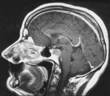
We describe two cases of pituitary involvement by Wegener's granulomatosis. At initial presentation, or during subsequent disease "flares," a pattern of pituitary abnormality was suggested. During periods of remission, we found the pituitary returned to a nearly normal appearance. Loss of the normal posterior pituitary T1 hyper-intensity matched a clinical persistence of diabetes insipidus, suggesting there is permanent damage to this structure by the initial disease process. (+info)The pituitary gland is a small, endocrine gland located at the base of the brain, in the sella turcica of the sphenoid bone. It is often called the "master gland" because it controls other glands and makes the hormones that trigger many body functions. The pituitary gland measures about 0.5 cm in height and 1 cm in width, and it weighs approximately 0.5 grams.
The pituitary gland is divided into two main parts: the anterior lobe (adenohypophysis) and the posterior lobe (neurohypophysis). The anterior lobe is further divided into three zones: the pars distalis, pars intermedia, and pars tuberalis. Each part of the pituitary gland has distinct functions and produces different hormones.
The anterior pituitary gland produces and releases several important hormones, including:
* Growth hormone (GH), which regulates growth and development in children and helps maintain muscle mass and bone strength in adults.
* Thyroid-stimulating hormone (TSH), which controls the production of thyroid hormones by the thyroid gland.
* Adrenocorticotropic hormone (ACTH), which stimulates the adrenal glands to produce cortisol and other steroid hormones.
* Follicle-stimulating hormone (FSH) and luteinizing hormone (LH), which regulate reproductive function in both males and females.
* Prolactin, which stimulates milk production in pregnant and lactating women.
The posterior pituitary gland stores and releases two hormones that are produced by the hypothalamus:
* Antidiuretic hormone (ADH), which helps regulate water balance in the body by controlling urine production.
* Oxytocin, which stimulates uterine contractions during childbirth and milk release during breastfeeding.
Overall, the pituitary gland plays a critical role in maintaining homeostasis and regulating various bodily functions, including growth, development, metabolism, and reproductive function.
The anterior pituitary, also known as the adenohypophysis, is the front portion of the pituitary gland. It is responsible for producing and secreting several important hormones that regulate various bodily functions. These hormones include:
* Growth hormone (GH), which stimulates growth and cell reproduction in bones and other tissues.
* Thyroid-stimulating hormone (TSH), which regulates the production of thyroid hormones by the thyroid gland.
* Adrenocorticotropic hormone (ACTH), which stimulates the adrenal glands to produce cortisol and other steroid hormones.
* Follicle-stimulating hormone (FSH) and luteinizing hormone (LH), which regulate reproductive function in both males and females by controlling the development and release of eggs or sperm.
* Prolactin, which stimulates milk production in pregnant and nursing women.
* Melanocyte-stimulating hormone (MSH), which regulates skin pigmentation and appetite.
The anterior pituitary gland is controlled by the hypothalamus, a small region of the brain located just above it. The hypothalamus produces releasing and inhibiting hormones that regulate the secretion of hormones from the anterior pituitary. These hormones are released into a network of blood vessels called the portal system, which carries them directly to the anterior pituitary gland.
Damage or disease of the anterior pituitary can lead to hormonal imbalances and various medical conditions, such as growth disorders, thyroid dysfunction, adrenal insufficiency, reproductive problems, and diabetes insipidus.
Pituitary neoplasms refer to abnormal growths or tumors in the pituitary gland, a small endocrine gland located at the base of the brain. These neoplasms can be benign (non-cancerous) or malignant (cancerous), with most being benign. They can vary in size and may cause various symptoms depending on their location, size, and hormonal activity.
Pituitary neoplasms can produce and secrete excess hormones, leading to a variety of endocrine disorders such as Cushing's disease (caused by excessive ACTH production), acromegaly (caused by excessive GH production), or prolactinoma (caused by excessive PRL production). They can also cause local compression symptoms due to their size, leading to headaches, vision problems, and cranial nerve palsies.
The exact causes of pituitary neoplasms are not fully understood, but genetic factors, radiation exposure, and certain inherited conditions may increase the risk of developing these tumors. Treatment options for pituitary neoplasms include surgical removal, radiation therapy, and medical management with drugs that can help control hormonal imbalances.
Pituitary diseases refer to a group of conditions that affect the pituitary gland, a small endocrine gland located at the base of the brain. The pituitary gland is responsible for producing and secreting several important hormones that regulate various bodily functions, including growth and development, metabolism, stress response, and reproduction.
Pituitary diseases can be classified into two main categories:
1. Pituitary tumors: These are abnormal growths in or around the pituitary gland that can affect its function. Pituitary tumors can be benign (non-cancerous) or malignant (cancerous), and they can vary in size. Some pituitary tumors produce excess hormones, leading to a variety of symptoms, while others may not produce any hormones but can still cause problems by compressing nearby structures in the brain.
2. Pituitary gland dysfunction: This refers to conditions that affect the normal function of the pituitary gland without the presence of a tumor. Examples include hypopituitarism, which is a condition characterized by decreased production of one or more pituitary hormones, and Sheehan's syndrome, which occurs when the pituitary gland is damaged due to severe blood loss during childbirth.
Symptoms of pituitary diseases can vary widely depending on the specific condition and the hormones that are affected. Treatment options may include surgery, radiation therapy, medication, or a combination of these approaches.
Pituitary hormones are chemical messengers produced and released by the pituitary gland, a small endocrine gland located at the base of the brain. The pituitary gland is often referred to as the "master gland" because it controls several other endocrine glands and regulates various bodily functions.
There are two main types of pituitary hormones: anterior pituitary hormones and posterior pituitary hormones, which are produced in different parts of the pituitary gland and have distinct functions.
Anterior pituitary hormones include:
1. Growth hormone (GH): regulates growth and metabolism.
2. Thyroid-stimulating hormone (TSH): stimulates the thyroid gland to produce thyroid hormones.
3. Adrenocorticotropic hormone (ACTH): stimulates the adrenal glands to produce cortisol and other steroid hormones.
4. Follicle-stimulating hormone (FSH) and luteinizing hormone (LH): regulate reproductive function in both males and females.
5. Prolactin: stimulates milk production in lactating women.
6. Melanocyte-stimulating hormone (MSH): regulates skin pigmentation and appetite.
Posterior pituitary hormones include:
1. Oxytocin: stimulates uterine contractions during childbirth and milk ejection during lactation.
2. Vasopressin (antidiuretic hormone, ADH): regulates water balance in the body by controlling urine production in the kidneys.
Overall, pituitary hormones play crucial roles in regulating growth, development, metabolism, reproductive function, and various other bodily functions. Abnormalities in pituitary hormone levels can lead to a range of medical conditions, such as dwarfism, acromegaly, Cushing's disease, infertility, and diabetes insipidus.
The posterior pituitary gland, also known as the neurohypophysis, is the posterior portion of the pituitary gland. It is primarily composed of nerve fibers that originate from the hypothalamus, a region of the brain. These nerve fibers release two important hormones: oxytocin and vasopressin (also known as antidiuretic hormone or ADH).
Oxytocin plays a role in social bonding, sexual reproduction, and childbirth. During childbirth, it stimulates uterine contractions to help facilitate delivery, and after birth, it helps to trigger the release of milk from the mother's breasts during breastfeeding.
Vasopressin, on the other hand, helps regulate water balance in the body by controlling the amount of water that is excreted by the kidneys. It does this by increasing the reabsorption of water in the collecting ducts of the kidney, which leads to a more concentrated urine and helps prevent dehydration.
Overall, the posterior pituitary gland plays a critical role in maintaining fluid balance, social bonding, and reproduction.
Prolactin is a hormone produced by the pituitary gland, a small gland located at the base of the brain. Its primary function is to stimulate milk production in women after childbirth, a process known as lactation. However, prolactin also plays other roles in the body, including regulating immune responses, metabolism, and behavior. In men, prolactin helps maintain the sexual glands and contributes to paternal behaviors.
Prolactin levels are usually low in both men and non-pregnant women but increase significantly during pregnancy and after childbirth. Various factors can affect prolactin levels, including stress, sleep, exercise, and certain medications. High prolactin levels can lead to medical conditions such as amenorrhea (absence of menstruation), galactorrhea (spontaneous milk production not related to childbirth), infertility, and reduced sexual desire in both men and women.
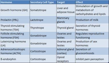
Anterior pituitary hormones are a group of six major hormones that are produced and released by the anterior portion (lobe) of the pituitary gland, a small endocrine gland located at the base of the brain. These hormones play crucial roles in regulating various bodily functions and activities. The six main anterior pituitary hormones are:
1. Growth Hormone (GH): Also known as somatotropin, GH is essential for normal growth and development in children and adolescents. It helps regulate body composition, metabolism, and bone density in adults.
2. Prolactin (PRL): A hormone that stimulates milk production in females after childbirth and is also involved in various reproductive and immune functions in both sexes.
3. Follicle-Stimulating Hormone (FSH): FSH regulates the development, growth, and maturation of follicles in the ovaries (in females) and sperm production in the testes (in males).
4. Luteinizing Hormone (LH): LH plays a key role in triggering ovulation in females and stimulating testosterone production in males.
5. Thyroid-Stimulating Hormone (TSH): TSH regulates the function of the thyroid gland, which is responsible for producing and releasing thyroid hormones that control metabolism and growth.
6. Adrenocorticotropic Hormone (ACTH): ACTH stimulates the adrenal glands to produce cortisol, a steroid hormone involved in stress response, metabolism, and immune function.
These anterior pituitary hormones are regulated by the hypothalamus, which is located above the pituitary gland. The hypothalamus releases releasing and inhibiting factors that control the synthesis and secretion of anterior pituitary hormones, creating a complex feedback system to maintain homeostasis in the body.
The pituitary gland is divided into three lobes: the anterior lobe (adenohypophysis), the posterior lobe (neurohypophysis), and the intermediate lobe (intermedia). The medical definition of 'Pituitary Gland, Intermediate' refers to this small and less defined region located between the anterior and posterior pituitary lobes.
The intermediate lobe is primarily responsible for producing and secreting several important hormones, most notably pro-opiomelanocortin (POMC)-derived peptides such as melanocyte-stimulating hormone (MSH) and endorphins. These hormones play crucial roles in various physiological processes, including skin pigmentation, energy balance, and pain modulation.
However, it is important to note that the intermediate lobe's activity and hormonal secretion are minimal in humans compared to other mammals. In fact, some researchers question whether the human intermediate lobe even functions at all under normal conditions due to its rudimentary nature. Nevertheless, understanding the structure and function of the pituitary gland's intermediate lobe is essential for comparative endocrinology and may provide insights into the evolution of the pituitary gland across different species.
Salivary glands are exocrine glands that produce saliva, which is secreted into the oral cavity to keep the mouth and throat moist, aid in digestion by initiating food breakdown, and help maintain dental health. There are three major pairs of salivary glands: the parotid glands located in the cheeks, the submandibular glands found beneath the jaw, and the sublingual glands situated under the tongue. Additionally, there are numerous minor salivary glands distributed throughout the oral cavity lining. These glands release their secretions through a system of ducts into the mouth.
Pituitary apoplexy is a medical emergency that involves bleeding into the pituitary gland (a small gland at the base of the brain) and/or sudden swelling of the pituitary gland. This can lead to compression of nearby structures, such as the optic nerves and the hypothalamus, causing symptoms like severe headache, visual disturbances, hormonal imbalances, and altered mental status. It is often associated with a pre-existing pituitary tumor (such as a pituitary adenoma), but can also occur in individuals without any known pituitary abnormalities. Immediate medical attention is required to manage this condition, which may include surgical intervention, hormone replacement therapy, and supportive care.
Hypopituitarism is a medical condition characterized by deficient secretion of one or more hormones produced by the pituitary gland, a small endocrine gland located at the base of the brain. The pituitary gland controls several other endocrine glands in the body, including the thyroid, adrenals, and sex glands (ovaries and testes).
Hypopituitarism can result from damage to the pituitary gland due to various causes such as tumors, surgery, radiation therapy, trauma, or inflammation. In some cases, hypopituitarism may also be caused by a dysfunction of the hypothalamus, a region in the brain that regulates the pituitary gland's function.
The symptoms and signs of hypopituitarism depend on which hormones are deficient and can include fatigue, weakness, decreased appetite, weight loss, low blood pressure, decreased sex drive, infertility, irregular menstrual periods, intolerance to cold, constipation, thinning hair, dry skin, and depression.
Treatment of hypopituitarism typically involves hormone replacement therapy to restore the deficient hormones' normal levels. The type and dosage of hormones used will depend on which hormones are deficient and may require regular monitoring and adjustments over time.
An adenoma is a benign (noncancerous) tumor that develops from glandular epithelial cells. These types of cells are responsible for producing and releasing fluids, such as hormones or digestive enzymes, into the surrounding tissues. Adenomas can occur in various organs and glands throughout the body, including the thyroid, pituitary, adrenal, and digestive systems.
Depending on their location, adenomas may cause different symptoms or remain asymptomatic. Some common examples of adenomas include:
1. Colorectal adenoma (also known as a polyp): These growths occur in the lining of the colon or rectum and can develop into colorectal cancer if left untreated. Regular screenings, such as colonoscopies, are essential for early detection and removal of these polyps.
2. Thyroid adenoma: This type of adenoma affects the thyroid gland and may result in an overproduction or underproduction of hormones, leading to conditions like hyperthyroidism (overactive thyroid) or hypothyroidism (underactive thyroid).
3. Pituitary adenoma: These growths occur in the pituitary gland, which is located at the base of the brain and controls various hormonal functions. Depending on their size and location, pituitary adenomas can cause vision problems, headaches, or hormonal imbalances that affect growth, reproduction, and metabolism.
4. Liver adenoma: These rare benign tumors develop in the liver and may not cause any symptoms unless they become large enough to press on surrounding organs or structures. In some cases, liver adenomas can rupture and cause internal bleeding.
5. Adrenal adenoma: These growths occur in the adrenal glands, which are located above the kidneys and produce hormones that regulate stress responses, metabolism, and blood pressure. Most adrenal adenomas are nonfunctioning, meaning they do not secrete excess hormones. However, functioning adrenal adenomas can lead to conditions like Cushing's syndrome or Conn's syndrome, depending on the type of hormone being overproduced.
It is essential to monitor and manage benign tumors like adenomas to prevent potential complications, such as rupture, bleeding, or hormonal imbalances. Treatment options may include surveillance with imaging studies, medication to manage hormonal issues, or surgical removal of the tumor in certain cases.
Growth Hormone (GH), also known as somatotropin, is a peptide hormone secreted by the somatotroph cells in the anterior pituitary gland. It plays a crucial role in regulating growth, cell reproduction, and regeneration by stimulating the production of another hormone called insulin-like growth factor 1 (IGF-1) in the liver and other tissues. GH also has important metabolic functions, such as increasing glucose levels, enhancing protein synthesis, and reducing fat storage. Its secretion is regulated by two hypothalamic hormones: growth hormone-releasing hormone (GHRH), which stimulates its release, and somatostatin (SRIF), which inhibits its release. Abnormal levels of GH can lead to various medical conditions, such as dwarfism or gigantism if there are deficiencies or excesses, respectively.
Adrenocorticotropic Hormone (ACTH) is a hormone produced and released by the anterior pituitary gland, a small endocrine gland located at the base of the brain. ACTH plays a crucial role in the regulation of the body's stress response and has significant effects on various physiological processes.
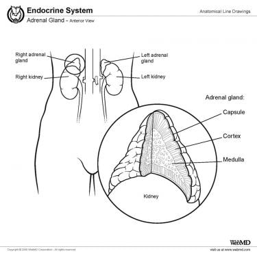
The primary function of ACTH is to stimulate the adrenal glands, which are triangular-shaped glands situated on top of the kidneys. The adrenal glands consist of two parts: the outer cortex and the inner medulla. ACTH specifically targets the adrenal cortex, where it binds to specific receptors and initiates a series of biochemical reactions leading to the production and release of steroid hormones, primarily cortisol (a glucocorticoid) and aldosterone (a mineralocorticoid).
Cortisol is involved in various metabolic processes, such as regulating blood sugar levels, modulating the immune response, and helping the body respond to stress. Aldosterone plays a vital role in maintaining electrolyte and fluid balance by promoting sodium reabsorption and potassium excretion in the kidneys.
ACTH release is controlled by the hypothalamus, another part of the brain, which produces corticotropin-releasing hormone (CRH). CRH stimulates the anterior pituitary gland to secrete ACTH, which in turn triggers cortisol production in the adrenal glands. This complex feedback system helps maintain homeostasis and ensures that appropriate amounts of cortisol are released in response to various physiological and psychological stressors.
Disorders related to ACTH can lead to hormonal imbalances, resulting in conditions such as Cushing's syndrome (excessive cortisol production) or Addison's disease (insufficient cortisol production). Proper diagnosis and management of these disorders typically involve assessing the function of the hypothalamic-pituitary-adrenal axis and addressing any underlying issues affecting ACTH secretion.
Mammary glands are specialized exocrine glands found in mammals, including humans and other animals. These glands are responsible for producing milk, which is used to nurse offspring after birth. The mammary glands are located in the breast region of female mammals and are usually rudimentary or absent in males.
In animals, mammary glands can vary in number and location depending on the species. For example, humans and other primates have two mammary glands, one in each breast. Cows, goats, and sheep, on the other hand, have multiple pairs of mammary glands located in their lower abdominal region.
Mammary glands are made up of several structures, including lobules, ducts, and connective tissue. The lobules contain clusters of milk-secreting cells called alveoli, which produce and store milk. The ducts transport the milk from the lobules to the nipple, where it is released during lactation.
Mammary glands are an essential feature of mammals, as they provide a source of nutrition for newborn offspring. They also play a role in the development and maintenance of the mother-infant bond, as nursing provides opportunities for physical contact and bonding between the mother and her young.
Luteinizing Hormone (LH) is a glycoprotein hormone, which is primarily produced and released by the anterior pituitary gland. In women, a surge of LH triggers ovulation, the release of an egg from the ovaries during the menstrual cycle. During pregnancy, LH stimulates the corpus luteum to produce progesterone. In men, LH stimulates the testes to produce testosterone. It plays a crucial role in sexual development, reproduction, and maintaining the reproductive system.
Gonadotropins are hormones produced and released by the anterior pituitary gland, a small endocrine gland located at the base of the brain. These hormones play crucial roles in regulating reproduction and sexual development. There are two main types of gonadotropins:
1. Follicle-Stimulating Hormone (FSH): FSH is essential for the growth and development of follicles in the ovaries (in females) or sperm production in the testes (in males). In females, FSH stimulates the maturation of eggs within the follicles.
2. Luteinizing Hormone (LH): LH triggers ovulation in females, causing the release of a mature egg from the dominant follicle. In males, LH stimulates the production and secretion of testosterone in the testes.
Together, FSH and LH work synergistically to regulate various aspects of reproductive function and sexual development. Their secretion is controlled by the hypothalamus, which releases gonadotropin-releasing hormone (GnRH) to stimulate the production and release of FSH and LH from the anterior pituitary gland.
Abnormal levels of gonadotropins can lead to various reproductive disorders, such as infertility or menstrual irregularities in females and issues related to sexual development or function in both sexes. In some cases, synthetic forms of gonadotropins may be used clinically to treat these conditions or for assisted reproductive technologies (ART).
Exocrine glands are a type of gland in the human body that produce and release substances through ducts onto an external or internal surface. These glands are responsible for secreting various substances such as enzymes, hormones, and lubricants that help in digestion, protection, and other bodily functions.
Exocrine glands can be further classified into three types based on their mode of secretion:
1. Merocrine glands: These glands release their secretions by exocytosis, where the secretory product is enclosed in a vesicle that fuses with the cell membrane and releases its contents outside the cell. Examples include sweat glands and mucous glands.
2. Apocrine glands: These glands release their secretions by pinching off a portion of the cytoplasm along with the secretory product. An example is the apocrine sweat gland found in the armpits and genital area.
3. Holocrine glands: These glands release their secretions by disintegrating and releasing the entire cell, including its organelles and secretory products. An example is the sebaceous gland found in the skin, which releases an oily substance called sebum.
The submandibular glands are one of the major salivary glands in the human body. They are located beneath the mandible (jawbone) and produce saliva that helps in digestion, lubrication, and protection of the oral cavity. The saliva produced by the submandibular glands contains enzymes like amylase and mucin, which aid in the digestion of carbohydrates and provide moisture to the mouth and throat. Any medical condition or disease that affects the submandibular gland may impact its function and could lead to problems such as dry mouth (xerostomia), swelling, pain, or infection.
Gonadotropin-Releasing Hormone (GnRH), also known as Luteinizing Hormone-Releasing Hormone (LHRH), is a hormonal peptide consisting of 10 amino acids. It is produced and released by the hypothalamus, an area in the brain that links the nervous system to the endocrine system via the pituitary gland.
GnRH plays a crucial role in regulating reproduction and sexual development through its control of two gonadotropins: follicle-stimulating hormone (FSH) and luteinizing hormone (LH). These gonadotropins, in turn, stimulate the gonads (ovaries or testes) to produce sex steroids and eggs or sperm.
GnRH acts on the anterior pituitary gland by binding to its specific receptors, leading to the release of FSH and LH. The hypothalamic-pituitary-gonadal axis is under negative feedback control, meaning that when sex steroid levels are high, they inhibit the release of GnRH, which subsequently decreases FSH and LH secretion.
GnRH agonists and antagonists have clinical applications in various medical conditions, such as infertility treatments, precocious puberty, endometriosis, uterine fibroids, prostate cancer, and hormone-responsive breast cancer.
A prolactinoma is a type of pituitary tumor that produces an excess amount of the hormone prolactin, leading to various symptoms. The pituitary gland, located at the base of the brain, is responsible for producing and releasing several hormones that regulate different bodily functions. Prolactin is one such hormone, primarily known for its role in stimulating milk production in women during lactation (breastfeeding).
Prolactinoma tumors can be classified into two types: microprolactinomas and macroprolactinomas. Microprolactinomas are smaller tumors, typically less than 10 millimeters in size, while macroprolactinomas are larger tumors, generally greater than 10 millimeters in size.
The overproduction of prolactin caused by these tumors can lead to several clinical manifestations, including:
1. Galactorrhea: Unusual and often spontaneous milk production or leakage from the nipples, which can occur in both men and women who do not have a recent history of pregnancy or breastfeeding.
2. Menstrual irregularities: In women, high prolactin levels can interfere with the normal functioning of other hormones, leading to menstrual irregularities such as infrequent periods (oligomenorrhea) or absent periods (amenorrhea), and sometimes infertility.
3. Sexual dysfunction: In both men and women, high prolactin levels can cause decreased libido and sexual desire. Men may also experience erectile dysfunction and reduced sperm production.
4. Bone loss: Over time, high prolactin levels can lead to decreased bone density and an increased risk of osteoporosis due to the disruption of other hormones that regulate bone health.
5. Headaches and visual disturbances: As the tumor grows, it may put pressure on surrounding structures in the brain, leading to headaches and potential vision problems such as blurred vision or decreased peripheral vision.
Diagnosis typically involves measuring prolactin levels in the blood and performing imaging tests like an MRI (magnetic resonance imaging) scan to assess the size of the tumor. Treatment usually consists of medication to lower prolactin levels, such as dopamine agonists (e.g., bromocriptine or cabergoline), which can also help shrink the tumor. In some cases, surgery may be necessary if medication is ineffective or if the tumor is large and causing severe symptoms.
Hypophysectomy is a surgical procedure that involves the removal or partial removal of the pituitary gland, also known as the hypophysis. The pituitary gland is a small endocrine gland located at the base of the brain, just above the nasal cavity, and is responsible for producing and secreting several important hormones that regulate various bodily functions.
Hypophysectomy may be performed for therapeutic or diagnostic purposes. In some cases, it may be used to treat pituitary tumors or other conditions that affect the function of the pituitary gland. It may also be performed as a research procedure in animal models to study the effects of pituitary hormone deficiency on various physiological processes.
The surgical approach for hypophysectomy may vary depending on the specific indication and the patient's individual anatomy. In general, however, the procedure involves making an incision in the skull and exposing the pituitary gland through a small opening in the bone. The gland is then carefully dissected and removed or partially removed as necessary.
Potential complications of hypophysectomy include damage to surrounding structures such as the optic nerves, which can lead to vision loss, and cerebrospinal fluid leaks. Additionally, removal of the pituitary gland can result in hormonal imbalances that may require long-term management with hormone replacement therapy.
The Sella Turcica, also known as the Turkish saddle, is a depression or fossa in the sphenoid bone located at the base of the skull. It forms a housing for the pituitary gland, which is a small endocrine gland often referred to as the "master gland" because it controls other glands and makes several essential hormones. The Sella Turcica has a saddle-like shape, with its anterior and posterior clinoids forming the front and back of the saddle, respectively. This region is of significant interest in neuroimaging and clinical settings, as various conditions such as pituitary tumors or other abnormalities may affect the size, shape, and integrity of the Sella Turcica.
Pituitary function tests are a group of diagnostic exams that evaluate the proper functioning of the pituitary gland, a small endocrine gland located at the base of the brain. The pituitary gland is responsible for producing and releasing several essential hormones that regulate various bodily functions, including growth, metabolism, stress response, reproduction, and lactation.
These tests typically involve measuring the levels of different hormones in the blood, stimulating or suppressing the pituitary gland with specific medications, and assessing the body's response to these challenges. Some common pituitary function tests include:
1. Growth hormone (GH) testing: Measures GH levels in the blood, often after a provocative test using substances like insulin, arginine, clonidine, or glucagon to stimulate GH release.
2. Thyroid-stimulating hormone (TSH) and free thyroxine (FT4) testing: Assesses the function of the thyroid gland by measuring TSH and FT4 levels in response to TRH (thyrotropin-releasing hormone) stimulation.
3. Adrenocorticotropic hormone (ACTH) and cortisol testing: Evaluates the hypothalamic-pituitary-adrenal axis by measuring ACTH and cortisol levels after a CRH (corticotropin-releasing hormone) stimulation test or an insulin tolerance test.
4. Prolactin (PRL) testing: Measures PRL levels in the blood, which can be elevated due to pituitary tumors or other conditions affecting the hypothalamus.
5. Follicle-stimulating hormone (FSH) and luteinizing hormone (LH) testing: Assesses reproductive function by measuring FSH and LH levels, often in conjunction with estradiol or testosterone levels.
6. Gonadotropin-releasing hormone (GnRH) stimulation test: Evaluates gonadal function by measuring FSH and LH levels after GnRH administration.
7. Growth hormone (GH) testing: Measures GH levels in response to various stimuli, such as insulin-like growth factor-1 (IGF-1), glucagon, or arginine.
8. Vasopressin (ADH) testing: Assesses the posterior pituitary function by measuring ADH levels and performing a water deprivation test.
These tests can help diagnose various pituitary disorders, such as hypopituitarism, hyperpituitarism, or pituitary tumors, and guide appropriate treatment strategies.
Pro-opiomelanocortin (POMC) is a precursor protein that gets cleaved into several biologically active peptides in the body. These peptides include adrenocorticotropic hormone (ACTH), beta-lipotropin, and multiple opioid peptides such as beta-endorphin, met-enkephalin, and leu-enkephalin.
ACTH stimulates the release of cortisol from the adrenal gland, while beta-lipotropin has various metabolic functions. The opioid peptides derived from POMC have pain-relieving (analgesic) and rewarding effects in the brain. Dysregulation of the POMC system has been implicated in several medical conditions, including obesity, addiction, and certain types of hormone deficiencies.
The adrenal glands are a pair of endocrine glands that are located on top of the kidneys. Each gland has two parts: the outer cortex and the inner medulla. The adrenal cortex produces hormones such as cortisol, aldosterone, and androgens, which regulate metabolism, blood pressure, and other vital functions. The adrenal medulla produces catecholamines, including epinephrine (adrenaline) and norepinephrine (noradrenaline), which help the body respond to stress by increasing heart rate, blood pressure, and alertness.
Follicle-Stimulating Hormone (FSH) is a glycoprotein hormone secreted and released by the anterior pituitary gland. In females, it promotes the growth and development of ovarian follicles in the ovary, which ultimately leads to the maturation and release of an egg (ovulation). In males, FSH stimulates the testes to produce sperm. It works in conjunction with luteinizing hormone (LH) to regulate reproductive processes. The secretion of FSH is controlled by the hypothalamic-pituitary-gonadal axis and its release is influenced by the levels of gonadotropin-releasing hormone (GnRH), estrogen, inhibin, and androgens.
Somatotrophs are a type of cell found within the anterior pituitary gland, a small endocrine gland located at the base of the brain. These cells are responsible for producing and secreting the hormone known as somatotropin or growth hormone (GH). This hormone plays a crucial role in regulating growth, cell reproduction, and regeneration. It also helps to regulate the body's metabolism and maintain proper body composition by promoting the breakdown of fats and the synthesis of proteins. Disorders related to somatotrophs can lead to conditions such as gigantism or dwarfism, depending on whether there is an overproduction or underproduction of growth hormone.
Pituitary Adenylate Cyclase-Activating Polypeptide (PACAP) is a neuropeptide that belongs to the vasoactive intestinal polypeptide (VIP)/secretin/glucagon family. It was first isolated from the ovine hypothalamus and later found in various tissues and organs throughout the body, including the brain, pituitary gland, and peripheral nerves.
PACAP exists in two forms, PACAP-38 and PACAP-27, which differ in their length but share the same amino acid sequence at the N-terminus. PACAP exerts its effects through specific G protein-coupled receptors, including PAC1, VPAC1, and VPAC2 receptors, which are widely distributed throughout the body.
PACAP has a wide range of biological activities, including neurotrophic, neuroprotective, vasodilatory, and immunomodulatory effects. In the pituitary gland, PACAP stimulates adenylate cyclase activity, leading to an increase in intracellular cAMP levels, which in turn regulates the release of various hormones, including growth hormone, prolactin, and thyroid-stimulating hormone.
Overall, PACAP is a crucial neuropeptide involved in various physiological processes, and its dysregulation has been implicated in several pathological conditions, such as neurodegenerative diseases, mood disorders, and cancer.
The hypothalamus is a small, vital region of the brain that lies just below the thalamus and forms part of the limbic system. It plays a crucial role in many important functions including:
1. Regulation of body temperature, hunger, thirst, fatigue, sleep, and circadian rhythms.
2. Production and regulation of hormones through its connection with the pituitary gland (the hypophysis). It controls the release of various hormones by producing releasing and inhibiting factors that regulate the anterior pituitary's function.
3. Emotional responses, behavior, and memory formation through its connections with the limbic system structures like the amygdala and hippocampus.
4. Autonomic nervous system regulation, which controls involuntary physiological functions such as heart rate, blood pressure, and digestion.
5. Regulation of the immune system by interacting with the autonomic nervous system.
Damage to the hypothalamus can lead to various disorders like diabetes insipidus, growth hormone deficiency, altered temperature regulation, sleep disturbances, and emotional or behavioral changes.
The parotid gland is the largest of the major salivary glands. It is a bilobed, accessory digestive organ that secretes serous saliva into the mouth via the parotid duct (Stensen's duct), located near the upper second molar tooth. The parotid gland is primarily responsible for moistening and lubricating food to aid in swallowing and digestion.
Anatomically, the parotid gland is located in the preauricular region, extending from the zygomatic arch superiorly to the angle of the mandible inferiorly, and from the masseter muscle anteriorly to the sternocleidomastoid muscle posteriorly. It is enclosed within a fascial capsule and has a rich blood supply from the external carotid artery and a complex innervation pattern involving both parasympathetic and sympathetic fibers.
Parotid gland disorders can include salivary gland stones (sialolithiasis), infections, inflammatory conditions, benign or malignant tumors, and autoimmune diseases such as Sjögren's syndrome.
Thyrotropin-Releasing Hormone (TRH) is a tripeptide hormone that is produced and released by the hypothalamus in the brain. Its main function is to regulate the release of thyroid-stimulating hormone (TSH) from the anterior pituitary gland. TRH acts on the pituitary gland to stimulate the synthesis and secretion of TSH, which then stimulates the thyroid gland to produce and release thyroid hormones (triiodothyronine (T3) and thyroxine (T4)) into the bloodstream.
TRH is a tripeptide amino acid sequence with the structure of pGlu-His-Pro-NH2, and it is synthesized as a larger precursor molecule called preprothyrotropin-releasing hormone (preproTRH) in the hypothalamus. PreproTRH undergoes post-translational processing to produce TRH, which is then stored in secretory vesicles and released into the hypophyseal portal system, where it travels to the anterior pituitary gland and binds to TRH receptors on thyrotroph cells.
In addition to its role in regulating TSH release, TRH has been shown to have other physiological functions, including modulation of feeding behavior, body temperature, and neurotransmitter release. Dysregulation of the TRH-TSH axis can lead to various thyroid disorders, such as hypothyroidism or hyperthyroidism.
Lactotrophs, also known as mammotrophs or prolactin cells, are a type of hormone-producing cell found in the anterior pituitary gland. They are responsible for producing and secreting the hormone prolactin, which plays a crucial role in lactation (milk production) in females after childbirth. Prolactin also has other functions in the body, such as regulating immune responses, metabolism, and behavior. Lactotrophs can be stimulated by factors like estrogen, thyroid-stimulating hormone (TSH), and stress, leading to increased prolactin secretion.
Gonadotrophs are a type of hormone-secreting cells located in the anterior pituitary gland, a small endocrine gland at the base of the brain. These cells produce and release two important gonadotropin hormones: follicle-stimulating hormone (FSH) and luteinizing hormone (LH).
Follicle-stimulating hormone (FSH) plays a crucial role in the reproductive system by stimulating the growth and development of ovarian follicles in females and sperm production in males. In females, FSH also promotes the production of estrogen during the menstrual cycle.
Luteinizing hormone (LH) is responsible for triggering ovulation in females, releasing a mature egg from the ovary into the fallopian tube. In addition, LH stimulates the production of progesterone by the remaining cells of the ruptured follicle, which forms the corpus luteum. In males, LH helps regulate testosterone production in the testes.
Gonadotrophs are essential for maintaining reproductive function and hormonal balance in both sexes. Their activity is controlled by the hypothalamus, another part of the brain that releases gonadotropin-releasing hormone (GnRH) to regulate FSH and LH secretion.
LHRH (Luteinizing Hormone-Releasing Hormone) receptors are a type of G protein-coupled receptor found on the surface of certain cells in the body, most notably in the anterior pituitary gland. These receptors bind to LHRH, a hormone that is produced and released by the hypothalamus in the brain.
When LHRH binds to its receptor, it triggers a series of intracellular signaling events that ultimately lead to the release of two other hormones from the anterior pituitary gland: luteinizing hormone (LH) and follicle-stimulating hormone (FSH). These hormones play critical roles in regulating reproductive function, including the development and maturation of sex cells (sperm and eggs), the production of sex steroid hormones (such as testosterone and estrogen), and the regulation of the menstrual cycle in females.
Disorders of the LHRH receptor or its signaling pathway can lead to a variety of reproductive disorders, including precocious puberty, delayed puberty, and infertility.
Thyrotropin, also known as thyroid-stimulating hormone (TSH), is a hormone secreted by the anterior pituitary gland. Its primary function is to regulate the production and release of thyroxine (T4) and triiodothyronine (T3) hormones from the thyroid gland. Thyrotropin binds to receptors on the surface of thyroid follicular cells, stimulating the uptake of iodide and the synthesis and release of T4 and T3. The secretion of thyrotropin is controlled by the hypothalamic-pituitary-thyroid axis: thyrotropin-releasing hormone (TRH) from the hypothalamus stimulates the release of thyrotropin, while T3 and T4 inhibit its release through a negative feedback mechanism.
An ACTH-secreting pituitary adenoma is a type of tumor that develops in the pituitary gland, a small gland located at the base of the brain. This type of tumor is also known as Cushing's disease.
ACTH stands for adrenocorticotropic hormone, which is a hormone produced and released by the pituitary gland. ACTH stimulates the adrenal glands (small glands located on top of the kidneys) to produce cortisol, a steroid hormone that helps regulate metabolism, helps the body respond to stress, and suppresses inflammation.
In an ACTH-secreting pituitary adenoma, the tumor cells produce and release excessive amounts of ACTH, leading to overproduction of cortisol by the adrenal glands. This can result in a constellation of symptoms known as Cushing's syndrome, which may include weight gain (especially around the trunk), fatigue, muscle weakness, mood changes, thinning of the skin, easy bruising, and increased susceptibility to infections.
Treatment for an ACTH-secreting pituitary adenoma typically involves surgical removal of the tumor, followed by medications to manage cortisol levels if necessary. Radiation therapy may also be used in some cases.
A chromophobe adenoma is a type of benign (non-cancerous) tumor that typically arises in the pituitary gland, which is a small endocrine gland located at the base of the brain. The term "chromophobe" refers to the appearance of the cells under a microscope - they lack pigment and have a characteristic appearance with abundant clear or lightly stained cytoplasm.
Chromophobe adenomas are slow-growing tumors that can vary in size, and they may cause symptoms due to pressure on surrounding structures or by producing excess hormones. The most common hormone produced by chromophobe adenomas is prolactin, leading to symptoms such as menstrual irregularities, milk production (galactorrhea), and decreased sexual function in women, and decreased libido, erectile dysfunction, and infertility in men.
Treatment for chromophobe adenomas typically involves surgical removal of the tumor, often through a transsphenoidal approach (through the nose and sphenoid sinus). In some cases, radiation therapy or medical management with hormone-blocking drugs may also be necessary. Regular follow-up with an endocrinologist is important to monitor for any recurrence or hormonal imbalances.
Follicle-stimulating hormone (FSH) is a glycoprotein hormone produced and released by the anterior pituitary gland. It plays crucial roles in the reproductive system, primarily by promoting the growth and development of follicles in the ovaries or sperm production in the testes.
The FSH molecule consists of two subunits: α (alpha) and β (beta). The α-subunit is common to several glycoprotein hormones, including thyroid-stimulating hormone (TSH), luteinizing hormone (LH), and human chorionic gonadotropin (hCG). In contrast, the β-subunit is unique to each hormone and determines its specific biological activity.
A medical definition of 'Follicle Stimulating Hormone, beta Subunit' refers to the distinct portion of the FSH molecule that is responsible for its particular functions in the body. The β-subunit of FSH enables the hormone to bind to its specific receptors in the gonads and initiate downstream signaling pathways leading to follicular development and spermatogenesis. Any alterations or mutations in the FSH beta subunit can lead to disruptions in reproductive processes, potentially causing infertility or other related disorders.
The posterior cranial fossa is a term used in anatomy to refer to the portion of the skull that forms the lower, back part of the cranial cavity. It is located between the occipital bone and the temporal bones, and it contains several important structures including the cerebellum, pons, medulla oblongata, and the lower cranial nerves (IX-XII). The posterior fossa also contains the foramen magnum, which is a large opening through which the spinal cord connects to the brainstem. This region of the skull is protected by the occipital bone, which forms the base of the skull and provides attachment for several neck muscles.
Sweat glands are specialized tubular structures in the skin that produce and secrete sweat, also known as perspiration. They are part of the body's thermoregulatory system, helping to maintain optimal body temperature by releasing water and heat through evaporation. There are two main types of sweat glands: eccrine and apocrine.
1. Eccrine sweat glands: These are distributed throughout the body, with a higher concentration on areas like the palms, soles, and forehead. They are responsible for producing a watery, odorless sweat that primarily helps to cool down the body through evaporation.
2. Apocrine sweat glands: These are mainly found in the axillary (armpit) region and around the anogenital area. They become active during puberty and produce a thick, milky fluid that does not have a strong odor on its own but can mix with bacteria on the skin's surface, leading to body odor.
Sweat glands are controlled by the autonomic nervous system, meaning they function involuntarily in response to various stimuli such as emotions, physical activity, or changes in environmental temperature.
Pituitary dwarfism, also known as growth hormone deficiency dwarfism or hypopituitarism dwarfism, is a type of dwarfism that results from insufficient production of growth hormone by the pituitary gland during childhood. The medical term for this condition is "growth hormone deficiency."
The pituitary gland is a small gland located at the base of the brain that produces several important hormones, including growth hormone. Growth hormone plays a critical role in regulating growth and development during childhood and adolescence. When the pituitary gland fails to produce enough growth hormone, children do not grow and develop normally, resulting in short stature and other symptoms associated with dwarfism.
Pituitary dwarfism can be caused by a variety of factors, including genetic mutations, brain tumors, trauma, or infection. In some cases, the cause may be unknown. Symptoms of pituitary dwarfism include short stature, delayed puberty, and other hormonal imbalances.
Treatment for pituitary dwarfism typically involves replacing the missing growth hormone with injections of synthetic growth hormone. This therapy can help promote normal growth and development, although it may not completely eliminate the short stature associated with the condition. Early diagnosis and treatment are essential to optimize outcomes and improve quality of life for individuals with pituitary dwarfism.
Pituitary ACTH hypersecretion, also known as Cushing's disease, is a condition characterized by the excessive production of adrenocorticotropic hormone (ACTH) from the pituitary gland. This results in an overproduction of cortisol, a steroid hormone produced by the adrenal glands, leading to a constellation of symptoms known as Cushing's syndrome.
In Cushing's disease, a benign tumor called an adenoma develops on the pituitary gland, causing it to release excess ACTH. This in turn stimulates the adrenal glands to produce more cortisol than necessary. The resulting high levels of cortisol can cause various symptoms such as weight gain, particularly around the trunk and face (central obesity), thinning of the skin, bruising, weakness, fatigue, mood changes, high blood pressure, and an increased risk of infections.
It is important to distinguish Cushing's disease from other causes of Cushing's syndrome, such as cortisol-producing adrenal tumors or exogenous sources of corticosteroid use, as the treatment approach may differ. Treatment for Cushing's disease typically involves surgical removal of the pituitary tumor, with additional medical management and/or radiation therapy in some cases.
Beta-lipotropin (β-LPH) is a 91-amino acid polypeptide hormone that is derived from proopiomelanocortin (POMC), along with other bioactive peptides such as adrenocorticotropic hormone (ACTH), melanocyte-stimulating hormones (MSH), and β-endorphin. It is produced and released by the anterior pituitary gland in response to stress or corticotropin-releasing hormone (CRH) stimulation.
β-Lipotropin has been found to have several physiological functions, including the regulation of lipid metabolism, appetite control, and pain perception. It also exhibits opioid activity due to its ability to bind to opioid receptors in the brain, although its potency is much lower compared to other endogenous opioids like β-endorphin.
In addition to its role as a hormone, β-lipotropin has been studied for its potential therapeutic applications, particularly in the treatment of obesity and addiction. However, further research is needed to fully understand its mechanisms and clinical efficacy.
Medical Definition:
Magnetic Resonance Imaging (MRI) is a non-invasive diagnostic imaging technique that uses a strong magnetic field and radio waves to create detailed cross-sectional or three-dimensional images of the internal structures of the body. The patient lies within a large, cylindrical magnet, and the scanner detects changes in the direction of the magnetic field caused by protons in the body. These changes are then converted into detailed images that help medical professionals to diagnose and monitor various medical conditions, such as tumors, injuries, or diseases affecting the brain, spinal cord, heart, blood vessels, joints, and other internal organs. MRI does not use radiation like computed tomography (CT) scans.
The Hypothalamo-Hypophyseal system, also known as the hypothalamic-pituitary system, is a crucial part of the endocrine system that regulates many bodily functions. It consists of two main components: the hypothalamus and the pituitary gland.
The hypothalamus is a region in the brain that receives information from various parts of the body and integrates them to regulate vital functions such as body temperature, hunger, thirst, sleep, and emotional behavior. It also produces and releases neurohormones that control the secretion of hormones from the pituitary gland.
The pituitary gland is a small gland located at the base of the brain, just below the hypothalamus. It consists of two parts: the anterior pituitary (also called adenohypophysis) and the posterior pituitary (also called neurohypophysis). The anterior pituitary produces and releases several hormones that regulate various bodily functions such as growth, metabolism, reproduction, and stress response. The posterior pituitary stores and releases hormones produced by the hypothalamus, including antidiuretic hormone (ADH) and oxytocin.
The hypothalamo-hypophyseal system works together to maintain homeostasis in the body by regulating various physiological processes through hormonal signaling. Dysfunction of this system can lead to several endocrine disorders, such as diabetes insipidus, pituitary tumors, and hypothalamic-pituitary axis disorders.
Sebaceous glands are microscopic, exocrine glands that are found in the dermis of mammalian skin. They are attached to hair follicles and produce an oily substance called sebum, which is composed of triglycerides, wax esters, squalene, and metabolites of fat-producing cells (fatty acids, cholesterol). Sebum is released through a duct onto the surface of the skin, where it forms a protective barrier that helps to prevent water loss, keeps the skin and hair moisturized, and has antibacterial properties.
Sebaceous glands are distributed throughout the body, but they are most numerous on the face, scalp, and upper trunk. They can also be found in other areas of the body such as the eyelids (where they are known as meibomian glands), the external ear canal, and the genital area.
Abnormalities in sebaceous gland function can lead to various skin conditions, including acne, seborrheic dermatitis, and certain types of skin cancer.
Growth Hormone-Releasing Hormone (GHRH) is a hormone that is produced and released by the hypothalamus, a small gland located in the brain. Its primary function is to stimulate the anterior pituitary gland to release growth hormone (GH) into the bloodstream. GH plays a crucial role in growth and development, particularly during childhood and adolescence, by promoting the growth of bones and muscles.
GHRH is a 44-amino acid peptide that binds to specific receptors on the surface of pituitary cells, triggering a series of intracellular signals that ultimately lead to the release of GH. The production and release of GHRH are regulated by various factors, including sleep, stress, exercise, and nutrition.
Abnormalities in the production or function of GHRH can lead to growth disorders, such as dwarfism or gigantism, as well as other hormonal imbalances. Therefore, understanding the role of GHRH in regulating GH release is essential for diagnosing and treating these conditions.
Pituitary hormone-releasing hormones (PRHs), also known as hypothalamic releasing hormones or hypothalamic hormones, are small neuropeptides produced and released by the hypothalamus - a small region of the brain. These hormones play crucial roles in regulating the secretion and release of various pituitary hormones, which in turn control several essential bodily functions, including growth, development, metabolism, stress response, reproduction, and lactation.
There are several PRHs, each with a specific target pituitary hormone:
1. Thyrotropin-releasing hormone (TRH): Stimulates the release of thyroid-stimulating hormone (TSH) from the anterior pituitary gland, which then promotes the production and release of thyroid hormones.
2. Gonadotropin-releasing hormone (GnRH): Regulates the secretion of follicle-stimulating hormone (FSH) and luteinizing hormone (LH) from the anterior pituitary gland, which are essential for reproductive functions.
3. Corticotropin-releasing hormone (CRH): Stimulates the release of adrenocorticotropic hormone (ACTH) from the anterior pituitary gland, which then promotes the production and release of cortisol and other glucocorticoids from the adrenal glands.
4. Growth hormone-releasing hormone (GHRH): Stimulates the release of growth hormone (GH) from the anterior pituitary gland, which is essential for growth, development, and metabolism regulation.
5. Somatostatin or growth hormone-inhibiting hormone (GHIH): Inhibits the release of GH from the anterior pituitary gland and also suppresses the secretion of thyroid hormones.
6. Prolactin-releasing hormone (PRH) or prolactin-releasing factor (PRF): Stimulates the release of prolactin from the anterior pituitary gland, which is essential for lactation and reproductive functions.
7. Prolactin-inhibiting hormone (PIH) or dopamine: Inhibits the release of prolactin from the anterior pituitary gland.
These releasing hormones and inhibitory hormones work together to maintain a delicate balance in various physiological processes, including growth, development, metabolism, stress response, and reproductive functions. Dysregulation of these hormonal systems can lead to various endocrine disorders and diseases.
Messenger RNA (mRNA) is a type of RNA (ribonucleic acid) that carries genetic information copied from DNA in the form of a series of three-base code "words," each of which specifies a particular amino acid. This information is used by the cell's machinery to construct proteins, a process known as translation. After being transcribed from DNA, mRNA travels out of the nucleus to the ribosomes in the cytoplasm where protein synthesis occurs. Once the protein has been synthesized, the mRNA may be degraded and recycled. Post-transcriptional modifications can also occur to mRNA, such as alternative splicing and addition of a 5' cap and a poly(A) tail, which can affect its stability, localization, and translation efficiency.
Transcription Factor Pit-1, also known as POU1F1 or pituitary-specific transcription factor 1, is a protein that plays a crucial role in the development and function of the anterior pituitary gland. It is a member of the POU domain family of transcription factors, which are characterized by a conserved DNA-binding domain.
Pit-1 is essential for the differentiation and proliferation of certain types of pituitary cells, including those that produce growth hormone (GH), prolactin (PRL), and thyroid-stimulating hormone (TSH). Pit-1 binds to specific DNA sequences in the promoter regions of these hormone genes, thereby activating their transcription and promoting hormone production.
Mutations in the gene encoding Pit-1 can lead to a variety of pituitary disorders, such as dwarfism due to GH deficiency, delayed puberty, and hypothyroidism due to TSH deficiency. Additionally, some studies have suggested that Pit-1 may also play a role in regulating energy balance and body weight, although the exact mechanisms are not fully understood.
The sublingual glands are a pair of salivary glands located in the floor of the mouth, beneath the tongue. They are the smallest of the major salivary glands and produce around 5-10% of the total saliva in the mouth. The sublingual glands secrete saliva containing electrolytes, enzymes (such as amylase), and antibacterial compounds that help in digestion, lubrication, and protection against microorganisms.
The sublingual glands' secretions are released through multiple small ducts called the ducts of Rivinus or minor sublingual ducts, as well as a larger duct called the duct of Wharton, which is a common excretory duct for both sublingual and submandibular glands.
Sublingual gland dysfunction can lead to conditions such as dry mouth (xerostomia), dental caries, or oral infections.
Radioimmunoassay (RIA) is a highly sensitive analytical technique used in clinical and research laboratories to measure concentrations of various substances, such as hormones, vitamins, drugs, or tumor markers, in biological samples like blood, urine, or tissues. The method relies on the specific interaction between an antibody and its corresponding antigen, combined with the use of radioisotopes to quantify the amount of bound antigen.
In a typical RIA procedure, a known quantity of a radiolabeled antigen (also called tracer) is added to a sample containing an unknown concentration of the same unlabeled antigen. The mixture is then incubated with a specific antibody that binds to the antigen. During the incubation period, the antibody forms complexes with both the radiolabeled and unlabeled antigens.
After the incubation, the unbound (free) radiolabeled antigen is separated from the antibody-antigen complexes, usually through a precipitation or separation step involving centrifugation, filtration, or chromatography. The amount of radioactivity in the pellet (containing the antibody-antigen complexes) is then measured using a gamma counter or other suitable radiation detection device.
The concentration of the unlabeled antigen in the sample can be determined by comparing the ratio of bound to free radiolabeled antigen in the sample to a standard curve generated from known concentrations of unlabeled antigen and their corresponding bound/free ratios. The higher the concentration of unlabeled antigen in the sample, the lower the amount of radiolabeled antigen that will bind to the antibody, resulting in a lower bound/free ratio.
Radioimmunoassays offer high sensitivity, specificity, and accuracy, making them valuable tools for detecting and quantifying low levels of various substances in biological samples. However, due to concerns about radiation safety and waste disposal, alternative non-isotopic immunoassay techniques like enzyme-linked immunosorbent assays (ELISAs) have become more popular in recent years.
Endorphins are a type of neurotransmitter, which are chemicals that transmit signals in the nervous system and brain. The term "endorphin" comes from "endogenous morphine," reflecting the fact that these substances are produced naturally within the body and have effects similar to opiate drugs like morphine.
Endorphins are released in response to stress or pain, but they also occur naturally during exercise, excitement, laughter, love, and orgasm. They work by interacting with the opiate receptors in the brain to reduce the perception of pain and promote feelings of pleasure and well-being. Endorphins also play a role in regulating various physiological processes, including appetite, mood, and sleep.
In summary, endorphins are natural painkillers and mood elevators produced by the body in response to stress, pain, or enjoyable activities.
Bromocriptine is a dopamine receptor agonist drug, which means it works by binding to and activating dopamine receptors in the brain. It has several therapeutic uses, including:
* Treatment of Parkinson's disease: Bromocriptine can be used alone or in combination with levodopa to help manage the symptoms of Parkinson's disease, such as stiffness, tremors, spasms, and poor muscle control.
* Suppression of lactation: Bromocriptine can be used to suppress milk production in women who are not breastfeeding or who have stopped breastfeeding but still have high levels of prolactin, a hormone that stimulates milk production.
* Treatment of pituitary tumors: Bromocriptine can be used to shrink certain types of pituitary tumors, such as prolactinomas, which are tumors that secrete excessive amounts of prolactin.
* Management of acromegaly: Bromocriptine can be used to manage the symptoms of acromegaly, a rare hormonal disorder characterized by abnormal growth and enlargement of body tissues, by reducing the production of growth hormone.
Bromocriptine is available in immediate-release and long-acting formulations, and it is usually taken orally. Common side effects of bromocriptine include nausea, dizziness, lightheadedness, and drowsiness. Serious side effects are rare but can include hallucinations, confusion, and priapism (prolonged erection).
The sphenoid sinuses are air-filled spaces located within the sphenoid bone, which is one of the bones that make up the skull base. These sinuses are located deep inside the skull, behind the eyes and nasal cavity. They are paired and separated by a thin bony septum, and each one opens into the corresponding nasal cavity through a small opening called the sphenoethmoidal recess. The sphenoid sinuses vary greatly in size and shape between individuals. They develop during childhood and continue to grow until early adulthood. The function of the sphenoid sinuses, like other paranasal sinuses, is not entirely clear, but they may contribute to reducing the weight of the skull, resonating voice during speech, and insulating the brain from trauma.
Luteinizing Hormone (LH) is a glycoprotein hormone secreted by the anterior pituitary gland. It plays a crucial role in regulating the reproductive system. The beta subunit of LH is one of the two non-identical polypeptide chains that make up the LH molecule (the other being the alpha subunit, which is common to several hormones).
The beta subunit of LH is unique to LH and is often used in assays to measure and determine the concentration of LH in blood or urine. It's responsible for the biological specificity and activity of the LH hormone. Any changes in the structure of this subunit can affect the function of LH, which in turn can have implications for reproductive processes such as ovulation and testosterone production.
Thyrotropin, also known as thyroid-stimulating hormone (TSH), is a hormone produced and released by the anterior pituitary gland. It plays a crucial role in regulating the function of the thyroid gland by stimulating the production and release of thyroid hormones, triiodothyronine (T3) and thyroxine (T4).
The TSH molecule is composed of two subunits: alpha and beta. The alpha subunit is common to several pituitary hormones, including TSH, follicle-stimulating hormone (FSH), luteinizing hormone (LH), and human chorionic gonadotropin (hCG). In contrast, the beta subunit is unique to each hormone, determining its specific biological activity.
Therefore, 'Thyrotropin, beta Subunit' refers to the distinct portion of the TSH molecule that confers its thyroid-stimulating properties and allows it to be identified and measured separately from other pituitary hormones sharing the common alpha subunit. Beta-subunit assays are sometimes used in clinical settings to evaluate thyroid function, as they can provide information about TSH levels independent of the common alpha subunit.
The Harderian gland is a specialized exocrine gland located in many vertebrate species, including birds and mammals. In humans, it is rudimentary and not fully developed. However, in other animals like rodents, lagomorphs (rabbits and hares), and some reptiles, this gland plays a significant role.
The Harderian gland is primarily responsible for producing and secreting lipids, which help to lubricate the eye's surface and the nictitating membrane (third eyelid). This lubrication ensures that the eyes remain moist and protected from dryness and external irritants. Additionally, the secretions of the Harderian gland contain immunoglobulins, which contribute to the animal's immune defense system by providing protection against pathogens.
In some animals, the Harderian gland also has a role in pheromone production and communication. The study and understanding of this gland are particularly important in toxicological research, as it is often used as an indicator of environmental pollutant exposure and their effects on wildlife.
Castration is a surgical procedure to remove the testicles in males or ovaries in females. In males, it is also known as orchiectomy. This procedure results in the inability to produce sex hormones and gametes (sperm in men and eggs in women), and can be done for various reasons such as medical treatment for certain types of cancer, to reduce sexual urges in individuals with criminal tendencies, or as a form of birth control in animals.
A Growth Hormone-Secreting Pituitary Adenoma (GH-secreting pituitary adenoma, or GHoma) is a type of benign tumor that develops in the pituitary gland and results in excessive production of growth hormone (GH). This leads to a condition known as acromegaly if it occurs in adults, or gigantism if it occurs in children before the closure of the growth plates.
Symptoms of GH-secreting pituitary adenoma may include:
1. Coarsening of facial features
2. Enlargement of hands and feet
3. Deepened voice due to thickening of vocal cords
4. Increased sweating and body odor
5. Joint pain and stiffness
6. Sleep apnea
7. Fatigue, weakness, or muscle wasting
8. Headaches
9. Vision problems
10. Irregular menstrual periods in women
11. Erectile dysfunction in men
Diagnosis typically involves measuring the levels of GH and insulin-like growth factor 1 (IGF-1) in the blood, along with imaging tests like MRI or CT scans to locate and characterize the tumor. Treatment options include surgical removal of the tumor, radiation therapy, and medication to control GH production. Regular follow-ups are necessary to monitor for potential recurrence.
Glycoprotein hormones are a group of hormones that share a similar structure and are made up of four subunits: two identical alpha subunits and two distinct beta subunits. The alpha subunit is common to all glycoprotein hormones, including thyroid-stimulating hormone (TSH), follicle-stimulating hormone (FSH), luteinizing hormone (LH), and human chorionic gonadotropin (hCG).
The alpha subunit of glycoprotein hormones is a 92 amino acid polypeptide chain that contains several disulfide bonds, which help to stabilize its structure. It is heavily glycosylated, meaning that it contains many carbohydrate groups attached to the protein backbone. The alpha subunit plays an important role in the biological activity of the hormone by interacting with a specific receptor on the target cell surface.
The alpha subunit contains several regions that are important for its function, including a signal peptide, a variable region, and a conserved region. The signal peptide is a short sequence of amino acids at the N-terminus of the protein that directs it to the endoplasmic reticulum for processing and secretion. The variable region contains several amino acid residues that differ between different glycoprotein hormones, while the conserved region contains amino acids that are identical or very similar in all glycoprotein hormones.
Together with the beta subunit, the alpha subunit forms the functional hormone molecule. The beta subunit determines the specificity of the hormone for its target cells and regulates its biological activity.
Immunohistochemistry (IHC) is a technique used in pathology and laboratory medicine to identify specific proteins or antigens in tissue sections. It combines the principles of immunology and histology to detect the presence and location of these target molecules within cells and tissues. This technique utilizes antibodies that are specific to the protein or antigen of interest, which are then tagged with a detection system such as a chromogen or fluorophore. The stained tissue sections can be examined under a microscope, allowing for the visualization and analysis of the distribution and expression patterns of the target molecule in the context of the tissue architecture. Immunohistochemistry is widely used in diagnostic pathology to help identify various diseases, including cancer, infectious diseases, and immune-mediated disorders.
Human Growth Hormone (HGH), also known as somatotropin, is a peptide hormone produced in the pituitary gland. It plays a crucial role in human development and growth by stimulating the production of another hormone called insulin-like growth factor 1 (IGF-1). IGF-1 promotes the growth and reproduction of cells throughout the body, particularly in bones and other tissues. HGH also helps regulate body composition, body fluids, muscle and bone growth, sugar and fat metabolism, and possibly heart function. It is essential for human development and continues to have important effects throughout life. The secretion of HGH decreases with age, which is thought to contribute to the aging process.
I apologize, but "sheep" is not a term that has a medical definition. It is a common animal with the scientific name Ovis aries. If you have any medical or health-related questions, I would be happy to try and help answer those for you.
Pituitary hormone-regulating hormone receptors refer to specific protein structures found on the surface of certain cells in the body. These receptors are responsible for detecting and responding to hormones produced by the hypothalamus, which regulate the function of the pituitary gland.
The pituitary gland is a small gland located at the base of the brain that plays a critical role in regulating various bodily functions, including growth and development, metabolism, reproduction, and stress response. The hypothalamus produces hormones that either stimulate or inhibit the release of pituitary hormones, which then act on target organs throughout the body to regulate their function.
Pituitary hormone-regulating hormone receptors are found on the surface of pituitary cells and are specific to individual hypothalamic hormones. When a hypothalamic hormone binds to its corresponding receptor, it triggers a series of intracellular signals that ultimately result in the release or inhibition of pituitary hormones.
Examples of pituitary hormone-regulating hormone receptors include:
* Thyroid-stimulating hormone (TSH) receptor, which responds to thyrotropin-releasing hormone (TRH) from the hypothalamus.
* Adrenocorticotropic hormone (ACTH) receptor, which responds to corticotropin-releasing hormone (CRH) from the hypothalamus.
* Growth hormone-releasing hormone (GHRH) receptor, which responds to GHRH from the hypothalamus.
* Gonadotropin-releasing hormone (GnRH) receptor, which responds to GnRH from the hypothalamus.
* Prolactin-inhibiting hormone (PIH) receptor, which responds to dopamine from the hypothalamus.
Abnormalities in pituitary hormone-regulating hormone receptors can lead to various endocrine disorders, such as hypothyroidism, Cushing's disease, acromegaly, and infertility.
Estradiol is a type of estrogen, which is a female sex hormone. It is the most potent and dominant form of estrogen in humans. Estradiol plays a crucial role in the development and maintenance of secondary sexual characteristics in women, such as breast development and regulation of the menstrual cycle. It also helps maintain bone density, protect the lining of the uterus, and is involved in cognition and mood regulation.
Estradiol is produced primarily by the ovaries, but it can also be synthesized in smaller amounts by the adrenal glands and fat cells. In men, estradiol is produced from testosterone through a process called aromatization. Abnormal levels of estradiol can contribute to various health issues, such as hormonal imbalances, infertility, osteoporosis, and certain types of cancer.
Beta-endorphins are naturally occurring opioid peptides that are produced in the brain and other parts of the body. They are synthesized from a larger precursor protein called proopiomelanocortin (POMC) and consist of 31 amino acids. Beta-endorphins have potent analgesic effects, which means they can reduce the perception of pain. They also play a role in regulating mood, emotions, and various physiological processes such as immune function and hormonal regulation.
Beta-endorphins bind to opioid receptors in the brain and other tissues, leading to a range of effects including pain relief, sedation, euphoria, and reduced anxiety. They are released in response to stress, physical activity, and certain physiological conditions such as pregnancy and lactation. Beta-endorphins have been studied for their potential therapeutic uses in the treatment of pain, addiction, and mood disorders. However, more research is needed to fully understand their mechanisms of action and potential side effects.
Central nervous system (CNS) cysts are abnormal fluid-filled sacs that develop in the brain or spinal cord. These cysts can be congenital, meaning they are present at birth and develop as a result of abnormal embryonic development, or they can be acquired later in life due to injury, infection, or disease.
CNS cysts can vary in size and may cause symptoms depending on their location and the amount of pressure they place on surrounding brain or spinal cord tissue. Symptoms may include headaches, seizures, weakness, numbness, or difficulty with coordination and balance. In some cases, CNS cysts may not cause any symptoms and may be discovered incidentally during imaging studies performed for other reasons.
There are several types of CNS cysts, including:
1. Arachnoid cysts: These are the most common type of CNS cyst and occur between the layers of the arachnoid membrane that covers the brain and spinal cord.
2. Colloid cysts: These cysts typically develop at the junction of the third and fourth ventricles in the brain and can obstruct the flow of cerebrospinal fluid (CSF), leading to increased intracranial pressure.
3. Ependymal cysts: These cysts arise from the ependymal cells that line the ventricular system of the brain and can cause symptoms by compressing surrounding brain tissue.
4. Neuroglial cysts: These cysts are composed of glial cells, which support and protect nerve cells in the CNS.
5. Pineal cysts: These cysts develop in the pineal gland, a small endocrine gland located near the center of the brain.
Treatment for CNS cysts depends on their size, location, and symptoms. In some cases, observation and monitoring may be all that is necessary. However, if the cyst is causing significant symptoms or is at risk of rupturing or obstructing CSF flow, surgical intervention may be required to remove or reduce the size of the cyst.
Hyperprolactinemia is a medical condition characterized by abnormally high levels of prolactin, a hormone produced by the pituitary gland. In women, this can lead to menstrual irregularities, milk production outside of pregnancy (galactorrhea), and infertility. In men, it can cause decreased libido, erectile dysfunction, breast enlargement (gynecomastia), and infertility. The condition can be caused by various factors, including pituitary tumors, certain medications, and hypothyroidism. Treatment typically involves addressing the underlying cause and may include medication to lower prolactin levels.
Melanocyte-stimulating hormones (MSH) are a group of peptide hormones that originate from the precursor protein proopiomelanocortin (POMC). They play crucial roles in various physiological processes, including pigmentation, energy balance, and appetite regulation.
There are several types of MSH, but the most well-known ones include α-MSH, β-MSH, and γ-MSH. These hormones bind to melanocortin receptors (MCRs), which are found in various tissues throughout the body. The binding of MSH to MCRs triggers a series of intracellular signaling events that ultimately lead to changes in cell behavior.
In the context of skin physiology, α-MSH and β-MSH bind to melanocortin 1 receptor (MC1R) on melanocytes, which are the cells responsible for producing pigment (melanin). This binding stimulates the production and release of eumelanin, a type of melanin that is brown or black in color. As a result, increased levels of MSH can lead to darkening of the skin, also known as hyperpigmentation.
Apart from their role in pigmentation, MSH hormones have been implicated in several other physiological processes. For instance, α-MSH has been shown to suppress appetite and promote weight loss by binding to melanocortin 4 receptor (MC4R) in the hypothalamus, a region of the brain that regulates energy balance. Additionally, MSH hormones have been implicated in inflammation, immune response, and sexual function.
Overall, melanocyte-stimulating hormones are a diverse group of peptide hormones that play important roles in various physiological processes, including pigmentation, energy balance, and appetite regulation.
Corticotrophs are a type of endocrine cell found in the anterior pituitary gland. They are responsible for producing and secreting adrenocorticotropic hormone (ACTH), which regulates the function of the adrenal gland. ACTH stimulates the production and release of cortisol, a steroid hormone that helps regulate metabolism, immune response, and stress response among other functions. Corticotrophs are controlled by the hypothalamus through the release of corticotropin-releasing hormone (CRH) and vasopressin. Dysfunction of corticotrophs can lead to various endocrine disorders, such as Cushing's disease, which is characterized by excessive production of ACTH and cortisol.
"Inbred strains of rats" are genetically identical rodents that have been produced through many generations of brother-sister mating. This results in a high degree of homozygosity, where the genes at any particular locus in the genome are identical in all members of the strain.
Inbred strains of rats are widely used in biomedical research because they provide a consistent and reproducible genetic background for studying various biological phenomena, including the effects of drugs, environmental factors, and genetic mutations on health and disease. Additionally, inbred strains can be used to create genetically modified models of human diseases by introducing specific mutations into their genomes.
Some commonly used inbred strains of rats include the Wistar Kyoto (WKY), Sprague-Dawley (SD), and Fischer 344 (F344) rat strains. Each strain has its own unique genetic characteristics, making them suitable for different types of research.
Diabetes Insipidus is a medical condition characterized by the excretion of large amounts of dilute urine (polyuria) and increased thirst (polydipsia). It is caused by a deficiency in the hormone vasopressin (also known as antidiuretic hormone or ADH), which regulates the body's water balance.
In normal physiology, vasopressin is released from the posterior pituitary gland in response to an increase in osmolality of the blood or a decrease in blood volume. This causes the kidneys to retain water and concentrate the urine. In Diabetes Insipidus, there is either a lack of vasopressin production (central diabetes insipidus) or a decreased response to vasopressin by the kidneys (nephrogenic diabetes insipidus).
Central Diabetes Insipidus can be caused by damage to the hypothalamus or pituitary gland, such as from tumors, trauma, or surgery. Nephrogenic Diabetes Insipidus can be caused by genetic factors, kidney disease, or certain medications that interfere with the action of vasopressin on the kidneys.
Treatment for Diabetes Insipidus depends on the underlying cause. In central diabetes insipidus, desmopressin, a synthetic analogue of vasopressin, can be administered to replace the missing hormone. In nephrogenic diabetes insipidus, treatment may involve addressing the underlying kidney disease or adjusting medications that interfere with vasopressin action. It is important for individuals with Diabetes Insipidus to maintain adequate hydration and monitor their fluid intake and urine output.
Salivary gland neoplasms refer to abnormal growths or tumors that develop in the salivary glands. These glands are responsible for producing saliva, which helps in digestion, lubrication of food and maintaining oral health. Salivary gland neoplasms can be benign (non-cancerous) or malignant (cancerous).
Benign neoplasms are slow-growing and typically do not spread to other parts of the body. They may cause symptoms such as swelling, painless lumps, or difficulty swallowing if they grow large enough to put pressure on surrounding tissues.
Malignant neoplasms, on the other hand, can be aggressive and have the potential to invade nearby structures and metastasize (spread) to distant organs. Symptoms of malignant salivary gland neoplasms may include rapid growth, pain, numbness, or paralysis of facial nerves.
Salivary gland neoplasms can occur in any of the major salivary glands (parotid, submandibular, and sublingual glands) or in the minor salivary glands located throughout the mouth and throat. The exact cause of these neoplasms is not fully understood, but risk factors may include exposure to radiation, certain viral infections, and genetic predisposition.
The diencephalon is a term used in anatomy to refer to the part of the brain that lies between the cerebrum and the midbrain. It includes several important structures, such as the thalamus, hypothalamus, epithalamus, and subthalamus.
The thalamus is a major relay station for sensory information, receiving input from all senses except smell and sending it to the appropriate areas of the cerebral cortex. The hypothalamus plays a crucial role in regulating various bodily functions, including hunger, thirst, body temperature, and sleep-wake cycles. It also produces hormones that regulate mood, growth, and development.
The epithalamus contains the pineal gland, which produces melatonin, a hormone that helps regulate sleep-wake cycles. The subthalamus is involved in motor control and coordination.
Overall, the diencephalon plays a critical role in integrating sensory information, regulating autonomic functions, and modulating behavior and emotion.
The median eminence is a small, elevated region located at the base of the hypothalamus in the brain. It plays a crucial role in the regulation of the endocrine system by controlling the release of hormones from the pituitary gland. The median eminence contains numerous specialized blood vessels called portal capillaries that carry hormones and neurotransmitters from the hypothalamus to the anterior pituitary gland.
The median eminence is also the site where several releasing and inhibiting hormones produced in the hypothalamus are secreted into the portal blood vessels, which then transport them to the anterior pituitary gland. These hormones include thyroid-stimulating hormone (TSH) releasing hormone, growth hormone-releasing hormone, prolactin-inhibiting hormone, and gonadotropin-releasing hormone, among others.
Once these hormones reach the anterior pituitary gland, they bind to specific receptors on the surface of target cells, triggering a cascade of intracellular signals that ultimately lead to the synthesis and release of various pituitary hormones. In this way, the median eminence serves as an essential link between the nervous system and the endocrine system, allowing for precise regulation of hormone secretion and overall homeostasis in the body.
Ovariectomy is a surgical procedure in which one or both ovaries are removed. It is also known as "ovary removal" or "oophorectomy." This procedure is often performed as a treatment for various medical conditions, including ovarian cancer, endometriosis, uterine fibroids, and pelvic pain. Ovariectomy can also be part of a larger surgical procedure called an hysterectomy, in which the uterus is also removed.
In some cases, an ovariectomy may be performed as a preventative measure for individuals at high risk of developing ovarian cancer. This is known as a prophylactic ovariectomy. After an ovariectomy, a person will no longer have menstrual periods and will be unable to become pregnant naturally. Hormone replacement therapy may be recommended in some cases to help manage symptoms associated with the loss of hormones produced by the ovaries.
Empty Sella Syndrome is a condition characterized by the absence or near-absence of the pituitary gland in the sella turcica, a bony structure at the base of the skull that houses the pituitary gland. This can occur due to the herniation of the arachnoid membrane, which surrounds the brain and spinal cord, into the sella turcica, compressing or replacing the pituitary gland.
In some cases, Empty Sella Syndrome may be asymptomatic and discovered incidentally on imaging studies. However, in other cases, it can lead to hormonal imbalances due to the disruption of the pituitary gland's function. Symptoms may include headaches, vision changes, menstrual irregularities, fatigue, and decreased libido. Treatment typically involves addressing any underlying hormonal deficiencies with medication or hormone replacement therapy.
Corticotropin-Releasing Hormone (CRH) is a hormone that is produced and released by the hypothalamus, a small gland located in the brain. CRH plays a critical role in the body's stress response system.
When the body experiences stress, the hypothalamus releases CRH, which then travels to the pituitary gland, another small gland located at the base of the brain. Once there, CRH stimulates the release of adrenocorticotropic hormone (ACTH) from the pituitary gland.
ACTH then travels through the bloodstream to the adrenal glands, which are located on top of the kidneys. ACTH stimulates the adrenal glands to produce and release cortisol, a hormone that helps the body respond to stress by regulating metabolism, immune function, and blood pressure, among other things.
Overall, CRH is an important part of the hypothalamic-pituitary-adrenal (HPA) axis, which regulates many bodily functions related to stress response, mood, and cognition. Dysregulation of the HPA axis and abnormal levels of CRH have been implicated in various psychiatric and medical conditions, including depression, anxiety disorders, post-traumatic stress disorder (PTSD), and Cushing's syndrome.
In situ hybridization (ISH) is a molecular biology technique used to detect and localize specific nucleic acid sequences, such as DNA or RNA, within cells or tissues. This technique involves the use of a labeled probe that is complementary to the target nucleic acid sequence. The probe can be labeled with various types of markers, including radioisotopes, fluorescent dyes, or enzymes.
During the ISH procedure, the labeled probe is hybridized to the target nucleic acid sequence in situ, meaning that the hybridization occurs within the intact cells or tissues. After washing away unbound probe, the location of the labeled probe can be visualized using various methods depending on the type of label used.
In situ hybridization has a wide range of applications in both research and diagnostic settings, including the detection of gene expression patterns, identification of viral infections, and diagnosis of genetic disorders.
Gonadotropins are hormones that stimulate the gonads (sex glands) to produce sex steroids and gametes (sex cells). In humans, there are two main types of gonadotropins: follicle-stimulating hormone (FSH) and luteinizing hormone (LH), which are produced and released by the anterior pituitary gland.
FSH plays a crucial role in the development and maturation of ovarian follicles in females and sperm production in males. LH triggers ovulation in females, causing the release of a mature egg from the ovary, and stimulates testosterone production in males.
Gonadotropins are often used in medical treatments to stimulate the gonads, such as in infertility therapies where FSH and LH are administered to induce ovulation or increase sperm production.
Hydrocortisone is a synthetic glucocorticoid, which is a class of steroid hormones. It is identical to the naturally occurring cortisol, a hormone produced by the adrenal gland that helps regulate metabolism and helps your body respond to stress. Hydrocortisone has anti-inflammatory effects and is used to treat various inflammatory conditions such as allergies, skin disorders, and autoimmune diseases. It works by suppressing the immune system's response to reduce swelling, redness, itching, and other symptoms caused by inflammation.
Hydrocortisone is available in different forms, including oral tablets, topical creams, lotions, gels, and ointments, as well as injectable solutions. The specific use and dosage depend on the condition being treated and the individual patient's medical history and current health status.
As with any medication, hydrocortisone can have side effects, especially when used in high doses or for extended periods. Common side effects include increased appetite, weight gain, mood changes, insomnia, and skin thinning. Long-term use of hydrocortisone may also increase the risk of developing osteoporosis, diabetes, cataracts, and other health problems. Therefore, it is essential to follow your healthcare provider's instructions carefully when using this medication.
Organ size refers to the volume or physical measurement of an organ in the body of an individual. It can be described in terms of length, width, and height or by using specialized techniques such as imaging studies (like CT scans or MRIs) to determine the volume. The size of an organ can vary depending on factors such as age, sex, body size, and overall health status. Changes in organ size may indicate various medical conditions, including growths, inflammation, or atrophy.
Pituitary hormone receptors are specialized protein molecules found on the surface of target cells in various organs and tissues throughout the body. These receptors selectively bind to specific pituitary hormones, which are released from the pituitary gland, a small endocrine gland located at the base of the brain. The binding of the hormone to its corresponding receptor triggers a series of intracellular signaling events that ultimately lead to physiological responses in the target cells.
There are several types of pituitary hormones, each with its own unique receptors, including:
1. Growth Hormone (GH) Receptors: These receptors are found on many tissues, such as liver, muscle, and bone. The binding of GH to these receptors stimulates the production of insulin-like growth factor 1 (IGF-1), which promotes cell growth and division, as well as other metabolic processes.
2. Adrenocorticotropic Hormone (ACTH) Receptors: These receptors are primarily located on cells in the adrenal gland, particularly in the adrenal cortex. The binding of ACTH to these receptors stimulates the production and release of cortisol, a steroid hormone involved in stress response, metabolism, and immune function.
3. Thyroid-Stimulating Hormone (TSH) Receptors: These receptors are found on the surface of thyroid follicular cells. The binding of TSH to these receptors triggers the production and release of thyroid hormones, triiodothyronine (T3) and thyroxine (T4), which regulate metabolism, growth, and development.
4. Follicle-Stimulating Hormone (FSH) Receptors: These receptors are present in the gonads (ovaries and testes). In females, FSH binds to these receptors to stimulate follicular growth and estrogen production, while in males, it promotes spermatogenesis.
5. Luteinizing Hormone (LH) Receptors: These receptors are also found in the gonads. In females, LH binding triggers ovulation and progesterone production, while in males, it stimulates testosterone production and sperm maturation.
6. Prolactin (PRL) Receptors: These receptors are located in various tissues, including the mammary glands, liver, and brain. The binding of PRL to these receptors promotes lactation, growth, and differentiation of mammary cells, as well as modulating immune function and behavior.
7. Melanocyte-Stimulating Hormone (MSH) Receptors: These receptors are found in the skin and central nervous system. The binding of MSH to these receptors regulates pigmentation, appetite, and energy balance.
8. Growth Hormone-Releasing Hormone (GHRH) Receptors: These receptors are present in the pituitary gland. The binding of GHRH to these receptors stimulates the release of growth hormone, which promotes growth, cell reproduction, and regeneration.
9. Somatostatin Receptors (SST): These receptors are located in various tissues, including the pancreas, brain, and gastrointestinal tract. The binding of somatostatin to these receptors inhibits the release of several hormones, such as growth hormone, insulin, and glucagon.
10. Corticotropin-Releasing Hormone (CRH) Receptors: These receptors are found in the hypothalamus and other brain regions. The binding of CRH to these receptors stimulates the release of adrenocorticotropic hormone (ACTH), which regulates stress response, metabolism, and immune function.
11. Thyrotropin-Releasing Hormone (TRH) Receptors: These receptors are present in the hypothalamus and pituitary gland. The binding of TRH to these receptors stimulates the release of thyroid-stimulating hormone (TSH), which regulates thyroid function and metabolism.
12. Gonadotropin-Releasing Hormone (GnRH) Receptors: These receptors are located in the hypothalamus and pituitary gland. The binding of GnRH to these receptors stimulates the release of follicle-stimulating hormone (FSH) and luteinizing hormone (LH), which regulate reproductive function.
13. Prolactin-Releasing Hormone (PRH) Receptors: These receptors are found in the hypothalamus and pituitary gland. The binding of PRH to these receptors stimulates the release of prolactin, which regulates lactation and other physiological processes.
14. Growth Hormone-Releasing Hormone (GHRH) Receptors: These receptors are located in the hypothalamus and pituitary gland. The binding of GHRH to these receptors stimulates the release of growth hormone, which regulates growth, metabolism, and other physiological processes.
15. Melanin-Concentrating Hormone (MCH) Receptors: These receptors are found in various brain regions and peripheral tissues. The binding of MCH to these receptors regulates energy balance, feeding behavior, and sleep-wake cycles.
16. Neuropeptide Y (NPY) Receptors: These receptors are located in various brain regions and peripheral tissues. The binding of NPY to these receptors regulates energy balance, feeding behavior, stress response, and cardiovascular function.
17. Corticotropin-Releasing Hormone (CRH) Receptors: These receptors are found in various brain regions and peripheral tissues. The binding of CRH to these receptors regulates the hypothalamic-pituitary-adrenal axis, stress response, and anxiety.
18. Oxytocin Receptors: These receptors are located in various brain regions and peripheral tissues. The binding of oxytocin to these receptors regulates social behavior, maternal care, and reproductive function.
19. Vasopressin Receptors: These receptors are found in various brain regions and peripheral tissues. The binding of vasopressin to these receptors regulates water balance, blood pressure, and social behavior.
20. Substance P Receptors (Neurokinin 1 Receptors): These receptors are located in various brain regions and peripheral tissues. The binding of substance P to these receptors regulates pain transmission, neuroinflammation, and stress response.
21. Melanocortin Receptors: These receptors are found in various brain regions and peripheral tissues. The binding of melanocortins to these receptors regulates energy balance, feeding behavior, and sexual function.
22. Endorphin Receptors (Mu, Delta, Kappa Opioid Receptors): These receptors are located in various brain regions and peripheral tissues. The binding of endorphins to these receptors modulates pain transmission, reward processing, and stress response.
23. Galanin Receptors: These receptors are found in various brain regions and peripheral tissues. The binding of galanin to these receptors regulates feeding behavior, anxiety, and nociception.
24. Somatostatin Receptors: These receptors are located in various brain regions and peripheral tissues. The binding of somatostatin to these receptors modulates neurotransmitter release, hormone secretion, and cell proliferation.
25. Neuropeptide Y Receptors: These receptors are found in various brain regions and peripheral tissues. The binding of neuropeptide Y to these receptors regulates feeding behavior, anxiety, and cardiovascular function.
26. Corticotropin-Releasing Hormone Receptors: These receptors are located in various brain regions and peripheral tissues. The binding of corticotropin-releasing hormone to these receptors modulates stress response, anxiety, and neuroinflammation.
27. Oxytocin Receptors: These receptors are found in various brain regions and peripheral tissues. The binding of oxytocin to these receptors regulates social behavior, maternal care, and anxiety.
28. Vasopressin Receptors: These receptors are located in various brain regions and peripheral tissues. The binding of vasopressin to these receptors modulates water balance, blood pressure, and social behavior.
2
A basophilic adenoma is a rare type of benign tumor that arises from the glandular cells of an endocrine gland, specifically the cells that produce and store hormones. The term "basophilic" refers to the appearance of the tumor cells under a microscope, which have a high affinity for basic dyes due to their rich content of ribonucleic acid (RNA).
Basophilic adenomas are most commonly found in the pituitary gland, a small endocrine gland located at the base of the brain. These tumors can produce and secrete excessive amounts of hormones, leading to various clinical symptoms depending on the type of hormone involved. The most common types of basophilic adenomas are prolactinomas, which secrete high levels of the hormone prolactin, and growth hormone-secreting adenomas, which produce excessive amounts of growth hormone.
Treatment for basophilic adenomas typically involves surgical removal of the tumor, followed by radiation therapy or medical management with drugs that suppress hormone production. The prognosis for patients with basophilic adenomas is generally good, with most individuals experiencing a significant improvement in symptoms and quality of life following treatment. However, regular follow-up care is necessary to monitor for recurrence and manage any residual hormonal imbalances.
The Posterior Cerebral Artery (PCA) is one of the major arteries that supplies blood to the brain. It is a branch of the basilar artery, which is formed by the union of the two vertebral arteries. The PCA supplies oxygenated blood to the occipital lobe (responsible for visual processing), the temporal lobe (involved in auditory and memory functions), and the thalamus and midbrain (relay station for sensory and motor signals).
The PCA has two segments: the precommunicating segment (P1) and the postcommunicating segment (P2). The P1 segment runs posteriorly along the cerebral peduncle, while the P2 segment courses around the midbrain to reach the occipital lobe.
Atherosclerosis, embolism, or other vascular conditions can affect the PCA and lead to a variety of neurological symptoms, including visual loss, memory impairment, and difficulty with language processing.
Neurogenic diabetes insipidus is a condition characterized by the production of large amounts of dilute urine (polyuria) and increased thirst (polydipsia) due to deficiency of antidiuretic hormone (ADH), also known as vasopressin, which is produced by the hypothalamus and stored in the posterior pituitary gland.
Neurogenic diabetes insipidus can occur when there is damage to the hypothalamus or pituitary gland, leading to a decrease in ADH production or release. Causes of neurogenic diabetes insipidus include brain tumors, head trauma, surgery, meningitis, encephalitis, and autoimmune disorders.
In this condition, the kidneys are unable to reabsorb water from the urine due to the lack of ADH, resulting in the production of large volumes of dilute urine. This can lead to dehydration, electrolyte imbalances, and other complications if not properly managed. Treatment typically involves replacing the missing ADH with a synthetic hormone called desmopressin, which can be administered as a nasal spray, oral tablet, or injection.
Molecular sequence data refers to the specific arrangement of molecules, most commonly nucleotides in DNA or RNA, or amino acids in proteins, that make up a biological macromolecule. This data is generated through laboratory techniques such as sequencing, and provides information about the exact order of the constituent molecules. This data is crucial in various fields of biology, including genetics, evolution, and molecular biology, allowing for comparisons between different organisms, identification of genetic variations, and studies of gene function and regulation.
Pregnancy is a physiological state or condition where a fertilized egg (zygote) successfully implants and grows in the uterus of a woman, leading to the development of an embryo and finally a fetus. This process typically spans approximately 40 weeks, divided into three trimesters, and culminates in childbirth. Throughout this period, numerous hormonal and physical changes occur to support the growing offspring, including uterine enlargement, breast development, and various maternal adaptations to ensure the fetus's optimal growth and well-being.
Melanotrophs are a type of neuroendocrine cell found in the pars intermedia of the anterior pituitary gland. They produce and secrete melanocyte-stimulating hormone (MSH), which plays a role in regulating pigmentation, energy homeostasis, and appetite. Melanotrophs are derived from the same precursor cells as corticotrophs, another type of neuroendocrine cell found in the anterior pituitary gland that produces and secretes adrenocorticotropic hormone (ACTH). Dysfunction of melanotrophs can lead to various endocrine disorders such as Cushing's disease or acromegaly.
The sphenoid bone is a complex, irregularly shaped bone located in the middle cranial fossa and forms part of the base of the skull. It articulates with several other bones, including the frontal, parietal, temporal, ethmoid, palatine, and zygomatic bones. The sphenoid bone has two main parts: the body and the wings.
The body of the sphenoid bone is roughly cuboid in shape and contains several important structures, such as the sella turcica, which houses the pituitary gland, and the sphenoid sinuses, which are air-filled cavities within the bone. The greater wings of the sphenoid bone extend laterally from the body and form part of the skull's lateral walls. They contain the superior orbital fissure, through which important nerves and blood vessels pass between the cranial cavity and the orbit of the eye.
The lesser wings of the sphenoid bone are thin, blade-like structures that extend anteriorly from the body and form part of the floor of the anterior cranial fossa. They contain the optic canal, which transmits the optic nerve and ophthalmic artery between the brain and the orbit of the eye.
Overall, the sphenoid bone plays a crucial role in protecting several important structures within the skull, including the pituitary gland, optic nerves, and ophthalmic arteries.
Developmental gene expression regulation refers to the processes that control the activation or repression of specific genes during embryonic and fetal development. These regulatory mechanisms ensure that genes are expressed at the right time, in the right cells, and at appropriate levels to guide proper growth, differentiation, and morphogenesis of an organism.
Developmental gene expression regulation is a complex and dynamic process involving various molecular players, such as transcription factors, chromatin modifiers, non-coding RNAs, and signaling molecules. These regulators can interact with cis-regulatory elements, like enhancers and promoters, to fine-tune the spatiotemporal patterns of gene expression during development.
Dysregulation of developmental gene expression can lead to various congenital disorders and developmental abnormalities. Therefore, understanding the principles and mechanisms governing developmental gene expression regulation is crucial for uncovering the etiology of developmental diseases and devising potential therapeutic strategies.
Cushing syndrome is a hormonal disorder that occurs when your body is exposed to high levels of the hormone cortisol for a long time. This can happen due to various reasons such as taking high doses of corticosteroid medications or tumors that produce cortisol or adrenocorticotropic hormone (ACTH).
The symptoms of Cushing syndrome may include:
* Obesity, particularly around the trunk and upper body
* Thinning of the skin, easy bruising, and purple or red stretch marks on the abdomen, thighs, breasts, and arms
* Weakened bones, leading to fractures
* High blood pressure
* High blood sugar
* Mental changes such as depression, anxiety, and irritability
* Increased fatigue and weakness
* Menstrual irregularities in women
* Decreased fertility in men
Cushing syndrome can be diagnosed through various tests, including urine and blood tests to measure cortisol levels, saliva tests, and imaging tests to locate any tumors. Treatment depends on the cause of the condition but may include surgery, radiation therapy, chemotherapy, or adjusting medication dosages.
Endocrine glands are ductless glands in the human body that release hormones directly into the bloodstream, which then carry the hormones to various tissues and organs in the body. These glands play a crucial role in regulating many of the body's functions, including metabolism, growth and development, tissue function, sexual function, reproduction, sleep, and mood.
Examples of endocrine glands include the pituitary gland, thyroid gland, parathyroid glands, adrenal glands, pineal gland, pancreas, ovaries, and testes. Each of these glands produces specific hormones that have unique effects on various target tissues in the body.
The endocrine system works closely with the nervous system to regulate many bodily functions through a complex network of feedback mechanisms. Disorders of the endocrine system can result in a wide range of symptoms and health problems, including diabetes, thyroid disease, growth disorders, and sexual dysfunction.
Neuropeptides are small protein-like molecules that are used by neurons to communicate with each other and with other cells in the body. They are produced in the cell body of a neuron, processed from larger precursor proteins, and then transported to the nerve terminal where they are stored in secretory vesicles. When the neuron is stimulated, the vesicles fuse with the cell membrane and release their contents into the extracellular space.
Neuropeptides can act as neurotransmitters or neuromodulators, depending on their target receptors and the duration of their effects. They play important roles in a variety of physiological processes, including pain perception, appetite regulation, stress response, and social behavior. Some neuropeptides also have hormonal functions, such as oxytocin and vasopressin, which are produced in the hypothalamus and released into the bloodstream to regulate reproductive and cardiovascular function, respectively.
There are hundreds of different neuropeptides that have been identified in the nervous system, and many of them have multiple functions and interact with other signaling molecules to modulate neural activity. Dysregulation of neuropeptide systems has been implicated in various neurological and psychiatric disorders, such as chronic pain, addiction, depression, and anxiety.
Lactation is the process by which milk is produced and secreted from the mammary glands of female mammals, including humans, for the nourishment of their young. This physiological function is initiated during pregnancy and continues until it is deliberately stopped or weaned off. The primary purpose of lactation is to provide essential nutrients, antibodies, and other bioactive components that support the growth, development, and immune system of newborns and infants.
The process of lactation involves several hormonal and physiological changes in a woman's body. During pregnancy, the hormones estrogen and progesterone stimulate the growth and development of the mammary glands. After childbirth, the levels of these hormones drop significantly, allowing another hormone called prolactin to take over. Prolactin is responsible for triggering the production of milk in the alveoli, which are tiny sacs within the breast tissue.
Another hormone, oxytocin, plays a crucial role in the release or "let-down" of milk from the alveoli to the nipple during lactation. This reflex is initiated by suckling or thinking about the baby, which sends signals to the brain to release oxytocin. The released oxytocin then binds to receptors in the mammary glands, causing the smooth muscles around the alveoli to contract and push out the milk through the ducts and into the nipple.
Lactation is a complex and highly regulated process that ensures the optimal growth and development of newborns and infants. It provides not only essential nutrients but also various bioactive components, such as immunoglobulins, enzymes, and growth factors, which protect the infant from infections and support their immune system.
In summary, lactation is the physiological process by which milk is produced and secreted from the mammary glands of female mammals for the nourishment of their young. It involves hormonal changes, including the actions of prolactin, oxytocin, estrogen, and progesterone, to regulate the production, storage, and release of milk.
Pituitary adenylate cyclase-activating polypeptide (PACAP) receptors are a type of G protein-coupled receptor that bind and respond to PACAP, a neuropeptide involved in various physiological functions such as neurotransmission, vasodilation, and hormone release. There are two main types of PACAP receptors: PAC1 and VPAC1/VPAC2. These receptors play important roles in the regulation of various bodily processes, including the stress response, circadian rhythms, and energy metabolism. Upon activation by PACAP, these receptors trigger a signaling cascade that leads to the activation of adenylate cyclase and an increase in intracellular cAMP levels, which in turn regulates various cellular responses.
The Posterior Cruciate Ligament (PCL) is one of the major ligaments in the knee, providing stability to the joint. It is a strong band of tissue located in the back of the knee, connecting the thighbone (femur) to the shinbone (tibia). The PCL limits the backward motion of the tibia relative to the femur and provides resistance to forces that tend to push the tibia backwards. It also assists in maintaining the overall alignment and function of the knee joint during various movements and activities. Injuries to the PCL are less common compared to injuries to the Anterior Cruciate Ligament (ACL) but can still occur due to high-energy trauma, such as motor vehicle accidents or sports incidents involving direct impact to the front of the knee.
The pituitary-adrenal system, also known as the hypothalamic-pituitary-adrenal (HPA) axis, is a complex set of interactions between the hypothalamus, the pituitary gland, and the adrenal glands. This system plays a crucial role in the body's response to stress through the release of hormones that regulate various physiological processes.
The hypothalamus, located within the brain, receives information from the nervous system about the internal and external environment and responds by releasing corticotropin-releasing hormone (CRH) and vasopressin. These hormones then travel to the anterior pituitary gland, where they stimulate the release of adrenocorticotropic hormone (ACTH).
ACTH is transported through the bloodstream to the adrenal glands, which are located on top of the kidneys. The adrenal glands consist of two parts: the outer cortex and the inner medulla. ACTH specifically targets the adrenal cortex, causing it to release cortisol and other glucocorticoids, as well as androgens such as dehydroepiandrosterone (DHEA).
Cortisol has numerous effects on metabolism, immune function, and cardiovascular regulation. It helps regulate blood sugar levels, suppresses the immune system, and aids in the breakdown of fats, proteins, and carbohydrates to provide energy during stressful situations. DHEA can be converted into male and female sex hormones (androgens and estrogens) in various tissues throughout the body.
The pituitary-adrenal system is tightly regulated through negative feedback mechanisms. High levels of cortisol, for example, inhibit the release of CRH and ACTH from the hypothalamus and pituitary gland, respectively, thereby limiting further cortisol production. Dysregulation of this system has been implicated in several medical conditions, including Cushing's syndrome (overproduction of cortisol) and Addison's disease (underproduction of cortisol).
Testosterone is a steroid hormone that belongs to androsten class of hormones. It is primarily secreted by the Leydig cells in the testes of males and, to a lesser extent, by the ovaries and adrenal glands in females. Testosterone is the main male sex hormone and anabolic steroid. It plays a key role in the development of masculine characteristics, such as body hair and muscle mass, and contributes to bone density, fat distribution, red cell production, and sex drive. In females, testosterone contributes to sexual desire and bone health. Testosterone is synthesized from cholesterol and its production is regulated by luteinizing hormone (LH) and follicle-stimulating hormone (FSH).
The parathyroid glands are four small endocrine glands located in the neck, usually near or behind the thyroid gland. They secrete parathyroid hormone (PTH), which plays a critical role in regulating calcium and phosphate levels in the blood and bones. PTH helps maintain the balance of these minerals by increasing the absorption of calcium from food in the intestines, promoting reabsorption of calcium in the kidneys, and stimulating the release of calcium from bones when needed. Additionally, PTH decreases the excretion of calcium through urine and reduces phosphate reabsorption in the kidneys, leading to increased phosphate excretion. Disorders of the parathyroid glands can result in conditions such as hyperparathyroidism (overactive glands) or hypoparathyroidism (underactive glands), which can have significant impacts on calcium and phosphate homeostasis and overall health.
Salivary gland diseases refer to a group of conditions that affect the function and structure of the salivary glands. These glands are responsible for producing saliva, which helps in digestion, lubrication, and protection of the mouth and throat. The major salivary glands include the parotid, submandibular, and sublingual glands.
There are several types of salivary gland diseases, including:
1. Salivary Gland Infections: These are usually caused by bacteria or viruses that infect the gland, ducts, or surrounding tissues. The most common infection is called sialadenitis, which can cause pain, swelling, redness, and difficulty swallowing.
2. Salivary Gland Stones (Sialolithiasis): These are small, hard deposits that form in the ducts of the salivary glands, causing blockages and leading to swelling, pain, and infection.
3. Salivary Gland Tumors: Both benign and malignant tumors can develop in the salivary glands. Benign tumors are usually slow-growing and cause localized swelling, while malignant tumors may be more aggressive and spread to other parts of the body.
4. Salivary Gland Dysfunction: This refers to conditions that affect the production or flow of saliva, such as Sjogren's syndrome, radiation therapy, dehydration, or certain medications.
5. Autoimmune Disorders: Conditions like Sjogren's syndrome, lupus, and rheumatoid arthritis can affect the salivary glands and cause inflammation, dry mouth, and other symptoms.
6. Salivary Gland Trauma: Injuries to the face or neck can damage the salivary glands and lead to swelling, bleeding, or decreased function.
Proper diagnosis and treatment of salivary gland diseases require a thorough evaluation by a healthcare professional, often involving imaging studies, laboratory tests, and biopsies. Treatment options may include antibiotics, surgery, radiation therapy, or changes in medication or lifestyle.
Pituitary irradiation is a medical procedure that involves the use of targeted radiation therapy to treat conditions affecting the pituitary gland, a small endocrine gland located at the base of the brain. The pituitary gland controls various hormonal functions in the body, and any abnormalities or tumors in this area can lead to hormonal imbalances and other related health issues.
In pituitary irradiation, a radiation oncologist uses external beam radiation therapy (EBRT) to deliver precise and focused doses of high-energy radiation to the pituitary gland. The goal is to destroy or shrink the tumor while minimizing damage to surrounding healthy tissues. This procedure can be used as a primary treatment option, an adjuvant therapy following surgery, or in cases where surgical intervention is not feasible or has been unsuccessful.
The effects of pituitary irradiation on hormone production may take months or even years to manifest fully. Patients will typically require regular follow-ups with their healthcare team to monitor hormonal levels and manage any potential side effects, which can include fatigue, headaches, vision changes, and cognitive impairment. In some cases, hormone replacement therapy might be necessary to address hormonal deficiencies resulting from the treatment.
Meibomian glands are sebaceous glands located in the eyelids, specifically at the rim of the eyelid near the lashes. They produce an oily substance called meibum that forms the outermost layer of the tear film, helping to prevent evaporation and keep the eye surface lubricated. The Meibomian glands play a crucial role in maintaining the health and comfort of the eyes by providing stability to the tear film and protecting the eye from irritants and dryness.
Tissue extracts refer to the substances or compounds that are extracted from various types of biological tissues, such as plants, animals, or microorganisms. These extracts contain bioactive molecules, including proteins, peptides, lipids, carbohydrates, nucleic acids, and other small molecules, which can have therapeutic or diagnostic potential. The process of tissue extraction involves homogenizing the tissue, followed by separation and purification of the desired components using various techniques such as centrifugation, filtration, chromatography, or precipitation.
In medical research and clinical settings, tissue extracts are often used to study the biochemical and molecular properties of cells and tissues, investigate disease mechanisms, develop diagnostic tests, and identify potential drug targets. Examples of tissue extracts include cell lysates, subcellular fractions, organelle preparations, plasma membrane extracts, nuclear extracts, and various types of protein or nucleic acid extracts. It is important to note that the quality and purity of tissue extracts can significantly impact the accuracy and reproducibility of experimental results, and appropriate controls and validation methods should be employed to ensure their proper use.
Neurosecretory systems are specialized components of the nervous system that produce and release chemical messengers called neurohormones. These neurohormones are released into the bloodstream and can have endocrine effects on various target organs in the body. The cells that make up neurosecretory systems, known as neurosecretory cells, are found in specific regions of the brain, such as the hypothalamus, and in peripheral nerves.
Neurosecretory systems play a critical role in regulating many physiological processes, including fluid and electrolyte balance, stress responses, growth and development, reproductive functions, and behavior. The neurohormones released by these systems can act synergistically or antagonistically to maintain homeostasis and coordinate the body's response to internal and external stimuli.
Neurosecretory cells are characterized by their ability to synthesize and store neurohormones in secretory granules, which are released upon stimulation. The release of neurohormones can be triggered by a variety of signals, including neural impulses, hormonal changes, and other physiological cues. Once released into the bloodstream, neurohormones can travel to distant target organs, where they bind to specific receptors and elicit a range of responses.
Overall, neurosecretory systems are an essential component of the neuroendocrine system, which plays a critical role in regulating many aspects of human physiology and behavior.
A base sequence in the context of molecular biology refers to the specific order of nucleotides in a DNA or RNA molecule. In DNA, these nucleotides are adenine (A), guanine (G), cytosine (C), and thymine (T). In RNA, uracil (U) takes the place of thymine. The base sequence contains genetic information that is transcribed into RNA and ultimately translated into proteins. It is the exact order of these bases that determines the genetic code and thus the function of the DNA or RNA molecule.
Neurosecretion is the process by which certain neurons, known as neurosecretory cells, release chemical messengers called neurosecretory hormones or neurotransmitters into the bloodstream or directly into the extracellular space. These neurosecretory hormones can have endocrine effects by acting on distant target organs via the bloodstream, or they can have paracrine or autocrine effects by acting on neighboring cells or on the same cell that released them, respectively.
Neurosecretory cells are found in specialized regions of the brain called neurosecretory nuclei. These cells have long processes called axons that terminate in swellings known as neurosecretory terminals. The neurosecretory hormones are synthesized within the cell body and then transported along the axon to the terminals, where they are stored in secretory vesicles.
The release of neurosecretory hormones is triggered by a variety of stimuli, including neural activity, changes in ion concentrations, and hormonal signals. The process of neurosecretion involves the fusion of the secretory vesicles with the plasma membrane, resulting in the exocytosis of the neurosecretory hormones into the extracellular space or bloodstream.
Neurosecretion plays important roles in regulating a variety of physiological processes, including growth, development, reproduction, and stress responses. Dysregulation of neurosecretion can contribute to the development of various diseases, such as diabetes, hypertension, and neurological disorders.
The lacrimal apparatus is a complex system in the eye that produces, stores, and drains tears. It consists of several components including:
1. Lacrimal glands: These are located in the upper outer part of the eyelid and produce tears to keep the eye surface moist and protected from external agents.
2. Tear ducts (lacrimal canaliculi): These are small tubes that drain tears from the surface of the eye into the lacrimal sac.
3. Lacrimal sac: This is a small pouch-like structure located in the inner part of the eyelid, which collects tears from the tear ducts and drains them into the nasolacrimal duct.
4. Nasolacrimal duct: This is a tube that runs from the lacrimal sac to the nose and drains tears into the nasal cavity.
The lacrimal apparatus helps maintain the health and comfort of the eye by keeping it lubricated, protecting it from infection, and removing any foreign particles or debris.
The testis, also known as the testicle, is a male reproductive organ that is part of the endocrine system. It is located in the scrotum, outside of the abdominal cavity. The main function of the testis is to produce sperm and testosterone, the primary male sex hormone.
The testis is composed of many tiny tubules called seminiferous tubules, where sperm are produced. These tubules are surrounded by a network of blood vessels, nerves, and supportive tissues. The sperm then travel through a series of ducts to the epididymis, where they mature and become capable of fertilization.
Testosterone is produced in the Leydig cells, which are located in the interstitial tissue between the seminiferous tubules. Testosterone plays a crucial role in the development and maintenance of male secondary sexual characteristics, such as facial hair, deep voice, and muscle mass. It also supports sperm production and sexual function.
Abnormalities in testicular function can lead to infertility, hormonal imbalances, and other health problems. Regular self-examinations and medical check-ups are recommended for early detection and treatment of any potential issues.
An ovary is a part of the female reproductive system in which ova or eggs are produced through the process of oogenesis. They are a pair of solid, almond-shaped structures located one on each side of the uterus within the pelvic cavity. Each ovary measures about 3 to 5 centimeters in length and weighs around 14 grams.
The ovaries have two main functions: endocrine (hormonal) function and reproductive function. They produce and release eggs (ovulation) responsible for potential fertilization and development of an embryo/fetus during pregnancy. Additionally, they are essential in the production of female sex hormones, primarily estrogen and progesterone, which regulate menstrual cycles, sexual development, and reproduction.
During each menstrual cycle, a mature egg is released from one of the ovaries into the fallopian tube, where it may be fertilized by sperm. If not fertilized, the egg, along with the uterine lining, will be shed, leading to menstruation.
In the field of medicine, "time factors" refer to the duration of symptoms or time elapsed since the onset of a medical condition, which can have significant implications for diagnosis and treatment. Understanding time factors is crucial in determining the progression of a disease, evaluating the effectiveness of treatments, and making critical decisions regarding patient care.
For example, in stroke management, "time is brain," meaning that rapid intervention within a specific time frame (usually within 4.5 hours) is essential to administering tissue plasminogen activator (tPA), a clot-busting drug that can minimize brain damage and improve patient outcomes. Similarly, in trauma care, the "golden hour" concept emphasizes the importance of providing definitive care within the first 60 minutes after injury to increase survival rates and reduce morbidity.
Time factors also play a role in monitoring the progression of chronic conditions like diabetes or heart disease, where regular follow-ups and assessments help determine appropriate treatment adjustments and prevent complications. In infectious diseases, time factors are crucial for initiating antibiotic therapy and identifying potential outbreaks to control their spread.
Overall, "time factors" encompass the significance of recognizing and acting promptly in various medical scenarios to optimize patient outcomes and provide effective care.
Homeodomain proteins are a group of transcription factors that play crucial roles in the development and differentiation of cells in animals and plants. They are characterized by the presence of a highly conserved DNA-binding domain called the homeodomain, which is typically about 60 amino acids long. The homeodomain consists of three helices, with the third helix responsible for recognizing and binding to specific DNA sequences.
Homeodomain proteins are involved in regulating gene expression during embryonic development, tissue maintenance, and organismal growth. They can act as activators or repressors of transcription, depending on the context and the presence of cofactors. Mutations in homeodomain proteins have been associated with various human diseases, including cancer, congenital abnormalities, and neurological disorders.
Some examples of homeodomain proteins include PAX6, which is essential for eye development, HOX genes, which are involved in body patterning, and NANOG, which plays a role in maintaining pluripotency in stem cells.
An amino acid sequence is the specific order of amino acids in a protein or peptide molecule, formed by the linking of the amino group (-NH2) of one amino acid to the carboxyl group (-COOH) of another amino acid through a peptide bond. The sequence is determined by the genetic code and is unique to each type of protein or peptide. It plays a crucial role in determining the three-dimensional structure and function of proteins.
The brain is the central organ of the nervous system, responsible for receiving and processing sensory information, regulating vital functions, and controlling behavior, movement, and cognition. It is divided into several distinct regions, each with specific functions:
1. Cerebrum: The largest part of the brain, responsible for higher cognitive functions such as thinking, learning, memory, language, and perception. It is divided into two hemispheres, each controlling the opposite side of the body.
2. Cerebellum: Located at the back of the brain, it is responsible for coordinating muscle movements, maintaining balance, and fine-tuning motor skills.
3. Brainstem: Connects the cerebrum and cerebellum to the spinal cord, controlling vital functions such as breathing, heart rate, and blood pressure. It also serves as a relay center for sensory information and motor commands between the brain and the rest of the body.
4. Diencephalon: A region that includes the thalamus (a major sensory relay station) and hypothalamus (regulates hormones, temperature, hunger, thirst, and sleep).
5. Limbic system: A group of structures involved in emotional processing, memory formation, and motivation, including the hippocampus, amygdala, and cingulate gyrus.
The brain is composed of billions of interconnected neurons that communicate through electrical and chemical signals. It is protected by the skull and surrounded by three layers of membranes called meninges, as well as cerebrospinal fluid that provides cushioning and nutrients.
Pituitary hormones refer to the chemical messengers produced and released by the pituitary gland, which is a small endocrine gland located at the base of the brain. The pituitary gland is divided into two main parts: the anterior lobe (also known as the adenohypophysis) and the posterior lobe (also known as the neurohypophysis).
Posterior pituitary hormones are those that are produced by the hypothalamus, a region of the brain located above the pituitary gland, and stored in the posterior pituitary before being released. There are two main posterior pituitary hormones:
1. Oxytocin: This hormone plays a role in social bonding, sexual reproduction, and childbirth. During childbirth, oxytocin stimulates uterine contractions to help facilitate delivery of the baby. After delivery, oxytocin continues to be released to stimulate milk production and letdown during breastfeeding.
2. Vasopressin (also known as antidiuretic hormone or ADH): This hormone helps regulate water balance in the body by controlling the amount of urine that is produced by the kidneys. When vasopressin is released, it causes the kidneys to retain water and increase blood volume, which can help to maintain blood pressure.
Together, these posterior pituitary hormones play important roles in regulating various physiological processes in the body.
Reverse Transcriptase Polymerase Chain Reaction (RT-PCR) is a laboratory technique used in molecular biology to amplify and detect specific DNA sequences. This technique is particularly useful for the detection and quantification of RNA viruses, as well as for the analysis of gene expression.
The process involves two main steps: reverse transcription and polymerase chain reaction (PCR). In the first step, reverse transcriptase enzyme is used to convert RNA into complementary DNA (cDNA) by reading the template provided by the RNA molecule. This cDNA then serves as a template for the PCR amplification step.
In the second step, the PCR reaction uses two primers that flank the target DNA sequence and a thermostable polymerase enzyme to repeatedly copy the targeted cDNA sequence. The reaction mixture is heated and cooled in cycles, allowing the primers to anneal to the template, and the polymerase to extend the new strand. This results in exponential amplification of the target DNA sequence, making it possible to detect even small amounts of RNA or cDNA.
RT-PCR is a sensitive and specific technique that has many applications in medical research and diagnostics, including the detection of viruses such as HIV, hepatitis C virus, and SARS-CoV-2 (the virus that causes COVID-19). It can also be used to study gene expression, identify genetic mutations, and diagnose genetic disorders.
Inhibins are a group of protein hormones that play a crucial role in regulating the function of the reproductive system, specifically by inhibiting the production of follicle-stimulating hormone (FSH) in the pituitary gland. They are produced and secreted primarily by the granulosa cells in the ovaries of females and Sertoli cells in the testes of males.
Inhibins consist of two subunits, an alpha subunit, and a beta subunit, which can be further divided into two types: inhibin A and inhibin B. Inhibin A is primarily produced by the granulosa cells of developing follicles in the ovary, while inhibin B is mainly produced by the Sertoli cells in the testes.
By regulating FSH production, inhibins help control the development and maturation of ovarian follicles in females and spermatogenesis in males. Abnormal levels of inhibins have been associated with various reproductive disorders, including polycystic ovary syndrome (PCOS) and certain types of cancer.
Brunner glands, also known as submucosal glands of Brunner, are tubulo-acinar exocrine glands located in the submucosa of the duodenum, which is the first part of the small intestine. These glands secrete alkaline mucus that helps neutralize the acidic chyme (partially digested food) entering from the stomach, providing a more favorable environment for the enzymes involved in nutrient absorption and protecting the duodenal mucosa from acid-induced damage.
Pituitary adenylate cyclase-activating polypeptide type I receptor (PAC1-R) is a type of G protein-coupled receptor that binds to and is activated by the neuropeptide pituitary adenylate cyclase-activating polypeptide (PACAP). PAC1-R is widely expressed in various tissues, including the central nervous system, endocrine organs, and the cardiovascular system. Activation of PAC1-R leads to the activation of adenylate cyclase and an increase in intracellular cAMP levels, which in turn activates downstream signaling pathways involved in a variety of physiological processes such as neurotransmission, hormone secretion, and vasodilation. Abnormalities in PAC1-R function have been implicated in several diseases, including migraine, depression, and certain types of cancer.
Sexual maturation is the process of physical development during puberty that leads to the ability to reproduce. This process involves the development of primary and secondary sexual characteristics, changes in hormone levels, and the acquisition of reproductive capabilities. In females, this includes the onset of menstruation and the development of breasts and hips. In males, this includes the deepening of the voice, growth of facial hair, and the production of sperm. Achieving sexual maturation is an important milestone in human development and typically occurs during adolescence.
Estrus is a term used in veterinary medicine to describe the physiological and behavioral state of female mammals that are ready to mate and conceive. It refers to the period of time when the female's reproductive system is most receptive to fertilization.
During estrus, the female's ovaries release one or more mature eggs (ovulation) into the fallopian tubes, where they can be fertilized by sperm from a male. This phase of the estrous cycle is often accompanied by changes in behavior and physical appearance, such as increased vocalization, restlessness, and swelling of the genital area.
The duration and frequency of estrus vary widely among different species of mammals. In some animals, such as dogs and cats, estrus occurs regularly at intervals of several weeks or months, while in others, such as cows and mares, it may only occur once or twice a year.
It's important to note that the term "estrus" is not used to describe human reproductive physiology. In humans, the equivalent phase of the menstrual cycle is called ovulation.
Hyperplasia is a medical term that refers to an abnormal increase in the number of cells in an organ or tissue, leading to an enlargement of the affected area. It's a response to various stimuli such as hormones, chronic irritation, or inflammation. Hyperplasia can be physiological, like the growth of breast tissue during pregnancy, or pathological, like in the case of benign or malignant tumors. The process is generally reversible if the stimulus is removed. It's important to note that hyperplasia itself is not cancerous, but some forms of hyperplasia can increase the risk of developing cancer over time.
Neuropeptide receptors are a type of cell surface receptor that bind to neuropeptides, which are small signaling molecules made up of short chains of amino acids. These receptors play an important role in the nervous system by mediating the effects of neuropeptides on various physiological processes, including neurotransmission, pain perception, and hormone release.
Neuropeptide receptors are typically composed of seven transmembrane domains and are classified into several families based on their structure and function. Some examples of neuropeptide receptor families include the opioid receptors, somatostatin receptors, and vasoactive intestinal peptide (VIP) receptors.
When a neuropeptide binds to its specific receptor, it activates a signaling pathway within the cell that leads to various cellular responses. These responses can include changes in gene expression, ion channel activity, and enzyme function. Overall, the activation of neuropeptide receptors helps to regulate many important functions in the body, including mood, appetite, and pain sensation.
Minor salivary glands are numerous small exocrine glands that produce saliva and are distributed throughout the oral cavity, nasal cavity, pharynx, larynx, and paranasal sinuses. They are classified as "minor" due to their smaller size compared to the three pairs of major salivary glands (parotid, submandibular, and sublingual). The minor salivary glands are primarily mucous glands, although some contain serous cells. They are responsible for producing approximately 5-10% of the total saliva in the mouth. These glands help moisten the oral cavity, protect the mucosal lining, and facilitate speaking, chewing, and swallowing.
Corticosterone is a hormone produced by the adrenal gland in many animals, including humans. It is a type of glucocorticoid steroid hormone that plays an important role in the body's response to stress, immune function, metabolism, and regulation of inflammation. Corticosterone helps to regulate the balance of sodium and potassium in the body and also plays a role in the development and functioning of the nervous system. It is the primary glucocorticoid hormone in rodents, while cortisol is the primary glucocorticoid hormone in humans and other primates.
Sprague-Dawley rats are a strain of albino laboratory rats that are widely used in scientific research. They were first developed by researchers H.H. Sprague and R.C. Dawley in the early 20th century, and have since become one of the most commonly used rat strains in biomedical research due to their relatively large size, ease of handling, and consistent genetic background.
Sprague-Dawley rats are outbred, which means that they are genetically diverse and do not suffer from the same limitations as inbred strains, which can have reduced fertility and increased susceptibility to certain diseases. They are also characterized by their docile nature and low levels of aggression, making them easier to handle and study than some other rat strains.
These rats are used in a wide variety of research areas, including toxicology, pharmacology, nutrition, cancer, and behavioral studies. Because they are genetically diverse, Sprague-Dawley rats can be used to model a range of human diseases and conditions, making them an important tool in the development of new drugs and therapies.
"Cattle" is a term used in the agricultural and veterinary fields to refer to domesticated animals of the genus *Bos*, primarily *Bos taurus* (European cattle) and *Bos indicus* (Zebu). These animals are often raised for meat, milk, leather, and labor. They are also known as bovines or cows (for females), bulls (intact males), and steers/bullocks (castrated males). However, in a strict medical definition, "cattle" does not apply to humans or other animals.
"Wistar rats" are a strain of albino rats that are widely used in laboratory research. They were developed at the Wistar Institute in Philadelphia, USA, and were first introduced in 1906. Wistar rats are outbred, which means that they are genetically diverse and do not have a fixed set of genetic characteristics like inbred strains.
Wistar rats are commonly used as animal models in biomedical research because of their size, ease of handling, and relatively low cost. They are used in a wide range of research areas, including toxicology, pharmacology, nutrition, cancer, cardiovascular disease, and behavioral studies. Wistar rats are also used in safety testing of drugs, medical devices, and other products.
Wistar rats are typically larger than many other rat strains, with males weighing between 500-700 grams and females weighing between 250-350 grams. They have a lifespan of approximately 2-3 years. Wistar rats are also known for their docile and friendly nature, making them easy to handle and work with in the laboratory setting.
Vasopressin, also known as antidiuretic hormone (ADH), is a hormone that helps regulate water balance in the body. It is produced by the hypothalamus and stored in the posterior pituitary gland. When the body is dehydrated or experiencing low blood pressure, vasopressin is released into the bloodstream, where it causes the kidneys to decrease the amount of urine they produce and helps to constrict blood vessels, thereby increasing blood pressure. This helps to maintain adequate fluid volume in the body and ensure that vital organs receive an adequate supply of oxygen-rich blood. In addition to its role in water balance and blood pressure regulation, vasopressin also plays a role in social behaviors such as pair bonding and trust.
'Gene expression regulation' refers to the processes that control whether, when, and where a particular gene is expressed, meaning the production of a specific protein or functional RNA encoded by that gene. This complex mechanism can be influenced by various factors such as transcription factors, chromatin remodeling, DNA methylation, non-coding RNAs, and post-transcriptional modifications, among others. Proper regulation of gene expression is crucial for normal cellular function, development, and maintaining homeostasis in living organisms. Dysregulation of gene expression can lead to various diseases, including cancer and genetic disorders.
Histochemistry is the branch of pathology that deals with the microscopic localization of cellular or tissue components using specific chemical reactions. It involves the application of chemical techniques to identify and locate specific biomolecules within tissues, cells, and subcellular structures. This is achieved through the use of various staining methods that react with specific antigens or enzymes in the sample, allowing for their visualization under a microscope. Histochemistry is widely used in diagnostic pathology to identify different types of tissues, cells, and structures, as well as in research to study cellular and molecular processes in health and disease.
Posterior uveitis is a type of uveitis that specifically affects the back portion of the uvea, which includes the choroid (a layer of blood vessels that provides nutrients to the outer layers of the retina), the retina (the light-sensitive tissue at the back of the eye), and the optic nerve (which carries visual information from the eye to the brain).
Posterior uveitis can cause symptoms such as blurred vision, floaters, sensitivity to light, and decreased vision. It may also lead to complications such as retinal scarring, cataracts, glaucoma, and retinal detachment if left untreated. The condition can be caused by a variety of factors, including infections, autoimmune diseases, and trauma. Treatment typically involves the use of corticosteroids or other immunosuppressive medications to reduce inflammation and prevent complications.
Estrogens are a group of steroid hormones that are primarily responsible for the development and regulation of female sexual characteristics and reproductive functions. They are also present in lower levels in males. The main estrogen hormone is estradiol, which plays a key role in promoting the growth and development of the female reproductive system, including the uterus, fallopian tubes, and breasts. Estrogens also help regulate the menstrual cycle, maintain bone density, and have important effects on the cardiovascular system, skin, hair, and cognitive function.
Estrogens are produced primarily by the ovaries in women, but they can also be produced in smaller amounts by the adrenal glands and fat cells. In men, estrogens are produced from the conversion of testosterone, the primary male sex hormone, through a process called aromatization.
Estrogen levels vary throughout a woman's life, with higher levels during reproductive years and lower levels after menopause. Estrogen therapy is sometimes used to treat symptoms of menopause, such as hot flashes and vaginal dryness, or to prevent osteoporosis in postmenopausal women. However, estrogen therapy also carries risks, including an increased risk of certain cancers, blood clots, and stroke, so it is typically recommended only for women who have a high risk of these conditions.
The cavernous sinus is a venous structure located in the middle cranial fossa, which is a depression in the skull that houses several important nerves and blood vessels. The cavernous sinus is situated on either side of the sphenoid bone, near the base of the skull, and it contains several important structures:
* The internal carotid artery, which supplies oxygenated blood to the brain
* The abducens nerve (cranial nerve VI), which controls lateral movement of the eye
* The oculomotor nerve (cranial nerve III), which controls most of the muscles that move the eye
* The trochlear nerve (cranial nerve IV), which controls one of the muscles that moves the eye
* The ophthalmic and maxillary divisions of the trigeminal nerve (cranial nerve V), which transmit sensory information from the face and head
The cavernous sinus is an important structure because it serves as a conduit for several critical nerves and blood vessels. However, it is also vulnerable to various pathological conditions such as thrombosis (blood clots), infection, tumors, or aneurysms, which can lead to serious neurological deficits or even death.
Gene expression is the process by which the information encoded in a gene is used to synthesize a functional gene product, such as a protein or RNA molecule. This process involves several steps: transcription, RNA processing, and translation. During transcription, the genetic information in DNA is copied into a complementary RNA molecule, known as messenger RNA (mRNA). The mRNA then undergoes RNA processing, which includes adding a cap and tail to the mRNA and splicing out non-coding regions called introns. The resulting mature mRNA is then translated into a protein on ribosomes in the cytoplasm through the process of translation.
The regulation of gene expression is a complex and highly controlled process that allows cells to respond to changes in their environment, such as growth factors, hormones, and stress signals. This regulation can occur at various stages of gene expression, including transcriptional activation or repression, RNA processing, mRNA stability, and translation. Dysregulation of gene expression has been implicated in many diseases, including cancer, genetic disorders, and neurological conditions.
Scent glands are specialized sebaceous (oil) or sudoriferous (sweat) glands in various animals that produce and release scents for different purposes, such as marking territory, attracting mates, or providing warning signals. These scents can be released through various methods, including rubbing, spraying, or secreting onto fur or skin. Examples of scent glands include the anal glands in dogs and cats, the musk glands in deer, and the civet gland in civets. In humans, scent glands are not as developed or specialized, but some sebaceous glands can produce scents associated with personal body odor.
Transcription factors are proteins that play a crucial role in regulating gene expression by controlling the transcription of DNA to messenger RNA (mRNA). They function by binding to specific DNA sequences, known as response elements, located in the promoter region or enhancer regions of target genes. This binding can either activate or repress the initiation of transcription, depending on the properties and interactions of the particular transcription factor. Transcription factors often act as part of a complex network of regulatory proteins that determine the precise spatiotemporal patterns of gene expression during development, differentiation, and homeostasis in an organism.
Hormones are defined as chemical messengers that are produced by endocrine glands or specialized cells and are transported through the bloodstream to tissues and organs, where they elicit specific responses. They play crucial roles in regulating various physiological processes such as growth, development, metabolism, reproduction, and mood. Examples of hormones include insulin, estrogen, testosterone, adrenaline, and thyroxine.
A biological assay is a method used in biology and biochemistry to measure the concentration or potency of a substance (like a drug, hormone, or enzyme) by observing its effect on living cells or tissues. This type of assay can be performed using various techniques such as:
1. Cell-based assays: These involve measuring changes in cell behavior, growth, or viability after exposure to the substance being tested. Examples include proliferation assays, apoptosis assays, and cytotoxicity assays.
2. Protein-based assays: These focus on measuring the interaction between the substance and specific proteins, such as enzymes or receptors. Examples include enzyme-linked immunosorbent assays (ELISAs), radioimmunoassays (RIAs), and pull-down assays.
3. Genetic-based assays: These involve analyzing the effects of the substance on gene expression, DNA structure, or protein synthesis. Examples include quantitative polymerase chain reaction (qPCR) assays, reporter gene assays, and northern blotting.
Biological assays are essential tools in research, drug development, and diagnostic applications to understand biological processes and evaluate the potential therapeutic efficacy or toxicity of various substances.
Organ specificity, in the context of immunology and toxicology, refers to the phenomenon where a substance (such as a drug or toxin) or an immune response primarily affects certain organs or tissues in the body. This can occur due to various reasons such as:
1. The presence of specific targets (like antigens in the case of an immune response or receptors in the case of drugs) that are more abundant in these organs.
2. The unique properties of certain cells or tissues that make them more susceptible to damage.
3. The way a substance is metabolized or cleared from the body, which can concentrate it in specific organs.
For example, in autoimmune diseases, organ specificity describes immune responses that are directed against antigens found only in certain organs, such as the thyroid gland in Hashimoto's disease. Similarly, some toxins or drugs may have a particular affinity for liver cells, leading to liver damage or specific drug interactions.
"Cells, cultured" is a medical term that refers to cells that have been removed from an organism and grown in controlled laboratory conditions outside of the body. This process is called cell culture and it allows scientists to study cells in a more controlled and accessible environment than they would have inside the body. Cultured cells can be derived from a variety of sources, including tissues, organs, or fluids from humans, animals, or cell lines that have been previously established in the laboratory.
Cell culture involves several steps, including isolation of the cells from the tissue, purification and characterization of the cells, and maintenance of the cells in appropriate growth conditions. The cells are typically grown in specialized media that contain nutrients, growth factors, and other components necessary for their survival and proliferation. Cultured cells can be used for a variety of purposes, including basic research, drug development and testing, and production of biological products such as vaccines and gene therapies.
It is important to note that cultured cells may behave differently than they do in the body, and results obtained from cell culture studies may not always translate directly to human physiology or disease. Therefore, it is essential to validate findings from cell culture experiments using additional models and ultimately in clinical trials involving human subjects.
Submandibular gland diseases refer to a group of disorders that affect the function or structure of the submandibular glands, which are salivary glands located beneath the jaw and produce saliva. These diseases can be categorized into inflammatory, infectious, obstructive, neoplastic (benign or malignant), and autoimmune disorders.
Some common submandibular gland diseases include:
1. Submandibular sialadenitis: Inflammation of the submandibular gland due to bacterial or viral infections, stones, or autoimmune conditions.
2. Salivary gland stones (sialolithiasis): Calcified deposits that obstruct the ducts leading from the submandibular gland, causing swelling and pain, especially during meals.
3. Submandibular gland tumors: Abnormal growths in the submandibular gland, which can be benign or malignant (cancerous). Malignant tumors may invade surrounding tissues and spread to other parts of the body.
4. Sjögren's syndrome: An autoimmune disorder that affects the exocrine glands, including the submandibular gland, leading to dry mouth and eyes.
5. IgG4-related disease: A systemic inflammatory condition characterized by the infiltration of IgG4-positive plasma cells into various organs, including the submandibular gland, causing swelling and damage.
6. Mikulicz's disease: A rare benign lymphoepithelial lesion that affects the salivary and lacrimal glands, including the submandibular gland, leading to enlargement and dryness of the affected glands.
7. Salivary gland dysfunction: Reduced or impaired saliva production due to aging, medications, radiation therapy, or systemic diseases, which can affect the submandibular gland.
Proper diagnosis and treatment of submandibular gland diseases require a thorough clinical evaluation, imaging studies, and sometimes biopsy or surgical intervention.
S100 proteins are a family of calcium-binding proteins that are involved in the regulation of various cellular processes, including cell growth and differentiation, intracellular signaling, and inflammation. They are found in high concentrations in certain types of cells, such as nerve cells (neurons), glial cells (supporting cells in the nervous system), and skin cells (keratinocytes).
The S100 protein family consists of more than 20 members, which are divided into several subfamilies based on their structural similarities. Some of the well-known members of this family include S100A1, S100B, S100 calcium-binding protein A8 (S100A8), and S100 calcium-binding protein A9 (S100A9).
Abnormal expression or regulation of S100 proteins has been implicated in various pathological conditions, such as neurodegenerative diseases, cancer, and inflammatory disorders. For example, increased levels of S100B have been found in the brains of patients with Alzheimer's disease, while overexpression of S100A8 and S100A9 has been associated with the development and progression of certain types of cancer.
Therefore, understanding the functions and regulation of S100 proteins is important for developing new diagnostic and therapeutic strategies for various diseases.
Tissue distribution, in the context of pharmacology and toxicology, refers to the way that a drug or xenobiotic (a chemical substance found within an organism that is not naturally produced by or expected to be present within that organism) is distributed throughout the body's tissues after administration. It describes how much of the drug or xenobiotic can be found in various tissues and organs, and is influenced by factors such as blood flow, lipid solubility, protein binding, and the permeability of cell membranes. Understanding tissue distribution is important for predicting the potential effects of a drug or toxin on different parts of the body, and for designing drugs with improved safety and efficacy profiles.
Oxytocin is a hormone that is produced in the hypothalamus and released by the posterior pituitary gland. It plays a crucial role in various physiological processes, including social bonding, childbirth, and breastfeeding. During childbirth, oxytocin stimulates uterine contractions to facilitate labor and delivery. After giving birth, oxytocin continues to be released in large amounts during breastfeeding, promoting milk letdown and contributing to the development of the maternal-infant bond.
In social contexts, oxytocin has been referred to as the "love hormone" or "cuddle hormone," as it is involved in social bonding, trust, and attachment. It can be released during physical touch, such as hugging or cuddling, and may contribute to feelings of warmth and closeness between individuals.
In addition to its roles in childbirth, breastfeeding, and social bonding, oxytocin has been implicated in other physiological functions, including regulating blood pressure, reducing anxiety, and modulating pain perception.
Hypothalamic hormones are a group of hormones that are produced and released by the hypothalamus, a small region at the base of the brain. These hormones play a crucial role in regulating various bodily functions, including temperature, hunger, thirst, sleep, and emotional behavior.
The hypothalamus produces two main types of hormones: releasing hormones and inhibiting hormones. Releasing hormones stimulate the pituitary gland to release its own hormones, while inhibiting hormones prevent the pituitary gland from releasing hormones.
Some examples of hypothalamic hormones include:
* Thyroid-releasing hormone (TRH), which stimulates the release of thyroid-stimulating hormone (TSH) from the pituitary gland.
* Growth hormone-releasing hormone (GHRH) and somatostatin, which regulate the release of growth hormone (GH) from the pituitary gland.
* Gonadotropin-releasing hormone (GnRH), which stimulates the release of follicle-stimulating hormone (FSH) and luteinizing hormone (LH) from the pituitary gland, which in turn regulate reproductive function.
* Corticotropin-releasing hormone (CRH), which stimulates the release of adrenocorticotropic hormone (ACTH) from the pituitary gland, which regulates the stress response.
* Prolactin-inhibiting hormone (PIH) and prolactin-releasing hormone (PRH), which regulate the release of prolactin from the pituitary gland, which is involved in lactation.
Overall, hypothalamic hormones play a critical role in maintaining homeostasis in the body by regulating various physiological processes.
Secretory rate refers to the amount or volume of a secretion produced by a gland or an organ over a given period of time. It is a measure of the productivity or activity level of the secreting structure. The secretory rate can be quantified for various bodily fluids, such as saliva, sweat, digestive enzymes, hormones, or milk, depending on the context and the specific gland or organ being studied.
In clinical settings, measuring the secretory rate might involve collecting and analyzing samples over a certain duration to estimate the production rate of the substance in question. This information can be helpful in diagnosing conditions related to impaired secretion, monitoring treatment responses, or understanding the physiological adaptations of the body under different circumstances.
LIM-homeodomain proteins are a family of transcription factors that contain both LIM domains and homeodomains. LIM domains are cysteine-rich motifs that function in protein-protein interactions, often mediating the formation of multimeric complexes. Homeodomains are DNA-binding domains that recognize and bind to specific DNA sequences, thereby regulating gene transcription.
LIM-homeodomain proteins play important roles in various developmental processes, including cell fate determination, differentiation, and migration. They have been implicated in the regulation of muscle, nerve, and cardiovascular development, as well as in cancer and other diseases. Some examples of LIM-homeodomain proteins include LMX1A, LHX2, and ISL1.
These proteins are characterized by the presence of two LIM domains at the N-terminus and a homeodomain at the C-terminus. The LIM domains are involved in protein-protein interactions, while the homeodomain is responsible for DNA binding and transcriptional regulation. Some LIM-homeodomain proteins also contain other functional domains, such as zinc fingers or leucine zippers, which contribute to their diverse functions.
Overall, LIM-homeodomain proteins are important regulators of gene expression and play critical roles in various developmental and disease processes.
The posterior hypothalamus is a region in the brain that plays a crucial role in various autonomic functions. It is located in the posterior part of the hypothalamus, which is a small region at the base of the brain that helps regulate many bodily functions, including body temperature, hunger, thirst, fatigue, sleep, and circadian rhythms.
The posterior hypothalamus contains several groups of neurons that are involved in the regulation of autonomic responses, such as the control of heart rate, blood pressure, and body temperature. It also plays a role in the regulation of hormones released from the pituitary gland, which is located below the hypothalamus.
One important function of the posterior hypothalamus is to help regulate body temperature. When the body's temperature rises, neurons in the posterior hypothalamus detect this change and send signals to other parts of the brain to initiate responses that help cool the body down, such as sweating and dilation of blood vessels near the skin surface. Conversely, when the body's temperature drops, the posterior hypothalamus helps to generate heat by stimulating muscle contractions and constricting blood vessels in the skin.
Overall, the posterior hypothalamus is an essential component of the brain's complex system for maintaining homeostasis and regulating various physiological functions.
Somatostatin is a hormone that inhibits the release of several hormones and also has a role in slowing down digestion. It is produced by the body in various parts of the body, including the hypothalamus (a part of the brain), the pancreas, and the gastrointestinal tract.
Somatostatin exists in two forms: somatostatin-14 and somatostatin-28, which differ in their length. Somatostatin-14 is the predominant form found in the brain, while somatostatin-28 is the major form found in the gastrointestinal tract.
Somatostatin has a wide range of effects on various physiological processes, including:
* Inhibiting the release of several hormones such as growth hormone, insulin, glucagon, and gastrin
* Slowing down digestion by inhibiting the release of digestive enzymes from the pancreas and reducing blood flow to the gastrointestinal tract
* Regulating neurotransmission in the brain
Somatostatin is used clinically as a diagnostic tool for detecting certain types of tumors that overproduce growth hormone or other hormones, and it is also used as a treatment for some conditions such as acromegaly (a condition characterized by excessive growth hormone production) and gastrointestinal disorders.
Thyroxine (T4) is a type of hormone produced and released by the thyroid gland, a small butterfly-shaped endocrine gland located in the front of your neck. It is one of two major hormones produced by the thyroid gland, with the other being triiodothyronine (T3).
Thyroxine plays a crucial role in regulating various metabolic processes in the body, including growth, development, and energy expenditure. Specifically, T4 helps to control the rate at which your body burns calories for energy, regulates protein, fat, and carbohydrate metabolism, and influences the body's sensitivity to other hormones.
T4 is produced by combining iodine and tyrosine, an amino acid found in many foods. Once produced, T4 circulates in the bloodstream and gets converted into its active form, T3, in various tissues throughout the body. Thyroxine has a longer half-life than T3, which means it remains active in the body for a more extended period.
Abnormal levels of thyroxine can lead to various medical conditions, such as hypothyroidism (underactive thyroid) or hyperthyroidism (overactive thyroid). These conditions can cause a range of symptoms, including weight gain or loss, fatigue, mood changes, and changes in heart rate and blood pressure.
Alpha-endorphin is a naturally occurring opioid peptide that is derived from the precursor protein proopiomelanocortin (POMC). It is one of several endorphins, which are endogenous opioid neuropeptides that bind to opiate receptors in the brain and play a role in pain regulation, reward, and addictive behaviors.
Alpha-endorphin is composed of 31 amino acids and is formed by the proteolytic cleavage of POMC in the anterior pituitary gland. It has been shown to have analgesic effects, as well as potential anxiolytic and antidepressant properties. However, its precise physiological functions and mechanisms of action are not fully understood.
In addition to alpha-endorphin, POMC can also be cleaved to produce other bioactive peptides, including adrenocorticotropic hormone (ACTH), beta-lipotropin, and several other endorphins, such as beta-endorphin and gamma-endorphin. These peptides have a range of physiological functions, including regulation of stress responses, appetite, and energy metabolism.
Hypothyroidism is a medical condition where the thyroid gland, which is a small butterfly-shaped gland located in the front of your neck, does not produce enough thyroid hormones. This results in a slowing down of the body's metabolic processes, leading to various symptoms such as fatigue, weight gain, constipation, cold intolerance, dry skin, hair loss, muscle weakness, and depression.
The two main thyroid hormones produced by the thyroid gland are triiodothyronine (T3) and thyroxine (T4). These hormones play crucial roles in regulating various bodily functions, including heart rate, body temperature, and energy levels. In hypothyroidism, the production of these hormones is insufficient, leading to a range of symptoms that can affect multiple organ systems.
Hypothyroidism can be caused by several factors, including autoimmune disorders (such as Hashimoto's thyroiditis), surgical removal of the thyroid gland, radiation therapy for neck cancer, certain medications, and congenital defects. Hypothyroidism is typically diagnosed through blood tests that measure levels of TSH (thyroid-stimulating hormone), T3, and T4. Treatment usually involves taking synthetic thyroid hormones to replace the missing hormones and alleviate symptoms.
Transgenic rats are genetically modified rats that have incorporated foreign DNA (transgene) into their own genome. This is typically done through the use of recombinant DNA techniques in the laboratory. The transgene can come from any species, including other mammals, plants, or even bacteria. Once the transgene is introduced into the rat's embryonic cells, it becomes a permanent part of the rat's genetic makeup and is passed on to its offspring.
Transgenic rats are used in biomedical research as models for studying human diseases, developing new therapies, and testing the safety and efficacy of drugs. They offer several advantages over traditional laboratory rats, including the ability to manipulate specific genes, study gene function and regulation, and investigate the underlying mechanisms of disease.
Some common applications of transgenic rats in research include:
1. Modeling human diseases: Transgenic rats can be engineered to develop symptoms and characteristics of human diseases, such as cancer, diabetes, Alzheimer's, and Parkinson's. This allows researchers to study the disease progression, test new treatments, and evaluate their effectiveness.
2. Gene function and regulation: By introducing specific genes into rats, scientists can investigate their role in various biological processes, such as development, aging, and metabolism. They can also study how genes are regulated and how they interact with each other.
3. Drug development and testing: Transgenic rats can be used to test the safety and efficacy of new drugs before they are tested in humans. By studying the effects of drugs on transgenic rats, researchers can gain insights into their potential benefits and risks.
4. Toxicology studies: Transgenic rats can be used to study the toxicity of chemicals, pollutants, and other substances. This helps ensure that new products and treatments are safe for human use.
In summary, transgenic rats are genetically modified rats that have incorporated foreign DNA into their own genome. They are widely used in biomedical research to model human diseases, study gene function and regulation, develop new therapies, and test the safety and efficacy of drugs.
Hypogonadism is a medical condition characterized by the inability of the gonads (testes in males and ovaries in females) to produce sufficient amounts of sex hormones, such as testosterone and estrogen. This can lead to various symptoms including decreased libido, erectile dysfunction in men, irregular menstrual periods in women, and reduced fertility in both sexes. Hypogonadism may be caused by genetic factors, aging, injury to the gonads, or certain medical conditions such as pituitary disorders. It can be treated with hormone replacement therapy.
Prolactin receptors are proteins found on the surface of various cells throughout the body that bind to the hormone prolactin. Once prolactin binds to its receptor, it activates a series of intracellular signaling pathways that regulate diverse physiological functions, including lactation, growth and development, metabolism, immune function, and behavior.
Prolactin receptors belong to the class I cytokine receptor family and are expressed in many tissues, including the mammary gland, pituitary gland, liver, kidney, adipose tissue, brain, and immune cells. In the mammary gland, prolactin signaling through its receptor is essential for milk production and breast development during pregnancy and lactation.
Abnormalities in prolactin receptor function have been implicated in several diseases, including cancer, infertility, and metabolic disorders. Therefore, understanding the structure, regulation, and function of prolactin receptors is crucial for developing new therapies to treat these conditions.
Ossification of the Posterior Longitudinal Ligament (OPLL) is a medical condition where there is abnormal growth and hardening (ossification) of the posterior longitudinal ligament in the spine. The posterior longitudinal ligament runs down the length of the spine, along the back of the vertebral bodies, and helps to maintain the stability and alignment of the spinal column.
In OPLL, the ossification of this ligament can cause narrowing of the spinal canal (spinal stenosis) and compression of the spinal cord or nerve roots. This condition is more commonly found in the cervical spine (neck), but it can also occur in the thoracic (chest) and lumbar (lower back) regions of the spine.
The symptoms of OPLL may include neck pain, stiffness, numbness, tingling, or weakness in the arms and/or legs, depending on the location and severity of the compression. In severe cases, it can lead to serious neurological deficits such as paralysis. The exact cause of OPLL is not fully understood, but it is believed to be related to genetic factors, aging, and mechanical stress on the spine.
A choristoma is a type of growth that occurs when normally functioning tissue is found in an abnormal location within the body. It is not cancerous or harmful, but it can cause problems if it presses on surrounding structures or causes symptoms. Choristomas are typically congenital, meaning they are present at birth, and are thought to occur due to developmental errors during embryonic growth. They can be found in various organs and tissues throughout the body, including the brain, eye, skin, and gastrointestinal tract.
The S100 calcium binding protein beta subunit, also known as S100B, is a member of the S100 family of proteins. These proteins are characterized by their ability to bind calcium ions and play a role in intracellular signaling pathways. The S100B protein is made up of two subunits, alpha and beta, which form a homodimer. It is primarily expressed in astrocytes, a type of glial cell found in the central nervous system.
S100B has been shown to have both intracellular and extracellular functions. Inside cells, it regulates various processes such as the dynamics of cytoskeleton, calcium homeostasis and cell proliferation. Extracellularly, S100B acts as a damage-associated molecular pattern (DAMP) molecule, released from damaged or stressed cells, where it can interact with receptors on other cells to induce inflammatory responses, neuronal death and contribute to the pathogenesis of several neurological disorders.
Elevated levels of S100B in cerebrospinal fluid (CSF) or blood are associated with various central nervous system injuries such as traumatic brain injury, spinal cord injury, stroke, neurodegenerative diseases and some types of cancer. Therefore, it is considered a biomarker for these conditions.
Progesterone is a steroid hormone that is primarily produced in the ovaries during the menstrual cycle and in pregnancy. It plays an essential role in preparing the uterus for implantation of a fertilized egg and maintaining the early stages of pregnancy. Progesterone works to thicken the lining of the uterus, creating a nurturing environment for the developing embryo.
During the menstrual cycle, progesterone is produced by the corpus luteum, a temporary structure formed in the ovary after an egg has been released from a follicle during ovulation. If pregnancy does not occur, the levels of progesterone will decrease, leading to the shedding of the uterine lining and menstruation.
In addition to its reproductive functions, progesterone also has various other effects on the body, such as helping to regulate the immune system, supporting bone health, and potentially influencing mood and cognition. Progesterone can be administered medically in the form of oral pills, intramuscular injections, or vaginal suppositories for various purposes, including hormone replacement therapy, contraception, and managing certain gynecological conditions.
Apocrine glands are a type of sweat gland found in mammals, including humans. They are most concentrated in areas with dense hair follicles, such as the axillae (armpits) and genital region. These glands release their secretions into the hair follicle, which then reaches the skin surface through the pores.
Apocrine glands become active during puberty and are associated with the production of odorous sweat. The sweat produced by apocrine glands is initially odorless but can acquire a smell when it comes into contact with bacteria on the skin surface, which break down the organic compounds in the sweat. This can contribute to body odor.
It's important to note that while apocrine glands are often associated with body odor, they do not cause body odor directly. The odor is produced when the sweat from apocrine glands mixes with bacteria on the skin surface.
Orchiectomy is a surgical procedure where one or both of the testicles are removed. It is also known as castration. This procedure can be performed for various reasons, including the treatment of testicular cancer, prostate cancer, or other conditions that may affect the testicles. It can also be done to reduce levels of male hormones in the body, such as in the case of transgender women undergoing gender affirming surgery. The specific medical definition may vary slightly depending on the context and the extent of the procedure.
Ergolines are a group of ergot alkaloids that have been widely used in the development of various pharmaceutical drugs. These compounds are known for their ability to bind to and stimulate specific receptors in the brain, particularly dopamine receptors. As a result, they have been explored for their potential therapeutic benefits in the treatment of various neurological and psychiatric conditions, such as Parkinson's disease, migraine, and depression.
However, ergolines can also have significant side effects, including hallucinations, nausea, and changes in blood pressure. In addition, some ergot alkaloids have been associated with a rare but serious condition called ergotism, which is characterized by symptoms such as muscle spasms, vomiting, and gangrene. Therefore, the use of ergolines must be carefully monitored and managed to ensure their safety and effectiveness.
Some specific examples of drugs that contain ergolines include:
* Dihydroergotamine (DHE): used for the treatment of migraine headaches
* Pergolide: used for the treatment of Parkinson's disease
* Cabergoline: used for the treatment of Parkinson's disease and certain types of hormonal disorders
It is important to note that while ergolines have shown promise in some therapeutic areas, they are not without their risks. As with any medication, it is essential to consult with a healthcare provider before using any drug containing ergolines to ensure that it is safe and appropriate for an individual's specific needs.
Thyrotropin-releasing hormone (TRH) receptors are a type of G protein-coupled receptor found in the pituitary gland and other tissues throughout the body. TRH is a tripeptide hormone that plays a crucial role in regulating the release of thyroid-stimulating hormone (TSH) from the anterior pituitary gland.
TRH receptors are activated when TRH binds to them, which triggers a signaling cascade that ultimately leads to an increase in intracellular calcium and the release of TSH. In addition to regulating TSH secretion, TRH receptors have been found to play a role in various physiological processes, including feeding behavior, energy metabolism, and neuroprotection.
Abnormalities in TRH receptor function have been implicated in several endocrine disorders, such as thyroid dysfunction and obesity. Therefore, understanding the structure and function of TRH receptors is essential for developing new therapeutic strategies to treat these conditions.
Transgenic mice are genetically modified rodents that have incorporated foreign DNA (exogenous DNA) into their own genome. This is typically done through the use of recombinant DNA technology, where a specific gene or genetic sequence of interest is isolated and then introduced into the mouse embryo. The resulting transgenic mice can then express the protein encoded by the foreign gene, allowing researchers to study its function in a living organism.
The process of creating transgenic mice usually involves microinjecting the exogenous DNA into the pronucleus of a fertilized egg, which is then implanted into a surrogate mother. The offspring that result from this procedure are screened for the presence of the foreign DNA, and those that carry the desired genetic modification are used to establish a transgenic mouse line.
Transgenic mice have been widely used in biomedical research to model human diseases, study gene function, and test new therapies. They provide a valuable tool for understanding complex biological processes and developing new treatments for a variety of medical conditions.
Melatonin is a hormone that is produced by the pineal gland in the brain. It helps regulate sleep-wake cycles and is often referred to as the "hormone of darkness" because its production is stimulated by darkness and inhibited by light. Melatonin plays a key role in synchronizing the circadian rhythm, the body's internal clock that regulates various biological processes over a 24-hour period.
Melatonin is primarily released at night, and its levels in the blood can rise and fall in response to changes in light and darkness in an individual's environment. Supplementing with melatonin has been found to be helpful in treating sleep disorders such as insomnia, jet lag, and delayed sleep phase syndrome. It may also have other benefits, including antioxidant properties and potential uses in the treatment of certain neurological conditions.
It is important to note that while melatonin supplements are available over-the-counter in many countries, they should still be used under the guidance of a healthcare professional, as their use can have potential side effects and interactions with other medications.
The thyroid gland is a major endocrine gland located in the neck, anterior to the trachea and extends from the lower third of the Adams apple to the suprasternal notch. It has two lateral lobes, connected by an isthmus, and sometimes a pyramidal lobe. This gland plays a crucial role in the metabolism, growth, and development of the human body through the production of thyroid hormones (triiodothyronine/T3 and thyroxine/T4) and calcitonin. The thyroid hormones regulate body temperature, heart rate, and the production of protein, while calcitonin helps in controlling calcium levels in the blood. The function of the thyroid gland is controlled by the hypothalamus and pituitary gland through the thyroid-stimulating hormone (TSH).
Signal transduction is the process by which a cell converts an extracellular signal, such as a hormone or neurotransmitter, into an intracellular response. This involves a series of molecular events that transmit the signal from the cell surface to the interior of the cell, ultimately resulting in changes in gene expression, protein activity, or metabolism.
The process typically begins with the binding of the extracellular signal to a receptor located on the cell membrane. This binding event activates the receptor, which then triggers a cascade of intracellular signaling molecules, such as second messengers, protein kinases, and ion channels. These molecules amplify and propagate the signal, ultimately leading to the activation or inhibition of specific cellular responses.
Signal transduction pathways are highly regulated and can be modulated by various factors, including other signaling molecules, post-translational modifications, and feedback mechanisms. Dysregulation of these pathways has been implicated in a variety of diseases, including cancer, diabetes, and neurological disorders.
F344 is a strain code used to designate an outbred stock of rats that has been inbreeded for over 100 generations. The F344 rats, also known as Fischer 344 rats, were originally developed at the National Institutes of Health (NIH) and are now widely used in biomedical research due to their consistent and reliable genetic background.
Inbred strains, like the F344, are created by mating genetically identical individuals (siblings or parents and offspring) for many generations until a state of complete homozygosity is reached, meaning that all members of the strain have identical genomes. This genetic uniformity makes inbred strains ideal for use in studies where consistent and reproducible results are important.
F344 rats are known for their longevity, with a median lifespan of around 27-31 months, making them useful for aging research. They also have a relatively low incidence of spontaneous tumors compared to other rat strains. However, they may be more susceptible to certain types of cancer and other diseases due to their inbred status.
It's important to note that while F344 rats are often used as a standard laboratory rat strain, there can still be some genetic variation between individual animals within the same strain, particularly if they come from different suppliers or breeding colonies. Therefore, it's always important to consider the source and history of any animal model when designing experiments and interpreting results.
The estrous cycle is the reproductive cycle in certain mammals, characterized by regular changes in the reproductive tract and behavior, which are regulated by hormonal fluctuations. It is most commonly observed in non-primate mammals such as dogs, cats, cows, pigs, and horses.
The estrous cycle consists of several stages:
1. Proestrus: This stage lasts for a few days and is characterized by the development of follicles in the ovaries and an increase in estrogen levels. During this time, the female may show signs of sexual receptivity, but will not allow mating to occur.
2. Estrus: This is the period of sexual receptivity, during which the female allows mating to take place. It typically lasts for a few days and is marked by a surge in luteinizing hormone (LH) and follicle-stimulating hormone (FSH), which triggers ovulation.
3. Metestrus: This stage follows ovulation and is characterized by the formation of a corpus luteum, a structure that produces progesterone to support pregnancy. If fertilization does not occur, the corpus luteum will eventually regress, leading to the next phase.
4. Diestrus: This is the final stage of the estrous cycle and can last for several weeks or months. During this time, the female's reproductive tract returns to its resting state, and she is not sexually receptive. If pregnancy has occurred, the corpus luteum will continue to produce progesterone until the placenta takes over this function later in pregnancy.
It's important to note that the human menstrual cycle is different from the estrous cycle. While both cycles involve hormonal fluctuations and changes in the reproductive tract, the menstrual cycle includes a shedding of the uterine lining (menstruation) if fertilization does not occur, which is not a feature of the estrous cycle.
Electron microscopy (EM) is a type of microscopy that uses a beam of electrons to create an image of the sample being examined, resulting in much higher magnification and resolution than light microscopy. There are several types of electron microscopy, including transmission electron microscopy (TEM), scanning electron microscopy (SEM), and reflection electron microscopy (REM).
In TEM, a beam of electrons is transmitted through a thin slice of the sample, and the electrons that pass through the sample are focused to form an image. This technique can provide detailed information about the internal structure of cells, viruses, and other biological specimens, as well as the composition and structure of materials at the atomic level.
In SEM, a beam of electrons is scanned across the surface of the sample, and the electrons that are scattered back from the surface are detected to create an image. This technique can provide information about the topography and composition of surfaces, as well as the structure of materials at the microscopic level.
REM is a variation of SEM in which the beam of electrons is reflected off the surface of the sample, rather than scattered back from it. This technique can provide information about the surface chemistry and composition of materials.
Electron microscopy has a wide range of applications in biology, medicine, and materials science, including the study of cellular structure and function, disease diagnosis, and the development of new materials and technologies.
X-ray computed tomography (CT or CAT scan) is a medical imaging method that uses computer-processed combinations of many X-ray images taken from different angles to produce cross-sectional (tomographic) images (virtual "slices") of the body. These cross-sectional images can then be used to display detailed internal views of organs, bones, and soft tissues in the body.
The term "computed tomography" is used instead of "CT scan" or "CAT scan" because the machines take a series of X-ray measurements from different angles around the body and then use a computer to process these data to create detailed images of internal structures within the body.
CT scanning is a noninvasive, painless medical test that helps physicians diagnose and treat medical conditions. CT imaging provides detailed information about many types of tissue including lung, bone, soft tissue and blood vessels. CT examinations can be performed on every part of the body for a variety of reasons including diagnosis, surgical planning, and monitoring of therapeutic responses.
In computed tomography (CT), an X-ray source and detector rotate around the patient, measuring the X-ray attenuation at many different angles. A computer uses this data to construct a cross-sectional image by the process of reconstruction. This technique is called "tomography". The term "computed" refers to the use of a computer to reconstruct the images.
CT has become an important tool in medical imaging and diagnosis, allowing radiologists and other physicians to view detailed internal images of the body. It can help identify many different medical conditions including cancer, heart disease, lung nodules, liver tumors, and internal injuries from trauma. CT is also commonly used for guiding biopsies and other minimally invasive procedures.
In summary, X-ray computed tomography (CT or CAT scan) is a medical imaging technique that uses computer-processed combinations of many X-ray images taken from different angles to produce cross-sectional images of the body. It provides detailed internal views of organs, bones, and soft tissues in the body, allowing physicians to diagnose and treat medical conditions.
Diethylstilbestrol (DES) is a synthetic form of the hormone estrogen that was prescribed to pregnant women from the 1940s until the early 1970s to prevent miscarriage, premature labor, and other complications of pregnancy. However, it was later discovered that DES could cause serious health problems in both the mothers who took it and their offspring.
DES is a non-selective estrogen agonist, meaning that it binds to and activates both estrogen receptors (ERα and ERβ) in the body. It has a higher binding affinity for ERα than for ERβ, which can lead to disruptions in normal hormonal signaling pathways.
In addition to its use as a pregnancy aid, DES has also been used in the treatment of prostate cancer, breast cancer, and other conditions associated with hormonal imbalances. However, due to its potential health risks, including an increased risk of certain cancers, DES is no longer widely used in clinical practice.
Some of the known health effects of DES exposure include:
* In women who were exposed to DES in utero (i.e., their mothers took DES during pregnancy):
+ A rare form of vaginal or cervical cancer called clear cell adenocarcinoma
+ Abnormalities of the reproductive system, such as structural changes in the cervix and vagina, and an increased risk of infertility, ectopic pregnancy, and preterm delivery
+ An increased risk of breast cancer later in life
* In men who were exposed to DES in utero:
+ Undescended testicles
+ Abnormalities of the penis and scrotum
+ A higher risk of testicular cancer
* In both men and women who were exposed to DES in utero or who took DES themselves:
+ An increased risk of certain types of breast cancer
+ A possible increased risk of cardiovascular disease, including high blood pressure and stroke.
It is important for individuals who have been exposed to DES to inform their healthcare providers of this fact, as it may have implications for their medical care and monitoring.
Thyroid hormones are hormones produced and released by the thyroid gland, a small endocrine gland located in the neck that helps regulate metabolism, growth, and development in the human body. The two main thyroid hormones are triiodothyronine (T3) and thyroxine (T4), which contain iodine atoms. These hormones play a crucial role in various bodily functions, including heart rate, body temperature, digestion, and brain development. They help regulate the rate at which your body uses energy, affects how sensitive your body is to other hormones, and plays a vital role in the development and differentiation of all cells of the human body. Thyroid hormone levels are regulated by the hypothalamus and pituitary gland through a feedback mechanism that helps maintain proper balance.
Hormone antagonists are substances or drugs that block the action of hormones by binding to their receptors without activating them, thereby preventing the hormones from exerting their effects. They can be classified into two types: receptor antagonists and enzyme inhibitors. Receptor antagonists bind directly to hormone receptors and prevent the hormone from binding, while enzyme inhibitors block the production or breakdown of hormones by inhibiting specific enzymes involved in their metabolism. Hormone antagonists are used in the treatment of various medical conditions, such as cancer, hormonal disorders, and cardiovascular diseases.
I'm sorry for any confusion, but "Sea Bream" is a common name for several species of fish that are often consumed as food, particularly in coastal regions where they are found. It is not a medical term or concept.
Sea breams belong to the family Sparidae and are marine fish that inhabit temperate and tropical waters worldwide. They are characterized by their laterally compressed bodies and large scales. Some common examples of sea bream include the red seabream (Pagrus major), black seabream (Spondyliosoma cantharus), and the gilthead seabream (Sparus aurata).
While there may be medical studies examining the health benefits or risks associated with consuming sea bream, the term itself does not have a specific medical definition.
Securin is not a medical term, but rather a biological concept related to cell division. It's a protein that plays a crucial role in the regulation of chromosome separation during cell division (mitosis).
During mitosis, sister chromatids (identical copies of a chromosome) are held together by cohesin proteins until it's time for them to separate and move to opposite ends of the cell. Securin is one of the proteins that helps regulate this process. Specifically, securin inhibits an enzyme called separase, which is responsible for cleaving the cohesin rings that hold sister chromatids together.
Once the cell is ready to separate its chromosomes, a protease called separase is activated and degrades securin. This allows separase to cleave the cohesin rings, leading to the separation of sister chromatids and the continuation of mitosis. If securin function is disrupted, it can lead to errors in chromosome segregation, which can contribute to genomic instability and diseases like cancer.
Submandibular gland neoplasms refer to abnormal growths or tumors that develop in the submandibular glands. These are one of the three pairs of major salivary glands located beneath the jaw and produce saliva that helps in digestion. Submandibular gland neoplasms can be benign (non-cancerous) or malignant (cancerous).
Benign neoplasms are typically slow-growing, do not invade surrounding tissues, and rarely spread to other parts of the body. Common types of benign submandibular gland neoplasms include pleomorphic adenomas and monomorphic adenomas.
Malignant neoplasms, on the other hand, are aggressive and can invade nearby structures or metastasize (spread) to distant organs. Common types of malignant submandibular gland neoplasms include mucoepidermoid carcinoma, adenoid cystic carcinoma, and acinic cell carcinoma.
Symptoms of submandibular gland neoplasms may include a painless swelling or mass in the neck, difficulty swallowing, speaking, or breathing, numbness or tingling in the tongue or lips, and unexplained weight loss. Treatment options depend on the type, size, location, and stage of the tumor but often involve surgical excision, radiation therapy, and/or chemotherapy. Regular follow-up care is essential to monitor for recurrence or metastasis.
Acromegaly is a rare hormonal disorder that typically occurs in middle-aged adults. It results from the pituitary gland producing too much growth hormone (GH) during adulthood. The excessive production of GH leads to abnormal growth of body tissues, particularly in the hands, feet, and face.
The term "acromegaly" is derived from two Greek words: "akros," meaning extremities, and "megaly," meaning enlargement. In most cases, acromegaly is caused by a benign tumor (adenoma) of the pituitary gland, which results in overproduction of GH.
Common symptoms include enlarged hands and feet, coarse facial features, deepened voice, joint pain, and sweating. If left untreated, acromegaly can lead to serious complications such as diabetes, hypertension, heart disease, and arthritis. Treatment usually involves surgical removal of the tumor, radiation therapy, or medication to control GH production.
A dose-response relationship in the context of drugs refers to the changes in the effects or symptoms that occur as the dose of a drug is increased or decreased. Generally, as the dose of a drug is increased, the severity or intensity of its effects also increases. Conversely, as the dose is decreased, the effects of the drug become less severe or may disappear altogether.
The dose-response relationship is an important concept in pharmacology and toxicology because it helps to establish the safe and effective dosage range for a drug. By understanding how changes in the dose of a drug affect its therapeutic and adverse effects, healthcare providers can optimize treatment plans for their patients while minimizing the risk of harm.
The dose-response relationship is typically depicted as a curve that shows the relationship between the dose of a drug and its effect. The shape of the curve may vary depending on the drug and the specific effect being measured. Some drugs may have a steep dose-response curve, meaning that small changes in the dose can result in large differences in the effect. Other drugs may have a more gradual dose-response curve, where larger changes in the dose are needed to produce significant effects.
In addition to helping establish safe and effective dosages, the dose-response relationship is also used to evaluate the potential therapeutic benefits and risks of new drugs during clinical trials. By systematically testing different doses of a drug in controlled studies, researchers can identify the optimal dosage range for the drug and assess its safety and efficacy.
I'm sorry for any confusion, but the term "metrial gland" does not have a recognized medical definition. It seems there might be some misunderstanding or misspelling in your query. If you intended to ask about "mammary glands," which are the milk-producing organs found in female mammals, I would be happy to provide more information on those!
Aging is a complex, progressive and inevitable process of bodily changes over time, characterized by the accumulation of cellular damage and degenerative changes that eventually lead to increased vulnerability to disease and death. It involves various biological, genetic, environmental, and lifestyle factors that contribute to the decline in physical and mental functions. The medical field studies aging through the discipline of gerontology, which aims to understand the underlying mechanisms of aging and develop interventions to promote healthy aging and extend the human healthspan.
Spinal fusion is a surgical procedure where two or more vertebrae in the spine are fused together to create a solid bone. The purpose of this procedure is to restrict movement between the fused vertebrae, which can help reduce pain and stabilize the spine. This is typically done using bone grafts or bone graft substitutes, along with hardware such as rods, screws, or cages to hold the vertebrae in place while they heal together. The procedure may be recommended for various spinal conditions, including degenerative disc disease, spinal stenosis, spondylolisthesis, scoliosis, or fractures.
Vasoactive Intestinal Peptide (VIP) is a 28-amino acid polypeptide hormone that has potent vasodilatory, secretory, and neurotransmitter effects. It is widely distributed throughout the body, including in the gastrointestinal tract, where it is synthesized and released by nerve cells (neurons) in the intestinal mucosa. VIP plays a crucial role in regulating various physiological functions such as intestinal secretion, motility, and blood flow. It also has immunomodulatory effects and may play a role in neuroprotection. High levels of VIP are found in the brain, where it acts as a neurotransmitter or neuromodulator and is involved in various cognitive functions such as learning, memory, and social behavior.
Photoperiod is a term used in chronobiology, which is the study of biological rhythms and their synchronization with environmental cycles. In medicine, photoperiod specifically refers to the duration of light and darkness in a 24-hour period, which can significantly impact various physiological processes in living organisms, including humans.
In human medicine, photoperiod is often considered in relation to circadian rhythms, which are internal biological clocks that regulate several functions such as sleep-wake cycles, hormone secretion, and metabolism. The length of the photoperiod can influence these rhythms and contribute to the development or management of certain medical conditions, like mood disorders, sleep disturbances, and metabolic disorders.
For instance, exposure to natural daylight or artificial light sources with specific intensities and wavelengths during particular times of the day can help regulate circadian rhythms and improve overall health. Conversely, disruptions in the photoperiod due to factors like shift work, jet lag, or artificial lighting can lead to desynchronization of circadian rhythms and related health issues.
Gamma-endorphin is a naturally occurring endogenous opioid peptide that is synthesized in the pituitary gland and hypothalamus. It is a fragment of a larger precursor protein called proopiomelanocortin (POMC). Gamma-endorphin consists of 17 amino acids, and its structure includes the sequence of several other endogenous opioid peptides, such as alpha-endorphin, beta-endorphin, and met-enkephalin.
Gamma-endorphin has potent analgesic effects and is involved in pain modulation, stress response, and reward processing. It binds to opioid receptors in the brain and spinal cord, activating signaling pathways that inhibit the transmission of pain signals and produce a sense of well-being. Gamma-endorphin has also been implicated in various physiological functions, including regulation of mood, appetite, and hormonal release.
In summary, gamma-endorphin is an endogenous opioid peptide that plays a crucial role in pain modulation, stress response, and reward processing, among other physiological functions.
Embryonic and fetal development is the process of growth and development that occurs from fertilization of the egg (conception) to birth. The terms "embryo" and "fetus" are used to describe different stages of this development:
* Embryonic development: This stage begins at fertilization and continues until the end of the 8th week of pregnancy. During this time, the fertilized egg (zygote) divides and forms a blastocyst, which implants in the uterus and begins to develop into a complex structure called an embryo. The embryo consists of three layers of cells that will eventually form all of the organs and tissues of the body. During this stage, the basic structures of the body, including the nervous system, heart, and gastrointestinal tract, begin to form.
* Fetal development: This stage begins at the end of the 8th week of pregnancy and continues until birth. During this time, the embryo is called a fetus, and it grows and develops rapidly. The organs and tissues that were formed during the embryonic stage continue to mature and become more complex. The fetus also begins to move and kick, and it can hear and respond to sounds from outside the womb.
Overall, embryonic and fetal development is a complex and highly regulated process that involves the coordinated growth and differentiation of cells and tissues. It is a critical period of development that lays the foundation for the health and well-being of the individual throughout their life.
I'm sorry for any confusion, but "horses" is not a medical term. It is the common name given to the species *Equus ferus caballus*, which are large domesticated mammals used for transportation, work, and recreation. If you have any questions about horses or a related topic that you would like a medical perspective on, please let me know and I'd be happy to help!
Molecular cloning is a laboratory technique used to create multiple copies of a specific DNA sequence. This process involves several steps:
1. Isolation: The first step in molecular cloning is to isolate the DNA sequence of interest from the rest of the genomic DNA. This can be done using various methods such as PCR (polymerase chain reaction), restriction enzymes, or hybridization.
2. Vector construction: Once the DNA sequence of interest has been isolated, it must be inserted into a vector, which is a small circular DNA molecule that can replicate independently in a host cell. Common vectors used in molecular cloning include plasmids and phages.
3. Transformation: The constructed vector is then introduced into a host cell, usually a bacterial or yeast cell, through a process called transformation. This can be done using various methods such as electroporation or chemical transformation.
4. Selection: After transformation, the host cells are grown in selective media that allow only those cells containing the vector to grow. This ensures that the DNA sequence of interest has been successfully cloned into the vector.
5. Amplification: Once the host cells have been selected, they can be grown in large quantities to amplify the number of copies of the cloned DNA sequence.
Molecular cloning is a powerful tool in molecular biology and has numerous applications, including the production of recombinant proteins, gene therapy, functional analysis of genes, and genetic engineering.
Complementary DNA (cDNA) is a type of DNA that is synthesized from a single-stranded RNA molecule through the process of reverse transcription. In this process, the enzyme reverse transcriptase uses an RNA molecule as a template to synthesize a complementary DNA strand. The resulting cDNA is therefore complementary to the original RNA molecule and is a copy of its coding sequence, but it does not contain non-coding regions such as introns that are present in genomic DNA.
Complementary DNA is often used in molecular biology research to study gene expression, protein function, and other genetic phenomena. For example, cDNA can be used to create cDNA libraries, which are collections of cloned cDNA fragments that represent the expressed genes in a particular cell type or tissue. These libraries can then be screened for specific genes or gene products of interest. Additionally, cDNA can be used to produce recombinant proteins in heterologous expression systems, allowing researchers to study the structure and function of proteins that may be difficult to express or purify from their native sources.
Analysis of Variance (ANOVA) is a statistical technique used to compare the means of two or more groups and determine whether there are any significant differences between them. It is a way to analyze the variance in a dataset to determine whether the variability between groups is greater than the variability within groups, which can indicate that the groups are significantly different from one another.
ANOVA is based on the concept of partitioning the total variance in a dataset into two components: variance due to differences between group means (also known as "between-group variance") and variance due to differences within each group (also known as "within-group variance"). By comparing these two sources of variance, ANOVA can help researchers determine whether any observed differences between groups are statistically significant, or whether they could have occurred by chance.
ANOVA is a widely used technique in many areas of research, including biology, psychology, engineering, and business. It is often used to compare the means of two or more experimental groups, such as a treatment group and a control group, to determine whether the treatment had a significant effect. ANOVA can also be used to compare the means of different populations or subgroups within a population, to identify any differences that may exist between them.
"Swine" is a common term used to refer to even-toed ungulates of the family Suidae, including domestic pigs and wild boars. However, in a medical context, "swine" often appears in the phrase "swine flu," which is a strain of influenza virus that typically infects pigs but can also cause illness in humans. The 2009 H1N1 pandemic was caused by a new strain of swine-origin influenza A virus, which was commonly referred to as "swine flu." It's important to note that this virus is not transmitted through eating cooked pork products; it spreads from person to person, mainly through respiratory droplets produced when an infected person coughs or sneezes.
Bovine trypanosomiasis, also known as Nagana, is a parasitic disease that affects cattle and other animals. It is caused by various species of the protozoan parasite Trypanosoma, which are transmitted through the bite of tsetse flies (Glossina spp.).
The disease is characterized by fever, anemia, weight loss, decreased milk production, abortion in pregnant animals, and eventually death if left untreated. The parasites invade the bloodstream and lymphatic system, causing damage to various organs and tissues.
Bovine trypanosomiasis is a major constraint to livestock production in sub-Saharan Africa, where it affects millions of animals and causes significant economic losses to farmers and pastoralists. Control measures include the use of trypanocidal drugs, insecticide-treated cattle, and the reduction or elimination of tsetse fly populations through various methods such as trapping and habitat modification.
Chromogranins are a group of proteins that are stored in the secretory vesicles of neuroendocrine cells, including neurons and endocrine cells. These proteins are co-released with neurotransmitters and hormones upon stimulation of the cells. Chromogranin A is the most abundant and best studied member of this protein family.
Chromogranins have several functions in the body. They play a role in the biogenesis, processing, and storage of neuropeptides and neurotransmitters within secretory vesicles. Additionally, chromogranins can be cleaved into smaller peptides, some of which have hormonal or regulatory activities. For example, vasostatin-1, a peptide derived from chromogranin A, has been shown to have vasodilatory and cardioprotective effects.
Measurement of chromogranin levels in blood can be used as a biomarker for the diagnosis and monitoring of neuroendocrine tumors, which are characterized by excessive secretion of chromogranins and other neuroendocrine markers.
Multiple Endocrine Neoplasia Type 1 (MEN1) is a rare inherited disorder characterized by the development of tumors in various endocrine glands. These tumors can be benign or malignant and may lead to overproduction of hormones, causing a variety of symptoms. The three main endocrine glands affected in MEN1 are:
1. Parathyroid glands: Over 90% of individuals with MEN1 develop multiple parathyroid tumors (parathyroid hyperplasia), leading to primary hyperparathyroidism, which results in high levels of calcium in the blood.
2. Pancreas: Up to 80% of individuals with MEN1 develop pancreatic neuroendocrine tumors (PNETs). These tumors can produce and release various hormones, such as gastrin, insulin, glucagon, and vasoactive intestinal peptide (VIP), leading to specific clinical syndromes like Zollinger-Ellison syndrome, hypoglycemia, or watery diarrhea.
3. Pituitary gland: Approximately 30-40% of individuals with MEN1 develop pituitary tumors, most commonly prolactinomas, which can cause menstrual irregularities, galactorrhea (milk production), and visual field defects.
MEN1 is caused by mutations in the MEN1 gene, located on chromosome 11, and it is inherited in an autosomal dominant manner. This means that a person has a 50% chance of inheriting the disease-causing mutation from an affected parent. The diagnosis of MEN1 typically requires meeting specific clinical criteria or having a positive genetic test for a pathogenic MEN1 gene variant. Regular monitoring and early intervention are crucial in managing this condition to prevent complications and improve outcomes.
The crystalline lens of the eye is covered by a transparent, elastic capsule known as the lens capsule. This capsule is made up of collagen and forms the continuous outer layer of the lens. It is highly resistant to both physical and chemical insults, which allows it to protect the lens fibers within. The lens capsule is important for maintaining the shape and transparency of the lens, which are essential for proper focusing of light onto the retina.
The Paraventricular Hypothalamic Nucleus (PVN) is a nucleus in the hypothalamus, which is a part of the brain that regulates various autonomic functions and homeostatic processes. The PVN plays a crucial role in the regulation of neuroendocrine and autonomic responses to stress, as well as the control of fluid and electrolyte balance, cardiovascular function, and energy balance.
The PVN is composed of several subdivisions, including the magnocellular and parvocellular divisions. The magnocellular neurons produce and release two neuropeptides, oxytocin and vasopressin (also known as antidiuretic hormone), into the circulation via the posterior pituitary gland. These neuropeptides play important roles in social behavior, reproduction, and fluid balance.
The parvocellular neurons, on the other hand, project to various brain regions and the pituitary gland, where they release neurotransmitters and neuropeptides that regulate the hypothalamic-pituitary-adrenal (HPA) axis, which is responsible for the stress response. The PVN also contains neurons that produce corticotropin-releasing hormone (CRH), a key neurotransmitter involved in the regulation of the HPA axis and the stress response.
Overall, the Paraventricular Hypothalamic Nucleus is an essential component of the brain's regulatory systems that help maintain homeostasis and respond to stressors. Dysfunction of the PVN has been implicated in various pathological conditions, including hypertension, obesity, and mood disorders.
Glucocorticoids are a class of steroid hormones that are naturally produced in the adrenal gland, or can be synthetically manufactured. They play an essential role in the metabolism of carbohydrates, proteins, and fats, and have significant anti-inflammatory effects. Glucocorticoids suppress immune responses and inflammation by inhibiting the release of inflammatory mediators from various cells, such as mast cells, eosinophils, and lymphocytes. They are frequently used in medical treatment for a wide range of conditions, including allergies, asthma, rheumatoid arthritis, dermatological disorders, and certain cancers. Prolonged use or high doses of glucocorticoids can lead to several side effects, such as weight gain, mood changes, osteoporosis, and increased susceptibility to infections.
Thyrotrophs, also known as thyroid-stimulating hormone (TSH) producing cells, are a type of endocrine cell located in the anterior pituitary gland. They synthesize and secrete TSH, which is a hormone that regulates the function of the thyroid gland by stimulating the production and release of thyroxine (T4) and triiodothyronine (T3), two important thyroid hormones. Thyrotrophs respond to the levels of thyroid hormones in the blood through a negative feedback mechanism, increasing or decreasing TSH secretion as needed to maintain proper levels of T4 and T3.
Triiodothyronine (T3) is a thyroid hormone, specifically the active form of thyroid hormone, that plays a critical role in the regulation of metabolism, growth, and development in the human body. It is produced by the thyroid gland through the iodination and coupling of the amino acid tyrosine with three atoms of iodine. T3 is more potent than its precursor, thyroxine (T4), which has four iodine atoms, as T3 binds more strongly to thyroid hormone receptors and accelerates metabolic processes at the cellular level.
In circulation, about 80% of T3 is bound to plasma proteins, while the remaining 20% is unbound or free, allowing it to enter cells and exert its biological effects. The primary functions of T3 include increasing the rate of metabolic reactions, promoting protein synthesis, enhancing sensitivity to catecholamines (e.g., adrenaline), and supporting normal brain development during fetal growth and early infancy. Imbalances in T3 levels can lead to various medical conditions, such as hypothyroidism or hyperthyroidism, which may require clinical intervention and management.
Immunoenzyme techniques are a group of laboratory methods used in immunology and clinical chemistry that combine the specificity of antibody-antigen reactions with the sensitivity and amplification capabilities of enzyme reactions. These techniques are primarily used for the detection, quantitation, or identification of various analytes (such as proteins, hormones, drugs, viruses, or bacteria) in biological samples.
In immunoenzyme techniques, an enzyme is linked to an antibody or antigen, creating a conjugate. This conjugate then interacts with the target analyte in the sample, forming an immune complex. The presence and amount of this immune complex can be visualized or measured by detecting the enzymatic activity associated with it.
There are several types of immunoenzyme techniques, including:
1. Enzyme-linked Immunosorbent Assay (ELISA): A widely used method for detecting and quantifying various analytes in a sample. In ELISA, an enzyme is attached to either the capture antibody or the detection antibody. After the immune complex formation, a substrate is added that reacts with the enzyme, producing a colored product that can be measured spectrophotometrically.
2. Immunoblotting (Western blot): A method used for detecting specific proteins in a complex mixture, such as a protein extract from cells or tissues. In this technique, proteins are separated by gel electrophoresis and transferred to a membrane, where they are probed with an enzyme-conjugated antibody directed against the target protein.
3. Immunohistochemistry (IHC): A method used for detecting specific antigens in tissue sections or cells. In IHC, an enzyme-conjugated primary or secondary antibody is applied to the sample, and the presence of the antigen is visualized using a chromogenic substrate that produces a colored product at the site of the antigen-antibody interaction.
4. Immunofluorescence (IF): A method used for detecting specific antigens in cells or tissues by employing fluorophore-conjugated antibodies. The presence of the antigen is visualized using a fluorescence microscope.
5. Enzyme-linked immunosorbent assay (ELISA): A method used for detecting and quantifying specific antigens or antibodies in liquid samples, such as serum or culture supernatants. In ELISA, an enzyme-conjugated detection antibody is added after the immune complex formation, and a substrate is added that reacts with the enzyme to produce a colored product that can be measured spectrophotometrically.
These techniques are widely used in research and diagnostic laboratories for various applications, including protein characterization, disease diagnosis, and monitoring treatment responses.
Gonads are the reproductive organs that produce gametes (sex cells) and sex hormones. In males, the gonads are the testes, which produce sperm and testosterone. In females, the gonads are the ovaries, which produce eggs and estrogen and progesterone. The development, function, and regulation of the gonads are crucial for reproductive health and fertility.
Estradiol receptors are a type of nuclear receptor protein that are activated by the hormone 17-β estradiol, which is a form of estrogen. These receptors are found in various tissues throughout the body, including the breasts, uterus, ovaries, prostate, and brain.
There are two main types of estradiol receptors, known as ERα and ERβ. Once activated by estradiol, these receptors function as transcription factors, binding to specific DNA sequences in the nucleus of cells and regulating the expression of target genes. This process plays a critical role in the development and maintenance of female sex characteristics, as well as in various physiological processes such as bone metabolism, cognitive function, and cardiovascular health.
Abnormalities in estradiol receptor signaling have been implicated in several diseases, including breast and endometrial cancers, osteoporosis, and neurological disorders. As a result, estradiol receptors are an important target for the development of therapies aimed at treating these conditions.
N-terminal acetyltransferases (NATs) are a family of enzymes that catalyze the transfer of an acetyl group from acetyl coenzyme A (acetyl-CoA) to the alpha-amino group of the first residue at the N-terminus of a protein. This post-translational modification, known as N-terminal acetylation, can affect various aspects of protein function, including stability, localization, and interaction with other proteins. NATs are involved in many cellular processes, such as gene expression regulation, DNA damage response, and cell signaling. Defects in NATs have been implicated in several human diseases, including cancer and neurodegenerative disorders.
The pineal gland, also known as the epiphysis cerebri, is a small endocrine gland located in the brain. It is shaped like a pinecone, hence its name, and is situated near the center of the brain, between the two hemispheres, attached to the third ventricle. The primary function of the pineal gland is to produce melatonin, a hormone that helps regulate sleep-wake cycles and circadian rhythms in response to light and darkness. Additionally, it plays a role in the onset of puberty and has been suggested to have other functions related to cognition, mood, and reproduction, although these are not as well understood.
Posterior Leukoencephalopathy Syndrome (PLS) is a neurological disorder characterized by the presence of vasogenic edema (swelling due to leakage of fluid from blood vessels) in the white matter (part of the brain that contains nerve fibers) of the posterior regions (occipital and parietal lobes) of the brain.
The symptoms of PLS can vary but often include headache, altered mental status, seizures, visual disturbances, and hypertension (high blood pressure). The exact cause of PLS is not fully understood, but it has been associated with certain conditions such as eclampsia, preeclampsia, kidney failure, autoimmune disorders, and the use of certain medications.
PLS is typically diagnosed based on clinical symptoms and imaging studies such as MRI or CT scans. Treatment usually involves addressing the underlying cause of PLS, controlling hypertension if present, and managing seizures if they occur. With prompt and appropriate treatment, most patients with PLS have a good prognosis and recover completely. However, in severe cases, PLS can lead to permanent neurological damage or even death.
A craniopharyngioma is a type of brain tumor that develops near the pituitary gland, which is a small gland located at the base of the brain. These tumors arise from remnants of Rathke's pouch, an embryonic structure involved in the development of the pituitary gland.
Craniopharyngiomas are typically slow-growing and benign (non-cancerous), but they can still cause significant health problems due to their location. They can compress nearby structures such as the optic nerves, hypothalamus, and pituitary gland, leading to symptoms like vision loss, hormonal imbalances, and cognitive impairment.
Treatment for craniopharyngiomas usually involves surgical removal of the tumor, followed by radiation therapy in some cases. Regular follow-up with a healthcare team is essential to monitor for recurrence and manage any long-term effects of treatment.
Computer-assisted image processing is a medical term that refers to the use of computer systems and specialized software to improve, analyze, and interpret medical images obtained through various imaging techniques such as X-ray, CT (computed tomography), MRI (magnetic resonance imaging), ultrasound, and others.
The process typically involves several steps, including image acquisition, enhancement, segmentation, restoration, and analysis. Image processing algorithms can be used to enhance the quality of medical images by adjusting contrast, brightness, and sharpness, as well as removing noise and artifacts that may interfere with accurate diagnosis. Segmentation techniques can be used to isolate specific regions or structures of interest within an image, allowing for more detailed analysis.
Computer-assisted image processing has numerous applications in medical imaging, including detection and characterization of lesions, tumors, and other abnormalities; assessment of organ function and morphology; and guidance of interventional procedures such as biopsies and surgeries. By automating and standardizing image analysis tasks, computer-assisted image processing can help to improve diagnostic accuracy, efficiency, and consistency, while reducing the potential for human error.
Cytoplasmic granules are small, membrane-bound organelles or inclusions found within the cytoplasm of cells. They contain various substances such as proteins, lipids, carbohydrates, and genetic material. Cytoplasmic granules have diverse functions depending on their specific composition and cellular location. Some examples include:
1. Secretory granules: These are found in secretory cells and store hormones, neurotransmitters, or enzymes before they are released by exocytosis.
2. Lysosomes: These are membrane-bound organelles that contain hydrolytic enzymes for intracellular digestion of waste materials, foreign substances, and damaged organelles.
3. Melanosomes: Found in melanocytes, these granules produce and store the pigment melanin, which is responsible for skin, hair, and eye color.
4. Weibel-Palade bodies: These are found in endothelial cells and store von Willebrand factor and P-selectin, which play roles in hemostasis and inflammation.
5. Peroxisomes: These are single-membrane organelles that contain enzymes for various metabolic processes, such as β-oxidation of fatty acids and detoxification of harmful substances.
6. Lipid bodies (also called lipid droplets): These are cytoplasmic granules that store neutral lipids, such as triglycerides and cholesteryl esters. They play a role in energy metabolism and intracellular signaling.
7. Glycogen granules: These are cytoplasmic inclusions that store glycogen, a polysaccharide used for energy storage in animals.
8. Protein bodies: Found in plants, these granules store excess proteins and help regulate protein homeostasis within the cell.
9. Electron-dense granules: These are found in certain immune cells, such as mast cells and basophils, and release mediators like histamine during an allergic response.
10. Granules of unknown composition or function may also be present in various cell types.
Arginine vasopressin (AVP), also known as antidiuretic hormone (ADH), is a hormone produced in the hypothalamus and stored in the posterior pituitary gland. It plays a crucial role in regulating water balance and blood pressure in the body.
AVP acts on the kidneys to promote water reabsorption, which helps maintain adequate fluid volume and osmotic balance in the body. It also constricts blood vessels, increasing peripheral vascular resistance and thereby helping to maintain blood pressure. Additionally, AVP has been shown to have effects on cognitive function, mood regulation, and pain perception.
Deficiencies or excesses of AVP can lead to a range of medical conditions, including diabetes insipidus (characterized by excessive thirst and urination), hyponatremia (low sodium levels in the blood), and syndrome of inappropriate antidiuretic hormone secretion (SIADH).
A drug implant is a medical device that is specially designed to provide controlled release of a medication into the body over an extended period of time. Drug implants can be placed under the skin or in various body cavities, depending on the specific medical condition being treated. They are often used when other methods of administering medication, such as oral pills or injections, are not effective or practical.
Drug implants come in various forms, including rods, pellets, and small capsules. The medication is contained within the device and is released slowly over time, either through diffusion or erosion of the implant material. This allows for a steady concentration of the drug to be maintained in the body, which can help to improve treatment outcomes and reduce side effects.
Some common examples of drug implants include:
1. Hormonal implants: These are small rods that are inserted under the skin of the upper arm and release hormones such as progestin or estrogen over a period of several years. They are often used for birth control or to treat conditions such as endometriosis or uterine fibroids.
2. Intraocular implants: These are small devices that are placed in the eye during surgery to release medication directly into the eye. They are often used to treat conditions such as age-related macular degeneration or diabetic retinopathy.
3. Bone cement implants: These are specially formulated cements that contain antibiotics and are used to fill bone defects or joint spaces during surgery. The antibiotics are released slowly over time, helping to prevent infection.
4. Implantable pumps: These are small devices that are placed under the skin and deliver medication directly into a specific body cavity, such as the spinal cord or the peritoneal cavity. They are often used to treat chronic pain or cancer.
Overall, drug implants offer several advantages over other methods of administering medication, including improved compliance, reduced side effects, and more consistent drug levels in the body. However, they may also have some disadvantages, such as the need for surgical placement and the potential for infection or other complications. As with any medical treatment, it is important to discuss the risks and benefits of drug implants with a healthcare provider.
Northern blotting is a laboratory technique used in molecular biology to detect and analyze specific RNA molecules (such as mRNA) in a mixture of total RNA extracted from cells or tissues. This technique is called "Northern" blotting because it is analogous to the Southern blotting method, which is used for DNA detection.
The Northern blotting procedure involves several steps:
1. Electrophoresis: The total RNA mixture is first separated based on size by running it through an agarose gel using electrical current. This separates the RNA molecules according to their length, with smaller RNA fragments migrating faster than larger ones.
2. Transfer: After electrophoresis, the RNA bands are denatured (made single-stranded) and transferred from the gel onto a nitrocellulose or nylon membrane using a technique called capillary transfer or vacuum blotting. This step ensures that the order and relative positions of the RNA fragments are preserved on the membrane, similar to how they appear in the gel.
3. Cross-linking: The RNA is then chemically cross-linked to the membrane using UV light or heat treatment, which helps to immobilize the RNA onto the membrane and prevent it from washing off during subsequent steps.
4. Prehybridization: Before adding the labeled probe, the membrane is prehybridized in a solution containing blocking agents (such as salmon sperm DNA or yeast tRNA) to minimize non-specific binding of the probe to the membrane.
5. Hybridization: A labeled nucleic acid probe, specific to the RNA of interest, is added to the prehybridization solution and allowed to hybridize (form base pairs) with its complementary RNA sequence on the membrane. The probe can be either a DNA or an RNA molecule, and it is typically labeled with a radioactive isotope (such as ³²P) or a non-radioactive label (such as digoxigenin).
6. Washing: After hybridization, the membrane is washed to remove unbound probe and reduce background noise. The washing conditions (temperature, salt concentration, and detergent concentration) are optimized based on the stringency required for specific hybridization.
7. Detection: The presence of the labeled probe is then detected using an appropriate method, depending on the type of label used. For radioactive probes, this typically involves exposing the membrane to X-ray film or a phosphorimager screen and analyzing the resulting image. For non-radioactive probes, detection can be performed using colorimetric, chemiluminescent, or fluorescent methods.
8. Data analysis: The intensity of the signal is quantified and compared to controls (such as housekeeping genes) to determine the relative expression level of the RNA of interest. This information can be used for various purposes, such as identifying differentially expressed genes in response to a specific treatment or comparing gene expression levels across different samples or conditions.
Perianal glands, also known as hepatoid glands or circumanal glands, are specialized sebaceous glands located in the perianal region of many mammals, including dogs and cats. These glands are found in the skin around the anus and are responsible for producing a scent that is unique to each individual animal. The secretions from these glands play a role in territorial marking and communication.
In humans, there are no true perianal glands, but there are some sweat glands located in the perianal region that can sometimes become inflamed or infected, leading to conditions such as hidradenitis suppurativa or perianal abscesses. However, these conditions are not related to the perianal glands found in animals.
Insulin-like growth factor I (IGF-I) is a hormone that plays a crucial role in growth and development. It is a small protein with structural and functional similarity to insulin, hence the name "insulin-like." IGF-I is primarily produced in the liver under the regulation of growth hormone (GH).
IGF-I binds to its specific receptor, the IGF-1 receptor, which is widely expressed throughout the body. This binding activates a signaling cascade that promotes cell proliferation, differentiation, and survival. In addition, IGF-I has anabolic effects on various tissues, including muscle, bone, and cartilage, contributing to their growth and maintenance.
IGF-I is essential for normal growth during childhood and adolescence, and it continues to play a role in maintaining tissue homeostasis throughout adulthood. Abnormal levels of IGF-I have been associated with various medical conditions, such as growth disorders, diabetes, and certain types of cancer.
A cell line is a culture of cells that are grown in a laboratory for use in research. These cells are usually taken from a single cell or group of cells, and they are able to divide and grow continuously in the lab. Cell lines can come from many different sources, including animals, plants, and humans. They are often used in scientific research to study cellular processes, disease mechanisms, and to test new drugs or treatments. Some common types of human cell lines include HeLa cells (which come from a cancer patient named Henrietta Lacks), HEK293 cells (which come from embryonic kidney cells), and HUVEC cells (which come from umbilical vein endothelial cells). It is important to note that cell lines are not the same as primary cells, which are cells that are taken directly from a living organism and have not been grown in the lab.
In a medical context, feedback refers to the information or data about the results of a process, procedure, or treatment that is used to evaluate and improve its effectiveness. This can include both quantitative data (such as vital signs or laboratory test results) and qualitative data (such as patient-reported symptoms or satisfaction). Feedback can come from various sources, including patients, healthcare providers, medical equipment, and electronic health records. It is an essential component of quality improvement efforts, allowing healthcare professionals to make informed decisions about changes to care processes and treatments to improve patient outcomes.
A mutation is a permanent change in the DNA sequence of an organism's genome. Mutations can occur spontaneously or be caused by environmental factors such as exposure to radiation, chemicals, or viruses. They may have various effects on the organism, ranging from benign to harmful, depending on where they occur and whether they alter the function of essential proteins. In some cases, mutations can increase an individual's susceptibility to certain diseases or disorders, while in others, they may confer a survival advantage. Mutations are the driving force behind evolution, as they introduce new genetic variability into populations, which can then be acted upon by natural selection.
Reproduction, in the context of biology and medicine, refers to the process by which organisms produce offspring. It is a complex process that involves the creation, development, and growth of new individuals from parent organisms. In sexual reproduction, this process typically involves the combination of genetic material from two parents through the fusion of gametes (sex cells) such as sperm and egg cells. This results in the formation of a zygote, which then develops into a new individual with a unique genetic makeup.
In contrast, asexual reproduction does not involve the fusion of gametes and can occur through various mechanisms such as budding, fragmentation, or parthenogenesis. Asexual reproduction results in offspring that are genetically identical to the parent organism.
Reproduction is a fundamental process that ensures the survival and continuation of species over time. It is also an area of active research in fields such as reproductive medicine, where scientists and clinicians work to understand and address issues related to human fertility, contraception, and genetic disorders.
"Body patterning" is a general term that refers to the process of forming and organizing various tissues and structures into specific patterns during embryonic development. This complex process involves a variety of molecular mechanisms, including gene expression, cell signaling, and cell-cell interactions. It results in the creation of distinct body regions, such as the head, trunk, and limbs, as well as the organization of internal organs and systems.
In medical terminology, "body patterning" may refer to specific developmental processes or abnormalities related to embryonic development. For example, in genetic disorders such as Poland syndrome or Holt-Oram syndrome, mutations in certain genes can lead to abnormal body patterning, resulting in the absence or underdevelopment of certain muscles, bones, or other structures.
It's important to note that "body patterning" is not a formal medical term with a specific definition, but rather a general concept used in developmental biology and genetics.
High-performance liquid chromatography (HPLC) is a type of chromatography that separates and analyzes compounds based on their interactions with a stationary phase and a mobile phase under high pressure. The mobile phase, which can be a gas or liquid, carries the sample mixture through a column containing the stationary phase.
In HPLC, the mobile phase is a liquid, and it is pumped through the column at high pressures (up to several hundred atmospheres) to achieve faster separation times and better resolution than other types of liquid chromatography. The stationary phase can be a solid or a liquid supported on a solid, and it interacts differently with each component in the sample mixture, causing them to separate as they travel through the column.
HPLC is widely used in analytical chemistry, pharmaceuticals, biotechnology, and other fields to separate, identify, and quantify compounds present in complex mixtures. It can be used to analyze a wide range of substances, including drugs, hormones, vitamins, pigments, flavors, and pollutants. HPLC is also used in the preparation of pure samples for further study or use.
The preoptic area (POA) is a region within the anterior hypothalamus of the brain. It is named for its location near the optic chiasm, where the optic nerves cross. The preoptic area is involved in various functions, including body temperature regulation, sexual behavior, and sleep-wake regulation.
The preoptic area contains several groups of neurons that are sensitive to changes in temperature and are responsible for generating heat through shivering or non-shivering thermogenesis. It also contains neurons that release inhibitory neurotransmitters such as GABA and galanin, which help regulate arousal and sleep.
Additionally, the preoptic area has been implicated in the regulation of sexual behavior, particularly in males. Certain populations of neurons within the preoptic area are involved in the expression of male sexual behavior, such as mounting and intromission.
Overall, the preoptic area is a critical region for the regulation of various physiological and behavioral functions, making it an important area of study in neuroscience research.
Chorionic Gonadotropin (hCG) is a hormone that is produced during pregnancy. It is produced by the placenta after implantation of the fertilized egg in the uterus. The main function of hCG is to prevent the disintegration of the corpus luteum, which is a temporary endocrine structure that forms in the ovary after ovulation and produces progesterone during early pregnancy. Progesterone is essential for maintaining the lining of the uterus and supporting the pregnancy.
hCG can be detected in the blood or urine as early as 10 days after conception, and its levels continue to rise throughout the first trimester of pregnancy. In addition to its role in maintaining pregnancy, hCG is also used as a clinical marker for pregnancy and to monitor certain medical conditions such as gestational trophoblastic diseases.
Activins are a type of protein that belongs to the transforming growth factor-beta (TGF-β) superfamily. They are produced and released by various cells in the body, including those in the ovaries, testes, pituitary gland, and other tissues. Activins play important roles in regulating several biological processes, such as cell growth, differentiation, and apoptosis (programmed cell death).
Activins bind to specific receptors on the surface of cells, leading to the activation of intracellular signaling pathways that control gene expression. They are particularly well-known for their role in reproductive biology, where they help regulate follicle stimulation and hormone production in the ovaries and testes. Activins also have been implicated in various disease processes, including cancer, fibrosis, and inflammation.
There are three main isoforms of activin in humans: activin A, activin B, and inhibin A. While activins and inhibins share similar structures and functions, they have opposite effects on the activity of the pituitary gland. Activins stimulate the production of follicle-stimulating hormone (FSH), while inhibins suppress it. This delicate balance between activins and inhibins helps regulate reproductive function and other physiological processes in the body.
Infratentorial neoplasms refer to tumors that originate in the region of the brain called the posterior fossa, which is located below the tentorium cerebelli (a membranous structure that separates the cerebrum from the cerebellum). This area contains several important structures such as the cerebellum, pons, medulla oblongata, and fourth ventricle. Infratentorial neoplasms can be benign or malignant and can arise from various cell types including nerve cells, glial cells, or supportive tissues. They can cause a variety of symptoms depending on their location and size, such as headache, vomiting, unsteady gait, weakness, numbness, vision changes, hearing loss, and difficulty swallowing or speaking. Treatment options may include surgery, radiation therapy, and chemotherapy.
Dexamethasone is a type of corticosteroid medication, which is a synthetic version of a natural hormone produced by the adrenal glands. It is often used to reduce inflammation and suppress the immune system in a variety of medical conditions, including allergies, asthma, rheumatoid arthritis, and certain skin conditions.
Dexamethasone works by binding to specific receptors in cells, which triggers a range of anti-inflammatory effects. These include reducing the production of chemicals that cause inflammation, suppressing the activity of immune cells, and stabilizing cell membranes.
In addition to its anti-inflammatory effects, dexamethasone can also be used to treat other medical conditions, such as certain types of cancer, brain swelling, and adrenal insufficiency. It is available in a variety of forms, including tablets, liquids, creams, and injectable solutions.
Like all medications, dexamethasone can have side effects, particularly if used for long periods of time or at high doses. These may include mood changes, increased appetite, weight gain, acne, thinning skin, easy bruising, and an increased risk of infections. It is important to follow the instructions of a healthcare provider when taking dexamethasone to minimize the risk of side effects.
An adenoma is a benign tumor that forms in glandular tissue. When referring to "acidophil," it describes the appearance of the cells under a microscope. Acidophils are cells that take up acidic dyes, giving them a distinct appearance. In the context of an adenoma, an acidophil adenoma would be a benign tumor composed of acidophil cells.
Acidophil adenomas are most commonly found in the pituitary gland and are also known as lactotroph or mammosomatotroph adenomas. These tumors can produce and release prolactin, growth hormone, or both, leading to various endocrine disorders such as hyperprolactinemia, acromegaly, or gigantism. Treatment options typically include surgical removal of the tumor or medical management with dopamine agonists or somatostatin analogs.
Ghrelin receptors are a type of G protein-coupled receptor found in the central nervous system and other tissues throughout the body. They are also known as growth hormone secretagogue receptor 1a (GHS-R1a) because they were initially identified as being activated by synthetic ligands called growth hormone secretagogues, which stimulate the release of growth hormone.
However, it was later discovered that ghrelin, a hormone produced in the stomach, is the natural endogenous ligand for these receptors. Ghrelin is often referred to as the "hunger hormone" because its levels rise before meals and decrease after eating, signaling to the brain that it's time to eat.
Activation of ghrelin receptors has been shown to have a variety of effects on the body, including stimulating appetite, increasing growth hormone secretion, promoting fat storage, and modulating glucose metabolism. Dysregulation of the ghrelin system has been implicated in various pathological conditions such as obesity, anorexia nervosa, and type 2 diabetes.
Body weight is the measure of the force exerted on a scale or balance by an object's mass, most commonly expressed in units such as pounds (lb) or kilograms (kg). In the context of medical definitions, body weight typically refers to an individual's total weight, which includes their skeletal muscle, fat, organs, and bodily fluids.
Healthcare professionals often use body weight as a basic indicator of overall health status, as it can provide insights into various aspects of a person's health, such as nutritional status, metabolic function, and risk factors for certain diseases. For example, being significantly underweight or overweight can increase the risk of developing conditions like malnutrition, diabetes, heart disease, and certain types of cancer.
It is important to note that body weight alone may not provide a complete picture of an individual's health, as it does not account for factors such as muscle mass, bone density, or body composition. Therefore, healthcare professionals often use additional measures, such as body mass index (BMI), waist circumference, and blood tests, to assess overall health status more comprehensively.
The posterior capsule of the lens is a thin, transparent layer of tissue that lies behind the lens cortex in the eye. It surrounds and helps to maintain the shape of the lens, which is necessary for focusing light onto the retina. The posterior capsule is one of the five layers that make up the adult human lens, along with the anterior capsule, lens epithelium, lens cortex, and lens nucleus.
Damage or opacification of the posterior capsule can result in a clouding of vision known as a posterior capsular opacity (PCO) or "secondary cataract." This is a common complication following cataract surgery, where the cloudy lens has been removed but the posterior capsule remains. In such cases, a laser procedure called a YAG capsulotomy may be performed to create an opening in the posterior capsule and restore clear vision.
Ovulation is the medical term for the release of a mature egg from an ovary during a woman's menstrual cycle. The released egg travels through the fallopian tube where it may be fertilized by sperm if sexual intercourse has occurred recently. If the egg is not fertilized, it will break down and leave the body along with the uterine lining during menstruation. Ovulation typically occurs around day 14 of a 28-day menstrual cycle, but the timing can vary widely from woman to woman and even from cycle to cycle in the same woman.
During ovulation, there are several physical changes that may occur in a woman's body, such as an increase in basal body temperature, changes in cervical mucus, and mild cramping or discomfort on one side of the lower abdomen (known as mittelschmerz). These symptoms can be used to help predict ovulation and improve the chances of conception.
It's worth noting that some medical conditions, such as polycystic ovary syndrome (PCOS) or premature ovarian failure, may affect ovulation and make it difficult for a woman to become pregnant. In these cases, medical intervention may be necessary to help promote ovulation and increase the chances of conception.
C57BL/6 (C57 Black 6) is an inbred strain of laboratory mouse that is widely used in biomedical research. The term "inbred" refers to a strain of animals where matings have been carried out between siblings or other closely related individuals for many generations, resulting in a population that is highly homozygous at most genetic loci.
The C57BL/6 strain was established in 1920 by crossing a female mouse from the dilute brown (DBA) strain with a male mouse from the black strain. The resulting offspring were then interbred for many generations to create the inbred C57BL/6 strain.
C57BL/6 mice are known for their robust health, longevity, and ease of handling, making them a popular choice for researchers. They have been used in a wide range of biomedical research areas, including studies of cancer, immunology, neuroscience, cardiovascular disease, and metabolism.
One of the most notable features of the C57BL/6 strain is its sensitivity to certain genetic modifications, such as the introduction of mutations that lead to obesity or impaired glucose tolerance. This has made it a valuable tool for studying the genetic basis of complex diseases and traits.
Overall, the C57BL/6 inbred mouse strain is an important model organism in biomedical research, providing a valuable resource for understanding the genetic and molecular mechanisms underlying human health and disease.
Melatonin receptors are a type of G protein-coupled receptor (GPCR) that bind to the hormone melatonin in animals. These receptors play a crucial role in regulating various physiological functions, including sleep-wake cycles, circadian rhythms, and seasonal reproduction.
There are two main types of melatonin receptors: MT1 (also known as Mel1a) and MT2 (Mel1b). Both receptor subtypes are widely expressed in the central nervous system, retina, and peripheral tissues. The activation of these receptors by melatonin leads to a range of downstream signaling events that ultimately result in changes in gene expression, cellular responses, and physiological processes.
MT1 receptors are involved in regulating sleep onset and promoting non-rapid eye movement (NREM) sleep. They have also been implicated in the regulation of mood, anxiety, and cognitive function. MT2 receptors play a role in regulating circadian rhythms and the timing of sleep-wake cycles. They are also involved in the regulation of pupillary light reflex, body temperature, and blood pressure.
Dysregulation of melatonin receptor signaling has been implicated in various sleep disorders, mood disorders, and neurodegenerative diseases. Therefore, understanding the function and regulation of melatonin receptors is an important area of research for developing novel therapeutic strategies for these conditions.
Species specificity is a term used in the field of biology, including medicine, to refer to the characteristic of a biological entity (such as a virus, bacterium, or other microorganism) that allows it to interact exclusively or preferentially with a particular species. This means that the biological entity has a strong affinity for, or is only able to infect, a specific host species.
For example, HIV is specifically adapted to infect human cells and does not typically infect other animal species. Similarly, some bacterial toxins are species-specific and can only affect certain types of animals or humans. This concept is important in understanding the transmission dynamics and host range of various pathogens, as well as in developing targeted therapies and vaccines.
Brain mapping is a broad term that refers to the techniques used to understand the structure and function of the brain. It involves creating maps of the various cognitive, emotional, and behavioral processes in the brain by correlating these processes with physical locations or activities within the nervous system. Brain mapping can be accomplished through a variety of methods, including functional magnetic resonance imaging (fMRI), positron emission tomography (PET) scans, electroencephalography (EEG), and others. These techniques allow researchers to observe which areas of the brain are active during different tasks or thoughts, helping to shed light on how the brain processes information and contributes to our experiences and behaviors. Brain mapping is an important area of research in neuroscience, with potential applications in the diagnosis and treatment of neurological and psychiatric disorders.
Birth injuries refer to damages or injuries that a baby suffers during the birthing process. These injuries can result from various factors, such as mechanical forces during delivery, medical negligence, or complications during pregnancy or labor. Some common examples of birth injuries include:
1. Brachial plexus injuries: Damage to the nerves that control movement and feeling in the arms and hands, often caused by excessive pulling or stretching during delivery.
2. Cephalohematoma: A collection of blood between the skull and the periosteum (the membrane covering the bone), usually caused by trauma during delivery.
3. Caput succedaneum: Swelling of the soft tissues of the baby's scalp, often resulting from pressure on the head during labor and delivery.
4. Fractures: Broken bones, such as a clavicle or skull fracture, can occur due to mechanical forces during delivery.
5. Intracranial hemorrhage: Bleeding in or around the brain, which can result from trauma during delivery or complications like high blood pressure in the mother.
6. Perinatal asphyxia: A lack of oxygen supply to the baby before, during, or immediately after birth, which can lead to brain damage and other health issues.
7. Subconjunctival hemorrhage: Bleeding under the conjunctiva (the clear membrane covering the eye), often caused by pressure on the head during delivery.
8. Spinal cord injuries: Damage to the spinal cord, which can result in paralysis or other neurological issues, may occur due to excessive force during delivery or medical negligence.
It's important to note that some birth injuries are unavoidable and may not be a result of medical malpractice. However, if a healthcare provider fails to provide the standard of care expected during pregnancy, labor, or delivery, they may be held liable for any resulting injuries.
The bulbourethral glands, also known as Cowper's glands, are a pair of pea-sized exocrine glands located in the male reproductive system. They are situated in the deep perineal pouch, posterior to the membranous part of the urethra and inferior to the prostate gland.
The bulbourethral glands produce a clear, slippery, alkaline secretion known as pre-ejaculate or Cowper's fluid. This fluid is released into the urethra through separate ducts during sexual arousal and serves to lubricate the urethra and neutralize any residual acidic urine in the urethra, creating a more favorable environment for sperm survival and transport.
Bulbourethral glands play an essential role in maintaining the health and functionality of the male reproductive system. Issues with these glands can lead to complications like painful ejaculation or discomfort during sexual activity.
The parietal lobe is a region of the brain that is located in the posterior part of the cerebral cortex, covering the upper and rear portions of the brain. It is involved in processing sensory information from the body, such as touch, temperature, and pain, as well as spatial awareness and perception, visual-spatial cognition, and the integration of different senses.
The parietal lobe can be divided into several functional areas, including the primary somatosensory cortex (which receives tactile information from the body), the secondary somatosensory cortex (which processes more complex tactile information), and the posterior parietal cortex (which is involved in spatial attention, perception, and motor planning).
Damage to the parietal lobe can result in various neurological symptoms, such as neglect of one side of the body, difficulty with spatial orientation, problems with hand-eye coordination, and impaired mathematical and language abilities.
Salivary gland calculi, also known as salivary duct stones or sialoliths, are small, hard deposits that form in the salivary glands or their ducts. These calculi typically consist of calcium salts and other minerals, and can vary in size from a few millimeters to over a centimeter in diameter.
Salivary gland calculi can cause a range of symptoms, including pain, swelling, and difficulty swallowing, particularly during meals. The obstruction of the salivary duct by the calculus can lead to infection or inflammation of the salivary gland (sialadenitis).
The most common location for salivary gland calculi is in the submandibular gland and its duct, followed by the parotid gland and then the sublingual gland. Treatment options for salivary gland calculi include conservative management with hydration, massage, and warm compresses, as well as more invasive procedures such as extracorporeal shock wave lithotripsy, sialendoscopy, or surgical removal of the calculus.
Cell differentiation is the process by which a less specialized cell, or stem cell, becomes a more specialized cell type with specific functions and structures. This process involves changes in gene expression, which are regulated by various intracellular signaling pathways and transcription factors. Differentiation results in the development of distinct cell types that make up tissues and organs in multicellular organisms. It is a crucial aspect of embryonic development, tissue repair, and maintenance of homeostasis in the body.
Adrenalectomy is a surgical procedure in which one or both adrenal glands are removed. The adrenal glands are small, triangular-shaped glands located on top of each kidney that produce hormones such as cortisol, aldosterone, and adrenaline (epinephrine).
There are several reasons why an adrenalectomy may be necessary. For example, the procedure may be performed to treat tumors or growths on the adrenal glands, such as pheochromocytomas, which can cause high blood pressure and other symptoms. Adrenalectomy may also be recommended for patients with Cushing's syndrome, a condition in which the body is exposed to too much cortisol, or for those with adrenal cancer.
During an adrenalectomy, the surgeon makes an incision in the abdomen or back and removes the affected gland or glands. In some cases, laparoscopic surgery may be used, which involves making several small incisions and using specialized instruments to remove the gland. After the procedure, patients may need to take hormone replacement therapy to compensate for the loss of adrenal gland function.
Paired box (PAX) transcription factors are a group of proteins that regulate gene expression during embryonic development and in some adult tissues. They are characterized by the presence of a paired box domain, a conserved DNA-binding motif that recognizes specific DNA sequences. PAX proteins play crucial roles in various developmental processes, such as the formation of the nervous system, eyes, and pancreas. Dysregulation of PAX genes has been implicated in several human diseases, including cancer.
Autoradiography is a medical imaging technique used to visualize and localize the distribution of radioactively labeled compounds within tissues or organisms. In this process, the subject is first exposed to a radioactive tracer that binds to specific molecules or structures of interest. The tissue is then placed in close contact with a radiation-sensitive film or detector, such as X-ray film or an imaging plate.
As the radioactive atoms decay, they emit particles (such as beta particles) that interact with the film or detector, causing chemical changes and leaving behind a visible image of the distribution of the labeled compound. The resulting autoradiogram provides information about the location, quantity, and sometimes even the identity of the molecules or structures that have taken up the radioactive tracer.
Autoradiography has been widely used in various fields of biology and medical research, including pharmacology, neuroscience, genetics, and cell biology, to study processes such as protein-DNA interactions, gene expression, drug metabolism, and neuronal connectivity. However, due to the use of radioactive materials and potential hazards associated with them, this technique has been gradually replaced by non-radioactive alternatives like fluorescence in situ hybridization (FISH) or immunofluorescence techniques.
Rodent-borne diseases are infectious diseases transmitted to humans (and other animals) by rodents, their parasites or by contact with rodent urine, feces, or saliva. These diseases can be caused by viruses, bacteria, fungi, or parasites. Some examples of rodent-borne diseases include Hantavirus Pulmonary Syndrome, Leptospirosis, Salmonellosis, Rat-bite fever, and Plague. It's important to note that rodents can also cause allergic reactions in some people through their dander, urine, or saliva. Proper sanitation, rodent control measures, and protective equipment when handling rodents can help prevent the spread of these diseases.
Morphogenesis is a term used in developmental biology and refers to the process by which cells give rise to tissues and organs with specific shapes, structures, and patterns during embryonic development. This process involves complex interactions between genes, cells, and the extracellular environment that result in the coordinated movement and differentiation of cells into specialized functional units.
Morphogenesis is a dynamic and highly regulated process that involves several mechanisms, including cell proliferation, death, migration, adhesion, and differentiation. These processes are controlled by genetic programs and signaling pathways that respond to environmental cues and regulate the behavior of individual cells within a developing tissue or organ.
The study of morphogenesis is important for understanding how complex biological structures form during development and how these processes can go awry in disease states such as cancer, birth defects, and degenerative disorders.
Dopamine D2 receptor is a type of metabotropic G protein-coupled receptor that binds to the neurotransmitter dopamine. It is one of five subtypes of dopamine receptors (D1-D5) and is encoded by the gene DRD2. The activation of D2 receptors leads to a decrease in the activity of adenylyl cyclase, which results in reduced levels of cAMP and modulation of ion channels.
D2 receptors are widely distributed throughout the central nervous system (CNS) and play important roles in various physiological functions, including motor control, reward processing, emotion regulation, and cognition. They are also involved in several neurological and psychiatric disorders, such as Parkinson's disease, schizophrenia, drug addiction, and Tourette syndrome.
D2 receptors have two main subtypes: D2 short (D2S) and D2 long (D2L). The D2S subtype is primarily located in the presynaptic terminals and functions as an autoreceptor that regulates dopamine release, while the D2L subtype is mainly found in the postsynaptic neurons and modulates intracellular signaling pathways.
Antipsychotic drugs, which are used to treat schizophrenia and other psychiatric disorders, work by blocking D2 receptors. However, excessive blockade of these receptors can lead to side effects such as extrapyramidal symptoms (EPS), tardive dyskinesia, and hyperprolactinemia. Therefore, the development of drugs that selectively target specific subtypes of dopamine receptors is an active area of research in the field of neuropsychopharmacology.
A circadian rhythm is a roughly 24-hour biological cycle that regulates various physiological and behavioral processes in living organisms. It is driven by the body's internal clock, which is primarily located in the suprachiasmatic nucleus (SCN) of the hypothalamus in the brain.
The circadian rhythm controls many aspects of human physiology, including sleep-wake cycles, hormone secretion, body temperature, and metabolism. It helps to synchronize these processes with the external environment, particularly the day-night cycle caused by the rotation of the Earth.
Disruptions to the circadian rhythm can have negative effects on health, leading to conditions such as insomnia, sleep disorders, depression, bipolar disorder, and even increased risk of chronic diseases like cancer, diabetes, and cardiovascular disease. Factors that can disrupt the circadian rhythm include shift work, jet lag, irregular sleep schedules, and exposure to artificial light at night.
Amino acids are organic compounds that serve as the building blocks of proteins. They consist of a central carbon atom, also known as the alpha carbon, which is bonded to an amino group (-NH2), a carboxyl group (-COOH), a hydrogen atom (H), and a variable side chain (R group). The R group can be composed of various combinations of atoms such as hydrogen, oxygen, sulfur, nitrogen, and carbon, which determine the unique properties of each amino acid.
There are 20 standard amino acids that are encoded by the genetic code and incorporated into proteins during translation. These include:
1. Alanine (Ala)
2. Arginine (Arg)
3. Asparagine (Asn)
4. Aspartic acid (Asp)
5. Cysteine (Cys)
6. Glutamine (Gln)
7. Glutamic acid (Glu)
8. Glycine (Gly)
9. Histidine (His)
10. Isoleucine (Ile)
11. Leucine (Leu)
12. Lysine (Lys)
13. Methionine (Met)
14. Phenylalanine (Phe)
15. Proline (Pro)
16. Serine (Ser)
17. Threonine (Thr)
18. Tryptophan (Trp)
19. Tyrosine (Tyr)
20. Valine (Val)
Additionally, there are several non-standard or modified amino acids that can be incorporated into proteins through post-translational modifications, such as hydroxylation, methylation, and phosphorylation. These modifications expand the functional diversity of proteins and play crucial roles in various cellular processes.
Amino acids are essential for numerous biological functions, including protein synthesis, enzyme catalysis, neurotransmitter production, energy metabolism, and immune response regulation. Some amino acids can be synthesized by the human body (non-essential), while others must be obtained through dietary sources (essential).
Deoxyribonucleic acid (DNA) is the genetic material present in the cells of organisms where it is responsible for the storage and transmission of hereditary information. DNA is a long molecule that consists of two strands coiled together to form a double helix. Each strand is made up of a series of four nucleotide bases - adenine (A), guanine (G), cytosine (C), and thymine (T) - that are linked together by phosphate and sugar groups. The sequence of these bases along the length of the molecule encodes genetic information, with A always pairing with T and C always pairing with G. This base-pairing allows for the replication and transcription of DNA, which are essential processes in the functioning and reproduction of all living organisms.
Protein precursors, also known as proproteins or prohormones, are inactive forms of proteins that undergo post-translational modification to become active. These modifications typically include cleavage of the precursor protein by specific enzymes, resulting in the release of the active protein. This process allows for the regulation and control of protein activity within the body. Protein precursors can be found in various biological processes, including the endocrine system where they serve as inactive hormones that can be converted into their active forms when needed.
A phenotype is the physical or biochemical expression of an organism's genes, or the observable traits and characteristics resulting from the interaction of its genetic constitution (genotype) with environmental factors. These characteristics can include appearance, development, behavior, and resistance to disease, among others. Phenotypes can vary widely, even among individuals with identical genotypes, due to differences in environmental influences, gene expression, and genetic interactions.
Treatment outcome is a term used to describe the result or effect of medical treatment on a patient's health status. It can be measured in various ways, such as through symptoms improvement, disease remission, reduced disability, improved quality of life, or survival rates. The treatment outcome helps healthcare providers evaluate the effectiveness of a particular treatment plan and make informed decisions about future care. It is also used in clinical research to compare the efficacy of different treatments and improve patient care.
Cyclic adenosine monophosphate (cAMP) is a key secondary messenger in many biological processes, including the regulation of metabolism, gene expression, and cellular excitability. It is synthesized from adenosine triphosphate (ATP) by the enzyme adenylyl cyclase and is degraded by the enzyme phosphodiesterase.
In the body, cAMP plays a crucial role in mediating the effects of hormones and neurotransmitters on target cells. For example, when a hormone binds to its receptor on the surface of a cell, it can activate a G protein, which in turn activates adenylyl cyclase to produce cAMP. The increased levels of cAMP then activate various effector proteins, such as protein kinases, which go on to regulate various cellular processes.
Overall, the regulation of cAMP levels is critical for maintaining proper cellular function and homeostasis, and abnormalities in cAMP signaling have been implicated in a variety of diseases, including cancer, diabetes, and neurological disorders.
Calcium is an essential mineral that is vital for various physiological processes in the human body. The medical definition of calcium is as follows:
Calcium (Ca2+) is a crucial cation and the most abundant mineral in the human body, with approximately 99% of it found in bones and teeth. It plays a vital role in maintaining structural integrity, nerve impulse transmission, muscle contraction, hormonal secretion, blood coagulation, and enzyme activation.
Calcium homeostasis is tightly regulated through the interplay of several hormones, including parathyroid hormone (PTH), calcitonin, and vitamin D. Dietary calcium intake, absorption, and excretion are also critical factors in maintaining optimal calcium levels in the body.
Hypocalcemia refers to low serum calcium levels, while hypercalcemia indicates high serum calcium levels. Both conditions can have detrimental effects on various organ systems and require medical intervention to correct.
Posterior cerebral artery (PCA) infarction refers to the death of brain tissue in the region of the brain supplied by the posterior cerebral artery due to insufficient blood supply. The PCA supplies blood to the occipital lobe (responsible for vision), parts of the temporal lobe, and other structures in the brain.
PCA infarction can result from various conditions that cause a blockage or reduction of blood flow in the PCA, such as embolism (a clot or debris traveling from another part of the body), thrombosis (a blood clot forming within the artery), or dissection (tearing of the artery wall). Symptoms of PCA infarction may include visual loss or disturbances, memory problems, language impairment, and other neurological deficits, depending on the extent and location of the infarction.
I'm sorry for any confusion, but "seasons" is not a term that has a medical definition. Seasons refer to the four divisions of the year (spring, summer, autumn or fall, and winter) based on the position of the earth in its orbit around the sun. If you have any questions related to health or medicine, I'd be happy to try to help answer those!
I believe there might be a misunderstanding in your question. "Dogs" is not a medical term or condition. It is the common name for a domesticated carnivore of the family Canidae, specifically the genus Canis, which includes wolves, foxes, and other extant and extinct species of mammals. Dogs are often kept as pets and companions, and they have been bred in a wide variety of forms and sizes for different purposes, such as hunting, herding, guarding, assisting police and military forces, and providing companionship and emotional support.
If you meant to ask about a specific medical condition or term related to dogs, please provide more context so I can give you an accurate answer.
Protein isoforms are different forms or variants of a protein that are produced from a single gene through the process of alternative splicing, where different exons (or parts of exons) are included in the mature mRNA molecule. This results in the production of multiple, slightly different proteins that share a common core structure but have distinct sequences and functions. Protein isoforms can also arise from genetic variations such as single nucleotide polymorphisms or mutations that alter the protein-coding sequence of a gene. These differences in protein sequence can affect the stability, localization, activity, or interaction partners of the protein isoform, leading to functional diversity and specialization within cells and organisms.
A "knockout" mouse is a genetically engineered mouse in which one or more genes have been deleted or "knocked out" using molecular biology techniques. This allows researchers to study the function of specific genes and their role in various biological processes, as well as potential associations with human diseases. The mice are generated by introducing targeted DNA modifications into embryonic stem cells, which are then used to create a live animal. Knockout mice have been widely used in biomedical research to investigate gene function, disease mechanisms, and potential therapeutic targets.
"Newborn animals" refers to the very young offspring of animals that have recently been born. In medical terminology, newborns are often referred to as "neonates," and they are classified as such from birth until about 28 days of age. During this time period, newborn animals are particularly vulnerable and require close monitoring and care to ensure their survival and healthy development.
The specific needs of newborn animals can vary widely depending on the species, but generally, they require warmth, nutrition, hydration, and protection from harm. In many cases, newborns are unable to regulate their own body temperature or feed themselves, so they rely heavily on their mothers for care and support.
In medical settings, newborn animals may be examined and treated by veterinarians to ensure that they are healthy and receiving the care they need. This can include providing medical interventions such as feeding tubes, antibiotics, or other treatments as needed to address any health issues that arise. Overall, the care and support of newborn animals is an important aspect of animal medicine and conservation efforts.
Brain chemistry refers to the chemical processes that occur within the brain, particularly those involving neurotransmitters, neuromodulators, and neuropeptides. These chemicals are responsible for transmitting signals between neurons (nerve cells) in the brain, allowing for various cognitive, emotional, and physical functions.
Neurotransmitters are chemical messengers that transmit signals across the synapse (the tiny gap between two neurons). Examples of neurotransmitters include dopamine, serotonin, norepinephrine, GABA (gamma-aminobutyric acid), and glutamate. Each neurotransmitter has a specific role in brain function, such as regulating mood, motivation, attention, memory, and movement.
Neuromodulators are chemicals that modify the effects of neurotransmitters on neurons. They can enhance or inhibit the transmission of signals between neurons, thereby modulating brain activity. Examples of neuromodulators include acetylcholine, histamine, and substance P.
Neuropeptides are small protein-like molecules that act as neurotransmitters or neuromodulators. They play a role in various physiological functions, such as pain perception, stress response, and reward processing. Examples of neuropeptides include endorphins, enkephalins, and oxytocin.
Abnormalities in brain chemistry can lead to various neurological and psychiatric conditions, such as depression, anxiety disorders, schizophrenia, Parkinson's disease, and Alzheimer's disease. Understanding brain chemistry is crucial for developing effective treatments for these conditions.
"Sex characteristics" refer to the anatomical, chromosomal, and genetic features that define males and females. These include both primary sex characteristics (such as reproductive organs like ovaries or testes) and secondary sex characteristics (such as breasts or facial hair) that typically develop during puberty. Sex characteristics are primarily determined by the presence of either X or Y chromosomes, with XX individuals usually developing as females and XY individuals usually developing as males, although variations and exceptions to this rule do occur.
Intracranial hypotension is a medical condition characterized by reduced pressure within the cranial cavity (the space containing brain and cerebrospinal fluid). This can occur due to several reasons, most commonly being a spontaneous or traumatic CSF leak (cerebrospinal fluid leak) from the dural membrane that surrounds the brain and spinal cord. The decrease in CSF pressure can cause various symptoms such as headaches (often positional), nausea, vomiting, neck pain, blurred vision, ringing in the ears, and cognitive impairment. Treatment typically involves identifying and addressing the underlying cause, which may include bed rest, hydration, caffeine, epidural blood patch procedures, or surgical repair of CSF leaks.
Promoter regions in genetics refer to specific DNA sequences located near the transcription start site of a gene. They serve as binding sites for RNA polymerase and various transcription factors that regulate the initiation of gene transcription. These regulatory elements help control the rate of transcription and, therefore, the level of gene expression. Promoter regions can be composed of different types of sequences, such as the TATA box and CAAT box, and their organization and composition can vary between different genes and species.
Retrospective studies, also known as retrospective research or looking back studies, are a type of observational study that examines data from the past to draw conclusions about possible causal relationships between risk factors and outcomes. In these studies, researchers analyze existing records, medical charts, or previously collected data to test a hypothesis or answer a specific research question.
Retrospective studies can be useful for generating hypotheses and identifying trends, but they have limitations compared to prospective studies, which follow participants forward in time from exposure to outcome. Retrospective studies are subject to biases such as recall bias, selection bias, and information bias, which can affect the validity of the results. Therefore, retrospective studies should be interpreted with caution and used primarily to generate hypotheses for further testing in prospective studies.
In the context of medicine and pharmacology, "kinetics" refers to the study of how a drug moves throughout the body, including its absorption, distribution, metabolism, and excretion (often abbreviated as ADME). This field is called "pharmacokinetics."
1. Absorption: This is the process of a drug moving from its site of administration into the bloodstream. Factors such as the route of administration (e.g., oral, intravenous, etc.), formulation, and individual physiological differences can affect absorption.
2. Distribution: Once a drug is in the bloodstream, it gets distributed throughout the body to various tissues and organs. This process is influenced by factors like blood flow, protein binding, and lipid solubility of the drug.
3. Metabolism: Drugs are often chemically modified in the body, typically in the liver, through processes known as metabolism. These changes can lead to the formation of active or inactive metabolites, which may then be further distributed, excreted, or undergo additional metabolic transformations.
4. Excretion: This is the process by which drugs and their metabolites are eliminated from the body, primarily through the kidneys (urine) and the liver (bile).
Understanding the kinetics of a drug is crucial for determining its optimal dosing regimen, potential interactions with other medications or foods, and any necessary adjustments for special populations like pediatric or geriatric patients, or those with impaired renal or hepatic function.
The thoracic vertebrae are the 12 vertebrae in the thoracic region of the spine, which is the portion between the cervical and lumbar regions. These vertebrae are numbered T1 to T12, with T1 being closest to the skull and T12 connecting to the lumbar region.
The main function of the thoracic vertebrae is to provide stability and support for the chest region, including protection for the vital organs within, such as the heart and lungs. Each thoracic vertebra has costal facets on its sides, which articulate with the heads of the ribs, forming the costovertebral joints. This connection between the spine and the ribcage allows for a range of movements while maintaining stability.
The thoracic vertebrae have a unique structure compared to other regions of the spine. They are characterized by having long, narrow bodies, small bony processes, and prominent spinous processes that point downwards. This particular shape and orientation of the thoracic vertebrae contribute to their role in limiting excessive spinal movement and providing overall trunk stability.
Alpha-MSH (α-MSH) stands for alpha-melanocyte stimulating hormone. It is a peptide hormone that is produced in the pituitary gland and other tissues in the body. Alpha-MSH plays a role in various physiological processes, including:
1. Melanin production: Alpha-MSH stimulates melanin production in the skin, which leads to skin tanning.
2. Appetite regulation: Alpha-MSH acts as a appetite suppressant by signaling to the brain that the stomach is full.
3. Inflammation and immune response: Alpha-MSH has anti-inflammatory effects and helps regulate the immune response.
4. Energy balance and metabolism: Alpha-MSH helps regulate energy balance and metabolism by signaling to the brain to increase or decrease food intake and energy expenditure.
Alpha-MSH exerts its effects by binding to melanocortin receptors, specifically MC1R, MC3R, MC4R, and MC5R. Dysregulation of alpha-MSH signaling has been implicated in various medical conditions, including obesity, anorexia nervosa, and certain skin disorders.
Salivary ducts are the excretory tubules that transport saliva from the major and minor salivary glands to the oral cavity. The main function of these ducts is to convey the salivary secretions, which contain enzymes and lubricants, into the mouth to aid in digestion, speech, and swallowing.
There are two pairs of major salivary glands: the parotid glands and the submandibular glands. Each pair has its own set of ducts. The parotid gland's saliva is drained through the parotid duct, also known as Stensen's duct, which opens into the oral cavity opposite the upper second molar tooth. The submandibular gland's saliva is transported through the submandibular duct, or Wharton's duct, which empties into the floor of the mouth near the base of the tongue.
Minor salivary glands are scattered throughout the oral cavity and pharynx, and their secretions are drained via small ducts directly into the oral mucosa.
Nerve tissue proteins are specialized proteins found in the nervous system that provide structural and functional support to nerve cells, also known as neurons. These proteins include:
1. Neurofilaments: These are type IV intermediate filaments that provide structural support to neurons and help maintain their shape and size. They are composed of three subunits - NFL (light), NFM (medium), and NFH (heavy).
2. Neuronal Cytoskeletal Proteins: These include tubulins, actins, and spectrins that provide structural support to the neuronal cytoskeleton and help maintain its integrity.
3. Neurotransmitter Receptors: These are specialized proteins located on the postsynaptic membrane of neurons that bind neurotransmitters released by presynaptic neurons, triggering a response in the target cell.
4. Ion Channels: These are transmembrane proteins that regulate the flow of ions across the neuronal membrane and play a crucial role in generating and transmitting electrical signals in neurons.
5. Signaling Proteins: These include enzymes, receptors, and adaptor proteins that mediate intracellular signaling pathways involved in neuronal development, differentiation, survival, and death.
6. Adhesion Proteins: These are cell surface proteins that mediate cell-cell and cell-matrix interactions, playing a crucial role in the formation and maintenance of neural circuits.
7. Extracellular Matrix Proteins: These include proteoglycans, laminins, and collagens that provide structural support to nerve tissue and regulate neuronal migration, differentiation, and survival.
Reference values, also known as reference ranges or reference intervals, are the set of values that are considered normal or typical for a particular population or group of people. These values are often used in laboratory tests to help interpret test results and determine whether a patient's value falls within the expected range.
The process of establishing reference values typically involves measuring a particular biomarker or parameter in a large, healthy population and then calculating the mean and standard deviation of the measurements. Based on these statistics, a range is established that includes a certain percentage of the population (often 95%) and excludes extreme outliers.
It's important to note that reference values can vary depending on factors such as age, sex, race, and other demographic characteristics. Therefore, it's essential to use reference values that are specific to the relevant population when interpreting laboratory test results. Additionally, reference values may change over time due to advances in measurement technology or changes in the population being studied.
Gonadal steroid hormones, also known as gonadal sex steroids, are hormones that are produced and released by the gonads (i.e., ovaries in women and testes in men). These hormones play a critical role in the development and maintenance of secondary sexual characteristics, reproductive function, and overall health.
The three main classes of gonadal steroid hormones are:
1. Androgens: These are male sex hormones that are primarily produced by the testes but also produced in smaller amounts by the ovaries and adrenal glands. The most well-known androgen is testosterone, which plays a key role in the development of male secondary sexual characteristics such as facial hair, deepening of the voice, and increased muscle mass.
2. Estrogens: These are female sex hormones that are primarily produced by the ovaries but also produced in smaller amounts by the adrenal glands. The most well-known estrogen is estradiol, which plays a key role in the development of female secondary sexual characteristics such as breast development and the menstrual cycle.
3. Progestogens: These are hormones that are produced by the ovaries during the second half of the menstrual cycle and play a key role in preparing the uterus for pregnancy. The most well-known progestogen is progesterone, which also plays a role in maintaining pregnancy and regulating the menstrual cycle.
Gonadal steroid hormones can have significant effects on various physiological processes, including bone density, cognitive function, mood, and sexual behavior. Disorders of gonadal steroid hormone production or action can lead to a range of health problems, including infertility, osteoporosis, and sexual dysfunction.
Western blotting is a laboratory technique used in molecular biology to detect and quantify specific proteins in a mixture of many different proteins. This technique is commonly used to confirm the expression of a protein of interest, determine its size, and investigate its post-translational modifications. The name "Western" blotting distinguishes this technique from Southern blotting (for DNA) and Northern blotting (for RNA).
The Western blotting procedure involves several steps:
1. Protein extraction: The sample containing the proteins of interest is first extracted, often by breaking open cells or tissues and using a buffer to extract the proteins.
2. Separation of proteins by electrophoresis: The extracted proteins are then separated based on their size by loading them onto a polyacrylamide gel and running an electric current through the gel (a process called sodium dodecyl sulfate-polyacrylamide gel electrophoresis or SDS-PAGE). This separates the proteins according to their molecular weight, with smaller proteins migrating faster than larger ones.
3. Transfer of proteins to a membrane: After separation, the proteins are transferred from the gel onto a nitrocellulose or polyvinylidene fluoride (PVDF) membrane using an electric current in a process called blotting. This creates a replica of the protein pattern on the gel but now immobilized on the membrane for further analysis.
4. Blocking: The membrane is then blocked with a blocking agent, such as non-fat dry milk or bovine serum albumin (BSA), to prevent non-specific binding of antibodies in subsequent steps.
5. Primary antibody incubation: A primary antibody that specifically recognizes the protein of interest is added and allowed to bind to its target protein on the membrane. This step may be performed at room temperature or 4°C overnight, depending on the antibody's properties.
6. Washing: The membrane is washed with a buffer to remove unbound primary antibodies.
7. Secondary antibody incubation: A secondary antibody that recognizes the primary antibody (often coupled to an enzyme or fluorophore) is added and allowed to bind to the primary antibody. This step may involve using a horseradish peroxidase (HRP)-conjugated or alkaline phosphatase (AP)-conjugated secondary antibody, depending on the detection method used later.
8. Washing: The membrane is washed again to remove unbound secondary antibodies.
9. Detection: A detection reagent is added to visualize the protein of interest by detecting the signal generated from the enzyme-conjugated or fluorophore-conjugated secondary antibody. This can be done using chemiluminescent, colorimetric, or fluorescent methods.
10. Analysis: The resulting image is analyzed to determine the presence and quantity of the protein of interest in the sample.
Western blotting is a powerful technique for identifying and quantifying specific proteins within complex mixtures. It can be used to study protein expression, post-translational modifications, protein-protein interactions, and more. However, it requires careful optimization and validation to ensure accurate and reproducible results.
Sequence homology, amino acid, refers to the similarity in the order of amino acids in a protein or a portion of a protein between two or more species. This similarity can be used to infer evolutionary relationships and functional similarities between proteins. The higher the degree of sequence homology, the more likely it is that the proteins are related and have similar functions. Sequence homology can be determined through various methods such as pairwise alignment or multiple sequence alignment, which compare the sequences and calculate a score based on the number and type of matching amino acids.
A chemical stimulation in a medical context refers to the process of activating or enhancing physiological or psychological responses in the body using chemical substances. These chemicals can interact with receptors on cells to trigger specific reactions, such as neurotransmitters and hormones that transmit signals within the nervous system and endocrine system.
Examples of chemical stimulation include the use of medications, drugs, or supplements that affect mood, alertness, pain perception, or other bodily functions. For instance, caffeine can chemically stimulate the central nervous system to increase alertness and decrease feelings of fatigue. Similarly, certain painkillers can chemically stimulate opioid receptors in the brain to reduce the perception of pain.
It's important to note that while chemical stimulation can have therapeutic benefits, it can also have adverse effects if used improperly or in excessive amounts. Therefore, it's essential to follow proper dosing instructions and consult with a healthcare provider before using any chemical substances for stimulation purposes.
Isoelectric focusing (IEF) is a technique used in electrophoresis, which is a method for separating proteins or other molecules based on their electrical charges. In IEF, a mixture of ampholytes (molecules that can carry both positive and negative charges) is used to create a pH gradient within a gel matrix. When an electric field is applied, the proteins or molecules migrate through the gel until they reach the point in the gradient where their net charge is zero, known as their isoelectric point (pI). At this point, they focus into a sharp band and stop moving, resulting in a highly resolved separation of the different components based on their pI. This technique is widely used in protein research for applications such as protein identification, characterization, and purification.
A fatal outcome is a term used in medical context to describe a situation where a disease, injury, or illness results in the death of an individual. It is the most severe and unfortunate possible outcome of any medical condition, and is often used as a measure of the severity and prognosis of various diseases and injuries. In clinical trials and research, fatal outcome may be used as an endpoint to evaluate the effectiveness and safety of different treatments or interventions.
Polymerase Chain Reaction (PCR) is a laboratory technique used to amplify specific regions of DNA. It enables the production of thousands to millions of copies of a particular DNA sequence in a rapid and efficient manner, making it an essential tool in various fields such as molecular biology, medical diagnostics, forensic science, and research.
The PCR process involves repeated cycles of heating and cooling to separate the DNA strands, allow primers (short sequences of single-stranded DNA) to attach to the target regions, and extend these primers using an enzyme called Taq polymerase, resulting in the exponential amplification of the desired DNA segment.
In a medical context, PCR is often used for detecting and quantifying specific pathogens (viruses, bacteria, fungi, or parasites) in clinical samples, identifying genetic mutations or polymorphisms associated with diseases, monitoring disease progression, and evaluating treatment effectiveness.
Genetic transcription is the process by which the information in a strand of DNA is used to create a complementary RNA molecule. This process is the first step in gene expression, where the genetic code in DNA is converted into a form that can be used to produce proteins or functional RNAs.
During transcription, an enzyme called RNA polymerase binds to the DNA template strand and reads the sequence of nucleotide bases. As it moves along the template, it adds complementary RNA nucleotides to the growing RNA chain, creating a single-stranded RNA molecule that is complementary to the DNA template strand. Once transcription is complete, the RNA molecule may undergo further processing before it can be translated into protein or perform its functional role in the cell.
Transcription can be either "constitutive" or "regulated." Constitutive transcription occurs at a relatively constant rate and produces essential proteins that are required for basic cellular functions. Regulated transcription, on the other hand, is subject to control by various intracellular and extracellular signals, allowing cells to respond to changing environmental conditions or developmental cues.
Dopamine agonists are a class of medications that mimic the action of dopamine, a neurotransmitter in the brain that regulates movement, emotion, motivation, and reinforcement of rewarding behaviors. These medications bind to dopamine receptors in the brain and activate them, leading to an increase in dopaminergic activity.
Dopamine agonists are used primarily to treat Parkinson's disease, a neurological disorder characterized by motor symptoms such as tremors, rigidity, bradykinesia (slowness of movement), and postural instability. By increasing dopaminergic activity in the brain, dopamine agonists can help alleviate some of these symptoms.
Examples of dopamine agonists include:
1. Pramipexole (Mirapex)
2. Ropinirole (Requip)
3. Rotigotine (Neupro)
4. Apomorphine (Apokyn)
Dopamine agonists may also be used off-label to treat other conditions, such as restless legs syndrome or certain types of dopamine-responsive dystonia. However, these medications can have significant side effects, including nausea, dizziness, orthostatic hypotension, compulsive behaviors (such as gambling, shopping, or sexual addiction), and hallucinations. Therefore, they should be used with caution and under the close supervision of a healthcare provider.
Adrenal gland neoplasms refer to abnormal growths or tumors in the adrenal glands. These glands are located on top of each kidney and are responsible for producing hormones that regulate various bodily functions such as metabolism, blood pressure, and stress response. Adrenal gland neoplasms can be benign (non-cancerous) or malignant (cancerous).
Benign adrenal tumors are called adenomas and are usually small and asymptomatic. However, some adenomas may produce excessive amounts of hormones, leading to symptoms such as high blood pressure, weight gain, and mood changes.
Malignant adrenal tumors are called adrenocortical carcinomas and are rare but aggressive cancers that can spread to other parts of the body. Symptoms of adrenocortical carcinoma may include abdominal pain, weight loss, and hormonal imbalances.
It is important to diagnose and treat adrenal gland neoplasms early to prevent complications and improve outcomes. Diagnostic tests may include imaging studies such as CT scans or MRIs, as well as hormone level testing and biopsy. Treatment options may include surgery, radiation therapy, chemotherapy, or a combination of these approaches.
 Inferior hypophysial artery
Optic nerve hypoplasia
Pituitary gland
Sella turcica
Helen Dyer
Galanin-like peptide
Pituitary apoplexy
Middle cranial fossa
Guan Bee Ong
Epidural space
Anterior pituitary
Oxytocin
Posterior communicating artery
Nocturia
Chiasmal syndrome
Neohormone
Neuroendocrinology
Kidney
Vasopressin
Renal physiology
Supraoptic nucleus
Posterior pituitary
Pituicytoma
Neurophysins
Anoplotherium
Median eminence
AVP gene
Osmoreceptor
Vincent du Vigneaud
Interpeduncular fossa
 DailyMed - PITUITARUM POSTERIUM- sus scrofa pituitary gland, posterior pellet
MedlinePlus - Search Results for: SUS SCROFA PITUITARY "GLAND," POSTERIOR
 Pituitary Tumors Pathology: Overview, Anterior Pituitary Gland Tumors - Pituitary Adenomas, Pituitary Adenoma Subtypes
Features of Hartsfield Syndrome With A Novel FGFR1 Mutation
 Anatomy II Quiz 1 - ProProfs Quiz
 Dr. David Tuman, MD - Ophthalmology Specialist in Wausau, WI | Healthgrades
 Overview of the Pituitary Gland - Hormonal and Metabolic Disorders - MSD Manual Consumer Version
Inferior hypophysial artery - Wikipedia
 Endocrine System | Anatomy Gallery | American Cancer Society
 Synthesis, transport, and release of posterior pituitary hormones - PubMed
 Aging changes in hormone production: MedlinePlus Medical Encyclopedia
Publication Detail
Genetic regulation of pituitary gland development in human and mouse - PubMed
Pituitary Gland Anatomy: Overview, Gross Anatomy, Microscopic Anatomy
 Fetal posterior cerebral artery | Radiology Reference Article | Radiopaedia.org
Biomarkers Search
Application of Reduced-FOV Diffusion-Weighted Imaging in Evaluation of Normal Pituitary Glands and Pituitary Macroadenomas |...
 Visceral disseminated varicella zoster virus infection after rituximab treatment for granulomatosis with polyangiitis - PubMed
 Development of Gonadotropes in the Chicken Embryonic Pituitary Gland
 The Newer Knowledge of the Nature of Personality | Price-Pottenger
 Pituitary crosstalk with bone, adipose tissue and brain | Nature Reviews Endocrinology
Anatomy and Physiology: Hypothalamus: The Head Honcho
 Pituitary tumor: MedlinePlus Medical Encyclopedia
Evidence for human transmission of amyloid-β pathology and cerebral amyloid angiopathy | Nature
 Registration Dossier - ECHA
 Thieme E-Journals - Asian Journal of Neurosurgery / Abstract
Stalk12
- These hypothalamic cell bodies produce hormones that undergo axonal transport through the pituitary stalk and into terminal axons within the neurohypophysis. (medscape.com)
- 3. Thickened pituitary stalk on MR images in patients with diabetes insipidus and Langerhans cell histiocytosis. (nih.gov)
- 5. Pituitary stalk thickening with diabetes insipidus preceding typical manifestations of Langerhans cell histiocytosis in children. (nih.gov)
- 10. Magnetic resonance imaging of thickened pituitary stalk proceeding to Langerhans cell histiocytosis in a child. (nih.gov)
- 11. Thickened pituitary stalk on magnetic resonance imaging in children with central diabetes insipidus. (nih.gov)
- 15. [MR-documented remission of pituitary stalk infiltration in patients with Langerhans cell histiocytosis following treatment with 2-chlorodeoxyadenosine]. (nih.gov)
- Background: The ectopic posterior pituitary (EPP) is a rare condition characterized by the ectopic location of the posterior pituitary lobe associated with varying degrees of stalk anomalies. (benthamscience.com)
- The pituitary stalk was not present. (benthamscience.com)
- Lahiri AK, Sundareyan R, Jenkins D, Nilak A. MRI of ectopic posterior pituitary gland with dysgenesis of pituitary stalk in a patient with hypogonadotropic hypogonadism. (benthamscience.com)
- Ectopic posterior pituitary and stalk abnormality predicts severity and coexisting hormone deficiencies in patients with congenital growth hormone deficiency. (benthamscience.com)
- These axons travel down through the MEDIAN EMINENCE , the hypothalamic infundibulum of the PITUITARY STALK , to the posterior lobe of the pituitary gland. (nih.gov)
- The anterior lobe develops Rathke's pouch, and the posterior lobe and pituitary stalk develop from overlaying neural ectoderm. (umich.edu)
Neurohypophysis8
- Pituicytoma, granular cell tumor of the neurohypophysis, and spindle cell oncocytoma are nonneuroendocrine tumors arising in the posterior pituitary and occur much less frequently than pituitary adenomas. (medscape.com)
- The inferior hypophysial artery supplies the pituitary gland, specifically the posterior pituitary (neurohypophysis). (wikipedia.org)
- Hormones secreted by neurohypophysis (posterior pituitary). (medscape.com)
- The pituitary gland is entirely ectodermal in origin but is composed of 2 functionally distinct structures that differ in embryologic development and anatomy: the adenohypophysis (anterior pituitary) and the neurohypophysis (posterior pituitary). (medscape.com)
- Antidiuretic hormone (ADH) and oxytocin (OT) are both produced in the hypothalamus, and then stored in the posterior pituitary gland (neurohypophysis), from which they are released. (infoplease.com)
- Neural tissue of the pituitary gland, also known as the neurohypophysis. (nih.gov)
- It consists of two lobes: the anterior pituitary (adenohypophysis) and the posterior pituitary (neurohypophysis). (zoologytalks.com)
- Neurohypophysis: Posterior lobe. (tomsk.ru)
Vasopressin4
- People with cranial diabetes insipidus don't release enough vasopressin This might be due to problems with the production of vasopressin by the hypothalamus or problems with the secretion of vasopressin by the posterior pituitary gland. (healthline.com)
- Central diabetes insipidus is characterized by insufficient vasopressin release from your pituitary gland. (healthline.com)
- Oxytocin Antidiuretic Hormone Oxytocin and antidiuretic hormone (vasopressin) [1] [2] are secreted by the posterior pituitary gland. (studymoose.com)
- The posterior pituitary stores and releases hormones synthesized by the hypothalamus, including oxytocin and vasopressin. (zoologytalks.com)
Adenomas14
- [ 2 ] of which the great majority in this region are pituitary adenomas. (medscape.com)
- [ 3 ] Pituitary adenomas are uncommon in the pediatric population, but most tumors of childhood are clinically functioning adenomas and are thought to be more aggressive. (medscape.com)
- Pituitary adenomas are neuroendocrine tumors derived from intrinsic cells of the adenohypophysis. (medscape.com)
- In this section the general characteristics of pituitary adenomas are discussed, followed by separate sections on subtypes of pituitary adenomas, pituitary neuroendocrine tumors grading, and pituitary carcinomas. (medscape.com)
- These patients included two granular cell tumors and six pituicytomas and all of them coexisted with pituitary adenomas, seven functioning and one nonfunctioning. (thieme-connect.com)
- 6 Feng Z, Mao Z, Wang Z, Liao B, Zhu Y, Wang H. Non-adenomatous pituitary tumours mimicking functioning pituitary adenomas. (thieme-connect.com)
- We treat pituitary adenomas, Rathke's cleft cyst and other central skull base tumors. (uofmhealth.org)
- Since it is the significant cause of pituitary overactivity, pituitary adenomas are worth considering on its own. (healthhype.com)
- Adenomas are mainly due to genetic alterations but less commonly it may be caused by overactivity of the hypothalamus which stimulates the pituitary to an extent where there is an overgrowth of tissue. (healthhype.com)
- The most common of the pituitary adenomas is a prolactinoma and as the name suggests it primarily secretes the hormone prolactin . (healthhype.com)
- Diagnosis and Management of Pituitary Adenomas: A Review. (nih.gov)
- Clinically non-functioning pituitary adenomas. (nih.gov)
- ESE Clinical Practice Guideline on functioning and nonfunctioning pituitary adenomas in pregnancy. (nih.gov)
- Clinical and Pathological Aspects of Silent Pituitary Adenomas. (nih.gov)
Ovaries2
- The pituitary helps control the release of hormones from other endocrine glands, such as the thyroid, sex glands (testes or ovaries), and adrenal glands . (nih.gov)
- Computed tomography scans are useful for evaluation of ovaries, adrenal glands, and the pancreas. (studymoose.com)
Hypopituitarism4
- Abnormalities in these processes are associated with congenital hypopituitarism, a spectrum of disorders that includes syndromic disorders such as septo-optic dysplasia, combined pituitary hormone deficiencies, and isolated hormone deficiencies, of which the commonest is GH deficiency. (nih.gov)
- 2. Evolving pituitary hormone deficiency is associated with pituitary vasculopathy: dynamic MR study in children with hypopituitarism, diabetes insipidus, and Langerhans cell histiocytosis. (nih.gov)
- Cancer or even a large adenoma, even a non-functioning one, can cause hypopituitarism (underactive pituitary gland) as it destroys the pituitary tissue. (healthhype.com)
- Hypopituitarism is a condition in which the pituitary gland does not release adequate hormones. (caliu.info)
Sellar3
- Numerous types of tumors may involve the pituitary gland and sellar region, reflecting the complex anatomy of this area. (medscape.com)
- Tumors of the posterior pituitary are a distinct group of low-grade sellar neoplasms. (thieme-connect.com)
- MRI Pituitary gland is a collection of MRI sequences put together to increase sensitivity and specificity for the evaluation of lesions of pituitary gland such as pituitary adenoma and various other sellar and suprasellar anomalies. (ganeshdiagnostic.com)
Endocrine gland4
- The pituitary gland is a pea-sized endocrine gland that sits at the base of the brain. (medscape.com)
- All endocrine organs have their hormones to enter a blood vessel (a vein from the endocrine gland) in order to ultimately reach their target organ (through an artery). (infoplease.com)
- Pituitary gland is a small oval shaped endocrine gland of our body, also known as Master gland as it regulates the function of other glands of our body. (ganeshdiagnostic.com)
- Insulin produced by endocrine gland portion (islets of Langerhans), digestive enzymes produced by exocrine gland portion (pancreatic ducts deliver digestive enzymes to duodenum). (tomsk.ru)
Thyroid gland4
- The thyroid gland is in the front part of the neck, below the thyroid cartilage (Adam's apple). (cancer.org)
- The thyroid gland is located in the neck. (medlineplus.gov)
- How the hormones of the thyroid gland influence the metabolic rate? (lifeeasy.org)
- The thyroid gland, located in the neck, produces thyroid hormones, primarily thyroxine (T4) and triiodothyronine (T3). (zoologytalks.com)
Tumors13
- Rates for pituitary tumors in the United States are slightly higher among Black persons (2.92 per 100,000 person-years) than among White persons (1.82 per 100,000 person-years). (medscape.com)
- In addition to tumors, a variety of nonneoplastic lesions may affect the pituitary gland, bringing a number of processes into the differential diagnosis of the tumors involving this region. (medscape.com)
- See also Pituitary Tumors , Pituitary Macroadenomas , Pituitary Microadenomas , Pituitary Apoplexy , and Pituitary Disease and Pregnancy . (medscape.com)
- Most pituitary tumors are noncancerous ( benign ). (nih.gov)
- Up to 10% to 20% of people have pituitary tumors. (nih.gov)
- The causes of pituitary tumors are unknown. (nih.gov)
- The pituitary gland can be affected by other brain tumors that develop in the same part of the brain (skull base), resulting in similar symptoms. (nih.gov)
- Some pituitary tumors produce too much of one or more hormones. (nih.gov)
- Most of the time, pituitary tumors can be surgically removed through the nose and sinuses (transsphenoidal). (nih.gov)
- Tumors and surgery can sometimes damage the posterior pituitary (back part of the gland). (nih.gov)
- The 2017 World Health Organization classification of tumors of the pituitary gland: a summary. (thieme-connect.com)
- 4 Guerrero-Pérez F, Marengo AP, Vidal N, Iglesias P, Villabona C. Primary tumors of the posterior pituitary: a systematic review. (thieme-connect.com)
- Central skull base tumors develop behind the eyes and around the pituitary gland. (uofmhealth.org)
Oxytocin2
- Oxytocin is produced in the hypothalamus and stored in and secreted by the posterior pituitary gland. (sielc.com)
- RZT can be used to treat many of the physiological discomforts in pregnancy and is particularly effective in facilitating the onset and progress of labour through stimulation of two pressure points on the big toes which refer to the pituitary gland reflex zones and can therefore initiate oxytocin release to encourage contractions. (expectancy.co.uk)
Kidneys4
- The adrenals are small glands that sit above each of the kidneys. (cancer.org)
- The adrenal glands are located just above the kidneys. (medlineplus.gov)
- Blood tests to see how your hypothalamus, pituitary gland, and kidneys react to dehydration. (somc.org)
- The adrenal glands, situated on top of the kidneys, consist of two parts: the outer cortex and the inner medulla. (zoologytalks.com)
Lobes3
- It is shaped like a butterfly, with 2 lobes - the right lobe and the left lobe - joined by a narrow piece of gland called the isthmus. (cancer.org)
- It has two lobes, namely the anterior lobe and the posterior lobe. (ganeshdiagnostic.com)
- There are two lobes of the pituitary: the anterior and posterior. (wrimy.com)
Infundibulum3
- The infundibulum pierces the diaphragma sellae in order to connect the pituitary to the hypothalamus. (medscape.com)
- The former lobe is made up of a cell body called the hypothalamus, while the posterior lobe is comprised of cells called the infundibulum. (caliu.info)
- Pituitary gland including infundibulum and posterior pituitary bright spot and the adjacent areas. (ganeshdiagnostic.com)
Scrofa pituitary1
- Your search for SUS SCROFA PITUITARY "GLAND," POSTERIOR did not return any results. (nih.gov)
Disorder4
- Inflammatory hypophysitis is a rare disorder of the pituitary gland characterized by focal or diffuse inflammatory infiltration and, ultimately, destruction of the gland. (medscape.com)
- This long-term follow-up study highlights functional changes in the posterior pituitary gland and features of bone metabolism disorder. (medscape.com)
- We evaluated his endocrinological function both in childhood and adulthood and found interesting changes in the posterior pituitary gland and features of bone metabolism disorder. (medscape.com)
- An applicant who first present with a history of pituitary gland disorder should be considered as having a condition that is of aeromedical significance. (aviation.govt.nz)
Ectopic1
- Ectopic posterior pituitary lobe and periventricular heterotopia: cerebral malformations with the same underlying mechanism? (benthamscience.com)
Sella7
- The pituitary is a pea-sized gland that is housed within a bony structure (sella turcica) at the base of the brain. (msdmanuals.com)
- The sella turcica protects the pituitary but allows very little room for expansion. (msdmanuals.com)
- The pituitary sits in a tiny bony space called the sella turcica. (cancer.org)
- The pituitary gland is enveloped by dura and sits within the sella turcica of the sphenoid bone. (medscape.com)
- The sella turcica is a saddle-shaped depression that surrounds the inferior, anterior, and posterior aspects of the pituitary. (medscape.com)
- The precontrast T1-weighted magnetic resonance images did not demonstrate the characteristic bright spot of posterior pituitary within the sella, which was higher in position, in the region of the median eminence. (benthamscience.com)
- The pituitary gland is located in the cranial cavity in a groove at the base of the skull known as the sella turnica. (healthhype.com)
Hypothalamic6
- The release of these pituitary hormones is mediated by hypothalamic neurohormones that are secreted from the median eminence (a site where axon terminals emanate from the hypothalamus) and that reach the adenohypophysis via a portal venous system. (medscape.com)
- 1. MR of the hypothalamic-pituitary axis in Langerhans cell histiocytosis. (nih.gov)
- The following table indicates which hypothalamic hormones trigger the release of pituitary (hypophyseal) hormones, and which inhibit their release. (infoplease.com)
- Other causes of hyperpituitarism are uncommon - this includes pituitary carcinoma (cancer) and hypothalamic dysfunction. (healthhype.com)
- The posterior hypothalamic nucleus and the mammillary nuclei are its main nuclei. (thebrain.blog)
- The secretion of thyroid hormones is controlled by the hypothalamus and the pituitary gland through the hypothalamic-pituitary-thyroid axis. (zoologytalks.com)
Show the pituitary2
- MRI will generally show the pituitary tumour. (aviation.govt.nz)
- When she was at the University of Greenwich, Denise worked on clarifying these points in a research study on predictions within the female menstrual cycle, which appeared to show the pituitary gland points in very different locations from any other style of reflexology. (expectancy.co.uk)
Changes in pituitary2
- Although the HS infants reported in the literature develop central diabetes insipidus, little is known about the serial changes in pituitary gland function during growth in HS patients. (medscape.com)
- These studies have prompted us to attribute the pathophysiology of certain human diseases, including osteoporosis, obesity and neurodegeneration, at least in part, to changes in pituitary hormone levels. (nature.com)
Central diabetes1
- 13. Persistent high MR signal of the posterior pituitary gland in central diabetes insipidus. (nih.gov)
Diabetes6
- 8. Correlation between magnetic resonance imaging of posterior pituitary and neurohypophyseal function in children with diabetes insipidus. (nih.gov)
- 14. MR of pituitary metastasis in a patient with diabetes insipidus. (nih.gov)
- 17. Anterior pituitary function and computed tomography/magnetic resonance imaging in patients with Langerhans cell histiocytosis and diabetes insipidus. (nih.gov)
- We report on a 30-year-old Japanese woman with granulomatosis with polyangiitis (GPA) complicated by pituitary diabetes insipidus and multiple lung granulomas. (nih.gov)
- Cranial diabetes insipidus develops due to difficulties with your hypothalamus or pituitary gland. (healthline.com)
- Cranial diabetes insipidus can develop by itself or together with a wider range of difficulties with pituitary hormones. (healthline.com)
Organs6
- The endocrine system includes the organs and glands that produce hormones. (cancer.org)
- Often referred to as the "master gland", the pituitary gland synthesizes and releases various hormones that affect several organs throughout the body (see the images below). (medscape.com)
- Traditional textbook physiology has ascribed unitary functions to hormones from the anterior and posterior pituitary gland, mainly in the regulation of effector hormone secretion from endocrine organs. (nature.com)
- These hormones are released by a series of glands around our body which, along with tissues and organs, make up our Endocrine System. (nuzest.com)
- So this particular substance that is released by the glands known as "hormone" is a member of a class of signalling molecules that target specific organs to regulate. (wrimy.com)
- Hormones are chemical substances produced by endocrine glands or specific cells within organs. (zoologytalks.com)
Stimulates2
- TRH production stimulates the pituitary gland to produce thyroid-stimulating hormone (TSH). (thebrain.blog)
- GHRH stimulates the pituitary gland to produce growth hormone. (thebrain.blog)
Normal pituitary3
- Normal pituitary versus pituitary adenoma. (medscape.com)
- Note the delicate acinar pattern of a normal pituitary gland (left), in contrast with disruption of the normal reticulin network in adenoma (right) (Wilder reticulin stain). (medscape.com)
- However, these mutations have led to a greater understanding of the genetic interactions that lead to normal pituitary development. (nih.gov)
Prolactin1
- Prolactin, corticotropin, and melanocyte-stimulating hormones are secreted by the anterior pituitary gland. (studymoose.com)
Adrenocorticotropic2
- The first consisted of a pituitary adenoma with intense adrenocorticotropic hormone immunostaining and the second lesion consisted of a proliferation of pituicytes arranged in vague fascicles or pituicytoma. (thieme-connect.com)
- It signals the pituitary gland to produce a hormone called adrenocorticotropic hormone (ACTH). (thebrain.blog)
Hormone producing1
- A hormone producing pituitary neuroendocrine tumor associated with a hormonal syndrome. (nih.gov)
Parathyroid glands1
- The parathyroid glands are four tiny glands located around the thyroid. (medlineplus.gov)
Regulate6
- Fig. 3: Pituitary hormones regulate adipose tissue mass accrual and body composition. (nature.com)
- The following table lists hormones of the hypothalamus and the pituitary hormones they regulate. (infoplease.com)
- The pituitary glands release numerous hormonal agents that regulate our body's metabolic process. (caliu.info)
- The pituitary gland produces hormones that regulate metabolism, fertility, growth, and the stress response. (umich.edu)
- It synthesizes and releases various hormones that regulate the secretion of pituitary hormones, known as releasing and inhibiting hormones. (zoologytalks.com)
- The anterior pituitary secretes a variety of hormones that regulate functions such as growth, metabolism, reproduction, and stress response. (zoologytalks.com)
Apoplexy1
- In rare cases, these symptoms occur suddenly and can be severe ( pituitary apoplexy ). (nih.gov)
Histology1
- Histology of a normal anterior pituitary gland. (medscape.com)
Adrenal gland3
- Each adrenal gland has 2 parts: the outer part, the cortex, and the inner part, the medulla. (cancer.org)
- I am 22 and have excessive hair growth since I could remember I have been tested for pcos adrenal gland tumor and I have MRI and cat scans done numerous times and everything came back good and noone can tell me what's wrong. (healthtap.com)
- What are the hormones secreted by Adrenal gland (Adrenal cortex, Adrenal medulla)? (lifeeasy.org)
Releases hormones2
- The pituitary also releases hormones that directly affect body tissues, such as bones and the breast milk glands. (nih.gov)
- By receiving info from the blood, the hypothalamus releases hormones that stimulate the pituitary, that in turn releases hormones that affect homeostasis. (infoplease.com)
Secretion1
- The pineal gland, located deep within the brain, is responsible for the synthesis and secretion of melatonin, a hormone that regulates the sleep-wake cycle and seasonal rhythms in response to light-dark cycles. (zoologytalks.com)
ACTH1
- 1 ]. On the one hand the SCN uses its projections to the subparaventricular nucleus (subPVN) and the dorsomedial hypothalamus (DMH) to modulate the activity of the CRH neurons in the PVN and the subsequent release of ACTH from the pituitary, whereas on the other hand it uses its GABAergic. (karger.com)
Pathology1
- We also analysed pituitary glands from individuals with Aβ pathology and found marked Aβ deposition in multiple cases. (nature.com)
Tumours2
- Do Pituitary gland tumours grow bigger fast or does it take long time for it to get bigger my doctor made me wait 6 months nd am still waiting for mri? (healthtap.com)
- Can MRI of brain see strokes/mini strokes and tumours on pituitary gland? (healthtap.com)
Growth7
- Pituitary tumorigenesis is driven by germline and somatic mutations as well as genetic and epigenetic mechanisms, interacting in complex ways still not entirely understood and influencing tumor formation, growth, and invasion. (medscape.com)
- A pituitary tumor is an abnormal growth in the pituitary gland. (nih.gov)
- A well-recognized iatrogenic route of transmission was by treatment of persons of short stature with preparations of human growth hormone, extracted from large pools of cadaver-sourced pituitary glands, some of which were inadvertently prion-contaminated. (nature.com)
- A pituitary adenoma (benign growth) is the most common cause of hyperpituitarism and therefore hyperpituitarism is almost synonymous with a pituitary adenoma. (healthhype.com)
- 14q22q23 microdeletion syndrome, also known as Frias syndrome, is a rare partial deletion of the long arm of chromosome 14 characterized by ocular anomalies (anopthalmia/microphthalmia, ptosis, hypertelorism, exophthalmos), pituitary anomalies (pituitary hypoplasia/aplasia with growth hormone deficiency and growth retardation) and hand/foot anomalies (polydactyly, short digits, pes cavus). (nih.gov)
- Somatostatin prevents the pituitary gland from releasing certain hormones, including growth hormone and thyroid-stimulating hormone. (thebrain.blog)
- The endocrine system is a chemical messenger system that consists of hormones which are secreted by group of endocrine glands .basically endocrinology of a human body is mainly about regulating the body temperature, growth rate and sex hormone. (wrimy.com)
Rathke's1
- The transition from Rathke's pouch to the adenohypophysis involves the formation of the pars distalis from the rapidly proliferating anterior wall, the pars intermedia from the less active posterior wall, and the pars tuberalis from an upward outgrowth of the anterior wall. (medscape.com)
Abnormalities2
- 9. Hypothalamo-pituitary abnormalities in adult patients with langerhans cell histiocytosis: clinical, endocrinological, and radiological features and response to treatment. (nih.gov)
- Imaging of pediatric pituitary abnormalities. (benthamscience.com)
Benign1
- A pituitary adenoma is a benign tumor of the pituitary gland usually predominant in one specific cell type. (healthhype.com)
Cortisol1
- Adrenal glands are affected greatly by stress - release of cortisol by cortex and epinephrine by medulla. (tomsk.ru)
Neural2
- The oral ectoderm and neural ectoderm that form the pituitary anlagen are in close contact during early embryogenesis, and this connection is critical for pituitary development. (medscape.com)
- Three types of stimuli trigger endocrine glands to manufacture and release their hormones: - Humoral stimuli - Neural stimuli - Hormonal stimuli. (slideshare.net)
Produces hormones1
- It produces hormones that control the other structures in the endocrine system, including the pituitary gland. (medlineplus.gov)
Cushing1
- Here, we present a case of 41-year-old woman with Cushing syndrome and two pituitary masses on magnetic resonance imaging. (thieme-connect.com)
Diseases4
- Pituitary hormones, such as follicle-stimulating hormone, act directly on both peripheral and central targets to contribute to the development of several diseases, such as osteoporosis, obesity and Alzheimer disease. (nature.com)
- The pituitary-multiorgan circuitry could be exploited therapeutically to prevent and treat diseases driven by pituitary hormones. (nature.com)
- These diseases usually manifest as hypersecretion or hyposecretion of PITUITARY HORMONES . (bvsalud.org)
- Most pituitary diseases can cause either an overactivity or underactivity of the pituitary gland. (healthhype.com)
Meningioma1
- 13 ) Pituitary adenoma, craniopharyngioma, meningioma or any metastases. (ganeshdiagnostic.com)
Pancreas1
- Pancreas - example of an organ which is both an endocrine and exocrine gland. (tomsk.ru)
Hormonal1
- If one is not getting enough development hormonal agent from the pituitary gland, it may lead to many different problems. (caliu.info)