Photorefractive Keratectomy
Lasers, Excimer
Corneal Opacity
Myopia
Refractive Surgical Procedures
Keratomileusis, Laser In Situ
Cornea
Corneal Topography
Corneal Stroma
Astigmatism
Corneal Surgery, Laser
Keratectomy, Subepithelial, Laser-Assisted
Visual Acuity
Fluorometholone
Keratotomy, Radial
Hyperopia
Epithelium, Corneal
Laser Therapy
Ophthalmic Nerve
Corneal Wavefront Aberration
Microscopy, Confocal
Rabbits
Cell Count
Human corneal ablation threshold using the 193-nm ArF excimer laser. (1/186)
PURPOSE: To determine the human corneal threshold ablation energy density for the 193-nm ArF excimer laser, approximating clinical conditions. METHODS: The VISX Star (Santa Clara, CA) 193-nm argon fluoride excimer laser was used to ablate the cornea in human eye bank eyes under clinical conditions. Corneas were exposed to energy densities of 10, 20, 30, 35, 40, 45, and 140 to 160 mJ/cm2. Corneas were fixed for light and transmission electron microscopy immediately after laser exposure. RESULTS: Different ablation thresholds for various corneal structural elements were observed. The ablation threshold for the collagen in the corneal stroma was determined to be 30 mJ/cm2. Keratocytes had ablation thresholds of 40 mJ/cm2. These different ablation thresholds accounted for the production of stromal peaks and valleys, with the keratocytes atop the peaks. CONCLUSIONS: Different corneal structural elements have different ablation threshold energy densities. (+info)Excimer laser ophthalmic surgery: evaluation of a new technology. (2/186)
The aim of this article is to provide information and an overview of the potential risks and benefits of excimer laser surgery, a new and promising technique in ophthalmic surgery. Although this review concentrates on the use of the laser for refractive purposes, novel therapeutic techniques are also discussed. It is hoped that this will enable general practitioners, optometrists and physicians to provide appropriate advice and counselling for patients. (+info)Analysis of glycosaminoglycans in rabbit cornea after excimer laser keratectomy. (3/186)
BACKGROUND/AIMS: The biochemical basis for the development of subepithelial opacity of the cornea after excimer laser keratectomy has yet to be fully defined. The aim of this study was to evaluate the alterations of glycosaminoglycans (GAGs) after excimer laser keratectomy. METHODS: Rabbit corneas were harvested on days 5, 10, 20, and 30 after excimer laser photoablation. The amount of main disaccharide units was determined by high performance liquid chromatography (HPLC). In addition, immunohistochemical studies were performed on corneal sections 20 days after the ablation. RESULTS: The concentrations of DeltaDi-0S at 5 and 10 days were significantly lower than before the ablation. DeltaDi-6S showed a significant increase 5 days after the ablation but DeltaDi-4S did not show any significant change. There was a significant increase in DeltaDi-HA at 20 and 30 days after ablation. In immunohistochemistry, the positive staining for DeltaDi-6S and hyaluronic acid was observed in the subepithelial region. These immunohistochemical results were well correlated with the HPLC findings. CONCLUSIONS: The increase in chondroitin-6 sulphate and hyaluronic acid may be related to corneal subepithelial opacity after excimer laser keratectomy. (+info)Recent advances in refractive surgery. (4/186)
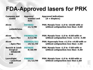
Refractive errors are some of the most common ophthalmic abnormalities world-wide and are associated with significant morbidity. Tremendous advances in treating refractive errors have occurred over the past 20 years. The arrival of the excimer laser has allowed a level of accuracy in modifying the cornea that was unattainable before. Although refractive surgery is generally safe and effective, it does carry some risks. Careful patient selection, meticulous surgical technique and frequent follow-up can avoid most complications. The experience of a surgical team can also affect the outcome and the incidence of complications. The future should bring continued improvement in outcomes, fewer complications and exciting new options for treating refractive errors. (+info)Spherical and aspherical photorefractive keratectomy and laser in-situ keratomileusis for moderate to high myopia: two prospective, randomized clinical trials. Summit technology PRK-LASIK study group. (5/186)
OBJECTIVE: Determine the outcomes of single-zone photorefractive keratectomy (SZPRK), aspherical photorefractive keratectomy (ASPRK), and laser in-situ keratomileusis (LASIK) for the correction of myopia between -6 and -12 diopters. DESIGN: Two simultaneous prospective, randomized, multi-center clinical trials. PARTICIPANTS: 286 first-treated eyes of 286 patients enrolled in one of two studies. In Study I, 134 eyes were randomized to SZPRK (58 eyes) or ASPRK (76 eyes). In Study II, 152 eyes were randomized to ASPRK (76 eyes) or to LASIK (76 eyes). INTERVENTION: All eyes received spherical one-pass excimer laser ablation as part of PRK or LASIK performed with the Summit Technologies Apex laser under an investigational device exemption, with attempted corrections between -6 and -12 diopters. MAIN OUTCOME MEASURES: Data on uncorrected and best spectacle-corrected visual acuity, predictability and stability of refraction, and complications were analyzed. Follow-up was 12 months. RESULTS: At 1 month postoperatively, more eyes in the LASIK group achieved 20/20 and 20/25 or better uncorrected visual acuity than PRK-treated eyes; at the 20/25 or better level, the difference was significant for LASIK (29/76 eyes, 38%) over SZPRK (10/58 eyes, 17%) (P = .0064). At all subsequent postoperative intervals, no difference was seen between treatment groups. Similarly, best corrected visual acuities were better for LASIK than all PRK eyes at 1 month postoperatively, and LASIK was better than SZPRK at 3 months follow-up (e.g., for 20/20 or better at 1 month, LASIK 50/76 eyes (66%) versus SZPRK 24/57 eyes (42%), P = .0066). PRK eyes had a mean loss of BCVA through 6 months, while LASIK eyes had a slight gain of mean BCVA through month 6; at 12 months, both ASPRK groups but not SZPRK continued to have a small mean loss of BCVA (e.g., compared to preoperative, mean BCVA at 12 months for SZPRK was + 0.3, LASIK was +.21, ASPRK I was -0.11, and ASPRK II -0.31 (SZPRK versus ASPRK II, P = .0116). Predictability was better for PRK than LASIK at all follow-up intervals (e.g., for manifest refraction spherical equivalent +/- 1.0 diopters at 6 months, ASPRK I 42/62 eyes (68%) versus LASIK 29/72 eyes (40%), P = .0014%). Stability was slightly but insignificantly less in the LASIK eyes compared to PRK eyes. All visual outcome measures were better for eyes with preoperative myopia between -6 and -8.9 D compared with eyes with myopia between -9 and -12 D. No consistent differences in refractive outcomes or postoperative corneal haze were seen between aspherical and single-zone ablations; haze diminished over 12 months and was judged to be vision-impairing in only one ASPRK eye. Microkeratome and flap complications occurred in 4 eyes, resulting in delay of completion of the procedure in 3 eyes but not causing long-term impairment. CONCLUSIONS: Improvement in uncorrected visual acuity and return of best corrected visual acuity was more rapid for LASIK than PRK, but efficacy outcomes in the longer term through 12 months were similar for all treatment groups. LASIK eyes tended toward undercorrection with the nomogram employed in this study compared to PRK, but the scatter was similar, suggesting little difference between these procedures for most patients by 6 months and thereafter. No consistent advantage was demonstrated between aspherical and single-zone ablation patterns. Predictability was much better for all procedures for corrections of -6 to -8.9 D compared with -9 to -12 D. Sporadic loss of best corrected vision in the PRK eyes not found in the LASIK eyes and other measures of visual function require further study. (+info)Enhancement ablation for the treatment of undercorrection after excimer laser in situ keratomileusis for correcting myopia. (6/186)
OBJECTIVE: To evaluate the treatment of undercorrection after the excimer laser in situ keratomileusis (LASIK) for correcting moderate and high myopia. METHODS: An enhancement ablation was performed in 48 eyes of 39 patients who had undergone LASIK but remained in undercorrection. Four procedures were performed within 1 month postoperatively, and the others performed between 3 and 10 months. The surgical technique includes the re-invert of the corneal cap from the temporal side, the excimer laser ablation, and the re-position of the cap. RESULTS: The undercorrection (spherical equivalent) ranged from -2.00 to -11.00 D, with a mean of -4.34D +/- 1.95 D. Following up after enhancement ablation was done after 4 to 12 months, the refractions in the 42 eyes were found to be within +/- 1.00 D. Undercorrection of -2.50 D to -5.00 D recurred in 6 eyes. Uncorrected visual acuity equals to the preoperative spectacle corrected visual acuity in 39 of 48 eyes (81.3%). Five eyes gained 1 line, 1 eye gained 2 lines and 4 eyes lost 1 line. No eyes had haze. CONCLUSION: Undercorrection after LASIK can be corrected by an enhancement ablation of the stroma under the primary corneal cap with a 193 nm ArF excimer laser, and the time for the enhancement of ablation is at 3 months postoperatively. (+info)Excimer laser effects on outflow facility and outflow pathway morphology. (7/186)
PURPOSE: To determine the relative contributions to aqueous outflow resistance of the tissues distal to the inner wall of Schlemm's canal. METHODS: While performing constant pressure perfusion at 10 mm Hg, a 193-nm excimer laser (Questek) was used to precisely remove portions of sclera, unroofing Schlemm's canal while leaving the inner wall intact. The laser beam was masked to produce a beam 2 mm by 1 mm. The laser output was constant at a fluency of 75 mJ/cm2 and 20 Hz. The excimer laser at a frequency of 1 Hz was used as the aiming beam. Photoablation was performed on human cadaver eyes at the limbus at an angle of 0 degrees to 45 degrees from the optical axis. As the excimer photoablations progressed, Schlemm's canal was visualized by the fluorescence of the Barany's solution containing fluorescein dye. After perfusion fixation the eyes were immersion-fixed overnight. The facility of outflow before (Co) and after (Ce) the excimer ablation was measured in 7 eyes. RESULTS: The facility of outflow increased in all eyes after the excimer sinusotomy, from a mean of 0.29+/-0.02 before the sinusotomy to 0.37+/-0.03 microl/min per mm Hg after (P < 0.05). The mean ratio of outflow facility after and before ablation (Ce/Co) was 1.27+/-0.08 (range, 1.20-1.39), a reduction of outflow resistance of 21.3%. Using the formula of Ellingsen and Grant (1972), percentage of resistance to outflow eliminated = 100 [1 - alphaCo/Ce - (1 - alpha)Co], where alpha = fraction of the circumference dissected. Assuming that because of circumferential flow approximately 50% of Schlemm's canal is drained by the single opening made in the outer wall ablation studies, this results in resistance to outflow eliminated of 35%, which is consistent with the calculated eliminated resistance derived from the data of Rosenquist et al., 1989. Light and scanning electron microscopy confirmed the integrity of the inner wall Schlemm's canal underlying the area of ablation. CONCLUSIONS: The results provide direct evidence indicating that approximately one third of resistance to outflow in the human eye lies distal to the inner wall Schlemm's canal in an enucleated perfused human eye. (+info)Analysis of the factors affecting decentration in photorefractive keratectomy and laser in situ keratomileusis for myopia. (8/186)
To evaluate the relationship between ablation zone decentration measured by corneal topography and various factors such as sex, age, order of operation, preoperative sedative prescription, ablation diameter and depth, type of procedure (photorefractive keratectomy = PRK, laser in situ keratomileusis = LASIK), and the use of a passive eye tracker, we examined the data of 80 eyes in 50 patients. The patients received PRK (43 eyes in 30 patients) or LASIK (37 eyes in 20 patients), and were followed for 3 months postoperatively. Statistical analysis of the data was performed using t-test, ANOVA and multiple regression analysis. The overall average ablation decentration from the pupil center was 0.43 +/- 0.27 mm, 0.35 +/- 0.22 mm in PRK and 0.47 +/- 0.30 mm in LASIK. Overall 91.3% of patients were decentered less than 0.75 mm and 95.0% were decentered less than 1.00 mm, while 93.9% of patients were decentered less than 0.75 mm in PRK and 88.7% were decentered less than 0.75 mm in LASIK. The most meridional displacement was toward the superonasal quadrant; 46% in PRK and 51% in LASIK. There was less decentration in males, in the 2nd-operated eye, in older age, PRK, in larger ablation diameter, and in shallower ablation depth, but these differences were not statistically significant. (+info)Photorefractive Keratectomy (PRK) is a type of refractive surgery used to correct vision issues such as nearsightedness, farsightedness, and astigmatism. It works by reshaping the cornea using a laser, which alters how light enters the eye and focuses on the retina.
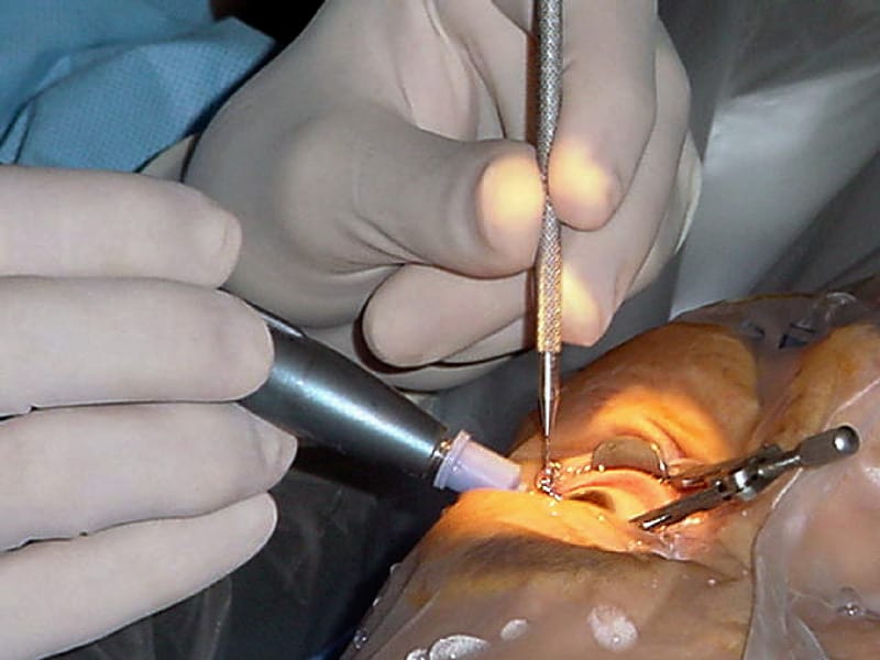
In PRK, the surgeon removes the thin outer layer of the cornea (epithelium) with an alcohol solution or a blunt surgical instrument before using the laser to reshape the underlying stromal layer. The epithelium then grows back during the healing process, which can take several days.
Compared to LASIK (another type of refractive surgery), PRK has a longer recovery time and may cause more discomfort in the first few days after surgery. However, it is an option for people who are not good candidates for LASIK due to thin corneas or other eye conditions.
It's important to note that while refractive surgeries like PRK can significantly improve vision and reduce dependence on glasses or contact lenses, they may not completely eliminate the need for corrective eyewear in all cases. Additionally, as with any surgical procedure, there are potential risks and complications associated with PRK, including infection, dry eye, and visual disturbances such as glare or halos around lights.
An excimer laser is a type of laser that is used in various medical procedures, particularly in ophthalmology and dermatology. The term "excimer" is derived from "excited dimer," which refers to a short-lived molecule formed when two atoms combine in an excited state.
Excimer lasers emit light at a specific wavelength that is determined by the type of gas used in the laser. In medical applications, excimer lasers typically use noble gases such as argon, krypton, or xenon, combined with halogens such as fluorine or chlorine. The most commonly used excimer laser in medical procedures is the excimer laser that uses a mixture of argon and fluoride gas to produce light at a wavelength of 193 nanometers (nm).
In ophthalmology, excimer lasers are primarily used for refractive surgery, such as LASIK and PRK, to correct vision problems like myopia, hyperopia, and astigmatism. The laser works by vaporizing tiny amounts of tissue from the cornea, reshaping its curvature to improve the way light is focused onto the retina.
In dermatology, excimer lasers are used for various skin conditions, including psoriasis, vitiligo, and atopic dermatitis. The laser works by emitting high-energy ultraviolet (UV) light that selectively targets and destroys the abnormal cells responsible for these conditions while leaving surrounding healthy tissue intact.
Excimer lasers are known for their precision, accuracy, and minimal side effects, making them a popular choice in medical procedures where fine detail and tissue preservation are critical.
Corneal opacity refers to a condition in which the cornea, the clear front part of the eye, becomes cloudy or opaque. This can occur due to various reasons such as injury, infection, degenerative changes, or inherited disorders. As a result, light is not properly refracted and vision becomes blurred or distorted. In some cases, corneal opacity can lead to complete loss of vision in the affected eye. Treatment options depend on the underlying cause and may include medication, corneal transplantation, or other surgical procedures.
Myopia, also known as nearsightedness, is a common refractive error of the eye. It occurs when the eye is either too long or the cornea (the clear front part of the eye) is too curved. As a result, light rays focus in front of the retina instead of directly on it, causing distant objects to appear blurry while close objects remain clear.
Myopia typically develops during childhood and can progress gradually or rapidly until early adulthood. It can be corrected with glasses, contact lenses, or refractive surgery such as LASIK. Regular eye examinations are essential for people with myopia to monitor any changes in their prescription and ensure proper correction.
While myopia is generally not a serious condition, high levels of nearsightedness can increase the risk of certain eye diseases, including cataracts, glaucoma, retinal detachment, and myopic degeneration. Therefore, it's crucial to manage myopia effectively and maintain regular follow-ups with an eye care professional.
Refractive surgical procedures are a type of ophthalmic surgery aimed at improving the refractive state of the eye and reducing or eliminating the need for corrective eyewear. These procedures reshape the cornea or alter the lens of the eye to correct nearsightedness (myopia), farsightedness (hyperopia), presbyopia, or astigmatism.
Examples of refractive surgical procedures include:
1. Laser-assisted in situ keratomileusis (LASIK): A laser is used to create a thin flap in the cornea, which is then lifted to allow reshaping of the underlying tissue with another laser. The flap is replaced, and the procedure is completed.
2. Photorefractive keratectomy (PRK): This procedure involves removing the outer layer of the cornea (epithelium) and using a laser to reshape the underlying tissue. A bandage contact lens is placed over the eye to protect it during healing.
3. LASEK (laser-assisted subepithelial keratomileusis): Similar to LASIK, but instead of creating a flap, the epithelium is loosened with an alcohol solution and moved aside. The laser treatment is applied, and the epithelium is replaced.
4. Small Incision Lenticule Extraction (SMILE): A femtosecond laser creates a small lenticule within the cornea, which is then removed through a tiny incision. This procedure reshapes the cornea to correct refractive errors.
5. Refractive lens exchange (RLE): The eye's natural lens is removed and replaced with an artificial intraocular lens (IOL) to correct refractive errors, similar to cataract surgery.
6. Implantable contact lenses: A thin, foldable lens is placed between the iris and the natural lens or behind the iris to improve the eye's focusing power.
These procedures are typically performed on an outpatient basis and may require topical anesthesia (eye drops) or local anesthesia. Potential risks and complications include infection, dry eye, visual disturbances, and changes in night vision. It is essential to discuss these potential risks with your ophthalmologist before deciding on a refractive surgery procedure.
Laser In Situ Keratomileusis (LASIK) is a type of refractive surgery used to correct vision issues such as myopia (nearsightedness), hyperopia (farsightedness), and astigmatism. The procedure involves reshaping the cornea, which is the clear, dome-shaped surface at the front of the eye, using an excimer laser.
In LASIK, a thin flap is created on the surface of the cornea using a femtosecond or microkeratome laser. The flap is then lifted, and the excimer laser is used to reshape the underlying tissue. After the reshaping is complete, the flap is replaced, allowing for quicker healing and visual recovery compared to other refractive surgery procedures.
LASIK is an outpatient procedure that typically takes about 30 minutes or less per eye. Most people can expect to see improved vision within a few days of the procedure, although it may take several weeks for vision to fully stabilize. LASIK has a high success rate and is generally considered safe when performed by a qualified surgeon. However, as with any surgical procedure, there are risks involved, including dry eye, infection, and visual complications such as glare or halos around lights.
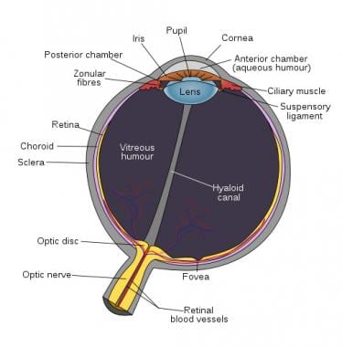
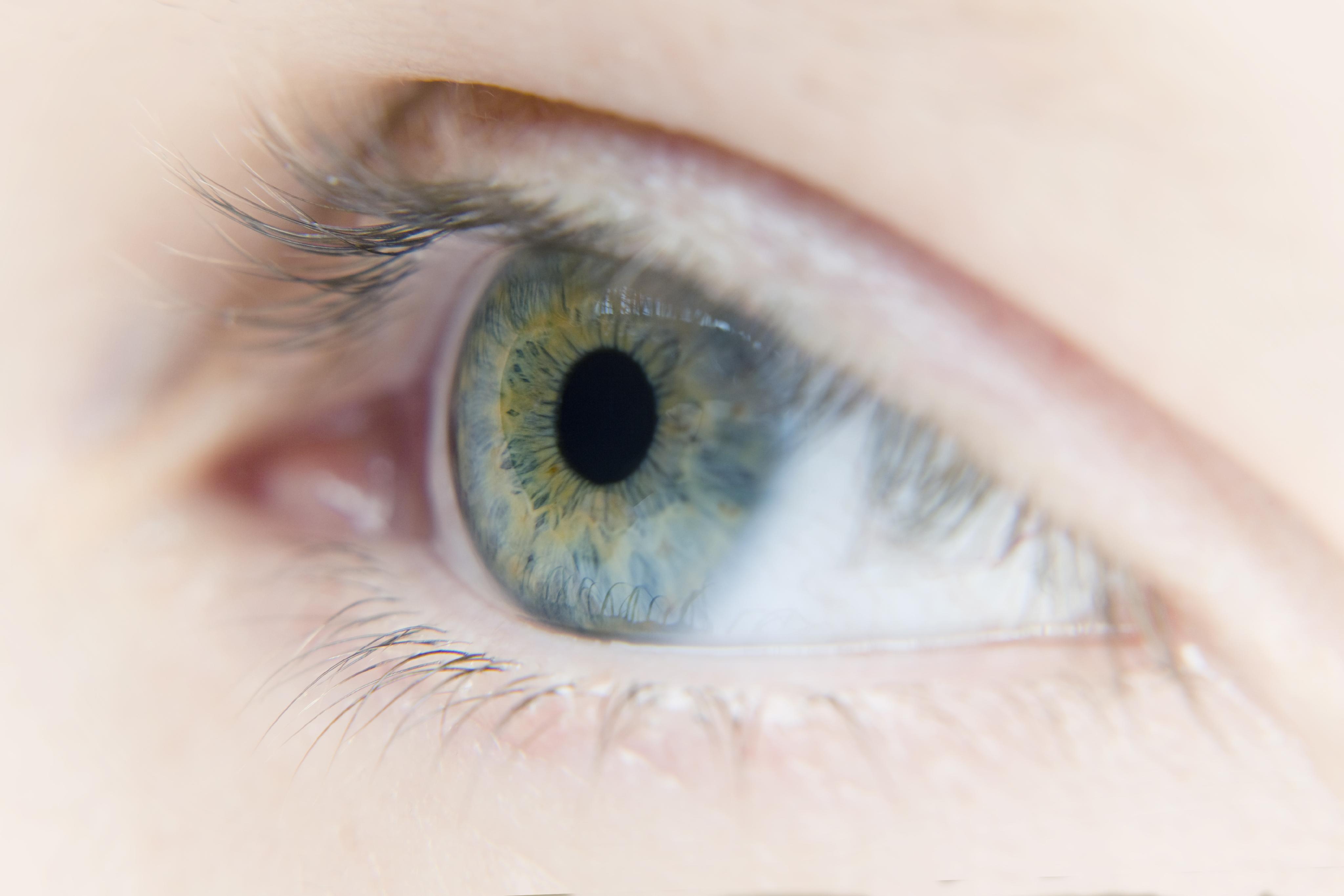
The cornea is the clear, dome-shaped surface at the front of the eye. It plays a crucial role in focusing vision. The cornea protects the eye from harmful particles and microorganisms, and it also serves as a barrier against UV light. Its transparency allows light to pass through and get focused onto the retina. The cornea does not contain blood vessels, so it relies on tears and the fluid inside the eye (aqueous humor) for nutrition and oxygen. Any damage or disease that affects its clarity and shape can significantly impact vision and potentially lead to blindness if left untreated.
Corneal topography is a non-invasive medical imaging technique used to create a detailed map of the surface curvature of the cornea, which is the clear, dome-shaped surface at the front of the eye. This procedure provides valuable information about the shape and condition of the cornea, helping eye care professionals assess various eye conditions such as astigmatism, keratoconus, and other corneal abnormalities. It can also be used in contact lens fitting, refractive surgery planning, and post-surgical evaluation.
The corneal stroma, also known as the substantia propria, is the thickest layer of the cornea, which is the clear, dome-shaped surface at the front of the eye. The cornea plays a crucial role in focusing vision.
The corneal stroma makes up about 90% of the cornea's thickness and is composed of parallel bundles of collagen fibers that are arranged in regular, repeating patterns. These fibers give the cornea its strength and transparency. The corneal stroma also contains a small number of cells called keratocytes, which produce and maintain the collagen fibers.
Disorders that affect the corneal stroma can cause vision loss or other eye problems. For example, conditions such as keratoconus, in which the cornea becomes thin and bulges outward, can distort vision and make it difficult to see clearly. Other conditions, such as corneal scarring or infection, can also affect the corneal stroma and lead to vision loss or other eye problems.
Astigmatism is a common eye condition that occurs when the cornea or lens has an irregular shape, causing blurred or distorted vision. The cornea and lens are typically smooth and curved uniformly in all directions, allowing light to focus clearly on the retina. However, if the cornea or lens is not smoothly curved and has a steeper curve in one direction than the other, it causes light to focus unevenly on the retina, leading to astigmatism.
Astigmatism can cause blurred vision at all distances, as well as eye strain, headaches, and fatigue. It is often present from birth and can be hereditary, but it can also develop later in life due to eye injuries or surgery. Astigmatism can be corrected with glasses, contact lenses, or refractive surgery such as LASIK.
Corneal surgery, laser refers to a type of surgical procedure performed on the cornea (the clear, dome-shaped surface at the front of the eye) using a laser. The most common type of laser used in corneal surgery is an excimer laser, which can be used to reshape the cornea and correct refractive errors such as nearsightedness, farsightedness, and astigmatism. This procedure is commonly known as LASIK (Laser-Assisted In Situ Keratomileusis).
Another type of laser corneal surgery is PRK (Photorefractive Keratectomy) which uses a laser to reshape the surface of the cornea. This procedure is typically used for patients who have thin corneas or other conditions that make them ineligible for LASIK.
Additionally, there are other types of laser corneal surgeries such as LASEK (Laser Epithelial Keratomileusis), Epi-LASIK (Epithelial Laser-Assisted Keratomileusis) and SBK (Sub Bowman's Keratomileusis) which are variations of the above procedures.
It is important to note that, as with any surgical procedure, laser corneal surgery has risks and potential complications, including dry eye, infection, and visual symptoms such as glare or halos around lights. It is essential for patients to have a thorough examination and consultation with an ophthalmologist before deciding if laser corneal surgery is the right choice for them.
Subepithelial laser-assisted keratectomy (SELAK) is a type of refractive surgery used to correct vision problems such as myopia (nearsightedness), hyperopia (farsightedness), and astigmatism. In this procedure, a precise and controlled laser beam is used to remove a thin layer of tissue from the cornea, specifically from the subepithelial region, which lies just beneath the surface epithelium.
The goal of SELAK is to reshape the cornea and improve its focusing power, thereby reducing or eliminating the need for corrective lenses such as glasses or contact lenses. The laser-assisted technique allows for a high degree of precision and customization, enabling the surgeon to tailor the procedure to each patient's individual needs.
It is important to note that while SELAK can be an effective treatment option for many people, it may not be suitable for everyone. A thorough eye examination and consultation with an eye care professional are necessary to determine whether this procedure is appropriate for a particular individual.
Ocular refraction is a medical term that refers to the bending of light as it passes through the optical media of the eye, including the cornea and lens. This process allows the eye to focus light onto the retina, creating a clear image. The refractive power of the eye is determined by the curvature and transparency of these structures.
In a normal eye, light rays are bent or refracted in such a way that they converge at a single point on the retina, producing a sharp and focused image. However, if the curvature of the cornea or lens is too steep or too flat, the light rays may not converge properly, resulting in a refractive error such as myopia (nearsightedness), hyperopia (farsightedness), or astigmatism.
Ocular refraction can be measured using a variety of techniques, including retinoscopy, automated refraction, and subjective refraction. These measurements are used to determine the appropriate prescription for corrective lenses such as eyeglasses or contact lenses. In some cases, ocular refractive errors may be corrected surgically through procedures such as LASIK or PRK.
Visual acuity is a measure of the sharpness or clarity of vision. It is usually tested by reading an eye chart from a specific distance, such as 20 feet (6 meters). The standard eye chart used for this purpose is called the Snellen chart, which contains rows of letters that decrease in size as you read down the chart.
Visual acuity is typically expressed as a fraction, with the numerator representing the testing distance and the denominator indicating the smallest line of type that can be read clearly. For example, if a person can read the line on the eye chart that corresponds to a visual acuity of 20/20, it means they have normal vision at 20 feet. If their visual acuity is 20/40, it means they must be as close as 20 feet to see what someone with normal vision can see at 40 feet.
It's important to note that visual acuity is just one aspect of overall vision and does not necessarily reflect other important factors such as peripheral vision, depth perception, color vision, or contrast sensitivity.
Fluorometholone is a type of corticosteroid medication that is often used in eye drops to treat various inflammatory conditions of the eye, such as allergies, uveitis, and keratitis. It works by reducing inflammation and suppressing the activity of the immune system in the eye.
Fluorometholone has a fluorinated molecule, which makes it more lipophilic (fat-soluble) than some other corticosteroids, allowing it to penetrate the eye tissue more effectively. It is available in various strengths and forms, including solutions and ointments, for topical application to the eye.
As with any medication, fluorometholone can have side effects, such as increased pressure inside the eye (glaucoma), cataracts, and delayed healing of wounds. It is important to follow the instructions of your healthcare provider when using this medication and report any unusual symptoms or concerns promptly.
Radial Keratotomy (RK) is a type of refractive surgery used to correct vision problems such as nearsightedness and astigmatism. The procedure involves making small, precise incisions in the cornea in a radial pattern, like the spokes of a wheel. These incisions cause the cornea to change shape, which can help to improve the way that light is focused onto the retina and reduce the need for corrective lenses.
During the procedure, the surgeon uses a specialized blade or laser to make the incisions in the cornea. The incisions are typically made at the periphery of the cornea, leaving the central portion of the cornea untouched. This helps to preserve the strength and stability of the cornea while still allowing it to change shape enough to improve vision.
Radial keratotomy was first developed in the 1970s and was widely used in the 1980s and 1990s. However, it has largely been replaced by newer procedures such as LASIK and PRK, which are considered to be safer and more effective. RK is still occasionally performed in cases where other procedures are not an option or when a patient prefers this type of surgery.
It's important to note that any surgical procedure carries risks, including infection, scarring, and changes in vision. Patients considering radial keratotomy should discuss the potential benefits and risks with their eye care provider before making a decision.
Hyperopia, also known as farsightedness, is a refractive error in which the eye does not focus light directly on the retina when looking at a distant object. Instead, light is focused behind the retina, causing close-up objects to appear blurry. This condition usually results from the eyeball being too short or the cornea having too little curvature. It can be corrected with eyeglasses, contact lenses, or refractive surgery.
The corneal epithelium is the outermost layer of the cornea, which is the clear, dome-shaped surface at the front of the eye. It is a stratified squamous epithelium, consisting of several layers of flat, scale-like cells that are tightly packed together. The corneal epithelium serves as a barrier to protect the eye from microorganisms, dust, and other foreign particles. It also provides a smooth surface for the refraction of light, contributes to the maintenance of corneal transparency, and plays a role in the eye's sensitivity to touch and pain. The corneal epithelium is constantly being renewed through the process of cell division and shedding, with new cells produced by stem cells located at the limbus, the border between the cornea and the conjunctiva.
Corneal diseases are a group of disorders that affect the cornea, which is the clear, dome-shaped surface at the front of the eye. The cornea plays an important role in focusing vision, and any damage or disease can cause significant visual impairment or loss. Some common types of corneal diseases include:
1. Keratoconus: A progressive disorder in which the cornea thins and bulges outward into a cone shape, causing distorted vision.
2. Fuchs' dystrophy: A genetic disorder that affects the inner layer of the cornea called the endothelium, leading to swelling, cloudiness, and decreased vision.
3. Dry eye syndrome: A condition in which the eyes do not produce enough tears or the tears evaporate too quickly, causing discomfort, redness, and blurred vision.
4. Corneal ulcers: Open sores on the cornea that can be caused by infection, trauma, or other factors.
5. Herpes simplex keratitis: A viral infection of the cornea that can cause recurrent episodes of inflammation, scarring, and vision loss.
6. Corneal dystrophies: Inherited disorders that affect the structure and clarity of the cornea, leading to visual impairment or blindness.
7. Bullous keratopathy: A condition in which the endothelium fails to pump fluid out of the cornea, causing it to swell and form blisters.
8. Corneal trauma: Injury to the cornea caused by foreign objects, chemicals, or other factors that can lead to scarring, infection, and vision loss.
Treatment for corneal diseases varies depending on the specific condition and severity of the disease. Options may include eyedrops, medications, laser surgery, corneal transplantation, or other treatments.
Wound healing is a complex and dynamic process that occurs after tissue injury, aiming to restore the integrity and functionality of the damaged tissue. It involves a series of overlapping phases: hemostasis, inflammation, proliferation, and remodeling.
1. Hemostasis: This initial phase begins immediately after injury and involves the activation of the coagulation cascade to form a clot, which stabilizes the wound and prevents excessive blood loss.
2. Inflammation: Activated inflammatory cells, such as neutrophils and monocytes/macrophages, infiltrate the wound site to eliminate pathogens, remove debris, and release growth factors that promote healing. This phase typically lasts for 2-5 days post-injury.
3. Proliferation: In this phase, various cell types, including fibroblasts, endothelial cells, and keratinocytes, proliferate and migrate to the wound site to synthesize extracellular matrix (ECM) components, form new blood vessels (angiogenesis), and re-epithelialize the wounded area. This phase can last up to several weeks depending on the size and severity of the wound.
4. Remodeling: The final phase of wound healing involves the maturation and realignment of collagen fibers, leading to the restoration of tensile strength in the healed tissue. This process can continue for months to years after injury, although the tissue may never fully regain its original structure and function.
It is important to note that wound healing can be compromised by several factors, including age, nutrition, comorbidities (e.g., diabetes, vascular disease), and infection, which can result in delayed healing or non-healing chronic wounds.
Laser therapy, also known as phototherapy or laser photobiomodulation, is a medical treatment that uses low-intensity lasers or light-emitting diodes (LEDs) to stimulate healing, reduce pain, and decrease inflammation. It works by promoting the increase of cellular metabolism, blood flow, and tissue regeneration through the process of photobiomodulation.
The therapy can be used on patients suffering from a variety of acute and chronic conditions, including musculoskeletal injuries, arthritis, neuropathic pain, and wound healing complications. The wavelength and intensity of the laser light are precisely controlled to ensure a safe and effective treatment.
During the procedure, the laser or LED device is placed directly on the skin over the area of injury or discomfort. The non-ionizing light penetrates the tissue without causing heat or damage, interacting with chromophores in the cells to initiate a series of photochemical reactions. This results in increased ATP production, modulation of reactive oxygen species, and activation of transcription factors that lead to improved cellular function and reduced pain.
In summary, laser therapy is a non-invasive, drug-free treatment option for various medical conditions, providing patients with an alternative or complementary approach to traditional therapies.
The ophthalmic nerve, also known as the first cranial nerve or CN I, is a sensory nerve that primarily transmits information about vision, including light intensity and color, and sensation in the eye and surrounding areas. It is responsible for the sensory innervation of the upper eyelid, conjunctiva, cornea, iris, ciliary body, and nasal cavity. The ophthalmic nerve has three major branches: the lacrimal nerve, frontal nerve, and nasociliary nerve. Damage to this nerve can result in various visual disturbances and loss of sensation in the affected areas.
Corneal wavefront aberration is a measurement of the irregularities in the shape and curvature of the cornea, which can affect the way light enters the eye and is focused on the retina. A wavefront aberration test uses a device to measure the refraction of light as it passes through the cornea and calculates the degree of any distortions or irregularities in the wavefront of the light. This information can be used to guide treatment decisions, such as the prescription for eyeglasses or contact lenses, or the planning of a surgical procedure to correct the aberration.
Corneal wavefront aberrations can be classified into two types: low-order and high-order aberrations. Low-order aberrations include myopia (nearsightedness), hyperopia (farsightedness), and astigmatism, which are common refractive errors that can be easily corrected with glasses or contact lenses. High-order aberrations are more complex irregularities in the wavefront of light that cannot be fully corrected with traditional eyeglasses or contact lenses. These may include coma, trefoil, and spherical aberration, among others.
High-order corneal wavefront aberrations can affect visual quality, causing symptoms such as glare, halos around lights, and decreased contrast sensitivity. They are often associated with conditions that cause changes in the shape of the cornea, such as keratoconus or corneal surgery. In some cases, high-order aberrations can be corrected with specialized contact lenses or refractive surgery procedures such as wavefront-guided LASIK or PRK.
The postoperative period is the time following a surgical procedure during which the patient's response to the surgery and anesthesia is monitored, and any complications or adverse effects are managed. This period can vary in length depending on the type of surgery and the individual patient's needs, but it typically includes the immediate recovery phase in the post-anesthesia care unit (PACU) or recovery room, as well as any additional time spent in the hospital for monitoring and management of pain, wound healing, and other aspects of postoperative care.
The goals of postoperative care are to ensure the patient's safety and comfort, promote optimal healing and rehabilitation, and minimize the risk of complications such as infection, bleeding, or other postoperative issues. The specific interventions and treatments provided during this period will depend on a variety of factors, including the type and extent of surgery performed, the patient's overall health and medical history, and any individualized care plans developed in consultation with the patient and their healthcare team.
In medical terms, "tears" are a clear, salty liquid that is produced by the tear glands (lacrimal glands) in our eyes. They serve to keep the eyes moist, protect against dust and other foreign particles, and help to provide clear vision by maintaining a smooth surface on the front of the eye. Tears consist of water, oil, and mucus, which help to prevent evaporation and ensure that the tears spread evenly across the surface of the eye. Emotional or reflexive responses, such as crying or yawning, can also stimulate the production of tears.
The endothelium of the cornea is the thin, innermost layer of cells that lines the inner surface of the cornea, which is the clear, dome-shaped structure at the front of the eye. This single layer of specialized cells is essential for maintaining the transparency and proper hydration of the cornea, allowing light to pass through it and focus on the retina.
The endothelial cells are hexagonal in shape and have tight junctions between them, creating a semi-permeable barrier that controls the movement of water and solutes between the corneal stroma (the middle layer of the cornea) and the anterior chamber (the space between the cornea and the iris). The endothelial cells actively pump excess fluid out of the cornea, maintaining a delicate balance of hydration that is critical for corneal clarity.
Damage to or dysfunction of the corneal endothelium can result in corneal edema (swelling), cloudiness, and loss of vision. Factors contributing to endothelial damage include aging, eye trauma, intraocular surgery, and certain diseases such as Fuchs' dystrophy and glaucoma.
Confocal microscopy is a powerful imaging technique used in medical and biological research to obtain high-resolution, contrast-rich images of thick samples. This super-resolution technology provides detailed visualization of cellular structures and processes at various depths within a specimen.
In confocal microscopy, a laser beam focused through a pinhole illuminates a small spot within the sample. The emitted fluorescence or reflected light from this spot is then collected by a detector, passing through a second pinhole that ensures only light from the focal plane reaches the detector. This process eliminates out-of-focus light, resulting in sharp images with improved contrast compared to conventional widefield microscopy.
By scanning the laser beam across the sample in a raster pattern and collecting fluorescence at each point, confocal microscopy generates optical sections of the specimen. These sections can be combined to create three-dimensional reconstructions, allowing researchers to study cellular architecture and interactions within complex tissues.
Confocal microscopy has numerous applications in medical research, including studying protein localization, tracking intracellular dynamics, analyzing cell morphology, and investigating disease mechanisms at the cellular level. Additionally, it is widely used in clinical settings for diagnostic purposes, such as analyzing skin lesions or detecting pathogens in patient samples.
I believe there may be some confusion in your question. "Rabbits" is a common name used to refer to the Lagomorpha species, particularly members of the family Leporidae. They are small mammals known for their long ears, strong legs, and quick reproduction.
However, if you're referring to "rabbits" in a medical context, there is a term called "rabbit syndrome," which is a rare movement disorder characterized by repetitive, involuntary movements of the fingers, resembling those of a rabbit chewing. It is also known as "finger-chewing chorea." This condition is usually associated with certain medications, particularly antipsychotics, and typically resolves when the medication is stopped or adjusted.
"Cell count" is a medical term that refers to the process of determining the number of cells present in a given volume or sample of fluid or tissue. This can be done through various laboratory methods, such as counting individual cells under a microscope using a specialized grid called a hemocytometer, or using automated cell counters that use light scattering and electrical impedance techniques to count and classify different types of cells.
Cell counts are used in a variety of medical contexts, including hematology (the study of blood and blood-forming tissues), microbiology (the study of microscopic organisms), and pathology (the study of diseases and their causes). For example, a complete blood count (CBC) is a routine laboratory test that includes a white blood cell (WBC) count, red blood cell (RBC) count, hemoglobin level, hematocrit value, and platelet count. Abnormal cell counts can indicate the presence of various medical conditions, such as infections, anemia, or leukemia.
Follow-up studies are a type of longitudinal research that involve repeated observations or measurements of the same variables over a period of time, in order to understand their long-term effects or outcomes. In medical context, follow-up studies are often used to evaluate the safety and efficacy of medical treatments, interventions, or procedures.
In a typical follow-up study, a group of individuals (called a cohort) who have received a particular treatment or intervention are identified and then followed over time through periodic assessments or data collection. The data collected may include information on clinical outcomes, adverse events, changes in symptoms or functional status, and other relevant measures.
The results of follow-up studies can provide important insights into the long-term benefits and risks of medical interventions, as well as help to identify factors that may influence treatment effectiveness or patient outcomes. However, it is important to note that follow-up studies can be subject to various biases and limitations, such as loss to follow-up, recall bias, and changes in clinical practice over time, which must be carefully considered when interpreting the results.
 Photorefractive keratectomy
 Photorefractive Keratectomy - EyeWiki
 Medical malpractice predictors and risk factors for ophthalmologists performing LASIK and photorefractive keratectomy surgery
 PRK (Photorefractive Keratectomy) (Pt. 1) - Kornmehl Laser Eye Associates
 Photorefractive keratectomy: a technique for laser refractive surgery. | Read by QxMD
 Topical Diclofenac Reduces Pain Following Photorefractive Keratectomy | JAMA Ophthalmology | JAMA Network
 PRK (Photorefractive Keratectomy) Laser Vision Correction
 Photo-Refractive Keratectomy (PRK)
 Photorefractive Keratectomy (PRK) Eye Surgery in Bangalore
 Photorefractive Keratectomy Laser Eye Surgery - HasenChat Music Japan
 View of Myopic regression after photorefractive keratectomy: a retrospective cohort study
 Am I a Cadidate for PRK (Photorefractive Keratectomy) Laser Vision Correction
 The Different Types of Laser Eye Surgery: Which One is Right for You? - Harcourt HealthHarcourt Health
 Farsightedness: Causes and corrective treatments
 Centre For Sight - London - England in Harley Street
 Laser-assisted in-situ keratomileusis (LASIK) versus photorefractive keratectomy (PRK) for myopia: Cochrane systematic review |...
 Corrective Eye Surgery Basics - AllAboutVision.com
 Laser Eye Surgery | LASIK | MedlinePlus
 Portable Vision Screening Devices Find Amblyopia in Children
 Corneal ectasia following photorefractive keratectomy: a confocal microscopic case report and literature review | Arq. bras....
 Browse All Multimedia - American Academy of Ophthalmology
 Refractive Surgery Services | RUSH
 Twinsburg Family Health and Surgery Center | Cleveland Clinic