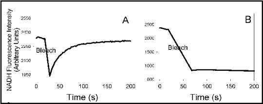
Photobleaching
Fluorescence Recovery After Photobleaching
Diffusion
Photochemistry
Microscopy, Fluorescence
Green Fluorescent Proteins
Fluorescent Dyes
Fluorescence
Luminescent Proteins
Fluorescein-5-isothiocyanate
Fluoresceins
Fluorescence Resonance Energy Transfer
Microscopy, Confocal
Lasers
Photons
Fluorescein
Recombinant Fusion Proteins
Rhodamines
Membrane Fluidity
Protein Transport
Cell Membrane
Dextrans
Models, Biological
Carbocyanines
Fluorescence Polarization
Viscosity
Cytoplasm
Ammonium Compounds
Cell Nucleus
Mathematics
Quantum Dots
Models, Theoretical
Actins
4-Chloro-7-nitrobenzofurazan
Protein Binding
HeLa Cells
Microtubules
Cytoskeleton
Solutions
Membrane Proteins
COS Cells
Cercopithecus aethiops
Phalloidine
Membrane Lipids
Photosensitizing Agents
Active Transport, Cell Nucleus
Photochemotherapy
Cells, Cultured
Tubulin
Endoplasmic Reticulum
Protein Structure, Tertiary
Chromatiaceae
Membranes, Artificial
Microscopy, Fluorescence, Multiphoton
Biological Transport
Phosphatidylcholines
Pyridinium Compounds
Diazepam Binding Inhibitor
Models, Chemical
Transfection
Computer Simulation
Lipid Bilayers
Silver
Optics and Photonics
Actin Cytoskeleton
Microscopy, Video
Dimyristoylphosphatidylcholine
Aminolevulinic Acid
Cell Nucleolus
Nuclear Envelope
Semiconductors
Photoreceptors, Microbial
Golgi Apparatus
Single-Cell Analysis
Scyphozoa
Microscopy
Cells
CHO Cells
Receptors, Concanavalin A
Irreversible photobleaching of bacteriorhodopsin in a high-temperature intermediate state. (1/363)
The photo-intermediate state of bacteriorhodopsin is a metastable state that spontaneously transforms to the ground state over the energy barrier of a local minimum. As the recovery of the photocycle to the ground state and irreversible photobleaching to the denatured state may occur from the same local energy minimum, depending on the temperature, the structural stability of bacteriorhodopsin under illumination at high temperature was measured in order to study the intra- and inter-molecular interactions that contribute to the recovery of the ground state. Visible CD spectra of bacteriorhodopsin began to change at 60 degrees C from a bilobed to positive type in accordance with an appearance of an absorption peak at 470 nm. Irreversible photobleaching, the light-induced denaturation, also started to occur at 60 degrees C, suggesting some correlation between irreversible photobleaching and the structural change to the high-temperature intermediate state. However, bacteriorhodopsin in the dark was stable up to 70 degrees C, suggesting that there is some additional factor that lends structural stability to bacteriorhodopsin in the dark. The contribution of protein-protein interactions to stability is discussed on the basis of the difference in the denaturation behaviors between light and dark conditions. (+info)Life cycle of MTs: persistent growth in the cell interior, asymmetric transition frequencies and effects of the cell boundary. (2/363)
Microtubule dynamics were investigated in CHO and NRK cells by novel experimental approaches designed to evaluate the microtubule behavior in the cell interior. These approaches were: (1) laser photobleaching of a path through the centrosome; (2) direct observation of microtubules in centrosome-containing cytoplasts; (3) GFP-CLIP-170 expression as a marker for microtubule plus end growth; and (iv) sequential subtraction analysis. The combination of these approaches allowed us to obtain data where the density of microtubules had previously prevented conventional methods to be applicable. In the steady state, nascent microtubules grew persistently from the centrosome towards the cell margin. Frequently, they arrived at the cell margin without undergoing any transition to the shortening phase. In contrast to the growth of microtubules, shortening of the plus ends from the periphery was non-persistent; that is, rescue was frequent and the extent of shortening showed a distribution of lengths reflecting a stochastic process. The combination of persistent growth and a cell boundary led to a difference in apparent microtubule behavior in the cell interior compared with that near the cell margin. Whereas microtubules in the cell interior showed asymmetric transition frequencies, their behavior near the cell margin showed frequent fluctuations between phases of shortening and growth. Complete microtubule turnover was accomplished by the relatively rare episodes of shortening back to the centrosome. Release from the centrosome with subsequent minus end shortening also occurred but was a minor mechanism for microtubule turnover compared with the plus end pathway. We propose a life cycle for a microtubule which consists of rapid growth from the centrosome to the cell margin followed by an indefinite period of fluctuations of phases of shortening and growth. We suggest that persistent growth and asymmetric transition frequencies serve the biological function of providing a mechanism by which microtubules may rapidly accommodate to the changing shape and advancing edge of motile cells. (+info)Rapid translocation of NTF2 through the nuclear pore of isolated nuclei and nuclear envelopes. (3/363)
The mechanism by which macromolecules are translocated through the nuclear pore complex (NPC) is little understood. However, recent measurements of nuclear transport in permeabilized cells showed that molecules binding to phenylalanine-glycine-rich repeats (FG repeats) in NPC proteins were translocated much faster through the NPC than molecules not interacting with FG repeats. We have studied that substrate preference of the NPC in isolated oocyte nuclei and purified nuclear envelopes by optical single transporter recording. NTF2, the transport receptor of RanGDP, was exported approximately 30 times faster than green fluorescent protein, an inert molecule of approximately the same size. The data confirm that restricted diffusion of inert molecules and facilitated transport of FG-repeat binding proteins are basic types of translocation through the NPC, demonstrating that the functional integrity of the NPC can be conserved in isolated nuclei and nuclear envelopes and thus opening new avenues to the analysis of nucleocytoplasmic transport. (+info)Lateral diffusion in substrate-supported lipid monolayers as a function of ambient relative humidity. (4/363)
We analyzed the influence of water activity on the lateral self-diffusion of supported phospholipid monolayers. Lipid monolayer membranes were supported by polysaccharide cushions (chitosan and agarose), or glass. A simple diffusion model was derived, based on activated diffusion with an activation energy, E(a), which depends on the hydration state of the lipid headgroup. A crucial assumption of the derived model is that E(a) can be calculated assuming an exponential decay of the humidity-dependent disjoining pressure in the monolayer/substrate interface with respect to the equilibrium separation distance. A plot of ln(D) against ln(p(0)/p), where D is the measured diffusion coefficient and p(0) and p are the partial water pressures at saturation and at a particular relative humidity, respectively, was observed to be linear in all cases (i.e., for differing lipids, lateral monolayer pressures, temperatures, and substrates), in accordance with the above-mentioned diffusion model. No indications for humidity-induced first-order phase transitions in the supported phospholipid monolayers were found. Many biological processes such as vesicle fusion and recognition processes involve dehydration/hydration cycles, and it can be expected that the water activity significantly affects the kinetics of these processes in a manner similar to that examined in the present work. (+info)Diffusion and convection in collagen gels: implications for transport in the tumor interstitium. (5/363)
Diffusion coefficients of tracer molecules in collagen type I gels prepared from 0-4.5% w/v solutions were measured by fluorescence recovery after photobleaching. When adjusted to account for in vivo tortuosity, diffusion coefficients in gels matched previous measurements in four human tumor xenografts with equivalent collagen concentrations. In contrast, hyaluronan solutions hindered diffusion to a lesser extent when prepared at concentrations equivalent to those reported in these tumors. Collagen permeability, determined from flow through gels under hydrostatic pressure, was compared with predictions obtained from application of the Brinkman effective medium model to diffusion data. Permeability predictions matched experimental results at low concentrations, but underestimated measured values at high concentrations. Permeability measurements in gels did not match previous measurements in tumors. Visualization of gels by transmission electron microscopy and light microscopy revealed networks of long collagen fibers at lower concentrations along with shorter fibers at high concentrations. Negligible assembly was detected in collagen solutions pregelation. However, diffusion was similarly hindered in pre and postgelation samples. Comparison of diffusion and convection data in these gels and tumors suggests that collagen may obstruct diffusion more than convection in tumors. These findings have significant implications for drug delivery in tumors and for tissue engineering applications. (+info)Agonist-induced PIP(2) hydrolysis inhibits cortical actin dynamics: regulation at a global but not at a micrometer scale. (6/363)
Phosphatidylinositol 4, 5-bisphosphate (PIP(2)) at the inner leaflet of the plasma membrane has been proposed to locally regulate the actin cytoskeleton. Indeed, recent studies that use GFP-tagged pleckstrin homology domains (GFP-PH) as fluorescent PIP(2) sensors suggest that this lipid is enriched in membrane microdomains. Here we report that this concept needs revision. Using three distinct fluorescent GFP-tagged pleckstrin homology domains, we show that highly mobile GFP-PH patches colocalize perfectly with various lipophilic membrane dyes and, hence, represent increased lipid content rather than PIP(2)-enriched microdomains. We show that bright patches are caused by submicroscopical folds and ruffles in the membrane that can be directly visualized at approximately 15 nm axial resolution with a novel numerically enhanced imaging method. F-actin motility is inhibited significantly by agonist-induced PIP(2) breakdown, and it resumes as soon as PIP(2) levels are back to normal. Thus, our data support a role for PIP(2) in the regulation of cortical actin, but they challenge a model in which spatial differences in PIP(2) regulation of the cytoskeleton exist at a micrometer scale. (+info)Connexin expression and gap junctional intercellular communication in human first trimester trophoblast. (7/363)
Connexin (Cx) expression and gap junctional intercellular communication (GJIC) are involved in development and differentiation processes, and recently mutation of connexin genes has been implicated in pathologies. In the human placenta, two distinct differentiation pathways of cytotrophoblastic cells coexist and lead to a fusion phenotype (villous trophoblast) and a proliferative/invasive phenotype (extravillous trophoblast). Here we characterized in situ and in vitro the expression of Cx transcripts and proteins in the villous and extravillous trophoblast of first trimester placenta. In addition, the GJIC functionality was investigated using the gap-fluorescence recovery after photobleaching (gap-FRAP) method. We demonstrated in the villous trophoblast the presence of Cx43 mRNA and of Cx43 protein localized between cytotrophoblastic cells and between cytotrophoblastic cells and syncytiotrophoblast. In vitro, a transient functional gap junctional intertrophoblastic communication was demonstrated during the trophoblast fusion leading to the multinucleated syncytiotrophoblast. During the proliferative process of the extravillous trophoblast, Cx40 is expressed in the proximal part of the cell columns. When cytotrophoblastic cells were cultured on Matrigel for 2 days, alpha5beta1 integrin expression was observed concomitant with the presence of Cx40 mRNA and of Cx40 protein between the cells. No evidence for a GJIC was detected in this induced extravillous phenotype. In addition, Cx32 was detected between some aggregated cells after 72 h of culture. Our data show that the presence of Cx43 allows an inter-trophoblastic GJIC and is associated with the fusion process leading to the villous syncytiotrophoblast and that the presence of Cx40 does not allow GJIC and is associated with the extravillous phenotype. (+info)The transcription cycle of RNA polymerase II in living cells. (8/363)
RNA polymerase II transcribes most eukaryotic genes. Its catalytic subunit was tagged with green fluorescent protein and expressed in Chinese hamster cells bearing a mutation in the same subunit; it complemented the defect and so was functional. Photobleaching revealed two kinetic fractions of polymerase in living nuclei: approximately 75% moved rapidly, but approximately 25% was transiently immobile (association t1/2 approximately 20 min) and transcriptionally active, as incubation with 5,6-dichloro-1-beta-D-ribofuranosylbenzimidazole eliminated it. No immobile but inactive fraction was detected, providing little support for the existence of a stable holoenzyme, or the slow stepwise assembly of a preinitiation complex on promoters or the nuclear substructure. Actinomycin D decreased the rapidly moving fraction, suggesting that engaged polymerases stall at intercalated molecules while others initiate. When wild-type cells containing only the endogenous enzyme were incubated with [3H]uridine, nascent transcripts became saturated with tritium with similar kinetics (t1/2 approximately 14 min). These data are consistent with a polymerase being mobile for one half to five sixths of a transcription cycle, and rapid assembly into the preinitiation complex. Then, most expressed transcription units would spend significant times unassociated with engaged polymerases. (+info)Photobleaching is a process in microscopy where fluorescent molecules, used as labels to visualize specific structures or proteins within cells, lose their ability to fluoresce after exposure to high-intensity light. This can occur due to the chemical alteration of the fluorophore's structure, which causes a loss of its ability to absorb and emit light. Photobleaching is often used in fluorescence recovery after photobleaching (FRAP) experiments to measure the mobility and diffusion rates of proteins within living cells. However, it can also be a limitation in long-term imaging studies as it reduces the signal-to-noise ratio and can lead to the loss of important information.
Fluorescence Recovery After Photobleaching (FRAP) is a microscopy technique used to study the mobility and diffusion of molecules in biological samples, particularly within living cells. This technique involves the use of an intense laser beam to photobleach (or permanently disable) the fluorescence of a specific region within a sample that has been labeled with a fluorescent probe or dye. The recovery of fluorescence in this bleached area is then monitored over time, as unbleached molecules from adjacent regions move into the bleached area through diffusion or active transport.
The rate and extent of fluorescence recovery can provide valuable information about the mobility, binding interactions, and dynamics of the labeled molecules within their native environment. FRAP is widely used in cell biology research to investigate various processes such as protein-protein interactions, membrane fluidity, organelle dynamics, and gene expression regulation.
Diffusion, in the context of medicine and physiology, refers to the process by which molecules move from an area of high concentration to an area of low concentration until they are evenly distributed throughout a space or solution. This passive transport mechanism does not require energy and relies solely on the random motion of particles. Diffusion is a vital process in many biological systems, including the exchange of gases in the lungs, the movement of nutrients and waste products across cell membranes, and the spread of drugs and other substances throughout tissues.
I'm sorry for any confusion, but "photochemistry" is not a term typically used in medical definitions. Photochemistry is a branch of chemistry that deals with the chemical effects of light. It involves the absorption of light by a substance, which can lead to the promotion of an electron to a higher energy state, and subsequently result in various chemical reactions.
In a medical context, photochemical processes might be discussed in relation to certain therapies or diagnostic techniques, such as photodynamic therapy for cancer treatment, where a photosensitizing agent is used that reacts with light to produce singlet oxygen or other reactive species to destroy nearby cells. However, it's not a term used to define a specific medical condition or concept in the same way that one might define "inflammation" or "metabolism."
Fluorescence microscopy is a type of microscopy that uses fluorescent dyes or proteins to highlight and visualize specific components within a sample. In this technique, the sample is illuminated with high-energy light, typically ultraviolet (UV) or blue light, which excites the fluorescent molecules causing them to emit lower-energy, longer-wavelength light, usually visible light in the form of various colors. This emitted light is then collected by the microscope and detected to produce an image.
Fluorescence microscopy has several advantages over traditional brightfield microscopy, including the ability to visualize specific structures or molecules within a complex sample, increased sensitivity, and the potential for quantitative analysis. It is widely used in various fields of biology and medicine, such as cell biology, neuroscience, and pathology, to study the structure, function, and interactions of cells and proteins.
There are several types of fluorescence microscopy techniques, including widefield fluorescence microscopy, confocal microscopy, two-photon microscopy, and total internal reflection fluorescence (TIRF) microscopy, each with its own strengths and limitations. These techniques can provide valuable insights into the behavior of cells and proteins in health and disease.
Green Fluorescent Protein (GFP) is not a medical term per se, but a scientific term used in the field of molecular biology. GFP is a protein that exhibits bright green fluorescence when exposed to light, particularly blue or ultraviolet light. It was originally discovered in the jellyfish Aequorea victoria.
In medical and biological research, scientists often use recombinant DNA technology to introduce the gene for GFP into other organisms, including bacteria, plants, and animals, including humans. This allows them to track the expression and localization of specific genes or proteins of interest in living cells, tissues, or even whole organisms.
The ability to visualize specific cellular structures or processes in real-time has proven invaluable for a wide range of research areas, from studying the development and function of organs and organ systems to understanding the mechanisms of diseases and the effects of therapeutic interventions.
Fluorescent dyes are substances that emit light upon excitation by absorbing light of a shorter wavelength. In a medical context, these dyes are often used in various diagnostic tests and procedures to highlight or mark certain structures or substances within the body. For example, fluorescent dyes may be used in imaging techniques such as fluorescence microscopy or fluorescence angiography to help visualize cells, tissues, or blood vessels. These dyes can also be used in flow cytometry to identify and sort specific types of cells. The choice of fluorescent dye depends on the specific application and the desired properties, such as excitation and emission spectra, quantum yield, and photostability.
Fluorescence is not a medical term per se, but it is widely used in the medical field, particularly in diagnostic tests, medical devices, and research. Fluorescence is a physical phenomenon where a substance absorbs light at a specific wavelength and then emits light at a longer wavelength. This process, often referred to as fluorescing, results in the emission of visible light that can be detected and measured.
In medical terms, fluorescence is used in various applications such as:
1. In-vivo imaging: Fluorescent dyes or probes are introduced into the body to highlight specific structures, cells, or molecules during imaging procedures. This technique can help doctors detect and diagnose diseases such as cancer, inflammation, or infection.
2. Microscopy: Fluorescence microscopy is a powerful tool for visualizing biological samples at the cellular and molecular level. By labeling specific proteins, nucleic acids, or other molecules with fluorescent dyes, researchers can observe their distribution, interactions, and dynamics within cells and tissues.
3. Surgical guidance: Fluorescence-guided surgery is a technique where surgeons use fluorescent markers to identify critical structures such as blood vessels, nerves, or tumors during surgical procedures. This helps ensure precise and safe surgical interventions.
4. Diagnostic tests: Fluorescence-based assays are used in various diagnostic tests to detect and quantify specific biomarkers or analytes. These assays can be performed using techniques such as enzyme-linked immunosorbent assay (ELISA), polymerase chain reaction (PCR), or flow cytometry.
In summary, fluorescence is a physical process where a substance absorbs and emits light at different wavelengths. In the medical field, this phenomenon is harnessed for various applications such as in-vivo imaging, microscopy, surgical guidance, and diagnostic tests.
Luminescent proteins are a type of protein that emit light through a chemical reaction, rather than by absorbing and re-emitting light like fluorescent proteins. This process is called bioluminescence. The light emitted by luminescent proteins is often used in scientific research as a way to visualize and track biological processes within cells and organisms.
One of the most well-known luminescent proteins is Green Fluorescent Protein (GFP), which was originally isolated from jellyfish. However, GFP is actually a fluorescent protein, not a luminescent one. A true example of a luminescent protein is the enzyme luciferase, which is found in fireflies and other bioluminescent organisms. When luciferase reacts with its substrate, luciferin, it produces light through a process called oxidation.
Luminescent proteins have many applications in research, including as reporters for gene expression, as markers for protein-protein interactions, and as tools for studying the dynamics of cellular processes. They are also used in medical imaging and diagnostics, as well as in the development of new therapies.
Fluorescence spectrometry is a type of analytical technique used to investigate the fluorescent properties of a sample. It involves the measurement of the intensity of light emitted by a substance when it absorbs light at a specific wavelength and then re-emits it at a longer wavelength. This process, known as fluorescence, occurs because the absorbed energy excites electrons in the molecules of the substance to higher energy states, and when these electrons return to their ground state, they release the excess energy as light.
Fluorescence spectrometry typically measures the emission spectrum of a sample, which is a plot of the intensity of emitted light versus the wavelength of emission. This technique can be used to identify and quantify the presence of specific fluorescent molecules in a sample, as well as to study their photophysical properties.
Fluorescence spectrometry has many applications in fields such as biochemistry, environmental science, and materials science. For example, it can be used to detect and measure the concentration of pollutants in water samples, to analyze the composition of complex biological mixtures, or to study the properties of fluorescent nanomaterials.
Fluorescein-5-isothiocyanate (FITC) is not a medical term per se, but a chemical compound commonly used in biomedical research and clinical diagnostics. Therefore, I will provide a general definition of this term:
Fluorescein-5-isothiocyanate (FITC) is a fluorescent dye with an absorption maximum at approximately 492-495 nm and an emission maximum at around 518-525 nm. It is widely used as a labeling reagent for various biological molecules, such as antibodies, proteins, and nucleic acids, to study their structure, function, and interactions in techniques like flow cytometry, immunofluorescence microscopy, and western blotting. The isothiocyanate group (-N=C=S) in the FITC molecule reacts with primary amines (-NH2) present in biological molecules to form a stable thiourea bond, enabling specific labeling of target molecules for detection and analysis.
Fluorescein is not a medical condition, but rather a diagnostic dye that is used in various medical tests and procedures. It is a fluorescent compound that absorbs light at one wavelength and emits light at another wavelength, which makes it useful for imaging and detecting various conditions.
In ophthalmology, fluorescein is commonly used in eye examinations to evaluate the health of the cornea, conjunctiva, and anterior chamber of the eye. A fluorescein dye is applied to the surface of the eye, and then the eye is examined under a blue light. The dye highlights any damage or abnormalities on the surface of the eye, such as scratches, ulcers, or inflammation.
Fluorescein is also used in angiography, a medical imaging technique used to examine blood vessels in the body. A fluorescein dye is injected into a vein, and then a special camera takes pictures of the dye as it flows through the blood vessels. This can help doctors diagnose and monitor conditions such as cancer, diabetes, and macular degeneration.
Overall, fluorescein is a valuable diagnostic tool that helps medical professionals detect and monitor various conditions in the body.
Fluorescence Resonance Energy Transfer (FRET) is not strictly a medical term, but it is a fundamental concept in biophysical and molecular biology research, which can have medical applications. Here's the definition of FRET:
Fluorescence Resonance Energy Transfer (FRET) is a distance-dependent energy transfer process between two fluorophores, often referred to as a donor and an acceptor. The process occurs when the emission spectrum of the donor fluorophore overlaps with the excitation spectrum of the acceptor fluorophore. When the donor fluorophore is excited, it can transfer its energy to the acceptor fluorophore through non-radiative dipole-dipole coupling, resulting in the emission of light from the acceptor at a longer wavelength than that of the donor.
FRET efficiency depends on several factors, including the distance between the two fluorophores, their relative orientation, and the spectral overlap between their excitation and emission spectra. FRET is typically efficient when the distance between the donor and acceptor is less than 10 nm (nanometers), making it a powerful tool for measuring molecular interactions, conformational changes, and distances at the molecular level.
In medical research, FRET has been used to study various biological processes, such as protein-protein interactions, enzyme kinetics, and gene regulation. It can also be used in developing biosensors for detecting specific molecules or analytes in clinical samples, such as blood or tissue.
Confocal microscopy is a powerful imaging technique used in medical and biological research to obtain high-resolution, contrast-rich images of thick samples. This super-resolution technology provides detailed visualization of cellular structures and processes at various depths within a specimen.
In confocal microscopy, a laser beam focused through a pinhole illuminates a small spot within the sample. The emitted fluorescence or reflected light from this spot is then collected by a detector, passing through a second pinhole that ensures only light from the focal plane reaches the detector. This process eliminates out-of-focus light, resulting in sharp images with improved contrast compared to conventional widefield microscopy.
By scanning the laser beam across the sample in a raster pattern and collecting fluorescence at each point, confocal microscopy generates optical sections of the specimen. These sections can be combined to create three-dimensional reconstructions, allowing researchers to study cellular architecture and interactions within complex tissues.
Confocal microscopy has numerous applications in medical research, including studying protein localization, tracking intracellular dynamics, analyzing cell morphology, and investigating disease mechanisms at the cellular level. Additionally, it is widely used in clinical settings for diagnostic purposes, such as analyzing skin lesions or detecting pathogens in patient samples.
A laser is not a medical term per se, but a physical concept that has important applications in medicine. The term "LASER" stands for "Light Amplification by Stimulated Emission of Radiation." It refers to a device that produces and amplifies light with specific characteristics, such as monochromaticity (single wavelength), coherence (all waves moving in the same direction), and high intensity.
In medicine, lasers are used for various therapeutic and diagnostic purposes, including surgery, dermatology, ophthalmology, and dentistry. They can be used to cut, coagulate, or vaporize tissues with great precision, minimizing damage to surrounding structures. Additionally, lasers can be used to detect and measure physiological parameters, such as blood flow and oxygen saturation.
It's important to note that while lasers are powerful tools in medicine, they must be used by trained professionals to ensure safe and effective treatment.
A photon is not a term that has a specific medical definition, as it is a fundamental concept in physics. Photons are elementary particles that carry electromagnetic energy, such as light. They have no mass or electric charge and exhibit both particle-like and wave-like properties. In the context of medicine, photons are often discussed in relation to various medical imaging techniques (e.g., X-ray imaging, CT scans, and PET scans) and therapeutic interventions like laser therapy and radiation therapy, where photons are used to diagnose or treat medical conditions.
Fluorescein is not a medical condition or term, but rather a diagnostic dye used in various medical tests and procedures. Medically, it is referred to as Fluorescein Sodium, a fluorescent compound that absorbs light at one wavelength and emits light at another longer wavelength when excited.
In the field of ophthalmology (eye care), Fluorescein is commonly used in:
1. Fluorescein angiography: A diagnostic test to examine blood flow in the retina and choroid, often used to diagnose and manage conditions like diabetic retinopathy, age-related macular degeneration, and retinal vessel occlusions.
2. Tear film assessment: Fluorescein dye is used to evaluate the quality of tear film and diagnose dry eye syndrome by observing the staining pattern on the cornea.
3. Corneal abrasions/foreign body detection: Fluorescein dye can help identify corneal injuries, such as abrasions or foreign bodies, under a cobalt blue light.
In other medical fields, fluorescein is also used in procedures like:
1. Urinary tract imaging: To detect urinary tract abnormalities and evaluate kidney function.
2. Lymphangiography: A procedure to visualize the lymphatic system.
3. Surgical navigation: In some surgical procedures, fluorescein is used as a marker for better visualization of specific structures or areas.
Recombinant fusion proteins are artificially created biomolecules that combine the functional domains or properties of two or more different proteins into a single protein entity. They are generated through recombinant DNA technology, where the genes encoding the desired protein domains are linked together and expressed as a single, chimeric gene in a host organism, such as bacteria, yeast, or mammalian cells.
The resulting fusion protein retains the functional properties of its individual constituent proteins, allowing for novel applications in research, diagnostics, and therapeutics. For instance, recombinant fusion proteins can be designed to enhance protein stability, solubility, or immunogenicity, making them valuable tools for studying protein-protein interactions, developing targeted therapies, or generating vaccines against infectious diseases or cancer.
Examples of recombinant fusion proteins include:
1. Etaglunatide (ABT-523): A soluble Fc fusion protein that combines the heavy chain fragment crystallizable region (Fc) of an immunoglobulin with the extracellular domain of the human interleukin-6 receptor (IL-6R). This fusion protein functions as a decoy receptor, neutralizing IL-6 and its downstream signaling pathways in rheumatoid arthritis.
2. Etanercept (Enbrel): A soluble TNF receptor p75 Fc fusion protein that binds to tumor necrosis factor-alpha (TNF-α) and inhibits its proinflammatory activity, making it a valuable therapeutic option for treating autoimmune diseases like rheumatoid arthritis, ankylosing spondylitis, and psoriasis.
3. Abatacept (Orencia): A fusion protein consisting of the extracellular domain of cytotoxic T-lymphocyte antigen 4 (CTLA-4) linked to the Fc region of an immunoglobulin, which downregulates T-cell activation and proliferation in autoimmune diseases like rheumatoid arthritis.
4. Belimumab (Benlysta): A monoclonal antibody that targets B-lymphocyte stimulator (BLyS) protein, preventing its interaction with the B-cell surface receptor and inhibiting B-cell activation in systemic lupus erythematosus (SLE).
5. Romiplostim (Nplate): A fusion protein consisting of a thrombopoietin receptor agonist peptide linked to an immunoglobulin Fc region, which stimulates platelet production in patients with chronic immune thrombocytopenia (ITP).
6. Darbepoetin alfa (Aranesp): A hyperglycosylated erythropoiesis-stimulating protein that functions as a longer-acting form of recombinant human erythropoietin, used to treat anemia in patients with chronic kidney disease or cancer.
7. Palivizumab (Synagis): A monoclonal antibody directed against the F protein of respiratory syncytial virus (RSV), which prevents RSV infection and is administered prophylactically to high-risk infants during the RSV season.
8. Ranibizumab (Lucentis): A recombinant humanized monoclonal antibody fragment that binds and inhibits vascular endothelial growth factor A (VEGF-A), used in the treatment of age-related macular degeneration, diabetic retinopathy, and other ocular disorders.
9. Cetuximab (Erbitux): A chimeric monoclonal antibody that binds to epidermal growth factor receptor (EGFR), used in the treatment of colorectal cancer and head and neck squamous cell carcinoma.
10. Adalimumab (Humira): A fully humanized monoclonal antibody that targets tumor necrosis factor-alpha (TNF-α), used in the treatment of various inflammatory diseases, including rheumatoid arthritis, psoriasis, and Crohn's disease.
11. Bevacizumab (Avastin): A recombinant humanized monoclonal antibody that binds to VEGF-A, used in the treatment of various cancers, including colorectal, lung, breast, and kidney cancer.
12. Trastuzumab (Herceptin): A humanized monoclonal antibody that targets HER2/neu receptor, used in the treatment of breast cancer.
13. Rituximab (Rituxan): A chimeric monoclonal antibody that binds to CD20 antigen on B cells, used in the treatment of non-Hodgkin's lymphoma and rheumatoid arthritis.
14. Palivizumab (Synagis): A humanized monoclonal antibody that binds to the F protein of respiratory syncytial virus, used in the prevention of respiratory syncytial virus infection in high-risk infants.
15. Infliximab (Remicade): A chimeric monoclonal antibody that targets TNF-α, used in the treatment of various inflammatory diseases, including Crohn's disease, ulcerative colitis, rheumatoid arthritis, and ankylosing spondylitis.
16. Natalizumab (Tysabri): A humanized monoclonal antibody that binds to α4β1 integrin, used in the treatment of multiple sclerosis and Crohn's disease.
17. Adalimumab (Humira): A fully human monoclonal antibody that targets TNF-α, used in the treatment of various inflammatory diseases, including rheumatoid arthritis, psoriatic arthritis, ankylosing spondylitis, Crohn's disease, and ulcerative colitis.
18. Golimumab (Simponi): A fully human monoclonal antibody that targets TNF-α, used in the treatment of rheumatoid arthritis, psoriatic arthritis, ankylosing spondylitis, and ulcerative colitis.
19. Certolizumab pegol (Cimzia): A PEGylated Fab' fragment of a humanized monoclonal antibody that targets TNF-α, used in the treatment of rheumatoid arthritis, psoriatic arthritis, ankylosing spondylitis, and Crohn's disease.
20. Ustekinumab (Stelara): A fully human monoclonal antibody that targets IL-12 and IL-23, used in the treatment of psoriasis, psoriatic arthritis, and Crohn's disease.
21. Secukinumab (Cosentyx): A fully human monoclonal antibody that targets IL-17A, used in the treatment of psoriasis, psoriatic arthritis, and ankylosing spondylitis.
22. Ixekizumab (Taltz): A fully human monoclonal antibody that targets IL-17A, used in the treatment of psoriasis and psoriatic arthritis.
23. Brodalumab (Siliq): A fully human monoclonal antibody that targets IL-17 receptor A, used in the treatment of psoriasis.
24. Sarilumab (Kevzara): A fully human monoclonal antibody that targets the IL-6 receptor, used in the treatment of rheumatoid arthritis.
25. Tocilizumab (Actemra): A humanized monoclonal antibody that targets the IL-6 receptor, used in the treatment of rheumatoid arthritis, systemic juvenile idiopathic arthritis, polyarticular juvenile idiopathic arthritis, giant cell arteritis, and chimeric antigen receptor T-cell-induced cytokine release syndrome.
26. Siltuximab (Sylvant): A chimeric monoclonal antibody that targets IL-6, used in the treatment of multicentric Castleman disease.
27. Satralizumab (Enspryng): A humanized monoclonal antibody that targets IL-6 receptor alpha, used in the treatment of neuromyelitis optica spectrum disorder.
28. Sirukumab (Plivensia): A human monoclonal antibody that targets IL-6, used in the treatment
Rhodamines are not a medical term, but rather a class of chemical compounds that are commonly used as dyes and fluorescent tracers in various fields, including biology, chemistry, and material science. They absorb light at one wavelength and emit it at another, longer wavelength, which makes them useful for tracking and visualizing processes in living cells and tissues.
In a medical context, rhodamines may be used as part of diagnostic tests or procedures, such as in fluorescence microscopy or flow cytometry, to label and detect specific cells or molecules of interest. However, they are not typically used as therapeutic agents themselves.
Membrane fluidity, in the context of cell biology, refers to the ability of the phospholipid bilayer that makes up the cell membrane to change its structure and organization in response to various factors. The membrane is not a static structure but rather a dynamic one, with its lipids constantly moving and changing position.
Membrane fluidity is determined by the fatty acid composition of the phospholipids that make up the bilayer. Lipids with unsaturated fatty acids have kinks in their hydrocarbon chains, which prevent them from packing closely together and increase membrane fluidity. In contrast, lipids with saturated fatty acids can pack closely together, reducing membrane fluidity.
Membrane fluidity is important for various cellular processes, including the movement of proteins within the membrane, the fusion of vesicles with the membrane during exocytosis and endocytosis, and the ability of the membrane to respond to changes in temperature and other environmental factors. Abnormalities in membrane fluidity have been linked to various diseases, including cancer, neurological disorders, and infectious diseases.
Protein transport, in the context of cellular biology, refers to the process by which proteins are actively moved from one location to another within or between cells. This is a crucial mechanism for maintaining proper cell function and regulation.
Intracellular protein transport involves the movement of proteins within a single cell. Proteins can be transported across membranes (such as the nuclear envelope, endoplasmic reticulum, Golgi apparatus, or plasma membrane) via specialized transport systems like vesicles and transport channels.
Intercellular protein transport refers to the movement of proteins from one cell to another, often facilitated by exocytosis (release of proteins in vesicles) and endocytosis (uptake of extracellular substances via membrane-bound vesicles). This is essential for communication between cells, immune response, and other physiological processes.
It's important to note that any disruption in protein transport can lead to various diseases, including neurological disorders, cancer, and metabolic conditions.
A cell membrane, also known as the plasma membrane, is a thin semi-permeable phospholipid bilayer that surrounds all cells in animals, plants, and microorganisms. It functions as a barrier to control the movement of substances in and out of the cell, allowing necessary molecules such as nutrients, oxygen, and signaling molecules to enter while keeping out harmful substances and waste products. The cell membrane is composed mainly of phospholipids, which have hydrophilic (water-loving) heads and hydrophobic (water-fearing) tails. This unique structure allows the membrane to be flexible and fluid, yet selectively permeable. Additionally, various proteins are embedded in the membrane that serve as channels, pumps, receptors, and enzymes, contributing to the cell's overall functionality and communication with its environment.
Dextrans are a type of complex glucose polymers that are formed by the action of certain bacteria on sucrose. They are branched polysaccharides consisting of linear chains of α-1,6 linked D-glucopyranosyl units with occasional α-1,3 branches.
Dextrans have a wide range of applications in medicine and industry. In medicine, dextrans are used as plasma substitutes, volume expanders, and anticoagulants. They are also used as carriers for drugs and diagnostic agents, and in the manufacture of immunoadsorbents for the removal of toxins and pathogens from blood.
Dextrans can be derived from various bacterial sources, but the most common commercial source is Leuconostoc mesenteroides B-512(F) or L. dextranicum. The molecular weight of dextrans can vary widely, ranging from a few thousand to several million Daltons, depending on the method of preparation and purification.
Dextrans are generally biocompatible and non-toxic, but they can cause allergic reactions in some individuals. Therefore, their use as medical products requires careful monitoring and testing for safety and efficacy.
In the context of medical terminology, "light" doesn't have a specific or standardized definition on its own. However, it can be used in various medical terms and phrases. For example, it could refer to:
1. Visible light: The range of electromagnetic radiation that can be detected by the human eye, typically between wavelengths of 400-700 nanometers. This is relevant in fields such as ophthalmology and optometry.
2. Therapeutic use of light: In some therapies, light is used to treat certain conditions. An example is phototherapy, which uses various wavelengths of ultraviolet (UV) or visible light for conditions like newborn jaundice, skin disorders, or seasonal affective disorder.
3. Light anesthesia: A state of reduced consciousness in which the patient remains responsive to verbal commands and physical stimulation. This is different from general anesthesia where the patient is completely unconscious.
4. Pain relief using light: Certain devices like transcutaneous electrical nerve stimulation (TENS) units have a 'light' setting, indicating lower intensity or frequency of electrical impulses used for pain management.
Without more context, it's hard to provide a precise medical definition of 'light'.
Biophysics is a interdisciplinary field that combines the principles and methods of physics with those of biology to study biological systems and phenomena. It involves the use of physical theories, models, and techniques to understand and explain the properties, functions, and behaviors of living organisms and their constituents, such as cells, proteins, and DNA.
Biophysics can be applied to various areas of biology, including molecular biology, cell biology, neuroscience, and physiology. It can help elucidate the mechanisms of biological processes at the molecular and cellular levels, such as protein folding, ion transport, enzyme kinetics, gene expression, and signal transduction. Biophysical methods can also be used to develop diagnostic and therapeutic tools for medical applications, such as medical imaging, drug delivery, and gene therapy.
Examples of biophysical techniques include X-ray crystallography, nuclear magnetic resonance (NMR) spectroscopy, electron microscopy, fluorescence microscopy, atomic force microscopy, and computational modeling. These methods allow researchers to probe the structure, dynamics, and interactions of biological molecules and systems with high precision and resolution, providing insights into their functions and behaviors.
Photolysis is a term used in medical and scientific contexts to describe a chemical reaction that is initiated by the absorption of light or photons. In this process, a molecule absorbs a photon, which provides sufficient energy to break a bond within the molecule, leading to the formation of two or more smaller molecules or radicals. This phenomenon is particularly relevant in fields such as pharmacology and toxicology, where photolysis can alter the chemical structure and biological activity of drugs and other substances upon exposure to light.
Biophysical phenomena refer to the observable events and processes that occur in living organisms, which can be explained and studied using the principles and methods of physics. These phenomena can include a wide range of biological processes at various levels of organization, from molecular interactions to whole-organism behaviors. Examples of biophysical phenomena include the mechanics of muscle contraction, the electrical activity of neurons, the transport of molecules across cell membranes, and the optical properties of biological tissues. By applying physical theories and techniques to the study of living systems, biophysicists seek to better understand the fundamental principles that govern life and to develop new approaches for diagnosing and treating diseases.
Biological models, also known as physiological models or organismal models, are simplified representations of biological systems, processes, or mechanisms that are used to understand and explain the underlying principles and relationships. These models can be theoretical (conceptual or mathematical) or physical (such as anatomical models, cell cultures, or animal models). They are widely used in biomedical research to study various phenomena, including disease pathophysiology, drug action, and therapeutic interventions.
Examples of biological models include:
1. Mathematical models: These use mathematical equations and formulas to describe complex biological systems or processes, such as population dynamics, metabolic pathways, or gene regulation networks. They can help predict the behavior of these systems under different conditions and test hypotheses about their underlying mechanisms.
2. Cell cultures: These are collections of cells grown in a controlled environment, typically in a laboratory dish or flask. They can be used to study cellular processes, such as signal transduction, gene expression, or metabolism, and to test the effects of drugs or other treatments on these processes.
3. Animal models: These are living organisms, usually vertebrates like mice, rats, or non-human primates, that are used to study various aspects of human biology and disease. They can provide valuable insights into the pathophysiology of diseases, the mechanisms of drug action, and the safety and efficacy of new therapies.
4. Anatomical models: These are physical representations of biological structures or systems, such as plastic models of organs or tissues, that can be used for educational purposes or to plan surgical procedures. They can also serve as a basis for developing more sophisticated models, such as computer simulations or 3D-printed replicas.
Overall, biological models play a crucial role in advancing our understanding of biology and medicine, helping to identify new targets for therapeutic intervention, develop novel drugs and treatments, and improve human health.
In the context of medicine and pharmacology, "kinetics" refers to the study of how a drug moves throughout the body, including its absorption, distribution, metabolism, and excretion (often abbreviated as ADME). This field is called "pharmacokinetics."
1. Absorption: This is the process of a drug moving from its site of administration into the bloodstream. Factors such as the route of administration (e.g., oral, intravenous, etc.), formulation, and individual physiological differences can affect absorption.
2. Distribution: Once a drug is in the bloodstream, it gets distributed throughout the body to various tissues and organs. This process is influenced by factors like blood flow, protein binding, and lipid solubility of the drug.
3. Metabolism: Drugs are often chemically modified in the body, typically in the liver, through processes known as metabolism. These changes can lead to the formation of active or inactive metabolites, which may then be further distributed, excreted, or undergo additional metabolic transformations.
4. Excretion: This is the process by which drugs and their metabolites are eliminated from the body, primarily through the kidneys (urine) and the liver (bile).
Understanding the kinetics of a drug is crucial for determining its optimal dosing regimen, potential interactions with other medications or foods, and any necessary adjustments for special populations like pediatric or geriatric patients, or those with impaired renal or hepatic function.
Carbocyanines are a class of organic compounds that contain a polymethine chain, which is a type of carbon-based structure with alternating single and double bonds, and one or more cyanine groups. A cyanine group is a functional group consisting of a nitrogen atom connected to two carbon atoms by double bonds, with the remaining valences on the carbon atoms being satisfied by other groups.
Carbocyanines are known for their strong absorption and fluorescence properties in the visible and near-infrared regions of the electromagnetic spectrum. These properties make them useful as dyes and fluorescent labels in various applications, including biomedical research, clinical diagnostics, and material science.
In medicine, carbocyanines are sometimes used as fluorescent contrast agents for imaging purposes. They can be injected into the body and accumulate in certain tissues or organs, where they emit light when excited by a specific wavelength of light. This allows doctors to visualize the distribution of the agent and potentially detect abnormalities such as tumors or inflammation.
It is important to note that while carbocyanines have potential medical applications, they are not themselves medications or drugs. They are tools used in various medical procedures and research.
Fluorescence Polarization (FP) is not a medical term per se, but a technique used in medical research and diagnostics. Here's a general definition:
Fluorescence Polarization is a biophysical technique used to measure the rotational movement of molecules in solution after they have been excited by polarized light. When a fluorophore (a fluorescent molecule) absorbs light, its electrons become excited and then return to their ground state, releasing energy in the form of light. This emitted light often has different properties than the incident light, one of which can be its polarization. If the fluorophore is large or bound to a large structure, it may not rotate significantly during the time between absorption and emission, resulting in emitted light that maintains the same polarization as the excitation light. Conversely, if the fluorophore is small or unbound, it will rotate rapidly during this period, and the emitted light will be depolarized. By measuring the degree of polarization of the emitted light, researchers can gain information about the size, shape, and mobility of the fluorophore and the molecules to which it is attached. This technique is widely used in various fields including life sciences, biochemistry, and diagnostics.
Viscosity is a physical property of a fluid that describes its resistance to flow. In medical terms, viscosity is often discussed in relation to bodily fluids such as blood or synovial fluid (found in joints). The unit of measurement for viscosity is the poise, although it is more commonly expressed in millipascals-second (mPa.s) in SI units. Highly viscous fluids flow more slowly than less viscous fluids. Changes in the viscosity of bodily fluids can have significant implications for health and disease; for example, increased blood viscosity has been associated with cardiovascular diseases, while decreased synovial fluid viscosity can contribute to joint pain and inflammation in conditions like osteoarthritis.
Cytoplasm is the material within a eukaryotic cell (a cell with a true nucleus) that lies between the nuclear membrane and the cell membrane. It is composed of an aqueous solution called cytosol, in which various organelles such as mitochondria, ribosomes, endoplasmic reticulum, Golgi apparatus, lysosomes, and vacuoles are suspended. Cytoplasm also contains a variety of dissolved nutrients, metabolites, ions, and enzymes that are involved in various cellular processes such as metabolism, signaling, and transport. It is where most of the cell's metabolic activities take place, and it plays a crucial role in maintaining the structure and function of the cell.
Ammonium compounds are chemical substances that contain the ammonium ion (NH4+). The ammonium ion is formed when ammonia (NH3) reacts with a hydrogen ion (H+) to form a bond. Ammonium compounds can be found in a variety of forms, including salts, acids, and bases.
In medicine, ammonium compounds may be used for various purposes. For example, ammonium chloride is sometimes used as a expectorant to help loosen mucus in the airways, while ammonium bicarbonate is used as a systemic alkalizer to treat metabolic acidosis.
However, it's important to note that some ammonium compounds can be toxic in high concentrations. For instance, exposure to high levels of ammonia gas (NH3) can cause respiratory irritation and damage to the lungs. Similarly, ingesting large amounts of ammonium chloride can lead to stomach upset, vomiting, and potentially life-threatening electrolyte imbalances.
Therefore, it's essential to use ammonium compounds only under the guidance of a healthcare professional and to follow recommended dosages carefully to avoid adverse effects.
The cell nucleus is a membrane-bound organelle found in the eukaryotic cells (cells with a true nucleus). It contains most of the cell's genetic material, organized as DNA molecules in complex with proteins, RNA molecules, and histones to form chromosomes.
The primary function of the cell nucleus is to regulate and control the activities of the cell, including growth, metabolism, protein synthesis, and reproduction. It also plays a crucial role in the process of mitosis (cell division) by separating and protecting the genetic material during this process. The nuclear membrane, or nuclear envelope, surrounding the nucleus is composed of two lipid bilayers with numerous pores that allow for the selective transport of molecules between the nucleoplasm (nucleus interior) and the cytoplasm (cell exterior).
The cell nucleus is a vital structure in eukaryotic cells, and its dysfunction can lead to various diseases, including cancer and genetic disorders.
I'm sorry for any confusion, but there seems to be a misunderstanding. Mathematics is not a medical term; it is a branch of science dedicated to the study of numbers, shapes, and structures. However, mathematics does have many applications in medicine, such as in modeling disease spread, analyzing medical images, or designing clinical trials. If you have any questions related to mathematics in a medical context, I'd be happy to help clarify those for you!
Organosilicon compounds are a class of chemical compounds that contain at least one organic group (a group of atoms composed mainly of carbon and hydrogen) bonded to a silicon atom. The organic group can be an alkyl group, aryl group, or any other group that is derived from a hydrocarbon.
The term "organosilicon" is used to describe the covalent bond between carbon and silicon atoms, which is a type of bond known as a "sigma bond." This bond is formed by the overlap of atomic orbitals between the carbon and silicon atoms. The resulting organosilicon compound can have a wide range of physical and chemical properties, depending on the nature of the organic group and the number of such groups attached to the silicon atom.
Organosilicon compounds are widely used in various industries, including electronics, coatings, adhesives, and pharmaceuticals. They are also used as intermediates in the synthesis of other chemical compounds. Some common examples of organosilicon compounds include silicones, which are polymers that contain repeating units of siloxane (Si-O-Si) bonds, and organofunctional silanes, which are used as coupling agents to improve the adhesion of materials to surfaces.
Quantum dots are not a medical term per se, but they are often referred to in the field of medical research and technology. Quantum dots are semiconductor nanocrystals that exhibit unique optical properties, making them useful for various applications in biology and medicine. They can range in size from 1 to 10 nanometers in diameter and can be composed of materials such as cadmium selenide (CdSe), indium arsenide (InAs), or lead sulfide (PbS).
In the medical context, quantum dots have been explored for use in bioimaging, biosensing, and drug delivery. Their small size and tunable optical properties make them ideal for tracking cells, proteins, and other biological molecules in real-time with high sensitivity and specificity. Additionally, quantum dots can be functionalized with various biomolecules, such as antibodies or peptides, to target specific cell types or disease markers.
However, it is important to note that the use of quantum dots in medical applications is still largely in the research stage, and there are concerns about their potential toxicity due to the heavy metals used in their composition. Therefore, further studies are needed to evaluate their safety and efficacy before they can be widely adopted in clinical settings.
The term "Theoretical Models" is used in various scientific fields, including medicine, to describe a representation of a complex system or phenomenon. It is a simplified framework that explains how different components of the system interact with each other and how they contribute to the overall behavior of the system. Theoretical models are often used in medical research to understand and predict the outcomes of diseases, treatments, or public health interventions.
A theoretical model can take many forms, such as mathematical equations, computer simulations, or conceptual diagrams. It is based on a set of assumptions and hypotheses about the underlying mechanisms that drive the system. By manipulating these variables and observing the effects on the model's output, researchers can test their assumptions and generate new insights into the system's behavior.
Theoretical models are useful for medical research because they allow scientists to explore complex systems in a controlled and systematic way. They can help identify key drivers of disease or treatment outcomes, inform the design of clinical trials, and guide the development of new interventions. However, it is important to recognize that theoretical models are simplifications of reality and may not capture all the nuances and complexities of real-world systems. Therefore, they should be used in conjunction with other forms of evidence, such as experimental data and observational studies, to inform medical decision-making.
Actin is a type of protein that forms part of the contractile apparatus in muscle cells, and is also found in various other cell types. It is a globular protein that polymerizes to form long filaments, which are important for many cellular processes such as cell division, cell motility, and the maintenance of cell shape. In muscle cells, actin filaments interact with another type of protein called myosin to enable muscle contraction. Actins can be further divided into different subtypes, including alpha-actin, beta-actin, and gamma-actin, which have distinct functions and expression patterns in the body.
4-Chloro-7-nitrobenzofurazan is not a medical term, but a chemical compound with the formula C6H2ClN3O4. It is an orange crystalline powder that is used in research and industrial applications, particularly as a reagent in chemical reactions. It is not a substance that is typically encountered in medical settings or treatments.
Protein binding, in the context of medical and biological sciences, refers to the interaction between a protein and another molecule (known as the ligand) that results in a stable complex. This process is often reversible and can be influenced by various factors such as pH, temperature, and concentration of the involved molecules.
In clinical chemistry, protein binding is particularly important when it comes to drugs, as many of them bind to proteins (especially albumin) in the bloodstream. The degree of protein binding can affect a drug's distribution, metabolism, and excretion, which in turn influence its therapeutic effectiveness and potential side effects.
Protein-bound drugs may be less available for interaction with their target tissues, as only the unbound or "free" fraction of the drug is active. Therefore, understanding protein binding can help optimize dosing regimens and minimize adverse reactions.
A cell line is a culture of cells that are grown in a laboratory for use in research. These cells are usually taken from a single cell or group of cells, and they are able to divide and grow continuously in the lab. Cell lines can come from many different sources, including animals, plants, and humans. They are often used in scientific research to study cellular processes, disease mechanisms, and to test new drugs or treatments. Some common types of human cell lines include HeLa cells (which come from a cancer patient named Henrietta Lacks), HEK293 cells (which come from embryonic kidney cells), and HUVEC cells (which come from umbilical vein endothelial cells). It is important to note that cell lines are not the same as primary cells, which are cells that are taken directly from a living organism and have not been grown in the lab.
HeLa cells are a type of immortalized cell line used in scientific research. They are derived from a cancer that developed in the cervical tissue of Henrietta Lacks, an African-American woman, in 1951. After her death, cells taken from her tumor were found to be capable of continuous division and growth in a laboratory setting, making them an invaluable resource for medical research.
HeLa cells have been used in a wide range of scientific studies, including research on cancer, viruses, genetics, and drug development. They were the first human cell line to be successfully cloned and are able to grow rapidly in culture, doubling their population every 20-24 hours. This has made them an essential tool for many areas of biomedical research.
It is important to note that while HeLa cells have been instrumental in numerous scientific breakthroughs, the story of their origin raises ethical questions about informed consent and the use of human tissue in research.
I'm sorry for any confusion, but "Solar Energy" is not a term typically used in medical definitions. Solar energy refers to the energy that comes from the sun and can be captured and converted into thermal or electrical energy. It is not a medical concept or treatment. If you have any questions related to health or medicine, I'd be happy to try to help answer those for you!
Microtubules are hollow, cylindrical structures composed of tubulin proteins in the cytoskeleton of eukaryotic cells. They play crucial roles in various cellular processes such as maintaining cell shape, intracellular transport, and cell division (mitosis and meiosis). Microtubules are dynamic, undergoing continuous assembly and disassembly, which allows them to rapidly reorganize in response to cellular needs. They also form part of important cellular structures like centrioles, basal bodies, and cilia/flagella.
The cytoskeleton is a complex network of various protein filaments that provides structural support, shape, and stability to the cell. It plays a crucial role in maintaining cellular integrity, intracellular organization, and enabling cell movement. The cytoskeleton is composed of three major types of protein fibers: microfilaments (actin filaments), intermediate filaments, and microtubules. These filaments work together to provide mechanical support, participate in cell division, intracellular transport, and help maintain the cell's architecture. The dynamic nature of the cytoskeleton allows cells to adapt to changing environmental conditions and respond to various stimuli.
In the context of medical terminology, "solutions" refers to a homogeneous mixture of two or more substances, in which one substance (the solute) is uniformly distributed within another substance (the solvent). The solvent is typically the greater component of the solution and is capable of dissolving the solute.
Solutions can be classified based on the physical state of the solvent and solute. For instance, a solution in which both the solvent and solute are liquids is called a liquid solution or simply a solution. A solid solution is one where the solvent is a solid and the solute is either a gas, liquid, or solid. Similarly, a gas solution refers to a mixture where the solvent is a gas and the solute can be a gas, liquid, or solid.
In medical applications, solutions are often used as vehicles for administering medications, such as intravenous (IV) fluids, oral rehydration solutions, eye drops, and topical creams or ointments. The composition of these solutions is carefully controlled to ensure the appropriate concentration and delivery of the active ingredients.
Membrane proteins are a type of protein that are embedded in the lipid bilayer of biological membranes, such as the plasma membrane of cells or the inner membrane of mitochondria. These proteins play crucial roles in various cellular processes, including:
1. Cell-cell recognition and signaling
2. Transport of molecules across the membrane (selective permeability)
3. Enzymatic reactions at the membrane surface
4. Energy transduction and conversion
5. Mechanosensation and signal transduction
Membrane proteins can be classified into two main categories: integral membrane proteins, which are permanently associated with the lipid bilayer, and peripheral membrane proteins, which are temporarily or loosely attached to the membrane surface. Integral membrane proteins can further be divided into three subcategories based on their topology:
1. Transmembrane proteins, which span the entire width of the lipid bilayer with one or more alpha-helices or beta-barrels.
2. Lipid-anchored proteins, which are covalently attached to lipids in the membrane via a glycosylphosphatidylinositol (GPI) anchor or other lipid modifications.
3. Monotopic proteins, which are partially embedded in the membrane and have one or more domains exposed to either side of the bilayer.
Membrane proteins are essential for maintaining cellular homeostasis and are targets for various therapeutic interventions, including drug development and gene therapy. However, their structural complexity and hydrophobicity make them challenging to study using traditional biochemical methods, requiring specialized techniques such as X-ray crystallography, nuclear magnetic resonance (NMR) spectroscopy, and single-particle cryo-electron microscopy (cryo-EM).
COS cells are a type of cell line that are commonly used in molecular biology and genetic research. The name "COS" is an acronym for "CV-1 in Origin," as these cells were originally derived from the African green monkey kidney cell line CV-1. COS cells have been modified through genetic engineering to express high levels of a protein called SV40 large T antigen, which allows them to efficiently take up and replicate exogenous DNA.
There are several different types of COS cells that are commonly used in research, including COS-1, COS-3, and COS-7 cells. These cells are widely used for the production of recombinant proteins, as well as for studies of gene expression, protein localization, and signal transduction.
It is important to note that while COS cells have been a valuable tool in scientific research, they are not without their limitations. For example, because they are derived from monkey kidney cells, there may be differences in the way that human genes are expressed or regulated in these cells compared to human cells. Additionally, because COS cells express SV40 large T antigen, they may have altered cell cycle regulation and other phenotypic changes that could affect experimental results. Therefore, it is important to carefully consider the choice of cell line when designing experiments and interpreting results.
In the field of medicine, "time factors" refer to the duration of symptoms or time elapsed since the onset of a medical condition, which can have significant implications for diagnosis and treatment. Understanding time factors is crucial in determining the progression of a disease, evaluating the effectiveness of treatments, and making critical decisions regarding patient care.
For example, in stroke management, "time is brain," meaning that rapid intervention within a specific time frame (usually within 4.5 hours) is essential to administering tissue plasminogen activator (tPA), a clot-busting drug that can minimize brain damage and improve patient outcomes. Similarly, in trauma care, the "golden hour" concept emphasizes the importance of providing definitive care within the first 60 minutes after injury to increase survival rates and reduce morbidity.
Time factors also play a role in monitoring the progression of chronic conditions like diabetes or heart disease, where regular follow-ups and assessments help determine appropriate treatment adjustments and prevent complications. In infectious diseases, time factors are crucial for initiating antibiotic therapy and identifying potential outbreaks to control their spread.
Overall, "time factors" encompass the significance of recognizing and acting promptly in various medical scenarios to optimize patient outcomes and provide effective care.
'Cercopithecus aethiops' is the scientific name for the monkey species more commonly known as the green monkey. It belongs to the family Cercopithecidae and is native to western Africa. The green monkey is omnivorous, with a diet that includes fruits, nuts, seeds, insects, and small vertebrates. They are known for their distinctive greenish-brown fur and long tail. Green monkeys are also important animal models in biomedical research due to their susceptibility to certain diseases, such as SIV (simian immunodeficiency virus), which is closely related to HIV.
Phalloidine is not a medical term, but it is often referenced in the field of toxicology and mycology. Phalloidine is a toxin found in certain species of mushrooms, including the death cap (Amanita phalloides) and the destroying angel (Amanita virosa). It is one of the most potent and deadly toxins known to affect humans.
Phalloidine is a cyclic peptide that inhibits the function of actin, a protein involved in cell movement and division. By interfering with actin's normal functioning, phalloidine causes severe damage to the liver, kidneys, and other organs, leading to symptoms such as vomiting, diarrhea, dehydration, electrolyte imbalances, and potentially fatal organ failure.
It is important to note that phalloidine poisoning can be difficult to diagnose and treat, and it often requires prompt medical attention and supportive care to manage the symptoms and prevent long-term damage or death.
Membrane lipids are the main component of biological membranes, forming a lipid bilayer in which various cellular processes take place. These lipids include phospholipids, glycolipids, and cholesterol. Phospholipids are the most abundant type, consisting of a hydrophilic head (containing a phosphate group) and two hydrophobic tails (composed of fatty acid chains). Glycolipids contain a sugar group attached to the lipid molecule. Cholesterol helps regulate membrane fluidity and permeability. Together, these lipids create a selectively permeable barrier that separates cells from their environment and organelles within cells.
Photosensitizing agents are substances that, when exposed to light, particularly ultraviolet or visible light, can cause chemical reactions leading to the production of reactive oxygen species. These reactive oxygen species can interact with biological tissues, leading to damage and a variety of phototoxic or photoallergic adverse effects.
Photosensitizing agents are used in various medical fields, including dermatology and oncology. In dermatology, they are often used in the treatment of conditions such as psoriasis and eczema, where a photosensitizer is applied to the skin and then activated with light to reduce inflammation and slow the growth of skin cells.
In oncology, photosensitizing agents are used in photodynamic therapy (PDT), a type of cancer treatment that involves administering a photosensitizer, allowing it to accumulate in cancer cells, and then exposing the area to light. The light activates the photosensitizer, which produces reactive oxygen species that damage the cancer cells, leading to their death.
Examples of photosensitizing agents include porphyrins, chlorophyll derivatives, and certain antibiotics such as tetracyclines and fluoroquinolones. It is important for healthcare providers to be aware of the potential for photosensitivity when prescribing these medications and to inform patients of the risks associated with exposure to light.
I'm sorry for any confusion, but "Active Transport, Cell Nucleus" is not a widely recognized or established medical term. Active transport typically refers to the energy-dependent process by which cells move molecules across their membranes against their concentration gradient. This process is facilitated by transport proteins and requires ATP as an energy source. However, this process primarily occurs in the cell membrane and not in the cell nucleus.
The cell nucleus, on the other hand, contains genetic material (DNA) and is responsible for controlling various cellular activities such as gene expression, replication, and repair. While there are transport processes that occur within the nucleus, they do not typically involve active transport in the same way that it occurs at the cell membrane.
Therefore, a medical definition of "Active Transport, Cell Nucleus" would not be applicable or informative in this context.
A gold colloid is not a medical term per se, but it is often used in the context of medical applications. It refers to a suspension of sub-nanometer to nanometer-sized gold particles in a fluid, usually water. These particles are small enough to remain suspended and not settle at the bottom due to Brownian motion. Gold colloids have been used in various medical applications, such as diagnostic tests, drug delivery systems, and photothermal therapies, due to their unique optical properties and biocompatibility.
The intranuclear space, also known as the nucleoplasm or karyolymph, refers to the internal environment of a eukaryotic cell's nucleus. It is the fluid-filled space inside the nuclear membrane where the genetic material, chromatin, and various nuclear organelles such as the nucleolus are suspended. The intranuclear space is involved in numerous essential cellular processes, including DNA replication, transcription, and repair.
Photochemotherapy is a medical treatment that combines the use of drugs and light to treat various skin conditions. The most common type of photochemotherapy is PUVA (Psoralen + UVA), where the patient takes a photosensitizing medication called psoralen, followed by exposure to ultraviolet A (UVA) light.
The psoralen makes the skin more sensitive to the UVA light, which helps to reduce inflammation and suppress the overactive immune response that contributes to many skin conditions. This therapy is often used to treat severe cases of psoriasis, eczema, and mycosis fungoides (a type of cutaneous T-cell lymphoma). It's important to note that photochemotherapy can increase the risk of skin cancer and cataracts, so it should only be administered under the close supervision of a healthcare professional.
"Cells, cultured" is a medical term that refers to cells that have been removed from an organism and grown in controlled laboratory conditions outside of the body. This process is called cell culture and it allows scientists to study cells in a more controlled and accessible environment than they would have inside the body. Cultured cells can be derived from a variety of sources, including tissues, organs, or fluids from humans, animals, or cell lines that have been previously established in the laboratory.
Cell culture involves several steps, including isolation of the cells from the tissue, purification and characterization of the cells, and maintenance of the cells in appropriate growth conditions. The cells are typically grown in specialized media that contain nutrients, growth factors, and other components necessary for their survival and proliferation. Cultured cells can be used for a variety of purposes, including basic research, drug development and testing, and production of biological products such as vaccines and gene therapies.
It is important to note that cultured cells may behave differently than they do in the body, and results obtained from cell culture studies may not always translate directly to human physiology or disease. Therefore, it is essential to validate findings from cell culture experiments using additional models and ultimately in clinical trials involving human subjects.
Tubulin is a type of protein that forms microtubules, which are hollow cylindrical structures involved in the cell's cytoskeleton. These structures play important roles in various cellular processes, including maintaining cell shape, cell division, and intracellular transport. There are two main types of tubulin proteins: alpha-tubulin and beta-tubulin. They polymerize to form heterodimers, which then assemble into microtubules. The assembly and disassembly of microtubules are dynamic processes that are regulated by various factors, including GTP hydrolysis, motor proteins, and microtubule-associated proteins (MAPs). Tubulin is an essential component of the eukaryotic cell and has been a target for anti-cancer drugs such as taxanes and vinca alkaloids.
The endoplasmic reticulum (ER) is a network of interconnected tubules and sacs that are present in the cytoplasm of eukaryotic cells. It is a continuous membranous organelle that plays a crucial role in the synthesis, folding, modification, and transport of proteins and lipids.
The ER has two main types: rough endoplasmic reticulum (RER) and smooth endoplasmic reticulum (SER). RER is covered with ribosomes, which give it a rough appearance, and is responsible for protein synthesis. On the other hand, SER lacks ribosomes and is involved in lipid synthesis, drug detoxification, calcium homeostasis, and steroid hormone production.
In summary, the endoplasmic reticulum is a vital organelle that functions in various cellular processes, including protein and lipid metabolism, calcium regulation, and detoxification.
Tertiary protein structure refers to the three-dimensional arrangement of all the elements (polypeptide chains) of a single protein molecule. It is the highest level of structural organization and results from interactions between various side chains (R groups) of the amino acids that make up the protein. These interactions, which include hydrogen bonds, ionic bonds, van der Waals forces, and disulfide bridges, give the protein its unique shape and stability, which in turn determines its function. The tertiary structure of a protein can be stabilized by various factors such as temperature, pH, and the presence of certain ions. Any changes in these factors can lead to denaturation, where the protein loses its tertiary structure and thus its function.
Thiocyanates are chemical compounds that contain the thiocyanate ion (SCN-), which consists of a sulfur atom, a carbon atom, and a nitrogen atom. The thiocyanate ion is formed by the removal of a hydrogen ion from thiocyanic acid (HSCN). Thiocyanates are used in various applications, including pharmaceuticals, agrochemicals, and industrial chemicals. In medicine, thiocyanates have been studied for their potential effects on the thyroid gland and their use as a treatment for cyanide poisoning. However, excessive exposure to thiocyanates can be harmful and may cause symptoms such as irritation of the eyes, skin, and respiratory tract, as well as potential impacts on thyroid function.
Chromatiaceae is a family of bacteria that are primarily characterized by their ability to photosynthesize and store energy in the form of sulfur granules. These bacteria are often found in aquatic environments, such as in salt marshes, freshwater sediments, and marine ecosystems. They are capable of using reduced sulfur compounds as an electron donor during photosynthesis, which distinguishes them from other photosynthetic bacteria that use water as an electron donor.
Chromatiaceae bacteria are gram-negative rods or curved rods, and they typically form distinct layers in the environment where they live. They are often found in stratified water columns, where they can form a layer of purple or brown-colored cells that are visible to the naked eye. The pigmentation comes from bacteriochlorophylls and carotenoids, which are used in light absorption during photosynthesis.
These bacteria play an important role in the biogeochemical cycling of sulfur and carbon in aquatic environments. They can help to remove excess nutrients from the water column, and they can also serve as a food source for other organisms in the ecosystem. However, some species of Chromatiaceae can also be associated with harmful algal blooms or other environmental disturbances that can have negative impacts on water quality and aquatic life.
Artificial membranes are synthetic or man-made materials that possess properties similar to natural biological membranes, such as selective permeability and barrier functions. These membranes can be designed to control the movement of molecules, ions, or cells across them, making them useful in various medical and biotechnological applications.
Examples of artificial membranes include:
1. Dialysis membranes: Used in hemodialysis for patients with renal failure, these semi-permeable membranes filter waste products and excess fluids from the blood while retaining essential proteins and cells.
2. Hemofiltration membranes: Utilized in extracorporeal circuits to remove larger molecules, such as cytokines or inflammatory mediators, from the blood during critical illnesses or sepsis.
3. Drug delivery systems: Artificial membranes can be used to encapsulate drugs, allowing for controlled release and targeted drug delivery in specific tissues or cells.
4. Tissue engineering: Synthetic membranes serve as scaffolds for cell growth and tissue regeneration, guiding the formation of new functional tissues.
5. Biosensors: Artificial membranes can be integrated into biosensing devices to selectively detect and quantify biomolecules, such as proteins or nucleic acids, in diagnostic applications.
6. Microfluidics: Artificial membranes are used in microfluidic systems for lab-on-a-chip applications, enabling the manipulation and analysis of small volumes of fluids for various medical and biological purposes.
Fluorescence microscopy is a type of optical microscopy that uses fluorescent probes to highlight and visualize specific components or structures within a sample. When these probes are excited by light of a specific wavelength, they emit light at longer wavelengths, creating a bright contrast against the dark background. This allows for high-resolution imaging of cells, tissues, and subcellular structures.
Multiphoton microscopy is a type of fluorescence microscopy that uses multiple photons of lower energy to excite the fluorophores, rather than a single high-energy photon. This technique offers several advantages over traditional fluorescence microscopy, including reduced photodamage and improved depth penetration in thick samples. Additionally, multiphoton microscopy can be used for techniques such as second harmonic generation (SHG) and third harmonic generation (THG), which provide additional contrast mechanisms for imaging.
In summary, fluorescence multiphoton microscopy is a powerful tool for high-resolution imaging of biological samples, offering improved depth penetration, reduced photodamage, and additional contrast mechanisms compared to traditional fluorescence microscopy.
Biological transport refers to the movement of molecules, ions, or solutes across biological membranes or through cells in living organisms. This process is essential for maintaining homeostasis, regulating cellular functions, and enabling communication between cells. There are two main types of biological transport: passive transport and active transport.
Passive transport does not require the input of energy and includes:
1. Diffusion: The random movement of molecules from an area of high concentration to an area of low concentration until equilibrium is reached.
2. Osmosis: The diffusion of solvent molecules (usually water) across a semi-permeable membrane from an area of lower solute concentration to an area of higher solute concentration.
3. Facilitated diffusion: The assisted passage of polar or charged substances through protein channels or carriers in the cell membrane, which increases the rate of diffusion without consuming energy.
Active transport requires the input of energy (in the form of ATP) and includes:
1. Primary active transport: The direct use of ATP to move molecules against their concentration gradient, often driven by specific transport proteins called pumps.
2. Secondary active transport: The coupling of the movement of one substance down its electrochemical gradient with the uphill transport of another substance, mediated by a shared transport protein. This process is also known as co-transport or counter-transport.
Phosphatidylcholines (PtdCho) are a type of phospholipids that are essential components of cell membranes in living organisms. They are composed of a hydrophilic head group, which contains a choline moiety, and two hydrophobic fatty acid chains. Phosphatidylcholines are crucial for maintaining the structural integrity and function of cell membranes, and they also serve as important precursors for the synthesis of signaling molecules such as acetylcholine. They can be found in various tissues and biological fluids, including blood, and are abundant in foods such as soybeans, eggs, and meat. Phosphatidylcholines have been studied for their potential health benefits, including their role in maintaining healthy lipid metabolism and reducing the risk of cardiovascular disease.
Pyridinium compounds are organic salts that contain a positively charged pyridinium ion. Pyridinium is a type of cation that forms when pyridine, a basic heterocyclic organic compound, undergoes protonation. The nitrogen atom in the pyridine ring accepts a proton (H+) and becomes positively charged, forming the pyridinium ion.
Pyridinium compounds have the general structure of C5H5NH+X-, where X- is an anion or negatively charged ion. These compounds are often used in research and industry, including as catalysts, intermediates in chemical synthesis, and in pharmaceuticals. Some pyridinium compounds have been studied for their potential therapeutic uses, such as in the treatment of bacterial infections or cancer. However, it is important to note that some pyridinium compounds can also be toxic or reactive, so they must be handled with care.
A Diazepam Binding Inhibitor (DBI) is a protein that inhibits the binding of benzodiazepines, such as diazepam, to their receptor site in the central nervous system. DBI is also known as the alpha-2-macroglobulin-like protein 1 or A2ML1. It is involved in regulating the activity of the GABA-A receptor complex, which plays a crucial role in inhibitory neurotransmission in the brain. When DBI binds to the benzodiazepine site on the GABA-A receptor, it prevents diazepam and other benzodiazepines from exerting their effects, which include sedation, anxiety reduction, muscle relaxation, and anticonvulsant activity.
A chemical model is a simplified representation or description of a chemical system, based on the laws of chemistry and physics. It is used to explain and predict the behavior of chemicals and chemical reactions. Chemical models can take many forms, including mathematical equations, diagrams, and computer simulations. They are often used in research, education, and industry to understand complex chemical processes and develop new products and technologies.
For example, a chemical model might be used to describe the way that atoms and molecules interact in a particular reaction, or to predict the properties of a new material. Chemical models can also be used to study the behavior of chemicals at the molecular level, such as how they bind to each other or how they are affected by changes in temperature or pressure.
It is important to note that chemical models are simplifications of reality and may not always accurately represent every aspect of a chemical system. They should be used with caution and validated against experimental data whenever possible.
Transfection is a term used in molecular biology that refers to the process of deliberately introducing foreign genetic material (DNA, RNA or artificial gene constructs) into cells. This is typically done using chemical or physical methods, such as lipofection or electroporation. Transfection is widely used in research and medical settings for various purposes, including studying gene function, producing proteins, developing gene therapies, and creating genetically modified organisms. It's important to note that transfection is different from transduction, which is the process of introducing genetic material into cells using viruses as vectors.
A computer simulation is a process that involves creating a model of a real-world system or phenomenon on a computer and then using that model to run experiments and make predictions about how the system will behave under different conditions. In the medical field, computer simulations are used for a variety of purposes, including:
1. Training and education: Computer simulations can be used to create realistic virtual environments where medical students and professionals can practice their skills and learn new procedures without risk to actual patients. For example, surgeons may use simulation software to practice complex surgical techniques before performing them on real patients.
2. Research and development: Computer simulations can help medical researchers study the behavior of biological systems at a level of detail that would be difficult or impossible to achieve through experimental methods alone. By creating detailed models of cells, tissues, organs, or even entire organisms, researchers can use simulation software to explore how these systems function and how they respond to different stimuli.
3. Drug discovery and development: Computer simulations are an essential tool in modern drug discovery and development. By modeling the behavior of drugs at a molecular level, researchers can predict how they will interact with their targets in the body and identify potential side effects or toxicities. This information can help guide the design of new drugs and reduce the need for expensive and time-consuming clinical trials.
4. Personalized medicine: Computer simulations can be used to create personalized models of individual patients based on their unique genetic, physiological, and environmental characteristics. These models can then be used to predict how a patient will respond to different treatments and identify the most effective therapy for their specific condition.
Overall, computer simulations are a powerful tool in modern medicine, enabling researchers and clinicians to study complex systems and make predictions about how they will behave under a wide range of conditions. By providing insights into the behavior of biological systems at a level of detail that would be difficult or impossible to achieve through experimental methods alone, computer simulations are helping to advance our understanding of human health and disease.
A lipid bilayer is a thin membrane made up of two layers of lipid molecules, primarily phospholipids. The hydrophilic (water-loving) heads of the lipids face outwards, coming into contact with watery environments on both sides, while the hydrophobic (water-fearing) tails point inward, away from the aqueous surroundings. This unique structure allows lipid bilayers to form a stable barrier that controls the movement of molecules and ions in and out of cells and organelles, thus playing a crucial role in maintaining cellular compartmentalization and homeostasis.
I'm sorry for any confusion, but "Silver" is not a medical term. It is a chemical element with the symbol Ag and atomic number 47. Silver is a soft, white, lustrous transition metal that occurs in its pure, free form (native silver) as an alloy with gold and other metals, and in minerals such as argentite and chlorargyrite.
In the medical field, silver compounds have been used for their antimicrobial properties. For example, silver sulfadiazine is a common topical cream used to prevent or treat wound infections. Colloidal silver, a suspension of silver particles in a liquid, has also been promoted as a dietary supplement and alternative treatment for various conditions, but its effectiveness and safety are not well-established.
Thiazolidinediones (TZDs), also known as glitazones, are a class of drugs used in the management of type 2 diabetes. They function as insulin sensitizers, improving the body's response to insulin, particularly in muscle, fat, and liver tissues. This helps to lower blood sugar levels.
Examples of TZDs include pioglitazone (Actos) and rosiglitazone (Avandia). While effective at controlling blood sugar, these medications have been associated with serious side effects such as an increased risk of heart failure, fractures, and bladder cancer. Therefore, their use is typically reserved for patients who cannot achieve good glucose control with other medications and who do not have a history of heart failure or bladder cancer.
It's important to note that the medical community continues to evaluate and re-evaluate the risks and benefits of thiazolidinediones, and their use may change based on new research findings. As always, patients should consult with their healthcare providers for personalized medical advice regarding their diabetes treatment plan.
I'm sorry for any confusion, but "Optics and Photonics" is a broad field that encompasses various scientific and engineering disciplines. It doesn't have a specific medical definition, as it's not a term typically used in a medical context.
However, I can provide a general definition:
Optics is the study of light and its interactions with matter. This includes how light is produced, controlled, transmitted, and detected. It involves phenomena such as reflection, refraction, diffraction, and interference.
Photonics, on the other hand, is a branch of optics that deals with the generation, detection, and manipulation of individual photons, the basic units of light. Photonics is often applied to technologies such as lasers, fiber optics, and optical communications.
In a medical context, these fields might be used in various diagnostic and therapeutic applications, such as endoscopes, ophthalmic devices, laser surgery, and imaging technologies like MRI and CT scans. But the terms "Optics" and "Photonics" themselves are not medical conditions or treatments.
The actin cytoskeleton is a complex, dynamic network of filamentous (threadlike) proteins that provides structural support and shape to cells, allows for cell movement and division, and plays a role in intracellular transport. Actin filaments are composed of actin monomers that polymerize to form long, thin fibers. These filaments can be organized into different structures, such as stress fibers, which provide tension and support, or lamellipodia and filopodia, which are involved in cell motility. The actin cytoskeleton is constantly remodeling in response to various intracellular and extracellular signals, allowing for changes in cell shape and behavior.
Video microscopy is a medical technique that involves the use of a microscope equipped with a video camera to capture and display real-time images of specimens on a monitor. This allows for the observation and documentation of dynamic processes, such as cell movement or chemical reactions, at a level of detail that would be difficult or impossible to achieve with the naked eye. Video microscopy can also be used in conjunction with image analysis software to measure various parameters, such as size, shape, and motion, of individual cells or structures within the specimen.
There are several types of video microscopy, including brightfield, darkfield, phase contrast, fluorescence, and differential interference contrast (DIC) microscopy. Each type uses different optical techniques to enhance contrast and reveal specific features of the specimen. For example, fluorescence microscopy uses fluorescent dyes or proteins to label specific structures within the specimen, allowing them to be visualized against a dark background.
Video microscopy is used in various fields of medicine, including pathology, microbiology, and neuroscience. It can help researchers and clinicians diagnose diseases, study disease mechanisms, develop new therapies, and understand fundamental biological processes at the cellular and molecular level.
Dimyristoylphosphatidylcholine (DMPC) is a type of phospholipid molecule that is commonly found in animal cell membranes. It is composed of two myristoyl fatty acid chains, a phosphate group, and a choline headgroup. DMPC has a gel-to-liquid crystalline phase transition temperature of around 23-25°C, which makes it a useful compound for studying the physical properties of lipid membranes and for creating model membrane systems in laboratory experiments.
Aminolevulinic acid (ALA) is a naturally occurring compound in the human body and is a key precursor in the biosynthesis of heme, which is a component of hemoglobin in red blood cells. It is also used as a photosensitizer in dermatology for the treatment of certain types of skin conditions such as actinic keratosis and basal cell carcinoma.
In medical terms, ALA is classified as an α-keto acid and a porphyrin precursor. It is synthesized in the mitochondria from glycine and succinyl-CoA in a reaction catalyzed by the enzyme aminolevulinic acid synthase. After its synthesis, ALA is transported to the cytosol where it undergoes further metabolism to form porphyrins, which are then used for heme biosynthesis in the mitochondria.
In dermatology, topical application of ALA followed by exposure to a specific wavelength of light can lead to the production of reactive oxygen species that destroy abnormal cells in the skin while leaving healthy cells unharmed. This makes it an effective treatment for precancerous and cancerous lesions on the skin.
It is important to note that ALA can cause photosensitivity, which means that patients who have undergone ALA-based treatments should avoid exposure to sunlight or other sources of bright light for a period of time after the treatment to prevent adverse reactions.
The nucleolus is a structure found within the nucleus of eukaryotic cells (cells that contain a true nucleus). It plays a central role in the production and assembly of ribosomes, which are complex molecular machines responsible for protein synthesis. The nucleolus is not a distinct organelle with a membrane surrounding it, but rather a condensed region within the nucleus where ribosomal biogenesis takes place.
The process of ribosome formation begins in the nucleolus with the transcription of ribosomal DNA (rDNA) genes into long precursor RNA molecules called rRNAs (ribosomal RNAs). Within the nucleolus, these rRNA molecules are cleaved, modified, and assembled together with ribosomal proteins to form small and large ribosomal subunits. Once formed, these subunits are transported through the nuclear pores to the cytoplasm, where they come together to form functional ribosomes that can engage in protein synthesis.
In addition to its role in ribosome biogenesis, the nucleolus has been implicated in other cellular processes such as stress response, cell cycle regulation, and aging. Changes in nucleolar structure and function have been associated with various diseases, including cancer and neurodegenerative disorders.
The nuclear envelope is a complex and double-membrane structure that surrounds the eukaryotic cell's nucleus. It consists of two distinct membranes: the outer nuclear membrane, which is continuous with the endoplasmic reticulum (ER) membrane, and the inner nuclear membrane, which is closely associated with the chromatin and nuclear lamina.
The nuclear envelope serves as a selective barrier between the nucleus and the cytoplasm, controlling the exchange of materials and information between these two cellular compartments. Nuclear pore complexes (NPCs) are embedded in the nuclear envelope at sites where the inner and outer membranes fuse, forming aqueous channels that allow for the passive or active transport of molecules, such as ions, metabolites, and RNA-protein complexes.
The nuclear envelope plays essential roles in various cellular processes, including DNA replication, transcription, RNA processing, and chromosome organization. Additionally, it is dynamically regulated during the cell cycle, undergoing disassembly and reformation during mitosis to facilitate equal distribution of genetic material between daughter cells.
I'm sorry for any confusion, but "semiconductors" is a term from the field of materials science and electronics rather than medicine. Semiconductors are materials, often silicon-based, that have properties between conductors and insulators. They are used in various electronic devices due to their unique property of controlling the flow of electrical current. If you have any medical questions, I'd be happy to help with those instead!
I'm sorry for any confusion, but the term "photoreceptors" is typically used in the context of vision and refers to specialized cells in the retina that convert light into electrical signals. These are found in animals, including humans.
On the other hand, "microbial" generally refers to organisms that are too small to be seen with the naked eye, such as bacteria, archaea, and some types of fungi and algae.
There isn't a widely accepted medical or scientific definition for "photoreceptors, microbial." However, it is known that some microorganisms can respond to light, although they do not have specialized cells like animal photoreceptors. Instead, they may use light-sensitive pigments to detect and respond to light. For example, certain bacteria contain a pigment called bacteriorhodopsin, which they use for light-driven ion transport across their membranes.
Therefore, if you're looking for information on how microorganisms respond to light, it would be more appropriate to search for "microbial photobiology" or "microbial phototaxis."
The Golgi apparatus, also known as the Golgi complex or simply the Golgi, is a membrane-bound organelle found in the cytoplasm of most eukaryotic cells. It plays a crucial role in the processing, sorting, and packaging of proteins and lipids for transport to their final destinations within the cell or for secretion outside the cell.
The Golgi apparatus consists of a series of flattened, disc-shaped sacs called cisternae, which are stacked together in a parallel arrangement. These stacks are often interconnected by tubular structures called tubules or vesicles. The Golgi apparatus has two main faces: the cis face, which is closest to the endoplasmic reticulum (ER) and receives proteins and lipids directly from the ER; and the trans face, which is responsible for sorting and dispatching these molecules to their final destinations.
The Golgi apparatus performs several essential functions in the cell:
1. Protein processing: After proteins are synthesized in the ER, they are transported to the cis face of the Golgi apparatus, where they undergo various post-translational modifications, such as glycosylation (the addition of sugar molecules) and sulfation. These modifications help determine the protein's final structure, function, and targeting.
2. Lipid modification: The Golgi apparatus also modifies lipids by adding or removing different functional groups, which can influence their properties and localization within the cell.
3. Protein sorting and packaging: Once proteins and lipids have been processed, they are sorted and packaged into vesicles at the trans face of the Golgi apparatus. These vesicles then transport their cargo to various destinations, such as lysosomes, plasma membrane, or extracellular space.
4. Intracellular transport: The Golgi apparatus serves as a central hub for intracellular trafficking, coordinating the movement of vesicles and other transport carriers between different organelles and cellular compartments.
5. Cell-cell communication: Some proteins that are processed and packaged in the Golgi apparatus are destined for secretion, playing crucial roles in cell-cell communication and maintaining tissue homeostasis.
In summary, the Golgi apparatus is a vital organelle involved in various cellular processes, including post-translational modification, sorting, packaging, and intracellular transport of proteins and lipids. Its proper functioning is essential for maintaining cellular homeostasis and overall organismal health.
Single-cell analysis is a branch of molecular biology that involves the examination and study of individual cells to reveal their genetic, protein, and functional heterogeneity. This approach allows researchers to understand the unique behaviors and characteristics of single cells within a population, which can be crucial in understanding complex biological systems and diseases such as cancer, where cell-to-cell variability plays an important role.
Single-cell analysis techniques include next-generation sequencing, microfluidics, mass spectrometry, and imaging, among others. These methods enable the measurement of various molecular markers, including DNA, RNA, proteins, and metabolites, at the single-cell level. The resulting data can provide insights into cellular processes such as gene expression, signaling pathways, and cell cycle status, which can help to reveal new biological mechanisms and therapeutic targets.
Overall, single-cell analysis has emerged as a powerful tool for studying complex biological systems and diseases, providing a more detailed and nuanced view of cell behavior than traditional bulk analysis methods.
Scyphozoa is a class in the phylum Cnidaria, which includes true jellyfish. Scyphozoans are free-swimming marine animals characterized by a medusa-like stage in their life cycle that is dominant and persistent. They have a bell-shaped body with tentacles hanging from the margin of the bell. The tentacles contain cnidocytes, specialized cells that deliver venom through nematocysts to capture prey. Scyphozoans have a simple nervous system and lack a brain or centralized nervous system. They also have a radial symmetry, meaning their body parts are arranged around a central axis. Some examples of Scyphozoa include the sea nettle, moon jelly, and lion's mane jellyfish.
Microscopy is a technical field in medicine that involves the use of microscopes to observe structures and phenomena that are too small to be seen by the naked eye. It allows for the examination of samples such as tissues, cells, and microorganisms at high magnifications, enabling the detection and analysis of various medical conditions, including infections, diseases, and cellular abnormalities.
There are several types of microscopy used in medicine, including:
1. Light Microscopy: This is the most common type of microscopy, which uses visible light to illuminate and magnify samples. It can be used to examine a wide range of biological specimens, such as tissue sections, blood smears, and bacteria.
2. Electron Microscopy: This type of microscopy uses a beam of electrons instead of light to produce highly detailed images of samples. It is often used in research settings to study the ultrastructure of cells and tissues.
3. Fluorescence Microscopy: This technique involves labeling specific molecules within a sample with fluorescent dyes, allowing for their visualization under a microscope. It can be used to study protein interactions, gene expression, and cell signaling pathways.
4. Confocal Microscopy: This type of microscopy uses a laser beam to scan a sample point by point, producing high-resolution images with reduced background noise. It is often used in medical research to study the structure and function of cells and tissues.
5. Scanning Probe Microscopy: This technique involves scanning a sample with a physical probe, allowing for the measurement of topography, mechanical properties, and other characteristics at the nanoscale. It can be used in medical research to study the structure and function of individual molecules and cells.
A cell is the basic structural and functional unit of all living organisms, excluding certain viruses. Cells are typically membrane-bound entities that contain genetic material (DNA or RNA), ribosomes, and other organelles that carry out various metabolic functions necessary for the survival and reproduction of the organism.
Cells can vary in size, shape, and complexity depending on the type of organism they belong to. In multicellular organisms, different cells specialize in performing specific functions, leading to a high degree of organization and cooperation within tissues and organs.
There are two main types of cells: prokaryotic cells (such as bacteria) and eukaryotic cells (such as those found in plants, animals, and fungi). Prokaryotic cells are simpler in structure and lack membrane-bound organelles, while eukaryotic cells have a more complex organization and contain various specialized structures enclosed within membranes.
Understanding the properties and behaviors of cells is crucial for understanding life at its most fundamental level and has important implications for fields such as medicine, biotechnology, and agriculture.
CHO cells, or Chinese Hamster Ovary cells, are a type of immortalized cell line that are commonly used in scientific research and biotechnology. They were originally derived from the ovaries of a female Chinese hamster (Cricetulus griseus) in the 1950s.
CHO cells have several characteristics that make them useful for laboratory experiments. They can grow and divide indefinitely under appropriate conditions, which allows researchers to culture large quantities of them for study. Additionally, CHO cells are capable of expressing high levels of recombinant proteins, making them a popular choice for the production of therapeutic drugs, vaccines, and other biologics.
In particular, CHO cells have become a workhorse in the field of biotherapeutics, with many approved monoclonal antibody-based therapies being produced using these cells. The ability to genetically modify CHO cells through various methods has further expanded their utility in research and industrial applications.
It is important to note that while CHO cells are widely used in scientific research, they may not always accurately represent human cell behavior or respond to drugs and other compounds in the same way as human cells do. Therefore, results obtained using CHO cells should be validated in more relevant systems when possible.
Concanavalin A (Con A) receptors are not a medical term per se, but rather a term used in the field of immunology and cell biology. Concanavalin A is a type of lectin, a protein that can bind to specific sugars found on the surface of cells. Con A receptors refer to the specific binding sites or proteins on the surface of certain types of cells, such as immune cells, that can recognize and bind to Concanavalin A.
When Con A binds to its receptors, it can activate various cellular responses, including changes in cell shape, movement, and metabolism. In research settings, Con A is often used as a tool to study the behavior of immune cells and other cell types that express Con A receptors. However, it's worth noting that Concanavalin A is not typically used in medical treatments or diagnoses.
Biopolymers are large molecules composed of repeating subunits known as monomers, which are derived from living organisms or synthesized by them. They can be natural or synthetic and are often classified based on their origin and structure. Some examples of biopolymers include proteins, nucleic acids (DNA and RNA), polysaccharides (such as cellulose and starch), and some types of polyesters (such as polyhydroxyalkanoates or PHAs). Biopolymers have a wide range of applications in various industries, including medicine, food, packaging, and biotechnology.
 Photobleaching
Fluorescence recovery after photobleaching
Fluorescence loss in photobleaching
Ethanol-induced non-lamellar phases in phospholipids
Total internal reflection fluorescence microscope
Tethered particle motion
Nanodiamond
Cone cell
Fluorescein isothiocyanate
Nanochemistry
Kaede (protein)
Nanoelectronics
Holotomography
Irene Georgakoudi
YOYO-1
Fick's laws of diffusion
Super-resolution microscopy
Immunofluorescence
Live-cell imaging
Rudolf Kohlrausch
STED microscopy
Rhodopsin
Raman microscope
Human hair color
Phycoerythrin
Adaptation (eye)
Hair bleaching
Spatial transcriptomics
Single-molecule FRET
Live single-cell imaging
Photobleaching - Wikipedia
 Photobleaching of Bacteriorhodopsin Solubilized with Triton X-100
 Fluorescence Recovery after Photobleaching | Nikon's MicroscopyU
 Premium Photo | Bleached wood textured design background
Photodynamics of the Bacteriochlorophyll-Carotenoid System. 2. Influence of Central Metal, Solvent and β-Carotene on...
Determining vitreous viscosity using fluorescence recovery after photobleaching<...
 Nucleolar RNA polymerase II drives ribosome biogenesis | Nature
 Fluorescence Recovery After Photobleaching - FluoroFinder
 Measuring Dynamics: Photobleaching and Photoactivation • iBiology
 Prevent Photobleaching With A UV Protecting Spray
 Fluorescence Recovery After Photobleaching (FRAP) | UA Microscopy Alliance
 Can DyeCycling break the photobleaching limit in single-molecule FRET?
- Research@WUR
Can DyeCycling break the photobleaching limit in single-molecule FRET?
- Research@WUR
Imaging of LED-excited autofluorescence photobleaching rates for skin lesion diagnostics<...
 Local mass transfer coefficients in bacterial biofilms using fluorescence recovery after photobleaching (frap)
 A Novel Role for Aquaporin-5 in Enhancing Microtubule Organization and Stability | PLOS ONE
 Fluorescence Lifetime Techniques: TCSPC, FRET, TRES, SSTD and more
 Quantitative fluorescence loss in photobleaching for analysis of protein transport and aggregation<...
 Photosynthesis without β-carotene | eLife
 CTP and parS coordinate ParB partition complex dynamics and ParA-ATPase activation for ParABS-mediated DNA partitioning |...
 Diffusion of mesoscopic probes in aqueous polymer solutions measured by fluorescence recovery after photobleaching -...
 Loss of the Ceramide Transfer Protein Augments EGF Receptor Signaling in Breast Cancer | Cancer Research | American Association...
 N-terminal palmitoylation within the appropriate amino acid environment conveys on NOS2 the ability to progress along the...
PLK4 promotes centriole duplication by phosphorylating STIL to link the procentriole cartwheel to the microtubule wall | eLife
 Daniel Sage - People - EPFL
Dynamic Heterogeneity in Ionic Liquids near the Glass Transition: Fluorescence Recovery After Photobleaching Measurements of...
 Photo-bleaching immunity encoded photonic suspension array for label-free multiplex analysis. | Chem Commun (Camb);(45): 7012...
 IJMS | Free Full-Text | FRCaMP, a Red Fluorescent Genetically Encoded Calcium Indicator Based on Calmodulin from...
 A localized pattern photobleaching method for the concurrent analysis of rapid and slow diffusion processes<...
Functional assessment of gap junctions in monolayer and three-dimensional cultures of human tendon cells using fluorescence...
 Epidermal growth factor (EGF) receptor clustering monitored by fluorescence resonance energy transfer using donor...
FRAP13
- Photobleaching may also be exploited to study the motion and/or diffusion of molecules, for example via the FRAP, in which movement of cellular components can be confirmed by observing a recovery of fluorescence at the site of photobleaching, or FLIP techniques, in which multiple rounds of photobleaching is done so that the spread of fluorescence loss can be observed in cell. (wikipedia.org)
- The mobility of a fluorescent protein fusion can be assessed using a technique known as fluorescence recovery after photobleaching (FRAP). (microscopyu.com)
- Fluorescence Recovery After Photobleaching (FRAP) may provide a novel means of measuring intravitreal diffusion that could be applied to drugs and physiological macromolecules. (kcl.ac.uk)
- METHODS: Vitreous viscosity and intravitreal macromolecular diffusion of fluorescently labelled macromolecules were investigated in porcine eyes using fluorescence recovery after photobleaching (FRAP). (kcl.ac.uk)
- It would also provide information about vitreous viscosity, which is relevant to drug elimination, and delivery.METHODS: Vitreous viscosity and intravitreal macromolecular diffusion of fluorescently labelled macromolecules were investigated in porcine eyes using fluorescence recovery after photobleaching (FRAP). (kcl.ac.uk)
- Fluorescence recovery after photobleaching (FRAP), also known as fluorescence redistribution after photobleaching, is a microscopy-based technique for tracking the movement of fluorescent molecules over time. (fluorofinder.com)
- To conduct a successful FRAP experiment, it is crucial to select fluorophores that can withstand image acquisition without photobleaching. (fluorofinder.com)
- Inverse FRAP ( iFRAP ) tracks the movement of fluorescent proteins out of intracellular components by photobleaching the area surrounding the region of interest. (fluorofinder.com)
- Although photobleaching is critical in the FRAP process, it should be avoided during imaging. (fluorofinder.com)
- Selecting fluorophores resistant to photobleaching - Green Fluorescent Protein (GFP) and its derivatives are often used as fusion proteins, and the Alexa Fluor® and DyLight™ dyes are also popular for FRAP. (fluorofinder.com)
- This technique is based upon the Fluorescence Return After Photobleaching (FRAP) which allows image analysis observation of the transport of fluorescently labeled molecules as they migrate into a micro-scale photobleached zone. (montana.edu)
- The aim of the present study was to investigate gap junction-mediated intercellular communication in healthy human tendon-derived cells using fluorescence recovery after photobleaching (FRAP). (ox.ac.uk)
- Fluidity of the supported membranes was analyzed using fluorescence recovery after photobleaching (FRAP). (aiche.org)
Fluorophores6
- Loss of activity caused by photobleaching can be controlled by reducing the intensity or time-span of light exposure, by increasing the concentration of fluorophores, by reducing the frequency and thus the photon energy of the input light, or by employing more robust fluorophores that are less prone to bleaching (e.g. (wikipedia.org)
- At light intensities used in single-molecule fluorescence imaging (0.1-1 kW/cm2 in typical experimental setups), even most robust fluorophores continue to emit for up to 10 seconds before photobleaching in a single step. (wikipedia.org)
- A high-intensity laser beam then photobleaches the designated area, leading to photon-induced chemical damage and covalent modification that impairs the fluorophores' ability to fluoresce. (fluorofinder.com)
- This technique utilizes two distinct fluorophores, the target photobleached fluorophore and an unbleached reference fluorophore colocalized with the target. (fluorofinder.com)
- 01:02:22.05 the fluorophores undergo photobleaching. (ibiology.org)
- The effect of photobleaching will increase the measurement run time as it effectively reduces the number of fluorophores emitting (i.e., acts like a decrease in concentration), and reduces the emission intensity. (horiba.com)
Molecules3
- In microscopy, photobleaching may complicate the observation of fluorescent molecules, since they will eventually be destroyed by the light exposure necessary to stimulate them into fluorescing. (wikipedia.org)
- However, photobleaching may also be used prior to applying the (primarily antibody-linked) fluorescent molecules, in an attempt to quench autofluorescence. (wikipedia.org)
- In practice, the fluorescently labeled molecules in a small region of the cell are irreversibly photobleached using high laser power, followed by monitoring the subsequent movement of the surrounding non-bleached fluorescent molecules into the photobleached region using low laser power. (microscopyu.com)
Microscopy2
- Epidermal growth factor (EGF) receptor clustering monitored by fluorescence resonance energy transfer using donor photobleaching and lifetime resolved fluorescence imaging microscopy. (mpg.de)
- e prone to photobleaching, or using microscopy methods which either yield poor resolution or require a sophisticated setup. (lu.se)
Green Fluores2
- We have investigated the effect of spine movement on spine calcium dynamics with two-photon photobleaching of enhanced green fluorescent protein and calcium imaging of action potential-elicited transients in spines from layer 2/3 pyramidal neurons in mouse visual cortex slices. (jneurosci.org)
- We have explored the effect of spine motility on calcium dynamics by the use of two-photon excitation to photobleach enhanced green fluorescent protein (EGFP) and image calcium indicators in motile spines from layer 2/3 pyramidal neurons in slices of mouse visual cortex. (jneurosci.org)
Lateral1
- A scanning pattern photobleaching method for the analysis of lateral transport is described and discussed. (utmb.edu)
Minimize1
- Both systems are equipped with a 775 nm STED laser, 560 and 633 nm excitation lasers, Star 580 and Star Red detection channels, 3D STED imaging, autofocus and Rescue imaging mode to minimize photobleaching. (lu.se)
Fluorescently1
- Fluorescently labeled tenocytes following photobleaching rapidly reacquired the fluorescent dye from neighboring cells, while HeLa cells, which do not communicate by gap junctions, remained bleached. (ox.ac.uk)
Photochemical alteration3
- In optics, photobleaching (sometimes termed fading) is the photochemical alteration of a dye or a fluorophore molecule such that it is permanently unable to fluoresce. (wikipedia.org)
- Photobleaching, or fading, is the photochemical alteration of a dye or a Fluorophore molecule. (davidfranksalon.com)
- Photobleaching of the skin means the temporary photochemical alteration of the skin, usually from the application of rapidly pulsed lasers. (massagemag.com)
FLIP1
- Fluorescence loss in photobleaching ( FLIP ) examines the interconnectivity of different cellular organelles by repeatedly photobleaching the same region of interest. (fluorofinder.com)
Imaging2
- Photobleaching is an important parameter to account for in real-time single-molecule fluorescence imaging in biophysics. (wikipedia.org)
- Several clinical cases and potential future applications of the proposed autofluorescence photobleaching rate imaging technique are discussed. (rsu.lv)
Measurements1
- Extensive kinetic measurements further revealed that the rate constant of photobleaching is strongly dependent on the detergent concentration, although the activation energy for photobleaching does not significantly change with the TX100 concentration. (bioone.org)
FRET2
Diffusion1
- Koppel, DE & Sheetz, MP 1983, ' A localized pattern photobleaching method for the concurrent analysis of rapid and slow diffusion processes ', Biophysical journal , vol. 43, no. 2, pp. 175-181. (utmb.edu)
Intensity2
- To a reasonable approximation, a given molecule will be destroyed after a constant exposure (intensity of emission X emission time X number of cycles) because, in a constant environment, each absorption-emission cycle has an equal probability of causing photobleaching. (wikipedia.org)
- Measured parameters include percent recovery, the relative amount of fluorescence that returns to the photobleached region relative to the initial intensity, and the regeneration time required to recover half of the original fluorescence. (fluorofinder.com)
Experimental2
- These experimental results suggest that photobleaching is due to a lack of intermolecular interactions inside the purple membrane lattice. (bioone.org)
- Analysis of simulated and experimental fluorescence recovery after photobleaching. (microscopyu.com)
Dynamics1
- Fluorescence localization after photobleaching ( FLAP ) is a useful approach for analyzing protein mobility and dynamics that occur within a short time frame. (fluorofinder.com)
Technique1
- Currently, this technique is fundamentally limited by irreversible photobleaching, causing the untimely end of the experiment and thus, a prohibitively narrow temporal bandwidth of ≤ 3 orders of magnitude. (wur.nl)
Cells1
- Functional assessment of gap junctions in monolayer and three-dimensional cultures of human tendon cells using fluorescence recovery after photobleaching. (ox.ac.uk)
Area1
- Is the Subject Area "Fluorescence recovery after photobleaching" applicable to this article? (plos.org)
Skin1
- 1 Another study indicated that super-pulsed lasers, particularly lasers pulsed in the nanoseconds range, penetrate the deepest, and the energy absorption increases in the target tissue as the treatment time continues, due to the very rapid pulsing photobleaching the skin 2 . (massagemag.com)
Fluorescence Recovery after9
- Fluorescence recovery after photobleaching) is used to characterize the mobility of cellular molecules. (leica-microsystems.com)
- The Fluorescence Recovery after Photobleaching dialog box appears. (nih.gov)
- Dissecting protein reaction dynamics in living cells by fluorescence recovery after photobleaching. (ox.ac.uk)
- In this protocol, we describe a broadly applicable method based on fluorescence recovery after photobleaching (FRAP) for determining how many reaction processes participate in the turnover of any given protein of interest, for characterizing their apparent association and dissociation rates, and for determining their relative importance in the turnover of the overall protein population. (ox.ac.uk)
- Evidence for a common mode of transcription factor interaction with chromatin as revealed by improved quantitative fluorescence recovery after photobleaching. (elsevierpure.com)
- Localization, self-association and fluorescence recovery after photobleaching (FRAP) studies on coilin phosphomutants all suggest this modification impacts the function of coilin and may thus contribute towards CB formation. (biologists.com)
- 17. Assessing cellular efficacy of bromodomain inhibitors using fluorescence recovery after photobleaching. (nih.gov)
- On the other hand, this phenomenon is exploited in the technique, FLUORESCENCE RECOVERY AFTER PHOTOBLEACHING , allowing measurement of the movements of proteins and LIPIDS in the CELL MEMBRANE . (bvsalud.org)
- Analysis of the dynamics of fluorescently labeled proteins expressed in cells is performed using numerous live-cell imaging approaches, including fluorescence recovery after photobleaching (FRAP), fluorescence-correlation spectroscopy (FCS), and photoactivation. (nih.gov)
Phototoxicity3
- Like other light sheet microscopes, the DISPIM provides rapid volume imaging with a low photodose and is therefore ideally suited for long term or high repetition rate imaging in samples where phototoxicity/photobleaching can be a problem. (nih.gov)
- DISPIM is ideal for imaging experiments in which photobleaching or phototoxicity is a critical issue. (nih.gov)
- 40X 0.8NA, dual views provide 330nm isotropic resolution with low photobleaching/phototoxicity. (nih.gov)
Confocal1
- Photobleaching experiments can be conducted with confocal laser scanning microscopes where the laser is used at high intensity for bleaching and low intensity for image recording. (leica-microsystems.com)
Photoactivation1
- Here, we discuss the techniques of photobleaching and photoactivation, which can reveal the location and movement of proteins. (nih.gov)
Acceptor1
- FRETcalc plugin for ImageJ program allows pixel-by-pixel analysis of FRET by acceptor photobleaching. (nih.gov)
Photodynamic Therapy1
- Monomer and Oligomer Transition of Zinc Phthalocyanine Is Key for Photobleaching in Photodynamic Therapy. (bvsalud.org)
Proteins1
- Here, we use molecular dynamics guided mutagenesis in conjunction with a recently developed microfluidic-based platform, which sorts cells based on their fluorescence photostability, to identify red fluorescent proteins with decreased photobleaching from a HeLa cell-based library. (elsevierpure.com)
Fluorescent1
- Our NorthernLights fluorescent secondary antibodies are bright and resistant to photobleaching. (rndsystems.com)
Dyes1
- Here we report on the possibility to profit from a property stemming from self-quenching: nanoparticles with a high number of dyes per NP (including self-quenched dyes) display much slower photobleaching compared to nanoparticles with a lower doping degree. (unibo.it)
Localization1
- Fluorescence Localization After Photobleaching (FLAP) is a ratiometric method which can be applied to two channels. (leica-microsystems.com)
Peptides1
- Monitoring the earliest amyloid-beta oligomers via quantized photobleaching of dye-labeled peptides. (anaspec.com)
Algorithm2
- This algorithm allows the determination of an association rate, a dissociation rate, and a diffusion transfer coefficient by measuring the recovery of fluorescence in a photobleached area by diffusion from the nonphotobleached surround. (nih.gov)
- As it runs, the algorithm propagates the photobleached and whole organ VOIs to the other image slices. (nih.gov)
Photostability1
- mutants EGFP is the best because of its high quantum yield, its low tendency of photobleaching as well as its relative photostability during post-bleach image acquisition. (leica-microsystems.com)
Minimal1
- iRFP exhibits minimal photobleaching compared to YFP. (nih.gov)
Method1
- The method relies on the selective destruction of a metabolite using a photobleaching pulse and measuring the rate of enzymatically driven recovery of the metabolite. (nih.gov)
High1
- The list below includes all high-impact factor Photobleaching journals as well as new up and comming journals where it might also be more affordable to publish. (oa.mg)
Single2
- Similar equations were derived for images that have a photobleached circle inside an unbleached region, and solutions were also implemented for the pure 1D diffusion case and for the single exponential model. (nih.gov)
- The scanning scheme serves the purpose of distributing photobleaching evenly by ensuring uniform dwell time on each frame for collecting data (single molecule events) throughout the specimen volume. (biorxiv.org)
Time2
- To a reasonable approximation, a given molecule will be destroyed after a constant exposure (intensity of emission X emission time X number of cycles) because, in a constant environment, each absorption-emission cycle has an equal probability of causing photobleaching. (wikipedia.org)
- The first images in a series of images over time are generally not photobleached. (nih.gov)
Loss2
- Photobleaching reduces reactive oxygen species (ROS) yields, leading to the compromise of and even the loss of the photodynamic effect of the PS. (bvsalud.org)
- Therefore, much effort has been devoted to minimizing photobleaching in order to ensure that there is no loss of photodynamic efficacy . (bvsalud.org)