Phosphoproteins
Dopamine and cAMP-Regulated Phosphoprotein 32
Phosphorylation
Phosphoprotein Phosphatases
Molecular Sequence Data
Amino Acid Sequence
Protein Kinases
Microfilament Proteins
Synapsins
Stathmin
Phosphopeptides
Phosphorus Radioisotopes
Electrophoresis, Polyacrylamide Gel
Phosphothreonine
Phosvitin
Electrophoresis, Gel, Two-Dimensional
Peptide Mapping
Base Sequence
Cell Adhesion Molecules
Osteopontin
Sialoglycoproteins
Serine
Protein Binding
Signal Transduction
Rabies virus
Nerve Tissue Proteins
Viral Structural Proteins
Cloning, Molecular
Precipitin Tests
Casein Kinase II
Lyssavirus
Tyrosine
Borna disease virus
Membrane Proteins
Nuclear Proteins
Nucleocapsid Proteins
Binding Sites
HeLa Cells
Sequence Homology, Amino Acid
Cyclic AMP
Cattle
Okadaic Acid
Cell Nucleus
Recombinant Fusion Proteins
Phosphotyrosine
Zyxin
Substrate Specificity
Measles virus
Dentin
Protein Processing, Post-Translational
Proteins
Nucleocapsid
Protein Phosphatase 1
Cells, Cultured
Blotting, Western
Casein Kinases
Protein-Tyrosine Kinases
Phosphorylase Phosphatase
Threonine
Cyclic GMP-Dependent Protein Kinases
Cyclic AMP-Dependent Protein Kinases
Adaptor Proteins, Signal Transducing
GAP-43 Protein
Protein-Serine-Threonine Kinases
Rabbits
Vesiculovirus
Carrier Proteins
RNA, Messenger
Fluorescent Antibody Technique
Transfection
Enzyme Activation
Phosphoric Monoester Hydrolases
Protein Phosphatase 2
Protein Kinase C
Amino Acids
Vesicular stomatitis Indiana virus
Protein Structure, Tertiary
Cytoskeletal Proteins
Subcellular Fractions
Viral Matrix Proteins
Mutation
DNA, Complementary
Calcium
GTP-Binding Protein Regulators
Cell Nucleolus
Henipavirus
Chromatography, Affinity
Paramyxoviridae
Intracellular Signaling Peptides and Proteins
Cytomegalovirus
Cercopithecus aethiops
Mass Spectrometry
Transcription, Genetic
Tumor Cells, Cultured
Rhabdoviridae
Macromolecular Substances
Peptide Fragments
Ribosomal Proteins
Actins
Manganese
Isoelectric Point
Rats, Inbred Strains
Viral Nonstructural Proteins
Cyclic GMP
Cytosol
Magnesium
Natal Teeth
Brain
Mandibulofacial Dysostosis
Immunoblotting
COS Cells
Autoradiography
Adenosine Triphosphate
Peptides
Cell Membrane
Cytoplasm
Cricetinae
Viral Core Proteins
Paramecium
DNA-Binding Proteins
Rinderpest virus
Calmodulin-Binding Proteins
Vero Cells
Protein Biosynthesis
Mitosis
Two-Hybrid System Techniques
Sequence Alignment
Oxazoles
Immunologic Techniques
Chromatography, Ion Exchange
Caudate Nucleus
Caseins
DNA Primers
Spectrometry, Mass, Matrix-Assisted Laser Desorption-Ionization
Electrophoresis, Paper
Mutagenesis, Site-Directed
DNA
Amino Acid Motifs
Trypsin
Profilins
Chromatography, DEAE-Cellulose
Tooth Calcification
Proto-Oncogene Proteins c-fyn
Neoplasm Proteins
Odontoblasts
Sodium-Hydrogen Antiporter
Cell Transformation, Viral
Oncogene Protein pp60(v-src)
Transcription Factors
Morbillivirus
Enzyme Inhibitors
src Homology Domains
Cytoskeleton
Incisor
Immunosorbent Techniques
Tetradecanoylphorbol Acetate
Virus Replication
Gene Expression
Brain Chemistry
Virion
Proline-Rich Protein Domains
Gene Expression Regulation
Phosphoglucomutase
Blood Platelets
Liver
Fibroblasts
RNA-Binding Proteins
Chromatography, Gel
Integrin-Binding Sialoprotein
Chickens
Calcium-Binding Proteins
Extracellular Matrix Proteins
Protein Array Analysis
src-Family Kinases
Plasmids
Sequence Homology, Nucleic Acid
Calmodulin
Tandem Mass Spectrometry
Tissue Distribution
Proto-Oncogene Proteins
Open Reading Frames
Endocytosis: EH domains lend a hand. (1/18461)
A number of proteins that have been implicated in endocytosis feature a conserved protein-interaction module known as an EH domain. The three-dimensional structure of an EH domain has recently been solved, and is likely to presage significant advances in understanding molecular mechanisms of endocytosis. (+info)The hematopoietic-specific adaptor protein gads functions in T-cell signaling via interactions with the SLP-76 and LAT adaptors. (2/18461)
BACKGROUND: The adaptor protein Gads is a Grb2-related protein originally identified on the basis of its interaction with the tyrosine-phosphorylated form of the docking protein Shc. Gads protein expression is restricted to hematopoietic tissues and cell lines. Gads contains a Src homology 2 (SH2) domain, which has previously been shown to have a similar binding specificity to that of Grb2. Gads also possesses two SH3 domains, but these have a distinct binding specificity to those of Grb2, as Gads does not bind to known Grb2 SH3 domain targets. Here, we investigated whether Gads is involved in T-cell signaling. RESULTS: We found that Gads is highly expressed in T cells and that the SLP-76 adaptor protein is a major Gads-associated protein in vivo. The constitutive interaction between Gads and SLP-76 was mediated by the carboxy-terminal SH3 domain of Gads and a 20 amino-acid proline-rich region in SLP-76. Gads also coimmunoprecipitated the tyrosine-phosphorylated form of the linker for activated T cells (LAT) adaptor protein following cross-linking of the T-cell receptor; this interaction was mediated by the Gads SH2 domain. Overexpression of Gads and SLP-76 resulted in a synergistic augmentation of T-cell signaling, as measured by activation of nuclear factor of activated T cells (NFAT), and this cooperation required a functional Gads SH2 domain. CONCLUSIONS: These results demonstrate that Gads plays an important role in T-cell signaling via its association with SLP-76 and LAT. Gads may promote cross-talk between the LAT and SLP-76 signaling complexes, thereby coupling membrane-proximal events to downstream signaling pathways. (+info)The splicing factor-associated protein, p32, regulates RNA splicing by inhibiting ASF/SF2 RNA binding and phosphorylation. (3/18461)
The cellular protein p32 was isolated originally as a protein tightly associated with the essential splicing factor ASF/SF2 during its purification from HeLa cells. ASF/SF2 is a member of the SR family of splicing factors, which stimulate constitutive splicing and regulate alternative RNA splicing in a positive or negative fashion, depending on where on the pre-mRNA they bind. Here we present evidence that p32 interacts with ASF/SF2 and SRp30c, another member of the SR protein family. We further show that p32 inhibits ASF/SF2 function as both a splicing enhancer and splicing repressor protein by preventing stable ASF/SF2 interaction with RNA, but p32 does not block SRp30c function. ASF/SF2 is highly phosphorylated in vivo, a modification required for stable RNA binding and protein-protein interaction during spliceosome formation, and this phosphorylation, either through HeLa nuclear extracts or through specific SR protein kinases, is inhibited by p32. Our results suggest that p32 functions as an ASF/SF2 inhibitory factor, regulating ASF/SF2 RNA binding and phosphorylation. These findings place p32 into a new group of proteins that control RNA splicing by sequestering an essential RNA splicing factor into an inhibitory complex. (+info)The Gab1 PH domain is required for localization of Gab1 at sites of cell-cell contact and epithelial morphogenesis downstream from the met receptor tyrosine kinase. (4/18461)
Stimulation of the hepatocyte growth factor (HGF) receptor tyrosine kinase, Met, induces mitogenesis, motility, invasion, and branching tubulogenesis of epithelial and endothelial cell lines in culture. We have previously shown that Gab1 is the major phosphorylated protein following stimulation of the Met receptor in epithelial cells that undergo a morphogenic program in response to HGF. Gab1 is a member of the family of IRS-1-like multisubstrate docking proteins and, like IRS-1, contains an amino-terminal pleckstrin homology domain, in addition to multiple tyrosine residues that are potential binding sites for proteins that contain SH2 or PTB domains. Following stimulation of epithelial cells with HGF, Gab1 associates with phosphatidylinositol 3-kinase and the tyrosine phosphatase SHP2. Met receptor mutants that are impaired in their association with Gab1 fail to induce branching tubulogenesis. Overexpression of Gab1 rescues the Met-dependent tubulogenic response in these cell lines. The ability of Gab1 to promote tubulogenesis is dependent on its pleckstrin homology domain. Whereas the wild-type Gab1 protein is localized to areas of cell-cell contact, a Gab1 protein lacking the pleckstrin homology domain is localized predominantly in the cytoplasm. Localization of Gab1 to areas of cell-cell contact is inhibited by LY294002, demonstrating that phosphatidylinositol 3-kinase activity is required. These data show that Gab1 is an important mediator of branching tubulogenesis downstream from the Met receptor and identify phosphatidylinositol 3-kinase and the Gab1 pleckstrin homology domain as crucial for subcellular localization of Gab1 and biological responses. (+info)The histone acetylase PCAF is a phorbol-ester-inducible coactivator of the IRF family that confers enhanced interferon responsiveness. (5/18461)
Transcription factors of the interferon regulatory factor (IRF) family bind to the type I interferon (IFN)-responsive element (ISRE) and activate transcription from IFN-inducible genes. To identify cofactors that associate with IRF proteins, DNA affinity binding assays were performed with nuclear extracts prepared from tissue culture cells. The results demonstrated that the endogenous IRFs bound to the ISRE are complexed with the histone acetylases, PCAF, GCN5, and p300/CREB binding protein and that histone acetylase activities are accumulated on the IRF-ISRE complexes. By testing recombinant proteins, we show that PCAF directly binds to some but not all members of the IRF family through distinct domains of the two proteins. This interaction was functionally significant, since transfection of PCAF strongly enhanced IRF-1- and IRF-2-dependent promoter activities. Further studies showed that expression of PCAF and other histone acetylases was markedly induced in U937 cells upon phorbol ester treatment, which led to increased recruitment of PCAF to the IRF-ISRE complexes. Coinciding with the induction of histone acetylases, phorbol ester markedly enhanced IFN-alpha-stimulated gene expression in U937 cells. Supporting the role for PCAF in conferring IFN responsiveness, transfection of PCAF into U937 cells led to a large increase in IFN-alpha-inducible promoter activity. These results demonstrate that PCAF is a phorbol ester-inducible coactivator of the IRF proteins which contributes to the establishment of type I IFN responsiveness. (+info)Mutations of oncoprotein 18/stathmin identify tubulin-directed regulatory activities distinct from tubulin association. (6/18461)
Oncoprotein 18/stathmin (Op18) is a recently identified phosphorylation-responsive regulator of the microtubule (MT) system. It was originally proposed that Op18 specifically regulates dynamic properties of MTs by associating with tubulin, but it has subsequently been proposed that Op18 acts simply by sequestering of tubulin heterodimers. We have dissected the mechanistic action of Op18 by generation of two distinct classes of mutants. One class has interruptions of the heptad repeats of a potential coiled-coil region of Op18, and the other involves substitution at all four phosphorylation sites with negatively charged Glu residues. Both types of mutation result in Op18 proteins with a limited decrease in tubulin complex formation. However, the MT-destabilizing activities of the coiled-coil mutants are more severely reduced in transfected leukemia cells than those of the Glu-substituted Op18 derivative, providing evidence for tubulin-directed regulatory activities distinct from tubulin complex formation. Analysis of Op18-mediated regulation of tubulin GTPase activity and taxol-promoted tubulin polymerization showed that while wild-type and Glu-substituted Op18 derivatives are active, the coiled-coil mutants are essentially inactive. This suggests that Op18-tubulin contact involves structural motifs that deliver a signal of regulatory importance to the MT system. (+info)BLNK required for coupling Syk to PLC gamma 2 and Rac1-JNK in B cells. (7/18461)
Signaling through the B cell receptor (BCR) is essential for B cell function and development. Despite the key role of Syk in BCR signaling, little is known about the mechanism by which Syk transmits downstream effectors. BLNK (B cell LiNKer protein), a substrate for Syk, is now shown to be essential in activating phospholipase C (PLC)gamma 2 and JNK. The BCR-induced PLC gamma 2 activation, but not the JNK activation, was restored by introduction of PLC gamma 2 membrane-associated form into BLNK-deficient B cells. As JNK activation requires both Rac1 and PLC gamma 2, our results suggest that BLNK regulates the Rac1-JNK pathway, in addition to modulating PLC gamma 2 localization. (+info)Characterization and partial purification of a novel neutrophil membrane-associated kinase capable of phosphorylating the respiratory burst component p47phox. (8/18461)
The phosphorylation of p47phox is widely viewed as an important step in the activation of the neutrophil respiratory burst oxidase system. The exact nature of the kinase(s) responsible remains to be elucidated. We show here that such a kinase was detected on neutrophil membranes activated by either PMA or formyl-methionyl-leucyl-phenylalanine. This enzyme is not intrinsic to the neutrophil membrane and could be eluted with 0.5 M NaCl. The kinase activity was partially purified and was found not to be due to the presence of previously suggested kinases, including protein kinase C isotypes, mitogen-activated protein kinase and protein kinase B. Gel filtration and renaturation in substrate gels suggest a molecular mass of between 45 and 51 kDa. The kinase activity was independent of calcium and lipids but was potently inhibited by staurosporine. Treatment with protein phosphatase 2Ac suggested that the kinase was activated by serine/threonine phosphorylation. Phosphopeptide maps indicated that the kinase phosphorylated p47phox on similar sites to those found in vivo. These results indicate that activation of neutrophils by PMA results in the activation of a membrane-associated kinase that may play a part in the regulation of neutrophil NADPH oxidase through its ability to phosphorylate p47phox. (+info)Phosphoproteins are proteins that have been post-translationally modified by the addition of a phosphate group (-PO3H2) onto specific amino acid residues, most commonly serine, threonine, or tyrosine. This process is known as phosphorylation and is mediated by enzymes called kinases. Phosphoproteins play crucial roles in various cellular processes such as signal transduction, cell cycle regulation, metabolism, and gene expression. The addition or removal of a phosphate group can activate or inhibit the function of a protein, thereby serving as a switch to control its activity. Phosphoproteins can be detected and quantified using techniques such as Western blotting, mass spectrometry, and immunofluorescence.
Dopamine and cAMP-regulated phosphoprotein 32 (DARPP-32) is a protein that plays a crucial role in the regulation of signal transduction pathways in the brain. It is primarily expressed in neurons of the striatum, a region involved in movement control, motivation, and reward processing.
DARPP-32 acts as a molecular switch in response to various neurotransmitters, including dopamine and glutamate. When phosphorylated by protein kinase A (PKA), DARPP-32 inhibits protein phosphatase-1 (PP-1), thereby enhancing the effects of PKA and promoting long-term changes in synaptic plasticity. Conversely, when phosphorylated by other kinases such as cyclin-dependent kinase 5 (Cdk5) or protein kinase C (PKC), DARPP-32 inhibits PKA, counteracting its effects.
Dysregulation of DARPP-32 has been implicated in several neurological and psychiatric disorders, including drug addiction, Parkinson's disease, and schizophrenia. Therefore, understanding the molecular mechanisms underlying DARPP-32 function is essential for developing novel therapeutic strategies to treat these conditions.
Phosphorylation is the process of adding a phosphate group (a molecule consisting of one phosphorus atom and four oxygen atoms) to a protein or other organic molecule, which is usually done by enzymes called kinases. This post-translational modification can change the function, localization, or activity of the target molecule, playing a crucial role in various cellular processes such as signal transduction, metabolism, and regulation of gene expression. Phosphorylation is reversible, and the removal of the phosphate group is facilitated by enzymes called phosphatases.
Phosphoprotein phosphatases (PPPs) are a family of enzymes that play a crucial role in the regulation of various cellular processes by removing phosphate groups from serine, threonine, and tyrosine residues on proteins. Phosphorylation is a post-translational modification that regulates protein function, localization, and stability, and dephosphorylation by PPPs is essential for maintaining the balance of this regulation.
The PPP family includes several subfamilies, such as PP1, PP2A, PP2B (also known as calcineurin), PP4, PP5, and PP6. Each subfamily has distinct substrate specificities and regulatory mechanisms. For example, PP1 and PP2A are involved in the regulation of metabolism, signal transduction, and cell cycle progression, while PP2B is involved in immune response and calcium signaling.
Dysregulation of PPPs has been implicated in various diseases, including cancer, neurodegenerative disorders, and cardiovascular disease. Therefore, understanding the function and regulation of PPPs is important for developing therapeutic strategies to target these diseases.
Molecular sequence data refers to the specific arrangement of molecules, most commonly nucleotides in DNA or RNA, or amino acids in proteins, that make up a biological macromolecule. This data is generated through laboratory techniques such as sequencing, and provides information about the exact order of the constituent molecules. This data is crucial in various fields of biology, including genetics, evolution, and molecular biology, allowing for comparisons between different organisms, identification of genetic variations, and studies of gene function and regulation.
An amino acid sequence is the specific order of amino acids in a protein or peptide molecule, formed by the linking of the amino group (-NH2) of one amino acid to the carboxyl group (-COOH) of another amino acid through a peptide bond. The sequence is determined by the genetic code and is unique to each type of protein or peptide. It plays a crucial role in determining the three-dimensional structure and function of proteins.
Molecular weight, also known as molecular mass, is the mass of a molecule. It is expressed in units of atomic mass units (amu) or daltons (Da). Molecular weight is calculated by adding up the atomic weights of each atom in a molecule. It is a useful property in chemistry and biology, as it can be used to determine the concentration of a substance in a solution, or to calculate the amount of a substance that will react with another in a chemical reaction.
Protein kinases are a group of enzymes that play a crucial role in many cellular processes by adding phosphate groups to other proteins, a process known as phosphorylation. This modification can activate or deactivate the target protein's function, thereby regulating various signaling pathways within the cell. Protein kinases are essential for numerous biological functions, including metabolism, signal transduction, cell cycle progression, and apoptosis (programmed cell death). Abnormal regulation of protein kinases has been implicated in several diseases, such as cancer, diabetes, and neurological disorders.
Microfilament proteins are a type of structural protein that form part of the cytoskeleton in eukaryotic cells. They are made up of actin monomers, which polymerize to form long, thin filaments. These filaments are involved in various cellular processes such as muscle contraction, cell division, and cell motility. Microfilament proteins also interact with other cytoskeletal components like intermediate filaments and microtubules to maintain the overall shape and integrity of the cell. Additionally, they play a crucial role in the formation of cell-cell junctions and cell-matrix adhesions, which are essential for tissue structure and function.
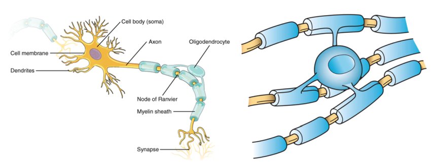
Synapsins are a family of proteins found in the presynaptic terminals of neurons. They play a crucial role in the regulation of neurotransmitter release and synaptic plasticity, which is the ability of synapses to strengthen or weaken over time in response to increases or decreases in their activity.
Synapsins are associated with the cytoskeleton of presynaptic terminals and help to tether vesicles containing neurotransmitters to the cytoskeleton. This allows for the rapid mobilization of vesicles to the active zone of the synapse, where they can be released in response to an action potential.
Synapsins are also involved in the regulation of vesicle pool size and the clustering of calcium channels at the active zone. They have been implicated in various neurological disorders, including epilepsy, fragile X syndrome, and Alzheimer's disease.
Stathmin, also known as oncoprotein 18 or OP18, is a microtubule-associated protein that plays a crucial role in the regulation of microtubule dynamics. It is involved in the destabilization of microtubules by promoting the depolymerization and inhibiting the polymerization of tubulin dimers. Stathmin has been found to be overexpressed in various types of cancer, making it a potential target for cancer therapy. Additionally, stathmin has been implicated in the regulation of cell division, differentiation, and motility, as well as in neuronal development and plasticity.
Viral proteins are the proteins that are encoded by the viral genome and are essential for the viral life cycle. These proteins can be structural or non-structural and play various roles in the virus's replication, infection, and assembly process. Structural proteins make up the physical structure of the virus, including the capsid (the protein shell that surrounds the viral genome) and any envelope proteins (that may be present on enveloped viruses). Non-structural proteins are involved in the replication of the viral genome and modulation of the host cell environment to favor viral replication. Overall, a thorough understanding of viral proteins is crucial for developing antiviral therapies and vaccines.
Phosphopeptides are short peptide sequences that contain one or more phosphorylated amino acid residues, most commonly serine, threonine, or tyrosine. Phosphorylation is a post-translational modification that plays a crucial role in regulating various cellular processes such as signal transduction, protein-protein interactions, enzyme activity, and protein degradation. The addition of a phosphate group to a peptide can alter its charge, conformation, stability, and interaction with other molecules, thereby modulating its function in the cell. Phosphopeptides are often generated by proteolytic digestion of phosphorylated proteins and are used as biomarkers or probes to study protein phosphorylation and signaling pathways in various biological systems.
Phosphorus radioisotopes are radioactive isotopes or variants of the element phosphorus that emit radiation. Phosphorus has several radioisotopes, with the most common ones being phosphorus-32 (^32P) and phosphorus-33 (^33P). These radioisotopes are used in various medical applications such as cancer treatment and diagnostic procedures.
Phosphorus-32 has a half-life of approximately 14.3 days and emits beta particles, making it useful for treating certain types of cancer, such as leukemia and lymphoma. It can also be used in brachytherapy, a type of radiation therapy that involves placing a radioactive source close to the tumor.
Phosphorus-33 has a shorter half-life of approximately 25.4 days and emits both beta particles and gamma rays. This makes it useful for diagnostic procedures, such as positron emission tomography (PET) scans, where the gamma rays can be detected and used to create images of the body's internal structures.
It is important to note that handling and using radioisotopes requires specialized training and equipment to ensure safety and prevent radiation exposure.
Electrophoresis, polyacrylamide gel (EPG) is a laboratory technique used to separate and analyze complex mixtures of proteins or nucleic acids (DNA or RNA) based on their size and electrical charge. This technique utilizes a matrix made of cross-linked polyacrylamide, a type of gel, which provides a stable and uniform environment for the separation of molecules.
In this process:
1. The polyacrylamide gel is prepared by mixing acrylamide monomers with a cross-linking agent (bis-acrylamide) and a catalyst (ammonium persulfate) in the presence of a buffer solution.
2. The gel is then poured into a mold and allowed to polymerize, forming a solid matrix with uniform pore sizes that depend on the concentration of acrylamide used. Higher concentrations result in smaller pores, providing better resolution for separating smaller molecules.
3. Once the gel has set, it is placed in an electrophoresis apparatus containing a buffer solution. Samples containing the mixture of proteins or nucleic acids are loaded into wells on the top of the gel.
4. An electric field is applied across the gel, causing the negatively charged molecules to migrate towards the positive electrode (anode) while positively charged molecules move toward the negative electrode (cathode). The rate of migration depends on the size, charge, and shape of the molecules.
5. Smaller molecules move faster through the gel matrix and will migrate farther from the origin compared to larger molecules, resulting in separation based on size. Proteins and nucleic acids can be selectively stained after electrophoresis to visualize the separated bands.
EPG is widely used in various research fields, including molecular biology, genetics, proteomics, and forensic science, for applications such as protein characterization, DNA fragment analysis, cloning, mutation detection, and quality control of nucleic acid or protein samples.
Phosphoserine is not a medical term per se, but rather a biochemical term. It refers to a post-translationally modified amino acid called serine that has a phosphate group attached to its side chain. This modification plays a crucial role in various cellular processes, including signal transduction and regulation of protein function. In medical contexts, abnormalities in the regulation of phosphorylation (the addition of a phosphate group) and dephosphorylation (the removal of a phosphate group) have been implicated in several diseases, such as cancer and neurological disorders.
Phosphothreonine is not a medical term per se, but rather a biochemical term that refers to a specific post-translational modification of the amino acid threonine. In this modification, a phosphate group is added to the hydroxyl side chain of threonine, which can affect the function and regulation of proteins in which it occurs.
In medical or clinical contexts, phosphothreonine may be mentioned in relation to various disease processes or signaling pathways that involve protein kinases, enzymes that add phosphate groups to specific amino acids (including threonine) in proteins. For example, abnormal regulation of protein kinases and phosphatases (enzymes that remove phosphate groups) can contribute to the development of cancer, neurological disorders, and other diseases.
A cell line is a culture of cells that are grown in a laboratory for use in research. These cells are usually taken from a single cell or group of cells, and they are able to divide and grow continuously in the lab. Cell lines can come from many different sources, including animals, plants, and humans. They are often used in scientific research to study cellular processes, disease mechanisms, and to test new drugs or treatments. Some common types of human cell lines include HeLa cells (which come from a cancer patient named Henrietta Lacks), HEK293 cells (which come from embryonic kidney cells), and HUVEC cells (which come from umbilical vein endothelial cells). It is important to note that cell lines are not the same as primary cells, which are cells that are taken directly from a living organism and have not been grown in the lab.
Phosvitin is not a medical term, but it is a protein found in egg yolk. It is a highly phosphorylated protein, meaning that many of its amino acids are bound to phosphate groups. This gives phosvitin a high negative charge and makes it an excellent chelator of positively charged ions such as calcium and iron.
Phosvitin is known for its ability to bind and store minerals, particularly iron, in the egg yolk. It plays a role in the development and nutrition of growing embryos in birds. In addition to its nutritional role, phosvitin has been studied for its potential health benefits due to its antioxidant properties and ability to bind heavy metals.
While not a medical term itself, phosvitin may be relevant to certain medical fields such as nutrition, biochemistry, and food science.
Two-dimensional (2D) gel electrophoresis is a type of electrophoretic technique used in the separation and analysis of complex protein mixtures. This method combines two types of electrophoresis – isoelectric focusing (IEF) and sodium dodecyl sulfate polyacrylamide gel electrophoresis (SDS-PAGE) – to separate proteins based on their unique physical and chemical properties in two dimensions.
In the first dimension, IEF separates proteins according to their isoelectric points (pI), which is the pH at which a protein carries no net electrical charge. The proteins are focused into narrow zones along a pH gradient established within a gel strip. In the second dimension, SDS-PAGE separates the proteins based on their molecular weights by applying an electric field perpendicular to the first dimension.
The separated proteins form distinct spots on the 2D gel, which can be visualized using various staining techniques. The resulting protein pattern provides valuable information about the composition and modifications of the protein mixture, enabling researchers to identify and compare different proteins in various samples. Two-dimensional gel electrophoresis is widely used in proteomics research, biomarker discovery, and quality control in protein production.
Peptide mapping is a technique used in proteomics and analytical chemistry to analyze and identify the sequence and structure of peptides or proteins. This method involves breaking down a protein into smaller peptide fragments using enzymatic or chemical digestion, followed by separation and identification of these fragments through various analytical techniques such as liquid chromatography (LC) and mass spectrometry (MS).
The resulting peptide map serves as a "fingerprint" of the protein, providing information about its sequence, modifications, and structure. Peptide mapping can be used for a variety of applications, including protein identification, characterization of post-translational modifications, and monitoring of protein degradation or cleavage.
In summary, peptide mapping is a powerful tool in proteomics that enables the analysis and identification of proteins and their modifications at the peptide level.
Microtubule proteins are a class of structural proteins that make up the microtubules, which are key components of the cytoskeleton in eukaryotic cells. The main microtubule protein is tubulin, which exists in two forms: alpha-tubulin and beta-tubulin. These tubulins polymerize to form heterodimers, which then assemble into protofilaments, which in turn aggregate to form hollow microtubules. Microtubules are dynamic structures that undergo continuous assembly and disassembly, and they play crucial roles in various cellular processes, including intracellular transport, cell division, and maintenance of cell shape. Other microtubule-associated proteins (MAPs) also bind to microtubules and regulate their stability, dynamics, and interactions with other cellular structures.
A base sequence in the context of molecular biology refers to the specific order of nucleotides in a DNA or RNA molecule. In DNA, these nucleotides are adenine (A), guanine (G), cytosine (C), and thymine (T). In RNA, uracil (U) takes the place of thymine. The base sequence contains genetic information that is transcribed into RNA and ultimately translated into proteins. It is the exact order of these bases that determines the genetic code and thus the function of the DNA or RNA molecule.
Cell adhesion molecules (CAMs) are a type of protein found on the surface of cells that mediate the attachment or adhesion of cells to either other cells or to the extracellular matrix (ECM), which is the network of proteins and carbohydrates that provides structural and biochemical support to surrounding cells.
CAMs play crucial roles in various biological processes, including tissue development, differentiation, repair, and maintenance of tissue architecture and function. They are also involved in cell signaling, migration, and regulation of the immune response.
There are several types of CAMs, classified based on their structure and function, such as immunoglobulin-like CAMs (IgCAMs), cadherins, integrins, and selectins. Dysregulation of CAMs has been implicated in various diseases, including cancer, inflammation, and neurological disorders.
Osteopontin (OPN) is a phosphorylated glycoprotein that is widely distributed in many tissues, including bone, teeth, and mineralized tissues. It plays important roles in various biological processes such as bone remodeling, immune response, wound healing, and tissue repair. In the skeletal system, osteopontin is involved in the regulation of bone formation and resorption by modulating the activity of osteoclasts and osteoblasts. It also plays a role in the development of chronic inflammatory diseases such as rheumatoid arthritis, atherosclerosis, and cancer metastasis to bones. Osteopontin is considered a potential biomarker for various disease states, including bone turnover, cardiovascular disease, and cancer progression.
Sialglycoproteins are a type of glycoprotein that have sialic acid as the terminal sugar in their oligosaccharide chains. These complex molecules are abundant on the surface of many cell types and play important roles in various biological processes, including cell recognition, cell-cell interactions, and protection against proteolytic degradation.
The presence of sialic acid on the outermost part of these glycoproteins makes them negatively charged, which can affect their interaction with other molecules such as lectins, antibodies, and enzymes. Sialglycoproteins are also involved in the regulation of various physiological functions, including blood coagulation, inflammation, and immune response.
Abnormalities in sialglycoprotein expression or structure have been implicated in several diseases, such as cancer, autoimmune disorders, and neurodegenerative conditions. Therefore, understanding the biology of sialoglycoproteins is important for developing new diagnostic and therapeutic strategies for these diseases.
Serine is an amino acid, which is a building block of proteins. More specifically, it is a non-essential amino acid, meaning that the body can produce it from other compounds, and it does not need to be obtained through diet. Serine plays important roles in the body, such as contributing to the formation of the protective covering of nerve fibers (myelin sheath), helping to synthesize another amino acid called tryptophan, and taking part in the metabolism of fatty acids. It is also involved in the production of muscle tissues, the immune system, and the forming of cell structures. Serine can be found in various foods such as soy, eggs, cheese, meat, peanuts, lentils, and many others.
Protein binding, in the context of medical and biological sciences, refers to the interaction between a protein and another molecule (known as the ligand) that results in a stable complex. This process is often reversible and can be influenced by various factors such as pH, temperature, and concentration of the involved molecules.
In clinical chemistry, protein binding is particularly important when it comes to drugs, as many of them bind to proteins (especially albumin) in the bloodstream. The degree of protein binding can affect a drug's distribution, metabolism, and excretion, which in turn influence its therapeutic effectiveness and potential side effects.
Protein-bound drugs may be less available for interaction with their target tissues, as only the unbound or "free" fraction of the drug is active. Therefore, understanding protein binding can help optimize dosing regimens and minimize adverse reactions.
Nucleoproteins are complexes formed by the association of proteins with nucleic acids (DNA or RNA). These complexes play crucial roles in various biological processes, such as packaging and protecting genetic material, regulating gene expression, and replication and repair of DNA. In these complexes, proteins interact with nucleic acids through electrostatic, hydrogen bonding, and other non-covalent interactions, leading to the formation of stable structures that help maintain the integrity and function of the genetic material. Some well-known examples of nucleoproteins include histones, which are involved in DNA packaging in eukaryotic cells, and reverse transcriptase, an enzyme found in retroviruses that transcribes RNA into DNA.
Signal transduction is the process by which a cell converts an extracellular signal, such as a hormone or neurotransmitter, into an intracellular response. This involves a series of molecular events that transmit the signal from the cell surface to the interior of the cell, ultimately resulting in changes in gene expression, protein activity, or metabolism.
The process typically begins with the binding of the extracellular signal to a receptor located on the cell membrane. This binding event activates the receptor, which then triggers a cascade of intracellular signaling molecules, such as second messengers, protein kinases, and ion channels. These molecules amplify and propagate the signal, ultimately leading to the activation or inhibition of specific cellular responses.
Signal transduction pathways are highly regulated and can be modulated by various factors, including other signaling molecules, post-translational modifications, and feedback mechanisms. Dysregulation of these pathways has been implicated in a variety of diseases, including cancer, diabetes, and neurological disorders.
In the context of medicine and pharmacology, "kinetics" refers to the study of how a drug moves throughout the body, including its absorption, distribution, metabolism, and excretion (often abbreviated as ADME). This field is called "pharmacokinetics."
1. Absorption: This is the process of a drug moving from its site of administration into the bloodstream. Factors such as the route of administration (e.g., oral, intravenous, etc.), formulation, and individual physiological differences can affect absorption.
2. Distribution: Once a drug is in the bloodstream, it gets distributed throughout the body to various tissues and organs. This process is influenced by factors like blood flow, protein binding, and lipid solubility of the drug.
3. Metabolism: Drugs are often chemically modified in the body, typically in the liver, through processes known as metabolism. These changes can lead to the formation of active or inactive metabolites, which may then be further distributed, excreted, or undergo additional metabolic transformations.
4. Excretion: This is the process by which drugs and their metabolites are eliminated from the body, primarily through the kidneys (urine) and the liver (bile).
Understanding the kinetics of a drug is crucial for determining its optimal dosing regimen, potential interactions with other medications or foods, and any necessary adjustments for special populations like pediatric or geriatric patients, or those with impaired renal or hepatic function.
Rabies is a viral disease that affects the nervous system of mammals, including humans. It's caused by the rabies virus (RV), which belongs to the family Rhabdoviridae and genus Lyssavirus. The virus has a bullet-shaped appearance under an electron microscope and is encased in a lipid envelope.
The rabies virus primarily spreads through the saliva of infected animals, usually via bites. Once inside the body, it travels along nerve fibers to the brain, where it multiplies rapidly and causes inflammation (encephalitis). The infection can lead to symptoms such as anxiety, confusion, hallucinations, seizures, paralysis, coma, and ultimately death if left untreated.
Rabies is almost always fatal once symptoms appear, but prompt post-exposure prophylaxis (PEP), which includes vaccination and sometimes rabies immunoglobulin, can prevent the disease from developing when administered after an exposure to a potentially rabid animal. Pre-exposure vaccination is also recommended for individuals at high risk of exposure, such as veterinarians and travelers visiting rabies-endemic areas.
Nerve tissue proteins are specialized proteins found in the nervous system that provide structural and functional support to nerve cells, also known as neurons. These proteins include:
1. Neurofilaments: These are type IV intermediate filaments that provide structural support to neurons and help maintain their shape and size. They are composed of three subunits - NFL (light), NFM (medium), and NFH (heavy).
2. Neuronal Cytoskeletal Proteins: These include tubulins, actins, and spectrins that provide structural support to the neuronal cytoskeleton and help maintain its integrity.
3. Neurotransmitter Receptors: These are specialized proteins located on the postsynaptic membrane of neurons that bind neurotransmitters released by presynaptic neurons, triggering a response in the target cell.
4. Ion Channels: These are transmembrane proteins that regulate the flow of ions across the neuronal membrane and play a crucial role in generating and transmitting electrical signals in neurons.
5. Signaling Proteins: These include enzymes, receptors, and adaptor proteins that mediate intracellular signaling pathways involved in neuronal development, differentiation, survival, and death.
6. Adhesion Proteins: These are cell surface proteins that mediate cell-cell and cell-matrix interactions, playing a crucial role in the formation and maintenance of neural circuits.
7. Extracellular Matrix Proteins: These include proteoglycans, laminins, and collagens that provide structural support to nerve tissue and regulate neuronal migration, differentiation, and survival.
Viral structural proteins are the protein components that make up the viral particle or capsid, providing structure and stability to the virus. These proteins are encoded by the viral genome and are involved in the assembly of new virus particles during the replication cycle. They can be classified into different types based on their location and function, such as capsid proteins, matrix proteins, and envelope proteins. Capsid proteins form the protein shell that encapsulates the viral genome, while matrix proteins are located between the capsid and the envelope, and envelope proteins are embedded in the lipid bilayer membrane that surrounds some viruses.
Molecular cloning is a laboratory technique used to create multiple copies of a specific DNA sequence. This process involves several steps:
1. Isolation: The first step in molecular cloning is to isolate the DNA sequence of interest from the rest of the genomic DNA. This can be done using various methods such as PCR (polymerase chain reaction), restriction enzymes, or hybridization.
2. Vector construction: Once the DNA sequence of interest has been isolated, it must be inserted into a vector, which is a small circular DNA molecule that can replicate independently in a host cell. Common vectors used in molecular cloning include plasmids and phages.
3. Transformation: The constructed vector is then introduced into a host cell, usually a bacterial or yeast cell, through a process called transformation. This can be done using various methods such as electroporation or chemical transformation.
4. Selection: After transformation, the host cells are grown in selective media that allow only those cells containing the vector to grow. This ensures that the DNA sequence of interest has been successfully cloned into the vector.
5. Amplification: Once the host cells have been selected, they can be grown in large quantities to amplify the number of copies of the cloned DNA sequence.
Molecular cloning is a powerful tool in molecular biology and has numerous applications, including the production of recombinant proteins, gene therapy, functional analysis of genes, and genetic engineering.
A precipitin test is a type of immunodiagnostic test used to detect and measure the presence of specific antibodies or antigens in a patient's serum. The test is based on the principle of antigen-antibody interaction, where the addition of an antigen to a solution containing its corresponding antibody results in the formation of an insoluble immune complex known as a precipitin.
In this test, a small amount of the patient's serum is added to a solution containing a known antigen or antibody. If the patient has antibodies or antigens that correspond to the added reagent, they will bind and form a visible precipitate. The size and density of the precipitate can be used to quantify the amount of antibody or antigen present in the sample.
Precipitin tests are commonly used in the diagnosis of various infectious diseases, autoimmune disorders, and allergies. They can also be used in forensic science to identify biological samples. However, they have largely been replaced by more modern immunological techniques such as enzyme-linked immunosorbent assays (ELISAs) and radioimmunoassays (RIAs).
Casein Kinase II (CK2) is a serine/threonine protein kinase that is widely expressed in eukaryotic cells and is involved in the regulation of various cellular processes. It is a heterotetrameric enzyme, consisting of two catalytic subunits (alpha and alpha') and two regulatory subunits (beta).
CK2 phosphorylates a wide range of substrates, including transcription factors, signaling proteins, and other kinases. It is known to play roles in cell cycle regulation, apoptosis, DNA damage response, and protein stability, among others. CK2 activity is often found to be elevated in various types of cancer, making it a potential target for cancer therapy.
Lyssavirus is a genus of viruses in the family Rhabdoviridae, order Mononegavirales. This genus includes several species of viruses that are closely related to the rabies virus and can cause similar diseases in various mammals, including humans. The lyssaviruses are bullet-shaped viruses with a single strand of negative-sense RNA. They infect nerve cells and spread through the nervous system, causing encephalitis, which is inflammation of the brain.
The most well-known member of this genus is the rabies virus, which is responsible for the disease rabies in humans and animals worldwide. Other members of this genus include Australian bat lyssavirus (ABLV), Duvenhage virus (DUVV), European bat lyssavirus types 1 and 2 (EBLV-1 and EBLV-2), Irkut virus (IRKV), Lagos bat virus (LBV), Mokola virus (MOKV), and Shimoni bat virus (SHIBV). These viruses are primarily found in bats, but some have been known to infect other mammals as well.
Prevention of lyssavirus infection is similar to that of rabies and includes avoiding contact with bats or other potential carriers, vaccinating domestic animals against rabies, and seeking prompt medical attention if a bite or scratch from a potentially infected animal occurs. Post-exposure prophylaxis (PEP) with rabies vaccine and immunoglobulin is also recommended for individuals who have been exposed to a lyssavirus.
Tyrosine is an non-essential amino acid, which means that it can be synthesized by the human body from another amino acid called phenylalanine. Its name is derived from the Greek word "tyros," which means cheese, as it was first isolated from casein, a protein found in cheese.
Tyrosine plays a crucial role in the production of several important substances in the body, including neurotransmitters such as dopamine, norepinephrine, and epinephrine, which are involved in various physiological processes, including mood regulation, stress response, and cognitive functions. It also serves as a precursor to melanin, the pigment responsible for skin, hair, and eye color.
In addition, tyrosine is involved in the structure of proteins and is essential for normal growth and development. Some individuals may require tyrosine supplementation if they have a genetic disorder that affects tyrosine metabolism or if they are phenylketonurics (PKU), who cannot metabolize phenylalanine, which can lead to elevated tyrosine levels in the blood. However, it is important to consult with a healthcare professional before starting any supplementation regimen.
Borna Disease Virus (BoDV) is a negative-stranded RNA virus that belongs to the family Bornaviridae. It is the causative agent of Borna disease, a neurological disorder primarily affecting horses and sheep in Europe, although it has also been found in other mammals including cats, dogs, rabbits, and humans.
The virus is named after the town of Borna in Saxony, Germany, where an outbreak of the disease occurred in horses in the late 19th century. BoDV is unique among animal viruses because it can establish a persistent infection in the central nervous system (CNS) of its hosts and has been shown to have neurotropic properties.
In humans, BoDV infection has been linked to cases of encephalitis, a potentially life-threatening inflammation of the brain. However, human infections with BoDV are rare and often associated with close contact with infected animals or their tissues. There is currently no specific treatment for Borna disease or BoDV infection, and prevention efforts focus on reducing exposure to the virus through appropriate handling and care of infected animals.
Membrane proteins are a type of protein that are embedded in the lipid bilayer of biological membranes, such as the plasma membrane of cells or the inner membrane of mitochondria. These proteins play crucial roles in various cellular processes, including:
1. Cell-cell recognition and signaling
2. Transport of molecules across the membrane (selective permeability)
3. Enzymatic reactions at the membrane surface
4. Energy transduction and conversion
5. Mechanosensation and signal transduction
Membrane proteins can be classified into two main categories: integral membrane proteins, which are permanently associated with the lipid bilayer, and peripheral membrane proteins, which are temporarily or loosely attached to the membrane surface. Integral membrane proteins can further be divided into three subcategories based on their topology:
1. Transmembrane proteins, which span the entire width of the lipid bilayer with one or more alpha-helices or beta-barrels.
2. Lipid-anchored proteins, which are covalently attached to lipids in the membrane via a glycosylphosphatidylinositol (GPI) anchor or other lipid modifications.
3. Monotopic proteins, which are partially embedded in the membrane and have one or more domains exposed to either side of the bilayer.
Membrane proteins are essential for maintaining cellular homeostasis and are targets for various therapeutic interventions, including drug development and gene therapy. However, their structural complexity and hydrophobicity make them challenging to study using traditional biochemical methods, requiring specialized techniques such as X-ray crystallography, nuclear magnetic resonance (NMR) spectroscopy, and single-particle cryo-electron microscopy (cryo-EM).
Nuclear proteins are a category of proteins that are primarily found in the nucleus of a eukaryotic cell. They play crucial roles in various nuclear functions, such as DNA replication, transcription, repair, and RNA processing. This group includes structural proteins like lamins, which form the nuclear lamina, and regulatory proteins, such as histones and transcription factors, that are involved in gene expression. Nuclear localization signals (NLS) often help target these proteins to the nucleus by interacting with importin proteins during active transport across the nuclear membrane.
Nucleocapsid proteins are structural proteins that are associated with the viral genome in many viruses. They play a crucial role in the formation and stability of the viral particle, also known as the virion. In particular, nucleocapsid proteins bind to the viral RNA or DNA genome and help to protect it from degradation by host cell enzymes. They also participate in the assembly and disassembly of the virion during the viral replication cycle.
In some viruses, such as coronaviruses, the nucleocapsid protein is also involved in regulating the transcription and replication of the viral genome. The nucleocapsid protein of SARS-CoV-2, for example, has been shown to interact with host cell proteins that are involved in the regulation of gene expression, which may contribute to the virus's ability to manipulate the host cell environment and evade the immune response.
Overall, nucleocapsid proteins are important components of many viruses and are often targeted by antiviral therapies due to their essential role in the viral replication cycle.
In the context of medical and biological sciences, a "binding site" refers to a specific location on a protein, molecule, or cell where another molecule can attach or bind. This binding interaction can lead to various functional changes in the original protein or molecule. The other molecule that binds to the binding site is often referred to as a ligand, which can be a small molecule, ion, or even another protein.
The binding between a ligand and its target binding site can be specific and selective, meaning that only certain ligands can bind to particular binding sites with high affinity. This specificity plays a crucial role in various biological processes, such as signal transduction, enzyme catalysis, or drug action.
In the case of drug development, understanding the location and properties of binding sites on target proteins is essential for designing drugs that can selectively bind to these sites and modulate protein function. This knowledge can help create more effective and safer therapeutic options for various diseases.
HeLa cells are a type of immortalized cell line used in scientific research. They are derived from a cancer that developed in the cervical tissue of Henrietta Lacks, an African-American woman, in 1951. After her death, cells taken from her tumor were found to be capable of continuous division and growth in a laboratory setting, making them an invaluable resource for medical research.
HeLa cells have been used in a wide range of scientific studies, including research on cancer, viruses, genetics, and drug development. They were the first human cell line to be successfully cloned and are able to grow rapidly in culture, doubling their population every 20-24 hours. This has made them an essential tool for many areas of biomedical research.
It is important to note that while HeLa cells have been instrumental in numerous scientific breakthroughs, the story of their origin raises ethical questions about informed consent and the use of human tissue in research.
Sequence homology, amino acid, refers to the similarity in the order of amino acids in a protein or a portion of a protein between two or more species. This similarity can be used to infer evolutionary relationships and functional similarities between proteins. The higher the degree of sequence homology, the more likely it is that the proteins are related and have similar functions. Sequence homology can be determined through various methods such as pairwise alignment or multiple sequence alignment, which compare the sequences and calculate a score based on the number and type of matching amino acids.
Cyclic adenosine monophosphate (cAMP) is a key secondary messenger in many biological processes, including the regulation of metabolism, gene expression, and cellular excitability. It is synthesized from adenosine triphosphate (ATP) by the enzyme adenylyl cyclase and is degraded by the enzyme phosphodiesterase.
In the body, cAMP plays a crucial role in mediating the effects of hormones and neurotransmitters on target cells. For example, when a hormone binds to its receptor on the surface of a cell, it can activate a G protein, which in turn activates adenylyl cyclase to produce cAMP. The increased levels of cAMP then activate various effector proteins, such as protein kinases, which go on to regulate various cellular processes.
Overall, the regulation of cAMP levels is critical for maintaining proper cellular function and homeostasis, and abnormalities in cAMP signaling have been implicated in a variety of diseases, including cancer, diabetes, and neurological disorders.
"Cattle" is a term used in the agricultural and veterinary fields to refer to domesticated animals of the genus *Bos*, primarily *Bos taurus* (European cattle) and *Bos indicus* (Zebu). These animals are often raised for meat, milk, leather, and labor. They are also known as bovines or cows (for females), bulls (intact males), and steers/bullocks (castrated males). However, in a strict medical definition, "cattle" does not apply to humans or other animals.
Okadaic acid is a type of toxin that is produced by certain species of marine algae, including Dinophysis and Prorocentrum. It is a potent inhibitor of protein phosphatases 1 and 2A, which are important enzymes that help regulate cellular processes in the body.
Okadaic acid can accumulate in shellfish that feed on these algae, and consumption of contaminated seafood can lead to a serious illness known as diarrhetic shellfish poisoning (DSP). Symptoms of DSP include nausea, vomiting, diarrhea, and abdominal cramps. In severe cases, it can also cause neurological symptoms such as dizziness, disorientation, and tingling or numbness in the lips, tongue, and fingers.
It is important to note that okadaic acid is not only a marine toxin but also used in scientific research as a tool to study the role of protein phosphatases in cellular processes. However, exposure to this compound should be avoided due to its toxic effects.
The cell nucleus is a membrane-bound organelle found in the eukaryotic cells (cells with a true nucleus). It contains most of the cell's genetic material, organized as DNA molecules in complex with proteins, RNA molecules, and histones to form chromosomes.
The primary function of the cell nucleus is to regulate and control the activities of the cell, including growth, metabolism, protein synthesis, and reproduction. It also plays a crucial role in the process of mitosis (cell division) by separating and protecting the genetic material during this process. The nuclear membrane, or nuclear envelope, surrounding the nucleus is composed of two lipid bilayers with numerous pores that allow for the selective transport of molecules between the nucleoplasm (nucleus interior) and the cytoplasm (cell exterior).
The cell nucleus is a vital structure in eukaryotic cells, and its dysfunction can lead to various diseases, including cancer and genetic disorders.
Recombinant fusion proteins are artificially created biomolecules that combine the functional domains or properties of two or more different proteins into a single protein entity. They are generated through recombinant DNA technology, where the genes encoding the desired protein domains are linked together and expressed as a single, chimeric gene in a host organism, such as bacteria, yeast, or mammalian cells.
The resulting fusion protein retains the functional properties of its individual constituent proteins, allowing for novel applications in research, diagnostics, and therapeutics. For instance, recombinant fusion proteins can be designed to enhance protein stability, solubility, or immunogenicity, making them valuable tools for studying protein-protein interactions, developing targeted therapies, or generating vaccines against infectious diseases or cancer.
Examples of recombinant fusion proteins include:
1. Etaglunatide (ABT-523): A soluble Fc fusion protein that combines the heavy chain fragment crystallizable region (Fc) of an immunoglobulin with the extracellular domain of the human interleukin-6 receptor (IL-6R). This fusion protein functions as a decoy receptor, neutralizing IL-6 and its downstream signaling pathways in rheumatoid arthritis.
2. Etanercept (Enbrel): A soluble TNF receptor p75 Fc fusion protein that binds to tumor necrosis factor-alpha (TNF-α) and inhibits its proinflammatory activity, making it a valuable therapeutic option for treating autoimmune diseases like rheumatoid arthritis, ankylosing spondylitis, and psoriasis.
3. Abatacept (Orencia): A fusion protein consisting of the extracellular domain of cytotoxic T-lymphocyte antigen 4 (CTLA-4) linked to the Fc region of an immunoglobulin, which downregulates T-cell activation and proliferation in autoimmune diseases like rheumatoid arthritis.
4. Belimumab (Benlysta): A monoclonal antibody that targets B-lymphocyte stimulator (BLyS) protein, preventing its interaction with the B-cell surface receptor and inhibiting B-cell activation in systemic lupus erythematosus (SLE).
5. Romiplostim (Nplate): A fusion protein consisting of a thrombopoietin receptor agonist peptide linked to an immunoglobulin Fc region, which stimulates platelet production in patients with chronic immune thrombocytopenia (ITP).
6. Darbepoetin alfa (Aranesp): A hyperglycosylated erythropoiesis-stimulating protein that functions as a longer-acting form of recombinant human erythropoietin, used to treat anemia in patients with chronic kidney disease or cancer.
7. Palivizumab (Synagis): A monoclonal antibody directed against the F protein of respiratory syncytial virus (RSV), which prevents RSV infection and is administered prophylactically to high-risk infants during the RSV season.
8. Ranibizumab (Lucentis): A recombinant humanized monoclonal antibody fragment that binds and inhibits vascular endothelial growth factor A (VEGF-A), used in the treatment of age-related macular degeneration, diabetic retinopathy, and other ocular disorders.
9. Cetuximab (Erbitux): A chimeric monoclonal antibody that binds to epidermal growth factor receptor (EGFR), used in the treatment of colorectal cancer and head and neck squamous cell carcinoma.
10. Adalimumab (Humira): A fully humanized monoclonal antibody that targets tumor necrosis factor-alpha (TNF-α), used in the treatment of various inflammatory diseases, including rheumatoid arthritis, psoriasis, and Crohn's disease.
11. Bevacizumab (Avastin): A recombinant humanized monoclonal antibody that binds to VEGF-A, used in the treatment of various cancers, including colorectal, lung, breast, and kidney cancer.
12. Trastuzumab (Herceptin): A humanized monoclonal antibody that targets HER2/neu receptor, used in the treatment of breast cancer.
13. Rituximab (Rituxan): A chimeric monoclonal antibody that binds to CD20 antigen on B cells, used in the treatment of non-Hodgkin's lymphoma and rheumatoid arthritis.
14. Palivizumab (Synagis): A humanized monoclonal antibody that binds to the F protein of respiratory syncytial virus, used in the prevention of respiratory syncytial virus infection in high-risk infants.
15. Infliximab (Remicade): A chimeric monoclonal antibody that targets TNF-α, used in the treatment of various inflammatory diseases, including Crohn's disease, ulcerative colitis, rheumatoid arthritis, and ankylosing spondylitis.
16. Natalizumab (Tysabri): A humanized monoclonal antibody that binds to α4β1 integrin, used in the treatment of multiple sclerosis and Crohn's disease.
17. Adalimumab (Humira): A fully human monoclonal antibody that targets TNF-α, used in the treatment of various inflammatory diseases, including rheumatoid arthritis, psoriatic arthritis, ankylosing spondylitis, Crohn's disease, and ulcerative colitis.
18. Golimumab (Simponi): A fully human monoclonal antibody that targets TNF-α, used in the treatment of rheumatoid arthritis, psoriatic arthritis, ankylosing spondylitis, and ulcerative colitis.
19. Certolizumab pegol (Cimzia): A PEGylated Fab' fragment of a humanized monoclonal antibody that targets TNF-α, used in the treatment of rheumatoid arthritis, psoriatic arthritis, ankylosing spondylitis, and Crohn's disease.
20. Ustekinumab (Stelara): A fully human monoclonal antibody that targets IL-12 and IL-23, used in the treatment of psoriasis, psoriatic arthritis, and Crohn's disease.
21. Secukinumab (Cosentyx): A fully human monoclonal antibody that targets IL-17A, used in the treatment of psoriasis, psoriatic arthritis, and ankylosing spondylitis.
22. Ixekizumab (Taltz): A fully human monoclonal antibody that targets IL-17A, used in the treatment of psoriasis and psoriatic arthritis.
23. Brodalumab (Siliq): A fully human monoclonal antibody that targets IL-17 receptor A, used in the treatment of psoriasis.
24. Sarilumab (Kevzara): A fully human monoclonal antibody that targets the IL-6 receptor, used in the treatment of rheumatoid arthritis.
25. Tocilizumab (Actemra): A humanized monoclonal antibody that targets the IL-6 receptor, used in the treatment of rheumatoid arthritis, systemic juvenile idiopathic arthritis, polyarticular juvenile idiopathic arthritis, giant cell arteritis, and chimeric antigen receptor T-cell-induced cytokine release syndrome.
26. Siltuximab (Sylvant): A chimeric monoclonal antibody that targets IL-6, used in the treatment of multicentric Castleman disease.
27. Satralizumab (Enspryng): A humanized monoclonal antibody that targets IL-6 receptor alpha, used in the treatment of neuromyelitis optica spectrum disorder.
28. Sirukumab (Plivensia): A human monoclonal antibody that targets IL-6, used in the treatment
Phosphotyrosine is not a medical term per se, but rather a biochemical term used in the field of medicine and life sciences.
Phosphotyrosine is a post-translational modification of tyrosine residues in proteins, where a phosphate group is added to the hydroxyl side chain of tyrosine by protein kinases. This modification plays a crucial role in intracellular signaling pathways and regulates various cellular processes such as cell growth, differentiation, and apoptosis. Abnormalities in phosphotyrosine-mediated signaling have been implicated in several diseases, including cancer and diabetes.
Zyxin is actually not a medical term itself, but rather a protein that has been studied in the context of cell biology and molecular biology. Zyxin is a component of focal adhesions, which are structures that connect the cytoskeleton (the structural framework inside cells) to the extracellular matrix (the material that provides support for cells).
Focal adhesions play important roles in cell signaling, migration, and adhesion. Zyxin is a phosphoprotein, which means it can be modified by the addition of a phosphate group, and this modification can affect its function within the cell. It has been implicated in various cellular processes such as actin dynamics, gene expression, and cell division.
While zyxin itself is not a medical term, abnormalities in the proteins or pathways associated with focal adhesions may contribute to certain diseases. For example, mutations in genes encoding components of focal adhesions have been linked to various genetic disorders such as some forms of muscular dystrophy and epidermolysis bullosa.
Substrate specificity in the context of medical biochemistry and enzymology refers to the ability of an enzyme to selectively bind and catalyze a chemical reaction with a particular substrate (or a group of similar substrates) while discriminating against other molecules that are not substrates. This specificity arises from the three-dimensional structure of the enzyme, which has evolved to match the shape, charge distribution, and functional groups of its physiological substrate(s).
Substrate specificity is a fundamental property of enzymes that enables them to carry out highly selective chemical transformations in the complex cellular environment. The active site of an enzyme, where the catalysis takes place, has a unique conformation that complements the shape and charge distribution of its substrate(s). This ensures efficient recognition, binding, and conversion of the substrate into the desired product while minimizing unwanted side reactions with other molecules.
Substrate specificity can be categorized as:
1. Absolute specificity: An enzyme that can only act on a single substrate or a very narrow group of structurally related substrates, showing no activity towards any other molecule.
2. Group specificity: An enzyme that prefers to act on a particular functional group or class of compounds but can still accommodate minor structural variations within the substrate.
3. Broad or promiscuous specificity: An enzyme that can act on a wide range of structurally diverse substrates, albeit with varying catalytic efficiencies.
Understanding substrate specificity is crucial for elucidating enzymatic mechanisms, designing drugs that target specific enzymes or pathways, and developing biotechnological applications that rely on the controlled manipulation of enzyme activities.
Measles virus is a single-stranded, negative-sense RNA virus belonging to the genus Morbillivirus in the family Paramyxoviridae. It is the causative agent of measles, a highly contagious infectious disease characterized by fever, cough, runny nose, and a red, blotchy rash. The virus primarily infects the respiratory tract and then spreads throughout the body via the bloodstream.
The genome of the measles virus is approximately 16 kilobases in length and encodes for eight proteins: nucleocapsid (N), phosphoprotein (P), matrix protein (M), fusion protein (F), hemagglutinin (H), large protein (L), and two non-structural proteins, V and C. The H protein is responsible for binding to the host cell receptor CD150 (SLAM) and mediating viral entry, while the F protein facilitates fusion of the viral and host cell membranes.
Measles virus is transmitted through respiratory droplets and direct contact with infected individuals. The virus can remain airborne for up to two hours in a closed space, making it highly contagious. Measles is preventable through vaccination, which has led to significant reductions in the incidence of the disease worldwide.
Phosphates, in a medical context, refer to the salts or esters of phosphoric acid. Phosphates play crucial roles in various biological processes within the human body. They are essential components of bones and teeth, where they combine with calcium to form hydroxyapatite crystals. Phosphates also participate in energy transfer reactions as phosphate groups attached to adenosine diphosphate (ADP) and adenosine triphosphate (ATP). Additionally, they contribute to buffer systems that help maintain normal pH levels in the body.
Abnormal levels of phosphates in the blood can indicate certain medical conditions. High phosphate levels (hyperphosphatemia) may be associated with kidney dysfunction, hyperparathyroidism, or excessive intake of phosphate-containing products. Low phosphate levels (hypophosphatemia) might result from malnutrition, vitamin D deficiency, or certain diseases affecting the small intestine or kidneys. Both hypophosphatemia and hyperphosphatemia can have significant impacts on various organ systems and may require medical intervention.
Dentin is the hard, calcified tissue that lies beneath the enamel and cementum of a tooth. It forms the majority of the tooth's structure and is composed primarily of mineral salts (hydroxyapatite), collagenous proteins, and water. Dentin has a tubular structure, with microscopic channels called dentinal tubules that radiate outward from the pulp chamber (the center of the tooth containing nerves and blood vessels) to the exterior of the tooth. These tubules contain fluid and nerve endings that are responsible for the tooth's sensitivity to various stimuli such as temperature changes, pressure, or decay. Dentin plays a crucial role in protecting the dental pulp while also providing support and structure to the overlying enamel and cementum.
Post-translational protein processing refers to the modifications and changes that proteins undergo after their synthesis on ribosomes, which are complex molecular machines responsible for protein synthesis. These modifications occur through various biochemical processes and play a crucial role in determining the final structure, function, and stability of the protein.
The process begins with the translation of messenger RNA (mRNA) into a linear polypeptide chain, which is then subjected to several post-translational modifications. These modifications can include:
1. Proteolytic cleavage: The removal of specific segments or domains from the polypeptide chain by proteases, resulting in the formation of mature, functional protein subunits.
2. Chemical modifications: Addition or modification of chemical groups to the side chains of amino acids, such as phosphorylation (addition of a phosphate group), glycosylation (addition of sugar moieties), methylation (addition of a methyl group), acetylation (addition of an acetyl group), and ubiquitination (addition of a ubiquitin protein).
3. Disulfide bond formation: The oxidation of specific cysteine residues within the polypeptide chain, leading to the formation of disulfide bonds between them. This process helps stabilize the three-dimensional structure of proteins, particularly in extracellular environments.
4. Folding and assembly: The acquisition of a specific three-dimensional conformation by the polypeptide chain, which is essential for its function. Chaperone proteins assist in this process to ensure proper folding and prevent aggregation.
5. Protein targeting: The directed transport of proteins to their appropriate cellular locations, such as the nucleus, mitochondria, endoplasmic reticulum, or plasma membrane. This is often facilitated by specific signal sequences within the protein that are recognized and bound by transport machinery.
Collectively, these post-translational modifications contribute to the functional diversity of proteins in living organisms, allowing them to perform a wide range of cellular processes, including signaling, catalysis, regulation, and structural support.
Proteins are complex, large molecules that play critical roles in the body's functions. They are made up of amino acids, which are organic compounds that are the building blocks of proteins. Proteins are required for the structure, function, and regulation of the body's tissues and organs. They are essential for the growth, repair, and maintenance of body tissues, and they play a crucial role in many biological processes, including metabolism, immune response, and cellular signaling. Proteins can be classified into different types based on their structure and function, such as enzymes, hormones, antibodies, and structural proteins. They are found in various foods, especially animal-derived products like meat, dairy, and eggs, as well as plant-based sources like beans, nuts, and grains.
A nucleocapsid is a protein structure that encloses the genetic material (nucleic acid) of certain viruses. It is composed of proteins encoded by the virus itself, which are synthesized inside the host cell and then assemble around the viral genome to form a stable complex.
The nucleocapsid plays an important role in the viral life cycle. It protects the viral genome from degradation by host enzymes and helps to facilitate the packaging of the genome into new virus particles during assembly. Additionally, the nucleocapsid can also play a role in the regulation of viral gene expression and replication.
In some viruses, such as coronaviruses, the nucleocapsid is encased within an envelope derived from the host cell membrane, while in others, it exists as a naked capsid. The structure and composition of the nucleocapsid can vary significantly between different virus families.
Protein Phosphatase 1 (PP1) is a type of serine/threonine protein phosphatase that plays a crucial role in the regulation of various cellular processes, including metabolism, signal transduction, and cell cycle progression. PP1 functions by removing phosphate groups from specific serine and threonine residues on target proteins, thereby reversing the effects of protein kinases and controlling protein activity, localization, and stability.
PP1 is a highly conserved enzyme found in eukaryotic cells and is composed of a catalytic subunit associated with one or more regulatory subunits that determine its substrate specificity, subcellular localization, and regulation. The human genome encodes several isoforms of the PP1 catalytic subunit, including PP1α, PP1β/δ, and PP1γ, which share a high degree of sequence similarity and functional redundancy.
PP1 has been implicated in various physiological processes, such as muscle contraction, glycogen metabolism, DNA replication, transcription, and RNA processing. Dysregulation of PP1 activity has been associated with several pathological conditions, including neurodegenerative diseases, cancer, and diabetes. Therefore, understanding the molecular mechanisms that regulate PP1 function is essential for developing novel therapeutic strategies to treat these disorders.
"Cells, cultured" is a medical term that refers to cells that have been removed from an organism and grown in controlled laboratory conditions outside of the body. This process is called cell culture and it allows scientists to study cells in a more controlled and accessible environment than they would have inside the body. Cultured cells can be derived from a variety of sources, including tissues, organs, or fluids from humans, animals, or cell lines that have been previously established in the laboratory.
Cell culture involves several steps, including isolation of the cells from the tissue, purification and characterization of the cells, and maintenance of the cells in appropriate growth conditions. The cells are typically grown in specialized media that contain nutrients, growth factors, and other components necessary for their survival and proliferation. Cultured cells can be used for a variety of purposes, including basic research, drug development and testing, and production of biological products such as vaccines and gene therapies.
It is important to note that cultured cells may behave differently than they do in the body, and results obtained from cell culture studies may not always translate directly to human physiology or disease. Therefore, it is essential to validate findings from cell culture experiments using additional models and ultimately in clinical trials involving human subjects.
Western blotting is a laboratory technique used in molecular biology to detect and quantify specific proteins in a mixture of many different proteins. This technique is commonly used to confirm the expression of a protein of interest, determine its size, and investigate its post-translational modifications. The name "Western" blotting distinguishes this technique from Southern blotting (for DNA) and Northern blotting (for RNA).
The Western blotting procedure involves several steps:
1. Protein extraction: The sample containing the proteins of interest is first extracted, often by breaking open cells or tissues and using a buffer to extract the proteins.
2. Separation of proteins by electrophoresis: The extracted proteins are then separated based on their size by loading them onto a polyacrylamide gel and running an electric current through the gel (a process called sodium dodecyl sulfate-polyacrylamide gel electrophoresis or SDS-PAGE). This separates the proteins according to their molecular weight, with smaller proteins migrating faster than larger ones.
3. Transfer of proteins to a membrane: After separation, the proteins are transferred from the gel onto a nitrocellulose or polyvinylidene fluoride (PVDF) membrane using an electric current in a process called blotting. This creates a replica of the protein pattern on the gel but now immobilized on the membrane for further analysis.
4. Blocking: The membrane is then blocked with a blocking agent, such as non-fat dry milk or bovine serum albumin (BSA), to prevent non-specific binding of antibodies in subsequent steps.
5. Primary antibody incubation: A primary antibody that specifically recognizes the protein of interest is added and allowed to bind to its target protein on the membrane. This step may be performed at room temperature or 4°C overnight, depending on the antibody's properties.
6. Washing: The membrane is washed with a buffer to remove unbound primary antibodies.
7. Secondary antibody incubation: A secondary antibody that recognizes the primary antibody (often coupled to an enzyme or fluorophore) is added and allowed to bind to the primary antibody. This step may involve using a horseradish peroxidase (HRP)-conjugated or alkaline phosphatase (AP)-conjugated secondary antibody, depending on the detection method used later.
8. Washing: The membrane is washed again to remove unbound secondary antibodies.
9. Detection: A detection reagent is added to visualize the protein of interest by detecting the signal generated from the enzyme-conjugated or fluorophore-conjugated secondary antibody. This can be done using chemiluminescent, colorimetric, or fluorescent methods.
10. Analysis: The resulting image is analyzed to determine the presence and quantity of the protein of interest in the sample.
Western blotting is a powerful technique for identifying and quantifying specific proteins within complex mixtures. It can be used to study protein expression, post-translational modifications, protein-protein interactions, and more. However, it requires careful optimization and validation to ensure accurate and reproducible results.
Casein kinases are a family of protein kinases that play important roles in various cellular processes, including signal transduction, cell cycle regulation, and DNA damage repair. These enzymes phosphorylate serine and threonine residues on their target proteins by transferring a phosphate group from ATP to the hydroxyl side chain of these amino acids.
There are several isoforms of casein kinases, including Casein Kinase 1 (CK1) and Casein Kinase 2 (CK2), which differ in their structure, substrate specificity, and cellular functions. CK1 is involved in various signaling pathways, such as the Wnt signaling pathway, and regulates processes such as gene transcription, DNA repair, and circadian rhythm. CK2, on the other hand, is a highly conserved serine/threonine protein kinase that plays a role in many cellular processes, including cell division, apoptosis, and transcriptional regulation.
Dysregulation of casein kinases has been implicated in various diseases, including cancer, neurodegenerative disorders, and cardiovascular disease. Therefore, these enzymes are considered important targets for the development of new therapeutic strategies.
Protein-Tyrosine Kinases (PTKs) are a type of enzyme that plays a crucial role in various cellular functions, including signal transduction, cell growth, differentiation, and metabolism. They catalyze the transfer of a phosphate group from ATP to the tyrosine residues of proteins, thereby modifying their activity, localization, or interaction with other molecules.
PTKs can be divided into two main categories: receptor tyrosine kinases (RTKs) and non-receptor tyrosine kinases (NRTKs). RTKs are transmembrane proteins that become activated upon binding to specific ligands, such as growth factors or hormones. NRTKs, on the other hand, are intracellular enzymes that can be activated by various signals, including receptor-mediated signaling and intracellular messengers.
Dysregulation of PTK activity has been implicated in several diseases, such as cancer, diabetes, and inflammatory disorders. Therefore, PTKs are important targets for drug development and therapy.
Phosphorylase phosphatase is an enzyme that plays a role in the regulation of glycogen metabolism. It works by removing phosphate groups from glycogen phosphorylase, which is an enzyme that breaks down glycogen into glucose-1-phosphate. The dephosphorylation of glycogen phosphorylase by phosphorylase phosphatase leads to the inactivation of the enzyme and therefore slows down the breakdown of glycogen. Phosphorylase phosphatase is itself regulated by various hormones and signaling molecules, allowing for fine-tuning of glycogen metabolism in response to changes in energy demand.
Recombinant proteins are artificially created proteins produced through the use of recombinant DNA technology. This process involves combining DNA molecules from different sources to create a new set of genes that encode for a specific protein. The resulting recombinant protein can then be expressed, purified, and used for various applications in research, medicine, and industry.
Recombinant proteins are widely used in biomedical research to study protein function, structure, and interactions. They are also used in the development of diagnostic tests, vaccines, and therapeutic drugs. For example, recombinant insulin is a common treatment for diabetes, while recombinant human growth hormone is used to treat growth disorders.
The production of recombinant proteins typically involves the use of host cells, such as bacteria, yeast, or mammalian cells, which are engineered to express the desired protein. The host cells are transformed with a plasmid vector containing the gene of interest, along with regulatory elements that control its expression. Once the host cells are cultured and the protein is expressed, it can be purified using various chromatography techniques.
Overall, recombinant proteins have revolutionized many areas of biology and medicine, enabling researchers to study and manipulate proteins in ways that were previously impossible.
Threonine is an essential amino acid, meaning it cannot be synthesized by the human body and must be obtained through the diet. Its chemical formula is HO2CCH(NH2)CH(OH)CH3. Threonine plays a crucial role in various biological processes, including protein synthesis, immune function, and fat metabolism. It is particularly important for maintaining the structural integrity of proteins, as it is often found in their hydroxyl-containing regions. Foods rich in threonine include animal proteins such as meat, dairy products, and eggs, as well as plant-based sources like lentils and soybeans.
Cyclic guanosine monophosphate (cGMP)-dependent protein kinases (PKGs) are a type of enzyme that add phosphate groups to other proteins, thereby modifying their function. These kinases are activated by cGMP, which is a second messenger molecule that helps transmit signals within cells. PKGs play important roles in various cellular processes, including smooth muscle relaxation, platelet aggregation, and cardiac contractility. They have been implicated in the regulation of a number of physiological functions, such as blood flow, inflammation, and learning and memory. There are two main isoforms of cGMP-dependent protein kinases, PKG I and PKG II, which differ in their tissue distribution, regulatory properties, and substrate specificity.
Cyclic AMP (cAMP)-dependent protein kinases, also known as protein kinase A (PKA), are a family of enzymes that play a crucial role in intracellular signaling pathways. These enzymes are responsible for the regulation of various cellular processes, including metabolism, gene expression, and cell growth and differentiation.
PKA is composed of two regulatory subunits and two catalytic subunits. When cAMP binds to the regulatory subunits, it causes a conformational change that leads to the dissociation of the catalytic subunits. The freed catalytic subunits then phosphorylate specific serine and threonine residues on target proteins, thereby modulating their activity.
The cAMP-dependent protein kinases are activated in response to a variety of extracellular signals, such as hormones and neurotransmitters, that bind to G protein-coupled receptors (GPCRs) or receptor tyrosine kinases (RTKs). These signals lead to the activation of adenylyl cyclase, which catalyzes the conversion of ATP to cAMP. The resulting increase in intracellular cAMP levels triggers the activation of PKA and the downstream phosphorylation of target proteins.
Overall, cAMP-dependent protein kinases are essential regulators of many fundamental cellular processes and play a critical role in maintaining normal physiology and homeostasis. Dysregulation of these enzymes has been implicated in various diseases, including cancer, diabetes, and neurological disorders.
Adaptor proteins are a type of protein that play a crucial role in intracellular signaling pathways by serving as a link between different components of the signaling complex. Specifically, "signal transducing adaptor proteins" refer to those adaptor proteins that are involved in signal transduction processes, where they help to transmit signals from the cell surface receptors to various intracellular effectors. These proteins typically contain modular domains that allow them to interact with multiple partners, thereby facilitating the formation of large signaling complexes and enabling the integration of signals from different pathways.
Signal transducing adaptor proteins can be classified into several families based on their structural features, including the Src homology 2 (SH2) domain, the Src homology 3 (SH3) domain, and the phosphotyrosine-binding (PTB) domain. These domains enable the adaptor proteins to recognize and bind to specific motifs on other signaling molecules, such as receptor tyrosine kinases, G protein-coupled receptors, and cytokine receptors.
One well-known example of a signal transducing adaptor protein is the growth factor receptor-bound protein 2 (Grb2), which contains an SH2 domain that binds to phosphotyrosine residues on activated receptor tyrosine kinases. Grb2 also contains an SH3 domain that interacts with proline-rich motifs on other signaling proteins, such as the guanine nucleotide exchange factor SOS. This interaction facilitates the activation of the Ras small GTPase and downstream signaling pathways involved in cell growth, differentiation, and survival.
Overall, signal transducing adaptor proteins play a critical role in regulating various cellular processes by modulating intracellular signaling pathways in response to extracellular stimuli. Dysregulation of these proteins has been implicated in various diseases, including cancer and inflammatory disorders.
Proteomics is the large-scale study and analysis of proteins, including their structures, functions, interactions, modifications, and abundance, in a given cell, tissue, or organism. It involves the identification and quantification of all expressed proteins in a biological sample, as well as the characterization of post-translational modifications, protein-protein interactions, and functional pathways. Proteomics can provide valuable insights into various biological processes, diseases, and drug responses, and has applications in basic research, biomedicine, and clinical diagnostics. The field combines various techniques from molecular biology, chemistry, physics, and bioinformatics to study proteins at a systems level.
GAP-43 protein, also known as growth-associated protein 43 or B-50, is a neuronal protein that is highly expressed during development and axonal regeneration. It is involved in the regulation of synaptic plasticity, nerve impulse transmission, and neurite outgrowth. GAP-43 is localized to the growth cones of growing axons and is thought to play a role in the guidance and navigation of axonal growth during development and regeneration. It is a member of the calcium/calmodulin-dependent protein kinase substrate family and undergoes phosphorylation by several protein kinases, including PKC (protein kinase C), which regulates its function. GAP-43 has been implicated in various neurological disorders, such as Alzheimer's disease, Parkinson's disease, and schizophrenia.
Protein-Serine-Threonine Kinases (PSTKs) are a type of protein kinase that catalyzes the transfer of a phosphate group from ATP to the hydroxyl side chains of serine or threonine residues on target proteins. This phosphorylation process plays a crucial role in various cellular signaling pathways, including regulation of metabolism, gene expression, cell cycle progression, and apoptosis. PSTKs are involved in many physiological and pathological processes, and their dysregulation has been implicated in several diseases, such as cancer, diabetes, and neurodegenerative disorders.
I believe there may be some confusion in your question. "Rabbits" is a common name used to refer to the Lagomorpha species, particularly members of the family Leporidae. They are small mammals known for their long ears, strong legs, and quick reproduction.
However, if you're referring to "rabbits" in a medical context, there is a term called "rabbit syndrome," which is a rare movement disorder characterized by repetitive, involuntary movements of the fingers, resembling those of a rabbit chewing. It is also known as "finger-chewing chorea." This condition is usually associated with certain medications, particularly antipsychotics, and typically resolves when the medication is stopped or adjusted.
Vesiculovirus is a genus of enveloped, negative-stranded RNA viruses in the family Rhabdoviridae. They are known to cause vesicular diseases (hence the name) in both animals and humans, characterized by the formation of blisters or vesicles on the skin. The most well-known member of this genus is the vesicular stomatitis virus (VSV), which primarily affects cattle, horses, and pigs, causing oral and foot lesions. However, VSV can also infect humans, resulting in a flu-like illness. Other members of the Vesiculovirus genus include the Isfahan virus, Chandipura virus, and the Piry virus. These viruses are transmitted through insect vectors such as mosquitoes and sandflies, and can cause significant economic losses in the agricultural industry.
Carrier proteins, also known as transport proteins, are a type of protein that facilitates the movement of molecules across cell membranes. They are responsible for the selective and active transport of ions, sugars, amino acids, and other molecules from one side of the membrane to the other, against their concentration gradient. This process requires energy, usually in the form of ATP (adenosine triphosphate).
Carrier proteins have a specific binding site for the molecule they transport, and undergo conformational changes upon binding, which allows them to move the molecule across the membrane. Once the molecule has been transported, the carrier protein returns to its original conformation, ready to bind and transport another molecule.
Carrier proteins play a crucial role in maintaining the balance of ions and other molecules inside and outside of cells, and are essential for many physiological processes, including nerve impulse transmission, muscle contraction, and nutrient uptake.
Messenger RNA (mRNA) is a type of RNA (ribonucleic acid) that carries genetic information copied from DNA in the form of a series of three-base code "words," each of which specifies a particular amino acid. This information is used by the cell's machinery to construct proteins, a process known as translation. After being transcribed from DNA, mRNA travels out of the nucleus to the ribosomes in the cytoplasm where protein synthesis occurs. Once the protein has been synthesized, the mRNA may be degraded and recycled. Post-transcriptional modifications can also occur to mRNA, such as alternative splicing and addition of a 5' cap and a poly(A) tail, which can affect its stability, localization, and translation efficiency.
The Fluorescent Antibody Technique (FAT) is a type of immunofluorescence assay used in laboratory medicine and pathology for the detection and localization of specific antigens or antibodies in tissues, cells, or microorganisms. In this technique, a fluorescein-labeled antibody is used to selectively bind to the target antigen or antibody, forming an immune complex. When excited by light of a specific wavelength, the fluorescein label emits light at a longer wavelength, typically visualized as green fluorescence under a fluorescence microscope.
The FAT is widely used in diagnostic microbiology for the identification and characterization of various bacteria, viruses, fungi, and parasites. It has also been applied in the diagnosis of autoimmune diseases and certain cancers by detecting specific antibodies or antigens in patient samples. The main advantage of FAT is its high sensitivity and specificity, allowing for accurate detection and differentiation of various pathogens and disease markers. However, it requires specialized equipment and trained personnel to perform and interpret the results.
Transfection is a term used in molecular biology that refers to the process of deliberately introducing foreign genetic material (DNA, RNA or artificial gene constructs) into cells. This is typically done using chemical or physical methods, such as lipofection or electroporation. Transfection is widely used in research and medical settings for various purposes, including studying gene function, producing proteins, developing gene therapies, and creating genetically modified organisms. It's important to note that transfection is different from transduction, which is the process of introducing genetic material into cells using viruses as vectors.
Enzyme activation refers to the process by which an enzyme becomes biologically active and capable of carrying out its specific chemical or biological reaction. This is often achieved through various post-translational modifications, such as proteolytic cleavage, phosphorylation, or addition of cofactors or prosthetic groups to the enzyme molecule. These modifications can change the conformation or structure of the enzyme, exposing or creating a binding site for the substrate and allowing the enzymatic reaction to occur.
For example, in the case of proteolytic cleavage, an inactive precursor enzyme, known as a zymogen, is cleaved into its active form by a specific protease. This is seen in enzymes such as trypsin and chymotrypsin, which are initially produced in the pancreas as inactive precursors called trypsinogen and chymotrypsinogen, respectively. Once they reach the small intestine, they are activated by enteropeptidase, a protease that cleaves a specific peptide bond, releasing the active enzyme.
Phosphorylation is another common mechanism of enzyme activation, where a phosphate group is added to a specific serine, threonine, or tyrosine residue on the enzyme by a protein kinase. This modification can alter the conformation of the enzyme and create a binding site for the substrate, allowing the enzymatic reaction to occur.
Enzyme activation is a crucial process in many biological pathways, as it allows for precise control over when and where specific reactions take place. It also provides a mechanism for regulating enzyme activity in response to various signals and stimuli, such as hormones, neurotransmitters, or changes in the intracellular environment.
The proteome is the entire set of proteins produced or present in an organism, system, organ, or cell at a certain time under specific conditions. It is a dynamic collection of protein species that changes over time, responding to various internal and external stimuli such as disease, stress, or environmental factors. The study of the proteome, known as proteomics, involves the identification and quantification of these protein components and their post-translational modifications, providing valuable insights into biological processes, functional pathways, and disease mechanisms.
Phosphoamino acids are post-translationally modified amino acid residues that contain a phosphate group (-HPO3) covalently attached to the side chain. The addition of a phosphate group can significantly alter the chemical and physical properties of an amino acid, such as its charge, hydrophilicity, and ability to participate in specific interactions within proteins.
In particular, phosphoamino acids are commonly found in proteins that regulate various cellular processes, including signal transduction, metabolism, and gene expression. The most prevalent phosphoamino acids are phosphoserine (pSer), phosphothreonine (pThr), and phosphotyrosine (pTyr). These modifications are introduced by specific enzymes called kinases, which transfer a phosphate group from ATP to the hydroxyl side chain of serine, threonine, or tyrosine residues.
Phosphoamino acids can be reversibly modified by opposing enzymes called phosphatases, which remove the phosphate group and restore the original amino acid. This dynamic regulation allows for precise control over protein function in response to various intracellular and extracellular signals.
Abnormalities in phosphoamino acid metabolism or signaling have been implicated in several diseases, including cancer, diabetes, and neurological disorders. Therefore, understanding the role of phosphoamino acids in cellular regulation is crucial for developing novel therapeutic strategies to treat these conditions.
Phosphoric monoester hydrolases are a class of enzymes that catalyze the hydrolysis of phosphoric monoesters into alcohol and phosphate. This class of enzymes includes several specific enzymes, such as phosphatases and nucleotidases, which play important roles in various biological processes, including metabolism, signal transduction, and regulation of cellular processes.
Phosphoric monoester hydrolases are classified under the EC number 3.1.3 by the Nomenclature Committee of the International Union of Biochemistry and Molecular Biology (IUBMB). The enzymes in this class share a common mechanism of action, which involves the nucleophilic attack on the phosphorus atom of the substrate by a serine or cysteine residue in the active site of the enzyme. This results in the formation of a covalent intermediate, which is then hydrolyzed to release the products.
Phosphoric monoester hydrolases are important therapeutic targets for the development of drugs that can modulate their activity. For example, inhibitors of phosphoric monoester hydrolases have been developed as potential treatments for various diseases, including cancer, neurodegenerative disorders, and infectious diseases.
Protein Phosphatase 2 (PP2A) is a type of serine/threonine protein phosphatase that plays a crucial role in the regulation of various cellular processes, including signal transduction, cell cycle progression, and metabolism. PP2A is a heterotrimeric enzyme composed of a catalytic subunit (C), a regulatory subunit A (A), and a variable regulatory subunit B (B). The different combinations of the B subunits confer specificity to PP2A, allowing it to regulate a diverse array of cellular targets.
PP2A is responsible for dephosphorylating many proteins that have been previously phosphorylated by protein kinases. This function is essential for maintaining the balance of phosphorylation and dephosphorylation in cells, which is necessary for proper protein function and cell signaling. Dysregulation of PP2A has been implicated in various diseases, including cancer, neurodegenerative disorders, and cardiovascular disease.
Protein Kinase C (PKC) is a family of serine-threonine kinases that play crucial roles in various cellular signaling pathways. These enzymes are activated by second messengers such as diacylglycerol (DAG) and calcium ions (Ca2+), which result from the activation of cell surface receptors like G protein-coupled receptors (GPCRs) and receptor tyrosine kinases (RTKs).
Once activated, PKC proteins phosphorylate downstream target proteins, thereby modulating their activities. This regulation is involved in numerous cellular processes, including cell growth, differentiation, apoptosis, and membrane trafficking. There are at least 10 isoforms of PKC, classified into three subfamilies based on their second messenger requirements and structural features: conventional (cPKC; α, βI, βII, and γ), novel (nPKC; δ, ε, η, and θ), and atypical (aPKC; ζ and ι/λ). Dysregulation of PKC signaling has been implicated in several diseases, such as cancer, diabetes, and neurological disorders.
Amino acids are organic compounds that serve as the building blocks of proteins. They consist of a central carbon atom, also known as the alpha carbon, which is bonded to an amino group (-NH2), a carboxyl group (-COOH), a hydrogen atom (H), and a variable side chain (R group). The R group can be composed of various combinations of atoms such as hydrogen, oxygen, sulfur, nitrogen, and carbon, which determine the unique properties of each amino acid.
There are 20 standard amino acids that are encoded by the genetic code and incorporated into proteins during translation. These include:
1. Alanine (Ala)
2. Arginine (Arg)
3. Asparagine (Asn)
4. Aspartic acid (Asp)
5. Cysteine (Cys)
6. Glutamine (Gln)
7. Glutamic acid (Glu)
8. Glycine (Gly)
9. Histidine (His)
10. Isoleucine (Ile)
11. Leucine (Leu)
12. Lysine (Lys)
13. Methionine (Met)
14. Phenylalanine (Phe)
15. Proline (Pro)
16. Serine (Ser)
17. Threonine (Thr)
18. Tryptophan (Trp)
19. Tyrosine (Tyr)
20. Valine (Val)
Additionally, there are several non-standard or modified amino acids that can be incorporated into proteins through post-translational modifications, such as hydroxylation, methylation, and phosphorylation. These modifications expand the functional diversity of proteins and play crucial roles in various cellular processes.
Amino acids are essential for numerous biological functions, including protein synthesis, enzyme catalysis, neurotransmitter production, energy metabolism, and immune response regulation. Some amino acids can be synthesized by the human body (non-essential), while others must be obtained through dietary sources (essential).
Vesicular stomatitis Indiana virus (VSIV) is a single-stranded, negative-sense RNA virus that belongs to the family Rhabdoviridae and genus Vesiculovirus. It is the causative agent of vesicular stomatitis (VS), a viral disease that primarily affects horses and cattle, but can also infect other species including swine, sheep, goats, and humans.
The virus is transmitted through direct contact with infected animals or their saliva, as well as through insect vectors such as black flies and sandflies. The incubation period for VS ranges from 2 to 8 days, after which infected animals develop fever, lethargy, and vesicular lesions in the mouth, nose, and feet. These lesions can be painful and may cause difficulty eating or walking.
In humans, VSIV infection is typically asymptomatic or causes mild flu-like symptoms such as fever, muscle aches, and headache. Occasionally, individuals may develop vesicular lesions on their skin or mucous membranes, particularly if they have had contact with infected animals.
Diagnosis of VSIV infection is typically made through virus isolation from lesion exudates or blood, as well as through serological testing. Treatment is generally supportive and aimed at relieving symptoms, as there are no specific antiviral therapies available for VS. Prevention measures include vaccination of susceptible animals, vector control, and biosecurity measures to prevent the spread of infection between animals.
Tertiary protein structure refers to the three-dimensional arrangement of all the elements (polypeptide chains) of a single protein molecule. It is the highest level of structural organization and results from interactions between various side chains (R groups) of the amino acids that make up the protein. These interactions, which include hydrogen bonds, ionic bonds, van der Waals forces, and disulfide bridges, give the protein its unique shape and stability, which in turn determines its function. The tertiary structure of a protein can be stabilized by various factors such as temperature, pH, and the presence of certain ions. Any changes in these factors can lead to denaturation, where the protein loses its tertiary structure and thus its function.
Cytoskeletal proteins are a type of structural proteins that form the cytoskeleton, which is the internal framework of cells. The cytoskeleton provides shape, support, and structure to the cell, and plays important roles in cell division, intracellular transport, and maintenance of cell shape and integrity.
There are three main types of cytoskeletal proteins: actin filaments, intermediate filaments, and microtubules. Actin filaments are thin, rod-like structures that are involved in muscle contraction, cell motility, and cell division. Intermediate filaments are thicker than actin filaments and provide structural support to the cell. Microtubules are hollow tubes that are involved in intracellular transport, cell division, and maintenance of cell shape.
Cytoskeletal proteins are composed of different subunits that polymerize to form filamentous structures. These proteins can be dynamically assembled and disassembled, allowing cells to change their shape and move. Mutations in cytoskeletal proteins have been linked to various human diseases, including cancer, neurological disorders, and muscular dystrophies.
Subcellular fractions refer to the separation and collection of specific parts or components of a cell, including organelles, membranes, and other structures, through various laboratory techniques such as centrifugation and ultracentrifugation. These fractions can be used in further biochemical and molecular analyses to study the structure, function, and interactions of individual cellular components. Examples of subcellular fractions include nuclear extracts, mitochondrial fractions, microsomal fractions (membrane vesicles), and cytosolic fractions (cytoplasmic extracts).
Viral matrix proteins are structural proteins that play a crucial role in the morphogenesis and life cycle of many viruses. They are often located between the viral envelope and the viral genome, serving as a scaffold for virus assembly and budding. These proteins also interact with other viral components, such as the viral genome, capsid proteins, and envelope proteins, to form an infectious virion. Additionally, matrix proteins can have regulatory functions, influencing viral transcription, replication, and host cell responses. The specific functions of viral matrix proteins vary among different virus families.
A mutation is a permanent change in the DNA sequence of an organism's genome. Mutations can occur spontaneously or be caused by environmental factors such as exposure to radiation, chemicals, or viruses. They may have various effects on the organism, ranging from benign to harmful, depending on where they occur and whether they alter the function of essential proteins. In some cases, mutations can increase an individual's susceptibility to certain diseases or disorders, while in others, they may confer a survival advantage. Mutations are the driving force behind evolution, as they introduce new genetic variability into populations, which can then be acted upon by natural selection.
Complementary DNA (cDNA) is a type of DNA that is synthesized from a single-stranded RNA molecule through the process of reverse transcription. In this process, the enzyme reverse transcriptase uses an RNA molecule as a template to synthesize a complementary DNA strand. The resulting cDNA is therefore complementary to the original RNA molecule and is a copy of its coding sequence, but it does not contain non-coding regions such as introns that are present in genomic DNA.
Complementary DNA is often used in molecular biology research to study gene expression, protein function, and other genetic phenomena. For example, cDNA can be used to create cDNA libraries, which are collections of cloned cDNA fragments that represent the expressed genes in a particular cell type or tissue. These libraries can then be screened for specific genes or gene products of interest. Additionally, cDNA can be used to produce recombinant proteins in heterologous expression systems, allowing researchers to study the structure and function of proteins that may be difficult to express or purify from their native sources.
Calcium is an essential mineral that is vital for various physiological processes in the human body. The medical definition of calcium is as follows:
Calcium (Ca2+) is a crucial cation and the most abundant mineral in the human body, with approximately 99% of it found in bones and teeth. It plays a vital role in maintaining structural integrity, nerve impulse transmission, muscle contraction, hormonal secretion, blood coagulation, and enzyme activation.
Calcium homeostasis is tightly regulated through the interplay of several hormones, including parathyroid hormone (PTH), calcitonin, and vitamin D. Dietary calcium intake, absorption, and excretion are also critical factors in maintaining optimal calcium levels in the body.
Hypocalcemia refers to low serum calcium levels, while hypercalcemia indicates high serum calcium levels. Both conditions can have detrimental effects on various organ systems and require medical intervention to correct.
GTP-binding protein regulators, also known as G proteins or guanine nucleotide-binding proteins, are a family of regulatory proteins that play a crucial role in intracellular signaling pathways. They function as molecular switches by binding to and hydrolyzing guanosine triphosphate (GTP) to guanosine diphosphate (GDP).
These regulators are composed of three subunits: α, β, and γ. The α-subunit contains the GTPase activity and can exist in two conformational states, one that is active when bound to GTP and another that is inactive when bound to GDP. When a signaling molecule, such as a hormone or neurotransmitter, binds to a G protein-coupled receptor (GPCR) on the cell membrane, it activates the associated G protein by promoting the exchange of GDP for GTP on the α-subunit.
Once activated, the α-subunit dissociates from the βγ-subunits and interacts with downstream effectors to propagate the signal within the cell. The α-subunit then hydrolyzes the bound GTP to GDP, which inactivates it and allows it to reassociate with the βγ-subunits, thereby terminating the signal.
G protein regulators can be further classified into several subfamilies based on their sequence homology and functional characteristics, including:
1. Heterotrimeric G proteins (Gα, Gβ, and Gγ)
2. Small GTPases (Ras, Rho, Rab, Arf, and Ran)
3. Regulators of G protein signaling (RGS) proteins
4. G protein-coupled receptor kinases (GRKs)
5. G protein-gated inwardly rectifying potassium channels (GIRKs)
Dysregulation of GTP-binding protein regulators has been implicated in various human diseases, such as cancer, neurodegenerative disorders, and cardiovascular diseases. Therefore, understanding their structure, function, and regulation is essential for developing novel therapeutic strategies to target these conditions.
The nucleolus is a structure found within the nucleus of eukaryotic cells (cells that contain a true nucleus). It plays a central role in the production and assembly of ribosomes, which are complex molecular machines responsible for protein synthesis. The nucleolus is not a distinct organelle with a membrane surrounding it, but rather a condensed region within the nucleus where ribosomal biogenesis takes place.
The process of ribosome formation begins in the nucleolus with the transcription of ribosomal DNA (rDNA) genes into long precursor RNA molecules called rRNAs (ribosomal RNAs). Within the nucleolus, these rRNA molecules are cleaved, modified, and assembled together with ribosomal proteins to form small and large ribosomal subunits. Once formed, these subunits are transported through the nuclear pores to the cytoplasm, where they come together to form functional ribosomes that can engage in protein synthesis.
In addition to its role in ribosome biogenesis, the nucleolus has been implicated in other cellular processes such as stress response, cell cycle regulation, and aging. Changes in nucleolar structure and function have been associated with various diseases, including cancer and neurodegenerative disorders.
Henipavirus is a genus of the Paramyxoviridae family, which consists of enveloped, negative-sense, single-stranded RNA viruses. This genus includes two major species that are known to cause severe disease in humans: Nipah virus and Hendra virus. These viruses are primarily hosted by fruit bats (Pteropus spp.), but they can also infect other animals such as pigs, horses, and cats, and can be transmitted to humans through close contact with infected animals or their secretions. Henipaviruses are classified as biosafety level 4 agents due to their high mortality rate and potential for causing severe epidemics. Infection with these viruses can lead to a range of clinical symptoms, including fever, respiratory distress, and encephalitis, which can be fatal in some cases.
Affinity chromatography is a type of chromatography technique used in biochemistry and molecular biology to separate and purify proteins based on their biological characteristics, such as their ability to bind specifically to certain ligands or molecules. This method utilizes a stationary phase that is coated with a specific ligand (e.g., an antibody, antigen, receptor, or enzyme) that selectively interacts with the target protein in a sample.
The process typically involves the following steps:
1. Preparation of the affinity chromatography column: The stationary phase, usually a solid matrix such as agarose beads or magnetic beads, is modified by covalently attaching the ligand to its surface.
2. Application of the sample: The protein mixture is applied to the top of the affinity chromatography column, allowing it to flow through the stationary phase under gravity or pressure.
3. Binding and washing: As the sample flows through the column, the target protein selectively binds to the ligand on the stationary phase, while other proteins and impurities pass through. The column is then washed with a suitable buffer to remove any unbound proteins and contaminants.
4. Elution of the bound protein: The target protein can be eluted from the column using various methods, such as changing the pH, ionic strength, or polarity of the buffer, or by introducing a competitive ligand that displaces the bound protein.
5. Collection and analysis: The eluted protein fraction is collected and analyzed for purity and identity, often through techniques like SDS-PAGE or mass spectrometry.
Affinity chromatography is a powerful tool in biochemistry and molecular biology due to its high selectivity and specificity, enabling the efficient isolation of target proteins from complex mixtures. However, it requires careful consideration of the binding affinity between the ligand and the protein, as well as optimization of the elution conditions to minimize potential damage or denaturation of the purified protein.
Paramyxoviridae is a family of negative-sense, single-stranded RNA viruses that include several medically important pathogens. These viruses are characterized by their enveloped particles and helical symmetry. The paramyxoviruses can cause respiratory infections, neurological disorders, and other systemic diseases in humans, animals, and birds.
Some notable members of the Paramyxoviridae family include:
* Human respirovirus (also known as human parainfluenza virus): causes upper and lower respiratory tract infections in children and adults.
* Human orthopneumovirus (also known as respiratory syncytial virus, or RSV): a major cause of bronchiolitis and pneumonia in infants and young children.
* Measles morbillivirus: causes measles, a highly contagious viral disease characterized by fever, rash, and cough.
* Mumps virus: causes mumps, an acute infectious disease that primarily affects the salivary glands.
* Hendra virus and Nipah virus: zoonotic paramyxoviruses that can cause severe respiratory and neurological disease in humans and animals.
Effective vaccines are available for some paramyxoviruses, such as measles and mumps, but there are currently no approved vaccines for others, such as RSV and Nipah virus. Antiviral therapies are also limited, with only a few options available for the treatment of severe paramyxovirus infections.
Intracellular signaling peptides and proteins are molecules that play a crucial role in transmitting signals within cells, which ultimately lead to changes in cell behavior or function. These signals can originate from outside the cell (extracellular) or within the cell itself. Intracellular signaling molecules include various types of peptides and proteins, such as:
1. G-protein coupled receptors (GPCRs): These are seven-transmembrane domain receptors that bind to extracellular signaling molecules like hormones, neurotransmitters, or chemokines. Upon activation, they initiate a cascade of intracellular signals through G proteins and secondary messengers.
2. Receptor tyrosine kinases (RTKs): These are transmembrane receptors that bind to growth factors, cytokines, or hormones. Activation of RTKs leads to autophosphorylation of specific tyrosine residues, creating binding sites for intracellular signaling proteins such as adapter proteins, phosphatases, and enzymes like Ras, PI3K, and Src family kinases.
3. Second messenger systems: Intracellular second messengers are small molecules that amplify and propagate signals within the cell. Examples include cyclic adenosine monophosphate (cAMP), cyclic guanosine monophosphate (cGMP), diacylglycerol (DAG), inositol triphosphate (IP3), calcium ions (Ca2+), and nitric oxide (NO). These second messengers activate or inhibit various downstream effectors, leading to changes in cellular responses.
4. Signal transduction cascades: Intracellular signaling proteins often form complex networks of interacting molecules that relay signals from the plasma membrane to the nucleus. These cascades involve kinases (protein kinases A, B, C, etc.), phosphatases, and adapter proteins, which ultimately regulate gene expression, cell cycle progression, metabolism, and other cellular processes.
5. Ubiquitination and proteasome degradation: Intracellular signaling pathways can also control protein stability by modulating ubiquitin-proteasome degradation. E3 ubiquitin ligases recognize specific substrates and conjugate them with ubiquitin molecules, targeting them for proteasomal degradation. This process regulates the abundance of key signaling proteins and contributes to signal termination or amplification.
In summary, intracellular signaling pathways involve a complex network of interacting proteins that relay signals from the plasma membrane to various cellular compartments, ultimately regulating gene expression, metabolism, and other cellular processes. Dysregulation of these pathways can contribute to disease development and progression, making them attractive targets for therapeutic intervention.
Cytomegalovirus (CMV) is a type of herpesvirus that can cause infection in humans. It is characterized by the enlargement of infected cells (cytomegaly) and is typically transmitted through close contact with an infected person, such as through saliva, urine, breast milk, or sexual contact.
CMV infection can also be acquired through organ transplantation, blood transfusions, or during pregnancy from mother to fetus. While many people infected with CMV experience no symptoms, it can cause serious complications in individuals with weakened immune systems, such as those undergoing cancer treatment or those who have HIV/AIDS.
In newborns, congenital CMV infection can lead to hearing loss, vision problems, and developmental delays. Pregnant women who become infected with CMV for the first time during pregnancy are at higher risk of transmitting the virus to their unborn child. There is no cure for CMV, but antiviral medications can help manage symptoms and reduce the risk of complications in severe cases.
'Cercopithecus aethiops' is the scientific name for the monkey species more commonly known as the green monkey. It belongs to the family Cercopithecidae and is native to western Africa. The green monkey is omnivorous, with a diet that includes fruits, nuts, seeds, insects, and small vertebrates. They are known for their distinctive greenish-brown fur and long tail. Green monkeys are also important animal models in biomedical research due to their susceptibility to certain diseases, such as SIV (simian immunodeficiency virus), which is closely related to HIV.
Mass spectrometry (MS) is an analytical technique used to identify and quantify the chemical components of a mixture or compound. It works by ionizing the sample, generating charged molecules or fragments, and then measuring their mass-to-charge ratio in a vacuum. The resulting mass spectrum provides information about the molecular weight and structure of the analytes, allowing for identification and characterization.
In simpler terms, mass spectrometry is a method used to determine what chemicals are present in a sample and in what quantities, by converting the chemicals into ions, measuring their masses, and generating a spectrum that shows the relative abundances of each ion type.
Genetic transcription is the process by which the information in a strand of DNA is used to create a complementary RNA molecule. This process is the first step in gene expression, where the genetic code in DNA is converted into a form that can be used to produce proteins or functional RNAs.
During transcription, an enzyme called RNA polymerase binds to the DNA template strand and reads the sequence of nucleotide bases. As it moves along the template, it adds complementary RNA nucleotides to the growing RNA chain, creating a single-stranded RNA molecule that is complementary to the DNA template strand. Once transcription is complete, the RNA molecule may undergo further processing before it can be translated into protein or perform its functional role in the cell.
Transcription can be either "constitutive" or "regulated." Constitutive transcription occurs at a relatively constant rate and produces essential proteins that are required for basic cellular functions. Regulated transcription, on the other hand, is subject to control by various intracellular and extracellular signals, allowing cells to respond to changing environmental conditions or developmental cues.
Cyclic ethers are a type of organic compound that contain an ether functional group (-O-) within a cyclic (ring-shaped) structure. In a cyclic ether, one or more oxygen atoms are part of the ring, which can consist of various numbers of carbon atoms. The simplest example of a cyclic ether is oxirane, also known as ethylene oxide, which contains a three-membered ring with two carbon atoms and one oxygen atom.
Cyclic ethers have diverse applications in the chemical industry, including their use as building blocks for the synthesis of other chemicals, pharmaceuticals, and materials. Some cyclic ethers, like tetrahydrofuran (THF), are common solvents due to their ability to dissolve a wide range of organic compounds. However, some cyclic ethers can be hazardous or toxic, so they must be handled with care during laboratory work and industrial processes.
'Tumor cells, cultured' refers to the process of removing cancerous cells from a tumor and growing them in controlled laboratory conditions. This is typically done by isolating the tumor cells from a patient's tissue sample, then placing them in a nutrient-rich environment that promotes their growth and multiplication.
The resulting cultured tumor cells can be used for various research purposes, including the study of cancer biology, drug development, and toxicity testing. They provide a valuable tool for researchers to better understand the behavior and characteristics of cancer cells outside of the human body, which can lead to the development of more effective cancer treatments.
It is important to note that cultured tumor cells may not always behave exactly the same way as they do in the human body, so findings from cell culture studies must be validated through further research, such as animal models or clinical trials.
Rhabdoviridae is a family of negative-sense, single-stranded RNA viruses that include several important human and animal pathogens. The name "Rhabdoviridae" comes from the Greek word "rhabdos," meaning rod, which refers to the characteristic bullet shape of these virions.
The family Rhabdoviridae is divided into six genera: Vesiculovirus, Lyssavirus, Ephemerovirus, Novirhabdovirus, Cytorhabdovirus, and Sphericalvirus. The most well-known member of this family is the rabies virus, which belongs to the genus Lyssavirus.
Rhabdoviruses have a simple structure, consisting of an envelope surrounding a helical nucleocapsid that contains the RNA genome. The virions are typically 100-430 nm in length and 45-100 nm in diameter, with a central electron-dense core surrounded by a less dense matrix protein layer.
Rhabdoviruses infect a wide range of hosts, including mammals, birds, fish, reptiles, and insects. They typically cause acute infections characterized by fever, lethargy, and other nonspecific symptoms. In severe cases, rhabdovirus infections can lead to serious neurological disorders, such as encephalitis or meningitis, and can be fatal if left untreated.
Transmission of rhabdoviruses occurs through various routes, depending on the specific virus and host. For example, rabies virus is typically transmitted through the bite of an infected animal, while other rhabdoviruses may be spread through contact with contaminated bodily fluids or aerosols.
Prevention and control measures for rhabdovirus infections depend on the specific virus and host. For example, rabies vaccination is effective in preventing infection in humans and animals, while other rhabdoviruses may be controlled through quarantine measures, insect control, or antiviral therapy.
Macromolecular substances, also known as macromolecules, are large, complex molecules made up of repeating subunits called monomers. These substances are formed through polymerization, a process in which many small molecules combine to form a larger one. Macromolecular substances can be naturally occurring, such as proteins, DNA, and carbohydrates, or synthetic, such as plastics and synthetic fibers.
In the context of medicine, macromolecular substances are often used in the development of drugs and medical devices. For example, some drugs are designed to bind to specific macromolecules in the body, such as proteins or DNA, in order to alter their function and produce a therapeutic effect. Additionally, macromolecular substances may be used in the creation of medical implants, such as artificial joints and heart valves, due to their strength and durability.
It is important for healthcare professionals to have an understanding of macromolecular substances and how they function in the body, as this knowledge can inform the development and use of medical treatments.
A peptide fragment is a short chain of amino acids that is derived from a larger peptide or protein through various biological or chemical processes. These fragments can result from the natural breakdown of proteins in the body during regular physiological processes, such as digestion, or they can be produced experimentally in a laboratory setting for research or therapeutic purposes.
Peptide fragments are often used in research to map the structure and function of larger peptides and proteins, as well as to study their interactions with other molecules. In some cases, peptide fragments may also have biological activity of their own and can be developed into drugs or diagnostic tools. For example, certain peptide fragments derived from hormones or neurotransmitters may bind to receptors in the body and mimic or block the effects of the full-length molecule.
Ribosomal proteins are a type of protein that play a crucial role in the structure and function of ribosomes, which are complex molecular machines found within all living cells. Ribosomes are responsible for translating messenger RNA (mRNA) into proteins during the process of protein synthesis.
Ribosomal proteins can be divided into two categories based on their location within the ribosome:
1. Large ribosomal subunit proteins: These proteins are associated with the larger of the two subunits of the ribosome, which is responsible for catalyzing peptide bond formation during protein synthesis.
2. Small ribosomal subunit proteins: These proteins are associated with the smaller of the two subunits of the ribosome, which is responsible for binding to the mRNA and decoding the genetic information it contains.
Ribosomal proteins have a variety of functions, including helping to stabilize the structure of the ribosome, assisting in the binding of substrates and cofactors necessary for protein synthesis, and regulating the activity of the ribosome. Mutations in ribosomal proteins can lead to a variety of human diseases, including developmental disorders, neurological conditions, and cancer.
Viral genes refer to the genetic material present in viruses that contains the information necessary for their replication and the production of viral proteins. In DNA viruses, the genetic material is composed of double-stranded or single-stranded DNA, while in RNA viruses, it is composed of single-stranded or double-stranded RNA.
Viral genes can be classified into three categories: early, late, and structural. Early genes encode proteins involved in the replication of the viral genome, modulation of host cell processes, and regulation of viral gene expression. Late genes encode structural proteins that make up the viral capsid or envelope. Some viruses also have structural genes that are expressed throughout their replication cycle.
Understanding the genetic makeup of viruses is crucial for developing antiviral therapies and vaccines. By targeting specific viral genes, researchers can develop drugs that inhibit viral replication and reduce the severity of viral infections. Additionally, knowledge of viral gene sequences can inform the development of vaccines that stimulate an immune response to specific viral proteins.
Actin is a type of protein that forms part of the contractile apparatus in muscle cells, and is also found in various other cell types. It is a globular protein that polymerizes to form long filaments, which are important for many cellular processes such as cell division, cell motility, and the maintenance of cell shape. In muscle cells, actin filaments interact with another type of protein called myosin to enable muscle contraction. Actins can be further divided into different subtypes, including alpha-actin, beta-actin, and gamma-actin, which have distinct functions and expression patterns in the body.
Manganese is not a medical condition, but it's an essential trace element that is vital for human health. Here is the medical definition of Manganese:
Manganese (Mn) is a trace mineral that is present in tiny amounts in the body. It is found mainly in bones, the liver, kidneys, and pancreas. Manganese helps the body form connective tissue, bones, blood clotting factors, and sex hormones. It also plays a role in fat and carbohydrate metabolism, calcium absorption, and blood sugar regulation. Manganese is also necessary for normal brain and nerve function.
The recommended dietary allowance (RDA) for manganese is 2.3 mg per day for adult men and 1.8 mg per day for adult women. Good food sources of manganese include nuts, seeds, legumes, whole grains, green leafy vegetables, and tea.
In some cases, exposure to high levels of manganese can cause neurological symptoms similar to Parkinson's disease, a condition known as manganism. However, this is rare and usually occurs in people who are occupationally exposed to manganese dust or fumes, such as welders.
The isoelectric point (pI) is a term used in biochemistry and molecular biology to describe the pH at which a molecule, such as a protein or peptide, carries no net electrical charge. At this pH, the positive and negative charges on the molecule are equal and balanced. The pI of a protein can be calculated based on its amino acid sequence and is an important property that affects its behavior in various chemical and biological environments. Proteins with different pIs may have different solubilities, stabilities, and interactions with other molecules, which can impact their function and role in the body.
"Inbred strains of rats" are genetically identical rodents that have been produced through many generations of brother-sister mating. This results in a high degree of homozygosity, where the genes at any particular locus in the genome are identical in all members of the strain.
Inbred strains of rats are widely used in biomedical research because they provide a consistent and reproducible genetic background for studying various biological phenomena, including the effects of drugs, environmental factors, and genetic mutations on health and disease. Additionally, inbred strains can be used to create genetically modified models of human diseases by introducing specific mutations into their genomes.
Some commonly used inbred strains of rats include the Wistar Kyoto (WKY), Sprague-Dawley (SD), and Fischer 344 (F344) rat strains. Each strain has its own unique genetic characteristics, making them suitable for different types of research.
Viral nonstructural proteins (NS) are viral proteins that are not part of the virion structure. They play various roles in the viral life cycle, such as replication of the viral genome, transcription, translation regulation, and modulation of the host cell environment to favor virus replication. These proteins are often produced in large quantities during infection and can manipulate or disrupt various cellular pathways to benefit the virus. They may also be involved in evasion of the host's immune response. The specific functions of viral nonstructural proteins vary depending on the type of virus.
Cyclic guanosine monophosphate (cGMP) is a important second messenger molecule that plays a crucial role in various biological processes within the human body. It is synthesized from guanosine triphosphate (GTP) by the enzyme guanylyl cyclase.
Cyclic GMP is involved in regulating diverse physiological functions, such as smooth muscle relaxation, cardiovascular function, and neurotransmission. It also plays a role in modulating immune responses and cellular growth and differentiation.
In the medical field, changes in cGMP levels or dysregulation of cGMP-dependent pathways have been implicated in various disease states, including pulmonary hypertension, heart failure, erectile dysfunction, and glaucoma. Therefore, pharmacological agents that target cGMP signaling are being developed as potential therapeutic options for these conditions.
Cytosol refers to the liquid portion of the cytoplasm found within a eukaryotic cell, excluding the organelles and structures suspended in it. It is the site of various metabolic activities and contains a variety of ions, small molecules, and enzymes. The cytosol is where many biochemical reactions take place, including glycolysis, protein synthesis, and the regulation of cellular pH. It is also where some organelles, such as ribosomes and vesicles, are located. In contrast to the cytosol, the term "cytoplasm" refers to the entire contents of a cell, including both the cytosol and the organelles suspended within it.
Magnesium is an essential mineral that plays a crucial role in various biological processes in the human body. It is the fourth most abundant cation in the body and is involved in over 300 enzymatic reactions, including protein synthesis, muscle and nerve function, blood glucose control, and blood pressure regulation. Magnesium also contributes to the structural development of bones and teeth.
In medical terms, magnesium deficiency can lead to several health issues, such as muscle cramps, weakness, heart arrhythmias, and seizures. On the other hand, excessive magnesium levels can cause symptoms like diarrhea, nausea, and muscle weakness. Magnesium supplements or magnesium-rich foods are often recommended to maintain optimal magnesium levels in the body.
Some common dietary sources of magnesium include leafy green vegetables, nuts, seeds, legumes, whole grains, and dairy products. Magnesium is also available in various forms as a dietary supplement, including magnesium oxide, magnesium citrate, magnesium chloride, and magnesium glycinate.
Monoclonal antibodies are a type of antibody that are identical because they are produced by a single clone of cells. They are laboratory-produced molecules that act like human antibodies in the immune system. They can be designed to attach to specific proteins found on the surface of cancer cells, making them useful for targeting and treating cancer. Monoclonal antibodies can also be used as a therapy for other diseases, such as autoimmune disorders and inflammatory conditions.
Monoclonal antibodies are produced by fusing a single type of immune cell, called a B cell, with a tumor cell to create a hybrid cell, or hybridoma. This hybrid cell is then able to replicate indefinitely, producing a large number of identical copies of the original antibody. These antibodies can be further modified and engineered to enhance their ability to bind to specific targets, increase their stability, and improve their effectiveness as therapeutic agents.
Monoclonal antibodies have several mechanisms of action in cancer therapy. They can directly kill cancer cells by binding to them and triggering an immune response. They can also block the signals that promote cancer growth and survival. Additionally, monoclonal antibodies can be used to deliver drugs or radiation directly to cancer cells, increasing the effectiveness of these treatments while minimizing their side effects on healthy tissues.
Monoclonal antibodies have become an important tool in modern medicine, with several approved for use in cancer therapy and other diseases. They are continuing to be studied and developed as a promising approach to treating a wide range of medical conditions.
A viral RNA (ribonucleic acid) is the genetic material found in certain types of viruses, as opposed to viruses that contain DNA (deoxyribonucleic acid). These viruses are known as RNA viruses. The RNA can be single-stranded or double-stranded and can exist as several different forms, such as positive-sense, negative-sense, or ambisense RNA. Upon infecting a host cell, the viral RNA uses the host's cellular machinery to translate the genetic information into proteins, leading to the production of new virus particles and the continuation of the viral life cycle. Examples of human diseases caused by RNA viruses include influenza, COVID-19 (SARS-CoV-2), hepatitis C, and polio.
Natal teeth refer to teeth that are present in a newborn baby's mouth at the time of birth. They are considered to be prematurely erupted teeth, as they emerge before 20 weeks of age, which is the normal time range for primary (baby) teeth to appear. Natal teeth can vary in number, size, and development, ranging from small, peg-like teeth to fully formed ones.
These teeth are not common, occurring in only about 1 in every 2,000 to 3,000 births. In some cases, natal teeth may be loose or wobbly due to their premature eruption and lack of a well-developed root system. They can sometimes cause discomfort or irritation to the baby during breastfeeding or bottle feeding.
It is essential to have natal teeth evaluated by a healthcare professional, such as a pediatrician or dentist, to determine if they pose any risks or need removal. In some instances, natal teeth may be removed due to concerns about potential damage to the baby's tongue or risk of aspiration. However, if the teeth are stable and not causing any issues, they can often be left alone and will eventually fall out on their own as the child grows and develops.
The brain is the central organ of the nervous system, responsible for receiving and processing sensory information, regulating vital functions, and controlling behavior, movement, and cognition. It is divided into several distinct regions, each with specific functions:
1. Cerebrum: The largest part of the brain, responsible for higher cognitive functions such as thinking, learning, memory, language, and perception. It is divided into two hemispheres, each controlling the opposite side of the body.
2. Cerebellum: Located at the back of the brain, it is responsible for coordinating muscle movements, maintaining balance, and fine-tuning motor skills.
3. Brainstem: Connects the cerebrum and cerebellum to the spinal cord, controlling vital functions such as breathing, heart rate, and blood pressure. It also serves as a relay center for sensory information and motor commands between the brain and the rest of the body.
4. Diencephalon: A region that includes the thalamus (a major sensory relay station) and hypothalamus (regulates hormones, temperature, hunger, thirst, and sleep).
5. Limbic system: A group of structures involved in emotional processing, memory formation, and motivation, including the hippocampus, amygdala, and cingulate gyrus.
The brain is composed of billions of interconnected neurons that communicate through electrical and chemical signals. It is protected by the skull and surrounded by three layers of membranes called meninges, as well as cerebrospinal fluid that provides cushioning and nutrients.
Mandibulofacial dysostosis is a genetic disorder that affects the development of the face and jaw. It is characterized by underdevelopment of the lower jaw (mandible) and facial bones, which can result in distinctive facial features such as a small chin, cleft palate, hearing loss, and dental abnormalities. This condition is often associated with other health issues, including respiratory problems and developmental delays. Mandibulofacial dysostosis is typically inherited in an autosomal dominant pattern, which means that only one copy of the altered gene is necessary to cause the disorder. It can also occur spontaneously due to a new genetic mutation. The specific symptoms and severity of mandibulofacial dysostosis can vary widely from person to person.
Immunoblotting, also known as western blotting, is a laboratory technique used in molecular biology and immunogenetics to detect and quantify specific proteins in a complex mixture. This technique combines the electrophoretic separation of proteins by gel electrophoresis with their detection using antibodies that recognize specific epitopes (protein fragments) on the target protein.
The process involves several steps: first, the protein sample is separated based on size through sodium dodecyl sulfate-polyacrylamide gel electrophoresis (SDS-PAGE). Next, the separated proteins are transferred onto a nitrocellulose or polyvinylidene fluoride (PVDF) membrane using an electric field. The membrane is then blocked with a blocking agent to prevent non-specific binding of antibodies.
After blocking, the membrane is incubated with a primary antibody that specifically recognizes the target protein. Following this, the membrane is washed to remove unbound primary antibodies and then incubated with a secondary antibody conjugated to an enzyme such as horseradish peroxidase (HRP) or alkaline phosphatase (AP). The enzyme catalyzes a colorimetric or chemiluminescent reaction that allows for the detection of the target protein.
Immunoblotting is widely used in research and clinical settings to study protein expression, post-translational modifications, protein-protein interactions, and disease biomarkers. It provides high specificity and sensitivity, making it a valuable tool for identifying and quantifying proteins in various biological samples.
COS cells are a type of cell line that are commonly used in molecular biology and genetic research. The name "COS" is an acronym for "CV-1 in Origin," as these cells were originally derived from the African green monkey kidney cell line CV-1. COS cells have been modified through genetic engineering to express high levels of a protein called SV40 large T antigen, which allows them to efficiently take up and replicate exogenous DNA.
There are several different types of COS cells that are commonly used in research, including COS-1, COS-3, and COS-7 cells. These cells are widely used for the production of recombinant proteins, as well as for studies of gene expression, protein localization, and signal transduction.
It is important to note that while COS cells have been a valuable tool in scientific research, they are not without their limitations. For example, because they are derived from monkey kidney cells, there may be differences in the way that human genes are expressed or regulated in these cells compared to human cells. Additionally, because COS cells express SV40 large T antigen, they may have altered cell cycle regulation and other phenotypic changes that could affect experimental results. Therefore, it is important to carefully consider the choice of cell line when designing experiments and interpreting results.
Autoradiography is a medical imaging technique used to visualize and localize the distribution of radioactively labeled compounds within tissues or organisms. In this process, the subject is first exposed to a radioactive tracer that binds to specific molecules or structures of interest. The tissue is then placed in close contact with a radiation-sensitive film or detector, such as X-ray film or an imaging plate.
As the radioactive atoms decay, they emit particles (such as beta particles) that interact with the film or detector, causing chemical changes and leaving behind a visible image of the distribution of the labeled compound. The resulting autoradiogram provides information about the location, quantity, and sometimes even the identity of the molecules or structures that have taken up the radioactive tracer.
Autoradiography has been widely used in various fields of biology and medical research, including pharmacology, neuroscience, genetics, and cell biology, to study processes such as protein-DNA interactions, gene expression, drug metabolism, and neuronal connectivity. However, due to the use of radioactive materials and potential hazards associated with them, this technique has been gradually replaced by non-radioactive alternatives like fluorescence in situ hybridization (FISH) or immunofluorescence techniques.
Adenosine Triphosphate (ATP) is a high-energy molecule that stores and transports energy within cells. It is the main source of energy for most cellular processes, including muscle contraction, nerve impulse transmission, and protein synthesis. ATP is composed of a base (adenine), a sugar (ribose), and three phosphate groups. The bonds between these phosphate groups contain a significant amount of energy, which can be released when the bond between the second and third phosphate group is broken, resulting in the formation of adenosine diphosphate (ADP) and inorganic phosphate. This process is known as hydrolysis and can be catalyzed by various enzymes to drive a wide range of cellular functions. ATP can also be regenerated from ADP through various metabolic pathways, such as oxidative phosphorylation or substrate-level phosphorylation, allowing for the continuous supply of energy to cells.
Peptides are short chains of amino acid residues linked by covalent bonds, known as peptide bonds. They are formed when two or more amino acids are joined together through a condensation reaction, which results in the elimination of a water molecule and the formation of an amide bond between the carboxyl group of one amino acid and the amino group of another.
Peptides can vary in length from two to about fifty amino acids, and they are often classified based on their size. For example, dipeptides contain two amino acids, tripeptides contain three, and so on. Oligopeptides typically contain up to ten amino acids, while polypeptides can contain dozens or even hundreds of amino acids.
Peptides play many important roles in the body, including serving as hormones, neurotransmitters, enzymes, and antibiotics. They are also used in medical research and therapeutic applications, such as drug delivery and tissue engineering.
A cell membrane, also known as the plasma membrane, is a thin semi-permeable phospholipid bilayer that surrounds all cells in animals, plants, and microorganisms. It functions as a barrier to control the movement of substances in and out of the cell, allowing necessary molecules such as nutrients, oxygen, and signaling molecules to enter while keeping out harmful substances and waste products. The cell membrane is composed mainly of phospholipids, which have hydrophilic (water-loving) heads and hydrophobic (water-fearing) tails. This unique structure allows the membrane to be flexible and fluid, yet selectively permeable. Additionally, various proteins are embedded in the membrane that serve as channels, pumps, receptors, and enzymes, contributing to the cell's overall functionality and communication with its environment.
Cytoplasm is the material within a eukaryotic cell (a cell with a true nucleus) that lies between the nuclear membrane and the cell membrane. It is composed of an aqueous solution called cytosol, in which various organelles such as mitochondria, ribosomes, endoplasmic reticulum, Golgi apparatus, lysosomes, and vacuoles are suspended. Cytoplasm also contains a variety of dissolved nutrients, metabolites, ions, and enzymes that are involved in various cellular processes such as metabolism, signaling, and transport. It is where most of the cell's metabolic activities take place, and it plays a crucial role in maintaining the structure and function of the cell.
Cricetinae is a subfamily of rodents that includes hamsters, gerbils, and relatives. These small mammals are characterized by having short limbs, compact bodies, and cheek pouches for storing food. They are native to various parts of the world, particularly in Europe, Asia, and Africa. Some species are popular pets due to their small size, easy care, and friendly nature. In a medical context, understanding the biology and behavior of Cricetinae species can be important for individuals who keep them as pets or for researchers studying their physiology.
Viral core proteins are the structural proteins that make up the viral capsid or protein shell, enclosing and protecting the viral genome. These proteins play a crucial role in the assembly of the virion, assist in the infection process by helping to deliver the viral genome into the host cell, and may also have functions in regulating viral replication. The specific composition and structure of viral core proteins vary among different types of viruses.
I'm sorry for any confusion, but "Paramecium" is not a medical term. It is a genus of unicellular organisms commonly found in freshwater environments. Paramecia are classified as ciliates due to the presence of hair-like structures called cilia on their surface. They use these cilia for locomotion and feeding. If you have any questions about biology or another topic, I'd be happy to try to help!
DNA-binding proteins are a type of protein that have the ability to bind to DNA (deoxyribonucleic acid), the genetic material of organisms. These proteins play crucial roles in various biological processes, such as regulation of gene expression, DNA replication, repair and recombination.
The binding of DNA-binding proteins to specific DNA sequences is mediated by non-covalent interactions, including electrostatic, hydrogen bonding, and van der Waals forces. The specificity of binding is determined by the recognition of particular nucleotide sequences or structural features of the DNA molecule.
DNA-binding proteins can be classified into several categories based on their structure and function, such as transcription factors, histones, and restriction enzymes. Transcription factors are a major class of DNA-binding proteins that regulate gene expression by binding to specific DNA sequences in the promoter region of genes and recruiting other proteins to modulate transcription. Histones are DNA-binding proteins that package DNA into nucleosomes, the basic unit of chromatin structure. Restriction enzymes are DNA-binding proteins that recognize and cleave specific DNA sequences, and are widely used in molecular biology research and biotechnology applications.
Rinderpest virus (RPV) is a species in the genus Morbillivirus and family Paramyxoviridae. It is an enveloped, negative-sense, single-stranded RNA virus that causes the highly contagious and often fatal disease called rinderpest in cattle, buffalo, and other even-toed ungulates (artiodactyls), including sheep, goats, and members of the deer family.
Historically, rinderpest has had devastating effects on livestock populations and has significantly impacted agricultural economies worldwide. The virus is primarily transmitted through direct contact with infected animals or their secretions and excretions. It mainly affects the respiratory and digestive systems of the host, causing symptoms such as fever, mouth sores, diarrhea, and severe weight loss.
Rinderpest was declared eradicated by the World Organization for Animal Health (OIE) in 2011, following a global effort to vaccinate animals and control the spread of the virus. It is one of only two viral diseases (the other being smallpox) that have been successfully eradicated through human intervention.
Calmodulin-binding proteins are a diverse group of proteins that have the ability to bind to calmodulin, a ubiquitous calcium-binding protein found in eukaryotic cells. Calmodulin plays a critical role in various cellular processes by regulating the activity of its target proteins in a calcium-dependent manner.
Calmodulin-binding proteins contain specific domains or motifs that enable them to interact with calmodulin. These domains can be classified into two main categories: IQ motifs and CaM motifs. The IQ motif is a short amino acid sequence that contains the consensus sequence IQXXXRGXXR, where X represents any amino acid. This motif binds to the C-lobe of calmodulin in a calcium-dependent manner. On the other hand, CaM motifs are longer sequences that can bind to both lobes of calmodulin with high affinity and in a calcium-dependent manner.
Calmodulin-binding proteins play crucial roles in various cellular functions, including signal transduction, gene regulation, cytoskeleton organization, and ion channel regulation. For example, calmodulin-binding proteins such as calcineurin and CaM kinases are involved in the regulation of immune responses, learning, and memory. Similarly, myosin regulatory light chains, which contain IQ motifs, play a critical role in muscle contraction by regulating the interaction between actin and myosin filaments.
In summary, calmodulin-binding proteins are a diverse group of proteins that interact with calmodulin to regulate various cellular processes. They contain specific domains or motifs that enable them to bind to calmodulin in a calcium-dependent manner, thereby modulating the activity of their target proteins.
Vero cells are a line of cultured kidney epithelial cells that were isolated from an African green monkey (Cercopithecus aethiops) in the 1960s. They are named after the location where they were initially developed, the Vervet Research Institute in Japan.
Vero cells have the ability to divide indefinitely under certain laboratory conditions and are often used in scientific research, including virology, as a host cell for viruses to replicate. This allows researchers to study the characteristics of various viruses, such as their growth patterns and interactions with host cells. Vero cells are also used in the production of some vaccines, including those for rabies, polio, and Japanese encephalitis.
It is important to note that while Vero cells have been widely used in research and vaccine production, they can still have variations between different cell lines due to factors like passage number or culture conditions. Therefore, it's essential to specify the exact source and condition of Vero cells when reporting experimental results.
Protein biosynthesis is the process by which cells generate new proteins. It involves two major steps: transcription and translation. Transcription is the process of creating a complementary RNA copy of a sequence of DNA. This RNA copy, or messenger RNA (mRNA), carries the genetic information to the site of protein synthesis, the ribosome. During translation, the mRNA is read by transfer RNA (tRNA) molecules, which bring specific amino acids to the ribosome based on the sequence of nucleotides in the mRNA. The ribosome then links these amino acids together in the correct order to form a polypeptide chain, which may then fold into a functional protein. Protein biosynthesis is essential for the growth and maintenance of all living organisms.
Mitosis is a type of cell division in which the genetic material of a single cell, called the mother cell, is equally distributed into two identical daughter cells. It's a fundamental process that occurs in multicellular organisms for growth, maintenance, and repair, as well as in unicellular organisms for reproduction.
The process of mitosis can be broken down into several stages: prophase, prometaphase, metaphase, anaphase, and telophase. During prophase, the chromosomes condense and become visible, and the nuclear envelope breaks down. In prometaphase, the nuclear membrane is completely disassembled, and the mitotic spindle fibers attach to the chromosomes at their centromeres.
During metaphase, the chromosomes align at the metaphase plate, an imaginary line equidistant from the two spindle poles. In anaphase, sister chromatids are pulled apart by the spindle fibers and move toward opposite poles of the cell. Finally, in telophase, new nuclear envelopes form around each set of chromosomes, and the chromosomes decondense and become less visible.
Mitosis is followed by cytokinesis, a process that divides the cytoplasm of the mother cell into two separate daughter cells. The result of mitosis and cytokinesis is two genetically identical cells, each with the same number and kind of chromosomes as the original parent cell.
A two-hybrid system technique is a type of genetic screening method used in molecular biology to identify protein-protein interactions within an organism, most commonly baker's yeast (Saccharomyces cerevisiae) or Escherichia coli. The name "two-hybrid" refers to the fact that two separate proteins are being examined for their ability to interact with each other.
The technique is based on the modular nature of transcription factors, which typically consist of two distinct domains: a DNA-binding domain (DBD) and an activation domain (AD). In a two-hybrid system, one protein of interest is fused to the DBD, while the second protein of interest is fused to the AD. If the two proteins interact, the DBD and AD are brought in close proximity, allowing for transcriptional activation of a reporter gene that is linked to a specific promoter sequence recognized by the DBD.
The main components of a two-hybrid system include:
1. Bait protein (fused to the DNA-binding domain)
2. Prey protein (fused to the activation domain)
3. Reporter gene (transcribed upon interaction between bait and prey proteins)
4. Promoter sequence (recognized by the DBD when brought in proximity due to interaction)
The two-hybrid system technique has several advantages, including:
1. Ability to screen large libraries of potential interacting partners
2. High sensitivity for detecting weak or transient interactions
3. Applicability to various organisms and protein types
4. Potential for high-throughput analysis
However, there are also limitations to the technique, such as false positives (interactions that do not occur in vivo) and false negatives (lack of detection of true interactions). Additionally, the fusion proteins may not always fold or localize correctly, leading to potential artifacts. Despite these limitations, two-hybrid system techniques remain a valuable tool for studying protein-protein interactions and have contributed significantly to our understanding of various cellular processes.
In genetics, sequence alignment is the process of arranging two or more DNA, RNA, or protein sequences to identify regions of similarity or homology between them. This is often done using computational methods to compare the nucleotide or amino acid sequences and identify matching patterns, which can provide insight into evolutionary relationships, functional domains, or potential genetic disorders. The alignment process typically involves adjusting gaps and mismatches in the sequences to maximize the similarity between them, resulting in an aligned sequence that can be visually represented and analyzed.
I'm sorry for any confusion, but "Oxazoles" is not a medical term, it is a chemical term. Oxazoles are heterocyclic aromatic organic compounds that contain a five-membered ring made up of one nitrogen atom, one oxygen atom, and three carbon atoms. They have the molecular formula C4H4NO.
Oxazoles do not have specific medical relevance, but they can be found in some natural and synthetic substances, including certain drugs and bioactive molecules. Some oxazole-containing compounds have been studied for their potential medicinal properties, such as anti-inflammatory, antimicrobial, and anticancer activities. However, these studies are primarily within the field of chemistry and pharmacology, not medicine itself.
Immunologic techniques are a group of laboratory methods that utilize the immune system's ability to recognize and respond to specific molecules, known as antigens. These techniques are widely used in medicine, biology, and research to detect, measure, or identify various substances, including proteins, hormones, viruses, bacteria, and other antigens.
Some common immunologic techniques include:
1. Enzyme-linked Immunosorbent Assay (ELISA): A sensitive assay used to detect and quantify antigens or antibodies in a sample. This technique uses an enzyme linked to an antibody or antigen, which reacts with a substrate to produce a colored product that can be measured and quantified.
2. Immunofluorescence: A microscopic technique used to visualize the location of antigens or antibodies in tissues or cells. This technique uses fluorescent dyes conjugated to antibodies, which bind to specific antigens and emit light when excited by a specific wavelength of light.
3. Western Blotting: A laboratory technique used to detect and identify specific proteins in a sample. This technique involves separating proteins based on their size using electrophoresis, transferring them to a membrane, and then probing the membrane with antibodies that recognize the protein of interest.
4. Immunoprecipitation: A laboratory technique used to isolate and purify specific antigens or antibodies from a complex mixture. This technique involves incubating the mixture with an antibody that recognizes the antigen or antibody of interest, followed by precipitation of the antigen-antibody complex using a variety of methods.
5. Radioimmunoassay (RIA): A sensitive assay used to detect and quantify antigens or antibodies in a sample. This technique uses radioactively labeled antigens or antibodies, which bind to specific antigens or antibodies in the sample, allowing for detection and quantification using a scintillation counter.
These techniques are important tools in medical diagnosis, research, and forensic science.
Ion exchange chromatography is a type of chromatography technique used to separate and analyze charged molecules (ions) based on their ability to exchange bound ions in a solid resin or gel with ions of similar charge in the mobile phase. The stationary phase, often called an ion exchanger, contains fixed ated functional groups that can attract counter-ions of opposite charge from the sample mixture.
In this technique, the sample is loaded onto an ion exchange column containing the charged resin or gel. As the sample moves through the column, ions in the sample compete for binding sites on the stationary phase with ions already present in the column. The ions that bind most strongly to the stationary phase will elute (come off) slower than those that bind more weakly.
Ion exchange chromatography can be performed using either cation exchangers, which exchange positive ions (cations), or anion exchangers, which exchange negative ions (anions). The pH and ionic strength of the mobile phase can be adjusted to control the binding and elution of specific ions.
Ion exchange chromatography is widely used in various applications such as water treatment, protein purification, and chemical analysis.
The caudate nucleus is a part of the brain located within the basal ganglia, a group of structures that are important for movement control and cognition. It has a distinctive C-shaped appearance and plays a role in various functions such as learning, memory, emotion, and motivation. The caudate nucleus receives inputs from several areas of the cerebral cortex and sends outputs to other basal ganglia structures, contributing to the regulation of motor behavior and higher cognitive processes.
Caseins are a group of phosphoproteins found in the milk of mammals, including cows and humans. They are the major proteins in milk, making up about 80% of the total protein content. Caseins are characterized by their ability to form micelles, or tiny particles, in milk when it is mixed with calcium. This property allows caseins to help transport calcium and other minerals throughout the body.
Caseins are also known for their nutritional value, as they provide essential amino acids and are easily digestible. They are often used as ingredients in infant formula and other food products. Additionally, caseins have been studied for their potential health benefits, such as reducing the risk of cardiovascular disease and improving bone health. However, more research is needed to confirm these potential benefits.
DNA primers are short single-stranded DNA molecules that serve as a starting point for DNA synthesis. They are typically used in laboratory techniques such as the polymerase chain reaction (PCR) and DNA sequencing. The primer binds to a complementary sequence on the DNA template through base pairing, providing a free 3'-hydroxyl group for the DNA polymerase enzyme to add nucleotides and synthesize a new strand of DNA. This allows for specific and targeted amplification or analysis of a particular region of interest within a larger DNA molecule.
Matrix-Assisted Laser Desorption/Ionization Mass Spectrometry (MALDI-MS) is a type of mass spectrometry that is used to analyze large biomolecules such as proteins and peptides. In this technique, the sample is mixed with a matrix compound, which absorbs laser energy and helps to vaporize and ionize the analyte molecules.
The matrix-analyte mixture is then placed on a target plate and hit with a laser beam, causing the matrix and analyte molecules to desorb from the plate and become ionized. The ions are then accelerated through an electric field and into a mass analyzer, which separates them based on their mass-to-charge ratio.
The separated ions are then detected and recorded as a mass spectrum, which can be used to identify and quantify the analyte molecules present in the sample. MALDI-MS is particularly useful for the analysis of complex biological samples, such as tissue extracts or biological fluids, because it allows for the detection and identification of individual components within those mixtures.
Paper electrophoresis is a laboratory technique used to separate and analyze mixtures of charged particles, such as proteins or nucleic acids (DNA or RNA), based on their differing rates of migration in an electric field. In this method, the sample is applied to a strip of paper, usually made of cellulose, which is then placed in a bath of electrophoresis buffer.
An electric current is applied across the bath, creating an electric field that causes the charged particles in the sample to migrate along the length of the paper. The rate of migration depends on the charge and size of the particle: more highly charged particles move faster, while larger particles move more slowly. This allows for the separation of the individual components of the mixture based on their electrophoretic mobility.
After the electrophoresis is complete, the separated components can be visualized using various staining techniques, such as protein stains for proteins or dyes specific to nucleic acids. The resulting pattern of bands can then be analyzed to identify and quantify the individual components in the mixture.
Paper electrophoresis has been largely replaced by other methods, such as slab gel electrophoresis, due to its lower resolution and limited separation capabilities. However, it is still used in some applications where a simple, rapid, and low-cost method is desired.
Site-directed mutagenesis is a molecular biology technique used to introduce specific and targeted changes to a specific DNA sequence. This process involves creating a new variant of a gene or a specific region of interest within a DNA molecule by introducing a planned, deliberate change, or mutation, at a predetermined site within the DNA sequence.
The methodology typically involves the use of molecular tools such as PCR (polymerase chain reaction), restriction enzymes, and/or ligases to introduce the desired mutation(s) into a plasmid or other vector containing the target DNA sequence. The resulting modified DNA molecule can then be used to transform host cells, allowing for the production of large quantities of the mutated gene or protein for further study.
Site-directed mutagenesis is a valuable tool in basic research, drug discovery, and biotechnology applications where specific changes to a DNA sequence are required to understand gene function, investigate protein structure/function relationships, or engineer novel biological properties into existing genes or proteins.
Deoxyribonucleic acid (DNA) is the genetic material present in the cells of organisms where it is responsible for the storage and transmission of hereditary information. DNA is a long molecule that consists of two strands coiled together to form a double helix. Each strand is made up of a series of four nucleotide bases - adenine (A), guanine (G), cytosine (C), and thymine (T) - that are linked together by phosphate and sugar groups. The sequence of these bases along the length of the molecule encodes genetic information, with A always pairing with T and C always pairing with G. This base-pairing allows for the replication and transcription of DNA, which are essential processes in the functioning and reproduction of all living organisms.
Amino acid motifs are recurring patterns or sequences of amino acids in a protein molecule. These motifs can be identified through various sequence analysis techniques and often have functional or structural significance. They can be as short as two amino acids in length, but typically contain at least three to five residues.
Some common examples of amino acid motifs include:
1. Active site motifs: These are specific sequences of amino acids that form the active site of an enzyme and participate in catalyzing chemical reactions. For example, the catalytic triad in serine proteases consists of three residues (serine, histidine, and aspartate) that work together to hydrolyze peptide bonds.
2. Signal peptide motifs: These are sequences of amino acids that target proteins for secretion or localization to specific organelles within the cell. For example, a typical signal peptide consists of a positively charged n-region, a hydrophobic h-region, and a polar c-region that directs the protein to the endoplasmic reticulum membrane for translocation.
3. Zinc finger motifs: These are structural domains that contain conserved sequences of amino acids that bind zinc ions and play important roles in DNA recognition and regulation of gene expression.
4. Transmembrane motifs: These are sequences of hydrophobic amino acids that span the lipid bilayer of cell membranes and anchor transmembrane proteins in place.
5. Phosphorylation sites: These are specific serine, threonine, or tyrosine residues that can be phosphorylated by protein kinases to regulate protein function.
Understanding amino acid motifs is important for predicting protein structure and function, as well as for identifying potential drug targets in disease-associated proteins.
Trypsin is a proteolytic enzyme, specifically a serine protease, that is secreted by the pancreas as an inactive precursor, trypsinogen. Trypsinogen is converted into its active form, trypsin, in the small intestine by enterokinase, which is produced by the intestinal mucosa.
Trypsin plays a crucial role in digestion by cleaving proteins into smaller peptides at specific arginine and lysine residues. This enzyme helps to break down dietary proteins into amino acids, allowing for their absorption and utilization by the body. Additionally, trypsin can activate other zymogenic pancreatic enzymes, such as chymotrypsinogen and procarboxypeptidases, thereby contributing to overall protein digestion.
Profilins are a type of protein that play a role in the regulation of actin filaments, which are important components of the cytoskeleton in cells. They bind to both actin and to small G-proteins called profilin-binding proteins (PBPs), and help to control the assembly and disassembly of actin filaments. Profilins have been found to be involved in various cellular processes, including cell motility, cytokinesis, and intracellular transport. They also play a role in the immune response by regulating the production of reactive oxygen species (ROS) and the release of histamine from mast cells. Mutations in profilin genes have been associated with certain diseases, such as neurodegenerative disorders and cancer.
Liquid chromatography (LC) is a type of chromatography technique used to separate, identify, and quantify the components in a mixture. In this method, the sample mixture is dissolved in a liquid solvent (the mobile phase) and then passed through a stationary phase, which can be a solid or a liquid that is held in place by a solid support.
The components of the mixture interact differently with the stationary phase and the mobile phase, causing them to separate as they move through the system. The separated components are then detected and measured using various detection techniques, such as ultraviolet (UV) absorbance or mass spectrometry.
Liquid chromatography is widely used in many areas of science and medicine, including drug development, environmental analysis, food safety testing, and clinical diagnostics. It can be used to separate and analyze a wide range of compounds, from small molecules like drugs and metabolites to large biomolecules like proteins and nucleic acids.
A capsid is the protein shell that encloses and protects the genetic material of a virus. It is composed of multiple copies of one or more proteins that are arranged in a specific structure, which can vary in shape and symmetry depending on the type of virus. The capsid plays a crucial role in the viral life cycle, including protecting the viral genome from host cell defenses, mediating attachment to and entry into host cells, and assisting with the assembly of new virus particles during replication.
DEAE-cellulose chromatography is a method of purification and separation of biological molecules such as proteins, nucleic acids, and enzymes. DEAE stands for diethylaminoethyl, which is a type of charged functional group that is covalently bound to cellulose, creating a matrix with positive charges.
In this method, the mixture of biological molecules is applied to a column packed with DEAE-cellulose. The positively charged DEAE groups attract and bind negatively charged molecules in the mixture, such as nucleic acids and proteins, while allowing uncharged or neutrally charged molecules to pass through.
By adjusting the pH, ionic strength, or concentration of salt in the buffer solution used to elute the bound molecules from the column, it is possible to selectively elute specific molecules based on their charge and binding affinity to the DEAE-cellulose matrix. This makes DEAE-cellulose chromatography a powerful tool for purifying and separating biological molecules with high resolution and efficiency.
Tooth calcification, also known as dental calculus or tartar formation, refers to the hardening of plaque on the surface of teeth. This process occurs when minerals from saliva combine with bacterial deposits and dental plaque, resulting in a hard, calcified substance that adheres to the tooth surface. Calcification can occur both above and below the gum line, and if not removed through professional dental cleanings, it can lead to periodontal disease, tooth decay, and other oral health issues.
Proto-oncogene proteins, such as c-Fyn, are normal cellular proteins that play crucial roles in various cellular processes, including signal transduction, cell growth, differentiation, and survival. They are involved in the regulation of the cell cycle and apoptosis (programmed cell death). Proto-oncogenes can become oncogenes when they undergo mutations or aberrant regulations, leading to uncontrolled cell growth and tumor formation.
The c-Fyn protein is a member of the Src family of non-receptor tyrosine kinases. It is encoded by the FYN gene, which is a proto-oncogene. The c-Fyn protein is involved in various signaling pathways that regulate cellular functions, such as:
1. Cell adhesion and motility: c-Fyn helps to regulate the formation of focal adhesions, structures that allow cells to interact with the extracellular matrix and move.
2. Immune response: c-Fyn is essential for T-cell activation and signaling, contributing to the immune response.
3. Neuronal development and function: c-Fyn plays a role in neurite outgrowth, synaptic plasticity, and learning and memory processes.
4. Cell proliferation and survival: c-Fyn can contribute to the regulation of cell cycle progression and apoptosis, depending on the context and specific signaling pathways it is involved in.
Dysregulation or mutations in the FYN gene or its protein product, c-Fyn, have been implicated in several diseases, including cancer, neurodegenerative disorders, and immune system dysfunctions.
A neoplasm is a tumor or growth that is formed by an abnormal and excessive proliferation of cells, which can be benign or malignant. Neoplasm proteins are therefore any proteins that are expressed or produced in these neoplastic cells. These proteins can play various roles in the development, progression, and maintenance of neoplasms.
Some neoplasm proteins may contribute to the uncontrolled cell growth and division seen in cancer, such as oncogenic proteins that promote cell cycle progression or inhibit apoptosis (programmed cell death). Others may help the neoplastic cells evade the immune system, allowing them to proliferate undetected. Still others may be involved in angiogenesis, the formation of new blood vessels that supply the tumor with nutrients and oxygen.
Neoplasm proteins can also serve as biomarkers for cancer diagnosis, prognosis, or treatment response. For example, the presence or level of certain neoplasm proteins in biological samples such as blood or tissue may indicate the presence of a specific type of cancer, help predict the likelihood of cancer recurrence, or suggest whether a particular therapy will be effective.
Overall, understanding the roles and behaviors of neoplasm proteins can provide valuable insights into the biology of cancer and inform the development of new diagnostic and therapeutic strategies.
Odontoblasts are defined as columnar-shaped cells that are located in the pulp tissue of teeth, specifically within the predentin region. They are responsible for the formation of dentin, one of the main components of a tooth, by synthesizing and depositing collagenous and non-collagenous proteins, as well as the mineral hydroxyapatite.
Odontoblasts have a single process that extends into the dentinal tubules, which are microscopic channels within the dentin matrix. These cells play a crucial role in sensing external stimuli, such as heat, cold, or pressure, and transmitting signals to the nerves located in the pulp tissue, thereby contributing to the tooth's sensitivity.
In summary, odontoblasts are specialized dental cells that produce dentin, provide structural support for teeth, and contribute to their sensory functions.
A Sodium-Hydrogen Antiporter (NHA) is a type of membrane transport protein that exchanges sodium ions (Na+) and protons (H+) across a biological membrane. It is also known as a Na+/H+ antiporter or exchanger. This exchange mechanism plays a crucial role in regulating pH, cell volume, and intracellular sodium concentration within various cells and organelles, including the kidney, brain, heart, and mitochondria.
In general, NHA transporters utilize the energy generated by the electrochemical gradient of sodium ions across a membrane to drive the uphill transport of protons from inside to outside the cell or organelle. This process helps maintain an optimal intracellular pH and volume, which is essential for proper cellular function and homeostasis.
There are several isoforms of Sodium-Hydrogen Antiporters found in different tissues and organelles, each with distinct physiological roles and regulatory mechanisms. Dysfunction or alterations in NHA activity have been implicated in various pathophysiological conditions, such as hypertension, heart failure, neurological disorders, and cancer.
Cell transformation, viral refers to the process by which a virus causes normal cells to become cancerous or tumorigenic. This occurs when the genetic material of the virus integrates into the DNA of the host cell and alters its regulation, leading to uncontrolled cell growth and division. Some viruses known to cause cell transformation include human papillomavirus (HPV), hepatitis B virus (HBV), and certain types of herpesviruses.
Ribonucleoproteins (RNPs) are complexes composed of ribonucleic acid (RNA) and proteins. They play crucial roles in various cellular processes, including gene expression, RNA processing, transport, stability, and degradation. Different types of RNPs exist, such as ribosomes, spliceosomes, and signal recognition particles, each having specific functions in the cell.
Ribosomes are large RNP complexes responsible for protein synthesis, where messenger RNA (mRNA) is translated into proteins. They consist of two subunits: a smaller subunit containing ribosomal RNA (rRNA) and proteins that recognize the start codon on mRNA, and a larger subunit with rRNA and proteins that facilitate peptide bond formation during translation.
Spliceosomes are dynamic RNP complexes involved in pre-messenger RNA (pre-mRNA) splicing, where introns (non-coding sequences) are removed, and exons (coding sequences) are joined together to form mature mRNA. Spliceosomes consist of five small nuclear ribonucleoproteins (snRNPs), each containing a specific small nuclear RNA (snRNA) and several proteins, as well as numerous additional proteins.
Other RNP complexes include signal recognition particles (SRPs), which are responsible for targeting secretory and membrane proteins to the endoplasmic reticulum during translation, and telomerase, an enzyme that maintains the length of telomeres (the protective ends of chromosomes) by adding repetitive DNA sequences using its built-in RNA component.
In summary, ribonucleoproteins are essential complexes in the cell that participate in various aspects of RNA metabolism and protein synthesis.
Transcription factors are proteins that play a crucial role in regulating gene expression by controlling the transcription of DNA to messenger RNA (mRNA). They function by binding to specific DNA sequences, known as response elements, located in the promoter region or enhancer regions of target genes. This binding can either activate or repress the initiation of transcription, depending on the properties and interactions of the particular transcription factor. Transcription factors often act as part of a complex network of regulatory proteins that determine the precise spatiotemporal patterns of gene expression during development, differentiation, and homeostasis in an organism.
Morbillivirus is a genus of viruses in the family Paramyxoviridae, order Mononegavirales. It includes several important human and animal pathogens that cause diseases with significant morbidity and mortality. The most well-known member of this genus is Measles virus (MV), which causes measles in humans, a highly contagious disease characterized by fever, rash, cough, and conjunctivitis.
Other important Morbilliviruses include:
* Rinderpest virus (RPV): This virus caused rinderpest, a severe disease in cattle and other cloven-hoofed animals, which was eradicated in 2011 through a global vaccination campaign.
* Canine Distemper Virus (CDV): A pathogen that affects dogs, wild canids, and several other mammalian species, causing a systemic disease with respiratory, gastrointestinal, and neurological symptoms.
* Phocine Distemper Virus (PDV) and Porpoise Morbillivirus (PMV): These viruses affect marine mammals, such as seals and porpoises, causing mass mortality events in their populations.
Morbilliviruses are enveloped, negative-sense, single-stranded RNA viruses with a genome size of approximately 15-16 kilobases. They have a pleomorphic shape and can vary in diameter from 150 to 750 nanometers. The viral envelope contains two glycoproteins: the hemagglutinin (H) protein, which mediates attachment to host cells, and the fusion (F) protein, which facilitates membrane fusion and viral entry.
Transmission of Morbilliviruses typically occurs through respiratory droplets or direct contact with infected individuals or animals. The viruses can cause acute infections with high fatality rates, particularly in naïve populations that lack immunity due to insufficient vaccination coverage or the absence of previous exposure.
In summary, Morbillivirus is a genus of viruses in the family Paramyxoviridae that includes several important human and animal pathogens causing acute respiratory infections with high fatality rates. Transmission occurs through respiratory droplets or direct contact, and vaccination plays a crucial role in preventing outbreaks and controlling disease spread.
Enzyme inhibitors are substances that bind to an enzyme and decrease its activity, preventing it from catalyzing a chemical reaction in the body. They can work by several mechanisms, including blocking the active site where the substrate binds, or binding to another site on the enzyme to change its shape and prevent substrate binding. Enzyme inhibitors are often used as drugs to treat various medical conditions, such as high blood pressure, abnormal heart rhythms, and bacterial infections. They can also be found naturally in some foods and plants, and can be used in research to understand enzyme function and regulation.
SRC homology domains, often abbreviated as SH domains, are conserved protein modules that were first identified in the SRC family of non-receptor tyrosine kinases. These domains are involved in various intracellular signaling processes and mediate protein-protein interactions. There are several types of SH domains, including:
1. SH2 domain: This domain is approximately 100 amino acids long and binds to specific phosphotyrosine-containing motifs in other proteins, thereby mediating signal transduction.
2. SH3 domain: This domain is about 60 amino acids long and recognizes proline-rich sequences in target proteins, playing a role in protein-protein interactions and intracellular signaling.
3. SH1 domain: Also known as the tyrosine kinase catalytic domain, this region contains the active site responsible for transferring a phosphate group from ATP to specific tyrosine residues on target proteins.
4. SH4 domain: This domain is present in some SRC family members and serves as a membrane-targeting module by interacting with lipids or transmembrane proteins.
These SH domains allow SRC kinases and other proteins containing them to participate in complex signaling networks that regulate various cellular processes, such as proliferation, differentiation, survival, and migration.
The cytoskeleton is a complex network of various protein filaments that provides structural support, shape, and stability to the cell. It plays a crucial role in maintaining cellular integrity, intracellular organization, and enabling cell movement. The cytoskeleton is composed of three major types of protein fibers: microfilaments (actin filaments), intermediate filaments, and microtubules. These filaments work together to provide mechanical support, participate in cell division, intracellular transport, and help maintain the cell's architecture. The dynamic nature of the cytoskeleton allows cells to adapt to changing environmental conditions and respond to various stimuli.
An incisor is a type of tooth that is primarily designed for biting off food pieces rather than chewing or grinding. They are typically chisel-shaped, flat, and have a sharp cutting edge. In humans, there are eight incisors - four on the upper jaw and four on the lower jaw, located at the front of the mouth. Other animals such as dogs, cats, and rodents also have incisors that they use for different purposes like tearing or gnawing.
Immunosorbent techniques are a group of laboratory methods used in immunology and clinical chemistry to isolate or detect specific proteins, antibodies, or antigens from a complex mixture. These techniques utilize the specific binding properties of antibodies or antigens to capture and concentrate target molecules.
The most common immunosorbent technique is the Enzyme-Linked Immunosorbent Assay (ELISA), which involves coating a solid surface with a capture antibody, allowing the sample to bind, washing away unbound material, and then detecting bound antigens or antibodies using an enzyme-conjugated detection reagent. The enzyme catalyzes a colorimetric reaction that can be measured and quantified, providing a sensitive and specific assay for the target molecule.
Other immunosorbent techniques include Radioimmunoassay (RIA), Immunofluorescence Assay (IFA), and Lateral Flow Immunoassay (LFIA). These methods have wide-ranging applications in research, diagnostics, and drug development.
Tetradecanoylphorbol acetate (TPA) is defined as a pharmacological agent that is a derivative of the phorbol ester family. It is a potent tumor promoter and activator of protein kinase C (PKC), a group of enzymes that play a role in various cellular processes such as signal transduction, proliferation, and differentiation. TPA has been widely used in research to study PKC-mediated signaling pathways and its role in cancer development and progression. It is also used in topical treatments for skin conditions such as psoriasis.
Virus replication is the process by which a virus produces copies or reproduces itself inside a host cell. This involves several steps:
1. Attachment: The virus attaches to a specific receptor on the surface of the host cell.
2. Penetration: The viral genetic material enters the host cell, either by invagination of the cell membrane or endocytosis.
3. Uncoating: The viral genetic material is released from its protective coat (capsid) inside the host cell.
4. Replication: The viral genetic material uses the host cell's machinery to produce new viral components, such as proteins and nucleic acids.
5. Assembly: The newly synthesized viral components are assembled into new virus particles.
6. Release: The newly formed viruses are released from the host cell, often through lysis (breaking) of the cell membrane or by budding off the cell membrane.
The specific mechanisms and details of virus replication can vary depending on the type of virus. Some viruses, such as DNA viruses, use the host cell's DNA polymerase to replicate their genetic material, while others, such as RNA viruses, use their own RNA-dependent RNA polymerase or reverse transcriptase enzymes. Understanding the process of virus replication is important for developing antiviral therapies and vaccines.
Gene expression is the process by which the information encoded in a gene is used to synthesize a functional gene product, such as a protein or RNA molecule. This process involves several steps: transcription, RNA processing, and translation. During transcription, the genetic information in DNA is copied into a complementary RNA molecule, known as messenger RNA (mRNA). The mRNA then undergoes RNA processing, which includes adding a cap and tail to the mRNA and splicing out non-coding regions called introns. The resulting mature mRNA is then translated into a protein on ribosomes in the cytoplasm through the process of translation.
The regulation of gene expression is a complex and highly controlled process that allows cells to respond to changes in their environment, such as growth factors, hormones, and stress signals. This regulation can occur at various stages of gene expression, including transcriptional activation or repression, RNA processing, mRNA stability, and translation. Dysregulation of gene expression has been implicated in many diseases, including cancer, genetic disorders, and neurological conditions.
Brain chemistry refers to the chemical processes that occur within the brain, particularly those involving neurotransmitters, neuromodulators, and neuropeptides. These chemicals are responsible for transmitting signals between neurons (nerve cells) in the brain, allowing for various cognitive, emotional, and physical functions.
Neurotransmitters are chemical messengers that transmit signals across the synapse (the tiny gap between two neurons). Examples of neurotransmitters include dopamine, serotonin, norepinephrine, GABA (gamma-aminobutyric acid), and glutamate. Each neurotransmitter has a specific role in brain function, such as regulating mood, motivation, attention, memory, and movement.
Neuromodulators are chemicals that modify the effects of neurotransmitters on neurons. They can enhance or inhibit the transmission of signals between neurons, thereby modulating brain activity. Examples of neuromodulators include acetylcholine, histamine, and substance P.
Neuropeptides are small protein-like molecules that act as neurotransmitters or neuromodulators. They play a role in various physiological functions, such as pain perception, stress response, and reward processing. Examples of neuropeptides include endorphins, enkephalins, and oxytocin.
Abnormalities in brain chemistry can lead to various neurological and psychiatric conditions, such as depression, anxiety disorders, schizophrenia, Parkinson's disease, and Alzheimer's disease. Understanding brain chemistry is crucial for developing effective treatments for these conditions.
A virion is the complete, infectious form of a virus outside its host cell. It consists of the viral genome (DNA or RNA) enclosed within a protein coat called the capsid, which is often surrounded by a lipid membrane called the envelope. The envelope may contain viral proteins and glycoproteins that aid in attachment to and entry into host cells during infection. The term "virion" emphasizes the infectious nature of the virus particle, as opposed to non-infectious components like individual capsid proteins or naked viral genome.
Proline-rich protein domains are segments within proteins that contain an unusually high concentration of the amino acid proline. These domains are often involved in mediating protein-protein interactions and can play a role in various cellular processes, such as signal transduction, gene regulation, and protein folding. They are also commonly found in extracellular matrix proteins and may be involved in cell adhesion and migration. The unique chemical properties of proline, including its ability to form rigid structures and disrupt alpha-helices, contribute to the functional specificity of these domains.
'Gene expression regulation' refers to the processes that control whether, when, and where a particular gene is expressed, meaning the production of a specific protein or functional RNA encoded by that gene. This complex mechanism can be influenced by various factors such as transcription factors, chromatin remodeling, DNA methylation, non-coding RNAs, and post-transcriptional modifications, among others. Proper regulation of gene expression is crucial for normal cellular function, development, and maintaining homeostasis in living organisms. Dysregulation of gene expression can lead to various diseases, including cancer and genetic disorders.
In the field of medicine, "time factors" refer to the duration of symptoms or time elapsed since the onset of a medical condition, which can have significant implications for diagnosis and treatment. Understanding time factors is crucial in determining the progression of a disease, evaluating the effectiveness of treatments, and making critical decisions regarding patient care.
For example, in stroke management, "time is brain," meaning that rapid intervention within a specific time frame (usually within 4.5 hours) is essential to administering tissue plasminogen activator (tPA), a clot-busting drug that can minimize brain damage and improve patient outcomes. Similarly, in trauma care, the "golden hour" concept emphasizes the importance of providing definitive care within the first 60 minutes after injury to increase survival rates and reduce morbidity.
Time factors also play a role in monitoring the progression of chronic conditions like diabetes or heart disease, where regular follow-ups and assessments help determine appropriate treatment adjustments and prevent complications. In infectious diseases, time factors are crucial for initiating antibiotic therapy and identifying potential outbreaks to control their spread.
Overall, "time factors" encompass the significance of recognizing and acting promptly in various medical scenarios to optimize patient outcomes and provide effective care.
Phosphoglucomutase (PGM) is an enzyme involved in carbohydrate metabolism, specifically in the glycolysis and gluconeogenesis pathways. It catalyzes the reversible conversion of glucose-6-phosphate (G6P) to glucose-1-phosphate (G1P), and vice versa.
In humans, there are three isoforms of phosphoglucomutase: PGM1, PGM2, and PGM3, which are encoded by different genes. These isoforms have distinct tissue distributions and functions. For example, PGM1 is widely expressed in various tissues, while PGM2 is primarily found in the brain and testis.
Phosphoglucomutase plays a crucial role in maintaining glucose homeostasis by interconverting G6P and G1P, which are precursors for glycogen synthesis and degradation, respectively. Deficiencies in phosphoglucomutase can lead to metabolic disorders such as muscle phosphorylase deficiency (McArdle disease) or type IV glycogen storage disease (GSD IV).
Blood platelets, also known as thrombocytes, are small, colorless cell fragments in our blood that play an essential role in normal blood clotting. They are formed in the bone marrow from large cells called megakaryocytes and circulate in the blood in an inactive state until they are needed to help stop bleeding. When a blood vessel is damaged, platelets become activated and change shape, releasing chemicals that attract more platelets to the site of injury. These activated platelets then stick together to form a plug, or clot, that seals the wound and prevents further blood loss. In addition to their role in clotting, platelets also help to promote healing by releasing growth factors that stimulate the growth of new tissue.
The liver is a large, solid organ located in the upper right portion of the abdomen, beneath the diaphragm and above the stomach. It plays a vital role in several bodily functions, including:
1. Metabolism: The liver helps to metabolize carbohydrates, fats, and proteins from the food we eat into energy and nutrients that our bodies can use.
2. Detoxification: The liver detoxifies harmful substances in the body by breaking them down into less toxic forms or excreting them through bile.
3. Synthesis: The liver synthesizes important proteins, such as albumin and clotting factors, that are necessary for proper bodily function.
4. Storage: The liver stores glucose, vitamins, and minerals that can be released when the body needs them.
5. Bile production: The liver produces bile, a digestive juice that helps to break down fats in the small intestine.
6. Immune function: The liver plays a role in the immune system by filtering out bacteria and other harmful substances from the blood.
Overall, the liver is an essential organ that plays a critical role in maintaining overall health and well-being.
Fibroblasts are specialized cells that play a critical role in the body's immune response and wound healing process. They are responsible for producing and maintaining the extracellular matrix (ECM), which is the non-cellular component present within all tissues and organs, providing structural support and biochemical signals for surrounding cells.
Fibroblasts produce various ECM proteins such as collagens, elastin, fibronectin, and laminins, forming a complex network of fibers that give tissues their strength and flexibility. They also help in the regulation of tissue homeostasis by controlling the turnover of ECM components through the process of remodeling.
In response to injury or infection, fibroblasts become activated and start to proliferate rapidly, migrating towards the site of damage. Here, they participate in the inflammatory response, releasing cytokines and chemokines that attract immune cells to the area. Additionally, they deposit new ECM components to help repair the damaged tissue and restore its functionality.
Dysregulation of fibroblast activity has been implicated in several pathological conditions, including fibrosis (excessive scarring), cancer (where they can contribute to tumor growth and progression), and autoimmune diseases (such as rheumatoid arthritis).
RNA-binding proteins (RBPs) are a class of proteins that selectively interact with RNA molecules to form ribonucleoprotein complexes. These proteins play crucial roles in the post-transcriptional regulation of gene expression, including pre-mRNA processing, mRNA stability, transport, localization, and translation. RBPs recognize specific RNA sequences or structures through their modular RNA-binding domains, which can be highly degenerate and allow for the recognition of a wide range of RNA targets. The interaction between RBPs and RNA is often dynamic and can be regulated by various post-translational modifications of the proteins or by environmental stimuli, allowing for fine-tuning of gene expression in response to changing cellular needs. Dysregulation of RBP function has been implicated in various human diseases, including neurological disorders and cancer.
Gel chromatography is a type of liquid chromatography that separates molecules based on their size or molecular weight. It uses a stationary phase that consists of a gel matrix made up of cross-linked polymers, such as dextran, agarose, or polyacrylamide. The gel matrix contains pores of various sizes, which allow smaller molecules to penetrate deeper into the matrix while larger molecules are excluded.
In gel chromatography, a mixture of molecules is loaded onto the top of the gel column and eluted with a solvent that moves down the column by gravity or pressure. As the sample components move down the column, they interact with the gel matrix and get separated based on their size. Smaller molecules can enter the pores of the gel and take longer to elute, while larger molecules are excluded from the pores and elute more quickly.
Gel chromatography is commonly used to separate and purify proteins, nucleic acids, and other biomolecules based on their size and molecular weight. It is also used in the analysis of polymers, colloids, and other materials with a wide range of applications in chemistry, biology, and medicine.
Integrin-binding sialoprotein (IBSP) is a non-collagenous protein found in bones and teeth. It is also known as bone sialoprotein II or acidic glycoprotein 34. IBSP plays a role in the regulation of biomineralization, which is the process by which minerals are deposited in biological tissues.
IBSP contains several functional domains that allow it to interact with other proteins and molecules. One such domain is an arginine-glycine-aspartic acid (RGD) motif, which can bind to integrin receptors on the surface of cells. This interaction helps regulate the attachment and behavior of cells in bone tissue.
IBSP also contains a large number of sialic acid residues, which give it its name and contribute to its negative charge. These residues may play a role in protecting the protein from degradation and helping it interact with other molecules in the extracellular matrix.
Overall, IBSP is an important component of bone tissue and plays a key role in regulating the formation and maintenance of bones and teeth.
"Chickens" is a common term used to refer to the domesticated bird, Gallus gallus domesticus, which is widely raised for its eggs and meat. However, in medical terms, "chickens" is not a standard term with a specific definition. If you have any specific medical concern or question related to chickens, such as food safety or allergies, please provide more details so I can give a more accurate answer.
Calcium-binding proteins (CaBPs) are a diverse group of proteins that have the ability to bind calcium ions (Ca^2+^) with high affinity and specificity. They play crucial roles in various cellular processes, including signal transduction, muscle contraction, neurotransmitter release, and protection against oxidative stress.
The binding of calcium ions to these proteins induces conformational changes that can either activate or inhibit their functions. Some well-known CaBPs include calmodulin, troponin C, S100 proteins, and parvalbumins. These proteins are essential for maintaining calcium homeostasis within cells and for mediating the effects of calcium as a second messenger in various cellular signaling pathways.
Extracellular matrix (ECM) proteins are a group of structural and functional molecules that provide support, organization, and regulation to the cells in tissues and organs. The ECM is composed of a complex network of proteins, glycoproteins, and carbohydrates that are secreted by the cells and deposited outside of them.
ECM proteins can be classified into several categories based on their structure and function, including:
1. Collagens: These are the most abundant ECM proteins and provide strength and stability to tissues. They form fibrils that can withstand high tensile forces.
2. Proteoglycans: These are complex molecules made up of a core protein and one or more glycosaminoglycan (GAG) chains. The GAG chains attract water, making proteoglycans important for maintaining tissue hydration and resilience.
3. Elastin: This is an elastic protein that allows tissues to stretch and recoil, such as in the lungs and blood vessels.
4. Fibronectins: These are large glycoproteins that bind to cells and ECM components, providing adhesion, migration, and signaling functions.
5. Laminins: These are large proteins found in basement membranes, which provide structural support for epithelial and endothelial cells.
6. Tenascins: These are large glycoproteins that modulate cell adhesion and migration, and regulate ECM assembly and remodeling.
Together, these ECM proteins create a microenvironment that influences cell behavior, differentiation, and function. Dysregulation of ECM proteins has been implicated in various diseases, including fibrosis, cancer, and degenerative disorders.
Protein array analysis is a high-throughput technology used to detect and measure the presence and activity of specific proteins in biological samples. This technique utilizes arrays or chips containing various capture agents, such as antibodies or aptamers, that are designed to bind to specific target proteins. The sample is then added to the array, allowing the target proteins to bind to their corresponding capture agents. After washing away unbound materials, a detection system is used to identify and quantify the bound proteins. This method can be used for various applications, including protein-protein interaction studies, biomarker discovery, and drug development. The results of protein array analysis provide valuable information about the expression levels, post-translational modifications, and functional states of proteins in complex biological systems.
SRC-family kinases (SFKs) are a group of non-receptor tyrosine kinases that play important roles in various cellular processes, including cell proliferation, differentiation, survival, and migration. They are named after the founding member, SRC, which was first identified as an oncogene in Rous sarcoma virus.
SFKs share a common structure, consisting of an N-terminal unique domain, a SH3 domain, a SH2 domain, a catalytic kinase domain, and a C-terminal regulatory tail with a negative regulatory tyrosine residue (Y527 in human SRC). In their inactive state, SFKs are maintained in a closed conformation through intramolecular interactions between the SH3 domain, SH2 domain, and the phosphorylated C-terminal tyrosine.
Upon activation by various signals, such as growth factors, cytokines, or integrin engagement, SFKs are activated through a series of events that involve dephosphorylation of the regulatory tyrosine residue, recruitment to membrane receptors via their SH2 and SH3 domains, and trans-autophosphorylation of the activation loop in the kinase domain.
Once activated, SFKs can phosphorylate a wide range of downstream substrates, including other protein kinases, adaptor proteins, and cytoskeletal components, thereby regulating various signaling pathways that control cell behavior. Dysregulation of SFK activity has been implicated in various diseases, including cancer, inflammation, and neurological disorders.
A plasmid is a small, circular, double-stranded DNA molecule that is separate from the chromosomal DNA of a bacterium or other organism. Plasmids are typically not essential for the survival of the organism, but they can confer beneficial traits such as antibiotic resistance or the ability to degrade certain types of pollutants.
Plasmids are capable of replicating independently of the chromosomal DNA and can be transferred between bacteria through a process called conjugation. They often contain genes that provide resistance to antibiotics, heavy metals, and other environmental stressors. Plasmids have also been engineered for use in molecular biology as cloning vectors, allowing scientists to replicate and manipulate specific DNA sequences.
Plasmids are important tools in genetic engineering and biotechnology because they can be easily manipulated and transferred between organisms. They have been used to produce vaccines, diagnostic tests, and genetically modified organisms (GMOs) for various applications, including agriculture, medicine, and industry.
Sequence homology in nucleic acids refers to the similarity or identity between the nucleotide sequences of two or more DNA or RNA molecules. It is often used as a measure of biological relationship between genes, organisms, or populations. High sequence homology suggests a recent common ancestry or functional constraint, while low sequence homology may indicate a more distant relationship or different functions.
Nucleic acid sequence homology can be determined by various methods such as pairwise alignment, multiple sequence alignment, and statistical analysis. The degree of homology is typically expressed as a percentage of identical or similar nucleotides in a given window of comparison.
It's important to note that the interpretation of sequence homology depends on the biological context and the evolutionary distance between the sequences compared. Therefore, functional and experimental validation is often necessary to confirm the significance of sequence homology.
Calmodulin is a small, ubiquitous calcium-binding protein that plays a critical role in various intracellular signaling pathways. It functions as a calcium sensor, binding to and regulating the activity of numerous target proteins upon calcium ion (Ca^2+^) binding. Calmodulin is expressed in all eukaryotic cells and participates in many cellular processes, including muscle contraction, neurotransmitter release, gene expression, metabolism, and cell cycle progression.
The protein contains four EF-hand motifs that can bind Ca^2+^ ions. Upon calcium binding, conformational changes occur in the calmodulin structure, exposing hydrophobic surfaces that facilitate its interaction with target proteins. Calmodulin's targets include enzymes (such as protein kinases and phosphatases), ion channels, transporters, and cytoskeletal components. By modulating the activity of these proteins, calmodulin helps regulate essential cellular functions in response to changes in intracellular Ca^2+^ concentrations.
Calmodulin's molecular weight is approximately 17 kDa, and it consists of a single polypeptide chain with 148-150 amino acid residues. The protein can be found in both the cytoplasm and the nucleus of cells. In addition to its role as a calcium sensor, calmodulin has been implicated in various pathological conditions, including cancer, neurodegenerative diseases, and cardiovascular disorders.
Tandem mass spectrometry (MS/MS) is a technique used to identify and quantify specific molecules, such as proteins or metabolites, within complex mixtures. This method uses two or more sequential mass analyzers to first separate ions based on their mass-to-charge ratio and then further fragment the selected ions into smaller pieces for additional analysis. The fragmentation patterns generated in MS/MS experiments can be used to determine the structure and identity of the original molecule, making it a powerful tool in various fields such as proteomics, metabolomics, and forensic science.
Tissue distribution, in the context of pharmacology and toxicology, refers to the way that a drug or xenobiotic (a chemical substance found within an organism that is not naturally produced by or expected to be present within that organism) is distributed throughout the body's tissues after administration. It describes how much of the drug or xenobiotic can be found in various tissues and organs, and is influenced by factors such as blood flow, lipid solubility, protein binding, and the permeability of cell membranes. Understanding tissue distribution is important for predicting the potential effects of a drug or toxin on different parts of the body, and for designing drugs with improved safety and efficacy profiles.
Proto-oncogene proteins are normal cellular proteins that play crucial roles in various cellular processes, such as signal transduction, cell cycle regulation, and apoptosis (programmed cell death). They are involved in the regulation of cell growth, differentiation, and survival under physiological conditions.
When proto-oncogene proteins undergo mutations or aberrations in their expression levels, they can transform into oncogenic forms, leading to uncontrolled cell growth and division. These altered proteins are then referred to as oncogene products or oncoproteins. Oncogenic mutations can occur due to various factors, including genetic predisposition, environmental exposures, and aging.
Examples of proto-oncogene proteins include:
1. Ras proteins: Involved in signal transduction pathways that regulate cell growth and differentiation. Activating mutations in Ras genes are found in various human cancers.
2. Myc proteins: Regulate gene expression related to cell cycle progression, apoptosis, and metabolism. Overexpression of Myc proteins is associated with several types of cancer.
3. EGFR (Epidermal Growth Factor Receptor): A transmembrane receptor tyrosine kinase that regulates cell proliferation, survival, and differentiation. Mutations or overexpression of EGFR are linked to various malignancies, such as lung cancer and glioblastoma.
4. Src family kinases: Intracellular tyrosine kinases that regulate signal transduction pathways involved in cell proliferation, survival, and migration. Dysregulation of Src family kinases is implicated in several types of cancer.
5. Abl kinases: Cytoplasmic tyrosine kinases that regulate various cellular processes, including cell growth, differentiation, and stress responses. Aberrant activation of Abl kinases, as seen in chronic myelogenous leukemia (CML), leads to uncontrolled cell proliferation.
Understanding the roles of proto-oncogene proteins and their dysregulation in cancer development is essential for developing targeted cancer therapies that aim to inhibit or modulate these aberrant signaling pathways.
An open reading frame (ORF) is a continuous stretch of DNA or RNA sequence that has the potential to be translated into a protein. It begins with a start codon (usually "ATG" in DNA, which corresponds to "AUG" in RNA) and ends with a stop codon ("TAA", "TAG", or "TGA" in DNA; "UAA", "UAG", or "UGA" in RNA). The sequence between these two points is called a coding sequence (CDS), which, when transcribed into mRNA and translated into amino acids, forms a polypeptide chain.
In eukaryotic cells, ORFs can be located in either protein-coding genes or non-coding regions of the genome. In prokaryotic cells, multiple ORFs may be present on a single strand of DNA, often organized into operons that are transcribed together as a single mRNA molecule.
It's important to note that not all ORFs necessarily represent functional proteins; some may be pseudogenes or result from errors in genome annotation. Therefore, additional experimental evidence is typically required to confirm the expression and functionality of a given ORF.
Purinergic P2Y receptor antagonists are a class of pharmaceutical compounds that block the activity of P2Y purinergic receptors, which are a type of G protein-coupled receptor found on the surface of various cells throughout the body. These receptors are activated by extracellular nucleotides such as ATP and ADP, and play important roles in regulating a variety of physiological processes, including inflammation, platelet aggregation, and neurotransmission.
P2Y receptor antagonists are used in the treatment of several medical conditions. For example, they can be used to prevent platelet aggregation and thrombosis in patients with cardiovascular disease or those at risk for stroke. They may also have potential therapeutic applications in the treatment of chronic pain, inflammatory disorders, and neurological conditions such as epilepsy and Parkinson's disease.
Some examples of P2Y receptor antagonists include clopidogrel (Plavix), ticlopidine (Ticlid), and cangrelor (Kengreal), which are used to prevent platelet aggregation and thrombosis, and suramin, a non-selective P2 receptor antagonist that has been investigated for its potential anti-cancer effects.
Immunoprecipitation (IP) is a research technique used in molecular biology and immunology to isolate specific antigens or antibodies from a mixture. It involves the use of an antibody that recognizes and binds to a specific antigen, which is then precipitated out of solution using various methods, such as centrifugation or chemical cross-linking.
In this technique, an antibody is first incubated with a sample containing the antigen of interest. The antibody specifically binds to the antigen, forming an immune complex. This complex can then be captured by adding protein A or G agarose beads, which bind to the constant region of the antibody. The beads are then washed to remove any unbound proteins, leaving behind the precipitated antigen-antibody complex.
Immunoprecipitation is a powerful tool for studying protein-protein interactions, post-translational modifications, and signal transduction pathways. It can also be used to detect and quantify specific proteins in biological samples, such as cells or tissues, and to identify potential biomarkers of disease.
Salivary Proline-Rich Proteins (PRPs) are a group of proteins that are abundantly found in human saliva. They are produced by the parotid and submandibular glands, and to a lesser extent, by the sublingual glands and minor salivary glands. PRPs are characterized by the presence of proline-rich peptide motifs in their structures. These proteins play crucial roles in maintaining oral health and homeostasis. They contribute to the formation and stability of the oral pellicle, a protein film that coats the surface of teeth and helps protect them from acidic and erosive challenges. PRPs also have antibacterial properties, inhibiting the growth of certain cariogenic bacteria, and promoting remineralization of tooth enamel. Additionally, they participate in the buffering capacity of saliva, helping to neutralize acids produced by oral bacteria and protecting the mouth from acidic challenges that can lead to dental erosion.
A chick embryo refers to the developing organism that arises from a fertilized chicken egg. It is often used as a model system in biological research, particularly during the stages of development when many of its organs and systems are forming and can be easily observed and manipulated. The study of chick embryos has contributed significantly to our understanding of various aspects of developmental biology, including gastrulation, neurulation, organogenesis, and pattern formation. Researchers may use various techniques to observe and manipulate the chick embryo, such as surgical alterations, cell labeling, and exposure to drugs or other agents.
The cell cycle is a series of events that take place in a cell leading to its division and duplication. It consists of four main phases: G1 phase, S phase, G2 phase, and M phase.
During the G1 phase, the cell grows in size and synthesizes mRNA and proteins in preparation for DNA replication. In the S phase, the cell's DNA is copied, resulting in two complete sets of chromosomes. During the G2 phase, the cell continues to grow and produces more proteins and organelles necessary for cell division.
The M phase is the final stage of the cell cycle and consists of mitosis (nuclear division) and cytokinesis (cytoplasmic division). Mitosis results in two genetically identical daughter nuclei, while cytokinesis divides the cytoplasm and creates two separate daughter cells.
The cell cycle is regulated by various checkpoints that ensure the proper completion of each phase before progressing to the next. These checkpoints help prevent errors in DNA replication and division, which can lead to mutations and cancer.
Mononegavirales is an order of viruses that includes several families of negative-strand RNA viruses, such as Paramyxoviridae, Rhabdoviridae, and Filoviridae. These viruses are characterized by their single, non-segmented strand of RNA that is negative-sense, meaning it cannot be directly translated into protein by the host cell's machinery. Instead, a complementary positive-sense RNA must first be synthesized before protein production can occur.
The order Mononegavirales includes many important human and animal pathogens, such as measles virus, mumps virus, respiratory syncytial virus (RSV), rabies virus, Ebola virus, and Marburg virus. These viruses can cause a range of diseases, from mild respiratory infections to severe hemorrhagic fevers.
The virions of Mononegavirales are typically enveloped, with a helical capsid that surrounds the RNA genome. The genome is usually around 10-15 kilobases in length and encodes several proteins, including an RNA-dependent RNA polymerase that is responsible for replicating and transcribing the viral RNA.
Mononegavirales viruses are transmitted through various routes, including respiratory droplets, bodily fluids, and contact with infected animals or fomites. Prevention and control measures include vaccination, personal protective equipment (PPE), and infection control practices.
The v-mos oncogene protein is derived from the retrovirus called Moloney murine sarcoma virus (Mo-MSV). This oncogene encodes for a serine/threonine protein kinase, which is involved in cell proliferation and differentiation. When incorporated into the host genome during viral infection, the v-mos oncogene can cause unregulated cell growth and tumor formation, leading to sarcomas in mice. The normal cellular homolog of v-mos is called c-mos, which plays a crucial role in regulating cell division and is tightly controlled in normal cells. However, mutations or aberrant activation of c-mos can also contribute to oncogenic transformation and tumorigenesis.
Avian sarcoma viruses (ASVs) are a group of retroviruses that primarily infect birds and cause various types of tumors, particularly sarcomas. These viruses contain an oncogene, which is a gene that has the ability to transform normal cells into cancerous ones. The oncogene in ASVs is often derived from cellular genes called proto-oncogenes, which are normally involved in regulating cell growth and division.
ASVs can be divided into two main types: non-defective and defective. Non-defective ASVs contain a complete set of viral genes that allow them to replicate independently, while defective ASVs lack some of the necessary viral genes and require assistance from other viruses to replicate.
One well-known example of an avian sarcoma virus is the Rous sarcoma virus (RSV), which was first discovered in chickens by Peyton Rous in 1910. RSV causes a highly malignant form of sarcoma in chickens and has been extensively studied as a model system for cancer research. The oncogene in RSV is called v-src, which is derived from the normal cellular gene c-src.
Avian sarcoma viruses have contributed significantly to our understanding of the molecular mechanisms underlying cancer development and have provided valuable insights into the role of oncogenes in tumorigenesis.
Phosphotransferases are a group of enzymes that catalyze the transfer of a phosphate group from a donor molecule to an acceptor molecule. This reaction is essential for various cellular processes, including energy metabolism, signal transduction, and biosynthesis.
The systematic name for this group of enzymes is phosphotransferase, which is derived from the general reaction they catalyze: D-donor + A-acceptor = D-donor minus phosphate + A-phosphate. The donor molecule can be a variety of compounds, such as ATP or a phosphorylated protein, while the acceptor molecule is typically a compound that becomes phosphorylated during the reaction.
Phosphotransferases are classified into several subgroups based on the type of donor and acceptor molecules they act upon. For example, kinases are a subgroup of phosphotransferases that transfer a phosphate group from ATP to a protein or other organic compound. Phosphatases, another subgroup, remove phosphate groups from molecules by transferring them to water.
Overall, phosphotransferases play a critical role in regulating many cellular functions and are important targets for drug development in various diseases, including cancer and neurological disorders.
Respiratory Syncytial Viruses (RSV) are a common type of virus that cause respiratory infections, particularly in young children and older adults. They are responsible for inflammation and narrowing of the small airways in the lungs, leading to breathing difficulties and other symptoms associated with bronchiolitis and pneumonia.
The term "syncytial" refers to the ability of these viruses to cause infected cells to merge and form large multinucleated cells called syncytia, which is a characteristic feature of RSV infections. The virus spreads through respiratory droplets when an infected person coughs or sneezes, and it can also survive on surfaces for several hours, making transmission easy.
RSV infections are most common during the winter months and can cause mild to severe symptoms depending on factors such as age, overall health, and underlying medical conditions. While RSV is typically associated with respiratory illnesses in children, it can also cause significant disease in older adults and immunocompromised individuals. Currently, there is no vaccine available for RSV, but antiviral medications and supportive care are used to manage severe infections.
A Structure-Activity Relationship (SAR) in the context of medicinal chemistry and pharmacology refers to the relationship between the chemical structure of a drug or molecule and its biological activity or effect on a target protein, cell, or organism. SAR studies aim to identify patterns and correlations between structural features of a compound and its ability to interact with a specific biological target, leading to a desired therapeutic response or undesired side effects.
By analyzing the SAR, researchers can optimize the chemical structure of lead compounds to enhance their potency, selectivity, safety, and pharmacokinetic properties, ultimately guiding the design and development of novel drugs with improved efficacy and reduced toxicity.
I'm sorry for any confusion, but "HN protein" is not a widely recognized or established medical term. HN is an abbreviation commonly used in virology to refer to the hemagglutinin-neuraminidase protein found on the surface of certain viruses, such as the paramyxoviridae family which includes viruses like parainfluenza and Hendra virus.
The HN protein plays a crucial role in the virus's ability to infect host cells. It helps the virus attach to and enter the host cell, and also assists in the release of new virus particles from infected cells. However, without more specific context, it's difficult to provide a more precise definition of "HN protein." If you have more details about the context in which this term was used, I'd be happy to try to provide a more specific answer.
Chemical precipitation is a process in which a chemical compound becomes a solid, insoluble form, known as a precipitate, from a liquid solution. This occurs when the concentration of the compound in the solution exceeds its solubility limit and forms a separate phase. The reaction that causes the formation of the precipitate can be a result of various factors such as changes in temperature, pH, or the addition of another chemical reagent.
In the medical field, chemical precipitation is used in diagnostic tests to detect and measure the presence of certain substances in body fluids, such as blood or urine. For example, a common test for kidney function involves adding a chemical reagent to a urine sample, which causes the excess protein in the urine to precipitate out of solution. The amount of precipitate formed can then be measured and used to diagnose and monitor kidney disease.
Chemical precipitation is also used in the treatment of certain medical conditions, such as heavy metal poisoning. In this case, a chelating agent is administered to bind with the toxic metal ions in the body, forming an insoluble compound that can be excreted through the urine or feces. This process helps to reduce the amount of toxic metals in the body and alleviate symptoms associated with poisoning.
I believe there may be some confusion in your question. Maleic anhydride is not a medical term, but rather a chemical compound with the formula C2H2O3. It is a white crystalline solid that is used in industrial applications such as the production of polymers and resins.
If you are asking about a medical condition related to exposure or sensitivity to maleic anhydride, I would recommend consulting a medical professional for accurate information. However, in general, inhalation or skin contact with maleic anhydride can cause irritation and respiratory symptoms, and prolonged exposure may lead to more serious health effects. People with sensitivities or allergies to the compound may experience more severe reactions.
Biological models, also known as physiological models or organismal models, are simplified representations of biological systems, processes, or mechanisms that are used to understand and explain the underlying principles and relationships. These models can be theoretical (conceptual or mathematical) or physical (such as anatomical models, cell cultures, or animal models). They are widely used in biomedical research to study various phenomena, including disease pathophysiology, drug action, and therapeutic interventions.
Examples of biological models include:
1. Mathematical models: These use mathematical equations and formulas to describe complex biological systems or processes, such as population dynamics, metabolic pathways, or gene regulation networks. They can help predict the behavior of these systems under different conditions and test hypotheses about their underlying mechanisms.
2. Cell cultures: These are collections of cells grown in a controlled environment, typically in a laboratory dish or flask. They can be used to study cellular processes, such as signal transduction, gene expression, or metabolism, and to test the effects of drugs or other treatments on these processes.
3. Animal models: These are living organisms, usually vertebrates like mice, rats, or non-human primates, that are used to study various aspects of human biology and disease. They can provide valuable insights into the pathophysiology of diseases, the mechanisms of drug action, and the safety and efficacy of new therapies.
4. Anatomical models: These are physical representations of biological structures or systems, such as plastic models of organs or tissues, that can be used for educational purposes or to plan surgical procedures. They can also serve as a basis for developing more sophisticated models, such as computer simulations or 3D-printed replicas.
Overall, biological models play a crucial role in advancing our understanding of biology and medicine, helping to identify new targets for therapeutic intervention, develop novel drugs and treatments, and improve human health.
3T3 cells are a type of cell line that is commonly used in scientific research. The name "3T3" is derived from the fact that these cells were developed by treating mouse embryo cells with a chemical called trypsin and then culturing them in a flask at a temperature of 37 degrees Celsius.
Specifically, 3T3 cells are a type of fibroblast, which is a type of cell that is responsible for producing connective tissue in the body. They are often used in studies involving cell growth and proliferation, as well as in toxicity tests and drug screening assays.
One particularly well-known use of 3T3 cells is in the 3T3-L1 cell line, which is a subtype of 3T3 cells that can be differentiated into adipocytes (fat cells) under certain conditions. These cells are often used in studies of adipose tissue biology and obesity.
It's important to note that because 3T3 cells are a type of immortalized cell line, they do not always behave exactly the same way as primary cells (cells that are taken directly from a living organism). As such, researchers must be careful when interpreting results obtained using 3T3 cells and consider any potential limitations or artifacts that may arise due to their use.
Chromosomal proteins, non-histone, are a diverse group of proteins that are associated with chromatin, the complex of DNA and histone proteins, but do not have the characteristic structure of histones. These proteins play important roles in various nuclear processes such as DNA replication, transcription, repair, recombination, and chromosome condensation and segregation during cell division. They can be broadly classified into several categories based on their functions, including architectural proteins, enzymes, transcription factors, and structural proteins. Examples of non-histone chromosomal proteins include high mobility group (HMG) proteins, poly(ADP-ribose) polymerases (PARPs), and condensins.
Vinculin is a protein found in many types of cells, including muscle and endothelial cells. It is primarily located at the sites of cell-cell and cell-matrix adhesions, where it plays important roles in cell adhesion, mechanotransduction, and cytoskeletal organization. Vinculin interacts with several other proteins, including actin, talin, and integrins, to form a complex network that helps regulate the connection between the extracellular matrix and the intracellular cytoskeleton. Mutations in the vinculin gene have been associated with certain inherited diseases, such as muscular dystrophy-cardiomyopathy syndrome.
Analytical chemistry techniques are a collection of methods and tools used to identify and quantify the chemical composition of matter. These techniques can be used to analyze the presence and amount of various chemicals in a sample, including ions, molecules, and atoms. Some common analytical chemistry techniques include:
1. Spectroscopy: This technique uses the interaction between electromagnetic radiation and matter to identify and quantify chemical species. There are many different types of spectroscopy, including UV-Vis, infrared (IR), fluorescence, and nuclear magnetic resonance (NMR) spectroscopy.
2. Chromatography: This technique separates the components of a mixture based on their physical or chemical properties, such as size, charge, or polarity. Common types of chromatography include gas chromatography (GC), liquid chromatography (LC), and thin-layer chromatography (TLC).
3. Mass spectrometry: This technique uses the mass-to-charge ratio of ions to identify and quantify chemical species. It can be used in combination with other techniques, such as GC or LC, to provide structural information about unknown compounds.
4. Electrochemical methods: These techniques use the movement of electrons to measure the concentration of chemical species. Examples include potentiometry, voltammetry, and amperometry.
5. Thermal analysis: This technique uses changes in the physical or chemical properties of a sample as it is heated or cooled to identify and quantify chemical species. Examples include differential scanning calorimetry (DSC) and thermogravimetric analysis (TGA).
These are just a few examples of the many analytical chemistry techniques that are available. Each technique has its own strengths and limitations, and the choice of which to use will depend on the specific needs of the analysis.
A cell line that is derived from tumor cells and has been adapted to grow in culture. These cell lines are often used in research to study the characteristics of cancer cells, including their growth patterns, genetic changes, and responses to various treatments. They can be established from many different types of tumors, such as carcinomas, sarcomas, and leukemias. Once established, these cell lines can be grown and maintained indefinitely in the laboratory, allowing researchers to conduct experiments and studies that would not be feasible using primary tumor cells. It is important to note that tumor cell lines may not always accurately represent the behavior of the original tumor, as they can undergo genetic changes during their time in culture.
The parotid gland is the largest of the major salivary glands. It is a bilobed, accessory digestive organ that secretes serous saliva into the mouth via the parotid duct (Stensen's duct), located near the upper second molar tooth. The parotid gland is primarily responsible for moistening and lubricating food to aid in swallowing and digestion.
Anatomically, the parotid gland is located in the preauricular region, extending from the zygomatic arch superiorly to the angle of the mandible inferiorly, and from the masseter muscle anteriorly to the sternocleidomastoid muscle posteriorly. It is enclosed within a fascial capsule and has a rich blood supply from the external carotid artery and a complex innervation pattern involving both parasympathetic and sympathetic fibers.
Parotid gland disorders can include salivary gland stones (sialolithiasis), infections, inflammatory conditions, benign or malignant tumors, and autoimmune diseases such as Sjögren's syndrome.
Dura Mater: The tough, outer membrane that covers the brain and spinal cord.
Hydroxyapatite: A naturally occurring mineral form of calcium apatite, also known as dahllite, with the formula Ca5(PO4)3(OH), is the primary mineral component of biological apatites found in bones and teeth.
Therefore, "Durapatite" isn't a recognized medical term, but it seems like it might be a combination of "dura mater" and "hydroxyapatite." If you meant to ask about a material used in medical or dental applications that combines properties of both dura mater and hydroxyapatite, please provide more context.
Fungal proteins are a type of protein that is specifically produced and present in fungi, which are a group of eukaryotic organisms that include microorganisms such as yeasts and molds. These proteins play various roles in the growth, development, and survival of fungi. They can be involved in the structure and function of fungal cells, metabolism, pathogenesis, and other cellular processes. Some fungal proteins can also have important implications for human health, both in terms of their potential use as therapeutic targets and as allergens or toxins that can cause disease.
Fungal proteins can be classified into different categories based on their functions, such as enzymes, structural proteins, signaling proteins, and toxins. Enzymes are proteins that catalyze chemical reactions in fungal cells, while structural proteins provide support and protection for the cell. Signaling proteins are involved in communication between cells and regulation of various cellular processes, and toxins are proteins that can cause harm to other organisms, including humans.
Understanding the structure and function of fungal proteins is important for developing new treatments for fungal infections, as well as for understanding the basic biology of fungi. Research on fungal proteins has led to the development of several antifungal drugs that target specific fungal enzymes or other proteins, providing effective treatment options for a range of fungal diseases. Additionally, further study of fungal proteins may reveal new targets for drug development and help improve our ability to diagnose and treat fungal infections.
Antibodies are proteins produced by the immune system in response to the presence of a foreign substance, such as a bacterium or virus. They are capable of identifying and binding to specific antigens (foreign substances) on the surface of these invaders, marking them for destruction by other immune cells. Antibodies are also known as immunoglobulins and come in several different types, including IgA, IgD, IgE, IgG, and IgM, each with a unique function in the immune response. They are composed of four polypeptide chains, two heavy chains and two light chains, that are held together by disulfide bonds. The variable regions of the heavy and light chains form the antigen-binding site, which is specific to a particular antigen.
Cementogenesis is the biological process of cementum formation, which is a hard connective tissue that covers the root surface of teeth. Cementum helps to attach the periodontal ligaments, providing stability and support to the teeth within the jawbone. This process involves the differentiation and activity of cementoblasts, which are the cells responsible for producing and mineralizing the cementum matrix.
The medical definition of 'cementogenesis' is:
1. The formation and development of cementum on the roots of teeth.
2. The biological process in which cementoblasts secrete and mineralize the extracellular matrix, leading to the growth and maturation of cementum.
3. A critical component of tooth development and maintenance, ensuring proper attachment and function of the teeth within the oral cavity.
Neoplastic cell transformation is a process in which a normal cell undergoes genetic alterations that cause it to become cancerous or malignant. This process involves changes in the cell's DNA that result in uncontrolled cell growth and division, loss of contact inhibition, and the ability to invade surrounding tissues and metastasize (spread) to other parts of the body.
Neoplastic transformation can occur as a result of various factors, including genetic mutations, exposure to carcinogens, viral infections, chronic inflammation, and aging. These changes can lead to the activation of oncogenes or the inactivation of tumor suppressor genes, which regulate cell growth and division.
The transformation of normal cells into cancerous cells is a complex and multi-step process that involves multiple genetic and epigenetic alterations. It is characterized by several hallmarks, including sustained proliferative signaling, evasion of growth suppressors, resistance to cell death, enabling replicative immortality, induction of angiogenesis, activation of invasion and metastasis, reprogramming of energy metabolism, and evading immune destruction.
Neoplastic cell transformation is a fundamental concept in cancer biology and is critical for understanding the molecular mechanisms underlying cancer development and progression. It also has important implications for cancer diagnosis, prognosis, and treatment, as identifying the specific genetic alterations that underlie neoplastic transformation can help guide targeted therapies and personalized medicine approaches.
Isoenzymes, also known as isoforms, are multiple forms of an enzyme that catalyze the same chemical reaction but differ in their amino acid sequence, structure, and/or kinetic properties. They are encoded by different genes or alternative splicing of the same gene. Isoenzymes can be found in various tissues and organs, and they play a crucial role in biological processes such as metabolism, detoxification, and cell signaling. Measurement of isoenzyme levels in body fluids (such as blood) can provide valuable diagnostic information for certain medical conditions, including tissue damage, inflammation, and various diseases.
Immunohistochemistry (IHC) is a technique used in pathology and laboratory medicine to identify specific proteins or antigens in tissue sections. It combines the principles of immunology and histology to detect the presence and location of these target molecules within cells and tissues. This technique utilizes antibodies that are specific to the protein or antigen of interest, which are then tagged with a detection system such as a chromogen or fluorophore. The stained tissue sections can be examined under a microscope, allowing for the visualization and analysis of the distribution and expression patterns of the target molecule in the context of the tissue architecture. Immunohistochemistry is widely used in diagnostic pathology to help identify various diseases, including cancer, infectious diseases, and immune-mediated disorders.
Protein transport, in the context of cellular biology, refers to the process by which proteins are actively moved from one location to another within or between cells. This is a crucial mechanism for maintaining proper cell function and regulation.
Intracellular protein transport involves the movement of proteins within a single cell. Proteins can be transported across membranes (such as the nuclear envelope, endoplasmic reticulum, Golgi apparatus, or plasma membrane) via specialized transport systems like vesicles and transport channels.
Intercellular protein transport refers to the movement of proteins from one cell to another, often facilitated by exocytosis (release of proteins in vesicles) and endocytosis (uptake of extracellular substances via membrane-bound vesicles). This is essential for communication between cells, immune response, and other physiological processes.
It's important to note that any disruption in protein transport can lead to various diseases, including neurological disorders, cancer, and metabolic conditions.
A muscle is a soft tissue in our body that contracts to produce force and motion. It is composed mainly of specialized cells called muscle fibers, which are bound together by connective tissue. There are three types of muscles: skeletal (voluntary), smooth (involuntary), and cardiac. Skeletal muscles attach to bones and help in movement, while smooth muscles are found within the walls of organs and blood vessels, helping with functions like digestion and circulation. Cardiac muscle is the specific type that makes up the heart, allowing it to pump blood throughout the body.
I'm sorry for any confusion, but "ink" is not a term that has a medical definition. Ink is a substance used for writing or printing, typically consisting of a colored pigment mixed with a liquid to make it flow. If you have any questions related to medicine or health, I would be happy to try and help answer those for you!
Pseudopodia are temporary projections or extensions of the cytoplasm in certain types of cells, such as white blood cells (leukocytes) and some amoebas. They are used for locomotion and engulfing particles or other cells through a process called phagocytosis.
In simpler terms, pseudopodia are like "false feet" that some cells use to move around and interact with their environment. The term comes from the Greek words "pseudes," meaning false, and "podos," meaning foot.
Tissue preservation is the process of preventing decomposition or autolysis (self-digestion) of tissues after they have been removed from a living organism. This is typically achieved through the use of fixatives, such as formaldehyde or glutaraldehyde, which stabilize proteins and other cellular structures by creating cross-links between them. Other methods of tissue preservation include freezing, dehydration, and embedding in paraffin or plastic resins. Properly preserved tissues can be stored for long periods of time and used for various research and diagnostic purposes, such as histology, immunohistochemistry, and molecular biology studies.
Cell fractionation is a laboratory technique used to separate different cellular components or organelles based on their size, density, and other physical properties. This process involves breaking open the cell (usually through homogenization), and then separating the various components using various methods such as centrifugation, filtration, and ultracentrifugation.
The resulting fractions can include the cytoplasm, mitochondria, nuclei, endoplasmic reticulum, Golgi apparatus, lysosomes, peroxisomes, and other organelles. Each fraction can then be analyzed separately to study the biochemical and functional properties of the individual components.
Cell fractionation is a valuable tool in cell biology research, allowing scientists to study the structure, function, and interactions of various cellular components in a more detailed and precise manner.
Cytomegalovirus (CMV) vaccines are medical products being developed to prevent or ameliorate infection and disease caused by the human cytomegalovirus. CMV is a type of herpesvirus that can cause serious health problems in people with weakened immune systems, such as those undergoing organ transplantation, people living with HIV/AIDS, and newborns infected with the virus before birth (congenital CMV infection).
There are currently no approved vaccines for CMV. However, several vaccine candidates are being investigated in clinical trials to evaluate their safety, immunogenicity, and efficacy. These vaccine candidates use various approaches, such as:
1. Live-attenuated viruses: These vaccines contain weakened forms of the virus that can stimulate an immune response without causing disease. An example is the Towne vaccine, which has been studied in clinical trials for several decades.
2. Recombinant proteins: These vaccines use specific viral proteins to induce an immune response. For instance, a glycoprotein B (gB) subunit vaccine has shown promising results in phase II clinical trials.
3. Virus-like particles (VLPs): VLPs mimic the structure of the virus but do not contain any viral genetic material. They can be used to induce an immune response without causing infection.
4. DNA vaccines: These vaccines use plasmids containing CMV genes to stimulate an immune response. A DNA vaccine encoding the CMV phosphoprotein 65 (pp65) has been tested in clinical trials.
5. mRNA vaccines: Similar to DNA vaccines, mRNA vaccines use genetic material to induce an immune response. Moderna Therapeutics is developing an mRNA vaccine candidate for CMV.
The development of a safe and effective CMV vaccine remains a significant public health priority, as CMV infection can lead to severe complications in vulnerable populations.
Microsomes are subcellular membranous vesicles that are obtained as a byproduct during the preparation of cellular homogenates. They are not naturally occurring structures within the cell, but rather formed due to fragmentation of the endoplasmic reticulum (ER) during laboratory procedures. Microsomes are widely used in various research and scientific studies, particularly in the fields of biochemistry and pharmacology.
Microsomes are rich in enzymes, including the cytochrome P450 system, which is involved in the metabolism of drugs, toxins, and other xenobiotics. These enzymes play a crucial role in detoxifying foreign substances and eliminating them from the body. As such, microsomes serve as an essential tool for studying drug metabolism, toxicity, and interactions, allowing researchers to better understand and predict the effects of various compounds on living organisms.
Dopamine is a type of neurotransmitter, which is a chemical messenger that transmits signals in the brain and nervous system. It plays several important roles in the body, including:
* Regulation of movement and coordination
* Modulation of mood and motivation
* Control of the reward and pleasure centers of the brain
* Regulation of muscle tone
* Involvement in memory and attention
Dopamine is produced in several areas of the brain, including the substantia nigra and the ventral tegmental area. It is released by neurons (nerve cells) and binds to specific receptors on other neurons, where it can either excite or inhibit their activity.
Abnormalities in dopamine signaling have been implicated in several neurological and psychiatric conditions, including Parkinson's disease, schizophrenia, and addiction.
Dithioerythritol is a chemical compound with the formula (HOCH₂)₂SS(CHOH)₂. It is a colorless, viscous liquid that is used as a reducing agent and antioxidant in various industrial and laboratory applications. In the medical field, it has been studied for its potential use as an anti-inflammatory and antiviral agent, although it is not currently approved for use as a drug. It may also be used as a reagent in diagnostic tests and as a solvent in pharmaceutical preparations.
Phosphorylases are enzymes that catalyze the phosphorolytic cleavage of a bond, often a glycosidic bond, in a carbohydrate molecule, releasing a sugar moiety and a phosphate group. This reaction is important in metabolic pathways such as glycogenolysis, where glycogen is broken down into glucose-1-phosphate by the action of glycogen phosphorylase. The resulting glucose-1-phosphate can then be further metabolized to produce energy. Phosphorylases are widely found in nature and play a crucial role in various biological processes, including energy metabolism and signal transduction.
The corpus striatum is a part of the brain that plays a crucial role in movement, learning, and cognition. It consists of two structures called the caudate nucleus and the putamen, which are surrounded by the external and internal segments of the globus pallidus. Together, these structures form the basal ganglia, a group of interconnected neurons that help regulate voluntary movement.
The corpus striatum receives input from various parts of the brain, including the cerebral cortex, thalamus, and other brainstem nuclei. It processes this information and sends output to the globus pallidus and substantia nigra, which then project to the thalamus and back to the cerebral cortex. This feedback loop helps coordinate and fine-tune movements, allowing for smooth and coordinated actions.
Damage to the corpus striatum can result in movement disorders such as Parkinson's disease, Huntington's disease, and dystonia. These conditions are characterized by abnormal involuntary movements, muscle stiffness, and difficulty initiating or controlling voluntary movements.
Northern blotting is a laboratory technique used in molecular biology to detect and analyze specific RNA molecules (such as mRNA) in a mixture of total RNA extracted from cells or tissues. This technique is called "Northern" blotting because it is analogous to the Southern blotting method, which is used for DNA detection.
The Northern blotting procedure involves several steps:
1. Electrophoresis: The total RNA mixture is first separated based on size by running it through an agarose gel using electrical current. This separates the RNA molecules according to their length, with smaller RNA fragments migrating faster than larger ones.
2. Transfer: After electrophoresis, the RNA bands are denatured (made single-stranded) and transferred from the gel onto a nitrocellulose or nylon membrane using a technique called capillary transfer or vacuum blotting. This step ensures that the order and relative positions of the RNA fragments are preserved on the membrane, similar to how they appear in the gel.
3. Cross-linking: The RNA is then chemically cross-linked to the membrane using UV light or heat treatment, which helps to immobilize the RNA onto the membrane and prevent it from washing off during subsequent steps.
4. Prehybridization: Before adding the labeled probe, the membrane is prehybridized in a solution containing blocking agents (such as salmon sperm DNA or yeast tRNA) to minimize non-specific binding of the probe to the membrane.
5. Hybridization: A labeled nucleic acid probe, specific to the RNA of interest, is added to the prehybridization solution and allowed to hybridize (form base pairs) with its complementary RNA sequence on the membrane. The probe can be either a DNA or an RNA molecule, and it is typically labeled with a radioactive isotope (such as ³²P) or a non-radioactive label (such as digoxigenin).
6. Washing: After hybridization, the membrane is washed to remove unbound probe and reduce background noise. The washing conditions (temperature, salt concentration, and detergent concentration) are optimized based on the stringency required for specific hybridization.
7. Detection: The presence of the labeled probe is then detected using an appropriate method, depending on the type of label used. For radioactive probes, this typically involves exposing the membrane to X-ray film or a phosphorimager screen and analyzing the resulting image. For non-radioactive probes, detection can be performed using colorimetric, chemiluminescent, or fluorescent methods.
8. Data analysis: The intensity of the signal is quantified and compared to controls (such as housekeeping genes) to determine the relative expression level of the RNA of interest. This information can be used for various purposes, such as identifying differentially expressed genes in response to a specific treatment or comparing gene expression levels across different samples or conditions.
Metalloproteins are proteins that contain one or more metal ions as a cofactor, which is required for their biological activity. These metal ions play crucial roles in the catalytic function, structural stability, and electron transfer processes of metalloproteins. The types of metals involved can include iron, zinc, copper, magnesium, calcium, or manganese, among others. Examples of metalloproteins are hemoglobin (contains heme-bound iron), cytochrome c (contains heme-bound iron and functions in electron transfer), and carbonic anhydrase (contains zinc and catalyzes the conversion between carbon dioxide and bicarbonate).
A "cell line, transformed" is a type of cell culture that has undergone a stable genetic alteration, which confers the ability to grow indefinitely in vitro, outside of the organism from which it was derived. These cells have typically been immortalized through exposure to chemical or viral carcinogens, or by introducing specific oncogenes that disrupt normal cell growth regulation pathways.
Transformed cell lines are widely used in scientific research because they offer a consistent and renewable source of biological material for experimentation. They can be used to study various aspects of cell biology, including signal transduction, gene expression, drug discovery, and toxicity testing. However, it is important to note that transformed cells may not always behave identically to their normal counterparts, and results obtained using these cells should be validated in more physiologically relevant systems when possible.
Alkaline phosphatase (ALP) is an enzyme found in various body tissues, including the liver, bile ducts, digestive system, bones, and kidneys. It plays a role in breaking down proteins and minerals, such as phosphate, in the body.
The medical definition of alkaline phosphatase refers to its function as a hydrolase enzyme that removes phosphate groups from molecules at an alkaline pH level. In clinical settings, ALP is often measured through blood tests as a biomarker for various health conditions.
Elevated levels of ALP in the blood may indicate liver or bone diseases, such as hepatitis, cirrhosis, bone fractures, or cancer. Therefore, physicians may order an alkaline phosphatase test to help diagnose and monitor these conditions. However, it is essential to interpret ALP results in conjunction with other diagnostic tests and clinical findings for accurate diagnosis and treatment.
Chromatography is a technique used in analytical chemistry for the separation, identification, and quantification of the components of a mixture. It is based on the differential distribution of the components of a mixture between a stationary phase and a mobile phase. The stationary phase can be a solid or liquid, while the mobile phase is a gas, liquid, or supercritical fluid that moves through the stationary phase carrying the sample components.
The interaction between the sample components and the stationary and mobile phases determines how quickly each component will move through the system. Components that interact more strongly with the stationary phase will move more slowly than those that interact more strongly with the mobile phase. This difference in migration rates allows for the separation of the components, which can then be detected and quantified.
There are many different types of chromatography, including paper chromatography, thin-layer chromatography (TLC), gas chromatography (GC), liquid chromatography (LC), and high-performance liquid chromatography (HPLC). Each type has its own strengths and weaknesses, and is best suited for specific applications.
In summary, chromatography is a powerful analytical technique used to separate, identify, and quantify the components of a mixture based on their differential distribution between a stationary phase and a mobile phase.
Canine distemper virus (CDV) is a single-stranded RNA virus that belongs to the family Paramyxoviridae and causes a contagious and serious disease in dogs and other animals. The virus primarily affects the respiratory, gastrointestinal, and central nervous systems of infected animals.
The symptoms of canine distemper can vary widely depending on the age and immune status of the animal, as well as the strain of the virus. Initial signs may include fever, lethargy, loss of appetite, and discharge from the eyes and nose. As the disease progresses, affected animals may develop vomiting, diarrhea, pneumonia, and neurological symptoms such as seizures, muscle twitching, and paralysis.
Canine distemper is highly contagious and can be spread through direct contact with infected animals or their respiratory secretions. The virus can also be transmitted through contaminated objects such as food bowls, water dishes, and bedding.
Prevention of canine distemper is achieved through vaccination, which is recommended for all dogs as a core vaccine. It is important to keep dogs up-to-date on their vaccinations and to avoid contact with unfamiliar or unvaccinated animals. There is no specific treatment for canine distemper, and therapy is generally supportive, focusing on managing symptoms and preventing complications.
Isoelectric focusing (IEF) is a technique used in electrophoresis, which is a method for separating proteins or other molecules based on their electrical charges. In IEF, a mixture of ampholytes (molecules that can carry both positive and negative charges) is used to create a pH gradient within a gel matrix. When an electric field is applied, the proteins or molecules migrate through the gel until they reach the point in the gradient where their net charge is zero, known as their isoelectric point (pI). At this point, they focus into a sharp band and stop moving, resulting in a highly resolved separation of the different components based on their pI. This technique is widely used in protein research for applications such as protein identification, characterization, and purification.
Restriction mapping is a technique used in molecular biology to identify the location and arrangement of specific restriction endonuclease recognition sites within a DNA molecule. Restriction endonucleases are enzymes that cut double-stranded DNA at specific sequences, producing fragments of various lengths. By digesting the DNA with different combinations of these enzymes and analyzing the resulting fragment sizes through techniques such as agarose gel electrophoresis, researchers can generate a restriction map - a visual representation of the locations and distances between recognition sites on the DNA molecule. This information is crucial for various applications, including cloning, genome analysis, and genetic engineering.
Calcium-calmodulin-dependent protein kinases (CAMKs) are a family of enzymes that play a crucial role in intracellular signaling pathways. They are activated by the binding of calcium ions and calmodulin, a ubiquitous calcium-binding protein, to their regulatory domain.
Once activated, CAMKs phosphorylate specific serine or threonine residues on target proteins, thereby modulating their activity, localization, or stability. This post-translational modification is essential for various cellular processes, including synaptic plasticity, gene expression, metabolism, and cell cycle regulation.
There are several subfamilies of CAMKs, including CaMKI, CaMKII, CaMKIII (also known as CaMKIV), and CaMK kinase (CaMKK). Each subfamily has distinct structural features, substrate specificity, and regulatory mechanisms. Dysregulation of CAMK signaling has been implicated in various pathological conditions, such as neurodegenerative diseases, cancer, and cardiovascular disorders.
Jurkat cells are a type of human immortalized T lymphocyte (a type of white blood cell) cell line that is commonly used in scientific research. They were originally isolated from the peripheral blood of a patient with acute T-cell leukemia. Jurkat cells are widely used as a model system to study T-cell activation, signal transduction, and apoptosis (programmed cell death). They are also used in the study of HIV infection and replication, as they can be infected with the virus and used to investigate viral replication and host cell responses.
Hydroxylamines are organic compounds that contain a hydroxy group (-OH) and an amino group (-NH2) in their structure. More specifically, they have the functional group R-N-OH, where R represents a carbon-containing radical. Hydroxylamines can be considered as derivatives of ammonia (NH3), where one hydrogen atom is replaced by a hydroxy group.
These compounds are important in organic chemistry and biochemistry due to their ability to act as reducing agents, nitrogen donors, and intermediates in various chemical reactions. They can be found in some natural substances and are also synthesized for use in pharmaceuticals, agrochemicals, and other industrial applications.
Examples of hydroxylamines include:
* Hydroxylamine (NH2OH) itself, which is a colorless liquid at room temperature with an odor similar to ammonia.
* N-Methylhydroxylamine (CH3NHOH), which is a solid that can be used as a reducing agent and a nucleophile in organic synthesis.
* Phenylhydroxylamine (C6H5NHOH), which is a solid used as an intermediate in the production of dyes, pharmaceuticals, and other chemicals.
It's important to note that hydroxylamines can be unstable and potentially hazardous, so they should be handled with care during laboratory work or industrial processes.
Peptide Elongation Factor 2 (PEF2), also known as Elongation Factor-G (EF-G) in prokaryotes or Translation Elongation Factor 2 (TEF2) in eukaryotes, is a vital protein involved in the elongation phase of protein synthesis, specifically during translation. It facilitates the translocation of peptidyl-tRNA from the A-site to the P-site of the ribosome, thereby enabling the addition of new amino acids to the growing polypeptide chain.
During this process, PEF2/EF-G/TEF2 binds to the ribosome and utilizes the energy from GTP hydrolysis to induce a conformational change in the ribosome, leading to the translocation of peptidyl-tRNA and mRNA. After completing the translocation step, PEF2/EF-G/TEF2 is released from the ribosome and can be reused in subsequent elongation cycles.
In summary, Peptide Elongation Factor 2 (PEF2) is a crucial player in protein synthesis that facilitates the movement of peptidyl-tRNA within the ribosome during translation, allowing for the continuous addition of amino acids to the nascent polypeptide chain.
"Saccharomyces cerevisiae" is not typically considered a medical term, but it is a scientific name used in the field of microbiology. It refers to a species of yeast that is commonly used in various industrial processes, such as baking and brewing. It's also widely used in scientific research due to its genetic tractability and eukaryotic cellular organization.
However, it does have some relevance to medical fields like medicine and nutrition. For example, certain strains of S. cerevisiae are used as probiotics, which can provide health benefits when consumed. They may help support gut health, enhance the immune system, and even assist in the digestion of certain nutrients.
In summary, "Saccharomyces cerevisiae" is a species of yeast with various industrial and potential medical applications.
Hydrogen-ion concentration, also known as pH, is a measure of the acidity or basicity of a solution. It is defined as the negative logarithm (to the base 10) of the hydrogen ion activity in a solution. The standard unit of measurement is the pH unit. A pH of 7 is neutral, less than 7 is acidic, and greater than 7 is basic.
In medical terms, hydrogen-ion concentration is important for maintaining homeostasis within the body. For example, in the stomach, a high hydrogen-ion concentration (low pH) is necessary for the digestion of food. However, in other parts of the body such as blood, a high hydrogen-ion concentration can be harmful and lead to acidosis. Conversely, a low hydrogen-ion concentration (high pH) in the blood can lead to alkalosis. Both acidosis and alkalosis can have serious consequences on various organ systems if not corrected.
Respiratory Syncytial Virus (RSV), bovine refers to a species-specific strain of the Respiratory Syncytial Virus that primarily infects cattle. It is a member of the Pneumoviridae family and Orthopneumovirus genus. This virus is closely related to human RSV, and it can cause respiratory infections in young calves, leading to symptoms such as nasal discharge, coughing, difficulty breathing, and pneumonia.
Bovine RSV shares many similarities with its human counterpart, including the ability to form syncytia (multinucleated giant cells) in infected tissues. However, bovine RSV is not known to infect humans or cause disease in humans. It is primarily studied as a model organism for understanding the biology and pathogenesis of RSV infections in general.
High-performance liquid chromatography (HPLC) is a type of chromatography that separates and analyzes compounds based on their interactions with a stationary phase and a mobile phase under high pressure. The mobile phase, which can be a gas or liquid, carries the sample mixture through a column containing the stationary phase.
In HPLC, the mobile phase is a liquid, and it is pumped through the column at high pressures (up to several hundred atmospheres) to achieve faster separation times and better resolution than other types of liquid chromatography. The stationary phase can be a solid or a liquid supported on a solid, and it interacts differently with each component in the sample mixture, causing them to separate as they travel through the column.
HPLC is widely used in analytical chemistry, pharmaceuticals, biotechnology, and other fields to separate, identify, and quantify compounds present in complex mixtures. It can be used to analyze a wide range of substances, including drugs, hormones, vitamins, pigments, flavors, and pollutants. HPLC is also used in the preparation of pure samples for further study or use.
Protein Tyrosine Phosphatases (PTPs) are a group of enzymes that play a crucial role in the regulation of various cellular processes, including cell growth, differentiation, and signal transduction. PTPs function by removing phosphate groups from tyrosine residues on proteins, thereby counteracting the effects of tyrosine kinases, which add phosphate groups to tyrosine residues to activate proteins.
PTPs are classified into several subfamilies based on their structure and function, including classical PTPs, dual-specificity PTPs (DSPs), and low molecular weight PTPs (LMW-PTPs). Each subfamily has distinct substrate specificities and regulatory mechanisms.
Classical PTPs are further divided into receptor-like PTPs (RPTPs) and non-receptor PTPs (NRPTPs). RPTPs contain a transmembrane domain and extracellular regions that mediate cell-cell interactions, while NRPTPs are soluble enzymes located in the cytoplasm.
DSPs can dephosphorylate both tyrosine and serine/threonine residues on proteins and play a critical role in regulating various signaling pathways, including the mitogen-activated protein kinase (MAPK) pathway.
LMW-PTPs are a group of small molecular weight PTPs that localize to different cellular compartments, such as the endoplasmic reticulum and mitochondria, and regulate various cellular processes, including protein folding and apoptosis.
Overall, PTPs play a critical role in maintaining the balance of phosphorylation and dephosphorylation events in cells, and dysregulation of PTP activity has been implicated in various diseases, including cancer, diabetes, and neurological disorders.
A viral genome is the genetic material (DNA or RNA) that is present in a virus. It contains all the genetic information that a virus needs to replicate itself and infect its host. The size and complexity of viral genomes can vary greatly, ranging from a few thousand bases to hundreds of thousands of bases. Some viruses have linear genomes, while others have circular genomes. The genome of a virus also contains the information necessary for the virus to hijack the host cell's machinery and use it to produce new copies of the virus. Understanding the genetic makeup of viruses is important for developing vaccines and antiviral treatments.
Cell division is the process by which a single eukaryotic cell (a cell with a true nucleus) divides into two identical daughter cells. This complex process involves several stages, including replication of DNA, separation of chromosomes, and division of the cytoplasm. There are two main types of cell division: mitosis and meiosis.
Mitosis is the type of cell division that results in two genetically identical daughter cells. It is a fundamental process for growth, development, and tissue repair in multicellular organisms. The stages of mitosis include prophase, prometaphase, metaphase, anaphase, and telophase, followed by cytokinesis, which divides the cytoplasm.
Meiosis, on the other hand, is a type of cell division that occurs in the gonads (ovaries and testes) during the production of gametes (sex cells). Meiosis results in four genetically unique daughter cells, each with half the number of chromosomes as the parent cell. This process is essential for sexual reproduction and genetic diversity. The stages of meiosis include meiosis I and meiosis II, which are further divided into prophase, prometaphase, metaphase, anaphase, and telophase.
In summary, cell division is the process by which a single cell divides into two daughter cells, either through mitosis or meiosis. This process is critical for growth, development, tissue repair, and sexual reproduction in multicellular organisms.
I believe there might be a slight confusion in your question. Sodium cholate is not a medical term, but it is related to the field of biochemistry and physiology. Sodium cholate is a type of bile salt, which is a surfactant that helps in the digestion and absorption of fats in the body.
To provide a more detailed definition:
Sodium cholate (also known as sodium deoxycholate) is a conjugated bile salt, formed from the bacterial modification of primary bile acids (cholic acid) in the intestine. It is a sodium salt of deoxycholic acid, which is a secondary bile acid produced by intestinal bacteria. Bile salts like sodium cholate play an essential role in emulsifying dietary lipids, facilitating their breakdown by enzymes and subsequent absorption in the small intestine.
While not a medical definition per se, understanding bile salts like sodium cholate is important for comprehending various physiological processes, including digestion and liver function.
Proline is an organic compound that is classified as a non-essential amino acid, meaning it can be produced by the human body and does not need to be obtained through the diet. It is encoded in the genetic code as the codon CCU, CCC, CCA, or CCG. Proline is a cyclic amino acid, containing an unusual secondary amine group, which forms a ring structure with its carboxyl group.
In proteins, proline acts as a structural helix breaker, disrupting the alpha-helix structure and leading to the formation of turns and bends in the protein chain. This property is important for the proper folding and function of many proteins. Proline also plays a role in the stability of collagen, a major structural protein found in connective tissues such as tendons, ligaments, and skin.
In addition to its role in protein structure, proline has been implicated in various cellular processes, including signal transduction, apoptosis, and oxidative stress response. It is also a precursor for the synthesis of other biologically important compounds such as hydroxyproline, which is found in collagen and elastin, and glutamate, an excitatory neurotransmitter in the brain.
A sequence deletion in a genetic context refers to the removal or absence of one or more nucleotides (the building blocks of DNA or RNA) from a specific region in a DNA or RNA molecule. This type of mutation can lead to the loss of genetic information, potentially resulting in changes in the function or expression of a gene. If the deletion involves a critical portion of the gene, it can cause diseases, depending on the role of that gene in the body. The size of the deleted sequence can vary, ranging from a single nucleotide to a large segment of DNA.
Contractile proteins are a type of protein found in muscle cells that are responsible for the ability of the muscle to contract and generate force. The two main types of contractile proteins are actin and myosin, which are arranged in sarcomeres, the functional units of muscle fibers. When stimulated by a nerve impulse, actin and myosin filaments slide past each other, causing the muscle to shorten and generate force. This process is known as excitation-contraction coupling. Other proteins, such as tropomyosin and troponin, regulate the interaction between actin and myosin and control muscle contraction.
Respirovirus is not typically used as a formal medical term in modern taxonomy. However, historically, it was used to refer to a genus of viruses within the family Paramyxoviridae, order Mononegavirales. This genus included several important human and animal pathogens that cause respiratory infections.
Human respiroviruses include:
1. Human parainfluenza virus (HPIV) types 1, 2, and 3: These viruses are a common cause of upper and lower respiratory tract infections, such as croup, bronchitis, and pneumonia, particularly in young children.
2. Sendai virus (also known as murine respirovirus): This virus primarily infects rodents but can occasionally cause mild respiratory illness in humans, especially those who work closely with these animals.
The term "respirovirus" is not officially recognized by the International Committee on Taxonomy of Viruses (ICTV) anymore, and these viruses are now classified under different genera within the subfamily Pneumovirinae: Human parainfluenza viruses 1 and 3 belong to the genus Orthorubulavirus, while Human parainfluenza virus 2 is placed in the genus Metapneumovirus.
A conserved sequence in the context of molecular biology refers to a pattern of nucleotides (in DNA or RNA) or amino acids (in proteins) that has remained relatively unchanged over evolutionary time. These sequences are often functionally important and are highly conserved across different species, indicating strong selection pressure against changes in these regions.
In the case of protein-coding genes, the corresponding amino acid sequence is deduced from the DNA sequence through the genetic code. Conserved sequences in proteins may indicate structurally or functionally important regions, such as active sites or binding sites, that are critical for the protein's activity. Similarly, conserved non-coding sequences in DNA may represent regulatory elements that control gene expression.
Identifying conserved sequences can be useful for inferring evolutionary relationships between species and for predicting the function of unknown genes or proteins.
Gene expression regulation, viral, refers to the processes that control the production of viral gene products, such as proteins and nucleic acids, during the viral life cycle. This can involve both viral and host cell factors that regulate transcription, RNA processing, translation, and post-translational modifications of viral genes.
Viral gene expression regulation is critical for the virus to replicate and produce progeny virions. Different types of viruses have evolved diverse mechanisms to regulate their gene expression, including the use of promoters, enhancers, transcription factors, RNA silencing, and epigenetic modifications. Understanding these regulatory processes can provide insights into viral pathogenesis and help in the development of antiviral therapies.
Vanadates are salts or esters of vanadic acid (HVO3), which contains the vanadium(V) ion. They contain the vanadate ion (VO3-), which consists of one vanadium atom and three oxygen atoms. Vanadates have been studied for their potential insulin-mimetic and antidiabetic effects, as well as their possible cardiovascular benefits. However, more research is needed to fully understand their mechanisms of action and potential therapeutic uses in medicine.
Peptidylprolyl Isomerase (PPIase) is an enzyme that catalyzes the cis-trans isomerization of peptidyl-prolyl bonds in proteins. This isomerization process, which involves the rotation around a proline bond, is a rate-limiting step in protein folding and can be a significant factor in the development of various diseases, including neurodegenerative disorders and cancer.
PPIases are classified into three families: cyclophilins, FK506-binding proteins (FKBPs), and parvulins. These enzymes play important roles in protein folding, trafficking, and degradation, as well as in signal transduction pathways and the regulation of gene expression.
Inhibitors of PPIases have been developed as potential therapeutic agents for various diseases, including transplant rejection, autoimmune disorders, and cancer. For example, cyclosporine A and FK506 are immunosuppressive drugs that inhibit cyclophilins and FKBPs, respectively, and are used to prevent transplant rejection.
Cell compartmentation, also known as intracellular compartmentalization, refers to the organization of cells into distinct functional and spatial domains. This is achieved through the separation of cellular components and biochemical reactions into membrane-bound organelles or compartments. Each compartment has its unique chemical composition and environment, allowing for specific biochemical reactions to occur efficiently and effectively without interfering with other processes in the cell.
Some examples of membrane-bound organelles include the nucleus, mitochondria, chloroplasts, endoplasmic reticulum, Golgi apparatus, lysosomes, peroxisomes, and vacuoles. These organelles have specific functions, such as energy production (mitochondria), protein synthesis and folding (endoplasmic reticulum and Golgi apparatus), waste management (lysosomes), and lipid metabolism (peroxisomes).
Cell compartmentation is essential for maintaining cellular homeostasis, regulating metabolic pathways, protecting the cell from potentially harmful substances, and enabling complex biochemical reactions to occur in a controlled manner. Dysfunction of cell compartmentation can lead to various diseases, including neurodegenerative disorders, cancer, and metabolic disorders.
Trifluoperazine is an antipsychotic medication that belongs to the class of drugs called phenothiazines. It works by blocking the action of dopamine, a neurotransmitter in the brain, and helps to reduce symptoms of schizophrenia such as hallucinations, delusions, paranoia, and disordered thought. Trifluoperazine may also be used to manage anxiety or agitation in certain medical conditions. It is available in the form of tablets for oral administration. As with any medication, trifluoperazine should be taken under the supervision of a healthcare provider due to potential side effects and risks associated with its use.
An epitope is a specific region on the surface of an antigen (a molecule that can trigger an immune response) that is recognized by an antibody, B-cell receptor, or T-cell receptor. It is also commonly referred to as an antigenic determinant. Epitopes are typically composed of linear amino acid sequences or conformational structures made up of discontinuous amino acids in the antigen. They play a crucial role in the immune system's ability to differentiate between self and non-self molecules, leading to the targeted destruction of foreign substances like viruses and bacteria. Understanding epitopes is essential for developing vaccines, diagnostic tests, and immunotherapies.
Interphase is a phase in the cell cycle during which the cell primarily performs its functions of growth and DNA replication. It is the longest phase of the cell cycle, consisting of G1 phase (during which the cell grows and prepares for DNA replication), S phase (during which DNA replication occurs), and G2 phase (during which the cell grows further and prepares for mitosis). During interphase, the chromosomes are in their relaxed, extended form and are not visible under the microscope. Interphase is followed by mitosis, during which the chromosomes condense and separate to form two genetically identical daughter cells.
I'm sorry for any confusion, but "Vanadium" is not a medical term. It is a chemical element with the symbol V and atomic number 23. Vanadium is not a biological or medical concept in itself, but it can be found in some living organisms in small amounts as a trace element.
However, vanadium compounds have been studied in the context of potential medicinal uses, such as insulin mimetic properties and possible effects on diabetes management. But these are still in the research stage and not yet established medical facts or practices. Therefore, I would be happy to provide more information about vanadium from a chemical or materials science perspective, but it is not typically considered within the realm of medical definitions.
A "knockout" mouse is a genetically engineered mouse in which one or more genes have been deleted or "knocked out" using molecular biology techniques. This allows researchers to study the function of specific genes and their role in various biological processes, as well as potential associations with human diseases. The mice are generated by introducing targeted DNA modifications into embryonic stem cells, which are then used to create a live animal. Knockout mice have been widely used in biomedical research to investigate gene function, disease mechanisms, and potential therapeutic targets.
"Oncorhynchus keta" is the scientific name for a species of fish more commonly known as chum salmon or dog salmon. It's a type of anadromous fish, which means it's born in fresh water, migrates to the ocean, then returns to fresh water to reproduce. Chum salmon are found in the North Pacific Ocean and have a distinctive appearance with irregularly shaped black spots on their body and a pale stripe along their sides. They are also known for their large canine-like teeth, especially during spawning season.
In medical terms, Oncorhynchus keta is not typically used as a diagnosis or treatment but may be referenced in the context of nutritional information, food safety guidelines, or environmental health studies related to fish consumption and contaminants.
An amino acid substitution is a type of mutation in which one amino acid in a protein is replaced by another. This occurs when there is a change in the DNA sequence that codes for a particular amino acid in a protein. The genetic code is redundant, meaning that most amino acids are encoded by more than one codon (a sequence of three nucleotides). As a result, a single base pair change in the DNA sequence may not necessarily lead to an amino acid substitution. However, if a change does occur, it can have a variety of effects on the protein's structure and function, depending on the nature of the substituted amino acids. Some substitutions may be harmless, while others may alter the protein's activity or stability, leading to disease.
Protein conformation refers to the specific three-dimensional shape that a protein molecule assumes due to the spatial arrangement of its constituent amino acid residues and their associated chemical groups. This complex structure is determined by several factors, including covalent bonds (disulfide bridges), hydrogen bonds, van der Waals forces, and ionic bonds, which help stabilize the protein's unique conformation.
Protein conformations can be broadly classified into two categories: primary, secondary, tertiary, and quaternary structures. The primary structure represents the linear sequence of amino acids in a polypeptide chain. The secondary structure arises from local interactions between adjacent amino acid residues, leading to the formation of recurring motifs such as α-helices and β-sheets. Tertiary structure refers to the overall three-dimensional folding pattern of a single polypeptide chain, while quaternary structure describes the spatial arrangement of multiple folded polypeptide chains (subunits) that interact to form a functional protein complex.
Understanding protein conformation is crucial for elucidating protein function, as the specific three-dimensional shape of a protein directly influences its ability to interact with other molecules, such as ligands, nucleic acids, or other proteins. Any alterations in protein conformation due to genetic mutations, environmental factors, or chemical modifications can lead to loss of function, misfolding, aggregation, and disease states like neurodegenerative disorders and cancer.
I apologize for any confusion, but "Paramecium tetraurelia" is not a medical term. It is the scientific name of a species of ciliate protists, which are single-celled organisms commonly found in freshwater environments. These organisms are often studied in biology and microbiology as models for cellular and molecular processes. If you have any questions related to medical terminology or definitions, I would be happy to help with those instead.
Cell cycle proteins are a group of regulatory proteins that control the progression of the cell cycle, which is the series of events that take place in a eukaryotic cell leading to its division and duplication. These proteins can be classified into several categories based on their functions during different stages of the cell cycle.
The major groups of cell cycle proteins include:
1. Cyclin-dependent kinases (CDKs): CDKs are serine/threonine protein kinases that regulate key transitions in the cell cycle. They require binding to a regulatory subunit called cyclin to become active. Different CDK-cyclin complexes are activated at different stages of the cell cycle.
2. Cyclins: Cyclins are a family of regulatory proteins that bind and activate CDKs. Their levels fluctuate throughout the cell cycle, with specific cyclins expressed during particular phases. For example, cyclin D is important for the G1 to S phase transition, while cyclin B is required for the G2 to M phase transition.
3. CDK inhibitors (CKIs): CKIs are regulatory proteins that bind to and inhibit CDKs, thereby preventing their activation. CKIs can be divided into two main families: the INK4 family and the Cip/Kip family. INK4 family members specifically inhibit CDK4 and CDK6, while Cip/Kip family members inhibit a broader range of CDKs.
4. Anaphase-promoting complex/cyclosome (APC/C): APC/C is an E3 ubiquitin ligase that targets specific proteins for degradation by the 26S proteasome. During the cell cycle, APC/C regulates the metaphase to anaphase transition and the exit from mitosis by targeting securin and cyclin B for degradation.
5. Other regulatory proteins: Several other proteins play crucial roles in regulating the cell cycle, such as p53, a transcription factor that responds to DNA damage and arrests the cell cycle, and the polo-like kinases (PLKs), which are involved in various aspects of mitosis.
Overall, cell cycle proteins work together to ensure the proper progression of the cell cycle, maintain genomic stability, and prevent uncontrolled cell growth, which can lead to cancer.
The GRB2 (Growth Factor Receptor-Bound Protein 2) adaptor protein is a cytoplasmic signaling molecule that plays a crucial role in intracellular signal transduction pathways, particularly those involved in cell growth, differentiation, and survival. It acts as a molecular adapter or scaffold, facilitating the interaction between various proteins to form multi-protein complexes and propagate signals from activated receptor tyrosine kinases (RTKs) to downstream effectors.
GRB2 contains several functional domains, including an N-terminal SH3 domain, a central SH2 domain, and a C-terminal SH3 domain. The SH2 domain is responsible for binding to specific phosphotyrosine residues on activated RTKs or other adaptor proteins, while the SH3 domains mediate interactions with proline-rich sequences in partner proteins.
Once GRB2 binds to an activated RTK, it recruits and activates the guanine nucleotide exchange factor SOS (Son of Sevenless), which in turn activates the RAS GTPase. Activated RAS then initiates a signaling cascade involving various kinases such as Raf, MEK, and ERK, ultimately leading to changes in gene expression and cellular responses.
In summary, GRB2 is an essential adaptor protein that facilitates the transmission of signals from activated growth factor receptors to downstream effectors, playing a critical role in regulating various cellular processes.
14-3-3 proteins are a family of conserved regulatory molecules found in eukaryotic cells. They are involved in various cellular processes, such as signal transduction, cell cycle regulation, and apoptosis (programmed cell death). These proteins bind to specific phosphoserine-containing motifs on their target proteins, thereby modulating their activity, localization, or stability. Dysregulation of 14-3-3 proteins has been implicated in several human diseases, including cancer, neurodegenerative disorders, and diabetes.
I'm sorry for any confusion, but "Hermissenda" is not a medical term or concept. It is actually the name of a genus of small sea slugs that are often used as model organisms in scientific research, particularly in the field of neuroscience. The Hermissenda crassicornis has been extensively studied due to its relatively simple nervous system and large neurons, which make it a useful subject for studying learning, memory, and sensory processing. However, it is not a term used in medical diagnosis or treatment.
C57BL/6 (C57 Black 6) is an inbred strain of laboratory mouse that is widely used in biomedical research. The term "inbred" refers to a strain of animals where matings have been carried out between siblings or other closely related individuals for many generations, resulting in a population that is highly homozygous at most genetic loci.
The C57BL/6 strain was established in 1920 by crossing a female mouse from the dilute brown (DBA) strain with a male mouse from the black strain. The resulting offspring were then interbred for many generations to create the inbred C57BL/6 strain.
C57BL/6 mice are known for their robust health, longevity, and ease of handling, making them a popular choice for researchers. They have been used in a wide range of biomedical research areas, including studies of cancer, immunology, neuroscience, cardiovascular disease, and metabolism.
One of the most notable features of the C57BL/6 strain is its sensitivity to certain genetic modifications, such as the introduction of mutations that lead to obesity or impaired glucose tolerance. This has made it a valuable tool for studying the genetic basis of complex diseases and traits.
Overall, the C57BL/6 inbred mouse strain is an important model organism in biomedical research, providing a valuable resource for understanding the genetic and molecular mechanisms underlying human health and disease.
Cyclic guanosine monophosphate (cGMP)-dependent protein kinase type I (PKG I) is a major enzyme responsible for mediating the effects of cGMP, which is a second messenger molecule involved in various cellular signaling pathways. PKG I is a serine/threonine protein kinase that is activated by binding to cGMP.
PKG I exists in two isoforms, alpha and beta, which are encoded by separate genes but share a similar structure and function. The enzyme consists of a regulatory domain, which contains the cGMP-binding sites, and a catalytic domain, which carries out the phosphorylation of target proteins.
PKG I plays a critical role in regulating various physiological processes, including smooth muscle relaxation, cardiac contractility, platelet aggregation, and neuronal signaling. It does so by phosphorylating specific protein targets that control these processes, such as ion channels, enzymes, and cytoskeletal proteins.
Defects in PKG I function have been implicated in several human diseases, including pulmonary hypertension, heart failure, and erectile dysfunction. Therefore, PKG I is an important therapeutic target for the development of drugs to treat these conditions.
Nipah virus (NiV) is a zoonotic virus (it is transmitted from animals to humans) that causes severe illness in both humans and animals. It was first identified during an outbreak of disease in pigs and people in Malaysia and Singapore in 1998-1999.
The natural host of the virus are fruit bats of the Pteropodidae Family, Pteropus genus. Transmission to humans may occur through direct contact with infected bats or consumption of date palm sap contaminated by excretions or secretions from infected bats. Human-to-human transmission is also possible through close contact with people's secretions and excretions.
Infection with NiV can lead to a range of clinical presentations, from asymptomatic infection to acute respiratory illness and severe encephalitis (inflammation of the brain). The case fatality rate is estimated to be about 40-75% in humans. There is no vaccine available for either humans or animals. Prevention strategies include avoiding consumption of raw date palm sap, wearing protective clothing while handling infected animals or their contaminated materials, and practicing good hygiene.
An antigen is any substance that can stimulate an immune response, particularly the production of antibodies. Viral antigens are antigens that are found on or produced by viruses. They can be proteins, glycoproteins, or carbohydrates present on the surface or inside the viral particle.
Viral antigens play a crucial role in the immune system's recognition and response to viral infections. When a virus infects a host cell, it may display its antigens on the surface of the infected cell. This allows the immune system to recognize and target the infected cells for destruction, thereby limiting the spread of the virus.
Viral antigens are also important targets for vaccines. Vaccines typically work by introducing a harmless form of a viral antigen to the body, which then stimulates the production of antibodies and memory T-cells that can recognize and respond quickly and effectively to future infections with the actual virus.
It's worth noting that different types of viruses have different antigens, and these antigens can vary between strains of the same virus. This is why there are often different vaccines available for different viral diseases, and why flu vaccines need to be updated every year to account for changes in the circulating influenza virus strains.
I'm sorry for any confusion, but "Protamine Kinase" is not a widely recognized or established term in medical or biological sciences. Protamines are small, arginine-rich proteins found in the sperm cells of many organisms, and they play a crucial role in the packaging and protection of DNA during spermatogenesis.
Kinases, on the other hand, are enzymes that catalyze the transfer of phosphate groups from ATP to specific amino acids in proteins, thereby modulating their function, localization, or stability.
A search of scientific literature reveals only a few instances where "protamine kinase" is mentioned, usually in the context of potential regulatory mechanisms during sperm maturation or fertilization. However, there is no widely accepted or well-characterized enzyme known as "protamine kinase." Therefore, it would be challenging to provide a concise and accurate medical definition for this term.
Blood proteins, also known as serum proteins, are a group of complex molecules present in the blood that are essential for various physiological functions. These proteins include albumin, globulins (alpha, beta, and gamma), and fibrinogen. They play crucial roles in maintaining oncotic pressure, transporting hormones, enzymes, vitamins, and minerals, providing immune defense, and contributing to blood clotting.
Albumin is the most abundant protein in the blood, accounting for about 60% of the total protein mass. It functions as a transporter of various substances, such as hormones, fatty acids, and drugs, and helps maintain oncotic pressure, which is essential for fluid balance between the blood vessels and surrounding tissues.
Globulins are divided into three main categories: alpha, beta, and gamma globulins. Alpha and beta globulins consist of transport proteins like lipoproteins, hormone-binding proteins, and enzymes. Gamma globulins, also known as immunoglobulins or antibodies, are essential for the immune system's defense against pathogens.
Fibrinogen is a protein involved in blood clotting. When an injury occurs, fibrinogen is converted into fibrin, which forms a mesh to trap platelets and form a clot, preventing excessive bleeding.
Abnormal levels of these proteins can indicate various medical conditions, such as liver or kidney disease, malnutrition, infections, inflammation, or autoimmune disorders. Blood protein levels are typically measured through laboratory tests like serum protein electrophoresis (SPE) and immunoelectrophoresis (IEP).
Borna disease is a rare, infectious disease that affects the nervous system of animals, including horses, sheep, and goats. It is caused by the Borna disease virus (BDV) and is named after the town of Borna in Saxony, Germany, where an outbreak occurred in 1894.
In humans, there have been reports of a similar illness called "human bornavirus infection," but it is still not well understood and its relationship to animal bornavirus infections is unclear. The Centers for Disease Control and Prevention (CDC) states that "there is no evidence that BDV causes disease in humans."
Symptoms of Borna disease in animals can vary widely, depending on the species infected and other factors. In horses, the disease is often characterized by changes in behavior, such as increased aggression or fearfulness, loss of appetite, and difficulty coordinating movements. In severe cases, it can lead to paralysis and death.
There is no specific treatment for Borna disease, and prevention efforts focus on limiting the spread of the virus through measures such as quarantine and vaccination of susceptible animals.
Purinergic P2Y12 receptors are a type of G protein-coupled receptor that bind to and are activated by adenosine diphosphate (ADP). These receptors play an important role in regulating platelet activation and aggregation, which is crucial for the normal hemostatic response to vascular injury.
The P2Y12 receptor is a key component of the platelet signaling pathway that leads to the activation of integrin αIIbβ3, which mediates platelet aggregation. Inhibition of the P2Y12 receptor with drugs such as clopidogrel or ticagrelor is a standard treatment for preventing thrombosis in patients at risk of arterial occlusion, such as those with acute coronary syndrome or following percutaneous coronary intervention.
P2Y12 receptors are also expressed on other cell types, including immune cells and neurons, where they play roles in inflammation, neurotransmission, and other physiological processes.
Species specificity is a term used in the field of biology, including medicine, to refer to the characteristic of a biological entity (such as a virus, bacterium, or other microorganism) that allows it to interact exclusively or preferentially with a particular species. This means that the biological entity has a strong affinity for, or is only able to infect, a specific host species.
For example, HIV is specifically adapted to infect human cells and does not typically infect other animal species. Similarly, some bacterial toxins are species-specific and can only affect certain types of animals or humans. This concept is important in understanding the transmission dynamics and host range of various pathogens, as well as in developing targeted therapies and vaccines.
A cell-free system is a biochemical environment in which biological reactions can occur outside of an intact living cell. These systems are often used to study specific cellular processes or pathways, as they allow researchers to control and manipulate the conditions in which the reactions take place. In a cell-free system, the necessary enzymes, substrates, and cofactors for a particular reaction are provided in a test tube or other container, rather than within a whole cell.
Cell-free systems can be derived from various sources, including bacteria, yeast, and mammalian cells. They can be used to study a wide range of cellular processes, such as transcription, translation, protein folding, and metabolism. For example, a cell-free system might be used to express and purify a specific protein, or to investigate the regulation of a particular metabolic pathway.
One advantage of using cell-free systems is that they can provide valuable insights into the mechanisms of cellular processes without the need for time-consuming and resource-intensive cell culture or genetic manipulation. Additionally, because cell-free systems are not constrained by the limitations of a whole cell, they offer greater flexibility in terms of reaction conditions and the ability to study complex or transient interactions between biological molecules.
Overall, cell-free systems are an important tool in molecular biology and biochemistry, providing researchers with a versatile and powerful means of investigating the fundamental processes that underlie life at the cellular level.
Protein isoforms are different forms or variants of a protein that are produced from a single gene through the process of alternative splicing, where different exons (or parts of exons) are included in the mature mRNA molecule. This results in the production of multiple, slightly different proteins that share a common core structure but have distinct sequences and functions. Protein isoforms can also arise from genetic variations such as single nucleotide polymorphisms or mutations that alter the protein-coding sequence of a gene. These differences in protein sequence can affect the stability, localization, activity, or interaction partners of the protein isoform, leading to functional diversity and specialization within cells and organisms.
Calcineurin is a calcium-calmodulin-activated serine/threonine protein phosphatase that plays a crucial role in signal transduction pathways involved in immune response and neuronal development. It consists of two subunits: the catalytic A subunit (calcineurin A) and the regulatory B subunit (calcineurin B). Calcineurin is responsible for dephosphorylating various substrates, including transcription factors, which leads to changes in their activity and ultimately affects gene expression. In the immune system, calcineurin plays a critical role in T-cell activation by dephosphorylating the nuclear factor of activated T-cells (NFAT), allowing it to translocate into the nucleus and induce the expression of cytokines and other genes involved in the immune response. Inhibitors of calcineurin, such as cyclosporine A and tacrolimus, are commonly used as immunosuppressive drugs to prevent organ rejection after transplantation.
Adenosine triphosphatases (ATPases) are a group of enzymes that catalyze the conversion of adenosine triphosphate (ATP) into adenosine diphosphate (ADP) and inorganic phosphate. This reaction releases energy, which is used to drive various cellular processes such as muscle contraction, transport of ions across membranes, and synthesis of proteins and nucleic acids.
ATPases are classified into several types based on their structure, function, and mechanism of action. Some examples include:
1. P-type ATPases: These ATPases form a phosphorylated intermediate during the reaction cycle and are involved in the transport of ions across membranes, such as the sodium-potassium pump and calcium pumps.
2. F-type ATPases: These ATPases are found in mitochondria, chloroplasts, and bacteria, and are responsible for generating a proton gradient across the membrane, which is used to synthesize ATP.
3. V-type ATPases: These ATPases are found in vacuolar membranes and endomembranes, and are involved in acidification of intracellular compartments.
4. A-type ATPases: These ATPases are found in the plasma membrane and are involved in various functions such as cell signaling and ion transport.
Overall, ATPases play a crucial role in maintaining the energy balance of cells and regulating various physiological processes.
T-lymphocytes, also known as T-cells, are a type of white blood cell that plays a key role in the adaptive immune system's response to infection. They are produced in the bone marrow and mature in the thymus gland. There are several different types of T-cells, including CD4+ helper T-cells, CD8+ cytotoxic T-cells, and regulatory T-cells (Tregs).
CD4+ helper T-cells assist in activating other immune cells, such as B-lymphocytes and macrophages. They also produce cytokines, which are signaling molecules that help coordinate the immune response. CD8+ cytotoxic T-cells directly kill infected cells by releasing toxic substances. Regulatory T-cells help maintain immune tolerance and prevent autoimmune diseases by suppressing the activity of other immune cells.
T-lymphocytes are important in the immune response to viral infections, cancer, and other diseases. Dysfunction or depletion of T-cells can lead to immunodeficiency and increased susceptibility to infections. On the other hand, an overactive T-cell response can contribute to autoimmune diseases and chronic inflammation.
 Phosphoprotein - Wikipedia
 Purdue Team IDs Breast Cancer-Linked Phosphoproteins in Extracellular Vesicles | GenomeWeb
 Phosphoprotein and PhosphoProteomics Analysis | Proteomics
 Vasodilator stimulated phosphoprotein (pig) | Protein Target - PubChem
 Pro-Q™ Diamond Phosphoprotein Gel Stain
 SCOPe 2.08: Fold d.378: Phosphoprotein oligomerization domain-like
 Protein phosphorylation is a key event of flagellar disassembly revealed by analysis of flagellar phosphoproteins during...
 Complex coacervation of Mg(ii) phospho-polymethacrylate, a synthetic analog of sandcastle worm adhesive phosphoproteins - Soft...
 Bio-Plex Phosphoprotein Non-Magnetic 8-plex Assay #X70IZ8ZZC0 | Bio-Rad
 Insulin receptor substrate-1 and Golgi phosphoprotein 3 are downstream targets of miR‑126 in esophageal squamous cell carcinoma
 Identifying Inconclusive Data in the SARS-CoV-2 Molecular Diagnostic Using Nucleocapsid Phosphoprotein Gene as a Target |...
 "STRUCTURE, FUNCTION AND EVOLUTION OF PHOSPHOPROTEIN P0 AND ITS UNIQUE " by Giovanni Pagano
 EN (en)
 Synapsin I (protein I), a nerve terminal-specific phosphoprotein. III. Its association with synaptic vesicles studied in a...
 Phosphoprotein Secretome of Tumor Cells as a Source of Candidates for Breast Cancer Biomarkers in Plasma*
 Effects of actinomycin D analogs on nucleolar phosphoprotein B23 (37,000 daltons/pI 5.1)<...
 Is Phosphoproteins a Valid Scrabble Word?
 STUDIES ON THE MECHANISM OF DIETHYLSTILBESTROL-INDUCED FORMATION OF PHOSPHOPROTEIN IN MALE CHICKENS - Wikidata
 Phosphoprotein phosphatase - Phosphatase - Enzymes - Target - Products
![Anti-Metapneumovirus Phosphoprotein [hMPV123] | Monoclonal Antibodies - Ximbio](data:image/png;base64,iVBORw0KGgoAAAANSUhEUgAAABAAAAAQCAMAAAAoLQ9TAAAANlBMVEX4SEX4SEX4SEX4SEX4SEX4SEX4SEX4SEVHcEz4SEX4SEX4SEX4SEX4SEX4SEX4SEX4SEX4SEX6NYF0AAAAEXRSTlMC9uyFrZgJAQDBczhIYSgb3FtikdoAAACHSURBVBiVNU5LFsQwCLJtGkn/3P+yA+aNi0QFgYiEKjLSTerHyaFOU+MFLwbZxMFGIRmBixQkInkWwxA79BxTA6mjUYdpeb+LYK74LwJd89I9S9S6rxmPFEojEazqTqaAiY/c9xKpBWa/lI1PbrFf4KkgdlnJ246HnGypoDuqGrcZfbobk8YP/EEJVU07lXwAAAAASUVORK5CYII=) Anti-Metapneumovirus Phosphoprotein [hMPV123] | Monoclonal Antibodies - Ximbio
Anti-Metapneumovirus Phosphoprotein [hMPV123] | Monoclonal Antibodies - Ximbio JCI Insight -
Citations to Mutations of MAP1B encoding a microtubule-associated phosphoprotein cause sensorineural hearing loss
JCI Insight -
Citations to Mutations of MAP1B encoding a microtubule-associated phosphoprotein cause sensorineural hearing loss Protein kinases and phosphoproteins in the nervous system. | Department of Anesthesiology
 Human SPP2(Secreted Phosphoprotein 2) ELISA Kit - ELISA Kits
 Photosystem II 10 kDa phosphoprotein PsbH superfamily domain assignments
![Recombinant Anti-p53 antibody [E26] KO Tested (ab32389) | Abcam](https://www.abcam.com/ps/products/32/ab32389/Images/ab32389-389819-antip53antibodyimmunocytochemistryimmunofluorescencehap1human.jpg)