Petrous Bone
Ear Neoplasms
Temporal Bone
Endolymphatic Sac
Bone and Bones
Cranial Fossa, Posterior
Bone Remodeling
Tomography, X-Ray Computed
Bone Density
Direct surgery of basilar trunk and vertebrobasilar junction aneurysms via the combined transpetrosal approach. (1/84)
Surgical access to aneurysms of the basilar trunk and vertebrobasilar junction is hampered by their direct proximity of these lesions to highly vulnerable neural structures like the brain stem and cranial nerves, as well by the bony structure of the petrous bone blocking the direct surgical approach to these aneurysms. Only recently lateral approaches directed through parts of the petrous bone have been reported for surgery of basilar trunk and vertebrobasilar junction aneurysms like the anterior transpetrosal, the retrolabyrinthine transsigmoid, as well as the combined supra-infratentorial posterior transpetrosal approach. As experience in the use of this approach is limited in the neurosurgical literature we present our surgical experiences in 11 patients with basilar trunk and vertebrobasilar junction aneurysms, operated on using the supra-infratentorial posterior transpetrosal approach. In 10 patients, including one patient with a giant partially thrombosed basilar trunk aneurysm, direct clipping of the aneurysm via the transpetrosal route was possible. In one patient with a giant vertebrobasilar junction aneurysm, the completely calcified aneurysm sac was resected after occlusion of the vertebral artery. Of the whole series, one patient died and in three patients postoperative accentuation of preexisting cranial nerve deficits occurred. Except transient cerebrospinal fluid leak in two patients, the postoperative course was uneventful in the remaining patients. Postoperative angiography demonstrated complete aneurysm clipping in ten patients and relief of preoperative brain stem compression in the patient with the giant vertebrobasilar junction aneurysm. It is concluded, that the supra-infratentorial posterior transpetrosal approach allows excellent access to the basilar artery trunk and vertebrobasilar junction and can be considered the approach of choice to selected aneurysms located in this area. (+info)Multichannel fenestrations of the petrous segment of the internal carotid artery. (2/84)
Multichannel fenestration of the internal carotid artery (ICA) is a rare, previously unreported developmental anomaly with unknown clinical significance. Although previously thought to have distinct embryologic origins, the presence of multiple channels in a short-segment fenestration favors a common developmental pathway for the origin of duplications and fenestrations: the persistence of a plexiform vascular network from the 4-mm to 5-mm embryologic stage of development. (+info)Low-dose high-resolution CT of the petrous bone. (3/84)
PURPOSE: To show that CT of the petrous bone can be realized using a low-dose technique. MATERIAL: and methods: A high-contrast phantom was scanned with 1.5 mm slice thickness and 60-510 mAs using the reconstruction algorithms standard, bone and edge. In 50 patients, the petrous bone was examined using the standard protocol at 510 mAS. Additionally, selected slices were made at 120 or 210 mAs. The resolution of relevant structures was compared. Phantom studies were repeated on a second CT-device; images of patients scanned with 80 mAs were analyzed in regard to resolution of osseous details. RESULTS: With the first CT-device structures of the phantom up to 0. 5 mm were depicted using 510 mAs and the edge kernel. With 120 mAs and the bone kernel structures of 0.6 mm could be distinguished. Although the same resolution was achieved with 60 mAs and the edge kernel, patient examinations showed a profound image noise. The results achieved with 120 mAs and the bone algorithm, however, were equal to that of 510 mAs. With the second device the same image quality was realized with only 80 mAs. CONCLUSION: CT-examinations of the petrous bone can be effected without loss of diagnostic information using only 15% of the radiation dose used for a standard brain examination. (+info)Direct clipping of a large basilar trunk aneurysm via the posterior petrosal (extended retrolabyrinthine presigmoid) approach--case report. (4/84)
A 53-year-old female presented with an unruptured, large basilar trunk aneurysm manifesting only as headache with no neurological deficits, including absence of cranial nerve dysfunction. Cerebral angiography disclosed a large aneurysm with a wide neck arising from the midbasilar artery. We treated the aneurysm surgically via the posterior petrosal approach. Five angled clips were applied sequentially to the aneurysm and the basilar artery was successfully reconstructed. Electrophysiological monitoring was continued during the operation and showed no changes. Following the operation, the patient suffered from transient right abducens nerve palsy, which persisted for 3 months. Postoperative angiography showed that the aneurysm was obliterated, and the patency of the basilar artery was preserved. (+info)Petrous apex cephaloceles. (5/84)
BACKGROUND AND PURPOSE: Petrous apex cephaloceles (PACs) are uncommon lesions that are usually incidental but may be symptomatic. We reviewed MR and CT studies in 10 patients with PACs to identify characteristic imaging features that facilitate their diagnosis. METHODS: MR and CT studies from 10 patients with PACs were reviewed retrospectively. In each case the PAC was characterized by lesion center, signal intensity or attenuation, adjacent petrous apex pneumatization, and its relationship to Meckel's cave. Intraoperative findings were reviewed in the three cases in which surgery was performed. RESULTS: All 10 patients had lobulated expansile cystic petrous apex lesions centered along the posterolateral margin of Meckel's cave. All cysts were contiguous with Meckel's cave. Three patients had bilateral PACs. Four patients had symptoms that could potentially be explained by the PAC, while findings in the other six were incidental observations. Three patients underwent surgery, during which two lesions were diagnosed as meningoceles while the third was diagnosed as an arachnoid cyst protruding through a dural defect. CONCLUSION: PACs represent a protrusion of meninges and CSF from the posterolateral portion of Meckel's cave into the petrous apex, which is their characteristic imaging appearance. PACs are usually incidental but may be symptomatic. Surgical intervention should be approached cautiously and undertaken only when symptoms are clearly linked to the presence of this lesion. (+info)Isolated abducens nerve paresis associated with incomplete Horner's syndrome caused by petrous apex fracture--case report and anatomical study. (6/84)
A 17-year-old male presented with a wound on the right temporal region, oozing hemorrhagic necrotic brain tissue and cerebrospinal fluid, following a fall. Computed tomography showed temporoparietal and petrous apex fractures on the right. Neurological examination revealed abducens nerve paresis, ptosis, and myosis on the right side. The patient was treated surgically for the removal of the free bony fragments at the fracture site and to close the dural tear. The abducens nerve paresis, ptosis, and myosis persisted at the 3rd monthly postoperative follow-up examination. The anatomy of the abducens nerve at the petroclival region was studied in four cadaveric heads. Two silicone-injected heads were used for microsurgical dissections and two for histological sections. The abducens nerve has three different angulations in the petroclival region, located at the dural entrance porus, the petrous apex, and the lateral wall of the cavernous segment of the internal carotid artery. The abducens nerve had fine anastomoses with the trigeminal nerve and the periarterial sympathetic plexus. There were fibrous connections extending inside the venous space of the petroclival area. The abducens nerve seems to be vulnerable to damage in the petroclival region, either directly by trauma to its dural porus and petrous apex or indirectly by stretching of the nerve through the nervous and/or fibrous connections. Concurrent functional loss of the abducens nerve and the periarterial sympathetic plexus clinically manifested as incomplete Horner's syndrome in our patient. (+info)Large giant cell reparative granuloma of the petrous bone--case report. (7/84)
A 41-year-old man presented with a large mass bulging over the suprazygomatic temporal region. Neuroradiological examination showed that the huge extra-axial mass with osteolytic character originated from the upper surface of the petrous bone. Preoperative obliteration of the feeding arteries with super-selective intravascular embolization was helpful for the total removal of the tumor. Histological examination revealed that the tumor consisted of massive fibrohistiocytic proliferation with numerous heavily hemosiderin-laden macrophages and numerous multinucleated giant cells. The most probable diagnosis was giant cell reparative granuloma. Therefore, no postoperative irradiation or other adjuvant therapy was given. (+info)Anterior transpetrosal approach for pontine cavernous angioma--case report. (8/84)
A 58-year-old male patient presented with headache and unsteady gait. Magnetic resonance imaging revealed hemorrhage from a pontine cavernous angioma. The patient experienced stepwise aggravation of symptoms due to repeated hemorrhages. We decided to surgically remove the pontine cavernous angioma through an anterior transpetrosal approach, since the angioma and hematoma were located near the ventrolateral surface of the pons. The brain stem was incised at a site caudal to the trigeminal nerve and the hematoma and angioma were totally removed. No additional neurological deficits were observed following surgery. Brain stem cavernous angiomas are usually removed via a trans-fourth ventricle or lateral suboccipital approach. However, these approaches may not be appropriate if the angioma is located ventrally to the pons. We propose that the anterior transpetrosal approach is the method of choice for ventrally located pontine cavernous angioma. (+info)The petrous bone is a part of the temporal bone, one of the 22 bones in the human skull. It is a thick and irregularly shaped bone located at the base of the skull and forms part of the ear and the cranial cavity. The petrous bone contains the cochlea, vestibule, and semicircular canals of the inner ear, which are responsible for hearing and balance. It also helps protect the brain from injury by forming part of the bony structure surrounding the brain.
The term "petrous" comes from the Latin word "petrosus," meaning "stony" or "rock-like," which describes the hard and dense nature of this bone. The petrous bone is one of the densest bones in the human body, making it highly resistant to fractures and other forms of damage.
In medical terminology, the term "petrous" may also be used to describe any structure that resembles a rock or is hard and dense, such as the petrous apex, which refers to the portion of the petrous bone that points towards the sphenoid bone.
Ear neoplasms refer to abnormal growths or tumors that occur in the ear. These growths can be benign (non-cancerous) or malignant (cancerous) and can affect any part of the ear, including the outer ear, middle ear, inner ear, and the ear canal.
Benign ear neoplasms are typically slow-growing and do not spread to other parts of the body. Examples include exostoses, osteomas, and ceruminous adenomas. These types of growths are usually removed surgically for cosmetic reasons or if they cause discomfort or hearing problems.
Malignant ear neoplasms, on the other hand, can be aggressive and may spread to other parts of the body. Examples include squamous cell carcinoma, basal cell carcinoma, and adenoid cystic carcinoma. These types of tumors often require more extensive treatment, such as surgery, radiation therapy, and chemotherapy.
It is important to note that any new growth or change in the ear should be evaluated by a healthcare professional to determine the nature of the growth and develop an appropriate treatment plan.
The cerebellopontine angle (CPA) is a narrow space located at the junction of the brainstem and the cerebellum, where the pons and cerebellum meet. This region is filled with several important nerves, blood vessels, and membranous coverings called meninges. The CPA is a common site for various neurological disorders because it contains critical structures such as:
1. Cerebellum: A part of the brain responsible for coordinating muscle movements, maintaining balance, and fine-tuning motor skills.
2. Pons: A portion of the brainstem that plays a role in several vital functions, including facial movements, taste sensation, sleep regulation, and respiration.
3. Cranial nerves: The CPA is home to the following cranial nerves:
* Vestibulocochlear nerve (CN VIII): This nerve has two components - cochlear and vestibular. The cochlear part is responsible for hearing, while the vestibular part contributes to balance and eye movement.
* Facial nerve (CN VII): This nerve controls facial expressions, taste sensation in the anterior two-thirds of the tongue, salivary gland function, and lacrimation (tear production).
4. Blood vessels: The CPA contains critical blood vessels like the anterior inferior cerebellar artery (AICA), which supplies blood to various parts of the brainstem, cerebellum, and cranial nerves.
5. Meninges: These are protective membranes surrounding the brain and spinal cord. In the CPA, the meninges include the dura mater, arachnoid mater, and pia mater.
Disorders that can affect the structures in the cerebellopontine angle include acoustic neuromas (vestibular schwannomas), meningiomas, epidermoids, and arteriovenous malformations. These conditions may cause symptoms such as hearing loss, tinnitus (ringing in the ears), vertigo (dizziness), facial weakness or numbness, difficulty swallowing, and imbalance.
The temporal bone is a paired bone that is located on each side of the skull, forming part of the lateral and inferior walls of the cranial cavity. It is one of the most complex bones in the human body and has several important structures associated with it. The main functions of the temporal bone include protecting the middle and inner ear, providing attachment for various muscles of the head and neck, and forming part of the base of the skull.
The temporal bone is divided into several parts, including the squamous part, the petrous part, the tympanic part, and the styloid process. The squamous part forms the lateral portion of the temporal bone and articulates with the parietal bone. The petrous part is the most medial and superior portion of the temporal bone and contains the inner ear and the semicircular canals. The tympanic part forms the lower and anterior portions of the temporal bone and includes the external auditory meatus or ear canal. The styloid process is a long, slender projection that extends downward from the inferior aspect of the temporal bone and serves as an attachment site for various muscles and ligaments.
The temporal bone plays a crucial role in hearing and balance, as it contains the structures of the middle and inner ear, including the oval window, round window, cochlea, vestibule, and semicircular canals. The stapes bone, one of the three bones in the middle ear, is entirely encased within the petrous portion of the temporal bone. Additionally, the temporal bone contains important structures for facial expression and sensation, including the facial nerve, which exits the skull through the stylomastoid foramen, a small opening in the temporal bone.
The endolymphatic sac is a small, fluid-filled structure that is part of the inner ear. It is located near the vestibular aqueduct and is responsible for maintaining the balance of fluids in the inner ear. The endolymphatic sac also plays a role in the resorption of endolymph, which is the fluid that fills the membranous labyrinth of the inner ear. Disorders of the endolymphatic sac can lead to conditions such as Meniere's disease, which is characterized by vertigo, hearing loss, and tinnitus.
"Bone" is the hard, dense connective tissue that makes up the skeleton of vertebrate animals. It provides support and protection for the body's internal organs, and serves as a attachment site for muscles, tendons, and ligaments. Bone is composed of cells called osteoblasts and osteoclasts, which are responsible for bone formation and resorption, respectively, and an extracellular matrix made up of collagen fibers and mineral crystals.
Bones can be classified into two main types: compact bone and spongy bone. Compact bone is dense and hard, and makes up the outer layer of all bones and the shafts of long bones. Spongy bone is less dense and contains large spaces, and makes up the ends of long bones and the interior of flat and irregular bones.
The human body has 206 bones in total. They can be further classified into five categories based on their shape: long bones, short bones, flat bones, irregular bones, and sesamoid bones.
The posterior cranial fossa is a term used in anatomy to refer to the portion of the skull that forms the lower, back part of the cranial cavity. It is located between the occipital bone and the temporal bones, and it contains several important structures including the cerebellum, pons, medulla oblongata, and the lower cranial nerves (IX-XII). The posterior fossa also contains the foramen magnum, which is a large opening through which the spinal cord connects to the brainstem. This region of the skull is protected by the occipital bone, which forms the base of the skull and provides attachment for several neck muscles.
Bone remodeling is the normal and continuous process by which bone tissue is removed from the skeleton (a process called resorption) and new bone tissue is formed (a process called formation). This ongoing cycle allows bones to repair microdamage, adjust their size and shape in response to mechanical stress, and maintain mineral homeostasis. The cells responsible for bone resorption are osteoclasts, while the cells responsible for bone formation are osteoblasts. These two cell types work together to maintain the structural integrity and health of bones throughout an individual's life.
During bone remodeling, the process can be divided into several stages:
1. Activation: The initiation of bone remodeling is triggered by various factors such as microdamage, hormonal changes, or mechanical stress. This leads to the recruitment and activation of osteoclast precursor cells.
2. Resorption: Osteoclasts attach to the bone surface and create a sealed compartment called a resorption lacuna. They then secrete acid and enzymes that dissolve and digest the mineralized matrix, creating pits or cavities on the bone surface. This process helps remove old or damaged bone tissue and releases calcium and phosphate ions into the bloodstream.
3. Reversal: After resorption is complete, the osteoclasts undergo apoptosis (programmed cell death), and mononuclear cells called reversal cells appear on the resorbed surface. These cells prepare the bone surface for the next stage by cleaning up debris and releasing signals that attract osteoblast precursors.
4. Formation: Osteoblasts, derived from mesenchymal stem cells, migrate to the resorbed surface and begin producing a new organic matrix called osteoid. As the osteoid mineralizes, it forms a hard, calcified structure that gradually replaces the resorbed bone tissue. The osteoblasts may become embedded within this newly formed bone as they differentiate into osteocytes, which are mature bone cells responsible for maintaining bone homeostasis and responding to mechanical stress.
5. Mineralization: Over time, the newly formed bone continues to mineralize, becoming stronger and more dense. This process helps maintain the structural integrity of the skeleton and ensures adequate calcium storage.
Throughout this continuous cycle of bone remodeling, hormones, growth factors, and mechanical stress play crucial roles in regulating the balance between resorption and formation. Disruptions to this delicate equilibrium can lead to various bone diseases, such as osteoporosis, where excessive resorption results in weakened bones and increased fracture risk.
X-ray computed tomography (CT or CAT scan) is a medical imaging method that uses computer-processed combinations of many X-ray images taken from different angles to produce cross-sectional (tomographic) images (virtual "slices") of the body. These cross-sectional images can then be used to display detailed internal views of organs, bones, and soft tissues in the body.
The term "computed tomography" is used instead of "CT scan" or "CAT scan" because the machines take a series of X-ray measurements from different angles around the body and then use a computer to process these data to create detailed images of internal structures within the body.
CT scanning is a noninvasive, painless medical test that helps physicians diagnose and treat medical conditions. CT imaging provides detailed information about many types of tissue including lung, bone, soft tissue and blood vessels. CT examinations can be performed on every part of the body for a variety of reasons including diagnosis, surgical planning, and monitoring of therapeutic responses.
In computed tomography (CT), an X-ray source and detector rotate around the patient, measuring the X-ray attenuation at many different angles. A computer uses this data to construct a cross-sectional image by the process of reconstruction. This technique is called "tomography". The term "computed" refers to the use of a computer to reconstruct the images.
CT has become an important tool in medical imaging and diagnosis, allowing radiologists and other physicians to view detailed internal images of the body. It can help identify many different medical conditions including cancer, heart disease, lung nodules, liver tumors, and internal injuries from trauma. CT is also commonly used for guiding biopsies and other minimally invasive procedures.
In summary, X-ray computed tomography (CT or CAT scan) is a medical imaging technique that uses computer-processed combinations of many X-ray images taken from different angles to produce cross-sectional images of the body. It provides detailed internal views of organs, bones, and soft tissues in the body, allowing physicians to diagnose and treat medical conditions.
Bone density refers to the amount of bone mineral content (usually measured in grams) in a given volume of bone (usually measured in cubic centimeters). It is often used as an indicator of bone strength and fracture risk. Bone density is typically measured using dual-energy X-ray absorptiometry (DXA) scans, which provide a T-score that compares the patient's bone density to that of a young adult reference population. A T-score of -1 or above is considered normal, while a T-score between -1 and -2.5 indicates osteopenia (low bone mass), and a T-score below -2.5 indicates osteoporosis (porous bones). Regular exercise, adequate calcium and vitamin D intake, and medication (if necessary) can help maintain or improve bone density and prevent fractures.
 Petrous part of the temporal bone - Wikipedia
Fracture dislocation of the petrous bone. | Journal of Neurology, Neurosurgery & Psychiatry
 Visual perception of the osseous labyrinth rendered from micro-CT scans of the petrous bone - Folia Medica Cracoviensia - PAS...
 MRI Petrous Bone with Contrast - DokkiScan
IERAPSI. Petrous bone surgical simulation platform
 Microsurgical Management of Middle Ear and Petrous Bone Cholesteatoma | 9783132000056 | Thieme Webshop
 Skull Base, Petrous Apex, Tumors: Practice Essentials, History of the Procedure, Problem
 Paediatric petrous temporal bone fractures: a 5-year experience at an Australian paediatric trauma centre - Leung
-...
Paediatric petrous temporal bone fractures: a 5-year experience at an Australian paediatric trauma centre - Leung
-...
 Anterior Transpetrosal Approach: 2-Dimensional Operative Video
 Shanidar Z: what did Neanderthals do with their dead?
 Mastoiditis
 A biomolecular anthropological investigation of William Adams, the first SAMURAI from England | Scientific Reports
 Capsicum Annuum ears symptoms - ABC Homeopathy
 Hicham Jalal - Articles - Scientific Research Publishing
Skull Fracture: Practice Essentials, History of the Procedure, Problem
Table of Contents - March 01, 1980, 1 (2) | American Journal of Neuroradiology
Sibenets labyrint - Wikipedia, den frie encyklopædi
 IndexCat
Ancient DNA and deep population structure in sub-Saharan African foragers | Nature
 Normal head and neck imaging examples | Radiology Reference Article | Radiopaedia.org
 Genes | Special Issue : State-of-the-Art in Forensic Genetics
 IIUM: Staff Directory
 Structure and Function of the Ear in Cats
![Atlas of Human Embryos [by: RF Gasser, PhD.] - Ch.8](data:image/png;base64,iVBORw0KGgoAAAANSUhEUgAAABAAAAAQCAYAAAAf8/9hAAAAh0lEQVQ4jd2Suw2EMBBEHxZV+C7dPkDUQXWuA9l9OAXaWCIjn7GOwAESLx3NaPYDT9OpqrYEmNYGPUCMO+MwX8R1WwD4fqaL5oNDxGJysw/uNNVYtwUfHADjMBPjjsnNIva2soj9CemTkILyBrXqJWdArcG/HSRMOdMd5c46VdWWKzz/SC/gACJUSba7VMpqAAAAAElFTkSuQmCC) Atlas of Human Embryos [by: RF Gasser, PhD.] - Ch.8
Atlas of Human Embryos [by: RF Gasser, PhD.] - Ch.8
 The Radiology Assistant : Trigeminal neuralgia
NIOSHTIC-2 Search Results - Full View
 Myles L. Pensak, MD,FACS
 Genetic DNA Identification from Bone Remains in Kinship Analysis Using Automate Extraction System | IntechOpen
 EMAGE mouse gene expression spatial database
 HuMAN-PaGe
Untersuchung antiker menschlicher ...
HuMAN-PaGe
Untersuchung antiker menschlicher ...
Temporal bone fractures1
- 1. Ishman SL, Friedland DR. Temporal bone fractures: traditional classification and clinical relevance. (radiopaedia.org)
Apex9
- The petrous apex lies at the anterior superior portion of the temporal bone. (medscape.com)
- Neoplastic and inflammatory lesions are the most common pathologic processes in the petrous apex. (medscape.com)
- An axial CT scan of the temporal bone shows an air-fluid level within the right petrous apex and fluid within the middle ear space and mastoid. (medscape.com)
- The presenting symptoms of petrous apex lesions can be specific, readily directing attention to the apex, or these symptoms can be vague and nonspecific, not clearly calling attention to the skull base. (medscape.com)
- Imaging studies are the primary method used to diagnose petrous apex lesions. (medscape.com)
- With the exception of petrous apicitis and skull base osteomyelitis, all lesions of the petrous apex are best treated surgically. (medscape.com)
- Petrous apex lesions were primarily diagnosed as complications of chronic otitis media. (medscape.com)
- The middle meningeal artery is coagulated and cut, and the greater superficial petrosal nerve is released to allow access to the petrous apex. (nih.gov)
- Temporal bone fracture is described relative to the long axis of the petrous temporal bone, which runs obliquely from the petrous apex posterolaterally through the mastoid air cells. (radiopaedia.org)
Cholesteatoma1
- My doctor recognized my disease as Petrous bone cholesteatoma in my left ear. (wsb-foundation.org)
Complications1
- Early identification of temporal bone trauma is essential to managing the injury and avoiding complications. (radiopaedia.org)
Trigeminal neuralgia1
- 1. Lee JK et al: Long-term outcome of gamma knife surgery using a retrogasserian petrous bone target for classic trigeminal neuralgia. (meduniver.com)
Computed Tomography2
- Computed tomography (CT) revealed that the bilateral frontal bone, right temporal bone and right parietal bone were diffusely and osteolytically destroyed with soft tissue lesions. (biomedcentral.com)
- An enlarged foramen and distorted rostral petrous temporal bone were seen with computed tomography imaging in one case. (avmi.net)
Axial2
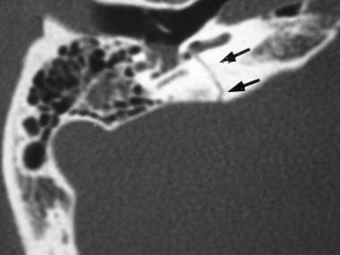
- A , Noncontrast axial CT scan shows subcutaneous mass ( arrow ) and local skull defect on the upper portion of the petrous bone. (ajnr.org)
- Axial temporal bone CT scans of 40 Turkish individuals were studied by measuring the relevant parameters and the results were statistically analysed and compared with those reported previously for Chinese and Europeans (3). (tubitak.gov.tr)
Apicitis2
- Improvements in antibiotic therapy, surgery for chronic otitis media, and the development of tympanostomy tubes have decreased incidence of suppurative petrous apicitis . (medscape.com)
- Petrous apicitis. (medscape.com)
Middle ear5
- The great challenge of therapy is to eradicate the pathologic growth while preserving hearing and other critical functions of the middle ear and petrous bone, respecting the proximity of vital neural and vascular structures, and the intricate three-dimensional relationships involved. (thieme.com)
- These three bones form a chain across the middle ear from the tympanum to the oval window of the inner ear. (petplace.com)
- The middle ear is connected to the inner ear through the oval window, which lies against the stapes bone. (petplace.com)
- These vibrations are then transmitted to the three small bones of the middle ear (the malleus, incus and stapes), which amplify the sound vibration. (petplace.com)
- The middle ear inhabits the petrous portion of the temporal bone and is filled with air secondary to communication with the nasopharynx via the auditory (eustachian) tube (see the following image). (medscape.com)
Fracture7
- Fracture dislocation of the petrous bone. (bmj.com)
- A transverse temporal bone fracture is shown in the image below. (medscape.com)
- Temporal bone fracture is usually a sequela of significant blunt head injury. (radiopaedia.org)
- Although the temporal bone includes the squamous part, forming the inferolateral part of the skull vault , generally the term temporal bone fracture refers to the involvement of the petrous part. (radiopaedia.org)
- Temporal bone fracture is thought to occur in ~20% (range 14-22%) of all calvarial fractures. (radiopaedia.org)
- Temporal bone fracture is suggested by Battle sign (post-auricular ecchymosis) and bleeding from the external auditory canal. (radiopaedia.org)
- Fracture of the petrous temporal bone is usually classified according to the main orientation of the fracture plane and/or involvement of the otic capsule . (radiopaedia.org)
Tumors1
- Flat tumors, termed en plaque, infiltrate the dura and grow as a thin carpet or sheet of tumor along the convexity dura, falx, or tentorium. (medscape.com)
Posterior3
- [ 1 ] MRI more accurately evaluates en plaque and posterior fossa meningiomas, which may be missed on CT scanning. (medscape.com)
- The results showed significant differences in the position of the sigmoid sinus, orientation of the petrous bone and posterior fossa dimensions. (tubitak.gov.tr)
- a prominence midway between the posterior margin of the foramen magnum and the superior angle of the bone. (worldofmedicalsaviours.com)
Lesions2
Petrosal1
- In other mammals, it is a separate bone, the petrosal bone. (wikipedia.org)
Auditory ossicles2
- Within this tympanic cavity are found the auditory ossicles, three tiny bones that vibrate when stimulated by sound waves. (petplace.com)
- The three auditory ossicles , or ear bones , form together with the eardrum the sound-conducting apparatus. (brainkart.com)
Medially1
- They are V3 anteriorly, the petrous ridge medially, GSPN laterally, and the meatal plane posteriorly. (nih.gov)
Parietal1
- The magnetic resonance imaging (MRI) results revealed that the bilateral frontal bone, right temporal bone, and right parietal bone were thickened with nodules. (biomedcentral.com)
20162
- In 2016, down in the "Deep Sounding" of the Solecki trench, while working on the eastern face, a rib emerged from the wall, followed by the arch of a lumbar vertebra, then the bones of a clenched right hand. (cam.ac.uk)
- In 2016, in one of the deepest parts of the trench, a rib emerged from the wall, followed by a lumbar vertebra, then the bones of a clenched right hand. (scitechdaily.com)
Occipital2
- The petrous part of the temporal bone is pyramid-shaped and is wedged in at the base of the skull between the sphenoid and occipital bones. (wikipedia.org)
- Occipital bone forms the region of the occiput(back of the head) and is trapezoid in shape. (worldofmedicalsaviours.com)
Surgical3
- This Report has been prepared in fulfilment of Deliverable D4.2, required as a result of Work Package 4 (Real time physically based surgical simulators) of the EU Framework V Project IERAPSI, An Integrated Environment for the Rehearsal and Planning of Surgical Interventions (IST-1999-12175) Deliverable D4.2 relates to the Petrous bone surgical simulation platform, the second and last of the two main expected results of Work Package 4. (p-arch.it)
- An accompanying video (available on the deliverable CD-ROM) further illustrates the petrous bone surgical simulation platform with live sequences comparing a real and a simulated surgical procedure performed on the temporal bone. (p-arch.it)
- The accessibility is influenced largely by the individual characteristics of the temporal bone and the variability of the surgical landmarks is the result of different skull base shapes among different races. (tubitak.gov.tr)
Neanderthal3
- We didn't think we'd be lucky enough to find more Neanderthal bones. (cam.ac.uk)
- More than 50 years later, a team of researchers have reopened the old Solecki trench to collect new sediment samples, and discovered the crushed skull and torso bones of another Shanidar Neanderthal. (scitechdaily.com)
- We didn't expect to find any Neanderthal bones. (scitechdaily.com)
Lateral1
- Its lateral wall is stouter where it arches up to bracket the temporal process of the zygomatic bone. (co.ma)
Cartilage2
- Could be involved in bone and cartilage formation. (abcam.com)
- The external acoustic meatus (external auditory canal) is formed by cartilage and bone (temporal). (medscape.com)
Imaging1
- Head CT with petrous temporal bone fine slice (≤1 mm) multiplanar bone window reformats is the imaging modality of choice. (radiopaedia.org)
Portion3
- The petrous portion is among the most basal elements of the skull and forms part of the endocranium. (wikipedia.org)
- The petrous portion of the temporal bone houses the organs for hearing, equilibrium and motion detection. (cdc.gov)
- It lies within the petrous portion of the temporal bone and consists of bags and ducts of the membranous labyrinth. (bvsalud.org)
Canal1
- Behind the internal acoustic meatus is a small slit almost hidden by a thin plate of bone, leading to a canal, the aquæductus vestibuli, which transmits the ductus endolymphaticus together with a small artery and vein. (wikipedia.org)
Surface1
- The roof of the tympanic cav-ity, the tegmental wall, is relatively thin and borders on the surface of the petrous pyra-mid. (brainkart.com)
Surgery1
- Predictive Effect of Bone Conduction Pattern on Hearing Outcomes of Stapes Surgery. (uc.edu)
Disease1
- MM is considered relative paucity of CNS invasion by MM an incurable disease despite various meth- in comparison with other tumours, whether ods of treatment, including autologous bone solid or haematological, remain unknown, marrow transplantation [ 3 ]. (who.int)
Teeth1
- Considering that these cases involved DNA extraction from teeth, bones and old human remains, automate system was felt to be the best option to reduce handling errors and increase the possibilities of obtaining good quality DNA. (intechopen.com)
Archaeological1
- Furthermore, the calcareous soils are favourable for bone preservation, and a remarkable assemblage of well-preserved archaeological animal and human remains has been recovered from the megalithic graves. (springer.com)
Site2
- After the excavation, the bones were reburied at the current site, where a tombstone dedicated to Miura Anjin (William Adams) now stands (Fig. 1 b). (nature.com)
- Twenty-four petrous bones from the Talheim site have previously been sampled and screened for DNA using high-throughput sequencing. (eurac.edu)
Upper1
- During 2018-19 they went on to uncover a complete skull, flattened by thousands of years of sediment, and upper body bones almost to the waist - with the left hand curled under the head like a small cushion. (scitechdaily.com)