Peroxisomal Bifunctional Enzyme
Enoyl-CoA Hydratase
3-Hydroxyacyl CoA Dehydrogenases
Peroxisomes
Microbodies
Phosphofructokinase-2
Peroxisomal Disorders
Multienzyme Complexes
Molecular Sequence Data
Amino Acid Sequence
Acyl-CoA Oxidase
Hydro-Lyases
Sleep Deprivation
Seizures
Tablets
Electroencephalography
Epilepsy, Absence
Pamphlets
Absence of spontaneous peroxisome proliferation in enoyl-CoA Hydratase/L-3-hydroxyacyl-CoA dehydrogenase-deficient mouse liver. Further support for the role of fatty acyl CoA oxidase in PPARalpha ligand metabolism. (1/41)
Peroxisomes contain a classical L-hydroxy-specific peroxisome proliferator-inducible beta-oxidation system and also a second noninducible D-hydroxy-specific beta-oxidation system. We previously generated mice lacking fatty acyl-CoA oxidase (AOX), the first enzyme of the L-hydroxy-specific classical beta-oxidation system; these AOX-/- mice exhibited sustained activation of peroxisome proliferator-activated receptor alpha (PPARalpha), resulting in profound spontaneous peroxisome proliferation in liver cells. These observations implied that AOX is responsible for the metabolic degradation of PPARalpha ligands. In this study, the function of enoyl-CoA hydratase/L-3-hydroxyacyl-CoA dehydrogenase (L-PBE), the second enzyme of this peroxisomal beta-oxidation system, was investigated by disrupting its gene. Mutant mice (L-PBE-/-) were viable and fertile and exhibited no detectable gross phenotypic defects. L-PBE-/- mice showed no hepatic steatosis and manifested no spontaneous peroxisome proliferation, unlike that encountered in livers of mice deficient in AOX. These results indicate that disruption of classical peroxisomal fatty acid beta-oxidation system distal to AOX step does not interfere with the inactivation of endogenous ligands of PPARalpha, further confirming that the AOX gene is indispensable for the physiological regulation of this receptor. The absence of appreciable changes in lipid metabolism also indicates that enoyl-CoAs, generated in the classical system in L-PBE-/- mice are diverted to D-hydroxy-specific system for metabolism by D-PBE. When challenged with a peroxisome proliferator, L-PBE-/- mice showed increases in the levels of hepatic mRNAs and proteins that are regulated by PPARalpha except for appreciable blunting of peroxisome proliferative response as compared with that observed in hepatocytes of wild type mice similarly treated. This blunting of peroxisome proliferative response is attributed to the absence of L-PBE protein in L-PBE-/- mouse liver, because all other proteins are induced essentially to the same extent in both wild type and L-PBE-/- mice. (+info)Orphan nuclear hormone receptor RevErbalpha modulates expression from the promoter of the hydratase-dehydrogenase gene by inhibiting peroxisome proliferator-activated receptor alpha-dependent transactivation. (2/41)
Peroxisome proliferator-activated receptor alpha (PPARalpha) heterodimerizes with the 9-cis-retinoic acid receptor (RXRalpha) to bind to peroxisome proliferator-response elements (PPRE) present in the upstream regions of a number of genes involved in metabolic homeostasis. Among these genes are those encoding fatty acyl-CoA oxidase (AOx) and enoyl-CoA hydratase/3-hydroxyacyl-CoA dehydrogenase (HD), the first two enzymes of the peroxisomal beta-oxidation pathway. Here we demonstrate that the orphan nuclear hormone receptor, RevErbalpha, modulates PPARalpha/RXRalpha- dependent transactivation in a response element-specific manner. In vitro binding analysis showed that RevErbalpha bound the HD-PPRE but not the AOx-PPRE. Determinants within the HD-PPRE required for RevErbalpha binding were distinct from those required for PPARalpha/RXRalpha binding. In transient transfections, RevErbalpha antagonized transactivation by PPARalpha/RXRalpha from an HD-PPRE luciferase reporter construct, whereas no effects were observed with an AOx-PPRE reporter construct. These data identify the HD gene as a target for RevErbalpha and illustrate cross-talk between the RevErbalpha and PPARalpha signaling pathways on the HD-PPRE. Our results suggest a novel role for RevErbalpha in regulating peroxisomal beta-oxidation. (+info)Peroxisomal bifunctional enzyme binds and activates the activation function-1 region of the peroxisome proliferator-activated receptor alpha. (3/41)
The transcriptional activity of peroxisome proliferator-activated receptors (PPARs), and of nuclear hormone receptors in general, is subject to modulation by cofactors. However, most currently known co-activating proteins interact in a ligand-dependent manner with the C-terminal ligand-regulated activation function (AF)-2 domain of nuclear receptors. Since PPARalpha exhibits a strong constitutive transactivating function contained within an N-terminal AF-1 region, it can be speculated that a different set of cofactors might interact with this region of PPARs. An affinity purification approach was used to identify the peroxisomal enoyl-CoA hydratase/3-hydroxyacyl-CoA dehydrogenase (bifunctional enzyme, BFE) as a protein which strongly and specifically interacted with the N-terminal 92 amino acids of PPARalpha. Protein-protein interaction assays with the cloned BFE confirmed this interaction, which could be mapped to amino acids 307-514 of the BFE and the N-terminal 70 amino acids of PPARalpha. Moreover, transient transfection experiments in hepatoma cells revealed a 2.2-fold increase in the basal and ligand-stimulated transcriptional activity of PPARalpha in the presence of BFE. This stimulatory effect is preferentially observed for the PPARalpha isoform and it is significantly stronger (4.8-fold) in non-hepatic cells, which presumably express lower levels of endogenous BFE. Hence, the BFE represents the first known cofactor capable of activating the AF-1 domain of PPAR without requiring additional regions of this receptor. These data are compatible with a model whereby the PPAR-regulated BFE is able to modulate its own expression through an enhancement of the activity of PPARalpha, representing a novel peroxisomal-nuclear feed-forward regulatory loop. (+info)Differential gene regulation in human versus rodent hepatocytes by peroxisome proliferator-activated receptor (PPAR) alpha. PPAR alpha fails to induce peroxisome proliferation-associated genes in human cells independently of the level of receptor expresson. (4/41)
We compared the ability of rat and human hepatocytes to respond to fenofibric acid and a novel potent phenylacetic acid peroxisome proliferator-activated receptor (PPAR) alpha agonist (compound 1). Fatty acyl-CoA oxidase (FACO) activity and mRNA were increased after treatment with either fenofibric acid or compound 1 in rat hepatocytes. In addition, apolipoprotein CIII mRNA was decreased by both fenofibric acid and compound 1 in rat hepatocytes. Both agonists decreased apolipoprotein CIII mRNA in human hepatocytes; however, very little change in FACO activity or mRNA was observed. Furthermore, other peroxisome proliferation (PP)-associated genes including peroxisomal 3-oxoacyl-CoA thiolase (THIO), peroxisomal enoyl-CoA hydratase/3-hydroxyacyl-CoA dehydrogenase (HD), peroxisomal membrane protein-70 (PMP-70) were not regulated by PPAR alpha agonists in human hepatocytes. Moreover, other genes that are regulated by PPAR alpha ligands in human hepatocytes such as mitochondrial HMG-CoA synthase and carnitine palmitoyl transferase-1 (CPT-1) were also regulated in HepG2 cells by PPAR alpha agonists. Several stably transfected HepG2 cell lines were established that overexpressed human PPAR alpha to levels between 6- and 26-fold over normal human hepatocytes. These PPAR alpha-overexpressing cells had higher basal mRNA levels of mitochondrial HMG-CoA synthase and CPT-1; however, basal FACO mRNA levels and other PP-associated genes including THIO, HD, or PMP-70 mRNA were not substantially affected. In addition, FACO, THIO, HD, and PMP-70 mRNA levels did not increase in response to PPAR alpha agonist treatment in the PPAR alpha-overexpressing cells, although mitochondrial HMG-CoA synthase and CPT-1 mRNAs were both induced. These results suggest that other factors besides PPAR alpha levels determine the species-specific response of human and rat hepatocytes to the induction of PP. (+info)The role of alpha-methylacyl-CoA racemase in bile acid synthesis. (5/41)
According to current views, the second peroxisomal beta-oxidation pathway is responsible for the degradation of the side chain of bile acid intermediates. Peroxisomal multifunctional enzyme type 2 [peroxisomal multifunctional 2-enoyl-CoA hydratase/(R)-3-hydroxyacyl-CoA dehydrogenase; MFE-2] catalyses the second (hydration) and third (dehydrogenation) reactions of the pathway. Deficiency of MFE-2 leads to accumulation of very-long-chain fatty acids, 2-methyl-branched fatty acids and C(27) bile acid intermediates in plasma, but bile acid synthesis is not blocked completely. In this study we describe an alternative pathway, which allows MFE-2 deficiency to be overcome. The alternative pathway consists of alpha-methylacyl-CoA racemase and peroxisomal multifunctional enzyme type 1 [peroxisomal multifunctional 2-enoyl-CoA hydratase/(S)-3-hydroxyacyl-CoA dehydrogenase; MFE-1]. (24E)-3alpha,7alpha,12alpha-Trihydroxy-5beta-cholest-24-enoyl-CoA, the presumed physiological isomer, is hydrated by MFE-1 with the formation of (24S,25S)-3alpha,7alpha,12alpha,24-tetrahydroxy-5beta-cholestanoyl-CoA [(24S,25S)-24-OH-THCA-CoA], which after conversion by a alpha-methylacyl-CoA racemase into the (24S,25R) isomer can again be dehydrogenated by MFE-1 to 24-keto-3alpha,7alpha,12alpha-trihydroxycholestanoyl-CoA, a physiological intermediate in cholic acid synthesis. The discovery of the alternative pathway of cholesterol side-chain oxidation will improve diagnosis of peroxisomal deficiencies by identification of serum 24-OH-THCA-CoA diastereomer profiles. (+info)Organization of the multifunctional enzyme type 1: interaction between N- and C-terminal domains is required for the hydratase-1/isomerase activity. (6/41)
Rat peroxisomal multifunctional enzyme type 1 (perMFE-1) is a monomeric protein of beta-oxidation. We have defined five functional domains (A, B, C, D and E) in the perMFE-1 based on comparison of the amino acid sequence with homologous proteins from databases and structural data of the hydratase-1/isomerases (H1/I) and (3 S )-hydroxyacyl-CoA dehydrogenases (HAD). Domain A (residues 1-190) comprises the H1/I fold and catalyses both 2-enoyl-CoA hydratase-1 and Delta(3)-Delta(2)-enoyl-CoA isomerase reactions. Domain B (residues 191-280) links domain A to the (3 S )-dehydrogenase region, which includes both domain C (residues 281-474) and domain D (residues 480-583). Domains C and D carry features of the dinucleotide-binding and the dimerization domains of monofunctional HADs respectively. Domain E (residues 584-722) has sequence similarity to domain D of the perMFE-1, which suggests that it has evolved via partial gene duplication. Experiments with engineered perMFE-1 variants demonstrate that the H1/I competence of domain A requires stabilizing interactions with domains D and E. The variant His-perMFE (residues 288-479)Delta, in which the domain C is deleted, is stable and has hydratase-1 activity. It is proposed that the extreme C-terminal domain E in perMFE-1 serves the following three functions: (i) participation in the folding of the N-terminus into a functionally competent H1/I fold, (ii) stabilization of the dehydrogenation domains by interaction with the domain D and (iii) the targeting of the perMFE-1 to peroxisomes via its C-terminal tripeptide. (+info)Induction of the three peroxisomal beta-oxidation enzymes is synergistically regulated by dexamethasone and fatty acids, and counteracted by insulin in Morris 7800C1 hepatoma cells in culture. (7/41)
This work describes the molecular mechanism of hormonal modulation of fatty-acid peroxisomal beta oxidation in liver. Morris 7800C1 hepatoma cells and isolated hepatocytes were cultured in the presence of myristic acid (1 mM) and tetradecylthioacetic acid, a 3-thia fatty acid (50 microM), separately or in combination with dexamethasone (0.25 microM) or insulin (0.4 microM). Myristic acid stimulated acyl-CoA oxidase and a synergistic action was observed with dexamethasone. Parallel changes were recognized in enzyme protein and mRNA levels as quantified from immunoblots and Northern analyses. Myristic acid and tetradecylthioacetic acid had similar effects on this enzyme, while insulin inhibited the basal activity and blocked all inductions by the fatty acids and dexamethasone. Parallel mRNA and immunoblot analyses of the subsequent enzymes in the peroxisomal beta-oxidation pathway, enoyl-CoA hydratase/3-hydroxyacyl-CoA dehydrogenase/delta 3,delta 2-enoyl-CoA isomerase and 3-oxoacyl-CoA thiolase, showed an even stronger induction by tetradecylthioacetic acid and dexamethasone, while the counteraction by insulin was maintained in both 7800C1 hepatoma cells and hepatocytes. In hepatoma cells, the thiolase always showed the most pronounced induction (about 40-fold) after 14 days, with parallel changes in protein and mRNA levels. The results suggest that the changes in peroxisomal beta-oxidation enzymes in 7800C1 hepatoma cells are due to a major effect on steady-state mRNA levels giving rise to corresponding alterations in enzyme protein. These results may be explained by regulation at the level of transcription of corresponding genes, but mRNA stability changes and/or translational effects may also be of importance. (+info)Identification of a peroxisome proliferator-responsive element upstream of the gene encoding rat peroxisomal enoyl-CoA hydratase/3-hydroxyacyl-CoA dehydrogenase. (8/41)
Ciprofibrate, a hypolipidemic drug that acts as a peroxisome proliferator, induces the transcription of genes encoding peroxisomal beta-oxidation enzymes. To identify cis-acting promoter elements involved in this induction, 5.8 kilobase pairs of promoter sequence from the gene encoding rat peroxisomal enoyl-CoA hydratase/3-hydroxyacyl-CoA dehydrogenase (EC 4.2.1.17/EC 1.1.1.35) was inserted upstream of a luciferase reporter gene. Transfection of this expression vector into rat hepatoma H4IIEC3 cells in the presence of ciprofibrate resulted in a 5- to 10-fold, cell type-specific increase in luciferase activity as compared to cells transfected in the absence of drug. A peroxisome proliferator-responsive element (PPRE) was localized to a 196-nucleotide region centered at position -2943 from the transcription start site. This PPRE conferred ciprofibrate responsiveness on a heterologous promoter and functioned independently of orientation or position. Gel retardation analysis with nuclear extracts demonstrated that ciprofibrate-treated or untreated H4IIEC3 cells, but not HeLa cells or monkey kidney cells, contained sequence-specific DNA binding factors that interact with the PPRE. These results have implications for understanding the mechanisms of coordinated transcriptional induction of genes encoding peroxisomal proteins by hypolipidemic agents and other peroxisome proliferators. (+info)A peroxisomal bifunctional enzyme is not a specific medical term, but it refers to a type of enzyme that has two distinct functional domains and is located within peroxisomes. Peroxisomes are small membrane-bound organelles found in the cells of many organisms, including humans, where they play a crucial role in various metabolic processes, such as fatty acid oxidation and detoxification of harmful substances.
The term "bifunctional" indicates that this enzyme possesses two distinct catalytic activities or functions within the same polypeptide chain. In the context of peroxisomal enzymes, bifunctional enzymes often participate in the breakdown of specific fatty acids, particularly very-long-chain fatty acids (VLCFAs) and branched-chain fatty acids (BCFAs).
An example of a peroxisomal bifunctional enzyme is the D-bifunctional protein (DBP), which has two catalytic domains: one for the oxidation of long-chain 2-enoyl-CoA intermediates and another for the hydratase/dehydrogenase activity. DBP plays a critical role in peroxisomal fatty acid beta-oxidation, particularly for VLCFAs and BCFAs, which cannot be efficiently metabolized by mitochondria.
Defects in peroxisomal bifunctional enzymes can lead to various genetic disorders, such as peroxisome biogenesis disorders (PBDs) or peroxisomal fatty acid oxidation disorders, which may result in severe neurological symptoms and developmental delays.
Enoyl-CoA hydratase is an enzyme that catalyzes the second step in the fatty acid oxidation process, also known as the beta-oxidation pathway. The systematic name for this reaction is (3R)-3-hydroxyacyl-CoA dehydratase.
The function of Enoyl-CoA hydratase is to convert trans-2-enoyl-CoA into 3-hydroxyacyl-CoA by adding a molecule of water (hydration) across the double bond in the substrate. This reaction forms a chiral center, resulting in the production of an (R)-stereoisomer of 3-hydroxyacyl-CoA.
The gene that encodes for Enoyl-CoA hydratase is called ECHS1, and mutations in this gene can lead to a rare genetic disorder known as Enoyl-CoA Hydratase Deficiency or ECHS1 Deficiency. This condition affects the breakdown of fatty acids in the body and can cause neurological symptoms such as developmental delay, seizures, and movement disorders.
3-Hydroxyacyl CoA Dehydrogenases (3-HADs) are a group of enzymes that play a crucial role in the beta-oxidation of fatty acids. These enzymes catalyze the third step of the beta-oxidation process, which involves the oxidation of 3-hydroxyacyl CoA to 3-ketoacyl CoA. This reaction is an essential part of the energy-generating process that occurs in the mitochondria of cells and allows for the breakdown of fatty acids into smaller molecules, which can then be used to produce ATP, the primary source of cellular energy.
There are several different isoforms of 3-HADs, each with specific substrate preferences and tissue distributions. The most well-known isoform is the mitochondrial 3-hydroxyacyl CoA dehydrogenase (M3HD), which is involved in the oxidation of medium and long-chain fatty acids. Other isoforms include the short-chain 3-hydroxyacyl CoA dehydrogenase (SCHAD) and the long-chain 3-hydroxyacyl CoA dehydrogenase (LCHAD), which are involved in the oxidation of shorter and longer chain fatty acids, respectively.
Deficiencies in 3-HADs can lead to serious metabolic disorders, such as 3-hydroxyacyl-CoA dehydrogenase deficiency (3-HAD deficiency), which is characterized by the accumulation of toxic levels of 3-hydroxyacyl CoAs in the body. Symptoms of this disorder can include hypoglycemia, muscle weakness, cardiomyopathy, and developmental delays. Early diagnosis and treatment of 3-HAD deficiency are essential to prevent serious complications and improve outcomes for affected individuals.
Peroxisomes are membrane-bound subcellular organelles found in the cytoplasm of eukaryotic cells. They play a crucial role in various cellular processes, including the breakdown of fatty acids and the detoxification of harmful substances such as hydrogen peroxide (H2O2). Peroxisomes contain numerous enzymes, including catalase, which converts H2O2 into water and oxygen, thus preventing oxidative damage to cellular components. They also participate in the biosynthesis of ether phospholipids, a type of lipid essential for the structure and function of cell membranes. Additionally, peroxisomes are involved in the metabolism of reactive oxygen species (ROS) and contribute to the regulation of intracellular redox homeostasis. Dysfunction or impairment of peroxisome function has been linked to several diseases, including neurological disorders, developmental abnormalities, and metabolic conditions.
Microbodies are small, membrane-bound organelles found in the cells of eukaryotic organisms. They typically measure between 0.2 to 0.5 micrometers in diameter and play a crucial role in various metabolic processes, particularly in the detoxification of harmful substances and the synthesis of lipids.
There are several types of microbodies, including:
1. Peroxisomes: These are the most common type of microbody. They contain enzymes that help break down fatty acids and amino acids, producing hydrogen peroxide as a byproduct. Another set of enzymes within peroxisomes then converts the harmful hydrogen peroxide into water and oxygen, thus detoxifying the cell.
2. Glyoxysomes: These microbodies are primarily found in plants and some fungi. They contain enzymes involved in the glyoxylate cycle, a metabolic pathway that helps convert stored fats into carbohydrates during germination.
3. Microbody-like particles (MLPs): These are smaller organelles found in certain protists and algae. Their functions are not well understood but are believed to be involved in lipid metabolism.
It is important to note that microbodies do not have a uniform structure or function across all eukaryotic cells, and their specific roles can vary depending on the organism and cell type.
Phosphofructokinase-2 (PFK-2) is an enzyme that plays a crucial role in regulating the rate of glycolysis, which is the metabolic pathway responsible for the conversion of glucose into energy. PFK-2 catalyzes the phosphorylation of fructose-6-phosphate to form fructose-1,6-bisphosphate and subsequently fructose-2,6-bisphosphate (F-2,6-BP). F-2,6-BP is a potent allosteric activator of another enzyme called phosphofructokinase-1 (PFK-1), which is the rate-limiting enzyme in glycolysis.
PFK-2 exists as a complex with another enzyme, fructose-2,6-bisphosphatase (FBPase-2), and together they form a bifunctional enzyme called PFK-2/FBPase-2. This enzyme can reversibly convert F-6-P to F-2,6-BP and vice versa depending on the cellular energy status. When cells have high energy levels, FBPase-2 is activated, which leads to a decrease in F-2,6-BP levels and an inhibition of glycolysis. Conversely, when cells require more energy, PFK-2 is activated, leading to an increase in F-2,6-BP levels and an activation of glycolysis.
Regulation of PFK-2 activity occurs through various mechanisms, including allosteric regulation by metabolites such as AMP, citrate, and phosphate, as well as covalent modification by protein kinases and phosphatases. Dysregulation of PFK-2 has been implicated in several diseases, including diabetes, cancer, and neurological disorders.
Peroxisomal disorders are a group of inherited metabolic diseases caused by defects in the function or structure of peroxisomes, which are specialized subcellular organelles found in the cells of animals, plants, and humans. These disorders can affect various aspects of metabolism, including fatty acid oxidation, bile acid synthesis, and plasma cholesterol levels.
Peroxisomal disorders can be classified into two main categories: single peroxisomal enzyme deficiencies and peroxisome biogenesis disorders (PBDs). Single peroxisomal enzyme deficiencies are characterized by a defect in a specific enzyme found within the peroxisome, while PBDs are caused by problems with the formation or assembly of the peroxisome itself.
Examples of single peroxisomal enzyme deficiencies include X-linked adrenoleukodystrophy (X-ALD), Refsum disease, and acyl-CoA oxidase deficiency. PBDs include Zellweger spectrum disorders, such as Zellweger syndrome, neonatal adrenoleukodystrophy, and infantile Refsum disease.
Symptoms of peroxisomal disorders can vary widely depending on the specific disorder and the severity of the enzyme or biogenesis defect. They may include neurological problems, vision and hearing loss, developmental delays, liver dysfunction, and skeletal abnormalities. Treatment typically focuses on managing symptoms and addressing any underlying metabolic imbalances.
Multienzyme complexes are specialized protein structures that consist of multiple enzymes closely associated or bound together, often with other cofactors and regulatory subunits. These complexes facilitate the sequential transfer of substrates along a series of enzymatic reactions, also known as a metabolic pathway. By keeping the enzymes in close proximity, multienzyme complexes enhance reaction efficiency, improve substrate specificity, and maintain proper stoichiometry between different enzymes involved in the pathway. Examples of multienzyme complexes include the pyruvate dehydrogenase complex, the citrate synthase complex, and the fatty acid synthetase complex.
Molecular sequence data refers to the specific arrangement of molecules, most commonly nucleotides in DNA or RNA, or amino acids in proteins, that make up a biological macromolecule. This data is generated through laboratory techniques such as sequencing, and provides information about the exact order of the constituent molecules. This data is crucial in various fields of biology, including genetics, evolution, and molecular biology, allowing for comparisons between different organisms, identification of genetic variations, and studies of gene function and regulation.
An amino acid sequence is the specific order of amino acids in a protein or peptide molecule, formed by the linking of the amino group (-NH2) of one amino acid to the carboxyl group (-COOH) of another amino acid through a peptide bond. The sequence is determined by the genetic code and is unique to each type of protein or peptide. It plays a crucial role in determining the three-dimensional structure and function of proteins.
Acyl-CoA oxidase is an enzyme that plays a crucial role in the breakdown of fatty acids within the body. It is located in the peroxisomes, which are small organelles found in the cells of living organisms. The primary function of acyl-CoA oxidase is to catalyze the initial step in the beta-oxidation of fatty acids, a process that involves the sequential removal of two-carbon units from fatty acid molecules in the form of acetyl-CoA.
The reaction catalyzed by acyl-CoA oxidase is as follows:
acyl-CoA + FAD → trans-2,3-dehydroacyl-CoA + FADH2 + H+
In this reaction, the enzyme removes a hydrogen atom from the fatty acyl-CoA molecule and transfers it to its cofactor, flavin adenine dinucleotide (FAD). This results in the formation of trans-2,3-dehydroacyl-CoA, FADH2, and a proton. The FADH2 produced during this reaction can then be used to generate ATP through the electron transport chain, while the trans-2,3-dehydroacyl-CoA undergoes further reactions in the beta-oxidation pathway.
There are two main isoforms of acyl-CoA oxidase found in humans: ACOX1 and ACOX2. ACOX1 is primarily responsible for oxidizing straight-chain fatty acids, while ACOX2 specializes in the breakdown of branched-chain fatty acids. Mutations in the genes encoding these enzymes can lead to various metabolic disorders, such as peroxisomal biogenesis disorders and Refsum disease.
I'm sorry for any confusion, but "Hydro-Lyases" is not a recognized medical term or category in biochemistry. It seems like there might be a misunderstanding or a typo in the term.
In biochemistry, "lyases" are enzymes that catalyze the removal of groups from substrates by means other than hydrolysis or oxidation, often forming a double bond or a ring-forming reaction. They are classified and named based on the type of bond they break.
If you meant to ask about a specific enzyme or reaction, could you please provide more context or clarify the term? I'd be happy to help further with accurate information.
Sleep deprivation is a condition that occurs when an individual fails to get sufficient quality sleep or the recommended amount of sleep, typically 7-9 hours for adults. This can lead to various physical and mental health issues. It can be acute, lasting for one night or a few days, or chronic, persisting over a longer period.
The consequences of sleep deprivation include:
1. Fatigue and lack of energy
2. Difficulty concentrating or remembering things
3. Mood changes, such as irritability or depression
4. Weakened immune system
5. Increased appetite and potential weight gain
6. Higher risk of accidents due to decreased reaction time
7. Health problems like high blood pressure, diabetes, and heart disease over time
Sleep deprivation can be caused by various factors, including stress, shift work, sleep disorders like insomnia or sleep apnea, poor sleep hygiene, and certain medications. It's essential to address the underlying causes of sleep deprivation to ensure proper rest and overall well-being.
A seizure is an uncontrolled, abnormal firing of neurons (brain cells) that can cause various symptoms such as convulsions, loss of consciousness, altered awareness, or changes in behavior. Seizures can be caused by a variety of factors including epilepsy, brain injury, infection, toxic substances, or genetic disorders. They can also occur without any identifiable cause, known as idiopathic seizures. Seizures are a medical emergency and require immediate attention.
In the context of medical terminology, tablets refer to pharmaceutical dosage forms that contain various active ingredients. They are often manufactured in a solid, compressed form and can be administered orally. Tablets may come in different shapes, sizes, colors, and flavors, depending on their intended use and the manufacturer's specifications.
Some tablets are designed to disintegrate or dissolve quickly in the mouth, making them easier to swallow, while others are formulated to release their active ingredients slowly over time, allowing for extended drug delivery. These types of tablets are known as sustained-release or controlled-release tablets.
Tablets may contain a single active ingredient or a combination of several ingredients, depending on the intended therapeutic effect. They are typically manufactured using a variety of excipients, such as binders, fillers, and disintegrants, which help to hold the tablet together and ensure that it breaks down properly when ingested.
Overall, tablets are a convenient and widely used dosage form for administering medications, offering patients an easy-to-use and often palatable option for receiving their prescribed treatments.
Electroencephalography (EEG) is a medical procedure that records electrical activity in the brain. It uses small, metal discs called electrodes, which are attached to the scalp with paste or a specialized cap. These electrodes detect tiny electrical charges that result from the activity of brain cells, and the EEG machine then amplifies and records these signals.
EEG is used to diagnose various conditions related to the brain, such as seizures, sleep disorders, head injuries, infections, and degenerative diseases like Alzheimer's or Parkinson's. It can also be used during surgery to monitor brain activity and ensure that surgical procedures do not interfere with vital functions.
EEG is a safe and non-invasive procedure that typically takes about 30 minutes to an hour to complete, although longer recordings may be necessary in some cases. Patients are usually asked to relax and remain still during the test, as movement can affect the quality of the recording.
Absence epilepsy is a type of epilepsy characterized by recurrent brief episodes of "absences," or staring spells, that can last from a few seconds to several minutes. These episodes are often accompanied by subtle body movements such as lip smacking or eyelid flutters. Absence epilepsy is most commonly diagnosed in children and adolescents, and it is more common in girls than boys.
The seizures in absence epilepsy are caused by abnormal electrical activity in the brain, specifically in a part of the brain called the cortex. These abnormal electrical discharges occur in a pattern that involves both sides of the brain simultaneously. This differs from other types of epilepsy, which may involve only one side of the brain or specific areas within a single hemisphere.
Absence seizures are typically brief and do not cause confusion or disorientation after they end. However, if they occur frequently, they can interfere with learning and social development. In some cases, absence epilepsy may be associated with other types of seizures, such as generalized tonic-clonic (grand mal) seizures or myoclonic jerks.
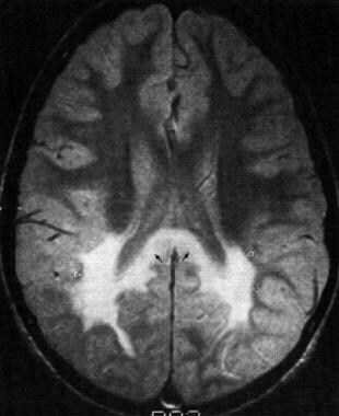
The diagnosis of absence epilepsy is usually made based on the characteristic symptoms and the results of an electroencephalogram (EEG), which can detect the abnormal electrical activity in the brain during a seizure. Treatment typically involves medication to control the seizures, such as ethosuximide or valproic acid. In some cases, a ketogenic diet may also be recommended as an alternative treatment option.
I'm sorry for any confusion, but "pamphlets" is not a medical term. It refers to a small paper booklet or leaflet that can be used to provide information on various topics, including non-medical subjects. If you have any questions about medical terminology or concepts, I'd be happy to help with those!
Neurology is a branch of medicine that deals with the study and treatment of diseases and disorders of the nervous system, which includes the brain, spinal cord, peripheral nerves, muscles, and autonomic nervous system. Neurologists are medical doctors who specialize in this field, diagnosing and treating conditions such as stroke, Alzheimer's disease, epilepsy, Parkinson's disease, multiple sclerosis, and various types of headaches and pain disorders. They use a variety of diagnostic tests, including imaging studies like MRI and CT scans, electrophysiological tests like EEG and EMG, and laboratory tests to evaluate nerve function and identify any underlying conditions or abnormalities. Treatment options may include medication, surgery, rehabilitation, or lifestyle modifications.
Beta-oxidation9
- The protein encoded by this gene is a bifunctional enzyme that is involved in the peroxisomal beta-oxidation pathway for fatty acids. (nih.gov)
- Defects in this gene that affect the peroxisomal fatty acid beta-oxidation activity are a cause of D-bifunctional protein deficiency (DBPD). (nih.gov)
- Another major function of the peroxisomal beta-oxidation system is related to the biosynthesis of polyunsaturated fatty acid (C22:6w3). (medscape.com)
- DBPD is a disorder of peroxisomal fatty acid beta-oxidation. (mitosciences.com)
- The protein encoded by this gene is a bifunctional enzyme and is one of the four enzymes of the peroxisomal beta-oxidation pathway. (utsouthwestern.edu)
- b Identification of putative peroxisomal enzymes and enyzmes of mitochondrial beta-oxidation. (biomedcentral.com)
- RAB5 guanine nucleotide exchange factor (VPS9)" "Zm00001e037538_P001","Zm00001e037538_P001","No alias","Peroxisomal fatty acid beta-oxidation multifunctional protein OS=Oryza sativa subsp. (ntu.edu.sg)
- Peroxisomes are intracellular organelles that contain enzymes for beta-oxidation. (msdmanuals.com)
- Involved in the initial and rate-limiting step of peroxisomal beta-oxidation of straight-chain saturated and unsaturated very-long-chain fatty acids (PubMed:7876265, PubMed:15060085, PubMed:17458872, PubMed:17603022, PubMed:32169171, PubMed:33234382). (swisspalm.org)
Biosynthesis2
- Although most xyloglucans (XyGs) biosynthesis enzymes have been identified, the molecular mechanism that defines XyG branching patterns is unclear. (bvsalud.org)
- He enjoys an international reputation for his research on phospholipases A, cardiolipin biosynthesis in eukaryotes, lysophospholipases, phosphatidylcholine biosynthesis for lung surfactant, plasmalogen biosynthesis in peroxisomes, diagnosis of peroxisomal disorders and most recently his work on alkyl-dihydroxyacetone phosphate synthase. (ornine.best)
Dehydrogenase5
- The D-bifunctional protein is composed of three enzymatic domains: the N-terminal short chain alcohol dehydrogenase reductase (SDR), central hydratase domain, and the C-terminal sterol carrier protein 2 (SDR). (wikipedia.org)
- It is a substrate for succinyl-CoA:3-ketoacid-coenzyme A transferase, hydroxymethylglutaryl-CoA synthase, short-chain 3-hydroxyacyl-CoA dehydrogenase, peroxisomal bifunctional enzyme, acetyl-CoA acetyltransferase, and 3-ketoacyl-CoA thiolase. (hmdb.ca)
- Peroxisomal bifunctional protein from rat liver is a trifunctional enzyme possessing 2-enoyl-CoA hydratase, 3-hydroxyacyl-CoA dehydrogenase, and delta 3, delta 2-enoyl-CoA isomerase activities. (rhea-db.org)
- This means that the bifunctional protein of rat liver is in fact a trifunctional enzyme possessing delta 3, delta 2-enoyl-CoA isomerase, 2-enoyl-CoA hydratase (EC 4.2.1.17), and L-3-hydroxyacyl-CoA dehydrogenase (EC 1.1.1.35) activities in the same polypeptide. (rhea-db.org)
- We found that the tricarboxylic acid cycle-associated enzyme α-ketoglutarate (α-KG) dehydrogenase (KGDH) entered the nucleus, where it interacted with various JMJs to regulate α-KG-dependent histone demethylations by JMJs, and thus controlled genome-wide gene expression in plants. (bvsalud.org)
Single peroxisomal1
- Connect with other caregivers and patients with Disorders with deficiency of a single peroxisomal enzyme and get the support you need. (rareguru.com)
Peroxisome1
- The initial description of the peroxisome (originally termed the microbody) appeared in 1954 in a doctoral thesis about mouse kidneys, almost 10 years after the first case description of peroxisomal disease was published. (medscape.com)
Disorders6
- Peroxisomal disorders are a group of genetically heterogeneous metabolic diseases that share dysfunction of peroxisomes. (medscape.com)
- In peroxisomal biogenesis disorders, abnormal accumulation of VLCFAs (C24, C26) is the hallmark of peroxisomal disorders. (medscape.com)
- Peroxisomal disorders are usually caused by a combination of peroxisomal assembly defects or by deficiencies of specific peroxisomal enzymes. (wikipedia.org)
- Migrational abnormalities are the most likely causes of the severe seizures and psychomotor retardation associated with many types of peroxisomal disorders. (medscape.com)
- Defects in this gene are a cause of peroxisomal disorders such as Zellweger syndrome. (utsouthwestern.edu)
- Therefore, peroxisomal disorders generally manifest with elevated VLCFA levels (except rhizomelic chondrodysplasia and Refsum disease). (msdmanuals.com)
Biogenesis1
- This defect of peroxisomal biogenesis is caused by PEX7 gene mutations and characterized by skeletal changes that include midface hypoplasia, strikingly short proximal limbs, frontal bossing, small nares, cataracts, ichthyosis, and profound psychomotor retardation. (msdmanuals.com)
Nomenclature2
- From Enzyme Nomenclature, 1992) EC 2.1.3.2. (uchicago.edu)
- From Enzyme Nomenclature, 1992) EC 5.4.2. (lookformedical.com)
Zellweger2
- Zellweger described the first case of a peroxisomal disorder in the 1940s. (medscape.com)
- D-BP deficiency is the most severe peroxisomal disorder, often resembling Zellweger syndrome. (wikipedia.org)
Deficiency4
- Rapid whole-genome sequencing identifies a homozygous novel variant, His540Arg, in HSD17B4 resulting in D-bifunctional protein deficiency disorder diagnosis. (nih.gov)
- D-Bifunctional protein deficiency is an autosomal recessive peroxisomal fatty acid oxidation disorder. (wikipedia.org)
- Defects in MFE2 are a cause of D-bifunctional protein deficiency (DBPD) [ MIM:261515 ]. (mitosciences.com)
- This disorder is caused by deficiency of the peroxisomal membrane transporter ALDP, which is coded for by the gene ABCD1 . (msdmanuals.com)
Membrane1
- A focus on Yocum and collaborator work who have developed an outstanding approach based on the anchoring of biosynthetic enzymes to the peroxisomal membrane. (univ-tours.fr)
Peroxisomes2
- Peroxisomes contain many different enzymes, such as catalase, and their main function is to neutralize free radicals and detoxify drugs. (wikipedia.org)
- The D-BP protein contains a peroxisomal targeting signal 1 (PTS1) unit at the C-terminus allowing for its transport into peroxisomes by the PTS1 receptor. (wikipedia.org)
Catalyzes3
- An enzyme that catalyzes the conversion of carbamoyl phosphate and L-aspartate to yield orthophosphate and N-carbamoyl-L-aspartate. (uchicago.edu)
- An enzyme that catalyzes the reversible isomerization of D-mannose-6-phosphate to form D-fructose-6-phosphate, an important step in glycolysis. (lookformedical.com)
- Along with animal peroxidases, these enzymes belong to a group of peroxidases containing a heme prosthetic group (ferriprotoporphyrin IX), which catalyzes a multistep oxidative reaction involving hydrogen peroxide as the electron acceptor. (unl.edu)
Proteins2
- Candidates for each step are identified by using ublast (a fast alternative to protein BLAST) against a database of manually-curated proteins (most of which are experimentally characterized) or by using HMMer with enzyme models (usually from TIGRFam ). (lbl.gov)
- Using a newly developed enzyme-coupled continuous activity assay, we compare differing NAS proteins identified through multiple sequence alignments and phylogenetic analyses. (bvsalud.org)
Gene3
- The responsible genetic defect occurs in 1 of at least 12 genes involved in peroxisomal formation or protein import (the PEX gene family). (msdmanuals.com)
- Gene sets follow the order of enzymes. (umassmed.edu)
- Not available " indicates that no score was available for the corresponding gene, enzyme, or reaction in the relevant database. (umassmed.edu)
Sequence1
- Comparison of the amino acid sequences of cyanogen bromide cleaved isomerase with the known sequence of the peroxisomal bifunctional protein from the rat identified them as the same molecule. (rhea-db.org)
Mitochondrial1
- The mitochondrial and peroxisomal isoenzymes were separated chromatographically and the peroxisomal isomerase purified to apparent homogeneity. (rhea-db.org)
Isomerization1
- In control experiments, the peroxisomal bifunctional protein purified according to published methods also catalyzed delta 3, delta 2-enoyl-CoA isomerization. (rhea-db.org)
Fatty acids1
- These enzymes overlap in function with those in mitochondria, with the exception that mitochondria lack enzymes to metabolize very long-chain fatty acids (VLCFA), those 20 to 26 carbons in length. (msdmanuals.com)
Liver2
- Peroxisomal delta 3, delta 2-enoyl-CoA isomerase (EC 5.3.3.8) was studied in the liver of rats treated with clofibrate. (rhea-db.org)
- In immunoblotting it was recognized by the antibody to peroxisomal bifunctional protein from rat liver. (rhea-db.org)
Distinct2
- They are distinguished from MULTIENZYME COMPLEXES in that their subunits are not found as distinct enzymes. (rush.edu)
- Through this commentary article, we highlight the outstanding work of the Zhou's group which recently describes a distinct approach relying on the efficient recycling of enzyme co-factors in yeast to fulfill the synthesis of caffeic acid (CaA) and ferulic acid (FA), two precursors of podophyllotoxin. (univ-tours.fr)
Synthesis1
- We propose that the specificity of XXT3, XXT4, and XXT5 is directed toward the prior synthesis of the acceptor substrate by the other two enzymes, XXT1 and XXT2. (bvsalud.org)
Specific2
- Reaction for 5.3.3.8 is not in the list of KEGG reactions as specific matches are not available for this enzyme in the KEGG database. (umassmed.edu)
- specific protein associated with the reaction, generic enzyme associated with the reaction, and the reaction itself. (umassmed.edu)
Group2
- A group of enzymes that catalyze an intramolecular transfer of a phosphate group. (lookformedical.com)
- It has been shown in some cases that the enzyme has a functional phosphate group, which can act as the donor. (lookformedical.com)
Publications2
- This graph shows the total number of publications written about "Multifunctional Enzymes" by people in this website by year, and whether "Multifunctional Enzymes" was a major or minor topic of these publications. (rush.edu)
- Below are the most recent publications written about "Multifunctional Enzymes" by people in Profiles. (rush.edu)
Description6
- Description: This is Double-antibody Sandwich Enzyme-linked immunosorbent assay for detection of Human Angiomotin (AMOT) in Tissue homogenates, cell lysates and other biological fluids. (myelisakit.com)
- Description: Enzyme-linked immunosorbent assay based on the Double-antibody Sandwich method for detection of Human Angiomotin (AMOT) in samples from Tissue homogenates, cell lysates and other biological fluids with no significant corss-reactivity with analogues from other species. (myelisakit.com)
- Description: This is Double-antibody Sandwich Enzyme-linked immunosorbent assay for detection of Mouse Hepatocyte Nuclear Factor 1 Alpha (HNF1a) in Tissue homogenates, cell lysates and other biological fluids. (wildpalm.net)
- Description: Enzyme-linked immunosorbent assay based on the Double-antibody Sandwich method for detection of Mouse Hepatocyte Nuclear Factor 1 Alpha (HNF1a) in samples from Tissue homogenates, cell lysates and other biological fluids with no significant corss-reactivity with analogues from other species. (wildpalm.net)
- Description: This is Double-antibody Sandwich Enzyme-linked immunosorbent assay for detection of Human GA Binding Protein Transcription Factor Alpha (GABPa) in serum, plasma, tissue homogenates, cell lysates and other biological fluids. (therabio.org)
- Description: Enzyme-linked immunosorbent assay based on the Double-antibody Sandwich method for detection of Human GA Binding Protein Transcription Factor Alpha (GABPa) in samples from serum, plasma, tissue homogenates, cell lysates and other biological fluids with no significant corss-reactivity with analogues from other species. (therabio.org)