Pericarditis, Constrictive
Pericarditis
Pericarditis, Tuberculous
Pericardiectomy
Pericardial Effusion
Cardiomyopathy, Restrictive
Pericardium
Cardiac Tamponade
Tuberculosis, Cardiovascular
Cardanolides
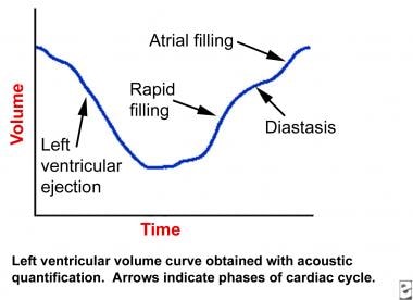
Phonocardiography
Protein-Losing Enteropathies
Echocardiography, Doppler
Cardenolides
Bronchiolitis Obliterans
Echocardiography
Echocardiography, Doppler, Pulsed
Left ventricular systolic and diastolic function after pericardiectomy in patients with constrictive pericarditis: Doppler echocardiographic findings and correlation with clinical status. (1/175)
OBJECTIVES: The study assessed changes in left ventricular systolic and diastolic function after pericardiectomy in patients with constrictive pericarditis and correlated postoperative Doppler echocardiographic findings with clinical status. BACKGROUND: Despite the efficacy of pericardiectomy, some patients with constrictive pericarditis fail to improve postoperatively. Data on serial evaluation of systolic and diastolic function after pericardiectomy and its relation to clinical status are not available. METHODS: From 1985 to 1995, a total of 58 patients with constrictive pericarditis underwent pericardiectomy and had at least one follow-up Doppler echocardiographic study with a respirometer: 23 patients had one examination within 3 months postoperatively, 19 had a study within 3 months and another one more than 3 months postoperatively, and 16 had one study more than 3 months postoperatively. RESULTS: In the early postoperative period, diastolic function was normal in 17 patients (40.5%), restrictive in 17 (40.5%), and constrictive in 8 (19%). Among 19 patients who had serial Doppler echocardiography, in 2 patients with restrictive physiology and 5 with constrictive physiology the results had become normal, and 1 patient who had had constrictive physiology had restrictive findings. In late follow-up, left ventricular end-diastolic diameter increased compared with preoperative measurement (p = 0.0009). Diastolic filling pattern at late follow-up was normal in 20 patients (57%), restrictive in 12 (34%) and constrictive in 3 (9%). There was a significant relationship between diastolic filling patterns and symptomatic status (chi2 = 20.9, p < 0.0001). Patients with persistent abnormal diastolic filling on Doppler echocardiography had had symptoms for a longer time preoperatively than did patients with normal diastolic physiology (p = 0.0471). CONCLUSIONS: Diastolic filling characteristics remain abnormal in a substantial number of patients with constrictive pericarditis after pericardiectomy. These abnormalities may resolve gradually but can persist. Diastolic filling abnormalities after pericardiectomy correlate well with clinical symptoms and tend to occur in patients who have had symptoms longer preoperatively. This finding supports the recommendation that pericardiectomy be performed promptly in symptomatic patients with constrictive pericarditis. (+info)Localized pericarditis with calcifications mimicking a pericardial tumor. (2/175)
A 62-year-old man was admitted with increasing palpitations. Radiography of the chest demonstrated a calcified mass. Magnetic resonance imaging revealed compression of the right ventricle by a tumor. At the time of cardiac catheterization, the coronary arteries were found not to supply blood flow of the mass, and no dip-and-plateau pattern was seen in the right ventricular pressure measurements. At the time of surgery, the mass was found to be a focal calcified thickening of the pericardium containing only pus. The thickening resembled an oval pericardial tumor. Microbiologic examination of the pus revealed Propionibacterium acnes. (+info)Constrictive pericarditis in the modern era: evolving clinical spectrum and impact on outcome after pericardiectomy. (3/175)
BACKGROUND: The clinical spectrum of constrictive pericarditis (CP) has been affected by a change in incidence of etiological factors. We sought to determine the impact of these changes on the outcome of pericardiectomy. METHODS AND RESULTS: The contemporary spectrum of CP in 135 patients (76% male) evaluated at the Mayo Clinic from 1985 to 1995 was compared with that of a historic cohort. Notable trends were an increasing frequency of CP due to cardiac surgery and mediastinal radiation and presentation in older patients (median age, 61 versus 45 years). Perioperative mortality decreased (6% versus 14%, P = 0.011), but late survival was inferior to that of an age- and sex-matched US population (57+/-8% at 10 years). The long-term outcome was predicted independently by 3 variables in stepwise logistic regression analyses: (1) age, (2) NYHA class, and most powerfully, (3) a postradiation cause. Of 90 late survivors in whom functional class could be determined, functional status had improved markedly (2.6+/-0.7 at baseline versus 1.5+/-0.8 at latest follow-up [P<0.0001]), with 83% being free of clinical symptoms. CONCLUSIONS: The evolving profile of CP, with increasingly older patients and those with radiation-induced disease in the past decade, significantly affects postoperative prognosis. Long-term results of pericardiectomy are disappointing for some patient groups, especially those with radiation-induced CP. By contrast, surgery alleviates or improves symptoms in the majority of late survivors. (+info)Carcinoid constrictive pericarditis. (4/175)
A 78 year old man presented with diarrhoea, anorexia, and progressive lower limb oedema. He was in atrial fibrillation and had a right pleural effusion and ascites. Ultrasound of the abdomen and 24 hour urinary hydroxyindoleacetic acid output indicated metastatic carcinoid syndrome. Cardiac catheterisation revealed pericardial constriction, and pericardial exploration showed a greatly thickened pericardium with no evidence of tumour invasion. The patient died within 24 hours of surgery. Necropsy findings were consistent with a diagnosis of constrictive pericarditis secondary to metastatic carcinoid syndrome. (+info)Cardiac disease late after chest radiotherapy for Hodgkin's disease: a case report. (5/175)
This report presents a case of occult constrictive pericarditis and mitral valve insufficiency following chest radiotherapy. A 44-year-old man had received radiotherapy for the treatment of Hodgkin's disease 8 years ago. At age 40 years, effusive pericarditis occurred and he was treated with intrapericardial drainage. Biopsy revealed a fibrotic and thickened pericardium. He developed congestive heart failure 3 years later. The patient was found to have occult constrictive pericarditis and mitral valve insufficiency. He underwent mitral valve replacement, tricuspid annul plasty, and pericardiectomy. Although there is the benefit of cure for the Hodgkin's disease, the prognosis after treatment is affected by radiotherapy-induced heart disease. After radiotherapy of the chest and mediastinum, long-term cardiological follow-up is recommended in order to detecting patients with radiation-induced heart disease, such as the present case. (+info)Constrictive pericarditis post allogeneic bone marrow transplant for Philadelphia-positive acute lymphoblastic leukaemia. (6/175)
We describe two cases of severe constrictive pericarditis arising after allogeneic BMT conditioning involving total body irradiation and melphalan to treat Philadelphia-chromosome positive ALL. Both patients required pericardectomy, resulting in marked improvement in ventricular filling. However, a degree of right-sided cardiac failure persisted in both patients secondary to restrictive cardiomyopathy. Constrictive pericarditis has not been previously described after BMT, but has been observed following thoracic radiotherapy for malignancy, usually involving a substantially higher radiation dose. Pericardial constriction and restrictive cardiomyopathy should be considered as causes of breathlessness and/or oedema occurring late after BMT. Bone Marrow Transplantation (2000) 25, 571-573. (+info)Primary pericardial mesothelioma presenting as constrictive pericarditis: a case report. (7/175)
Primary malignant pericardial mesothelioma is a rare tumor and the case reported here presented as constrictive pericarditis. The patient's symptoms progressed day by day despite treatment with digitalis, diuretics and catecholamines. Although a computed tomographic scan of the chest, echocardiography and pericardiocentesis were performed, a preoperative definitive diagnosis could not be obtained. Emergency pericardiectomy and partial resection of the tumor were carried out with the aid of a percutaneous cardiopulmonary supporting system, but the patient died of cardiac failure on postoperative day 3. The tumor appeared to be the biphasic type of diffuse malignant mesothelioma. The prognosis for pericardial mesothelioma is extremely poor due to its late presentation and difficulty in completely removing it surgically and, unfortunately, there still is not a radical therapy for this tumor. (+info)Subacute tuberculous pericarditis with fibroelastic constriction diagnosed upon pericardiectomy. (8/175)
A patient with subacute pericarditis showed no evidence suggesting tuberculosis until pericardiectomy was performed because of hemodynamic deterioration. The excised pericardium had a rubbery fibroelastic consistency; histologically, there were granulomatous changes characteristic of tuberculosis. Although tuberculous pericarditis is a difficult diagnosis, this case illustrates the diagnostic and therapeutic importance of early pericardiectomy before myocardial inflammatory infiltration occurs together with end-stage pericardial fibrosis and calcification. (+info)Constrictive pericarditis is a medical condition characterized by the inflammation and thickening of the pericardium, which is the sac-like membrane that surrounds the heart. This inflammation leads to scarring and thickening of the pericardium, causing it to become stiff and inflexible. As a result, the heart's ability to fill with blood between beats is restricted, leading to symptoms such as shortness of breath, fatigue, and fluid retention.
In contrastive pericarditis, the thickened and scarred pericardium restricts the normal movement of the heart within the chest cavity, leading to a characteristic pattern of hemodynamic abnormalities. These include equalization of diastolic pressures in all cardiac chambers, increased systemic venous pressure, and decreased cardiac output.
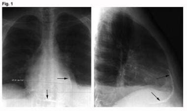
The most common causes of constrictive pericarditis include prior infection, radiation therapy, autoimmune disorders, and previous heart surgery. Diagnosis typically involves a combination of medical history, physical examination, imaging studies such as echocardiography or MRI, and sometimes invasive testing such as cardiac catheterization. Treatment may involve medications to manage symptoms and reduce inflammation, as well as surgical removal of the pericardium (pericardiectomy) in severe cases.
Pericarditis is a medical condition characterized by inflammation of the pericardium, which is the thin sac-like membrane that surrounds the heart and contains serous fluid to reduce friction during heartbeats. The inflammation can cause symptoms such as chest pain, shortness of breath, and sometimes fever.
The pericardium has two layers: the visceral pericardium, which is tightly adhered to the heart's surface, and the parietal pericardium, which lines the inner surface of the chest cavity. Normally, there is a small amount of fluid between these two layers, allowing for smooth movement of the heart within the chest cavity.
In pericarditis, the inflammation causes the pericardial layers to become irritated and swollen, leading to an accumulation of excess fluid in the pericardial space. This can result in a condition called pericardial effusion, which can further complicate the situation by putting pressure on the heart and impairing its function.
Pericarditis may be caused by various factors, including viral or bacterial infections, autoimmune disorders, heart attacks, trauma, or cancer. Treatment typically involves addressing the underlying cause, managing symptoms, and reducing inflammation with medications such as nonsteroidal anti-inflammatory drugs (NSAIDs), colchicine, or corticosteroids. In severe cases, pericardiocentesis (removal of excess fluid from the pericardial space) or surgical intervention may be necessary.
Tuberculous pericarditis is a specific form of pericarditis (inflammation of the pericardium, the thin sac-like membrane that surrounds the heart) that is caused by the bacterial infection of Mycobacterium tuberculosis. This type of pericarditis is more common in areas where tuberculosis is prevalent and can lead to serious complications if not diagnosed and treated promptly.
In tuberculous pericarditis, the bacteria typically spread from the lungs (the most common site of TB infection) or other infected organs through the bloodstream to the pericardium. The infection causes an inflammatory response, leading to the accumulation of fluid in the pericardial space (pericardial effusion), which can put pressure on the heart and impair its function. In some cases, the inflammation may lead to the formation of scar tissue, causing the pericardium to thicken and constrict, a condition known as constrictive pericarditis.
Symptoms of tuberculous pericarditis can include chest pain, cough, fever, fatigue, weight loss, and difficulty breathing. Diagnosis typically involves a combination of medical history, physical examination, imaging tests (such as echocardiography, CT scan, or MRI), and laboratory tests (including analysis of the pericardial fluid). Treatment usually consists of a long course of antibiotics specific to TB, along with anti-inflammatory medications and close monitoring for potential complications.
Pericardiectomy is a surgical procedure that involves the removal of all or part of the pericardium, which is the sac-like membrane surrounding the heart. This surgery is typically performed to treat chronic or recurrent pericarditis, constrictive pericarditis, or pericardial effusions that do not respond to other treatments. Pericardiectomy can help reduce symptoms such as chest pain, shortness of breath, and fluid buildup around the heart, improving the patient's quality of life and overall prognosis.
Pericardial effusion is an abnormal accumulation of fluid in the pericardial space, which is the potential space between the two layers of the pericardium - the fibrous and serous layers. The pericardium is a sac that surrounds the heart to provide protection and lubrication for the heart's movement during each heartbeat. Normally, there is only a small amount of fluid (5-15 mL) in this space to ensure smooth motion of the heart. However, when an excessive amount of fluid accumulates, it can cause increased pressure on the heart, leading to various complications such as decreased cardiac output and even cardiac tamponade, a life-threatening condition that requires immediate medical attention.
Pericardial effusion may result from several causes, including infections (viral, bacterial, or fungal), inflammatory conditions (such as rheumatoid arthritis, lupus, or cancer), trauma, heart surgery, kidney failure, or iatrogenic causes. The symptoms of pericardial effusion can vary depending on the rate and amount of fluid accumulation. Slowly developing effusions may not cause any symptoms, while rapid accumulations can lead to chest pain, shortness of breath, cough, palpitations, or even hypotension (low blood pressure). Diagnosis is usually confirmed through imaging techniques such as echocardiography, CT scan, or MRI. Treatment depends on the underlying cause and severity of the effusion, ranging from close monitoring to drainage procedures or medications to address the root cause.
Restrictive cardiomyopathy (RCM) is a type of heart muscle disorder characterized by impaired relaxation and filling of the lower chambers of the heart (the ventricles), leading to reduced pump function. This is caused by stiffening or rigidity of the heart muscle, often due to fibrosis or scarring. The stiffness prevents the ventricles from filling properly with blood during the diastolic phase, which can result in symptoms such as shortness of breath, fatigue, and fluid retention.
RCM is a less common form of cardiomyopathy compared to dilated or hypertrophic cardiomyopathies. It can be idiopathic (no known cause) or secondary to other conditions like amyloidosis, sarcoidosis, or storage diseases. Diagnosis typically involves a combination of medical history, physical examination, echocardiography, and sometimes cardiac MRI or biopsy. Treatment is focused on managing symptoms and addressing underlying causes when possible.
The pericardium is the double-walled sac that surrounds the heart. It has an outer fibrous layer and an inner serous layer, which further divides into two parts: the parietal layer lining the fibrous pericardium and the visceral layer (epicardium) closely adhering to the heart surface.
The space between these two layers is filled with a small amount of lubricating serous fluid, allowing for smooth movement of the heart within the pericardial cavity. The pericardium provides protection, support, and helps maintain the heart's normal position within the chest while reducing friction during heart contractions.
Cardiac tamponade is a serious medical condition that occurs when there is excessive fluid or blood accumulation in the pericardial sac, which surrounds the heart. This accumulation puts pressure on the heart, preventing it from filling properly and reducing its ability to pump blood effectively. As a result, cardiac output decreases, leading to symptoms such as low blood pressure, shortness of breath, chest pain, and a rapid pulse. If left untreated, cardiac tamponade can be life-threatening, requiring emergency medical intervention to drain the fluid and relieve the pressure on the heart.
Pericardiocentesis is a medical procedure where a needle or a catheter is inserted into the pericardial sac, the thin fluid-filled space surrounding the heart, to remove excess fluids or air that has accumulated. This buildup can put pressure on the heart and impede its function, leading to various cardiac symptoms such as chest pain, shortness of breath, and palpitations. The procedure is often guided by echocardiography or fluoroscopy to ensure proper placement and minimize risks. Pericardiocentesis may be performed as an emergency treatment or a scheduled intervention, depending on the patient's condition.
"Cardiovascular Tuberculosis" refers to a form of tuberculosis (TB) where the bacteria (Mycobacterium tuberculosis) infects the heart or the blood vessels. This is a less common manifestation of TB, but it can have serious consequences if left untreated.
In cardiovascular TB, the bacteria can cause inflammation and damage to the heart muscle (myocarditis), the sac surrounding the heart (pericarditis), or the coronary arteries that supply blood to the heart muscle. This can lead to symptoms such as chest pain, shortness of breath, coughing, fatigue, and fever. In severe cases, it can cause heart failure or life-threatening arrhythmias.
Cardiovascular TB is usually treated with a combination of antibiotics that are effective against the TB bacteria. The treatment may last for several months to ensure that all the bacteria have been eliminated. In some cases, surgery may be necessary to repair or replace damaged heart valves or vessels. Early diagnosis and treatment can help prevent serious complications and improve outcomes in patients with cardiovascular TB.
Cardanolides are a type of steroid compound that are found in certain plants, particularly in the family Apocynaceae. These compounds have a characteristic structure that includes a five-membered lactone ring attached to a steroid nucleus, and they are known for their ability to inhibit the sodium-potassium pump (Na+/K+-ATPase) in animal cells. This property makes cardanolides toxic to many organisms, including humans, and they have been used as heart poisons and insecticides.
One of the most well-known cardanolides is ouabain, which is found in the seeds of several African plants and has been used traditionally as a medicine for various purposes, including as a heart stimulant and a poison for hunting. Other examples of cardanolides include digoxin and digitoxin, which are derived from the foxglove plant (Digitalis purpurea) and are used in modern medicine to treat heart failure and atrial arrhythmias.
It's worth noting that while cardanolides have important medical uses, they can also be highly toxic if ingested or otherwise introduced into the body in large amounts. Therefore, it's essential to use these compounds only under the supervision of a qualified healthcare professional.
Chylous ascites is a medical condition characterized by the accumulation of milky, fat-containing fluid in the peritoneal cavity, which is the space within the abdomen that contains the intestines, liver, and other organs. The fluid, called chyle, is normally found in the lymphatic system and is formed when dietary fats are absorbed from the small intestine.
Chylous ascites can occur as a result of damage to the lymphatic vessels that transport chyle from the intestines to the bloodstream. This damage can be caused by various conditions, such as trauma, surgery, tumors, inflammation, or congenital abnormalities. When the lymphatic vessels are damaged, chyle leaks into the peritoneal cavity and accumulates there, leading to ascites.
Symptoms of chylous ascites may include abdominal distension, pain, nausea, vomiting, and weight loss. The condition can be diagnosed through various tests, such as imaging studies or analysis of the fluid in the peritoneal cavity. Treatment typically involves addressing the underlying cause of the condition, as well as managing symptoms and preventing complications. This may include dietary modifications, medications to reduce lymphatic flow, or surgical interventions to repair damaged lymphatic vessels.
Suppuration is the process of forming or discharging pus. It is a condition that results from infection, tissue death (necrosis), or injury, where white blood cells (leukocytes) accumulate to combat the infection and subsequently die, forming pus. The pus consists of dead leukocytes, dead tissue, debris, and microbes (bacteria, fungi, or protozoa). Suppuration can occur in various body parts such as the lungs (empyema), brain (abscess), or skin (carbuncle, furuncle). Treatment typically involves draining the pus and administering appropriate antibiotics to eliminate the infection.
Phonocardiography is a non-invasive medical procedure that involves the graphical representation and analysis of sounds produced by the heart. It uses a device called a phonocardiograph to record these sounds, which are then displayed as waveforms on a screen. The procedure is often used in conjunction with other diagnostic techniques, such as electrocardiography (ECG), to help diagnose various heart conditions, including valvular heart disease and heart murmurs.
During the procedure, a specialized microphone called a phonendoscope is placed on the chest wall over the area of the heart. The microphone picks up the sounds generated by the heart's movements, such as the closing and opening of the heart valves, and transmits them to the phonocardiograph. The phonocardiograph then converts these sounds into a visual representation, which can be analyzed for any abnormalities or irregularities in the heart's function.
Phonocardiography is a valuable tool for healthcare professionals, as it can provide important insights into the health and functioning of the heart. By analyzing the waveforms produced during phonocardiography, doctors can identify any potential issues with the heart's valves or other structures, which may require further investigation or treatment. Overall, phonocardiography is an essential component of modern cardiac diagnostics, helping to ensure that patients receive accurate and timely diagnoses for their heart conditions.
Protein-losing enteropathies (PLE) refer to a group of conditions characterized by excessive loss of proteins from the gastrointestinal tract into the intestinal lumen and ultimately into the stool. This results in hypoproteinemia, which is a decrease in the concentration of proteins in the bloodstream, particularly albumin.
The protein loss can occur due to various reasons such as increased permeability of the intestinal mucosa, lymphatic obstruction, or inflammatory processes affecting the gastrointestinal tract. Common causes of PLE include conditions such as inflammatory bowel disease, intestinal lymphangiectasia, celiac disease, Whipple's disease, and menetrier's disease.
Symptoms of PLE may include edema, ascites, weight loss, diarrhea, and fatigue. The diagnosis of PLE typically involves measuring the concentration of proteins in the stool, as well as other diagnostic tests to determine the underlying cause. Treatment of PLE depends on the underlying cause and may involve dietary modifications, medications, or surgical interventions.
Cardiovascular infections, also known as infective endocarditis, are infections that affect the inner layer of the heart, including the heart valves. These infections are usually caused by bacteria, but they can also be caused by fungi or other microorganisms. They can occur when bacteria or other germs enter the bloodstream and then settle in the heart.
There are several types of cardiovascular infections, including:
* Native Valve Endocarditis: This occurs when an infection affects the heart valves that are present at birth.
* Prosthetic Valve Endocarditis: This occurs when an infection affects an artificial heart valve.
* Intracardiac Device-Related Infections: These infections can occur in people who have devices such as pacemakers or implantable defibrillators.
* Infectious Myocarditis: This is an inflammation of the heart muscle caused by an infection.
Symptoms of cardiovascular infections may include fever, chills, fatigue, shortness of breath, chest pain, and a new or changing heart murmur. Treatment typically involves several weeks of antibiotics, and in some cases, surgery may be necessary to remove the infected tissue. Prevention measures include good oral hygiene, prompt treatment of skin infections, and prophylactic antibiotics for certain high-risk individuals undergoing dental or surgical procedures.
Doppler echocardiography is a type of ultrasound test that uses high-frequency sound waves to produce detailed images of the heart and its blood vessels. It measures the direction and speed of blood flow in the heart and major blood vessels leading to and from the heart. This helps to evaluate various conditions such as valve problems, congenital heart defects, and heart muscle diseases.
In Doppler echocardiography, a small handheld device called a transducer is placed on the chest, which emits sound waves that bounce off the heart and blood vessels. The transducer then picks up the returning echoes, which are processed by a computer to create moving images of the heart.
The Doppler effect is used to measure the speed and direction of blood flow. This occurs when the frequency of the sound waves changes as they bounce off moving objects, such as red blood cells. By analyzing these changes, the ultrasound machine can calculate the velocity and direction of blood flow in different parts of the heart.
Doppler echocardiography is a non-invasive test that does not require any needles or dyes. It is generally safe and painless, although patients may experience some discomfort from the pressure applied by the transducer on the chest. The test usually takes about 30 to 60 minutes to complete.
Chylothorax is a medical condition characterized by the accumulation of lymphatic fluid called chyle in the pleural space, which is the space between the lungs and the chest wall. Chyle is a milky-white fluid that contains nutrients, electrolytes, and immune cells, and it is normally transported through the thoracic duct to the bloodstream.
Chylothorax can occur due to various reasons, such as trauma, surgery, tumors, or congenital abnormalities that disrupt the normal flow of chyle. As a result, chyle leaks into the pleural space, causing symptoms such as cough, chest pain, difficulty breathing, and fever.
The diagnosis of chylothorax is usually made through imaging studies such as chest X-ray or CT scan, and confirmed by analyzing the fluid for the presence of chylomicrons, which are lipid particles found in chyle. The treatment options for chylothorax include dietary modifications, such as a low-fat diet with medium-chain triglycerides, chest tube drainage, and surgical interventions such as thoracic duct ligation or pleurodesis.
The mitral valve, also known as the bicuspid valve, is a two-leaflet valve located between the left atrium and left ventricle in the heart. Its function is to ensure unidirectional flow of blood from the left atrium into the left ventricle during the cardiac cycle. The mitral valve consists of two leaflets (anterior and posterior), the chordae tendineae, papillary muscles, and the left atrial and ventricular myocardium. Dysfunction of the mitral valve can lead to various heart conditions such as mitral regurgitation or mitral stenosis.
Cardenolides are a type of steroid compound that are found in certain plants and animals. These compounds have a characteristic structure that includes a five-membered lactone ring, which is attached to a steroid nucleus. Cardenolides are well known for their toxicity to many organisms, including humans, and they have been used for both medicinal and poisonous purposes.
One of the most famous cardenolides is digitoxin, which is derived from the foxglove plant (Digitalis purpurea). Digitoxin has been used as a medication to treat heart conditions such as congestive heart failure, as it can help to strengthen heart contractions and regulate heart rhythm. However, because of its narrow therapeutic index and potential for toxicity, digitoxin is not commonly used today.
Other cardenolides include ouabain, which is found in the seeds of the African plant Acokanthera ouabaio, and bufadienolides, which are found in the skin and parotid glands of toads. These compounds have also been studied for their potential medicinal uses, but they are not widely used in clinical practice due to their toxicity.
It is important to note that cardenolides can be highly toxic to humans and animals, and exposure to these compounds can cause a range of symptoms including nausea, vomiting, diarrhea, seizures, and even death. As such, it is essential to use caution when handling or coming into contact with plants or animals that contain cardenolides.
Bronchiolitis obliterans is a medical condition characterized by the inflammation and scarring (fibrosis) of the bronchioles, which are the smallest airways in the lungs. This results in the narrowing or complete obstruction of the airways, leading to difficulty breathing and reduced lung function.
The condition is often caused by a respiratory infection, such as adenovirus or mycoplasma pneumonia, but it can also be associated with exposure to certain chemicals, drugs, or radiation therapy. In some cases, the cause may be unknown.
Symptoms of bronchiolitis obliterans include cough, shortness of breath, wheezing, and crackles heard on lung examination. Diagnosis typically involves a combination of medical history, physical exam, imaging studies (such as chest X-ray or CT scan), and pulmonary function tests. In some cases, a biopsy may be necessary to confirm the diagnosis.
Treatment for bronchiolitis obliterans is focused on managing symptoms and preventing further lung damage. This may include bronchodilators to help open up the airways, corticosteroids to reduce inflammation, and oxygen therapy to help with breathing. In severe cases, a lung transplant may be necessary.
Echocardiography is a medical procedure that uses sound waves to produce detailed images of the heart's structure, function, and motion. It is a non-invasive test that can help diagnose various heart conditions, such as valve problems, heart muscle damage, blood clots, and congenital heart defects.
During an echocardiogram, a transducer (a device that sends and receives sound waves) is placed on the chest or passed through the esophagus to obtain images of the heart. The sound waves produced by the transducer bounce off the heart structures and return to the transducer, which then converts them into electrical signals that are processed to create images of the heart.
There are several types of echocardiograms, including:
* Transthoracic echocardiography (TTE): This is the most common type of echocardiogram and involves placing the transducer on the chest.
* Transesophageal echocardiography (TEE): This type of echocardiogram involves passing a specialized transducer through the esophagus to obtain images of the heart from a closer proximity.
* Stress echocardiography: This type of echocardiogram is performed during exercise or medication-induced stress to assess how the heart functions under stress.
* Doppler echocardiography: This type of echocardiogram uses sound waves to measure blood flow and velocity in the heart and blood vessels.
Echocardiography is a valuable tool for diagnosing and managing various heart conditions, as it provides detailed information about the structure and function of the heart. It is generally safe, non-invasive, and painless, making it a popular choice for doctors and patients alike.
Echocardiography, Doppler, pulsed is a type of diagnostic medical test that uses ultrasound to create detailed images of the heart's structures and assess their function. In this technique, high-frequency sound waves are directed at the heart using a handheld device called a transducer, which is placed on the chest wall. The sound waves bounce off the heart structures and return to the transducer, which then sends the information to a computer that converts it into images.
Pulsed Doppler echocardiography is a specific type of Doppler ultrasound that allows for the measurement of blood flow velocities in the heart and great vessels. In this technique, the transducer emits short bursts or "pulses" of sound waves and then measures the time it takes for the echoes to return. By analyzing the frequency shifts of the returning echoes, the velocity and direction of blood flow can be determined. This information is particularly useful in evaluating valvular function, assessing the severity of valvular lesions, and identifying areas of turbulent or abnormal blood flow.
Overall, echocardiography, Doppler, pulsed is a valuable tool for diagnosing and managing a wide range of cardiovascular conditions, including heart valve disorders, congenital heart defects, cardiomyopathies, and pericardial diseases.
A fatal outcome is a term used in medical context to describe a situation where a disease, injury, or illness results in the death of an individual. It is the most severe and unfortunate possible outcome of any medical condition, and is often used as a measure of the severity and prognosis of various diseases and injuries. In clinical trials and research, fatal outcome may be used as an endpoint to evaluate the effectiveness and safety of different treatments or interventions.
 Constrictive pericarditis
Pulsus paradoxus
Pericarditis
Cardiac muscle
Edward Delos Churchill
Viktor Schmieden
Obstructive shock
Roberto Ferrari (cardiologist)
Kussmaul's sign
Walter Lawrence Jr.
Restrictive cardiomyopathy
Norman Chevers
The Leopard (Nesbø novel)
Protein losing enteropathy
Friedreich's sign
IgG4-related disease
CT pulmonary angiogram
Shock (circulatory)
Outline of cardiology
Pericardial effusion
Pericardiectomy
Pericardium
Waldmann disease
Leon Manteuffel-Szoege
Heart failure with preserved ejection fraction
Walter Broadbent
Jugular venous pressure
List of paradoxes
Adolf Kussmaul
Hydralazine
Constrictive pericarditis - Wikipedia
 Pericarditis - constrictive: MedlinePlus Medical Encyclopedia
 Constrictive Pericarditis: Background, Pathophysiology, Etiology
Constrictive Pericarditis -- eCureMe.com
A difficult diagnosis - constrictive pericarditis and its treatment: a case report
 Constrictive Pericarditis (Constrictive Pericarditides): Symptoms, Diagnosis and Treatment - Symptoma
 Constrictive pericarditis: lessons from the past five years | RCP Journals
 Constrictive pericarditis | medtigo
 Chronic Constrictive Pericarditis - Easy MBBS
Constrictive Pericarditis Versus Restrictive Cardiomyopathy<...
 Constrictive pericarditis after cardiac surgery<...
Rheumatoid constrictive pericarditis - Nuffield Department of Orthopaedics, Rheumatology and Musculoskeletal Sciences
 Holdings: Effusive-constrictive pericarditis as the manifestation of an unexpected diagnosis
 Constrictive Pericarditis. | Hospital Medicine Virtual Journal Club | Washington University in St. Louis
Tricuspid Valve Regurgitation in Patients Undergoing Pericardiectomy for Constrictive Pericarditis<...
 Journal of Postgraduate Gynecology & Obstetrics: Pregnancy in a case of Constrictive pericarditis
Elevation of the JVP in constrictive pericarditis - Centre for Tropical Medicine and Global Health
 Tuberculous constrictive pericarditis with concurrent active pulmonary tuberculous infection: a case report | Cases Journal |...
 Chronic Pericarditis - Heart and Blood Vessel Disorders - Merck Manuals Consumer Version
Cor Pulmonale: Overview, Presentation, DDx
Long-term results of radical pericardiectomy for constrictive pericarditis in Korean population | Journal of Cardiothoracic...
Pericarditis: MedlinePlus Medical Encyclopedia
 Constrictive Pericarditis Presenting as Bilateral Pleural Effusion: A Report of Two Cases. | Cureus;10(4): e2451, 2018 Apr 09....
 Sweat Electrolyte Test: Uses, Procedures, Results and More
Immunoglobulin G4-related constrictive pericarditis and the importance of a thorough workup: a case report | BMC Cardiovascular...
 "Unchain My Heart: Constrictive Pericarditis in the Setting of Chronic " by Neiman Ramjattan DO, Justin L. Guthier DO et al.
 PS10:184 Constrictive pericarditis as the first presentation of systemic lupus erythematosus: a case report and literature...
![Akdeniz B[au] - Search Results - PubMed](data:image/png;base64,iVBORw0KGgoAAAANSUhEUgAAABAAAAAQCAMAAAAoLQ9TAAAARVBMVEVHcEwoU45gYmYAUpQAUpRPYGVgYmZLXnJgYmYAUZUAUpRJXnIAUpQAUpRgYmYAUpRgYmZgYmZhYmYAUpQAUpQAUpRgYmaDiPJuAAAAFXRSTlMADOJ+6QewGO8/uTRqtH7GdFJ11p1bCL3TAAAAZUlEQVQYlV2PVw7AIAxDTeney7n/UcsoldX3E+VJOAboEi7MBpHWMs1ADlG8u7UYWauwyZFeRQVPOhG2o+aiwhByJxUx91Jxhje3iJSqGfHuLKI0+0TpXvY1twCOPlFh5pa/++MB0vIOBm+1zaoAAAAASUVORK5CYII=) Akdeniz B[au] - Search Results - PubMed
Akdeniz B[au] - Search Results - PubMed
 Dr. Barry Weinstock, MD, Cardiology Specialist - Altamonte Springs, FL | Sharecare
Tuberculous pericarditis9
- However, it takes a period of time from tuberculous pericarditis to constrictive pericarditis. (biomedcentral.com)
- BARCELONA -- Adding steroids to anti-tuberculosis treatment for patients with tuberculous pericarditis can reduce the risk of an important complication and the resulting hospital admissions, researchers said here. (medpagetoday.com)
- Tuberculous pericarditis "is the most important and the most serious form of pericardial disease in the world," Mayosi said, occurring in about 10% of the 10 million new TB patients every year. (medpagetoday.com)
- Much of the disease burden of tuberculous pericarditis is in developing countries, where many TB patients are also co-infected with HIV, Mayosi noted. (medpagetoday.com)
- Some 1,400 patients with probably or confirmed tuberculous pericarditis were randomly assigned to get prednisolone or placebo for 6 weeks. (medpagetoday.com)
- What is the pathogenesis of tuberculous pericarditis? (imperial.ac.uk)
- Background and Objectives: In East Asia, tuberculous pericarditis still occurs in immunocompetent patients. (koreamed.org)
- We aimed to investigate clinical course of tuberculous pericarditis and the trends of echocardiographic parameters for constrictive. (koreamed.org)
- Tuberculous pericarditis has become rare in developed countries but remains common in other areas. (health.am)
Acute8
- Acute and subacute forms of pericarditis (which may or may not be symptomatic) may deposit fibrin, which, in turn, can evoke a pericardial effusion. (medscape.com)
- Acute Pericarditis can result in Pericardial Tamponade and can lead to chronic or constrictive Pericarditis . (ecureme.com)
- Around 9% of individuals experiencing acute pericarditis develop constrictive physiology, with infectious causes remaining the primary culprits in the developing world. (medtigo.com)
- The pathophysiology of chronic constrictive pericarditis implicates the gradual closure of the pericardial cavity due to the formation of granulation tissue, which occurs during the healing process of an acute episode of fibrinous through the absorption of a chronic pericardial effusion. (medtigo.com)
- Rarely, after acute purulent pericarditis. (easymbbs.com)
- Acute Pericarditis Acute pericarditis is inflammation of the pericardium (the flexible two-layered sac that envelops the heart) that begins suddenly, is often painful, and causes fluid and blood components such. (merckmanuals.com)
- Management of acute and recurrent pericarditis: JACC State-of-the-art Review. (medlineplus.gov)
- Viral infections (especially infections with coxsackieviruses and echoviruses but also influenza, Epstein-Barr, varicella, hepatitis, mumps, and HIV viruses) are the commonest cause of acute pericarditis and probably are responsible for many cases classified as idiopathic. (health.am)
Congestive heart f2
- Operative indication was congestive heart failure caused by constrictive pericarditis. (biomedcentral.com)
- BACKGROUND: Constrictive pericarditis is an uncommon condition that could be easily confused with congestive heart failure. (koreamed.org)
Idiopathic2
- The cause of constrictive pericarditis in the developing world are idiopathic in origin, though likely infectious in nature. (wikipedia.org)
- Conclusions SLE should be included in differential diagnosis of constrictive pericarditis, especially in 'idiopathic' cases and in the context of poor response to tuberculosis treatment. (bmj.com)
Fibrotic pericardium5
- Constrictive pericarditis is a medical condition characterized by a thickened, fibrotic pericardium, limiting the heart's ability to function normally. (wikipedia.org)
- Causes of constrictive pericarditis include: Tuberculosis Incomplete drainage of purulent pericarditis Fungal and parasitic infections Chronic pericarditis Postviral pericarditis Postsurgical Following MI, post-myocardial infarction In association with pulmonary asbestos The pathophysiological characteristics of constrictive pericarditis are due to a thickened, fibrotic pericardium that forms a non-compliant shell around the heart. (wikipedia.org)
- Constrictive pericarditis occurs when a thickened fibrotic pericardium, of whatever cause, impedes normal diastolic filling. (medscape.com)
- In cases of constrictive pericarditis, the thickened, fibrotic pericardium hinders the filling of the heart's ventricles due to spatial constraint. (symptoma.com)
- Constrictive pericarditis is a disorder in which a chronically thickened and fibrotic pericardium limits cardiac fillings. (jpgo.org)
Case of constrictive pericarditis4
- [1,2] Our case report presents medical management of a 26 years old multigravida, a diagnosed case of constrictive pericarditis secondary to tuberculosis which was diagnosed 5 years ago. (jpgo.org)
- This is a rare case of constrictive pericarditis secondary to tuberculosis in pregnancy. (jpgo.org)
- This case exemplifies the difficulties faced by clinicians when reviewing a possible case of constrictive pericarditis, while highlighting the importance of a multimodality assessment. (biomedcentral.com)
- We present a challenging diagnostic case of constrictive pericarditis due to IgG4-related disease in accordance with the CARE reporting checklist. (biomedcentral.com)
Pericardium17
- The definitive treatment for constrictive pericarditis is pericardial stripping, which is a surgical procedure where the entire pericardium is peeled away from the heart. (wikipedia.org)
- Constrictive pericarditis is a process where the sac-like covering of the heart (the pericardium) becomes thickened and scarred. (medlineplus.gov)
- [ 1 ] This usually involves the parietal pericardium, although it can involve the visceral pericardium (see Constrictive-Effusive Pericarditis ). (medscape.com)
- This often leads to pericardial organization, chronic fibrotic scarring, and calcification, most often involving the parietal pericardium (see Constrictive-Effusive Pericarditis for visceral pericardial disease). (medscape.com)
- In constrictive Pericarditis there is a thickening of the pericardium and attachment to the heart that may restrict its normal movements. (ecureme.com)
- Uremia Uremic pericarditis is thought to result from inflammation of the visceral and parietal layers of the pericardium by metabolic toxins that accumulate in the body owing to kidney failure . (symptoma.com)
- Constrictive pericarditis is characterized by the inflammation and stiffening of the pericardium. (medtigo.com)
- Constrictive pericarditis is a potentially treatable cause of diastolic heart failure that arises because a diseased, inelastic pericardium restricts ventricular diastolic expansion. (wustl.edu)
- On evaluation, CT scan showed calcified pericardium suggestive of constrictive pericarditis. (jpgo.org)
- [3] Constrictive pericarditis is the end stage of the healing process of inflamed pericardium which takes several months to years for dense fibrosis and calcification. (jpgo.org)
- Constrictive pericarditis is a process of chronic fibrous thickening of the pericardium, which is frequently accompanied with calcification and prevents the diastolic filling of the heart, reducing venous return and lowering output [ 1 , 2 ]. (biomedcentral.com)
- Chronic pericarditis is inflammation of the pericardium (the flexible two-layered sac that envelops the heart) that begins gradually, is long-lasting, and results in fluid accumulation in the pericardial space or thickening of the pericardium. (merckmanuals.com)
- In chronic effusive pericarditis, fluid slowly accumulates in the pericardial space, between the two layers of the pericardium. (merckmanuals.com)
- Chronic constrictive pericarditis, which is rare, usually results when scarlike (fibrous) tissue forms throughout the pericardium. (merckmanuals.com)
- Pericarditis is a condition in which the sac-like covering around the heart (pericardium) becomes inflamed. (medlineplus.gov)
- Constrictive pericarditis rarely occurs secondary to severe asbestos-induced fibrosis or calcification of the pericardium. (cdc.gov)
- Constrictive pericarditis is a disease of the pericardium resulting from chronic inflammation and/or scar responsible for a clinical feature of left and right ventricular failure. (koreamed.org)
Symptoms8
- Signs and symptoms of constrictive pericarditis are consistent with the following: fatigue, swollen abdomen, difficulty breathing (dyspnea), swelling of legs and general weakness. (wikipedia.org)
- Call your provider if you have symptoms of constrictive pericarditis. (medlineplus.gov)
- Constrictive pericarditis symptoms overlap those of diseases as diverse as myocardial infarction (MI), aortic dissection, pneumonia, influenza, and connective tissue disorders. (medscape.com)
- The classic diagnostic conundrum associated with constrictive pericarditis is the difficulty distinguishing this condition from restrictive cardiomyopathy (see Restrictive Cardiomyopathy ) and other syndromes associated with elevated right-sided pressure that all share similar symptoms, physical findings, and hemodynamics. (medscape.com)
- Abstract The diagnosis of constrictive pericarditis requires a high degree of clinical suspicion, for the signs and symptoms of this disease can be falsely attributed to other causes. (cam.ac.uk)
- Constrictive pericarditis is a clinical condition characterized by the appearance of signs and symptoms of right heart failure due to loss of pericardial compliance. (uitm.edu.my)
- Scarring results in severe restriction of filling of all the cardiac chambers and orifices of great vesseles which produces signs and symptoms of chronic constrictive pericarditis. (jpgo.org)
- [4] Dyspnea, fatigue, palpitation and edema are common symptoms of constrictive pericarditis. (jpgo.org)
Systemic3
- Effusive constrictive pericarditis in systemic sclerosis by: Zaeem Ahmed, et al. (uitm.edu.my)
- Objective Although fibrinous and exudative pericarditis is a common feature of Systemic lupus erythematosus (SLE), found in 62% of lupus patients on autopsy, very few cases progress to (effusive) - constrictive pericarditis. (bmj.com)
- We describe the unusual occurrence of constrictive pericarditis (CP) in a patient with Systemic Lupus Erythematosus. (bmj.com)
Pericardiectomy for constrictive3
- We hypothesized that tricuspid valve regurgitation was associated with increased risk of mortality after pericardiectomy for constrictive pericarditis. (elsevierpure.com)
- We reviewed the records of 518 patients who received pericardiectomy for constrictive pericarditis between January 2000 and December 2016. (elsevierpure.com)
- Tricuspid valve regurgitation is a common and clinically important comorbidity in patients operated with pericardiectomy for constrictive pericarditis. (elsevierpure.com)
Patients12
- A poor outcome is almost always the result after a pericardiectomy is performed for constrictive pericarditis whose origin was radiation-induced, further some patients may develop heart failure post-operatively. (wikipedia.org)
- There are still patients who develop constrictive pericarditis. (rcpjournals.org)
- Patients with constrictive pericarditis present to specialists in different disciplines. (rcpjournals.org)
- Constrictive pericarditis should be considered in patients with rheumatoid arthritis who develop unexplained cardiac failure. (ox.ac.uk)
- Patients with severe constrictive pericarditis usually have a limited stroke volume caused by poor diastolic filling. (jpgo.org)
- Patients with constrictive pericarditis have poor diastolic filling pattern with raised atrial pressure. (jpgo.org)
- In some particular endemic area, it is not uncommon to see patients with tuberculosis pericarditis. (biomedcentral.com)
- In hospital settings, constrictive pericarditis is not usually considered as a differential in patients presenting with pleural effusion . (bvsalud.org)
- On the other hand, 4.4% of those getting prednisolone had constrictive pericarditis, compared with 7.8% of placebo patients, yielding a hazard ratio of 0.56 that was significant ( P =0.009). (medpagetoday.com)
- But HIV-positive patients have a clearly greater risk of cancer, suggesting the "use of glucocorticoids should be curtailed in this population unless the risk of constrictive pericarditis is high," Chaisson and Post argued. (medpagetoday.com)
- In symptomatic patients, septal "wobble" on echocardiography may be an important sign of constrictive. (koreamed.org)
- Rare patients will continue to experience recurrences chronically, sometimes leading to constrictive pericarditis, when pericardial resection may be required. (health.am)
Forms of pericarditis1
- Constrictive pericarditis usually arises as a consequence of other forms of pericarditis, but may also develop after a heart attack or heart surgery . (symptoma.com)
20221
- 2022. "Constrictive Pericarditis Revealing Rare Case of ALH Amyloidosis With Underlying Lymphoplasmacytic Lymphoma (Waldenstrom Macroglobulinemia). (stanford.edu)
Tuberculosis5
- On a global scale, tuberculosis is the primary cause of constrictive pericarditis, contributing to approximately 50% of cases in individuals with tuberculous pericardial effusion, even when undergoing antitubercular treatment. (medtigo.com)
- In some endemic areas, Mycobacterium tuberculosis infection should be taken into consideration during diagnostic evaluations for constrictive pericarditis. (biomedcentral.com)
- Mycobaterium tuberculosis is the most common cause of constrictive pericarditis in endemic area [ 6 ]. (biomedcentral.com)
- One of our cases developed constrictive pericarditis with concurrent active tuberculosis . (bvsalud.org)
- This is a rare presentation because, normally, constrictive pericarditis is a late complication of tuberculosis . (bvsalud.org)
Occurs1
- Occasionally, constrictive pericarditis occurs more quickly (for example, within a few weeks after heart surgery) and is considered subacute. (merckmanuals.com)
Chronic pericarditis1
- There are two main types of chronic pericarditis. (merckmanuals.com)
Effusive constrictive5
- Effusive-constrictive pericard. (uitm.edu.my)
- Transient effusive constrictive pericarditis by: Erkan Ayhan, et al. (uitm.edu.my)
- Pericardial waffle for effusive‐constrictive pericarditis by: Omid Kiamanesh, et al. (uitm.edu.my)
- An echocardiogram demonstrated features of effusive-constrictive pericarditis. (bmj.com)
- BACKGROUND: Effusive-constrictive pericarditis (ECP) is traditionally diagnosed by using the expensive and invasive technique of direct pressure measurements in the pericardial space and the right atrium. (koreamed.org)
Pleural effusion2
- Constrictive Pericarditis Presenting as Bilateral Pleural Effusion: A Report of Two Cases. (bvsalud.org)
- We suggest that when dealing with cases of bilateral pleural effusion , the etiology of constrictive pericarditis should be considered. (bvsalud.org)
Diagnosis and Treatment1
- Rapid diagnosis and treatment of constrictive pericarditis are crucial to reduce mortality. (biomedcentral.com)
Clinical4
- In particular, restrictive cardiomyopathy has many similar clinical features to constrictive pericarditis, and differentiating them in a particular individual is often a diagnostic dilemma. (wikipedia.org)
- BNP blood test - tests for the existence of the cardiac hormone brain natriuretic peptide, which is only present in restrictive cardiomyopathy but not in constrictive pericarditis Conventional cardiac catheterization Physical examination - can reveal clinical features including Kussmaul's sign and a pericardial knock. (wikipedia.org)
- The preservation of good ventricular function on echocardiography in the face of clinical evidence of myocardial insufficiency raised the possibility of constrictive pericarditis, which was confirmed on cardiac catheterization. (ox.ac.uk)
- BACKGROUND: The aim of this study was to analyze the preoperative attributes and clinical impacts of complete pericardiectomy in chronic constrictive pericarditis. (koreamed.org)
Disease2
- Therefore, we reported a TB constrictive pericarditis with rare disease progress. (biomedcentral.com)
- Rarely, constrictive pericarditis can occur secondary to asbestos-associated disease. (cdc.gov)
Restrictive1
- The preservation of myocardial function in early diastole aids in distinguishing constrictive pericarditis from restrictive cardiomyopathy . (medscape.com)
Inflammatory2
- The presentation and course of inflammatory pericarditis depend on its cause, but all syndromes are often (not always) associated with chest pain , which is usually pleuritic and postural (relieved by sitting). (health.am)
- The echocardiogram may disclose pericardial effusions and indicate their hemodynamic significance, but it is often normal in inflammatory pericarditis. (health.am)
Cardiac surgery1
- Constrictive pericarditis may develop as a midterm or late complication of cardiac surgery. (unicatt.it)
Myocarditis1
- Myocarditis and pericarditis. (medlineplus.gov)
Radiation-induced1
- We excluded cases of radiation induced constrictive pericarditis, tuberculous-related constrictive pericarditis, and concomitant tricuspid valve intervention. (elsevierpure.com)
Ventricular1
- BACKGROUND: Constrictive pericarditis after coronary artery bypass surgery has been known to affect cardiac output by limiting diastolic ventricular filling. (koreamed.org)
Pulmonary6
- In both chronic constrictive pericarditis and cardiac tamponade, there is a convergence of pressures in the right ventricle (RV), right atrium (RA), left ventricle (LV), and pulmonary wedge pressure. (medtigo.com)
- In cardiac tamponade, the pressures decrease with inspiration, whereas in constrictive pericarditis, the RA pressure remains relatively steady while the pulmonary wedge pressure declines. (medtigo.com)
- There is still no report of tuberculous constrictive pericarditis concurrent with active pulmonary TB infection in a patient without previous pulmonary TB infection history. (biomedcentral.com)
- We report the case of a 63-year-old Taiwanese man with tuberculous constrictive pericarditis concurrent with active pulmonary tuberculous infection presenting with progressive extremities edema, puffy face, abdominal distension and dyspnea on exertion found to be caused by right heart failure. (biomedcentral.com)
- Yet, at the final admission, a re-assessment echocardiogram followed by cardiac computed tomography, magnetic resonance and right heart catheterization raised a possible diagnosis of constrictive pericarditis with a finding of abnormal pulmonary venous return. (biomedcentral.com)
- A possible diagnosis of constrictive pericarditis with an incidental finding of anomalous pulmonary venous return was considered. (biomedcentral.com)
Patient1
- Methods This is a chart review- based report of a lupus patient who had constrictive pericarditis as a presenting feature and a systematic literature review of previously published cases. (bmj.com)
Rarely1
- The extent of pericardiectomy is an important issue in constrictive pericarditis but its impact on long-term outcomes has been rarely reported. (biomedcentral.com)
Concomitant1
- The bottom line of the Investigation of the Management of Pericarditis trial was that "we need to be selective in the use of steroids," Mayosi said, because despite some benefits, there remains the risk of cancers in people with concomitant HIV. (medpagetoday.com)
Diastolic1
- Due to chronic constrictive pericarditis, there is a decrease in end-diastolic volume and, subsequently, a reduction in cardiac output and stroke volume. (medtigo.com)
Cases5
- In some cases, constrictive pericarditis is not preventable. (medlineplus.gov)
- In cases of chronic constrictive pericarditis, there is a notable disparity between intrathoracic and intracardiac pressures. (medtigo.com)
- We reviewed other cases of tuberculous constrictive pericarditis from the literature and described the peculiarities of this case. (biomedcentral.com)
- The cause of pericarditis is unknown or unproven in many cases. (medlineplus.gov)
- According to the literature , associated pleural effusions in cases of constrictive pericarditis could be left-sided. (bvsalud.org)
Differential1
- An increased suspicion of constriction helps move constrictive pericarditis to the top of a lengthy differential diagnosis list and facilitates correct diagnosis and timely therapy. (medscape.com)
Occurrence1
- Constrictive pericarditis is a rare occurrence in pregnancy. (jpgo.org)
Mild1
- Pericarditis can range from mild illness that gets better on its own, to a life-threatening condition. (medlineplus.gov)
Treatment1
- The reduction in constrictive pericarditis and hospital admission with prednisolone treatment is "clinically meaningful," commented Richard Chaisson, MD , and Wendy Post, MD , both of the Johns Hopkins University School of Medicine. (medpagetoday.com)