Chondromalacia Patellae
Patellar Ligament
Patellofemoral Joint
Nail-Patella Syndrome
Cartilage, Articular
Dislocations
Prochloron
Osteoarthritis, Knee
Joint Instability
Tibia
Range of Motion, Articular
Patellofemoral Pain Syndrome
Quadriceps Muscle
Bone Wires
Mollusca
Joint Diseases
Anatomic Landmarks
Biomechanical Phenomena
Lead
Tendons
Bone Malalignment
Magnetic Resonance Imaging
Bone Diseases, Developmental
The clinical manifestations and pathomechanics of contracture of the extensor mechanism of the knee. (1/666)
Experience with thirty-eight Asian children and adolescents who presented with either stiffness of the knee, genu recurvatum, habitual dislocation of the patella or congenital lateral dislocation of the patella showed that all those disorders were manifestations of contracture of the extensor mechanism, which fell into two groups according to the components involved. In Group I the main components affected were in the midline of the limb, namely rectus femoris and vastus intermedius; these patients presented with varying degrees of stiffness of the knee, or worse, with genu recurvatum. In Group II the main components involved were lateral to the midline of the limb, namely vastus lateralis and the ilio-tibial band; these patients presented with habitual dislocation of the patella, or worse, congenital lateral dislocation of the patella. In both groups untreated patients developed secondary adaptive changes such as subluxation of the tibia or marked genu valgum which made operative procedures more formidable and less effective. Release of the contracture should therefore be performed as early as possible. (+info)An operation for chronic prepatellar bursitis. (2/666)
An operation for chronic prepatellar bursitis is described in which only the posterior wall of the bursa is excised, thus preserving, undamaged, healthy and normally sensitive skin. This procedure is easier and less traumatic than complete excision of the bursa and results in fewer complications. It is suggested that removal of tha anterior wall of the bursa results in unnecessary and harmful interference with the underlying skin. The operation described gives a good functional and structural result; leaving the anterior wall of the bursa does not predispose to recurrence. (+info)The locked patella. An unusual complication of haemophilia. (3/666)
Mechanical derangements of the knee are an uncommon complication of chronic haemophiliac arthropathy. Two patients with locking of the patella were treated by manipulation. The mechanism of the injury was forced flexion of the knee joint beyond the limit of its restricted range. The injury is a serious one and may take six months to recover. (+info)Histomorphometry of the aging human patella: histologic criteria and controls. (4/666)
OBJECTIVE: A histomorphometric analysis of patellae from necropsies on persons between the third and tenth decades of life was carried out to trace the natural history of osteoarthritis. DESIGN: Minutiae of the histological changes in the surface and basilar portions of the articular cartilage were developed as criteria for the quantitation. A total of 99 patellas were harvested in the stated age range. The present study reports the results of ten grossly and radiologically normal specimens from subjects 23-32 years old served as controls. RESULTS: None of the control patellae were entirely histologically normal. Abnormality of the cartilage surface did not consistently proceed remodeling at the attachment to the subchondral plate. CONCLUSIONS: This observation throws into question the concept that osteoarthritis has a single histogenesis or always arises in articular cartilage. (+info)Knee pain and the infrapatellar branch of the saphenous nerve. (5/666)
Pain over the front of the knee is common after surgery or trauma but often a definite diagnosis is difficult to make. Over the past year we have seen five cases in which the pain could be ascribed to damage to a branch of the infrapatellar branch of the saphenous nerve. Two were subsequent to trauma and three to surgical procedures. In all five cases surgical exploration gave symptomatic relief. Eight cadaveric knees were prosected to explore further the anatomy of this nerve in relation to the injuries. Injury to one of these branches should be considered in cases of persistent anterior, anteromedial or anterolateral knee pain or neurological symptoms following surgery or trauma. (+info)Bone scintigraphy in chronic knee pain: comparison with magnetic resonance imaging. (6/666)
OBJECTIVE: To compare increased bone uptake of 99Tcm-MDP and magnetic resonance (MR) detected subchondral lesions, osteophytes, and cartilage defects in the knee in middle aged people with long-standing knee pain. METHODS: Fifty eight people (aged 41-58 years, mean 50) with chronic knee pain, with or without radiographic knee osteoarthritis, were examined with bone scintigraphy. The pattern and the grade of increased bone uptake was assessed. On the same day, a MR examination on a 1.0 T imager was performed. The presence and the grade of subchondral lesions, osteophytes, and cartilage defects were registered. RESULTS: The kappa values describing the correlation between increased bone uptake and MR detected subchondral lesions varied between 0.79 and 0.49, and between increased bone uptake and MR detected osteophytes or cartilage defects the values were < 0.54. The kappa values describing the correlation between the grade of bone uptake and the grade of the different MR findings was < 0.57. CONCLUSIONS: Good agreement was found between increased bone uptake and MR detected subchondral lesion. The agreement between increased bone uptake and osteophytes or cartilage defects was in general poor as well as the agreement between the grade of bone uptake and the grade of the MR findings. (+info)Knee cartilage topography, thickness, and contact areas from MRI: in-vitro calibration and in-vivo measurements. (7/666)
OBJECTIVE: This study assessed the three-dimensional accuracy of magnetic resonance imaging (MRI) for measuring articular surface topographies and cartilage thicknesses of human cadaveric knee joints, by comparison with the calibrated stereophotogrammetric (SPG) method. METHODS: Six fresh frozen cadaveric knees and the knees of four volunteers were imaged with a three-dimensional spoiled gradient-recalled acquisition with fat suppression using a linear extremity coil in a 1.5 T superconducting magnet. The imaging voxel size was 0.47 x 0.47 x 1.0 mm. Both a manual and a semi-automated segmentation method were employed to extract topographic measurements from MRI. Following MRI, each of the six cadaveric knees was dissected and its articular surfaces quantified using stereophotogrammetry. The MRI surface measurements were compared numerically with the SPG measurements. RESULTS: For six cadaveric knees, the average accuracies of cartilage and subchondral bone surface measurements were found to be 0.22 mm and 0.14 mm respectively and the thickness measurements demonstrated an average accuracy of 0.31 mm. It was found that while most of the error may be attributed to random measurement error, the accuracy was somewhat affected by systematic errors. For each bone of the knee, accuracies were most favorable in the patella, followed by the femur and then the tibia. The more efficient semi-automated method provided equally good and sometimes better accuracies than manual segmentation. CONCLUSIONS: This study demonstrates that clinical MRI can provide accurate measurements of cartilage topography, thickness, contact areas and surface curvatures of the knee. (+info)Localization of a gene for familial patella aplasia-hypoplasia (PTLAH) to chromosome 17q21-22. (8/666)
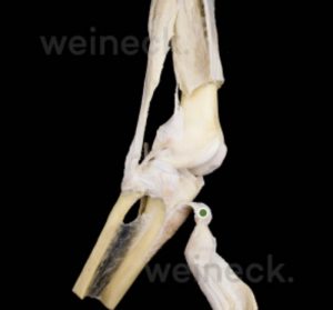
Patella aplasia-hypoplasia (PTLAH) is a rare genetic defect characterized by congenital absence or marked reduction of the patella. PTLAH can occur either as an isolated defect or in association with other malformations, and it characteristically occurs in the nail-patella syndrome and in some chromosome imbalances. We report the first evidence of linkage for isolated PTLAH in an extended Venezuelan family. After exclusion of the candidate chromosome regions where disorders associated with PTLAH have been mapped, a genomewide scan was performed that supported mapping of the disease locus within a region of 12 cM on chromosome 17q22. Two marker loci (D17S787 and D17S1604) typed from this region gave maximum LOD scores >3. Accordingly, multipoint analysis gave a maximum LOD score of 3.39, with a most likely location for the disease gene between D17S787 and D17S1604. Sequencing of the noggin gene, a candidate mapping between these markers, failed to reveal any mutation in affected subjects. (+info)The patella, also known as the kneecap, is a sesamoid bone located at the front of the knee joint. It is embedded in the tendon of the quadriceps muscle and serves to protect the knee joint and increase the leverage of the extensor mechanism, allowing for greater extension force of the lower leg. The patella moves within a groove on the femur called the trochlea during flexion and extension of the knee.
Chondromalacia patellae is a medical condition that refers to the softening and degeneration of the articular cartilage on the undersurface of the patella, or kneecap. This cartilage, which provides a smooth, lubricated surface for joint movement, can become damaged due to various reasons such as overuse, misalignment of the patella, or direct trauma. The resulting damage can cause pain and inflammation in the knee, particularly during activities that involve bending or straightening the leg. In some cases, chondromalacia patellae may also lead to the formation of bone spurs or osteophytes, which can further exacerbate the symptoms and limit joint mobility. Treatment for chondromalacia patellae typically involves a combination of rest, physical therapy, and pain management strategies, such as anti-inflammatory medications or corticosteroid injections. In severe cases, surgery may be required to repair or replace the damaged cartilage.
The patellar ligament, also known as the patellar tendon, is a strong band of tissue that connects the bottom part of the kneecap (patella) to the top part of the shinbone (tibia). This ligament plays a crucial role in enabling the extension and straightening of the leg during activities such as walking, running, and jumping. Injuries to the patellar ligament, such as tendonitis or tears, can cause pain and difficulty with mobility.
Cartilage diseases refer to conditions that affect the cartilaginous tissues in the body. Cartilage is a firm, flexible connective tissue found in many areas of the body, including the joints, ribcage, ears, and nose. It provides structure and support, allows for smooth movement between bones, and protects the ends of bones from friction.
There are several types of cartilage diseases, including:
1. Osteoarthritis (OA): This is a degenerative joint disease that occurs when the protective cartilage that cushions the ends of your bones wears down over time. It can cause pain, stiffness, and loss of mobility in the affected joints.
2. Rheumatoid arthritis (RA): This is an autoimmune disorder that causes inflammation in the lining of the joints, leading to cartilage damage and bone erosion.
3. Traumatic arthritis: This occurs when a joint is injured, causing damage to the cartilage and resulting in pain, stiffness, and loss of mobility.
4. Infectious arthritis: This occurs when a joint becomes infected, leading to inflammation and potential damage to the cartilage.
5. Chondromalacia patellae: This is a condition that affects the cartilage on the back of the kneecap, causing pain and stiffness in the knee.
6. Costochondritis: This is an inflammation of the cartilage in the ribcage, causing chest pain and discomfort.
7. Nasal septal deviation: This is a condition where the cartilage that separates the nostrils is crooked or off-center, causing difficulty breathing through the nose.
8. Osteochondritis dissecans (OCD): This is a joint condition that occurs when a piece of cartilage and bone in a joint becomes detached, causing pain and stiffness.
9. Synovial chondromatosis: This is a rare condition where nodules made up of cartilage form in the lining of a joint, causing pain, swelling, and limited mobility.
Treatment for cartilage diseases varies depending on the specific condition and severity, but may include medication, physical therapy, surgery, or a combination of these.
The patellofemoral joint is the articulation between the patella (kneecap) and the femur (thigh bone). It is a synovial joint, which means it is surrounded by a joint capsule containing synovial fluid to lubricate the joint. This joint is responsible for providing stability to the knee extensor mechanism and allows for smooth movement of the patella during activities like walking, running, and jumping. Pain or dysfunction in this joint can result in various conditions such as patellofemoral pain syndrome, chondromalacia patella, or patellar dislocation.
Patellar dislocation is a medical condition characterized by the displacement of the patella (kneecap) from its normal position in the femoral groove, which is a part of the femur (thighbone). This displacement usually occurs laterally, meaning that the patella moves toward the outer side of the knee.
Patellar dislocation can happen as a result of direct trauma or due to various factors that increase the laxity of the medial patellofemoral ligament and tightness of the lateral structures, leading to abnormal tracking of the patella. These factors include anatomical variations, muscle imbalances, genetic predisposition, or degenerative changes in the knee joint.
Dislocation of the patella can cause pain, swelling, and difficulty in moving the knee. In some cases, it might be associated with other injuries such as fractures or damage to the articular cartilage and surrounding soft tissues. Immediate medical attention is required for proper diagnosis and treatment, which may involve reduction, immobilization, physical therapy, bracing, or even surgery in severe cases.
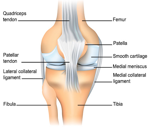
The knee joint, also known as the tibiofemoral joint, is the largest and one of the most complex joints in the human body. It is a synovial joint that connects the thighbone (femur) to the shinbone (tibia). The patella (kneecap), which is a sesamoid bone, is located in front of the knee joint and helps in the extension of the leg.
The knee joint is made up of three articulations: the femorotibial joint between the femur and tibia, the femoropatellar joint between the femur and patella, and the tibiofibular joint between the tibia and fibula. These articulations are surrounded by a fibrous capsule that encloses the synovial membrane, which secretes synovial fluid to lubricate the joint.
The knee joint is stabilized by several ligaments, including the medial and lateral collateral ligaments, which provide stability to the sides of the joint, and the anterior and posterior cruciate ligaments, which prevent excessive forward and backward movement of the tibia relative to the femur. The menisci, which are C-shaped fibrocartilaginous structures located between the femoral condyles and tibial plateaus, also help to stabilize the joint by absorbing shock and distributing weight evenly across the articular surfaces.
The knee joint allows for flexion, extension, and a small amount of rotation, making it essential for activities such as walking, running, jumping, and sitting.
Nail-Patella Syndrome (NPS) is a genetic disorder that affects the development of certain bones and organs. It's also known as Fong's syndrome, Hereditary Onycho-Osteodysplasia, or Turner-Kieser syndrome. The name comes from its most prominent features: abnormalities of the nails and kneecaps (patellae).
The main characteristics of NPS include:
1. Nail changes: These are often the first sign of the condition. The nails may be thin, underdeveloped, or absent, especially on the thumbs and index fingers. They can also be ridged, pitted, or discolored.
2. Patella (kneecap) abnormalities: About 70% of people with NPS have kneecaps that are small, irregularly shaped, or displaced from their normal position. This can cause knee pain and instability.
3. Elbow abnormalities: People with NPS may have elbow deformities, such as dislocated radial heads (one of the bones in the forearm).
4. Illic crest (pelvic bone) abnormalities: Some people with NPS have iliac horns, which are bony growths on the pelvis that don't cause any symptoms but can be detected through imaging tests.
5. Glaucoma: Around 10% of individuals with NPS develop glaucoma, a condition characterized by increased pressure within the eye, leading to optic nerve damage and potential vision loss if left untreated.
6. Kidney issues: Up to 40% of people with NPS experience kidney problems, such as proteinuria (excessive protein in urine) or kidney failure.
Nail-Patella Syndrome is caused by mutations in the LMX1B gene and is inherited in an autosomal dominant manner, meaning that only one copy of the altered gene is needed to cause the disorder. However, about 20% to 30% of cases result from new mutations and have no family history of the condition.
Articular cartilage is the smooth, white tissue that covers the ends of bones where they come together to form joints. It provides a cushion between bones and allows for smooth movement by reducing friction. Articular cartilage also absorbs shock and distributes loads evenly across the joint, protecting the bones from damage. It is avascular, meaning it does not have its own blood supply, and relies on the surrounding synovial fluid for nutrients. Over time, articular cartilage can wear down or become damaged due to injury or disease, leading to conditions such as osteoarthritis.
A dislocation is a condition in which a bone slips out of its normal position in a joint. This can happen as a result of trauma or injury, such as a fall or direct blow to the body. Dislocations can cause pain, swelling, and limited mobility in the affected area. In some cases, a dislocation may also damage surrounding tissues, such as ligaments, tendons, and nerves.
Dislocations are typically treated by reducing the dislocation, which means putting the bone back into its normal position. This is usually done with the help of medication to relieve pain and relaxation techniques to help the person stay still during the reduction. In some cases, surgery may be necessary to repair damaged tissues or if the dislocation cannot be reduced through other methods. After the dislocation has been reduced, the joint may be immobilized with a splint or sling to allow it to heal properly.
It is important to seek medical attention promptly if you suspect that you have a dislocation. If left untreated, a dislocation can lead to further complications, such as joint instability and chronic pain.
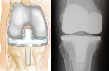
A knee prosthesis, also known as a knee replacement or artificial knee joint, is a medical device used to replace the damaged or diseased weight-bearing surfaces of the knee joint. It typically consists of three components: the femoral component (made of metal) that fits over the end of the thighbone (femur), the tibial component (often made of metal and plastic) that fits into the top of the shinbone (tibia), and a patellar component (usually made of plastic) that replaces the damaged surface of the kneecap.
The primary goal of knee prosthesis is to relieve pain, restore function, and improve quality of life for individuals with advanced knee joint damage due to conditions such as osteoarthritis, rheumatoid arthritis, or traumatic injuries. The procedure to implant a knee prosthesis is called knee replacement surgery or total knee arthroplasty (TKA).
Arthroplasty, replacement, knee is a surgical procedure where the damaged or diseased joint surface of the knee is removed and replaced with an artificial joint or prosthesis. The procedure involves resurfacing the worn-out ends of the femur (thigh bone) and tibia (shin bone) with metal components, and the back of the kneecap with a plastic button. This surgery is usually performed to relieve pain and restore function in patients with severe knee osteoarthritis, rheumatoid arthritis, or traumatic injuries that have damaged the joint beyond repair. The goal of knee replacement surgery is to improve mobility, reduce pain, and enhance the quality of life for the patient.
I'm sorry for any confusion, but "Prochloron" does not have a widely accepted medical definition in the context of human health or diseases. It is a genus name in prokaryotic taxonomy, referring to a group of cyanobacteria (blue-green algae) that are capable of photosynthesis and contain chlorophyll a and b, similar to plants. They are often found as symbionts in marine animals, particularly in certain species of ascidians (sea squirts). If you have any questions related to healthcare, medicine, or biology, please feel free to ask!
Osteoarthritis (OA) of the knee is a degenerative joint disease that affects the articular cartilage and subchondral bone in the knee joint. It is characterized by the breakdown and eventual loss of the smooth, cushioning cartilage that covers the ends of bones and allows for easy movement within joints. As the cartilage wears away, the bones rub against each other, causing pain, stiffness, and limited mobility. Osteoarthritis of the knee can also lead to the formation of bone spurs (osteophytes) and cysts in the joint. This condition is most commonly found in older adults, but it can also occur in younger people as a result of injury or overuse. Risk factors include obesity, family history, previous joint injuries, and repetitive stress on the knee joint. Treatment options typically include pain management, physical therapy, and in some cases, surgery.
Joint instability is a condition characterized by the loss of normal joint function and increased risk of joint injury due to impaired integrity of the supporting structures, such as ligaments, muscles, or cartilage. This can result in excessive movement or laxity within the joint, leading to decreased stability and increased susceptibility to dislocations or subluxations. Joint instability may cause pain, swelling, and limited range of motion, and it can significantly impact a person's mobility and quality of life. It is often caused by trauma, degenerative conditions, or congenital abnormalities and may require medical intervention, such as physical therapy, bracing, or surgery, to restore joint stability.
The femur is the medical term for the thigh bone, which is the longest and strongest bone in the human body. It connects the hip bone to the knee joint and plays a crucial role in supporting the weight of the body and allowing movement during activities such as walking, running, and jumping. The femur is composed of a rounded head, a long shaft, and two condyles at the lower end that articulate with the tibia and patella to form the knee joint.
The tibia, also known as the shin bone, is the larger of the two bones in the lower leg and part of the knee joint. It supports most of the body's weight and is a major insertion point for muscles that flex the foot and bend the leg. The tibia articulates with the femur at the knee joint and with the fibula and talus bone at the ankle joint. Injuries to the tibia, such as fractures, are common in sports and other activities that put stress on the lower leg.
Articular Range of Motion (AROM) is a term used in physiotherapy and orthopedics to describe the amount of movement available in a joint, measured in degrees of a circle. It refers to the range through which synovial joints can actively move without causing pain or injury. AROM is assessed by measuring the degree of motion achieved by active muscle contraction, as opposed to passive range of motion (PROM), where the movement is generated by an external force.
Assessment of AROM is important in evaluating a patient's functional ability and progress, planning treatment interventions, and determining return to normal activities or sports participation. It is also used to identify any restrictions in joint mobility that may be due to injury, disease, or surgery, and to monitor the effectiveness of rehabilitation programs.
Patellofemoral Pain Syndrome (PFPS) is a broad term used to describe pain arising from the front of the knee, specifically where the patella (kneecap) meets the femur (thigh bone). It is often described as a diffuse, aching pain in the anterior knee, typically worsening with activities that load the patellofemoral joint such as climbing stairs, running, jumping or prolonged sitting.
PFPS can be caused by various factors including overuse, muscle imbalances, poor biomechanics, or abnormal tracking of the patella. Treatment usually involves a combination of physical therapy to improve strength and flexibility, activity modification, and sometimes bracing or orthotics for better alignment.
Arthralgia is a medical term that refers to pain in the joints. It does not involve inflammation, which would be referred to as arthritis. The pain can range from mild to severe and may occur in one or multiple joints. Arthralgia can have various causes, including injuries, infections, degenerative conditions, or systemic diseases. In some cases, the underlying cause of arthralgia remains unknown. Treatment typically focuses on managing the pain and addressing the underlying condition if it can be identified.
Osteochondritis is a joint condition where a piece of cartilage or bone in the joint separates from its attachment due to a lack of blood supply. This can cause pain, stiffness, and potentially restricted movement in the affected joint. It often occurs in weight-bearing joints like the knee or ankle, and is more common in children and adolescents. The separated piece may sometimes float around in the joint space, causing further damage to the cartilage and bone. If left untreated, it can lead to long-term joint problems. Also known as osteochondrosis or osteochondritis dissecans.
Knee injuries refer to damages or harm caused to the structures surrounding or within the knee joint, which may include the bones (femur, tibia, and patella), cartilage (meniscus and articular cartilage), ligaments (ACL, PCL, MCL, and LCL), tendons (patellar and quadriceps), muscles, bursae, and other soft tissues. These injuries can result from various causes, such as trauma, overuse, degeneration, or sports-related activities. Symptoms may include pain, swelling, stiffness, instability, reduced range of motion, and difficulty walking or bearing weight on the affected knee. Common knee injuries include fractures, dislocations, meniscal tears, ligament sprains or ruptures, and tendonitis. Proper diagnosis and treatment are crucial to ensure optimal recovery and prevent long-term complications.
The Quadriceps muscle, also known as the Quadriceps Femoris, is a large muscle group located in the front of the thigh. It consists of four individual muscles - the Rectus Femoris, Vastus Lateralis, Vastus Intermedius, and Vastus Medialis. These muscles work together to extend the leg at the knee joint and flex the thigh at the hip joint. The Quadriceps muscle is crucial for activities such as walking, running, jumping, and kicking.
I'm not aware of a medical term called "bone wires." The term "wiring" is used in orthopedic surgery to describe the use of metal wire to hold bones or fractures in place during healing. However, I couldn't find any specific medical definition or term related to "bone wires." It may be a colloquialism, a term used in a specific context, or a term from science fiction. If you could provide more context about where you encountered this term, I might be able to give a more accurate answer.
In medical terms, the knee is referred to as the largest and one of the most complex joints in the human body. It is a hinge joint that connects the thigh bone (femur) to the shin bones (tibia and fibula), enabling movements like flexion, extension, and a small amount of rotation. The knee also contains several other components such as menisci, ligaments, tendons, and bursae, which provide stability, cushioning, and protection during movement.
Mollusca is not a medical term per se, but a major group of invertebrate animals that includes snails, clams, octopuses, and squids. However, medically, some mollusks can be relevant as they can act as vectors for various diseases, such as schistosomiasis (transmitted by freshwater snails) and fascioliasis (transmitted by aquatic snails). Therefore, a medical definition might describe Mollusca as a phylum of mostly marine invertebrates that can sometimes play a role in the transmission of certain infectious diseases.
Joint diseases is a broad term that refers to various conditions affecting the joints, including but not limited to:
1. Osteoarthritis (OA): A degenerative joint disease characterized by the breakdown of cartilage and underlying bone, leading to pain, stiffness, and potential loss of function.
2. Rheumatoid Arthritis (RA): An autoimmune disorder causing inflammation in the synovial membrane lining the joints, resulting in swelling, pain, and joint damage if left untreated.
3. Infectious Arthritis: Joint inflammation caused by bacterial, viral, or fungal infections that spread through the bloodstream or directly enter the joint space.
4. Gout: A type of arthritis resulting from the buildup of uric acid crystals in the joints, typically affecting the big toe and characterized by sudden attacks of severe pain, redness, and swelling.
5. Psoriatic Arthritis (PsA): An inflammatory joint disease associated with psoriasis, causing symptoms such as pain, stiffness, and swelling in the joints and surrounding tissues.
6. Juvenile Idiopathic Arthritis (JIA): A group of chronic arthritis conditions affecting children, characterized by joint inflammation, pain, and stiffness.
7. Ankylosing Spondylitis: A form of arthritis primarily affecting the spine, causing inflammation, pain, and potential fusion of spinal vertebrae.
8. Bursitis: Inflammation of the fluid-filled sacs (bursae) that cushion joints, leading to pain and swelling.
9. Tendinitis: Inflammation or degeneration of tendons, which connect muscles to bones, often resulting in pain and stiffness near joints.
These conditions can impact the function and mobility of affected joints, causing discomfort and limiting daily activities. Proper diagnosis and treatment are essential for managing joint diseases and preserving joint health.
Anatomic landmarks are specific, identifiable structures or features on the body that are used as references in medicine and surgery. These landmarks can include bones, muscles, joints, or other visible or palpable features that help healthcare professionals identify specific locations, orient themselves during procedures, or measure changes in the body.
Examples of anatomic landmarks include:
* The anterior iliac spine, a bony prominence on the front of the pelvis that can be used to locate the hip joint.
* The cubital fossa, a depression at the elbow where the median nerve and brachial artery can be palpated.
* The navel (umbilicus), which serves as a reference point for measuring distances in the abdomen.
* The xiphoid process, a small piece of cartilage at the bottom of the breastbone that can be used to locate the heart and other structures in the chest.
Anatomic landmarks are important for accurate diagnosis, treatment planning, and surgical procedures, as they provide reliable and consistent reference points that can help ensure safe and effective care.
Biomechanics is the application of mechanical laws to living structures and systems, particularly in the field of medicine and healthcare. A biomechanical phenomenon refers to a observable event or occurrence that involves the interaction of biological tissues or systems with mechanical forces. These phenomena can be studied at various levels, from the molecular and cellular level to the tissue, organ, and whole-body level.
Examples of biomechanical phenomena include:
1. The way that bones and muscles work together to produce movement (known as joint kinematics).
2. The mechanical behavior of biological tissues such as bone, cartilage, tendons, and ligaments under various loads and stresses.
3. The response of cells and tissues to mechanical stimuli, such as the way that bone tissue adapts to changes in loading conditions (known as Wolff's law).
4. The biomechanics of injury and disease processes, such as the mechanisms of joint injury or the development of osteoarthritis.
5. The use of mechanical devices and interventions to treat medical conditions, such as orthopedic implants or assistive devices for mobility impairments.
Understanding biomechanical phenomena is essential for developing effective treatments and prevention strategies for a wide range of medical conditions, from musculoskeletal injuries to neurological disorders.
In the context of medicine, "lead" most commonly refers to lead exposure or lead poisoning. Lead is a heavy metal that can be harmful to the human body, even at low levels. It can enter the body through contaminated air, water, food, or soil, and it can also be absorbed through the skin.
Lead poisoning occurs when lead builds up in the body over time, causing damage to the brain, nervous system, red blood cells, and kidneys. Symptoms of lead poisoning may include abdominal pain, constipation, fatigue, headache, irritability, memory problems, and in severe cases, seizures, coma, or even death.
Lead exposure is particularly dangerous for children, as their developing bodies are more sensitive to the harmful effects of lead. Even low levels of lead exposure can cause learning disabilities, behavioral problems, and developmental delays in children. Therefore, it's important to minimize lead exposure and seek medical attention if lead poisoning is suspected.
A cadaver is a deceased body that is used for medical research or education. In the field of medicine, cadavers are often used in anatomy lessons, surgical training, and other forms of medical research. The use of cadavers allows medical professionals to gain a deeper understanding of the human body and its various systems without causing harm to living subjects. Cadavers may be donated to medical schools or obtained through other means, such as through consent of the deceased or their next of kin. It is important to handle and treat cadavers with respect and dignity, as they were once living individuals who deserve to be treated with care even in death.
A tendon is the strong, flexible band of tissue that connects muscle to bone. It helps transfer the force produced by the muscle to allow various movements of our body parts. Tendons are made up of collagen fibers arranged in parallel bundles and have a poor blood supply, making them prone to injuries and slow to heal. Examples include the Achilles tendon, which connects the calf muscle to the heel bone, and the patellar tendon, which connects the kneecap to the shinbone.
Bone malalignment is a term used to describe the abnormal alignment or positioning of bones in relation to each other. This condition can occur as a result of injury, deformity, surgery, or disease processes that affect the bones and joints. Bone malalignment can cause pain, stiffness, limited mobility, and an increased risk of further injury. In some cases, bone malalignment may require treatment such as bracing, physical therapy, or surgery to correct the alignment and improve function.
Medical Definition:
Magnetic Resonance Imaging (MRI) is a non-invasive diagnostic imaging technique that uses a strong magnetic field and radio waves to create detailed cross-sectional or three-dimensional images of the internal structures of the body. The patient lies within a large, cylindrical magnet, and the scanner detects changes in the direction of the magnetic field caused by protons in the body. These changes are then converted into detailed images that help medical professionals to diagnose and monitor various medical conditions, such as tumors, injuries, or diseases affecting the brain, spinal cord, heart, blood vessels, joints, and other internal organs. MRI does not use radiation like computed tomography (CT) scans.
Arthroscopy is a minimally invasive surgical procedure where an orthopedic surgeon uses an arthroscope (a thin tube with a light and camera on the end) to diagnose and treat problems inside a joint. The surgeon makes a small incision, inserts the arthroscope into the joint, and then uses the attached camera to view the inside of the joint on a monitor. They can then insert other small instruments through additional incisions to repair or remove damaged tissue.
Arthroscopy is most commonly used for joints such as the knee, shoulder, hip, ankle, and wrist. It offers several advantages over traditional open surgery, including smaller incisions, less pain and bleeding, faster recovery time, and reduced risk of infection. The procedure can be used to diagnose and treat a wide range of conditions, including torn ligaments or cartilage, inflamed synovial tissue, loose bone or cartilage fragments, and joint damage caused by arthritis.
Developmental bone diseases are a group of medical conditions that affect the growth and development of bones. These diseases are present at birth or develop during childhood and adolescence, when bones are growing rapidly. They can result from genetic mutations, hormonal imbalances, or environmental factors such as poor nutrition.
Some examples of developmental bone diseases include:
1. Osteogenesis imperfecta (OI): Also known as brittle bone disease, OI is a genetic disorder that affects the body's production of collagen, a protein necessary for healthy bones. People with OI have fragile bones that break easily and may also experience other symptoms such as blue sclerae (whites of the eyes), hearing loss, and joint laxity.
2. Achondroplasia: This is the most common form of dwarfism, caused by a genetic mutation that affects bone growth. People with achondroplasia have short limbs and a large head relative to their body size.
3. Rickets: A condition caused by vitamin D deficiency or an inability to absorb or use vitamin D properly. This leads to weak, soft bones that can bow or bend easily, particularly in children.
4. Fibrous dysplasia: A rare bone disorder where normal bone is replaced with fibrous tissue, leading to weakened bones and deformities.
5. Scoliosis: An abnormal curvature of the spine that can develop during childhood or adolescence. While not strictly a developmental bone disease, scoliosis can be caused by various underlying conditions such as cerebral palsy, muscular dystrophy, or spina bifida.
Treatment for developmental bone diseases varies depending on the specific condition and its severity. Treatment may include medication, physical therapy, bracing, or surgery to correct deformities and improve function. Regular follow-up with a healthcare provider is essential to monitor growth, manage symptoms, and prevent complications.
 Patella - Wikipedia
 Chondromalacia patella: Causes, treatment, and more
 Patella Fractures: Practice Essentials, Anatomy, Etiology
 Patella (Kneecap): Anatomy and Function
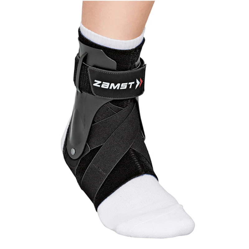
 AliMed Open Patella Brace
 'patella crescenda' a sprouting dish that grows fresh...
 Fundraiser by Lisa Tuttle : Luxating Patella Surgery
 Chondromalacia patellae (thing) by baritalia - Everything2.com
 Tension Band for Patella Frx : Wheeless' Textbook of Orthopaedics
 Patella tendonitis
 Actimove Adjustable Patella Strap Knee Support - Universal Black
 Automatic analysis of morphological parameters of the patella based on three-dimensional (3D) surface data
 Patellaplasty, as an Alternative to Replacing Patella in Total Knee Arthroplasty
 Patella Species - Poppe Images
 CEP Mid Support Patella Strap
- Compression Store
CEP Mid Support Patella Strap
- Compression Store Open Patella Hinge-Free Knee Support | PhysioRoom
 Patella vulgata
 MarBEF Data System - ERMS - Patella lowei d'Orbigny, 1840
 Knee Sleeve with Open Patella | McDavid
 Patella Femoral Syndrome - Sports Injury Info
 Patella Name Meaning & Patella Family History at Ancestry.ca®
 Buy Precision Patella Strap online | Longsjo.com
 Patella Injuries | Texas Orthopedics
 WoRMS - World Register of Marine Species - Patella pellucida Linnaeus, 1758
 Nail-Patella Syndrome-A Novel Mutation in the LMX1B Gene - Amrita Vishwa Vidyapeetham
 Patella Brace, One size - Personnelle : Orthopedics | Jean Coutu


.jpg)