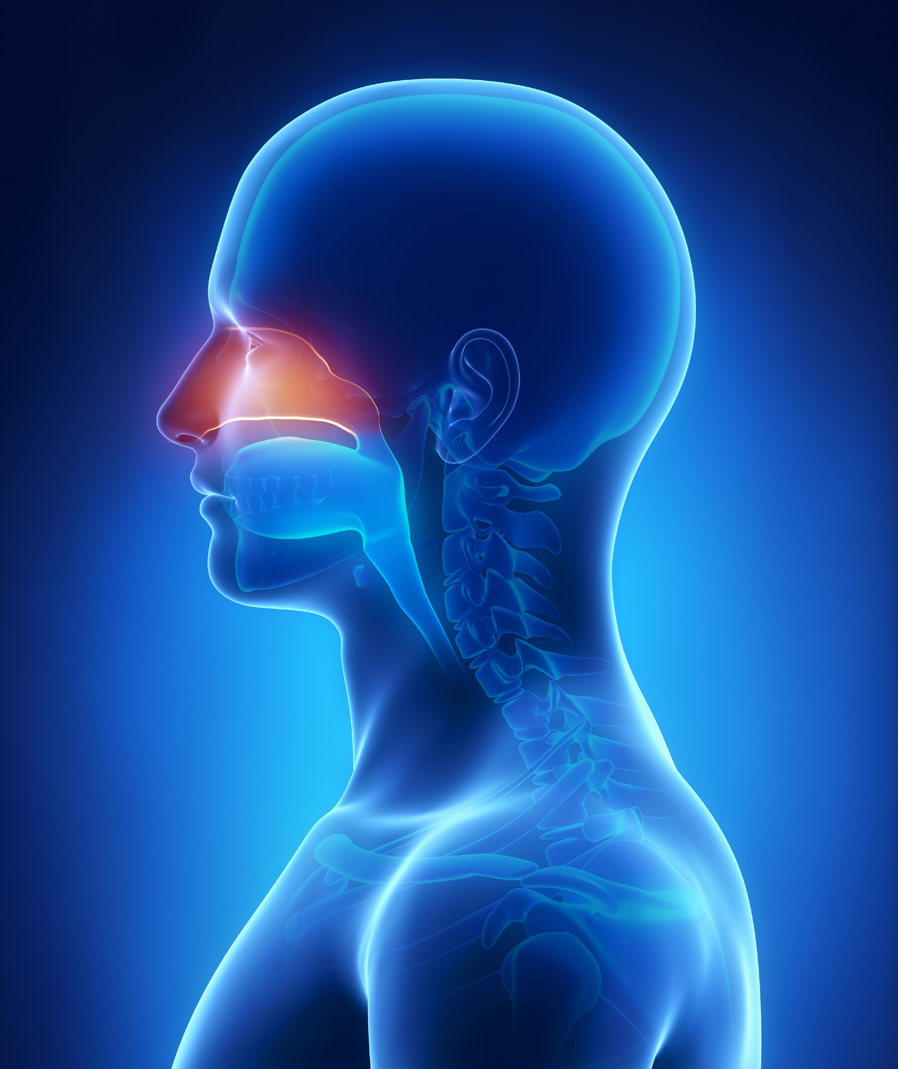
Paranasal Sinuses
Paranasal Sinus Diseases
Maxillary Sinus Neoplasms
Maxillary Sinus
Frontal Sinus
Ethmoid Sinus
Nasal Cavity
Sphenoid Sinus
Mucocele
Turbinates
Nasal Polyps
Nasal Septum
Otorhinolaryngologic Surgical Procedures
Osteoma
Tomography, X-Ray Computed
Ethmoid Bone
Sphenoid Sinusitis
Rhinometry, Acoustic
Frontal Sinusitis
Exophthalmos
Esthesioneuroblastoma, Olfactory
Endoscopy
Ethmoid Sinusitis
Cranial Sinuses
Nasal Obstruction
Tampons, Surgical
Kartagener Syndrome
Cranial Fossa, Middle
Skull Base Neoplasms
Cavernous Sinus
Papilloma, Inverted
Maxillary Sinusitis
Levocardia
Nose
Carotid Sinus
Nasal Mucosa
Wood
Sphenoid Bone
Skull Base
Mucormycosis
Coronary Sinus
Sinus Thrombosis, Intracranial
Magnetic Resonance Imaging
Fatal Outcome
Sick Sinus Syndrome
Retrospective Studies
Pneumocephalus associated with ethmoidal sinus osteoma--case report. (1/249)
A 35-year-old female suffered sudden onset of severe headache upon blowing her nose. No rhinorrhea or signs of meningeal irritation were noted. Computed tomography (CT) with bone windows clearly delineated a bony mass in the right ethmoid sinus, extending into the orbit and intracranially. Conventional CT demonstrated multiple air bubbles in the cisterns and around the mass in the right frontal skull base, suggesting that the mass was associated with entry of the air bubbles into the cranial cavity. T1- and T2-weighted magnetic resonance (MR) imaging showed a low-signal lesion that appeared to be an osteoma but did not show any air bubbles. Through a wide bilateral frontal craniotomy, the cauliflower-like osteoma was found to be protruding intracranially through the skull base and the overlying dura mater. The osteoma was removed, and the dural defect was covered with a fascia graft. Histological examination confirmed that the lesion was an osteoma. The operative procedure resolved the problem of air entry. CT is superior to MR imaging for diagnosing pneumocephalus, by providing a better assessment of bony destruction and better detection of small amounts of intracranial air. (+info)Benign expansile lesions of the sphenoid sinus: differentiation from normal asymmetry of the lateral recesses. (2/249)
BACKGROUND AND PURPOSE: There is a wide range of normal variation is sphenoid sinus development, especially in the size of the lateral recesses. The purpose of this study was to determine imaging characteristics that may help differentiate between opacification of a developmentally asymmetric lateral recess and a true expansile lesion of the sphenoid sinus. METHODS: Coronal CT was performed in seven patients with expansile or erosive benign lesions of the sphenoid sinus, and results were compared to a control population of 72 subjects with unopacified sphenoid sinuses. The degree of asymmetry of lateral recess development was assessed with particular attention to the separation of vidian's canal and the foramen rotundum (vidian-rotundum distance). The images were also examined for evidence of: erosion, defined as loss of the normal thin bony margin on at least two contiguous sections; apparent thinning of the sinus wall, defined as a focal apparent decrease in thickness again on at least two contiguous sections; and for vidian's canal or foramen rotundum rim erosion or flattening. RESULTS: Of the seven patients with expansile lesions, vidian's canal margin erosion was present in seven, unequivocal sinus expansion in three, wall erosion in three, wall thinning in three, erosion of the foramen rotundum in two, and flattening in the foramen rotundum in four. Forty-one of the 72 controls had lateral recess formation, 28 of which were asymmetric. The distance between vidian's canal and the foramen rotundum (vidian-rotundum distance) relied on the presence or absence of pneumatization, with a significantly larger distance in the presence of greater wing pneumatization. Examination of 24 controls revealed apparent thinning of the sinus wall, typically at the carotid groove, but no flattening, thinning, or erosion of the vidian canal or of the foramen rotundum. CONCLUSION: Examination of controls and patients with expansile or erosive lesions of the sphenoid sinus revealed side-to-side asymmetry in the development of the sinus and lateral recess, making subtle expansion difficult to assess. Furthermore, variability in the vidian-rotundum distance correlated with degree of pneumatization, and did not necessarily reflect expansion. Thus, in the absence of gross sinus wall erosion, flattening or erosion of the rims of vidian's canal or the foramen rotundum provides the most specific evidence of an expansile or erosive process within the sinus. (+info)Amphicrine tumor. (3/249)
The term amphicrine refers to cells, and tumors, which show both exocrine and endocrine features. Author s aim was to analyse the characteristics of these neoplasms. 40 suspicious cases were reviewed. Mucin-stains (PAS, diastase-PAS, Stains-all, Alcian-blue), immunohistochemistry (antibodies against Neuron-Specific Enolase (NSE), and Chromogranin A (CGA), and electronmicroscopic studies were performed to demonstrate exocrine and/or endocrine features of the tumor cells. By means of these methods, 16 cases turned out to be amphicrine tumors. Among them, there were 4 sinonasal, 1 bronchial, 1 mediastinal, 8 gastrointestinal and 2 suprarenal gland neoplasms. In connection to the subject, a brief review is given of amphicrine tumor, regarding its etiological and pathological aspects. These tumors form a distinct clinicopathological entity and should be separated from both neuroendocrine tumors and adenocarcinomas. (+info)Recurrent inverted papilloma: diagnosis with pharmacokinetic dynamic gadolinium-enhanced MR imaging. (4/249)
BACKGROUND AND PURPOSE: Dynamic gadolinium-enhanced MR imaging has been used successfully to identify post-treatment recurrence or postoperative changes in rectal and cervical carcinoma. Our purpose was to evaluate the usefulness of dynamic gadolinium-enhanced MR imaging for distinguishing recurrent inverted papilloma (IP) from postoperative changes. METHODS: Fifteen patients with 20 pathologically proved lesions (recurrent IP, 12; fibrosis or granulation tissue, eight) were enrolled in the study. Three observers, blinded to pathologic results, independently evaluated conventional MR images, including T1-weighted (unenhanced and postcontrast), proton-density-weighted, and T2-weighted spin-echo images. Results then were determined by consensus. Dynamic images were obtained using fast spin-echo sequences at 5, 30, 60, 90, 120, 150, 180, and 300 seconds after the injection of gadolinium-diethylene-triamine penta-acetic acid. Time-signal intensity curves of suspected lesions were analyzed by a pharmacokinetic model. The calculated amplitude and tissue distribution time were used to characterize tissue, and their values were displayed as a color-coded overlay. RESULTS: T2-weighted images yielded a sensitivity of 67%, a specificity of 75%, and an accuracy of 70% in the diagnosis of recurrent IP. Contrast-enhanced T1-weighted images yielded a sensitivity of 75%, a specificity of 50%, and an accuracy of 65%. Pharmacokinetic analysis showed that recurrent IP had faster (distribution time, 41 versus 88 seconds) and higher (amplitude, 2.4 versus 1.2 arbitrary units) enhancement than did fibrosis or granulation tissue. A cut-off of 65 seconds for distribution time and 1.6 units for amplitude yielded a sensitivity of 100% and a specificity of 100% for diagnosing recurrent IP. CONCLUSION: Dynamic MR imaging can differentiate accurately recurrent IP from postoperative changes and seems to be a valuable diagnostic tool. (+info)Inverted sinonasal papilloma : a molecular genetic appraisal of its putative status as a Precursor to squamous cell carcinoma. (5/249)
Inverted papilloma (IP) is a proliferative lesion of the epithelium lining the sinonasal tract. Although IP often recurs after surgical excision and is sometimes associated with squamous cell carcinoma of the sinonasal cavity (SNSCC), its presumed neoplastic nature and putative role as a precursor to squamous cell carcinoma have not been confirmed at the molecular genetic level. We analyzed the pattern of X chromosome inactivation in IPs from nine female patients. Inactivation of a single allele is seen in monoclonal proliferations and may be indicative of a neoplastic process. We also analyzed 28 IPs and 6 concurrent SNSCCs for loss of heterozygosity (LOH) on chromosomal arms 3p, 9p21, 11q13, 13q11, and 17p13. Losses at these loci occur frequently during neoplastic transformation of the upper respiratory tract and can be detected in squamous cell carcinomas and the progenitor lesions from which they arise. X chromosome analysis was informative in four of the nine IPs. All four lesions demonstrated a monoclonal pattern of inactivation. LOH was not detected in any nondysplastic areas from the 28 IPs, but LOH at one or more chromosomal loci was present in all six of the concurrent SNSCCs. We conclude that IPs are monoclonal proliferations, yet they do not fit the profile of a prototypic precursor lesion. Unlike squamous epithelial dysplasia, IPs do not routinely harbor several of the key genetic alterations that are associated with malignant transformation of the upper respiratory tract. (+info)Cavernous sinus syndrome associated with nonsecretory myeloma. (6/249)
The case of a 53-year-old man who developed cavernous sinus syndrome (CSS) four years after being diagnosed as having nonsecretory myeloma is described. He was admitted with diplopia and dull pain over the right infraorbital and zygomatic region in June 1997. The cause of CSS was the intracranial involvement of myeloma, which was diagnosed by fiberscopic biopsy. The results of endocrinologic evaluation were almost normal. The response to radiotherapy and chemotherapy was mild. CSS caused by nonsecretory myeloma is rare and its prognosis is poor. More aggressive chemotherapy with stem cell support may be indicated. (+info)Recurrence of clival chordoma along the surgical pathway. (7/249)
Chordomas are locally aggressive malignant tumors of notochordal origin whose metastatic potential is increasingly recognized. Surgical pathway recurrence has been noted only rarely in the literature. We present three patients with clival chordomas whose sole or initial recurrence was along the pathway of prior surgical access. A characteristic mass found along the pathway of prior surgical access for resection of a chordoma should suggest recurrent chordoma. (+info)A case report of sinonasal teratocarcinosarcoma. (8/249)
A sinonasal teratocarcinosarcoma (SNTCS) is a rare and aggressive malignant neoplasm histologically characterized by the combination of one or more epithelial elements and mesenchymal components. We report a case of a 78-year-old man with SNTCS involving the nasal cavity and paranasal sinuses. He complained of epiphora and exophthalmos with weight loss. Physical and diagnostic images resulted T4N0M0. The tumor was completely and widely resected via a trans-facial approach to perform total maxillectomy with orbital exenteration. The clinical presentation, pathologic features, and clinical course are described with a review of the literature. (+info)Paranasal sinuses are air-filled cavities in the skull that surround the nasal cavity. There are four pairs of paranasal sinuses, including the maxillary, frontal, ethmoid, and sphenoid sinuses. These sinuses help to warm, humidify, and filter the air we breathe. They also contribute to our voice resonance and provide a slight cushioning effect for the skull. The openings of the paranasal sinuses lead directly into the nasal cavity, allowing mucus produced in the sinuses to drain into the nose. Infections or inflammation of the paranasal sinuses can result in conditions such as sinusitis.
Paranasal sinus neoplasms refer to abnormal growths or tumors that develop within the paranasal sinuses, which are air-filled cavities located inside the skull near the nasal cavity. These tumors can be benign (noncancerous) or malignant (cancerous), and they can arise from various types of tissue within the sinuses, such as the lining of the sinuses (mucosa), bone, or other soft tissues.
Paranasal sinus neoplasms can cause a variety of symptoms, including nasal congestion, nosebleeds, facial pain or numbness, and visual disturbances. The diagnosis of these tumors typically involves a combination of imaging studies (such as CT or MRI scans) and biopsy to determine the type and extent of the tumor. Treatment options may include surgery, radiation therapy, chemotherapy, or a combination of these approaches, depending on the specific type and stage of the neoplasm.
Paranasal sinus diseases refer to a group of medical conditions that affect the paranasal sinuses, which are air-filled cavities located within the skull near the nasal cavity. These sinuses include the maxillary, frontal, ethmoid, and sphenoid sinuses.
Paranasal sinus diseases can be caused by a variety of factors, including viral, bacterial, or fungal infections, allergies, structural abnormalities, or autoimmune disorders. Some common paranasal sinus diseases include:
1. Sinusitis: Inflammation or infection of the sinuses, which can cause symptoms such as nasal congestion, thick nasal discharge, facial pain or pressure, and reduced sense of smell.
2. Nasal polyps: Soft, benign growths that develop in the lining of the nasal passages or sinuses, which can obstruct airflow and cause difficulty breathing through the nose.
3. Sinonasal tumors: Abnormal growths that can be benign or malignant, which can cause symptoms such as nasal congestion, facial pain, and bleeding from the nose.
4. Sinus cysts: Fluid-filled sacs that form in the sinuses, which can cause symptoms similar to those of sinusitis.
5. Fungal sinusitis: Infection of the sinuses with fungi, which can cause symptoms such as nasal congestion, facial pain, and thick, discolored mucus.
Treatment for paranasal sinus diseases depends on the underlying cause and severity of the condition. Treatment options may include medications, such as antibiotics, antihistamines, or corticosteroids, as well as surgical intervention in more severe cases.
Maxillary sinus neoplasms refer to abnormal growths or tumors that develop in the maxillary sinuses, which are located in the upper part of your cheekbones, below your eyes. These growths can be benign (non-cancerous) or malignant (cancerous).
Benign neoplasms may include conditions such as an osteoma (a benign bone tumor), a papilloma (a benign growth of the lining of the sinus), or a fibrous dysplasia (a condition where bone is replaced by fibrous tissue).
Malignant neoplasms, on the other hand, can be primary (originating in the maxillary sinuses) or secondary (spreading to the maxillary sinuses from another site in the body). Common types of malignant tumors that arise in the maxillary sinus include squamous cell carcinoma, adenocarcinoma, and mucoepidermoid carcinoma.
Symptoms of maxillary sinus neoplasms may include nasal congestion, nosebleeds, facial pain or numbness, vision changes, and difficulty swallowing or speaking. Treatment options depend on the type, size, and location of the tumor but may include surgery, radiation therapy, chemotherapy, or a combination of these approaches.
The maxillary sinuses, also known as the antrums of Highmore, are the largest of the four pairs of paranasal sinuses located in the maxilla bones. They are air-filled cavities that surround the nasolacrimal duct and are situated superior to the upper teeth and lateral to the nasal cavity. Each maxillary sinus is lined with a mucous membrane, which helps to warm, humidify, and filter the air we breathe. Inflammation or infection of the maxillary sinuses can result in conditions such as sinusitis, leading to symptoms like facial pain, headaches, and nasal congestion.
A frontal sinus is a paired, air-filled paranasal sinus located in the frontal bone of the skull, above the eyes and behind the forehead. It is one of the four pairs of sinuses found in the human head. The frontal sinuses are lined with mucous membrane and are interconnected with the nasal cavity through small openings called ostia. They help to warm, humidify, and filter the air we breathe, and contribute to the resonance of our voice. Variations in size, shape, and asymmetry of frontal sinuses are common among individuals.
The ethmoid sinuses are a pair of air-filled spaces located in the ethmoid bone, which is a part of the skull that forms the upper portion of the nasal cavity and the inner eye socket. These sinuses are divided into anterior and posterior groups and are present in adults, but not at birth. They continue to grow and develop until early adulthood.
The ethmoid sinuses are lined with mucous membrane, which helps to warm, humidify, and filter the air we breathe. They are surrounded by a network of blood vessels and nerves, making them susceptible to inflammation and infection. Inflammation of the ethmoid sinuses can lead to conditions such as sinusitis, which can cause symptoms such as nasal congestion, headache, and facial pain.
Sinusitis, also known as rhinosinusitis, is a medical condition characterized by inflammation of the paranasal sinuses, which are air-filled cavities located within the skull near the nose. The inflammation can be caused by viral, bacterial, or fungal infections, as well as allergies, structural issues, or autoimmune disorders.
In sinusitis, the mucous membranes lining the sinuses become swollen and may produce excess mucus, leading to symptoms such as nasal congestion, thick green or yellow nasal discharge, facial pain or pressure, reduced sense of smell, cough, fatigue, and fever.
Sinusitis can be classified into acute (lasting less than 4 weeks), subacute (lasting 4-12 weeks), chronic (lasting more than 12 weeks), or recurrent (multiple episodes within a year). Treatment options depend on the underlying cause and severity of symptoms, and may include antibiotics, nasal corticosteroids, decongestants, saline irrigation, and in some cases, surgery.
Nose neoplasms refer to abnormal growths or tumors in the nasal cavity or paranasal sinuses. These growths can be benign (non-cancerous) or malignant (cancerous). Benign neoplasms are typically slow-growing and do not spread to other parts of the body, while malignant neoplasms can invade surrounding tissues and have the potential to metastasize.
Nose neoplasms can cause various symptoms such as nasal congestion, nosebleeds, difficulty breathing through the nose, loss of smell, facial pain or numbness, and visual changes if they affect the eye. The diagnosis of nose neoplasms usually involves a combination of physical examination, imaging studies (such as CT or MRI scans), and biopsy to determine the type and extent of the growth. Treatment options depend on the type, size, location, and stage of the neoplasm and may include surgery, radiation therapy, chemotherapy, or a combination of these approaches.
The nasal cavity is the air-filled space located behind the nose, which is divided into two halves by the nasal septum. It is lined with mucous membrane and is responsible for several functions including respiration, filtration, humidification, and olfaction (smell). The nasal cavity serves as an important part of the upper respiratory tract, extending from the nares (nostrils) to the choanae (posterior openings of the nasal cavity that lead into the pharynx). It contains specialized structures such as turbinate bones, which help to warm, humidify and filter incoming air.
The sphenoid sinuses are air-filled spaces located within the sphenoid bone, which is one of the bones that make up the skull base. These sinuses are located deep inside the skull, behind the eyes and nasal cavity. They are paired and separated by a thin bony septum, and each one opens into the corresponding nasal cavity through a small opening called the sphenoethmoidal recess. The sphenoid sinuses vary greatly in size and shape between individuals. They develop during childhood and continue to grow until early adulthood. The function of the sphenoid sinuses, like other paranasal sinuses, is not entirely clear, but they may contribute to reducing the weight of the skull, resonating voice during speech, and insulating the brain from trauma.
A mucocele is a mucus-containing cystic lesion that results from the accumulation of mucin within a damaged minor salivary gland duct or mucous gland. It is typically caused by trauma, injury, or blockage of the duct. Mucocele appears as a round, dome-shaped, fluid-filled swelling, which may be bluish or clear in color. They are most commonly found on the lower lip but can also occur on other areas of the oral cavity. Mucocele is generally painless unless it becomes secondarily infected; however, it can cause discomfort during speaking, chewing, or swallowing, and may affect aesthetics. Treatment usually involves surgical excision of the mucocele to prevent recurrence.
In medical terms, turbinates refer to the curled bone shelves that are present inside the nasal passages. They are covered by a mucous membrane and are responsible for warming, humidifying, and filtering the air that we breathe in through our nose. There are three pairs of turbinates in each nasal passage: inferior, middle, and superior turbinates. The inferior turbinate is the largest and most significant contributor to nasal airflow resistance. Inflammation or enlargement of the turbinates can lead to nasal congestion and difficulty breathing through the nose.
Nasal polyps are benign (noncancerous) growths that originate from the lining of your nasal passages or sinuses. They most often occur in the area where the sinuses open into the nasal cavity. Small nasal polyps may not cause any problems. But if they grow large enough, they can block your nasal passages and lead to breathing issues, frequent infections and loss of smell.
Nasal polyps are associated with chronic inflammation due to conditions such as asthma, allergic rhinitis or chronic sinusitis. Treatment typically includes medication to reduce the size of the polyps or surgery to remove them. Even after successful treatment, nasal polyps often return.
The nasal septum is the thin, flat wall of bone and cartilage that separates the two sides (nostrils) of the nose. Its primary function is to support the structures of the nose, divide the nostrils, and regulate airflow into the nasal passages. The nasal septum should be relatively centered, but it's not uncommon for a deviated septum to occur, where the septum is displaced to one side, which can sometimes cause blockage or breathing difficulties in the more affected nostril.
Otorhinolaryngologic surgical procedures are surgeries that are performed on the head and neck region, specifically involving the ear, nose, and throat (ENT) regions. This field is also known as otolaryngology-head and neck surgery. The procedures can range from relatively minor ones, such as removing a small nasal polyp or inserting ear tubes, to more complex surgeries like cochlear implantation, endoscopic sinus surgery, or removal of tumors in the head and neck region. These surgical procedures are typically performed by specialized physicians called otorhinolaryngologists (also known as ENT surgeons) who have completed extensive training in this area.
Osteoma is a benign (noncancerous) tumor that is made up of mature bone tissue. It usually grows slowly over a period of years and is most commonly found in the skull or jaw, although it can occur in other bones of the body as well. Osteomas are typically small, but they can grow to be several centimeters in size. They may cause symptoms if they press on nearby tissues or structures, such as nerves or blood vessels. In some cases, osteomas may not cause any symptoms and may only be discovered during routine imaging studies. Treatment for osteoma is typically not necessary unless it is causing problems or growing rapidly. If treatment is needed, it may involve surgical removal of the tumor.
X-ray computed tomography (CT or CAT scan) is a medical imaging method that uses computer-processed combinations of many X-ray images taken from different angles to produce cross-sectional (tomographic) images (virtual "slices") of the body. These cross-sectional images can then be used to display detailed internal views of organs, bones, and soft tissues in the body.
The term "computed tomography" is used instead of "CT scan" or "CAT scan" because the machines take a series of X-ray measurements from different angles around the body and then use a computer to process these data to create detailed images of internal structures within the body.
CT scanning is a noninvasive, painless medical test that helps physicians diagnose and treat medical conditions. CT imaging provides detailed information about many types of tissue including lung, bone, soft tissue and blood vessels. CT examinations can be performed on every part of the body for a variety of reasons including diagnosis, surgical planning, and monitoring of therapeutic responses.
In computed tomography (CT), an X-ray source and detector rotate around the patient, measuring the X-ray attenuation at many different angles. A computer uses this data to construct a cross-sectional image by the process of reconstruction. This technique is called "tomography". The term "computed" refers to the use of a computer to reconstruct the images.
CT has become an important tool in medical imaging and diagnosis, allowing radiologists and other physicians to view detailed internal images of the body. It can help identify many different medical conditions including cancer, heart disease, lung nodules, liver tumors, and internal injuries from trauma. CT is also commonly used for guiding biopsies and other minimally invasive procedures.
In summary, X-ray computed tomography (CT or CAT scan) is a medical imaging technique that uses computer-processed combinations of many X-ray images taken from different angles to produce cross-sectional images of the body. It provides detailed internal views of organs, bones, and soft tissues in the body, allowing physicians to diagnose and treat medical conditions.
The ethmoid bone is a paired, thin, and lightweight bone that forms part of the skull's anterior cranial fossa and contributes to the formation of the orbit and nasal cavity. It is located between the frontal bone above and the maxilla and palatine bones below. The ethmoid bone has several important features:
1. Cribriform plate: This is the horizontal, sieve-like portion that forms part of the anterior cranial fossa and serves as the roof of the nasal cavity. It contains small openings (foramina) through which olfactory nerves pass.
2. Perpendicular plate: The perpendicular plate is a vertical structure that projects downward from the cribriform plate, forming part of the nasal septum and separating the left and right nasal cavities.
3. Superior and middle nasal conchae: These are curved bony projections within the lateral walls of the nasal cavity that help to warm, humidify, and filter incoming air.
4. Lacrimal bone: The ethmoid bone articulates with the lacrimal bone, forming part of the medial wall of the orbit.
5. Frontal process: This is a thin, vertical plate that articulates with the frontal bone above the orbit.
6. Sphenoidal process: The sphenoidal process connects the ethmoid bone to the sphenoid bone posteriorly.
The ethmoid bone plays a crucial role in protecting the brain and providing structural support for the eyes, as well as facilitating respiration by warming, humidifying, and filtering incoming air.
Sphenoid sinusitis is a medical condition characterized by the inflammation or infection of the sphenoid sinuses, which are air-filled cavities located in the sphenoid bone at the center of the skull base, behind the eyes. These sinuses are relatively small and difficult to access, making infections less common than in other sinuses. However, when sphenoid sinusitis does occur, it can cause various symptoms such as headaches, facial pain, nasal congestion, fever, and vision problems. Sphenoid sinusitis may result from bacterial or fungal infections, allergies, or autoimmune disorders. Diagnosis typically involves a combination of clinical evaluation, imaging studies like CT scans, and sometimes endoscopic examination. Treatment options include antibiotics for bacterial infections, antifungal medications for fungal infections, nasal sprays, decongestants, pain relievers, and, in severe or recurrent cases, surgical intervention.
Rhinitis is a medical condition characterized by inflammation and irritation of the nasal passages, leading to symptoms such as sneezing, runny nose, congestion, and postnasal drip. It can be caused by various factors, including allergies (such as pollen, dust mites, or pet dander), infections (viral or bacterial), environmental irritants (such as smoke or pollution), and hormonal changes. Depending on the cause, rhinitis can be classified as allergic rhinitis, non-allergic rhinitis, infectious rhinitis, or hormonal rhinitis. Treatment options vary depending on the underlying cause but may include medications such as antihistamines, decongestants, nasal sprays, and immunotherapy (allergy shots).
Acoustic rhinometry is a diagnostic technique used to measure the cross-sectional area and volume of the nasal cavity. It utilizes sound waves to create a visual representation of the nasal passages' shape and size. By measuring the reflection of sound waves as they travel through the nasal cavity, acoustic rhinometry can help identify any abnormalities or obstructions in the nasal passage that may be causing difficulty breathing through the nose. This technique is non-invasive and quick, making it a useful tool for evaluating nasal airflow and diagnosing conditions such as nasal congestion, sinusitis, and nasal polyps.
Frontal sinusitis is a type of sinus infection that specifically involves the frontal sinuses, which are located in the forehead region above the eyes. The condition is characterized by inflammation and infection of the mucous membrane lining the frontal sinuses, leading to symptoms such as headaches, facial pain or pressure, nasal congestion, and thick nasal discharge.
Frontal sinusitis can be caused by viral, bacterial, or fungal infections, as well as structural issues like nasal polyps or deviated septum that obstruct the sinus drainage pathways. Treatment options for frontal sinitis may include antibiotics, nasal decongestants, corticosteroids, saline nasal irrigation, and in some cases, endoscopic sinus surgery to alleviate obstructions and improve sinus drainage.
Epistaxis is the medical term for nosebleed. It refers to the bleeding from the nostrils or nasal cavity, which can be caused by various factors such as dryness, trauma, inflammation, high blood pressure, or use of blood-thinning medications. Nosebleeds can range from minor nuisances to potentially life-threatening emergencies, depending on the severity and underlying cause. If you are experiencing a nosebleed that does not stop after 20 minutes of applying direct pressure, or if you are coughing up or vomiting blood, seek medical attention immediately.
Nose diseases, also known as rhinologic disorders, refer to a wide range of conditions that affect the nose and its surrounding structures. These may include:
1. Nasal Allergies (Allergic Rhinitis): An inflammation of the inner lining of the nose caused by an allergic reaction to substances such as pollen, dust mites, or mold.
2. Sinusitis: Inflammation or infection of the sinuses, which are air-filled cavities in the skull that surround the nasal cavity.
3. Nasal Polyps: Soft, fleshy growths that develop on the lining of the nasal passages or sinuses.
4. Deviated Septum: A condition where the thin wall (septum) between the two nostrils is displaced to one side, causing difficulty breathing through the nose.
5. Rhinitis Medicamentosa: Nasal congestion caused by overuse of decongestant nasal sprays.
6. Nosebleeds (Epistaxis): Bleeding from the nostrils, which can be caused by a variety of factors including dryness, trauma, or underlying medical conditions.
7. Nasal Fractures: Breaks in the bone structure of the nose, often caused by trauma.
8. Tumors: Abnormal growths that can occur in the nasal passages or sinuses. These can be benign or malignant.
9. Choanal Atresia: A congenital condition where the back of the nasal passage is blocked, often by a thin membrane or bony partition.
10. Nasal Valve Collapse: A condition where the side walls of the nose collapse inward during breathing, causing difficulty breathing through the nose.
These are just a few examples of the many diseases that can affect the nose.
Orbital neoplasms refer to abnormal growths or tumors that develop in the orbit, which is the bony cavity that contains the eyeball, muscles, nerves, fat, and blood vessels. These neoplasms can be benign (non-cancerous) or malignant (cancerous), and they can arise from various types of cells within the orbit.
Orbital neoplasms can cause a variety of symptoms depending on their size, location, and rate of growth. Common symptoms include protrusion or displacement of the eyeball, double vision, limited eye movement, pain, swelling, and numbness in the face. In some cases, orbital neoplasms may not cause any noticeable symptoms, especially if they are small and slow-growing.
There are many different types of orbital neoplasms, including:
1. Optic nerve glioma: a rare tumor that arises from the optic nerve's supportive tissue.
2. Orbital meningioma: a tumor that originates from the membranes covering the brain and extends into the orbit.
3. Lacrimal gland tumors: benign or malignant growths that develop in the lacrimal gland, which produces tears.
4. Orbital lymphangioma: a non-cancerous tumor that arises from the lymphatic vessels in the orbit.
5. Rhabdomyosarcoma: a malignant tumor that develops from the skeletal muscle cells in the orbit.
6. Metastatic tumors: cancerous growths that spread to the orbit from other parts of the body, such as the breast, lung, or prostate.
The diagnosis and treatment of orbital neoplasms depend on several factors, including the type, size, location, and extent of the tumor. Imaging tests, such as CT scans and MRI, are often used to visualize the tumor and determine its extent. A biopsy may also be performed to confirm the diagnosis and determine the tumor's type and grade. Treatment options include surgery, radiation therapy, chemotherapy, or a combination of these approaches.
Exophthalmos is a medical condition that refers to the abnormal protrusion or bulging of one or both eyes beyond the normal orbit (eye socket). This condition is also known as proptosis. Exophthalmos can be caused by various factors, including thyroid eye disease (Graves' ophthalmopathy), tumors, inflammation, trauma, or congenital abnormalities. It can lead to various symptoms such as double vision, eye discomfort, redness, and difficulty closing the eyes. Treatment of exophthalmos depends on the underlying cause and may include medications, surgery, or radiation therapy.
In medical terms, the orbit refers to the bony cavity or socket in the skull that contains and protects the eye (eyeball) and its associated structures, including muscles, nerves, blood vessels, fat, and the lacrimal gland. The orbit is made up of several bones: the frontal bone, sphenoid bone, zygomatic bone, maxilla bone, and palatine bone. These bones form a pyramid-like shape that provides protection for the eye while also allowing for a range of movements.
Acquired nose deformities refer to structural changes or abnormalities in the shape of the nose that occur after birth, as opposed to congenital deformities which are present at birth. These deformities can result from various factors such as trauma, injury, infection, tumors, or surgical procedures. Depending on the severity and cause of the deformity, it may affect both the aesthetic appearance and functionality of the nose, potentially causing difficulty in breathing, sinus problems, or sleep apnea. Treatment options for acquired nose deformities may include minimally invasive procedures, such as fillers or laser surgery, or more extensive surgical interventions, such as rhinoplasty or septoplasty, to restore both form and function to the nose.
Esthesioneuroblastoma, also known as olfactory neuroblastoma, is a rare type of malignant tumor that develops in the upper part of the nasal cavity, near the area responsible for the sense of smell (olfaction). It arises from the olfactory nerve cells and typically affects adults between 20 to 50 years old, although it can occur at any age.
Esthesioneuroblastomas are characterized by their aggressive growth and potential to spread to other parts of the head and neck, as well as distant organs such as the lungs, bones, and bone marrow. Symptoms may include nasal congestion, nosebleeds, loss of smell, facial pain or numbness, bulging eyes, and visual disturbances.
Diagnosis is usually made through a combination of clinical examination, imaging studies (such as MRI or CT scans), and biopsy. Treatment typically involves surgical resection of the tumor, followed by radiation therapy and/or chemotherapy to reduce the risk of recurrence. Regular follow-up care is essential due to the possibility of late relapse.
Overall, prognosis varies depending on factors such as the stage of the disease at diagnosis, the patient's age, and the effectiveness of treatment. While some individuals may experience long-term survival or even cure, others may face more aggressive tumor behavior and a higher risk of recurrence.
Endoscopy is a medical procedure that involves the use of an endoscope, which is a flexible tube with a light and camera at the end, to examine the interior of a body cavity or organ. The endoscope is inserted through a natural opening in the body, such as the mouth or anus, or through a small incision. The images captured by the camera are transmitted to a monitor, allowing the physician to visualize the internal structures and detect any abnormalities, such as inflammation, ulcers, or tumors. Endoscopy can also be used for diagnostic purposes, such as taking tissue samples for biopsy, or for therapeutic purposes, such as removing polyps or performing minimally invasive surgeries.
The skull is the bony structure that encloses and protects the brain, the eyes, and the ears. It is composed of two main parts: the cranium, which contains the brain, and the facial bones. The cranium is made up of several fused flat bones, while the facial bones include the upper jaw (maxilla), lower jaw (mandible), cheekbones, nose bones, and eye sockets (orbits).
The skull also provides attachment points for various muscles that control chewing, moving the head, and facial expressions. Additionally, it contains openings for blood vessels, nerves, and the spinal cord to pass through. The skull's primary function is to protect the delicate and vital structures within it from injury and trauma.
Ethmoid sinusitis is a medical condition that refers to the inflammation or infection of the ethmoid sinuses. The ethmoid sinuses are a pair of small, air-filled cavities located in the upper part of the nasal cavity, near the eyes. They are surrounded by delicate bone structures and are connected to the nasal cavity by narrow channels.
Ethmoid sinusitis can occur as a result of a viral, bacterial, or fungal infection, or it may be caused by allergies, environmental factors, or structural abnormalities in the nasal passages. When the ethmoid sinuses become inflamed or infected, they can cause symptoms such as:
* Nasal congestion or stuffiness
* Pain or pressure in the forehead, between the eyes, or in the cheeks
* Headaches or facial pain
* Thick, discolored nasal discharge
* Postnasal drip
* Coughing or sneezing
* Fever
* Fatigue
Ethmoid sinusitis can be acute (lasting for a short period of time) or chronic (persisting for several weeks or months). If left untreated, ethmoid sinusitis can lead to complications such as the spread of infection to other parts of the body, including the eyes and brain. Treatment for ethmoid sinusitis may include antibiotics, decongestants, nasal sprays, or surgery in severe cases.
Cranial sinuses are a part of the venous system in the human head. They are air-filled spaces located within the skull and are named according to their location. The cranial sinuses include:
1. Superior sagittal sinus: It runs along the top of the brain, inside the skull, and drains blood from the scalp and the veins of the brain.
2. Inferior sagittal sinus: It runs along the bottom of the brain and drains into the straight sinus.
3. Straight sinus: It is located at the back of the brain and receives blood from the inferior sagittal sinus and great cerebral vein.
4. Occipital sinuses: They are located at the back of the head and drain blood from the scalp and skull.
5. Cavernous sinuses: They are located on each side of the brain, near the temple, and receive blood from the eye and surrounding areas.
6. Sphenoparietal sinus: It is a small sinus that drains blood from the front part of the brain into the cavernous sinus.
7. Petrosquamosal sinuses: They are located near the ear and drain blood from the scalp and skull.
The cranial sinuses play an essential role in draining blood from the brain and protecting it from injury.
Orbital diseases refer to a group of medical conditions that affect the orbit, which is the bony cavity in the skull that contains the eye, muscles, nerves, fat, and blood vessels. These diseases can cause various symptoms such as eyelid swelling, protrusion or displacement of the eyeball, double vision, pain, and limited extraocular muscle movement.
Orbital diseases can be broadly classified into inflammatory, infectious, neoplastic (benign or malignant), vascular, traumatic, and congenital categories. Some examples of orbital diseases include:
* Orbital cellulitis: a bacterial or fungal infection that causes swelling and inflammation in the orbit
* Graves' disease: an autoimmune disorder that affects the thyroid gland and can cause protrusion of the eyeballs (exophthalmos)
* Orbital tumors: benign or malignant growths that develop in the orbit, such as optic nerve gliomas, lacrimal gland tumors, and lymphomas
* Carotid-cavernous fistulas: abnormal connections between the carotid artery and cavernous sinus, leading to pulsatile proptosis and other symptoms
* Orbital fractures: breaks in the bones surrounding the orbit, often caused by trauma
* Congenital anomalies: structural abnormalities present at birth, such as craniofacial syndromes or dermoid cysts.
Proper diagnosis and management of orbital diseases require a multidisciplinary approach involving ophthalmologists, neurologists, radiologists, and other specialists.
Nasal obstruction is a medical condition that refers to any blockage or restriction in the normal flow of air through the nasal passages. This can be caused by various factors such as inflammation, swelling, or physical abnormalities in the nasal cavity. Common causes of nasal obstruction include allergies, sinusitis, deviated septum, enlarged turbinates, and nasal polyps. Symptoms may include difficulty breathing through the nose, nasal congestion, and nasal discharge. Treatment options depend on the underlying cause and may include medications, surgery, or lifestyle changes.
Surgical tampons are medical devices that are used to pack or plug a cavity or wound in the body during surgical procedures. They are typically made of gauze, rayon, or synthetic materials and come in various shapes and sizes to accommodate different surgical needs. Surgical tampons can help control bleeding, prevent the accumulation of fluids, and maintain the position of organs or tissues during surgery. After the procedure, they are usually removed or allowed to dissolve naturally. It is important to note that surgical tampons should not be confused with feminine hygiene tampons used for menstruation.
Kartagener Syndrome is a rare genetic disorder that primarily affects the respiratory system. It is characterized by the triad of chronic sinusitis, bronchiectasis (damage and widening of the airways in the lungs), and situs inversus totalis - a condition where the major visceral organs are mirrored or reversed from their normal positions.
In Kartagener Syndrome, the cilia (tiny hair-like structures) lining the respiratory tract are abnormal or dysfunctional, which impairs their ability to clear mucus and other particles. This leads to recurrent respiratory infections, bronchiectasis, and ultimately, progressive lung damage.
The condition is inherited as an autosomal recessive trait, meaning that an individual must inherit two copies of the defective gene - one from each parent - to develop the syndrome. Kartagener Syndrome is a subtype of primary ciliary dyskinesia (PCD), a group of disorders affecting ciliary structure and function.
The middle cranial fossa is a depression or hollow in the skull that forms the upper and central portion of the cranial cavity. It is located between the anterior cranial fossa (which lies anteriorly) and the posterior cranial fossa (which lies posteriorly). The middle cranial fossa contains several important structures, including the temporal lobes of the brain, the pituitary gland, the optic chiasm, and the cavernous sinuses. It is also where many of the cranial nerves pass through on their way to the brain.
The middle cranial fossa can be further divided into two parts: the anterior and posterior fossae. The anterior fossa contains the optic chiasm and the pituitary gland, while the posterior fossa contains the temporal lobes of the brain and the cavernous sinuses.
The middle cranial fossa is formed by several bones of the skull, including the sphenoid bone, the temporal bone, and the parietal bone. The shape and size of the middle cranial fossa can vary from person to person, and abnormalities in its structure can be associated with various medical conditions, such as pituitary tumors or aneurysms.
Skull base neoplasms refer to abnormal growths or tumors located in the skull base, which is the region where the skull meets the spine and where the brain connects with the blood vessels and nerves that supply the head and neck. These neoplasms can be benign (non-cancerous) or malignant (cancerous), and they can arise from various types of cells in this area, including bone, nerve, glandular, and vascular tissue.
Skull base neoplasms can cause a range of symptoms depending on their size, location, and growth rate. Some common symptoms include headaches, vision changes, hearing loss, facial numbness or weakness, difficulty swallowing, and balance problems. Treatment options for skull base neoplasms may include surgery, radiation therapy, chemotherapy, or a combination of these approaches. The specific treatment plan will depend on the type, size, location, and stage of the tumor, as well as the patient's overall health and medical history.
Maxillary fractures, also known as Le Fort fractures, are complex fractures that involve the upper jaw or maxilla. Named after the French surgeon René Le Fort who first described them in 1901, these fractures are categorized into three types (Le Fort I, II, III) based on the pattern and level of bone involvement.
1. Le Fort I fracture: This type of maxillary fracture involves a horizontal separation through the lower part of the maxilla, just above the teeth's roots. It often results from direct blows to the lower face or chin.
2. Le Fort II fracture: A Le Fort II fracture is characterized by a pyramidal-shaped fracture pattern that extends from the nasal bridge through the inferior orbital rim and maxilla, ending at the pterygoid plates. This type of fracture usually results from forceful impacts to the midface or nose.
3. Le Fort III fracture: A Le Fort III fracture is a severe craniofacial injury that involves both the upper and lower parts of the face. It is also known as a "craniofacial dysjunction" because it separates the facial bones from the skull base. The fracture line extends through the nasal bridge, orbital rims, zygomatic arches, and maxilla, ending at the pterygoid plates. Le Fort III fractures typically result from high-impact trauma to the face, such as car accidents or assaults.
These fractures often require surgical intervention for proper alignment and stabilization of the facial bones.
The cavernous sinus is a venous structure located in the middle cranial fossa, which is a depression in the skull that houses several important nerves and blood vessels. The cavernous sinus is situated on either side of the sphenoid bone, near the base of the skull, and it contains several important structures:
* The internal carotid artery, which supplies oxygenated blood to the brain
* The abducens nerve (cranial nerve VI), which controls lateral movement of the eye
* The oculomotor nerve (cranial nerve III), which controls most of the muscles that move the eye
* The trochlear nerve (cranial nerve IV), which controls one of the muscles that moves the eye
* The ophthalmic and maxillary divisions of the trigeminal nerve (cranial nerve V), which transmit sensory information from the face and head
The cavernous sinus is an important structure because it serves as a conduit for several critical nerves and blood vessels. However, it is also vulnerable to various pathological conditions such as thrombosis (blood clots), infection, tumors, or aneurysms, which can lead to serious neurological deficits or even death.
The Sinus of Valsalva are three pouch-like dilations or outpouchings located at the upper part (root) of the aorta, just above the aortic valve. They are named after Antonio Maria Valsalva, an Italian anatomist and physician. These sinuses are divided into three parts:
1. Right Sinus of Valsalva: It is located to the right of the ascending aorta and usually gives rise to the right coronary artery.
2. Left Sinus of Valsalva: It is situated to the left of the ascending aorta and typically gives rise to the left coronary artery.
3. Non-coronary Sinus of Valsalva: This sinus is located in between the right and left coronary sinuses, and it does not give rise to any coronary arteries.
These sinuses play a crucial role during the cardiac cycle, particularly during ventricular contraction (systole). The pressure difference between the aorta and the ventricles causes the aortic valve cusps to be pushed into these sinuses, preventing the backflow of blood from the aorta into the ventricles.
Anatomical variations in the size and shape of the Sinuses of Valsalva can occur, and certain conditions like congenital heart diseases (e.g., aortic valve stenosis or bicuspid aortic valve) may affect their structure and function. Additionally, aneurysms or ruptures of the sinuses can lead to severe complications, such as cardiac tamponade, endocarditis, or stroke.
Inverted papilloma is a specific type of benign (non-cancerous) growth that occurs in the mucosal lining of the nasal cavity or paranasal sinuses. It is also known as schneiderian papilloma or cylindrical cell papilloma.
This condition is characterized by the growth of finger-like projections (papillae) that invert or grow inward into the underlying tissue, hence the name "inverted." The lesions are usually composed of an outer layer of stratified squamous epithelium and an inner core of connective tissue.
Inverted papillomas can cause symptoms such as nasal congestion, nosebleeds, sinus pressure, and difficulty breathing through the nose. In some cases, they may also lead to more serious complications, including recurrence after removal and a small risk of malignant transformation into squamous cell carcinoma.
It is important to note that while inverted papillomas are benign, they can still cause significant problems due to their location and tendency to recur. Therefore, they typically require surgical removal and close follow-up with an otolaryngologist (ear, nose, and throat specialist).
Maxillary sinusitis is a medical condition characterized by inflammation or infection of the maxillary sinuses, which are air-filled cavities located in the upper part of the cheekbones. These sinuses are lined with mucous membranes that produce mucus to help filter and humidify the air we breathe.
When the maxillary sinuses become inflamed or infected, they can fill with fluid and pus, leading to symptoms such as:
* Pain or pressure in the cheeks, upper teeth, or behind the eyes
* Nasal congestion or stuffiness
* Runny nose or postnasal drip
* Reduced sense of smell or taste
* Headache or facial pain
* Fatigue or fever (in cases of bacterial infection)
Maxillary sinusitis can be caused by viruses, bacteria, or fungi, and may also result from allergies, structural abnormalities, or exposure to environmental irritants such as smoke or pollution. Treatment typically involves managing symptoms with over-the-counter remedies or prescription medications, such as decongestants, antihistamines, or antibiotics. In some cases, more invasive treatments such as sinus surgery may be necessary.
Levocardia is a term used in cardiac morphology to describe the normal position of the heart within the chest. In levocardia, the heart's apex points toward the left side of the chest, and the heart's chambers and great vessels are arranged in their usual anatomical positions. This is in contrast to dextrocardia, where the heart's position is mirrored and its apex points toward the right side of the chest.
It's important to note that levocardia refers solely to the position of the heart within the chest and does not provide any information about the internal structure or function of the heart. A heart in levocardia can still have congenital heart defects or other cardiac abnormalities, although these are separate issues from the heart's position within the chest.
A nose, in a medical context, refers to the external part of the human body that is located on the face and serves as the primary organ for the sense of smell. It is composed of bone and cartilage, with a thin layer of skin covering it. The nose also contains nasal passages that are lined with mucous membranes and tiny hairs known as cilia. These structures help to filter, warm, and moisturize the air we breathe in before it reaches our lungs. Additionally, the nose plays an essential role in the process of verbal communication by shaping the sounds we make when we speak.
A nasal spray is a medication delivery device that delivers a liquid formulation directly into the nostrils, where it can then be absorbed through the nasal mucosa and into the bloodstream. Nasal sprays are commonly used to administer medications for local effects in the nose, such as decongestants, corticosteroids, and antihistamines, as well as for systemic absorption of drugs like vaccines and pain relievers.
The medication is typically contained in a small bottle or container that is pressurized or uses a pump mechanism to create a fine mist or spray. This allows the medication to be easily and precisely administered in a controlled dose, reducing the risk of overdose or incorrect dosing. Nasal sprays are generally easy to use, non-invasive, and can provide rapid onset of action for certain medications.
The carotid sinus is a small, dilated area located at the bifurcation (or fork) of the common carotid artery into the internal and external carotid arteries. It is a baroreceptor region, which means it contains specialized sensory nerve endings that can detect changes in blood pressure. When the blood pressure increases, the walls of the carotid sinus stretch, activating these nerve endings and sending signals to the brain. The brain then responds by reducing the heart rate and relaxing the blood vessels, which helps to lower the blood pressure back to normal.
The carotid sinus is an important part of the body's autonomic nervous system, which regulates various involuntary functions such as heart rate, blood pressure, and digestion. It plays a crucial role in maintaining cardiovascular homeostasis and preventing excessive increases in blood pressure that could potentially damage vital organs.
Nasal mucosa refers to the mucous membrane that lines the nasal cavity. It is a delicate, moist, and specialized tissue that contains various types of cells including epithelial cells, goblet cells, and glands. The primary function of the nasal mucosa is to warm, humidify, and filter incoming air before it reaches the lungs.
The nasal mucosa produces mucus, which traps dust, allergens, and microorganisms, preventing them from entering the respiratory system. The cilia, tiny hair-like structures on the surface of the epithelial cells, help move the mucus towards the back of the throat, where it can be swallowed or expelled.
The nasal mucosa also contains a rich supply of blood vessels and immune cells that help protect against infections and inflammation. It plays an essential role in the body's defense system by producing antibodies, secreting antimicrobial substances, and initiating local immune responses.
I'm sorry for any confusion, but "Wood" is not a medical term. It is a common name for various hard, fibrous tissues that make up the trunks and branches of trees and shrubs, as well as a term used for a wide range of items made from these materials. If you have any medical concerns or questions, I would be happy to try and help answer those for you.
Anatomy is the branch of biology that deals with the study of the structure of organisms and their parts. In medicine, anatomy is the detailed study of the structures of the human body and its organs. It can be divided into several subfields, including:
1. Gross anatomy: Also known as macroscopic anatomy, this is the study of the larger structures of the body, such as the organs and organ systems, using techniques such as dissection and observation.
2. Histology: This is the study of tissues at the microscopic level, including their structure, composition, and function.
3. Embryology: This is the study of the development of the embryo and fetus from conception to birth.
4. Neuroanatomy: This is the study of the structure and organization of the nervous system, including the brain and spinal cord.
5. Comparative anatomy: This is the study of the structures of different species and how they have evolved over time.
Anatomy is a fundamental subject in medical education, as it provides the basis for understanding the function of the human body and the underlying causes of disease.
The sphenoid bone is a complex, irregularly shaped bone located in the middle cranial fossa and forms part of the base of the skull. It articulates with several other bones, including the frontal, parietal, temporal, ethmoid, palatine, and zygomatic bones. The sphenoid bone has two main parts: the body and the wings.
The body of the sphenoid bone is roughly cuboid in shape and contains several important structures, such as the sella turcica, which houses the pituitary gland, and the sphenoid sinuses, which are air-filled cavities within the bone. The greater wings of the sphenoid bone extend laterally from the body and form part of the skull's lateral walls. They contain the superior orbital fissure, through which important nerves and blood vessels pass between the cranial cavity and the orbit of the eye.
The lesser wings of the sphenoid bone are thin, blade-like structures that extend anteriorly from the body and form part of the floor of the anterior cranial fossa. They contain the optic canal, which transmits the optic nerve and ophthalmic artery between the brain and the orbit of the eye.
Overall, the sphenoid bone plays a crucial role in protecting several important structures within the skull, including the pituitary gland, optic nerves, and ophthalmic arteries.
The skull base is the lower part of the skull that forms the floor of the cranial cavity and the roof of the facial skeleton. It is a complex anatomical region composed of several bones, including the frontal, sphenoid, temporal, occipital, and ethmoid bones. The skull base supports the brain and contains openings for blood vessels and nerves that travel between the brain and the face or neck. The skull base can be divided into three regions: the anterior cranial fossa, middle cranial fossa, and posterior cranial fossa, which house different parts of the brain.
Mucormycosis is a serious and often life-threatening invasive fungal infection caused by the Mucorales family of fungi. It primarily affects people with weakened immune systems, such as those with uncontrolled diabetes, cancer, organ transplant recipients, or those who have been treated with high doses of corticosteroids.
The infection typically begins in the respiratory tract after inhaling spores from the environment, but it can also occur through skin wounds or gastrointestinal exposure to the fungi. The infection can quickly spread to other parts of the body, including the sinuses, brain, and lungs, causing tissue damage and necrosis.
Symptoms of mucormycosis depend on the site of infection but may include fever, cough, shortness of breath, chest pain, headache, sinus congestion, facial swelling, and blackened areas of skin or tissue. Treatment typically involves a combination of antifungal medications, surgical debridement of infected tissue, and management of underlying medical conditions that increase the risk of infection.
The coronary sinus is a large vein that receives blood from the heart's muscle tissue. It is located on the posterior side of the heart and is a part of the cardiovascular system. The coronary sinus collects oxygen-depleted blood from the myocardium (the heart muscle) and drains it into the right atrium, where it will then be pumped to the lungs for oxygenation.
The coronary sinus is an essential structure in medical procedures such as cardiac catheterization and electrophysiological studies. It is also a common site for the implantation of pacemakers and other cardiac devices.
Intracranial sinus thrombosis is a medical condition characterized by the formation of a blood clot (thrombus) within the intracranial venous sinuses, which are responsible for draining blood from the brain. The condition can lead to various neurological symptoms and complications, such as increased intracranial pressure, headaches, seizures, visual disturbances, and altered consciousness. Intracranial sinus thrombosis may result from various factors, including hypercoagulable states, infections, trauma, and malignancies. Immediate medical attention is necessary for proper diagnosis and treatment to prevent potential long-term neurological damage or even death.
Medical Definition:
Magnetic Resonance Imaging (MRI) is a non-invasive diagnostic imaging technique that uses a strong magnetic field and radio waves to create detailed cross-sectional or three-dimensional images of the internal structures of the body. The patient lies within a large, cylindrical magnet, and the scanner detects changes in the direction of the magnetic field caused by protons in the body. These changes are then converted into detailed images that help medical professionals to diagnose and monitor various medical conditions, such as tumors, injuries, or diseases affecting the brain, spinal cord, heart, blood vessels, joints, and other internal organs. MRI does not use radiation like computed tomography (CT) scans.
A fatal outcome is a term used in medical context to describe a situation where a disease, injury, or illness results in the death of an individual. It is the most severe and unfortunate possible outcome of any medical condition, and is often used as a measure of the severity and prognosis of various diseases and injuries. In clinical trials and research, fatal outcome may be used as an endpoint to evaluate the effectiveness and safety of different treatments or interventions.
Sick Sinus Syndrome (SSS) is a term used to describe a group of abnormal heart rhythm disturbances that originates in the sinoatrial node (the natural pacemaker of the heart). This syndrome is characterized by impaired functioning of the sinoatrial node, resulting in various abnormalities such as sinus bradycardia (abnormally slow heart rate), sinus arrest (complete cessation of sinus node activity), and/or sinoatrial exit block (failure of the electrical impulse to leave the sinus node and spread to the atria).
People with SSS may experience symptoms such as palpitations, dizziness, fatigue, shortness of breath, or syncope (fainting) due to inadequate blood supply to the brain caused by slow heart rate. The diagnosis of SSS is typically made based on the patient's symptoms and the results of an electrocardiogram (ECG), Holter monitoring, or event recorder that shows evidence of abnormal sinus node function. Treatment options for SSS may include lifestyle modifications, medications, or implantation of a pacemaker to regulate the heart rate.
Aspergillosis is a medical condition that is caused by the infection of the Aspergillus fungi. This fungus is commonly found in decaying organic matter, such as leaf litter and compost piles, and can also be found in some indoor environments like air conditioning systems and old buildings with water damage.
There are several types of aspergillosis, including:
1. Allergic bronchopulmonary aspergillosis (ABPA): This type of aspergillosis occurs when a person's immune system overreacts to the Aspergillus fungi, causing inflammation in the airways and lungs. ABPA is often seen in people with asthma or cystic fibrosis.
2. Invasive aspergillosis: This is a serious and potentially life-threatening condition that occurs when the Aspergillus fungi invade the bloodstream and spread to other organs, such as the brain, heart, or kidneys. Invasive aspergillosis typically affects people with weakened immune systems, such as those undergoing chemotherapy or organ transplantation.
3. Aspergilloma: Also known as a "fungus ball," an aspergilloma is a growth of the Aspergillus fungi that forms in a preexisting lung cavity, such as one caused by previous lung disease or injury. While an aspergilloma itself is not typically harmful, it can cause symptoms like coughing up blood or chest pain if it grows too large or becomes infected.
Symptoms of aspergillosis can vary depending on the type and severity of the infection. Treatment may include antifungal medications, surgery to remove the fungal growth, or management of underlying conditions that increase the risk of infection.
Exhalation is the act of breathing out or exhaling, which is the reverse process of inhalation. During exhalation, the diaphragm relaxes and moves upwards, while the chest muscles also relax, causing the chest cavity to decrease in size. This decrease in size puts pressure on the lungs, causing them to deflate and expel air.
Exhalation is a passive process that occurs naturally after inhalation, but it can also be actively controlled during activities such as speaking, singing, or playing a wind instrument. In medical terms, exhalation may also be referred to as expiration.
Retrospective studies, also known as retrospective research or looking back studies, are a type of observational study that examines data from the past to draw conclusions about possible causal relationships between risk factors and outcomes. In these studies, researchers analyze existing records, medical charts, or previously collected data to test a hypothesis or answer a specific research question.
Retrospective studies can be useful for generating hypotheses and identifying trends, but they have limitations compared to prospective studies, which follow participants forward in time from exposure to outcome. Retrospective studies are subject to biases such as recall bias, selection bias, and information bias, which can affect the validity of the results. Therefore, retrospective studies should be interpreted with caution and used primarily to generate hypotheses for further testing in prospective studies.
 Osteoma
List of MeSH codes (C08)
List of MeSH codes (C09)
Extramedullary hematopoiesis
List of MeSH codes (C04)
List of cancer types
Inverted papilloma
Proton therapy
Plasmablastic lymphoma
Primary effusion lymphoma
Inflammatory myofibroblastic tumour
Polyp (medicine)
Paraganglioma
Glossary of medicine
Adenoid cystic carcinoma
Rosai-Dorfman disease
Rhinoplasty
Epstein-Barr virus-associated lymphoproliferative diseases
 Nasal Cancer | Paranasal Sinuses | MedlinePlus
 Open surgery versus endoscopic surgery in benign neoplasm involving the frontal sinus
 Malignant Tumors of the Nasal Cavity: Practice Essentials, Epidemiology, Etiology
Osteoma - Wikipedia
 DIDACTIC CURRICULUM<...
Richard V. Smith - Publications - Albert Einstein College of Medicine
Maxillary Sinus Neoplasms | Colorado PROFILES
 JPMA - Journal Of Pakistan Medical Association
 Simulation of control processes, stability and stabilization of systems with program constraints - статья
 Search
Update from the 5th Edition of the World Health Organization Classification of Head and Neck Tumors: Nasal Cavity, Paranasal...
 Neoplasm staging. Medical search. Definitions
 دانلود کتاب تصویربرداری در انکولوژی هازبند و رزنیک (2 جلدی) Husband and Reznek's Imaging in Oncology, 2-Vol, 3ed
Bio2Vec
 Paranasal sinus sarcoma (Concept Id: C1335342)
- MedGen - NCBI
Paranasal sinus sarcoma (Concept Id: C1335342)
- MedGen - NCBI
 Husband And Reznek"S Imaging In Oncology
 Российская Детская Клиническая Больница
Neck Cancer With Unknown Primary Site: History of the Procedure, Problem, Epidemiology
 head and neck, endocrine and breast Flashcards by Bryce Haac | Brainscape
Salivary Gland Neoplasms: Practice Essentials, Etiology, Pathophysiology
 a-store - Lavky marketplace
 Ascospores | Mold-Help.org - The world's largest TOXIC MOLD website
 Alexander B. Simonetta, MD | McGovern Medical School
 Magnetic Resonance Imaging Signs May Antedate Visual Loss in Chiasmal Radiation Injury | Neurology | JAMA Ophthalmology | JAMA...
 Administering Meloxicam Before Colorectal Surgery Linked to Decreased Opioid Use - Clinical Pain Advisor
 Osteorradionecrose
Dissertations.se: ÖSTERGÖTLANDS LÄNS LANDSTING.
Keratopathy | Ento Key
 Clinical Outcomes of Intra-arterial Chemoradiotherapy and Neoadjuvant Chemoradiotherapy Followed by Surgery for Maxillary Sinus...
Tumors14
- Sinonasal malignant neoplasms are rare tumors that constitute about 3% of tumors in the upper respiratory tract. (medscape.com)
- Due to the contiguity of the nasal cavities with the paranasal sinuses, identifying the specific site of origin of large sinonasal tumors is often difficult. (medscape.com)
- Hence, malignant tumors of the nasal cavities are often grouped with those of the paranasal sinuses. (medscape.com)
- Although tumors of the nasal cavities are equally divided between benign and malignant types, most tumors of the paranasal sinuses are malignant. (medscape.com)
- Approximately 55% of sinonasal tumors originate from the maxillary sinuses, 35% from the nasal cavities, 9% from the ethmoid sinuses, and the remainder from the frontal and sphenoid sinuses. (medscape.com)
- Tumors or cancer of the MAXILLARY SINUS. (ucdenver.edu)
- Lee YY, Dimery IW, Van Tassel P, De Pena C, Blacklock JB, Goepfert H. Superselective intra-arterial chemotherapy of advanced paranasal sinus tumors. (ucdenver.edu)
- Although researchers have learned much from the study of this diverse group of tumors over the years, the diagnosis and treatment of salivary gland neoplasms remain complex and challenging problems for the head and neck surgeon. (medscape.com)
- Salivary gland neoplasms make up 6% of all head and neck tumors. (medscape.com)
- [ 4 , 5 ] ) Benign neoplasms occur more frequently in women than in men, but malignant tumors are distributed equally between the sexes. (medscape.com)
- Almost half of all submandibular gland neoplasms and most sublingual and minor salivary gland tumors are malignant. (medscape.com)
- Maxillary sinus carcinoma is the most common type of paranasal sinus cancer ( 1 ), and squamous cell carcinoma is the primary histological type of maxillary sinus tumors. (brieflands.com)
- Maxillary sinus tumors often advance locally without lymph node metastasis due to limited lymphatic drainage ( 6 ). (brieflands.com)
- Diseases affecting or involving the PARANASAL SINUSES and generally manifesting as inflammation, abscesses, cysts, or tumors. (rush.edu)
Malignant neoplasms3
- Malignant neoplasms show a greater degree of anaplasia and have the properties of invasion and metastasis, compared to benign neoplasms . (lookformedical.com)
- [ 1 ] The incidence of salivary gland neoplasms as a whole is approximately 5.5 cases per 100,000 individuals in the United States, with malignant neoplasms accounting for 0.9 cases per 100,000. (medscape.com)
- Nonsquamous cell cancers, including minor salivary gland cancers, sarcomas, and melanomas, account for the other half (see the histologic distribution of hard palate malignant neoplasms and the histologic types and frequencies of minor salivary gland neoplasms of the palate below). (medscape.com)
Oral cavity2
- Head and neck cancers (HNCs) are a group of neoplasms located in the area of oral cavity, pharynx, larynx, paranasal sinuses, nasal cavity and salivary glands. (sciforum.net)
- Head and neck cancer is cancer that starts in the lip, oral cavity (mouth), nasal cavity (inside the nose), paranasal sinuses, pharynx, larynx or parotid glands. (icd.codes)
Salivary12
- Neoplasms that arise in the salivary glands are relatively rare, yet they represent a wide variety of both benign and malignant histologic subtypes as seen in the image below. (medscape.com)
- Some common salivary gland neoplasms are listed in the table below. (medscape.com)
- Salivary gland neoplasms most commonly appear in the sixth decade of life. (medscape.com)
- Among salivary gland neoplasms, 80% arise in the parotid glands, 10-15% arise in the submandibular glands, and the remainder arise in the sublingual and minor salivary glands. (medscape.com)
- Salivary gland neoplasms are rare in children. (medscape.com)
- In children, 35% of salivary gland neoplasms are malignant. (medscape.com)
- Contrasting information was derived through a literature review by Louredo et al, which indicated that in pediatric patients, most salivary gland neoplasms (75.4%) are malignant. (medscape.com)
- Salivary gland neoplasms occurred with slightly greater frequency in girls (57.4% of patients) than in boys. (medscape.com)
- [ 1 ] The incidence of salivary gland neoplasms as a whole is approximately 1.5 cases per 100,000 individuals in the United States. (medscape.com)
- The etiology of salivary gland neoplasms is not fully understood. (medscape.com)
- Recent evidence suggests that the bicellular stem cell theory is the more probable etiology of salivary gland neoplasms. (medscape.com)
- C08.9 is a billable ICD code used to specify a diagnosis of malignant neoplasm of major salivary gland, unspecified. (icd.codes)
Colorectal neoplasms1
- Patients who did not undergo full colonoscopy preoperatively should undergo colonoscopy within 3-6 months postoperatively to exclude other synchronous colorectal neoplasms and 1 year thereafter. (health.am)
Malignancies1
- Unlike other head and neck malignancies, maxillary sinus carcinomas are often diagnosed in locally advanced stages ( 2 ), owing to their localization and lack of symptoms in early stages. (brieflands.com)
Squamous6
- Paranasal sinus squamous cell carcinoma incidence and survival based on Surveillance, Epidemiology, and End Results data, 1973 to 2009. (ucdenver.edu)
- Ogawa K, Toita T, Kakinohana Y, Adachi G, Kojya S, Itokazu T, Shinhama A, Matsumura J, Murayama S. Postoperative radiotherapy for squamous cell carcinoma of the maxillary sinus: analysis of local control and late complications. (ucdenver.edu)
- Approximately 90% of these neoplasms are squamous cell carcinoma (SCC), with the remainder being adenocarcinoma, melanoma , and other rare histologic variants. (medscape.com)
- Yunaiyama D, Tsukahara K, Okubo M, Saito K . Clinical Outcomes of Intra-arterial Chemoradiotherapy and Neoadjuvant Chemoradiotherapy Followed by Surgery for Maxillary Sinus Squamous Cell Carcinoma. (brieflands.com)
- Although intra-arterial chemoradiotherapy (CRT) followed by surgery has been the standard of care for patients with advanced maxillary sinus squamous cell carcinoma (MSSCC), concurrent intra-arterial chemotherapy and high-dose radiotherapy without surgery has emerged as a promising alternative. (brieflands.com)
- Therefore, chemotherapy followed by surgical resection, with or without radiotherapy, is the treatment of choice for patients with locally advanced maxillary sinus squamous cell carcinoma (MSSCC) ( 7 - 10 ). (brieflands.com)
Carcinoma1
- Primary small cell undifferentiated (neuroendocrine) carcinoma of the maxillary sinus. (ucdenver.edu)
Epithelial neoplasms1
- The object of the present research has been to elucidate the associations between malignant epithelial neoplasms of the nasal cavities and paranasal sinuses and workplace exposures to ascertained as well as suspected cancer causing agents and processes.The research includes six independent case-control studies, located in various Italian regions. (dissertations.se)
Sphenoid sinus2
- A biopsy showed that a left cavernous sinus, clivus, and sphenoid sinus mass demonstrated on an MRI scan was an atypical meningioma. (jamanetwork.com)
- Endoscopic management of sphenoid sinus cerebrospinal fluid leaks. (rush.edu)
Nose3
- Your paranasal sinuses are small hollow spaces around the nose. (medlineplus.gov)
- Osteoma represents the most common benign neoplasm of the nose and paranasal sinuses. (wikipedia.org)
- My expertise and areas of special interest are in head and neck oncology, nose and sinus surgeries, hearing loss, rehabilitation and ear surgeries, snoring and sleep apnea surgeries. (drsanu.com)
Nasal cavities1
- The frontal sinus, because of its proper anatomic features, has a particular relation with nasal cavities. (nih.gov)
Skull Base2
- There are new entities, emerging entities, and significant updates to the taxonomy and characterization of tumor and tumor-like lesions, specifically in this article as it relates to nasal cavity, paranasal sinuses and skull base. (elsevierpure.com)
- Paranasal sinus mucoceles with skull-base and/or orbital erosion: is the endoscopic approach sufficient? (rush.edu)
Computed tomography2
- Computed tomography (CT) scans confirmed the findings with the suspicion diagnosis of a frontal sinus mucocele. (biomedcentral.com)
- The application of computed tomography (CT) in the paranasal sinuses study has allowed the detail assessment of inflammation, cysts, benign, and malignant conditions. (apjhs.com)
Diseases2
- Background: Paranasal sinus (PNS) diseases affect a wide range of population and include a broad spectrum of diseases ranging from inflammatory conditions to neoplasms, both benign and malignant. (apjhs.com)
- Conclusion: CT is considered the gold standard for preoperative evaluation of PNS diseases for appropriate patient selection for functional endoscopic sinus surgery. (apjhs.com)
Cancer5
- Cancer of the nasal cavity and paranasal sinuses is rare. (medlineplus.gov)
- Cancer of the nasal cavity is a malignant tumor that affects the nasal cavity, maxillary, frontal, ethmoid or sphenoid paranasal sinuses. (mknc.ru)
- Cancer of the paranasal sinuses in 75% of cases occurs in the area of the maxillary sinus, in 10-15% - in the nasal cavity and the main (lattice) sinus, in 1-2% - in the area of the sphenoid and frontal sinuses. (mknc.ru)
- Treatment tactics for cancer of the paranasal sinuses and nasal cavity are determined individually, taking into account the localization, size, histological type and prevalence of the neoplasm. (mknc.ru)
- In the Moscow Clinical Scientific and Practical Center named after A. S. Loginov, the treatment of patients with cancer of the nasal cavity and paranasal sinuses is carried out jointly by doctors of diagnostic services, endoscopists, surgeons, chemotherapists, radiologists, pathologists. (mknc.ru)
Lesions1
- Other types of pathology in the brain stem segment are neoplasms (mostly glioma and metastases), vascular lesions (infarction, cavernoma) and infections (rhombencephalitis). (radiologyassistant.nl)
Papilloma2
- Surgical management of frontal sinus inverted papilloma: a systematic review. (rush.edu)
- Inverted papilloma is a rare, benign but locally aggressive neoplasm that arises in the Schneiderian epithelium which lines the nasal cavity and paranasal sinuses. (drsanu.com)
Arises3
- The second neoplasm may have the same or different histological type and can occur in the same or different organs as the previous neoplasm but in all cases arises from an independent oncogenic event. (lookformedical.com)
- A malignant soft tissue neoplasm that arises from the paranasal sinus. (nih.gov)
- Extramedullary plasmacytoma is a rare plasma cell neoplasm that arises most frequently in the head and neck and mainly involves the sinonasal or nasopharyngeal regions. (journalmc.org)
Frontal sinuses1
- Contrast enhanced magnetic resonance imaging (MRI) revealed involvement of left ethmoid and frontal sinuses. (biomedcentral.com)
Ethmoid sinuses1
- Contrast-enhanced T1-weighted MRI showed a multilobulated, partly solid and cystic mass with central calcifications of the sphenoid cavity and clivus reaching both internal carotid arteries, and invading the ethmoid sinuses and the nasal cavity. (biomedcentral.com)
Inflammation1
- Chronic rhinosinusitis (CRS) is a clinical condition characterized by inflammation of the paranasal sinuses that typically lasts beyond twelve weeks. (drsanu.com)
Pathology1
- Surgical outcomes of drillout procedures for complex frontal sinus pathology. (rush.edu)
Glioma1
- In all cases of intranasal or pharyngeal neoplasm the diagnosis of meningioma, craniopharyngeoma and glioma should be considered as differential diagnosis to optimize the surgical procedure. (biomedcentral.com)
Maxillary5
- Maxillary Sinus Neoplasms" is a descriptor in the National Library of Medicine's controlled vocabulary thesaurus, MeSH (Medical Subject Headings) . (ucdenver.edu)
- This graph shows the total number of publications written about "Maxillary Sinus Neoplasms" by people in this website by year, and whether "Maxillary Sinus Neoplasms" was a major or minor topic of these publications. (ucdenver.edu)
- Below are the most recent publications written about "Maxillary Sinus Neoplasms" by people in Profiles. (ucdenver.edu)
- Complex odontoma of the maxillary sinus. (ucdenver.edu)
- Dental sockets provide a pathway of invasion to the alveolar process of the maxillary bone and into the maxillary sinus. (medscape.com)
Extramedullary plasmacytoma1
- We describe a 68-year-old patient who developed an extramedullary plasmacytoma coexisting with fungus balls in the paranasal sinuses and was initially treated as fungal sinusitis. (journalmc.org)
Primary1
- Transfer of a neoplasm from its primary site to lymph nodes or to distant parts of the body by way of the lymphatic system. (lookformedical.com)
Cystic1
- Management of frontal sinus mucoceles with posterior table erosion in the pretransplant cystic fibrosis population. (rush.edu)
Metastases1
- Abnormal growths of tissue that follow a previous neoplasm but are not metastases of the latter. (lookformedical.com)
Pleomorphic1
- The most common tumor of the parotid gland is the pleomorphic adenoma , which represents about 60% of all parotid neoplasms, as seen in the table below. (medscape.com)
Lymphoma2
- Extranodal Natural Killer/T Cell Lymphoma Nasal Type (EN-NK/T-CL-NT) is a non-Hodgkin extranodal lymphoma of unfavorable prognosis due to its aggressive nature. (bvsalud.org)
- Gum biopsy shows Extranodal NK/T Cell Lymphoma Nasal Type (EN-NK/T-CL-NT). (bvsalud.org)