Osteogenesis Imperfecta
Osteogenesis, Distraction
Osteoblasts
Bone Regeneration
Procollagen
Calcification, Physiologic
Bone and Bones
Mandible
Core Binding Factor Alpha 1 Subunit
External Fixators
Mesenchymal Stromal Cells
Alkaline Phosphatase
Bone Matrix
Bone Morphogenetic Protein 2
Bony Callus
Tibia
Collagen
Collagen Type I
Ilizarov Technique
Periosteum
Dentinogenesis Imperfecta
Mandibular Osteotomy
Bone Development
Cell Differentiation
Fracture Healing
Osteocalcin
Integrin-Binding Sialoprotein
Osseointegration
Osteocytes
Chondrogenesis
Glycine
Bone Morphogenetic Proteins
Cells, Cultured
Jaw Fixation Techniques
Mutation
Bone Substitutes
Bone Lengthening
Alveolar Process
Cartilage
Bone Marrow Cells
Stromal Cells
Genes, Lethal
Tissue Scaffolds
Bone Remodeling
Bone Density
Durapatite
Diphosphonates
Fractures, Spontaneous
Tissue Engineering
Implants, Experimental
Scleral Diseases
Elaeagnaceae
Bone Demineralization Technique
Leg Length Inequality
Alveolar Ridge Augmentation
Fibroblasts
Cyclophilins
Tropocollagen
Osteopontin
Pedigree
Skin
Chondrodysplasia Punctata
Ehlers-Danlos Syndrome
Base Sequence
Procollagen N-Endopeptidase
Tibial Fractures
Oral Surgical Procedures
Bone Diseases, Developmental
Calcium Phosphates
Extracellular Matrix Proteins
Phenotype
Dental Implants
Alendronate
Bone Density Conservation Agents
Molecular Sequence Data
Biomechanical Phenomena
Musculoskeletal Abnormalities
Ribs
Craniosynostoses
Ear Ossicles
Genes, Dominant
Bone Morphogenetic Protein 7
Titanium
Bone Morphogenetic Protein 4
Stress, Mechanical
Cyanogen Bromide
Osteoporosis
Bone Nails
Sialoglycoproteins
Elastic Modulus
Fractures, Ununited
Signal Transduction
RNA, Messenger
Osteoclasts
Thanatophoric Dysplasia
Mandibular Prosthesis Implantation
Mandibular Prosthesis
Stem Cells
Periodontal Ligament
Soft Tissue Injuries
Fibrillar Collagens
Biocompatible Materials
Femoral Neoplasms
Gene Expression Regulation
Sclera
Capillary Fragility
Bone Morphogenetic Protein 1
Dental Prosthesis, Implant-Supported
Benzothiepins
Heterozygote
Oval Window, Ear
Dental Sac
Disease Models, Animal
Silk
Dental Arch
Alleles
Extracellular Matrix
Neovascularization, Physiologic
Sotos Syndrome
Cell Lineage
HSP47 Heat-Shock Proteins
Chin
Prostheses and Implants
Mandibular Diseases
Core Binding Factor alpha Subunits
Fetal Research
Scoliosis
Amino Acid Sequence
Dental Implantation, Endosseous
Gene Expression Regulation, Developmental
Retrognathia
Hydroxylysine
Maxilla
Dentinogenesis
Tensile Strength
Coated Materials, Biocompatible
The use of variable lactate/malic dehydrogenase ratios to distinguish between progenitor cells of cartilage and bone in the embryonic chick. (1/3582)
The activities of LDH and MDH have been studied, both in differentiated cartilage and bone from the embryonic chick, and in the pool of mixed osteogenic and chondrogenic stem cells found on the quadratojugal, a membrane bone. In confirmation of the model proposed by Reddi & Huggins (1971) we found that the LDH/MDH ratio was greater than 1 in cartilage and less than 1 in bone. Furthermore we established, for the first time, that ratios occurred in the chondrogenic and osteogenic stem cells, similar to the ratios in their differentiated counterparts. Alteration in LDH/MDH resulted from variations in the level of LDH/mug protein. MDH/mug protein remained constant, even when LDH/MDH was changing. We interpret these results in terms of adaptation of chondrogenic progenitor cells for anaerobic metabolism and anticipate that our model will be applicable to other skeletal systems where stem cells are being studied. (+info)Tumor necrosis factor receptor family member RANK mediates osteoclast differentiation and activation induced by osteoprotegerin ligand. (2/3582)
A receptor that mediates osteoprotegerin ligand (OPGL)-induced osteoclast differentiation and activation has been identified via genomic analysis of a primary osteoclast precursor cell cDNA library and is identical to the tumor necrosis factor receptor (TNFR) family member RANK. The RANK mRNA was highly expressed by isolated bone marrow-derived osteoclast progenitors and by mature osteoclasts in vivo. Recombinant OPGL binds specifically to RANK expressed by transfected cell lines and purified osteoclast progenitors. Transgenic mice expressing a soluble RANK-Fc fusion protein have severe osteopetrosis because of a reduction in osteoclasts, similar to OPG transgenic mice. Recombinant RANK-Fc binds with high affinity to OPGL in vitro and blocks osteoclast differentiation and activation in vitro and in vivo. Furthermore, polyclonal Ab against the RANK extracellular domain promotes osteoclastogenesis in bone marrow cultures suggesting that RANK activation mediates the effects of OPGL on the osteoclast pathway. These data indicate that OPGL-induced osteoclastogenesis is directly mediated through RANK on osteoclast precursor cells. (+info)Hindlimb patterning and mandible development require the Ptx1 gene. (3/3582)
The restricted expression of the Ptx1 (Pitx1) gene in the posterior half of the lateral plate mesoderm has suggested that it may play a role in specification of posterior structures, in particular, specification of hindlimb identity. Ptx1 is also expressed in the most anterior ectoderm, the stomodeum, and in the first branchial arch. Ptx1 expression overlaps with that of Ptx2 in stomodeum and in posterior left lateral plate mesoderm. We now show that targeted inactivation of the mouse Ptx1 gene severely impairs hindlimb development: the ilium and knee cartilage are absent and the long bones are underdeveloped. Greater reduction of the right femur size in Ptx1 null mice suggests partial compensation by Ptx2 on the left side. The similarly sized tibia and fibula of mutant hindlimbs may be taken to resemble forelimb bones: however, the mutant limb buds appear to have retained their molecular identity as assessed by forelimb expression of Tbx5 and by hindlimb expression of Tbx4, even though Tbx4 expression is decreased in Ptx1 null mice. The hindlimb defects appear to be, at least partly, due to abnormal chondrogenesis. Since the most affected structures derive from the dorsal side of hindlimb buds, the data suggest that Ptx1 is responsible for patterning of these dorsal structures and that as such it may control development of hindlimb-specific features. Ptx1 inactivation also leads to loss of bones derived from the proximal part of the mandibular mesenchyme. The dual role of Ptx1 revealed by the gene knockout may reflect features of the mammalian jaw and hindlimbs that were acquired at a similar time during tetrapod evolution. (+info)The development of the fetal sternum: a cross-sectional sonographic study. (4/3582)
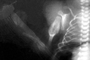
OBJECTIVE: To assess the relationship between gestational age and sonographic appearance of the various sternal components and establish growth during human gestation. DESIGN: A prospective cross-sectional study. METHODS: The study was performed on 252 consecutive normal singleton pregnancies from 19 weeks of gestation until term, using transabdominal high-resolution ultrasound techniques. The sternal length, as well as the number of ossification centers at each gestational age, were recorded. RESULTS: The first occasion at which a fetal human sternum could be visualized with two to three ossification centers was at 19 weeks' gestational age. The fifth ossification center was first visualized at 29 weeks' gestation. The mean +/- SE of sternal length varied from 15 +/- 0.98 mm (95% confidence interval (CI) 12.79-17.21) at 19-20 weeks, to 36.50 +/- 0.29 mm (95% CI 35.58-37.42) at 37-38 weeks' gestation. Sternal length as a function of gestational age was expressed by the regression equation: sternal length (mm) = -11.06 + 1.39 x gestational age (weeks). The correlation coefficient, r = 0.924 for sternal length, was found to be highly statistically significant (p < 0.0001). CONCLUSIONS: The presented data offer normative measurements of the fetal sternum which may be helpful in the prenatal diagnosis of congenital syndromes that include, among other manifestations, abnormalities of sternal development. (+info)Effect of strontium on the epiphyseal cartilage plate of rat tibiae-histological and radiographic studies. (5/3582)
Following dietary administration of strontium carbonate, histological and radiographic changes in the epiphyseal cartilage plate of the rat tibiae were examined in the present study. The weight gain of the rat fed a strontium diet was less than that of the control rats. Longitudinal growth of tibiae and endochondral ossification were inhibited by strontium administration. The widths of both proximal and distal cartilage plates increased enormously as has also been shown by other investigators. Sizes of chondroblasts in columns of proximal cartilage plate in rats fed a strontium diet were smaller than those of the control rats and were not different between upper and lower parts. It is suggested that strontium inhibits bone growth through the inhibitory action on the maturation process of chondroblasts and the succeeding endochondral ossification. (+info)A quantitative assessment of the healing of intramembranous and endochondral autogenous bone grafts. (6/3582)
The aim of the study was to assess quantitatively the amount of new bone formed in the early stages of healing of intramembranous and endochondral autogenous bone grafts so as to gain further insight into their integration with host bone. Eighteen critical size defects were created in the parietal bone of nine New Zealand White rabbits. In the experimental group (five rabbits), each rabbit was grafted with intramembranous bone in one defect and with endochondral bone in the other. In the control group (four rabbits), one defect was left empty (passive control) and the other was grafted with rabbit skin collagen (active control). After 14 days, the rabbits were killed and the defects were prepared for histological analysis. Serial sections were made across the whole defect. Each defect was divided into five regions spaced 1500 microns apart. Two sections were randomly drawn from each region. Quantitative analysis was performed on 100 sections using an image analyser computer software system to assess the amount of new bone formed in each defect. No bone was detected across the defect in either the active or passive controls. One-hundred-and-sixty-six per cent more new bone was formed in defects grafted with intramembranous bone than those grafted with endochondral bone. This represented an extremely significant difference (P < 0.0001, unpaired t-test) between the two groups. The results show that intramembranous autogenous bone produced more bone than the endochondral bone when grafted in the skull. Clinically, it is recommended that intramembranous bone is used to replace lost membranous bone in the oral cavity, as well as in skull defects, whenever possible. (+info)Differential patterns of altered bone formation in different bone compartments in established osteoporosis. (7/3582)
AIM: To investigate the level of bone formation in the different bone compartments in cases of established osteoporosis, as previous work has concentrated on trabecular bone alone. METHODS: Bone formation rates were measured histomorphometrically, in the periosteal (P), cortical (C), subcortical (SC), and trabecular (T) compartments in iliac crest biopsies from 159 patients with established osteoporosis. The values were standardised using age and sex matched control data and patterns of differential change determined by analysis of parametric status (increased, normal, reduced). RESULTS: Mean bone formation was reduced in all four compartments. This was more marked (4.4/4.1 standard deviations below the mean in C/T, v 2.3/0.9 in P/SC) and more frequent (reduced in 81.5%/78.3% in T/C, v 43.3%/44% in P/SC) in the trabecular and cortical compartments than in the periosteal or subcortical bone. Parametric status was equal in trabecular and cortical bone in 85.4% of cases, and in periosteal and subcortical bone in 65.7%, but in all four compartments in only 35.1%, indicating differential alteration of bone formation in the two sets of compartments (T/C v P/SC). CONCLUSIONS: Altered trabecular bone formation is important in osteoporosis, but there are differential patterns of alteration in the other three compartments, emphasising the presence of different microenvironments in bone; thus the effect on the cortical compartment was similar to that on the trabecular, while the subcortical and periosteal compartments also showed linkage. The linkage between the two pairs was divergent, indicating different control of bone formation, with resultant different patterns of perturbation in osteoporosis. (+info)Effects of XT-44, a phosphodiesterase 4 inhibitor, in osteoblastgenesis and osteoclastgenesis in culture and its therapeutic effects in rat osteopenia models. (8/3582)
We have reported that denbufylline, a phosphodiesterase 4 (PDE4) inhibitor, inhibits bone loss in Walker256/S tumor-bearing rats, suggesting therapeutic potentiality of a PDE4 inhibitor in osteopenia. In the present study, effects of a new PDE4 inhibitor, 1-n-butyl-3-n-propylxanthine (XT-44), in bone were evaluated in cell cultures and animal experiments. In rat bone marrow culture, XT-44 stimulated mineralized-nodule formation, whereas it inhibited osteoclast-like cell formation in mouse bone marrow culture. In Walker256/S-bearing rats (6-week-old female Wistar Imamichi rats), rapid decrease in bone mineral density (BMD) was prominent, and oral administration of XT-44 (0.3 mg/kg, every 2 days) inhibited the decrease in BMD. In the second animal experiment, female Wistar rats (6-week-old) were sciatic neurectomized, and XT-44 was orally administered to these rats every 2 days for 4 weeks. XT-44 administration (0.3 mg/kg) recovered BMD in these neurectomized animals. Furthermore, 19-week-old, female Wistar rats were ovariectomized (OVX), and 15 weeks after surgery, these rats were orally administered XT-44 every 2 days for 8 weeks. XT-44 treatment (1 mg/kg) increased the BMD of OVX rats. These results indicate that XT-44 could be a candidate as a therapeutic drug for treating osteopenia including osteoporosis. (+info)Osteogenesis Imperfecta (OI), also known as brittle bone disease, is a group of genetic disorders that mainly affect the bones. It is characterized by bones that break easily, often from little or no apparent cause. This happens because the body produces an insufficient amount of collagen or poor quality collagen, which are crucial for the formation of healthy bones.
The severity of OI can vary greatly, even within the same family. Some people with OI have only a few fractures in their lifetime while others may have hundreds. Other symptoms can include blue or gray sclera (the white part of the eye), hearing loss, short stature, curved or bowed bones, loose joints, and a triangular face shape.
There are several types of OI, each caused by different genetic mutations. Most types of OI are inherited in an autosomal dominant pattern, meaning only one copy of the altered gene is needed to cause the condition. However, some types are inherited in an autosomal recessive pattern, which means that two copies of the altered gene must be present for the condition to occur.
There is no cure for OI, but treatment can help manage symptoms and prevent complications. Treatment may include medication to strengthen bones, physical therapy, bracing, and surgery.
Osteogenesis is the process of bone formation or development. It involves the differentiation and maturation of osteoblasts, which are bone-forming cells that synthesize and deposit the organic matrix of bone tissue, composed mainly of type I collagen. This organic matrix later mineralizes to form the inorganic crystalline component of bone, primarily hydroxyapatite.
There are two primary types of osteogenesis: intramembranous and endochondral. Intramembranous osteogenesis occurs directly within connective tissue, where mesenchymal stem cells differentiate into osteoblasts and form bone tissue without an intervening cartilage template. This process is responsible for the formation of flat bones like the skull and clavicles.
Endochondral osteogenesis, on the other hand, involves the initial development of a cartilaginous model or template, which is later replaced by bone tissue. This process forms long bones, such as those in the limbs, and occurs through several stages involving chondrocyte proliferation, hypertrophy, and calcification, followed by invasion of blood vessels and osteoblasts to replace the cartilage with bone tissue.
Abnormalities in osteogenesis can lead to various skeletal disorders and diseases, such as osteogenesis imperfecta (brittle bone disease), achondroplasia (a form of dwarfism), and cleidocranial dysplasia (a disorder affecting skull and collarbone development).
Osteogenesis, distraction refers to a surgical procedure and controlled rehabilitation process used in orthopedic surgery, oral and maxillofacial surgery, and neurosurgery to lengthen bones or correct bone deformities. The term "osteogenesis" means bone formation, while "distraction" refers to the gradual separation of bone segments.
In this procedure, a surgeon first cuts the bone (osteotomy) and then applies an external or internal distraction device that slowly moves apart the cut ends of the bone. Over time, new bone forms in the gap between the separated bone segments through a process called distraction osteogenesis. This results in increased bone length or correction of deformities.
Distraction osteogenesis is often used to treat various conditions such as limb length discrepancies, craniofacial deformities, and spinal deformities. The procedure requires careful planning, precise surgical technique, and close postoperative management to ensure optimal outcomes.
Osteoblasts are specialized bone-forming cells that are derived from mesenchymal stem cells. They play a crucial role in the process of bone formation and remodeling. Osteoblasts synthesize, secrete, and mineralize the organic matrix of bones, which is mainly composed of type I collagen.
These cells have receptors for various hormones and growth factors that regulate their activity, such as parathyroid hormone, vitamin D, and transforming growth factor-beta. When osteoblasts are not actively producing bone matrix, they can become trapped within the matrix they produce, where they differentiate into osteocytes, which are mature bone cells that play a role in maintaining bone structure and responding to mechanical stress.
Abnormalities in osteoblast function can lead to various bone diseases, such as osteoporosis, osteogenesis imperfecta, and Paget's disease of bone.
Bone regeneration is the biological process of new bone formation that occurs after an injury or removal of a portion of bone. This complex process involves several stages, including inflammation, migration and proliferation of cells, matrix deposition, and mineralization, leading to the restoration of the bone's structure and function.
The main cells involved in bone regeneration are osteoblasts, which produce new bone matrix, and osteoclasts, which resorb damaged or old bone tissue. The process is tightly regulated by various growth factors, hormones, and signaling molecules that promote the recruitment, differentiation, and activity of these cells.
Bone regeneration can occur naturally in response to injury or surgical intervention, such as fracture repair or dental implant placement. However, in some cases, bone regeneration may be impaired due to factors such as age, disease, or trauma, leading to delayed healing or non-union of the bone. In these situations, various strategies and techniques, including the use of bone grafts, scaffolds, and growth factors, can be employed to enhance and support the bone regeneration process.
Procollagen is the precursor protein of collagen, which is a major structural protein in the extracellular matrix of various connective tissues, such as tendons, ligaments, skin, and bones. Procollagen is synthesized inside the cell (in the rough endoplasmic reticulum) and then processed by enzymes to remove specific segments, resulting in the formation of tropocollagen, which are the basic units of collagen fibrils.
Procollagen consists of three polypeptide chains (two alpha-1 and one alpha-2 chain), each containing a central triple-helical domain flanked by non-helical regions at both ends. These non-helical regions, called propeptides, are cleaved off during the processing of procollagen to tropocollagen, allowing the individual collagen molecules to align and form fibrils through covalent cross-linking.
Abnormalities in procollagen synthesis or processing can lead to various connective tissue disorders, such as osteogenesis imperfecta (brittle bone disease) and Ehlers-Danlos syndrome (a group of disorders characterized by joint hypermobility, skin hyperextensibility, and tissue fragility).
Physiologic calcification is the normal deposit of calcium salts in body tissues and organs. It is a natural process that occurs as part of the growth and development of the human body, as well as during the repair and remodeling of tissues.
Calcium is an essential mineral that plays a critical role in many bodily functions, including bone formation, muscle contraction, nerve impulse transmission, and blood clotting. In order to maintain proper levels of calcium in the body, excess calcium that is not needed for these functions may be deposited in various tissues as a normal part of the aging process.
Physiologic calcification typically occurs in areas such as the walls of blood vessels, the lungs, and the heart valves. While these calcifications are generally harmless, they can sometimes lead to complications, particularly if they occur in large amounts or in sensitive areas. For example, calcification of the coronary arteries can increase the risk of heart disease, while calcification of the lung tissue can cause respiratory symptoms.
It is important to note that pathologic calcification, on the other hand, refers to the abnormal deposit of calcium salts in tissues and organs, which can be caused by various medical conditions such as chronic kidney disease, hyperparathyroidism, and certain infections. Pathologic calcification is not a normal process and can lead to serious health complications if left untreated.
"Bone" is the hard, dense connective tissue that makes up the skeleton of vertebrate animals. It provides support and protection for the body's internal organs, and serves as a attachment site for muscles, tendons, and ligaments. Bone is composed of cells called osteoblasts and osteoclasts, which are responsible for bone formation and resorption, respectively, and an extracellular matrix made up of collagen fibers and mineral crystals.
Bones can be classified into two main types: compact bone and spongy bone. Compact bone is dense and hard, and makes up the outer layer of all bones and the shafts of long bones. Spongy bone is less dense and contains large spaces, and makes up the ends of long bones and the interior of flat and irregular bones.
The human body has 206 bones in total. They can be further classified into five categories based on their shape: long bones, short bones, flat bones, irregular bones, and sesamoid bones.
The mandible, also known as the lower jaw, is the largest and strongest bone in the human face. It forms the lower portion of the oral cavity and plays a crucial role in various functions such as mastication (chewing), speaking, and swallowing. The mandible is a U-shaped bone that consists of a horizontal part called the body and two vertical parts called rami.
The mandible articulates with the skull at the temporomandibular joints (TMJs) located in front of each ear, allowing for movements like opening and closing the mouth, protrusion, retraction, and side-to-side movement. The mandible contains the lower teeth sockets called alveolar processes, which hold the lower teeth in place.
In medical terminology, the term "mandible" refers specifically to this bone and its associated structures.
Core Binding Factor Alpha 1 Subunit, also known as CBF-A1 or RUNX1, is a protein that plays a crucial role in hematopoiesis, which is the process of blood cell development. It is a member of the core binding factor (CBF) complex, which regulates gene transcription and is essential for the differentiation and maturation of hematopoietic stem cells into mature blood cells.
The CBF complex consists of three subunits: CBF-A, CBF-B, and a histone deacetylase (HDAC). The CBF-A subunit can have several isoforms, including CBF-A1, which is encoded by the RUNX1 gene. Mutations in the RUNX1 gene have been associated with various hematological disorders, such as acute myeloid leukemia (AML), familial platelet disorder with propensity to develop AML, and thrombocytopenia with absent radii syndrome.
CBF-A1/RUNX1 functions as a transcription factor that binds to DNA at specific sequences called core binding factors, thereby regulating the expression of target genes involved in hematopoiesis. Proper regulation of these genes is essential for normal blood cell development and homeostasis.
The skull is the bony structure that encloses and protects the brain, the eyes, and the ears. It is composed of two main parts: the cranium, which contains the brain, and the facial bones. The cranium is made up of several fused flat bones, while the facial bones include the upper jaw (maxilla), lower jaw (mandible), cheekbones, nose bones, and eye sockets (orbits).
The skull also provides attachment points for various muscles that control chewing, moving the head, and facial expressions. Additionally, it contains openings for blood vessels, nerves, and the spinal cord to pass through. The skull's primary function is to protect the delicate and vital structures within it from injury and trauma.
An external fixator is a type of orthopedic device used in the treatment of severe fractures or deformities of bones. It consists of an external frame that is attached to the bone with pins or wires that pass through the skin and into the bone. This provides stability to the injured area while allowing for alignment and adjustment of the bone during the healing process.
External fixators are typically used in cases where traditional casting or internal fixation methods are not feasible, such as when there is extensive soft tissue damage, infection, or when a limb needs to be gradually stretched or shortened. They can also be used in reconstructive surgery for bone defects or deformities.
The external frame of the fixator is made up of bars and clamps that are adjustable, allowing for precise positioning and alignment of the bones. The pins or wires that attach to the bone are carefully inserted through small incisions in the skin, and are held in place by the clamps on the frame.
External fixators can be used for a period of several weeks to several months, depending on the severity of the injury and the individual's healing process. During this time, the patient may require regular adjustments and monitoring by an orthopedic surgeon or other medical professional. Once the bone has healed sufficiently, the external fixator can be removed in a follow-up procedure.
Mesenchymal Stromal Cells (MSCs) are a type of adult stem cells found in various tissues, including bone marrow, adipose tissue, and umbilical cord blood. They have the ability to differentiate into multiple cell types, such as osteoblasts, chondrocytes, and adipocytes, under specific conditions. MSCs also possess immunomodulatory properties, making them a promising tool in regenerative medicine and therapeutic strategies for various diseases, including autoimmune disorders and tissue injuries. It is important to note that the term "Mesenchymal Stem Cells" has been replaced by "Mesenchymal Stromal Cells" in the scientific community to better reflect their biological characteristics and potential functions.
Alkaline phosphatase (ALP) is an enzyme found in various body tissues, including the liver, bile ducts, digestive system, bones, and kidneys. It plays a role in breaking down proteins and minerals, such as phosphate, in the body.
The medical definition of alkaline phosphatase refers to its function as a hydrolase enzyme that removes phosphate groups from molecules at an alkaline pH level. In clinical settings, ALP is often measured through blood tests as a biomarker for various health conditions.
Elevated levels of ALP in the blood may indicate liver or bone diseases, such as hepatitis, cirrhosis, bone fractures, or cancer. Therefore, physicians may order an alkaline phosphatase test to help diagnose and monitor these conditions. However, it is essential to interpret ALP results in conjunction with other diagnostic tests and clinical findings for accurate diagnosis and treatment.
Bone matrix refers to the non-cellular component of bone that provides structural support and functions as a reservoir for minerals, such as calcium and phosphate. It is made up of organic and inorganic components. The organic component consists mainly of type I collagen fibers, which provide flexibility and tensile strength to the bone. The inorganic component is primarily composed of hydroxyapatite crystals, which give bone its hardness and compressive strength. Bone matrix also contains other proteins, growth factors, and signaling molecules that regulate bone formation, remodeling, and repair.
Bone Morphogenetic Protein 2 (BMP-2) is a growth factor that belongs to the transforming growth factor-beta (TGF-β) superfamily. It plays a crucial role in bone and cartilage formation, as well as in the regulation of wound healing and embryonic development. BMP-2 stimulates the differentiation of mesenchymal stem cells into osteoblasts, which are cells responsible for bone formation.
BMP-2 has been approved by the US Food and Drug Administration (FDA) as a medical device to promote bone growth in certain spinal fusion surgeries and in the treatment of open fractures that have not healed properly. It is usually administered in the form of a collagen sponge soaked with recombinant human BMP-2 protein, which is a laboratory-produced version of the natural protein.
While BMP-2 has shown promising results in some clinical applications, its use is not without risks and controversies. Some studies have reported adverse effects such as inflammation, ectopic bone formation, and increased rates of cancer, which have raised concerns about its safety and efficacy. Therefore, it is essential to weigh the benefits and risks of BMP-2 therapy on a case-by-case basis and under the guidance of a qualified healthcare professional.
Bony callus is a medical term that refers to the specialized tissue that forms in response to a bone fracture. It is a crucial part of the natural healing process, as it helps to stabilize and protect the broken bone while it mends.
When a bone is fractured, the body responds by initiating an inflammatory response, which triggers the production of various cells and signaling molecules that promote healing. As part of this process, specialized cells called osteoblasts begin to produce new bone tissue at the site of the fracture. This tissue is initially soft and pliable, allowing it to bridge the gap between the broken ends of the bone.
Over time, this soft callus gradually hardens and calcifies, forming a bony callus that helps to stabilize the fracture and provide additional support as the bone heals. The bony callus is typically composed of a mixture of woven bone (which is less organized than normal bone) and more structured lamellar bone (which is similar in structure to normal bone).
As the bone continues to heal, the bony callus may be gradually remodeled and reshaped by osteoclasts, which are specialized cells that break down and remove excess or unwanted bone tissue. This process helps to restore the bone's original shape and strength, allowing it to function normally again.
It is worth noting that excessive bony callus formation can sometimes lead to complications, such as stiffness, pain, or decreased range of motion in the affected limb. In some cases, surgical intervention may be necessary to remove or reduce the size of the bony callus and promote proper healing.
The tibia, also known as the shin bone, is the larger of the two bones in the lower leg and part of the knee joint. It supports most of the body's weight and is a major insertion point for muscles that flex the foot and bend the leg. The tibia articulates with the femur at the knee joint and with the fibula and talus bone at the ankle joint. Injuries to the tibia, such as fractures, are common in sports and other activities that put stress on the lower leg.
Collagen is the most abundant protein in the human body, and it is a major component of connective tissues such as tendons, ligaments, skin, and bones. Collagen provides structure and strength to these tissues and helps them to withstand stretching and tension. It is made up of long chains of amino acids, primarily glycine, proline, and hydroxyproline, which are arranged in a triple helix structure. There are at least 16 different types of collagen found in the body, each with slightly different structures and functions. Collagen is important for maintaining the integrity and health of tissues throughout the body, and it has been studied for its potential therapeutic uses in various medical conditions.
Collagen Type I is the most abundant form of collagen in the human body, found in various connective tissues such as tendons, ligaments, skin, and bones. It is a structural protein that provides strength and integrity to these tissues. Collagen Type I is composed of three alpha chains, two alpha-1(I) chains, and one alpha-2(I) chain, arranged in a triple helix structure. This type of collagen is often used in medical research and clinical applications, such as tissue engineering and regenerative medicine, due to its excellent mechanical properties and biocompatibility.
X-ray microtomography, often referred to as micro-CT, is a non-destructive imaging technique used to visualize and analyze the internal structure of objects with high spatial resolution. It is based on the principles of computed tomography (CT), where multiple X-ray images are acquired at different angles and then reconstructed into cross-sectional slices using specialized software. These slices can be further processed to create 3D visualizations, allowing researchers and clinicians to examine the internal structure and composition of samples in great detail. Micro-CT is widely used in materials science, biology, medicine, and engineering for various applications such as material characterization, bone analysis, and defect inspection.
The Ilizarov technique is a surgical method used for limb lengthening and reconstruction. It involves the use of an external fixation device, which consists of rings connected by adjustable rods and wires that are attached to the bone. This apparatus allows for gradual distraction (slow, steady stretching) of the bone, allowing new bone tissue to grow in the gap created by the distraction. The Ilizarov technique can be used to treat various conditions such as limb length discrepancies, bone deformities, and nonunions (failed healing of a fracture). It is named after its developer, Gavriil Abramovich Ilizarov, a Soviet orthopedic surgeon.
The periosteum is a highly vascularized and innervated tissue that surrounds the outer surface of bones, except at the articular surfaces. It consists of two layers: an outer fibrous layer containing blood vessels, nerves, and fibroblasts; and an inner cellular layer called the cambium or osteogenic layer, which contains progenitor cells capable of bone formation and repair.
The periosteum plays a crucial role in bone growth, remodeling, and healing by providing a source of osteoprogenitor cells and blood supply. It also contributes to the sensation of pain in response to injury or inflammation of the bone. Additionally, the periosteum can respond to mechanical stress by activating bone formation, making it an essential component in orthopedic treatments such as distraction osteogenesis.
Dentinogenesis Imperfecta (DI) is a genetic disorder that affects the development and formation of dentin, which is the hard tissue beneath the enamel of teeth. This condition results in teeth that are discolored, translucent, and sensitive to temperature changes. The teeth may also wear down easily and be prone to fractures.
There are two main types of Dentinogenesis Imperfecta: type I and type II. Type I is associated with a genetic disorder called osteogenesis imperfecta (OI), which affects the development of bones as well as teeth. Type II, on the other hand, is not associated with OI and only affects the teeth.
The inheritance pattern for Dentinogenesis Imperfecta is autosomal dominant, meaning that a child has a 50% chance of inheriting the condition if one parent has it. However, some cases may be due to new mutations in the gene and not inherited from a parent. Treatment for DI typically involves dental restorations such as crowns or veneers to improve the appearance and function of the teeth.
A mandibular osteotomy is a surgical procedure that involves making a cut in the mandible (lower jawbone). This procedure is often performed to correct various dental and maxillofacial conditions such as jaw misalignment, sleep apnea, or jaw tumors. The specific type of osteotomy performed depends on the individual patient's needs and may involve making cuts at different locations along the mandible.
During the procedure, the surgeon makes an incision in the gum tissue to expose the mandible and then uses specialized instruments to make a precise cut in the bone. The surgeon can then move the jawbone into the desired position and secure it with plates, screws, or wires. In some cases, bone grafting may also be necessary to provide additional support.
After the procedure, patients may experience swelling, bruising, and discomfort, which can be managed with pain medication and cold compresses. Patients are usually advised to follow a soft diet for several weeks while the jaw heals. The recovery period can vary depending on the individual patient's healing process, but most patients can return to their normal activities within a few weeks.
Bone development, also known as ossification, is the process by which bone tissue is formed and grows. This complex process involves several different types of cells, including osteoblasts, which produce new bone matrix, and osteoclasts, which break down and resorb existing bone tissue.
There are two main types of bone development: intramembranous and endochondral ossification. Intramembranous ossification occurs when bone tissue forms directly from connective tissue, while endochondral ossification involves the formation of a cartilage model that is later replaced by bone.
During fetal development, most bones develop through endochondral ossification, starting as a cartilage template that is gradually replaced by bone tissue. However, some bones, such as those in the skull and clavicles, develop through intramembranous ossification.
Bone development continues after birth, with new bone tissue being laid down and existing tissue being remodeled throughout life. This ongoing process helps to maintain the strength and integrity of the skeleton, allowing it to adapt to changing mechanical forces and repair any damage that may occur.
Cell differentiation is the process by which a less specialized cell, or stem cell, becomes a more specialized cell type with specific functions and structures. This process involves changes in gene expression, which are regulated by various intracellular signaling pathways and transcription factors. Differentiation results in the development of distinct cell types that make up tissues and organs in multicellular organisms. It is a crucial aspect of embryonic development, tissue repair, and maintenance of homeostasis in the body.
Fracture healing is the natural process by which a broken bone repairs itself. When a fracture occurs, the body responds by initiating a series of biological and cellular events aimed at restoring the structural integrity of the bone. This process involves the formation of a hematoma (a collection of blood) around the fracture site, followed by the activation of inflammatory cells that help to clean up debris and prepare the area for repair.
Over time, specialized cells called osteoblasts begin to lay down new bone matrix, or osteoid, along the edges of the broken bone ends. This osteoid eventually hardens into new bone tissue, forming a bridge between the fracture fragments. As this process continues, the callus (a mass of newly formed bone and connective tissue) gradually becomes stronger and more compact, eventually remodeling itself into a solid, unbroken bone.
The entire process of fracture healing can take several weeks to several months, depending on factors such as the severity of the injury, the patient's age and overall health, and the location of the fracture. In some cases, medical intervention may be necessary to help promote healing or ensure proper alignment of the bone fragments. This may include the use of casts, braces, or surgical implants such as plates, screws, or rods.
Osteocalcin is a protein that is produced by osteoblasts, which are the cells responsible for bone formation. It is one of the most abundant non-collagenous proteins found in bones and plays a crucial role in the regulation of bone metabolism. Osteocalcin contains a high affinity for calcium ions, making it essential for the mineralization of the bone matrix.
Once synthesized, osteocalcin is secreted into the extracellular matrix, where it binds to hydroxyapatite crystals, helping to regulate their growth and contributing to the overall strength and integrity of the bones. Osteocalcin also has been found to play a role in other physiological processes outside of bone metabolism, such as modulating insulin sensitivity, energy metabolism, and male fertility.
In summary, osteocalcin is a protein produced by osteoblasts that plays a critical role in bone formation, mineralization, and turnover, and has been implicated in various other physiological processes.
Adipogenesis is the process by which precursor cells differentiate into mature adipocytes, or fat cells. This complex biological process involves a series of molecular and cellular events that are regulated by various genetic and epigenetic factors.
During adipogenesis, preadipocytes undergo a series of changes that include cell cycle arrest, morphological alterations, and the expression of specific genes that are involved in lipid metabolism and insulin sensitivity. These changes ultimately result in the formation of mature adipocytes that are capable of storing energy in the form of lipids.
Abnormalities in adipogenesis have been linked to various health conditions, including obesity, type 2 diabetes, and metabolic syndrome. Understanding the molecular mechanisms that regulate adipogenesis is an active area of research, as it may lead to the development of new therapies for these and other related diseases.
Heterotopic ossification (HO) is a medical condition where bone tissue forms outside the skeleton, in locations where it does not typically exist. This process can occur in various soft tissues, such as muscles, tendons, ligaments, or even inside joint capsules. The abnormal bone growth can lead to pain, stiffness, limited range of motion, and, in some cases, loss of function in the affected area.
There are several types of heterotopic ossification, including:
1. Myositis ossificans - This form is often associated with trauma or injury, such as muscle damage from a fracture, surgery, or direct blow. It typically affects young, active individuals and usually resolves on its own within months to a few years.
2. Neurogenic heterotopic ossification (NHO) - Also known as "traumatic heterotopic ossification," this form is often linked to spinal cord injuries, brain injuries, or central nervous system damage. NHO can cause significant impairment and may require surgical intervention in some cases.
3. Fibrodysplasia ossificans progressiva (FOP) - This rare, genetic disorder causes progressive heterotopic ossification throughout the body, starting in early childhood. The condition significantly impacts mobility and quality of life, with no known cure.
The exact mechanisms behind heterotopic ossification are not fully understood, but it is believed that a combination of factors, including inflammation, tissue injury, and genetic predisposition, contribute to its development. Treatment options may include nonsteroidal anti-inflammatory drugs (NSAIDs), radiation therapy, physical therapy, or surgical removal of the abnormal bone growth, depending on the severity and location of the HO.
Integrin-binding sialoprotein (IBSP) is a non-collagenous protein found in bones and teeth. It is also known as bone sialoprotein II or acidic glycoprotein 34. IBSP plays a role in the regulation of biomineralization, which is the process by which minerals are deposited in biological tissues.
IBSP contains several functional domains that allow it to interact with other proteins and molecules. One such domain is an arginine-glycine-aspartic acid (RGD) motif, which can bind to integrin receptors on the surface of cells. This interaction helps regulate the attachment and behavior of cells in bone tissue.
IBSP also contains a large number of sialic acid residues, which give it its name and contribute to its negative charge. These residues may play a role in protecting the protein from degradation and helping it interact with other molecules in the extracellular matrix.
Overall, IBSP is an important component of bone tissue and plays a key role in regulating the formation and maintenance of bones and teeth.
Osseointegration is a direct structural and functional connection between living bone and the surface of an implant. It's a process where the bone grows in and around the implant, which is typically made of titanium or another biocompatible material. This process provides a solid foundation for dental prosthetics, such as crowns, bridges, or dentures, or for orthopedic devices like artificial limbs. The success of osseointegration depends on various factors, including the patient's overall health, the quality and quantity of available bone, and the surgical technique used for implant placement.
Osteocytes are the most abundant cell type in mature bone tissue. They are star-shaped cells that are located inside the mineralized matrix of bones, with their processes extending into small spaces called lacunae and canaliculi. Osteocytes are derived from osteoblasts, which are bone-forming cells that become trapped within the matrix they produce.
Osteocytes play a crucial role in maintaining bone homeostasis by regulating bone remodeling, sensing mechanical stress, and modulating mineralization. They communicate with each other and with osteoblasts and osteoclasts (bone-resorbing cells) through a network of interconnected processes and via the release of signaling molecules. Osteocytes can also respond to changes in their environment, such as hormonal signals or mechanical loading, by altering their gene expression and releasing factors that regulate bone metabolism.
Dysfunction of osteocytes has been implicated in various bone diseases, including osteoporosis, osteogenesis imperfecta, and Paget's disease of bone.
The femur is the medical term for the thigh bone, which is the longest and strongest bone in the human body. It connects the hip bone to the knee joint and plays a crucial role in supporting the weight of the body and allowing movement during activities such as walking, running, and jumping. The femur is composed of a rounded head, a long shaft, and two condyles at the lower end that articulate with the tibia and patella to form the knee joint.
Chondrogenesis is the process of cartilage formation during embryonic development and in the healing of certain types of injuries. It involves the differentiation of mesenchymal stem cells into chondrocytes, which are the specialized cells that produce and maintain the extracellular matrix of cartilage.
During chondrogenesis, the mesenchymal stem cells condense and form a template for the future cartilaginous tissue. These cells then differentiate into chondrocytes, which begin to produce and deposit collagen type II, proteoglycans, and other extracellular matrix components that give cartilage its unique biochemical and mechanical properties.
Chondrogenesis is a critical process for the development of various structures in the body, including the skeletal system, where it plays a role in the formation of articular cartilage, growth plates, and other types of cartilage. Understanding the molecular mechanisms that regulate chondrogenesis is important for developing therapies to treat cartilage injuries and degenerative diseases such as osteoarthritis.
Micrognathism is a medical term that refers to a condition where the lower jaw (mandible) is abnormally small or underdeveloped. This can result in various dental and skeletal problems, including an improper bite (malocclusion), difficulty speaking, chewing, or swallowing, and sleep apnea. Micrognathism may be congenital or acquired later in life due to trauma, disease, or surgical removal of part of the jaw. Treatment options depend on the severity of the condition and can include orthodontic treatment, surgery, or a combination of both.
Glycine is a simple amino acid that plays a crucial role in the body. According to the medical definition, glycine is an essential component for the synthesis of proteins, peptides, and other biologically important compounds. It is also involved in various metabolic processes, such as the production of creatine, which supports muscle function, and the regulation of neurotransmitters, affecting nerve impulse transmission and brain function. Glycine can be found as a free form in the body and is also present in many dietary proteins.
A mandibular fracture is a break or crack in the lower jaw (mandible) bone. It can occur at any point along the mandible, but common sites include the condyle (the rounded end near the ear), the angle (the curved part of the jaw), and the symphysis (the area where the two halves of the jaw meet in the front). Mandibular fractures are typically caused by trauma, such as a direct blow to the face or a fall. Symptoms may include pain, swelling, bruising, difficulty chewing or speaking, and malocclusion (misalignment) of the teeth. Treatment usually involves immobilization with wires or screws to allow the bone to heal properly.
Bone Morphogenetic Proteins (BMPs) are a group of growth factors that play crucial roles in the development, growth, and repair of bones and other tissues. They belong to the Transforming Growth Factor-β (TGF-β) superfamily and were first discovered when researchers found that certain proteins extracted from demineralized bone matrix had the ability to induce new bone formation.
BMPs stimulate the differentiation of mesenchymal stem cells into osteoblasts, which are the cells responsible for bone formation. They also promote the recruitment and proliferation of these cells, enhancing the overall process of bone regeneration. In addition to their role in bone biology, BMPs have been implicated in various other biological processes, including embryonic development, wound healing, and the regulation of fat metabolism.
There are several types of BMPs (BMP-2, BMP-4, BMP-7, etc.) that exhibit distinct functions and expression patterns. Due to their ability to stimulate bone formation, recombinant human BMPs have been used in clinical applications, such as spinal fusion surgery and non-healing fracture treatment. However, the use of BMPs in medicine has been associated with certain risks and complications, including uncontrolled bone growth, inflammation, and cancer development, which necessitates further research to optimize their therapeutic potential.
Osteotomy is a surgical procedure in which a bone is cut to shorten, lengthen, or change its alignment. It is often performed to correct deformities or to realign bones that have been damaged by trauma or disease. The bone may be cut straight across (transverse osteotomy) or at an angle (oblique osteotomy). After the bone is cut, it can be realigned and held in place with pins, plates, or screws until it heals. This procedure is commonly performed on bones in the leg, such as the femur or tibia, but can also be done on other bones in the body.
"Cells, cultured" is a medical term that refers to cells that have been removed from an organism and grown in controlled laboratory conditions outside of the body. This process is called cell culture and it allows scientists to study cells in a more controlled and accessible environment than they would have inside the body. Cultured cells can be derived from a variety of sources, including tissues, organs, or fluids from humans, animals, or cell lines that have been previously established in the laboratory.
Cell culture involves several steps, including isolation of the cells from the tissue, purification and characterization of the cells, and maintenance of the cells in appropriate growth conditions. The cells are typically grown in specialized media that contain nutrients, growth factors, and other components necessary for their survival and proliferation. Cultured cells can be used for a variety of purposes, including basic research, drug development and testing, and production of biological products such as vaccines and gene therapies.
It is important to note that cultured cells may behave differently than they do in the body, and results obtained from cell culture studies may not always translate directly to human physiology or disease. Therefore, it is essential to validate findings from cell culture experiments using additional models and ultimately in clinical trials involving human subjects.
Jaw fixation techniques, also known as maxillomandibular fixation (MMF), are procedures used in dental and oral surgery to hold the jaw in a specific position. This is typically done by wiring the upper and lower teeth together or using elastic bands and other devices to keep the jaws aligned. The technique is often used after surgical procedures on the jaw, such as corrective jaw surgery (orthognathic surgery) or fracture repair, to help promote proper healing and alignment of the bones. It may also be used in the management of temporomandibular joint disorders or other conditions affecting the jaw. The duration of jaw fixation can vary depending on the specific procedure and individual patient needs, but it typically lasts several weeks.
Bone diseases is a broad term that refers to various medical conditions that affect the bones. These conditions can be categorized into several groups, including:
1. Developmental and congenital bone diseases: These are conditions that affect bone growth and development before or at birth. Examples include osteogenesis imperfecta (brittle bone disease), achondroplasia (dwarfism), and cleidocranial dysostosis.
2. Metabolic bone diseases: These are conditions that affect the body's ability to maintain healthy bones. They are often caused by hormonal imbalances, vitamin deficiencies, or problems with mineral metabolism. Examples include osteoporosis, osteomalacia, and Paget's disease of bone.
3. Inflammatory bone diseases: These are conditions that cause inflammation in the bones. They can be caused by infections, autoimmune disorders, or other medical conditions. Examples include osteomyelitis, rheumatoid arthritis, and ankylosing spondylitis.
4. Degenerative bone diseases: These are conditions that cause the bones to break down over time. They can be caused by aging, injury, or disease. Examples include osteoarthritis, avascular necrosis, and diffuse idiopathic skeletal hyperostosis (DISH).
5. Tumors and cancers of the bone: These are conditions that involve abnormal growths in the bones. They can be benign or malignant. Examples include osteosarcoma, chondrosarcoma, and Ewing sarcoma.
6. Fractures and injuries: While not strictly a "disease," fractures and injuries are common conditions that affect the bones. They can result from trauma, overuse, or weakened bones. Examples include stress fractures, compound fractures, and dislocations.
Overall, bone diseases can cause a wide range of symptoms, including pain, stiffness, deformity, and decreased mobility. Treatment for these conditions varies depending on the specific diagnosis but may include medication, surgery, physical therapy, or lifestyle changes.
Cranial sutures are the fibrous joints that connect and hold together the bones of the skull (cranium) in humans and other animals. These sutures provide flexibility for the skull during childbirth and growth, allowing the skull to expand as the brain grows in size, especially during infancy and early childhood.
There are several cranial sutures in the human skull, including:
1. The sagittal suture, which runs along the midline of the skull, connecting the two parietal bones.
2. The coronal suture, which connects the frontal bone to the two parietal bones.
3. The lambdoid suture, which connects the occipital bone to the two parietal bones.
4. The squamosal suture, which connects the temporal bone to the parietal bone.
5. The frontosphenoidal and sphenoethmoidal sutures, which connect the frontal bone, sphenoid bone, and ethmoid bone in the anterior cranial fossa.
These sutures are typically made up of a specialized type of connective tissue called Sharpey's fibers, which interdigitate with each other to form a strong yet flexible joint. Over time, as the skull bones fully fuse together, these sutures become less prominent and eventually ossify (turn into bone). In some cases, abnormalities in cranial suture development or fusion can lead to medical conditions such as craniosynostosis.
A mutation is a permanent change in the DNA sequence of an organism's genome. Mutations can occur spontaneously or be caused by environmental factors such as exposure to radiation, chemicals, or viruses. They may have various effects on the organism, ranging from benign to harmful, depending on where they occur and whether they alter the function of essential proteins. In some cases, mutations can increase an individual's susceptibility to certain diseases or disorders, while in others, they may confer a survival advantage. Mutations are the driving force behind evolution, as they introduce new genetic variability into populations, which can then be acted upon by natural selection.
Bone substitutes are materials that are used to replace missing or damaged bone in the body. They can be made from a variety of materials, including natural bone from other parts of the body or from animals, synthetic materials, or a combination of both. The goal of using bone substitutes is to provide structural support and promote the growth of new bone tissue.
Bone substitutes are often used in dental, orthopedic, and craniofacial surgery to help repair defects caused by trauma, tumors, or congenital abnormalities. They can also be used to augment bone volume in procedures such as spinal fusion or joint replacement.
There are several types of bone substitutes available, including:
1. Autografts: Bone taken from another part of the patient's body, such as the hip or pelvis.
2. Allografts: Bone taken from a deceased donor and processed to remove any cells and infectious materials.
3. Xenografts: Bone from an animal source, typically bovine or porcine, that has been processed to remove any cells and infectious materials.
4. Synthetic bone substitutes: Materials such as calcium phosphate ceramics, bioactive glass, and polymer-based materials that are designed to mimic the properties of natural bone.
The choice of bone substitute material depends on several factors, including the size and location of the defect, the patient's medical history, and the surgeon's preference. It is important to note that while bone substitutes can provide structural support and promote new bone growth, they may not have the same strength or durability as natural bone. Therefore, they may not be suitable for all applications, particularly those that require high load-bearing capacity.
Bone lengthening is a surgical procedure that involves cutting and then gradually stretching the bone apart, allowing new bone to grow in its place. This process is also known as distraction osteogenesis. The goal of bone lengthening is to increase the length of a bone, either to improve function or to correct a deformity.
The procedure typically involves making an incision in the skin over the bone and using specialized tools to cut through the bone. Once the bone is cut, a device called an external fixator is attached to the bone on either side of the cut. The external fixator is then gradually adjusted over time to slowly stretch the bone apart, creating a gap between the two ends of the bone. As the bone is stretched, new bone tissue begins to grow in the space between the two ends, eventually filling in the gap and lengthening the bone.
Bone lengthening can be used to treat a variety of conditions, including limb length discrepancies, congenital deformities, and injuries that result in bone loss. It is typically performed by an orthopedic surgeon and may require several months of follow-up care to ensure proper healing and growth of the new bone tissue.
The alveolar process is the curved part of the jawbone (mandible or maxilla) that contains sockets or hollow spaces (alveoli) for the teeth to be embedded. These processes are covered with a specialized mucous membrane called the gingiva, which forms a tight seal around the teeth to help protect the periodontal tissues and maintain oral health.
The alveolar process is composed of both compact and spongy bone tissue. The compact bone forms the outer layer, while the spongy bone is found inside the alveoli and provides support for the teeth. When a tooth is lost or extracted, the alveolar process begins to resorb over time due to the lack of mechanical stimulation from the tooth's chewing forces. This can lead to changes in the shape and size of the jawbone, which may require bone grafting procedures before dental implant placement.
Cartilage is a type of connective tissue that is found throughout the body in various forms. It is made up of specialized cells called chondrocytes, which are embedded in a firm, flexible matrix composed of collagen fibers and proteoglycans. This unique structure gives cartilage its characteristic properties of being both strong and flexible.
There are three main types of cartilage in the human body: hyaline cartilage, elastic cartilage, and fibrocartilage.
1. Hyaline cartilage is the most common type and is found in areas such as the articular surfaces of bones (where they meet to form joints), the nose, trachea, and larynx. It has a smooth, glassy appearance and provides a smooth, lubricated surface for joint movement.
2. Elastic cartilage contains more elastin fibers than hyaline cartilage, which gives it greater flexibility and resilience. It is found in structures such as the external ear and parts of the larynx and epiglottis.
3. Fibrocartilage has a higher proportion of collagen fibers and fewer chondrocytes than hyaline or elastic cartilage. It is found in areas that require high tensile strength, such as the intervertebral discs, menisci (found in joints like the knee), and the pubic symphysis.
Cartilage plays a crucial role in supporting and protecting various structures within the body, allowing for smooth movement and providing a cushion between bones to absorb shock and prevent wear and tear. However, cartilage has limited capacity for self-repair and regeneration, making damage or degeneration of cartilage tissue a significant concern in conditions such as osteoarthritis.
Bone marrow cells are the types of cells found within the bone marrow, which is the spongy tissue inside certain bones in the body. The main function of bone marrow is to produce blood cells. There are two types of bone marrow: red and yellow. Red bone marrow is where most blood cell production takes place, while yellow bone marrow serves as a fat storage site.
The three main types of bone marrow cells are:
1. Hematopoietic stem cells (HSCs): These are immature cells that can differentiate into any type of blood cell, including red blood cells, white blood cells, and platelets. They have the ability to self-renew, meaning they can divide and create more hematopoietic stem cells.
2. Red blood cell progenitors: These are immature cells that will develop into mature red blood cells, also known as erythrocytes. Red blood cells carry oxygen from the lungs to the body's tissues and carbon dioxide back to the lungs.
3. Myeloid and lymphoid white blood cell progenitors: These are immature cells that will develop into various types of white blood cells, which play a crucial role in the body's immune system by fighting infections and diseases. Myeloid progenitors give rise to granulocytes (neutrophils, eosinophils, and basophils), monocytes, and megakaryocytes (which eventually become platelets). Lymphoid progenitors differentiate into B cells, T cells, and natural killer (NK) cells.
Bone marrow cells are essential for maintaining a healthy blood cell count and immune system function. Abnormalities in bone marrow cells can lead to various medical conditions, such as anemia, leukopenia, leukocytosis, thrombocytopenia, or thrombocytosis, depending on the specific type of blood cell affected. Additionally, bone marrow cells are often used in transplantation procedures to treat patients with certain types of cancer, such as leukemia and lymphoma, or other hematologic disorders.
A bone fracture is a medical condition in which there is a partial or complete break in the continuity of a bone due to external or internal forces. Fractures can occur in any bone in the body and can vary in severity from a small crack to a shattered bone. The symptoms of a bone fracture typically include pain, swelling, bruising, deformity, and difficulty moving the affected limb. Treatment for a bone fracture may involve immobilization with a cast or splint, surgery to realign and stabilize the bone, or medication to manage pain and prevent infection. The specific treatment approach will depend on the location, type, and severity of the fracture.
Stromal cells, also known as stromal/stroma cells, are a type of cell found in various tissues and organs throughout the body. They are often referred to as the "connective tissue" or "supporting framework" of an organ because they play a crucial role in maintaining the structure and function of the tissue. Stromal cells include fibroblasts, adipocytes (fat cells), and various types of progenitor/stem cells. They produce and maintain the extracellular matrix, which is the non-cellular component of tissues that provides structural support and biochemical cues for other cells. Stromal cells also interact with immune cells and participate in the regulation of the immune response. In some contexts, "stromal cells" can also refer to cells found in the microenvironment of tumors, which can influence cancer growth and progression.
A lethal gene is a type of gene that causes the death of an organism or prevents it from surviving to maturity. This can occur when the gene contains a mutation that disrupts the function of a protein essential for the organism's survival. In some cases, the presence of two copies of a lethal gene (one inherited from each parent) can result in a condition that is incompatible with life, and the organism will not survive beyond embryonic development or shortly after birth.
Lethal genes can also contribute to genetic disorders, where the disruption of protein function caused by the mutation leads to progressive degeneration and ultimately death. In some cases, lethal genes may only cause harm when expressed in certain tissues or at specific stages of development, leading to a range of phenotypes from embryonic lethality to adult-onset disorders.
It's important to note that the term "lethal" is relative and can depend on various factors such as genetic background, environmental conditions, and the presence of modifier genes. Additionally, some lethal genes may be targeted for gene editing or other therapeutic interventions to prevent their harmful effects.
Tissue scaffolds, also known as bioactive scaffolds or synthetic extracellular matrices, refer to three-dimensional structures that serve as templates for the growth and organization of cells in tissue engineering and regenerative medicine. These scaffolds are designed to mimic the natural extracellular matrix (ECM) found in biological tissues, providing a supportive environment for cell attachment, proliferation, differentiation, and migration.
Tissue scaffolds can be made from various materials, including naturally derived biopolymers (e.g., collagen, alginate, chitosan, hyaluronic acid), synthetic polymers (e.g., polycaprolactone, polylactic acid, poly(lactic-co-glycolic acid)), or a combination of both. The choice of material depends on the specific application and desired properties, such as biocompatibility, biodegradability, mechanical strength, and porosity.
The primary functions of tissue scaffolds include:
1. Cell attachment: Providing surfaces for cells to adhere, spread, and form stable focal adhesions.
2. Mechanical support: Offering a structural framework that maintains the desired shape and mechanical properties of the engineered tissue.
3. Nutrient diffusion: Ensuring adequate transport of nutrients, oxygen, and waste products throughout the scaffold to support cell survival and function.
4. Guided tissue growth: Directing the organization and differentiation of cells through spatial cues and biochemical signals.
5. Biodegradation: Gradually degrading at a rate that matches tissue regeneration, allowing for the replacement of the scaffold with native ECM produced by the cells.
Tissue scaffolds have been used in various applications, such as wound healing, bone and cartilage repair, cardiovascular tissue engineering, and neural tissue regeneration. The design and fabrication of tissue scaffolds are critical aspects of tissue engineering, aiming to create functional substitutes for damaged or diseased tissues and organs.
Bone remodeling is the normal and continuous process by which bone tissue is removed from the skeleton (a process called resorption) and new bone tissue is formed (a process called formation). This ongoing cycle allows bones to repair microdamage, adjust their size and shape in response to mechanical stress, and maintain mineral homeostasis. The cells responsible for bone resorption are osteoclasts, while the cells responsible for bone formation are osteoblasts. These two cell types work together to maintain the structural integrity and health of bones throughout an individual's life.
During bone remodeling, the process can be divided into several stages:
1. Activation: The initiation of bone remodeling is triggered by various factors such as microdamage, hormonal changes, or mechanical stress. This leads to the recruitment and activation of osteoclast precursor cells.
2. Resorption: Osteoclasts attach to the bone surface and create a sealed compartment called a resorption lacuna. They then secrete acid and enzymes that dissolve and digest the mineralized matrix, creating pits or cavities on the bone surface. This process helps remove old or damaged bone tissue and releases calcium and phosphate ions into the bloodstream.
3. Reversal: After resorption is complete, the osteoclasts undergo apoptosis (programmed cell death), and mononuclear cells called reversal cells appear on the resorbed surface. These cells prepare the bone surface for the next stage by cleaning up debris and releasing signals that attract osteoblast precursors.
4. Formation: Osteoblasts, derived from mesenchymal stem cells, migrate to the resorbed surface and begin producing a new organic matrix called osteoid. As the osteoid mineralizes, it forms a hard, calcified structure that gradually replaces the resorbed bone tissue. The osteoblasts may become embedded within this newly formed bone as they differentiate into osteocytes, which are mature bone cells responsible for maintaining bone homeostasis and responding to mechanical stress.
5. Mineralization: Over time, the newly formed bone continues to mineralize, becoming stronger and more dense. This process helps maintain the structural integrity of the skeleton and ensures adequate calcium storage.
Throughout this continuous cycle of bone remodeling, hormones, growth factors, and mechanical stress play crucial roles in regulating the balance between resorption and formation. Disruptions to this delicate equilibrium can lead to various bone diseases, such as osteoporosis, where excessive resorption results in weakened bones and increased fracture risk.
Bone density refers to the amount of bone mineral content (usually measured in grams) in a given volume of bone (usually measured in cubic centimeters). It is often used as an indicator of bone strength and fracture risk. Bone density is typically measured using dual-energy X-ray absorptiometry (DXA) scans, which provide a T-score that compares the patient's bone density to that of a young adult reference population. A T-score of -1 or above is considered normal, while a T-score between -1 and -2.5 indicates osteopenia (low bone mass), and a T-score below -2.5 indicates osteoporosis (porous bones). Regular exercise, adequate calcium and vitamin D intake, and medication (if necessary) can help maintain or improve bone density and prevent fractures.
Bone transplantation, also known as bone grafting, is a surgical procedure in which bone or bone-like material is transferred from one part of the body to another or from one person to another. The graft may be composed of cortical (hard outer portion) bone, cancellous (spongy inner portion) bone, or a combination of both. It can be taken from different sites in the same individual (autograft), from another individual of the same species (allograft), or from an animal source (xenograft). The purpose of bone transplantation is to replace missing bone, provide structural support, and stimulate new bone growth. This procedure is commonly used in orthopedic, dental, and maxillofacial surgeries to repair bone defects caused by trauma, tumors, or congenital conditions.
Dura Mater: The tough, outer membrane that covers the brain and spinal cord.
Hydroxyapatite: A naturally occurring mineral form of calcium apatite, also known as dahllite, with the formula Ca5(PO4)3(OH), is the primary mineral component of biological apatites found in bones and teeth.
Therefore, "Durapatite" isn't a recognized medical term, but it seems like it might be a combination of "dura mater" and "hydroxyapatite." If you meant to ask about a material used in medical or dental applications that combines properties of both dura mater and hydroxyapatite, please provide more context.
Diphosphonates are a class of medications that are used to treat bone diseases, such as osteoporosis and Paget's disease. They work by binding to the surface of bones and inhibiting the activity of bone-resorbing cells called osteoclasts. This helps to slow down the breakdown and loss of bone tissue, which can help to reduce the risk of fractures.
Diphosphonates are typically taken orally in the form of tablets, but some forms may be given by injection. Commonly prescribed diphosphonates include alendronate (Fosamax), risedronate (Actonel), and ibandronate (Boniva). Side effects of diphosphonates can include gastrointestinal symptoms such as nausea, heartburn, and abdominal pain. In rare cases, they may also cause esophageal ulcers or osteonecrosis of the jaw.
It is important to follow the instructions for taking diphosphonates carefully, as they must be taken on an empty stomach with a full glass of water and the patient must remain upright for at least 30 minutes after taking the medication to reduce the risk of esophageal irritation. Regular monitoring of bone density and kidney function is also recommended while taking these medications.
Spontaneous fractures are bone breaks that occur without any identifiable trauma or injury. They are typically caused by underlying medical conditions that weaken the bones, making them more susceptible to breaking under normal stress or weight. The most common cause of spontaneous fractures is osteoporosis, a condition characterized by weak and brittle bones. Other potential causes include various bone diseases, certain cancers, long-term use of corticosteroids, and genetic disorders affecting bone strength.
It's important to note that while the term "spontaneous" implies that the fracture occurred without any apparent cause, it is usually the result of an underlying medical condition. Therefore, if you experience a spontaneous fracture, seeking medical attention is crucial to diagnose and manage the underlying cause to prevent future fractures and related complications.
Tissue engineering is a branch of biomedical engineering that combines the principles of engineering, materials science, and biological sciences to develop functional substitutes for damaged or diseased tissues and organs. It involves the creation of living, three-dimensional structures that can restore, maintain, or improve tissue function. This is typically accomplished through the use of cells, scaffolds (biodegradable matrices), and biologically active molecules. The goal of tissue engineering is to develop biological substitutes that can ultimately restore normal function and structure in damaged tissues or organs.
Experimental implants refer to medical devices that are not yet approved by regulatory authorities for general use in medical practice. These are typically being tested in clinical trials to evaluate their safety and efficacy. The purpose of experimental implants is to determine whether they can be used as a viable treatment option for various medical conditions. They may include, but are not limited to, devices such as artificial joints, heart valves, or spinal cord stimulators that are still in the developmental or testing stage. Participation in clinical trials involving experimental implants is voluntary and usually requires informed consent from the patient.
Scleral diseases refer to conditions that affect the sclera, which is the tough, white outer coating of the eye. The sclera helps to maintain the shape of the eye and provides protection for the internal structures. Scleral diseases can cause inflammation, degeneration, or thinning of the sclera, leading to potential vision loss or other complications. Some examples of scleral diseases include:
1. Scleritis: an inflammatory condition that causes pain, redness, and sensitivity in the affected area of the sclera. It can be associated with autoimmune disorders, infections, or trauma.
2. Episcleritis: a less severe form of inflammation that affects only the episclera, a thin layer of tissue overlying the sclera. Symptoms include redness and mild discomfort but typically no pain.
3. Pinguecula: a yellowish, raised deposit of protein and fat that forms on the conjunctiva, the clear membrane covering the sclera. While not a disease itself, a pinguecula can cause irritation or discomfort and may progress to a more severe condition called a pterygium.
4. Pterygium: a fleshy growth that extends from the conjunctiva onto the cornea, potentially obstructing vision. It is often associated with prolonged sun exposure and can be removed surgically if it becomes problematic.
5. Scleral thinning or melting: a rare but serious condition where the sclera degenerates or liquefies, leading to potential perforation of the eye. This can occur due to autoimmune disorders, infections, or as a complication of certain surgical procedures.
6. Ocular histoplasmosis syndrome (OHS): a condition caused by the Histoplasma capsulatum fungus, which can lead to scarring and vision loss if it involves the macula, the central part of the retina responsible for sharp, detailed vision.
It is essential to consult an ophthalmologist or eye care professional if you experience any symptoms related to scleral diseases to receive proper diagnosis and treatment.
Elaeagnaceae is a family of flowering plants that includes around 50–60 species in 3–4 genera: Elaeagnus, Hippophae, Shepherdia, and Tetradiclis. These plants are often found in temperate and subtropical regions of the world, and they are known for their small, inconspicuous flowers and silvery or brownish scales that cover their leaves and stems.
Some species of Elaeagnus and Hippophae produce edible fruits that are high in antioxidants and other nutrients, making them popular in traditional medicine and as functional foods. For example, the fruit of sea buckthorn (Hippophae rhamnoides) is rich in vitamin C, vitamin E, and carotenoids, while the fruit of autumn olive (Elaeagnus umbellata) contains high levels of lycopene and other antioxidants.
Overall, Elaeagnaceae is a diverse family of plants that includes both ornamental and medicinal species, as well as some invasive species that can cause problems in certain ecosystems.
I'm not able to directly access real-time medical databases or search functions, but I can provide you with some information on the topic.
Bone demineralization is a process often associated with various medical and scientific techniques. It generally refers to the reduction of mineral content in bones, which can occur due to certain medical conditions, aging, or as part of laboratory procedures.
A bone demineralization technique in a laboratory setting typically involves the use of chemical solutions to remove minerals, mainly calcium and phosphate, from bone samples. This process is often used in research and scientific studies to isolate the organic matrix of bones, allowing for the study of its properties and components, such as collagen.
The demineralization process usually involves soaking bone specimens in a weak acid solution, like ethylenediaminetetraacetic acid (EDTA) or acetic acid, for several days to weeks, depending on the size and density of the bones. The procedure must be carefully controlled to avoid damaging the organic matrix while ensuring complete demineralization.
Keep in mind that this is a simplified explanation, and specific techniques and protocols may vary based on the research question and bone type being studied.
The ulna is one of the two long bones in the forearm, the other being the radius. It runs from the elbow to the wrist and is located on the medial side of the forearm, next to the bone called the humerus in the upper arm. The ulna plays a crucial role in the movement of the forearm and also serves as an attachment site for various muscles.
Arthrogryposis is a medical term that describes a condition characterized by the presence of multiple joint contractures at birth. A contracture occurs when the range of motion in a joint is limited, making it difficult or impossible to move the joint through its full range of motion. In arthrogryposis, these contractures are present in two or more areas of the body.
The term "arthrogryposis" comes from two Greek words: "arthro," meaning joint, and "gyros," meaning curved or bent. Therefore, arthrogryposis literally means "curving of the joints."
There are many different types of arthrogryposis, each with its own specific set of symptoms and causes. However, in general, arthrogryposis is caused by decreased fetal movement during pregnancy, which can be due to a variety of factors such as genetic mutations, nervous system abnormalities, or environmental factors that restrict fetal movement.
Treatment for arthrogryposis typically involves a combination of physical therapy, bracing, and surgery to help improve joint mobility and function. The prognosis for individuals with arthrogryposis varies depending on the severity and type of contractures present, as well as the underlying cause of the condition.
'Leg length inequality' (LLIS) is a condition where there is a discrepancy in the lengths of an individual's lower extremities, specifically the bones of the thigh (femur) and/or the leg (tibia/fibula). This discrepancy can be congenital or acquired due to various causes such as fractures, infections, or surgical procedures. The inequality can lead to functional scoliosis, lower back pain, and other musculoskeletal issues. It is typically diagnosed through physical examination and imaging studies like X-rays, and may be treated with various methods including orthotics, shoe lifts, or in some cases, surgical intervention.
Alveolar ridge augmentation is a surgical procedure in dentistry that aims to reconstruct or enhance the volume and shape of the alveolar ridge, which is the bony ridge that supports the dental arch and holds the teeth in place. This procedure is often performed in preparation for dental implant placement when the jawbone lacks sufficient width, height, or density to support the implant securely.
The alveolar ridge augmentation process typically involves several steps:
1. Assessment: The dentist or oral surgeon evaluates the patient's oral condition and takes dental images (such as X-rays or CBCT scans) to determine the extent of bone loss and plan the surgical procedure accordingly.
2. Grafting material selection: Depending on the specific needs of the patient, various grafting materials can be used, including autografts (patient's own bone), allografts (bone from a human donor), xenografts (bone from an animal source), or synthetic materials.
3. Surgical procedure: The oral surgeon exposes the deficient area of the alveolar ridge and carefully places the grafting material, ensuring proper contour and stabilization. In some cases, a barrier membrane may be used to protect the graft and promote healing.
4. Healing period: After the surgery, a healing period is required for the grafted bone to integrate with the existing jawbone. This process can take several months, depending on factors such as the size of the graft and the patient's overall health.
5. Implant placement: Once the alveolar ridge augmentation has healed and sufficient bone volume has been achieved, dental implants can be placed to support replacement teeth, such as crowns, bridges, or dentures.
Alveolar ridge augmentation is a valuable technique for restoring jawbone structure and function, enabling patients with significant bone loss to receive dental implants and enjoy improved oral health and aesthetics.
Fibroblasts are specialized cells that play a critical role in the body's immune response and wound healing process. They are responsible for producing and maintaining the extracellular matrix (ECM), which is the non-cellular component present within all tissues and organs, providing structural support and biochemical signals for surrounding cells.
Fibroblasts produce various ECM proteins such as collagens, elastin, fibronectin, and laminins, forming a complex network of fibers that give tissues their strength and flexibility. They also help in the regulation of tissue homeostasis by controlling the turnover of ECM components through the process of remodeling.
In response to injury or infection, fibroblasts become activated and start to proliferate rapidly, migrating towards the site of damage. Here, they participate in the inflammatory response, releasing cytokines and chemokines that attract immune cells to the area. Additionally, they deposit new ECM components to help repair the damaged tissue and restore its functionality.
Dysregulation of fibroblast activity has been implicated in several pathological conditions, including fibrosis (excessive scarring), cancer (where they can contribute to tumor growth and progression), and autoimmune diseases (such as rheumatoid arthritis).
Cyclophilins are a family of proteins that have peptidyl-prolyl isomerase activity, which means they help with the folding and functioning of other proteins in cells. They were first identified as binding proteins for the immunosuppressive drug cyclosporine A, hence their name.
Cyclophilins are found in various organisms, including humans, and play important roles in many cellular processes such as signal transduction, protein trafficking, and gene expression. In addition to their role in normal cell function, cyclophilins have also been implicated in several diseases, including viral infections, cancer, and neurodegenerative disorders.
In medicine, the most well-known use of cyclophilins is as a target for immunosuppressive drugs used in organ transplantation. Cyclosporine A and its derivatives work by binding to cyclophilins, which inhibits their activity and subsequently suppresses the immune response.
Tropocollagen is the fundamental unit of collagen, a protein that provides strength and structure to various tissues in the body. It is composed of three polypeptide chains coiled together in a triple helix structure. These chains are rich in the amino acids proline and hydroxyproline, which contribute to the stability of the helical structure. Tropocollagen molecules can further assemble into larger fibrils and fibers, providing tensile strength to tissues such as tendons, ligaments, and skin.
Osteopontin (OPN) is a phosphorylated glycoprotein that is widely distributed in many tissues, including bone, teeth, and mineralized tissues. It plays important roles in various biological processes such as bone remodeling, immune response, wound healing, and tissue repair. In the skeletal system, osteopontin is involved in the regulation of bone formation and resorption by modulating the activity of osteoclasts and osteoblasts. It also plays a role in the development of chronic inflammatory diseases such as rheumatoid arthritis, atherosclerosis, and cancer metastasis to bones. Osteopontin is considered a potential biomarker for various disease states, including bone turnover, cardiovascular disease, and cancer progression.
I must clarify that the term "pedigree" is not typically used in medical definitions. Instead, it is often employed in genetics and breeding, where it refers to the recorded ancestry of an individual or a family, tracing the inheritance of specific traits or diseases. In human genetics, a pedigree can help illustrate the pattern of genetic inheritance in families over multiple generations. However, it is not a medical term with a specific clinical definition.
In medical terms, the skin is the largest organ of the human body. It consists of two main layers: the epidermis (outer layer) and dermis (inner layer), as well as accessory structures like hair follicles, sweat glands, and oil glands. The skin plays a crucial role in protecting us from external factors such as bacteria, viruses, and environmental hazards, while also regulating body temperature and enabling the sense of touch.
Chondrodysplasia punctata is a group of genetic disorders that affect the development of bones and cartilage. The condition is characterized by stippled calcifications, or spots of calcium deposits, in the cartilage that can be seen on X-rays. These spots are typically found at the ends of long bones, in the sternum, and in the pelvis.
The symptoms of chondrodysplasia punctata can vary widely depending on the specific type of the disorder. Some people with the condition may have short stature, bowed legs, and other skeletal abnormalities, while others may have only mild symptoms or no symptoms at all. The condition can also be associated with developmental delays, intellectual disability, and other health problems.
There are several different types of chondrodysplasia punctata, each caused by a different genetic mutation. Some forms of the disorder are inherited in an autosomal recessive manner, meaning that an individual must inherit two copies of the mutated gene (one from each parent) in order to develop the condition. Other forms of chondrodysplasia punctata are inherited in an X-linked dominant manner, meaning that a single copy of the mutated gene (on the X chromosome) is enough to cause the disorder in females. Males, who have only one X chromosome, will typically be more severely affected by X-linked dominant disorders.
There is no cure for chondrodysplasia punctata, and treatment is focused on managing the symptoms of the condition. This may include physical therapy, bracing or surgery to correct skeletal abnormalities, and medications to manage pain or other health problems.
Ehlers-Danlos syndrome (EDS) is a group of inherited disorders that affect connective tissues, which are the proteins and chemicals in the body that provide structure and support for skin, bones, blood vessels, and other organs. People with EDS have stretching (elastic) skin and joints that are too loose (hypermobile). There are several types of EDS, each with its own set of symptoms and level of severity. Some of the more common types include:
* Classical EDS: This type is characterized by skin that can be stretched far beyond normal and bruises easily. Affected individuals may also have joints that dislocate easily.
* Hypermobile EDS: This type is marked by joint hypermobility, which can lead to frequent dislocations and subluxations (partial dislocations). Some people with this type of EDS also have Marfan syndrome-like features, such as long fingers and a curved spine.
* Vascular EDS: This type is caused by changes in the COL3A1 gene and is characterized by thin, fragile skin that tears or bruises easily. People with vascular EDS are at risk of serious complications, such as arterial rupture and organ perforation.
* Kyphoscoliosis EDS: This type is marked by severe kyphoscoliosis (a forward curvature of the spine) and joint laxity. Affected individuals may also have fragile skin that tears or bruises easily.
EDS is typically inherited in an autosomal dominant manner, meaning that a person only needs to inherit one copy of the altered gene from either parent to develop the condition. However, some types of EDS are inherited in an autosomal recessive manner, which means that a person must inherit two copies of the altered gene (one from each parent) to develop the condition.
There is no cure for EDS, and treatment is focused on managing symptoms and preventing complications. This may include physical therapy to strengthen muscles and improve joint stability, bracing to support joints, and surgery to repair damaged tissues or organs.
A base sequence in the context of molecular biology refers to the specific order of nucleotides in a DNA or RNA molecule. In DNA, these nucleotides are adenine (A), guanine (G), cytosine (C), and thymine (T). In RNA, uracil (U) takes the place of thymine. The base sequence contains genetic information that is transcribed into RNA and ultimately translated into proteins. It is the exact order of these bases that determines the genetic code and thus the function of the DNA or RNA molecule.
Procollagen N-Endopeptidase, also known as ADAMTS2 (A Disintegrin And Metalloproteinase with Thrombospondin type 1 motif, member 2), is an enzyme involved in the processing and maturation of procollagens. Specifically, it cleaves off the N-terminal extension peptides from procollagen types I, II, and III, allowing for the formation of stable collagen fibrils. Mutations in the ADAMTS2 gene can lead to various connective tissue disorders, such as Ehlers-Danlos syndrome and dermatosparaxis type of cutis laxa.
A tibial fracture is a medical term that refers to a break in the shin bone, which is called the tibia. The tibia is the larger of the two bones in the lower leg and is responsible for supporting much of your body weight. Tibial fractures can occur in various ways, such as from high-energy trauma like car accidents or falls, or from low-energy trauma in individuals with weakened bones due to osteoporosis or other medical conditions.
Tibial fractures can be classified into different types based on the location, pattern, and severity of the break. Some common types of tibial fractures include:
1. Transverse fracture: A straight break that goes across the bone.
2. Oblique fracture: A diagonal break that slopes across the bone.
3. Spiral fracture: A break that spirals around the bone, often caused by twisting or rotational forces.
4. Comminuted fracture: A break where the bone is shattered into multiple pieces.
5. Open fracture: A break in which the bone pierces through the skin, increasing the risk of infection.
6. Closed fracture: A break in which the bone does not pierce through the skin.
Tibial fractures can cause symptoms such as pain, swelling, bruising, deformity, and difficulty walking or bearing weight on the affected leg. Treatment for tibial fractures may include immobilization with a cast or brace, surgery to realign and stabilize the bone with plates, screws, or rods, and rehabilitation to restore strength, mobility, and function to the injured limb.
Oral surgical procedures refer to various types of surgeries performed in the oral cavity and maxillofacial region, which includes the mouth, jaws, face, and skull. These procedures are typically performed by oral and maxillofacial surgeons, who are dental specialists with extensive training in surgical procedures involving the mouth, jaws, and face.
Some common examples of oral surgical procedures include:
1. Tooth extractions: This involves removing a tooth that is damaged beyond repair or causing problems for the surrounding teeth. Wisdom tooth removal is a common type of tooth extraction.
2. Dental implant placement: This procedure involves placing a small titanium post in the jawbone to serve as a replacement root for a missing tooth. A dental crown is then attached to the implant, creating a natural-looking and functional replacement tooth.
3. Jaw surgery: Also known as orthognathic surgery, this procedure involves repositioning the jaws to correct bite problems or facial asymmetry.
4. Biopsy: This procedure involves removing a small sample of tissue from the oral cavity for laboratory analysis, often to diagnose suspicious lesions or growths.
5. Lesion removal: This procedure involves removing benign or malignant growths from the oral cavity, such as tumors or cysts.
6. Temporomandibular joint (TMJ) surgery: This procedure involves treating disorders of the TMJ, which connects the jawbone to the skull and allows for movement when eating, speaking, and yawning.
7. Facial reconstruction: This procedure involves rebuilding or reshaping the facial bones after trauma, cancer surgery, or other conditions that affect the face.
Overall, oral surgical procedures are an important part of dental and medical care, helping to diagnose and treat a wide range of conditions affecting the mouth, jaws, and face.
Developmental bone diseases are a group of medical conditions that affect the growth and development of bones. These diseases are present at birth or develop during childhood and adolescence, when bones are growing rapidly. They can result from genetic mutations, hormonal imbalances, or environmental factors such as poor nutrition.
Some examples of developmental bone diseases include:
1. Osteogenesis imperfecta (OI): Also known as brittle bone disease, OI is a genetic disorder that affects the body's production of collagen, a protein necessary for healthy bones. People with OI have fragile bones that break easily and may also experience other symptoms such as blue sclerae (whites of the eyes), hearing loss, and joint laxity.
2. Achondroplasia: This is the most common form of dwarfism, caused by a genetic mutation that affects bone growth. People with achondroplasia have short limbs and a large head relative to their body size.
3. Rickets: A condition caused by vitamin D deficiency or an inability to absorb or use vitamin D properly. This leads to weak, soft bones that can bow or bend easily, particularly in children.
4. Fibrous dysplasia: A rare bone disorder where normal bone is replaced with fibrous tissue, leading to weakened bones and deformities.
5. Scoliosis: An abnormal curvature of the spine that can develop during childhood or adolescence. While not strictly a developmental bone disease, scoliosis can be caused by various underlying conditions such as cerebral palsy, muscular dystrophy, or spina bifida.
Treatment for developmental bone diseases varies depending on the specific condition and its severity. Treatment may include medication, physical therapy, bracing, or surgery to correct deformities and improve function. Regular follow-up with a healthcare provider is essential to monitor growth, manage symptoms, and prevent complications.
Calcium phosphates are a group of minerals that are important components of bones and teeth. They are also found in some foods and are used in dietary supplements and medical applications. Chemically, calcium phosphates are salts of calcium and phosphoric acid, and they exist in various forms, including hydroxyapatite, which is the primary mineral component of bone tissue. Other forms of calcium phosphates include monocalcium phosphate, dicalcium phosphate, and tricalcium phosphate, which are used as food additives and dietary supplements. Calcium phosphates are important for maintaining strong bones and teeth, and they also play a role in various physiological processes, such as nerve impulse transmission and muscle contraction.
Extracellular matrix (ECM) proteins are a group of structural and functional molecules that provide support, organization, and regulation to the cells in tissues and organs. The ECM is composed of a complex network of proteins, glycoproteins, and carbohydrates that are secreted by the cells and deposited outside of them.
ECM proteins can be classified into several categories based on their structure and function, including:
1. Collagens: These are the most abundant ECM proteins and provide strength and stability to tissues. They form fibrils that can withstand high tensile forces.
2. Proteoglycans: These are complex molecules made up of a core protein and one or more glycosaminoglycan (GAG) chains. The GAG chains attract water, making proteoglycans important for maintaining tissue hydration and resilience.
3. Elastin: This is an elastic protein that allows tissues to stretch and recoil, such as in the lungs and blood vessels.
4. Fibronectins: These are large glycoproteins that bind to cells and ECM components, providing adhesion, migration, and signaling functions.
5. Laminins: These are large proteins found in basement membranes, which provide structural support for epithelial and endothelial cells.
6. Tenascins: These are large glycoproteins that modulate cell adhesion and migration, and regulate ECM assembly and remodeling.
Together, these ECM proteins create a microenvironment that influences cell behavior, differentiation, and function. Dysregulation of ECM proteins has been implicated in various diseases, including fibrosis, cancer, and degenerative disorders.
A phenotype is the physical or biochemical expression of an organism's genes, or the observable traits and characteristics resulting from the interaction of its genetic constitution (genotype) with environmental factors. These characteristics can include appearance, development, behavior, and resistance to disease, among others. Phenotypes can vary widely, even among individuals with identical genotypes, due to differences in environmental influences, gene expression, and genetic interactions.
A growth plate, also known as an epiphyseal plate or physis, is a layer of cartilaginous tissue found near the ends of long bones in children and adolescents. This region is responsible for the longitudinal growth of bones during development. The growth plate contains actively dividing cells that differentiate into chondrocytes, which produce and deposit new matrix, leading to bone elongation. Once growth is complete, usually in late adolescence or early adulthood, the growth plates ossify (harden) and are replaced by solid bone, transforming into the epiphyseal line.
Dental implants are artificial tooth roots that are surgically placed into the jawbone to replace missing or extracted teeth. They are typically made of titanium, a biocompatible material that can fuse with the bone over time in a process called osseointegration. Once the implant has integrated with the bone, a dental crown, bridge, or denture can be attached to it to restore function and aesthetics to the mouth.
Dental implants are a popular choice for tooth replacement because they offer several advantages over traditional options like dentures or bridges. They are more stable and comfortable, as they do not rely on adjacent teeth for support and do not slip or move around in the mouth. Additionally, dental implants can help to preserve jawbone density and prevent facial sagging that can occur when teeth are missing.
The process of getting dental implants typically involves several appointments with a dental specialist called a prosthodontist or an oral surgeon. During the first appointment, the implant is placed into the jawbone, and the gum tissue is stitched closed. Over the next few months, the implant will fuse with the bone. Once this process is complete, a second surgery may be necessary to expose the implant and attach an abutment, which connects the implant to the dental restoration. Finally, the crown, bridge, or denture is attached to the implant, providing a natural-looking and functional replacement for the missing tooth.
Alendronate is a medication that falls under the class of bisphosphonates. It is commonly used in the treatment and prevention of osteoporosis in postmenopausal women and men, as well as in the management of glucocorticoid-induced osteoporosis and Paget's disease of bone.
Alendronate works by inhibiting the activity of osteoclasts, which are cells responsible for breaking down and reabsorbing bone tissue. By reducing the activity of osteoclasts, alendronate helps to slow down bone loss and increase bone density, thereby reducing the risk of fractures.
The medication is available in several forms, including tablets and oral solutions, and is typically taken once a week for osteoporosis prevention and treatment. It is important to follow the dosing instructions carefully, as improper administration can reduce the drug's effectiveness or increase the risk of side effects. Common side effects of alendronate include gastrointestinal symptoms such as heartburn, stomach pain, and nausea.
Bone density conservation agents, also known as anti-resorptive agents or bone-sparing drugs, are a class of medications that help to prevent the loss of bone mass and reduce the risk of fractures. They work by inhibiting the activity of osteoclasts, the cells responsible for breaking down and reabsorbing bone tissue during the natural remodeling process.
Examples of bone density conservation agents include:
1. Bisphosphonates (e.g., alendronate, risedronate, ibandronate, zoledronic acid) - These are the most commonly prescribed class of bone density conservation agents. They bind to hydroxyapatite crystals in bone tissue and inhibit osteoclast activity, thereby reducing bone resorption.
2. Denosumab (Prolia) - This is a monoclonal antibody that targets RANKL (Receptor Activator of Nuclear Factor-κB Ligand), a key signaling molecule involved in osteoclast differentiation and activation. By inhibiting RANKL, denosumab reduces osteoclast activity and bone resorption.
3. Selective estrogen receptor modulators (SERMs) (e.g., raloxifene) - These medications act as estrogen agonists or antagonists in different tissues. In bone tissue, SERMs mimic the bone-preserving effects of estrogen by inhibiting osteoclast activity and reducing bone resorption.
4. Hormone replacement therapy (HRT) - Estrogen hormone replacement therapy has been shown to preserve bone density in postmenopausal women; however, its use is limited due to increased risks of breast cancer, cardiovascular disease, and thromboembolic events.
5. Calcitonin - This hormone, secreted by the thyroid gland, inhibits osteoclast activity and reduces bone resorption. However, it has largely been replaced by other more effective bone density conservation agents.
These medications are often prescribed for individuals at high risk of fractures due to conditions such as osteoporosis or metabolic disorders that affect bone health. It is essential to follow the recommended dosage and administration guidelines to maximize their benefits while minimizing potential side effects. Regular monitoring of bone density, blood calcium levels, and other relevant parameters is also necessary during treatment with these medications.
Molecular sequence data refers to the specific arrangement of molecules, most commonly nucleotides in DNA or RNA, or amino acids in proteins, that make up a biological macromolecule. This data is generated through laboratory techniques such as sequencing, and provides information about the exact order of the constituent molecules. This data is crucial in various fields of biology, including genetics, evolution, and molecular biology, allowing for comparisons between different organisms, identification of genetic variations, and studies of gene function and regulation.
Biomechanics is the application of mechanical laws to living structures and systems, particularly in the field of medicine and healthcare. A biomechanical phenomenon refers to a observable event or occurrence that involves the interaction of biological tissues or systems with mechanical forces. These phenomena can be studied at various levels, from the molecular and cellular level to the tissue, organ, and whole-body level.
Examples of biomechanical phenomena include:
1. The way that bones and muscles work together to produce movement (known as joint kinematics).
2. The mechanical behavior of biological tissues such as bone, cartilage, tendons, and ligaments under various loads and stresses.
3. The response of cells and tissues to mechanical stimuli, such as the way that bone tissue adapts to changes in loading conditions (known as Wolff's law).
4. The biomechanics of injury and disease processes, such as the mechanisms of joint injury or the development of osteoarthritis.
5. The use of mechanical devices and interventions to treat medical conditions, such as orthopedic implants or assistive devices for mobility impairments.
Understanding biomechanical phenomena is essential for developing effective treatments and prevention strategies for a wide range of medical conditions, from musculoskeletal injuries to neurological disorders.
Musculoskeletal abnormalities refer to structural and functional disorders that affect the musculoskeletal system, which includes the bones, muscles, cartilages, tendons, ligaments, joints, and other related tissues. These abnormalities can result from genetic factors, trauma, overuse, degenerative processes, infections, or tumors. They may cause pain, stiffness, limited mobility, deformity, weakness, and susceptibility to injuries. Examples of musculoskeletal abnormalities include osteoarthritis, rheumatoid arthritis, scoliosis, kyphosis, lordosis, fractures, dislocations, tendinitis, bursitis, myopathies, and various congenital conditions.
In medical terms, ribs are the long, curved bones that make up the ribcage in the human body. They articulate with the thoracic vertebrae posteriorly and connect to the sternum anteriorly via costal cartilages. There are 12 pairs of ribs in total, and they play a crucial role in protecting the lungs and heart, allowing room for expansion and contraction during breathing. Ribs also provide attachment points for various muscles involved in respiration and posture.
Craniosynostosis is a medical condition that affects the skull of a developing fetus or infant. It is characterized by the premature closure of one or more of the fibrous sutures between the bones of the skull (cranial sutures). These sutures typically remain open during infancy to allow for the growth and development of the brain.
When a suture closes too early, it can restrict the growth of the surrounding bones and cause an abnormal shape of the head. The severity of craniosynostosis can vary depending on the number of sutures involved and the extent of the premature closure. In some cases, craniosynostosis can also lead to increased pressure on the brain, which can cause a range of neurological symptoms.
There are several types of craniosynostoses, including:
1. Sagittal synostosis: This is the most common type and involves the premature closure of the sagittal suture, which runs from front to back along the top of the head. This can cause the skull to grow long and narrow, a condition known as scaphocephaly.
2. Coronal synostosis: This type involves the premature closure of one or both of the coronal sutures, which run from the temples to the front of the head. When one suture is affected, it can cause the forehead to bulge and the eye socket on that side to sink in (anterior plagiocephaly). When both sutures are affected, it can cause a flattened appearance of the forehead and a prominent back of the head (brachycephaly).
3. Metopic synostosis: This type involves the premature closure of the metopic suture, which runs from the top of the forehead to the bridge of the nose. It can cause a triangular shape of the forehead and a prominent ridge along the midline of the skull (trigonocephaly).
4. Lambdoid synostosis: This is the least common type and involves the premature closure of the lambdoid suture, which runs along the back of the head. It can cause an asymmetrical appearance of the head and face, as well as possible neurological symptoms.
In some cases, multiple sutures may be affected, leading to more complex craniofacial abnormalities. Treatment for craniosynostosis typically involves surgery to release the fused suture(s) and reshape the skull. The timing of the surgery depends on the type and severity of the condition but is usually performed within the first year of life. Early intervention can help prevent further complications, such as increased intracranial pressure and developmental delays.
The ear ossicles are the three smallest bones in the human body, which are located in the middle ear. They play a crucial role in the process of hearing by transmitting and amplifying sound vibrations from the eardrum to the inner ear. The three ear ossicles are:
1. Malleus (hammer): The largest of the three bones, it is shaped like a hammer and connects to the eardrum.
2. Incus (anvil): The middle-sized bone, it looks like an anvil and connects the malleus to the stapes.
3. Stapes (stirrup): The smallest and lightest bone in the human body, it resembles a stirrup and transmits vibrations from the incus to the inner ear.
Together, these tiny bones work to efficiently transfer sound waves from the air to the fluid-filled cochlea of the inner ear, enabling us to hear.
Dominant genes refer to the alleles (versions of a gene) that are fully expressed in an individual's phenotype, even if only one copy of the gene is present. In dominant inheritance patterns, an individual needs only to receive one dominant allele from either parent to express the associated trait. This is in contrast to recessive genes, where both copies of the gene must be the recessive allele for the trait to be expressed. Dominant genes are represented by uppercase letters (e.g., 'A') and recessive genes by lowercase letters (e.g., 'a'). If an individual inherits one dominant allele (A) from either parent, they will express the dominant trait (A).
Bone Morphogenetic Protein 7 (BMP-7) is a growth factor belonging to the transforming growth factor-beta (TGF-β) superfamily. It plays crucial roles in the development and maintenance of various tissues, including bones, cartilages, and kidneys. In bones, BMP-7 stimulates the differentiation of mesenchymal stem cells into osteoblasts, which are bone-forming cells, thereby promoting bone formation and regeneration. It also has potential therapeutic applications in the treatment of various musculoskeletal disorders, such as fracture healing, spinal fusion, and osteoporosis.
Titanium is not a medical term, but rather a chemical element (symbol Ti, atomic number 22) that is widely used in the medical field due to its unique properties. Medically, it is often referred to as a biocompatible material used in various medical applications such as:
1. Orthopedic implants: Titanium and its alloys are used for making joint replacements (hips, knees, shoulders), bone plates, screws, and rods due to their high strength-to-weight ratio, excellent corrosion resistance, and biocompatibility.
2. Dental implants: Titanium is also commonly used in dental applications like implants, crowns, and bridges because of its ability to osseointegrate, or fuse directly with bone tissue, providing a stable foundation for replacement teeth.
3. Cardiovascular devices: Titanium alloys are used in the construction of heart valves, pacemakers, and other cardiovascular implants due to their non-magnetic properties, which prevent interference with magnetic resonance imaging (MRI) scans.
4. Medical instruments: Due to its resistance to corrosion and high strength, titanium is used in the manufacturing of various medical instruments such as surgical tools, needles, and catheters.
In summary, Titanium is a chemical element with unique properties that make it an ideal material for various medical applications, including orthopedic and dental implants, cardiovascular devices, and medical instruments.
Bone Morphogenetic Protein 4 (BMP-4) is a growth factor that belongs to the transforming growth factor-beta (TGF-β) superfamily. It plays crucial roles in various biological processes, including embryonic development, cell growth, and differentiation. In the skeletal system, BMP-4 stimulates the formation of bone and cartilage by inducing the differentiation of mesenchymal stem cells into chondrocytes and osteoblasts. It also regulates the maintenance and repair of bones throughout life. An imbalance in BMP-4 signaling has been associated with several skeletal disorders, such as heterotopic ossification and osteoarthritis.
Polyglycolic acid (PGA) is a synthetic polymer of glycolic acid, which is commonly used in surgical sutures. It is a biodegradable material that degrades in the body through hydrolysis into glycolic acid, which can be metabolized and eliminated from the body. PGA sutures are often used for approximating tissue during surgical procedures due to their strength, handling properties, and predictable rate of absorption. The degradation time of PGA sutures is typically around 60-90 days, depending on factors such as the size and location of the suture.
Recessive genes refer to the alleles (versions of a gene) that will only be expressed when an individual has two copies of that particular allele, one inherited from each parent. If an individual inherits one recessive allele and one dominant allele for a particular gene, the dominant allele will be expressed and the recessive allele will have no effect on the individual's phenotype (observable traits).
Recessive genes can still play a role in determining an individual's genetic makeup and can be passed down through generations even if they are not expressed. If two carriers of a recessive gene have children, there is a 25% chance that their offspring will inherit two copies of the recessive allele and exhibit the associated recessive trait.
Examples of genetic disorders caused by recessive genes include cystic fibrosis, sickle cell anemia, and albinism.
A femoral fracture is a medical term that refers to a break in the thigh bone, which is the longest and strongest bone in the human body. The femur extends from the hip joint to the knee joint and is responsible for supporting the weight of the upper body and allowing movement of the lower extremity. Femoral fractures can occur due to various reasons such as high-energy trauma, low-energy trauma in individuals with weak bones (osteoporosis), or as a result of a direct blow to the thigh.
Femoral fractures can be classified into different types based on their location, pattern, and severity. Some common types of femoral fractures include:
1. Transverse fracture: A break that occurs straight across the bone.
2. Oblique fracture: A break that occurs at an angle across the bone.
3. Spiral fracture: A break that occurs in a helical pattern around the bone.
4. Comminuted fracture: A break that results in multiple fragments of the bone.
5. Open or compound fracture: A break in which the bone pierces through the skin.
6. Closed or simple fracture: A break in which the bone does not pierce through the skin.
Femoral fractures can cause severe pain, swelling, bruising, and difficulty walking or bearing weight on the affected leg. Diagnosis typically involves a physical examination, medical history, and imaging tests such as X-rays or CT scans. Treatment may involve surgical intervention, including the use of metal rods, plates, or screws to stabilize the bone, followed by rehabilitation and physical therapy to restore mobility and strength.
Mechanical stress, in the context of physiology and medicine, refers to any type of force that is applied to body tissues or organs, which can cause deformation or displacement of those structures. Mechanical stress can be either external, such as forces exerted on the body during physical activity or trauma, or internal, such as the pressure changes that occur within blood vessels or other hollow organs.
Mechanical stress can have a variety of effects on the body, depending on the type, duration, and magnitude of the force applied. For example, prolonged exposure to mechanical stress can lead to tissue damage, inflammation, and chronic pain. Additionally, abnormal or excessive mechanical stress can contribute to the development of various musculoskeletal disorders, such as tendinitis, osteoarthritis, and herniated discs.
In order to mitigate the negative effects of mechanical stress, the body has a number of adaptive responses that help to distribute forces more evenly across tissues and maintain structural integrity. These responses include changes in muscle tone, joint positioning, and connective tissue stiffness, as well as the remodeling of bone and other tissues over time. However, when these adaptive mechanisms are overwhelmed or impaired, mechanical stress can become a significant factor in the development of various pathological conditions.
Cyanogen bromide is a solid compound with the chemical formula (CN)Br. It is a highly reactive and toxic substance that is used in research and industrial settings for various purposes, such as the production of certain types of resins and gels. Cyanogen bromide is an alkyl halide, which means it contains a bromine atom bonded to a carbon atom that is also bonded to a cyano group (a nitrogen atom bonded to a carbon atom with a triple bond).
Cyanogen bromide is classified as a class B poison, which means it can cause harm or death if swallowed, inhaled, or absorbed through the skin. It can cause irritation and burns to the eyes, skin, and respiratory tract, and prolonged exposure can lead to more serious health effects, such as damage to the nervous system and kidneys. Therefore, it is important to handle cyanogen bromide with care and to use appropriate safety precautions when working with it.
Osteoporosis is a systemic skeletal disease characterized by low bone mass, deterioration of bone tissue, and disruption of bone architecture, leading to increased risk of fractures, particularly in the spine, wrist, and hip. It mainly affects older people, especially postmenopausal women, due to hormonal changes that reduce bone density. Osteoporosis can also be caused by certain medications, medical conditions, or lifestyle factors such as smoking, alcohol abuse, and a lack of calcium and vitamin D in the diet. The diagnosis is often made using bone mineral density testing, and treatment may include medication to slow bone loss, promote bone formation, and prevent fractures.
Jaw abnormalities, also known as maxillofacial abnormalities, refer to any structural or functional deviations from the normal anatomy and physiology of the jaw bones (mandible and maxilla) and the temporomandibular joint (TMJ). These abnormalities can be present at birth (congenital) or acquired later in life due to various factors such as trauma, infection, tumors, or degenerative diseases.
Examples of jaw abnormalities include:
1. Micrognathia: a condition where the lower jaw is underdeveloped and appears recessed or small.
2. Prognathism: a condition where the lower jaw protrudes forward beyond the normal position.
3. Maxillary hypoplasia/aplasia: a condition where the upper jaw is underdeveloped or absent.
4. Mandibular hypoplasia/aplasia: a condition where the lower jaw is underdeveloped or absent.
5. Condylar hyperplasia: a condition where one or both of the condyles (the rounded ends of the mandible that articulate with the skull) continue to grow abnormally, leading to an asymmetrical jaw and facial deformity.
6. TMJ disorders: conditions affecting the temporomandibular joint, causing pain, stiffness, and limited movement.
7. Jaw tumors or cysts: abnormal growths that can affect the function and structure of the jaw bones.
Jaw abnormalities can cause various problems, including difficulty with chewing, speaking, breathing, and swallowing, as well as aesthetic concerns. Treatment options may include orthodontic treatment, surgery, or a combination of both, depending on the severity and nature of the abnormality.
Cell shape refers to the physical form or configuration of a cell, which is determined by the cytoskeleton (the internal framework of the cell) and the extracellular matrix (the external environment surrounding the cell). The shape of a cell can vary widely depending on its type and function. For example, some cells are spherical, such as red blood cells, while others are elongated or irregularly shaped. Changes in cell shape can be indicative of various physiological or pathological processes, including development, differentiation, migration, and disease.
I believe you are referring to "bone pins" or "bone nails" rather than "bone nails." These terms are used in the medical field to describe surgical implants made of metal or biocompatible materials that are used to stabilize and hold together fractured bones during the healing process. They can also be used in spinal fusion surgery to provide stability and promote bone growth between vertebrae.
Bone pins or nails typically have a threaded or smooth shaft, with a small diameter that allows them to be inserted into the medullary canal of long bones such as the femur or tibia. They may also have a head or eyelet on one end that allows for attachment to external fixation devices or other surgical instruments.
The use of bone pins and nails has revolutionized orthopedic surgery, allowing for faster healing times, improved stability, and better functional outcomes for patients with fractures or spinal deformities.
Sialglycoproteins are a type of glycoprotein that have sialic acid as the terminal sugar in their oligosaccharide chains. These complex molecules are abundant on the surface of many cell types and play important roles in various biological processes, including cell recognition, cell-cell interactions, and protection against proteolytic degradation.
The presence of sialic acid on the outermost part of these glycoproteins makes them negatively charged, which can affect their interaction with other molecules such as lectins, antibodies, and enzymes. Sialglycoproteins are also involved in the regulation of various physiological functions, including blood coagulation, inflammation, and immune response.
Abnormalities in sialglycoprotein expression or structure have been implicated in several diseases, such as cancer, autoimmune disorders, and neurodegenerative conditions. Therefore, understanding the biology of sialoglycoproteins is important for developing new diagnostic and therapeutic strategies for these diseases.
The Elastic Modulus, also known as Young's modulus, is a measure of the stiffness of a material. It is defined as the ratio of stress (force per unit area) to strain (partial deformation or change in length per unit length) in the elastic range of deformation of a material. In other words, it measures how much a material will deform (change in length or size) when subjected to a given amount of force. A higher elastic modulus indicates that a material is stiffer and less likely to deform, while a lower elastic modulus indicates that a material is more flexible and will deform more easily. The elastic modulus is typically expressed in units of Pascals (Pa) or Gigapascals (GPa).
Ununited fracture is a medical term used to describe a fractured bone that has failed to heal properly. This condition is also known as a nonunion fracture. In a normal healing process, the broken ends of the bone will grow together, or "unite," over time as new bone tissue forms. However, in some cases, the bones may not reconnect due to various reasons such as infection, poor blood supply, excessive motion at the fracture site, or inadequate stabilization of the fracture.
Ununited fractures can cause significant pain, swelling, and deformity in the affected area. They may also lead to a decreased range of motion, weakness, and instability in the joint near the fracture. Treatment for ununited fractures typically involves surgical intervention to promote bone healing, such as bone grafting or internal fixation with screws or plates. In some cases, electrical stimulation or ultrasound therapy may also be used to help promote bone growth and healing.
Signal transduction is the process by which a cell converts an extracellular signal, such as a hormone or neurotransmitter, into an intracellular response. This involves a series of molecular events that transmit the signal from the cell surface to the interior of the cell, ultimately resulting in changes in gene expression, protein activity, or metabolism.
The process typically begins with the binding of the extracellular signal to a receptor located on the cell membrane. This binding event activates the receptor, which then triggers a cascade of intracellular signaling molecules, such as second messengers, protein kinases, and ion channels. These molecules amplify and propagate the signal, ultimately leading to the activation or inhibition of specific cellular responses.
Signal transduction pathways are highly regulated and can be modulated by various factors, including other signaling molecules, post-translational modifications, and feedback mechanisms. Dysregulation of these pathways has been implicated in a variety of diseases, including cancer, diabetes, and neurological disorders.
Messenger RNA (mRNA) is a type of RNA (ribonucleic acid) that carries genetic information copied from DNA in the form of a series of three-base code "words," each of which specifies a particular amino acid. This information is used by the cell's machinery to construct proteins, a process known as translation. After being transcribed from DNA, mRNA travels out of the nucleus to the ribosomes in the cytoplasm where protein synthesis occurs. Once the protein has been synthesized, the mRNA may be degraded and recycled. Post-transcriptional modifications can also occur to mRNA, such as alternative splicing and addition of a 5' cap and a poly(A) tail, which can affect its stability, localization, and translation efficiency.
The spine, also known as the vertebral column, is a complex structure in the human body that is part of the axial skeleton. It is composed of 33 individual vertebrae (except in some people where there are fewer due to fusion of certain vertebrae), intervertebral discs, facet joints, ligaments, muscles, and nerves.
The spine has several important functions:
1. Protection: The spine protects the spinal cord, which is a major component of the nervous system, by enclosing it within a bony canal.
2. Support: The spine supports the head and upper body, allowing us to maintain an upright posture and facilitating movement of the trunk and head.
3. Movement: The spine enables various movements such as flexion (bending forward), extension (bending backward), lateral flexion (bending sideways), and rotation (twisting).
4. Weight-bearing: The spine helps distribute weight and pressure evenly across the body, reducing stress on individual vertebrae and other structures.
5. Blood vessel and nerve protection: The spine protects vital blood vessels and nerves that pass through it, including the aorta, vena cava, and spinal nerves.
The spine is divided into five regions: cervical (7 vertebrae), thoracic (12 vertebrae), lumbar (5 vertebrae), sacrum (5 fused vertebrae), and coccyx (4 fused vertebrae, also known as the tailbone). Each region has unique characteristics that allow for specific functions and adaptations to the body's needs.
Goldenhar Syndrome, also known as Oculoauriculovertebral Spectrum (OAVS), is a rare congenital condition characterized by a combination of abnormalities affecting the development of the eyes, ears, jaw, and spine. The specific features of this syndrome can vary significantly from one individual to another, but they often include underdevelopment or absence of one ear (microtia) or both ears (anotia), benign growths or cysts in the ear (preauricular tags or sinuses), abnormalities in the formation of the jaw (hemifacial microsomia), and a variety of eye problems such as small eyes (microphthalmia) or anophthalmia (absence of one or both eyes). In addition, some individuals with Goldenhar Syndrome may have vertebral abnormalities, including scoliosis or spina bifida.
The exact cause of Goldenhar Syndrome is not fully understood, but it is believed to be related to disturbances in the development of the first and second branchial arches during embryonic development. These structures give rise to the facial bones, muscles, ears, and nerves. In some cases, genetic factors may play a role, but most cases appear to occur spontaneously, without a clear family history.
Treatment for Goldenhar Syndrome typically involves a multidisciplinary approach, with input from specialists such as plastic surgeons, ophthalmologists, audiologists, and orthodontists. Treatment may include reconstructive surgery to address facial asymmetry or ear abnormalities, hearing aids or other devices to improve hearing, and corrective lenses or surgery to address eye problems. Regular monitoring and follow-up care are also important to ensure optimal outcomes and to address any new issues that may arise over time.
Osteoclasts are large, multinucleated cells that are primarily responsible for bone resorption, a process in which they break down and dissolve the mineralized matrix of bones. They are derived from monocyte-macrophage precursor cells of hematopoietic origin and play a crucial role in maintaining bone homeostasis by balancing bone formation and bone resorption.
Osteoclasts adhere to the bone surface and create an isolated microenvironment, called the "resorption lacuna," between their cell membrane and the bone surface. Here, they release hydrogen ions into the lacuna through a process called proton pumping, which lowers the pH and dissolves the mineral component of the bone matrix. Additionally, osteoclasts secrete proteolytic enzymes, such as cathepsin K, that degrade the organic components, like collagen, in the bone matrix.
An imbalance in osteoclast activity can lead to various bone diseases, including osteoporosis and Paget's disease, where excessive bone resorption results in weakened and fragile bones.
Thnanatophoric Dysplasia is a severe skeletal disorder characterized by extreme short limbs, a narrow chest, and large head. It is one of the most common types of short-limbed dwarfism. The name "thanatophoric" comes from the Greek word thanatos, meaning death, as this condition is often lethal in the newborn period or shortly thereafter due to respiratory distress.
The disorder is caused by mutations in the FGFR3 gene, which provides instructions for making a protein that is part of a group of proteins called fibroblast growth factor receptors. These receptors play critical roles in many important processes during embryonic development, such as controlling bone growth.
There are two major types of thanatophoric dysplasia: type I and type II. Type I is characterized by curved thigh bones (femurs) and a clover-leaf shaped skull. Type II is characterized by straight femurs and an unossified (not fully developed) vertebral column.
The diagnosis of thanatophoric dysplasia can be made prenatally through ultrasound examination or postnatally through physical examination, X-rays, and genetic testing. Unfortunately, due to the severity of the condition, there is no cure for thanatophoric dysplasia and management is supportive in nature, focusing on providing comfort and addressing any complications that may arise.
Cell proliferation is the process by which cells increase in number, typically through the process of cell division. In the context of biology and medicine, it refers to the reproduction of cells that makes up living tissue, allowing growth, maintenance, and repair. It involves several stages including the transition from a phase of quiescence (G0 phase) to an active phase (G1 phase), DNA replication in the S phase, and mitosis or M phase, where the cell divides into two daughter cells.
Abnormal or uncontrolled cell proliferation is a characteristic feature of many diseases, including cancer, where deregulated cell cycle control leads to excessive and unregulated growth of cells, forming tumors that can invade surrounding tissues and metastasize to distant sites in the body.
Mandibular prosthesis implantation is a dental surgical procedure that involves the placement of dental implants into the mandible (lower jawbone) to support and retain a prosthetic restoration, such as a denture or fixed bridge. This procedure is typically performed to restore oral function, aesthetics, and quality of life for patients who have lost all or most of their natural lower teeth due to injury, decay, or other reasons.
The implantation process typically involves several steps. First, the dental surgeon will carefully evaluate the patient's jawbone density and overall oral health to determine if they are a good candidate for the procedure. If so, the surgeon will then place one or more titanium implants into the mandible, using specialized surgical techniques to ensure proper placement and alignment.
After the implant(s) have been placed, the patient will typically undergo a healing period of several months, during which time the jawbone will gradually fuse with the implant(s) in a process called osseointegration. Once this process is complete, the surgeon will attach an abutment to each implant, which will serve as a connector between the implant and the prosthetic restoration.
Finally, the dental prosthesis (such as a denture or bridge) will be fabricated and attached to the abutments, providing a stable and secure replacement for the missing teeth. With proper care and maintenance, mandibular prosthesis implantation can provide a long-lasting and effective solution for patients with significant tooth loss.
A newborn infant is a baby who is within the first 28 days of life. This period is also referred to as the neonatal period. Newborns require specialized care and attention due to their immature bodily systems and increased vulnerability to various health issues. They are closely monitored for signs of well-being, growth, and development during this critical time.
A mandibular prosthesis is a type of dental prosthesis that replaces all or part of the lower jaw (mandible). It is typically used when teeth are missing in the lower jaw due to injury, decay, or other reasons. The prosthesis can be removable or fixed and is designed to restore function, such as chewing and speaking, as well as aesthetics.
Removable mandibular prostheses are often made of acrylic resin and may include artificial teeth attached to a pink-colored base that resembles gum tissue. The prosthesis is held in place with suction or with the help of dental adhesives.
Fixed mandibular prostheses, on the other hand, are typically used when there is enough bone structure remaining in the jaw to support them. These types of prostheses may be anchored to dental implants, which are small titanium screws that are surgically placed in the jawbone. The implants fuse with the bone over time, providing a stable foundation for the prosthesis.
In addition to restoring function and aesthetics, mandibular prostheses can also help prevent further dental problems, such as jaw misalignment and bite issues, that can occur when teeth are missing in the lower jaw.
According to the National Institutes of Health (NIH), stem cells are "initial cells" or "precursor cells" that have the ability to differentiate into many different cell types in the body. They can also divide without limit to replenish other cells for as long as the person or animal is still alive.
There are two main types of stem cells: embryonic stem cells, which come from human embryos, and adult stem cells, which are found in various tissues throughout the body. Embryonic stem cells have the ability to differentiate into all cell types in the body, while adult stem cells have more limited differentiation potential.
Stem cells play an essential role in the development and repair of various tissues and organs in the body. They are currently being studied for their potential use in the treatment of a wide range of diseases and conditions, including cancer, diabetes, heart disease, and neurological disorders. However, more research is needed to fully understand the properties and capabilities of these cells before they can be used safely and effectively in clinical settings.
The periodontal ligament, also known as the "PDL," is the soft tissue that connects the tooth root to the alveolar bone within the dental alveolus (socket). It consists of collagen fibers organized into groups called principal fibers and accessory fibers. These fibers are embedded into both the cementum of the tooth root and the alveolar bone, providing shock absorption during biting and chewing forces, allowing for slight tooth movement, and maintaining the tooth in its position within the socket.
The periodontal ligament plays a crucial role in the health and maintenance of the periodontium, which includes the gingiva (gums), cementum, alveolar bone, and the periodontal ligament itself. Inflammation or infection of the periodontal ligament can lead to periodontal disease, potentially causing tooth loss if not treated promptly and appropriately.
Soft tissue injuries refer to damages that occur in the body's connective tissues, such as ligaments, tendons, and muscles. These injuries can be caused by various events, including accidents, falls, or sports-related impacts. Common soft tissue injuries include sprains, strains, and contusions (bruises).
Sprains occur when the ligaments, which connect bones to each other, are stretched or torn. This usually happens in the joints like ankles, knees, or wrists. Strains, on the other hand, involve injuries to the muscles or tendons, often resulting from overuse or sudden excessive force. Contusions occur when blood vessels within the soft tissues get damaged due to a direct blow or impact, causing bleeding and subsequent bruising in the affected area.
Soft tissue injuries can cause pain, swelling, stiffness, and limited mobility. In some cases, these injuries may require medical treatment, including physical therapy, medication, or even surgery, depending on their severity and location. It is essential to seek proper medical attention for soft tissue injuries to ensure appropriate healing and prevent long-term complications or chronic pain.
Fibrillar collagens are a type of collagen that form rope-like fibrils in the extracellular matrix of connective tissues. They are composed of three polypeptide chains, called alpha chains, which are coiled together in a triple helix structure. The most common types of fibrillar collagens are Type I, II, III, V, and XI. These collagens provide strength and support to tissues such as tendons, ligaments, skin, and bones. They also play important roles in the regulation of cell behavior and tissue development. Mutations in genes encoding fibrillar collagens can lead to a variety of connective tissue disorders, including osteogenesis imperfecta, Ehlers-Danlos syndrome, and Marfan syndrome.
Ankylosis is a medical term that refers to the abnormal joining or fusion of bones, typically in a joint. This can occur as a result of various conditions such as injury, infection, or inflammatory diseases like rheumatoid arthritis. The fusion of bones can restrict movement and cause stiffness in the affected joint. In some cases, ankylosis can lead to deformity and disability if not treated promptly and effectively.
There are different types of ankylosis depending on the location and extent of bone fusion. For instance, when it affects the spine, it is called "ankylosing spondylitis," which is a chronic inflammatory disease that can cause stiffness and pain in the joints between the vertebrae.
Treatment for ankylosis depends on the underlying cause and severity of the condition. In some cases, physical therapy or surgery may be necessary to restore mobility and function to the affected joint.
Biocompatible materials are non-toxic and non-reacting substances that can be used in medical devices, tissue engineering, and drug delivery systems without causing harm or adverse reactions to living tissues or organs. These materials are designed to mimic the properties of natural tissues and are able to integrate with biological systems without being rejected by the body's immune system.
Biocompatible materials can be made from a variety of substances, including metals, ceramics, polymers, and composites. The specific properties of these materials, such as their mechanical strength, flexibility, and biodegradability, are carefully selected to meet the requirements of their intended medical application.
Examples of biocompatible materials include titanium used in dental implants and joint replacements, polyethylene used in artificial hips, and hydrogels used in contact lenses and drug delivery systems. The use of biocompatible materials has revolutionized modern medicine by enabling the development of advanced medical technologies that can improve patient outcomes and quality of life.
Femoral neoplasms refer to abnormal growths or tumors that develop in the femur, which is the long thigh bone in the human body. These neoplasms can be benign (non-cancerous) or malignant (cancerous). Benign femoral neoplasms are slow-growing and rarely spread to other parts of the body, while malignant neoplasms are aggressive and can invade nearby tissues and organs, as well as metastasize (spread) to distant sites.
There are various types of femoral neoplasms, including osteochondromas, enchondromas, chondrosarcomas, osteosarcomas, and Ewing sarcomas, among others. The specific type of neoplasm is determined by the cell type from which it arises and its behavior.
Symptoms of femoral neoplasms may include pain, swelling, stiffness, or weakness in the thigh, as well as a palpable mass or limited mobility. Diagnosis typically involves imaging studies such as X-rays, CT scans, or MRI, as well as biopsy to determine the type and grade of the tumor. Treatment options may include surgery, radiation therapy, chemotherapy, or a combination of these approaches, depending on the type, size, location, and stage of the neoplasm.
'Gene expression regulation' refers to the processes that control whether, when, and where a particular gene is expressed, meaning the production of a specific protein or functional RNA encoded by that gene. This complex mechanism can be influenced by various factors such as transcription factors, chromatin remodeling, DNA methylation, non-coding RNAs, and post-transcriptional modifications, among others. Proper regulation of gene expression is crucial for normal cellular function, development, and maintaining homeostasis in living organisms. Dysregulation of gene expression can lead to various diseases, including cancer and genetic disorders.
Bone resorption is the process by which bone tissue is broken down and absorbed into the body. It is a normal part of bone remodeling, in which old or damaged bone tissue is removed and new tissue is formed. However, excessive bone resorption can lead to conditions such as osteoporosis, in which bones become weak and fragile due to a loss of density. This process is carried out by cells called osteoclasts, which break down the bone tissue and release minerals such as calcium into the bloodstream.
The sclera is the tough, white, fibrous outer coating of the eye in humans and other vertebrates, covering about five sixths of the eyeball's surface. It provides protection for the delicate inner structures of the eye and maintains its shape. The sclera is composed mainly of collagen and elastic fiber, making it strong and resilient. Its name comes from the Greek word "skleros," which means hard.
Capillary fragility refers to the susceptibility of the small blood vessels, or capillaries, to damage and rupture. Capillaries are tiny, hair-like vessels that form a network between arteries and veins, allowing oxygenated blood to flow from the heart to the rest of the body, and deoxygenated blood to return to the heart.
Capillary fragility can be caused by various factors, including genetics, aging, certain medical conditions (such as hypertension, diabetes, and vitamin C deficiency), and medications (such as corticosteroids). When capillaries become fragile, they may rupture easily, leading to bleeding under the skin, bruising, or other symptoms.
In clinical settings, capillary fragility is often assessed through a test called the "tourniquet test," which measures the time it takes for bruising to appear after applying pressure to a small area of the skin. A longer-than-normal time may indicate capillary fragility. However, this test has limitations and is not always reliable in diagnosing capillary fragility.
In the field of medicine, "time factors" refer to the duration of symptoms or time elapsed since the onset of a medical condition, which can have significant implications for diagnosis and treatment. Understanding time factors is crucial in determining the progression of a disease, evaluating the effectiveness of treatments, and making critical decisions regarding patient care.
For example, in stroke management, "time is brain," meaning that rapid intervention within a specific time frame (usually within 4.5 hours) is essential to administering tissue plasminogen activator (tPA), a clot-busting drug that can minimize brain damage and improve patient outcomes. Similarly, in trauma care, the "golden hour" concept emphasizes the importance of providing definitive care within the first 60 minutes after injury to increase survival rates and reduce morbidity.
Time factors also play a role in monitoring the progression of chronic conditions like diabetes or heart disease, where regular follow-ups and assessments help determine appropriate treatment adjustments and prevent complications. In infectious diseases, time factors are crucial for initiating antibiotic therapy and identifying potential outbreaks to control their spread.
Overall, "time factors" encompass the significance of recognizing and acting promptly in various medical scenarios to optimize patient outcomes and provide effective care.
Facial asymmetry refers to a condition in which the facial features are not identical or proportionate on both sides of a vertical line drawn down the middle of the face. This can include differences in the size, shape, or positioning of facial features such as the eyes, ears, nose, cheeks, and jaw. Facial asymmetry can be mild and barely noticeable, or it can be more severe and affect a person's appearance and/or functionality of the mouth and jaw.
Facial asymmetry can be present at birth (congenital) or can develop later in life due to various factors such as injury, surgery, growth disorders, nerve damage, or tumors. In some cases, facial asymmetry may not cause any medical problems and may only be of cosmetic concern. However, in other cases, it may indicate an underlying medical condition that requires treatment.
Depending on the severity and cause of the facial asymmetry, treatment options may include cosmetic procedures such as fillers or surgery, orthodontic treatment, physical therapy, or medication to address any underlying conditions.
Chondrocytes are the specialized cells that produce and maintain the extracellular matrix of cartilage tissue. They are responsible for synthesizing and secreting the collagen fibers, proteoglycans, and other components that give cartilage its unique properties, such as elasticity, resiliency, and resistance to compression. Chondrocytes are located within lacunae, or small cavities, in the cartilage matrix, and they receive nutrients and oxygen through diffusion from the surrounding tissue fluid. They are capable of adapting to changes in mechanical stress by modulating the production and organization of the extracellular matrix, which allows cartilage to withstand various loads and maintain its structural integrity. Chondrocytes play a crucial role in the development, maintenance, and repair of cartilaginous tissues throughout the body, including articular cartilage, costal cartilage, and growth plate cartilage.
Bone Morphogenetic Protein 1 (BMP-1) is a member of the transforming growth factor-beta (TGF-β) superfamily of proteins, which are signaling molecules involved in various biological processes such as cell growth, differentiation, and development. BMP-1 plays a crucial role in bone and cartilage formation during embryonic development and fracture healing in adults. It is also known to be involved in the regulation of extracellular matrix (ECM) remodeling and tissue homeostasis.
BMP-1 functions by binding to specific receptors on the cell surface, leading to the activation of intracellular signaling pathways that regulate gene expression and cell behavior. BMP-1 is synthesized as a preproprotein and undergoes proteolytic processing to generate the mature, active form of the protein.
Defects in BMP-1 function have been implicated in various human diseases, including skeletal disorders, fibrotic conditions, and cancer. Therefore, understanding the molecular mechanisms underlying BMP-1 signaling is important for developing therapeutic strategies to treat these conditions.
A homozygote is an individual who has inherited the same allele (version of a gene) from both parents and therefore possesses two identical copies of that allele at a specific genetic locus. This can result in either having two dominant alleles (homozygous dominant) or two recessive alleles (homozygous recessive). In contrast, a heterozygote has inherited different alleles from each parent for a particular gene.
The term "homozygote" is used in genetics to describe the genetic makeup of an individual at a specific locus on their chromosomes. Homozygosity can play a significant role in determining an individual's phenotype (observable traits), as having two identical alleles can strengthen the expression of certain characteristics compared to having just one dominant and one recessive allele.
A dental prosthesis that is supported by dental implants is an artificial replacement for one or more missing teeth. It is a type of dental restoration that is anchored to the jawbone using one or more titanium implant posts, which are surgically placed into the bone. The prosthesis is then attached to the implants, providing a stable and secure fit that closely mimics the function and appearance of natural teeth.
There are several types of implant-supported dental prostheses, including crowns, bridges, and dentures. A single crown may be used to replace a single missing tooth, while a bridge or denture can be used to replace multiple missing teeth. The specific type of prosthesis used will depend on the number and location of the missing teeth, as well as the patient's individual needs and preferences.
Implant-supported dental prostheses offer several advantages over traditional removable dentures, including improved stability, comfort, and functionality. They also help to preserve jawbone density and prevent facial sagging that can occur when teeth are missing. However, they do require a surgical procedure to place the implants, and may not be suitable for all patients due to factors such as bone density or overall health status.
Benzothiepins are a class of heterocyclic compounds that contain a benzene fused to a thiepin ring. They do not have a specific medical definition, as they are not a type of drug or medication. However, some benzothiepin derivatives have been synthesized and studied for their potential pharmacological activity, particularly as anti-inflammatory and analgesic agents.
One example of a benzothiepin derivative is benzothiophene, which has been investigated for its anti-inflammatory properties. However, it is not widely used in clinical practice due to its potential toxicity. Therefore, the term 'benzothiepins' does not have a well-established medical meaning and is primarily used in the context of chemistry and pharmacology research.
The metacarpus is the medical term for the part of the hand located between the carpus (wrist) and the digits (fingers). It consists of five bones, known as the metacarpal bones, which are numbered 1 to 5 from the thumb side to the little finger side. Each metacarpal bone has a base, a shaft, and a head. The bases of the metacarpal bones articulate with the carpal bones to form the wrist joint, while the heads of the metacarpal bones form the knuckles at the back of the hand.
The metacarpus plays an essential role in hand function as it provides stability and support for the movement of the fingers and thumb. Injuries or conditions affecting the metacarpus can significantly impact hand function, causing pain, stiffness, weakness, or deformity.
A heterozygote is an individual who has inherited two different alleles (versions) of a particular gene, one from each parent. This means that the individual's genotype for that gene contains both a dominant and a recessive allele. The dominant allele will be expressed phenotypically (outwardly visible), while the recessive allele may or may not have any effect on the individual's observable traits, depending on the specific gene and its function. Heterozygotes are often represented as 'Aa', where 'A' is the dominant allele and 'a' is the recessive allele.
The oval window ( fenestra vestibuli ) is a small opening in the inner ear, specifically in the bony labyrinth of the temporal bone. It connects the middle ear to the vestibular system of the inner ear, more precisely to the vestibule. The oval window is covered by the base of the stapes, one of the three smallest bones in the human body, also known as the stirrup. This arrangement allows for the transmission of vibratory energy from the tympanic membrane (eardrum) to the inner ear, which is essential for hearing.
The dental sac, also known as the dental follicle, is a soft tissue structure that surrounds the developing tooth crown during odontogenesis, which is the process of tooth development. It is derived from the ectoderm and mesenchyme of the embryonic oral cavity. The dental sac gives rise to several important structures associated with the tooth, including the periodontal ligament, cementum, and the alveolar bone that surrounds and supports the tooth in the jaw.
The dental sac plays a critical role in tooth development by regulating the mineralization of the tooth crown and providing a protective environment for the developing tooth. It also contains cells called odontoblasts, which are responsible for producing dentin, one of the hard tissues that make up the tooth. Abnormalities in the development or growth of the dental sac can lead to various dental anomalies, such as impacted teeth, dilacerated roots, and other developmental disorders.
The hard palate is the anterior, bony part of the roof of the mouth, forming a vertical partition between the oral and nasal cavities. It is composed of the maxilla and palatine bones, and provides attachment for the muscles of the soft palate, which functions in swallowing, speaking, and breathing. The hard palate also contains taste buds that contribute to our ability to taste food.
Animal disease models are specialized animals, typically rodents such as mice or rats, that have been genetically engineered or exposed to certain conditions to develop symptoms and physiological changes similar to those seen in human diseases. These models are used in medical research to study the pathophysiology of diseases, identify potential therapeutic targets, test drug efficacy and safety, and understand disease mechanisms.
The genetic modifications can include knockout or knock-in mutations, transgenic expression of specific genes, or RNA interference techniques. The animals may also be exposed to environmental factors such as chemicals, radiation, or infectious agents to induce the disease state.
Examples of animal disease models include:
1. Mouse models of cancer: Genetically engineered mice that develop various types of tumors, allowing researchers to study cancer initiation, progression, and metastasis.
2. Alzheimer's disease models: Transgenic mice expressing mutant human genes associated with Alzheimer's disease, which exhibit amyloid plaque formation and cognitive decline.
3. Diabetes models: Obese and diabetic mouse strains like the NOD (non-obese diabetic) or db/db mice, used to study the development of type 1 and type 2 diabetes, respectively.
4. Cardiovascular disease models: Atherosclerosis-prone mice, such as ApoE-deficient or LDLR-deficient mice, that develop plaque buildup in their arteries when fed a high-fat diet.
5. Inflammatory bowel disease models: Mice with genetic mutations affecting intestinal barrier function and immune response, such as IL-10 knockout or SAMP1/YitFc mice, which develop colitis.
Animal disease models are essential tools in preclinical research, but it is important to recognize their limitations. Differences between species can affect the translatability of results from animal studies to human patients. Therefore, researchers must carefully consider the choice of model and interpret findings cautiously when applying them to human diseases.
I'm sorry for any confusion, but "Silk" is not a medical term. Silk is a natural protein fiber, mainly composed of fibroin and produced by certain insect larvae to form cocoons. It's commonly used in textiles and other industries for its softness, smoothness, and strength. If you have any questions related to medical terminology or health-related topics, I'd be happy to help with those instead!
The dental arch refers to the curved shape formed by the upper or lower teeth when they come together. The dental arch follows the curve of the jaw and is important for proper bite alignment and overall oral health. The dental arches are typically described as having a U-shaped appearance, with the front teeth forming a narrower section and the back teeth forming a wider section. The shape and size of the dental arch can vary from person to person, and any significant deviations from the typical shape or size may indicate an underlying orthodontic issue that requires treatment.
An allele is a variant form of a gene that is located at a specific position on a specific chromosome. Alleles are alternative forms of the same gene that arise by mutation and are found at the same locus or position on homologous chromosomes.
Each person typically inherits two copies of each gene, one from each parent. If the two alleles are identical, a person is said to be homozygous for that trait. If the alleles are different, the person is heterozygous.
For example, the ABO blood group system has three alleles, A, B, and O, which determine a person's blood type. If a person inherits two A alleles, they will have type A blood; if they inherit one A and one B allele, they will have type AB blood; if they inherit two B alleles, they will have type B blood; and if they inherit two O alleles, they will have type O blood.
Alleles can also influence traits such as eye color, hair color, height, and other physical characteristics. Some alleles are dominant, meaning that only one copy of the allele is needed to express the trait, while others are recessive, meaning that two copies of the allele are needed to express the trait.
The extracellular matrix (ECM) is a complex network of biomolecules that provides structural and biochemical support to cells in tissues and organs. It is composed of various proteins, glycoproteins, and polysaccharides, such as collagens, elastin, fibronectin, laminin, and proteoglycans. The ECM plays crucial roles in maintaining tissue architecture, regulating cell behavior, and facilitating communication between cells. It provides a scaffold for cell attachment, migration, and differentiation, and helps to maintain the structural integrity of tissues by resisting mechanical stresses. Additionally, the ECM contains various growth factors, cytokines, and chemokines that can influence cellular processes such as proliferation, survival, and differentiation. Overall, the extracellular matrix is essential for the normal functioning of tissues and organs, and its dysregulation can contribute to various pathological conditions, including fibrosis, cancer, and degenerative diseases.
Eye manifestations refer to any changes or abnormalities in the eye that can be observed or detected. These manifestations can be related to various medical conditions, diseases, or disorders affecting the eye or other parts of the body. They can include structural changes, such as swelling or bulging of the eye, as well as functional changes, such as impaired vision or sensitivity to light. Examples of eye manifestations include cataracts, glaucoma, diabetic retinopathy, macular degeneration, and uveitis.
Wound healing is a complex and dynamic process that occurs after tissue injury, aiming to restore the integrity and functionality of the damaged tissue. It involves a series of overlapping phases: hemostasis, inflammation, proliferation, and remodeling.
1. Hemostasis: This initial phase begins immediately after injury and involves the activation of the coagulation cascade to form a clot, which stabilizes the wound and prevents excessive blood loss.
2. Inflammation: Activated inflammatory cells, such as neutrophils and monocytes/macrophages, infiltrate the wound site to eliminate pathogens, remove debris, and release growth factors that promote healing. This phase typically lasts for 2-5 days post-injury.
3. Proliferation: In this phase, various cell types, including fibroblasts, endothelial cells, and keratinocytes, proliferate and migrate to the wound site to synthesize extracellular matrix (ECM) components, form new blood vessels (angiogenesis), and re-epithelialize the wounded area. This phase can last up to several weeks depending on the size and severity of the wound.
4. Remodeling: The final phase of wound healing involves the maturation and realignment of collagen fibers, leading to the restoration of tensile strength in the healed tissue. This process can continue for months to years after injury, although the tissue may never fully regain its original structure and function.
It is important to note that wound healing can be compromised by several factors, including age, nutrition, comorbidities (e.g., diabetes, vascular disease), and infection, which can result in delayed healing or non-healing chronic wounds.
Physiologic neovascularization is the natural and controlled formation of new blood vessels in the body, which occurs as a part of normal growth and development, as well as in response to tissue repair and wound healing. This process involves the activation of endothelial cells, which line the interior surface of blood vessels, and their migration, proliferation, and tube formation to create new capillaries. Physiologic neovascularization is tightly regulated by a balance of pro-angiogenic and anti-angiogenic factors, ensuring that it occurs only when and where it is needed. It plays crucial roles in various physiological processes, such as embryonic development, tissue regeneration, and wound healing.
A "mutant strain of mice" in a medical context refers to genetically engineered mice that have specific genetic mutations introduced into their DNA. These mutations can be designed to mimic certain human diseases or conditions, allowing researchers to study the underlying biological mechanisms and test potential therapies in a controlled laboratory setting.
Mutant strains of mice are created through various techniques, including embryonic stem cell manipulation, gene editing technologies such as CRISPR-Cas9, and radiation-induced mutagenesis. These methods allow scientists to introduce specific genetic changes into the mouse genome, resulting in mice that exhibit altered physiological or behavioral traits.
These strains of mice are widely used in biomedical research because their short lifespan, small size, and high reproductive rate make them an ideal model organism for studying human diseases. Additionally, the mouse genome has been well-characterized, and many genetic tools and resources are available to researchers working with these animals.
Examples of mutant strains of mice include those that carry mutations in genes associated with cancer, neurodegenerative disorders, metabolic diseases, and immunological conditions. These mice provide valuable insights into the pathophysiology of human diseases and help advance our understanding of potential therapeutic interventions.
Sotos Syndrome is a genetic disorder characterized by excessive early growth and developmental delay. It is also known as cerebral gigantism. The symptoms typically include:
1. Large size at birth, with rapid postnatal growth leading to tall stature in early childhood.
2. Developmental delay, often becoming apparent after the first year of life. This may include delayed milestones in sitting, standing, walking, and speaking.
3. Macrocephaly (large head size).
4. Characteristic facial features such as a high forehead, prominent jaw, and wide-spaced eyes.
5. Learning difficulties or intellectual disability, ranging from mild to severe.
6. Increased risk of seizures, particularly in infancy and childhood.
7. Behavioral problems such as ADHD (Attention Deficit Hyperactivity Disorder) or autism spectrum disorders.
The syndrome is caused by mutations in the NSD1 gene, which is located on chromosome 5. This gene provides instructions for making a protein that helps regulate gene expression. In Sotos Syndrome, the mutated NSD1 gene doesn't function properly, leading to overgrowth and developmental delay. The syndrome is usually inherited in an autosomal dominant manner, meaning that only one copy of the altered gene, inherited from either parent, is sufficient to cause the disorder. However, most cases result from new (de novo) mutations in the gene and occur in people with no family history of the disorder.
'Cell lineage' is a term used in biology and medicine to describe the developmental history or relationship of a cell or group of cells to other cells, tracing back to the original progenitor or stem cell. It refers to the series of cell divisions and differentiation events that give rise to specific types of cells in an organism over time.
In simpler terms, cell lineage is like a family tree for cells, showing how they are related to each other through a chain of cell division and specialization events. This concept is important in understanding the development, growth, and maintenance of tissues and organs in living beings.
HSP47 (Heat Shock Protein 47) is a type of molecular chaperone that assists in the proper folding and assembly of collagen molecules within the endoplasmic reticulum (ER) of eukaryotic cells. It is also known as SERPINH1, which stands for serine protease inhibitor, clade H (heat shock protein 47).
HSP47 binds to procollagen molecules in a highly specific manner and helps facilitate their correct folding and assembly into higher-order structures. Once the collagen molecules are properly assembled, HSP47 dissociates from them and allows for their transport out of the ER and further processing in the Golgi apparatus.
HSP47 is upregulated under conditions of cellular stress, such as heat shock or oxidative stress, which can lead to an accumulation of misfolded proteins within the ER. This upregulation helps to enhance the protein folding capacity of the ER and prevent the aggregation of misfolded proteins, thereby maintaining cellular homeostasis.
Defects in HSP47 function have been implicated in various connective tissue disorders, such as osteogenesis imperfecta and Ehlers-Danlos syndrome, which are characterized by abnormal collagen structure and function.
The "chin" is the lower, prominent part of the front portion of the jaw in humans and other animals. In medical terms, it is often referred to as the mentum or the symphysis of the mandible. The chin helps in protecting the soft tissues of the mouth and throat during activities such as eating, speaking, and swallowing. It also plays a role in shaping the overall appearance of the face. Anatomically, the chin is formed by the fusion of the two halves of the mandible (lower jawbone) at the symphysis menti.
Prostheses: Artificial substitutes or replacements for missing body parts, such as limbs, eyes, or teeth. They are designed to restore the function, appearance, or mobility of the lost part. Prosthetic devices can be categorized into several types, including:
1. External prostheses: Devices that are attached to the outside of the body, like artificial arms, legs, hands, and feet. These may be further classified into:
a. Cosmetic or aesthetic prostheses: Primarily designed to improve the appearance of the affected area.
b. Functional prostheses: Designed to help restore the functionality and mobility of the lost limb.
2. Internal prostheses: Implanted artificial parts that replace missing internal organs, bones, or tissues, such as heart valves, hip joints, or intraocular lenses.
Implants: Medical devices or substances that are intentionally placed inside the body to replace or support a missing or damaged biological structure, deliver medication, monitor physiological functions, or enhance bodily functions. Examples of implants include:
1. Orthopedic implants: Devices used to replace or reinforce damaged bones, joints, or cartilage, such as knee or hip replacements.
2. Cardiovascular implants: Devices that help support or regulate heart function, like pacemakers, defibrillators, and artificial heart valves.
3. Dental implants: Artificial tooth roots that are placed into the jawbone to support dental prostheses, such as crowns, bridges, or dentures.
4. Neurological implants: Devices used to stimulate nerves, brain structures, or spinal cord tissues to treat various neurological conditions, like deep brain stimulators for Parkinson's disease or cochlear implants for hearing loss.
5. Ophthalmic implants: Artificial lenses that are placed inside the eye to replace a damaged or removed natural lens, such as intraocular lenses used in cataract surgery.
Mandibular diseases refer to conditions that affect the mandible, or lower jawbone. These diseases can be classified as congenital (present at birth) or acquired (developing after birth). They can also be categorized based on the tissues involved, such as bone, muscle, or cartilage. Some examples of mandibular diseases include:
1. Mandibular fractures: These are breaks in the lower jawbone that can result from trauma or injury.
2. Osteomyelitis: This is an infection of the bone and surrounding tissues, which can affect the mandible.
3. Temporomandibular joint (TMJ) disorders: These are conditions that affect the joint that connects the jawbone to the skull, causing pain and limited movement.
4. Mandibular tumors: These are abnormal growths that can be benign or malignant, and can develop in any of the tissues of the mandible.
5. Osteonecrosis: This is a condition where the bone tissue dies due to lack of blood supply, which can affect the mandible.
6. Cleft lip and palate: This is a congenital deformity that affects the development of the face and mouth, including the lower jawbone.
7. Mandibular hypoplasia: This is a condition where the lower jawbone does not develop properly, leading to a small or recessed chin.
8. Developmental disorders: These are conditions that affect the growth and development of the mandible, such as condylar hyperplasia or hemifacial microsomia.
Core Binding Factor (CBF) is a transcription factor that plays a crucial role in the development and differentiation of various tissues, including hematopoietic cells. It is composed of two subunits: alpha (CBFA or AML1) and beta (CBFB or PEBP2b).
The CBFA subunit, also known as RUNX1, is a transcription factor that binds to DNA and regulates the expression of target genes involved in hematopoiesis, neurogenesis, and other developmental processes. It contains a highly conserved DNA-binding domain called the runt homology domain (RHD) that recognizes specific DNA sequences.
Mutations in CBFA have been associated with various hematological disorders, including acute myeloid leukemia (AML), myelodysplastic syndrome (MDS), and familial platelet disorder with predisposition to AML (FDPA). These mutations can lead to altered gene expression, impaired differentiation, and increased proliferation of hematopoietic cells, contributing to the development of these diseases.
Fetal research refers to the scientific study of fetal tissues, organs, and fluids for the purpose of advancing our understanding of human development, health, and disease. This may involve the use of fetal tissue from elective abortions, spontaneous miscarriages, or therapeutic abortions performed for medical reasons. The research can provide valuable insights into various aspects of biology and medicine, including genetic disorders, birth defects, infectious diseases, and developmental abnormalities. It has the potential to lead to the development of new treatments and therapies for a wide range of medical conditions. However, fetal research is a highly controversial topic due to ethical considerations and restrictions may vary depending on the jurisdiction.
Scoliosis is a medical condition characterized by an abnormal lateral curvature of the spine, which most often occurs in the thoracic or lumbar regions. The curvature can be "C" or "S" shaped and may also include rotation of the vertebrae. Mild scoliosis doesn't typically cause problems, but severe cases can interfere with breathing and other bodily functions.
The exact cause of most scoliosis is unknown, but it may be related to genetic factors. It often develops in the pre-teen or teenage years, particularly in girls, and is more commonly found in individuals with certain neuromuscular disorders such as cerebral palsy and muscular dystrophy.
Treatment for scoliosis depends on the severity of the curve, its location, and the age and expected growth of the individual. Mild cases may only require regular monitoring to ensure the curve doesn't worsen. More severe cases may require bracing or surgery to correct the curvature and prevent it from getting worse.
An amino acid sequence is the specific order of amino acids in a protein or peptide molecule, formed by the linking of the amino group (-NH2) of one amino acid to the carboxyl group (-COOH) of another amino acid through a peptide bond. The sequence is determined by the genetic code and is unique to each type of protein or peptide. It plays a crucial role in determining the three-dimensional structure and function of proteins.
Endosseous dental implantation is a medical procedure that involves the placement of an artificial tooth root (dental implant) directly into the jawbone. The term "endosseous" refers to the surgical placement of the implant within the bone (endo- meaning "within" and -osseous meaning "bony"). This type of dental implant is the most common and widely used method for replacing missing teeth.
During the procedure, a small incision is made in the gum tissue to expose the jawbone, and a hole is drilled into the bone to receive the implant. The implant is then carefully positioned and secured within the bone. Once the implant has integrated with the bone (a process that can take several months), a dental crown or bridge is attached to the implant to restore function and aesthetics to the mouth.
Endosseous dental implantation is a safe and effective procedure that has a high success rate, making it an excellent option for patients who are missing one or more teeth due to injury, decay, or other causes.
Developmental gene expression regulation refers to the processes that control the activation or repression of specific genes during embryonic and fetal development. These regulatory mechanisms ensure that genes are expressed at the right time, in the right cells, and at appropriate levels to guide proper growth, differentiation, and morphogenesis of an organism.
Developmental gene expression regulation is a complex and dynamic process involving various molecular players, such as transcription factors, chromatin modifiers, non-coding RNAs, and signaling molecules. These regulators can interact with cis-regulatory elements, like enhancers and promoters, to fine-tune the spatiotemporal patterns of gene expression during development.
Dysregulation of developmental gene expression can lead to various congenital disorders and developmental abnormalities. Therefore, understanding the principles and mechanisms governing developmental gene expression regulation is crucial for uncovering the etiology of developmental diseases and devising potential therapeutic strategies.
Retrognathia is a dental and maxillofacial term that refers to a condition where the mandible (lower jaw) is positioned further back than normal, relative to the maxilla (upper jaw). This results in the chin appearing recessed or set back, and can lead to various functional and aesthetic problems. In severe cases, retrognathia can interfere with speaking, chewing, and breathing, and may require orthodontic or surgical intervention for correction.
Hydroxylysine is a modified form of the amino acid lysine, which is formed by the addition of a hydroxyl group (-OH) to the lysine molecule. This process is known as hydroxylation and is catalyzed by the enzyme lysyl hydroxylase.
In the human body, hydroxylysine is an important component of collagen, which is a protein that provides structure and strength to tissues such as skin, tendons, ligaments, and bones. Hydroxylysine helps to stabilize the triple-helix structure of collagen by forming cross-links between individual collagen molecules.
Abnormalities in hydroxylysine metabolism can lead to various connective tissue disorders, such as Ehlers-Danlos syndrome and osteogenesis imperfecta, which are characterized by joint hypermobility, skin fragility, and bone fractures.
Tooth extraction is a dental procedure in which a tooth that is damaged or poses a threat to oral health is removed from its socket in the jawbone. This may be necessary due to various reasons such as severe tooth decay, gum disease, fractured teeth, crowded teeth, or for orthodontic treatment purposes. The procedure is performed by a dentist or an oral surgeon, under local anesthesia to numb the area around the tooth, ensuring minimal discomfort during the extraction process.
The maxilla is a paired bone that forms the upper jaw in vertebrates. In humans, it is a major bone in the face and plays several important roles in the craniofacial complex. Each maxilla consists of a body and four processes: frontal process, zygomatic process, alveolar process, and palatine process.
The maxillae contribute to the formation of the eye sockets (orbits), nasal cavity, and the hard palate of the mouth. They also contain the upper teeth sockets (alveoli) and help form the lower part of the orbit and the cheekbones (zygomatic arches).
Here's a quick rundown of its key functions:
1. Supports the upper teeth and forms the upper jaw.
2. Contributes to the formation of the eye sockets, nasal cavity, and hard palate.
3. Helps shape the lower part of the orbit and cheekbones.
4. Partakes in the creation of important sinuses, such as the maxillary sinus, which is located within the body of the maxilla.
Dentinogenesis is the process of dentin formation, which is one of the main components of teeth. Dentin is a hard, calcified tissue that lies beneath the tooth's enamel and cementum layers, providing structural support and protection to the pulp tissue containing nerves and blood vessels. The process of dentinogenesis involves the differentiation and activation of odontoblasts, which are specialized cells that synthesize and secrete the organic and inorganic components of dentin matrix. These components include collagenous proteins and hydroxyapatite crystals, which form a highly mineralized tissue that is both strong and flexible. Dentinogenesis continues throughout life as new layers of dentin are formed in response to various stimuli such as tooth wear, dental caries, or injury.
Tensile strength is a material property that measures the maximum amount of tensile (pulling) stress that a material can withstand before failure, such as breaking or fracturing. It is usually measured in units of force per unit area, such as pounds per square inch (psi) or pascals (Pa). In the context of medical devices or biomaterials, tensile strength may be used to describe the mechanical properties of materials used in implants, surgical tools, or other medical equipment. High tensile strength is often desirable in these applications to ensure that the material can withstand the stresses and forces it will encounter during use.
Biocompatible coated materials refer to surfaces or substances that are treated or engineered with a layer or film designed to interact safely and effectively with living tissues or biological systems, without causing harm or adverse reactions. The coating material is typically composed of biomaterials that can withstand the conditions of the specific application while promoting a positive response from the body.
The purpose of these coatings may vary depending on the medical device or application. For example, they might be used to enhance the lubricity and wear resistance of implantable devices, reduce the risk of infection, promote integration with surrounding tissues, control drug release, or prevent the formation of biofilms.
Biocompatible coated materials must undergo rigorous testing and evaluation to ensure their safety and efficacy in various clinical settings. This includes assessing potential cytotoxicity, genotoxicity, sensitization, hemocompatibility, carcinogenicity, and other factors that could impact the body's response to the material.
Examples of biocompatible coating materials include:
1. Hydrogels: Cross-linked networks of hydrophilic polymers that can be used for drug delivery, tissue engineering, or as lubricious coatings on medical devices.
2. Self-assembling monolayers (SAMs): Organosilane or thiol-based molecules that form a stable, well-ordered film on surfaces, which can be further functionalized to promote specific biological interactions.
3. Poly(ethylene glycol) (PEG): A biocompatible polymer often used as a coating material due to its ability to reduce protein adsorption and cell attachment, making it useful for preventing biofouling or thrombosis on medical devices.
4. Bioactive glass: A type of biomaterial composed of silica-based glasses that can stimulate bone growth and healing when used as a coating material in orthopedic or dental applications.
5. Drug-eluting coatings: Biocompatible polymers impregnated with therapeutic agents, designed to release the drug over time to promote healing, prevent infection, or inhibit restenosis in various medical devices.
Osteochondrodysplasias are a group of genetic disorders that affect the development of bones and cartilage. These conditions can result in dwarfism or short stature, as well as other skeletal abnormalities. Osteochondrodysplasias can be caused by mutations in genes that regulate bone and cartilage growth, and they are often characterized by abnormalities in the shape, size, and/or structure of the bones and cartilage.
There are many different types of osteochondrodysplasias, each with its own specific symptoms and patterns of inheritance. Some common examples include achondroplasia, thanatophoric dysplasia, and spondyloepiphyseal dysplasia. These conditions can vary in severity, and some may be associated with other health problems, such as respiratory difficulties or neurological issues.
Treatment for osteochondrodysplasias typically focuses on managing the symptoms and addressing any related health concerns. This may involve physical therapy, bracing or surgery to correct skeletal abnormalities, and treatment for any associated medical conditions. In some cases, genetic counseling may also be recommended for individuals with osteochondrodysplasias and their families.
 Distraction osteogenesis
Osteogenesis imperfecta
List of people with osteogenesis imperfecta
Bone disease
Bone
Collagen, type I, alpha 1
Connective tissue disease
Dentinogenesis imperfecta
Folding@home
Scoliosis
Kevin Shinkwin, Baron Shinkwin
Peter Radtke
Randy Guss
LGBT history in India
Osteochondrodysplasia
Rick Howland
List of OMIM disorder codes
Kupenda for the Children
CBFB
Susan Dymecki
Popcorn calcification
David Sillence
Shriners Hospital for Children - Canada
Lhermitte's sign
Zoledronic acid
Bisphosphonate
List of physically disabled politicians
Tarah Lynne Schaeffer
Trigonocephaly
List of disability rights activists
Distraction osteogenesis - Wikipedia
 Osteogenesis imperfecta: MedlinePlus Medical Encyclopedia
 LCD - Osteogenesis Stimulators (L33796)
 Osteogenesis Imperfecta (OI) Medication: Bisphosphonates
 Super enhancers targeting ZBTB16 in osteogenesis protect against osteoporosis | Bone Research
 Osteogenesis imperfecta tooth level phenotype analysis: Cross-sectional study
Orphanet: Osteogenesis imperfecta
 Osteogenesis Imperfecta / Brittle Bone Disease
Development of a device for the delivery of agents to bone during distraction osteogenesis
Osteogenesis imperfecta - Wikipedia Bahasa Melayu, ensiklopedia bebas
 Maxillary distraction osteogenesis versus orthognathic surgery for cleft patients | Cochrane
 Paediatric Osteogenesis Imperfecta National Team - BBS
 Osteogenesis Imperfecta : Health Medical Information : MedExplorer - Pharmacy, Health, Medical Network Since 1995
 Osteogenesis Imperfecta (Brittle Bone Disease) | CURE International
 Osteogenesis imperfecta: from diagnosis and multidisciplinary treatment to future perspectives
| Swiss Medical Weekly
Osteogenesis imperfecta: from diagnosis and multidisciplinary treatment to future perspectives
| Swiss Medical Weekly
 Frontiers | Current Application of Beta-Tricalcium Phosphate in Bone Repair and Its Mechanism to Regulate Osteogenesis
 Magiran | Radiographic Evaluation of Osteogenesis Time in the Wing Bones of Myna (Acridotheres tristis)
 AGM - OIFE - Osteogenesis Imperfecta Federation Europe
COSMIC Archives - OIFE - Osteogenesis Imperfecta Federation Europe
 Osteogenesis Imperfecta Congenita in Dizygotic Twins | Archives of Disease in Childhood
 Genomia: Testing of dogs: Osteogenesis imperfecta in Beagles
 Osteogenesis Imperfecta - Children's Health Issues - MSD Manual Consumer Version
 Notch Signaling Augments BMP9-Induced Bone Formation by Promoting the Osteogenesis-Angiogenesis Coupling Process in Mesenchymal...
 Severe Osteogenesis imperfecta with oligodontia: think of MESD - Kölner UniversitätsPublikationsServer
 CHOC Grand Rounds-Neonatal Mandibular Distraction Osteogenesis - Sundine, Michael (drsundine.com)
 Distraction osteogenesis in the m... | Archive ouverte UNIGE
 Bergen Open Research Archive: Contact osteogenesis by biodegradable 3D-printed poly(lactide-co-trimethylene carbonate)
Onset and Stages of Osteogenesis in the Rabbit (Oryctolagus cuniculus) using Diaphonisation
 Osteogenesis imperfecta: Video, Anatomy & Definition | Osmosis
 Distraction Osteogenesis
Distraction osteogenesis58
- Distraction osteogenesis (DO), also called callus distraction, callotasis and osteodistraction, is a process used in orthopedic surgery, podiatric surgery, and oral and maxillofacial surgery to repair skeletal deformities and in reconstructive surgery. (wikipedia.org)
- Various agents have been theoretically and experimentally implicated as mediators of distraction osteogenesis (DO). (nih.gov)
- This study demonstrates for the first time a vehicle device for the delivery of an inert dye to the regenerate site during distraction osteogenesis. (nih.gov)
- This vehicle offers the potential of delivery of various factors implicated in distraction osteogenesis (i.e., mitogens) in an attempt to alter this process and also substances (i.e., chemotherapy, antibiotics, etc.) for use in the treatment of various osteopathies. (nih.gov)
- An alternative intervention is known as distraction osteogenesis, which achieves bone lengthening by gradual mechanical distraction (cutting of bone and moving the ends apart incrementally to allow new bone to form in the gap). (cochrane.org)
- This review, produced through Cochrane Oral Health , examines the benefits and risks of distraction osteogenesis for advancing the upper jaw compared to conventional orthognathic surgery in adolescents and adults. (cochrane.org)
- Upper jaw was more stable in the distraction osteogenesis group than the conventional osteotomy group five years after surgery. (cochrane.org)
- This review found only one small randomised controlled trial concerning the effectiveness of distraction osteogenesis compared to conventional orthognathic surgery. (cochrane.org)
- An alternative intervention is distraction osteogenesis, which achieves bone lengthening by gradual mechanical distraction. (cochrane.org)
- To provide evidence regarding the effects and long-term results of maxillary distraction osteogenesis compared to orthognathic surgery for the treatment of hypoplastic maxilla in people with cleft lip and palate. (cochrane.org)
- Distraction osteogenesis in the m. (unige.ch)
- The aim of this study was to systematically review literature reporting on the use of external distraction osteogenesis (DO) and internal DO in the treatment of severe maxillary hypoplasia in cleft and palate patients. (unige.ch)
- We used cleft lip and palate and distraction osteogenesis as key words. (unige.ch)
- What Is Distraction Osteogenesis? (nevadaoralandfacialsurgery.com)
- Distraction osteogenesis is a facial reconstruction method to pull the existing bone apart. (nevadaoralandfacialsurgery.com)
- Instead of forcibly inserting a vacancy and filling it with other bone segments ( bone graft ), a distraction osteogenesis procedure uses a tool to draw two pieces of bone away from each other. (nevadaoralandfacialsurgery.com)
- Distraction osteogenesis treats deformities of the oral and facial skeleton to prepare your alveolar bone for dental implants . (nevadaoralandfacialsurgery.com)
- The aim was to evaluate long-term speech outcome in a consecutive series of patients treated with distraction osteogenesis (DO). (lu.se)
- A method that has helped significantly in restoring some craniofacial deformities, ie maxillary or midfacial hypoplasia, is Distraction Osteogenesis. (medicleft.com)
- The techniques of Distraction Osteogenesis have offered solutions to problems that in the past were considered extremely impossible to treat. (medicleft.com)
- The Distraction Osteogenesis device is applied for two to three months. (medicleft.com)
- Distraction osteogenesis (DO) is a relatively new method of treatment for selected deformities and defects of the oral and facial skeleton. (omfs4ni.com)
- In the 1950s, the Russian orthopedic surgeon, Dr. Gabriel Ilizarov slowly perfected the surgical and postoperative management of distraction osteogenesis treatment to correct deformities and repair defects of the arms and legs. (omfs4ni.com)
- Distraction osteogenesis was initially used to treat defects of the oral and facial region in 1990. (omfs4ni.com)
- Since then, the surgical and technological advances made in the field of distraction osteogenesis have provided the oral and maxillofacial surgeons with a safe and predictable method to treat selected deformities of the oral and facial skeleton. (omfs4ni.com)
- Drs. Anderson and Junck uses distraction osteogenesis to treat selected deformities and defects of the oral and facial skeleton. (omfs4ni.com)
- If you have questions about distraction osteogenesis, please call our office and schedule an appointment with Drs. Anderson and Junck. (omfs4ni.com)
- What does the term distraction osteogenesis mean? (omfs4ni.com)
- Simply stated, distraction osteogenesis means the slow movement apart (distraction) of two bony segments in a manner such that new bone is allowed to fill in the gap created by the separating bony segments. (omfs4ni.com)
- Is the surgery for distraction osteogenesis more involved than "traditional surgery" for a similar procedure? (omfs4ni.com)
- No. Distraction osteogenesis surgery is usually done on an outpatient basis with most of the patients going home the same day of surgery. (omfs4ni.com)
- Since all distraction osteogenesis surgical procedures are done while the patient is under general anesthesia, pain during the surgical procedure is not an issue. (omfs4ni.com)
- What are the benefits of distraction osteogenesis vs. traditional surgery for a similar condition? (omfs4ni.com)
- Distraction osteogenesis surgical procedures typically produce less pain and swelling than the traditional surgical procedure for a similar condition. (omfs4ni.com)
- Distraction osteogenesis eliminates the need for bone grafts, and therefore, another surgical site. (omfs4ni.com)
- Lastly, distraction osteogenesis is associated with greater stability when used in major cases where significant movement of bony segments are involved. (omfs4ni.com)
- What are the disadvantages of distraction osteogenesis? (omfs4ni.com)
- Distraction osteogenesis requires the patient to return to the surgeon's office frequently during the initial two weeks after surgery. (omfs4ni.com)
- Can distraction osteogenesis be used instead of bone grafts to add bone to my jaws? (omfs4ni.com)
- Does distraction osteogenesis leave scars on the face? (omfs4ni.com)
- No. distraction osteogenesis works well on patients of all ages. (omfs4ni.com)
- Distraction osteogenesis (DO) is a relatively new method of treatment for selected deformities and defects of the jaw and skull. (advancedoms.com)
- Distraction osteogenesis eliminates the need for a second surgical site to harvest bone graft material. (advancedoms.com)
- Dr. McCoy-Collins uses distraction osteogenesis to treat selected deformities and defects of the oral and facial skeleton. (capitoldental.net)
- If you have questions about distraction osteogenesis, please call our office and schedule an appointment with Dr. McCoy-Collins. (capitoldental.net)
- These procedures are designed to expand, stretch, or lengthen a bone, and are termed "Distraction Osteogenesis. (oralsurgeryny.com)
- Distraction Osteogenesis can be applied to the facial bones, jawbones, or even small segments of the jaws to expand or lengthen them. (oralsurgeryny.com)
- How is Distraction Osteogenesis used in the jaws? (oralsurgeryny.com)
- These types of treatments are examples of Distraction Osteogenesis. (oralsurgeryny.com)
- To optimize the amount of bone in an area prior to implant placement, Distraction Osteogenesis may be recommended to expand or lengthen a small area of the jaw that is to be treated with dental implants. (oralsurgeryny.com)
- Distraction Osteogenesis can even be used to lengthen or expand a portion of the upper or lower jaw, or if needed, the entire jaw. (oralsurgeryny.com)
- How is Distraction Osteogenesis performed on the jaws? (oralsurgeryny.com)
- Why use Distraction Osteogenesis instead of bone grafting? (oralsurgeryny.com)
- If the amount and direction of the required bone augmentation, the space available, or the movement necessary is beyond what can predictably be attained from conventional bone grafting procedures, then Distraction Osteogenesis techniques may be advised. (oralsurgeryny.com)
- Remember, Distraction Osteogenesis procedures use the patient's own living bone for the expansion and augmentation. (oralsurgeryny.com)
- Sometimes a combination of Distraction Osteogenesis and conventional bone grafting is required. (oralsurgeryny.com)
- If you have questions about distraction osteogenesis, please call our office at New York Office Phone Number 646-874-4004 and schedule an appointment with Dr. Tuchman. (manhattan-oralsurgery.com)
- If you have questions about distraction osteogenesis, please call our office at San Diego Office Phone Number 858-484-6418 and schedule an appointment with Dr. Kuklok. (cmsurgery.com)
Brittle Bone Di4
- Osteogenesis imperfecta (OI) or "brittle bone disease" is a congenital disorder in which a person is born with very brittle bones, usually due to either a complete lack of or incorrectly formed type I collagen. (hss.edu)
- Osteogenesis imperfecta (OI), also known as "brittle bone disease," is a rare genetic disorder that affects the bones. (upmc.com)
- Osteogenesis imperfecta (OI) is a heterogeneous genetic disorder of bone and connective tissue, also known as brittle bone disease. (northwestern.edu)
- Osteogenesis imperfecta (OI), also known as brittle-bone disease, is a genetic (inherited) disorder characterized by bones that break easily without a specific cause. (stanfordhealthcare.org)
Form of osteogenesis imperfecta2
- Chu ML, Williams CJ, Pepe G, Hirsch JL, Prockop DJ, Ramirez F. Internal deletion in a collagen gene in a perinatal lethal form of osteogenesis imperfecta. (smw.ch)
- Type III is progressive and the most severe nonlethal form of osteogenesis imperfecta. (msdmanuals.com)
Types of osteogenesis imperfecta3
- Five clinically distinct types of osteogenesis imperfecta (OI) have been identified. (orpha.net)
- There are 4 main types of osteogenesis imperfecta (I, II, III, and IV) along with other rare types. (msdmanuals.com)
- There are more than a dozen identified types of osteogenesis imperfecta. (upmc.com)
Patients with osteogenesis imperfecta4
- Multidisciplinary management improves quality of life for patients with osteogenesis imperfecta. (smw.ch)
- Ashournia H, Johansen FT, Folkestad L, Diederichsen AC, Brixen K. Heart disease in patients with osteogenesis imperfecta - A systematic review. (smw.ch)
- Hearing loss is present in 50 to 65% of all patients with osteogenesis imperfecta and may occur in any of the 4 main types. (msdmanuals.com)
- All patients with osteogenesis imperfecta who will undergo telescoping femoral nail application in Assiut University Hospital - Department of Orthopaedic and Trauma Surgery between April 2022 and March 2023. (who.int)
People with Osteogenesis Imperfecta1
- Most people with osteogenesis imperfecta have fragile bones, and about 50 to 65% have hearing loss. (msdmanuals.com)
Sclerae1
- Osteogenesis imperfecta is a hereditary collagen disorder causing diffuse abnormal fragility of bone and is sometimes accompanied by sensorineural hearing loss, blue sclerae, dentinogenesis imperfecta, and joint hypermobility. (msdmanuals.com)
Genetic6
- Genetic heterogeneity in osteogenesis imperfecta. (medscape.com)
- Osteogenesis imperfecta is a genetic disorder characterized by bones that break easily--often from little or no apparent cause. (medexplorer.com)
- Sillence DO, Senn A, Danks D. Genetic heterogeneity in osteogenesis imperfecta. (smw.ch)
- Osteogenesis imperfecta (OI) is a genetic bone disorder characterized by impaired production of type 1 collagen, an essential protein in the development and maintenance of bones and connective tissue. (osmosis.org)
- Osteogenesis Imperfecta (OI) means imperfect bone formation, a term used to describe genetic disorders of the bone, causing fractures. (stanfordhealthcare.org)
- Introduction: Osteogenesis imperfecta is a rare genetic condition, which the main clinical manifestation is bone fragility with different degrees of severity. (bvsalud.org)
Dentinogenesis Imperfecta2
- Children may have discolored and poorly developed teeth (called dentinogenesis imperfecta) depending on the type of osteogenesis imperfecta. (msdmanuals.com)
- This study was conducted in order to familiarize health professionals with possible systemic manifestations of osteogenesis imperfecta associated with dentinogenesis imperfecta, as well as the oral aspects of this condition, since the severity of the disease is quite variable. (bvsalud.org)
Imperfecta Foundation1
- An estimated 25,000 to 50,000 Americans have OI, according to the Osteogenesis Imperfecta Foundation . (upmc.com)
Fractures3
- With osteogenesis imperfecta, the goal is to keep the child walking, to protect the bone from breaking all the time so that they can walk, and also decrease the number of fractures that they have in a lifetime. (cure.org)
- Osteogenesis imperfecta is an inherited connective tissue disease characterized by extreme bone fragility that leads to numerous fractures. (genomia.cz)
- If a child has frequent fractures and other symptoms of osteogenesis imperfecta, doctors may perform tests to diagnose the condition. (upmc.com)
Deformities1
- Type IV osteogenesis imperfecta ranges widely in severity and can cause deformities. (msdmanuals.com)
Imperfect bone formation1
- Osteogenesis imperfecta means "imperfect bone formation. (upmc.com)
Lobstein2
- Osteogenesis imperfecta ( OI dan kadang-kadang dikenali sebagai penyakit tulang rapuh, atau "Lobstein syndrome" [1] ) adalah gangguan tulang genetik. (wikipedia.org)
- In 1835, Lobstein coined the term osteogenesis imperfecta and was one of the first to correctly understand the etiology of the condition. (medscape.com)
Severe8
- Bisphosphonates are widely used in the treatment of moderate to severe osteogenesis imperfecta, from infancy to adulthood. (smw.ch)
- Montpetit K, Palomo T, Glorieux FH, Fassier F, Rauch F. Multidisciplinary Treatment of Severe Osteogenesis Imperfecta: Functional Outcomes at Skeletal Maturity. (smw.ch)
- Campbell, B.G., Wootton, J.A.M., Macleod, J.N., Minor, R.R. : Canine COL1A2 mutation resulting in C-terminal truncation of pro-alpha 2(I) and severe osteogenesis imperfecta Journal of Bone & Mineral Research 16:1147-1153, 2001. (genomia.cz)
- Osteogenesis imperfecta can range from mild to severe. (msdmanuals.com)
- Type II osteogenesis imperfecta is the most severe type and causes death. (msdmanuals.com)
- Type III osteogenesis imperfecta is the most severe type that does not cause death. (msdmanuals.com)
- Type II (neonatal lethal type or osteogenesis imperfecta congenita) is the most severe and is lethal. (msdmanuals.com)
- Severe osteogenesis imperfecta can be detected in utero by level II ultrasonography. (msdmanuals.com)
Diagnosis5
- Trejo P, Rauch F. Osteogenesis imperfecta in children and adolescents-new developments in diagnosis and treatment. (medscape.com)
- Osteogenesis imperfecta: clinical diagnosis, nomenclature and severity assessment. (smw.ch)
- After birth, doctors base the diagnosis of osteogenesis imperfecta on the symptoms and on a physical examination. (msdmanuals.com)
- Diagnosis of osteogenesis imperfecta is usually clinical, but there are no standardized criteria. (msdmanuals.com)
- The diagnosis of osteogenesis imperfecta type III was performed based on clinical and radiographic findings along with the patient's previous medical history. (bvsalud.org)
MeSH1
- Osteogenesis Imperfecta" is a descriptor in the National Library of Medicine's controlled vocabulary thesaurus, MeSH (Medical Subject Headings) . (childrensmercy.org)
Bisphosphonates1
- Bisphosphonates are drugs that have been used off label for the treatment of osteogenesis imperfecta (OI). (medscape.com)
Osteoporosis3
- The present study demonstrated that SEs were indispensable for MSC osteogenesis and involved in osteoporosis development. (nature.com)
- ZBTB16 , positively regulated by SEs, promoted MSC osteogenesis but was expressed at lower levels in osteoporosis. (nature.com)
- Therefore, our study shows that SEs orchestrate the osteogenesis of MSCs by targeting ZBTB16 expression, which provides an attractive focus and therapeutic target for osteoporosis. (nature.com)
Mesenchymal2
- As the major cell precursors in osteogenesis, mesenchymal stem cells (MSCs) are indispensable for bone homeostasis and development. (nature.com)
- Different protein kinase C isozymes exert opposing effects on osteogenesis of human mesenchymal stem cells. (utwente.nl)
Angiogenesis5
- The increased bone vascularization in Serpinf1 (−/−) mice correlated with increased number of CD31(+)/Endomucin(+) endothelial cells, which are involved in the coupling angiogenesis and osteogenesis. (northwestern.edu)
- Global transcriptome analysis by RNA-Seq of Serpinf1 (−/−) mouse osteoblasts reveals osteogenesis and angiogenesis as the biological processes most impacted by loss of PEDF. (northwestern.edu)
- Magnesium (Mg), a necessary element during bone development , has been reported with effectively osteogenesis and angiogenesis capacity. (bvsalud.org)
- The adhesion, angiogenesis and osteogenesis were all improved in Mg doping groups in vitro , while the Ta-PDA-Mg2 exhibited the best performances. (bvsalud.org)
- Therefore, Mg doped 3D printed Ta scaffold could improve surface bioactivity and lead to better osteogenesis and angiogenesis, which may provide novel strategy to develop bioactive customized implants in orthopedic applications. (bvsalud.org)
Fragile bones1
- Osteogenesis imperfecta is a condition causing extremely fragile bones. (medlineplus.gov)
VASCULARIZATION2
- These data suggest that functional antagonism between PEDF and TGF-β pathways controls osteogenesis and bone vascularization and is implicated in type VI OI pathogenesis. (northwestern.edu)
- Immobilizing magnesium ions on 3D printed porous tantalum scaffolds with polydopamine for improved vascularization and osteogenesis. (bvsalud.org)
Treatment4
- Forin V. [Paediatric osteogenesis imperfecta: medical and physical treatment]. (medscape.com)
- Esposito P, Plotkin H. Surgical treatment of osteogenesis imperfecta: current concepts. (medscape.com)
- There is no cure for osteogenesis imperfecta, but treatment can help people manage their symptoms and live a healthier life. (upmc.com)
- Knowing the predictors of quality of life (QoL) of children with osteogenesis imperfecta (OI) can optimize their treatment. (clinmedjournals.org)
Lethal2
Hereditary disease1
- Osteogenesis Imperfecta (OI) is a rare hereditary disease characterized by bone fragility or low bone mass. (clinmedjournals.org)
Surgical2
- Will my insurance company cover the cost of osteogenesis surgical procedure? (omfs4ni.com)
- Most insurance companies will cover the cost of the osteogenesis surgical procedure provided that there is adequate and accurate documentation of the patients condition. (omfs4ni.com)
Bones2
- Based on this study, osteogenesis time in the wing bones of myna is 70 days after hatching and some of the many contrasts that were mentioned about other birds can be evaluated and compared and also, there was no significant difference between male and female myna birds in relation to osteogenesis time. (magiran.com)
- Osteogenesis imperfecta is a hereditary disorder that disrupts the proper formation of bones and makes bones abnormally fragile. (msdmanuals.com)
Pathology1
- The molecular pathology of osteogenesis imperfecta. (medscape.com)
Pulmonary1
- A multicenter study to evaluate pulmonary function in osteogenesis imperfecta. (childrensmercy.org)
Syndrome1
- Smith R, Francis MJ, Houghton GR. The Brittle Bone Syndrome: Osteogenesis Imperfecta . (medscape.com)
Collagen1
- In osteogenesis imperfecta, synthesis of collagen, one of the normal components of bone, is impaired in most affected people because of mutations in the genes that play an important role in the development of collagen. (msdmanuals.com)
Intervention1
- If there is not early intervention on children like Farida with osteogenesis imperfecta, chances are they will lose the ability to walk. (cure.org)
Radiology1
- Radiology of Osteogenesis Imperfecta, Rickets and Other Bony Fragility States. (smw.ch)
Connective1
- Osteogenesis imperfecta is an inherited connective tissue disorder with wide phenotypic and molecular heterogeneity. (smw.ch)
Symptoms1
- There are many different symptoms of osteogenesis imperfecta. (upmc.com)
Defect1
- This was confirmed in vivo in the calvarial defect model, where PCL disclosed distant osteogenesis, while PLATMC disclosed greater areas of new bone and obvious contact osteogenesis on surface. (uib.no)
Children8
- Modern approach to children with osteogenesis imperfecta. (medscape.com)
- 1 in every 15,000 children suffers from osteogenesis imperfecta worldwide. (cure.org)
- Risedronate in children with osteogenesis imperfecta: a randomised, double-blind, placebo-controlled trial. (smw.ch)
- Height and weight development during four years of therapy with cyclical intravenous pamidronate in children and adolescents with osteogenesis imperfecta types I, III, and IV. (smw.ch)
- Sometimes heart or lung diseases develop in children with osteogenesis imperfecta. (msdmanuals.com)
- The effect of whole body vibration training on bone and muscle function in children with osteogenesis imperfecta. (galileo-training.com)
- To evaluate the predictors of quality of life (QoL) of children and adolescents with osteogenesis imperfecta (OI). (clinmedjournals.org)
- Mid-term Results of Femoral and Tibial Osteotomies and Fassier-Duval Nailing in Children With Osteogenesis Imperfecta. (childrensmercy.org)
Advances1
- Advances in osteogenesis imperfecta. (medscape.com)