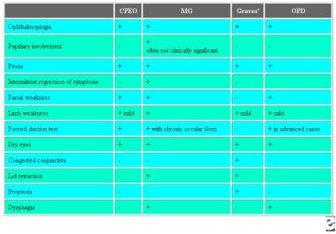
Ophthalmoplegia
Ophthalmoplegia, Chronic Progressive External
Kearns-Sayre Syndrome
Blepharoptosis
Multiple Sclerosis
DNA, Mitochondrial
Miller Fisher Syndrome
Oxymetholone
Carcinogenicity Tests
Mitochondria, Muscle
Kidney Tubules
Ocular Motility Disorders
Chronic Disease
Rats, Inbred F344
Anisocoria
Disease Progression
Cavernous Sinus
Herpes Zoster Ophthalmicus
Exophthalmos
Magnetic Resonance Imaging
Oculomotor Muscles
Point Mutation
Tolosa-Hunt Syndrome
Diplopia
Oculomotor Nerve Diseases
Kidney
Disease Models, Animal
Mitochondrial Diseases
Mitochondrial Encephalomyopathies
Wernicke Encephalopathy
Abducens Nerve
Brain
Abducens Nerve Diseases
Ataxia
Multiple Sclerosis, Chronic Progressive
Adenine Nucleotide Translocator 1
Cranial Nerve Diseases
Nystagmus, Pathologic
Oculomotor Nerve
Diffuse Cerebral Sclerosis of Schilder
Neuromuscular Diseases
Orbital Pseudotumor
Intestinal Pseudo-Obstruction
Electrooculography
Gangliosides
Pons
Facial Paralysis
Trochlear Nerve Diseases
Myelitis, Transverse
DNA-Directed DNA Polymerase
Fatal Outcome
Guillain-Barre Syndrome
Cerebellar Ataxia
Paranasal Sinus Diseases
Prednisolone
Heterogeneous presentation in A3243G mutation in the mitochondrial tRNA(Leu(UUR)) gene. (1/73)
AIMS: To clarify the phenotype-genotype relation associated with the A3243G mitochondrial DNA mutation. METHODS: Five unrelated probands harbouring the A3243G mutation but presenting different clinical phenotype were analysed. Probands include Leigh syndrome (LS(3243)), mitochondrial myopathy, encephalopathy, lactic acidosis and stroke like episodes (MELAS(3243)), progressive external ophthalmoplegia (PEO(3243)), and mitochondrial diabetes mellitus (MDM(3243)). Extensive clinical, histological, biochemical, and molecular genetic studies were performed on five families. RESULTS: All patients showed ragged red fibres (RRF), and focal cytochrome c oxidase (COX) deficiency except for the patient with MDM(3243). The mutation load was highest in the proband with LS(3243) (>90%), who also presented the highest proportion of RRF (68%) and COX negative fibres (10%), and severe complex I plus IV deficiency. These proportions were lower in the probands with PEO(3243) and with MDM(3243). CONCLUSION: The most severe clinical phenotype, LS(3243), was associated with the highest proportion of the A3243G mutation as well as the most prominent histological and biochemical abnormalities. (+info)Flux control of cytochrome c oxidase in human skeletal muscle. (2/73)
In the present work, by titrating cytochrome c oxidase (COX) with the specific inhibitor KCN, the flux control coefficient and the metabolic reserve capacity of COX have been determined in human saponin-permeabilized muscle fibers. In the presence of the substrates glutamate and malate, a 2.3 +/- 0.2-fold excess capacity of COX was observed in ADP-stimulated human skeletal muscle fibers. This value was found to be dependent on the mitochondrial substrate supply. In the combined presence of glutamate, malate, and succinate, which supported an approximately 1.4-fold higher rate of respiration, only a 1.4 +/- 0.2-fold excess capacity of COX was determined. In agreement with these findings, the flux control of COX increased, in the presence of the three substrates, from 0.27 +/- 0.03 to 0.36 +/- 0.08. These results indicate a tight in vivo control of respiration by COX in human skeletal muscle. This tight control may have significant implications for mitochondrial myopathies. In support of this conclusion, the analysis of skeletal muscle fibers from two patients with chronic progressive external ophthalmoplegia, which carried deletions in 11 and 49% of their mitochondrial DNA, revealed a substantially lowered reserve capacity and increased flux control coefficient of COX, indicating severe rate limitations of oxidative phosphorylation by this enzyme. (+info)Role of adenine nucleotide translocator 1 in mtDNA maintenance. (3/73)
Autosomal dominant progressive external ophthalmoplegia is a rare human disease that shows a Mendelian inheritance pattern, but is characterized by large-scale mitochondrial DNA (mtDNA) deletions. We have identified two heterozygous missense mutations in the nuclear gene encoding the heart/skeletal muscle isoform of the adenine nucleotide translocator (ANT1) in five families and one sporadic patient. The familial mutation substitutes a proline for a highly conserved alanine at position 114 in the ANT1 protein. The analogous mutation in yeast caused a respiratory defect. These results indicate that ANT has a role in mtDNA maintenance and that a mitochondrial disease can be caused by a dominant mechanism. (+info)Brain activation in normal subjects and in patients affected by mitochondrial disease without clinical central nervous system involvement: a phosphorus magnetic resonance spectroscopy study. (4/73)
It remains unclear whether brain energetics is disturbed in patients with mitochondrial disease without clinical central nervous system involvement (MDW). The authors used the high temporal and spatial resolution phosphorus magnetic resonance spectroscopy (31P MRS) technique that they developed to study high energy phosphates (HEPs) and intracellular pH (pH) in the visual cortex of 9 normal subjects and 5 MDW patients with single mtDNA deletion at rest, during, and after visual activation. In normal subjects, HEPs remained unchanged during activation but rose significantly (by 17%) during recovery, and pH increased during visual activation with a slow return to rest values. In MDW patients, HEPs were within the normal range at rest and did not change during activation, but fell significantly (by 22%) in the recovery period; pH did not reveal a homogeneous pattern. In the brain of patients with MDW, energy balance remains normal until oxidative metabolism is intensively stressed, as during a postactivation phase. The heterogeneity of the physicochemical environment (that is, pH) suggests various degrees of subclinical brain involvement. The combined use of MRS and brain activation is fundamental for the study of brain energetics and may prove an important diagnostic tool in patients with MDW. (+info)A unique junctional palindromic sequence in mitochondrial DNA from a patient with progressive external ophthalmoplegia. (5/73)
A polymerase chain reaction (PCR) based procedure was modified to determine the deletion of mitochondrial DNA (mtDNA). The protocol consists of coamplification both of deleted and wild-type segments of mtDNA using a long PCR technique; evaluation of the deleted portion within the amplified DNA segments by restriction enzyme digestion followed by densitometrical analysis; and direct subcloning into a plasmid vector for DNA sequencing. The procedure revealed a 5.3 kb deletion of mtDNA in the biopsied muscle tissue obtained from a patient clinically diagnosed with progressive external ophthalmoplegia. The 5' and 3' sequences at both sides of the breakpoint comprise a 17 bp palindrome and 5 bp tandem repeats, suggesting that the deletion might occur through slipped mispairing and other novel mechanisms. This improved procedure has the potential to detect deletions occurring in the entire length of mtDNA, and mighty be useful for clinical screening of progressive external ophthalmoplegia. (+info)Mitochondrial gene defect in patients with chronic progressive external ophthalmoplegia. (6/73)
OBJECTIVE: To detect the gene defect of mitochondrial DNA (mtDNA) from skeletal muscles in 2 patients with chronic progressive external ophthalmoplegia (CPEO). METHODS: After extraction of mtDNA, Southern hybridization was performed after restrictive digestion by Pvu II, EcoRI, Hind III, and Sacl. Then, we carried out polymerase chain reaction (PCR) and the enzyme digestion of the PCR products. Finally, mtDNA sequencing was done by automatic DNA sequence analyzer. RESULTS: In case 1, a 5 kb deletion was found by Southern blot analysis and PCR. And dosage analysis showed a heteroplasmic change with 44% mtDNAs deleted. In case 2, PCR plus restriction endonuclease Pvu II digestion demonstrated a mutation which was confirmed by DNA sequencing to be a single base substitution (T-->C) inducing a novel Pvu II site around 10,909 on mtDNA sequence. The laser image analyzer measurement revealed the mutation was almost homologous (99.4% mutant). CONCLUSIONS: In case 1, a 5 kb deletion found in mtDNA is called "common deletion" according to the literature. In case 2, a novel Pvu II site was found. It seems to be a de novo point mutation affecting ND4 in published CPEO research and is first reported in Chinese population. This point mutation does not induce an amino acid(Phe) change according to the published human mitochondrial genetic code as well as the mtDNA sequence. Whether it affects the translation efficiency or transportation of signals between mitochondrial and nuclear genome needs further studies. (+info)Characterization of a novel human putative mitochondrial transporter homologous to the yeast mitochondrial RNA splicing proteins 3 and 4. (7/73)
We report here a novel human gene, hMRS3/4, encoding a putative mitochondrial transporter structurally and functionally homologous to the yeast mitochondrial RNA splicing proteins 3 and 4. These proteins belong to the family of mitochondrial carrier proteins (MCF) and are likely to function as solute carriers. hMRS3/4 spans approximately 10 kb of genomic DNA on chromosome 10q24 and consists of four exons that encode a 364-aa protein with six transmembrane domains. A putative splice variant, encoding a 177-aa protein with three transmembrane domains, was also identified. hMRS3/4 has a well-conserved signature sequence of MCF and is targeted into the mitochondria. When expressed in yeast, hMRS3/4 efficiently restores the mitochondrial functions in mrs3(o)mrs4(o) knock-out mutants. Ubiquitous expression in human tissues and a well-conserved structure and function suggest an important role for hMRS3/4 in human cells. (+info)Active site mutation in DNA polymerase gamma associated with progressive external ophthalmoplegia causes error-prone DNA synthesis. (8/73)
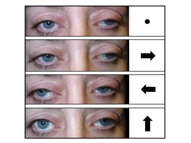
Progressive external ophthalmoplegia (PEO) is a heritable mitochondrial disorder characterized by the accumulation of multiple point mutations and large deletions in mtDNA. Autosomal dominant PEO was recently shown to co-segregate with a heterozygous Y955C mutation in the human gene encoding the sole mitochondrial DNA polymerase, DNA polymerase gamma (pol gamma). Since Tyr-955 is a highly conserved residue critical for nucleotide recognition among family A DNA polymerases, we analyzed the effects of the Y955C mutation on the kinetics and fidelity of DNA synthesis by the purified human mutant polymerase in complex with its accessory subunit. The Y955C enzyme retains a wild-type catalytic rate (k(cat)) but suffers a 45-fold decrease in apparent binding affinity for the incoming nucleoside triphosphate (K(m)). The Y955C derivative is 2-fold less accurate for base pair substitutions than wild-type pol gamma despite the action of intrinsic exonucleolytic proofreading. The full mutator effect of the Y955C substitution was revealed by genetic inactivation of the exonuclease, and error rates for certain mismatches were elevated by 10-100-fold. The error-prone DNA synthesis observed for the Y955C pol gamma is consistent with the accumulation of mtDNA mutations in patients with PEO. (+info)Ophthalmoplegia is a medical term that refers to the paralysis or weakness of the eye muscles, which can result in double vision (diplopia) or difficulty moving the eyes. It can be caused by various conditions, including nerve damage, muscle disorders, or neurological diseases such as myasthenia gravis or multiple sclerosis. Ophthalmoplegia can affect one or more eye muscles and can be partial or complete. Depending on the underlying cause, ophthalmoplegia may be treatable with medications, surgery, or other interventions.
Chronic Progressive External Ophthalmoplegia (CPEO) is a rare, progressive neuromuscular disorder that affects the extraocular muscles, which are responsible for eye movement. This results in progressive weakness and paralysis of these muscles, leading to limitations in eye movement and, subsequently, binocular vision.
The term "chronic" refers to the slow, gradual progression of symptoms over time, while "progressive" highlights the worsening nature of the condition. "External" indicates that the extraocular muscles are involved, as opposed to the "internal" ophthalmoplegia, which would refer to the paralysis of the iris and ciliary body muscles within the eye.
CPEO is characterized by symmetrical, bilateral paresis (partial paralysis) or complete paralysis of the extraocular muscles, leading to drooping eyelids (ptosis), limited eye movements in all directions, and double vision (diplopia). The onset of symptoms typically occurs during adulthood, but it can also manifest in childhood.
CPEO is often associated with mitochondrial DNA abnormalities or mutations, which can lead to impaired energy production within the cells. This specific type of ophthalmoplegia is generally not linked to other neurological or systemic symptoms, but it can co-occur with additional manifestations in some cases, forming a broader spectrum of mitochondrial disorders known as Kearns-Sayre syndrome (KSS) or oculocraniosomatic syndrome.
There is no cure for CPEO, and management primarily focuses on addressing the symptoms and improving quality of life. Treatment options may include surgical interventions to correct ptosis or strabismus (squint), as well as supportive care such as visual aids and rehabilitation strategies.
Kearns-Sayre Syndrome (KSS) is a rare, progressive genetic disorder that affects the function of the mitochondria, which are the energy-producing structures in cells. It is classified as a type of mitochondrial myopathy and is typically associated with symptoms that appear before the age of 20.
The medical definition of Kearns-Sayre Syndrome includes the following criteria:
1. Onset before 20 years of age
2. Progressive external ophthalmoplegia (PEO), which is characterized by weakness and paralysis of the eye muscles, leading to drooping eyelids (ptosis) and limited eye movement
3. Retinitis pigmentosa, a degenerative condition affecting the retina that can lead to vision loss
4. A cardiac conduction defect, such as heart block
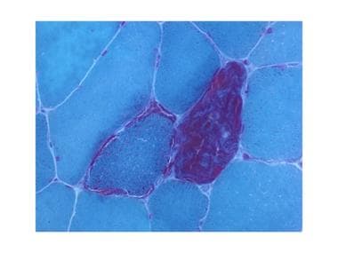
5. Ragged red fibers on muscle biopsy
6. At least one major criteria or two minor criteria must be present:
* Major criteria include cerebellar ataxia (lack of coordination), deafness, or increased protein in the cerebrospinal fluid
* Minor criteria include pigmentary retinopathy, heart block, or a high level of creatine kinase in the blood.
Kearns-Sayre Syndrome is caused by a single large-scale deletion of genes in the mitochondrial DNA and is usually sporadic, meaning it occurs randomly and is not inherited from parents. The condition can be diagnosed through genetic testing, muscle biopsy, or other clinical tests. Treatment is focused on managing symptoms and may include physical therapy, surgery for ptosis, hearing aids, and pacemakers for heart block.
Blepharoptosis is a medical term that refers to the drooping or falling of the upper eyelid. It is usually caused by weakness or paralysis of the muscle that raises the eyelid, known as the levator palpebrae superioris. This condition can be present at birth or acquired later in life due to various factors such as aging, nerve damage, eye surgery complications, or certain medical conditions like myasthenia gravis or brain tumors. Blepharoptosis may obstruct vision and cause difficulty with daily activities, and treatment options include eyedrops, eye patches, or surgical correction.
Multiple Sclerosis (MS) is a chronic autoimmune disease that affects the central nervous system (CNS), which includes the brain, spinal cord, and optic nerves. In MS, the immune system mistakenly attacks the protective covering of nerve fibers, called myelin, leading to damage and scarring (sclerosis). This results in disrupted communication between the brain and the rest of the body, causing a variety of neurological symptoms that can vary widely from person to person.
The term "multiple" refers to the numerous areas of scarring that occur throughout the CNS in this condition. The progression, severity, and specific symptoms of MS are unpredictable and may include vision problems, muscle weakness, numbness or tingling, difficulty with balance and coordination, cognitive impairment, and mood changes. There is currently no cure for MS, but various treatments can help manage symptoms, modify the course of the disease, and improve quality of life for those affected.
Mitochondrial DNA (mtDNA) is the genetic material present in the mitochondria, which are specialized structures within cells that generate energy. Unlike nuclear DNA, which is present in the cell nucleus and inherited from both parents, mtDNA is inherited solely from the mother.
MtDNA is a circular molecule that contains 37 genes, including 13 genes that encode for proteins involved in oxidative phosphorylation, a process that generates energy in the form of ATP. The remaining genes encode for rRNAs and tRNAs, which are necessary for protein synthesis within the mitochondria.
Mutations in mtDNA can lead to a variety of genetic disorders, including mitochondrial diseases, which can affect any organ system in the body. These mutations can also be used in forensic science to identify individuals and establish biological relationships.
Miller Fisher Syndrome (MFS) is a rare neurological disorder that is considered a variant of Guillain-Barré syndrome. It is characterized by the triad of symptoms including ophthalmoplegia (paralysis of the eye muscles), ataxia (loss of coordination and balance), and areflexia (absence of reflexes). Some patients may also experience weakness or paralysis in the limbs, and some cases may involve bulbar symptoms such as dysphagia (difficulty swallowing) and dysarthria (slurred speech). The syndrome is caused by an immune response that damages the nerves, and it often follows a viral infection. Treatment typically includes supportive care, plasma exchange, or intravenous immunoglobulin therapy to help reduce the severity of the symptoms.
Oxymetholone is an anabolic steroid medication, which is used to treat various medical conditions such as anemia due to lack of red blood cells and wasting syndrome in people with HIV infection. It works by increasing the production of erythropoietin, a hormone that stimulates the production of red blood cells. Oxymetholone also helps to improve muscle mass and appetite.
It is important to note that oxymetholone is a controlled substance and has potential for serious side effects, including liver toxicity, masculinization in women, and cardiovascular risks. Therefore, it should only be used under the close supervision of a healthcare provider and for legitimate medical purposes.
Carcinogenicity tests are a type of toxicity test used to determine the potential of a chemical or physical agent to cause cancer. These tests are typically conducted on animals, such as rats or mice, and involve exposing the animals to the agent over a long period of time, often for the majority of their lifespan. The animals are then closely monitored for any signs of tumor development or other indicators of cancer.
The results of carcinogenicity tests can be used by regulatory agencies, such as the U.S. Environmental Protection Agency (EPA) and the Food and Drug Administration (FDA), to help determine safe exposure levels for chemicals and other agents. The tests are also used by industry to assess the potential health risks associated with their products and to develop safer alternatives.
It is important to note that carcinogenicity tests have limitations, including the use of animals, which may not always accurately predict the effects of a chemical on humans. Additionally, these tests can be time-consuming and expensive, which has led to the development of alternative test methods, such as in vitro (test tube) assays and computational models, that aim to provide more efficient and ethical alternatives for carcinogenicity testing.
Mitochondria in muscle, also known as the "powerhouses" of the cell, are organelles that play a crucial role in generating energy for muscle cells through a process called cellular respiration. They convert the chemical energy found in glucose and oxygen into ATP (adenosine triphosphate), which is the main source of energy used by cells.
Muscle cells contain a high number of mitochondria due to their high energy demands for muscle contraction and relaxation. The number and size of mitochondria in muscle fibers can vary depending on the type of muscle fiber, with slow-twitch, aerobic fibers having more numerous and larger mitochondria than fast-twitch, anaerobic fibers.
Mitochondrial dysfunction has been linked to various muscle disorders, including mitochondrial myopathies, which are characterized by muscle weakness, exercise intolerance, and other symptoms related to impaired energy production in the muscle cells.
Kidney tubules are the structural and functional units of the kidney responsible for reabsorption, secretion, and excretion of various substances. They are part of the nephron, which is the basic unit of the kidney's filtration and reabsorption process.
There are three main types of kidney tubules:
1. Proximal tubule: This is the initial segment of the kidney tubule that receives the filtrate from the glomerulus. It is responsible for reabsorbing approximately 65% of the filtrate, including water, glucose, amino acids, and electrolytes.
2. Loop of Henle: This U-shaped segment of the tubule consists of a thin descending limb, a thin ascending limb, and a thick ascending limb. The loop of Henle helps to concentrate urine by creating an osmotic gradient that allows water to be reabsorbed in the collecting ducts.
3. Distal tubule: This is the final segment of the kidney tubule before it empties into the collecting duct. It is responsible for fine-tuning the concentration of electrolytes and pH balance in the urine by selectively reabsorbing or secreting substances such as sodium, potassium, chloride, and hydrogen ions.
Overall, kidney tubules play a critical role in maintaining fluid and electrolyte balance, regulating acid-base balance, and removing waste products from the body.
Ocular motility disorders refer to a group of conditions that affect the movement of the eyes. These disorders can result from nerve damage, muscle dysfunction, or brain injuries. They can cause abnormal eye alignment, limited range of motion, and difficulty coordinating eye movements. Common symptoms include double vision, blurry vision, strabismus (crossed eyes), nystagmus (involuntary eye movement), and difficulty tracking moving objects. Ocular motility disorders can be congenital or acquired and may require medical intervention to correct or manage the condition.
A chronic disease is a long-term medical condition that often progresses slowly over a period of years and requires ongoing management and care. These diseases are typically not fully curable, but symptoms can be managed to improve quality of life. Common chronic diseases include heart disease, stroke, cancer, diabetes, arthritis, and COPD (chronic obstructive pulmonary disease). They are often associated with advanced age, although they can also affect children and younger adults. Chronic diseases can have significant impacts on individuals' physical, emotional, and social well-being, as well as on healthcare systems and society at large.
F344 is a strain code used to designate an outbred stock of rats that has been inbreeded for over 100 generations. The F344 rats, also known as Fischer 344 rats, were originally developed at the National Institutes of Health (NIH) and are now widely used in biomedical research due to their consistent and reliable genetic background.
Inbred strains, like the F344, are created by mating genetically identical individuals (siblings or parents and offspring) for many generations until a state of complete homozygosity is reached, meaning that all members of the strain have identical genomes. This genetic uniformity makes inbred strains ideal for use in studies where consistent and reproducible results are important.
F344 rats are known for their longevity, with a median lifespan of around 27-31 months, making them useful for aging research. They also have a relatively low incidence of spontaneous tumors compared to other rat strains. However, they may be more susceptible to certain types of cancer and other diseases due to their inbred status.
It's important to note that while F344 rats are often used as a standard laboratory rat strain, there can still be some genetic variation between individual animals within the same strain, particularly if they come from different suppliers or breeding colonies. Therefore, it's always important to consider the source and history of any animal model when designing experiments and interpreting results.
Anisocoria is a medical term that refers to an inequality in the size of the pupils in each eye. The pupil is the black, circular opening in the center of the iris (the colored part of the eye) that allows light to enter and strike the retina. Normally, the pupils are equal in size and react similarly when exposed to light or darkness. However, in anisocoria, one pupil is larger or smaller than the other.
Anisocoria can be caused by various factors, including neurological conditions, trauma, eye diseases, or medications that affect the pupillary reflex. In some cases, anisocoria may be a normal variant and not indicative of any underlying medical condition. However, if it is a new finding or associated with other symptoms such as pain, headache, vision changes, or decreased level of consciousness, it should be evaluated by a healthcare professional to determine the cause and appropriate treatment.
Disease progression is the worsening or advancement of a medical condition over time. It refers to the natural course of a disease, including its development, the severity of symptoms and complications, and the impact on the patient's overall health and quality of life. Understanding disease progression is important for developing appropriate treatment plans, monitoring response to therapy, and predicting outcomes.
The rate of disease progression can vary widely depending on the type of medical condition, individual patient factors, and the effectiveness of treatment. Some diseases may progress rapidly over a short period of time, while others may progress more slowly over many years. In some cases, disease progression may be slowed or even halted with appropriate medical interventions, while in other cases, the progression may be inevitable and irreversible.
In clinical practice, healthcare providers closely monitor disease progression through regular assessments, imaging studies, and laboratory tests. This information is used to guide treatment decisions and adjust care plans as needed to optimize patient outcomes and improve quality of life.
The cavernous sinus is a venous structure located in the middle cranial fossa, which is a depression in the skull that houses several important nerves and blood vessels. The cavernous sinus is situated on either side of the sphenoid bone, near the base of the skull, and it contains several important structures:
* The internal carotid artery, which supplies oxygenated blood to the brain
* The abducens nerve (cranial nerve VI), which controls lateral movement of the eye
* The oculomotor nerve (cranial nerve III), which controls most of the muscles that move the eye
* The trochlear nerve (cranial nerve IV), which controls one of the muscles that moves the eye
* The ophthalmic and maxillary divisions of the trigeminal nerve (cranial nerve V), which transmit sensory information from the face and head
The cavernous sinus is an important structure because it serves as a conduit for several critical nerves and blood vessels. However, it is also vulnerable to various pathological conditions such as thrombosis (blood clots), infection, tumors, or aneurysms, which can lead to serious neurological deficits or even death.
Kidney disease, also known as nephropathy or renal disease, refers to any functional or structural damage to the kidneys that impairs their ability to filter blood, regulate electrolytes, produce hormones, and maintain fluid balance. This damage can result from a wide range of causes, including diabetes, hypertension, glomerulonephritis, polycystic kidney disease, lupus, infections, drugs, toxins, and congenital or inherited disorders.
Depending on the severity and progression of the kidney damage, kidney diseases can be classified into two main categories: acute kidney injury (AKI) and chronic kidney disease (CKD). AKI is a sudden and often reversible loss of kidney function that occurs over hours to days, while CKD is a progressive and irreversible decline in kidney function that develops over months or years.
Symptoms of kidney diseases may include edema, proteinuria, hematuria, hypertension, electrolyte imbalances, metabolic acidosis, anemia, and decreased urine output. Treatment options depend on the underlying cause and severity of the disease and may include medications, dietary modifications, dialysis, or kidney transplantation.
Herpes Zoster Ophthalmicus (HZO) is a type of herpes zoster (shingles) infection that affects the ophthalmic division (V1) of the trigeminal nerve. It is caused by the varicella-zoster virus, which also causes chickenpox. After a person recovers from chickenpox, the virus remains inactive in the body and can reactivate later as shingles, often many years after the initial infection.
When the virus reactivates and affects the ophthalmic division of the trigeminal nerve, it can cause a painful rash on the forehead, nose, and around one eye. The rash may be accompanied by other symptoms such as headache, fever, and fatigue. In some cases, HZO can also affect the eye itself, causing inflammation, corneal ulcers, and vision loss if left untreated.
It is important to seek medical attention promptly if you suspect you have HZO, as early treatment with antiviral medications can help reduce the severity of symptoms and prevent complications.
Exophthalmos is a medical condition that refers to the abnormal protrusion or bulging of one or both eyes beyond the normal orbit (eye socket). This condition is also known as proptosis. Exophthalmos can be caused by various factors, including thyroid eye disease (Graves' ophthalmopathy), tumors, inflammation, trauma, or congenital abnormalities. It can lead to various symptoms such as double vision, eye discomfort, redness, and difficulty closing the eyes. Treatment of exophthalmos depends on the underlying cause and may include medications, surgery, or radiation therapy.
Medical Definition:
Magnetic Resonance Imaging (MRI) is a non-invasive diagnostic imaging technique that uses a strong magnetic field and radio waves to create detailed cross-sectional or three-dimensional images of the internal structures of the body. The patient lies within a large, cylindrical magnet, and the scanner detects changes in the direction of the magnetic field caused by protons in the body. These changes are then converted into detailed images that help medical professionals to diagnose and monitor various medical conditions, such as tumors, injuries, or diseases affecting the brain, spinal cord, heart, blood vessels, joints, and other internal organs. MRI does not use radiation like computed tomography (CT) scans.
The oculomotor muscles are a group of extraocular muscles that control the movements of the eye. They include:
1. Superior rectus: This muscle is responsible for elevating the eye and helping with inward rotation (intorsion) when looking downwards.
2. Inferior rectus: It depresses the eye and helps with outward rotation (extorsion) when looking upwards.
3. Medial rectus: This muscle adducts, or moves, the eye towards the midline of the face.
4. Inferior oblique: The inferior oblique muscle intorts and elevates the eye.
5. Superior oblique: It extorts and depresses the eye.
These muscles work together to allow for smooth and precise movements of the eyes, enabling tasks such as tracking moving objects, reading, and maintaining visual fixation on a single point in space.
A point mutation is a type of genetic mutation where a single nucleotide base (A, T, C, or G) in DNA is altered, deleted, or substituted with another nucleotide. Point mutations can have various effects on the organism, depending on the location of the mutation and whether it affects the function of any genes. Some point mutations may not have any noticeable effect, while others might lead to changes in the amino acids that make up proteins, potentially causing diseases or altering traits. Point mutations can occur spontaneously due to errors during DNA replication or be inherited from parents.
Kidney neoplasms refer to abnormal growths or tumors in the kidney tissues that can be benign (non-cancerous) or malignant (cancerous). These growths can originate from various types of kidney cells, including the renal tubules, glomeruli, and the renal pelvis.
Malignant kidney neoplasms are also known as kidney cancers, with renal cell carcinoma being the most common type. Benign kidney neoplasms include renal adenomas, oncocytomas, and angiomyolipomas. While benign neoplasms are generally not life-threatening, they can still cause problems if they grow large enough to compromise kidney function or if they undergo malignant transformation.
Early detection and appropriate management of kidney neoplasms are crucial for improving patient outcomes and overall prognosis. Regular medical check-ups, imaging studies, and urinalysis can help in the early identification of these growths, allowing for timely intervention and treatment.
Tolosa-Hunt syndrome is a rare disorder characterized by the inflammation of the nerve structures (including the fifth and sixth cranial nerves) within the cavernous sinus, a venous space near the base of the skull. This inflammation can lead to various symptoms such as:
1. Unilateral or bilateral orbital pain, which may be severe and deep, often radiating around the eye and temple.
2. Ophthalmoplegia (paralysis of the eye muscles), causing double vision (diplopia) and limited eye movement in specific directions.
3. Ptosis (drooping of the eyelid).
4. Other possible symptoms include decreased sensation around the forehead, cheek, or upper jaw, and loss of taste on the anterior part of the tongue.
The exact cause of Tolosa-Hunt syndrome is unknown, but it's believed to be related to an autoimmune response or a non-specific inflammatory process. It can also occur in conjunction with other medical conditions like neoplasms (tumors) or infections. The diagnosis typically involves imaging studies such as MRI and CT scans, along with blood tests and a thorough neurological examination.
Treatment usually includes corticosteroids to reduce inflammation and alleviate symptoms. In some cases, immunosuppressive medications or radiation therapy may be necessary. If left untreated, Tolosa-Hunt syndrome can lead to permanent visual impairment or other neurological deficits.
Diplopia is a medical term that refers to the condition where a person sees two images of a single object. It is commonly known as double vision. This can occur due to various reasons, such as nerve damage or misalignment of the eyes. Diplopia can be temporary or chronic and can affect one or both eyes. If you're experiencing diplopia, it's essential to consult an eye care professional for proper evaluation and treatment.
The oculomotor nerve, also known as the third cranial nerve (CN III), is responsible for controlling several important eye movements and functions. Oculomotor nerve diseases refer to conditions that affect this nerve and can lead to various symptoms related to eye movement and function. Here's a medical definition of oculomotor nerve diseases:
Oculomotor nerve diseases are a group of medical disorders characterized by the dysfunction or damage to the oculomotor nerve (CN III), resulting in impaired eye movements, abnormalities in pupillary response, and potential effects on eyelid position. These conditions can be congenital, acquired, or traumatic in nature and may lead to partial or complete paralysis of the nerve. Common oculomotor nerve diseases include oculomotor nerve palsy, third nerve ganglionopathies, and compressive oculomotor neuropathies caused by various pathologies such as aneurysms, tumors, or infections.
Mitochondrial myopathies are a group of genetic disorders caused by mutations in the mitochondrial DNA or nuclear DNA that affect the function of the mitochondria, which are the energy-producing structures in cells. These mutations can result in impaired muscle function and other symptoms, depending on the specific type and severity of the disorder.
Mitochondrial myopathies can present at any age and can cause a range of symptoms, including muscle weakness, exercise intolerance, fatigue, muscle pain, and difficulty with coordination and balance. Some people with mitochondrial myopathies may also experience neurological symptoms such as seizures, developmental delays, and hearing or vision loss.
The diagnosis of mitochondrial myopathies typically involves a combination of clinical evaluation, muscle biopsy, genetic testing, and other diagnostic tests to assess mitochondrial function. Treatment is generally supportive and may include physical therapy, medications to manage symptoms, and nutritional support. In some cases, specific therapies such as vitamin or coenzyme Q10 supplementation may be recommended based on the underlying genetic defect.
Proteinuria is a medical term that refers to the presence of excess proteins, particularly albumin, in the urine. Under normal circumstances, only small amounts of proteins should be found in the urine because the majority of proteins are too large to pass through the glomeruli, which are the filtering units of the kidneys.
However, when the glomeruli become damaged or diseased, they may allow larger molecules such as proteins to leak into the urine. Persistent proteinuria is often a sign of kidney disease and can indicate damage to the glomeruli. It is usually detected through a routine urinalysis and may be confirmed with further testing.
The severity of proteinuria can vary, and it can be a symptom of various underlying conditions such as diabetes, hypertension, glomerulonephritis, and other kidney diseases. Treatment for proteinuria depends on the underlying cause and may include medications to control blood pressure, manage diabetes, or reduce protein loss in the urine.
A kidney, in medical terms, is one of two bean-shaped organs located in the lower back region of the body. They are essential for maintaining homeostasis within the body by performing several crucial functions such as:
1. Regulation of water and electrolyte balance: Kidneys help regulate the amount of water and various electrolytes like sodium, potassium, and calcium in the bloodstream to maintain a stable internal environment.
2. Excretion of waste products: They filter waste products from the blood, including urea (a byproduct of protein metabolism), creatinine (a breakdown product of muscle tissue), and other harmful substances that result from normal cellular functions or external sources like medications and toxins.
3. Endocrine function: Kidneys produce several hormones with important roles in the body, such as erythropoietin (stimulates red blood cell production), renin (regulates blood pressure), and calcitriol (activated form of vitamin D that helps regulate calcium homeostasis).
4. pH balance regulation: Kidneys maintain the proper acid-base balance in the body by excreting either hydrogen ions or bicarbonate ions, depending on whether the blood is too acidic or too alkaline.
5. Blood pressure control: The kidneys play a significant role in regulating blood pressure through the renin-angiotensin-aldosterone system (RAAS), which constricts blood vessels and promotes sodium and water retention to increase blood volume and, consequently, blood pressure.
Anatomically, each kidney is approximately 10-12 cm long, 5-7 cm wide, and 3 cm thick, with a weight of about 120-170 grams. They are surrounded by a protective layer of fat and connected to the urinary system through the renal pelvis, ureters, bladder, and urethra.
Animal disease models are specialized animals, typically rodents such as mice or rats, that have been genetically engineered or exposed to certain conditions to develop symptoms and physiological changes similar to those seen in human diseases. These models are used in medical research to study the pathophysiology of diseases, identify potential therapeutic targets, test drug efficacy and safety, and understand disease mechanisms.
The genetic modifications can include knockout or knock-in mutations, transgenic expression of specific genes, or RNA interference techniques. The animals may also be exposed to environmental factors such as chemicals, radiation, or infectious agents to induce the disease state.
Examples of animal disease models include:
1. Mouse models of cancer: Genetically engineered mice that develop various types of tumors, allowing researchers to study cancer initiation, progression, and metastasis.
2. Alzheimer's disease models: Transgenic mice expressing mutant human genes associated with Alzheimer's disease, which exhibit amyloid plaque formation and cognitive decline.
3. Diabetes models: Obese and diabetic mouse strains like the NOD (non-obese diabetic) or db/db mice, used to study the development of type 1 and type 2 diabetes, respectively.
4. Cardiovascular disease models: Atherosclerosis-prone mice, such as ApoE-deficient or LDLR-deficient mice, that develop plaque buildup in their arteries when fed a high-fat diet.
5. Inflammatory bowel disease models: Mice with genetic mutations affecting intestinal barrier function and immune response, such as IL-10 knockout or SAMP1/YitFc mice, which develop colitis.
Animal disease models are essential tools in preclinical research, but it is important to recognize their limitations. Differences between species can affect the translatability of results from animal studies to human patients. Therefore, researchers must carefully consider the choice of model and interpret findings cautiously when applying them to human diseases.
An adenoma is a benign (noncancerous) tumor that develops from glandular epithelial cells. These types of cells are responsible for producing and releasing fluids, such as hormones or digestive enzymes, into the surrounding tissues. Adenomas can occur in various organs and glands throughout the body, including the thyroid, pituitary, adrenal, and digestive systems.
Depending on their location, adenomas may cause different symptoms or remain asymptomatic. Some common examples of adenomas include:
1. Colorectal adenoma (also known as a polyp): These growths occur in the lining of the colon or rectum and can develop into colorectal cancer if left untreated. Regular screenings, such as colonoscopies, are essential for early detection and removal of these polyps.
2. Thyroid adenoma: This type of adenoma affects the thyroid gland and may result in an overproduction or underproduction of hormones, leading to conditions like hyperthyroidism (overactive thyroid) or hypothyroidism (underactive thyroid).
3. Pituitary adenoma: These growths occur in the pituitary gland, which is located at the base of the brain and controls various hormonal functions. Depending on their size and location, pituitary adenomas can cause vision problems, headaches, or hormonal imbalances that affect growth, reproduction, and metabolism.
4. Liver adenoma: These rare benign tumors develop in the liver and may not cause any symptoms unless they become large enough to press on surrounding organs or structures. In some cases, liver adenomas can rupture and cause internal bleeding.
5. Adrenal adenoma: These growths occur in the adrenal glands, which are located above the kidneys and produce hormones that regulate stress responses, metabolism, and blood pressure. Most adrenal adenomas are nonfunctioning, meaning they do not secrete excess hormones. However, functioning adrenal adenomas can lead to conditions like Cushing's syndrome or Conn's syndrome, depending on the type of hormone being overproduced.
It is essential to monitor and manage benign tumors like adenomas to prevent potential complications, such as rupture, bleeding, or hormonal imbalances. Treatment options may include surveillance with imaging studies, medication to manage hormonal issues, or surgical removal of the tumor in certain cases.
Mitochondrial diseases are a group of disorders caused by dysfunctions in the mitochondria, which are the energy-producing structures in cells. These diseases can affect people of any age and can manifest in various ways, depending on which organs or systems are affected. Common symptoms include muscle weakness, neurological problems, cardiac disease, diabetes, and vision/hearing loss. Mitochondrial diseases can be inherited from either the mother's or father's side, or they can occur spontaneously due to genetic mutations. They can range from mild to severe and can even be life-threatening in some cases.
Mitochondrial Encephalomyopathies are a group of genetic disorders that primarily affect the mitochondria, which are the energy-producing structures in cells. "Encephalo" refers to the brain, while "myopathy" refers to muscle disease. Therefore, Mitochondrial Encephalomyopathies are conditions that cause both neurological and muscular symptoms due to impaired mitochondrial function.
These disorders can affect any organ in the body, but they primarily impact the brain, nerves, and muscles. Symptoms may include muscle weakness, seizures, developmental delays, hearing loss, vision loss, heart problems, and lactic acidosis (a buildup of lactic acid in the blood).
Mitochondrial Encephalomyopathies can be caused by mutations in either the mitochondrial DNA or nuclear DNA. They are often inherited from the mother, as mitochondria are passed down through the maternal line. However, some cases can also result from new mutations that occur spontaneously.
Due to the complex nature of these disorders and their varying symptoms, diagnosis and treatment can be challenging. Treatment typically focuses on managing specific symptoms and may include medications, dietary changes, and physical therapy.
Wernicke Encephalopathy is a neuropsychiatric disorder that is caused by a deficiency of thiamine (vitamin B1). It is characterized by a classic triad of symptoms: confusion, oculomotor dysfunction (such as nystagmus and ophthalmoplegia), and gait ataxia. Other symptoms can include memory loss, apathy, and hypothermia.
Wernicke Encephalopathy is most commonly seen in alcoholics due to poor nutrition, but it can also occur in people with conditions that cause malabsorption or increased thiamine requirements, such as AIDS, cancer, and chronic diarrhea. Immediate treatment with thiamine replacement therapy is necessary to prevent progression of the disease and potential permanent neurological damage. If left untreated, Wernicke Encephalopathy can lead to Korsakoff's syndrome, a chronic memory disorder.
The abducens nerve, also known as the sixth cranial nerve (CN VI), is a motor nerve that controls the lateral rectus muscle of the eye. This muscle is responsible for moving the eye away from the midline (towards the temple) and enables the eyes to look towards the side while keeping them aligned. Any damage or dysfunction of the abducens nerve can result in strabismus, where the eyes are misaligned and point in different directions, specifically an adduction deficit, also known as abducens palsy or sixth nerve palsy.
The brain is the central organ of the nervous system, responsible for receiving and processing sensory information, regulating vital functions, and controlling behavior, movement, and cognition. It is divided into several distinct regions, each with specific functions:
1. Cerebrum: The largest part of the brain, responsible for higher cognitive functions such as thinking, learning, memory, language, and perception. It is divided into two hemispheres, each controlling the opposite side of the body.
2. Cerebellum: Located at the back of the brain, it is responsible for coordinating muscle movements, maintaining balance, and fine-tuning motor skills.
3. Brainstem: Connects the cerebrum and cerebellum to the spinal cord, controlling vital functions such as breathing, heart rate, and blood pressure. It also serves as a relay center for sensory information and motor commands between the brain and the rest of the body.
4. Diencephalon: A region that includes the thalamus (a major sensory relay station) and hypothalamus (regulates hormones, temperature, hunger, thirst, and sleep).
5. Limbic system: A group of structures involved in emotional processing, memory formation, and motivation, including the hippocampus, amygdala, and cingulate gyrus.
The brain is composed of billions of interconnected neurons that communicate through electrical and chemical signals. It is protected by the skull and surrounded by three layers of membranes called meninges, as well as cerebrospinal fluid that provides cushioning and nutrients.
The abducens nerve, also known as the sixth cranial nerve, is responsible for controlling the lateral rectus muscle of the eye, which enables the eye to move outward. Abducens nerve diseases refer to conditions that affect this nerve and can result in various symptoms, primarily affecting eye movement.
Here are some medical definitions related to abducens nerve diseases:
1. Abducens Nerve Palsy: A condition characterized by weakness or paralysis of the abducens nerve, causing difficulty in moving the affected eye outward. This results in double vision (diplopia), especially when gazing towards the side of the weakened nerve. Abducens nerve palsy can be congenital, acquired, or caused by various factors such as trauma, tumors, aneurysms, infections, or diseases like diabetes and multiple sclerosis.
2. Sixth Nerve Palsy: Another term for abducens nerve palsy, referring to the weakness or paralysis of the sixth cranial nerve.
3. Internuclear Ophthalmoplegia (INO): A neurological condition affecting eye movement, often caused by a lesion in the medial longitudinal fasciculus (MLF), a bundle of nerve fibers that connects the abducens nucleus with the oculomotor nucleus. INO results in impaired adduction (inward movement) of the eye on the side of the lesion and nystagmus (involuntary eye movements) of the abducting eye on the opposite side when attempting to look towards the side of the lesion.
4. One-and-a-Half Syndrome: A rare neurological condition characterized by a combination of INO and internuclear ophthalmoplegia with horizontal gaze palsy on the same side, caused by damage to both the abducens nerve and the paramedian pontine reticular formation (PPRF). This results in limited or no ability to move the eyes towards the side of the lesion and impaired adduction of the eye on the opposite side.
5. Brainstem Encephalitis: Inflammation of the brainstem, which can affect the abducens nerve and other cranial nerves, leading to various neurological symptoms such as diplopia (double vision), ataxia (loss of balance and coordination), and facial weakness. Brainstem encephalitis can be caused by infectious agents, autoimmune disorders, or paraneoplastic syndromes.
6. Multiple Sclerosis (MS): An autoimmune disorder characterized by inflammation and demyelination of the central nervous system, including the brainstem and optic nerves. MS can cause various neurological symptoms, such as diplopia, nystagmus, and INO, due to damage to the abducens nerve and other cranial nerves.
7. Wernicke's Encephalopathy: A neurological disorder caused by thiamine (vitamin B1) deficiency, often seen in alcoholics or individuals with malnutrition. Wernicke's encephalopathy can affect the brainstem and cause various symptoms such as diplopia, ataxia, confusion, and oculomotor abnormalities.
8. Pontine Glioma: A rare type of brain tumor that arises from the glial cells in the pons (a part of the brainstem). Pontine gliomas can cause various neurological symptoms such as diplopia, facial weakness, and difficulty swallowing due to their location in the brainstem.
9. Brainstem Cavernous Malformation: A benign vascular lesion that arises from the small blood vessels in the brainstem. Brainstem cavernous malformations can cause various neurological symptoms such as diplopia, ataxia, and facial weakness due to their location in the brainstem.
10. Pituitary Adenoma: A benign tumor that arises from the pituitary gland, located at the base of the brain. Large pituitary adenomas can compress the optic nerves and cause various visual symptoms such as diplopia, visual field defects, and decreased vision.
11. Craniopharyngioma: A benign tumor that arises from the remnants of the Rathke's pouch, a structure that gives rise to the anterior pituitary gland. Craniopharyngiomas can cause various neurological and endocrine symptoms such as diplopia, visual field defects, headaches, and hormonal imbalances due to their location near the optic nerves and pituitary gland.
12. Meningioma: A benign tumor that arises from the meninges, the protective covering of the brain and spinal cord. Meningiomas can cause various neurological symptoms such as diplopia, headaches, and seizures depending on their location in the brain or spinal cord.
13. Chordoma: A rare type of malignant tumor that arises from the remnants of the notochord, a structure that gives rise to the spine during embryonic development. Chordomas can cause various neurological and endocrine symptoms such as diplopia, visual field defects, headaches, and hormonal imbalances due to their location near the brainstem and spinal cord.
14. Metastatic Brain Tumors: Malignant tumors that spread from other parts of the body to the brain. Metastatic brain tumors can cause various neurological symptoms such as diplopia, headaches, seizures, and cognitive impairment depending on their location in the brain.
15. Other Rare Brain Tumors: There are many other rare types of brain tumors that can cause diplopia or other neurological symptoms, including gliomas, ependymomas, pineal region tumors, and others. These tumors require specialized diagnosis and treatment by neuro-oncologists and neurosurgeons with expertise in these rare conditions.
In summary, diplopia can be caused by various brain tumors, including pituitary adenomas, meningiomas, chordomas, metastatic brain tumors, and other rare types of tumors. It is important to seek medical attention promptly if you experience diplopia or other neurological symptoms, as early diagnosis and treatment can improve outcomes and quality of life.
Ataxia is a medical term that refers to a group of disorders affecting coordination, balance, and speech. It is characterized by a lack of muscle control during voluntary movements, causing unsteady or awkward movements, and often accompanied by tremors. Ataxia can affect various parts of the body, such as the limbs, trunk, eyes, and speech muscles. The condition can be congenital or acquired, and it can result from damage to the cerebellum, spinal cord, or sensory nerves. There are several types of ataxia, including hereditary ataxias, degenerative ataxias, cerebellar ataxias, and acquired ataxias, each with its own specific causes, symptoms, and prognosis. Treatment for ataxia typically focuses on managing symptoms and improving quality of life, as there is no cure for most forms of the disorder.
Multiple Sclerosis (MS), Chronic Progressive is a form of Multiple Sclerosis, a chronic autoimmune disease that affects the central nervous system (CNS). In this form, the disease follows a steady progression with no distinct relapses or remissions. The symptoms worsen over time, leading to a decline in physical functioning and increased disability.
The term "chronic progressive" is used to describe the course of the disease, which is characterized by a continuous worsening of neurological functions from the onset, or after an initial relapsing-remitting phase. There are two types of chronic progressive MS: primary and secondary.
1. Primary Chronic Progressive MS (PCP): This form of MS shows a steady progression of symptoms from the beginning, with no distinct remissions or relapses. The disability accumulates gradually over time, and the person may experience varying degrees of physical and cognitive impairment.
2. Secondary Chronic Progressive MS (SCP): In this form, an individual initially has a relapsing-remitting course of MS (RRMS), characterized by unpredictable relapses followed by periods of partial or complete recovery (remissions). However, after some time, the disease transitions to a steady progression of symptoms and disability, even without distinct relapses. This is known as secondary chronic progressive MS.
The exact cause of Multiple Sclerosis remains unknown; however, it is believed to be influenced by genetic, environmental, and immunological factors. The disease involves the immune system attacking the myelin sheath, a protective covering surrounding nerve fibers in the CNS. This results in lesions or scars (scleroses) that disrupt communication between the brain, spinal cord, and other parts of the body, leading to various physical, cognitive, and sensory symptoms.
Management of Chronic Progressive MS typically involves a multidisciplinary approach, focusing on symptom management, rehabilitation, and maintaining quality of life. Currently, there are no approved disease-modifying therapies specifically for chronic progressive MS; however, some medications used to treat relapsing-remitting MS may help slow the progression of disability in certain individuals with secondary chronic progressive MS.
Adenine Nucleotide Translocator 1 (ANT1) is a protein found in the inner mitochondrial membrane of cells. It plays a crucial role in cellular energy metabolism by facilitating the exchange of adenosine diphosphate (ADP) and adenosine triphosphate (ATP) across the mitochondrial membrane.
In simpler terms, ANT1 helps to transport ATP, which is a major source of energy for cells, out of the mitochondria and exchange it for ADP, which can be converted back into ATP through cellular respiration. This process is essential for maintaining the energy balance within the cell and supporting various physiological functions.
Mutations in the gene that encodes ANT1 have been associated with certain mitochondrial disorders, such as autosomal recessive progressive external ophthalmoplegia (arPEO) and maternally inherited diabetes and deafness (MIDD). These genetic conditions can result in a range of symptoms, including muscle weakness, exercise intolerance, and neurological problems.
Cranial nerve diseases refer to conditions that affect the cranial nerves, which are a set of 12 pairs of nerves that originate from the brainstem and control various functions in the head and neck. These functions include vision, hearing, taste, smell, movement of the eyes and face, and sensation in the face.
Diseases of the cranial nerves can result from a variety of causes, including injury, infection, inflammation, tumors, or degenerative conditions. The specific symptoms that a person experiences will depend on which cranial nerve is affected and how severely it is damaged.
For example, damage to the optic nerve (cranial nerve II) can cause vision loss or visual disturbances, while damage to the facial nerve (cranial nerve VII) can result in weakness or paralysis of the face. Other common symptoms of cranial nerve diseases include pain, numbness, tingling, and hearing loss.
Treatment for cranial nerve diseases varies depending on the underlying cause and severity of the condition. In some cases, medication or surgery may be necessary to treat the underlying cause and relieve symptoms. Physical therapy or rehabilitation may also be recommended to help individuals regain function and improve their quality of life.
Pathological nystagmus is an abnormal, involuntary movement of the eyes that can occur in various directions (horizontal, vertical, or rotatory) and can be rhythmical or arrhythmic. It is typically a result of a disturbance in the vestibular system, central nervous system, or ocular motor pathways. Pathological nystagmus can cause visual symptoms such as blurred vision, difficulty with fixation, and oscillopsia (the sensation that one's surroundings are moving). The type, direction, and intensity of the nystagmus may vary depending on the underlying cause, which can include conditions such as brainstem or cerebellar lesions, multiple sclerosis, drug toxicity, inner ear disorders, and congenital abnormalities.
The oculomotor nerve, also known as the third cranial nerve (CN III), is a motor nerve that originates from the midbrain. It controls the majority of the eye muscles, including the levator palpebrae superioris muscle that raises the upper eyelid, and the extraocular muscles that enable various movements of the eye such as looking upward, downward, inward, and outward. Additionally, it carries parasympathetic fibers responsible for pupillary constriction and accommodation (focusing on near objects). Damage to this nerve can result in various ocular motor disorders, including strabismus, ptosis, and pupillary abnormalities.
Muscular diseases, also known as myopathies, refer to a group of conditions that affect the functionality and health of muscle tissue. These diseases can be inherited or acquired and may result from inflammation, infection, injury, or degenerative processes. They can cause symptoms such as weakness, stiffness, cramping, spasms, wasting, and loss of muscle function.
Examples of muscular diseases include:
1. Duchenne Muscular Dystrophy (DMD): A genetic disorder that results in progressive muscle weakness and degeneration due to a lack of dystrophin protein.
2. Myasthenia Gravis: An autoimmune disease that causes muscle weakness and fatigue, typically affecting the eyes and face, throat, and limbs.
3. Inclusion Body Myositis (IBM): A progressive muscle disorder characterized by muscle inflammation and wasting, typically affecting older adults.
4. Polymyositis: An inflammatory myopathy that causes muscle weakness and inflammation throughout the body.
5. Metabolic Myopathies: A group of inherited disorders that affect muscle metabolism, leading to exercise intolerance, muscle weakness, and other symptoms.
6. Muscular Dystonias: Involuntary muscle contractions and spasms that can cause abnormal postures or movements.
It is important to note that muscular diseases can have a significant impact on an individual's quality of life, mobility, and overall health. Proper diagnosis and treatment are crucial for managing symptoms and improving outcomes.
Orbital neoplasms refer to abnormal growths or tumors that develop in the orbit, which is the bony cavity that contains the eyeball, muscles, nerves, fat, and blood vessels. These neoplasms can be benign (non-cancerous) or malignant (cancerous), and they can arise from various types of cells within the orbit.
Orbital neoplasms can cause a variety of symptoms depending on their size, location, and rate of growth. Common symptoms include protrusion or displacement of the eyeball, double vision, limited eye movement, pain, swelling, and numbness in the face. In some cases, orbital neoplasms may not cause any noticeable symptoms, especially if they are small and slow-growing.
There are many different types of orbital neoplasms, including:
1. Optic nerve glioma: a rare tumor that arises from the optic nerve's supportive tissue.
2. Orbital meningioma: a tumor that originates from the membranes covering the brain and extends into the orbit.
3. Lacrimal gland tumors: benign or malignant growths that develop in the lacrimal gland, which produces tears.
4. Orbital lymphangioma: a non-cancerous tumor that arises from the lymphatic vessels in the orbit.
5. Rhabdomyosarcoma: a malignant tumor that develops from the skeletal muscle cells in the orbit.
6. Metastatic tumors: cancerous growths that spread to the orbit from other parts of the body, such as the breast, lung, or prostate.
The diagnosis and treatment of orbital neoplasms depend on several factors, including the type, size, location, and extent of the tumor. Imaging tests, such as CT scans and MRI, are often used to visualize the tumor and determine its extent. A biopsy may also be performed to confirm the diagnosis and determine the tumor's type and grade. Treatment options include surgery, radiation therapy, chemotherapy, or a combination of these approaches.
Diffuse cerebral sclerosis of Schilder, also known as Schilder's disease, is a rare inflammatory demyelinating disorder of the central nervous system. It primarily affects children and young adults, but can occur at any age. The condition is characterized by widespread destruction of the myelin sheath, which surrounds and protects nerve fibers in the brain.
The hallmark feature of Schilder's disease is the presence of multiple, large, symmetrical lesions in the white matter of both cerebral hemispheres. These lesions are typically located in the parieto-occipital regions of the brain and can extend to involve other areas as well.
The symptoms of Schilder's disease vary depending on the location and extent of the lesions, but may include:
* Progressive intellectual decline
* Seizures
* Visual disturbances
* Weakness or paralysis on one side of the body (hemiparesis)
* Loss of sensation in various parts of the body
* Speech difficulties
* Behavioral changes, such as irritability, mood swings, and depression
The exact cause of Schilder's disease is not known, but it is believed to be an autoimmune disorder, in which the body's own immune system mistakenly attacks the myelin sheath. There is no cure for Schilder's disease, and treatment typically involves corticosteroids or other immunosuppressive therapies to reduce inflammation and slow the progression of the disease. Despite treatment, many patients with Schilder's disease experience significant disability and may require long-term care.
Neuromuscular diseases are a group of disorders that involve the peripheral nervous system, which includes the nerves and muscles outside of the brain and spinal cord. These conditions can affect both children and adults, and they can be inherited or acquired. Neuromuscular diseases can cause a wide range of symptoms, including muscle weakness, numbness, tingling, pain, cramping, and twitching. Some common examples of neuromuscular diseases include muscular dystrophy, amyotrophic lateral sclerosis (ALS), peripheral neuropathy, and myasthenia gravis. The specific symptoms and severity of these conditions can vary widely depending on the underlying cause and the specific muscles and nerves that are affected. Treatment for neuromuscular diseases may include medications, physical therapy, assistive devices, or surgery, depending on the individual case.
Orbital pseudotumor, also known as orbital inflammatory syndrome or idiopathic orbital inflammation, is a non-specific term used to describe a group of conditions characterized by inflammation in the orbit (the bony cavity surrounding the eye) without any identifiable cause. It is not a true tumor, but rather an inflammatory reaction that can mimic the symptoms and signs of a tumor.
The condition can affect people of any age, although it is more common in middle-aged adults. The exact cause of orbital pseudotumor is unknown, but it is believed to be related to an abnormal immune response or inflammation triggered by various factors such as infections, trauma, or autoimmune disorders.
Symptoms of orbital pseudotumor may include eye pain, redness, swelling, protrusion of the eyeball (proptosis), double vision, and decreased vision. Diagnostic tests such as imaging studies (CT or MRI scans) and biopsy may be used to rule out other causes of orbital inflammation. Treatment typically involves corticosteroids to reduce inflammation, although other immunosuppressive medications may be necessary in severe cases. In some cases, the condition may resolve on its own without treatment.
Intestinal pseudo-obstruction, also known as paralytic ileus or functional obstruction, is a gastrointestinal motility disorder characterized by the absence of mechanical obstruction in the intestines, but with symptoms mimicking a mechanical small bowel obstruction. These symptoms may include abdominal distention, cramping, nausea, vomiting, and constipation or difficulty passing stools.
The condition is caused by impaired intestinal motility due to dysfunction of the nerves or muscles that control the movement of food and waste through the digestive system. It can be a chronic or acute condition and may occur as a primary disorder or secondary to other medical conditions, such as surgery, trauma, infections, metabolic disorders, neurological diseases, or certain medications.
Diagnosis of intestinal pseudo-obstruction typically involves imaging studies, such as X-rays or CT scans, to rule out mechanical obstruction and confirm the presence of dilated bowel loops. Manometry and other specialized tests may also be used to assess intestinal motility. Treatment options include medications to stimulate intestinal motility, dietary modifications, and in severe cases, surgery or intravenous nutrition.
Electrooculography (EOG) is a technique for measuring the resting potential of the eye and the changes in this potential that occur with eye movements. It involves placing electrodes near the eyes to detect the small electric fields generated by the movement of the eyeball within the surrounding socket. This technique is used in research and clinical settings to study eye movements and their control, as well as in certain diagnostic applications such as assessing the function of the oculomotor system in patients with neurological disorders.
A syndrome, in medical terms, is a set of symptoms that collectively indicate or characterize a disease, disorder, or underlying pathological process. It's essentially a collection of signs and/or symptoms that frequently occur together and can suggest a particular cause or condition, even though the exact physiological mechanisms might not be fully understood.
For example, Down syndrome is characterized by specific physical features, cognitive delays, and other developmental issues resulting from an extra copy of chromosome 21. Similarly, metabolic syndromes like diabetes mellitus type 2 involve a group of risk factors such as obesity, high blood pressure, high blood sugar, and abnormal cholesterol or triglyceride levels that collectively increase the risk of heart disease, stroke, and diabetes.
It's important to note that a syndrome is not a specific diagnosis; rather, it's a pattern of symptoms that can help guide further diagnostic evaluation and management.
Eye movements, also known as ocular motility, refer to the voluntary or involuntary motion of the eyes that allows for visual exploration of our environment. There are several types of eye movements, including:
1. Saccades: rapid, ballistic movements that quickly shift the gaze from one point to another.
2. Pursuits: smooth, slow movements that allow the eyes to follow a moving object.
3. Vergences: coordinated movements of both eyes in opposite directions, usually in response to a three-dimensional stimulus.
4. Vestibulo-ocular reflex (VOR): automatic eye movements that help stabilize the gaze during head movement.
5. Optokinetic nystagmus (OKN): rhythmic eye movements that occur in response to large moving visual patterns, such as when looking out of a moving vehicle.
Abnormalities in eye movements can indicate neurological or ophthalmological disorders and are often assessed during clinical examinations.
In medical terms, the orbit refers to the bony cavity or socket in the skull that contains and protects the eye (eyeball) and its associated structures, including muscles, nerves, blood vessels, fat, and the lacrimal gland. The orbit is made up of several bones: the frontal bone, sphenoid bone, zygomatic bone, maxilla bone, and palatine bone. These bones form a pyramid-like shape that provides protection for the eye while also allowing for a range of movements.
Gangliosides are a type of complex lipid molecule known as sialic acid-containing glycosphingolipids. They are predominantly found in the outer leaflet of the cell membrane, particularly in the nervous system. Gangliosides play crucial roles in various biological processes, including cell recognition, signal transduction, and cell adhesion. They are especially abundant in the ganglia (nerve cell clusters) of the peripheral and central nervous systems, hence their name.
Gangliosides consist of a hydrophobic ceramide portion and a hydrophilic oligosaccharide chain that contains one or more sialic acid residues. The composition and structure of these oligosaccharide chains can vary significantly among different gangliosides, leading to the classification of various subtypes, such as GM1, GD1a, GD1b, GT1b, and GQ1b.
Abnormalities in ganglioside metabolism or expression have been implicated in several neurological disorders, including Parkinson's disease, Alzheimer's disease, and various lysosomal storage diseases like Tay-Sachs and Gaucher's diseases. Additionally, certain bacterial toxins, such as botulinum neurotoxin and tetanus toxin, target gangliosides to gain entry into neuronal cells, causing their toxic effects.
The pons is a part of the brainstem that lies between the medulla oblongata and the midbrain. Its name comes from the Latin word "ponte" which means "bridge," as it serves to connect these two regions of the brainstem. The pons contains several important structures, including nerve fibers that carry signals between the cerebellum (the part of the brain responsible for coordinating muscle movements) and the rest of the nervous system. It also contains nuclei (clusters of neurons) that help regulate various functions such as respiration, sleep, and facial movements.
Facial paralysis is a loss of facial movement due to damage or dysfunction of the facial nerve (cranial nerve VII). This nerve controls the muscles involved in facial expressions, such as smiling, frowning, and closing the eyes. Damage to one side of the facial nerve can cause weakness or paralysis on that side of the face.
Facial paralysis can result from various conditions, including:
1. Bell's palsy - an idiopathic (unknown cause) inflammation of the facial nerve
2. Trauma - skull fractures, facial injuries, or surgical trauma to the facial nerve
3. Infections - Lyme disease, herpes zoster (shingles), HIV/AIDS, or bacterial infections like meningitis
4. Tumors - benign or malignant growths that compress or invade the facial nerve
5. Stroke - damage to the brainstem where the facial nerve originates
6. Congenital conditions - some people are born with facial paralysis due to genetic factors or birth trauma
Symptoms of facial paralysis may include:
* Inability to move one or more parts of the face, such as the eyebrows, eyelids, mouth, or cheeks
* Drooping of the affected side of the face
* Difficulty closing the eye on the affected side
* Changes in saliva and tear production
* Altered sense of taste
* Pain around the ear or jaw
* Speech difficulties due to weakened facial muscles
Treatment for facial paralysis depends on the underlying cause. In some cases, such as Bell's palsy, spontaneous recovery may occur within a few weeks to months. However, physical therapy, medications, and surgical interventions might be necessary in other situations to improve function and minimize complications.
The trochlear nerve, also known as the fourth cranial nerve (CN IV), is responsible for controlling the movement of the eye. It innervates the superior oblique muscle, which helps in depressing and rotating the eye downwards and outwards. Trochlear nerve diseases refer to conditions that affect this nerve and impair its function, leading to symptoms such as double vision (diplopia), vertical misalignment of the eyes, and difficulty with depth perception.
Trochlear nerve diseases can be caused by various factors, including trauma, compression, inflammation, infection, or tumors. Some common conditions that affect the trochlear nerve include:
1. Trochlear nerve palsy: This is a weakness or paralysis of the trochlear nerve, which can cause vertical and torsional diplopia, especially when looking downwards or to the side. It can be congenital or acquired due to trauma, compression, or other causes.
2. Aneurysm: Aneurysms in the vicinity of the trochlear nerve can compress or damage it, leading to palsy and diplopia.
3. Meningitis: Inflammation of the meninges (the membranes surrounding the brain and spinal cord) due to infection or other causes can affect the trochlear nerve and cause palsy.
4. Multiple sclerosis (MS): This is a chronic autoimmune disease that affects the central nervous system, including the cranial nerves. MS can cause demyelination of the trochlear nerve, leading to palsy and diplopia.
5. Diabetes: People with diabetes are at risk of developing diabetic neuropathy, which can affect any peripheral nerve, including the trochlear nerve.
6. Tumors: Space-occupying lesions in the brain or skull base, such as meningiomas, schwannomas, or pituitary adenomas, can compress the trochlear nerve and cause palsy.
The diagnosis of trochlear nerve diseases involves a thorough neurological examination, including assessment of eye movements and alignment. Imaging studies such as MRI or CT scans may be ordered to identify any structural lesions causing compression or damage to the nerve. Treatment depends on the underlying cause and may involve surgical intervention, medication, or observation.
Transverse Myelitis is a neurological disorder that involves inflammation of the spinal cord, leading to damage in both sides of the cord. This results in varying degrees of motor, sensory, and autonomic dysfunction, typically defined by the level of the spine that's affected. Symptoms may include a sudden onset of lower back pain, muscle weakness, paraesthesia or loss of sensation, and bowel/bladder dysfunction. The exact cause is often unknown but can be associated with infections, autoimmune disorders, or other underlying conditions.
DNA-directed DNA polymerase is a type of enzyme that synthesizes new strands of DNA by adding nucleotides to an existing DNA template in a 5' to 3' direction. These enzymes are essential for DNA replication, repair, and recombination. They require a single-stranded DNA template, a primer with a free 3' hydroxyl group, and the four deoxyribonucleoside triphosphates (dNTPs) as substrates to carry out the polymerization reaction.
DNA polymerases also have proofreading activity, which allows them to correct errors that occur during DNA replication by removing mismatched nucleotides and replacing them with the correct ones. This helps ensure the fidelity of the genetic information passed from one generation to the next.
There are several different types of DNA polymerases, each with specific functions and characteristics. For example, DNA polymerase I is involved in both DNA replication and repair, while DNA polymerase III is the primary enzyme responsible for DNA replication in bacteria. In eukaryotic cells, DNA polymerase alpha, beta, gamma, delta, and epsilon have distinct roles in DNA replication, repair, and maintenance.
Orbital diseases refer to a group of medical conditions that affect the orbit, which is the bony cavity in the skull that contains the eye, muscles, nerves, fat, and blood vessels. These diseases can cause various symptoms such as eyelid swelling, protrusion or displacement of the eyeball, double vision, pain, and limited extraocular muscle movement.
Orbital diseases can be broadly classified into inflammatory, infectious, neoplastic (benign or malignant), vascular, traumatic, and congenital categories. Some examples of orbital diseases include:
* Orbital cellulitis: a bacterial or fungal infection that causes swelling and inflammation in the orbit
* Graves' disease: an autoimmune disorder that affects the thyroid gland and can cause protrusion of the eyeballs (exophthalmos)
* Orbital tumors: benign or malignant growths that develop in the orbit, such as optic nerve gliomas, lacrimal gland tumors, and lymphomas
* Carotid-cavernous fistulas: abnormal connections between the carotid artery and cavernous sinus, leading to pulsatile proptosis and other symptoms
* Orbital fractures: breaks in the bones surrounding the orbit, often caused by trauma
* Congenital anomalies: structural abnormalities present at birth, such as craniofacial syndromes or dermoid cysts.
Proper diagnosis and management of orbital diseases require a multidisciplinary approach involving ophthalmologists, neurologists, radiologists, and other specialists.
A fatal outcome is a term used in medical context to describe a situation where a disease, injury, or illness results in the death of an individual. It is the most severe and unfortunate possible outcome of any medical condition, and is often used as a measure of the severity and prognosis of various diseases and injuries. In clinical trials and research, fatal outcome may be used as an endpoint to evaluate the effectiveness and safety of different treatments or interventions.
Guillain-Barré syndrome (GBS) is a rare autoimmune disorder in which the body's immune system mistakenly attacks the peripheral nervous system, leading to muscle weakness, tingling sensations, and sometimes paralysis. The peripheral nervous system includes the nerves that control our movements and transmit signals from our skin, muscles, and joints to our brain.
The onset of GBS usually occurs after a viral or bacterial infection, such as respiratory or gastrointestinal infections, or following surgery, vaccinations, or other immune system triggers. The exact cause of the immune response that leads to GBS is not fully understood.
GBS typically progresses rapidly over days or weeks, with symptoms reaching their peak within 2-4 weeks after onset. Most people with GBS experience muscle weakness that starts in the lower limbs and spreads upward to the upper body, arms, and face. In severe cases, the diaphragm and chest muscles may become weakened, leading to difficulty breathing and requiring mechanical ventilation.
The diagnosis of GBS is based on clinical symptoms, nerve conduction studies, and sometimes cerebrospinal fluid analysis. Treatment typically involves supportive care, such as pain management, physical therapy, and respiratory support if necessary. In addition, plasma exchange (plasmapheresis) or intravenous immunoglobulin (IVIG) may be used to reduce the severity of symptoms and speed up recovery.
While most people with GBS recover completely or with minimal residual symptoms, some may experience long-term disability or require ongoing medical care. The prognosis for GBS varies depending on the severity of the illness and the individual's age and overall health.
Cerebellar ataxia is a type of ataxia, which refers to a group of disorders that cause difficulties with coordination and movement. Cerebellar ataxia specifically involves the cerebellum, which is the part of the brain responsible for maintaining balance, coordinating muscle movements, and regulating speech and eye movements.
The symptoms of cerebellar ataxia may include:
* Unsteady gait or difficulty walking
* Poor coordination of limb movements
* Tremors or shakiness, especially in the hands
* Slurred or irregular speech
* Abnormal eye movements, such as nystagmus (rapid, involuntary movement of the eyes)
* Difficulty with fine motor tasks, such as writing or buttoning a shirt
Cerebellar ataxia can be caused by a variety of underlying conditions, including:
* Genetic disorders, such as spinocerebellar ataxia or Friedreich's ataxia
* Brain injury or trauma
* Stroke or brain hemorrhage
* Infections, such as meningitis or encephalitis
* Exposure to toxins, such as alcohol or certain medications
* Tumors or other growths in the brain
Treatment for cerebellar ataxia depends on the underlying cause. In some cases, there may be no cure, and treatment is focused on managing symptoms and improving quality of life. Physical therapy, occupational therapy, and speech therapy can help improve coordination, balance, and communication skills. Medications may also be used to treat specific symptoms, such as tremors or muscle spasticity. In some cases, surgery may be recommended to remove tumors or repair damage to the brain.
Paranasal sinus diseases refer to a group of medical conditions that affect the paranasal sinuses, which are air-filled cavities located within the skull near the nasal cavity. These sinuses include the maxillary, frontal, ethmoid, and sphenoid sinuses.
Paranasal sinus diseases can be caused by a variety of factors, including viral, bacterial, or fungal infections, allergies, structural abnormalities, or autoimmune disorders. Some common paranasal sinus diseases include:
1. Sinusitis: Inflammation or infection of the sinuses, which can cause symptoms such as nasal congestion, thick nasal discharge, facial pain or pressure, and reduced sense of smell.
2. Nasal polyps: Soft, benign growths that develop in the lining of the nasal passages or sinuses, which can obstruct airflow and cause difficulty breathing through the nose.
3. Sinonasal tumors: Abnormal growths that can be benign or malignant, which can cause symptoms such as nasal congestion, facial pain, and bleeding from the nose.
4. Sinus cysts: Fluid-filled sacs that form in the sinuses, which can cause symptoms similar to those of sinusitis.
5. Fungal sinusitis: Infection of the sinuses with fungi, which can cause symptoms such as nasal congestion, facial pain, and thick, discolored mucus.
Treatment for paranasal sinus diseases depends on the underlying cause and severity of the condition. Treatment options may include medications, such as antibiotics, antihistamines, or corticosteroids, as well as surgical intervention in more severe cases.
Prednisolone is a synthetic glucocorticoid drug, which is a class of steroid hormones. It is commonly used in the treatment of various inflammatory and autoimmune conditions due to its potent anti-inflammatory and immunosuppressive effects. Prednisolone works by binding to specific receptors in cells, leading to changes in gene expression that reduce the production of substances involved in inflammation, such as cytokines and prostaglandins.
Prednisolone is available in various forms, including tablets, syrups, and injectable solutions. It can be used to treat a wide range of medical conditions, including asthma, rheumatoid arthritis, inflammatory bowel disease, allergies, skin conditions, and certain types of cancer.
Like other steroid medications, prednisolone can have significant side effects if used in high doses or for long periods of time. These may include weight gain, mood changes, increased risk of infections, osteoporosis, diabetes, and adrenal suppression. As a result, the use of prednisolone should be closely monitored by a healthcare professional to ensure that its benefits outweigh its risks.
I must clarify that the term "pedigree" is not typically used in medical definitions. Instead, it is often employed in genetics and breeding, where it refers to the recorded ancestry of an individual or a family, tracing the inheritance of specific traits or diseases. In human genetics, a pedigree can help illustrate the pattern of genetic inheritance in families over multiple generations. However, it is not a medical term with a specific clinical definition.
 Chronic progressive external ophthalmoplegia
Albrecht von Graefe (ophthalmologist)
Paraplegin
MT-TY
Ribonuclease H
Non-coding RNA
Ptosis (eyelid)
Biology of bipolar disorder
Mitochondrial myopathy
List of MeSH codes (C05)
List of MeSH codes (C23)
PEO
List of MeSH codes (C10)
List of MeSH codes (C18)
Kearns-Sayre syndrome
List of MeSH codes (C11)
POLG2
Differential diagnoses of anorexia nervosa
Krembil Research Institute
Mitochondrion
List of diseases (P)
List of diseases (C)
Management of multiple sclerosis
Mitochondrial DNA depletion syndrome
List of OMIM disorder codes
Multiple sclerosis signs and symptoms
Encephalitis lethargica
List of dangerous snakes
Chronic progressive external ophthalmoplegia - Wikipedia
 Chronic Progressive External Ophthalmoplegia (CPEO): Practice Essentials, Background, Pathophysiology
Geometry.Net - Health Conditions: Chronic Progressive External Ophthalmoplegia
Frontalis sling operation using silicone rod for the correction of ptosis in chronic progressive external ophthalmoplegia |...
 Progressive external ophthalmoplegia: MedlinePlus Genetics
Chronic Progressive External Ophthalmoplegia: Background, Pathophysiology, Epidemiology
 Chronic progressive external ophthalmoplegia | Rare Diseases | RareGuru
 Ocular motility findings in chronic progressive external ophthalmoplegia - ePrints - Newcastle University
 Clonal expansion of mitochondrial DNA with multiple deletions in autosomal dominant progressive external ophthalmoplegia
 Patient Education Library - Hamilton Health Sciences
 MutationsCodingControl | MITOMAP | Foswiki
 Genes | Free Full-Text | Multisystem Proteinopathy Due to VCP Mutations: A Review of Clinical Heterogeneity and Genetic...
Kearns-Sayre Syndrome: Practice Essentials, Pathophysiology, Epidemiology
 Ptosis Correction - StatPearls - NCBI Bookshelf
 JCI Insight -
Heteroplasmic and homoplasmic m.616T|C in mitochondria tRNAPhe promote isolated chronic kidney disease and...
JCI Insight -
Heteroplasmic and homoplasmic m.616T|C in mitochondria tRNAPhe promote isolated chronic kidney disease and...
 הרפואה - עיתון ההסתדרות הרפואית בישראל | דצמבר 2000
Mitarbeiter | Max-Planck-Institut für Kognitions- und Neurowissenschaften
 Renal tubulopathy, diabetes mellitus, and cerebellar ataxia due to duplication of mitochondrial DNA - About the Disease -...
 2020-2021 BCSC Basic and Clinical Science Course™
 Mitochondrial Medicine - Research output
- Lund University
Mitochondrial Medicine - Research output
- Lund University
 Mitochondrial Oxidative Phosphorylation Disorders - Pediatrics - MSD Manual Professional Edition
Neuroscience2014 | Per session
A Heterozygous Truncating Mutation in RRM2B Causes Autosomal-Dominant Progressive External Ophthalmoplegia with Multiple mtDNA...
Keratitis - Wikipedia
 DeCS 2012 - February 22, 2012 version
DeCS 2009 - February 20, 2009 version
DeCS 2013 - December 17, 2013 version
DeCS 2011 - December 22, 2011 version
DeCS 2011 - December 22, 2011 version
DeCS 2012 - February 22, 2012 version
CPEO12
- Chronic progressive external ophthalmoplegia (CPEO) is a type of eye disorder characterized by slowly progressive inability to move the eyes and eyebrows. (wikipedia.org)
- In patients who present with blephparoptosis and extraocular movement disorders, the clinican must be attentive in there assessment to include in the differential diagnsis a mitochondrial disorder such as Chronic Progressive External Ophthalmoplegia (CPEO). (medscape.com)
- Chronic progressive external ophthalmoplegia (CPEO), also known as progressive external ophthalmoplegia (PEO), is a disorder characterized by slowly progressive paralysis of the extraocular muscles. (medscape.com)
- chronic progressive externalophthalmoplegia (CPEO) with 'ragged red fibers'a case report. (geometry.net)
- Chronic progressive external ophthalmoplegia (CPEO) is a rare clinical syndrome characterized by slowly progressive paralysis of extraocular muscles. (geometry.net)
- Chronic progressive external ophthalmoplegia (CPEO) is a condition characterized mainly by a loss of the muscle functions involved in eye and eyelid movement. (rareguru.com)
- The signs and symptoms of chronic progressive external ophthalmoplegia (CPEO) typically begin in young adults between the ages of 18 and 40. (rareguru.com)
- Chronic progressive external ophthalmoplegia (CPEO) can be inherited, or it can occur sporadically (due to a new mutation in an individual with no history of the condition in the family). (rareguru.com)
- Chronic progressive external ophthalmoplegia (CPEO) can be found in other forms of mitochondrial myopathies. (rareguru.com)
- Types of SLSMD disorders include (1) Pearson syndrome, (2) Kearns-Sayre syndrome (KSS), and (3) chronic progressive external ophthalmoplegia (CPEO). (thechampfoundation.org)
- Pearson syndrome , which is a sideroblastic anemia of childhood, pancytopenia, and exocrine pancreatic failure, and chronic progressive external ophthalmoplegia (CPEO) , which consists of external ophthalmoplegia, bilateral ptosis, and proximal myopathy. (medscape.com)
- This type of DNA rearrangement has later been shown to occur frequently in the muscle of patients with chronic progressive external ophthalmoplegia (CPEO), Kearns-Sayre syndrome (KSS) and Pearson's marrow-pancreas syndrome and other multisystemic disorders and male infertility (17, 18). (ijfs.ir)
Ptosis5
- Patients usually experience bilateral, symmetrical, progressive ptosis, followed by ophthalmoparesis months to years later. (medscape.com)
- Signs and symptoms tend to begin in early adulthood and most commonly include weakness or paralysis of the muscles that move the eye (ophthalmoplegia) and drooping of the eyelids (ptosis). (rareguru.com)
- The most common symptoms in affected individuals include drooping eyelids (ptosis) and weakness or paralysis of the eye muscles (ophthalmoplegia). (rareguru.com)
- Botulism leads to bilateral ptosis associated with poorly reactive pupils and ophthalmoplegia. (aao.org)
- A 19-year-old man presented 6 months postimplantation of permanent pacemaker for complete heart block with bilateral nonfatigable symmetric ptosis, diminished levator superioris function, and symmetric ophthalmoplegia (figure 1). (stanford.edu)
Myopathy5
- Diseases associated with ophthalmoplegia are ocular myopathy, which affects muscles, and internuclear ophthalmoplegia, a disorder caused by multiple sclerosis , a disease which affects nerves. (geometry.net)
- Causes and symptoms Ocular myopathy is also known as mitochondrial encephalomyelopathy with ophthalmoplegia or progressive external ophthalmoplegia. (geometry.net)
- People with progressive external ophthalmoplegia may also have general weakness of the muscles used for movement (myopathy), particularly those in the neck, arms, or legs. (medlineplus.gov)
- Chronic progressive external ophthalmoplegia with inflammatory myopathy. (medscape.com)
- Phenotype-genotype studies found that 20% of DOA patients develop a more severe phenotype called "DOA plus" (DOA+), which is characterized by extraocular multi-systemic features, including neurosensory hearing loss, or less commonly chronic progressive external ophthalmoplegia, myopathy, peripheral neuropathy, multiple sclerosis-like illness, spastic paraplegia or cataracts (Yu-Wai-Man et al. (preventiongenetics.com)
Autosomal dominant progressive external ophtha2
- We studied several members of a Swedish family with autosomal dominant progressive external ophthalmoplegia and multiple mitochondrial DNA deletions by polymerase chain reaction analysis of single muscle fibers and by in situ hybridization, combined with enzyme histochemical analysis. (nih.gov)
- Autosomal-dominant progressive external ophthalmoplegia (adPEO) is a mitochondrial disorder that is characterized by accumulation of multiple mitochondrial DNA (mtDNA) deletions in postmitotic tissues. (elsevierpure.com)
Pigmentary retinopathy1
- This mitochondrial disorder is characterized by the triad of onset before age 20, chronic progressive external ophthalmoplegia, and pigmentary retinopathy. (stanford.edu)
Mitochondrial disorder1
- A mitochondrial disorder featuring the triad of chronic progressive EXTERNAL OPHTHALMOPLEGIA, cardiomyopathy (CARDIOMYOPATHIES) with conduction block (HEART BLOCK), and RETINITIS PIGMENTOSA. (nih.gov)
Internuclear ophthalmoplegia1
- Typical symptoms of relapses may be referable to demyelinating pathology involving the optic nerves (e.g. optic neuritis), brainstem (e.g. internuclear ophthalmoplegia) or spinal cord (e.g. partial myelitis), although non-specific symptoms referable to the cerebral hemispheres or other brain regions can also occur (Katz Sand and Lublin, 2013). (medscape.com)
Deletions3
- Researchers have not determined how deletions of mtDNA or mutations in mtDNA genes lead to the specific signs and symptoms of progressive external ophthalmoplegia, although the features of the condition are probably related to impaired oxidative phosphorylation. (medlineplus.gov)
- Sporadic progressive external ophthalmoplegia and Kearns-Sayre syndrome are usually associated with single large-scale mitochondrial DNA deletions in muscle. (nih.gov)
- In progressive external ophthalmoplegia with autosomal dominant inheritance, multiple mitochondrial DNA deletions have been reported. (nih.gov)
Cardiomyopathy1
- Mutations in or dysregulation of ANTs is associated with progressive external ophthalmoplegia, cardiomyopathy, nonsyndromic intellectual disability, apoptosis, and the Warburg effect. (elsevierpure.com)
Myasthenia gravis2
- Other diseases like Graves' disease, myasthenia gravis and glioma that may cause an external ophthalmoplegia must be ruled out. (wikipedia.org)
- Suspicion of the presence of systemic disorders such as myasthenia gravis, chronic progressive external ophthalmoplegia, oculopharyngeal dystrophy, and myotonic dystrophy (discussed in Chapter 14) requires questions regarding the patient's general strength, fatigability, dysphagia, and family history. (aao.org)
Weakness or paralysis1
- Another characteristic feature of progressive external ophthalmoplegia is weakness or paralysis of the muscles that move the eye (ophthalmoplegia). (medlineplus.gov)
Muscles6
- Definition Ophthalmoplegia is a paralysis or weakness of one or more of the muscles that control eye movement. (geometry.net)
- Progressive external ophthalmoplegia is a condition characterized by weakness of the eye muscles. (medlineplus.gov)
- Like progressive external ophthalmoplegia, the other conditions in this spectrum can involve weakness of the eye muscles. (medlineplus.gov)
- Carlow TJ, Depper MH, Orrison WW Jr. MR of extraocular muscles in chronic progressive external ophthalmoplegia. (medscape.com)
- Les ophtalmoplégies chroniques sont des affections caractérisées par une faiblesse progressive des muscles moteurs des globes oculaires et du releveur de la paupière supérieure. (myobase.org)
- Clinically and genetically heterogeneous hereditary spastic paraplegia (HSP) is a group of disorders in which primary symptom is insidiously progressive spasticity (rigid muscles) and weakness of the lower limbs. (preventiongenetics.com)
Ataxia2
- These disorders can affect unique anatomic structures such as the eye (Leber hereditary optic neuropathy) or multiple systems, resulting in a variegated presentation (ataxia neuropathy syndrome [ANS], of which sensory, ataxia, neuropathy, dysarthria, and ophthalmoplegia [SANDO] is one). (medscape.com)
- Areflexia, ataxia, and ophthalmoplegia characterize Miller Fisher syndrome, a variant of Guillain-Barré syndrome (discussed later in this chapter). (aao.org)
Ocular1
- The progressive ophthalmoplegia is often unnoticed till decreased ocular motility limits peripheral vision. (wikipedia.org)
Peripheral2
- Mitochondrial NeuroGastroIntestinal Encephalomyopathy (MNGIE) syndrome is characterized by the association of gastrointestinal dysmotility peripheral neuropathy chronic progressive external ophthalmoplegia and leukoencephalopathy. (globalgenes.org)
- Guillain-Barré syndrome (GBS) is an uncommon autoimmune disorder characterized by progressive, bilateral weakness and diminished deep tendon reflexes due to peripheral nerve damage. (cdc.gov)
Mutations3
- Progressive external ophthalmoplegia can result from mutations in one of several different genes. (medlineplus.gov)
- Less commonly, mutations that change single nucleotides in genes found in mtDNA, such as the MT-TL1 gene, cause progressive external ophthalmoplegia. (medlineplus.gov)
- We investigated the mtDNA mutations in muscle biopsies obtained from eight Chinese patients with chronic progressive external ophthalmoplegia by Southern blot analysis and direct DNA sequencing. (tmu.edu.tw)
Muscle weakness1
- Although muscle weakness is the primary symptom of progressive external ophthalmoplegia, this condition can be accompanied by other signs and symptoms. (medlineplus.gov)
Syndrome1
- Neuropsychological testing of patients with chronic progressive external ophthalmoplegia and Kearns-Sayre Syndrome reveals distinct frontal and parieto-occipital deficits. (mpg.de)
Symptoms of progressive1
- Approximately 15% of patients follow a primary progressive or progressive relapsing course from disease onset, usually characterized by symptoms of progressive myelopathy (gait instability, spasticity, bladder symptoms) and cognitive impairment. (medscape.com)
Patients5
- citation needed] Ophthalmoplegia (the inability or difficulty to move the eye) is usually symmetrical, therefore, patients are not affected by diplopia (double vision). (wikipedia.org)
- Description Because the eyes do not move together in ophthalmoplegia, patients may complain of double vision. (geometry.net)
- Connect with other caregivers and patients with Chronic progressive external ophthalmoplegia and get the support you need. (rareguru.com)
- Cette première édition de ce guide clinique sur les maladies mitochondriale couvre les signes et symptômes, les méthodes diagnostiques, les fondements biochimiques et moléculaires ainsi que la prise en charge des patients. (myobase.org)
- Effects of shared medical appointments on quality of life and cost-effectiveness for patients with a chronic neuromuscular disease. (myobase.org)
Disorders1
- Progressive external ophthalmoplegia is part of a spectrum of disorders with overlapping signs and symptoms. (medlineplus.gov)
Individuals2
- Affected individuals have to turn their head to see in different directions, especially as the ophthalmoplegia worsens. (medlineplus.gov)
- Progressive neurological signs were observed in 2/6 surviving individuals. (nature.com)
Neurological disease2
- Heart failure and sudden cardiac arrest occur at various ages with inter- and intrafamilial phenotypic variability, and presentation can include progressive neurological disease. (nature.com)
- Multiple sclerosis is a common, chronic demyelinating neurological disease primarily affecting young adults, with a prevalence of ~0.1% in the Caucasian population (Miller and Leary, 2007). (medscape.com)
Gene1
- A novel mitochondrial tRNA(Ala) gene variant causes chronic progressive external ophthalmoplegia in a patient with Huntington disease. (unibs.it)
Affects2
- This is in contrast to progressive supranuclear palsy (PSP), which typically affects vertical gaze and spares horizontal gaze. (wikipedia.org)
- Double vision is especially troublesome if the ophthalmoplegia comes on suddenly or affects each eye differently. (geometry.net)
Diseases2
- As with most chronic neurologic diseases, mortality increases with disability. (geometry.net)
- Because it is so often associated with diseases affecting many levels of the neurologic system, it is often referred to as "ophthalmoplegia plus. (geometry.net)
Disease1
- Because ophthalmoplegia is caused by another, underlying disease, it is often associated with other neurologic symptoms, including limb weakness, lack of coordination, and numbness. (geometry.net)
MtDNA1
- Defects in these proteins affect mtDNA maintenance, probably leading to stalled replication forks, consequent mtDNA deletion formation, and progressive respiratory chain deficiency. (elsevierpure.com)
Deletion1
- With large-scale deletion of mitochondrial DNA, progressive replacement of muscle by fat cells has been reported. (medscape.com)
Conditions2
- However, these conditions have many additional features not shared by most people with progressive external ophthalmoplegia. (medlineplus.gov)
- Is chronic progressive external ophthalmoplegia found in any other conditions? (rareguru.com)
Condition2
- Progressiveexternal ophthalmoplegia itself is not a lifethreatening condition. (geometry.net)
- Progressive external ophthalmoplegia is a condition caused by defects in mitochondria, which are structures within cells that use oxygen to convert the energy from food into a form cells can use. (medlineplus.gov)
Present1
- The transfer RNAs associated with progressive external ophthalmoplegia are present in mitochondria and help assemble the proteins that carry out the steps of oxidative phosphorylation. (medlineplus.gov)
Signs1
- What are the signs and symptoms of chronic progressive external ophthalmoplegia? (rareguru.com)
Unknown1
- The prevalence of progressive external ophthalmoplegia is unknown. (medlineplus.gov)