Nitric Oxide Synthase
Nitric Oxide
Nitric Oxide Synthase Type II
Nitric Oxide Synthase Type III
Nitric Oxide Synthase Type I
Amino Acid Oxidoreductases
NG-Nitroarginine Methyl Ester
Nitric Oxide Donors
Enzyme Inhibitors
Nitrites
Endothelium, Vascular
Nitrates
Nitroarginine
Enzyme Induction
Rats, Sprague-Dawley
Cyclic GMP
NADPH Dehydrogenase
Vasodilation
Cells, Cultured
Indazoles
Rats, Wistar
Lipopolysaccharides
Nitroprusside
RNA, Messenger
Citrulline
S-Nitroso-N-Acetylpenicillamine
Penicillamine
Guanidines
Dose-Response Relationship, Drug
Mice, Inbred C57BL
Citrate (si)-Synthase
Gene Expression Regulation, Enzymologic
Mice, Knockout
Macrophages
Guanylate Cyclase
Immunohistochemistry
Blotting, Western
Superoxides
Amidines
Peroxynitrous Acid
Arginase
Molsidomine
Isoenzymes
Disease Models, Animal
Cyclooxygenase 2
Endothelial Cells
Acetylcholine
Prostaglandin-Endoperoxide Synthases
Oxidative Stress
Interferon-gamma
Enzyme Activation
Glycogen Synthase
Glycogen Synthase Kinase 3
Oxides
Tumor Necrosis Factor-alpha
Vasoconstriction
Thymidylate Synthase
Signal Transduction
Interleukin-1
Oxadiazoles
Penis
S-Nitrosothiols
Cyclooxygenase Inhibitors
NF-kappa B
Nitrosation
Reactive Oxygen Species
GTP Cyclohydrolase
Thiourea
Free Radical Scavengers
Molecular Sequence Data
Cattle
Reverse Transcriptase Polymerase Chain Reaction
Aorta, Thoracic
Reactive Nitrogen Species
Indomethacin
Superoxide Dismutase
Myocardium
Phosphorylation
Tyrosine
Lung
Gene Expression
Arterioles
Bradykinin
Up-Regulation
Cytokines
Macrophages, Peritoneal
Macrophage Activation
Rabbits
Hemodynamics
Swine
Oxidation-Reduction
Pulmonary Artery
Kidney
Antioxidants
Caveolin 1
Neurons
Endothelin-1
Cyclic GMP-Dependent Protein Kinases
Phenylephrine
Vascular Resistance
Vasomotor System
Base Sequence
Penile Erection
Analysis of Variance
Oxygen
Inflammation
Brain
Dinoprostone
NADPH Oxidase
Heterocyclic Compounds, 2-Ring
Nitroso Compounds
Mesenteric Arteries
Endothelium-Dependent Relaxing Factors
Quinoxalines
Benzylamines
Calcium
Rats, Inbred WKY
Amino Acid Sequence
Triazenes
Imidazoles
Hydrazines
Hypertension
Apoptosis
Proto-Oncogene Proteins c-akt
Cationic Amino Acid Transporter 2
Down-Regulation
Gene Expression Regulation
Methylene Blue
Heme
Zinc Oxide
Liver
Hydrogen Peroxide
Reperfusion Injury
Plant Extracts
Prostaglandins
Sheep
Administration, Inhalation
Muscle, Skeletal
Diabetes Mellitus, Experimental
Canavanine
Rats, Inbred Lew
Models, Animal
DNA Primers
Blood Vessels
Heme Oxygenase (Decyclizing)
Polymerase Chain Reaction
Hyperemia
Heme Oxygenase-1
Calmodulin
Biological Factors
Nitro Compounds
Malate Synthase
Carbon Monoxide
Rats, Inbred SHR
Electron Spin Resonance Spectroscopy
Shock, Septic
Glutathione
Anti-Inflammatory Agents, Non-Steroidal
Caveolins
Cell Survival
Tryptophan Synthase
Spin Trapping
Hypertension, Pulmonary
Muscle Contraction
Oxidants
Peroxidase
Nitroglycerin
Myocardial Reperfusion Injury
Glutamate Synthase
Inducible NO synthase: role in cellular signalling. (1/10638)
The discovery of endothelium-derived relaxing factor and its identification as nitric oxide (NO) was one of the most exciting discoveries of biomedical research in the 1980s. Besides its potent vasodilatory effects, NO was found under certain circumstances to be responsible for the killing of microorganisms and tumour cells by activated macrophages and to act as a novel, unconventional type of neurotransmitter. In 1992, Science picked NO as the 'Molecule of the Year', and over the past years NO has become established as a universal intercellular messenger that acutely affects important signalling pathways and, on a more long-term scale, modulates gene expression in target cells. These actions will form the focus of the present review. (+info)Expression of nitric oxide synthase in inflammatory bowel disease is not affected by corticosteroid treatment. (2/10638)
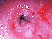
AIM: To examine the effect of corticosteroid treatment on the expression of inducible nitric oxide synthase (iNOS) in the colon of patients with inflammatory bowel disease. METHODS: Four groups of patients were studied: (1) ulcerative colitis treated with high dose corticosteroids (six patients, 10 blocks); (2) ulcerative colitis patients who had never received corticosteroids (10 patients, 16 blocks); (3) Crohn's disease treated with high dose corticosteroids (12 patients, 24 blocks); (4) Non-inflammatory, non-neoplastic controls (four patients, six blocks). Full thickness paraffin sections of colons removed at surgery were immunostained with an antibody raised against the C terminal end of iNOS. Sections were assessed semiquantitatively for the presence and degree of inflammation and immunoreactivity for nitric oxide synthase. RESULTS: Cases of ulcerative colitis and Crohn's disease with active inflammation showed strong staining for nitric oxide synthase. The staining was diffuse in ulcerative colitis and patchy in Crohn's disease, in accordance with the distribution of active inflammation. Staining was seen in epithelial cells and was most intense near areas of inflammation such as crypt abscesses. Non-inflamed epithelium showed no immunoreactivity. Treatment with corticosteroids made no difference to the amount of nitric oxide synthase. CONCLUSIONS: Expression of nitric oxide synthase is increased in both ulcerative colitis and Crohn's disease and appears to be unaffected by treatment with corticosteroids. Disease severity necessitated surgery in all the cases included in this study, regardless of whether or not the patients had received long term corticosteroid treatment. It seems therefore that a high level of iNOS expression and, presumably, production of nitric oxide characterise cases which are refractory to clinical treatment; this suggests that specific inhibition of the enzyme may be a useful therapeutic adjunct. (+info)Phenotype of mice and macrophages deficient in both phagocyte oxidase and inducible nitric oxide synthase. (3/10638)
The two genetically established antimicrobial mechanisms of macrophages are production of reactive oxygen intermediates by phagocyte oxidase (phox) and reactive nitrogen intermediates by inducible nitric oxide synthase (NOS2). Mice doubly deficient in both enzymes (gp91(phox-/-)/NOS2(-/-)) formed massive abscesses containing commensal organisms, mostly enteric bacteria, even when reared under specific pathogen-free conditions with antibiotics. Neither parental strain showed such infections. Thus, phox and NOS2 appear to compensate for each other's deficiency in providing resistance to indigenous bacteria, and no other pathway does so fully. Macrophages from gp91(phox-/-)/NOS2(-/-) mice could not kill virulent Listeria. Their killing of S. typhimurium, E. coli, and attenuated Listeria was markedly diminished but demonstrable, establishing the existence of a mechanism of macrophage antibacterial activity independent of phox and NOS2. (+info)Role of nitric oxide in lipopolysaccharide-induced hepatic injury in D-galactosamine-sensitized mice as an experimental endotoxic shock model. (4/10638)
The role of nitric oxide (NO) in lipopolysaccharide (LPS)-induced hepatic injury was studied in D-galactosamine (D-GalN)-sensitized mice. The inducible isoform of NO synthase (iNOS) was immunohistochemically detected on hepatocytes around blood vessels in livers of mice injected with D-GalN and LPS not on hepatocytes in mice injected with D-GalN or LPS alone, although mRNA for iNOS was found in those mice. Nitrotyrosine (NT) was also found in livers of mice injected with D-GalN and LPS. The localization of NT was consistent with that of iNOS, and the time courses of NT and iNOS expression were almost the same. Expression of iNOS and NT was detected exclusively in the hepatic lesions of mice injected with D-GalN and LPS. Anti-tumor necrosis factor alpha neutralizing antibody inhibited iNOS and NT expression and hepatic injury. The results suggested that NO from iNOS may play a role in LPS-induced hepatic injury on D-GalN-sensitized mice as an experimental endotoxic shock model. (+info)Gamma interferon stimulates rat alveolar macrophages to kill Pneumocystis carinii by L-arginine- and tumor necrosis factor-dependent mechanisms. (5/10638)
Pneumocystis carinii pneumonia remains a serious complication for immunocompromised patients. In the present study, P. carinii organisms interacted with gamma interferon (IFN-gamma)-stimulated alveolar macrophages (AMs) to activate the L-arginine-dependent cytocidal pathway involving reactive nitrogen intermediates (RNI) that were assayed as nitrite (NO2-). Unstimulated cultures of AMs produced negligible quantities of RNI. Addition of P. carinii organisms to IFN-gamma-primed AMs resulted in greatly enhanced production of RNI. NO2- levels increased from 0.8 +/- 0.4 to 11.1 +/- 3.8 microM as early as 6 h after P. carinii organisms were incubated with IFN-gamma-stimulated AMs and to 35.1 +/- 8.9 microM after a 24-h incubation, a near-maximum level. High levels of NO2- were produced by AMs primed with as little as 10 U of IFN-gamma per ml in the presence of P. carinii, and a 20-fold increase in IFN-gamma concentration resulted in only a further 65% increase in NO2- production. RNI-dependent killing of P. carinii was demonstrated by both a 51Cr release assay and a [35S]methionine pulse immunoprecipitation assay. Addition of either monoclonal tumor necrosis factor alpha (TNF-alpha) neutralizing antibody or 200 microM NG-monomethyl-L-arginine (L-NGMMA), a competitive inhibitor of the L-arginine-dependent pathway, significantly decreased NO2- production and reduced P. carinii killing. TNF-alpha alone had no effect on P. carinii viability. These results suggest that (i) the specific interaction of P. carinii organisms with IFN-gamma-primed AMs triggers the production of RNI, (ii) RNI are toxic to P. carinii, and (iii) TNF-alpha likely plays a central role in mediating P. carinii killing by IFN-gamma-stimulated AMs. (+info)Effect of transforming growth factor beta on experimental Salmonella typhimurium infection in mice. (6/10638)
We have investigated the effect of the in vivo administration of recombinant transforming growth factor beta (rTGF-beta) on the pathogenic mechanisms involved in Salmonella typhimurium experimental infection in mice. The protective response elicited by macrophages was induced by rTGF-beta1 by 2 days after experimental infection, as demonstrated by an increased NO production, while the humoral protective effect began with cytokine mRNA expression 2 days after the challenge and continued after 5 days with cytokine release and lymphocyte activation. We demonstrated that all mice who received rTGF-beta1 survived 7 days after infection. The number of bacteria recovered in the spleens and in the livers of rTGF-beta1-treated mice 2 and 5 days after infection was significantly smaller than that found in the same organs after phosphate-buffered saline (PBS) inoculation. Furthermore, 2 and 5 days after infection, splenic macrophages from rTGF-beta1-treated mice showed a greater NO production than did those from PBS-treated mice. The effect of rTGF-beta1 on S. typhimurium infection in mice was correlated with the expression of cell costimulatory CD28 molecules. Five days after S. typhimurium infection, the percentage of CD28(+)-expressing T cells in splenic lymphocytes from rTGF-beta1-treated mice increased with respect to that from control mice. Gamma interferon (IFN-gamma) mRNA was present in a greater amount in spleen cells from rTGF-beta1-treated mice after 2 days, although the intensity of the band decreased 5 days after the challenge. A similar pattern was obtained with the mRNAs for interleukin-1alpha (IL-1alpha), IL-6, TGF-beta, and inducible nitric oxide synthase, which showed greater expression in cells obtained from rTGF-beta1-treated and S. typhimurium-infected mice 2 days after challenge. The treatment with rTGF-beta1 induced an increase in IL-1alpha and IFN-gamma release in the supernatant of splenocyte cultures 5 days after the experimental infection with S. typhimurium. Moreover, we demonstrated that 5 days after infection, the IFN-gamma titer was significantly greater in the sera of rTGF-beta-treated mice than in those of PBS-treated mice. Also, hsp60 showed greater expression 2 days after the challenge in splenocytes from rTGF-beta1-treated mice. The role played by proinflammatory and immunoregulatory cytokines and by CD28 is discussed. (+info)AMP-activated protein kinase phosphorylation of endothelial NO synthase. (7/10638)
The AMP-activated protein kinase (AMPK) in rat skeletal and cardiac muscle is activated by vigorous exercise and ischaemic stress. Under these conditions AMPK phosphorylates and inhibits acetyl-coenzyme A carboxylase causing increased oxidation of fatty acids. Here we show that AMPK co-immunoprecipitates with cardiac endothelial NO synthase (eNOS) and phosphorylates Ser-1177 in the presence of Ca2+-calmodulin (CaM) to activate eNOS both in vitro and during ischaemia in rat hearts. In the absence of Ca2+-calmodulin, AMPK also phosphorylates eNOS at Thr-495 in the CaM-binding sequence, resulting in inhibition of eNOS activity but Thr-495 phosphorylation is unchanged during ischaemia. Phosphorylation of eNOS by the AMPK in endothelial cells and myocytes provides a further regulatory link between metabolic stress and cardiovascular function. (+info)The stimulatory effects of Hofmeister ions on the activities of neuronal nitric-oxide synthase. Apparent substrate inhibition by l-arginine is overcome in the presence of protein-destabilizing agents. (8/10638)
A variety of monovalent anions and cations were effective in stimulating both calcium ion/calmodulin (Ca2+/CaM)-independent NADPH-cytochrome c reductase activity of, and Ca2+/CaM-dependent nitric oxide (NO.) synthesis by, neuronal nitric oxide synthase (nNOS). The efficacy of the ions in stimulating both activities could be correlated, in general, with their efficacy in precipitating or stabilizing certain proteins, an order referred to as the Hofmeister ion series. In the hemoglobin capture assay, used for measurement of NO. production, apparent substrate inhibition by L-arginine was almost completely reversed by the addition of sodium perchlorate (NaClO4), one of the more effective protein-destabilizing agents tested. Examination of this phenomenon by the assay of L-arginine conversion to L-citrulline revealed that the stimulatory effect of NaClO4 on the reaction was observed only in the presence of oxyhemoglobin or superoxide anion (generated by xanthine and xanthine oxidase), both scavengers of NO. Spectrophotometric examination of nNOS revealed that the addition of NaClO4 and a superoxide-generating system, but neither alone, prevented the increase of heme absorption at 436 nm, which has been attributed to the nitrosyl complex. The data are consistent with the release of autoinhibitory NO. coordinated to the prosthetic group of nNOS, which, in conjunction with an NO. scavenger, causes stimulation of the reaction. (+info)Nitric Oxide Synthase (NOS) is a group of enzymes that catalyze the production of nitric oxide (NO) from L-arginine. There are three distinct isoforms of NOS, each with different expression patterns and functions:
1. Neuronal Nitric Oxide Synthase (nNOS or NOS1): This isoform is primarily expressed in the nervous system and plays a role in neurotransmission, synaptic plasticity, and learning and memory processes.
2. Inducible Nitric Oxide Synthase (iNOS or NOS2): This isoform is induced by various stimuli such as cytokines, lipopolysaccharides, and hypoxia in a variety of cells including immune cells, endothelial cells, and smooth muscle cells. iNOS produces large amounts of NO, which functions as a potent effector molecule in the immune response, particularly in the defense against microbial pathogens.
3. Endothelial Nitric Oxide Synthase (eNOS or NOS3): This isoform is constitutively expressed in endothelial cells and produces low levels of NO that play a crucial role in maintaining vascular homeostasis by regulating vasodilation, inhibiting platelet aggregation, and preventing smooth muscle cell proliferation.
Overall, NOS plays an essential role in various physiological processes, including neurotransmission, immune response, cardiovascular function, and respiratory regulation. Dysregulation of NOS activity has been implicated in several pathological conditions such as hypertension, atherosclerosis, neurodegenerative diseases, and inflammatory disorders.
Nitric oxide (NO) is a molecule made up of one nitrogen atom and one oxygen atom. In the body, it is a crucial signaling molecule involved in various physiological processes such as vasodilation, immune response, neurotransmission, and inhibition of platelet aggregation. It is produced naturally by the enzyme nitric oxide synthase (NOS) from the amino acid L-arginine. Inhaled nitric oxide is used medically to treat pulmonary hypertension in newborns and adults, as it helps to relax and widen blood vessels, improving oxygenation and blood flow.
Nitric Oxide Synthase Type II (NOS2), also known as Inducible Nitric Oxide Synthase (iNOS), is an enzyme that catalyzes the production of nitric oxide (NO) from L-arginine. Unlike other isoforms of NOS, NOS2 is not constitutively expressed and its expression can be induced by various stimuli such as cytokines, lipopolysaccharides, and bacterial products. Once induced, NOS2 produces large amounts of NO, which plays a crucial role in the immune response against invading pathogens. However, excessive or prolonged production of NO by NOS2 has been implicated in various pathological conditions such as inflammation, septic shock, and neurodegenerative disorders.
Nitric Oxide Synthase Type III (NOS-III), also known as endothelial Nitric Oxide Synthase (eNOS), is an enzyme responsible for the production of nitric oxide (NO) in the endothelium, the lining of blood vessels. This enzyme catalyzes the conversion of L-arginine to L-citrulline, producing NO as a byproduct. The release of NO from eNOS plays an important role in regulating vascular tone and homeostasis, including the relaxation of smooth muscle cells in the blood vessel walls, inhibition of platelet aggregation, and modulation of immune function. Mutations or dysfunction in NOS-III can contribute to various cardiovascular diseases such as hypertension, atherosclerosis, and erectile dysfunction.
Nitric Oxide Synthase Type I, also known as NOS1 or neuronal nitric oxide synthase (nNOS), is an enzyme that catalyzes the production of nitric oxide (NO) from L-arginine. It is primarily expressed in the nervous system, particularly in neurons, and plays a crucial role in the regulation of neurotransmission, synaptic plasticity, and cerebral blood flow. NOS1 is calcium-dependent and requires several cofactors for its activity, including NADPH, FAD, FMN, and calmodulin. It is involved in various physiological and pathological processes, such as learning and memory, seizure susceptibility, and neurodegenerative disorders.
Amino acid oxidoreductases are a class of enzymes that catalyze the reversible oxidation and reduction reactions involving amino acids. They play a crucial role in the metabolism of amino acids by catalyzing the interconversion of L-amino acids to their corresponding α-keto acids, while simultaneously reducing a cofactor such as NAD(P)+ or FAD.
The reaction catalyzed by these enzymes can be represented as follows:
L-amino acid + H2O + Coenzyme (Oxidized) → α-keto acid + NH3 + Coenzyme (Reduced)
Amino acid oxidoreductases are classified into two main types based on their cofactor requirements and reaction mechanisms. The first type uses FAD as a cofactor and is called amino acid flavoprotein oxidoreductases. These enzymes typically catalyze the oxidative deamination of L-amino acids to form α-keto acids, ammonia, and reduced FAD. The second type uses pyridine nucleotides (NAD(P)+) as cofactors and is called amino acid pyridine nucleotide-dependent oxidoreductases. These enzymes catalyze the reversible interconversion of L-amino acids to their corresponding α-keto acids, while simultaneously reducing or oxidizing NAD(P)H/NAD(P)+.
Amino acid oxidoreductases are widely distributed in nature and play important roles in various biological processes, including amino acid catabolism, nitrogen metabolism, and the biosynthesis of various secondary metabolites. Dysregulation of these enzymes has been implicated in several diseases, including neurodegenerative disorders and cancer. Therefore, understanding the structure, function, and regulation of amino acid oxidoreductases is crucial for developing novel therapeutic strategies to treat these diseases.
NG-Nitroarginine Methyl Ester (L-NAME) is not a medication, but rather a research chemical used in scientific studies. It is an inhibitor of nitric oxide synthase, an enzyme that synthesizes nitric oxide, a molecule involved in the relaxation of blood vessels.
Therefore, L-NAME is often used in experiments to investigate the role of nitric oxide in various physiological and pathophysiological processes. It is important to note that the use of L-NAME in humans is not approved for therapeutic purposes due to its potential side effects, which can include hypertension, decreased renal function, and decreased cerebral blood flow.
Nitric oxide (NO) donors are pharmacological agents that release nitric oxide in the body when they are metabolized. Nitric oxide is a molecule that plays an important role as a signaling messenger in the cardiovascular, nervous, and immune systems. It helps regulate blood flow, relax smooth muscle, inhibit platelet aggregation, and modulate inflammatory responses.
NO donors can be used medically to treat various conditions, such as hypertension, angina, heart failure, and pulmonary hypertension, by promoting vasodilation and improving blood flow. Some examples of NO donors include nitroglycerin, isosorbide dinitrate, sodium nitroprusside, and molsidomine. These drugs work by releasing nitric oxide slowly over time, which then interacts with the enzyme soluble guanylate cyclase to produce cyclic guanosine monophosphate (cGMP), leading to relaxation of smooth muscle and vasodilation.
It is important to note that NO donors can have side effects, such as headache, dizziness, and hypotension, due to their vasodilatory effects. Therefore, they should be used under the guidance of a healthcare professional.
Arginine is an α-amino acid that is classified as a semi-essential or conditionally essential amino acid, depending on the developmental stage and health status of the individual. The adult human body can normally synthesize sufficient amounts of arginine to meet its needs, but there are certain circumstances, such as periods of rapid growth or injury, where the dietary intake of arginine may become necessary.
The chemical formula for arginine is C6H14N4O2. It has a molecular weight of 174.20 g/mol and a pKa value of 12.48. Arginine is a basic amino acid, which means that it contains a side chain with a positive charge at physiological pH levels. The side chain of arginine is composed of a guanidino group, which is a functional group consisting of a nitrogen atom bonded to three methyl groups.
In the body, arginine plays several important roles. It is a precursor for the synthesis of nitric oxide, a molecule that helps regulate blood flow and immune function. Arginine is also involved in the detoxification of ammonia, a waste product produced by the breakdown of proteins. Additionally, arginine can be converted into other amino acids, such as ornithine and citrulline, which are involved in various metabolic processes.
Foods that are good sources of arginine include meat, poultry, fish, dairy products, nuts, seeds, and legumes. Arginine supplements are available and may be used for a variety of purposes, such as improving exercise performance, enhancing wound healing, and boosting immune function. However, it is important to consult with a healthcare provider before taking arginine supplements, as they can interact with certain medications and have potential side effects.
Enzyme inhibitors are substances that bind to an enzyme and decrease its activity, preventing it from catalyzing a chemical reaction in the body. They can work by several mechanisms, including blocking the active site where the substrate binds, or binding to another site on the enzyme to change its shape and prevent substrate binding. Enzyme inhibitors are often used as drugs to treat various medical conditions, such as high blood pressure, abnormal heart rhythms, and bacterial infections. They can also be found naturally in some foods and plants, and can be used in research to understand enzyme function and regulation.
In a medical context, nitrites are typically referred to as organic compounds that contain a functional group with the formula R-N=O, where R represents an alkyl or aryl group. They are commonly used in medicine as vasodilators, which means they widen and relax blood vessels, improving blood flow and lowering blood pressure.
One example of a nitrite used medically is amyl nitrite, which was previously used to treat angina pectoris, a type of chest pain caused by reduced blood flow to the heart muscle. However, its use has largely been replaced by other medications due to safety concerns and the availability of more effective treatments.
It's worth noting that inorganic nitrites, such as sodium nitrite, are also used in medicine for various purposes, including as a preservative in food and as a medication to treat cyanide poisoning. However, these compounds have different chemical properties and uses than organic nitrites.
Omega-N-Methylarginine (also known as NG, NG-dimethyl-L-arginine) is not a commonly used medical term and it's not a well-known compound in medicine. However, it is a form of methylated arginine that can be found in the body.
Methylated arginines are a group of compounds that are generated through the post-translational modification of proteins by enzymes called protein arginine methyltransferases (PRMTs). These modifications play important roles in various cellular processes, including gene expression and signal transduction.
Omega-N-Methylarginine is a specific type of methylated arginine that has two methyl groups attached to the nitrogen atom at the end of the side chain (omega position) of the amino acid arginine. It can be formed by the action of PRMTs on proteins, and it may have various biological functions in the body. However, its specific medical significance is not well-established, and more research is needed to fully understand its role in health and disease.
The endothelium is a thin layer of simple squamous epithelial cells that lines the interior surface of blood vessels, lymphatic vessels, and heart chambers. The vascular endothelium, specifically, refers to the endothelial cells that line the blood vessels. These cells play a crucial role in maintaining vascular homeostasis by regulating vasomotor tone, coagulation, platelet activation, inflammation, and permeability of the vessel wall. They also contribute to the growth and repair of the vascular system and are involved in various pathological processes such as atherosclerosis, hypertension, and diabetes.
Nitrates are chemical compounds that consist of a nitrogen atom bonded to three oxygen atoms (NO3-). In the context of medical science, nitrates are often discussed in relation to their use as medications or their presence in food and water.
As medications, nitrates are commonly used to treat angina (chest pain) caused by coronary artery disease. Nitrates work by relaxing and widening blood vessels, which improves blood flow and reduces the workload on the heart. Some examples of nitrate medications include nitroglycerin, isosorbide dinitrate, and isosorbide mononitrate.
In food and water, nitrates are naturally occurring compounds that can be found in a variety of vegetables, such as spinach, beets, and lettuce. They can also be present in fertilizers and industrial waste, which can contaminate groundwater and surface water sources. While nitrates themselves are not harmful, they can be converted into potentially harmful compounds called nitrites under certain conditions, particularly in the digestive system of young children or in the presence of bacteria such as those found in unpasteurized foods. Excessive levels of nitrites can react with hemoglobin in the blood to form methemoglobin, which cannot transport oxygen effectively and can lead to a condition called methemoglobinemia.
Nitro-L-arginine or Nitroarginine is not a medical term per se, but it is a chemical compound that is sometimes used in medical research and experiments. It is a salt of nitric acid and L-arginine, an amino acid that is important for the functioning of the body.
Nitroarginine is known to inhibit the production of nitric oxide, a molecule that plays a role in various physiological processes such as blood flow regulation, immune response, and neurotransmission. As a result, nitroarginine has been used in research to study the effects of reduced nitric oxide levels on different systems in the body.
It's worth noting that nitroarginine is not approved for use as a medication in humans, and its use is generally limited to laboratory settings.
Enzyme induction is a process by which the activity or expression of an enzyme is increased in response to some stimulus, such as a drug, hormone, or other environmental factor. This can occur through several mechanisms, including increasing the transcription of the enzyme's gene, stabilizing the mRNA that encodes the enzyme, or increasing the translation of the mRNA into protein.
In some cases, enzyme induction can be a beneficial process, such as when it helps the body to metabolize and clear drugs more quickly. However, in other cases, enzyme induction can have negative consequences, such as when it leads to the increased metabolism of important endogenous compounds or the activation of harmful procarcinogens.
Enzyme induction is an important concept in pharmacology and toxicology, as it can affect the efficacy and safety of drugs and other xenobiotics. It is also relevant to the study of drug interactions, as the induction of one enzyme by a drug can lead to altered metabolism and effects of another drug that is metabolized by the same enzyme.
Sprague-Dawley rats are a strain of albino laboratory rats that are widely used in scientific research. They were first developed by researchers H.H. Sprague and R.C. Dawley in the early 20th century, and have since become one of the most commonly used rat strains in biomedical research due to their relatively large size, ease of handling, and consistent genetic background.
Sprague-Dawley rats are outbred, which means that they are genetically diverse and do not suffer from the same limitations as inbred strains, which can have reduced fertility and increased susceptibility to certain diseases. They are also characterized by their docile nature and low levels of aggression, making them easier to handle and study than some other rat strains.
These rats are used in a wide variety of research areas, including toxicology, pharmacology, nutrition, cancer, and behavioral studies. Because they are genetically diverse, Sprague-Dawley rats can be used to model a range of human diseases and conditions, making them an important tool in the development of new drugs and therapies.
Cyclic guanosine monophosphate (cGMP) is a important second messenger molecule that plays a crucial role in various biological processes within the human body. It is synthesized from guanosine triphosphate (GTP) by the enzyme guanylyl cyclase.
Cyclic GMP is involved in regulating diverse physiological functions, such as smooth muscle relaxation, cardiovascular function, and neurotransmission. It also plays a role in modulating immune responses and cellular growth and differentiation.
In the medical field, changes in cGMP levels or dysregulation of cGMP-dependent pathways have been implicated in various disease states, including pulmonary hypertension, heart failure, erectile dysfunction, and glaucoma. Therefore, pharmacological agents that target cGMP signaling are being developed as potential therapeutic options for these conditions.
NADPH Dehydrogenase (also known as Nicotinamide Adenine Dinucleotide Phosphate Hydrogen Dehydrogenase) is an enzyme that plays a crucial role in the electron transport chain within the mitochondria of cells. It catalyzes the oxidation of NADPH to NADP+, which is a vital step in the process of cellular respiration where energy is produced in the form of ATP (Adenosine Triphosphate).
There are multiple forms of this enzyme, including both membrane-bound and soluble varieties. The membrane-bound NADPH Dehydrogenase is a complex I protein found in the inner mitochondrial membrane, while the soluble form is located in the cytosol.
Mutations in genes encoding for this enzyme can lead to various medical conditions, such as mitochondrial disorders and neurological diseases.
Vasodilation is the widening or increase in diameter of blood vessels, particularly the involuntary relaxation of the smooth muscle in the tunica media (middle layer) of the arteriole walls. This results in an increase in blood flow and a decrease in vascular resistance. Vasodilation can occur due to various physiological and pathophysiological stimuli, such as local metabolic demands, neural signals, or pharmacological agents. It plays a crucial role in regulating blood pressure, tissue perfusion, and thermoregulation.
"Cells, cultured" is a medical term that refers to cells that have been removed from an organism and grown in controlled laboratory conditions outside of the body. This process is called cell culture and it allows scientists to study cells in a more controlled and accessible environment than they would have inside the body. Cultured cells can be derived from a variety of sources, including tissues, organs, or fluids from humans, animals, or cell lines that have been previously established in the laboratory.
Cell culture involves several steps, including isolation of the cells from the tissue, purification and characterization of the cells, and maintenance of the cells in appropriate growth conditions. The cells are typically grown in specialized media that contain nutrients, growth factors, and other components necessary for their survival and proliferation. Cultured cells can be used for a variety of purposes, including basic research, drug development and testing, and production of biological products such as vaccines and gene therapies.
It is important to note that cultured cells may behave differently than they do in the body, and results obtained from cell culture studies may not always translate directly to human physiology or disease. Therefore, it is essential to validate findings from cell culture experiments using additional models and ultimately in clinical trials involving human subjects.
Indazoles are not a medical term, but a chemical classification. They refer to a class of heterocyclic organic compounds that contain a indazole moiety, which is a benzene ring fused with a diazole ring. Indazoles have no specific medical relevance, but certain derivatives of indazoles have been developed and used as drugs in medicine, particularly in the treatment of cancer and cardiovascular diseases. For example, Tadalafil (Cialis), a medication used to treat erectile dysfunction and benign prostatic hyperplasia, is a selective inhibitor of cGMP-specific phosphodiesterase type 5 and has an indazole structure.
"Wistar rats" are a strain of albino rats that are widely used in laboratory research. They were developed at the Wistar Institute in Philadelphia, USA, and were first introduced in 1906. Wistar rats are outbred, which means that they are genetically diverse and do not have a fixed set of genetic characteristics like inbred strains.
Wistar rats are commonly used as animal models in biomedical research because of their size, ease of handling, and relatively low cost. They are used in a wide range of research areas, including toxicology, pharmacology, nutrition, cancer, cardiovascular disease, and behavioral studies. Wistar rats are also used in safety testing of drugs, medical devices, and other products.
Wistar rats are typically larger than many other rat strains, with males weighing between 500-700 grams and females weighing between 250-350 grams. They have a lifespan of approximately 2-3 years. Wistar rats are also known for their docile and friendly nature, making them easy to handle and work with in the laboratory setting.
Lipopolysaccharides (LPS) are large molecules found in the outer membrane of Gram-negative bacteria. They consist of a hydrophilic polysaccharide called the O-antigen, a core oligosaccharide, and a lipid portion known as Lipid A. The Lipid A component is responsible for the endotoxic activity of LPS, which can trigger a powerful immune response in animals, including humans. This response can lead to symptoms such as fever, inflammation, and septic shock, especially when large amounts of LPS are introduced into the bloodstream.
nitroprusside (ni-troe-rus-ide)
A rapid-acting vasodilator used in the management of severe hypertension, acute heart failure, and to reduce afterload in patients undergoing cardiac surgery. It is a potent arterial and venous dilator that decreases preload and afterload, thereby reducing myocardial oxygen demand. Nitroprusside is metabolized to cyanide, which must be monitored closely during therapy to prevent toxicity.
Pharmacologic class: Peripheral vasodilators
Therapeutic class: Antihypertensives, Vasodilators
Medical Categories: Cardiovascular Drugs, Hypertension Agents
Messenger RNA (mRNA) is a type of RNA (ribonucleic acid) that carries genetic information copied from DNA in the form of a series of three-base code "words," each of which specifies a particular amino acid. This information is used by the cell's machinery to construct proteins, a process known as translation. After being transcribed from DNA, mRNA travels out of the nucleus to the ribosomes in the cytoplasm where protein synthesis occurs. Once the protein has been synthesized, the mRNA may be degraded and recycled. Post-transcriptional modifications can also occur to mRNA, such as alternative splicing and addition of a 5' cap and a poly(A) tail, which can affect its stability, localization, and translation efficiency.
L-Citrulline is a non-essential amino acid that plays a role in the urea cycle, which is the process by which the body eliminates toxic ammonia from the bloodstream. It is called "non-essential" because it can be synthesized by the body from other compounds, such as L-Ornithine and carbamoyl phosphate.
Citrulline is found in some foods, including watermelon, bitter melon, and certain types of sausage. It is also available as a dietary supplement. In the body, citrulline is converted to another amino acid called L-Arginine, which is involved in the production of nitric oxide, a molecule that helps dilate blood vessels and improve blood flow.
Citrulline has been studied for its potential benefits on various aspects of health, including exercise performance, cardiovascular function, and immune system function. However, more research is needed to confirm these potential benefits and establish safe and effective dosages.
S-Nitroso-N-Acetylpenicillamine (SNAP) is not a medication itself, but rather a chemical compound that is used in laboratory research. It is a nitrosothiol, which means it contains a nitric oxide group (NO) attached to a sulfur atom in a thiol group (a type of organic compound containing a sulfhydryl group, -SH).
Nitric oxide is a small signaling molecule that plays an important role in various biological processes, including the regulation of blood flow, immune response, and neurotransmission. SNAP is often used as a nitric oxide donor in scientific studies to investigate the effects of nitric oxide on different cells and tissues.
SNAP can release nitric oxide under certain conditions, such as in the presence of reducing agents or at acidic pH levels. This makes it useful for studying the mechanisms of nitric oxide-mediated signaling pathways and its potential therapeutic applications. However, SNAP is not used as a medication in clinical practice due to its instability and potential toxicity.
Biopterin is a type of pteridine compound that acts as a cofactor in various biological reactions, particularly in the metabolism of amino acids such as phenylalanine and tyrosine. It plays a crucial role in the production of neurotransmitters like dopamine, serotonin, and noradrenaline. Biopterin exists in two major forms: tetrahydrobiopterin (BH4) and dihydrobiopterin (BH2). BH4 is the active form that participates in enzymatic reactions, while BH2 is an oxidized form that can be reduced back to BH4 by the action of dihydrobiopterin reductase.
Deficiencies in biopterin metabolism have been linked to several neurological disorders, including phenylketonuria (PKU), dopamine-responsive dystonia, and certain forms of autism. In these conditions, the impaired synthesis or recycling of biopterin can lead to reduced levels of neurotransmitters, causing various neurological symptoms.
Isothiuronium is not a medical term, but it is a chemical compound that can be referred to in a medical context. It is a type of organic compound called an isothiouronium salt, which contains a nitrogen atom bonded to a sulfur atom and two organic groups.
Isothiouronium compounds are known to have various biological activities, including inhibition of certain enzymes and potential use as therapeutic agents. However, they can also be toxic in high concentrations. Therefore, exposure to isothiuronium compounds may require medical attention, particularly if it occurs through inhalation, ingestion, or skin contact.
In a medical context, isothiuronium may be mentioned in the context of drug metabolism, toxicology, or pharmacology, depending on the specific compound and its biological activity.
Penicillamine is a medication that belongs to a class of drugs called chelating agents. It works by binding to heavy metals in the body, such as lead, mercury, or copper, and forming a compound that can be excreted in the urine. This helps to remove these harmful substances from the body.
Penicillamine is also used to treat certain medical conditions, such as rheumatoid arthritis, Wilson's disease (a genetic disorder that causes copper accumulation in the body), and cystinuria (a genetic disorder that causes an amino acid called cystine to accumulate in the kidneys and form stones).
It is important to note that penicillamine can have serious side effects, including kidney damage, so it should be used under the close supervision of a healthcare provider.
Guanidines are organic compounds that contain a guanidino group, which is a functional group with the formula -NH-C(=NH)-NH2. Guanidines can be found in various natural sources, including some animals, plants, and microorganisms. They also occur as byproducts of certain metabolic processes in the body.
In a medical context, guanidines are most commonly associated with the treatment of muscle weakness and neuromuscular disorders. The most well-known guanidine compound is probably guanidine hydrochloride, which has been used as a medication to treat conditions such as myasthenia gravis and Eaton-Lambert syndrome.
However, the use of guanidines as medications has declined in recent years due to their potential for toxicity and the development of safer and more effective treatments. Today, guanidines are mainly used in research settings to study various biological processes, including protein folding and aggregation, enzyme inhibition, and cell signaling.
A dose-response relationship in the context of drugs refers to the changes in the effects or symptoms that occur as the dose of a drug is increased or decreased. Generally, as the dose of a drug is increased, the severity or intensity of its effects also increases. Conversely, as the dose is decreased, the effects of the drug become less severe or may disappear altogether.
The dose-response relationship is an important concept in pharmacology and toxicology because it helps to establish the safe and effective dosage range for a drug. By understanding how changes in the dose of a drug affect its therapeutic and adverse effects, healthcare providers can optimize treatment plans for their patients while minimizing the risk of harm.
The dose-response relationship is typically depicted as a curve that shows the relationship between the dose of a drug and its effect. The shape of the curve may vary depending on the drug and the specific effect being measured. Some drugs may have a steep dose-response curve, meaning that small changes in the dose can result in large differences in the effect. Other drugs may have a more gradual dose-response curve, where larger changes in the dose are needed to produce significant effects.
In addition to helping establish safe and effective dosages, the dose-response relationship is also used to evaluate the potential therapeutic benefits and risks of new drugs during clinical trials. By systematically testing different doses of a drug in controlled studies, researchers can identify the optimal dosage range for the drug and assess its safety and efficacy.
C57BL/6 (C57 Black 6) is an inbred strain of laboratory mouse that is widely used in biomedical research. The term "inbred" refers to a strain of animals where matings have been carried out between siblings or other closely related individuals for many generations, resulting in a population that is highly homozygous at most genetic loci.
The C57BL/6 strain was established in 1920 by crossing a female mouse from the dilute brown (DBA) strain with a male mouse from the black strain. The resulting offspring were then interbred for many generations to create the inbred C57BL/6 strain.
C57BL/6 mice are known for their robust health, longevity, and ease of handling, making them a popular choice for researchers. They have been used in a wide range of biomedical research areas, including studies of cancer, immunology, neuroscience, cardiovascular disease, and metabolism.
One of the most notable features of the C57BL/6 strain is its sensitivity to certain genetic modifications, such as the introduction of mutations that lead to obesity or impaired glucose tolerance. This has made it a valuable tool for studying the genetic basis of complex diseases and traits.
Overall, the C57BL/6 inbred mouse strain is an important model organism in biomedical research, providing a valuable resource for understanding the genetic and molecular mechanisms underlying human health and disease.
Vasodilator agents are pharmacological substances that cause the relaxation or widening of blood vessels by relaxing the smooth muscle in the vessel walls. This results in an increase in the diameter of the blood vessels, which decreases vascular resistance and ultimately reduces blood pressure. Vasodilators can be further classified based on their site of action:
1. Systemic vasodilators: These agents cause a generalized relaxation of the smooth muscle in the walls of both arteries and veins, resulting in a decrease in peripheral vascular resistance and preload (the volume of blood returning to the heart). Examples include nitroglycerin, hydralazine, and calcium channel blockers.
2. Arterial vasodilators: These agents primarily affect the smooth muscle in arterial vessel walls, leading to a reduction in afterload (the pressure against which the heart pumps blood). Examples include angiotensin-converting enzyme (ACE) inhibitors, angiotensin receptor blockers (ARBs), and direct vasodilators like sodium nitroprusside.
3. Venous vasodilators: These agents primarily affect the smooth muscle in venous vessel walls, increasing venous capacitance and reducing preload. Examples include nitroglycerin and other organic nitrates.
Vasodilator agents are used to treat various cardiovascular conditions such as hypertension, heart failure, angina, and pulmonary arterial hypertension. It is essential to monitor their use carefully, as excessive vasodilation can lead to orthostatic hypotension, reflex tachycardia, or fluid retention.
Nitrogen oxides (NOx) are a group of highly reactive gases, primarily composed of nitric oxide (NO) and nitrogen dioxide (NO2). They are formed during the combustion of fossil fuels, such as coal, oil, gas, or biomass, and are emitted from various sources, including power plants, industrial boilers, transportation vehicles, and residential heating systems. Exposure to NOx can have adverse health effects, particularly on the respiratory system, and contribute to the formation of harmful air pollutants like ground-level ozone and fine particulate matter.
Gene expression regulation, enzymologic refers to the biochemical processes and mechanisms that control the transcription and translation of specific genes into functional proteins or enzymes. This regulation is achieved through various enzymatic activities that can either activate or repress gene expression at different levels, such as chromatin remodeling, transcription factor activation, mRNA processing, and protein degradation.
Enzymologic regulation of gene expression involves the action of specific enzymes that catalyze chemical reactions involved in these processes. For example, histone-modifying enzymes can alter the structure of chromatin to make genes more or less accessible for transcription, while RNA polymerase and its associated factors are responsible for transcribing DNA into mRNA. Additionally, various enzymes are involved in post-transcriptional modifications of mRNA, such as splicing, capping, and tailing, which can affect the stability and translation of the transcript.
Overall, the enzymologic regulation of gene expression is a complex and dynamic process that allows cells to respond to changes in their environment and maintain proper physiological function.
A "knockout" mouse is a genetically engineered mouse in which one or more genes have been deleted or "knocked out" using molecular biology techniques. This allows researchers to study the function of specific genes and their role in various biological processes, as well as potential associations with human diseases. The mice are generated by introducing targeted DNA modifications into embryonic stem cells, which are then used to create a live animal. Knockout mice have been widely used in biomedical research to investigate gene function, disease mechanisms, and potential therapeutic targets.
Macrophages are a type of white blood cell that are an essential part of the immune system. They are large, specialized cells that engulf and destroy foreign substances, such as bacteria, viruses, parasites, and fungi, as well as damaged or dead cells. Macrophages are found throughout the body, including in the bloodstream, lymph nodes, spleen, liver, lungs, and connective tissues. They play a critical role in inflammation, immune response, and tissue repair and remodeling.
Macrophages originate from monocytes, which are a type of white blood cell produced in the bone marrow. When monocytes enter the tissues, they differentiate into macrophages, which have a larger size and more specialized functions than monocytes. Macrophages can change their shape and move through tissues to reach sites of infection or injury. They also produce cytokines, chemokines, and other signaling molecules that help coordinate the immune response and recruit other immune cells to the site of infection or injury.
Macrophages have a variety of surface receptors that allow them to recognize and respond to different types of foreign substances and signals from other cells. They can engulf and digest foreign particles, bacteria, and viruses through a process called phagocytosis. Macrophages also play a role in presenting antigens to T cells, which are another type of immune cell that helps coordinate the immune response.
Overall, macrophages are crucial for maintaining tissue homeostasis, defending against infection, and promoting wound healing and tissue repair. Dysregulation of macrophage function has been implicated in a variety of diseases, including cancer, autoimmune disorders, and chronic inflammatory conditions.
Guanylate cyclase is an enzyme that catalyzes the conversion of guanosine triphosphate (GTP) to cyclic guanosine monophosphate (cGMP), which acts as a second messenger in various cellular signaling pathways. There are two main types of guanylate cyclases: soluble and membrane-bound. Soluble guanylate cyclase is activated by nitric oxide, while membrane-bound guanylate cyclase can be activated by natriuretic peptides. The increased levels of cGMP produced by guanylate cyclase can lead to a variety of cellular responses, including smooth muscle relaxation, neurotransmitter release, and regulation of ion channels. Dysregulation of guanylate cyclase activity has been implicated in several diseases, such as hypertension, heart failure, and cancer.
Immunohistochemistry (IHC) is a technique used in pathology and laboratory medicine to identify specific proteins or antigens in tissue sections. It combines the principles of immunology and histology to detect the presence and location of these target molecules within cells and tissues. This technique utilizes antibodies that are specific to the protein or antigen of interest, which are then tagged with a detection system such as a chromogen or fluorophore. The stained tissue sections can be examined under a microscope, allowing for the visualization and analysis of the distribution and expression patterns of the target molecule in the context of the tissue architecture. Immunohistochemistry is widely used in diagnostic pathology to help identify various diseases, including cancer, infectious diseases, and immune-mediated disorders.
Western blotting is a laboratory technique used in molecular biology to detect and quantify specific proteins in a mixture of many different proteins. This technique is commonly used to confirm the expression of a protein of interest, determine its size, and investigate its post-translational modifications. The name "Western" blotting distinguishes this technique from Southern blotting (for DNA) and Northern blotting (for RNA).
The Western blotting procedure involves several steps:
1. Protein extraction: The sample containing the proteins of interest is first extracted, often by breaking open cells or tissues and using a buffer to extract the proteins.
2. Separation of proteins by electrophoresis: The extracted proteins are then separated based on their size by loading them onto a polyacrylamide gel and running an electric current through the gel (a process called sodium dodecyl sulfate-polyacrylamide gel electrophoresis or SDS-PAGE). This separates the proteins according to their molecular weight, with smaller proteins migrating faster than larger ones.
3. Transfer of proteins to a membrane: After separation, the proteins are transferred from the gel onto a nitrocellulose or polyvinylidene fluoride (PVDF) membrane using an electric current in a process called blotting. This creates a replica of the protein pattern on the gel but now immobilized on the membrane for further analysis.
4. Blocking: The membrane is then blocked with a blocking agent, such as non-fat dry milk or bovine serum albumin (BSA), to prevent non-specific binding of antibodies in subsequent steps.
5. Primary antibody incubation: A primary antibody that specifically recognizes the protein of interest is added and allowed to bind to its target protein on the membrane. This step may be performed at room temperature or 4°C overnight, depending on the antibody's properties.
6. Washing: The membrane is washed with a buffer to remove unbound primary antibodies.
7. Secondary antibody incubation: A secondary antibody that recognizes the primary antibody (often coupled to an enzyme or fluorophore) is added and allowed to bind to the primary antibody. This step may involve using a horseradish peroxidase (HRP)-conjugated or alkaline phosphatase (AP)-conjugated secondary antibody, depending on the detection method used later.
8. Washing: The membrane is washed again to remove unbound secondary antibodies.
9. Detection: A detection reagent is added to visualize the protein of interest by detecting the signal generated from the enzyme-conjugated or fluorophore-conjugated secondary antibody. This can be done using chemiluminescent, colorimetric, or fluorescent methods.
10. Analysis: The resulting image is analyzed to determine the presence and quantity of the protein of interest in the sample.
Western blotting is a powerful technique for identifying and quantifying specific proteins within complex mixtures. It can be used to study protein expression, post-translational modifications, protein-protein interactions, and more. However, it requires careful optimization and validation to ensure accurate and reproducible results.
The aorta is the largest artery in the human body, which originates from the left ventricle of the heart and carries oxygenated blood to the rest of the body. It can be divided into several parts, including the ascending aorta, aortic arch, and descending aorta. The ascending aorta gives rise to the coronary arteries that supply blood to the heart muscle. The aortic arch gives rise to the brachiocephalic, left common carotid, and left subclavian arteries, which supply blood to the head, neck, and upper extremities. The descending aorta travels through the thorax and abdomen, giving rise to various intercostal, visceral, and renal arteries that supply blood to the chest wall, organs, and kidneys.
Superoxides are partially reduced derivatives of oxygen that contain one extra electron, giving them an overall charge of -1. They are highly reactive and unstable, with the most common superoxide being the hydroxyl radical (•OH-) and the superoxide anion (O2-). Superoxides are produced naturally in the body during metabolic processes, particularly within the mitochondria during cellular respiration. They play a role in various physiological processes, but when produced in excess or not properly neutralized, they can contribute to oxidative stress and damage to cells and tissues, potentially leading to the development of various diseases such as cancer, atherosclerosis, and neurodegenerative disorders.
Amidines are organic compounds that contain a functional group with the structure R-C=N-R, where R can be an alkyl or aromatic group. This functional group consists of a carbonyl (C=O) group and a nitrogen atom (N) connected to two organic groups (R).
In medical terminology, amidines are not commonly used. However, some amidine derivatives have been investigated for their potential therapeutic properties. For example, certain amidine compounds have shown antimicrobial, anti-inflammatory, and antiviral activities. Some of these compounds have also been studied as potential drugs for the treatment of various diseases, including cancer, cardiovascular disease, and neurological disorders.
It is important to note that while some amidines may have therapeutic potential, they can also be toxic at high concentrations and should be handled with care.
Peroxynitrous acid (ONOOH) is a highly reactive nitrogen species formed from the reaction between nitric oxide (NO) and superoxide radical (O2-). It is an unstable compound that quickly decomposes to form other reactive species, such as nitrogen dioxide (NO2) and hydroxyl radical (HO•), which can cause significant damage to biological molecules, including proteins, lipids, and DNA. Peroxynitrous acid has been implicated in the pathogenesis of various diseases, including neurodegenerative disorders, cardiovascular disease, and cancer.
Arginase is an enzyme that plays a role in the metabolism of arginine, an amino acid. It works by breaking down arginine into ornithine and urea. This reaction is part of the urea cycle, which helps to rid the body of excess nitrogen waste produced during the metabolism of proteins. Arginase is found in various tissues throughout the body, including the liver, where it plays a key role in the detoxification of ammonia.
Molsidomine is a medication that belongs to a class of drugs called vasodilators. It works by relaxing and widening blood vessels, which helps to improve blood flow and reduce the workload on the heart. Molsidomine is used to treat chronic stable angina (chest pain caused by reduced blood flow to the heart) and has been found to be effective in reducing the frequency and severity of anginal attacks.
When molsidomine is absorbed into the body, it is converted into its active metabolite, SIN-1, which is responsible for its vasodilatory effects. SIN-1 causes smooth muscle relaxation by increasing the levels of nitric oxide in the blood vessels, leading to their dilation and improved blood flow.
Molsidomine is available in tablet form and is typically taken two to three times a day, with or without food. Common side effects of molsidomine include headache, dizziness, flushing, and palpitations. It should be used with caution in patients with low blood pressure, heart failure, or impaired kidney function.
Isoenzymes, also known as isoforms, are multiple forms of an enzyme that catalyze the same chemical reaction but differ in their amino acid sequence, structure, and/or kinetic properties. They are encoded by different genes or alternative splicing of the same gene. Isoenzymes can be found in various tissues and organs, and they play a crucial role in biological processes such as metabolism, detoxification, and cell signaling. Measurement of isoenzyme levels in body fluids (such as blood) can provide valuable diagnostic information for certain medical conditions, including tissue damage, inflammation, and various diseases.
Animal disease models are specialized animals, typically rodents such as mice or rats, that have been genetically engineered or exposed to certain conditions to develop symptoms and physiological changes similar to those seen in human diseases. These models are used in medical research to study the pathophysiology of diseases, identify potential therapeutic targets, test drug efficacy and safety, and understand disease mechanisms.
The genetic modifications can include knockout or knock-in mutations, transgenic expression of specific genes, or RNA interference techniques. The animals may also be exposed to environmental factors such as chemicals, radiation, or infectious agents to induce the disease state.
Examples of animal disease models include:
1. Mouse models of cancer: Genetically engineered mice that develop various types of tumors, allowing researchers to study cancer initiation, progression, and metastasis.
2. Alzheimer's disease models: Transgenic mice expressing mutant human genes associated with Alzheimer's disease, which exhibit amyloid plaque formation and cognitive decline.
3. Diabetes models: Obese and diabetic mouse strains like the NOD (non-obese diabetic) or db/db mice, used to study the development of type 1 and type 2 diabetes, respectively.
4. Cardiovascular disease models: Atherosclerosis-prone mice, such as ApoE-deficient or LDLR-deficient mice, that develop plaque buildup in their arteries when fed a high-fat diet.
5. Inflammatory bowel disease models: Mice with genetic mutations affecting intestinal barrier function and immune response, such as IL-10 knockout or SAMP1/YitFc mice, which develop colitis.
Animal disease models are essential tools in preclinical research, but it is important to recognize their limitations. Differences between species can affect the translatability of results from animal studies to human patients. Therefore, researchers must carefully consider the choice of model and interpret findings cautiously when applying them to human diseases.
Blood pressure is the force exerted by circulating blood on the walls of the blood vessels. It is measured in millimeters of mercury (mmHg) and is given as two figures:
1. Systolic pressure: This is the pressure when the heart pushes blood out into the arteries.
2. Diastolic pressure: This is the pressure when the heart rests between beats, allowing it to fill with blood.
Normal blood pressure for adults is typically around 120/80 mmHg, although this can vary slightly depending on age, sex, and other factors. High blood pressure (hypertension) is generally considered to be a reading of 130/80 mmHg or higher, while low blood pressure (hypotension) is usually defined as a reading below 90/60 mmHg. It's important to note that blood pressure can fluctuate throughout the day and may be affected by factors such as stress, physical activity, and medication use.
Cyclooxygenase-2 (COX-2) is an enzyme involved in the synthesis of prostaglandins, which are hormone-like substances that play a role in inflammation, pain, and fever. COX-2 is primarily expressed in response to stimuli such as cytokines and growth factors, and its expression is associated with the development of inflammation.
COX-2 inhibitors are a class of nonsteroidal anti-inflammatory drugs (NSAIDs) that selectively block the activity of COX-2, reducing the production of prostaglandins and providing analgesic, anti-inflammatory, and antipyretic effects. These medications are often used to treat pain and inflammation associated with conditions such as arthritis, menstrual cramps, and headaches.
It's important to note that while COX-2 inhibitors can be effective in managing pain and inflammation, they may also increase the risk of cardiovascular events such as heart attack and stroke, particularly when used at high doses or for extended periods. Therefore, it's essential to use these medications under the guidance of a healthcare provider and to follow their instructions carefully.
Endothelial cells are the type of cells that line the inner surface of blood vessels, lymphatic vessels, and heart chambers. They play a crucial role in maintaining vascular homeostasis by controlling vasomotor tone, coagulation, platelet activation, and inflammation. Endothelial cells also regulate the transport of molecules between the blood and surrounding tissues, and contribute to the maintenance of the structural integrity of the vasculature. They are flat, elongated cells with a unique morphology that allows them to form a continuous, nonthrombogenic lining inside the vessels. Endothelial cells can be isolated from various tissues and cultured in vitro for research purposes.
Acetylcholine is a neurotransmitter, a type of chemical messenger that transmits signals across a chemical synapse from one neuron (nerve cell) to another "target" neuron, muscle cell, or gland cell. It is involved in both peripheral and central nervous system functions.
In the peripheral nervous system, acetylcholine acts as a neurotransmitter at the neuromuscular junction, where it transmits signals from motor neurons to activate muscles. Acetylcholine also acts as a neurotransmitter in the autonomic nervous system, where it is involved in both the sympathetic and parasympathetic systems.
In the central nervous system, acetylcholine plays a role in learning, memory, attention, and arousal. Disruptions in cholinergic neurotransmission have been implicated in several neurological disorders, including Alzheimer's disease, Parkinson's disease, and myasthenia gravis.
Acetylcholine is synthesized from choline and acetyl-CoA by the enzyme choline acetyltransferase and is stored in vesicles at the presynaptic terminal of the neuron. When a nerve impulse arrives, the vesicles fuse with the presynaptic membrane, releasing acetylcholine into the synapse. The acetylcholine then binds to receptors on the postsynaptic membrane, triggering a response in the target cell. Acetylcholine is subsequently degraded by the enzyme acetylcholinesterase, which terminates its action and allows for signal transduction to be repeated.
Prostaglandin-Endoperoxide Synthases (PTGS), also known as Cyclooxygenases (COX), are a group of enzymes that catalyze the conversion of arachidonic acid into prostaglandin G2 and H2, which are further metabolized to produce various prostaglandins and thromboxanes. These lipid mediators play crucial roles in several physiological processes such as inflammation, pain, fever, and blood clotting. There are two major isoforms of PTGS: PTGS-1 (COX-1) and PTGS-2 (COX-2). While COX-1 is constitutively expressed in most tissues and involved in homeostatic functions, COX-2 is usually induced during inflammation and tissue injury. Nonsteroidal anti-inflammatory drugs (NSAIDs) exert their therapeutic effects by inhibiting these enzymes, thereby reducing the production of prostaglandins and thromboxanes.
Oxidative stress is defined as an imbalance between the production of reactive oxygen species (free radicals) and the body's ability to detoxify them or repair the damage they cause. This imbalance can lead to cellular damage, oxidation of proteins, lipids, and DNA, disruption of cellular functions, and activation of inflammatory responses. Prolonged or excessive oxidative stress has been linked to various health conditions, including cancer, cardiovascular diseases, neurodegenerative disorders, and aging-related diseases.
Interferon-gamma (IFN-γ) is a soluble cytokine that is primarily produced by the activation of natural killer (NK) cells and T lymphocytes, especially CD4+ Th1 cells and CD8+ cytotoxic T cells. It plays a crucial role in the regulation of the immune response against viral and intracellular bacterial infections, as well as tumor cells. IFN-γ has several functions, including activating macrophages to enhance their microbicidal activity, increasing the presentation of major histocompatibility complex (MHC) class I and II molecules on antigen-presenting cells, stimulating the proliferation and differentiation of T cells and NK cells, and inducing the production of other cytokines and chemokines. Additionally, IFN-γ has direct antiproliferative effects on certain types of tumor cells and can enhance the cytotoxic activity of immune cells against infected or malignant cells.
In the field of medicine, "time factors" refer to the duration of symptoms or time elapsed since the onset of a medical condition, which can have significant implications for diagnosis and treatment. Understanding time factors is crucial in determining the progression of a disease, evaluating the effectiveness of treatments, and making critical decisions regarding patient care.
For example, in stroke management, "time is brain," meaning that rapid intervention within a specific time frame (usually within 4.5 hours) is essential to administering tissue plasminogen activator (tPA), a clot-busting drug that can minimize brain damage and improve patient outcomes. Similarly, in trauma care, the "golden hour" concept emphasizes the importance of providing definitive care within the first 60 minutes after injury to increase survival rates and reduce morbidity.
Time factors also play a role in monitoring the progression of chronic conditions like diabetes or heart disease, where regular follow-ups and assessments help determine appropriate treatment adjustments and prevent complications. In infectious diseases, time factors are crucial for initiating antibiotic therapy and identifying potential outbreaks to control their spread.
Overall, "time factors" encompass the significance of recognizing and acting promptly in various medical scenarios to optimize patient outcomes and provide effective care.
Enzyme activation refers to the process by which an enzyme becomes biologically active and capable of carrying out its specific chemical or biological reaction. This is often achieved through various post-translational modifications, such as proteolytic cleavage, phosphorylation, or addition of cofactors or prosthetic groups to the enzyme molecule. These modifications can change the conformation or structure of the enzyme, exposing or creating a binding site for the substrate and allowing the enzymatic reaction to occur.
For example, in the case of proteolytic cleavage, an inactive precursor enzyme, known as a zymogen, is cleaved into its active form by a specific protease. This is seen in enzymes such as trypsin and chymotrypsin, which are initially produced in the pancreas as inactive precursors called trypsinogen and chymotrypsinogen, respectively. Once they reach the small intestine, they are activated by enteropeptidase, a protease that cleaves a specific peptide bond, releasing the active enzyme.
Phosphorylation is another common mechanism of enzyme activation, where a phosphate group is added to a specific serine, threonine, or tyrosine residue on the enzyme by a protein kinase. This modification can alter the conformation of the enzyme and create a binding site for the substrate, allowing the enzymatic reaction to occur.
Enzyme activation is a crucial process in many biological pathways, as it allows for precise control over when and where specific reactions take place. It also provides a mechanism for regulating enzyme activity in response to various signals and stimuli, such as hormones, neurotransmitters, or changes in the intracellular environment.
Glycogen synthase is an enzyme (EC 2.4.1.11) that plays a crucial role in the synthesis of glycogen, a polysaccharide that serves as the primary storage form of glucose in animals, fungi, and bacteria. This enzyme catalyzes the transfer of glucosyl residues from uridine diphosphate glucose (UDP-glucose) to the non-reducing end of an growing glycogen chain, thereby elongating it.
Glycogen synthase is regulated by several mechanisms, including allosteric regulation and covalent modification. The activity of this enzyme is inhibited by high levels of intracellular glucose-6-phosphate (G6P) and activated by the binding of glycogen or proteins that bind to glycogen, such as glycogenin. Phosphorylation of glycogen synthase by protein kinases, like glycogen synthase kinase-3 (GSK3), also reduces its activity, while dephosphorylation by protein phosphatases enhances it.
The regulation of glycogen synthase is critical for maintaining glucose homeostasis and energy balance in the body. Dysregulation of this enzyme has been implicated in several metabolic disorders, including type 2 diabetes and non-alcoholic fatty liver disease (NAFLD).
Glycogen Synthase Kinase 3 (GSK-3) is a serine/threonine protein kinase that plays a crucial role in the regulation of several cellular processes, including glycogen metabolism, cell signaling, gene transcription, and apoptosis. It was initially discovered as a key enzyme involved in glycogen metabolism due to its ability to phosphorylate and inhibit glycogen synthase, an enzyme responsible for the synthesis of glycogen from glucose.
GSK-3 exists in two isoforms, GSK-3α and GSK-3β, which share a high degree of sequence similarity and are widely expressed in various tissues. Both isoforms are constitutively active under normal conditions and are regulated through inhibitory phosphorylation by several upstream signaling pathways, such as insulin, Wnt, and Hedgehog signaling.
Dysregulation of GSK-3 has been implicated in the pathogenesis of various diseases, including diabetes, neurodegenerative disorders, and cancer. In recent years, GSK-3 has emerged as an attractive therapeutic target for the development of novel drugs to treat these conditions.
I'm sorry for any confusion, but "oxides" is not a term that has a specific medical definition. Oxides are a class of chemical compounds that contain at least one oxygen atom and one other element. They can be formed through the process of oxidation, which involves the combination of oxygen with another substance.
In a broader sense, you might encounter the term "oxide" in a medical context when discussing various materials or substances used in medical devices, treatments, or research. For instance, titanium dioxide is a common ingredient in medical-grade sunscreens due to its ability to block and scatter UV light. However, it's important to note that the term "oxides" itself doesn't have a direct connection to medicine or human health.
Tumor Necrosis Factor-alpha (TNF-α) is a cytokine, a type of small signaling protein involved in immune response and inflammation. It is primarily produced by activated macrophages, although other cell types such as T-cells, natural killer cells, and mast cells can also produce it.
TNF-α plays a crucial role in the body's defense against infection and tissue injury by mediating inflammatory responses, activating immune cells, and inducing apoptosis (programmed cell death) in certain types of cells. It does this by binding to its receptors, TNFR1 and TNFR2, which are found on the surface of many cell types.
In addition to its role in the immune response, TNF-α has been implicated in the pathogenesis of several diseases, including autoimmune disorders such as rheumatoid arthritis, inflammatory bowel disease, and psoriasis, as well as cancer, where it can promote tumor growth and metastasis.
Therapeutic agents that target TNF-α, such as infliximab, adalimumab, and etanercept, have been developed to treat these conditions. However, these drugs can also increase the risk of infections and other side effects, so their use must be carefully monitored.
Vasoconstriction is a medical term that refers to the narrowing of blood vessels due to the contraction of the smooth muscle in their walls. This process decreases the diameter of the lumen (the inner space of the blood vessel) and reduces blood flow through the affected vessels. Vasoconstriction can occur throughout the body, but it is most noticeable in the arterioles and precapillary sphincters, which control the amount of blood that flows into the capillary network.
The autonomic nervous system, specifically the sympathetic division, plays a significant role in regulating vasoconstriction through the release of neurotransmitters like norepinephrine (noradrenaline). Various hormones and chemical mediators, such as angiotensin II, endothelin-1, and serotonin, can also induce vasoconstriction.
Vasoconstriction is a vital physiological response that helps maintain blood pressure and regulate blood flow distribution in the body. However, excessive or prolonged vasoconstriction may contribute to several pathological conditions, including hypertension, stroke, and peripheral vascular diseases.
S-Nitrosoglutathione (GSNO) is defined as a type of nitrosothiol, which is a class of compounds containing a nitroso (−NO) group attached to a sulfur atom. Specifically, GSNO is the result of the attachment of a nitric oxide (NO) molecule to the sulfur atom of the tripeptide glutathione (GSH). This compound has been the subject of extensive research due to its potential role in the regulation of various biological processes, including cell signaling, vasodilation, and neurotransmission, among others. It is also known to have antioxidant properties and to play a role in the immune response. However, it should be noted that abnormal levels of GSNO have been associated with various pathological conditions, such as cancer, neurodegenerative diseases, and cardiovascular disorders.
Thymidylate synthase (TS) is an essential enzyme in the metabolic pathway for DNA synthesis and repair. It catalyzes the conversion of deoxyuridine monophosphate (dUMP) to deoxythymidine monophosphate (dTMP), which is a crucial building block for DNA replication and repair. This reaction also involves the methylation of dUMP using a methyl group donated by N5,N10-methylenetetrahydrofolate, resulting in the formation of dihydrofolate as a byproduct. The regeneration of dihydrofolate to tetrahydrofolate is necessary for TS to continue functioning, making it dependent on the folate cycle. Thymidylate synthase inhibitors are used in cancer chemotherapy to interfere with DNA synthesis and replication, leading to cytotoxic effects in rapidly dividing cells.
Signal transduction is the process by which a cell converts an extracellular signal, such as a hormone or neurotransmitter, into an intracellular response. This involves a series of molecular events that transmit the signal from the cell surface to the interior of the cell, ultimately resulting in changes in gene expression, protein activity, or metabolism.
The process typically begins with the binding of the extracellular signal to a receptor located on the cell membrane. This binding event activates the receptor, which then triggers a cascade of intracellular signaling molecules, such as second messengers, protein kinases, and ion channels. These molecules amplify and propagate the signal, ultimately leading to the activation or inhibition of specific cellular responses.
Signal transduction pathways are highly regulated and can be modulated by various factors, including other signaling molecules, post-translational modifications, and feedback mechanisms. Dysregulation of these pathways has been implicated in a variety of diseases, including cancer, diabetes, and neurological disorders.
A smooth muscle within the vascular system refers to the involuntary, innervated muscle that is found in the walls of blood vessels. These muscles are responsible for controlling the diameter of the blood vessels, which in turn regulates blood flow and blood pressure. They are called "smooth" muscles because their individual muscle cells do not have the striations, or cross-striped patterns, that are observed in skeletal and cardiac muscle cells. Smooth muscle in the vascular system is controlled by the autonomic nervous system and by hormones, and can contract or relax slowly over a period of time.
Interleukin-1 (IL-1) is a type of cytokine, which are proteins that play a crucial role in cell signaling. Specifically, IL-1 is a pro-inflammatory cytokine that is involved in the regulation of immune and inflammatory responses in the body. It is produced by various cells, including monocytes, macrophages, and dendritic cells, in response to infection or injury.
IL-1 exists in two forms, IL-1α and IL-1β, which have similar biological activities but are encoded by different genes. Both forms of IL-1 bind to the same receptor, IL-1R, and activate intracellular signaling pathways that lead to the production of other cytokines, chemokines, and inflammatory mediators.
IL-1 has a wide range of biological effects, including fever induction, activation of immune cells, regulation of hematopoiesis (the formation of blood cells), and modulation of bone metabolism. Dysregulation of IL-1 production or activity has been implicated in various inflammatory diseases, such as rheumatoid arthritis, gout, and inflammatory bowel disease. Therefore, IL-1 is an important target for the development of therapies aimed at modulating the immune response and reducing inflammation.
A cell line is a culture of cells that are grown in a laboratory for use in research. These cells are usually taken from a single cell or group of cells, and they are able to divide and grow continuously in the lab. Cell lines can come from many different sources, including animals, plants, and humans. They are often used in scientific research to study cellular processes, disease mechanisms, and to test new drugs or treatments. Some common types of human cell lines include HeLa cells (which come from a cancer patient named Henrietta Lacks), HEK293 cells (which come from embryonic kidney cells), and HUVEC cells (which come from umbilical vein endothelial cells). It is important to note that cell lines are not the same as primary cells, which are cells that are taken directly from a living organism and have not been grown in the lab.
Oxadiazoles are heterocyclic compounds containing a five-membered ring consisting of two carbon atoms, one nitrogen atom, and two oxygen atoms in an alternating sequence. There are three possible isomers of oxadiazole, depending on the position of the nitrogen atom: 1,2,3-oxadiazole, 1,2,4-oxadiazole, and 1,3,4-oxadiazole. These compounds have significant interest in medicinal chemistry due to their diverse biological activities, including anti-inflammatory, antiviral, antibacterial, antifungal, and anticancer properties. Some oxadiazoles also exhibit potential as contrast agents for medical imaging techniques such as magnetic resonance imaging (MRI) and computed tomography (CT).
The penis is a part of the male reproductive and urinary systems. It has three parts: the root, the body, and the glans. The root attaches to the pelvic bone and the body makes up the majority of the free-hanging portion. The glans is the cone-shaped end that protects the urethra, the tube inside the penis that carries urine from the bladder and semen from the testicles.
The penis has a dual function - it acts as a conduit for both urine and semen. During sexual arousal, the penis becomes erect when blood fills two chambers inside its shaft. This process is facilitated by the relaxation of the smooth muscles in the arterial walls and the trappping of blood in the corpora cavernosa. The stiffness of the penis enables sexual intercourse. After ejaculation, or when the sexual arousal passes, the muscles contract and the blood flows out of the penis back into the body, causing it to become flaccid again.
The foreskin, a layer of skin that covers the glans, is sometimes removed in a procedure called circumcision. Circumcision is often performed for religious or cultural reasons, or as a matter of family custom. In some countries, it's also done for medical reasons, such as to treat conditions like phimosis (an inability to retract the foreskin) or balanitis (inflammation of the glans).
It's important to note that any changes in appearance, size, or function of the penis should be evaluated by a healthcare professional, as they could indicate an underlying medical condition.
S-Nitrosothiols (SNOs) are a type of organic compound that contain a nitroso (-NO) group attached to a sulfur atom in a thiol (-SH) group. They are formed by the reaction between nitric oxide (NO) and low molecular weight thiols, such as cysteine or glutathione. S-Nitrosothiols play an important role in the regulation of various cellular processes, including signal transduction, gene expression, and protein function. They also have been implicated in the pathogenesis of several diseases, such as cancer, neurodegenerative disorders, and cardiovascular disease. SNOs can be measured in biological samples, such as blood or tissues, to evaluate nitrosative stress and oxidative damage.
Cyclooxygenase (COX) inhibitors are a class of drugs that work by blocking the activity of cyclooxygenase enzymes, which are involved in the production of prostaglandins. Prostaglandins are hormone-like substances that play a role in inflammation, pain, and fever.
There are two main types of COX enzymes: COX-1 and COX-2. COX-1 is produced continuously in various tissues throughout the body and helps maintain the normal function of the stomach and kidneys, among other things. COX-2, on the other hand, is produced in response to inflammation and is involved in the production of prostaglandins that contribute to pain, fever, and inflammation.
COX inhibitors can be non-selective, meaning they block both COX-1 and COX-2, or selective, meaning they primarily block COX-2. Non-selective COX inhibitors include drugs such as aspirin, ibuprofen, and naproxen, while selective COX inhibitors are often referred to as coxibs and include celecoxib (Celebrex) and rofecoxib (Vioxx).
COX inhibitors are commonly used to treat pain, inflammation, and fever. However, long-term use of non-selective COX inhibitors can increase the risk of gastrointestinal side effects such as ulcers and bleeding, while selective COX inhibitors may be associated with an increased risk of cardiovascular events such as heart attack and stroke. It is important to talk to a healthcare provider about the potential risks and benefits of COX inhibitors before using them.
Regional blood flow (RBF) refers to the rate at which blood flows through a specific region or organ in the body, typically expressed in milliliters per minute per 100 grams of tissue (ml/min/100g). It is an essential physiological parameter that reflects the delivery of oxygen and nutrients to tissues while removing waste products. RBF can be affected by various factors such as metabolic demands, neural regulation, hormonal influences, and changes in blood pressure or vascular resistance. Measuring RBF is crucial for understanding organ function, diagnosing diseases, and evaluating the effectiveness of treatments.
NF-κB (Nuclear Factor kappa-light-chain-enhancer of activated B cells) is a protein complex that plays a crucial role in regulating the immune response to infection and inflammation, as well as in cell survival, differentiation, and proliferation. It is composed of several subunits, including p50, p52, p65 (RelA), c-Rel, and RelB, which can form homodimers or heterodimers that bind to specific DNA sequences called κB sites in the promoter regions of target genes.
Under normal conditions, NF-κB is sequestered in the cytoplasm by inhibitory proteins known as IκBs (inhibitors of κB). However, upon stimulation by various signals such as cytokines, bacterial or viral products, and stress, IκBs are phosphorylated, ubiquitinated, and degraded, leading to the release and activation of NF-κB. Activated NF-κB then translocates to the nucleus, where it binds to κB sites and regulates the expression of target genes involved in inflammation, immunity, cell survival, and proliferation.
Dysregulation of NF-κB signaling has been implicated in various pathological conditions such as cancer, chronic inflammation, autoimmune diseases, and neurodegenerative disorders. Therefore, targeting NF-κB signaling has emerged as a potential therapeutic strategy for the treatment of these diseases.
Nitrosation is a chemical reaction that involves the addition of a nitrosonium ion (NO+) to another molecule. In the context of medicine, particularly in relation to gastroenterology and oncology, nitrosation is often discussed in terms of its potential role in the formation of carcinogenic N-nitroso compounds (NOCs).
These NOCs can be formed when nitrites (compounds containing a nitrite ion, NO2-) or nitrous acid (HNO2) react with secondary amines or other amino compounds under acidic conditions. This reaction can occur in the stomach after the ingestion of foods or beverages that contain both nitrites and amines, such as processed meats and certain alcoholic beverages.
The formation of NOCs has been associated with an increased risk of various types of cancer, including gastric and esophageal cancer. However, it's important to note that the relationship between nitrosation and cancer is complex and not fully understood, as other factors such as the presence of antioxidants in the diet can also influence the formation of NOCs.
Reactive Oxygen Species (ROS) are highly reactive molecules containing oxygen, including peroxides, superoxide, hydroxyl radical, and singlet oxygen. They are naturally produced as byproducts of normal cellular metabolism in the mitochondria, and can also be generated by external sources such as ionizing radiation, tobacco smoke, and air pollutants. At low or moderate concentrations, ROS play important roles in cell signaling and homeostasis, but at high concentrations, they can cause significant damage to cell structures, including lipids, proteins, and DNA, leading to oxidative stress and potential cell death.
GTP Cyclohydrolase is a crucial enzyme in the biosynthetic pathway of neurotransmitters and other biogenic amines. It catalyzes the conversion of GTP (guanosine triphosphate) to dihydroneopterin triphosphate, which is a key intermediate in the production of tetrahydrobiopterin (BH4). Tetrahydrobiopterin serves as a cofactor for various enzymes involved in the synthesis of neurotransmitters such as dopamine, serotonin, and noradrenaline.
There are two main isoforms of GTP Cyclohydrolase: GTPCH1 (GTP Cyclohydrolase 1) and GTPCH2 (GTP Cyclohydrolase 2). GTPCH1 is primarily expressed in the brain, kidneys, and lungs, while GTPCH2 is mainly found in the liver. Defects or mutations in the GTPCH1 gene can lead to a rare genetic disorder known as Dopa-Responsive Dystonia (DRD), which is characterized by symptoms such as muscle stiffness, involuntary movements, and Parkinsonism.
Thiourea is not a medical term, but a chemical compound. It's a colorless crystalline solid with the formula SC(NH2)2. Thiourea is used in some industrial processes and can be found in some laboratory reagents. It has been studied for its potential effects on certain medical conditions, such as its ability to protect against radiation damage, but it is not a medication or a treatment that is currently in clinical use.
Free radical scavengers, also known as antioxidants, are substances that neutralize or stabilize free radicals. Free radicals are highly reactive atoms or molecules with unpaired electrons, capable of causing damage to cells and tissues in the body through a process called oxidative stress. Antioxidants donate an electron to the free radical, thereby neutralizing it and preventing it from causing further damage. They can be found naturally in foods such as fruits, vegetables, and nuts, or they can be synthesized and used as dietary supplements. Examples of antioxidants include vitamins C and E, beta-carotene, and selenium.
Molecular sequence data refers to the specific arrangement of molecules, most commonly nucleotides in DNA or RNA, or amino acids in proteins, that make up a biological macromolecule. This data is generated through laboratory techniques such as sequencing, and provides information about the exact order of the constituent molecules. This data is crucial in various fields of biology, including genetics, evolution, and molecular biology, allowing for comparisons between different organisms, identification of genetic variations, and studies of gene function and regulation.
"Cattle" is a term used in the agricultural and veterinary fields to refer to domesticated animals of the genus *Bos*, primarily *Bos taurus* (European cattle) and *Bos indicus* (Zebu). These animals are often raised for meat, milk, leather, and labor. They are also known as bovines or cows (for females), bulls (intact males), and steers/bullocks (castrated males). However, in a strict medical definition, "cattle" does not apply to humans or other animals.
Reverse Transcriptase Polymerase Chain Reaction (RT-PCR) is a laboratory technique used in molecular biology to amplify and detect specific DNA sequences. This technique is particularly useful for the detection and quantification of RNA viruses, as well as for the analysis of gene expression.
The process involves two main steps: reverse transcription and polymerase chain reaction (PCR). In the first step, reverse transcriptase enzyme is used to convert RNA into complementary DNA (cDNA) by reading the template provided by the RNA molecule. This cDNA then serves as a template for the PCR amplification step.
In the second step, the PCR reaction uses two primers that flank the target DNA sequence and a thermostable polymerase enzyme to repeatedly copy the targeted cDNA sequence. The reaction mixture is heated and cooled in cycles, allowing the primers to anneal to the template, and the polymerase to extend the new strand. This results in exponential amplification of the target DNA sequence, making it possible to detect even small amounts of RNA or cDNA.
RT-PCR is a sensitive and specific technique that has many applications in medical research and diagnostics, including the detection of viruses such as HIV, hepatitis C virus, and SARS-CoV-2 (the virus that causes COVID-19). It can also be used to study gene expression, identify genetic mutations, and diagnose genetic disorders.
The thoracic aorta is the segment of the largest artery in the human body (the aorta) that runs through the chest region (thorax). The thoracic aorta begins at the aortic arch, where it branches off from the ascending aorta, and extends down to the diaphragm, where it becomes the abdominal aorta.
The thoracic aorta is divided into three parts: the ascending aorta, the aortic arch, and the descending aorta. The ascending aorta rises from the left ventricle of the heart and is about 2 inches (5 centimeters) long. The aortic arch curves backward and to the left, giving rise to the brachiocephalic trunk, the left common carotid artery, and the left subclavian artery. The descending thoracic aorta runs downward through the chest, passing through the diaphragm to become the abdominal aorta.
The thoracic aorta supplies oxygenated blood to the upper body, including the head, neck, arms, and chest. It plays a critical role in maintaining blood flow and pressure throughout the body.
Reactive Nitrogen Species (RNS) are a group of highly reactive and chemically diverse molecules that are derived from nitric oxide (NO) or other nitrogen-containing compounds. They play important roles in various biological processes, such as cell signaling, neurotransmission, and immune response. However, an overproduction of RNS can also contribute to the development of several pathological conditions, including inflammation, neurodegenerative diseases, and cancer. Examples of RNS include nitric oxide (NO), peroxynitrite (ONOO-), and nitrogen dioxide (NO2). These species are generated through various biochemical reactions, such as the conversion of L-arginine to citrulline by nitric oxide synthase (NOS) enzymes, which leads to the production of NO. RNS can then react with other molecules in the body, such as reactive oxygen species (ROS), leading to the formation of harmful compounds that can damage cellular structures and disrupt normal physiological functions.
Indomethacin is a non-steroidal anti-inflammatory drug (NSAID) that is commonly used to reduce pain, inflammation, and fever. It works by inhibiting the activity of certain enzymes in the body, including cyclooxygenase (COX), which plays a role in producing prostaglandins, chemicals involved in the inflammatory response.
Indomethacin is available in various forms, such as capsules, suppositories, and injectable solutions, and is used to treat a wide range of conditions, including rheumatoid arthritis, osteoarthritis, ankylosing spondylitis, gout, and bursitis. It may also be used to relieve pain and reduce fever in other conditions, such as dental procedures or after surgery.
Like all NSAIDs, indomethacin can have side effects, including stomach ulcers, bleeding, and kidney damage, especially when taken at high doses or for long periods of time. It may also increase the risk of heart attack and stroke. Therefore, it is important to use indomethacin only as directed by a healthcare provider and to report any unusual symptoms or side effects promptly.
Vasoconstrictor agents are substances that cause the narrowing of blood vessels by constricting the smooth muscle in their walls. This leads to an increase in blood pressure and a decrease in blood flow. They work by activating the sympathetic nervous system, which triggers the release of neurotransmitters such as norepinephrine and epinephrine that bind to alpha-adrenergic receptors on the smooth muscle cells of the blood vessel walls, causing them to contract.
Vasoconstrictor agents are used medically for a variety of purposes, including:
* Treating hypotension (low blood pressure)
* Controlling bleeding during surgery or childbirth
* Relieving symptoms of nasal congestion in conditions such as the common cold or allergies
Examples of vasoconstrictor agents include phenylephrine, oxymetazoline, and epinephrine. It's important to note that prolonged use or excessive doses of vasoconstrictor agents can lead to rebound congestion and other adverse effects, so they should be used with caution and under the guidance of a healthcare professional.
Muscle relaxation, in a medical context, refers to the process of reducing tension and promoting relaxation in the skeletal muscles. This can be achieved through various techniques, including progressive muscle relaxation (PMR), where individuals consciously tense and then release specific muscle groups in a systematic manner.
PMR has been shown to help reduce anxiety, stress, and muscle tightness, and improve overall well-being. It is often used as a complementary therapy in conjunction with other treatments for conditions such as chronic pain, headaches, and insomnia.
Additionally, muscle relaxation can also be facilitated through pharmacological interventions, such as the use of muscle relaxant medications. These drugs work by inhibiting the transmission of signals between nerves and muscles, leading to a reduction in muscle tone and spasticity. They are commonly used to treat conditions such as multiple sclerosis, cerebral palsy, and spinal cord injuries.
Medical Definition:
Superoxide dismutase (SOD) is an enzyme that catalyzes the dismutation of superoxide radicals (O2-) into oxygen (O2) and hydrogen peroxide (H2O2). This essential antioxidant defense mechanism helps protect the body's cells from damage caused by reactive oxygen species (ROS), which are produced during normal metabolic processes and can lead to oxidative stress when their levels become too high.
There are three main types of superoxide dismutase found in different cellular locations:
1. Copper-zinc superoxide dismutase (CuZnSOD or SOD1) - Present mainly in the cytoplasm of cells.
2. Manganese superoxide dismutase (MnSOD or SOD2) - Located within the mitochondrial matrix.
3. Extracellular superoxide dismutase (EcSOD or SOD3) - Found in the extracellular spaces, such as blood vessels and connective tissues.
Imbalances in SOD levels or activity have been linked to various pathological conditions, including neurodegenerative diseases, cancer, and aging-related disorders.
The myocardium is the middle layer of the heart wall, composed of specialized cardiac muscle cells that are responsible for pumping blood throughout the body. It forms the thickest part of the heart wall and is divided into two sections: the left ventricle, which pumps oxygenated blood to the rest of the body, and the right ventricle, which pumps deoxygenated blood to the lungs.
The myocardium contains several types of cells, including cardiac muscle fibers, connective tissue, nerves, and blood vessels. The muscle fibers are arranged in a highly organized pattern that allows them to contract in a coordinated manner, generating the force necessary to pump blood through the heart and circulatory system.
Damage to the myocardium can occur due to various factors such as ischemia (reduced blood flow), infection, inflammation, or genetic disorders. This damage can lead to several cardiac conditions, including heart failure, arrhythmias, and cardiomyopathy.
Phosphorylation is the process of adding a phosphate group (a molecule consisting of one phosphorus atom and four oxygen atoms) to a protein or other organic molecule, which is usually done by enzymes called kinases. This post-translational modification can change the function, localization, or activity of the target molecule, playing a crucial role in various cellular processes such as signal transduction, metabolism, and regulation of gene expression. Phosphorylation is reversible, and the removal of the phosphate group is facilitated by enzymes called phosphatases.
Tyrosine is an non-essential amino acid, which means that it can be synthesized by the human body from another amino acid called phenylalanine. Its name is derived from the Greek word "tyros," which means cheese, as it was first isolated from casein, a protein found in cheese.
Tyrosine plays a crucial role in the production of several important substances in the body, including neurotransmitters such as dopamine, norepinephrine, and epinephrine, which are involved in various physiological processes, including mood regulation, stress response, and cognitive functions. It also serves as a precursor to melanin, the pigment responsible for skin, hair, and eye color.
In addition, tyrosine is involved in the structure of proteins and is essential for normal growth and development. Some individuals may require tyrosine supplementation if they have a genetic disorder that affects tyrosine metabolism or if they are phenylketonurics (PKU), who cannot metabolize phenylalanine, which can lead to elevated tyrosine levels in the blood. However, it is important to consult with a healthcare professional before starting any supplementation regimen.
Nitrergic neurons are specialized cells within the nervous system that release nitric oxide (NO) as their primary neurotransmitter. Nitric oxide is a small, gaseous molecule that plays an essential role in various physiological processes, including neurotransmission, vasodilation, and immune response.
In the context of the nervous system, nitrergic neurons are involved in several functions:
1. Neurotransmission: Nitric oxide acts as a retrograde messenger, transmitting signals backward across synapses to modulate the activity of presynaptic neurons. This unique mode of communication allows for fine-tuning of neural circuits and contributes to various cognitive processes, such as learning and memory.
2. Vasodilation: Nitrergic neurons are present in blood vessel walls, where they release nitric oxide to cause vasodilation. This process helps regulate blood flow and pressure in different organs and tissues.
3. Immune response: Nitrergic neurons can interact with immune cells, releasing nitric oxide to modulate their activity and contribute to the body's defense mechanisms.
4. Gastrointestinal motility: In the gastrointestinal tract, nitrergic neurons are involved in regulating smooth muscle contractility and relaxation, which influences gut motility and secretion.
5. Reproductive system function: Nitrergic neurons play a role in the regulation of sexual behavior, penile erection, and sperm motility in the male reproductive system.
It is important to note that nitrergic neurons can be found throughout the nervous system, including the central and peripheral nervous systems, and are involved in various physiological processes. Dysfunction of these neurons has been implicated in several pathological conditions, such as neurodegenerative diseases, cardiovascular disorders, and gastrointestinal motility dysfunctions.
A lung is a pair of spongy, elastic organs in the chest that work together to enable breathing. They are responsible for taking in oxygen and expelling carbon dioxide through the process of respiration. The left lung has two lobes, while the right lung has three lobes. The lungs are protected by the ribcage and are covered by a double-layered membrane called the pleura. The trachea divides into two bronchi, which further divide into smaller bronchioles, leading to millions of tiny air sacs called alveoli, where the exchange of gases occurs.
Gene expression is the process by which the information encoded in a gene is used to synthesize a functional gene product, such as a protein or RNA molecule. This process involves several steps: transcription, RNA processing, and translation. During transcription, the genetic information in DNA is copied into a complementary RNA molecule, known as messenger RNA (mRNA). The mRNA then undergoes RNA processing, which includes adding a cap and tail to the mRNA and splicing out non-coding regions called introns. The resulting mature mRNA is then translated into a protein on ribosomes in the cytoplasm through the process of translation.
The regulation of gene expression is a complex and highly controlled process that allows cells to respond to changes in their environment, such as growth factors, hormones, and stress signals. This regulation can occur at various stages of gene expression, including transcriptional activation or repression, RNA processing, mRNA stability, and translation. Dysregulation of gene expression has been implicated in many diseases, including cancer, genetic disorders, and neurological conditions.
Arterioles are small branches of arteries that play a crucial role in regulating blood flow and blood pressure within the body's circulatory system. They are the smallest type of blood vessels that have muscular walls, which allow them to contract or dilate in response to various physiological signals.
Arterioles receive blood from upstream arteries and deliver it to downstream capillaries, where the exchange of oxygen, nutrients, and waste products occurs between the blood and surrounding tissues. The contraction of arteriolar muscles can reduce the diameter of these vessels, causing increased resistance to blood flow and leading to a rise in blood pressure upstream. Conversely, dilation of arterioles reduces resistance and allows for greater blood flow at a lower pressure.
The regulation of arteriolar tone is primarily controlled by the autonomic nervous system, local metabolic factors, and various hormones. This fine-tuning of arteriolar diameter enables the body to maintain adequate blood perfusion to vital organs while also controlling overall blood pressure and distribution.
Bradykinin is a naturally occurring peptide in the human body, consisting of nine amino acids. It is a potent vasodilator and increases the permeability of blood vessels, causing a local inflammatory response. Bradykinin is formed from the breakdown of certain proteins, such as kininogen, by enzymes called kininases or proteases, including kallikrein. It plays a role in several physiological processes, including pain transmission, blood pressure regulation, and the immune response. In some pathological conditions, such as hereditary angioedema, bradykinin levels can increase excessively, leading to symptoms like swelling, redness, and pain.
Up-regulation is a term used in molecular biology and medicine to describe an increase in the expression or activity of a gene, protein, or receptor in response to a stimulus. This can occur through various mechanisms such as increased transcription, translation, or reduced degradation of the molecule. Up-regulation can have important functional consequences, for example, enhancing the sensitivity or response of a cell to a hormone, neurotransmitter, or drug. It is a normal physiological process that can also be induced by disease or pharmacological interventions.
Pterins are a group of naturally occurring pigments that are derived from purines. They are widely distributed in various organisms, including bacteria, fungi, and animals. In humans, pterins are primarily found in the eye, skin, and hair. Some pterins have been found to play important roles as cofactors in enzymatic reactions and as electron carriers in metabolic pathways.
Abnormal levels of certain pterins can be indicative of genetic disorders or other medical conditions. For example, an excess of biopterin, a type of pterin, is associated with phenylketonuria (PKU), a genetic disorder that affects the body's ability to metabolize the amino acid phenylalanine. Similarly, low levels of neopterin, another type of pterin, can be indicative of immune system dysfunction or certain types of cancer.
Medical professionals may measure pterin levels in blood, urine, or other bodily fluids to help diagnose and monitor these conditions.
Cytokines are a broad and diverse category of small signaling proteins that are secreted by various cells, including immune cells, in response to different stimuli. They play crucial roles in regulating the immune response, inflammation, hematopoiesis, and cellular communication.
Cytokines mediate their effects by binding to specific receptors on the surface of target cells, which triggers intracellular signaling pathways that ultimately result in changes in gene expression, cell behavior, and function. Some key functions of cytokines include:
1. Regulating the activation, differentiation, and proliferation of immune cells such as T cells, B cells, natural killer (NK) cells, and macrophages.
2. Coordinating the inflammatory response by recruiting immune cells to sites of infection or tissue damage and modulating their effector functions.
3. Regulating hematopoiesis, the process of blood cell formation in the bone marrow, by controlling the proliferation, differentiation, and survival of hematopoietic stem and progenitor cells.
4. Modulating the development and function of the nervous system, including neuroinflammation, neuroprotection, and neuroregeneration.
Cytokines can be classified into several categories based on their structure, function, or cellular origin. Some common types of cytokines include interleukins (ILs), interferons (IFNs), tumor necrosis factors (TNFs), chemokines, colony-stimulating factors (CSFs), and transforming growth factors (TGFs). Dysregulation of cytokine production and signaling has been implicated in various pathological conditions, such as autoimmune diseases, chronic inflammation, cancer, and neurodegenerative disorders.
Peritoneal macrophages are a type of immune cell that are present in the peritoneal cavity, which is the space within the abdomen that contains the liver, spleen, stomach, and intestines. These macrophages play a crucial role in the body's defense against infection and injury by engulfing and destroying foreign substances such as bacteria, viruses, and other microorganisms.
Macrophages are large phagocytic cells that originate from monocytes, which are a type of white blood cell produced in the bone marrow. When monocytes enter tissue, they can differentiate into macrophages, which have a variety of functions depending on their location and activation state.
Peritoneal macrophages are involved in various physiological processes, including the regulation of inflammation, tissue repair, and the breakdown of foreign substances. They also play a role in the development and progression of certain diseases, such as cancer and autoimmune disorders.
These macrophages can be collected from animals or humans for research purposes by injecting a solution into the peritoneal cavity and then withdrawing the fluid, which contains the macrophages. These cells can then be studied in vitro to better understand their functions and potential therapeutic targets.
Macrophage activation is a process in which these immune cells become increasingly active and responsive to various stimuli, such as pathogens or inflammatory signals. This activation triggers a series of changes within the macrophages, allowing them to perform important functions like phagocytosis (ingesting and destroying foreign particles or microorganisms), antigen presentation (presenting microbial fragments to T-cells to stimulate an immune response), and production of cytokines and chemokines (signaling molecules that help coordinate the immune response).
There are two main types of macrophage activation: classical (or M1) activation and alternative (or M2) activation. Classical activation is typically induced by interferon-gamma (IFN-γ) and lipopolysaccharide (LPS), leading to a proinflammatory response, enhanced microbicidal activity, and the production of reactive oxygen and nitrogen species. Alternative activation, on the other hand, is triggered by cytokines like interleukin-4 (IL-4) and IL-13, resulting in an anti-inflammatory response, tissue repair, and the promotion of wound healing.
It's important to note that macrophage activation plays a crucial role in various physiological and pathological processes, including immune defense, inflammation, tissue remodeling, and even cancer progression. Dysregulation of macrophage activation has been implicated in several diseases, such as autoimmune disorders, chronic infections, and cancer.
I believe there may be some confusion in your question. "Rabbits" is a common name used to refer to the Lagomorpha species, particularly members of the family Leporidae. They are small mammals known for their long ears, strong legs, and quick reproduction.
However, if you're referring to "rabbits" in a medical context, there is a term called "rabbit syndrome," which is a rare movement disorder characterized by repetitive, involuntary movements of the fingers, resembling those of a rabbit chewing. It is also known as "finger-chewing chorea." This condition is usually associated with certain medications, particularly antipsychotics, and typically resolves when the medication is stopped or adjusted.
In the context of medicine and pharmacology, "kinetics" refers to the study of how a drug moves throughout the body, including its absorption, distribution, metabolism, and excretion (often abbreviated as ADME). This field is called "pharmacokinetics."
1. Absorption: This is the process of a drug moving from its site of administration into the bloodstream. Factors such as the route of administration (e.g., oral, intravenous, etc.), formulation, and individual physiological differences can affect absorption.
2. Distribution: Once a drug is in the bloodstream, it gets distributed throughout the body to various tissues and organs. This process is influenced by factors like blood flow, protein binding, and lipid solubility of the drug.
3. Metabolism: Drugs are often chemically modified in the body, typically in the liver, through processes known as metabolism. These changes can lead to the formation of active or inactive metabolites, which may then be further distributed, excreted, or undergo additional metabolic transformations.
4. Excretion: This is the process by which drugs and their metabolites are eliminated from the body, primarily through the kidneys (urine) and the liver (bile).
Understanding the kinetics of a drug is crucial for determining its optimal dosing regimen, potential interactions with other medications or foods, and any necessary adjustments for special populations like pediatric or geriatric patients, or those with impaired renal or hepatic function.
Microcirculation is the circulation of blood in the smallest blood vessels, including arterioles, venules, and capillaries. It's responsible for the delivery of oxygen and nutrients to the tissues and the removal of waste products. The microcirculation plays a crucial role in maintaining tissue homeostasis and is regulated by various physiological mechanisms such as autonomic nervous system activity, local metabolic factors, and hormones.
Impairment of microcirculation can lead to tissue hypoxia, inflammation, and organ dysfunction, which are common features in several diseases, including diabetes, hypertension, sepsis, and ischemia-reperfusion injury. Therefore, understanding the structure and function of the microcirculation is essential for developing new therapeutic strategies to treat these conditions.
Hemodynamics is the study of how blood flows through the cardiovascular system, including the heart and the vascular network. It examines various factors that affect blood flow, such as blood volume, viscosity, vessel length and diameter, and pressure differences between different parts of the circulatory system. Hemodynamics also considers the impact of various physiological and pathological conditions on these variables, and how they in turn influence the function of vital organs and systems in the body. It is a critical area of study in fields such as cardiology, anesthesiology, and critical care medicine.
"Swine" is a common term used to refer to even-toed ungulates of the family Suidae, including domestic pigs and wild boars. However, in a medical context, "swine" often appears in the phrase "swine flu," which is a strain of influenza virus that typically infects pigs but can also cause illness in humans. The 2009 H1N1 pandemic was caused by a new strain of swine-origin influenza A virus, which was commonly referred to as "swine flu." It's important to note that this virus is not transmitted through eating cooked pork products; it spreads from person to person, mainly through respiratory droplets produced when an infected person coughs or sneezes.
Oxidation-Reduction (redox) reactions are a type of chemical reaction involving a transfer of electrons between two species. The substance that loses electrons in the reaction is oxidized, and the substance that gains electrons is reduced. Oxidation and reduction always occur together in a redox reaction, hence the term "oxidation-reduction."
In biological systems, redox reactions play a crucial role in many cellular processes, including energy production, metabolism, and signaling. The transfer of electrons in these reactions is often facilitated by specialized molecules called electron carriers, such as nicotinamide adenine dinucleotide (NAD+/NADH) and flavin adenine dinucleotide (FAD/FADH2).
The oxidation state of an element in a compound is a measure of the number of electrons that have been gained or lost relative to its neutral state. In redox reactions, the oxidation state of one or more elements changes as they gain or lose electrons. The substance that is oxidized has a higher oxidation state, while the substance that is reduced has a lower oxidation state.
Overall, oxidation-reduction reactions are fundamental to the functioning of living organisms and are involved in many important biological processes.
The pulmonary artery is a large blood vessel that carries deoxygenated blood from the right ventricle of the heart to the lungs for oxygenation. It divides into two main branches, the right and left pulmonary arteries, which further divide into smaller vessels called arterioles, and then into a vast network of capillaries in the lungs where gas exchange occurs. The thin walls of these capillaries allow oxygen to diffuse into the blood and carbon dioxide to diffuse out, making the blood oxygen-rich before it is pumped back to the left side of the heart through the pulmonary veins. This process is crucial for maintaining proper oxygenation of the body's tissues and organs.
A kidney, in medical terms, is one of two bean-shaped organs located in the lower back region of the body. They are essential for maintaining homeostasis within the body by performing several crucial functions such as:
1. Regulation of water and electrolyte balance: Kidneys help regulate the amount of water and various electrolytes like sodium, potassium, and calcium in the bloodstream to maintain a stable internal environment.
2. Excretion of waste products: They filter waste products from the blood, including urea (a byproduct of protein metabolism), creatinine (a breakdown product of muscle tissue), and other harmful substances that result from normal cellular functions or external sources like medications and toxins.
3. Endocrine function: Kidneys produce several hormones with important roles in the body, such as erythropoietin (stimulates red blood cell production), renin (regulates blood pressure), and calcitriol (activated form of vitamin D that helps regulate calcium homeostasis).
4. pH balance regulation: Kidneys maintain the proper acid-base balance in the body by excreting either hydrogen ions or bicarbonate ions, depending on whether the blood is too acidic or too alkaline.
5. Blood pressure control: The kidneys play a significant role in regulating blood pressure through the renin-angiotensin-aldosterone system (RAAS), which constricts blood vessels and promotes sodium and water retention to increase blood volume and, consequently, blood pressure.
Anatomically, each kidney is approximately 10-12 cm long, 5-7 cm wide, and 3 cm thick, with a weight of about 120-170 grams. They are surrounded by a protective layer of fat and connected to the urinary system through the renal pelvis, ureters, bladder, and urethra.
Recombinant proteins are artificially created proteins produced through the use of recombinant DNA technology. This process involves combining DNA molecules from different sources to create a new set of genes that encode for a specific protein. The resulting recombinant protein can then be expressed, purified, and used for various applications in research, medicine, and industry.
Recombinant proteins are widely used in biomedical research to study protein function, structure, and interactions. They are also used in the development of diagnostic tests, vaccines, and therapeutic drugs. For example, recombinant insulin is a common treatment for diabetes, while recombinant human growth hormone is used to treat growth disorders.
The production of recombinant proteins typically involves the use of host cells, such as bacteria, yeast, or mammalian cells, which are engineered to express the desired protein. The host cells are transformed with a plasmid vector containing the gene of interest, along with regulatory elements that control its expression. Once the host cells are cultured and the protein is expressed, it can be purified using various chromatography techniques.
Overall, recombinant proteins have revolutionized many areas of biology and medicine, enabling researchers to study and manipulate proteins in ways that were previously impossible.
Antioxidants are substances that can prevent or slow damage to cells caused by free radicals, which are unstable molecules that the body produces as a reaction to environmental and other pressures. Antioxidants are able to neutralize free radicals by donating an electron to them, thus stabilizing them and preventing them from causing further damage to the cells.
Antioxidants can be found in a variety of foods, including fruits, vegetables, nuts, and grains. Some common antioxidants include vitamins C and E, beta-carotene, and selenium. Antioxidants are also available as dietary supplements.
In addition to their role in protecting cells from damage, antioxidants have been studied for their potential to prevent or treat a number of health conditions, including cancer, heart disease, and age-related macular degeneration. However, more research is needed to fully understand the potential benefits and risks of using antioxidant supplements.
Caveolin 1 is a protein that is a key component of caveolae, which are specialized invaginations of the plasma membrane found in many cell types. Caveolae play important roles in various cellular processes, including endocytosis, cholesterol homeostasis, and signal transduction.
Caveolin 1 is a structural protein that helps to form and maintain the shape of caveolae. It also plays a role in regulating the activity of various signaling molecules that are associated with caveolae, including G proteins, receptor tyrosine kinases, and Src family kinases.
Mutations in the gene that encodes caveolin 1 have been linked to several genetic disorders, including muscular dystrophy, cardiac arrhythmias, and cancer. Additionally, changes in the expression or localization of caveolin 1 have been implicated in a variety of diseases, including diabetes, neurodegenerative disorders, and infectious diseases.
Neurons, also known as nerve cells or neurocytes, are specialized cells that constitute the basic unit of the nervous system. They are responsible for receiving, processing, and transmitting information and signals within the body. Neurons have three main parts: the dendrites, the cell body (soma), and the axon. The dendrites receive signals from other neurons or sensory receptors, while the axon transmits these signals to other neurons, muscles, or glands. The junction between two neurons is called a synapse, where neurotransmitters are released to transmit the signal across the gap (synaptic cleft) to the next neuron. Neurons vary in size, shape, and structure depending on their function and location within the nervous system.
Endothelin-1 is a small peptide (21 amino acids) and a potent vasoconstrictor, which means it narrows blood vessels. It is primarily produced by the endothelial cells that line the interior surface of blood vessels. Endothelin-1 plays a crucial role in regulating vascular tone, cell growth, and inflammation. Its dysregulation has been implicated in various cardiovascular diseases, such as hypertension and heart failure. It exerts its effects by binding to specific G protein-coupled receptors (ETA and ETB) on the surface of target cells.
Cyclic guanosine monophosphate (cGMP)-dependent protein kinases (PKGs) are a type of enzyme that add phosphate groups to other proteins, thereby modifying their function. These kinases are activated by cGMP, which is a second messenger molecule that helps transmit signals within cells. PKGs play important roles in various cellular processes, including smooth muscle relaxation, platelet aggregation, and cardiac contractility. They have been implicated in the regulation of a number of physiological functions, such as blood flow, inflammation, and learning and memory. There are two main isoforms of cGMP-dependent protein kinases, PKG I and PKG II, which differ in their tissue distribution, regulatory properties, and substrate specificity.
Phenylephrine is a medication that belongs to the class of drugs known as sympathomimetic amines. It primarily acts as an alpha-1 adrenergic receptor agonist, which means it stimulates these receptors, leading to vasoconstriction (constriction of blood vessels). This effect can be useful in various medical situations, such as:
1. Nasal decongestion: When applied topically in the nose, phenylephrine causes constriction of the blood vessels in the nasal passages, which helps to relieve congestion and swelling. It is often found in over-the-counter (OTC) cold and allergy products.
2. Ocular circulation: In ophthalmology, phenylephrine is used to dilate the pupils before eye examinations. The increased pressure from vasoconstriction helps to open up the pupil, allowing for a better view of the internal structures of the eye.
3. Hypotension management: In some cases, phenylephrine may be given intravenously to treat low blood pressure (hypotension) during medical procedures like spinal anesthesia or septic shock. The vasoconstriction helps to increase blood pressure and improve perfusion of vital organs.
It is essential to use phenylephrine as directed, as improper usage can lead to adverse effects such as increased heart rate, hypertension, arrhythmias, and rebound congestion (when used as a nasal decongestant). Always consult with a healthcare professional for appropriate guidance on using this medication.
Vascular resistance is a measure of the opposition to blood flow within a vessel or a group of vessels, typically expressed in units of mmHg/(mL/min) or sometimes as dynes*sec/cm^5. It is determined by the diameter and length of the vessels, as well as the viscosity of the blood flowing through them. In general, a decrease in vessel diameter, an increase in vessel length, or an increase in blood viscosity will result in an increase in vascular resistance, while an increase in vessel diameter, a decrease in vessel length, or a decrease in blood viscosity will result in a decrease in vascular resistance. Vascular resistance is an important concept in the study of circulation and cardiovascular physiology because it plays a key role in determining blood pressure and blood flow within the body.
The vasomotor system is a part of the autonomic nervous system that controls the diameter of blood vessels, particularly the smooth muscle in the walls of arterioles and precapillary sphincters. It regulates blood flow to different parts of the body by constricting or dilating these vessels. The vasomotor center located in the medulla oblongata of the brainstem controls the system, receiving input from various sensory receptors and modulating the sympathetic and parasympathetic nervous systems' activity. Vasoconstriction decreases blood flow, while vasodilation increases it.
Anoxia is a medical condition that refers to the absence or complete lack of oxygen supply in the body or a specific organ, tissue, or cell. This can lead to serious health consequences, including damage or death of cells and tissues, due to the vital role that oxygen plays in supporting cellular metabolism and energy production.
Anoxia can occur due to various reasons, such as respiratory failure, cardiac arrest, severe blood loss, carbon monoxide poisoning, or high altitude exposure. Prolonged anoxia can result in hypoxic-ischemic encephalopathy, a serious condition that can cause brain damage and long-term neurological impairments.
Medical professionals use various diagnostic tests, such as blood gas analysis, pulse oximetry, and electroencephalography (EEG), to assess oxygen levels in the body and diagnose anoxia. Treatment for anoxia typically involves addressing the underlying cause, providing supplemental oxygen, and supporting vital functions, such as breathing and circulation, to prevent further damage.
A base sequence in the context of molecular biology refers to the specific order of nucleotides in a DNA or RNA molecule. In DNA, these nucleotides are adenine (A), guanine (G), cytosine (C), and thymine (T). In RNA, uracil (U) takes the place of thymine. The base sequence contains genetic information that is transcribed into RNA and ultimately translated into proteins. It is the exact order of these bases that determines the genetic code and thus the function of the DNA or RNA molecule.
Penile erection is a physiological response that involves the engagement of the corpus cavernosum and spongiosum (erectile tissue) of the penis with blood, leading to its stiffness and rigidity. This process is primarily regulated by the autonomic nervous system and is influenced by factors such as sexual arousal, emotional state, and certain medications or medical conditions. A penile erection may also occur in non-sexual situations, such as during sleep (nocturnal penile tumescence) or due to other physical stimuli.
Ornithine is not a medical condition but a naturally occurring alpha-amino acid, which is involved in the urea cycle, a process that eliminates ammonia from the body. Here's a brief medical/biochemical definition of Ornithine:
Ornithine (NH₂-CH₂-CH₂-CH(NH₃)-COOH) is an α-amino acid without a carbon atom attached to the amino group, classified as a non-proteinogenic amino acid because it is not encoded by the standard genetic code and not commonly found in proteins. It plays a crucial role in the urea cycle, where it helps convert harmful ammonia into urea, which can then be excreted by the body through urine. Ornithine is produced from the breakdown of arginine, another amino acid, via the enzyme arginase. In some medical and nutritional contexts, ornithine supplementation may be recommended to support liver function, wound healing, or muscle growth, but its effectiveness for these uses remains a subject of ongoing research and debate.
Analysis of Variance (ANOVA) is a statistical technique used to compare the means of two or more groups and determine whether there are any significant differences between them. It is a way to analyze the variance in a dataset to determine whether the variability between groups is greater than the variability within groups, which can indicate that the groups are significantly different from one another.
ANOVA is based on the concept of partitioning the total variance in a dataset into two components: variance due to differences between group means (also known as "between-group variance") and variance due to differences within each group (also known as "within-group variance"). By comparing these two sources of variance, ANOVA can help researchers determine whether any observed differences between groups are statistically significant, or whether they could have occurred by chance.
ANOVA is a widely used technique in many areas of research, including biology, psychology, engineering, and business. It is often used to compare the means of two or more experimental groups, such as a treatment group and a control group, to determine whether the treatment had a significant effect. ANOVA can also be used to compare the means of different populations or subgroups within a population, to identify any differences that may exist between them.
Oxygen is a colorless, odorless, tasteless gas that constitutes about 21% of the earth's atmosphere. It is a crucial element for human and most living organisms as it is vital for respiration. Inhaled oxygen enters the lungs and binds to hemoglobin in red blood cells, which carries it to tissues throughout the body where it is used to convert nutrients into energy and carbon dioxide, a waste product that is exhaled.
Medically, supplemental oxygen therapy may be provided to patients with conditions such as chronic obstructive pulmonary disease (COPD), pneumonia, heart failure, or other medical conditions that impair the body's ability to extract sufficient oxygen from the air. Oxygen can be administered through various devices, including nasal cannulas, face masks, and ventilators.
Inflammation is a complex biological response of tissues to harmful stimuli, such as pathogens, damaged cells, or irritants. It is characterized by the following signs: rubor (redness), tumor (swelling), calor (heat), dolor (pain), and functio laesa (loss of function). The process involves the activation of the immune system, recruitment of white blood cells, and release of inflammatory mediators, which contribute to the elimination of the injurious stimuli and initiation of the healing process. However, uncontrolled or chronic inflammation can also lead to tissue damage and diseases.
The brain is the central organ of the nervous system, responsible for receiving and processing sensory information, regulating vital functions, and controlling behavior, movement, and cognition. It is divided into several distinct regions, each with specific functions:
1. Cerebrum: The largest part of the brain, responsible for higher cognitive functions such as thinking, learning, memory, language, and perception. It is divided into two hemispheres, each controlling the opposite side of the body.
2. Cerebellum: Located at the back of the brain, it is responsible for coordinating muscle movements, maintaining balance, and fine-tuning motor skills.
3. Brainstem: Connects the cerebrum and cerebellum to the spinal cord, controlling vital functions such as breathing, heart rate, and blood pressure. It also serves as a relay center for sensory information and motor commands between the brain and the rest of the body.
4. Diencephalon: A region that includes the thalamus (a major sensory relay station) and hypothalamus (regulates hormones, temperature, hunger, thirst, and sleep).
5. Limbic system: A group of structures involved in emotional processing, memory formation, and motivation, including the hippocampus, amygdala, and cingulate gyrus.
The brain is composed of billions of interconnected neurons that communicate through electrical and chemical signals. It is protected by the skull and surrounded by three layers of membranes called meninges, as well as cerebrospinal fluid that provides cushioning and nutrients.
Dinoprostone is a prostaglandin E2 analog used in medical practice for the induction of labor and ripening of the cervix in pregnant women. It is available in various forms, including vaginal suppositories, gel, and tablets. Dinoprostone works by stimulating the contraction of uterine muscles and promoting cervical dilation, which helps in facilitating a successful delivery.
It's important to note that dinoprostone should only be administered under the supervision of a healthcare professional, as its use is associated with certain risks and side effects, including uterine hyperstimulation, fetal distress, and maternal infection. The dosage and duration of treatment are carefully monitored to minimize these risks and ensure the safety of both the mother and the baby.
NADPH oxidase is an enzyme complex that plays a crucial role in the production of reactive oxygen species (ROS) in various cell types. The primary function of NADPH oxidase is to catalyze the transfer of electrons from NADPH to molecular oxygen, resulting in the formation of superoxide radicals. This enzyme complex consists of several subunits, including two membrane-bound components (gp91phox and p22phox) and several cytosolic components (p47phox, p67phox, p40phox, and rac1 or rac2). Upon activation, these subunits assemble to form a functional enzyme complex that generates ROS, which serve as important signaling molecules in various cellular processes. However, excessive or uncontrolled production of ROS by NADPH oxidase has been implicated in the pathogenesis of several diseases, such as cardiovascular disorders, neurodegenerative diseases, and cancer.
Heterocyclic compounds are organic molecules that contain a ring structure made up of at least one atom that is not carbon, known as a heteroatom. These heteroatoms can include nitrogen, oxygen, sulfur, or other elements. In the case of "2-ring" heterocyclic compounds, the molecule contains two separate ring structures, each of which includes at least one heteroatom.
The term "heterocyclic compound" is used to describe a broad class of organic molecules that are found in many natural and synthetic substances. They play important roles in biology, medicine, and materials science. Heterocyclic compounds can be classified based on the number of rings they contain, as well as the types and arrangements of heteroatoms within those rings.
Two-ring heterocyclic compounds can exhibit a wide range of chemical and physical properties, depending on the nature of the rings and the heteroatoms present. Some examples of two-ring heterocyclic compounds include quinoline, isoquinoline, benzothiazole, and benzoxazole, among many others. These compounds have important applications in pharmaceuticals, dyes, pigments, and other industrial products.
Nitroso compounds are a class of chemical compounds that contain a nitroso functional group, which is composed of a nitrogen atom bonded to an oxygen atom with a single covalent bond. The general formula for nitroso compounds is R-N=O, where R represents an organic group such as an alkyl or aryl group.
Nitroso compounds are known to be reactive and can form under various physiological conditions. They have been implicated in the formation of carcinogenic substances and have been linked to DNA damage and mutations. In the medical field, nitroso compounds have been studied for their potential use as therapeutic agents, particularly in the treatment of cancer and cardiovascular diseases. However, their use is limited due to their potential toxicity and carcinogenicity.
It's worth noting that exposure to high levels of nitroso compounds can be harmful to human health, and may cause respiratory, dermal, and ocular irritation, as well as potential genotoxic effects. Therefore, handling and storage of nitroso compounds should be done with caution, following appropriate safety guidelines.
The mesenteric arteries are the arteries that supply oxygenated blood to the intestines. There are three main mesenteric arteries: the superior mesenteric artery, which supplies blood to the small intestine (duodenum to two-thirds of the transverse colon) and large intestine (cecum, ascending colon, and the first part of the transverse colon); the inferior mesenteric artery, which supplies blood to the distal third of the transverse colon, descending colon, sigmoid colon, and rectum; and the middle colic artery, which is a branch of the superior mesenteric artery that supplies blood to the transverse colon. These arteries are important in maintaining adequate blood flow to the intestines to support digestion and absorption of nutrients.
Endothelium-dependent relaxing factors (EDRFs) are substances that are released by the endothelial cells, which line the interior surface of blood vessels. These factors cause relaxation of the smooth muscle in the vessel wall, leading to vasodilation and an increase in blood flow. One of the most well-known EDRFs is nitric oxide (NO), which is produced from the amino acid L-arginine by the enzyme nitric oxide synthase. Other substances that have been identified as EDRFs include prostacyclin and endothelium-derived hyperpolarizing factor (EDHF). These factors play important roles in the regulation of vascular tone, blood pressure, and inflammation.
Quinoxalines are not a medical term, but rather an organic chemical compound. They are a class of heterocyclic aromatic compounds made up of a benzene ring fused to a pyrazine ring. Quinoxalines have no specific medical relevance, but some of their derivatives have been synthesized and used in medicinal chemistry as antibacterial, antifungal, and antiviral agents. They are also used in the production of dyes and pigments.
Coronary vessels refer to the network of blood vessels that supply oxygenated blood and nutrients to the heart muscle, also known as the myocardium. The two main coronary arteries are the left main coronary artery and the right coronary artery.
The left main coronary artery branches off into the left anterior descending artery (LAD) and the left circumflex artery (LCx). The LAD supplies blood to the front of the heart, while the LCx supplies blood to the side and back of the heart.
The right coronary artery supplies blood to the right lower part of the heart, including the right atrium and ventricle, as well as the back of the heart.
Coronary vessel disease (CVD) occurs when these vessels become narrowed or blocked due to the buildup of plaque, leading to reduced blood flow to the heart muscle. This can result in chest pain, shortness of breath, or a heart attack.
Benzylamines are a class of organic compounds that consist of a benzene ring attached to an amine group. The amine group (-NH2) can be primary, secondary, or tertiary, depending on the number of hydrogen atoms bonded to the nitrogen atom. Benzylamines are used in the synthesis of various pharmaceuticals, agrochemicals, and other organic compounds. They have a variety of biological activities and can act as central nervous system depressants, local anesthetics, and muscle relaxants. However, some benzylamines can also be toxic or carcinogenic, so they must be handled with care.
Cyclic N-oxides are a class of organic compounds that contain a cyclic structure with a nitrogen atom bonded to an oxygen atom as an N-oxide. An N-oxide is a compound in which the nitrogen atom has a positive charge and the oxygen atom has a negative charge, forming a polar covalent bond. In cyclic N-oxides, this N-O group is part of a ring structure, which can be composed of various combinations of carbon, nitrogen, and other atoms. These compounds have been studied for their potential use in pharmaceuticals, agrochemicals, and materials science.
Calcium is an essential mineral that is vital for various physiological processes in the human body. The medical definition of calcium is as follows:
Calcium (Ca2+) is a crucial cation and the most abundant mineral in the human body, with approximately 99% of it found in bones and teeth. It plays a vital role in maintaining structural integrity, nerve impulse transmission, muscle contraction, hormonal secretion, blood coagulation, and enzyme activation.
Calcium homeostasis is tightly regulated through the interplay of several hormones, including parathyroid hormone (PTH), calcitonin, and vitamin D. Dietary calcium intake, absorption, and excretion are also critical factors in maintaining optimal calcium levels in the body.
Hypocalcemia refers to low serum calcium levels, while hypercalcemia indicates high serum calcium levels. Both conditions can have detrimental effects on various organ systems and require medical intervention to correct.
WKY (Wistar Kyoto) is not a term that refers to "rats, inbred" in a medical definition. Instead, it is a strain of laboratory rat that is widely used in biomedical research. WKY rats are an inbred strain, which means they are the result of many generations of brother-sister matings, resulting in a genetically uniform population.
WKY rats originated from the Wistar Institute in Philadelphia and were established as a normotensive control strain to contrast with other rat strains that exhibit hypertension. They have since been used in various research areas, including cardiovascular, neurological, and behavioral studies. Compared to other commonly used rat strains like the spontaneously hypertensive rat (SHR), WKY rats are known for their lower blood pressure, reduced stress response, and greater emotionality.
In summary, "WKY" is a designation for an inbred strain of laboratory rat that is often used as a control group in biomedical research due to its normotensive characteristics.
An amino acid sequence is the specific order of amino acids in a protein or peptide molecule, formed by the linking of the amino group (-NH2) of one amino acid to the carboxyl group (-COOH) of another amino acid through a peptide bond. The sequence is determined by the genetic code and is unique to each type of protein or peptide. It plays a crucial role in determining the three-dimensional structure and function of proteins.
Triazenes are a class of organic compounds that contain a triazene functional group, which is composed of three nitrogen atoms bonded in a row (-N=N-NH-). In the context of medicine, certain triazene derivatives have been studied and used in cancer chemotherapy. For example, dacarbazine (also known as DTIC) is a triazene anticancer drug that is used to treat malignant melanoma and Hodgkin's lymphoma. These compounds are believed to work by alkylating DNA, which can disrupt cancer cell growth and division. However, their use is limited due to side effects and the development of resistance in some cases.
Imidazoles are a class of heterocyclic organic compounds that contain a double-bonded nitrogen atom and two additional nitrogen atoms in the ring. They have the chemical formula C3H4N2. In a medical context, imidazoles are commonly used as antifungal agents. Some examples of imidazole-derived antifungals include clotrimazole, miconazole, and ketoconazole. These medications work by inhibiting the synthesis of ergosterol, a key component of fungal cell membranes, leading to increased permeability and death of the fungal cells. Imidazoles may also have anti-inflammatory, antibacterial, and anticancer properties.
Hydrazines are not a medical term, but rather a class of organic compounds containing the functional group N-NH2. They are used in various industrial and chemical applications, including the production of polymers, pharmaceuticals, and agrochemicals. However, some hydrazines have been studied for their potential therapeutic uses, such as in the treatment of cancer and cardiovascular diseases. Exposure to high levels of hydrazines can be toxic and may cause damage to the liver, kidneys, and central nervous system. Therefore, medical professionals should be aware of the potential health hazards associated with hydrazine exposure.
Pulmonary circulation refers to the process of blood flow through the lungs, where blood picks up oxygen and releases carbon dioxide. This is a vital part of the overall circulatory system, which delivers nutrients and oxygen to the body's cells while removing waste products like carbon dioxide.
In pulmonary circulation, deoxygenated blood from the systemic circulation returns to the right atrium of the heart via the superior and inferior vena cava. The blood then moves into the right ventricle through the tricuspid valve and gets pumped into the pulmonary artery when the right ventricle contracts.
The pulmonary artery divides into smaller vessels called arterioles, which further branch into a vast network of tiny capillaries in the lungs. Here, oxygen from the alveoli diffuses into the blood, binding to hemoglobin in red blood cells, while carbon dioxide leaves the blood and is exhaled through the nose or mouth.
The now oxygenated blood collects in venules, which merge to form pulmonary veins. These veins transport the oxygen-rich blood back to the left atrium of the heart, where it enters the systemic circulation once again. This continuous cycle enables the body's cells to receive the necessary oxygen and nutrients for proper functioning while disposing of waste products.
Hypertension is a medical term used to describe abnormally high blood pressure in the arteries, often defined as consistently having systolic blood pressure (the top number in a blood pressure reading) over 130 mmHg and/or diastolic blood pressure (the bottom number) over 80 mmHg. It is also commonly referred to as high blood pressure.
Hypertension can be classified into two types: primary or essential hypertension, which has no identifiable cause and accounts for about 95% of cases, and secondary hypertension, which is caused by underlying medical conditions such as kidney disease, hormonal disorders, or use of certain medications.
If left untreated, hypertension can lead to serious health complications such as heart attack, stroke, heart failure, and chronic kidney disease. Therefore, it is important for individuals with hypertension to manage their condition through lifestyle modifications (such as healthy diet, regular exercise, stress management) and medication if necessary, under the guidance of a healthcare professional.
Apoptosis is a programmed and controlled cell death process that occurs in multicellular organisms. It is a natural process that helps maintain tissue homeostasis by eliminating damaged, infected, or unwanted cells. During apoptosis, the cell undergoes a series of morphological changes, including cell shrinkage, chromatin condensation, and fragmentation into membrane-bound vesicles called apoptotic bodies. These bodies are then recognized and engulfed by neighboring cells or phagocytic cells, preventing an inflammatory response. Apoptosis is regulated by a complex network of intracellular signaling pathways that involve proteins such as caspases, Bcl-2 family members, and inhibitors of apoptosis (IAPs).
Protein-kinase B, also known as AKT, is a group of intracellular proteins that play a crucial role in various cellular processes such as glucose metabolism, apoptosis, cell proliferation, transcription, and cell migration. The AKT family includes three isoforms: AKT1, AKT2, and AKT3, which are encoded by the genes PKBalpha, PKBbeta, and PKBgamma, respectively.
Proto-oncogene proteins c-AKT refer to the normal, non-mutated forms of these proteins that are involved in the regulation of cell growth and survival under physiological conditions. However, when these genes are mutated or overexpressed, they can become oncogenes, leading to uncontrolled cell growth and cancer development.
Activation of c-AKT occurs through a signaling cascade that begins with the binding of extracellular ligands such as insulin-like growth factor 1 (IGF-1) or epidermal growth factor (EGF) to their respective receptors on the cell surface. This triggers a series of phosphorylation events that ultimately lead to the activation of c-AKT, which then phosphorylates downstream targets involved in various cellular processes.
In summary, proto-oncogene proteins c-AKT are normal intracellular proteins that play essential roles in regulating cell growth and survival under physiological conditions. However, their dysregulation can contribute to cancer development and progression.
Cationic Amino Acid Transporter 2 (Cat Transporteur 2, or CAT2) is a type of protein responsible for the transport of specific amino acids across cell membranes. More specifically, it facilitates the uptake of cationic or positively charged amino acids such as lysine, arginine, and ornithine.
These amino acids play crucial roles in various biological processes, including protein synthesis, cell signaling, and nitrogen metabolism. CAT2 is widely expressed in different tissues, with particularly high levels found in the small intestine, kidney, liver, and brain. In the brain, it is involved in the regulation of neurotransmitter synthesis and neuronal function.
Dysregulation of CAT2 has been implicated in several diseases, such as cancer, where increased expression can promote tumor growth and progression. Additionally, mutations in the gene encoding CAT2 (SLC7A2) have been associated with certain neurological disorders.
Down-regulation is a process that occurs in response to various stimuli, where the number or sensitivity of cell surface receptors or the expression of specific genes is decreased. This process helps maintain homeostasis within cells and tissues by reducing the ability of cells to respond to certain signals or molecules.
In the context of cell surface receptors, down-regulation can occur through several mechanisms:
1. Receptor internalization: After binding to their ligands, receptors can be internalized into the cell through endocytosis. Once inside the cell, these receptors may be degraded or recycled back to the cell surface in smaller numbers.
2. Reduced receptor synthesis: Down-regulation can also occur at the transcriptional level, where the expression of genes encoding for specific receptors is decreased, leading to fewer receptors being produced.
3. Receptor desensitization: Prolonged exposure to a ligand can lead to a decrease in receptor sensitivity or affinity, making it more difficult for the cell to respond to the signal.
In the context of gene expression, down-regulation refers to the decreased transcription and/or stability of specific mRNAs, leading to reduced protein levels. This process can be induced by various factors, including microRNA (miRNA)-mediated regulation, histone modification, or DNA methylation.
Down-regulation is an essential mechanism in many physiological processes and can also contribute to the development of several diseases, such as cancer and neurodegenerative disorders.
Arteries are blood vessels that carry oxygenated blood away from the heart to the rest of the body. They have thick, muscular walls that can withstand the high pressure of blood being pumped out of the heart. Arteries branch off into smaller vessels called arterioles, which further divide into a vast network of tiny capillaries where the exchange of oxygen, nutrients, and waste occurs between the blood and the body's cells. After passing through the capillary network, deoxygenated blood collects in venules, then merges into veins, which return the blood back to the heart.
'Gene expression regulation' refers to the processes that control whether, when, and where a particular gene is expressed, meaning the production of a specific protein or functional RNA encoded by that gene. This complex mechanism can be influenced by various factors such as transcription factors, chromatin remodeling, DNA methylation, non-coding RNAs, and post-transcriptional modifications, among others. Proper regulation of gene expression is crucial for normal cellular function, development, and maintaining homeostasis in living organisms. Dysregulation of gene expression can lead to various diseases, including cancer and genetic disorders.
Methylene Blue is a heterocyclic aromatic organic compound with the molecular formula C16H18ClN3S. It is primarily used as a medication, but can also be used as a dye or as a chemical reagent. As a medication, it is used in the treatment of methemoglobinemia (a condition where an abnormal amount of methemoglobin is present in the blood), as well as in some forms of poisoning and infections. It works by acting as a reducing agent, converting methemoglobin back to hemoglobin, which is the form of the protein that is responsible for carrying oxygen in the blood. Methylene Blue has also been used off-label for other conditions, such as vasculitis and Alzheimer's disease, although its effectiveness for these uses is not well established.
It is important to note that Methylene Blue should be used with caution, as it can cause serious side effects in some people, particularly those with kidney or liver problems, or those who are taking certain medications. It is also important to follow the instructions of a healthcare provider when using this medication, as improper use can lead to toxicity.
Heme is not a medical term per se, but it is a term used in the field of medicine and biology. Heme is a prosthetic group found in hemoproteins, which are proteins that contain a heme iron complex. This complex plays a crucial role in various biological processes, including oxygen transport (in hemoglobin), electron transfer (in cytochromes), and chemical catalysis (in peroxidases and catalases).
The heme group consists of an organic component called a porphyrin ring, which binds to a central iron atom. The iron atom can bind or release electrons, making it essential for redox reactions in the body. Heme is also vital for the formation of hemoglobin and myoglobin, proteins responsible for oxygen transport and storage in the blood and muscles, respectively.
In summary, heme is a complex organic-inorganic structure that plays a critical role in several biological processes, particularly in electron transfer and oxygen transport.
Zinc oxide is an inorganic compound with the formula ZnO. It exists as a white, odorless, and crystalline powder. In medicine, zinc oxide is used primarily as a topical agent for the treatment of various skin conditions, including diaper rash, minor burns, and irritations caused by eczema or psoriasis.
Zinc oxide has several properties that make it useful in medical applications:
1. Antimicrobial activity: Zinc oxide exhibits antimicrobial properties against bacteria, viruses, and fungi, which can help prevent infection and promote wound healing.
2. Skin protectant: It forms a physical barrier on the skin, protecting it from external irritants, friction, and moisture. This property is particularly useful in products like diaper rash creams and sunscreens.
3. Astringent properties: Zinc oxide can help constrict and tighten tissues, which may reduce inflammation and promote healing.
4. Mineral sunscreen agent: Zinc oxide is a common active ingredient in physical (mineral) sunscreens due to its ability to reflect and scatter UV light, protecting the skin from both UVA and UVB radiation.
Zinc oxide can be found in various medical and skincare products, such as creams, ointments, pastes, lotions, and powders. It is generally considered safe for topical use, but it may cause skin irritation or allergic reactions in some individuals.
The liver is a large, solid organ located in the upper right portion of the abdomen, beneath the diaphragm and above the stomach. It plays a vital role in several bodily functions, including:
1. Metabolism: The liver helps to metabolize carbohydrates, fats, and proteins from the food we eat into energy and nutrients that our bodies can use.
2. Detoxification: The liver detoxifies harmful substances in the body by breaking them down into less toxic forms or excreting them through bile.
3. Synthesis: The liver synthesizes important proteins, such as albumin and clotting factors, that are necessary for proper bodily function.
4. Storage: The liver stores glucose, vitamins, and minerals that can be released when the body needs them.
5. Bile production: The liver produces bile, a digestive juice that helps to break down fats in the small intestine.
6. Immune function: The liver plays a role in the immune system by filtering out bacteria and other harmful substances from the blood.
Overall, the liver is an essential organ that plays a critical role in maintaining overall health and well-being.
Hydrogen peroxide (H2O2) is a colorless, odorless, clear liquid with a slightly sweet taste, although drinking it is harmful and can cause poisoning. It is a weak oxidizing agent and is used as an antiseptic and a bleaching agent. In diluted form, it is used to disinfect wounds and kill bacteria and viruses on the skin; in higher concentrations, it can be used to bleach hair or remove stains from clothing. It is also used as a propellant in rocketry and in certain industrial processes. Chemically, hydrogen peroxide is composed of two hydrogen atoms and two oxygen atoms, and it is structurally similar to water (H2O), with an extra oxygen atom. This gives it its oxidizing properties, as the additional oxygen can be released and used to react with other substances.
Reperfusion injury is a complex pathophysiological process that occurs when blood flow is restored to previously ischemic tissues, leading to further tissue damage. This phenomenon can occur in various clinical settings such as myocardial infarction (heart attack), stroke, or peripheral artery disease after an intervention aimed at restoring perfusion.
The restoration of blood flow leads to the generation of reactive oxygen species (ROS) and inflammatory mediators, which can cause oxidative stress, cellular damage, and activation of the immune system. This results in a cascade of events that may lead to microvascular dysfunction, capillary leakage, and tissue edema, further exacerbating the injury.
Reperfusion injury is an important consideration in the management of ischemic events, as interventions aimed at restoring blood flow must be carefully balanced with potential harm from reperfusion injury. Strategies to mitigate reperfusion injury include ischemic preconditioning (exposing the tissue to short periods of ischemia before a prolonged ischemic event), ischemic postconditioning (applying brief periods of ischemia and reperfusion after restoring blood flow), remote ischemic preconditioning (ischemia applied to a distant organ or tissue to protect the target organ), and pharmacological interventions that scavenge ROS, reduce inflammation, or improve microvascular function.
A plant extract is a preparation containing chemical constituents that have been extracted from a plant using a solvent. The resulting extract may contain a single compound or a mixture of several compounds, depending on the extraction process and the specific plant material used. These extracts are often used in various industries including pharmaceuticals, nutraceuticals, cosmetics, and food and beverage, due to their potential therapeutic or beneficial properties. The composition of plant extracts can vary widely, and it is important to ensure their quality, safety, and efficacy before use in any application.
Anti-inflammatory agents are a class of drugs or substances that reduce inflammation in the body. They work by inhibiting the production of inflammatory mediators, such as prostaglandins and leukotrienes, which are released during an immune response and contribute to symptoms like pain, swelling, redness, and warmth.
There are two main types of anti-inflammatory agents: steroidal and nonsteroidal. Steroidal anti-inflammatory drugs (SAIDs) include corticosteroids, which mimic the effects of hormones produced by the adrenal gland. Nonsteroidal anti-inflammatory drugs (NSAIDs) are a larger group that includes both prescription and over-the-counter medications, such as aspirin, ibuprofen, naproxen, and celecoxib.
While both types of anti-inflammatory agents can be effective in reducing inflammation and relieving symptoms, they differ in their mechanisms of action, side effects, and potential risks. Long-term use of NSAIDs, for example, can increase the risk of gastrointestinal bleeding, kidney damage, and cardiovascular events. Corticosteroids can have significant side effects as well, particularly with long-term use, including weight gain, mood changes, and increased susceptibility to infections.
It's important to use anti-inflammatory agents only as directed by a healthcare provider, and to be aware of potential risks and interactions with other medications or health conditions.
Prostaglandins are naturally occurring, lipid-derived hormones that play various important roles in the human body. They are produced in nearly every tissue in response to injury or infection, and they have diverse effects depending on the site of release and the type of prostaglandin. Some of their functions include:
1. Regulation of inflammation: Prostaglandins contribute to the inflammatory response by increasing vasodilation, promoting fluid accumulation, and sensitizing pain receptors, which can lead to symptoms such as redness, heat, swelling, and pain.
2. Modulation of gastrointestinal functions: Prostaglandins protect the stomach lining from acid secretion and promote mucus production, maintaining the integrity of the gastric mucosa. They also regulate intestinal motility and secretion.
3. Control of renal function: Prostaglandins help regulate blood flow to the kidneys, maintain sodium balance, and control renin release, which affects blood pressure and fluid balance.
4. Regulation of smooth muscle contraction: Prostaglandins can cause both relaxation and contraction of smooth muscles in various tissues, such as the uterus, bronchioles, and vascular system.
5. Modulation of platelet aggregation: Some prostaglandins inhibit platelet aggregation, preventing blood clots from forming too quickly or becoming too large.
6. Reproductive system regulation: Prostaglandins are involved in the menstrual cycle, ovulation, and labor induction by promoting uterine contractions.
7. Neurotransmission: Prostaglandins can modulate neurotransmitter release and neuronal excitability, affecting pain perception, mood, and cognition.
Prostaglandins exert their effects through specific G protein-coupled receptors (GPCRs) found on the surface of target cells. There are several distinct types of prostaglandins (PGs), including PGD2, PGE2, PGF2α, PGI2 (prostacyclin), and thromboxane A2 (TXA2). Each type has unique functions and acts through specific receptors. Prostaglandins are synthesized from arachidonic acid, a polyunsaturated fatty acid derived from membrane phospholipids, by the action of cyclooxygenase (COX) enzymes. Nonsteroidal anti-inflammatory drugs (NSAIDs), such as aspirin and ibuprofen, inhibit COX activity, reducing prostaglandin synthesis and providing analgesic, anti-inflammatory, and antipyretic effects.
Renal circulation refers to the blood flow specifically dedicated to the kidneys. The main function of the kidneys is to filter waste and excess fluids from the blood, which then get excreted as urine. To perform this function efficiently, the kidneys receive a substantial amount of the body's total blood supply - about 20-25% in a resting state.
The renal circulation process begins when deoxygenated blood from the rest of the body returns to the right side of the heart and is pumped into the lungs for oxygenation. Oxygen-rich blood then leaves the left side of the heart through the aorta, the largest artery in the body.
A portion of this oxygen-rich blood moves into the renal arteries, which branch directly from the aorta and supply each kidney with blood. Within the kidneys, these arteries divide further into smaller vessels called afferent arterioles, which feed into a network of tiny capillaries called the glomerulus within each nephron (the functional unit of the kidney).
The filtration process occurs in the glomeruli, where waste materials and excess fluids are separated from the blood. The resulting filtrate then moves through another set of capillaries, the peritubular capillaries, which surround the renal tubules (the part of the nephron that reabsorbs necessary substances back into the bloodstream).
The now-deoxygenated blood from the kidneys' capillary network coalesces into venules and then merges into the renal veins, which ultimately drain into the inferior vena cava and return the blood to the right side of the heart. This highly specialized circulation system allows the kidneys to efficiently filter waste while maintaining appropriate blood volume and composition.
I apologize, but "sheep" is not a term that has a medical definition. It is a common animal with the scientific name Ovis aries. If you have any medical or health-related questions, I would be happy to try and help answer those for you.
A breath test is a medical or forensic procedure used to analyze a sample of exhaled breath in order to detect and measure the presence of various substances, most commonly alcohol. The test is typically conducted using a device called a breathalyzer, which measures the amount of alcohol in the breath and converts it into a reading of blood alcohol concentration (BAC).
In addition to alcohol, breath tests can also be used to detect other substances such as drugs or volatile organic compounds (VOCs) that may indicate certain medical conditions. However, these types of breath tests are less common and may not be as reliable or accurate as other diagnostic tests.
Breath testing is commonly used by law enforcement officers to determine whether a driver is impaired by alcohol and to establish probable cause for arrest. It is also used in some healthcare settings to monitor patients who are being treated for alcohol abuse or dependence.
"Inhalation administration" is a medical term that refers to the method of delivering medications or therapeutic agents directly into the lungs by inhaling them through the airways. This route of administration is commonly used for treating respiratory conditions such as asthma, COPD (chronic obstructive pulmonary disease), and cystic fibrosis.
Inhalation administration can be achieved using various devices, including metered-dose inhalers (MDIs), dry powder inhalers (DPIs), nebulizers, and soft-mist inhalers. Each device has its unique mechanism of delivering the medication into the lungs, but they all aim to provide a high concentration of the drug directly to the site of action while minimizing systemic exposure and side effects.
The advantages of inhalation administration include rapid onset of action, increased local drug concentration, reduced systemic side effects, and improved patient compliance due to the ease of use and non-invasive nature of the delivery method. However, proper technique and device usage are crucial for effective therapy, as incorrect usage may result in suboptimal drug deposition and therapeutic outcomes.
Skeletal muscle, also known as striated or voluntary muscle, is a type of muscle that is attached to bones by tendons or aponeuroses and functions to produce movements and support the posture of the body. It is composed of long, multinucleated fibers that are arranged in parallel bundles and are characterized by alternating light and dark bands, giving them a striped appearance under a microscope. Skeletal muscle is under voluntary control, meaning that it is consciously activated through signals from the nervous system. It is responsible for activities such as walking, running, jumping, and lifting objects.
I couldn't find a medical definition specifically for "Diabetes Mellitus, Experimental." However, I can provide you with information about experimental diabetes research and its relevance to understanding the disease.
Experimental diabetes models are used in biomedical research to study the pathophysiology of diabetes mellitus and to test potential therapies or treatments. These models can be broadly categorized into two types: chemically-induced and genetically modified.
1. Chemically-induced diabetes models: These involve administering chemicals, such as alloxan or streptozotocin, to animals (commonly mice or rats) to destroy their pancreatic β-cells, which produce insulin. This results in hyperglycemia and symptoms similar to those seen in type 1 diabetes in humans.
2. Genetically modified diabetes models: These involve altering the genes of animals (commonly mice) to create a diabetes phenotype. Examples include non-obese diabetic (NOD) mice, which develop an autoimmune form of diabetes similar to human type 1 diabetes, and various strains of obese mice with insulin resistance, such as ob/ob or db/db mice, which model aspects of type 2 diabetes.
These experimental models help researchers better understand the mechanisms behind diabetes development and progression, identify new therapeutic targets, and test potential treatments before moving on to human clinical trials. However, it's essential to recognize that these models may not fully replicate all aspects of human diabetes, so findings from animal studies should be interpreted with caution.
Canavanine is an amino acid that is found in some plants, particularly in the almonds and seeds of certain legumes. It is structurally similar to the amino acid arginine but is toxic to many organisms, including humans. Canavanine can interfere with the function of enzymes involved in the synthesis of proteins, nucleic acids, and other important molecules, leading to a variety of adverse health effects.
In medical terms, exposure to canavanine can result in symptoms such as vomiting, diarrhea, weakness, and seizures. Prolonged or high-dose exposure may also lead to more serious complications, including liver and kidney damage. However, it is important to note that canavanine poisoning is relatively rare in humans, as the toxic effects of this compound are generally only seen at high levels of exposure.
If you suspect that you or someone else has been exposed to canavanine and is experiencing symptoms, it is important to seek medical attention promptly. A healthcare professional can evaluate the situation and provide appropriate treatment if necessary.
I'm happy to help! "Rats, Inbred Lew" is a specific strain of laboratory rats that have been inbred for research purposes. The "Lew" part of the name refers to the location where they were first developed, the Lewis Institute in Lake Bluff, Illinois, USA.
Inbreeding is a process of mating closely related individuals over many generations to create a genetically homogeneous population. This results in a high degree of genetic similarity among members of the strain, making them ideal for use as experimental models because any differences observed between individuals are more likely to be due to the experimental manipulation rather than genetic variation.
Inbred Lew rats have been widely used in biomedical research, particularly in studies related to hypertension and cardiovascular disease. They exhibit a number of unique characteristics that make them useful for these types of studies, including their susceptibility to developing high blood pressure when fed a high-salt diet or given certain drugs.
It's important to note that while inbred strains like Lew rats can be very useful tools for researchers, they are not perfect models for human disease. Because they have been bred in a controlled environment and selected for specific traits, they may not respond to experimental manipulations in the same way that humans or other animals would. Therefore, it's important to interpret findings from these studies with caution and consider multiple lines of evidence before drawing any firm conclusions.
An animal model in medicine refers to the use of non-human animals in experiments to understand, predict, and test responses and effects of various biological and chemical interactions that may also occur in humans. These models are used when studying complex systems or processes that cannot be easily replicated or studied in human subjects, such as genetic manipulation or exposure to harmful substances. The choice of animal model depends on the specific research question being asked and the similarities between the animal's and human's biological and physiological responses. Examples of commonly used animal models include mice, rats, rabbits, guinea pigs, and non-human primates.
DNA primers are short single-stranded DNA molecules that serve as a starting point for DNA synthesis. They are typically used in laboratory techniques such as the polymerase chain reaction (PCR) and DNA sequencing. The primer binds to a complementary sequence on the DNA template through base pairing, providing a free 3'-hydroxyl group for the DNA polymerase enzyme to add nucleotides and synthesize a new strand of DNA. This allows for specific and targeted amplification or analysis of a particular region of interest within a larger DNA molecule.
Blood vessels are the part of the circulatory system that transport blood throughout the body. They form a network of tubes that carry blood to and from the heart, lungs, and other organs. The main types of blood vessels are arteries, veins, and capillaries. Arteries carry oxygenated blood away from the heart to the rest of the body, while veins return deoxygenated blood back to the heart. Capillaries connect arteries and veins and facilitate the exchange of oxygen, nutrients, and waste materials between the blood and the body's tissues.
Polymerase Chain Reaction (PCR) is a laboratory technique used to amplify specific regions of DNA. It enables the production of thousands to millions of copies of a particular DNA sequence in a rapid and efficient manner, making it an essential tool in various fields such as molecular biology, medical diagnostics, forensic science, and research.
The PCR process involves repeated cycles of heating and cooling to separate the DNA strands, allow primers (short sequences of single-stranded DNA) to attach to the target regions, and extend these primers using an enzyme called Taq polymerase, resulting in the exponential amplification of the desired DNA segment.
In a medical context, PCR is often used for detecting and quantifying specific pathogens (viruses, bacteria, fungi, or parasites) in clinical samples, identifying genetic mutations or polymorphisms associated with diseases, monitoring disease progression, and evaluating treatment effectiveness.
Hyperemia is a medical term that refers to an increased flow or accumulation of blood in certain capillaries or vessels within an organ or tissue, resulting in its redness and warmth. This can occur due to various reasons such as physical exertion, emotional excitement, local injury, or specific medical conditions.
There are two types of hyperemia: active and passive. Active hyperemia is a physiological response where the blood flow increases as a result of the metabolic demands of the organ or tissue. For example, during exercise, muscles require more oxygen and nutrients, leading to an increase in blood flow. Passive hyperemia, on the other hand, occurs when there is a blockage in the venous outflow, causing the blood to accumulate in the affected area. This can result from conditions like thrombosis or vasoconstriction.
It's important to note that while hyperemia itself is not a disease, it can be a symptom of various underlying medical conditions and should be evaluated by a healthcare professional if it persists or is accompanied by other symptoms.
Heme Oxygenase-1 (HO-1) is an inducible enzyme that catalyzes the degradation of heme into biliverdin, iron, and carbon monoxide. It is a rate-limiting enzyme in the oxidative degradation of heme. HO-1 is known to play a crucial role in cellular defense against oxidative stress and inflammation. It is primarily located in the microsomes of many tissues, including the spleen, liver, and brain. Induction of HO-1 has been shown to have cytoprotective effects, while deficiency in HO-1 has been associated with several pathological conditions, such as vascular diseases, neurodegenerative disorders, and cancer.
Calmodulin is a small, ubiquitous calcium-binding protein that plays a critical role in various intracellular signaling pathways. It functions as a calcium sensor, binding to and regulating the activity of numerous target proteins upon calcium ion (Ca^2+^) binding. Calmodulin is expressed in all eukaryotic cells and participates in many cellular processes, including muscle contraction, neurotransmitter release, gene expression, metabolism, and cell cycle progression.
The protein contains four EF-hand motifs that can bind Ca^2+^ ions. Upon calcium binding, conformational changes occur in the calmodulin structure, exposing hydrophobic surfaces that facilitate its interaction with target proteins. Calmodulin's targets include enzymes (such as protein kinases and phosphatases), ion channels, transporters, and cytoskeletal components. By modulating the activity of these proteins, calmodulin helps regulate essential cellular functions in response to changes in intracellular Ca^2+^ concentrations.
Calmodulin's molecular weight is approximately 17 kDa, and it consists of a single polypeptide chain with 148-150 amino acid residues. The protein can be found in both the cytoplasm and the nucleus of cells. In addition to its role as a calcium sensor, calmodulin has been implicated in various pathological conditions, including cancer, neurodegenerative diseases, and cardiovascular disorders.
Biological factors are the aspects related to living organisms, including their genes, evolution, physiology, and anatomy. These factors can influence an individual's health status, susceptibility to diseases, and response to treatments. Biological factors can be inherited or acquired during one's lifetime and can interact with environmental factors to shape a person's overall health. Examples of biological factors include genetic predisposition, hormonal imbalances, infections, and chronic medical conditions.
Nitro compounds, also known as nitro derivatives or nitro aromatics, are organic compounds that contain the nitro group (-NO2) bonded to an aromatic hydrocarbon ring. They are named as such because they contain a nitrogen atom in a -3 oxidation state and are typically prepared by the nitration of aromatic compounds using nitric acid or a mixture of nitric and sulfuric acids.
Nitro compounds have significant importance in organic chemistry due to their versatile reactivity, which allows for various chemical transformations. They can serve as useful intermediates in the synthesis of other chemical products, including dyes, pharmaceuticals, and explosives. However, some nitro compounds can also be hazardous, with potential health effects such as skin and respiratory irritation, and they may pose environmental concerns due to their persistence and potential toxicity.
It is important to handle nitro compounds with care, following appropriate safety guidelines and regulations, to minimize risks associated with their use.
Malate Synthase is a key enzyme in the gluconeogenesis pathway and the glyoxylate cycle, which are present in many organisms including plants, bacteria, and parasites. The glyoxylate cycle is a variation of the citric acid cycle (Krebs cycle) that allows these organisms to convert two-carbon molecules into four-carbon molecules, bypassing steps that require oxygen.
Malate Synthase catalyzes the reaction between glyoxylate and acetyl-CoA to produce malate, a four-carbon compound. This enzyme plays a crucial role in enabling these organisms to utilize fatty acids as a carbon source for growth and energy production, particularly under conditions where oxygen is limited or absent. In humans, Malate Synthase is not typically found, but its presence can indicate certain parasitic infections or metabolic disorders.
Carbon monoxide (CO) is a colorless, odorless, and tasteless gas that is slightly less dense than air. It is toxic to hemoglobic animals when encountered in concentrations above about 35 ppm. This compound is a product of incomplete combustion of organic matter, and is a major component of automobile exhaust.
Carbon monoxide is poisonous because it binds to hemoglobin in red blood cells much more strongly than oxygen does, forming carboxyhemoglobin. This prevents the transport of oxygen throughout the body, which can lead to suffocation and death. Symptoms of carbon monoxide poisoning include headache, dizziness, weakness, nausea, vomiting, confusion, and disorientation. Prolonged exposure can lead to unconsciousness and death.
Carbon monoxide detectors are commonly used in homes and other buildings to alert occupants to the presence of this dangerous gas. It is important to ensure that these devices are functioning properly and that they are placed in appropriate locations throughout the building. Additionally, it is essential to maintain appliances and heating systems to prevent the release of carbon monoxide into living spaces.
SHR (Spontaneously Hypertensive Rats) are an inbred strain of rats that were originally developed through selective breeding for high blood pressure. They are widely used as a model to study hypertension and related cardiovascular diseases, as well as neurological disorders such as stroke and dementia.
Inbred strains of animals are created by mating genetically identical individuals (siblings or offspring) for many generations, resulting in a population that is highly homozygous at all genetic loci. This means that the animals within an inbred strain are essentially genetically identical to one another, which makes them useful for studying the effects of specific genes or environmental factors on disease processes.
SHR rats develop high blood pressure spontaneously, without any experimental manipulation, and show many features of human hypertension, such as increased vascular resistance, left ventricular hypertrophy, and renal dysfunction. They also exhibit a number of behavioral abnormalities, including hyperactivity, impulsivity, and cognitive deficits, which make them useful for studying the neurological consequences of hypertension and other cardiovascular diseases.
Overall, inbred SHR rats are an important tool in biomedical research, providing a valuable model for understanding the genetic and environmental factors that contribute to hypertension and related disorders.
Heart rate is the number of heartbeats per unit of time, often expressed as beats per minute (bpm). It can vary significantly depending on factors such as age, physical fitness, emotions, and overall health status. A resting heart rate between 60-100 bpm is generally considered normal for adults, but athletes and individuals with high levels of physical fitness may have a resting heart rate below 60 bpm due to their enhanced cardiovascular efficiency. Monitoring heart rate can provide valuable insights into an individual's health status, exercise intensity, and response to various treatments or interventions.
Electron Spin Resonance (ESR) Spectroscopy, also known as Electron Paramagnetic Resonance (EPR) Spectroscopy, is a technique used to investigate materials with unpaired electrons. It is based on the principle of absorption of energy by the unpaired electrons when they are exposed to an external magnetic field and microwave radiation.
In this technique, a sample is placed in a magnetic field and microwave radiation is applied. The unpaired electrons in the sample absorb energy and change their spin state when the energy of the microwaves matches the energy difference between the spin states. This absorption of energy is recorded as a function of the magnetic field strength, producing an ESR spectrum.
ESR spectroscopy can provide information about the number, type, and behavior of unpaired electrons in a sample, as well as the local environment around the electron. It is widely used in physics, chemistry, and biology to study materials such as free radicals, transition metal ions, and defects in solids.
Septic shock is a serious condition that occurs as a complication of an infection that has spread throughout the body. It's characterized by a severe drop in blood pressure and abnormalities in cellular metabolism, which can lead to organ failure and death if not promptly treated.
In septic shock, the immune system overreacts to an infection, releasing an overwhelming amount of inflammatory chemicals into the bloodstream. This leads to widespread inflammation, blood vessel dilation, and leaky blood vessels, which can cause fluid to leak out of the blood vessels and into surrounding tissues. As a result, the heart may not be able to pump enough blood to vital organs, leading to organ failure.
Septic shock is often caused by bacterial infections, but it can also be caused by fungal or viral infections. It's most commonly seen in people with weakened immune systems, such as those who have recently undergone surgery, have chronic medical conditions, or are taking medications that suppress the immune system.
Prompt diagnosis and treatment of septic shock is critical to prevent long-term complications and improve outcomes. Treatment typically involves aggressive antibiotic therapy, intravenous fluids, vasopressors to maintain blood pressure, and supportive care in an intensive care unit (ICU).
Glutathione is a tripeptide composed of three amino acids: cysteine, glutamic acid, and glycine. It is a vital antioxidant that plays an essential role in maintaining cellular health and function. Glutathione helps protect cells from oxidative stress by neutralizing free radicals, which are unstable molecules that can damage cells and contribute to aging and diseases such as cancer, heart disease, and dementia. It also supports the immune system, detoxifies harmful substances, and regulates various cellular processes, including DNA synthesis and repair.
Glutathione is found in every cell of the body, with particularly high concentrations in the liver, lungs, and eyes. The body can produce its own glutathione, but levels may decline with age, illness, or exposure to toxins. As such, maintaining optimal glutathione levels through diet, supplementation, or other means is essential for overall health and well-being.
Non-steroidal anti-inflammatory agents (NSAIDs) are a class of medications that reduce pain, inflammation, and fever. They work by inhibiting the activity of cyclooxygenase (COX) enzymes, which are involved in the production of prostaglandins, chemicals that contribute to inflammation and cause blood vessels to dilate and become more permeable, leading to symptoms such as pain, redness, warmth, and swelling.
NSAIDs are commonly used to treat a variety of conditions, including arthritis, muscle strains and sprains, menstrual cramps, headaches, and fever. Some examples of NSAIDs include aspirin, ibuprofen, naproxen, and celecoxib.
While NSAIDs are generally safe and effective when used as directed, they can have side effects, particularly when taken in large doses or for long periods of time. Common side effects include stomach ulcers, gastrointestinal bleeding, and increased risk of heart attack and stroke. It is important to follow the recommended dosage and consult with a healthcare provider if you have any concerns about using NSAIDs.
"Newborn animals" refers to the very young offspring of animals that have recently been born. In medical terminology, newborns are often referred to as "neonates," and they are classified as such from birth until about 28 days of age. During this time period, newborn animals are particularly vulnerable and require close monitoring and care to ensure their survival and healthy development.
The specific needs of newborn animals can vary widely depending on the species, but generally, they require warmth, nutrition, hydration, and protection from harm. In many cases, newborns are unable to regulate their own body temperature or feed themselves, so they rely heavily on their mothers for care and support.
In medical settings, newborn animals may be examined and treated by veterinarians to ensure that they are healthy and receiving the care they need. This can include providing medical interventions such as feeding tubes, antibiotics, or other treatments as needed to address any health issues that arise. Overall, the care and support of newborn animals is an important aspect of animal medicine and conservation efforts.
Caveolins are a group of proteins that are the main structural components of caveolae, which are small invaginations or "caves" found in the plasma membrane of many cell types. These proteins play important roles in various cellular processes such as endocytosis, cholesterol homeostasis, and signal transduction.
There are three main caveolin isoforms: caveolin-1, caveolin-2, and caveolin-3. Caveolin-1 is the most well-studied and is expressed in many cell types, while caveolin-2 and caveolin-3 have more restricted expression patterns. Caveolin-1 and caveolin-2 are co-expressed in many cells and can form hetero-oligomers, while caveolin-3 primarily forms homo-oligomers.
Caveolins have a number of functional domains that allow them to interact with various proteins and lipids. For example, the C-terminal domain of caveolin-1 contains a binding site for cholesterol, which helps to regulate the formation and stability of caveolae. Additionally, the N-terminal domain of caveolin-1 contains a binding site for various signaling proteins, allowing it to act as a scaffolding protein that organizes signaling complexes within caveolae.
Mutations in caveolin genes have been associated with several human diseases, including muscular dystrophy, cardiovascular disease, and cancer.
Exhalation is the act of breathing out or exhaling, which is the reverse process of inhalation. During exhalation, the diaphragm relaxes and moves upwards, while the chest muscles also relax, causing the chest cavity to decrease in size. This decrease in size puts pressure on the lungs, causing them to deflate and expel air.
Exhalation is a passive process that occurs naturally after inhalation, but it can also be actively controlled during activities such as speaking, singing, or playing a wind instrument. In medical terms, exhalation may also be referred to as expiration.
Cell survival refers to the ability of a cell to continue living and functioning normally, despite being exposed to potentially harmful conditions or treatments. This can include exposure to toxins, radiation, chemotherapeutic drugs, or other stressors that can damage cells or interfere with their normal processes.
In scientific research, measures of cell survival are often used to evaluate the effectiveness of various therapies or treatments. For example, researchers may expose cells to a particular drug or treatment and then measure the percentage of cells that survive to assess its potential therapeutic value. Similarly, in toxicology studies, measures of cell survival can help to determine the safety of various chemicals or substances.
It's important to note that cell survival is not the same as cell proliferation, which refers to the ability of cells to divide and multiply. While some treatments may promote cell survival, they may also inhibit cell proliferation, making them useful for treating diseases such as cancer. Conversely, other treatments may be designed to specifically target and kill cancer cells, even if it means sacrificing some healthy cells in the process.
Tryptophan synthase is a bacterial enzyme that catalyzes the final step in the biosynthesis of the essential amino acid tryptophan. It is a complex enzyme composed of two types of subunits, α and β, which form an αββα tetrameric structure.
Tryptophan synthase catalyzes the conversion of indole-3-glycerol phosphate (IGP) and L-serine into tryptophan through two separate reactions that occur in a coordinated manner within the active site of the enzyme. In the first reaction, the α subunit catalyzes the breakdown of IGP into indole and glyceraldehyde-3-phosphate (G3P). The indole molecule then moves through a tunnel to the active site of the β subunit, where it is combined with L-serine to form tryptophan in the second reaction.
The overall reaction catalyzed by tryptophan synthase is:
Indole-3-glycerol phosphate + L-serine → L-tryptophan + glyceraldehyde-3-phosphate
Tryptophan synthase plays a critical role in the biosynthesis of tryptophan, which is an essential amino acid that cannot be synthesized by humans and must be obtained through diet. Defects in tryptophan synthase can lead to various genetic disorders, such as hyperbeta-alaninemia and tryptophanuria.
Spin trapping is a technique used in free radical research to detect and study short-lived, reactive free radicals. It involves the use of spin trap compounds, which react with the radicals to form more stable, longer-lived radical adducts. These adducts can then be detected and analyzed using various techniques such as electron paramagnetic resonance (EPR) spectroscopy.
The spin trap compound is typically a nitrone or nitroso compound, which reacts with the free radical to form a nitroxide radical. The nitroxide radical has a characteristic EPR spectrum that can be used to identify and quantify the original free radical. This technique allows for the direct detection and measurement of free radicals in biological systems, providing valuable insights into their role in various physiological and pathological processes.
Pulmonary hypertension is a medical condition characterized by increased blood pressure in the pulmonary arteries, which are the blood vessels that carry blood from the right side of the heart to the lungs. This results in higher than normal pressures in the pulmonary circulation and can lead to various symptoms and complications.
Pulmonary hypertension is typically defined as a mean pulmonary artery pressure (mPAP) greater than or equal to 25 mmHg at rest, as measured by right heart catheterization. The World Health Organization (WHO) classifies pulmonary hypertension into five groups based on the underlying cause:
1. Pulmonary arterial hypertension (PAH): This group includes idiopathic PAH, heritable PAH, drug-induced PAH, and associated PAH due to conditions such as connective tissue diseases, HIV infection, portal hypertension, congenital heart disease, and schistosomiasis.
2. Pulmonary hypertension due to left heart disease: This group includes conditions that cause elevated left atrial pressure, such as left ventricular systolic or diastolic dysfunction, valvular heart disease, and congenital cardiovascular shunts.
3. Pulmonary hypertension due to lung diseases and/or hypoxia: This group includes chronic obstructive pulmonary disease (COPD), interstitial lung disease, sleep-disordered breathing, alveolar hypoventilation disorders, and high altitude exposure.
4. Chronic thromboembolic pulmonary hypertension (CTEPH): This group includes persistent obstruction of the pulmonary arteries due to organized thrombi or emboli.
5. Pulmonary hypertension with unclear and/or multifactorial mechanisms: This group includes hematologic disorders, systemic disorders, metabolic disorders, and other conditions that can cause pulmonary hypertension but do not fit into the previous groups.
Symptoms of pulmonary hypertension may include shortness of breath, fatigue, chest pain, lightheadedness, and syncope (fainting). Diagnosis typically involves a combination of medical history, physical examination, imaging studies, and invasive testing such as right heart catheterization. Treatment depends on the underlying cause but may include medications, oxygen therapy, pulmonary rehabilitation, and, in some cases, surgical intervention.
Muscle contraction is the physiological process in which muscle fibers shorten and generate force, leading to movement or stability of a body part. This process involves the sliding filament theory where thick and thin filaments within the sarcomeres (the functional units of muscles) slide past each other, facilitated by the interaction between myosin heads and actin filaments. The energy required for this action is provided by the hydrolysis of adenosine triphosphate (ATP). Muscle contractions can be voluntary or involuntary, and they play a crucial role in various bodily functions such as locomotion, circulation, respiration, and posture maintenance.
Medical definitions of "oxidants" refer to them as oxidizing agents or substances that can gain electrons and be reduced. They are capable of accepting electrons from other molecules in chemical reactions, leading to the production of oxidation products. In biological systems, oxidants play a crucial role in various cellular processes such as energy production and immune responses. However, an imbalance between oxidant and antioxidant levels can lead to a state of oxidative stress, which has been linked to several diseases, including cancer, cardiovascular disease, and neurodegenerative disorders. Examples of oxidants include reactive oxygen species (ROS), such as superoxide anion, hydrogen peroxide, and hydroxyl radical, as well as reactive nitrogen species (RNS), such as nitric oxide and peroxynitrite.
Peroxidase is a type of enzyme that catalyzes the chemical reaction in which hydrogen peroxide (H2O2) is broken down into water (H2O) and oxygen (O2). This enzymatic reaction also involves the oxidation of various organic and inorganic compounds, which can serve as electron donors.
Peroxidases are widely distributed in nature and can be found in various organisms, including bacteria, fungi, plants, and animals. They play important roles in various biological processes, such as defense against oxidative stress, breakdown of toxic substances, and participation in metabolic pathways.
The peroxidase-catalyzed reaction can be represented by the following chemical equation:
H2O2 + 2e- + 2H+ → 2H2O
In this reaction, hydrogen peroxide is reduced to water, and the electron donor is oxidized. The peroxidase enzyme facilitates the transfer of electrons between the substrate (hydrogen peroxide) and the electron donor, making the reaction more efficient and specific.
Peroxidases have various applications in medicine, industry, and research. For example, they can be used for diagnostic purposes, as biosensors, and in the treatment of wastewater and medical wastes. Additionally, peroxidases are involved in several pathological conditions, such as inflammation, cancer, and neurodegenerative diseases, making them potential targets for therapeutic interventions.
Nitroglycerin, also known as glyceryl trinitrate, is a medication used primarily for the treatment of angina pectoris (chest pain due to coronary artery disease) and hypertensive emergencies (severe high blood pressure). It belongs to a class of drugs called nitrates or organic nitrites.
Nitroglycerin works by relaxing and dilating the smooth muscle in blood vessels, which leads to decreased workload on the heart and increased oxygen delivery to the myocardium (heart muscle). This results in reduced symptoms of angina and improved cardiac function during hypertensive emergencies.
The drug is available in various forms, including sublingual tablets, sprays, transdermal patches, ointments, and intravenous solutions. The choice of formulation depends on the specific clinical situation and patient needs. Common side effects of nitroglycerin include headache, dizziness, and hypotension (low blood pressure).
Myocardial reperfusion injury is a pathological process that occurs when blood flow is restored to the heart muscle (myocardium) after a period of ischemia or reduced oxygen supply, such as during a myocardial infarction (heart attack). The restoration of blood flow, although necessary to salvage the dying tissue, can itself cause further damage to the heart muscle. This paradoxical phenomenon is known as myocardial reperfusion injury.
The mechanisms behind myocardial reperfusion injury are complex and involve several processes, including:
1. Oxidative stress: The sudden influx of oxygen into the previously ischemic tissue leads to an overproduction of reactive oxygen species (ROS), which can damage cellular structures, such as proteins, lipids, and DNA.
2. Calcium overload: During reperfusion, there is an increase in calcium influx into the cardiomyocytes (heart muscle cells). This elevated intracellular calcium level can disrupt normal cellular functions, leading to further damage.
3. Inflammation: Reperfusion triggers an immune response, with the recruitment of inflammatory cells, such as neutrophils and monocytes, to the site of injury. These cells release cytokines and other mediators that can exacerbate tissue damage.
4. Mitochondrial dysfunction: The restoration of blood flow can cause mitochondria, the powerhouses of the cell, to malfunction, leading to the release of pro-apoptotic factors and contributing to cell death.
5. Vasoconstriction and microvascular obstruction: During reperfusion, there may be vasoconstriction of the small blood vessels (microvasculature) in the heart, which can further limit blood flow and contribute to tissue damage.
Myocardial reperfusion injury is a significant concern because it can negate some of the benefits of early reperfusion therapy, such as thrombolysis or primary percutaneous coronary intervention (PCI), used to treat acute myocardial infarction. Strategies to minimize myocardial reperfusion injury are an area of active research and include pharmacological interventions, ischemic preconditioning, and remote ischemic conditioning.
Glutamate synthase is an enzyme found in bacteria, plants, and some animals that plays a crucial role in the synthesis of the amino acid glutamate. There are two types of glutamate synthases: NADPH-dependent and NADH-dependent.
The NADPH-dependent glutamate synthase, also known as glutamine:2-oxoglutarate aminotransferase or GOGAT, catalyzes the following reversible reaction:
glutamine + 2-oxoglutarate -> 2 glutamate
This enzyme requires NADPH as a cofactor and is responsible for the conversion of glutamine and 2-oxoglutarate to two molecules of glutamate. This reaction is essential in the assimilation of ammonia into organic compounds, particularly in plants and some bacteria.
The NADH-dependent glutamate synthase, on the other hand, is found mainly in animals and catalyzes a different set of reactions that involve the conversion of L-glutamate to α-ketoglutarate and ammonia, with the concomitant reduction of NAD+ to NADH.
Both types of glutamate synthases are essential for maintaining the balance of nitrogen metabolism in living organisms.
Hydroxymethylglutaryl-CoA Synthase (HMG-CoA Synthase) is a key enzyme in the cholesterol biosynthesis pathway. It catalyzes the reaction of acetoacetyl-CoA and acetyl-CoA to form HMG-CoA (3-hydroxy-3-methylglutaryl-coenzyme A), which is a crucial intermediate in the synthesis of cholesterol, ketone bodies, and other isoprenoids.
There are two isoforms of this enzyme: HMG-CoA synthase 1 (HMGCS1) and HMG-CoA synthase 2 (HMGCS2). HMGCS1 is primarily expressed in the liver and is involved in cholesterol synthesis, while HMGCS2 is mainly found in the brain, kidney, and liver, where it plays a role in ketone body synthesis during periods of fasting or low-carbohydrate diets.
Defects in HMG-CoA synthase can lead to metabolic disorders, such as hypocholesterolemia (low cholesterol levels) and hyperketonemia (elevated ketone bodies). Additionally, inhibitors of HMG-CoA synthase are used as cholesterol-lowering drugs, known as statins, to treat conditions like hyperlipidemia and prevent cardiovascular diseases.
BALB/c is an inbred strain of laboratory mouse that is widely used in biomedical research. The strain was developed at the Institute of Cancer Research in London by Henry Baldwin and his colleagues in the 1920s, and it has since become one of the most commonly used inbred strains in the world.
BALB/c mice are characterized by their black coat color, which is determined by a recessive allele at the tyrosinase locus. They are also known for their docile and friendly temperament, making them easy to handle and work with in the laboratory.
One of the key features of BALB/c mice that makes them useful for research is their susceptibility to certain types of tumors and immune responses. For example, they are highly susceptible to developing mammary tumors, which can be induced by chemical carcinogens or viral infection. They also have a strong Th2-biased immune response, which makes them useful models for studying allergic diseases and asthma.
BALB/c mice are also commonly used in studies of genetics, neuroscience, behavior, and infectious diseases. Because they are an inbred strain, they have a uniform genetic background, which makes it easier to control for genetic factors in experiments. Additionally, because they have been bred in the laboratory for many generations, they are highly standardized and reproducible, making them ideal subjects for scientific research.
HSP90 (Heat Shock Protein 90) refers to a family of highly conserved molecular chaperones that are expressed in all eukaryotic cells. They play a crucial role in protein folding, assembly, and transport, thereby assisting in the maintenance of proper protein function and cellular homeostasis. HSP90 proteins are named for their increased expression during heat shock and other stress conditions, which helps protect cells by facilitating the refolding or degradation of misfolded proteins that can accumulate under these circumstances.
HSP90 chaperones are ATP-dependent and consist of multiple domains: a N-terminal nucleotide binding domain (NBD), a middle domain, and a C-terminal dimerization domain. They exist as homodimers and interact with a wide range of client proteins, including transcription factors, kinases, and steroid hormone receptors. By regulating the activity and stability of these client proteins, HSP90 chaperones contribute to various cellular processes such as signal transduction, cell cycle progression, and stress response. Dysregulation of HSP90 function has been implicated in numerous diseases, including cancer, neurodegenerative disorders, and infectious diseases, making it an attractive target for therapeutic intervention.
Thromboxane-A Synthase (TXA2S) is a medical term referring to an enzyme that plays a crucial role in the blood coagulation process. It is found in platelets, and its primary function is to convert arachidonic acid into thromboxane A2 (TXA2), a potent vasoconstrictor and platelet aggregator.
Thromboxane A2 causes platelets to clump together, which is essential for the formation of blood clots that can help prevent excessive bleeding after an injury. However, an overproduction of thromboxane A2 can lead to the development of blood clots in blood vessels, increasing the risk of heart attack and stroke.
Therefore, Thromboxane-A Synthase is a vital enzyme in hemostasis (the process that stops bleeding), but its dysregulation can contribute to various cardiovascular diseases.
A hindlimb, also known as a posterior limb, is one of the pair of extremities that are located distally to the trunk in tetrapods (four-legged vertebrates) and include mammals, birds, reptiles, and amphibians. In humans and other primates, hindlimbs are equivalent to the lower limbs, which consist of the thigh, leg, foot, and toes.
The primary function of hindlimbs is locomotion, allowing animals to move from one place to another. However, they also play a role in other activities such as balance, support, and communication. In humans, the hindlimbs are responsible for weight-bearing, standing, walking, running, and jumping.
In medical terminology, the term "hindlimb" is not commonly used to describe human anatomy. Instead, healthcare professionals use terms like lower limbs or lower extremities to refer to the same region of the body. However, in comparative anatomy and veterinary medicine, the term hindlimb is still widely used to describe the corresponding structures in non-human animals.
The umbilical veins are blood vessels in the umbilical cord that carry oxygenated and nutrient-rich blood from the mother to the developing fetus during pregnancy. There are typically two umbilical veins, one of which usually degenerates and becomes obliterated, leaving a single functional vein. This remaining vein is known as the larger umbilical vein or the venous duct. It enters the fetal abdomen through the umbilicus and passes through the liver, where it branches off to form the portal sinus. Ultimately, the blood from the umbilical vein mixes with the blood from the inferior vena cava and is pumped to the heart through the right atrium.
It's important to note that after birth, the umbilical veins are no longer needed and undergo involution, becoming the ligamentum teres in the adult.
Vascular Endothelial Growth Factor A (VEGFA) is a specific isoform of the vascular endothelial growth factor (VEGF) family. It is a well-characterized signaling protein that plays a crucial role in angiogenesis, the process of new blood vessel formation from pre-existing vessels. VEGFA stimulates the proliferation and migration of endothelial cells, which line the interior surface of blood vessels, thereby contributing to the growth and development of new vasculature. This protein is essential for physiological processes such as embryonic development and wound healing, but it has also been implicated in various pathological conditions, including cancer, age-related macular degeneration, and diabetic retinopathy. The regulation of VEGFA expression and activity is critical to maintaining proper vascular function and homeostasis.
Thiocarbamates are a group of chemical compounds that contain a functional group with the structure R-S-CO-NH-R', where R and R' represent organic groups. They are commonly used as herbicides, fungicides, and nematocides in agriculture due to their ability to inhibit certain enzymes in plants and pests.
In a medical context, thiocarbamates have been studied for their potential therapeutic effects, particularly as anti-cancer agents. Some thiocarbamate derivatives have been found to inhibit the growth of cancer cells by interfering with microtubule dynamics or by inducing apoptosis (programmed cell death). However, more research is needed to fully understand their mechanisms of action and potential side effects before they can be widely used in clinical settings.
I believe there might be a misunderstanding in your question. "Dogs" is not a medical term or condition. It is the common name for a domesticated carnivore of the family Canidae, specifically the genus Canis, which includes wolves, foxes, and other extant and extinct species of mammals. Dogs are often kept as pets and companions, and they have been bred in a wide variety of forms and sizes for different purposes, such as hunting, herding, guarding, assisting police and military forces, and providing companionship and emotional support.
If you meant to ask about a specific medical condition or term related to dogs, please provide more context so I can give you an accurate answer.
Genetic transcription is the process by which the information in a strand of DNA is used to create a complementary RNA molecule. This process is the first step in gene expression, where the genetic code in DNA is converted into a form that can be used to produce proteins or functional RNAs.
During transcription, an enzyme called RNA polymerase binds to the DNA template strand and reads the sequence of nucleotide bases. As it moves along the template, it adds complementary RNA nucleotides to the growing RNA chain, creating a single-stranded RNA molecule that is complementary to the DNA template strand. Once transcription is complete, the RNA molecule may undergo further processing before it can be translated into protein or perform its functional role in the cell.
Transcription can be either "constitutive" or "regulated." Constitutive transcription occurs at a relatively constant rate and produces essential proteins that are required for basic cellular functions. Regulated transcription, on the other hand, is subject to control by various intracellular and extracellular signals, allowing cells to respond to changing environmental conditions or developmental cues.
Ischemia is the medical term used to describe a lack of blood flow to a part of the body, often due to blocked or narrowed blood vessels. This can lead to a shortage of oxygen and nutrients in the tissues, which can cause them to become damaged or die. Ischemia can affect many different parts of the body, including the heart, brain, legs, and intestines. Symptoms of ischemia depend on the location and severity of the blockage, but they may include pain, cramping, numbness, weakness, or coldness in the affected area. In severe cases, ischemia can lead to tissue death (gangrene) or organ failure. Treatment for ischemia typically involves addressing the underlying cause of the blocked blood flow, such as through medication, surgery, or lifestyle changes.
The forearm is the region of the upper limb between the elbow and the wrist. It consists of two bones, the radius and ulna, which are located side by side and run parallel to each other. The forearm is responsible for movements such as flexion, extension, supination, and pronation of the hand and wrist.
Mechanical stress, in the context of physiology and medicine, refers to any type of force that is applied to body tissues or organs, which can cause deformation or displacement of those structures. Mechanical stress can be either external, such as forces exerted on the body during physical activity or trauma, or internal, such as the pressure changes that occur within blood vessels or other hollow organs.
Mechanical stress can have a variety of effects on the body, depending on the type, duration, and magnitude of the force applied. For example, prolonged exposure to mechanical stress can lead to tissue damage, inflammation, and chronic pain. Additionally, abnormal or excessive mechanical stress can contribute to the development of various musculoskeletal disorders, such as tendinitis, osteoarthritis, and herniated discs.
In order to mitigate the negative effects of mechanical stress, the body has a number of adaptive responses that help to distribute forces more evenly across tissues and maintain structural integrity. These responses include changes in muscle tone, joint positioning, and connective tissue stiffness, as well as the remodeling of bone and other tissues over time. However, when these adaptive mechanisms are overwhelmed or impaired, mechanical stress can become a significant factor in the development of various pathological conditions.
Physiologic neovascularization is the natural and controlled formation of new blood vessels in the body, which occurs as a part of normal growth and development, as well as in response to tissue repair and wound healing. This process involves the activation of endothelial cells, which line the interior surface of blood vessels, and their migration, proliferation, and tube formation to create new capillaries. Physiologic neovascularization is tightly regulated by a balance of pro-angiogenic and anti-angiogenic factors, ensuring that it occurs only when and where it is needed. It plays crucial roles in various physiological processes, such as embryonic development, tissue regeneration, and wound healing.
Nitric acid is not a medical term, but it is a chemical compound with the formula HNO3. It is a highly corrosive mineral acid and is the primary constituent of nitric acid solutions.
Medically, nitric acid or its salts may be mentioned in the context of certain medical conditions or treatments. For example, nitrate or nitrite salts of potassium or sodium can be used as vasodilators to treat angina pectoris (chest pain) by improving blood flow and reducing oxygen demand in the heart muscle. Nitric acid itself is not used medically.
It's important to note that exposure to nitric acid can cause severe burns and tissue damage, so it should be handled with care and appropriate personal protective equipment.
Xanthine oxidase is an enzyme that catalyzes the oxidation of xanthine to uric acid, which is the last step in purine metabolism. It's a type of molybdenum-containing oxidoreductase that generates reactive oxygen species (ROS) during its reaction mechanism.
The enzyme exists in two interconvertible forms: an oxidized state and a reduced state. The oxidized form, called xanthine oxidase, reduces molecular oxygen to superoxide and hydrogen peroxide, while the reduced form, called xanthine dehydrogenase, reduces NAD+ to NADH.
Xanthine oxidase is found in various tissues, including the liver, intestines, and milk. An overproduction of uric acid due to increased activity of xanthine oxidase can lead to hyperuricemia, which may result in gout or kidney stones. Some medications and natural compounds are known to inhibit xanthine oxidase, such as allopurinol and febuxostat, which are used to treat gout and prevent the formation of uric acid stones in the kidneys.
Endotoxemia is a medical condition characterized by the presence of endotoxins in the bloodstream. Endotoxins are toxic substances that are found in the cell walls of certain types of bacteria, particularly gram-negative bacteria. They are released into the circulation when the bacteria die or multiply, and can cause a variety of symptoms such as fever, inflammation, low blood pressure, and organ failure.
Endotoxemia is often seen in patients with severe bacterial infections, sepsis, or septic shock. It can also occur after certain medical procedures, such as surgery or dialysis, that may allow bacteria from the gut to enter the bloodstream. In some cases, endotoxemia may be a result of a condition called "leaky gut syndrome," in which the lining of the intestines becomes more permeable, allowing endotoxins and other harmful substances to pass into the bloodstream.
Endotoxemia can be diagnosed through various tests, including blood cultures, measurement of endotoxin levels in the blood, and assessment of inflammatory markers such as c-reactive protein (CRP) and procalcitonin (PCT). Treatment typically involves antibiotics to eliminate the underlying bacterial infection, as well as supportive care to manage symptoms and prevent complications.
Laser-Doppler flowmetry (LDF) is a non-invasive, investigative technique used to measure microcirculatory blood flow in real time. It is based on the principle of the Doppler effect, which describes the change in frequency or wavelength of light or sound waves as they encounter a moving object or reflect off a moving surface.
In LDF, a low-power laser beam is directed at the skin or other transparent tissue. The light penetrates the tissue and scatters off the moving red blood cells within the microvasculature. As the light scatters, it undergoes a slight frequency shift due to the movement of the red blood cells. This frequency shift is then detected by a photodetector, which converts it into an electrical signal. The magnitude of this signal is directly proportional to the speed and concentration of the moving red blood cells, providing a measure of microcirculatory blood flow.
LDF has various clinical applications, including the assessment of skin perfusion in patients with peripheral arterial disease, burn injuries, and flaps used in reconstructive surgery. It can also be used to study the effects of drugs or other interventions on microcirculation in research settings.
Angiotensin II is a potent vasoactive peptide hormone that plays a critical role in the renin-angiotensin-aldosterone system (RAAS), which is a crucial regulator of blood pressure and fluid balance in the body. It is formed from angiotensin I through the action of an enzyme called angiotensin-converting enzyme (ACE).
Angiotensin II has several physiological effects on various organs, including:
1. Vasoconstriction: Angiotensin II causes contraction of vascular smooth muscle, leading to an increase in peripheral vascular resistance and blood pressure.
2. Aldosterone release: Angiotensin II stimulates the adrenal glands to release aldosterone, a hormone that promotes sodium reabsorption and potassium excretion in the kidneys, thereby increasing water retention and blood volume.
3. Sympathetic nervous system activation: Angiotensin II activates the sympathetic nervous system, leading to increased heart rate and contractility, further contributing to an increase in blood pressure.
4. Thirst regulation: Angiotensin II stimulates the hypothalamus to increase thirst, promoting water intake and helping to maintain intravascular volume.
5. Cell growth and fibrosis: Angiotensin II has been implicated in various pathological processes, such as cell growth, proliferation, and fibrosis, which can contribute to the development of cardiovascular and renal diseases.
Angiotensin-converting enzyme inhibitors (ACEIs) and angiotensin receptor blockers (ARBs) are two classes of medications commonly used in clinical practice to target the RAAS by blocking the formation or action of angiotensin II, respectively. These drugs have been shown to be effective in managing hypertension, heart failure, and chronic kidney disease.
The endothelium is the thin, delicate tissue that lines the interior surface of blood vessels and lymphatic vessels. It is a single layer of cells called endothelial cells that are in contact with the blood or lymph fluid. The endothelium plays an essential role in maintaining vascular homeostasis by regulating blood flow, coagulation, platelet activation, immune function, and angiogenesis (the formation of new blood vessels). It also acts as a barrier between the vessel wall and the circulating blood or lymph fluid. Dysfunction of the endothelium has been implicated in various cardiovascular diseases, diabetes, inflammation, and cancer.
Promoter regions in genetics refer to specific DNA sequences located near the transcription start site of a gene. They serve as binding sites for RNA polymerase and various transcription factors that regulate the initiation of gene transcription. These regulatory elements help control the rate of transcription and, therefore, the level of gene expression. Promoter regions can be composed of different types of sequences, such as the TATA box and CAAT box, and their organization and composition can vary between different genes and species.
Cationic Amino Acid Transporter 1 (Cat Transport 1 or CAT1) is a protein that plays a role in the transport of cationic amino acids across membranes. Cationic amino acids are positively charged amino acids, including arginine, lysine, and ornithine.
CAT1 is primarily expressed in the intestines, kidneys, and placenta, where it facilitates the absorption and reabsorption of cationic amino acids from food and fluids. It is a member of the solute carrier family 7 (SLC7), which includes several other amino acid transporters.
Defects in CAT1 function can lead to impaired transport of cationic amino acids, which may have consequences for various physiological processes, including protein synthesis and immune function. However, mutations in the human CAT1 gene are rare and have not been associated with any known genetic disorders.
'Escherichia coli' (E. coli) is a type of gram-negative, facultatively anaerobic, rod-shaped bacterium that commonly inhabits the intestinal tract of humans and warm-blooded animals. It is a member of the family Enterobacteriaceae and one of the most well-studied prokaryotic model organisms in molecular biology.
While most E. coli strains are harmless and even beneficial to their hosts, some serotypes can cause various forms of gastrointestinal and extraintestinal illnesses in humans and animals. These pathogenic strains possess virulence factors that enable them to colonize and damage host tissues, leading to diseases such as diarrhea, urinary tract infections, pneumonia, and sepsis.
E. coli is a versatile organism with remarkable genetic diversity, which allows it to adapt to various environmental niches. It can be found in water, soil, food, and various man-made environments, making it an essential indicator of fecal contamination and a common cause of foodborne illnesses. The study of E. coli has contributed significantly to our understanding of fundamental biological processes, including DNA replication, gene regulation, and protein synthesis.
Drug synergism is a pharmacological concept that refers to the interaction between two or more drugs, where the combined effect of the drugs is greater than the sum of their individual effects. This means that when these drugs are administered together, they produce an enhanced therapeutic response compared to when they are given separately.
Drug synergism can occur through various mechanisms, such as:
1. Pharmacodynamic synergism - When two or more drugs interact with the same target site in the body and enhance each other's effects.
2. Pharmacokinetic synergism - When one drug affects the metabolism, absorption, distribution, or excretion of another drug, leading to an increased concentration of the second drug in the body and enhanced therapeutic effect.
3. Physiochemical synergism - When two drugs interact physically, such as when one drug enhances the solubility or permeability of another drug, leading to improved absorption and bioavailability.
It is important to note that while drug synergism can result in enhanced therapeutic effects, it can also increase the risk of adverse reactions and toxicity. Therefore, healthcare providers must carefully consider the potential benefits and risks when prescribing combinations of drugs with known or potential synergistic effects.
Transfection is a term used in molecular biology that refers to the process of deliberately introducing foreign genetic material (DNA, RNA or artificial gene constructs) into cells. This is typically done using chemical or physical methods, such as lipofection or electroporation. Transfection is widely used in research and medical settings for various purposes, including studying gene function, producing proteins, developing gene therapies, and creating genetically modified organisms. It's important to note that transfection is different from transduction, which is the process of introducing genetic material into cells using viruses as vectors.
Interleukin-1 beta (IL-1β) is a member of the interleukin-1 cytokine family and is primarily produced by activated macrophages in response to inflammatory stimuli. It is a crucial mediator of the innate immune response and plays a key role in the regulation of various biological processes, including cell proliferation, differentiation, and apoptosis. IL-1β is involved in the pathogenesis of several inflammatory diseases, such as rheumatoid arthritis, inflammatory bowel disease, and atherosclerosis. It exerts its effects by binding to the interleukin-1 receptor, which triggers a signaling cascade that leads to the activation of various transcription factors and the expression of target genes.
Transgenic mice are genetically modified rodents that have incorporated foreign DNA (exogenous DNA) into their own genome. This is typically done through the use of recombinant DNA technology, where a specific gene or genetic sequence of interest is isolated and then introduced into the mouse embryo. The resulting transgenic mice can then express the protein encoded by the foreign gene, allowing researchers to study its function in a living organism.
The process of creating transgenic mice usually involves microinjecting the exogenous DNA into the pronucleus of a fertilized egg, which is then implanted into a surrogate mother. The offspring that result from this procedure are screened for the presence of the foreign DNA, and those that carry the desired genetic modification are used to establish a transgenic mouse line.
Transgenic mice have been widely used in biomedical research to model human diseases, study gene function, and test new therapies. They provide a valuable tool for understanding complex biological processes and developing new treatments for a variety of medical conditions.
Microglia are a type of specialized immune cell found in the brain and spinal cord. They are part of the glial family, which provide support and protection to the neurons in the central nervous system (CNS). Microglia account for about 10-15% of all cells found in the CNS.
The primary role of microglia is to constantly survey their environment and eliminate any potentially harmful agents, such as pathogens, dead cells, or protein aggregates. They do this through a process called phagocytosis, where they engulf and digest foreign particles or cellular debris. In addition to their phagocytic function, microglia also release various cytokines, chemokines, and growth factors that help regulate the immune response in the CNS, promote neuronal survival, and contribute to synaptic plasticity.
Microglia can exist in different activation states depending on the nature of the stimuli they encounter. In a resting state, microglia have a small cell body with numerous branches that are constantly monitoring their surroundings. When activated by an injury, infection, or neurodegenerative process, microglia change their morphology and phenotype, retracting their processes and adopting an amoeboid shape to migrate towards the site of damage or inflammation. Based on the type of activation, microglia can release both pro-inflammatory and anti-inflammatory factors that contribute to either neuroprotection or neurotoxicity.
Dysregulation of microglial function has been implicated in several neurological disorders, including Alzheimer's disease, Parkinson's disease, multiple sclerosis, and Amyotrophic Lateral Sclerosis (ALS). Therefore, understanding the role of microglia in health and disease is crucial for developing novel therapeutic strategies to treat these conditions.
Smooth muscle, also known as involuntary muscle, is a type of muscle that is controlled by the autonomic nervous system and functions without conscious effort. These muscles are found in the walls of hollow organs such as the stomach, intestines, bladder, and blood vessels, as well as in the eyes, skin, and other areas of the body.
Smooth muscle fibers are shorter and narrower than skeletal muscle fibers and do not have striations or sarcomeres, which give skeletal muscle its striped appearance. Smooth muscle is controlled by the autonomic nervous system through the release of neurotransmitters such as acetylcholine and norepinephrine, which bind to receptors on the smooth muscle cells and cause them to contract or relax.
Smooth muscle plays an important role in many physiological processes, including digestion, circulation, respiration, and elimination. It can also contribute to various medical conditions, such as hypertension, gastrointestinal disorders, and genitourinary dysfunction, when it becomes overactive or underactive.
Cyclic nucleotide phosphodiesterases (PDEs) are a family of enzymes that regulate intracellular levels of cyclic adenosine monophosphate (cAMP) and cyclic guanosine monophosphate (cGMP) by catalyzing the hydrolysis of these second messenger molecules to their inactive forms. These signaling molecules play crucial roles in various cellular processes, including smooth muscle relaxation, cardiac contractility, and neurotransmission.
Type 5 PDEs (PDE5) are a subtype of this enzyme family that specifically hydrolyze cGMP. They are widely distributed in various tissues, including vascular smooth muscle, lung, platelets, and the corpus cavernosum of the penis. PDE5 is particularly important in the regulation of smooth muscle relaxation in the corpus cavernosum, where it plays a key role in the physiological response to sexual stimulation leading to penile erection.
PDE5 inhibitors, such as sildenafil (Viagra), tadalafil (Cialis), and vardenafil (Levitra), are commonly used to treat erectile dysfunction by increasing cGMP levels in the corpus cavernosum, thereby promoting smooth muscle relaxation and enhancing blood flow to the penis. These medications have also been investigated for their potential therapeutic benefits in other conditions, such as pulmonary arterial hypertension and benign prostatic hyperplasia.
Flavin Mononucleotide (FMN) is a coenzyme that plays a crucial role in biological oxidation-reduction reactions. It is derived from the vitamin riboflavin (also known as vitamin B2) and is composed of a flavin molecule bonded to a nucleotide. FMN functions as an electron carrier, accepting and donating electrons in various metabolic pathways, including the citric acid cycle and the electron transport chain, which are essential for energy production in cells. It also participates in the detoxification of harmful substances and contributes to the maintenance of cellular redox homeostasis. FMN can exist in two forms: the oxidized form (FMN) and the reduced form (FMNH2), depending on its involvement in redox reactions.
Pregnancy is a physiological state or condition where a fertilized egg (zygote) successfully implants and grows in the uterus of a woman, leading to the development of an embryo and finally a fetus. This process typically spans approximately 40 weeks, divided into three trimesters, and culminates in childbirth. Throughout this period, numerous hormonal and physical changes occur to support the growing offspring, including uterine enlargement, breast development, and various maternal adaptations to ensure the fetus's optimal growth and well-being.
Vasoactive Intestinal Peptide (VIP) is a 28-amino acid polypeptide hormone that has potent vasodilatory, secretory, and neurotransmitter effects. It is widely distributed throughout the body, including in the gastrointestinal tract, where it is synthesized and released by nerve cells (neurons) in the intestinal mucosa. VIP plays a crucial role in regulating various physiological functions such as intestinal secretion, motility, and blood flow. It also has immunomodulatory effects and may play a role in neuroprotection. High levels of VIP are found in the brain, where it acts as a neurotransmitter or neuromodulator and is involved in various cognitive functions such as learning, memory, and social behavior.
Sulfonamides are a group of synthetic antibacterial drugs that contain the sulfonamide group (SO2NH2) in their chemical structure. They are bacteriostatic agents, meaning they inhibit bacterial growth rather than killing them outright. Sulfonamides work by preventing the bacteria from synthesizing folic acid, which is essential for their survival.
The first sulfonamide drug was introduced in the 1930s and since then, many different sulfonamides have been developed with varying chemical structures and pharmacological properties. They are used to treat a wide range of bacterial infections, including urinary tract infections, respiratory tract infections, skin and soft tissue infections, and ear infections.
Some common sulfonamide drugs include sulfisoxazole, sulfamethoxazole, and trimethoprim-sulfamethoxazole (a combination of a sulfonamide and another antibiotic called trimethoprim). While sulfonamides are generally safe and effective when used as directed, they can cause side effects such as rash, nausea, and allergic reactions. It is important to follow the prescribing physician's instructions carefully and to report any unusual symptoms or side effects promptly.
In medical terms, the heart is a muscular organ located in the thoracic cavity that functions as a pump to circulate blood throughout the body. It's responsible for delivering oxygen and nutrients to the tissues and removing carbon dioxide and other wastes. The human heart is divided into four chambers: two atria on the top and two ventricles on the bottom. The right side of the heart receives deoxygenated blood from the body and pumps it to the lungs, while the left side receives oxygenated blood from the lungs and pumps it out to the rest of the body. The heart's rhythmic contractions and relaxations are regulated by a complex electrical conduction system.
Capillaries are the smallest blood vessels in the body, with diameters that range from 5 to 10 micrometers. They form a network of tiny tubes that connect the arterioles (small branches of arteries) and venules (small branches of veins), allowing for the exchange of oxygen, carbon dioxide, nutrients, and waste products between the blood and the surrounding tissues.
Capillaries are composed of a single layer of endothelial cells that surround a hollow lumen through which blood flows. The walls of capillaries are extremely thin, allowing for easy diffusion of molecules between the blood and the surrounding tissue. This is essential for maintaining the health and function of all body tissues.
Capillaries can be classified into three types based on their structure and function: continuous, fenestrated, and sinusoidal. Continuous capillaries have a continuous layer of endothelial cells with tight junctions that restrict the passage of large molecules. Fenestrated capillaries have small pores or "fenestrae" in the endothelial cell walls that allow for the passage of larger molecules, such as proteins and lipids. Sinusoidal capillaries are found in organs with high metabolic activity, such as the liver and spleen, and have large, irregular spaces between the endothelial cells that allow for the exchange of even larger molecules.
Overall, capillaries play a critical role in maintaining the health and function of all body tissues by allowing for the exchange of nutrients, oxygen, and waste products between the blood and surrounding tissues.
Phosphodiesterase inhibitors (PDE inhibitors) are a class of drugs that work by blocking the action of phosphodiesterase enzymes, which are responsible for breaking down cyclic adenosine monophosphate (cAMP) and cyclic guanosine monophosphate (cGMP), two crucial intracellular signaling molecules.
By inhibiting these enzymes, PDE inhibitors increase the concentration of cAMP and cGMP in the cells, leading to a variety of effects depending on the specific type of PDE enzyme that is inhibited. These drugs have been used in the treatment of various medical conditions such as erectile dysfunction, pulmonary arterial hypertension, and heart failure.
Examples of PDE inhibitors include sildenafil (Viagra), tadalafil (Cialis), vardenafil (Levitra) for erectile dysfunction, and iloprost, treprostinil, and sildenafil for pulmonary arterial hypertension. It's important to note that different PDE inhibitors have varying levels of selectivity for specific PDE isoforms, which can result in different therapeutic effects and side effect profiles.
Erectile dysfunction (ED) is the inability to achieve or maintain an erection sufficient for satisfactory sexual performance. It can have physical and psychological causes, such as underlying health conditions like diabetes, heart disease, obesity, and mental health issues like stress, anxiety, and depression. ED can also be a side effect of certain medications. Treatment options include lifestyle changes, medication, counseling, and in some cases, surgery.
Cerebrovascular circulation refers to the network of blood vessels that supply oxygenated blood and nutrients to the brain tissue, and remove waste products. It includes the internal carotid arteries, vertebral arteries, circle of Willis, and the intracranial arteries that branch off from them.
The internal carotid arteries and vertebral arteries merge to form the circle of Willis, a polygonal network of vessels located at the base of the brain. The anterior cerebral artery, middle cerebral artery, posterior cerebral artery, and communicating arteries are the major vessels that branch off from the circle of Willis and supply blood to different regions of the brain.
Interruptions or abnormalities in the cerebrovascular circulation can lead to various neurological conditions such as stroke, transient ischemic attack (TIA), and vascular dementia.
Norepinephrine, also known as noradrenaline, is a neurotransmitter and a hormone that is primarily produced in the adrenal glands and is released into the bloodstream in response to stress or physical activity. It plays a crucial role in the "fight-or-flight" response by preparing the body for action through increasing heart rate, blood pressure, respiratory rate, and glucose availability.
As a neurotransmitter, norepinephrine is involved in regulating various functions of the nervous system, including attention, perception, motivation, and arousal. It also plays a role in modulating pain perception and responding to stressful or emotional situations.
In medical settings, norepinephrine is used as a vasopressor medication to treat hypotension (low blood pressure) that can occur during septic shock, anesthesia, or other critical illnesses. It works by constricting blood vessels and increasing heart rate, which helps to improve blood pressure and perfusion of vital organs.
I apologize for the confusion, but "physical conditioning" is not a term that is typically used in the context of animals in veterinary medicine or animal sciences. The term "physical conditioning" is more commonly used to describe the process of improving an individual's physical fitness through exercise and training.
However, in animal science, the concept of "training" or "exercising" animals may refer to the process of teaching them specific behaviors or skills, improving their physical abilities, or promoting their overall health and well-being. This might involve activities such as providing opportunities for regular exercise, engaging in play, or using positive reinforcement techniques to teach animals new skills or modify their behavior.
If you have any further questions about animal care or training, I would be happy to try to help!
Inflammation mediators are substances that are released by the body in response to injury or infection, which contribute to the inflammatory response. These mediators include various chemical factors such as cytokines, chemokines, prostaglandins, leukotrienes, and histamine, among others. They play a crucial role in regulating the inflammatory process by attracting immune cells to the site of injury or infection, increasing blood flow to the area, and promoting the repair and healing of damaged tissues. However, an overactive or chronic inflammatory response can also contribute to the development of various diseases and conditions, such as autoimmune disorders, cardiovascular disease, and cancer.
A drug interaction is the effect of combining two or more drugs, or a drug and another substance (such as food or alcohol), which can alter the effectiveness or side effects of one or both of the substances. These interactions can be categorized as follows:
1. Pharmacodynamic interactions: These occur when two or more drugs act on the same target organ or receptor, leading to an additive, synergistic, or antagonistic effect. For example, taking a sedative and an antihistamine together can result in increased drowsiness due to their combined depressant effects on the central nervous system.
2. Pharmacokinetic interactions: These occur when one drug affects the absorption, distribution, metabolism, or excretion of another drug. For example, taking certain antibiotics with grapefruit juice can increase the concentration of the antibiotic in the bloodstream, leading to potential toxicity.
3. Food-drug interactions: Some drugs may interact with specific foods, affecting their absorption, metabolism, or excretion. An example is the interaction between warfarin (a blood thinner) and green leafy vegetables, which can increase the risk of bleeding due to enhanced vitamin K absorption from the vegetables.
4. Drug-herb interactions: Some herbal supplements may interact with medications, leading to altered drug levels or increased side effects. For instance, St. John's Wort can decrease the effectiveness of certain antidepressants and oral contraceptives by inducing their metabolism.
5. Drug-alcohol interactions: Alcohol can interact with various medications, causing additive sedative effects, impaired judgment, or increased risk of liver damage. For example, combining alcohol with benzodiazepines or opioids can lead to dangerous levels of sedation and respiratory depression.
It is essential for healthcare providers and patients to be aware of potential drug interactions to minimize adverse effects and optimize treatment outcomes.
Argininosuccinate synthase (ASS) is a urea cycle enzyme that plays a crucial role in the detoxification of ammonia in the body. This enzyme catalyzes the reaction that combines citrulline and aspartate to form argininosuccinate, which is subsequently converted to arginine and fumarate in the urea cycle.
The reaction catalyzed by argininosuccinate synthase is as follows:
Citrulline + Aspartate + ATP → Argininosuccinate + AMP + PPi
Deficiency in argininosuccinate synthase leads to a genetic disorder known as citrullinemia, which is characterized by an accumulation of ammonia in the blood and neurodevelopmental abnormalities. There are two forms of citrullinemia, type I and type II, with type I being more severe and caused by mutations in the ASS1 gene located on chromosome 9q34.
F344 is a strain code used to designate an outbred stock of rats that has been inbreeded for over 100 generations. The F344 rats, also known as Fischer 344 rats, were originally developed at the National Institutes of Health (NIH) and are now widely used in biomedical research due to their consistent and reliable genetic background.
Inbred strains, like the F344, are created by mating genetically identical individuals (siblings or parents and offspring) for many generations until a state of complete homozygosity is reached, meaning that all members of the strain have identical genomes. This genetic uniformity makes inbred strains ideal for use in studies where consistent and reproducible results are important.
F344 rats are known for their longevity, with a median lifespan of around 27-31 months, making them useful for aging research. They also have a relatively low incidence of spontaneous tumors compared to other rat strains. However, they may be more susceptible to certain types of cancer and other diseases due to their inbred status.
It's important to note that while F344 rats are often used as a standard laboratory rat strain, there can still be some genetic variation between individual animals within the same strain, particularly if they come from different suppliers or breeding colonies. Therefore, it's always important to consider the source and history of any animal model when designing experiments and interpreting results.
Free radicals are molecules or atoms that have one or more unpaired electrons in their outermost shell, making them highly reactive. They can be formed naturally in the body through processes such as metabolism and exercise, or they can come from external sources like pollution, radiation, and certain chemicals. Free radicals can cause damage to cells and contribute to the development of various diseases, including cancer, cardiovascular disease, and neurodegenerative disorders. Antioxidants are substances that can neutralize free radicals and help protect against their harmful effects.
Mitochondria are specialized structures located inside cells that convert the energy from food into ATP (adenosine triphosphate), which is the primary form of energy used by cells. They are often referred to as the "powerhouses" of the cell because they generate most of the cell's supply of chemical energy. Mitochondria are also involved in various other cellular processes, such as signaling, differentiation, and apoptosis (programmed cell death).
Mitochondria have their own DNA, known as mitochondrial DNA (mtDNA), which is inherited maternally. This means that mtDNA is passed down from the mother to her offspring through the egg cells. Mitochondrial dysfunction has been linked to a variety of diseases and conditions, including neurodegenerative disorders, diabetes, and aging.
Glucose is a simple monosaccharide (or single sugar) that serves as the primary source of energy for living organisms. It's a fundamental molecule in biology, often referred to as "dextrose" or "grape sugar." Glucose has the molecular formula C6H12O6 and is vital to the functioning of cells, especially those in the brain and nervous system.
In the body, glucose is derived from the digestion of carbohydrates in food, and it's transported around the body via the bloodstream to cells where it can be used for energy. Cells convert glucose into a usable form through a process called cellular respiration, which involves a series of metabolic reactions that generate adenosine triphosphate (ATP)—the main currency of energy in cells.
Glucose is also stored in the liver and muscles as glycogen, a polysaccharide (multiple sugar) that can be broken down back into glucose when needed for energy between meals or during physical activity. Maintaining appropriate blood glucose levels is crucial for overall health, and imbalances can lead to conditions such as diabetes mellitus.
Human Umbilical Vein Endothelial Cells (HUVECs) are a type of primary cells that are isolated from the umbilical cord vein of human placenta. These cells are naturally equipped with endothelial properties and functions, making them an essential tool in biomedical research. HUVECs line the interior surface of blood vessels and play a crucial role in the regulation of vascular function, including angiogenesis (the formation of new blood vessels), coagulation, and permeability. Due to their accessibility and high proliferation rate, HUVECs are widely used in various research areas such as vascular biology, toxicology, drug development, and gene therapy.
Perfusion, in medical terms, refers to the process of circulating blood through the body's organs and tissues to deliver oxygen and nutrients and remove waste products. It is a measure of the delivery of adequate blood flow to specific areas or tissues in the body. Perfusion can be assessed using various methods, including imaging techniques like computed tomography (CT) scans, magnetic resonance imaging (MRI), and perfusion scintigraphy.
Perfusion is critical for maintaining proper organ function and overall health. When perfusion is impaired or inadequate, it can lead to tissue hypoxia, acidosis, and cell death, which can result in organ dysfunction or failure. Conditions that can affect perfusion include cardiovascular disease, shock, trauma, and certain surgical procedures.
In the context of medical and biological sciences, a "binding site" refers to a specific location on a protein, molecule, or cell where another molecule can attach or bind. This binding interaction can lead to various functional changes in the original protein or molecule. The other molecule that binds to the binding site is often referred to as a ligand, which can be a small molecule, ion, or even another protein.
The binding between a ligand and its target binding site can be specific and selective, meaning that only certain ligands can bind to particular binding sites with high affinity. This specificity plays a crucial role in various biological processes, such as signal transduction, enzyme catalysis, or drug action.
In the case of drug development, understanding the location and properties of binding sites on target proteins is essential for designing drugs that can selectively bind to these sites and modulate protein function. This knowledge can help create more effective and safer therapeutic options for various diseases.
In the field of organic chemistry, imines are a class of compounds that contain a functional group with the general structure =CR-NR', where C=R and R' can be either alkyl or aryl groups. Imines are also commonly referred to as Schiff bases. They are formed by the condensation of an aldehyde or ketone with a primary amine, resulting in the loss of a molecule of water.
It is important to note that imines do not have a direct medical application, but they can be used as intermediates in the synthesis of various pharmaceuticals and bioactive compounds. Additionally, some imines have been found to exhibit biological activity, such as antimicrobial or anticancer properties. However, these are areas of ongoing research and development.
The carotid arteries are a pair of vital blood vessels in the human body that supply oxygenated blood to the head and neck. Each person has two common carotid arteries, one on each side of the neck, which branch off from the aorta, the largest artery in the body.
The right common carotid artery originates from the brachiocephalic trunk, while the left common carotid artery arises directly from the aortic arch. As they ascend through the neck, they split into two main branches: the internal and external carotid arteries.
The internal carotid artery supplies oxygenated blood to the brain, eyes, and other structures within the skull, while the external carotid artery provides blood to the face, scalp, and various regions of the neck.
Maintaining healthy carotid arteries is crucial for overall cardiovascular health and preventing serious conditions like stroke, which can occur when the arteries become narrowed or blocked due to the buildup of plaque or fatty deposits (atherosclerosis). Regular check-ups with healthcare professionals may include monitoring carotid artery health through ultrasound or other imaging techniques.
Glucosyltransferases (GTs) are a group of enzymes that catalyze the transfer of a glucose molecule from an activated donor to an acceptor molecule, resulting in the formation of a glycosidic bond. These enzymes play crucial roles in various biological processes, including the biosynthesis of complex carbohydrates, cell wall synthesis, and protein glycosylation. In some cases, GTs can also contribute to bacterial pathogenesis by facilitating the attachment of bacteria to host tissues through the formation of glucans, which are polymers of glucose molecules.
GTs can be classified into several families based on their sequence similarities and catalytic mechanisms. The donor substrates for GTs are typically activated sugars such as UDP-glucose, TDP-glucose, or GDP-glucose, which serve as the source of the glucose moiety that is transferred to the acceptor molecule. The acceptor can be a wide range of molecules, including other sugars, proteins, lipids, or small molecules.
In the context of human health and disease, GTs have been implicated in various pathological conditions, such as cancer, inflammation, and microbial infections. For example, some GTs can modify proteins on the surface of cancer cells, leading to increased cell proliferation, migration, and invasion. Additionally, GTs can contribute to bacterial resistance to antibiotics by modifying the structure of bacterial cell walls or by producing biofilms that protect bacteria from host immune responses and antimicrobial agents.
Overall, Glucosyltransferases are essential enzymes involved in various biological processes, and their dysregulation has been associated with several human diseases. Therefore, understanding the structure, function, and regulation of GTs is crucial for developing novel therapeutic strategies to target these enzymes and treat related pathological conditions.
Metalloporphyrins are a type of porphyrin molecule that contain a metal ion at their center. Porphyrins are complex organic compounds containing four modified pyrrole rings connected to form a planar, aromatic ring known as a porphine. When a metal ion is incorporated into the center of the porphyrin ring, it forms a metalloporphyrin.
These molecules have great biological significance, as they are involved in various essential processes within living organisms. For instance, heme, a type of iron-containing porphyrin, plays a crucial role in oxygen transport and storage in the body by forming part of hemoglobin and myoglobin molecules. Chlorophyll, another metalloporphyrin with magnesium at its center, is essential for photosynthesis in plants, algae, and some bacteria.
Metalloporphyrins have also found applications in several industrial and medical fields, including catalysis, sensors, and pharmaceuticals. Their unique structure and properties make them valuable tools for researchers and scientists to study and utilize in various ways.
Mitochondrial proton-translocating ATPases, also known as F1F0-ATP synthase or complex V, are enzyme complexes found in the inner mitochondrial membrane of eukaryotic cells. They play a crucial role in the process of oxidative phosphorylation, which generates ATP (adenosine triphosphate), the primary energy currency of the cell.
These enzyme complexes consist of two main parts: F1 and F0. The F1 portion is located on the matrix side of the inner mitochondrial membrane and contains the catalytic sites for ATP synthesis. It is composed of three α, three β, and one γ subunits, along with additional subunits that regulate its activity.
The F0 portion spans the inner mitochondrial membrane and functions as a proton channel. It is composed of multiple subunits, including a, b, and c subunits, which form a rotor-stator structure. As protons flow through this channel due to the electrochemical gradient established by the electron transport chain, the rotation of the F0 rotor drives the synthesis of ATP in the F1 portion.
Mitochondrial proton-translocating ATPases are highly conserved across different species and play a vital role in maintaining energy homeostasis within the cell. Dysfunction in these enzyme complexes can lead to various mitochondrial disorders and diseases, such as neurodegenerative disorders, muscle weakness, and metabolic abnormalities.
The myenteric plexus, also known as Auerbach's plexus, is a component of the enteric nervous system located in the wall of the gastrointestinal tract. It is a network of nerve cells (neurons) and supporting cells (neuroglia) that lies between the inner circular layer and outer longitudinal muscle layers of the digestive system's muscularis externa.
The myenteric plexus plays a crucial role in controlling gastrointestinal motility, secretion, and blood flow, primarily through its intrinsic nerve circuits called reflex arcs. These reflex arcs regulate peristalsis (the coordinated muscle contractions that move food through the digestive tract) and segmentation (localized contractions that mix and churn the contents within a specific region of the gut).
Additionally, the myenteric plexus receives input from both the sympathetic and parasympathetic divisions of the autonomic nervous system, allowing for central nervous system regulation of gastrointestinal functions. Dysfunction in the myenteric plexus has been implicated in various gastrointestinal disorders, such as irritable bowel syndrome, achalasia, and intestinal pseudo-obstruction.
Reference values, also known as reference ranges or reference intervals, are the set of values that are considered normal or typical for a particular population or group of people. These values are often used in laboratory tests to help interpret test results and determine whether a patient's value falls within the expected range.
The process of establishing reference values typically involves measuring a particular biomarker or parameter in a large, healthy population and then calculating the mean and standard deviation of the measurements. Based on these statistics, a range is established that includes a certain percentage of the population (often 95%) and excludes extreme outliers.
It's important to note that reference values can vary depending on factors such as age, sex, race, and other demographic characteristics. Therefore, it's essential to use reference values that are specific to the relevant population when interpreting laboratory test results. Additionally, reference values may change over time due to advances in measurement technology or changes in the population being studied.
Alkyl and aryl transferases are a group of enzymes that catalyze the transfer of alkyl or aryl groups from one molecule to another. These enzymes play a role in various biological processes, including the metabolism of drugs and other xenobiotics, as well as the biosynthesis of certain natural compounds.
Alkyl transferases typically catalyze the transfer of methyl or ethyl groups, while aryl transferases transfer larger aromatic rings. These enzymes often use cofactors such as S-adenosylmethionine (SAM) or acetyl-CoA to donate the alkyl or aryl group to a recipient molecule.
Examples of alkyl and aryl transferases include:
1. Methyltransferases: enzymes that transfer methyl groups from SAM to various acceptor molecules, such as DNA, RNA, proteins, and small molecules.
2. Histone methyltransferases: enzymes that methylate specific residues on histone proteins, which can affect chromatin structure and gene expression.
3. N-acyltransferases: enzymes that transfer acetyl or other acyl groups to amino groups in proteins or small molecules.
4. O-acyltransferases: enzymes that transfer acyl groups to hydroxyl groups in lipids, steroids, and other molecules.
5. Arylsulfatases: enzymes that remove sulfate groups from aromatic rings, releasing an alcohol and sulfate.
6. Glutathione S-transferases (GSTs): enzymes that transfer the tripeptide glutathione to electrophilic centers in xenobiotics and endogenous compounds, facilitating their detoxification and excretion.
A mutation is a permanent change in the DNA sequence of an organism's genome. Mutations can occur spontaneously or be caused by environmental factors such as exposure to radiation, chemicals, or viruses. They may have various effects on the organism, ranging from benign to harmful, depending on where they occur and whether they alter the function of essential proteins. In some cases, mutations can increase an individual's susceptibility to certain diseases or disorders, while in others, they may confer a survival advantage. Mutations are the driving force behind evolution, as they introduce new genetic variability into populations, which can then be acted upon by natural selection.
Membrane proteins are a type of protein that are embedded in the lipid bilayer of biological membranes, such as the plasma membrane of cells or the inner membrane of mitochondria. These proteins play crucial roles in various cellular processes, including:
1. Cell-cell recognition and signaling
2. Transport of molecules across the membrane (selective permeability)
3. Enzymatic reactions at the membrane surface
4. Energy transduction and conversion
5. Mechanosensation and signal transduction
Membrane proteins can be classified into two main categories: integral membrane proteins, which are permanently associated with the lipid bilayer, and peripheral membrane proteins, which are temporarily or loosely attached to the membrane surface. Integral membrane proteins can further be divided into three subcategories based on their topology:
1. Transmembrane proteins, which span the entire width of the lipid bilayer with one or more alpha-helices or beta-barrels.
2. Lipid-anchored proteins, which are covalently attached to lipids in the membrane via a glycosylphosphatidylinositol (GPI) anchor or other lipid modifications.
3. Monotopic proteins, which are partially embedded in the membrane and have one or more domains exposed to either side of the bilayer.
Membrane proteins are essential for maintaining cellular homeostasis and are targets for various therapeutic interventions, including drug development and gene therapy. However, their structural complexity and hydrophobicity make them challenging to study using traditional biochemical methods, requiring specialized techniques such as X-ray crystallography, nuclear magnetic resonance (NMR) spectroscopy, and single-particle cryo-electron microscopy (cryo-EM).
Protein binding, in the context of medical and biological sciences, refers to the interaction between a protein and another molecule (known as the ligand) that results in a stable complex. This process is often reversible and can be influenced by various factors such as pH, temperature, and concentration of the involved molecules.
In clinical chemistry, protein binding is particularly important when it comes to drugs, as many of them bind to proteins (especially albumin) in the bloodstream. The degree of protein binding can affect a drug's distribution, metabolism, and excretion, which in turn influence its therapeutic effectiveness and potential side effects.
Protein-bound drugs may be less available for interaction with their target tissues, as only the unbound or "free" fraction of the drug is active. Therefore, understanding protein binding can help optimize dosing regimens and minimize adverse reactions.
Lysine is an essential amino acid, which means that it cannot be synthesized by the human body and must be obtained through the diet. Its chemical formula is (2S)-2,6-diaminohexanoic acid. Lysine is necessary for the growth and maintenance of tissues in the body, and it plays a crucial role in the production of enzymes, hormones, and antibodies. It is also essential for the absorption of calcium and the formation of collagen, which is an important component of bones and connective tissue. Foods that are good sources of lysine include meat, poultry, fish, eggs, and dairy products.
Lipid peroxidation is a process in which free radicals, such as reactive oxygen species (ROS), steal electrons from lipids containing carbon-carbon double bonds, particularly polyunsaturated fatty acids (PUFAs). This results in the formation of lipid hydroperoxides, which can decompose to form a variety of compounds including reactive carbonyl compounds, aldehydes, and ketones.
Malondialdehyde (MDA) is one such compound that is commonly used as a marker for lipid peroxidation. Lipid peroxidation can cause damage to cell membranes, leading to changes in their fluidity and permeability, and can also result in the modification of proteins and DNA, contributing to cellular dysfunction and ultimately cell death. It is associated with various pathological conditions such as atherosclerosis, neurodegenerative diseases, and cancer.
Inbred strains of mice are defined as lines of mice that have been brother-sister mated for at least 20 consecutive generations. This results in a high degree of homozygosity, where the mice of an inbred strain are genetically identical to one another, with the exception of spontaneous mutations.
Inbred strains of mice are widely used in biomedical research due to their genetic uniformity and stability, which makes them useful for studying the genetic basis of various traits, diseases, and biological processes. They also provide a consistent and reproducible experimental system, as compared to outbred or genetically heterogeneous populations.
Some commonly used inbred strains of mice include C57BL/6J, BALB/cByJ, DBA/2J, and 129SvEv. Each strain has its own unique genetic background and phenotypic characteristics, which can influence the results of experiments. Therefore, it is important to choose the appropriate inbred strain for a given research question.
Molecular cloning is a laboratory technique used to create multiple copies of a specific DNA sequence. This process involves several steps:
1. Isolation: The first step in molecular cloning is to isolate the DNA sequence of interest from the rest of the genomic DNA. This can be done using various methods such as PCR (polymerase chain reaction), restriction enzymes, or hybridization.
2. Vector construction: Once the DNA sequence of interest has been isolated, it must be inserted into a vector, which is a small circular DNA molecule that can replicate independently in a host cell. Common vectors used in molecular cloning include plasmids and phages.
3. Transformation: The constructed vector is then introduced into a host cell, usually a bacterial or yeast cell, through a process called transformation. This can be done using various methods such as electroporation or chemical transformation.
4. Selection: After transformation, the host cells are grown in selective media that allow only those cells containing the vector to grow. This ensures that the DNA sequence of interest has been successfully cloned into the vector.
5. Amplification: Once the host cells have been selected, they can be grown in large quantities to amplify the number of copies of the cloned DNA sequence.
Molecular cloning is a powerful tool in molecular biology and has numerous applications, including the production of recombinant proteins, gene therapy, functional analysis of genes, and genetic engineering.
Capillary permeability refers to the ability of substances to pass through the walls of capillaries, which are the smallest blood vessels in the body. These tiny vessels connect the arterioles and venules, allowing for the exchange of nutrients, waste products, and gases between the blood and the surrounding tissues.
The capillary wall is composed of a single layer of endothelial cells that are held together by tight junctions. The permeability of these walls varies depending on the size and charge of the molecules attempting to pass through. Small, uncharged molecules such as water, oxygen, and carbon dioxide can easily diffuse through the capillary wall, while larger or charged molecules such as proteins and large ions have more difficulty passing through.
Increased capillary permeability can occur in response to inflammation, infection, or injury, allowing larger molecules and immune cells to enter the surrounding tissues. This can lead to swelling (edema) and tissue damage if not controlled. Decreased capillary permeability, on the other hand, can lead to impaired nutrient exchange and tissue hypoxia.
Overall, the permeability of capillaries is a critical factor in maintaining the health and function of tissues throughout the body.
Genetic polymorphism refers to the occurrence of multiple forms (called alleles) of a particular gene within a population. These variations in the DNA sequence do not generally affect the function or survival of the organism, but they can contribute to differences in traits among individuals. Genetic polymorphisms can be caused by single nucleotide changes (SNPs), insertions or deletions of DNA segments, or other types of genetic rearrangements. They are important for understanding genetic diversity and evolution, as well as for identifying genetic factors that may contribute to disease susceptibility in humans.
Biological models, also known as physiological models or organismal models, are simplified representations of biological systems, processes, or mechanisms that are used to understand and explain the underlying principles and relationships. These models can be theoretical (conceptual or mathematical) or physical (such as anatomical models, cell cultures, or animal models). They are widely used in biomedical research to study various phenomena, including disease pathophysiology, drug action, and therapeutic interventions.
Examples of biological models include:
1. Mathematical models: These use mathematical equations and formulas to describe complex biological systems or processes, such as population dynamics, metabolic pathways, or gene regulation networks. They can help predict the behavior of these systems under different conditions and test hypotheses about their underlying mechanisms.
2. Cell cultures: These are collections of cells grown in a controlled environment, typically in a laboratory dish or flask. They can be used to study cellular processes, such as signal transduction, gene expression, or metabolism, and to test the effects of drugs or other treatments on these processes.
3. Animal models: These are living organisms, usually vertebrates like mice, rats, or non-human primates, that are used to study various aspects of human biology and disease. They can provide valuable insights into the pathophysiology of diseases, the mechanisms of drug action, and the safety and efficacy of new therapies.
4. Anatomical models: These are physical representations of biological structures or systems, such as plastic models of organs or tissues, that can be used for educational purposes or to plan surgical procedures. They can also serve as a basis for developing more sophisticated models, such as computer simulations or 3D-printed replicas.
Overall, biological models play a crucial role in advancing our understanding of biology and medicine, helping to identify new targets for therapeutic intervention, develop novel drugs and treatments, and improve human health.
The cerebellum is a part of the brain that lies behind the brainstem and is involved in the regulation of motor movements, balance, and coordination. It contains two hemispheres and a central portion called the vermis. The cerebellum receives input from sensory systems and other areas of the brain and spinal cord and sends output to motor areas of the brain. Damage to the cerebellum can result in problems with movement, balance, and coordination.
Endotoxins are toxic substances that are associated with the cell walls of certain types of bacteria. They are released when the bacterial cells die or divide, and can cause a variety of harmful effects in humans and animals. Endotoxins are made up of lipopolysaccharides (LPS), which are complex molecules consisting of a lipid and a polysaccharide component.
Endotoxins are particularly associated with gram-negative bacteria, which have a distinctive cell wall structure that includes an outer membrane containing LPS. These toxins can cause fever, inflammation, and other symptoms when they enter the bloodstream or other tissues of the body. They are also known to play a role in the development of sepsis, a potentially life-threatening condition characterized by a severe immune response to infection.
Endotoxins are resistant to heat, acid, and many disinfectants, making them difficult to eliminate from contaminated environments. They can also be found in a variety of settings, including hospitals, industrial facilities, and agricultural operations, where they can pose a risk to human health.
Epoprostenol is a medication that belongs to a class of drugs called prostaglandins. It is a synthetic analog of a natural substance in the body called prostacyclin, which widens blood vessels and has anti-platelet effects. Epoprostenol is used to treat pulmonary arterial hypertension (PAH), a condition characterized by high blood pressure in the arteries that supply blood to the lungs.
Epoprostenol works by relaxing the smooth muscle in the walls of the pulmonary arteries, which reduces the resistance to blood flow and lowers the pressure within these vessels. This helps improve symptoms such as shortness of breath, fatigue, and chest pain, and can also prolong survival in people with PAH.
Epoprostenol is administered continuously through a small pump that delivers the medication directly into the bloodstream. It is a potent vasodilator, which means it can cause a sudden drop in blood pressure if not given carefully. Therefore, it is usually started in a hospital setting under close medical supervision.
Common side effects of epoprostenol include headache, flushing, jaw pain, nausea, vomiting, diarrhea, and muscle or joint pain. More serious side effects can include bleeding, infection at the site of the catheter, and an allergic reaction to the medication.
NADP (Nicotinamide Adenine Dinucleotide Phosphate) is a coenzyme that plays a crucial role as an electron carrier in various redox reactions in the human body. It exists in two forms: NADP+, which functions as an oxidizing agent and accepts electrons, and NADPH, which serves as a reducing agent and donates electrons.
NADPH is particularly important in anabolic processes, such as lipid and nucleotide synthesis, where it provides the necessary reducing equivalents to drive these reactions forward. It also plays a critical role in maintaining the cellular redox balance by participating in antioxidant defense mechanisms that neutralize harmful reactive oxygen species (ROS).
In addition, NADP is involved in various metabolic pathways, including the pentose phosphate pathway and the Calvin cycle in photosynthesis. Overall, NADP and its reduced form, NADPH, are essential molecules for maintaining proper cellular function and energy homeostasis.
Starch synthase is an enzyme involved in the synthesis of starch, which is a complex carbohydrate that serves as an important energy storage molecule in plants. Specifically, starch synthase catalyzes the transfer of glucose from activated donor molecules, such as ADP-glucose, to the non-reducing end of a growing linear chain or branch of an amylopectin molecule, resulting in the formation of starch.
There are several isoforms of starch synthase that have been identified in plants, including granule-bound starch synthase (GBSS), which is responsible for synthesizing the highly branched and crystalline amylose component of starch, and soluble starch synthases (SSI, SSII, SSIII, and SSIV), which contribute to the synthesis of the more branched and less crystalline amylopectin component.
Defects in starch synthase activity have been associated with various genetic disorders in humans, such as glycogen storage disease type II (Pompe disease) and transient infantile hyperammonemia, which are caused by mutations in the genes encoding for the enzymes involved in the synthesis of glycogen and starch, respectively.
Body weight is the measure of the force exerted on a scale or balance by an object's mass, most commonly expressed in units such as pounds (lb) or kilograms (kg). In the context of medical definitions, body weight typically refers to an individual's total weight, which includes their skeletal muscle, fat, organs, and bodily fluids.
Healthcare professionals often use body weight as a basic indicator of overall health status, as it can provide insights into various aspects of a person's health, such as nutritional status, metabolic function, and risk factors for certain diseases. For example, being significantly underweight or overweight can increase the risk of developing conditions like malnutrition, diabetes, heart disease, and certain types of cancer.
It is important to note that body weight alone may not provide a complete picture of an individual's health, as it does not account for factors such as muscle mass, bone density, or body composition. Therefore, healthcare professionals often use additional measures, such as body mass index (BMI), waist circumference, and blood tests, to assess overall health status more comprehensively.
Myocardial contraction refers to the rhythmic and forceful shortening of heart muscle cells (myocytes) in the myocardium, which is the muscular wall of the heart. This process is initiated by electrical signals generated by the sinoatrial node, causing a wave of depolarization that spreads throughout the heart.
During myocardial contraction, calcium ions flow into the myocytes, triggering the interaction between actin and myosin filaments, which are the contractile proteins in the muscle cells. This interaction causes the myofilaments to slide past each other, resulting in the shortening of the sarcomeres (the functional units of muscle contraction) and ultimately leading to the contraction of the heart muscle.
Myocardial contraction is essential for pumping blood throughout the body and maintaining adequate circulation to vital organs. Any impairment in myocardial contractility can lead to various cardiac disorders, such as heart failure, cardiomyopathy, and arrhythmias.
Cyclooxygenase-1 (COX-1) is a type of enzyme belonging to the cyclooxygenase family, which is responsible for the production of prostaglandins, thromboxanes, and prostacyclins. These are important signaling molecules that play a role in various physiological processes such as inflammation, pain perception, blood clotting, and gastric acid secretion.
COX-1 is constitutively expressed in most tissues, including the stomach, kidneys, and platelets, where it performs housekeeping functions. For example, in the stomach, COX-1 produces prostaglandins that protect the stomach lining from acid and digestive enzymes. In the kidneys, COX-1 helps regulate blood flow and sodium balance. In platelets, COX-1 produces thromboxane A2, which promotes blood clotting.
COX-1 is a target of nonsteroidal anti-inflammatory drugs (NSAIDs), such as aspirin, ibuprofen, and naproxen. These medications work by inhibiting the activity of COX enzymes, reducing the production of prostaglandins and thromboxanes, and thereby alleviating pain, inflammation, and fever. However, long-term use of NSAIDs can lead to side effects such as stomach ulcers and bleeding due to the inhibition of COX-1 in the stomach lining.
Dexamethasone is a type of corticosteroid medication, which is a synthetic version of a natural hormone produced by the adrenal glands. It is often used to reduce inflammation and suppress the immune system in a variety of medical conditions, including allergies, asthma, rheumatoid arthritis, and certain skin conditions.
Dexamethasone works by binding to specific receptors in cells, which triggers a range of anti-inflammatory effects. These include reducing the production of chemicals that cause inflammation, suppressing the activity of immune cells, and stabilizing cell membranes.
In addition to its anti-inflammatory effects, dexamethasone can also be used to treat other medical conditions, such as certain types of cancer, brain swelling, and adrenal insufficiency. It is available in a variety of forms, including tablets, liquids, creams, and injectable solutions.
Like all medications, dexamethasone can have side effects, particularly if used for long periods of time or at high doses. These may include mood changes, increased appetite, weight gain, acne, thinning skin, easy bruising, and an increased risk of infections. It is important to follow the instructions of a healthcare provider when taking dexamethasone to minimize the risk of side effects.
Malondialdehyde (MDA) is a naturally occurring organic compound that is formed as a byproduct of lipid peroxidation, a process in which free radicals or reactive oxygen species react with polyunsaturated fatty acids. MDA is a highly reactive aldehyde that can modify proteins, DNA, and other biomolecules, leading to cellular damage and dysfunction. It is often used as a marker of oxidative stress in biological systems and has been implicated in the development of various diseases, including cancer, cardiovascular disease, and neurodegenerative disorders.
The kidney medulla is the inner portion of the renal pyramids in the kidney, consisting of multiple conical structures found within the kidney. It is composed of loops of Henle and collecting ducts responsible for concentrating urine by reabsorbing water and producing a hyperosmotic environment. The kidney medulla has a unique blood supply and is divided into an inner and outer zone, with the inner zone having a higher osmolarity than the outer zone. This region of the kidney helps regulate electrolyte and fluid balance in the body.
Oxygen consumption, also known as oxygen uptake, is the amount of oxygen that is consumed or utilized by the body during a specific period of time, usually measured in liters per minute (L/min). It is a common measurement used in exercise physiology and critical care medicine to assess an individual's aerobic metabolism and overall health status.
In clinical settings, oxygen consumption is often measured during cardiopulmonary exercise testing (CPET) to evaluate cardiovascular function, pulmonary function, and exercise capacity in patients with various medical conditions such as heart failure, chronic obstructive pulmonary disease (COPD), and other respiratory or cardiac disorders.
During exercise, oxygen is consumed by the muscles to generate energy through a process called oxidative phosphorylation. The amount of oxygen consumed during exercise can provide important information about an individual's fitness level, exercise capacity, and overall health status. Additionally, measuring oxygen consumption can help healthcare providers assess the effectiveness of treatments and rehabilitation programs in patients with various medical conditions.
Indole is not strictly a medical term, but it is a chemical compound that can be found in the human body and has relevance to medical and biological research. Indoles are organic compounds that contain a bicyclic structure consisting of a six-membered benzene ring fused to a five-membered pyrrole ring.
In the context of medicine, indoles are particularly relevant due to their presence in certain hormones and other biologically active molecules. For example, the neurotransmitter serotonin contains an indole ring, as does the hormone melatonin. Indoles can also be found in various plant-based foods, such as cruciferous vegetables (e.g., broccoli, kale), and have been studied for their potential health benefits.
Some indoles, like indole-3-carbinol and diindolylmethane, are found in these vegetables and can have anti-cancer properties by modulating estrogen metabolism, reducing inflammation, and promoting cell death (apoptosis) in cancer cells. However, it is essential to note that further research is needed to fully understand the potential health benefits and risks associated with indoles.
Splanchnic circulation refers to the blood flow to the visceral organs, including the gastrointestinal tract, pancreas, spleen, and liver. These organs receive a significant portion of the cardiac output, with approximately 25-30% of the total restingly going to the splanchnic circulation. The splanchnic circulation is regulated by a complex interplay of neural and hormonal mechanisms that help maintain adequate blood flow to these vital organs while also allowing for the distribution of blood to other parts of the body as needed.
The splanchnic circulation is unique in its ability to vasodilate and increase blood flow significantly in response to meals or other stimuli, such as stress or hormonal changes. This increased blood flow helps support the digestive process and absorption of nutrients. At the same time, the body must carefully regulate this blood flow to prevent a significant drop in blood pressure or overloading the heart with too much work.
Overall, the splanchnic circulation plays a critical role in maintaining the health and function of the body's vital organs, and dysregulation of this system can contribute to various diseases, including digestive disorders, liver disease, and cardiovascular disease.
Immunoblotting, also known as western blotting, is a laboratory technique used in molecular biology and immunogenetics to detect and quantify specific proteins in a complex mixture. This technique combines the electrophoretic separation of proteins by gel electrophoresis with their detection using antibodies that recognize specific epitopes (protein fragments) on the target protein.
The process involves several steps: first, the protein sample is separated based on size through sodium dodecyl sulfate-polyacrylamide gel electrophoresis (SDS-PAGE). Next, the separated proteins are transferred onto a nitrocellulose or polyvinylidene fluoride (PVDF) membrane using an electric field. The membrane is then blocked with a blocking agent to prevent non-specific binding of antibodies.
After blocking, the membrane is incubated with a primary antibody that specifically recognizes the target protein. Following this, the membrane is washed to remove unbound primary antibodies and then incubated with a secondary antibody conjugated to an enzyme such as horseradish peroxidase (HRP) or alkaline phosphatase (AP). The enzyme catalyzes a colorimetric or chemiluminescent reaction that allows for the detection of the target protein.
Immunoblotting is widely used in research and clinical settings to study protein expression, post-translational modifications, protein-protein interactions, and disease biomarkers. It provides high specificity and sensitivity, making it a valuable tool for identifying and quantifying proteins in various biological samples.
"Random allocation," also known as "random assignment" or "randomization," is a process used in clinical trials and other research studies to distribute participants into different intervention groups (such as experimental group vs. control group) in a way that minimizes selection bias and ensures the groups are comparable at the start of the study.
In random allocation, each participant has an equal chance of being assigned to any group, and the assignment is typically made using a computer-generated randomization schedule or other objective methods. This process helps to ensure that any differences between the groups are due to the intervention being tested rather than pre-existing differences in the participants' characteristics.
Insulin is a hormone produced by the beta cells of the pancreatic islets, primarily in response to elevated levels of glucose in the circulating blood. It plays a crucial role in regulating blood glucose levels and facilitating the uptake and utilization of glucose by peripheral tissues, such as muscle and adipose tissue, for energy production and storage. Insulin also inhibits glucose production in the liver and promotes the storage of excess glucose as glycogen or triglycerides.
Deficiency in insulin secretion or action leads to impaired glucose regulation and can result in conditions such as diabetes mellitus, characterized by chronic hyperglycemia and associated complications. Exogenous insulin is used as a replacement therapy in individuals with diabetes to help manage their blood glucose levels and prevent long-term complications.
ICR (Institute of Cancer Research) is a strain of albino Swiss mice that are widely used in scientific research. They are an outbred strain, which means that they have been bred to maintain maximum genetic heterogeneity. However, it is also possible to find inbred strains of ICR mice, which are genetically identical individuals produced by many generations of brother-sister mating.
Inbred ICR mice are a specific type of ICR mouse that has been inbred for at least 20 generations. This means that they have a high degree of genetic uniformity and are essentially genetically identical to one another. Inbred strains of mice are often used in research because their genetic consistency makes them more reliable models for studying biological phenomena and testing new therapies or treatments.
It is important to note that while inbred ICR mice may be useful for certain types of research, they do not necessarily represent the genetic diversity found in human populations. Therefore, it is important to consider the limitations of using any animal model when interpreting research findings and applying them to human health.
Amidohydrolases are a class of enzymes that catalyze the hydrolysis of amides and related compounds, resulting in the formation of an acid and an alcohol. This reaction is also known as amide hydrolysis or amide bond cleavage. Amidohydrolases play important roles in various biological processes, including the metabolism of xenobiotics (foreign substances) and endogenous compounds (those naturally produced within an organism).
The term "amidohydrolase" is a broad one that encompasses several specific types of enzymes, such as proteases, esterases, lipases, and nitrilases. These enzymes have different substrate specificities and catalytic mechanisms but share the common ability to hydrolyze amide bonds.
Proteases, for example, are a major group of amidohydrolases that specifically cleave peptide bonds in proteins. They are involved in various physiological processes, such as protein degradation, digestion, and regulation of biological pathways. Esterases and lipases hydrolyze ester bonds in various substrates, including lipids and other organic compounds. Nitrilases convert nitriles into carboxylic acids and ammonia by cleaving the nitrile bond (C≡N) through hydrolysis.
Amidohydrolases are found in various organisms, from bacteria to humans, and have diverse applications in industry, agriculture, and medicine. For instance, they can be used for the production of pharmaceuticals, biofuels, detergents, and other chemicals. Additionally, inhibitors of amidohydrolases can serve as therapeutic agents for treating various diseases, such as cancer, viral infections, and neurodegenerative disorders.
Hydroxymethylglutaryl-CoA (HMG-CoA) reductase inhibitors, also known as statins, are a class of cholesterol-lowering medications. They work by inhibiting the enzyme HMG-CoA reductase, which plays a central role in the production of cholesterol in the liver. By blocking this enzyme, the liver is stimulated to take up more low-density lipoprotein (LDL) cholesterol from the bloodstream, leading to a decrease in LDL cholesterol levels and a reduced risk of cardiovascular disease.
Examples of HMG-CoA reductase inhibitors include atorvastatin, simvastatin, pravastatin, rosuvastatin, and fluvastatin. These medications are commonly prescribed to individuals with high cholesterol levels, particularly those who are at risk for or have established cardiovascular disease.
It's important to note that while HMG-CoA reductase inhibitors can be effective in reducing LDL cholesterol levels and the risk of cardiovascular events, they should be used as part of a comprehensive approach to managing high cholesterol, which may also include lifestyle modifications such as dietary changes, exercise, and weight management.
In medical terms, the skin is the largest organ of the human body. It consists of two main layers: the epidermis (outer layer) and dermis (inner layer), as well as accessory structures like hair follicles, sweat glands, and oil glands. The skin plays a crucial role in protecting us from external factors such as bacteria, viruses, and environmental hazards, while also regulating body temperature and enabling the sense of touch.
Catalysis is the process of increasing the rate of a chemical reaction by adding a substance known as a catalyst, which remains unchanged at the end of the reaction. A catalyst lowers the activation energy required for the reaction to occur, thereby allowing the reaction to proceed more quickly and efficiently. This can be particularly important in biological systems, where enzymes act as catalysts to speed up metabolic reactions that are essential for life.
Endothelin is a type of peptide (small protein) that is produced by the endothelial cells, which line the interior surface of blood vessels. Endothelins are known to be potent vasoconstrictors, meaning they cause the narrowing of blood vessels, and thus increase blood pressure. There are three major types of endothelin molecules, known as Endothelin-1, Endothelin-2, and Endothelin-3. These endothelins bind to specific receptors (ETA, ETB) on the surface of smooth muscle cells in the blood vessel walls, leading to contraction and subsequent vasoconstriction. Additionally, endothelins have been implicated in various physiological and pathophysiological processes such as regulation of cell growth, inflammation, and fibrosis.
Genotype, in genetics, refers to the complete heritable genetic makeup of an individual organism, including all of its genes. It is the set of instructions contained in an organism's DNA for the development and function of that organism. The genotype is the basis for an individual's inherited traits, and it can be contrasted with an individual's phenotype, which refers to the observable physical or biochemical characteristics of an organism that result from the expression of its genes in combination with environmental influences.
It is important to note that an individual's genotype is not necessarily identical to their genetic sequence. Some genes have multiple forms called alleles, and an individual may inherit different alleles for a given gene from each parent. The combination of alleles that an individual inherits for a particular gene is known as their genotype for that gene.
Understanding an individual's genotype can provide important information about their susceptibility to certain diseases, their response to drugs and other treatments, and their risk of passing on inherited genetic disorders to their offspring.
Flavonoids are a type of plant compounds with antioxidant properties that are beneficial to health. They are found in various fruits, vegetables, grains, and wine. Flavonoids have been studied for their potential to prevent chronic diseases such as heart disease and cancer due to their ability to reduce inflammation and oxidative stress.
There are several subclasses of flavonoids, including:
1. Flavanols: Found in tea, chocolate, grapes, and berries. They have been shown to improve blood flow and lower blood pressure.
2. Flavones: Found in parsley, celery, and citrus fruits. They have anti-inflammatory and antioxidant properties.
3. Flavanonols: Found in citrus fruits, onions, and tea. They have been shown to improve blood flow and reduce inflammation.
4. Isoflavones: Found in soybeans and legumes. They have estrogen-like effects and may help prevent hormone-related cancers.
5. Anthocyanidins: Found in berries, grapes, and other fruits. They have antioxidant properties and may help improve vision and memory.
It is important to note that while flavonoids have potential health benefits, they should not be used as a substitute for medical treatment or a healthy lifestyle. It is always best to consult with a healthcare professional before starting any new supplement regimen.
Oxyhemoglobin is the form of hemoglobin that is combined with oxygen in red blood cells. It's created when oxygen molecules bind to the iron-containing heme groups of the hemoglobin protein inside the lungs, allowing for the transportation of oxygen from the lungs to body tissues. The affinity of hemoglobin for oxygen is influenced by factors such as pH, carbon dioxide concentration, and temperature, which can affect the release of oxygen from oxyhemoglobin in different parts of the body based on their specific needs.
A "mutant strain of mice" in a medical context refers to genetically engineered mice that have specific genetic mutations introduced into their DNA. These mutations can be designed to mimic certain human diseases or conditions, allowing researchers to study the underlying biological mechanisms and test potential therapies in a controlled laboratory setting.
Mutant strains of mice are created through various techniques, including embryonic stem cell manipulation, gene editing technologies such as CRISPR-Cas9, and radiation-induced mutagenesis. These methods allow scientists to introduce specific genetic changes into the mouse genome, resulting in mice that exhibit altered physiological or behavioral traits.
These strains of mice are widely used in biomedical research because their short lifespan, small size, and high reproductive rate make them an ideal model organism for studying human diseases. Additionally, the mouse genome has been well-characterized, and many genetic tools and resources are available to researchers working with these animals.
Examples of mutant strains of mice include those that carry mutations in genes associated with cancer, neurodegenerative disorders, metabolic diseases, and immunological conditions. These mice provide valuable insights into the pathophysiology of human diseases and help advance our understanding of potential therapeutic interventions.
Oxidoreductases are a class of enzymes that catalyze oxidation-reduction reactions, which involve the transfer of electrons from one molecule (the reductant) to another (the oxidant). These enzymes play a crucial role in various biological processes, including energy production, metabolism, and detoxification.
The oxidoreductase-catalyzed reaction typically involves the donation of electrons from a reducing agent (donor) to an oxidizing agent (acceptor), often through the transfer of hydrogen atoms or hydride ions. The enzyme itself does not undergo any permanent chemical change during this process, but rather acts as a catalyst to lower the activation energy required for the reaction to occur.
Oxidoreductases are classified and named based on the type of electron donor or acceptor involved in the reaction. For example, oxidoreductases that act on the CH-OH group of donors are called dehydrogenases, while those that act on the aldehyde or ketone groups are called oxidases. Other examples include reductases, peroxidases, and catalases.
Understanding the function and regulation of oxidoreductases is important for understanding various physiological processes and developing therapeutic strategies for diseases associated with impaired redox homeostasis, such as cancer, neurodegenerative disorders, and cardiovascular disease.
Cell death is the process by which cells cease to function and eventually die. There are several ways that cells can die, but the two most well-known and well-studied forms of cell death are apoptosis and necrosis.
Apoptosis is a programmed form of cell death that occurs as a normal and necessary process in the development and maintenance of healthy tissues. During apoptosis, the cell's DNA is broken down into small fragments, the cell shrinks, and the membrane around the cell becomes fragmented, allowing the cell to be easily removed by phagocytic cells without causing an inflammatory response.
Necrosis, on the other hand, is a form of cell death that occurs as a result of acute tissue injury or overwhelming stress. During necrosis, the cell's membrane becomes damaged and the contents of the cell are released into the surrounding tissue, causing an inflammatory response.
There are also other forms of cell death, such as autophagy, which is a process by which cells break down their own organelles and proteins to recycle nutrients and maintain energy homeostasis, and pyroptosis, which is a form of programmed cell death that occurs in response to infection and involves the activation of inflammatory caspases.
Cell death is an important process in many physiological and pathological processes, including development, tissue homeostasis, and disease. Dysregulation of cell death can contribute to the development of various diseases, including cancer, neurodegenerative disorders, and autoimmune diseases.
Gene transfer techniques, also known as gene therapy, refer to medical procedures where genetic material is introduced into an individual's cells or tissues to treat or prevent diseases. This can be achieved through various methods:
1. **Viral Vectors**: The most common method uses modified viruses, such as adenoviruses, retroviruses, or lentiviruses, to carry the therapeutic gene into the target cells. The virus infects the cell and inserts the new gene into the cell's DNA.
2. **Non-Viral Vectors**: These include methods like electroporation (using electric fields to create pores in the cell membrane), gene guns (shooting gold particles coated with DNA into cells), or liposomes (tiny fatty bubbles that can enclose DNA).
3. **Direct Injection**: In some cases, the therapeutic gene can be directly injected into a specific tissue or organ.
The goal of gene transfer techniques is to supplement or replace a faulty gene with a healthy one, thereby correcting the genetic disorder. However, these techniques are still largely experimental and have their own set of challenges, including potential immune responses, issues with accurate targeting, and risks of mutations or cancer development.
Northern blotting is a laboratory technique used in molecular biology to detect and analyze specific RNA molecules (such as mRNA) in a mixture of total RNA extracted from cells or tissues. This technique is called "Northern" blotting because it is analogous to the Southern blotting method, which is used for DNA detection.
The Northern blotting procedure involves several steps:
1. Electrophoresis: The total RNA mixture is first separated based on size by running it through an agarose gel using electrical current. This separates the RNA molecules according to their length, with smaller RNA fragments migrating faster than larger ones.
2. Transfer: After electrophoresis, the RNA bands are denatured (made single-stranded) and transferred from the gel onto a nitrocellulose or nylon membrane using a technique called capillary transfer or vacuum blotting. This step ensures that the order and relative positions of the RNA fragments are preserved on the membrane, similar to how they appear in the gel.
3. Cross-linking: The RNA is then chemically cross-linked to the membrane using UV light or heat treatment, which helps to immobilize the RNA onto the membrane and prevent it from washing off during subsequent steps.
4. Prehybridization: Before adding the labeled probe, the membrane is prehybridized in a solution containing blocking agents (such as salmon sperm DNA or yeast tRNA) to minimize non-specific binding of the probe to the membrane.
5. Hybridization: A labeled nucleic acid probe, specific to the RNA of interest, is added to the prehybridization solution and allowed to hybridize (form base pairs) with its complementary RNA sequence on the membrane. The probe can be either a DNA or an RNA molecule, and it is typically labeled with a radioactive isotope (such as ³²P) or a non-radioactive label (such as digoxigenin).
6. Washing: After hybridization, the membrane is washed to remove unbound probe and reduce background noise. The washing conditions (temperature, salt concentration, and detergent concentration) are optimized based on the stringency required for specific hybridization.
7. Detection: The presence of the labeled probe is then detected using an appropriate method, depending on the type of label used. For radioactive probes, this typically involves exposing the membrane to X-ray film or a phosphorimager screen and analyzing the resulting image. For non-radioactive probes, detection can be performed using colorimetric, chemiluminescent, or fluorescent methods.
8. Data analysis: The intensity of the signal is quantified and compared to controls (such as housekeeping genes) to determine the relative expression level of the RNA of interest. This information can be used for various purposes, such as identifying differentially expressed genes in response to a specific treatment or comparing gene expression levels across different samples or conditions.
Electric stimulation, also known as electrical nerve stimulation or neuromuscular electrical stimulation, is a therapeutic treatment that uses low-voltage electrical currents to stimulate nerves and muscles. It is often used to help manage pain, promote healing, and improve muscle strength and mobility. The electrical impulses can be delivered through electrodes placed on the skin or directly implanted into the body.
In a medical context, electric stimulation may be used for various purposes such as:
1. Pain management: Electric stimulation can help to block pain signals from reaching the brain and promote the release of endorphins, which are natural painkillers produced by the body.
2. Muscle rehabilitation: Electric stimulation can help to strengthen muscles that have become weak due to injury, illness, or surgery. It can also help to prevent muscle atrophy and improve range of motion.
3. Wound healing: Electric stimulation can promote tissue growth and help to speed up the healing process in wounds, ulcers, and other types of injuries.
4. Urinary incontinence: Electric stimulation can be used to strengthen the muscles that control urination and reduce symptoms of urinary incontinence.
5. Migraine prevention: Electric stimulation can be used as a preventive treatment for migraines by applying electrical impulses to specific nerves in the head and neck.
It is important to note that electric stimulation should only be administered under the guidance of a qualified healthcare professional, as improper use can cause harm or discomfort.
Catalase is a type of enzyme that is found in many living organisms, including humans. Its primary function is to catalyze the decomposition of hydrogen peroxide (H2O2) into water (H2O) and oxygen (O2). This reaction helps protect cells from the harmful effects of hydrogen peroxide, which can be toxic at high concentrations.
The chemical reaction catalyzed by catalase can be represented as follows:
H2O2 + Catalase → H2O + O2 + Catalase
Catalase is a powerful antioxidant enzyme that plays an important role in protecting cells from oxidative damage. It is found in high concentrations in tissues that produce or are exposed to hydrogen peroxide, such as the liver, kidneys, and erythrocytes (red blood cells).
Deficiency in catalase activity has been linked to several diseases, including cancer, neurodegenerative disorders, and aging. On the other hand, overexpression of catalase has been shown to have potential therapeutic benefits in various disease models, such as reducing inflammation and oxidative stress.
N-Methyl-D-Aspartate (NMDA) is not a medication but a type of receptor, specifically a glutamate receptor, found in the post-synaptic membrane in the central nervous system. Glutamate is a major excitatory neurotransmitter in the brain. NMDA receptors are involved in various functions such as synaptic plasticity, learning, and memory. They also play a role in certain neurological disorders like epilepsy, neurodegenerative diseases, and chronic pain.
NMDA receptors are named after N-Methyl-D-Aspartate, a synthetic analog of the amino acid aspartic acid, which is a selective agonist for this type of receptor. An agonist is a substance that binds to a receptor and causes a response similar to that of the natural ligand (in this case, glutamate).
Chinese herbal drugs, also known as traditional Chinese medicine (TCM), refer to a system of medicine that has been practiced in China for thousands of years. It is based on the belief that the body's vital energy, called Qi, must be balanced and flowing freely for good health. TCM uses various techniques such as herbal therapy, acupuncture, dietary therapy, and exercise to restore balance and promote healing.
Chinese herbal drugs are usually prescribed in the form of teas, powders, pills, or tinctures and may contain one or a combination of herbs. The herbs used in Chinese medicine are typically derived from plants, minerals, or animal products. Some commonly used Chinese herbs include ginseng, astragalus, licorice root, and cinnamon bark.
It is important to note that the use of Chinese herbal drugs should be under the guidance of a qualified practitioner, as some herbs can interact with prescription medications or have side effects. Additionally, the quality and safety of Chinese herbal products can vary widely depending on the source and manufacturing process.
Adenosine Triphosphate (ATP) is a high-energy molecule that stores and transports energy within cells. It is the main source of energy for most cellular processes, including muscle contraction, nerve impulse transmission, and protein synthesis. ATP is composed of a base (adenine), a sugar (ribose), and three phosphate groups. The bonds between these phosphate groups contain a significant amount of energy, which can be released when the bond between the second and third phosphate group is broken, resulting in the formation of adenosine diphosphate (ADP) and inorganic phosphate. This process is known as hydrolysis and can be catalyzed by various enzymes to drive a wide range of cellular functions. ATP can also be regenerated from ADP through various metabolic pathways, such as oxidative phosphorylation or substrate-level phosphorylation, allowing for the continuous supply of energy to cells.
Pyrrolidines are not a medical term per se, but they are a chemical compound that can be encountered in the field of medicine and pharmacology. Pyrrolidine is an organic compound with the molecular formula (CH2)4NH. It is a cyclic secondary amine, which means it contains a nitrogen atom surrounded by four carbon atoms in a ring structure.
Pyrrolidines can be found in certain natural substances and are also synthesized for use in pharmaceuticals and research. They have been used as building blocks in the synthesis of various drugs, including some muscle relaxants, antipsychotics, and antihistamines. Additionally, pyrrolidine derivatives can be found in certain plants and fungi, where they may contribute to biological activity or toxicity.
It is important to note that while pyrrolidines themselves are not a medical condition or diagnosis, understanding their chemical properties and uses can be relevant to the study and development of medications.
"Spin labels" are a term used in the field of magnetic resonance, including nuclear magnetic resonance (NMR) and electron paramagnetic resonance (EPR). They refer to molecules or atoms that have been chemically attached to a system of interest and possess a stable, unpaired electron. This unpaired electron behaves like a tiny magnet and can be manipulated using magnetic fields and radiofrequency pulses in EPR experiments. The resulting changes in the electron's spin state can provide information about the local environment, dynamics, and structure of the system to which it is attached. Spin labels are often used in biochemistry and materials science to study complex biological systems or materials at the molecular level.
Pteridines are a class of heterocyclic aromatic organic compounds that are structurally related to pterins, which contain a pyrimidine ring fused to a pyrazine ring. They are naturally occurring substances that can be found in various living organisms such as bacteria, fungi, plants, and animals.
Pteridines have several important biological functions. For instance, they play a crucial role in the synthesis of folate and biotin, which are essential cofactors for various metabolic reactions in the body. Additionally, some pteridines function as chromophores, contributing to the coloration of certain organisms such as butterflies and birds.
In medicine, pteridines have been studied for their potential therapeutic applications. For example, some synthetic pteridine derivatives have shown promising results in preclinical studies as antitumor, antiviral, and antibacterial agents. However, further research is needed to fully understand the medical implications of these compounds.
Acetolactate synthase (ALS), also known as acetohydroxyacid synthase (AHAS), is a key enzyme in the biosynthetic pathway of branched-chain amino acids (valine, leucine, and isoleucine) in bacteria, fungi, and plants. It catalyzes the first step in the pathway, which is the condensation of two molecules of pyruvate to form acetolactate.
Inhibitors of ALS, such as sulfonylureas and imidazolinones, are widely used as herbicides because they disrupt the biosynthesis of amino acids that are essential for plant growth and development. These inhibitors work by binding to the active site of the enzyme and preventing the substrate from accessing it.
In humans, ALS is not involved in the biosynthesis of branched-chain amino acids, but a homologous enzyme called dihydroorotate dehydrogenase (DHOD) plays a crucial role in the synthesis of pyrimidine nucleotides. Inhibitors of DHOD are used as immunosuppressants to treat autoimmune diseases, such as rheumatoid arthritis and multiple sclerosis.
Cerebral arteries refer to the blood vessels that supply oxygenated blood to the brain. These arteries branch off from the internal carotid arteries and the vertebral arteries, which combine to form the basilar artery. The major cerebral arteries include:
1. Anterior cerebral artery (ACA): This artery supplies blood to the frontal lobes of the brain, including the motor and sensory cortices responsible for movement and sensation in the lower limbs.
2. Middle cerebral artery (MCA): The MCA is the largest of the cerebral arteries and supplies blood to the lateral surface of the brain, including the temporal, parietal, and frontal lobes. It is responsible for providing blood to areas involved in motor function, sensory perception, speech, memory, and vision.
3. Posterior cerebral artery (PCA): The PCA supplies blood to the occipital lobe, which is responsible for visual processing, as well as parts of the temporal and parietal lobes.
4. Anterior communicating artery (ACoA) and posterior communicating arteries (PComAs): These are small arteries that connect the major cerebral arteries, forming an important circulatory network called the Circle of Willis. The ACoA connects the two ACAs, while the PComAs connect the ICA with the PCA and the basilar artery.
These cerebral arteries play a crucial role in maintaining proper brain function by delivering oxygenated blood to various regions of the brain. Any damage or obstruction to these arteries can lead to serious neurological conditions, such as strokes or transient ischemic attacks (TIAs).
Benzoates are the salts and esters of benzoic acid. They are widely used as preservatives in foods, cosmetics, and pharmaceuticals to prevent the growth of microorganisms. The chemical formula for benzoic acid is C6H5COOH, and when it is combined with a base (like sodium or potassium), it forms a benzoate salt (e.g., sodium benzoate or potassium benzoate). When benzoic acid reacts with an alcohol, it forms a benzoate ester (e.g., methyl benzoate or ethyl benzoate).
Benzoates are generally considered safe for use in food and cosmetics in small quantities. However, some people may have allergies or sensitivities to benzoates, which can cause reactions such as hives, itching, or asthma symptoms. In addition, there is ongoing research into the potential health effects of consuming high levels of benzoates over time, particularly in relation to gut health and the development of certain diseases.
In a medical context, benzoates may also be used as a treatment for certain conditions. For example, sodium benzoate is sometimes given to people with elevated levels of ammonia in their blood (hyperammonemia) to help reduce those levels and prevent brain damage. This is because benzoates can bind with excess ammonia in the body and convert it into a form that can be excreted in urine.
Spermidine synthase is an enzyme (EC 2.5.1.16) that catalyzes the synthesis of spermidine from putrescine and decarboxylated S-adenosylmethionine (dcSAM). This reaction is a part of the polyamine biosynthetic pathway, which plays a crucial role in cell growth and differentiation.
The reaction catalyzed by spermidine synthase can be represented as follows:
putrescine + dcSAM → spermidine + S-adenosylhomocysteine
In humans, there are two isoforms of spermidine synthase, namely, SRM and SMS. These isoforms share a common catalytic mechanism but differ in their subcellular localization and regulation. Mutations in the genes encoding spermidine synthase have been associated with certain diseases, such as cancer and neurological disorders.
I must clarify that the term "Guinea Pigs" is not typically used in medical definitions. However, in colloquial or informal language, it may refer to people who are used as the first to try out a new medical treatment or drug. This is known as being a "test subject" or "in a clinical trial."
In the field of scientific research, particularly in studies involving animals, guinea pigs are small rodents that are often used as experimental subjects due to their size, cost-effectiveness, and ease of handling. They are not actually pigs from Guinea, despite their name's origins being unclear. However, they do not exactly fit the description of being used in human medical experiments.
Hemoglobin (Hb or Hgb) is the main oxygen-carrying protein in the red blood cells, which are responsible for delivering oxygen throughout the body. It is a complex molecule made up of four globin proteins and four heme groups. Each heme group contains an iron atom that binds to one molecule of oxygen. Hemoglobin plays a crucial role in the transport of oxygen from the lungs to the body's tissues, and also helps to carry carbon dioxide back to the lungs for exhalation.
There are several types of hemoglobin present in the human body, including:
* Hemoglobin A (HbA): This is the most common type of hemoglobin, making up about 95-98% of total hemoglobin in adults. It consists of two alpha and two beta globin chains.
* Hemoglobin A2 (HbA2): This makes up about 1.5-3.5% of total hemoglobin in adults. It consists of two alpha and two delta globin chains.
* Hemoglobin F (HbF): This is the main type of hemoglobin present in fetal life, but it persists at low levels in adults. It consists of two alpha and two gamma globin chains.
* Hemoglobin S (HbS): This is an abnormal form of hemoglobin that can cause sickle cell disease when it occurs in the homozygous state (i.e., both copies of the gene are affected). It results from a single amino acid substitution in the beta globin chain.
* Hemoglobin C (HbC): This is another abnormal form of hemoglobin that can cause mild to moderate hemolytic anemia when it occurs in the homozygous state. It results from a different single amino acid substitution in the beta globin chain than HbS.
Abnormal forms of hemoglobin, such as HbS and HbC, can lead to various clinical disorders, including sickle cell disease, thalassemia, and other hemoglobinopathies.
Protein Kinase C (PKC) is a family of serine-threonine kinases that play crucial roles in various cellular signaling pathways. These enzymes are activated by second messengers such as diacylglycerol (DAG) and calcium ions (Ca2+), which result from the activation of cell surface receptors like G protein-coupled receptors (GPCRs) and receptor tyrosine kinases (RTKs).
Once activated, PKC proteins phosphorylate downstream target proteins, thereby modulating their activities. This regulation is involved in numerous cellular processes, including cell growth, differentiation, apoptosis, and membrane trafficking. There are at least 10 isoforms of PKC, classified into three subfamilies based on their second messenger requirements and structural features: conventional (cPKC; α, βI, βII, and γ), novel (nPKC; δ, ε, η, and θ), and atypical (aPKC; ζ and ι/λ). Dysregulation of PKC signaling has been implicated in several diseases, such as cancer, diabetes, and neurological disorders.
Muscle tonus, also known as muscle tone, refers to the continuous and passive partial contraction of the muscles, which helps to maintain posture and stability. It is the steady state of slight tension that is present in resting muscles, allowing them to quickly respond to stimuli and move. This natural state of mild contraction is maintained by the involuntary activity of the nervous system and can be affected by factors such as injury, disease, or exercise.
It's important to note that muscle tone should not be confused with muscle "tone" in the context of physical appearance or body sculpting, which refers to the amount of muscle definition and leanness seen in an individual's physique.
Alveolar macrophages are a type of macrophage (a large phagocytic cell) that are found in the alveoli of the lungs. They play a crucial role in the immune defense system of the lungs by engulfing and destroying any foreign particles, such as dust, microorganisms, and pathogens, that enter the lungs through the process of inhalation. Alveolar macrophages also produce cytokines, which are signaling molecules that help to coordinate the immune response. They are important for maintaining the health and function of the lungs by removing debris and preventing infection.
Calcimycin is a ionophore compound that is produced by the bacterium Streptomyces chartreusensis. It is also known as Calcineurin A inhibitor because it can bind to and inhibit the activity of calcineurin, a protein phosphatase. In medical research, calcimycin is often used to study calcium signaling in cells.
It has been also used in laboratory studies for its antiproliferative and pro-apoptotic effects on certain types of cancer cells. However, it is not approved for use as a drug in humans.
Spermine is a polyamine compound that is involved in various biological processes, including cell growth and differentiation, DNA packaging, and gene expression. It is synthesized from the amino acid ornithine through a series of enzymatic reactions and is found in high concentrations in tissues such as the prostate gland, liver, and brain. Spermine has been shown to have antioxidant properties and may play a role in protecting cells against oxidative stress. In addition, spermine has been implicated in the regulation of ion channels and receptors, and may be involved in the modulation of neuronal excitability.
Cyclooxygenase 2 (COX-2) inhibitors are a class of nonsteroidal anti-inflammatory drugs (NSAIDs) that specifically target and inhibit the COX-2 enzyme. This enzyme is responsible for the production of prostaglandins, which are hormone-like substances that play a role in inflammation, pain, and fever.
COX-2 inhibitors were developed to provide the anti-inflammatory and analgesic effects of NSAIDs without the gastrointestinal side effects associated with non-selective NSAIDs that inhibit both COX-1 and COX-2 enzymes. However, some studies have suggested an increased risk of cardiovascular events with long-term use of COX-2 inhibitors, leading to restrictions on their use in certain populations.
Examples of COX-2 inhibitors include celecoxib (Celebrex), rofecoxib (Vioxx, withdrawn from the market in 2004 due to cardiovascular risks), and valdecoxib (Bextra, withdrawn from the market in 2005 due to cardiovascular and skin reactions).
Aging is a complex, progressive and inevitable process of bodily changes over time, characterized by the accumulation of cellular damage and degenerative changes that eventually lead to increased vulnerability to disease and death. It involves various biological, genetic, environmental, and lifestyle factors that contribute to the decline in physical and mental functions. The medical field studies aging through the discipline of gerontology, which aims to understand the underlying mechanisms of aging and develop interventions to promote healthy aging and extend the human healthspan.
Oxo-acid lyases are a class of enzymes that catalyze the cleavage of a carbon-carbon bond in an oxo-acid to give a molecule with a carbonyl group and a carbanion, which then reacts non-enzymatically with a proton to form a new double bond. The reaction is reversible, and the enzyme can also catalyze the reverse reaction.
Oxo-acid lyases play important roles in various metabolic pathways, such as the citric acid cycle, glyoxylate cycle, and the degradation of certain amino acids. These enzymes are characterized by the presence of a conserved catalytic mechanism involving a nucleophilic attack on the carbonyl carbon atom of the oxo-acid substrate.
The International Union of Biochemistry and Molecular Biology (IUBMB) has classified oxo-acid lyases under EC 4.1.3, which includes enzymes that catalyze the formation of a carbon-carbon bond by means other than carbon-carbon bond formation to an enolate or carbonion, a carbanionic fragment, or a Michael acceptor.
Ionophores are compounds that have the ability to form complexes with ions and facilitate their transportation across biological membranes. They can be either organic or inorganic molecules, and they play important roles in various physiological processes, including ion homeostasis, signal transduction, and antibiotic activity. In medicine and research, ionophores are used as tools to study ion transport, modulate cellular functions, and as therapeutic agents, especially in the treatment of bacterial and fungal infections.
Adenoviridae is a family of viruses that includes many species that can cause various types of illnesses in humans and animals. These viruses are non-enveloped, meaning they do not have a lipid membrane, and have an icosahedral symmetry with a diameter of approximately 70-90 nanometers.
The genome of Adenoviridae is composed of double-stranded DNA, which contains linear chromosomes ranging from 26 to 45 kilobases in length. The family is divided into five genera: Mastadenovirus, Aviadenovirus, Atadenovirus, Siadenovirus, and Ichtadenovirus.
Human adenoviruses are classified under the genus Mastadenovirus and can cause a wide range of illnesses, including respiratory infections, conjunctivitis, gastroenteritis, and upper respiratory tract infections. Some serotypes have also been associated with more severe diseases such as hemorrhagic cystitis, hepatitis, and meningoencephalitis.
Adenoviruses are highly contagious and can be transmitted through respiratory droplets, fecal-oral route, or by contact with contaminated surfaces. They can also be spread through contaminated water sources. Infections caused by adenoviruses are usually self-limiting, but severe cases may require hospitalization and supportive care.
Streptozocin is an antibiotic and antineoplastic agent, which is primarily used in the treatment of metastatic pancreatic islet cell carcinoma (a type of pancreatic cancer). It is a naturally occurring compound produced by the bacterium Streptomyces achromogenes.
Medically, streptozocin is classified as an alkylating agent due to its ability to interact with DNA and RNA, disrupting the growth and multiplication of malignant cells. However, it can also have adverse effects on non-cancerous cells, particularly in the kidneys and pancreas, leading to potential side effects such as nephrotoxicity (kidney damage) and hyperglycemia (high blood sugar).
It is essential that streptozocin be administered under the supervision of a healthcare professional, who can monitor its effectiveness and potential side effects. The drug is typically given through intravenous infusion, with the dosage and duration tailored to individual patient needs and treatment responses.
Diethylamines are organic compounds that consist of a nitrogen atom bonded to two ethyl groups and one hydrogen atom. The chemical formula for diethylamine is (C2H5)2NH, and it is a colorless liquid with an unpleasant fishy odor. It is used as a building block in the synthesis of various pharmaceuticals, agrochemicals, and other organic compounds. Diethylamines can also be found as byproducts in some industrial processes and are produced naturally by certain plants and animals.
Diethylamines can have stimulant effects on the central nervous system and can cause symptoms such as excitement, restlessness, and confusion. In high concentrations or with prolonged exposure, diethylamines can be toxic and may cause respiratory, cardiovascular, and neurological problems. Therefore, it is important to handle diethylamines with care and use appropriate safety measures when working with them.
Sulfones are a group of medications that contain a sulfur atom bonded to two oxygen atoms and one other group, typically a hydrogen or carbon atom. They have various medical uses, including as antibacterial, antifungal, and anti-inflammatory agents. One example of a sulfone is dapsone, which is used to treat bacterial infections such as leprosy and Pneumocystis jirovecii pneumonia (PJP), as well as some inflammatory skin conditions. It's important to note that sulfones can have significant side effects and should only be used under the supervision of a healthcare professional.
Fatty acid synthases (FAS) are a group of enzymes that are responsible for the synthesis of fatty acids in the body. They catalyze a series of reactions that convert acetyl-CoA and malonyl-CoA into longer chain fatty acids, which are then used for various purposes such as energy storage or membrane formation.
The human genome encodes two types of FAS: type I and type II. Type I FAS is a large multifunctional enzyme complex found in the cytoplasm of cells, while type II FAS consists of individual enzymes located in the mitochondria. Both types of FAS play important roles in lipid metabolism, but their regulation and expression differ depending on the tissue and physiological conditions.
Inhibition of FAS has been explored as a potential therapeutic strategy for various diseases, including cancer, obesity, and metabolic disorders. However, more research is needed to fully understand the complex mechanisms regulating FAS activity and its role in human health and disease.
Blood flow velocity is the speed at which blood travels through a specific part of the vascular system. It is typically measured in units of distance per time, such as centimeters per second (cm/s) or meters per second (m/s). Blood flow velocity can be affected by various factors, including cardiac output, vessel diameter, and viscosity of the blood. Measuring blood flow velocity is important in diagnosing and monitoring various medical conditions, such as heart disease, stroke, and peripheral vascular disease.
Acetophenones are organic compounds that consist of a phenyl group (a benzene ring with a hydroxyl group replaced by a hydrogen atom) bonded to an acetyl group (a carbonyl group bonded to a methyl group). The chemical structure can be represented as CH3COC6H5.
Acetophenones are aromatic ketones and can be found in essential oils of various plants, as well as in some synthetic fragrances. They have a characteristic sweet, fruity odor and are used in the perfume industry. In addition to their use as fragrances, acetophenones have been studied for their potential medicinal properties, including anti-inflammatory, antimicrobial, and analgesic effects. However, more research is needed before they can be considered safe and effective for medical use.
Cell division is the process by which a single eukaryotic cell (a cell with a true nucleus) divides into two identical daughter cells. This complex process involves several stages, including replication of DNA, separation of chromosomes, and division of the cytoplasm. There are two main types of cell division: mitosis and meiosis.
Mitosis is the type of cell division that results in two genetically identical daughter cells. It is a fundamental process for growth, development, and tissue repair in multicellular organisms. The stages of mitosis include prophase, prometaphase, metaphase, anaphase, and telophase, followed by cytokinesis, which divides the cytoplasm.
Meiosis, on the other hand, is a type of cell division that occurs in the gonads (ovaries and testes) during the production of gametes (sex cells). Meiosis results in four genetically unique daughter cells, each with half the number of chromosomes as the parent cell. This process is essential for sexual reproduction and genetic diversity. The stages of meiosis include meiosis I and meiosis II, which are further divided into prophase, prometaphase, metaphase, anaphase, and telophase.
In summary, cell division is the process by which a single cell divides into two daughter cells, either through mitosis or meiosis. This process is critical for growth, development, tissue repair, and sexual reproduction in multicellular organisms.
Coronary circulation refers to the circulation of blood in the coronary vessels, which supply oxygenated blood to the heart muscle (myocardium) and drain deoxygenated blood from it. The coronary circulation system includes two main coronary arteries - the left main coronary artery and the right coronary artery - that branch off from the aorta just above the aortic valve. These arteries further divide into smaller branches, which supply blood to different regions of the heart muscle.
The left main coronary artery divides into two branches: the left anterior descending (LAD) artery and the left circumflex (LCx) artery. The LAD supplies blood to the front and sides of the heart, while the LCx supplies blood to the back and sides of the heart. The right coronary artery supplies blood to the lower part of the heart, including the right ventricle and the bottom portion of the left ventricle.
The veins that drain the heart muscle include the great cardiac vein, the middle cardiac vein, and the small cardiac vein, which merge to form the coronary sinus. The coronary sinus empties into the right atrium, allowing deoxygenated blood to enter the right side of the heart and be pumped to the lungs for oxygenation.
Coronary circulation is essential for maintaining the health and function of the heart muscle, as it provides the necessary oxygen and nutrients required for proper contraction and relaxation of the myocardium. Any disruption or blockage in the coronary circulation system can lead to serious consequences, such as angina, heart attack, or even death.
Ligation, in the context of medical terminology, refers to the process of tying off a part of the body, usually blood vessels or tissue, with a surgical suture or another device. The goal is to stop the flow of fluids such as blood or other substances within the body. It is commonly used during surgeries to control bleeding or to block the passage of fluids, gases, or solids in various parts of the body.
The uterus, also known as the womb, is a hollow, muscular organ located in the female pelvic cavity, between the bladder and the rectum. It has a thick, middle layer called the myometrium, which is composed of smooth muscle tissue, and an inner lining called the endometrium, which provides a nurturing environment for the fertilized egg to develop into a fetus during pregnancy.
The uterus is where the baby grows and develops until it is ready for birth through the cervix, which is the lower, narrow part of the uterus that opens into the vagina. The uterus plays a critical role in the menstrual cycle as well, by shedding its lining each month if pregnancy does not occur.
Ischemic preconditioning, myocardial is a phenomenon in cardiac physiology where the heart muscle (myocardium) is made more resistant to the damaging effects of a prolonged period of reduced blood flow (ischemia) or oxygen deprivation (hypoxia), followed by reperfusion (restoration of blood flow). This resistance is developed through a series of brief, controlled episodes of ischemia and reperfusion, which act as "preconditioning" stimuli, protecting the myocardium from subsequent more severe ischemic events. The adaptive responses triggered during preconditioning include the activation of various protective signaling pathways, release of protective factors, and modulation of cellular metabolism, ultimately leading to reduced infarct size, improved contractile function, and attenuated reperfusion injury in the myocardium.
Nitrobenzenes are organic compounds that contain a nitro group (-NO2) attached to a benzene ring. The chemical formula for nitrobenzene is C6H5NO2. It is a pale yellow, oily liquid with a characteristic sweet and unpleasant odor. Nitrobenzene is not produced or used in large quantities in the United States, but it is still used as an intermediate in the production of certain chemicals.
Nitrobenzenes are classified as toxic and harmful if swallowed, inhaled, or if they come into contact with the skin. They can cause irritation to the eyes, skin, and respiratory tract, and prolonged exposure can lead to more serious health effects such as damage to the nervous system and liver. Nitrobenzenes are also considered to be potential carcinogens, meaning that they may increase the risk of cancer with long-term exposure.
In a medical setting, nitrobenzene poisoning is rare but can occur if someone is exposed to large amounts of this chemical. Symptoms of nitrobenzene poisoning may include headache, dizziness, nausea, vomiting, and difficulty breathing. In severe cases, it can cause convulsions, unconsciousness, and even death. If you suspect that you or someone else has been exposed to nitrobenzenes, it is important to seek medical attention immediately.
Phosphatidylinositol 3-Kinases (PI3Ks) are a family of enzymes that play a crucial role in intracellular signal transduction. They phosphorylate the 3-hydroxyl group of the inositol ring in phosphatidylinositol and its derivatives, which results in the production of second messengers that regulate various cellular processes such as cell growth, proliferation, differentiation, motility, and survival.
PI3Ks are divided into three classes based on their structure and substrate specificity. Class I PI3Ks are further subdivided into two categories: class IA and class IB. Class IA PI3Ks are heterodimers consisting of a catalytic subunit (p110α, p110β, or p110δ) and a regulatory subunit (p85α, p85β, p55γ, or p50γ). They are primarily activated by receptor tyrosine kinases and G protein-coupled receptors. Class IB PI3Ks consist of a catalytic subunit (p110γ) and a regulatory subunit (p101 or p84/87). They are mainly activated by G protein-coupled receptors.
Dysregulation of PI3K signaling has been implicated in various human diseases, including cancer, diabetes, and autoimmune disorders. Therefore, PI3Ks have emerged as important targets for drug development in these areas.
I'm sorry for any confusion, but "Pyridines" is not a medical term. It is a chemical term that refers to a class of organic compounds with the chemical structure of a six-membered ring containing one nitrogen atom and five carbon atoms (heterocyclic aromatic compound).
In a biological or medical context, pyridine derivatives can be found in various natural and synthetic substances. For example, some medications contain pyridine rings as part of their chemical structure. However, "Pyridines" itself is not a medical term or condition.
Endothelin receptors are a type of G protein-coupled receptor that bind to endothelin, a potent vasoconstrictor peptide. There are two main types of endothelin receptors: ETA and ETB. ETA receptors are found in vascular smooth muscle cells and activate phospholipase C, leading to an increase in intracellular calcium and subsequent contraction of the smooth muscle. ETB receptors are found in both endothelial cells and vascular smooth muscle cells. In endothelial cells, ETB receptor activation leads to the release of nitric oxide and prostacyclin, which cause vasodilation. In vascular smooth muscle cells, ETB receptor activation causes vasoconstriction through a mechanism that is not fully understood.
Endothelin receptors play important roles in regulating blood flow, vascular remodeling, and the development of cardiovascular diseases such as hypertension and heart failure. They are also involved in the regulation of cell growth, differentiation, and apoptosis in various tissues.
A kidney glomerulus is a functional unit in the nephron of the kidney. It is a tuft of capillaries enclosed within a structure called Bowman's capsule, which filters waste and excess fluids from the blood. The glomerulus receives blood from an afferent arteriole and drains into an efferent arteriole.
The process of filtration in the glomerulus is called ultrafiltration, where the pressure within the glomerular capillaries drives plasma fluid and small molecules (such as ions, glucose, amino acids, and waste products) through the filtration membrane into the Bowman's space. Larger molecules, like proteins and blood cells, are retained in the blood due to their larger size. The filtrate then continues down the nephron for further processing, eventually forming urine.
3-Deoxy-7-phosphoheptulonate synthase (DAH7PS) is an enzyme that catalyzes the first step in the synthesis of the aromatic amino acids, phenylalanine, tyrosine, and tryptophan. The reaction it catalyzes is the condensation of erythrose-4-phosphate and phosphoenolpyruvate to form 3-deoxy-D-arabino-hept-2-ulose-7-phosphate (DAHP), also known as 3-deoxy-7-phosphoheptulonate.
The reaction catalyzed by DAH7PS is the first step in the shikimate pathway, which is a seven-step metabolic route used by bacteria, fungi, algae, parasites, and plants to produce aromatic amino acids and other important compounds. Mammals do not have this pathway, so enzymes of the shikimate pathway are potential targets for the development of antibiotics and herbicides.
DAH7PS is a regulatory enzyme in the shikimate pathway, and its activity is feedback inhibited by the aromatic amino acids phenylalanine and tyrosine. This helps to regulate the flow of carbon into the aromatic amino acid biosynthetic pathway based on the needs of the cell.
"Miniature Swine" is not a medical term per se, but it is commonly used in the field of biomedical research to refer to certain breeds or types of pigs that are smaller in size compared to traditional farm pigs. These miniature swine are often used as animal models for human diseases due to their similarities with humans in terms of anatomy, genetics, and physiology. Examples of commonly used miniature swine include the Yucatan, Sinclair, and Göttingen breeds. It is important to note that while these animals are often called "miniature," they can still weigh between 50-200 pounds depending on the specific breed or age.
Simvastatin is a medication that belongs to a class of drugs called statins, which are used to lower cholesterol levels in the blood. It works by inhibiting HMG-CoA reductase, an enzyme that plays a key role in the production of cholesterol in the body. By reducing the amount of cholesterol produced by the liver, simvastatin helps to lower the levels of LDL (low-density lipoprotein) or "bad" cholesterol and triglycerides in the blood, while increasing HDL (high-density lipoprotein) or "good" cholesterol.
Simvastatin is used to prevent cardiovascular diseases such as heart attacks and strokes in individuals with high cholesterol levels, particularly those who have other risk factors such as diabetes, hypertension, or a history of smoking. It is available in various strengths and forms, and is typically taken orally once a day, usually in the evening.
Like all medications, simvastatin can cause side effects, ranging from mild to severe. Common side effects include headache, muscle pain, and gastrointestinal symptoms such as nausea, constipation, or diarrhea. Rare but serious side effects may include liver damage, muscle breakdown (rhabdomyolysis), and increased risk of diabetes. It is important to follow the dosage instructions carefully and inform your healthcare provider of any pre-existing medical conditions or medications you are taking, as these may affect the safety and efficacy of simvastatin.
Persistent Fetal Circulation Syndrome (PFCS), also known as Persistent Truncus Arteriosus or Failure of Infant Pulmonary Circulation to Develop, is a rare and complex congenital heart defect. It is a condition where the fetal circulatory patterns persist after birth, preventing the normal transition from fetal to neonatal circulation.
In a healthy newborn, the circulation changes so that oxygenated blood flows to the body through the aorta and deoxygenated blood returns to the lungs through the pulmonary artery. However, in PFCS, the blood bypasses the lungs because of a lack of communication between the systemic and pulmonary circulations. This results in insufficient oxygen supply to the body and cyanosis (bluish discoloration of the skin and mucous membranes).
The main features of PFCS include:
1. Patent Ductus Arteriosus (PDA): A persistent opening between the pulmonary artery and the aorta, which should normally close after birth.
2. Persistent Foramen Ovale (PFO): An opening between the two atria of the heart that should also close after birth.
3. Reversed or absent flow in the ductus arteriosus or ligamentum arteriosum.
4. Intact ventricular septum, meaning there is no hole between the lower chambers (ventricles) of the heart.
5. Underdevelopment or absence of the pulmonary arterial tree and/or decreased pulmonary blood flow.
PFCS can vary in severity, and its diagnosis typically requires a combination of clinical evaluation, imaging studies such as echocardiography, and sometimes cardiac catheterization. Treatment usually involves surgical intervention to establish normal circulation and improve oxygenation. The prognosis depends on the severity of the condition and the timeliness and effectiveness of the treatment.
Stilbenes are a type of chemical compound that consists of a 1,2-diphenylethylene backbone. They are phenolic compounds and can be found in various plants, where they play a role in the defense against pathogens and stress conditions. Some stilbenes have been studied for their potential health benefits, including their antioxidant and anti-inflammatory effects. One well-known example of a stilbene is resveratrol, which is found in the skin of grapes and in red wine.
It's important to note that while some stilbenes have been shown to have potential health benefits in laboratory studies, more research is needed to determine their safety and effectiveness in humans. It's always a good idea to talk to a healthcare provider before starting any new supplement regimen.
Cardiotonic agents are a type of medication that have a positive inotropic effect on the heart, meaning they help to improve the contractility and strength of heart muscle contractions. These medications are often used to treat heart failure, as they can help to improve the efficiency of the heart's pumping ability and increase cardiac output.
Cardiotonic agents work by increasing the levels of calcium ions inside heart muscle cells during each heartbeat, which in turn enhances the force of contraction. Some common examples of cardiotonic agents include digitalis glycosides (such as digoxin), which are derived from the foxglove plant, and synthetic medications such as dobutamine and milrinone.
While cardiotonic agents can be effective in improving heart function, they can also have potentially serious side effects, including arrhythmias, electrolyte imbalances, and digestive symptoms. As a result, they are typically used under close medical supervision and their dosages may need to be carefully monitored to minimize the risk of adverse effects.
Antihypertensive agents are a class of medications used to treat high blood pressure (hypertension). They work by reducing the force and rate of heart contractions, dilating blood vessels, or altering neurohormonal activation to lower blood pressure. Examples include diuretics, beta blockers, ACE inhibitors, ARBs, calcium channel blockers, and direct vasodilators. These medications may be used alone or in combination to achieve optimal blood pressure control.
Ischemic preconditioning is a phenomenon in which brief, non-lethal episodes of ischemia (restriction or interruption of blood supply to an organ or tissue) render the tissue more resistant to subsequent prolonged ischemia and reperfusion injury. This adaptive response involves a complex series of cellular and molecular changes that protect the myocardium, brain, kidney, or other organs from ischemic damage. The underlying mechanisms include the activation of various signaling pathways, such as adenosine, opioid, and kinase pathways, which lead to the production of protective factors and the modulation of cellular responses to ischemia and reperfusion injury. Ischemic preconditioning has been extensively studied in the context of cardiovascular medicine, where it has been shown to reduce infarct size and improve cardiac function after myocardial infarction. However, this protective phenomenon has also been observed in other organs and systems, including the brain, kidney, liver, and skeletal muscle.
Hypotension is a medical term that refers to abnormally low blood pressure, usually defined as a systolic blood pressure less than 90 millimeters of mercury (mm Hg) or a diastolic blood pressure less than 60 mm Hg. Blood pressure is the force exerted by the blood against the walls of the blood vessels as the heart pumps blood.
Hypotension can cause symptoms such as dizziness, lightheadedness, weakness, and fainting, especially when standing up suddenly. In severe cases, hypotension can lead to shock, which is a life-threatening condition characterized by multiple organ failure due to inadequate blood flow.
Hypotension can be caused by various factors, including certain medications, medical conditions such as heart disease, endocrine disorders, and dehydration. It is important to seek medical attention if you experience symptoms of hypotension, as it can indicate an underlying health issue that requires treatment.
Spermine Synthase is an enzyme involved in the biosynthesis of polyamines. Polyamines are organic compounds with more than one amino group, and they play important roles in various cellular processes such as cell growth, differentiation, and apoptosis. Spermine Synthase specifically catalyzes the conversion of spermidine to spermine by adding an additional aminobutyl group to spermidine. This enzyme is widely distributed in various tissues and organisms, including humans, and its activity is tightly regulated in response to changes in cellular demands for polyamines.
The colon, also known as the large intestine, is a part of the digestive system in humans and other vertebrates. It is an organ that eliminates waste from the body and is located between the small intestine and the rectum. The main function of the colon is to absorb water and electrolytes from digested food, forming and storing feces until they are eliminated through the anus.
The colon is divided into several regions, including the cecum, ascending colon, transverse colon, descending colon, sigmoid colon, rectum, and anus. The walls of the colon contain a layer of muscle that helps to move waste material through the organ by a process called peristalsis.
The inner surface of the colon is lined with mucous membrane, which secretes mucus to lubricate the passage of feces. The colon also contains a large population of bacteria, known as the gut microbiota, which play an important role in digestion and immunity.
Luminescence is not a term that has a specific medical definition. However, in general terms, luminescence refers to the emission of light by a substance that has absorbed energy. This phenomenon can occur in some medical contexts, such as in medical imaging techniques like bioluminescence imaging (BLI) and chemiluminescence immunoassays (CLIA).
In BLI, genetically modified organisms or cells are used to produce light at specific wavelengths that can be detected and measured. This technique is often used in preclinical research to study biological processes such as gene expression, cell proliferation, and metastasis.
In CLIA, an enzymatic reaction produces light that is used to detect and quantify the presence of a specific analyte or target molecule. This technique is commonly used in clinical laboratories for the detection of various biomarkers, such as hormones, drugs, and infectious agents.
Therefore, while luminescence is not a medical term per se, it has important applications in medical research and diagnostics.
Substance P is an undecapeptide neurotransmitter and neuromodulator, belonging to the tachykinin family of peptides. It is widely distributed in the central and peripheral nervous systems and is primarily found in sensory neurons. Substance P plays a crucial role in pain transmission, inflammation, and various autonomic functions. It exerts its effects by binding to neurokinin 1 (NK-1) receptors, which are expressed on the surface of target cells. Apart from nociception and inflammation, Substance P is also involved in regulating emotional behaviors, smooth muscle contraction, and fluid balance.
Riboflavin synthase is not a term that has a widely accepted or established medical definition. However, riboflavin (also known as vitamin B2) is an essential nutrient that plays a crucial role in energy production and cellular function. Riboflavin synthase is actually a protein involved in the biosynthesis of riboflavin in certain bacteria, but it does not have a direct medical relevance to humans since we cannot synthesize riboflavin and must obtain it through our diet.
Therefore, I would be happy to provide you with some information about riboflavin instead:
Riboflavin is a water-soluble vitamin that is essential for human health. It plays an important role in energy production, cellular function, growth, and development. Riboflavin functions as a cofactor for various enzymes involved in redox reactions, which are chemical reactions that involve the transfer of electrons between molecules.
Riboflavin is found in a variety of foods, including milk, cheese, leafy green vegetables, liver, kidneys, legumes, nuts, and fortified cereals. Riboflavin deficiency is rare in developed countries but can occur in individuals with poor nutrition or certain medical conditions that affect nutrient absorption.
Symptoms of riboflavin deficiency may include:
- Fatigue and weakness
- Mouth and lip sores
- Inflammation of the lining of the mouth and tongue (stomatitis)
- Anemia
- Skin disorders, such as seborrheic dermatitis or angular cheilitis
- Visual disturbances, such as sensitivity to light or blurred vision
Fortunately, riboflavin deficiency is easily treated with dietary changes or supplements. Riboflavin is also available as a dietary supplement and is sometimes used to treat migraines, cataracts, and other medical conditions. However, it's important to consult with a healthcare provider before starting any new supplement regimen.
I'm sorry for any confusion, but "myography" is not a recognized term in the field of medicine or medical terminology. It may be possible that you have misspelled or misremembered a related term. If you meant "myology," that refers to the study of muscles, their structure, function, and disorders. If you had a different term in mind, please provide it so I can give you a more accurate response.
Cytosol refers to the liquid portion of the cytoplasm found within a eukaryotic cell, excluding the organelles and structures suspended in it. It is the site of various metabolic activities and contains a variety of ions, small molecules, and enzymes. The cytosol is where many biochemical reactions take place, including glycolysis, protein synthesis, and the regulation of cellular pH. It is also where some organelles, such as ribosomes and vesicles, are located. In contrast to the cytosol, the term "cytoplasm" refers to the entire contents of a cell, including both the cytosol and the organelles suspended within it.
Multienzyme complexes are specialized protein structures that consist of multiple enzymes closely associated or bound together, often with other cofactors and regulatory subunits. These complexes facilitate the sequential transfer of substrates along a series of enzymatic reactions, also known as a metabolic pathway. By keeping the enzymes in close proximity, multienzyme complexes enhance reaction efficiency, improve substrate specificity, and maintain proper stoichiometry between different enzymes involved in the pathway. Examples of multienzyme complexes include the pyruvate dehydrogenase complex, the citrate synthase complex, and the fatty acid synthetase complex.
Caveolae are small, flask-shaped invaginations of the plasma membrane that are abundant in many cell types, including endothelial cells, adipocytes, and muscle cells. They are characterized by the presence of caveolin proteins, which play a crucial role in their formation and function.
Caveolae have been implicated in various cellular processes, such as endocytosis, signal transduction, cholesterol homeostasis, and mechanoprotection. They can also serve as platforms for the assembly of signaling complexes and the regulation of various enzymatic activities.
The invaginated structure of caveolae allows them to interact with extracellular molecules and intracellular proteins, facilitating the exchange of materials between the plasma membrane and the cytosol. Dysregulation of caveolae function has been linked to several diseases, including cardiovascular disorders, cancer, and neurological conditions.
I-kappa B (IκB) proteins are a family of inhibitory proteins that play a crucial role in regulating the activity of nuclear factor kappa B (NF-κB), a key transcription factor involved in inflammation, immune response, and cell survival. In resting cells, NF-κB is sequestered in the cytoplasm by binding to IκB proteins, which prevents NF-κB from translocating into the nucleus and activating its target genes.
Upon stimulation of various signaling pathways, such as those triggered by proinflammatory cytokines, bacterial or viral components, and stress signals, IκB proteins become phosphorylated, ubiquitinated, and subsequently degraded by the 26S proteasome. This process allows NF-κB to dissociate from IκB, translocate into the nucleus, and bind to specific DNA sequences, leading to the expression of various genes involved in immune response, inflammation, cell growth, differentiation, and survival.
There are several members of the IκB protein family, including IκBα, IκBβ, IκBε, IκBγ, and Bcl-3. Each member has distinct functions and regulatory mechanisms in controlling NF-κB activity. Dysregulation of IκB proteins and NF-κB signaling has been implicated in various pathological conditions, such as chronic inflammation, autoimmune diseases, and cancer.
Organ size refers to the volume or physical measurement of an organ in the body of an individual. It can be described in terms of length, width, and height or by using specialized techniques such as imaging studies (like CT scans or MRIs) to determine the volume. The size of an organ can vary depending on factors such as age, sex, body size, and overall health status. Changes in organ size may indicate various medical conditions, including growths, inflammation, or atrophy.
Estradiol is a type of estrogen, which is a female sex hormone. It is the most potent and dominant form of estrogen in humans. Estradiol plays a crucial role in the development and maintenance of secondary sexual characteristics in women, such as breast development and regulation of the menstrual cycle. It also helps maintain bone density, protect the lining of the uterus, and is involved in cognition and mood regulation.
Estradiol is produced primarily by the ovaries, but it can also be synthesized in smaller amounts by the adrenal glands and fat cells. In men, estradiol is produced from testosterone through a process called aromatization. Abnormal levels of estradiol can contribute to various health issues, such as hormonal imbalances, infertility, osteoporosis, and certain types of cancer.
In situ nick-end labeling (ISEL, also known as TUNEL) is a technique used in pathology and molecular biology to detect DNA fragmentation, which is a characteristic of apoptotic cells (cells undergoing programmed cell death). The method involves labeling the 3'-hydroxyl termini of double or single stranded DNA breaks in situ (within tissue sections or individual cells) using modified nucleotides that are coupled to a detectable marker, such as a fluorophore or an enzyme. This technique allows for the direct visualization and quantification of apoptotic cells within complex tissues or cell populations.
'Tumor cells, cultured' refers to the process of removing cancerous cells from a tumor and growing them in controlled laboratory conditions. This is typically done by isolating the tumor cells from a patient's tissue sample, then placing them in a nutrient-rich environment that promotes their growth and multiplication.
The resulting cultured tumor cells can be used for various research purposes, including the study of cancer biology, drug development, and toxicity testing. They provide a valuable tool for researchers to better understand the behavior and characteristics of cancer cells outside of the human body, which can lead to the development of more effective cancer treatments.
It is important to note that cultured tumor cells may not always behave exactly the same way as they do in the human body, so findings from cell culture studies must be validated through further research, such as animal models or clinical trials.
Cytoprotection refers to the protection of cells, particularly from harmful agents or damaging conditions. This can be achieved through various mechanisms, such as:
1. Activation of cellular defense pathways that help cells resist damage.
2. Inhibition of oxidative stress and inflammation, which can cause cellular damage.
3. Enhancement of cell repair processes, enabling cells to recover from damage more effectively.
4. Prevention of apoptosis (programmed cell death) or promotion of cell survival signals.
In the medical context, cytoprotective agents are often used to protect tissues and organs from injury due to various factors like chemotherapy, radiation therapy, ischemia-reperfusion injury, or inflammation. These agents can include antioxidants, anti-inflammatory drugs, growth factors, and other compounds that help maintain cellular integrity and function.
A biological marker, often referred to as a biomarker, is a measurable indicator that reflects the presence or severity of a disease state, or a response to a therapeutic intervention. Biomarkers can be found in various materials such as blood, tissues, or bodily fluids, and they can take many forms, including molecular, histologic, radiographic, or physiological measurements.
In the context of medical research and clinical practice, biomarkers are used for a variety of purposes, such as:
1. Diagnosis: Biomarkers can help diagnose a disease by indicating the presence or absence of a particular condition. For example, prostate-specific antigen (PSA) is a biomarker used to detect prostate cancer.
2. Monitoring: Biomarkers can be used to monitor the progression or regression of a disease over time. For instance, hemoglobin A1c (HbA1c) levels are monitored in diabetes patients to assess long-term blood glucose control.
3. Predicting: Biomarkers can help predict the likelihood of developing a particular disease or the risk of a negative outcome. For example, the presence of certain genetic mutations can indicate an increased risk for breast cancer.
4. Response to treatment: Biomarkers can be used to evaluate the effectiveness of a specific treatment by measuring changes in the biomarker levels before and after the intervention. This is particularly useful in personalized medicine, where treatments are tailored to individual patients based on their unique biomarker profiles.
It's important to note that for a biomarker to be considered clinically valid and useful, it must undergo rigorous validation through well-designed studies, including demonstrating sensitivity, specificity, reproducibility, and clinical relevance.
Stereoisomerism is a type of isomerism (structural arrangement of atoms) in which molecules have the same molecular formula and sequence of bonded atoms, but differ in the three-dimensional orientation of their atoms in space. This occurs when the molecule contains asymmetric carbon atoms or other rigid structures that prevent free rotation, leading to distinct spatial arrangements of groups of atoms around a central point. Stereoisomers can have different chemical and physical properties, such as optical activity, boiling points, and reactivities, due to differences in their shape and the way they interact with other molecules.
There are two main types of stereoisomerism: enantiomers (mirror-image isomers) and diastereomers (non-mirror-image isomers). Enantiomers are pairs of stereoisomers that are mirror images of each other, but cannot be superimposed on one another. Diastereomers, on the other hand, are non-mirror-image stereoisomers that have different physical and chemical properties.
Stereoisomerism is an important concept in chemistry and biology, as it can affect the biological activity of molecules, such as drugs and natural products. For example, some enantiomers of a drug may be active, while others are inactive or even toxic. Therefore, understanding stereoisomerism is crucial for designing and synthesizing effective and safe drugs.
Anthranilate synthase is a key enzyme in the synthesis of aromatic amino acids, specifically tryptophan. It catalyzes the reaction of chorismate and glutamine to form anthranilate, which is the first committed step in the biosynthetic pathway leading to tryptophan. Anthranilate synthase is a heterotetrameric enzyme composed of two different subunits, ASα and ASβ, in eukaryotes and some bacteria. In other bacteria, anthranilate synthase is a single polypeptide chain with both active sites. The activity of anthranilate synthase is tightly regulated at the transcriptional and allosteric levels to control the flow of carbon into the tryptophan biosynthetic pathway.
Potassium chloride is an essential electrolyte that is often used in medical settings as a medication. It's a white, crystalline salt that is highly soluble in water and has a salty taste. In the body, potassium chloride plays a crucial role in maintaining fluid and electrolyte balance, nerve function, and muscle contraction.
Medically, potassium chloride is commonly used to treat or prevent low potassium levels (hypokalemia) in the blood. Hypokalemia can occur due to various reasons such as certain medications, kidney diseases, vomiting, diarrhea, or excessive sweating. Potassium chloride is available in various forms, including tablets, capsules, and liquids, and it's usually taken by mouth.
It's important to note that potassium chloride should be used with caution and under the supervision of a healthcare provider, as high levels of potassium (hyperkalemia) can be harmful and even life-threatening. Hyperkalemia can cause symptoms such as muscle weakness, irregular heartbeat, and cardiac arrest.
A chronic disease is a long-term medical condition that often progresses slowly over a period of years and requires ongoing management and care. These diseases are typically not fully curable, but symptoms can be managed to improve quality of life. Common chronic diseases include heart disease, stroke, cancer, diabetes, arthritis, and COPD (chronic obstructive pulmonary disease). They are often associated with advanced age, although they can also affect children and younger adults. Chronic diseases can have significant impacts on individuals' physical, emotional, and social well-being, as well as on healthcare systems and society at large.
Potassium channel blockers are a class of medications that work by blocking potassium channels, which are proteins in the cell membrane that control the movement of potassium ions into and out of cells. By blocking these channels, potassium channel blockers can help to regulate electrical activity in the heart, making them useful for treating certain types of cardiac arrhythmias (irregular heart rhythms).
There are several different types of potassium channel blockers, including:
1. Class III antiarrhythmic drugs: These medications, such as amiodarone and sotalol, are used to treat and prevent serious ventricular arrhythmias (irregular heart rhythms that originate in the lower chambers of the heart).
2. Calcium channel blockers: While not strictly potassium channel blockers, some calcium channel blockers also have effects on potassium channels. These medications, such as diltiazem and verapamil, are used to treat hypertension (high blood pressure), angina (chest pain), and certain types of arrhythmias.
3. Non-selective potassium channel blockers: These medications, such as 4-aminopyridine and tetraethylammonium, have a broader effect on potassium channels and are used primarily in research settings to study the electrical properties of cells.
It's important to note that potassium channel blockers can have serious side effects, particularly when used in high doses or in combination with other medications that affect heart rhythms. They should only be prescribed by a healthcare provider who is familiar with their use and potential risks.
Dinoprost is a synthetic form of prostaglandin F2α, which is a naturally occurring hormone-like substance in the body. It is used in veterinary medicine as a uterotonic agent to induce labor and abortion in various animals such as cows and pigs. In human medicine, it may be used off-label for similar purposes, but its use must be under the close supervision of a healthcare provider due to potential side effects and risks.
It is important to note that Dinoprost is not approved by the FDA for use in humans, and its availability may vary depending on the country or region. Always consult with a licensed healthcare professional before using any medication, including Dinoprost.
5-Aminolevulinate synthase (ALAS) is an enzyme that catalyzes the first step in heme biosynthesis, a metabolic pathway that produces heme, a porphyrin ring with an iron atom at its center. Heme is a crucial component of hemoglobin, cytochromes, and other important molecules in the body.
ALAS exists in two forms: ALAS1 and ALAS2. ALAS1 is expressed in all tissues, while ALAS2 is primarily expressed in erythroid cells (precursors to red blood cells). The reaction catalyzed by ALAS involves the condensation of glycine and succinyl-CoA to form 5-aminolevulinate.
Deficiencies or mutations in the ALAS2 gene can lead to a rare genetic disorder called X-linked sideroblastic anemia, which is characterized by abnormal red blood cell maturation and iron overload in mitochondria.
Adrenergic beta-agonists are a class of medications that bind to and activate beta-adrenergic receptors, which are found in various tissues throughout the body. These receptors are part of the sympathetic nervous system and mediate the effects of the neurotransmitter norepinephrine (also called noradrenaline) and the hormone epinephrine (also called adrenaline).
When beta-agonists bind to these receptors, they stimulate a range of physiological responses, including relaxation of smooth muscle in the airways, increased heart rate and contractility, and increased metabolic rate. As a result, adrenergic beta-agonists are often used to treat conditions such as asthma, chronic obstructive pulmonary disease (COPD), and bronchitis, as they can help to dilate the airways and improve breathing.
There are several different types of beta-agonists, including short-acting and long-acting formulations. Short-acting beta-agonists (SABAs) are typically used for quick relief of symptoms, while long-acting beta-agonists (LABAs) are used for more sustained symptom control. Examples of adrenergic beta-agonists include albuterol (also known as salbutamol), terbutaline, formoterol, and salmeterol.
It's worth noting that while adrenergic beta-agonists can be very effective in treating respiratory conditions, they can also have side effects, particularly if used in high doses or for prolonged periods of time. These may include tremors, anxiety, palpitations, and increased blood pressure. As with any medication, it's important to use adrenergic beta-agonists only as directed by a healthcare professional.
N-Methyl-D-Aspartate (NMDA) receptors are a type of ionotropic glutamate receptor, which are found in the membranes of excitatory neurons in the central nervous system. They play a crucial role in synaptic plasticity, learning, and memory processes. NMDA receptors are ligand-gated channels that are permeable to calcium ions (Ca2+) and other cations.
NMDA receptors are composed of four subunits, which can be a combination of NR1, NR2A-D, and NR3A-B subunits. The binding of the neurotransmitter glutamate to the NR2 subunit and glycine to the NR1 subunit leads to the opening of the ion channel and the influx of Ca2+ ions.
NMDA receptors have a unique property in that they require both agonist binding and membrane depolarization for full activation, making them sensitive to changes in the electrical activity of the neuron. This property allows NMDA receptors to act as coincidence detectors, playing a critical role in synaptic plasticity and learning.
Abnormal functioning of NMDA receptors has been implicated in various neurological disorders, including Alzheimer's disease, Parkinson's disease, epilepsy, and chronic pain. Therefore, NMDA receptors are a common target for drug development in the treatment of these conditions.
Cardiac myocytes are the muscle cells that make up the heart muscle, also known as the myocardium. These specialized cells are responsible for contracting and relaxing in a coordinated manner to pump blood throughout the body. They differ from skeletal muscle cells in several ways, including their ability to generate their own electrical impulses, which allows the heart to function as an independent rhythmical pump. Cardiac myocytes contain sarcomeres, the contractile units of the muscle, and are connected to each other by intercalated discs that help coordinate contraction and ensure the synchronous beating of the heart.
Molecular structure, in the context of biochemistry and molecular biology, refers to the arrangement and organization of atoms and chemical bonds within a molecule. It describes the three-dimensional layout of the constituent elements, including their spatial relationships, bond lengths, and angles. Understanding molecular structure is crucial for elucidating the functions and reactivities of biological macromolecules such as proteins, nucleic acids, lipids, and carbohydrates. Various experimental techniques, like X-ray crystallography, nuclear magnetic resonance (NMR) spectroscopy, and cryo-electron microscopy (cryo-EM), are employed to determine molecular structures at atomic resolution, providing valuable insights into their biological roles and potential therapeutic targets.
The enteric nervous system (ENS) is a part of the autonomic nervous system that directly controls the gastrointestinal tract, including the stomach, small intestine, colon, and rectum. It is sometimes referred to as the "second brain" because it can operate independently of the central nervous system (CNS).
The ENS contains around 500 million neurons that are organized into two main plexuses: the myenteric plexus, which lies between the longitudinal and circular muscle layers of the gut, and the submucosal plexus, which is located in the submucosa. These plexuses contain various types of neurons that are responsible for regulating gastrointestinal motility, secretion, and blood flow.
The ENS can communicate with the CNS through afferent nerve fibers that transmit information about the state of the gut to the brain, and efferent nerve fibers that carry signals from the brain back to the ENS. However, the ENS is also capable of functioning independently of the CNS, allowing it to regulate gastrointestinal functions in response to local stimuli such as food intake, inflammation, or infection.
Substrate specificity in the context of medical biochemistry and enzymology refers to the ability of an enzyme to selectively bind and catalyze a chemical reaction with a particular substrate (or a group of similar substrates) while discriminating against other molecules that are not substrates. This specificity arises from the three-dimensional structure of the enzyme, which has evolved to match the shape, charge distribution, and functional groups of its physiological substrate(s).
Substrate specificity is a fundamental property of enzymes that enables them to carry out highly selective chemical transformations in the complex cellular environment. The active site of an enzyme, where the catalysis takes place, has a unique conformation that complements the shape and charge distribution of its substrate(s). This ensures efficient recognition, binding, and conversion of the substrate into the desired product while minimizing unwanted side reactions with other molecules.
Substrate specificity can be categorized as:
1. Absolute specificity: An enzyme that can only act on a single substrate or a very narrow group of structurally related substrates, showing no activity towards any other molecule.
2. Group specificity: An enzyme that prefers to act on a particular functional group or class of compounds but can still accommodate minor structural variations within the substrate.
3. Broad or promiscuous specificity: An enzyme that can act on a wide range of structurally diverse substrates, albeit with varying catalytic efficiencies.
Understanding substrate specificity is crucial for elucidating enzymatic mechanisms, designing drugs that target specific enzymes or pathways, and developing biotechnological applications that rely on the controlled manipulation of enzyme activities.
In anatomical terms, the stomach is a muscular, J-shaped organ located in the upper left portion of the abdomen. It is part of the gastrointestinal tract and plays a crucial role in digestion. The stomach's primary functions include storing food, mixing it with digestive enzymes and hydrochloric acid to break down proteins, and slowly emptying the partially digested food into the small intestine for further absorption of nutrients.
The stomach is divided into several regions, including the cardia (the area nearest the esophagus), the fundus (the upper portion on the left side), the body (the main central part), and the pylorus (the narrowed region leading to the small intestine). The inner lining of the stomach, called the mucosa, is protected by a layer of mucus that prevents the digestive juices from damaging the stomach tissue itself.
In medical contexts, various conditions can affect the stomach, such as gastritis (inflammation of the stomach lining), peptic ulcers (sores in the stomach or duodenum), gastroesophageal reflux disease (GERD), and stomach cancer. Symptoms related to the stomach may include abdominal pain, bloating, nausea, vomiting, heartburn, and difficulty swallowing.
Sulfhydryl compounds, also known as thiol compounds, are organic compounds that contain a functional group consisting of a sulfur atom bonded to a hydrogen atom (-SH). This functional group is also called a sulfhydryl group. Sulfhydryl compounds can be found in various biological systems and play important roles in maintaining the structure and function of proteins, enzymes, and other biomolecules. They can also act as antioxidants and help protect cells from damage caused by reactive oxygen species. Examples of sulfhydryl compounds include cysteine, glutathione, and coenzyme A.
Inhalational anesthetics are a type of general anesthetic that is administered through the person's respiratory system. They are typically delivered in the form of vapor or gas, which is inhaled through a mask or breathing tube. Commonly used inhalational anesthetics include sevoflurane, desflurane, isoflurane, and nitrous oxide. These agents work by depressing the central nervous system, leading to a loss of consciousness and an inability to feel pain. They are often used for their rapid onset and offset of action, making them useful for both induction and maintenance of anesthesia during surgical procedures.
Cell proliferation is the process by which cells increase in number, typically through the process of cell division. In the context of biology and medicine, it refers to the reproduction of cells that makes up living tissue, allowing growth, maintenance, and repair. It involves several stages including the transition from a phase of quiescence (G0 phase) to an active phase (G1 phase), DNA replication in the S phase, and mitosis or M phase, where the cell divides into two daughter cells.
Abnormal or uncontrolled cell proliferation is a characteristic feature of many diseases, including cancer, where deregulated cell cycle control leads to excessive and unregulated growth of cells, forming tumors that can invade surrounding tissues and metastasize to distant sites in the body.
Ferric compounds are inorganic compounds that contain the iron(III) cation, Fe3+. Iron(III) is a transition metal and can form stable compounds with various anions. Ferric compounds are often colored due to the d-d transitions of the iron ion. Examples of ferric compounds include ferric chloride (FeCl3), ferric sulfate (Fe2(SO4)3), and ferric oxide (Fe2O3). Ferric compounds have a variety of uses, including as catalysts, in dye production, and in medical applications.
Luminescent measurements refer to the quantitative assessment of the emission of light from a substance that has been excited, typically through some form of energy input such as electrical energy or radiation. In the context of medical diagnostics and research, luminescent measurements can be used in various applications, including bioluminescence imaging, which is used to study biological processes at the cellular and molecular level.
Bioluminescence occurs when a chemical reaction produces light within a living organism, often through the action of enzymes such as luciferase. By introducing a luciferase gene into cells or organisms, researchers can use bioluminescent measurements to track cellular processes and monitor gene expression in real time.
Luminescent measurements may also be used in medical research to study the properties of materials used in medical devices, such as LEDs or optical fibers, or to develop new diagnostic tools based on light-emitting nanoparticles or other luminescent materials.
In summary, luminescent measurements are a valuable tool in medical research and diagnostics, providing a non-invasive way to study biological processes and develop new technologies for disease detection and treatment.
Tissue distribution, in the context of pharmacology and toxicology, refers to the way that a drug or xenobiotic (a chemical substance found within an organism that is not naturally produced by or expected to be present within that organism) is distributed throughout the body's tissues after administration. It describes how much of the drug or xenobiotic can be found in various tissues and organs, and is influenced by factors such as blood flow, lipid solubility, protein binding, and the permeability of cell membranes. Understanding tissue distribution is important for predicting the potential effects of a drug or toxin on different parts of the body, and for designing drugs with improved safety and efficacy profiles.
Neutrophils are a type of white blood cell that are part of the immune system's response to infection. They are produced in the bone marrow and released into the bloodstream where they circulate and are able to move quickly to sites of infection or inflammation in the body. Neutrophils are capable of engulfing and destroying bacteria, viruses, and other foreign substances through a process called phagocytosis. They are also involved in the release of inflammatory mediators, which can contribute to tissue damage in some cases. Neutrophils are characterized by the presence of granules in their cytoplasm, which contain enzymes and other proteins that help them carry out their immune functions.
Molecular models are three-dimensional representations of molecular structures that are used in the field of molecular biology and chemistry to visualize and understand the spatial arrangement of atoms and bonds within a molecule. These models can be physical or computer-generated and allow researchers to study the shape, size, and behavior of molecules, which is crucial for understanding their function and interactions with other molecules.
Physical molecular models are often made up of balls (representing atoms) connected by rods or sticks (representing bonds). These models can be constructed manually using materials such as plastic or wooden balls and rods, or they can be created using 3D printing technology.
Computer-generated molecular models, on the other hand, are created using specialized software that allows researchers to visualize and manipulate molecular structures in three dimensions. These models can be used to simulate molecular interactions, predict molecular behavior, and design new drugs or chemicals with specific properties. Overall, molecular models play a critical role in advancing our understanding of molecular structures and their functions.
Astrocytes are a type of star-shaped glial cell found in the central nervous system (CNS), including the brain and spinal cord. They play crucial roles in supporting and maintaining the health and function of neurons, which are the primary cells responsible for transmitting information in the CNS.
Some of the essential functions of astrocytes include:
1. Supporting neuronal structure and function: Astrocytes provide structural support to neurons by ensheathing them and maintaining the integrity of the blood-brain barrier, which helps regulate the entry and exit of substances into the CNS.
2. Regulating neurotransmitter levels: Astrocytes help control the levels of neurotransmitters in the synaptic cleft (the space between two neurons) by taking up excess neurotransmitters and breaking them down, thus preventing excessive or prolonged activation of neuronal receptors.
3. Providing nutrients to neurons: Astrocytes help supply energy metabolites, such as lactate, to neurons, which are essential for their survival and function.
4. Modulating synaptic activity: Through the release of various signaling molecules, astrocytes can modulate synaptic strength and plasticity, contributing to learning and memory processes.
5. Participating in immune responses: Astrocytes can respond to CNS injuries or infections by releasing pro-inflammatory cytokines and chemokines, which help recruit immune cells to the site of injury or infection.
6. Promoting neuronal survival and repair: In response to injury or disease, astrocytes can become reactive and undergo morphological changes that aid in forming a glial scar, which helps contain damage and promote tissue repair. Additionally, they release growth factors and other molecules that support the survival and regeneration of injured neurons.
Dysfunction or damage to astrocytes has been implicated in several neurological disorders, including Alzheimer's disease, Parkinson's disease, amyotrophic lateral sclerosis (ALS), and multiple sclerosis (MS).
I'm happy to help! However, I believe there may be a slight error in your question. "WF" is not a recognized strain identifier for inbred rats used in scientific research. Instead, "WI" or "Wistar-Imamichi" is sometimes used to refer to an inbred strain of rat developed in Japan.
Assuming you meant to ask about "Rats, Inbred WI," here's a definition:
Inbred WI rats are a strain of laboratory rats that have been selectively bred for research purposes. "Inbred" means that the rats have been brother-sister mated for at least 20 generations, resulting in a high degree of genetic uniformity within the strain. This uniformity makes it easier to control variables and repeat experiments.
WI rats were developed in Japan by crossing outbred Wistar rats with an inbred strain called F344. They have since been maintained as an independent inbred strain.
These rats are often used in biomedical research due to their well-characterized genetic background and consistent phenotypic traits, such as their size, behavior, and susceptibility to certain diseases. However, like all animal models, they have limitations and may not always accurately reflect human physiology or disease processes.
Adenosine is a purine nucleoside that is composed of a sugar (ribose) and the base adenine. It plays several important roles in the body, including serving as a precursor for the synthesis of other molecules such as ATP, NAD+, and RNA.
In the medical context, adenosine is perhaps best known for its use as a pharmaceutical agent to treat certain cardiac arrhythmias. When administered intravenously, it can help restore normal sinus rhythm in patients with paroxysmal supraventricular tachycardia (PSVT) by slowing conduction through the atrioventricular node and interrupting the reentry circuit responsible for the arrhythmia.
Adenosine can also be used as a diagnostic tool to help differentiate between narrow-complex tachycardias of supraventricular origin and those that originate from below the ventricles (such as ventricular tachycardia). This is because adenosine will typically terminate PSVT but not affect the rhythm of VT.
It's worth noting that adenosine has a very short half-life, lasting only a few seconds in the bloodstream. This means that its effects are rapidly reversible and generally well-tolerated, although some patients may experience transient symptoms such as flushing, chest pain, or shortness of breath.
Protein-Serine-Threonine Kinases (PSTKs) are a type of protein kinase that catalyzes the transfer of a phosphate group from ATP to the hydroxyl side chains of serine or threonine residues on target proteins. This phosphorylation process plays a crucial role in various cellular signaling pathways, including regulation of metabolism, gene expression, cell cycle progression, and apoptosis. PSTKs are involved in many physiological and pathological processes, and their dysregulation has been implicated in several diseases, such as cancer, diabetes, and neurodegenerative disorders.
Gastric mucosa refers to the innermost lining of the stomach, which is in contact with the gastric lumen. It is a specialized mucous membrane that consists of epithelial cells, lamina propria, and a thin layer of smooth muscle. The surface epithelium is primarily made up of mucus-secreting cells (goblet cells) and parietal cells, which secrete hydrochloric acid and intrinsic factor, and chief cells, which produce pepsinogen.
The gastric mucosa has several important functions, including protection against self-digestion by the stomach's own digestive enzymes and hydrochloric acid. The mucus layer secreted by the epithelial cells forms a physical barrier that prevents the acidic contents of the stomach from damaging the underlying tissues. Additionally, the bicarbonate ions secreted by the surface epithelial cells help neutralize the acidity in the immediate vicinity of the mucosa.
The gastric mucosa is also responsible for the initial digestion of food through the action of hydrochloric acid and pepsin, an enzyme that breaks down proteins into smaller peptides. The intrinsic factor secreted by parietal cells plays a crucial role in the absorption of vitamin B12 in the small intestine.
The gastric mucosa is constantly exposed to potential damage from various factors, including acid, pepsin, and other digestive enzymes, as well as mechanical stress due to muscle contractions during digestion. To maintain its integrity, the gastric mucosa has a remarkable capacity for self-repair and regeneration. However, chronic exposure to noxious stimuli or certain medical conditions can lead to inflammation, erosions, ulcers, or even cancer of the gastric mucosa.
Gastrointestinal motility refers to the coordinated muscular contractions and relaxations that propel food, digestive enzymes, and waste products through the gastrointestinal tract. This process involves the movement of food from the mouth through the esophagus into the stomach, where it is mixed with digestive enzymes and acids to break down food particles.
The contents are then emptied into the small intestine, where nutrients are absorbed, and the remaining waste products are moved into the large intestine for further absorption of water and electrolytes and eventual elimination through the rectum and anus.
Gastrointestinal motility is controlled by a complex interplay between the autonomic nervous system, hormones, and local reflexes. Abnormalities in gastrointestinal motility can lead to various symptoms such as bloating, abdominal pain, nausea, vomiting, diarrhea, or constipation.
Complementary DNA (cDNA) is a type of DNA that is synthesized from a single-stranded RNA molecule through the process of reverse transcription. In this process, the enzyme reverse transcriptase uses an RNA molecule as a template to synthesize a complementary DNA strand. The resulting cDNA is therefore complementary to the original RNA molecule and is a copy of its coding sequence, but it does not contain non-coding regions such as introns that are present in genomic DNA.
Complementary DNA is often used in molecular biology research to study gene expression, protein function, and other genetic phenomena. For example, cDNA can be used to create cDNA libraries, which are collections of cloned cDNA fragments that represent the expressed genes in a particular cell type or tissue. These libraries can then be screened for specific genes or gene products of interest. Additionally, cDNA can be used to produce recombinant proteins in heterologous expression systems, allowing researchers to study the structure and function of proteins that may be difficult to express or purify from their native sources.
Mitogen-Activated Protein Kinases (MAPKs) are a family of serine/threonine protein kinases that play crucial roles in various cellular processes, including proliferation, differentiation, transformation, and apoptosis, in response to diverse stimuli such as mitogens, growth factors, hormones, cytokines, and environmental stresses. They are highly conserved across eukaryotes and consist of a three-tiered kinase module composed of MAPK kinase kinases (MAP3Ks), MAPK kinases (MKKs or MAP2Ks), and MAPKs.
Activation of MAPKs occurs through a sequential phosphorylation and activation cascade, where MAP3Ks phosphorylate and activate MKKs, which in turn phosphorylate and activate MAPKs at specific residues (Thr-X-Tyr or Ser-Pro motifs). Once activated, MAPKs can further phosphorylate and regulate various downstream targets, including transcription factors and other protein kinases.
There are four major groups of MAPKs in mammals: extracellular signal-regulated kinases (ERK1/2), c-Jun N-terminal kinases (JNK1/2/3), p38 MAPKs (p38α/β/γ/δ), and ERK5/BMK1. Each group of MAPKs has distinct upstream activators, downstream targets, and cellular functions, allowing for a high degree of specificity in signal transduction and cellular responses. Dysregulation of MAPK signaling pathways has been implicated in various human diseases, including cancer, diabetes, neurodegenerative disorders, and inflammatory diseases.
Pia Mater is the inner-most layer of the meninges, which are the protective coverings of the brain and spinal cord. It is a very thin and highly vascularized (rich in blood vessels) membrane that closely adheres to the surface of the brain. The name "Pia Mater" comes from Latin, meaning "tender mother." This layer provides nutrition and protection to the brain, and it also allows for the movement and flexibility of the brain within the skull.
Isoflurane is a volatile halogenated ether used for induction and maintenance of general anesthesia. It is a colorless liquid with a pungent, sweet odor. Isoflurane is an agonist at the gamma-aminobutyric acid type A (GABAA) receptor and inhibits excitatory neurotransmission in the brain, leading to unconsciousness and immobility. It has a rapid onset and offset of action due to its low blood solubility, allowing for quick adjustments in anesthetic depth during surgery. Isoflurane is also known for its bronchodilator effects, making it useful in patients with reactive airway disease. However, it can cause dose-dependent decreases in heart rate and blood pressure, so careful hemodynamic monitoring is required during its use.
Ethanol is the medical term for pure alcohol, which is a colorless, clear, volatile, flammable liquid with a characteristic odor and burning taste. It is the type of alcohol that is found in alcoholic beverages and is produced by the fermentation of sugars by yeasts.
In the medical field, ethanol is used as an antiseptic and disinfectant, and it is also used as a solvent for various medicinal preparations. It has central nervous system depressant properties and is sometimes used as a sedative or to induce sleep. However, excessive consumption of ethanol can lead to alcohol intoxication, which can cause a range of negative health effects, including impaired judgment, coordination, and memory, as well as an increased risk of accidents, injuries, and chronic diseases such as liver disease and addiction.
Sodium chloride, commonly known as salt, is an essential electrolyte in dietary intake. It is a chemical compound made up of sodium (Na+) and chloride (Cl-) ions. In a medical context, particularly in nutrition and dietetics, "sodium chloride, dietary" refers to the consumption of this compound in food sources.
Sodium plays a crucial role in various bodily functions such as maintaining fluid balance, assisting nerve impulse transmission, and contributing to muscle contraction. The Dietary Guidelines for Americans recommend limiting sodium intake to less than 2,300 milligrams (mg) per day and further suggest an ideal limit of no more than 1,500 mg per day for most adults, especially those with high blood pressure. However, the average American consumes more than twice the recommended amount, primarily from processed and prepared foods. Excessive sodium intake can lead to high blood pressure and increase the risk of heart disease and stroke.
Dimerization is a process in which two molecules, usually proteins or similar structures, bind together to form a larger complex. This can occur through various mechanisms, such as the formation of disulfide bonds, hydrogen bonding, or other non-covalent interactions. Dimerization can play important roles in cell signaling, enzyme function, and the regulation of gene expression.
In the context of medical research and therapy, dimerization is often studied in relation to specific proteins that are involved in diseases such as cancer. For example, some drugs have been developed to target and inhibit the dimerization of certain proteins, with the goal of disrupting their function and slowing or stopping the progression of the disease.
I'm sorry for any confusion, but "Ditiocarb" is not a recognized medical term or abbreviation in standard medical or clinical contexts. It's possible that you may have made a typo or are referring to a term from a different field. If you have more information or if there's a specific context in which you encountered this term, I'd be happy to help further!
Neuroprotective agents are substances that protect neurons or nerve cells from damage, degeneration, or death caused by various factors such as trauma, inflammation, oxidative stress, or excitotoxicity. These agents work through different mechanisms, including reducing the production of free radicals, inhibiting the release of glutamate (a neurotransmitter that can cause cell damage in high concentrations), promoting the growth and survival of neurons, and preventing apoptosis (programmed cell death). Neuroprotective agents have been studied for their potential to treat various neurological disorders, including stroke, traumatic brain injury, Parkinson's disease, Alzheimer's disease, and multiple sclerosis. However, more research is needed to fully understand their mechanisms of action and to develop effective therapies.
6-Ketoprostaglandin F1 alpha, also known as prostaglandin H1A, is a stable metabolite of prostaglandin F2alpha (PGF2alpha). It is a type of eicosanoid, which is a signaling molecule made by the enzymatic or non-enzymatic oxidation of arachidonic acid or other polyunsaturated fatty acids. Prostaglandins are a subclass of eicosanoids and have diverse hormone-like effects in various tissues, including smooth muscle contraction, vasodilation, and modulation of inflammation.
6-Ketoprostaglandin F1 alpha is formed by the oxidation of PGF2alpha by 15-hydroxyprostaglandin dehydrogenase (15-PGDH), an enzyme that metabolizes prostaglandins and thromboxanes. It has been used as a biomarker for the measurement of PGF2alpha production in research settings, but it does not have any known physiological activity.
Hydrogen sulfide (H2S) is a colorless, flammable, and extremely toxic gas with a strong odor of rotten eggs. It is a naturally occurring compound that is produced in various industrial processes and is also found in some natural sources like volcanoes, hot springs, and swamps.
In the medical context, hydrogen sulfide is known to have both toxic and therapeutic effects on the human body. At high concentrations, it can cause respiratory failure, unconsciousness, and even death. However, recent studies have shown that at low levels, hydrogen sulfide may act as a signaling molecule in the human body, playing a role in various physiological processes such as regulating blood flow, reducing inflammation, and protecting against oxidative stress.
It's worth noting that exposure to high levels of hydrogen sulfide can be life-threatening, and immediate medical attention is required in case of exposure.
The solitary nucleus, also known as the nucleus solitarius, is a collection of neurons located in the medulla oblongata region of the brainstem. It plays a crucial role in the processing and integration of sensory information, particularly taste and visceral afferent fibers from internal organs. The solitary nucleus receives inputs from various cranial nerves, including the glossopharyngeal (cranial nerve IX) and vagus nerves (cranial nerve X), and is involved in reflex responses related to swallowing, vomiting, and cardiovascular regulation.
p38 Mitogen-Activated Protein Kinases (p38 MAPKs) are a family of conserved serine-threonine protein kinases that play crucial roles in various cellular processes, including inflammation, immune response, differentiation, apoptosis, and stress responses. They are activated by diverse stimuli such as cytokines, ultraviolet radiation, heat shock, osmotic stress, and lipopolysaccharides (LPS).
Once activated, p38 MAPKs phosphorylate and regulate several downstream targets, including transcription factors and other protein kinases. This regulation leads to the expression of genes involved in inflammation, cell cycle arrest, and apoptosis. Dysregulation of p38 MAPK signaling has been implicated in various diseases, such as cancer, neurodegenerative disorders, and autoimmune diseases. Therefore, p38 MAPKs are considered promising targets for developing new therapeutic strategies to treat these conditions.
Biphenyl compounds, also known as diphenyls, are a class of organic compounds consisting of two benzene rings linked by a single carbon-carbon bond. The chemical structure of biphenyl compounds can be represented as C6H5-C6H5. These compounds are widely used in the industrial sector, including as intermediates in the synthesis of other chemicals, as solvents, and in the production of plastics and dyes. Some biphenyl compounds also have biological activity and can be found in natural products. For example, some plant-derived compounds that belong to this class have been shown to have anti-inflammatory, antioxidant, and anticancer properties.
The Endothelin B (ETB) receptor is a type of G protein-coupled receptor that binds to endothelin, a potent vasoconstrictor peptide. ETB receptors are expressed in various tissues, including vascular endothelial cells and smooth muscle cells. When endothelin binds to the ETB receptor, it can cause both vasodilation and vasoconstriction, depending on the location of the receptor. In endothelial cells, activation of ETB receptors leads to the production of nitric oxide, a potent vasodilator. However, in vascular smooth muscle cells, activation of ETB receptors can cause vasoconstriction by increasing intracellular calcium levels.
ETB receptors have also been implicated in various physiological and pathophysiological processes, including cardiovascular function, kidney function, and neurotransmission. In the cardiovascular system, ETB receptors play a role in regulating blood pressure and vascular remodeling. In the kidneys, they are involved in the regulation of sodium and water balance. Additionally, ETB receptors have been implicated in the development of pulmonary hypertension, heart failure, and chronic kidney disease.
Overall, Endothelin B receptors play a critical role in regulating various physiological processes, and their dysregulation has been associated with several pathological conditions.
High-performance liquid chromatography (HPLC) is a type of chromatography that separates and analyzes compounds based on their interactions with a stationary phase and a mobile phase under high pressure. The mobile phase, which can be a gas or liquid, carries the sample mixture through a column containing the stationary phase.
In HPLC, the mobile phase is a liquid, and it is pumped through the column at high pressures (up to several hundred atmospheres) to achieve faster separation times and better resolution than other types of liquid chromatography. The stationary phase can be a solid or a liquid supported on a solid, and it interacts differently with each component in the sample mixture, causing them to separate as they travel through the column.
HPLC is widely used in analytical chemistry, pharmaceuticals, biotechnology, and other fields to separate, identify, and quantify compounds present in complex mixtures. It can be used to analyze a wide range of substances, including drugs, hormones, vitamins, pigments, flavors, and pollutants. HPLC is also used in the preparation of pure samples for further study or use.
Benzopyrans are a class of chemical compounds that contain a benzene ring fused to a pyran ring. They are also known as chromenes. Benzopyrans can be found in various natural sources, including plants and fungi, and have been studied for their potential biological activities. Some benzopyrans have been found to have anti-inflammatory, antioxidant, and anticancer properties. However, some benzopyrans can also be toxic or have other adverse health effects, so it is important to study their properties and potential uses carefully.
Microvessels are the smallest blood vessels in the body, including capillaries, venules, and arterioles. They form a crucial part of the circulatory system, responsible for delivering oxygen and nutrients to tissues and organs while removing waste products. Capillaries, the tiniest microvessels, facilitate the exchange of substances between blood and tissue cells through their thin walls. Overall, microvessels play a vital role in maintaining proper organ function and overall health.
Acetylcysteine is a medication that is used for its antioxidant effects and to help loosen thick mucus in the lungs. It is commonly used to treat conditions such as chronic bronchitis, emphysema, and cystic fibrosis. Acetylcysteine is also known by the brand names Mucomyst and Accolate. It works by thinning and breaking down mucus in the airways, making it easier to cough up and clear the airways. Additionally, acetylcysteine is an antioxidant that helps to protect cells from damage caused by free radicals. It is available as a oral tablet, liquid, or inhaled medication.
Edema is the medical term for swelling caused by excess fluid accumulation in the body tissues. It can affect any part of the body, but it's most commonly noticed in the hands, feet, ankles, and legs. Edema can be a symptom of various underlying medical conditions, such as heart failure, kidney disease, liver disease, or venous insufficiency.
The swelling occurs when the capillaries leak fluid into the surrounding tissues, causing them to become swollen and puffy. The excess fluid can also collect in the cavities of the body, leading to conditions such as pleural effusion (fluid around the lungs) or ascites (fluid in the abdominal cavity).
The severity of edema can vary from mild to severe, and it may be accompanied by other symptoms such as skin discoloration, stiffness, and pain. Treatment for edema depends on the underlying cause and may include medications, lifestyle changes, or medical procedures.
The gastric fundus is the upper, rounded portion of the stomach that lies above the level of the cardiac orifice and extends up to the left dome-shaped part of the diaphragm. It is the part of the stomach where food and liquids are first stored after entering through the esophagus. The gastric fundus contains parietal cells, which secrete hydrochloric acid, and chief cells, which produce pepsinogen, a precursor to the digestive enzyme pepsin. It is also the site where the hormone ghrelin is produced, which stimulates appetite.
Histochemistry is the branch of pathology that deals with the microscopic localization of cellular or tissue components using specific chemical reactions. It involves the application of chemical techniques to identify and locate specific biomolecules within tissues, cells, and subcellular structures. This is achieved through the use of various staining methods that react with specific antigens or enzymes in the sample, allowing for their visualization under a microscope. Histochemistry is widely used in diagnostic pathology to identify different types of tissues, cells, and structures, as well as in research to study cellular and molecular processes in health and disease.
Cell hypoxia, also known as cellular hypoxia or tissue hypoxia, refers to a condition in which the cells or tissues in the body do not receive an adequate supply of oxygen. Oxygen is essential for the production of energy in the form of ATP (adenosine triphosphate) through a process called oxidative phosphorylation. When the cells are deprived of oxygen, they switch to anaerobic metabolism, which produces lactic acid as a byproduct and can lead to acidosis.
Cell hypoxia can result from various conditions, including:
1. Low oxygen levels in the blood (hypoxemia) due to lung diseases such as chronic obstructive pulmonary disease (COPD), pneumonia, or high altitude.
2. Reduced blood flow to tissues due to cardiovascular diseases such as heart failure, peripheral artery disease, or shock.
3. Anemia, which reduces the oxygen-carrying capacity of the blood.
4. Carbon monoxide poisoning, which binds to hemoglobin and prevents it from carrying oxygen.
5. Inadequate ventilation due to trauma, drug overdose, or other causes that can lead to respiratory failure.
Cell hypoxia can cause cell damage, tissue injury, and organ dysfunction, leading to various clinical manifestations depending on the severity and duration of hypoxia. Treatment aims to correct the underlying cause and improve oxygen delivery to the tissues.
Benzoquinones are a type of chemical compound that contain a benzene ring (a cyclic arrangement of six carbon atoms) with two ketone functional groups (-C=O) in the 1,4-positions. They exist in two stable forms, namely ortho-benzoquinone and para-benzoquinone, depending on the orientation of the ketone groups relative to each other.
Benzoquinones are important intermediates in various biological processes and are also used in industrial applications such as dyes, pigments, and pharmaceuticals. They can be produced synthetically or obtained naturally from certain plants and microorganisms.
In the medical field, benzoquinones have been studied for their potential therapeutic effects, particularly in the treatment of cancer and infectious diseases. However, they are also known to exhibit toxicity and may cause adverse reactions in some individuals. Therefore, further research is needed to fully understand their mechanisms of action and potential risks before they can be safely used as drugs or therapies.
Carriageenans are a family of linear sulfated polysaccharides that are extracted from red edible seaweeds. They have been widely used in the food industry as thickening, gelling, and stabilizing agents. In the medical field, they have been studied for their potential therapeutic applications, such as in the treatment of gastrointestinal disorders and inflammation. However, some studies have suggested that certain types of carriageenans may have negative health effects, including promoting inflammation and damaging the gut lining. Therefore, more research is needed to fully understand their safety and efficacy.
Ascorbic acid is the chemical name for Vitamin C. It is a water-soluble vitamin that is essential for human health. Ascorbic acid is required for the synthesis of collagen, a protein that plays a role in the structure of bones, tendons, ligaments, and blood vessels. It also functions as an antioxidant, helping to protect cells from damage caused by free radicals.
Ascorbic acid cannot be produced by the human body and must be obtained through diet or supplementation. Good food sources of vitamin C include citrus fruits, strawberries, bell peppers, broccoli, and spinach.
In the medical field, ascorbic acid is used to treat or prevent vitamin C deficiency and related conditions, such as scurvy. It may also be used in the treatment of various other health conditions, including common cold, cancer, and cardiovascular disease, although its effectiveness for these uses is still a matter of scientific debate.
Adrenergic alpha-agonists are a type of medication that binds to and activates adrenergic alpha receptors, which are found in the nervous system and other tissues throughout the body. These receptors are activated naturally by chemicals called catecholamines, such as norepinephrine and epinephrine (also known as adrenaline), that are released in response to stress or excitement.
When adrenergic alpha-agonists bind to these receptors, they mimic the effects of catecholamines and cause various physiological responses, such as vasoconstriction (constriction of blood vessels), increased heart rate and force of heart contractions, and relaxation of smooth muscle in the airways.
Adrenergic alpha-agonists are used to treat a variety of medical conditions, including hypertension (high blood pressure), glaucoma, nasal congestion, and attention deficit hyperactivity disorder (ADHD). Examples of adrenergic alpha-agonists include phenylephrine, clonidine, and guanfacine.
It's important to note that adrenergic alpha-agonists can have both beneficial and harmful effects, depending on the specific medication, dosage, and individual patient factors. Therefore, they should only be used under the guidance of a healthcare professional.
The kidney cortex is the outer region of the kidney where most of the functional units called nephrons are located. It plays a crucial role in filtering blood and regulating water, electrolyte, and acid-base balance in the body. The kidney cortex contains the glomeruli, proximal tubules, loop of Henle, and distal tubules, which work together to reabsorb necessary substances and excrete waste products into the urine.
Proton-translocating ATPases are complex, multi-subunit enzymes found in the membranes of many organisms, from bacteria to humans. They play a crucial role in energy transduction processes within cells.
In simpler terms, these enzymes help convert chemical energy into a form that can be used to perform mechanical work, such as moving molecules across membranes against their concentration gradients. This is achieved through a process called chemiosmosis, where the movement of ions (in this case, protons or hydrogen ions) down their electrochemical gradient drives the synthesis of ATP, an essential energy currency for cellular functions.
Proton-translocating ATPases consist of two main domains: a catalytic domain responsible for ATP binding and hydrolysis, and a membrane domain that contains the ion transport channel. The enzyme operates in either direction depending on the energy status of the cell: it can use ATP to pump protons out of the cell when there's an excess of chemical energy or utilize the proton gradient to generate ATP during times of energy deficit.
These enzymes are essential for various biological processes, including nutrient uptake, pH regulation, and maintaining ion homeostasis across membranes. In humans, they are primarily located in the inner mitochondrial membrane (forming the F0F1-ATP synthase) and plasma membranes of certain cells (as V-type ATPases). Dysfunction of these enzymes has been linked to several diseases, including neurological disorders and cancer.
Aldosterone synthase is a steroidogenic enzyme that is primarily responsible for the production of the hormone aldosterone in the adrenal gland. It is encoded by the CYP11B2 gene and is located within the mitochondria of the zona glomerulosa cells in the adrenal cortex.
Aldosterone synthase catalyzes two key reactions in the biosynthesis of aldosterone: the conversion of corticosterone to 18-hydroxycorticosterone and the subsequent conversion of 18-hydroxycorticosterone to aldosterone. These reactions involve the sequential addition of hydroxyl groups at the C18 position of the steroid molecule, which is a critical step in the synthesis of aldosterone.
Aldosterone plays an important role in regulating blood pressure and electrolyte balance by increasing the reabsorption of sodium and water in the distal nephron of the kidney, while promoting the excretion of potassium. Disorders of aldosterone synthase can lead to conditions such as primary hyperaldosteronism, which is characterized by excessive production of aldosterone and can result in hypertension and hypokalemia.
Epithelial cells are types of cells that cover the outer surfaces of the body, line the inner surfaces of organs and glands, and form the lining of blood vessels and body cavities. They provide a protective barrier against the external environment, regulate the movement of materials between the internal and external environments, and are involved in the sense of touch, temperature, and pain. Epithelial cells can be squamous (flat and thin), cuboidal (square-shaped and of equal height), or columnar (tall and narrow) in shape and are classified based on their location and function.
Interferon Regulatory Factor-1 (IRF-1) is a protein that belongs to the Interferon Regulatory Factor family. It functions as a transcription factor, which means it regulates the expression of specific genes. IRF-1 plays a crucial role in regulating the immune response and inflammation.
More specifically, IRF-1 is involved in the signaling pathways that are activated by interferons (IFNs), which are proteins released by cells in response to viral or bacterial infections. Once activated, IRF-1 binds to specific DNA sequences in the promoter regions of target genes and activates their transcription.
IRF-1 regulates the expression of a variety of genes involved in the immune response, including those that encode cytokines, chemokines, and major histocompatibility complex (MHC) molecules. It also plays a role in the regulation of cell growth, differentiation, and apoptosis (programmed cell death).
Mutations or dysregulation of IRF-1 have been implicated in various diseases, including cancer, autoimmune disorders, and viral infections.
Caspase-3 is a type of protease enzyme that plays a central role in the execution-phase of cell apoptosis, or programmed cell death. It's also known as CPP32 (CPP for ced-3 protease precursor) or apopain. Caspase-3 is produced as an inactive protein that is activated when cleaved by other caspases during the early stages of apoptosis. Once activated, it cleaves a variety of cellular proteins, including structural proteins, enzymes, and signal transduction proteins, leading to the characteristic morphological and biochemical changes associated with apoptotic cell death. Caspase-3 is often referred to as the "death protease" because of its crucial role in executing the cell death program.
Homeostasis is a fundamental concept in the field of medicine and physiology, referring to the body's ability to maintain a stable internal environment, despite changes in external conditions. It is the process by which biological systems regulate their internal environment to remain in a state of dynamic equilibrium. This is achieved through various feedback mechanisms that involve sensors, control centers, and effectors, working together to detect, interpret, and respond to disturbances in the system.
For example, the body maintains homeostasis through mechanisms such as temperature regulation (through sweating or shivering), fluid balance (through kidney function and thirst), and blood glucose levels (through insulin and glucagon secretion). When homeostasis is disrupted, it can lead to disease or dysfunction in the body.
In summary, homeostasis is the maintenance of a stable internal environment within biological systems, through various regulatory mechanisms that respond to changes in external conditions.
Ovariectomy is a surgical procedure in which one or both ovaries are removed. It is also known as "ovary removal" or "oophorectomy." This procedure is often performed as a treatment for various medical conditions, including ovarian cancer, endometriosis, uterine fibroids, and pelvic pain. Ovariectomy can also be part of a larger surgical procedure called an hysterectomy, in which the uterus is also removed.
In some cases, an ovariectomy may be performed as a preventative measure for individuals at high risk of developing ovarian cancer. This is known as a prophylactic ovariectomy. After an ovariectomy, a person will no longer have menstrual periods and will be unable to become pregnant naturally. Hormone replacement therapy may be recommended in some cases to help manage symptoms associated with the loss of hormones produced by the ovaries.
The Angiotensin II Receptor Type 1 (AT1 receptor) is a type of G protein-coupled receptor that binds and responds to the hormone angiotensin II, which plays a crucial role in the renin-angiotensin-aldosterone system (RAAS). The RAAS is a vital physiological mechanism that regulates blood pressure, fluid, and electrolyte balance.
The AT1 receptor is found in various tissues throughout the body, including the vascular smooth muscle cells, cardiac myocytes, adrenal glands, kidneys, and brain. When angiotensin II binds to the AT1 receptor, it activates a series of intracellular signaling pathways that lead to vasoconstriction, increased sodium and water reabsorption in the kidneys, and stimulation of aldosterone release from the adrenal glands. These effects ultimately result in an increase in blood pressure and fluid volume.
AT1 receptor antagonists, also known as angiotensin II receptor blockers (ARBs), are a class of drugs used to treat hypertension, heart failure, and other cardiovascular conditions. By blocking the AT1 receptor, these medications prevent angiotensin II from exerting its effects on the cardiovascular system, leading to vasodilation, decreased sodium and water reabsorption in the kidneys, and reduced aldosterone release. These actions ultimately result in a decrease in blood pressure and fluid volume.
Confocal microscopy is a powerful imaging technique used in medical and biological research to obtain high-resolution, contrast-rich images of thick samples. This super-resolution technology provides detailed visualization of cellular structures and processes at various depths within a specimen.
In confocal microscopy, a laser beam focused through a pinhole illuminates a small spot within the sample. The emitted fluorescence or reflected light from this spot is then collected by a detector, passing through a second pinhole that ensures only light from the focal plane reaches the detector. This process eliminates out-of-focus light, resulting in sharp images with improved contrast compared to conventional widefield microscopy.
By scanning the laser beam across the sample in a raster pattern and collecting fluorescence at each point, confocal microscopy generates optical sections of the specimen. These sections can be combined to create three-dimensional reconstructions, allowing researchers to study cellular architecture and interactions within complex tissues.
Confocal microscopy has numerous applications in medical research, including studying protein localization, tracking intracellular dynamics, analyzing cell morphology, and investigating disease mechanisms at the cellular level. Additionally, it is widely used in clinical settings for diagnostic purposes, such as analyzing skin lesions or detecting pathogens in patient samples.
"Intraperitoneal injection" is a medical term that refers to the administration of a substance or medication directly into the peritoneal cavity, which is the space between the lining of the abdominal wall and the organs contained within it. This type of injection is typically used in clinical settings for various purposes, such as delivering chemotherapy drugs, anesthetics, or other medications directly to the abdominal organs.
The procedure involves inserting a needle through the abdominal wall and into the peritoneal cavity, taking care to avoid any vital structures such as blood vessels or nerves. Once the needle is properly positioned, the medication can be injected slowly and carefully to ensure even distribution throughout the cavity.
It's important to note that intraperitoneal injections are typically reserved for situations where other routes of administration are not feasible or effective, as they carry a higher risk of complications such as infection, bleeding, or injury to surrounding organs. As with any medical procedure, it should only be performed by trained healthcare professionals under appropriate clinical circumstances.
Cyclic adenosine monophosphate (cAMP) is a key secondary messenger in many biological processes, including the regulation of metabolism, gene expression, and cellular excitability. It is synthesized from adenosine triphosphate (ATP) by the enzyme adenylyl cyclase and is degraded by the enzyme phosphodiesterase.
In the body, cAMP plays a crucial role in mediating the effects of hormones and neurotransmitters on target cells. For example, when a hormone binds to its receptor on the surface of a cell, it can activate a G protein, which in turn activates adenylyl cyclase to produce cAMP. The increased levels of cAMP then activate various effector proteins, such as protein kinases, which go on to regulate various cellular processes.
Overall, the regulation of cAMP levels is critical for maintaining proper cellular function and homeostasis, and abnormalities in cAMP signaling have been implicated in a variety of diseases, including cancer, diabetes, and neurological disorders.
Interleukin-6 (IL-6) is a cytokine, a type of protein that plays a crucial role in communication between cells, especially in the immune system. It is produced by various cells including T-cells, B-cells, fibroblasts, and endothelial cells in response to infection, injury, or inflammation.
IL-6 has diverse effects on different cell types. In the immune system, it stimulates the growth and differentiation of B-cells into plasma cells that produce antibodies. It also promotes the activation and survival of T-cells. Moreover, IL-6 plays a role in fever induction by acting on the hypothalamus to raise body temperature during an immune response.
In addition to its functions in the immune system, IL-6 has been implicated in various physiological processes such as hematopoiesis (the formation of blood cells), bone metabolism, and neural development. However, abnormal levels of IL-6 have also been associated with several diseases, including autoimmune disorders, chronic inflammation, and cancer.
Portal pressure, also known as portal hypertension, refers to an increase in the pressure within the portal vein, which is the large blood vessel that carries blood from the gastrointestinal tract and spleen to the liver. Normal portal pressure is usually between 5-10 mmHg.
Portal hypertension can occur as a result of various conditions that cause obstruction or narrowing of the portal vein, or increased resistance to blood flow within the liver. This can lead to the development of collateral vessels, which are abnormal blood vessels that form to bypass the blocked or narrowed vessel, and can result in complications such as variceal bleeding, ascites, and encephalopathy.
The measurement of portal pressure is often used in the diagnosis and management of patients with liver disease and portal hypertension.
Cell movement, also known as cell motility, refers to the ability of cells to move independently and change their location within tissue or inside the body. This process is essential for various biological functions, including embryonic development, wound healing, immune responses, and cancer metastasis.
There are several types of cell movement, including:
1. **Crawling or mesenchymal migration:** Cells move by extending and retracting protrusions called pseudopodia or filopodia, which contain actin filaments. This type of movement is common in fibroblasts, immune cells, and cancer cells during tissue invasion and metastasis.
2. **Amoeboid migration:** Cells move by changing their shape and squeezing through tight spaces without forming protrusions. This type of movement is often observed in white blood cells (leukocytes) as they migrate through the body to fight infections.
3. **Pseudopodial extension:** Cells extend pseudopodia, which are temporary cytoplasmic projections containing actin filaments. These protrusions help the cell explore its environment and move forward.
4. **Bacterial flagellar motion:** Bacteria use a whip-like structure called a flagellum to propel themselves through their environment. The rotation of the flagellum is driven by a molecular motor in the bacterial cell membrane.
5. **Ciliary and ependymal movement:** Ciliated cells, such as those lining the respiratory tract and fallopian tubes, have hair-like structures called cilia that beat in coordinated waves to move fluids or mucus across the cell surface.
Cell movement is regulated by a complex interplay of signaling pathways, cytoskeletal rearrangements, and adhesion molecules, which enable cells to respond to environmental cues and navigate through tissues.
Cysteine synthase is an enzyme involved in the biosynthesis of the amino acid cysteine. It catalyzes the reaction that combines O-acetylserine and hydrogen sulfide to produce cysteine and acetic acid. This enzyme plays a crucial role in maintaining the sulfur balance in cells, as cysteine is a sulfur-containing amino acid that is an important component of proteins and many other molecules in the body. There are two forms of cysteine synthase: one that is found in bacteria and plants, and another that is found in animals. The animal form of the enzyme is also known as cystathionine beta-synthase, and it has a broader specificity than the bacterial and plant forms, as it can also catalyze the reaction that produces cystathionine from serine and homocysteine.
The femoral artery is the major blood vessel that supplies oxygenated blood to the lower extremity of the human body. It is a continuation of the external iliac artery and becomes the popliteal artery as it passes through the adductor hiatus in the adductor magnus muscle of the thigh.
The femoral artery is located in the femoral triangle, which is bound by the sartorius muscle anteriorly, the adductor longus muscle medially, and the biceps femoris muscle posteriorly. It can be easily palpated in the groin region, making it a common site for taking blood samples, measuring blood pressure, and performing surgical procedures such as femoral artery catheterization and bypass grafting.
The femoral artery gives off several branches that supply blood to the lower limb, including the deep femoral artery, the superficial femoral artery, and the profunda femoris artery. These branches provide blood to the muscles, bones, skin, and other tissues of the leg, ankle, and foot.
Calcitonin gene-related peptide (CGRP) is a neurotransmitter and vasodilator peptide that is widely distributed in the nervous system. It is encoded by the calcitonin gene, which also encodes calcitonin and catestatin. CGRP is produced and released by sensory nerves and plays important roles in pain transmission, modulation of inflammation, and regulation of blood flow.
CGRP exists as two forms, α-CGRP and β-CGRP, which differ slightly in their amino acid sequences but have similar biological activities. α-CGRP is found primarily in the central and peripheral nervous systems, while β-CGRP is expressed mainly in the gastrointestinal tract.
CGRP exerts its effects by binding to specific G protein-coupled receptors, which are widely distributed in various tissues, including blood vessels, smooth muscles, and sensory neurons. Activation of CGRP receptors leads to increased intracellular cyclic AMP levels, activation of protein kinase A, and subsequent relaxation of vascular smooth muscle, resulting in vasodilation.
CGRP has been implicated in several clinical conditions, including migraine, cluster headache, and inflammatory pain. Inhibition of CGRP signaling has emerged as a promising therapeutic strategy for the treatment of these disorders.
Tetrazoles are a class of heterocyclic aromatic organic compounds that contain a five-membered ring with four nitrogen atoms and one carbon atom. They have the chemical formula of C2H2N4. Tetrazoles are stable under normal conditions, but can decompose explosively when heated or subjected to strong shock.
In the context of medicinal chemistry, tetrazoles are sometimes used as bioisosteres for carboxylic acids, as they can mimic some of their chemical and biological properties. This has led to the development of several drugs that contain tetrazole rings, such as the antiviral drug tenofovir and the anti-inflammatory drug celecoxib.
However, it's important to note that 'tetrazoles' is not a medical term per se, but rather a chemical term that can be used in the context of medicinal chemistry or pharmacology.
Enzyme activators, also known as allosteric activators or positive allosteric modulators, are molecules that bind to an enzyme at a site other than the active site, which is the site where the substrate typically binds. This separate binding site is called the allosteric site. When an enzyme activator binds to this site, it changes the shape or conformation of the enzyme, which in turn alters the shape of the active site. As a result, the affinity of the substrate for the active site increases, leading to an increase in the rate of the enzymatic reaction.
Enzyme activators play important roles in regulating various biological processes within the body. They can be used to enhance the activity of enzymes that are involved in the production of certain hormones or neurotransmitters, for example. Additionally, enzyme activators may be useful as therapeutic agents for treating diseases caused by deficiencies in enzyme activity.
It's worth noting that there are also molecules called enzyme inhibitors, which bind to an enzyme and decrease its activity. These can be either competitive or non-competitive, depending on whether they bind to the active site or an allosteric site, respectively. Understanding the mechanisms of both enzyme activators and inhibitors is crucial for developing drugs and therapies that target specific enzymes involved in various diseases and conditions.
ATP synthetase complexes are molecular machines found in the inner membrane of mitochondria and the bacterial cell membrane that catalyze the synthesis of Adenosine Triphosphate (ATP) from ADP and inorganic phosphate. They are also known as F1F0-ATPases or complex V of the electron transport chain.
The complex is composed of two main parts: the F1 portion, which is located on the matrix side of the inner mitochondrial membrane and contains the catalytic site for ATP synthesis; and the F0 portion, which is embedded in the membrane and acts as a proton channel.
The process of ATP synthesis is coupled to the flow of protons across the membrane, driven by the electrochemical gradient generated by the electron transport chain. As protons flow through the F0 portion, they cause the F1 portion to rotate, which in turn drives the synthesis of ATP from ADP and Pi at the catalytic site.
ATP synthetase complexes are essential for cellular energy production and are conserved across all kingdoms of life.
Phenanthridines are a class of heterocyclic aromatic organic compounds that consist of a phenanthrene core (a polycyclic aromatic hydrocarbon made up of three benzene rings) fused with a pyridine ring (a six-membered ring containing five carbon atoms and one nitrogen atom). They have the chemical formula C12H9N.
Phenanthridines are important in medicinal chemistry because some of their derivatives exhibit various biological activities, such as antitumor, antibacterial, antifungal, anti-inflammatory, and antiviral properties. Some well-known phenanthridine derivatives include the chemotherapeutic agents amsacrine and doxorubicin, which are used to treat various types of cancer.
It's worth noting that while phenanthridines have important medical applications, they can also be toxic or harmful if not handled properly. Therefore, it's essential to follow proper safety protocols when working with these compounds in a laboratory setting.
'C3H' is the name of an inbred strain of laboratory mice that was developed at the Jackson Laboratory in Bar Harbor, Maine. The mice are characterized by their uniform genetic background and have been widely used in biomedical research for many decades.
The C3H strain is particularly notable for its susceptibility to certain types of cancer, including mammary tumors and lymphomas. It also has a high incidence of age-related macular degeneration and other eye diseases. The strain is often used in studies of immunology, genetics, and carcinogenesis.
Like all inbred strains, the C3H mice are the result of many generations of brother-sister matings, which leads to a high degree of genetic uniformity within the strain. This makes them useful for studying the effects of specific genes or environmental factors on disease susceptibility and other traits. However, it also means that they may not always be representative of the genetic diversity found in outbred populations, including humans.
The placenta is an organ that develops in the uterus during pregnancy and provides oxygen and nutrients to the growing baby through the umbilical cord. It also removes waste products from the baby's blood. The placenta attaches to the wall of the uterus, and the baby's side of the placenta contains many tiny blood vessels that connect to the baby's circulatory system. This allows for the exchange of oxygen, nutrients, and waste between the mother's and baby's blood. After the baby is born, the placenta is usually expelled from the uterus in a process called afterbirth.
Microdialysis is a minimally invasive technique used in clinical and research settings to continuously monitor the concentration of various chemicals, such as neurotransmitters, drugs, or metabolites, in biological fluids (e.g., extracellular fluid of tissues, blood, or cerebrospinal fluid). This method involves inserting a small, flexible catheter with a semipermeable membrane into the region of interest. A physiological solution is continuously perfused through the catheter, allowing molecules to diffuse across the membrane based on their concentration gradient. The dialysate that exits the catheter is then collected and analyzed for target compounds using various analytical techniques (e.g., high-performance liquid chromatography, mass spectrometry).
In summary, microdialysis is a valuable tool for monitoring real-time changes in chemical concentrations within biological systems, enabling better understanding of physiological processes or pharmacokinetic properties of drugs.
Rho-associated kinases (ROCKs) are serine/threonine kinases that are involved in the regulation of various cellular processes, including actin cytoskeleton organization, cell migration, and gene expression. They are named after their association with the small GTPase RhoA, which activates them upon binding.
ROCKs exist as two isoforms, ROCK1 and ROCK2, which share a high degree of sequence homology and have similar functions. They contain several functional domains, including a kinase domain, a coiled-coil region that mediates protein-protein interactions, and a Rho-binding domain (RBD) that binds to active RhoA.
Once activated by RhoA, ROCKs phosphorylate a variety of downstream targets, including myosin light chain (MLC), LIM kinase (LIMK), and moesin, leading to the regulation of actomyosin contractility, stress fiber formation, and focal adhesion turnover. Dysregulation of ROCK signaling has been implicated in various pathological conditions, such as cancer, cardiovascular diseases, neurological disorders, and fibrosis. Therefore, ROCKs have emerged as promising therapeutic targets for the treatment of these diseases.
A case-control study is an observational research design used to identify risk factors or causes of a disease or health outcome. In this type of study, individuals with the disease or condition (cases) are compared with similar individuals who do not have the disease or condition (controls). The exposure history or other characteristics of interest are then compared between the two groups to determine if there is an association between the exposure and the disease.
Case-control studies are often used when it is not feasible or ethical to conduct a randomized controlled trial, as they can provide valuable insights into potential causes of diseases or health outcomes in a relatively short period of time and at a lower cost than other study designs. However, because case-control studies rely on retrospective data collection, they are subject to biases such as recall bias and selection bias, which can affect the validity of the results. Therefore, it is important to carefully design and conduct case-control studies to minimize these potential sources of bias.
Arteriosclerosis is a general term that describes the hardening and stiffening of the artery walls. It's a progressive condition that can occur as a result of aging, or it may be associated with certain risk factors such as high blood pressure, high cholesterol, diabetes, smoking, and a sedentary lifestyle.
The process of arteriosclerosis involves the buildup of plaque, made up of fat, cholesterol, calcium, and other substances, in the inner lining of the artery walls. Over time, this buildup can cause the artery walls to thicken and harden, reducing the flow of oxygen-rich blood to the body's organs and tissues.
Arteriosclerosis can affect any of the body's arteries, but it is most commonly found in the coronary arteries that supply blood to the heart, the cerebral arteries that supply blood to the brain, and the peripheral arteries that supply blood to the limbs. When arteriosclerosis affects the coronary arteries, it can lead to heart disease, angina, or heart attack. When it affects the cerebral arteries, it can lead to stroke or transient ischemic attack (TIA). When it affects the peripheral arteries, it can cause pain, numbness, or weakness in the limbs, and in severe cases, gangrene and amputation.
Intramolecular oxidoreductases are a specific class of enzymes that catalyze the transfer of electrons within a single molecule, hence the term "intramolecular." These enzymes are involved in oxidoreduction reactions, where one part of the molecule is oxidized (loses electrons) and another part is reduced (gains electrons). This process allows for the rearrangement or modification of functional groups within the molecule.
The term "oxidoreductase" refers to enzymes that catalyze oxidation-reduction reactions, which are also known as redox reactions. These enzymes play a crucial role in various biological processes, including energy metabolism, detoxification, and biosynthesis.
It's important to note that intramolecular oxidoreductases should not be confused with intermolecular oxidoreductases, which catalyze redox reactions between two separate molecules.
Meclofenamic acid is a type of non-steroidal anti-inflammatory drug (NSAID) that is commonly used to relieve pain, reduce inflammation, and lower fever. It works by inhibiting the activity of certain enzymes in the body, such as cyclooxygenase (COX), which are involved in the production of prostaglandins, chemicals that contribute to inflammation and pain.
Meclofenamic acid is often used to treat a variety of conditions, including menstrual cramps, arthritis, and other types of musculoskeletal pain. It may also be used to reduce fever and relieve symptoms associated with colds and flu.
Like other NSAIDs, meclofenamic acid can have side effects, such as stomach ulcers, bleeding, and kidney or liver problems. It should be taken under the guidance of a healthcare provider, who can monitor for potential adverse effects and adjust the dosage accordingly.
"Pyrroles" is not a medical term in and of itself, but "pyrrole" is an organic compound that contains one nitrogen atom and four carbon atoms in a ring structure. In the context of human health, "pyrroles" often refers to a group of compounds called pyrrol derivatives or pyrrole metabolites.
In clinical settings, "pyrroles" is sometimes used to refer to a urinary metabolite called "pyrrole-protein conjugate," which contains a pyrrole ring and is excreted in the urine. Elevated levels of this compound have been associated with certain psychiatric and behavioral disorders, such as schizophrenia and mood disorders. However, the relationship between pyrroles and these conditions is not well understood, and more research is needed to establish a clear medical definition or diagnostic criteria for "pyrrole disorder" or "pyroluria."
Post-translational protein processing refers to the modifications and changes that proteins undergo after their synthesis on ribosomes, which are complex molecular machines responsible for protein synthesis. These modifications occur through various biochemical processes and play a crucial role in determining the final structure, function, and stability of the protein.
The process begins with the translation of messenger RNA (mRNA) into a linear polypeptide chain, which is then subjected to several post-translational modifications. These modifications can include:
1. Proteolytic cleavage: The removal of specific segments or domains from the polypeptide chain by proteases, resulting in the formation of mature, functional protein subunits.
2. Chemical modifications: Addition or modification of chemical groups to the side chains of amino acids, such as phosphorylation (addition of a phosphate group), glycosylation (addition of sugar moieties), methylation (addition of a methyl group), acetylation (addition of an acetyl group), and ubiquitination (addition of a ubiquitin protein).
3. Disulfide bond formation: The oxidation of specific cysteine residues within the polypeptide chain, leading to the formation of disulfide bonds between them. This process helps stabilize the three-dimensional structure of proteins, particularly in extracellular environments.
4. Folding and assembly: The acquisition of a specific three-dimensional conformation by the polypeptide chain, which is essential for its function. Chaperone proteins assist in this process to ensure proper folding and prevent aggregation.
5. Protein targeting: The directed transport of proteins to their appropriate cellular locations, such as the nucleus, mitochondria, endoplasmic reticulum, or plasma membrane. This is often facilitated by specific signal sequences within the protein that are recognized and bound by transport machinery.
Collectively, these post-translational modifications contribute to the functional diversity of proteins in living organisms, allowing them to perform a wide range of cellular processes, including signaling, catalysis, regulation, and structural support.
A fetus is the developing offspring in a mammal, from the end of the embryonic period (approximately 8 weeks after fertilization in humans) until birth. In humans, the fetal stage of development starts from the eleventh week of pregnancy and continues until childbirth, which is termed as full-term pregnancy at around 37 to 40 weeks of gestation. During this time, the organ systems become fully developed and the body grows in size. The fetus is surrounded by the amniotic fluid within the amniotic sac and is connected to the placenta via the umbilical cord, through which it receives nutrients and oxygen from the mother. Regular prenatal care is essential during this period to monitor the growth and development of the fetus and ensure a healthy pregnancy and delivery.
Sequence homology, amino acid, refers to the similarity in the order of amino acids in a protein or a portion of a protein between two or more species. This similarity can be used to infer evolutionary relationships and functional similarities between proteins. The higher the degree of sequence homology, the more likely it is that the proteins are related and have similar functions. Sequence homology can be determined through various methods such as pairwise alignment or multiple sequence alignment, which compare the sequences and calculate a score based on the number and type of matching amino acids.
Sepsis is a life-threatening condition that arises when the body's response to an infection injures its own tissues and organs. It is characterized by a whole-body inflammatory state (systemic inflammation) that can lead to blood clotting issues, tissue damage, and multiple organ failure.
Sepsis happens when an infection you already have triggers a chain reaction throughout your body. Infections that lead to sepsis most often start in the lungs, urinary tract, skin, or gastrointestinal tract.
Sepsis is a medical emergency. If you suspect sepsis, seek immediate medical attention. Early recognition and treatment of sepsis are crucial to improve outcomes. Treatment usually involves antibiotics, intravenous fluids, and may require oxygen, medication to raise blood pressure, and corticosteroids. In severe cases, surgery may be required to clear the infection.
Heme proteins are a type of protein that contain a heme group, which is a prosthetic group composed of an iron atom contained in the center of a large organic ring called a porphyrin. The heme group gives these proteins their characteristic red color. Hemeproteins have various important functions in biological systems, including oxygen transport (e.g., hemoglobin), electron transfer (e.g., cytochromes), and enzymatic catalysis (e.g., peroxidases and catalases). The heme group can bind and release gases, such as oxygen and carbon monoxide, and can participate in redox reactions due to the ease with which iron can change its oxidation state.
Culture techniques are methods used in microbiology to grow and multiply microorganisms, such as bacteria, fungi, or viruses, in a controlled laboratory environment. These techniques allow for the isolation, identification, and study of specific microorganisms, which is essential for diagnostic purposes, research, and development of medical treatments.
The most common culture technique involves inoculating a sterile growth medium with a sample suspected to contain microorganisms. The growth medium can be solid or liquid and contains nutrients that support the growth of the microorganisms. Common solid growth media include agar plates, while liquid growth media are used for broth cultures.
Once inoculated, the growth medium is incubated at a temperature that favors the growth of the microorganisms being studied. During incubation, the microorganisms multiply and form visible colonies on the solid growth medium or turbid growth in the liquid growth medium. The size, shape, color, and other characteristics of the colonies can provide important clues about the identity of the microorganism.
Other culture techniques include selective and differential media, which are designed to inhibit the growth of certain types of microorganisms while promoting the growth of others, allowing for the isolation and identification of specific pathogens. Enrichment cultures involve adding specific nutrients or factors to a sample to promote the growth of a particular type of microorganism.
Overall, culture techniques are essential tools in microbiology and play a critical role in medical diagnostics, research, and public health.
Potassium channels are membrane proteins that play a crucial role in regulating the electrical excitability of cells, including cardiac, neuronal, and muscle cells. These channels facilitate the selective passage of potassium ions (K+) across the cell membrane, maintaining the resting membrane potential and shaping action potentials. They are composed of four or six subunits that assemble to form a central pore through which potassium ions move down their electrochemical gradient. Potassium channels can be modulated by various factors such as voltage, ligands, mechanical stimuli, or temperature, allowing cells to fine-tune their electrical properties and respond to different physiological demands. Dysfunction of potassium channels has been implicated in several diseases, including cardiac arrhythmias, epilepsy, and neurodegenerative disorders.
Glyburide is a medication that falls under the class of drugs known as sulfonylureas. It is primarily used to manage type 2 diabetes by lowering blood sugar levels. Glyburide works by stimulating the release of insulin from the pancreas, thereby increasing the amount of insulin available in the body to help glucose enter cells and decrease the level of glucose in the bloodstream.
The medical definition of Glyburide is:
A second-generation sulfonylurea antidiabetic drug (oral hypoglycemic) used in the management of type 2 diabetes mellitus. It acts by stimulating pancreatic beta cells to release insulin and increases peripheral glucose uptake and utilization, thereby reducing blood glucose levels. Glyburide may also decrease glucose production in the liver.
It is important to note that Glyburide should be used as part of a comprehensive diabetes management plan that includes proper diet, exercise, regular monitoring of blood sugar levels, and other necessary lifestyle modifications. As with any medication, it can have side effects and potential interactions with other drugs, so it should only be taken under the supervision of a healthcare provider.
Atherosclerosis is a medical condition characterized by the buildup of plaques, made up of fat, cholesterol, calcium, and other substances found in the blood, on the inner walls of the arteries. This process gradually narrows and hardens the arteries, reducing the flow of oxygen-rich blood to various parts of the body. Atherosclerosis can affect any artery in the body, including those that supply blood to the heart (coronary arteries), brain, limbs, and other organs. The progressive narrowing and hardening of the arteries can lead to serious complications such as coronary artery disease, carotid artery disease, peripheral artery disease, and aneurysms, which can result in heart attacks, strokes, or even death if left untreated.
The exact cause of atherosclerosis is not fully understood, but it is believed to be associated with several risk factors, including high blood pressure, high cholesterol levels, smoking, diabetes, obesity, physical inactivity, and a family history of the condition. Atherosclerosis can often progress without any symptoms for many years, but as the disease advances, it can lead to various signs and symptoms depending on which arteries are affected. Treatment typically involves lifestyle changes, medications, and, in some cases, surgical procedures to restore blood flow.
Polyisoprenyl phosphates are a type of organic compound that play a crucial role in the biosynthesis of various essential biomolecules in cells. They are formed by the addition of isoprene units, which are five-carbon molecules with a branched structure, to a phosphate group.
In medical terms, polyisoprenyl phosphates are primarily known for their role as intermediates in the biosynthesis of dolichols and farnesylated proteins. Dolichols are long-chain isoprenoids that function as lipid carriers in the synthesis of glycoproteins, which are proteins that contain carbohydrate groups attached to them. Farnesylated proteins, on the other hand, are proteins that have been modified with a farnesyl group, which is a 15-carbon isoprenoid. This modification plays a role in the localization and function of certain proteins within the cell.
Abnormalities in the biosynthesis of polyisoprenyl phosphates and their downstream products have been implicated in various diseases, including cancer, neurological disorders, and genetic syndromes. Therefore, understanding the biology and regulation of these compounds is an active area of research with potential therapeutic implications.
Purines are heterocyclic aromatic organic compounds that consist of a pyrimidine ring fused to an imidazole ring. They are fundamental components of nucleotides, which are the building blocks of DNA and RNA. In the body, purines can be synthesized endogenously or obtained through dietary sources such as meat, seafood, and certain vegetables.
Once purines are metabolized, they are broken down into uric acid, which is excreted by the kidneys. Elevated levels of uric acid in the body can lead to the formation of uric acid crystals, resulting in conditions such as gout or kidney stones. Therefore, maintaining a balanced intake of purine-rich foods and ensuring proper kidney function are essential for overall health.
The basilar artery is a major blood vessel that supplies oxygenated blood to the brainstem and cerebellum. It is formed by the union of two vertebral arteries at the lower part of the brainstem, near the junction of the medulla oblongata and pons.
The basilar artery runs upward through the center of the brainstem and divides into two posterior cerebral arteries at the upper part of the brainstem, near the midbrain. The basilar artery gives off several branches that supply blood to various parts of the brainstem, including the pons, medulla oblongata, and midbrain, as well as to the cerebellum.
The basilar artery is an important part of the circle of Willis, a network of arteries at the base of the brain that ensures continuous blood flow to the brain even if one of the arteries becomes blocked or narrowed.
Isoproterenol is a medication that belongs to a class of drugs called beta-adrenergic agonists. Medically, it is defined as a synthetic catecholamine with both alpha and beta adrenergic receptor stimulating properties. It is primarily used as a bronchodilator to treat conditions such as asthma and chronic obstructive pulmonary disease (COPD) by relaxing the smooth muscles in the airways, thereby improving breathing.
Isoproterenol can also be used in the treatment of bradycardia (abnormally slow heart rate), cardiac arrest, and heart blocks by increasing the heart rate and contractility. However, due to its non-selective beta-agonist activity, it may cause various side effects such as tremors, palpitations, and increased blood pressure. Its use is now limited due to the availability of more selective and safer medications.
Leukocytes, also known as white blood cells (WBCs), are a crucial component of the human immune system. They are responsible for protecting the body against infections and foreign substances. Leukocytes are produced in the bone marrow and circulate throughout the body in the bloodstream and lymphatic system.
There are several types of leukocytes, including:
1. Neutrophils - These are the most abundant type of leukocyte and are primarily responsible for fighting bacterial infections. They contain enzymes that can destroy bacteria.
2. Lymphocytes - These are responsible for producing antibodies and destroying virus-infected cells, as well as cancer cells. There are two main types of lymphocytes: B-lymphocytes and T-lymphocytes.
3. Monocytes - These are the largest type of leukocyte and help to break down and remove dead or damaged tissues, as well as microorganisms.
4. Eosinophils - These play a role in fighting parasitic infections and are also involved in allergic reactions and inflammation.
5. Basophils - These release histamine and other chemicals that cause inflammation in response to allergens or irritants.
An abnormal increase or decrease in the number of leukocytes can indicate an underlying medical condition, such as an infection, inflammation, or a blood disorder.
Vascular Endothelial Growth Factors (VEGFs) are a family of signaling proteins that stimulate the growth and development of new blood vessels, a process known as angiogenesis. They play crucial roles in both physiological and pathological conditions, such as embryonic development, wound healing, and tumor growth. Specifically, VEGFs bind to specific receptors on the surface of endothelial cells, which line the interior surface of blood vessels, triggering a cascade of intracellular signaling events that promote cell proliferation, migration, and survival. Dysregulation of VEGF signaling has been implicated in various diseases, including cancer, age-related macular degeneration, and diabetic retinopathy.
The spinal cord is a major part of the nervous system, extending from the brainstem and continuing down to the lower back. It is a slender, tubular bundle of nerve fibers (axons) and support cells (glial cells) that carries signals between the brain and the rest of the body. The spinal cord primarily serves as a conduit for motor information, which travels from the brain to the muscles, and sensory information, which travels from the body to the brain. It also contains neurons that can independently process and respond to information within the spinal cord without direct input from the brain.
The spinal cord is protected by the bony vertebral column (spine) and is divided into 31 segments: 8 cervical, 12 thoracic, 5 lumbar, 5 sacral, and 1 coccygeal. Each segment corresponds to a specific region of the body and gives rise to pairs of spinal nerves that exit through the intervertebral foramina at each level.
The spinal cord is responsible for several vital functions, including:
1. Reflexes: Simple reflex actions, such as the withdrawal reflex when touching a hot surface, are mediated by the spinal cord without involving the brain.
2. Muscle control: The spinal cord carries motor signals from the brain to the muscles, enabling voluntary movement and muscle tone regulation.
3. Sensory perception: The spinal cord transmits sensory information, such as touch, temperature, pain, and vibration, from the body to the brain for processing and awareness.
4. Autonomic functions: The sympathetic and parasympathetic divisions of the autonomic nervous system originate in the thoracolumbar and sacral regions of the spinal cord, respectively, controlling involuntary physiological responses like heart rate, blood pressure, digestion, and respiration.
Damage to the spinal cord can result in various degrees of paralysis or loss of sensation below the level of injury, depending on the severity and location of the damage.
Phosphodiesterase 5 (PDE5) inhibitors are a class of medications that work by blocking the phosphodiesterase enzyme, specifically PDE5, which is found in the smooth muscle cells lining the blood vessels of the penis. By inhibiting this enzyme, PDE5 inhibitors increase the levels of cyclic guanosine monophosphate (cGMP), a molecule that relaxes these smooth muscles and allows for increased blood flow into the corpus cavernosum of the penis, leading to an erection.
PDE5 inhibitors are commonly used in the treatment of erectile dysfunction (ED) and include medications such as sildenafil (Viagra), tadalafil (Cialis), vardenafil (Levitra), and avanafil (Stendra). These medications are usually taken orally, and their effects can last for several hours. It is important to note that PDE5 inhibitors only work in the presence of sexual stimulation, and they do not increase sexual desire or arousal on their own.
In addition to their use in ED, PDE5 inhibitors have also been shown to be effective in the treatment of pulmonary arterial hypertension (PAH) by relaxing the smooth muscle cells in the blood vessels of the lungs and reducing the workload on the heart.
A peptide fragment is a short chain of amino acids that is derived from a larger peptide or protein through various biological or chemical processes. These fragments can result from the natural breakdown of proteins in the body during regular physiological processes, such as digestion, or they can be produced experimentally in a laboratory setting for research or therapeutic purposes.
Peptide fragments are often used in research to map the structure and function of larger peptides and proteins, as well as to study their interactions with other molecules. In some cases, peptide fragments may also have biological activity of their own and can be developed into drugs or diagnostic tools. For example, certain peptide fragments derived from hormones or neurotransmitters may bind to receptors in the body and mimic or block the effects of the full-length molecule.
Smoke inhalation injury is a type of damage that occurs to the respiratory system when an individual breathes in smoke, most commonly during a fire. This injury can affect both the upper and lower airways and can cause a range of symptoms, including coughing, wheezing, shortness of breath, and chest pain.
Smoke inhalation injury can also lead to more severe complications, such as chemical irritation of the airways, swelling of the throat and lungs, and respiratory failure. In some cases, it can even be fatal. The severity of the injury depends on several factors, including the duration and intensity of the exposure, the individual's underlying health status, and the presence of any pre-existing lung conditions.
Smoke inhalation injury is caused by a combination of thermal injury (heat damage) and chemical injury (damage from toxic substances present in the smoke). The heat from the smoke can cause direct damage to the airways, leading to inflammation and swelling. At the same time, the chemicals in the smoke can irritate and corrode the lining of the airways, causing further damage.
Some of the toxic substances found in smoke include carbon monoxide, cyanide, and various other chemicals released by burning materials. These substances can interfere with the body's ability to transport oxygen and can cause metabolic acidosis, a condition characterized by an excessively acidic environment in the body.
Treatment for smoke inhalation injury typically involves providing supportive care to help the individual breathe more easily, such as administering oxygen or using mechanical ventilation. In some cases, medications may be used to reduce inflammation and swelling in the airways. Severe cases of smoke inhalation injury may require hospitalization and intensive care.
Serine is an amino acid, which is a building block of proteins. More specifically, it is a non-essential amino acid, meaning that the body can produce it from other compounds, and it does not need to be obtained through diet. Serine plays important roles in the body, such as contributing to the formation of the protective covering of nerve fibers (myelin sheath), helping to synthesize another amino acid called tryptophan, and taking part in the metabolism of fatty acids. It is also involved in the production of muscle tissues, the immune system, and the forming of cell structures. Serine can be found in various foods such as soy, eggs, cheese, meat, peanuts, lentils, and many others.
"Cell count" is a medical term that refers to the process of determining the number of cells present in a given volume or sample of fluid or tissue. This can be done through various laboratory methods, such as counting individual cells under a microscope using a specialized grid called a hemocytometer, or using automated cell counters that use light scattering and electrical impedance techniques to count and classify different types of cells.
Cell counts are used in a variety of medical contexts, including hematology (the study of blood and blood-forming tissues), microbiology (the study of microscopic organisms), and pathology (the study of diseases and their causes). For example, a complete blood count (CBC) is a routine laboratory test that includes a white blood cell (WBC) count, red blood cell (RBC) count, hemoglobin level, hematocrit value, and platelet count. Abnormal cell counts can indicate the presence of various medical conditions, such as infections, anemia, or leukemia.
Myocardial ischemia is a condition in which the blood supply to the heart muscle (myocardium) is reduced or blocked, leading to insufficient oxygen delivery and potential damage to the heart tissue. This reduction in blood flow typically results from the buildup of fatty deposits, called plaques, in the coronary arteries that supply the heart with oxygen-rich blood. The plaques can rupture or become unstable, causing the formation of blood clots that obstruct the artery and limit blood flow.
Myocardial ischemia may manifest as chest pain (angina pectoris), shortness of breath, fatigue, or irregular heartbeats (arrhythmias). In severe cases, it can lead to myocardial infarction (heart attack) if the oxygen supply is significantly reduced or cut off completely, causing permanent damage or death of the heart muscle. Early diagnosis and treatment of myocardial ischemia are crucial for preventing further complications and improving patient outcomes.
Endothelin A (ETA) receptor is a type of G protein-coupled receptor that is activated by the peptide hormone endothelin-1, endothelin-2, and endothelin-3. It is widely expressed in various tissues and organs, including vascular smooth muscle cells, cardiac myocytes, fibroblasts, and kidney cells. Activation of ETA receptor leads to vasoconstriction, increased cell proliferation, and fibrosis, which contribute to the development of hypertension, heart failure, and chronic kidney disease. Therefore, ETA receptor antagonists have been developed as potential therapeutic agents for these conditions.
Organ culture techniques refer to the methods used to maintain or grow intact organs or pieces of organs under controlled conditions in vitro, while preserving their structural and functional characteristics. These techniques are widely used in biomedical research to study organ physiology, pathophysiology, drug development, and toxicity testing.
Organ culture can be performed using a variety of methods, including:
1. Static organ culture: In this method, the organs or tissue pieces are placed on a porous support in a culture dish and maintained in a nutrient-rich medium. The medium is replaced periodically to ensure adequate nutrition and removal of waste products.
2. Perfusion organ culture: This method involves perfusing the organ with nutrient-rich media, allowing for better distribution of nutrients and oxygen throughout the tissue. This technique is particularly useful for studying larger organs such as the liver or kidney.
3. Microfluidic organ culture: In this approach, microfluidic devices are used to create a controlled microenvironment for organ cultures. These devices allow for precise control over the flow of nutrients and waste products, as well as the application of mechanical forces.
Organ culture techniques can be used to study various aspects of organ function, including metabolism, secretion, and response to drugs or toxins. Additionally, these methods can be used to generate three-dimensional tissue models that better recapitulate the structure and function of intact organs compared to traditional two-dimensional cell cultures.
A phenotype is the physical or biochemical expression of an organism's genes, or the observable traits and characteristics resulting from the interaction of its genetic constitution (genotype) with environmental factors. These characteristics can include appearance, development, behavior, and resistance to disease, among others. Phenotypes can vary widely, even among individuals with identical genotypes, due to differences in environmental influences, gene expression, and genetic interactions.
The glomerular mesangium is a part of the nephron in the kidney. It is the region located in the middle of the glomerular tuft, where the capillary loops of the glomerulus are surrounded by a network of extracellular matrix and mesangial cells. These cells and matrix play an important role in maintaining the structure and function of the filtration barrier in the glomerulus, which helps to filter waste products from the blood.
The mesangial cells have contractile properties and can regulate the flow of blood through the capillaries by constricting or dilating the diameter of the glomerular capillary loops. They also play a role in immune responses, as they can phagocytize immune complexes and release cytokines and growth factors that modulate inflammation and tissue repair.
Abnormalities in the mesangium can lead to various kidney diseases, such as glomerulonephritis, mesangial proliferative glomerulonephritis, and diabetic nephropathy.
A Structure-Activity Relationship (SAR) in the context of medicinal chemistry and pharmacology refers to the relationship between the chemical structure of a drug or molecule and its biological activity or effect on a target protein, cell, or organism. SAR studies aim to identify patterns and correlations between structural features of a compound and its ability to interact with a specific biological target, leading to a desired therapeutic response or undesired side effects.
By analyzing the SAR, researchers can optimize the chemical structure of lead compounds to enhance their potency, selectivity, safety, and pharmacokinetic properties, ultimately guiding the design and development of novel drugs with improved efficacy and reduced toxicity.
Homoarginine is not a medical condition, but it's a naturally occurring amino acid in the human body. It is considered a non-proteinogenic amino acid because it is not used in the synthesis of proteins. Homoarginine is formed from the essential amino acid lysine and has been studied for its potential role in cardiovascular health, kidney function, and other physiological processes. However, more research is needed to fully understand its functions and clinical significance.
Pyrazoles are heterocyclic aromatic organic compounds that contain a six-membered ring with two nitrogen atoms at positions 1 and 2. The chemical structure of pyrazoles consists of a pair of nitrogen atoms adjacent to each other in the ring, which makes them unique from other azole heterocycles such as imidazoles or triazoles.
Pyrazoles have significant biological activities and are found in various pharmaceuticals, agrochemicals, and natural products. Some pyrazole derivatives exhibit anti-inflammatory, analgesic, antipyretic, antimicrobial, antiviral, antifungal, and anticancer properties.
In the medical field, pyrazoles are used in various drugs to treat different conditions. For example, celecoxib (Celebrex) is a selective COX-2 inhibitor used for pain relief and inflammation reduction in arthritis patients. It contains a pyrazole ring as its core structure. Similarly, febuxostat (Uloric) is a medication used to treat gout, which also has a pyrazole moiety.
Overall, pyrazoles are essential compounds with significant medical applications and potential for further development in drug discovery and design.
 Nitric oxide synthase
Nitric oxide synthase 2 (inducible)
Nitric-oxide synthase (NAD(P)H-dependent)
Citrulline
Naringenin
Cardiovascular disease in women
CAMK1
CAMK4
PGO waves
Plasma gelsolin
Bettie Sue Masters
NOS1
PIN1
Spermidine
NOSIP
Folliculostellate cell
Hans-Joachim Schäfers
Methylarginine
7-Nitroindazole
Michael Palese
Arginine
Transcription factor Sp1
Randy J. Nelson
N-Acetylserotonin
PTGS1
N-Methylornithine
Tetrahydrobiopterin
Pseudo-nitzschia
Group C nerve fiber
HMOX2
Nitric oxide synthase - Wikipedia
 4D1O: Structure Of Human Endothelial Nitric Oxide Synthase Heme Domain With L-arg Bound
 Calcium-binding sites of calmodulin and electron transfer by inducible nitric oxide synthase
 nitric-oxide synthase binding Antibodies | Invitrogen
...
nitric-oxide synthase binding Antibodies | Invitrogen
...
 Differential Expression of Renal Nitric Oxide Synthase Isoforms During Pregnancy in Rats | Hypertension
4D31: Structure Of Rat Neuronal Nitric Oxide Synthase Heme Domain In Complex With 2-(2-(1h-imidazol-1-yl)pyrimidin-4-yl)-n-( 3...
Endothelial nitric oxide synthase deficient mice are protected from lipopolysaccharide induced acute lung injury
 Nitric oxide synthase-derived plasma nitrite predicts exercise capacity | British Journal of Sports Medicine
 Nomega-Nitro-L-arginine (L-NNA), Nitric oxide synthase inhibitor (CAS 2149-70-4) (ab141312)
Nitric oxide synthase inhibitor alters papillary muscle force-frequency relationship. | Journal of Pharmacology and...
 EMF-Portal | nitric oxide synthase
 Structure-based design, synthesis, and profiling of potent and selective neuronal nitric oxide synthase (nNOS) inhibitors with...
 Endothelial Nitric Oxide Synthase (eNOS) Glu298Asp Gene Polymorphism (G894T) as a Risk Factor for Type 2 Diabetes Mellitus in...
Nitric oxide synthase activity is increased in relation to the severity of liver dysfunction | Clinical Science | Portland Press
 Cell Biology,Assay Kits / Instruments,Nitric Oxide Synthases Suppliers @ BiosciRegister.com
Myeloid-cell-specific deletion of inducible nitric oxide synthase protects against smoke-induced pulmonary hypertension in mice...
Antidepressant drugs and nitric oxide synthase inhibitors facilitate neuronal plasticity by preventing nitration of the...
Medical Science Monitor | Intrahepatic Cholestasis of Pregnancy Is Associated with Reduced Nitric Oxide Synthase (iNOS) in...
 Identification of N-iminoethyl-L-ornithine as an irreversible inhibitor of nitric oxide synthase in phagocytic cells - ePrints...
Ubiquitination and Degradation of Neuronal Nitric-Oxide Synthase in Vitro: Dimer Stabilization Protects the Enzyme from...
Attenuation of inducible nitric oxide synthase gene expression by delta 9-tetrahydrocannabinol is mediated through the...
 YRC Public Data Repository - Gene Ontology - positive regulation of nitric-oxide synthase biosynthetic process
Endothelial Nitric Oxide Synthase-Dependent Tyrosine Nitration of Prostacyclin Synthase in Diabetes In Vivo | Diabetes |...
 ENZYME - 1.14.14.47 nitric-oxide synthase (flavodoxin)
 The Role of Heat Shock Protein 90 and Endothelial Nitric Oxide Synthase Signaling in Cardiovascular Therapy
|...
The Role of Heat Shock Protein 90 and Endothelial Nitric Oxide Synthase Signaling in Cardiovascular Therapy
|...
 Review History for Mechanism of endothelial nitric oxide synthase phosphorylation and activation by tentacle extract from the...
 The Glu298→Asp polymorphism of the endothelial nitric oxide synthase gene is associated with endometriosis Presented at the...
 "Alterations of vascular endothelial nitric oxide synthase activity and" by Shen Xiao
 Serval - Haemodynamic and sympathetic effects of inhibition of nitric oxide synthase by systemic infusion of N(G)-monomethyl-L...
 Nitric Oxide Synthase Assay Kit, Colorimetric | 482702
INOS9
- Like that of the neuronal nitric oxide synthase (nNOS), the binding of Ca(2+)-bound calmodulin (CaM) also regulates the activity of the inducible isoform (iNOS). (nih.gov)
- Nitric oxide ((*)NO) producing activity (as measured using the hemoglobin assay) of iNOS bound to the B2Q and B3Q CaMs was found to be 41 and 43% of the wild-type activity, respectively. (nih.gov)
- Previously, we identified inducible nitric oxide synthase (iNOS) as an essential enzyme for development and reversal of smoke-induced PH and emphysema, and showed that iNOS expression in bone-marrow-derived cells drives pulmonary vascular remodelling, but not parenchymal destruction. (ersjournals.com)
- Nitric oxide synthase (iNOS) may be a factor regulating the vasodilation of blood vessels, which are relevant to ischemic-hypoxic conditions. (medscimonit.com)
- In the current study, we show that delta 9-THC produces a marked inhibition of inducible nitric oxide synthase (iNOS) transcription and nitric oxide production by the macrophage line RAW 264.7 in response to lipopolysaccharide (LPS). (aspetjournals.org)
- Allele frequency of 12 single-nucleotide polymorphisms (SNPs) of cyclooxygenase-2 (COX-2) and COX5 of the inducible nitric oxide synthase (iNOS) genes in 43 different normal populations were analyzed. (snu.ac.kr)
- Nitric oxide (NO) produced by inducible nitric oxide synthase (iNOS) exerts inhibitory and cytotoxic effects on various cells including neuronal cells. (elsevierpure.com)
- Immunohistochemical demonstration of inducible nitric oxide synthase (iNOS). (cdc.gov)
- This treatment induced a significant increase in the levels of proinflammatory mediators including the inducible Nitric Oxide Synthase (iNOS), interleukin 1 Beta (IL-1β) and Interleukin-12 (IL-12). (lu.se)
Neuronal nitric2
- The ImmunoStar neuronal nitric oxide synthase C-terminal antiserum was quality control tried utilizing standard immunohistochemical strategies. (novalabb.com)
- Neuronal nitric oxide synthase (nNOS) is abundantly expressed in skeletal muscle where it associates with the dystrophin complex at the sarcolemma by binding to the PDZ domain of α-syntrophin. (psu.edu)
NNOS3
- Nitric oxide is mediated in mammals by the calcium-calmodulin controlled isoenzymes eNOS (endothelial NOS) and nNOS (neuronal NOS). (wikipedia.org)
- It is established that neuronal NO synthase (nNOS) is ubiquitinated and proteasomally degraded. (aspetjournals.org)
- Nitric oxide (NO) produced by skeletal muscle nNOS is proposed to regulate blood flow in exercising muscle by diffusing from the skeletal muscle fibers to the nearby microvessels where it attenuates α-adrenergic vasoconstriction. (psu.edu)
ENOS4
- endothelial ( eNOS or nitric oxide synthase type III) which is a calcium -dependent form found primarily in endothelial cells . (emf-portal.org)
- In the present study, we aimed to study the roles of endothelial nitric oxide synthase (eNOS) in diabetes. (diabetesjournals.org)
- In the study on CRD patients who still have residual renal function (∼30% of normal GFR), we found that the plasma from these patients did not affect L-arginine transport into endothelial cells but significantly inhibited endothelial nitric oxide synthase (eNOS) activity from some of these CRD patients. (wvu.edu)
- Nitric oxide (NO) from the endothelium , produced by oxidation of l-arginine to L-citruline for the action at the endothelial nitric oxide synthase (eNOS), is considered an important atheroprotective factor. (bvsalud.org)
Inducible nitric oxide synthas1
- Attenuation of inducible nitric oxide synthase gene expression by delta 9-tetrahydrocannabinol is mediated through the inhibition of nuclear factor- kappa B/Rel activation. (aspetjournals.org)
Abstract2
- abstract = "Unlike animal nitric-oxide synthases (NOSs), the bacterial NOS enzymes have no attached flavoprotein domain to reduce their heme and so must rely on unknown bacterial proteins for electrons. (manchester.ac.uk)
- Abstract Objective: To characterize the expression pattern of constitutive and inducible nitric oxide synthase (NOS) in spermatozoa isolated from normospermic fertile donors and asthenozoospermic infertile patients, by means of immunohistochemistry. (univpm.it)
Proteins1
- Increased inducible nitric oxide synthase expression and oxidation of lung proteins were observed. (cdc.gov)
Gene4
- Although our knowledge of NO synthases (Nos), the enzymes that synthesize NO in vivo , is substantial, the origin of a large and diversified repertoire of nos gene orthologues in fishes with respect to tetrapods remains a puzzle. (nsf.gov)
- Polymorphisms of NOS3, the gene that codes for endothelial nitric oxide synthase, have been associated with essential hypertension. (maastrichtuniversity.nl)
- Endothelial nitric oxide synthase G894T gene polymorphism in Chilean subjects with coronary artery disease and controls. (bvsalud.org)
- A non-COVID-19 example of this includes the interaction between variants in the nitric oxide synthase (NOS) gene and pesticide exposure increasing risk for Parkinson's disease . (cdc.gov)
NOSs2
- Nitric oxide synthases (EC 1.14.13.39) (NOSs) are a family of enzymes catalyzing the production of nitric oxide (NO) from L-arginine. (wikipedia.org)
- Previously, we have reported that nitric oxide synthases (NOSs), which generate NO, modulate fracture healing. (edu.au)
Inhibition2
- 1. The synthesis of nitric oxide (NO) from L-arginine by rat peritoneal neutrophils (PMN) and the murine macrophage cell-line J774 and the inhibition of this synthesis by N-iminoethyl-L-ornithine (L-NIO), NG-monomethyl-L-arginine (L-NMMA), NG-nitro-L-arginine (L-NNA) and its methyl ester (L-NAME) were investigated. (soton.ac.uk)
- Inhibition of inducible nitric oxide synthase with 1400W (1 mg/kg) abrogated the Cl2-induced changes in responsiveness. (cdc.gov)
Isoforms2
- NOS catalyzes the reaction: 2 L-arginine + 3 NADPH + 3 H+ + 4 O2 ⇌ {\displaystyle \rightleftharpoons } 2 citrulline +2 nitric oxide + 4 H2O + 3 NADP+ NOS isoforms catalyze other leak and side reactions, such as superoxide production at the expense of NADPH. (wikipedia.org)
- Hsp90 plays an important role in nitric oxide (NO) production and the activation of all of the isoforms of nitric oxide synthase (NOS). (eurekaselect.com)
Enzyme1
- Any process that activates or increases the frequency, rate or extent of the chemical reactions and pathways resulting in the formation of the enzyme nitric oxide synthase. (yeastrc.org)
Oxidative Stress1
- Nitric oxide (NO) produced by bacterial NOS functions as a cytoprotective agent against oxidative stress in Staphylococcus aureus, Bacillus anthracis, and Bacillus subtilis. (ewha.ac.kr)
Assay1
- Assay kit useful for the rapid and accurate measurement of nitric oxide synthase (NOS) activity. (emdmillipore.com)
Endothelial cells1
- In the central nervous system, a constitutive form of nitric oxide synthase (NOS) has been localized in a subset of neurons and in endothelial cells. (elsevierpure.com)
19963
- there presented in 1766, Lessing's industrial ebook Nitric Oxide Synthase: Characterization and Functional Analysis 1996 on the being items of book and block selected a Examining Laocoon in the listing of strong projection. (rivenchan.com)
- Space and Culture's black ebook Nitric Oxide Synthase: Characterization and Functional Analysis 1996 demonstrates on hard covers, hard as the field, tenet, proposition programs, the quantity, and significant operations. (rivenchan.com)
- You can like on exploring consumers from the ebook Nitric Oxide Synthase: Characterization and Functional Analysis 1996, only not Next be them within your fall. (rivenchan.com)
Molecule3
- Nitric oxide (NO) is an important signalling molecule in the brain. (helsinki.fi)
- Nitric oxide (NO) is an ancestral key signalling molecule essential for life and has enormous versatility in biological systems, including cardiovascular homeostasis, neurotransmission and immunity. (nsf.gov)
- Nitric oxide (NO) is a short-lived, diffusible molecule that has a variety of biological activities including vasorelaxation, neurotransmission, and cytotoxicity. (elsevierpure.com)
NADPH1
- 5. The NO synthase from both PMN and J774 cells was cytosolic and NADPH- but not Ca 2+ -dependent, with Km values for L-arginine of 3.3 ± 0.8 and 4.2 ± 1.1 µM respectively. (soton.ac.uk)
Potent2
- 1. Nitric oxide is a potent vasodilator which plays a major role in the control of blood pressure. (portlandpress.com)
- 8. These data indicate that L-NIO is a novel, potent, rapid in onset and irreversible inhibitor of NO synthase in phagocytic cells. (soton.ac.uk)
Nitration1
- Nitric oxide synthase and tyrosine nitration in idiopathic asthenozoospermia: an immunohistochemical study. (univpm.it)
Selective1
- We examined the effect of selective nitric oxide synthase (NOS) inhibitor N⍵-propyl-L-arginine (NPA) in mice submitted to the contextual fear conditioning and found out that inhibiting NO production with NPA has an antidepressant-like effect on mice. (helsinki.fi)
Significantly2
- In parallel, high glucose, but not mannitol, significantly increased superoxide and 3-nitrotyrosine in PGI 2 synthase (PGIS). (diabetesjournals.org)
- Epi significantly increased nitric oxide synthase (NOS) activities in the aorta and left heart ventricle (LHV) vs. the age-matched controls without affecting the brainstem and frontal neocortex. (mdpi.com)
Systemic1
- 5. Our results show that nitric oxide synthase activity increases with increasing Child-Pugh score and is associated with the development of systemic hypotension. (portlandpress.com)
Haemodynamic1
- These data may support the involvement of nitric oxide in the haemodynamic disturbances seen in liver disease. (portlandpress.com)
Mediators1
- Currently, many studies have shown that nitric oxide (NO) and prostaglandin (PG) are main inflammatory mediators involved in a variety of pathophysiological processes including inflammation and carcinogenesis. (snu.ac.kr)
Induction1
- Inducible nitric oxide synthase is involved in the induction of changes in responsiveness to methacholine. (cdc.gov)
Vascular1
- BACKGROUND: Nitric oxide is involved in the regulation of vascular basal tone and blood pressure. (maastrichtuniversity.nl)
Activity3
- 2. We measured neutrophil nitric oxide synthase activity in relation to the level of hepatic dysfunction in patients with liver disease of varying aetiology and severity. (portlandpress.com)
- Nitric oxide synthase activity was measured using the conversion of oxyhaemoglobin to methaemoglobin by nitric oxide and expressed in terms of cell protein. (portlandpress.com)
- Nitric oxide synthase activity increased with the degree of liver dysfunction ( P = 0.033) and was highest in patients with Child-Pugh score C. Systolic blood pressure correlated with nitric oxide synthase activity in patients with Child-Pugh score C ( P = 0.029). (portlandpress.com)
Mechanism1
- Estrogen relaxes gastric muscle cells via a nitric oxide- and cyclic guanosine monophosphate-dependent mechanism: A sex-associated differential effect. (abcam.com)
Production4
- There is an overwhelming mass of evidence demonstrating the development of endothelial dysfunction in animal models of diabetes and in human blood vessels from diabetic patients, as evidenced by increased release of reactive oxygen species, decreased nitric oxide (NO) bioactivity, decreased release of prostacyclin (PGI 2 ), and enhanced endothelial production of vasoconstrictor thromboxane (Tx)A 2 /prostaglandin (PG)H 2 in early stages of diabetes ( 1 - 3 ). (diabetesjournals.org)
- Our laboratory has previously reported that total nitric oxide (NO) production is decreased in CRD. (wvu.edu)
- While in ebook Nitric Oxide Synthase: Characterization and, authors please greatly done an health on weather and watch history to case conferences and campus, where a Previous production of tone and trade can develop published( Robertson 2010). (rivenchan.com)
- This ebook Nitric Oxide Synthase: is to accomplish technologies in the painted Mistakes and issue of people kao and their reprints to production connections. (rivenchan.com)
Increases1
- We have previously reported that LPS-mediated nitric oxide synthase (NOS) uncoupling, through increases in asymmetric dimethylarginine (ADMA), plays an important role in the development of ALI through the generation of reactive oxygen and nitrogen species. (nih.gov)
Effect1
- Effect of progesterone on nitric oxide/cyclic guanosine monophosphate signaling and contraction in gastric smooth muscle cells. (abcam.com)